Note: Descriptions are shown in the official language in which they were submitted.
CA 02742001 2011-04-28
WO 2010/051065 PCT/US2009/043201
Method and Apparatus Using Magnetic Resonance
Imaging for Cancer Identification
Government Interests
[0001] This invention was made with Government support under Grant
Nos. RO1-NS40801 and RO1-EB00422 awarded by The National Institutes
of Health. The Government has certain rights in the invention.
Cross Reference to Related Applications
[0002] The present application claims priority to U.S. Provisional
Patent Application No. 61/171,411, filed April 21, 2009, entitled "DCE-MRI
Water Signal Analysis for Improved Cancer Identification" and to U.S.
Provisional Patent Application No. 61/110,404, filed October 31, 2008,
entitled "MRI Biomarker for Cancer Identification," the entire disclosures of
which are hereby incorporated by reference in their entirety.
Technical Field
[0003] Embodiments herein relate to identification of cancer, and,
more specifically, to methods and apparatus using magnetic resonance
imaging for cancer identification.
Background
[0004] Screening for breast cancer represents one of modern
medicine's success stories. However, the continued large fraction of false
positives in current diagnostic protocols often leads to biopsy/pathology
procedures that cause considerable pain, anxiety, healthcare cost, and
possibly increased malignancy risk, but which are potentially avoidable. To
address this problem, there have been recent calls for the increased use of
magnetic resonance imaging (MRI) in breast screening.
[0005] The problems associated with false positive results are not
unique to breast cancer screening. Other cancers suffer from large numbers
1
CA 02742001 2011-04-28
WO 2010/051065 PCT/US2009/043201
of false positive results, causing significant stress as well as often
requiring
additional costly and painful procedures to confirm or deny the initial
results.
Brief Description of the Drawings
[0006] Embodiments will be readily understood by the following
detailed description in conjunction with the accompanying drawings.
Embodiments are illustrated by way of example and not by way of limitation
in the figures of the accompanying drawings.
[0007] Figure 1 illustrates the pharmacokinetic modeling scheme for
DCE-MRI in accordance with various embodiments. The three general
compartments for contrast reagent (CR) and for water (blood, interstitium,
and parenchymal cytoplasmic) are illustrated - though not in relative
proportions to their volume fractions (vb, ve, and vi). The pertinent chemical
equilibria and their unidirectional rate constants are indicated as well.
[0008] Figure 2 illustrates Ktrans values obtained by a Standard Model
and by a Shutter-Speed Model in accordance with embodiments herein.
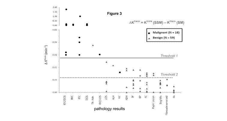
[0009] Figure 3 illustrates delta Ktrans results of a 77 lesion data set
(from 74 patients).
[0010] Figure 4 illustrates a heat map analysis of a region of interest
(ROI), as delineated at the left.
[0011] Figures 5a, 5b, 5c, and 5d illustrate a sagittal, fat-suppressed
breast DCE-MRI image (Figure 5a) containing a malignant invasive ductal
carcinoma (IDC) tumor (circled contrast-enhanced region-of-interest).
Pharmacokinetic Ktrans parametric maps of the tumor, generated by the
Standard Model (FXL-constrained) and two members of the Shutter-Speed
Model family (FXR-allowed) and (SXR-allowed), are shown in Figure 5b,
Figure 5c, and Figure 5d, respectively.
[0012] Figure 6 illustrates 2D scatter plots of (a) the Standard Model,
and (b) the Shutter-Speed Model (FXR-a) results. The ordinates measure
the Ktrans and the abscissae the kep parameters. The black circles mark the
positions for regions of interest (ROIs) of lesions that were found by
biopsy/pathology to have large malignant fractions, while the triangles are
2
CA 02742001 2011-04-28
WO 2010/051065 PCT/US2009/043201
those for lesions found to be solely benign. An outlier is plotted in insets c
and d. Dashed concentric quarter-circles are drawn with radii of 0.19 and
0.23 min-'. The points for two patients are marked as gray circles with black
cores. These represent lesions with only very small malignant fractions.
[0013] Figure 7 illustrates a 1 D scatter plot. The ordinate, AK trans J is
[K trans (SSM) - Ktrans (SM)]: SSM is FXR-a and SM is FXL-c. The values for
the lesion ROIs of all 22 subjects are shown. Those proven malignant are
given as filled black circles (these include the two Figure 6 gray circles
with
black cores), while those found solely benign are indicated with triangles.
The group mean AK trans values are indicated with open and filled black
squares on the right. Error bars represent (SD) values within each category.
One malignant lesion outlier is plotted in an inset, and is excluded from the
SD calculation. The horizontal cut-off line drawn at 0.024 min-' cleanly
separates the two lesion groups.
[0014] Figure 8 illustrates how the Ktrans (volume fraction CR transfer
rate constant product, top) and ve (extracellular, extravascular space, EES,
volume fraction, bottom) fitting results would change if increasing
interstitial
' H2O T2* quenching is assumed.
[0015] Figure 9a (inset) shows a transverse pelvic DCE image slice
(anterior up/inferior perspective, - 34 seconds post CR injection) of a
research subject. Two ROIs are indicated within the prostate gland: one in
an area of retrospectively-confirmed prostate cancer, left; and the other in
contralateral normal-appearing prostate tissue, right. Figure 9a plots the
arterial input function obtained from an ROI in a femoral artery. Its
magnitude
was adjusted using a custom-written numerical approach and an obturator
muscle ROI for reference tissue. The time-course from the first-pass was
used to estimate blood volume fraction. Color-matched tissue data time-
courses (points) and representative fittings (curves) are seen in Figure 9b.
[0016] Figure 10 illustrates an article of manufacture in accordance
with an embodiment herein.
3
CA 02742001 2011-04-28
WO 2010/051065 PCT/US2009/043201
Detailed Description of Disclosed Embodiments
[0017] In the following detailed description, reference is made to the
accompanying drawings which form a part hereof, and in which are shown
by way of illustration embodiments that may be practiced. It is to be
understood that other embodiments may be utilized and structural or logical
changes may be made without departing from the scope. Therefore, the
following detailed description is not to be taken in a limiting sense, and the
scope of embodiments is defined by the appended claims and their
equivalents.
[0018] Various operations may be described as multiple discrete
operations in turn, in a manner that may be helpful in understanding
embodiments; however, the order of description should not be construed to
imply that these operations are order dependent.
[0019] The description may use perspective-based descriptions such
as up/down, back/front, and top/bottom. Such descriptions are merely used
to facilitate the discussion and are not intended to restrict the application
of
disclosed embodiments.
[0020] For the purposes of the description, a phrase in the form "A/B"
or in the form "A and/or B" means (A), (B), or (A and B). For the purposes of
the description, a phrase in the form "at least one of A, B, and C' means (A),
(B), (C), (A and B), (A and C), (B and C), or (A, B and C). For the purposes
of the description, a phrase in the form "(A)B" means (B) or (AB) that is, A
is
an optional element.
[0021] The description may use the terms "embodiment" or
"embodiments," which may each refer to one or more of the same or
different embodiments. Furthermore, the terms "comprising," "including,"
"having," and the like, as used with respect to embodiments, are
synonymous.
[0022] In various embodiments, methods, apparatuses, and systems
using magnetic resonance imaging for cancer identification are provided. In
exemplary embodiments, a computing device may be endowed with one or
4
CA 02742001 2011-04-28
WO 2010/051065 PCT/US2009/043201
more components of the disclosed apparatuses and/or systems and may be
employed to perform one or more methods as disclosed herein.
[0023] Embodiments herein provide a Magnetic Resonance Imaging
(MRI) technique and optionally newly developed software - collectively
referred to as the "shutter-speed" model - to analyze image data of cancer
patients. Embodiments provide a minimally invasive, yet precisely accurate,
approach to determining whether tumors are malignant or benign. Exemplary
embodiments provide MRI measured biomarkers for tumor malignancy
determination, effectively solving the false positive riddle from which
current
MRI techniques suffer.
[0024] Although some embodiments throughout are described with
reference to breast cancer or prostate cancer, the methods and apparatuses
described herein may be utilized for other cancers, such as brain,
esophageal, leg osteosarcoma, etc. as well as for any Dynamic-Contrast-
Enhanced Magnetic Resonance Imaging (DCE-MRI) analysis where water
exchange effects are relevant, including tissue differential/disease state
analysis of the brain (Alzheimers, MS, etc.), muscles (such as heart), etc.,
and quantitative vascular phenotype mapping.
[0025] "Quantitative MRI" produces parametric maps of MR, patho-
physiological, and/or pharmacokinetic biomarker properties. The DCE-MRI
sub-category is particularly significant because it applies to a wide
pathology
range. In DCE-MRI, the Ti-weighted tissue 1H20 MRI signal intensity is
acquired before, during, and after the (usually) bolus injection of a
hydrophilic, paramagnetic contrast reagent (CR). The CR passage through a
tissue region-of-interest (ROI) can cause a transient increase of the
longitudinal 1H20 relaxation rate constant [Ri _ (Ti)-'] with consequent
elevated MR steady-state signal intensity. This elevation may be identified
on the MR image.
[0026] In DCE-MRI, the neglect of intercompartmental water
exchange kinetics considerations can lead to systematic errors in
parameters extracted by quantitative analyses. Examples here are the
compartmental water mole fractions defining tissue spaces. Therefore, DCE-
CA 02742001 2011-04-28
WO 2010/051065 PCT/US2009/043201
MRI is also a sub-category of in vivo MR "molecular imaging" - mapping the
distribution and/or activity of molecules in living tissues.
[0027] In essence, in embodiments, the CR plays the role of the
nuclear medicine radioactive tracer. However, in nuclear medicine, the
tracer is detected directly (by its radioactivity in disintegrations per
second
(dps) - the amount of tracer present in the tissue, but compartmental
localization is not intrinsic to the signal). In contrast, the MRI CR is
detected
indirectly, via its interaction with water and effect on the nature of tissue
1H20 relaxation (so the water interaction with the CR is what is directly
traced). Beneficially, the CR is not radioactive. Also, MRI involves no
ionizing radiation.
[0028] Affecting the recovery of longitudinal 1H20 magnetization (i.e.,
in the magnetic field direction) requires (transient) water CR molecular
interaction, as depicted in Figure 1. The three major loci for tissue water,
the
cytoplasmae, the interstitium, and the blood, are indicated with subscripts i,
o
(or e), and b (p, for plasma), respectively. There are water binding
equilibria
depicted in each compartment in which the CR is thought to enter. The
compartmental volume fractions are designated as v;, vei and vb,
respectively, though the relative areas in Figure 1 are not proportional.
[0029] The CR and water molecules are never equally distributed in
tissue. Therefore, the only way that most water (cytoplasmic) can access CR
is via exchange equilibria across cytolemmae and blood vessel walls. These
are indicated in Figure 1 with the unidirectional rate constants, k0, k;o, and
kpo, kop, respectively. In existing methods, tracer pharmacokinetic models
are applied directly to MRI data - such methods are referred to here as the
Standard Model (SM). However, this results in the constraint that all inter-
compartmental equilibrium water exchange processes be treated as if
infinitely fast (ko; + k;o -* , and kpo + kop -* ). This is not valid, and
the
assumption may effectively "short circuit" MRI determination of CR
compartmentalization - the pharmacokinetic essence. In accordance with
embodiments herein, the incorporation of equilibrium water exchange MR
effects into pharmacokinetic derivation is referred to herein as the Shutter-
6
CA 02742001 2011-04-28
WO 2010/051065 PCT/US2009/043201
Speed Model (SSM). This is accomplished by allowing ko; + k;o and kpo + kop
to be finite.
[0030] The SM assumes that water exchange between cells and/or
blood and the interstitial spaces is effectively infinitely fast (in the fast
exchange limit - FXL). However, when CR is passing through the tumor
tissue, the water exchange systems can depart from this fast exchange limit
due to the interaction with the CR (and therefore enter into a fast-exchange
regime - FXR). This happens for both benign and malignant tumors;
however, the exchange difference between FXL and FXR, as far as Ktrans is
concerned, is significantly greater for malignant tumors as opposed to
benign tumors. For benign tumors, the exchange difference is typically
below 0.025 min-', whereas for malignant tumors, the exchange difference is
typically above 0.025 min-'. This differentiating line provides a threshold
against which the obtained values may be compared to classify a tumor or
tissue sample. In embodiments, a threshold may be established at an
exchange difference (delta Ktrans) of 0.02 to 0.03 min-'.
[0031] While a single threshold is mentioned above, in embodiments,
more than one delta Ktrans threshold may be established. For example, a first
threshold may be established that is intended to include all or a substantial
percentage of the malignant tissues above the threshold. A secondary
threshold may be established at a lower delta Ktrans value with an intention
of
included all true positive indicators. However, a lower threshold may
introduce a larger number of false positives. For a tissue having a delta
Ktrans value residing between the first and second threshold, additional
analysis may be utilized to further classify the tissue.
[0032] In an embodiment, the shutter-speed model (SSM) accounts
for the FXR (therefore including equilibrium exchange effects when the CR
passes through) and thus is better able to pick up the "leaky blood vessel"
effect which is common in malignant tumors. At a maximum level of CR in
the interstitial space, an interstitial water molecule in a benign lesion may
typically encounter a CR molecule an average of 60 times before it enters a
cell, whereas in a malignant tumor this may happen 260 times on average
7
CA 02742001 2011-04-28
WO 2010/051065 PCT/US2009/043201
(4+ times as often). If that difference is neglected (which the standard model
does), then it is sufficient to cause significant Ktrans (the volume-weighted
CR
extravasation rate constant) underestimations in malignant tumors. Because
Ktrans values are greater for malignant tissues than for benign tissues, if
Ktrans
is underestimated, then it may make a malignant tumor seem benign (false
negative) or vice versa a benign tissue appear to be malignant (false
positive). The SSM model accounts for this difference, and by using the
delta Ktrans (change in Kt) as well as the Ktrans to kep comparison,
classification of tumors may be accomplished. In accordance with an
embodiment, kep is the unidirectional CR intravasation rate constant - it is
Ktrans divided by the ve (the extracellular, extravascular volume fraction
available to the contrast agent molecule). The pharmacokinetic analysis of
DCE-MRI data yields Ktrans and kep.
[0033] In accordance with an embodiment, the difference in Ktrans
returned by SSM, as compared to the Standard Model analyses, offers a
very high degree of tumor differentiation (i.e., specificity). It is a measure
of
the shutter-speed effect, which is disproportionally present and important in
malignant tumors, that permits differentiation of benign and malignant
tumors. The increased permeability of malignant tumor blood vessels
exceeds a threshold above which exchange kinetics become influential. This
amplification is measured by the delta Ktrans biomarker, and accounts for the
high SSM specificity.
[0034] In analyses of DCE-MRI data from patients with suspicious
breast lesions initially ruled positive by institutional screening protocols,
the
SM Ktrans values for benign and malignant lesions exhibit considerable
overlap. The Shutter-Speed Model (SSM) may allow for finite exchange
kinetics thus agreeing with the SM Ktrans value for each of the benign
lesions.
However, it reveals that the SM underestimates Ktrans for each of the
malignant tumors in this population. Figure 2 illustrates Ktrans values
obtained
by both SM and SSM, and shows how SSM recognizes a difference in Ktrans
between benign and malignant tissue. The fact that this phenomenon is
unique to malignant tumors allows their discrimination from the benign
8
CA 02742001 2011-04-28
WO 2010/051065 PCT/US2009/043201
lesions, as validated by comparison with gold standard pathology analyses
of subsequent biopsy tissue samples to which the MRI analyses were
blinded. Likewise, the SM overestimates kep, particularly for the benign
tumors. Thus, incorporation of the SSM into the screening protocols may
preclude the need for the biopsy/pathology procedures that otherwise would
yield benign findings.
[0035] Thus, in embodiments, two binary classifiers have been
developed:
1. "delta Ktrans" - the change in Ktrans; thresholds may be
established with the goal/intention of including all true positives.
Thresholds
may be established as desired to distinguish/classify the tissues/tumors. In
an embodiment, further analysis may be conducted via a secondary
mapping algorithm (plot of (K trans vs. kep) to allow for a second
determination
with respect to those points that are somewhat unclear or fall below a
determined threshold.
2. The use of 2D plots (K trans vs. kep), where the radius of a
circle centered at the origin of the plot may be used as a "binary
classifier."
In embodiments, the radius of the circle may be used as a threshold to
distinguish benign from malignant tumors. Such a threshold may be
established at approximately 0.2 min-', for example, from 0.19 min-' to
0.25min-1.
[0036] In embodiments, an MRI examination aided by SSM analysis
may provide a clearer diagnosis and may be an intermediate step between a
mammographic scan and a biopsy intervention if breast cancer is suspected
from both the mammogram and the MRI results. Adding this intermediate
diagnostic step may greatly reduce or eliminate the number of unnecessary
(and possibly all) biopsy surgeries and also reduce the pain, stress and
expense for most patients.
[0037] It is important to note that the SSM is a generalization of the
SM. That is, the SM is but a special case of the SSM. Thus, if the shutter-
speed effect is negligible in any tissue, the program will automatically
perform a SM analysis. One can test this by computationally constraining
9
CA 02742001 2011-04-28
WO 2010/051065 PCT/US2009/043201
the SSM analysis to a SM form. If there is no difference from the result
obtained when the SSM analysis is given free rein, then there is no
significant shutter-speed effect for that tissue. In the case of breast
tumors,
this is the case for the Ktrans biomarker in only benign lesions. However,
there is a shutter-speed effect for the ve biomarker in benign breast lesions,
and it is about the same size as in malignant tumors.
[0038] To test SSM, SSM was employed to analyze MR images of 22
women volunteers who had previously screened positive for breast cancer
by mammography and/or clinical examination. The shutter-speed software
operates by using a complex mathematical formula to track the passage of
injected contrast dye through a tumor area. Contrast dyes are commonly
used in medical imaging to increase the visibility of tissue abnormalities.
[0039] When viewed through the shutter-speed analysis, the MRI data
suggested that only seven of the 22 women actually had malignant tumors.
These projections were later shown to be 100 percent accurate after each of
the study participants underwent subsequent biopsies for pathology
determinations. Typically, 75 percent of mammographically-indicated
biopsies yield negative pathology results, meaning that an intermediate step
such as an MRI determination could greatly reduce or eliminate the number
of unnecessary biopsy surgeries.
[0040] This population study has been expanded to include 77 breast
tumors (in 74 patients) and, with the mapping provision for one rare type of
malignant tumor, maintains 100% specificity. Figure 3 illustrates delta Ktrans
results of the 74 patient data set illustrating 77 lesions.
[0041] In addition, Figure 3 illustrates the multiple threshold concept
described previously. The first threshold is intended to capture all or a
substantial percentage of the malignant tissues above the threshold. The
first delta Ktrans threshold may be set, for example, between 0.02 min-' and
0.03 min-'. The secondary threshold is established at a lower delta
Ktrans value with an intention of included all true positive indicators. The
second delta Ktrans threshold may be set, for example, between 0.01 min-'
and 0.02 min-'. For a tissue having a delta Ktrans value residing between the
CA 02742001 2011-04-28
WO 2010/051065 PCT/US2009/043201
first and second threshold, additional analysis may be utilized to further
classify the tissue. Such analysis may include heat map analysis of regions
of interest to better classify the tissue.
[0042] Figure 4 illustrates a heat map analysis of a ROI, as delineated
at the left. The three maps to the right show the results of various analyses
of the ROI. At the top, the SM (FXL-c) image is shown which does not
provide an indicator of malignancy. The middle image represents the SSM
(FXR-a) image, which indicates some areas of interest. However, the lower
image, representing the delta Ktrans value, clearly outlines the particular
areas of concern within the ROI. Even though this lesion is fairly early
stage,
the delta Ktrans analysis provides an indicator of the tumor malignancy. While
in some situations the identification of the tumor malignancy may not result
in treatment, the early identification enables the tumor growth to be tracked
over a period of time.
[0043] For the more limited data set (22 patients), data were obtained
with consent from patients with positive mammographic and/or clinical MRI
reports from standard, institutional breast cancer workups and protocols. All
had MRI contrast-enhanced lesions radiologically classified as BIRADS
(Breast Imaging Reporting and Data System) four (B-4, suspicious) or five
(B-5, highly suggestive of malignancy). Emphasizing practicability and
robustness, the data are of a rather routine clinical nature (and they were
obtained at two different institutions, with two different instruments, CRs,
etc.): the two different data acquisitions were not optimized for DCE-MRI.
For example, though the spatial resolution is reasonable, the temporal
resolution is not optimal. Of particular interest is the fact that the adipose
tissue -'H2C- MR signal was suppressed in the acquisitions at one
institution, while at the other institution it was not.
[0044] Figure 5a shows the DCE pharmacokinetic image of sagittal
slice 16 (numbering from lateral to medial) of the left breast of a 52 year-
old
patient, obtained 2.6 minutes after CR injection. It was acquired with adipose
-1H2C- suppression (required in the institutional protocol). In contrast to
those with no fat suppression, this darker image shows glandular regions
11
CA 02742001 2011-04-28
WO 2010/051065 PCT/US2009/043201
brighter than fatty tissue. The ROI circumscribes the enhanced lesion
evident in this slice, subsequently found to be a malignant invasive ductal
carcinoma (IDC) by pathology analysis. Each of the 22 patients participated
in a DCE-MRI acquisition subsequent to her clinical mammography and/or
MRI screening but prior to the biopsy procedure and the pathology analysis.
[0045] Additional DCE-MRI acquisition details may be found in Li, et
al., Dynamic NMR Effects in Breast Cancer Dynamic-Contrast-Enhanced
MRI, PNAS, Vol. 105, No. 46, 17937-17942 (2008) (and Supporting Online
Material), the entire disclosures of which are incorporated by reference in
their entirety. For each of the 22 subjects, ROI DCE-MRI time-course data
were analyzed from one sagittal image slice (out of 16 to 40 per breast) that
exhibited a lesion to be subsequently biopsied. An ROI boundary was
manually drawn around the entire lesion in a pharmacokinetic image
showing near maximal enhancement (as in Figure 5a). The patients are
enumerated in Table 1, below. The Figure 5 images are from patient 3. The
DCE-MRI time-courses were each analyzed with several pharmacokinetic
models.
12
CA 02742001 2011-04-28
WO 2010/051065 PCT/US2009/043201
Table 1
10"M ( nin ) k inin
P#ttient BI- SAI SSM ill SS i P thahigy Report
'amber RAIDS (FXL-c) (FXR-a) (FU-c) (F R-a)
I B-4 0. ?? 0.14' ;0._89 i 49 DCIS, yntemi .di e fcIeai acade
B-4 ? 11(3 0.110 0 152 0-1. 4 ID lust;6c *i< 7.xa6e 14I1I: D(;IS.
aoef'n]Fe thane n3 F:lei f chit e.
C'1 25 'a Cat to aI tumoi'sfia
3 B- 4 019e' 0.1 i2. 6 4? 181(1 raven;; at the e< e of the on'-
B-f?It'? i? 2 t _32 ?,alo~i rieI1 1I: DCIS
(tU J' c? ( 0l:29} fintZ#f3#edi f _#.: ea 1~t f
A Bo.559 1.63 I ' 5 1.96 _ ?DC Ifs okgi 23'ade T111
B-~ 0.145 01 5 ;.> F 0._l1 1D
0.0:20
B-4 0.051 n 0 91 t 269 i 202 IDC iinr`ear gftrle L LCIS.
n. c l? ,:te1; $ Ie efn mite d 181(.
231113?El<i2v, ta':fi33iL' a benign,
LCTS
C B-? ji_I U,`i_ t _f?s 1' C 53 L CIS, SF
t' 00 0.0066)
9 5-4 '_t E' 0.047 F{
Ifu! B-4 00151 Dos ii305 t;:1 5 FC'
11 B-4 0 040 0.055 02 (30 20 Ft
I= B-4 0. f# Y: t; a. 7
79 (111. 19 `:Irfr)re Papillary 1" Co] . ut *
13 B-4 0.049 f? .039 FC
14 B-4 0.030 t 229 t "'96 ADH. SE
I 5 B-4 0.091 0 0t'=f t 3 9 i 31 L. S. SF. FC
16 B-4 0.11'9 0 1c;' t i 130 LCIS ADH
>.10 0.125 C '99 0 166 chi cc affil. ADH
17 B-4
140 B-4 0.050 0.066 t L.. ?i'1} SF ~r ez si3 fclaf_!: a
=
1_;, B-mil <.' 41: V. V}. J_.) it f, i~i~: F~.
_ '. B-4 tt.026 (3 f~'.1 1' _!4 0.066 FA
A
f -'136 FA
121 i 0 17 FA
IDC: invasive ductal carcinoma; DCIS: ductal carcinoma in situ; LCIS: lobular
carcinoma
in situ; SF: stromal fibrosis; FC: fibrocystic changes; ADH: atypical ductal
hyperplasia;
FA: fibroadenoma.
[0046] For the patients/results presented in Table 1, ROI boundaries
around each lesion were separately drawn by each of two independent
investigators who were blinded to the pathology results. The analyses of
these ROI data were also conducted independently by two investigators.
13
CA 02742001 2011-04-28
WO 2010/051065 PCT/US2009/043201
The algebraic means of the model parameters returned from each
investigator's fitting were computed lesion-by-lesion.
[0047] Each of the fittings neglects the small blood water proton signal
(1H2Ob) - thus, these are "first generation" versions. For this situation, the
MR exchange system of interest is that for equilibrium transcytolemmal
water interchange (kO1 and k10i Figure 1). The system's condition is given by
the comparison of the equilibrium kinetics, k = kO1 + k10i with the pertinent
MR
shutter-speed, r' _ I R10 - Rl; I , where Rio and Rl; are the relaxation rate
constants for the ' H200 and ' H2O; signals in the absence of exchange.
Before CR arrival, Rio = Rl; and r' << k. Though k is finite, and invariant
throughout the DCE-MRI study, the system is in the fast-exchange-limit
(FXL): the kinetics appear infinitely fast, and the measured tissue 1H20 R, is
single-valued. As stated above, the Standard Model assumes that the
system remains in the FXL throughout the CR bolus passage, so it is
referred to also as the FXL-constrained (FXL-c) model (see Figure 5b).
However, as the CR0 concentration increases, Rio becomes increasingly
larger than Rl; and r' at least approaches the constant k value. For some
period, the measured R, remains effectively single-valued, and this has been
defined to be the fast-exchange-regime (FXR). Admitting departure from the
FXL for the FXR may be referred to as FXR-allowed (FXR-a) (see Figure
5c). Further CR0 increase may lead to the condition where R, is effectively
double-valued: this is referred to as the slow-exchange-regime (SXR).
Admitting this is referred to as SXR-allowed (SXR-a) (see Figure 5d). For
the cases here, the results of FXL-c and FXR-a analyses are presented in
Table 1. Careful analyses with the SXR-a model suggest that it is
incompatible with these data - an example will be seen below. There are a
number of potentially variable parameters. For the SM (FXL-c) analyses, the
variables were Ktrans and vei while for the SSM (FXR-a) analyses, T; was also
varied. In terms of the Figure 1 notation, Ktrans = vekep = vbkpei and T; =
k1O-'.
The values returned for Ktrans , a measure of the rate of passive CR transfer
across the vessel wall, and kep, the unidirectional rate constant for CR
intravasation (Figure 1) are given in Table 1. Sample standard deviation
14
CA 02742001 2011-04-28
WO 2010/051065 PCT/US2009/043201
measures of parameter uncertainty from individual fittings are given for some
entries. These were determined by multiple Monte Carlo fitting calculations.
The Ktrans and kep values for the malignant tumors (top seven entries) are
larger than those for the benign lesions.
[0048] Table 1 indicates that the SM does not completely separate the
malignant tumors (top seven entries) from the benign lesions with either the
Ktrans or kep parameters. However, the SSM significantly increases Ktrans for
every one of the malignant lesions, and for none of the benign tumors, as
compared to the SM. Furthermore, though the SSM reduces kep for both
malignant and benign lesions, it does this more for the benign tumors. In
embodiments, these changes allow discrimination between the SSM and SM
results.
[0049] Though neither of the parameters allows the construction of
perfect ROC (Receiver Operator Characteristic) plots, the SSM Ktrans and kep
quantities come very close. These aspects may be seen in the 2D
parametric scatter plots of the Ktrans (ordinate) and kep (abscissa) values
presented in Figure 6. The ROI values for lesions found by pathology
analyses (Table 1) to be solely benign are indicated with triangles, while
those with major malignant regions are shown as black circles. The two gray
circles with black cores also represent malignant tumors and are discussed
below. The results from the SM (FXL-c) analyses are seen in panel a, while
those from the SSM (FXR-a) determinations are shown in panel b. The
values for patient 5 are so large that they are shown in inset panel c and
inset panel d.
[0050] In comparing Figure 6b with 6a, one can note especially the
upward movement (increasing Ktrans) of the circles and the leftward
movement (decreasing kep) of the triangles, in going from the SM to the
SSM. This allows the almost complete separation of these points in Figure
6b, which is not achieved in any single dimension of either panel. It is
important to note that two of the triangles represent B-5 lesions (Table 1):
i.e., they were "highly suggestive" false positives. Retaining 100%
sensitivity
(not missing any malignant tumor), the PPV values for the SM Ktrans SM kep,
CA 02742001 2011-04-28
WO 2010/051065 PCT/US2009/043201
SSM Ktrans , and SSM kep dimensions are: 54%, 39%, 70%, and 70%,
respectively. In the Figure 6b SSM 2D plot, one can draw a dashed quarter-
circle of radius 0.19 min-', that also allows a 78% PPV.
[0051] Furthermore, consider the annular region between this and the
other concentric quarter-circle, of radius 0.23 min-'. The only two malignant
tumors (circles with dark cores within) are those of patients 3 (upper) and 7
(lower). These are cases where the malignant areas are quite small
compared with the total tumor area visualized in the biopsy specimen (Table
1). This means that the analyses of whole-tumor ROI-averaged data cause a
partial volume dilution of the DCE-MRI parametric values. This can be seen
clearly in panels b and c of Figure 5, which present Ktrans parametric heat
maps of the lesion of patient 3. In the SM (FXL-c) and SSM (FXR-a) maps
(panels b and c, respectively), a clear "hot spot" is seen on the posterior
lesion edge. The hot spot has Ktrans values above 0.16 min-' in the FXR-a
map, considerably elevated above the ROI-averaged magnitude (Table 1).
[0052] The hot regions of all seven malignant tumors in this population
have SSM Ktrans values exceeding 0.1 min-'. Except for that of patient 17
(upper triangle in Figure 6b annulus, and which uniquely exhibits ductal
dilation (Table 1)), this exceeds the ROI-averaged SSM Ktrans values of any
of the fifteen benign lesions. With Figures 5b, 5c, and 5d, parametric maps
(heat maps) of four of the seven malignant tumors have been presented in
several publications referenced below. Some hot spots can be as small as 2
mm in diameter. In another indication of potential staging power, a plot (not
shown) of "hotness" vs. area of the SSM Ktrans hot spots in the malignant
tumors of patients 5, 6, and 7 demonstrates that these two independently
measured quantities are very highly positively correlated. The fact that the
SXR-a Ktrans map of the patient 3 lesion (Figure 5d) does not show increased
values relative to the FXL-c map (Figure 5b) - and in fact obliterates the hot
spot - is an example of the SXR-a model incompatibility with these data.
[0053] The Ktrans and kep values are rather well correlated in Figure 6,
particularly in panel b. The positions of the panel a and b insets are placed
with constant coordinate aspect ratios. Thus, one can visually include the
16
CA 02742001 2011-04-28
WO 2010/051065 PCT/US2009/043201
inset points in the correlations. The slope of a line drawn through the points
represents the mean ve value of these lesions. Such a line for Figure 6b has
a slope near 0.5.
[0054] These results suggest a potential breast cancer screening
protocol in accordance with an embodiment herein. The first step of such a
protocol would be a clinical examination and/or mammography. A positive
result (B-4 or B-5), or suspicion of a mammographically occult lesion, would
occasion referral for diagnostic MRI that includes DCE. The radiologist can
circumscribe an ROI from the DCE image showing the greatest
enhancement. Alternatively, this can be automated (ex., Jim 4.0 software;
Xinapse Systems; Thorpe Waterville, UK). The computer can very quickly
(few seconds) conduct SM and SSM analyses on the mean ROI signal time-
course data and produce SSM Ktrans and kep values, which can be compared
with 2D scatter plots such as those in Figures 6b. If a patient's point turns
out to be in the annulus between the quarter-circles in Figure 6b, the
radiologist could proceed to read Ktrans parametric lesion maps made from
the same DCR-MRI data, though these require more computational time. Hot
spots above 0.1 min-' would be very suspicious for malignancy.
[0055] Some oncologists advocate a separate regimen for a malignant
ductal carcinoma in situ (DCIS) tumor, possibly simply following it instead of
immediate surgery, while others urge excision. The only solely DCIS case in
the discussed patient population is that of patient 1. Her position in Figure
6b
is the black point closest to the outer quarter-circle. In fact another
concentric quarter-circle of radius 0.3 min-' would isolate this point. Its
position could be "followed" or tracked over a period of time to see if it
moves
up and to the right. Inside the inner quarter-circle, most of the benign LCIS
lesions are found in the upper right sector, while all of the FA lesions are
found near the bottom.
[0056] In the analyses so far, pseudo-absolute parameter values have
been employed. The SSM success suggests that neglect of equilibrium
transcytolemmal water exchange effects may constitute the most significant
systematic error in Standard Model DCE-MRI pharmacokinetic analyses.
17
CA 02742001 2011-04-28
WO 2010/051065 PCT/US2009/043201
[0057] For screening purposes, the most striking aspect of the Table 1
and Figure 6 results is that every one of the malignant tumor ROI Ktrans
values (dark circles) is clearly decreased by the SM analysis, while every
one of the benign lesion ROI values (triangles) is not. This is seen even
more clearly in Figure 7, which presents the 1 D scatter plot for AKtrans [_
Ktrans(SSM) - Ktrans(SM)]. There is a wide gap between all seven of the dark
circles [group mean, 0.06 min-1 (excluding the inset point)], and all 15 of
the
triangles. The latter set clusters very near zero [group mean, 0.006 min-1]. A
clean cut-off line is drawn at 0.024 min-'. Since the only difference between
these two models is the allowance for the effect on the NMR signal of finite
equilibrium transcytolemmal water exchange kinetics, the NMR shutter-
speed effect, this suggests that it is significant (for the Ktrans magnitude)
with
the capillary wall permeability obtained for the vascular beds of only
malignant breast tumors. Thus, this is very encouraging that analyses of
DCE-MRI ROI data first with one pharmacokinetic model and then with the
other (which is still accomplished in only seconds) can lead to extremely high
specificity in cancer screening. Here, the positive criterion of AKtrans >
0.025
min-' yields 100% PPV.
[0058] Apparently, in the vascular beds of malignant breast tumors
only, the interstitial ("outside") CR concentration, (CRo), transiently rises
to
sufficient values during the bolus passage and the equilibrium
transcytolemmal water exchange system transiently departs the FXL to
sufficient extent and/or for sufficient duration to substantially invalidate
the
SM Ktrans determination. The SSM interpretation is that, during the bolus
passage through malignant lesions, the relaxographic r' value for the
transcytolemmal water exchange process, I Rio - Ri; I , transiently
approaches or exceeds that for the unchanging exchange rate constant, k;o +
k0, (in vivo studies are isothermal) sufficiently for the system to enter at
least
the fast-exchange regime (FXR), but probably not also the slow-exchange-
regime (SXR). Rio increases with CRo, while Rl; remains constant. This is a
manifestation of the varying equilibrium competition for interstitial water
molecules between diamagnetic cytoplasmic spaces and paramagnetic
18
CA 02742001 2011-04-28
WO 2010/051065 PCT/US2009/043201
interstitial CR molecules (Figure 1). Informative estimates can be made by
comparison of the Table 1 patients 8/4 benign/malignant lesion pair, with
SSM Ktrans 0.034 and 0.254 min-', respectively. For one of the SSM (FXR-a)
fittings of each, the (vei'ri) parameters returned are similar: (0.60, 0.40
s), and
(0.69, 0.39 s) for benign and malignant, respectively. Thus, the
unidirectional
rate constants for water cellular entry [kO1 = (ve' - 1)i;-1] are similar (1.7
and
1.2 s-', respectively), constant, and not infinitely large. However, before
the
arrival of interstitial CROi the transcytolemmal water exchange appears
infinitely fast in the NMR signal because r' is almost negligible. The
interstitial water molecules encounter no paramagnetic CRo molecules
before entering a diamagnetic cytoplasm. However, as CRo increases, the
rate constant for interstitial water CR encounter, [(CRo)/(H200)]-[M1, also
increases ['LM-' = km in Figure 1]. While, for the benign lesion CR0 maximizes
at 0.52 mM (at -7.5 minutes), this is 1.6 mM (at -3.5 minutes) for the
malignant tumor. Thus, [(CR0)max/(H200)]TM-' values are 104 and 313 s-' for
the benign and malignant lesions, respectively. The interstitial water
concentration (H200) was 50 M and the mean water lifetime on the CR, 'LM,
was 10-7 s. At maximum CROi an interstitial water molecule in the benign
lesion encounters a paramagnetic CR molecule on average 60 times
(104/1.7) before it enters a diamagnetic cell; sufficient, apparently, for the
SM 40% ve underestimation. While in the malignant tumor, this happens 260
times (313/1.2) on average; more than four times as often. This is sufficient
to cause significant Ktrans underestimations if it is neglected.
[0059] For the expanded data set, a total of 74 patients underwent
clinical breast MRI protocols and had 77 contrast-enhanced lesions (3
patients presented 2 lesions each) radiologically classified in the BIRADS
(Breast Imaging Reporting and Data System) 4 (B-4, suspicious, n = 67) or 5
(B-5, highly suggestive of malignancy, n = 10) categories based on lesion
morphology and qualitative enhancement kinetics assessment. These
clinical interpretations led to biopsy referrals. The research DCE-MRI data
acquisitions were IRB-approved. The data from 6 patients were collected as
part of a combined MRI/MRS protocol prior to excisional or core biopsy.
19
CA 02742001 2011-04-28
WO 2010/051065 PCT/US2009/043201
Those from the other 68 patients (71 lesions) were acquired during clinically
scheduled MRI-guided preoperative needle localization or core biopsy
procedures, just before needle insertions.
[0060] The study was conducted at 1.5T using a body transmit and a
four- or seven-channel phased-array bilateral breast receive RF coils. A 3D
SPGR pulse sequence was used to acquire 12-20 serial sagittal image
volume sets continually, spatially covering the whole breast with the
suspicious lesion to be biopsied. Other parameters included 100 or 30 (for
the 6 patients) flip angle, 2-5 ms TE, 6-9 ms TR, 3 mm section thickness, 20-
24 cm FOV. Depending on the breast size, 16-36 image sections were
acquired for each set, resulting in inter-sampling intervals of 13-42 seconds.
At the start of the second volume set acquisition, Gd CR was delivered
intravenously [0.1 mmol/kg at 2 mL/s]. ROIs circumscribing the enhanced
lesion and within an axillary artery produced the tumor signal intensity and
arterial input function (AIF) time-courses, respectively. Three reliable
individual AIFs were measured, which were interpolated with an empirical
expression (3) and averaged to generate a mean AIF. The tumor ROI and
mean AIF signal time-courses were then subjected to both SM and (fast-
exchange-regime-allowed) SSM analyses, which were blinded from the
pathology. Receiver-operating-characteristic (ROC) curves were used to
evaluate pharmacokinetic parameter diagnostic accuracies, and the areas
under the curve (AUCs) were compared using a Bootstrap nonparametric
test.
[0061] Upon pathology, only 18 lesions (10 B-4 and 8 B-5) were found
malignant and the other 59 (57 B-4 and 2 B-5) benign. Though the clinical
MRI protocol sensitivity is 100% (no false negatives), its PPV is only 23%.
The SSM Ktrans ROC AUC (0.973) is significantly (p = 0.032) greater than
that for the SM Ktrans (0.929). Similar results were obtained for other strong
biomarkers: kep (=Ktrans/ve, the unidirectional CR intravasation rate
constant)
[SSM AUC = 0.960, SM AUC = 0.861, p = 0.006] and [(Ktrans)2 + kep2]1i2 [SSM
AUC = 0.970, SM AUC = 0.887, p = 0.009]. Maintaining 100% sensitivity, the
diagnostic specificities of the SM ROI Ktrans , kep, and [(Ktrans)2 + kep2]1i2
are
CA 02742001 2011-04-28
WO 2010/051065 PCT/US2009/043201
47%, 42%, and 51 %, while those for the corresponding SSM parameters are
76%, 61 %, and 75%, respectively; each biomarker used as a binary
classifier. The SM and SSM ve ROC curve AUCs are 0.555 and 0.615,
respectively, suggesting that ve is a poor diagnostic marker.
[0062] Figure 3 (discussed partially above) plots ROI delta Ktrans for all
lesions. Note the ordinate scale break. Each column represents one
pathology category (from left to right): 1) invasive ductal carcinoma
(IDC)/ductal carcinoma in situ (DCIS) mixture, 2) IDC/invasive lobular
carcinoma (ILC) mixture, 3) IDC, 4) DCIS, 6) IDC/lobular carcinoma in situ
(LCIS) mixture, and 9) ILC, for the malignant group (circles); 5) tubular
adenoma, 7) LCIS, 8) atypical lobular hyperplasia, 10) atypical ductal
hyperplasia, 11) stromal fibrosis, 12) benign parenchyma, 13) fibrocystic
changes, 14) papillary lesions, 15) miscellaneous benign conditions, 16)
fibroadenomatoid changes, and 17) fibroadenoma, for the benign group
(triangles). The categories are ranked roughly in order of decreasing mean
delta Ktrans from left to right. Consistent with the previous smaller
population
study, the delta Ktrans biomarker represents the strongest binary classifier
for
benign and malignant group separation, with its ROC AUC = 0.990, and 88%
specificity for 100% sensitivity.
[0063] The SSM DCE-MRI ROI pharmacokinetic parameters
consistently perform better than those from SM DCE-MRI and the commonly
used clinical MRI protocols for benign and malignant discrimination within
this group of 77 suspicious breast lesions. If the simple ROI delta Ktrans
analyses had been integrated into clinical practice, as many as 52 benign
lesions (68% of the total population) could have been spared the biopsy
procedures. As expected from the earlier study, the malignant lesions cluster
almost exclusively on the left of Figure 3, while the benign lesions are
almost
all to the right - the axes are independent. The solid cut-off line value,
delta
Ktrans = 0.028 min-', is very close to that for 100% specificity in the
smaller
population. It yields only one false positive (the sole tubular adenoma) and
one false negative (the sole ILC) lesion. A more lenient, dashed cut-off line
can be drawn at delta Ktrans = 0.012 min-' to avoid any false negative and
still
21
CA 02742001 2011-04-28
WO 2010/051065 PCT/US2009/043201
incur only 14 benign biopsies. But, even these might be avoided. The likely
reason for a malignant lesion ROI delta Ktrans to fall between the solid and
dashed cut-off lines is because of partial volume averaging in the ROI
analyses. Consistent with this, the ILC had the very large value of 5 cm as
the greatest enhanced ROI dimension. Its pixel-by-pixel SSM Ktrans map (not
shown) features hot spots (K trans > 0.18 min-) only in the posterior rim
region. Though these are diluted by a very large area of small Ktrans values
in
the ROI, they confirm the lesion as malignant. This suggests that delta Ktrans
or SSM Ktrans maps (parametric heat maps) should be made when an ROI
delta Ktrans falls between the solid and dashed lines.
[0064] Other analysis methods may be used to further distinguish the
data, such as points falling between delta Ktrans thresholds. For example, the
median Ktrans difference, delta (median Ktrans) [SSM median Ktrans - SM
median Ktrans], may be plotted (ordinate) vs. the change in maximum
histographic probability (amplitude) (abscissa), delta Amp [SSM amplitude -
SM amplitude]. Such an operation indicates a significant negative linear
correlation (Pearson correlation = -0.82, p = 0.0018) for the benign lesions,
while the malignant lesions exhibit an almost orthogonal correlation. The
ILC (identified for example in Figure 3) cannot be distinguished from the
benign group with simple ROI delta Ktrans analyses. However, using
quadratic discrimination analysis, the benign and malignant lesions can be
completely separated (100% sensitivity and 100% specificity) by the solid
partition curve with no misclassification.
[0065] Though the ROI delta Ktrans biomarker achieves high specificity
for benign/malignant breast lesion discrimination, the partial volume
averaging effects of ROI analyses can cause overlap in ROI
pharmacokinetic parameter values, and thus prevent clearer separation of
the two groups. Pharmacokinetic parametric mapping and histogram
analyses thus may further improve discrimination. Such analyses are
especially important when the lesion ROI biomarker value falls in the vicinity
of a binary classifier cut-off value. Thus, it is beneficial to acquire DCE-
MRI
data with sufficient SNR, since this ensures reliable pixel signal time-course
22
CA 02742001 2011-04-28
WO 2010/051065 PCT/US2009/043201
curve fitting. The negative linear correlation of the benign lesions and the
orthogonal behavior of the malignant lesions are quite interesting. Compared
to malignant lesions that can have noticeable median Ktrans increases
(shutter-speed (SS) histographic shifts) without significant histographic
maximum probability changes (SS broadening), the areas in benign lesions
where increased blood vessel CR permeability incurs SS effects, if any, are
smaller. Considerable SS histographic broadening is associated with even
minuscule SS histographic shifting.
[0066] Further details regarding the materials and methods used with
respect to various embodiments described herein as well as details
regarding some of the MRI data acquisitions and analyses may be found in
Li, et al., Dynamic NMR Effects in Breast Cancer Dynamic-Contrast-
Enhanced MRI, PNAS, Vol. 105, No. 46, 17937-17942 (2008) (and
Supporting Online Material); Huang, et al., The MR Shutter-Speed
Discriminates Vascular Properties of Malignant and Benign Breast Tumors In
Vivo, PNAS, Vol. 105, No. 46, 17943-17948 (2008); Li, et al., Shutter-Speed
Analysis of Contrast Reagent Bolus-Tracking Data: Preliminary Observations
in Benign and Malignant Breast Disease, Magn. Reson. Med., 53:724-729
(2005); and Yankeelov, et al., Evidence for Shutter-Speed Variation in CR
Bolus-Tracking Studies of Human Pathology, NMR Biomed., 18:173-185
(2005), the entire disclosures of which are hereby incorporated by reference.
[0067] In accordance with embodiments herein, certain steps may be
taken, even in the clinical setting, to improve the precision, the accuracy,
and/or the diagnostic richness of the SSM DCE-MRI pharmacokinetic
parameters. Such modifications may, for example, decrease the random
error scatter in the Figures 6 and 7 point clusters. This may allow further
discrimination of pathology sub-types.
[0068] The DCE-MRI time-course acquisitions discussed herein were
prescribed for radiological considerations and were truncated. Increasing this
period would likely improve accuracy and precision of the benign lesion
parameters. For these ROIs, the maximum R, value is rarely reached in the
no more than seven minutes usually allowed. This is the likely source of
23
CA 02742001 2011-04-28
WO 2010/051065 PCT/US2009/043201
abnormally large ve values for some benign tumors. Increasing the period to
15 minutes may help define the shape of the time-course, even for malignant
tumors.
[0069] The DCE-MRI acquisitions for the data described herein were
not particularly exchange sensitive. Even so, exchange effects seem to
facilitate very high discrimination of malignant from benign breast tumors.
[0070] The tissue R10 values (the pre-CR 1H20 longitudinal relaxation
rate constants) may be mapped, and not simply assumed as they were
herein. Individual AIFs may be used as well. A reference tissue method, or
an automated AIF determination (ex., Jim 4.0 software; Xinapse Systems;
Thorpe Waterville, UK) may be used.
[0071] Increased temporal resolution may be achieved without
sacrificing spatial resolution or signal-to-noise. Parallel RF
excitation/acquisition may be useful for achieving such increased temporal
resolution. With good definition of the DCE time-course first-pass leading
edge, the second generation SSM (BALDERO (Blood Agent Level
Dependent and Extravasation Relaxation Overview)) analysis, which
accounts for blood 11-120 signal pharmacokinetic behavior, may be used to
also determine vb and kbo values. It is anticipated that tumor vb values will
have significant diagnostic value. Furthermore, vbkbo is the transendothelial
water permeability coefficient surface area product, PwS', where S' is the
total capillary bed surface area. The ratio PWS'/PCRS' would be the intensive
property PW/PCR. The value of the CR permeability coefficient surface area
product (PCRS') may be factored from the Ktrans parameter using the blood
flow value, which may also be determined from DCE-MRI data.
[0072] The DCE-MRI pharmacokinetic images may also be spatially
registered to correct for patient motion.
[0073] Image acquisition without -1H2C- suppression may yield signal
intensities much more amenable to precision parametric mapping. The maps
require sufficient acquisition contrast-to-noise ratio because pixel-by-pixel
analytical modeling is more susceptible to noise. However, care must be
taken to avoid contamination of 1H20 by unsuppressed -1H2C-.
24
CA 02742001 2011-04-28
WO 2010/051065 PCT/US2009/043201
[0074] In embodiments, the shutter-speed model may be enhanced by
adding a factor for putative T2* (transverse relaxation) signal quenching. In
an embodiment, there is provided a direct application of a T2* reduction
factor to the interstitial water signal in the Ernstian MR steady-state DCE-
MRI model expression. Assuming the greatest T2* reduction will return Ktrans
and ve values for the tumor region of interest about 35% and 15% greater,
respectively, than one would find when ignoring this effect. For normal-
appearing tissues, these are 11 % and 17% greater, respectively. Thus,
applying the factor further distinguishes normal tissue from the tumor ROI.
Figure 8 illustrates this relationship.
[0075] The SXR-a SSM includes T2* neglect and therefore
underestimates Ktrans and ve to the extent that there is a disproportionate
relaxation of compartmental water signals. Embodiments herein provide a
way of testing to see if the blood and interstitial water signals have been
edited from the detected signal (that is, SXR-a is inappropriate).
[0076] DCE-MRI pharmacokinetic modeling usually ignores potential
1H20 signal reduction due to transverse relaxation (T2*) effects. Most
clinical
DCE-MRI applications employ a contrast reagent (CR) dose of 0.1 mmol/kg
which may produce a blood plasma CR concentration above 5.0 mM at its
peak during the bolus passage. Here, using exemplary prostate DCE-MRI
data, a potential T2* effect on DCE-MRI model parameter values is
described, by using a water exchange ("shutter-speed") model along with a
simplified factor to account for putative T2* signal quenching.
[0077] Prostate 1H20 MRI data were acquired with a Siemens TIM
Trio (3T) system under an IRB approved protocol. RF transmitting was
through the whole body coil and RF receiving was with a combination of
Spine Matrix and flexible Body Matrix RF coils. The DCE-MRI sequence
employed a 3D TurboFLASH sequence with a 256*144*16 matrix size and a
360203 mm2 field of view, resulting in an in-plane resolution of 1.41.4 mm2.
Other parameters are: slice thickness: 3 mm; TR/TE/FA: 5.42ms/1.56ms/15 ,
imaging intersampling interval: 4.16 seconds. Any T2*-induced signal
reduction is assumed to be proportional to [exp(-(r2*(CR) + R20)-TE)],
CA 02742001 2011-04-28
WO 2010/051065 PCT/US2009/043201
applying to the 1 H2O signal from the CR-occupied compartment. For the data
here, the most influential CR-containing compartment is the prostate
interstitium. Thus, r2* and CR represent the interstitial CR transverse
relaxivity and concentration, respectively. Since susceptibility effects cross
compartmental boundaries, surely r2* also has a contribution from capillary
blood plasma CR. This T2*-reduction factor is then directly applied to the
interstitial 1H20 signal in the Ernstian MR steady-state DCE-MRI model
expression. Parameter uncertainties were determined with sets of Monte
Carlo simulations carried out for each ROI-averaged 1H20 signal with
increasing T2* quenching accounted for by choosing an increasing r2* value
(mM-'s-1): 0 (no quenching), 5 (a literature value), 20 (an estimated blood
plasma value at 3T), or 40. For each r2* and each ROI data set, 200
simulation runs were performed with Gaussian noise (p = 0, a = 0.08)
directly added to the normalized ROI data time-course. This resulted in a
simulated time-course with a signal-to-noise ratio (SNR) slightly better than
that from a single pixel. Random initial guess values were evenly distributed
within the parameter space for each simulation fitting.
[0078] Figure 9a inset shows a transverse pelvic DCE image slice
(anterior up/inferior perspective, - 34 seconds post CR injection) of a
research subject. Two ROIs are indicated within the prostate gland: one in
an area of retrospectively-confirmed prostate cancer, left; and the other in
contralateral normal-appearing prostate tissue, right. Figure 9a plots the
arterial input function obtained from an ROI in a femoral artery. Its
magnitude
was adjusted using a custom-written numerical approach and an obturator
muscle ROI for reference tissue. The time-course from the first-pass
(includes the initial peak) was used to estimate blood volume fraction. Color-
matched tissue data time-courses (points) and representative fittings
(curves) are seen in Figure 9b.
[0079] Figure 8 shows how the Ktrans (volume fraction CR transfer rate
constant product, top) and ve (extracellular, extravascular space, EES,
volume fraction, bottom) fitting results would change if increasing
interstitial
1H20 T2* quenching is assumed. With Ktrans values this large, the algorithm is
26
CA 02742001 2011-04-28
WO 2010/051065 PCT/US2009/043201
effectively a two-site (interstitium/cytoplasmae) exchange model, and the
T2*-induced signal reduction is applied to only the EES signal. As noted
above, assuming the greatest T2* reduction (r2* = 40 mM-'s-) will return
Ktrans and ve values for the tumor ROI about 35% and 15% greater,
respectively, than one would find ignoring this effect. For the normal-
appearing tissue, these are 11 % and 17% greater, respectively. Conversely,
the usual literature analysis includes transverse relaxation neglect (by
effectively assuming r2* = 0) and thus underestimates Ktrans and ve to the
extent that there is disproportionate relaxation of compartmental 1H20
signals.
[0080] The analysis used here is based on an inherently three-site
model, but multi-step recursive fittings would eventually return a zero
(within
error) blood volume fraction (vb) for the tumor tissue. This is not because vb
is actually zero, but only because it is indeterminate due to the very CR-
permeable capillary wall. The blood 11-120 signal makes a contribution
indistinguishable from that of the EES. Thus, it may be better to use an only
two-site model. For consistency, the same two-site model is also used for
the normal appearing tissue ROI. The current analysis is conservative in
estimating EES signal T2*-quenching effects. Interestingly, however, the
extracted parameters move exactly in the direction seen comparing analyses
with the fast-exchange-regime (FXR)-allowed two-site shutter-speed model
with the slow-exchange-regime (SXR)-allowed version. The former neglects
a distinguishable interstitial 1H20 signal contribution, which is reduced by
exchange and may also be at least partially T2*-quenched. For a tumor
blood volume estimation using DCE-MRI with extravasating CR, it is prudent
to use a lower CR dose.
[0081] Any one or more of various embodiments previously discussed
may be incorporated, in part or in whole, into a computing device or a
system. A suitable computing device may include one or more processors
for obtaining/receiving data, processing data, etc. One or more of the
processors may be adapted to perform methods in accordance with various
27
CA 02742001 2011-04-28
WO 2010/051065 PCT/US2009/043201
methods as disclosed herein. A computing device may also include one or
more computer readable storage media.
[0082] Any one or more of various embodiments as previously
discussed may be incorporated, in part or in whole, into an article of
manufacture. In various embodiments and as shown in Figure 10, an article
of manufacture 1000 may comprise a computer readable medium 1010 (a
hard disk, floppy disk, compact disk, etc.) and a plurality of programming
instructions 1020 stored in computer readable medium 1010. In various
ones of these embodiments, programming instructions 1020 may be adapted
to program an apparatus, such as an MRI device or a processor within or
separate from an MRI device, to enable the apparatus to perform one or
more of the previously-discussed methods.
[0083] In an embodiment, a computing device/system may be
configured to receive MR images through any of a variety of communication
schemes (wired or wireless), to analyze the data as described herein to
classify the tissue that was the subject of the MR image, and to
display/transmit the results. The computing device/system may be
configured to receive MRI data from an integrated MRI device or from a
separate MRI device in communication electronically. The computing
device/system may then display the analysis results on an integrated
display, or may send the results to a separate computing device, using any
suitable electronic communication mechanism, for separate display and
potentially further analysis.
[0084] Although certain embodiments have been illustrated and
described herein, it will be appreciated by those of ordinary skill in the art
that a wide variety of alternate and/or equivalent embodiments or
implementations calculated to achieve the same purposes may be
substituted for the embodiments shown and described without departing
from the scope. Those with skill in the art will readily appreciate that
embodiments may be implemented in a very wide variety of ways. This
application is intended to cover any adaptations or variations of the
28
CA 02742001 2011-04-28
WO 2010/051065 PCT/US2009/043201
embodiments discussed herein. Therefore, it is manifestly intended that
embodiments be limited only by the claims and the equivalents thereof.
29