Note: Descriptions are shown in the official language in which they were submitted.
CA 02742162 2011-04-28
WO 2010/056109 PCT/NL2009/000208
P29508PO00/JFL
Title: Sample analysis apparatus and a method of analysing a sample
The invention relates to a sample analysis apparatus comprising: at least one
radiation system constructed and arranged to irradiate the sample in a vial;
and an analyser
provided with a camera constructed and arranged to analyse the radiation
received from the
sample in the vial. The invention also relates to a method of analysing a
sample comprising:
providing a vial with a sample releasable to a holder; and, irradiating the
sample in the vial
with radiation.
Sample analysis apparatus may be used for analysing samples such as fluids
(e.g
liquid and gasses) and/or suspensions. In a sample analysis apparatus
according to the prior
art a radiation system and an analyser may be provided in a vial to analyse
the sample in the
vial. A disadvantage may be that the sample may contaminate the sample
analysis
apparatus and/or that the sample analysis apparatus may contaminate the
sample. If the
sample analysis apparatus is used for analysing crystals growing in the vial
the crystals may
be growing on the sample analysis apparatus and thereby deteriorating the
functioning of
the sample analysis apparatus and/or the sample analysis apparatus may
influence the
growing of the crystals. Alternatively, for example according to NL1026306 the
sample
analysis apparatus may be mounted to a test tube. If the test tube is to be
cleaned, the
cleaner should take care not to damage the sample analysis apparatus.
It is an object of the. invention to provide an improved sample analysis
apparatus and
an improved method of analysing a sample.
According to the invention the sample analysis apparatus is provided with at
least one
radiation system constructed and arranged to irradiate the sample in a vial;
an analyser
provided with a camera constructed and arranged to analyse the radiation
received from the
sample in the vial, wherein the apparatus is provided with a holder
constructed and arranged
to releasable hold the vial and provided with an optical path for the
radiation system to
irradiate the sample and for the camera to make images of the sample, wherein
the camera
comprises a telecentric lens and a detector.
By having the sample in the vial and holding the sample releasable in the
holder it
becomes easier to clean the vial after analysing the sample because the vial
can be
CA 02742162 2011-04-28
WO 2010/056109 PCT/NL2009/000208
2
separated from the sample analysis apparatus. The sample in the vial will also
not be
contaminated by the sample analysis apparatus because it is kept in the vial.
Crystals
growing in the vial may not be influenced by the sample analysis apparatus
because the
crystal are kept in the vial and are not in contact with the sample analysis
apparatus. The
throughput of the sample analysis apparatus is also improved because it is
easy to
exchange a vial in the holder for the next vial. By using a telecentric lens
the size and shape
of an image formed of the sample is independent of the distance or position to
the sample,
which is advantageous for imaging particles that may be moving in the vial.
According to a further embodiment the at least one radiation system comprises
a
radiation source, for example a light emitting diode. The at least one
radiation system may
irradiate the sample in the vial with pulsed irradiation. The pulsed
irradiation may make
standstill images of the sample if the sample is moving in the vial, for
example caused by
stirring in the sample. The at least one radiation system comprises a diffuser
for ensuring a
uniform irradiation of the sample in the vial.
15. According to a further embodiment of the invention the holder comprises a
temperature control system provided with a thermometer for controlling the
temperature of
the vial. The temperature control system may comprise a heater for heating the
vial and/or a
cooler for cooling the vial.
According to a further embodiment the holder is provided with a hole for
releasing or
receiving the vial. The at least one radiation system and the camera are
constructed and
arranged opposite each other with the hole in between so that the at least one
radiation
system is irradiating the camera with radiation traversing through the vial in
the hole.
According to a further embodiment the at least one radiation system and the
camera
may be arranged on the same side of the hole and the at least one radiation
system is
arranged for irradiating the sample in the vial from the same direction as the
camera is
arranged for making images of radiation diffuse reflected form the sample in
the vial. The at
least one radiation system may comprise a semitransparent mirror arranged
between the
camera and the hole so as to irradiate the sample in the vial from the same
direction as the
camera makes images of the sample in the vial.
According to yet a further embodiment the at least one radiation system
comprises a
ring light around the camera so as to irradiate the sample in the vial from
the same direction
as the camera is arranged for making images of the sample in the vial.
According to a further embodiment the at least one radiation system is
irradiating the
vial with an angle with respect to the direction the camera is making images
from the sample
in the vial.
According to a further embodiment of the invention the apparatus is provided
with a
comparing system for comparing a radiation intensity of radiation received
from the sample
CA 02742162 2011-04-28
WO 2010/056109 PCT/NL2009/000208
3
in the vial with a threshold value. The apparatus may be provided with a
radiation adjustment
system to adjust an intensity of the radiation irradiated by the at least one
radiation system if
the radiation intensity received from the sample is not equal to the threshold
value.
According to a further embodiment of the invention the apparatus may be
provided
with two radiation systems, the apparatus being constructed and arranged so
that the
camera detects radiation traversing through the vial from a first of the two
radiation systems
and detects radiation diffuse reflected from the sample in the vial from a
second of the two
radiation systems and the apparatus is provided with a radiation system
switching device for
switching between irradiation by the first radiation system and irradiation by
the second
radiation system. The apparatus may be provided with: a comparing system for
comparing a
radiation intensity of radiation received from the sample in the vial with a
threshold value,
wherein the switching device switches from irradiation by the first radiation
system to
irradiation by the second radiation system if the irradiation intensity of
radiation traversing
through the vial is below the threshold value. If the sample becomes so turbid
that there is
hardly any radiation traversing through the sample the switching device may
switch from
backside irradiation from the first radiation system to front side irradiation
from the second
radiation system.
According to an embodiment of the invention the analyser may be provided with
an
image processing module for processing of the images made by the camera, the
image
processing module being provided with software for image analysis of the
particle shape and
size distribution of particles in the sample in the vial.
According to an embodiment of the invention the apparatus comprises a magnetic
or
mechanical drive for driving a stirrer in the vial.
According to a further embodiment the telecentric lens may be constructed and
arranged as an object-space telecentric lens creating images of the same size
for objects at
any distance in the sample. Alternatively the telecentric lens may be
constructed and
arranged as an image-space telecentric lens creating images of the same size
regardless of
the distance between the lens and the detector. The telecentric lens may be
double
telecentric.
According to a further embodiment of the invention the invention relates to
method of
analysing a sample comprising:
providing a vial with a sample releasable to a holder;
irradiating the sample in the vial with radiation;
recording images of the sample in the vial with a camera and analysing the
images
with an image processing module so as to determine information about the
particle size
and/or size distribution of particles in the sample.
CA 02742162 2011-04-28
WO 2010/056109 PCT/NL2009/000208
4
According to an embodiment of the invention the method further comprises using
the
information about the particle size and/or size distribution of particles in
the sample for
changing reaction conditions in the vial. Changing the reaction conditions of
the vial may
comprise heating or cooling of the vial.
Embodiments of the invention will now be described, by way of example only,
with
reference to the accompanying schematic drawings in which corresponding
reference
symbols indicate corresponding parts, and in which:
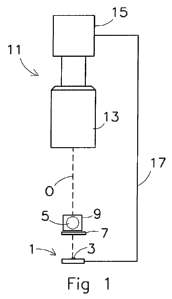
Figure 1 depicts a schematic top view on a sample analysis apparatus according
to a
first embodiment of the invention;
Figure 2 depicts a schematic top view on a sample analysis apparatus according
to a
second embodiment of the invention;
Figure 3 depicts a schematic top view on a sample analysis apparatus according
to a
third embodiment of the invention; and,
Figure 4 depicts a three dimensional view on a holder constructed for use in a
sample
analysis apparatus according to the invention.
Figure 1 schematically depicts a top view on a sample analysis apparatus
according
to a first embodiment of the invention. The apparatus comprises a radiation
system 1
configured to irradiate a sample in a vial 5. The radiation system 1 may
comprise a radiation
source or it may receive its radiation from elsewhere, for example by a fibre.
The radiation
source may be a light emitting diode (LED) 3. An advantage of an LED is that
the LED 3
does not generate much heat, which may influence the sample in the vial 5.
Another
advantage of a LED is that it may generate irradiation with a narrow
bandwidth. The user
may select the LED with a certain specific wavelength which is advantageously
for a certain
sample. The wavelength irradiated by the radiation system may be between 300
and 3000
nm, which includes ultraviolet radiation to deep infrared radiation.
The radiation system 1 may provide pulsed irradiation. The sample in the vial
5 may
comprise moving particles and by providing pulsed irradiation stand still
images can be made
from the particles in the sample. Alternatively, the apparatus may be provided
with a shutter
to make stand still images but because a shutter comprises moving parts pulsed
irradiation is
more advantageous.
CA 02742162 2011-04-28
WO 2010/056109 PCT/NL2009/000208
The radiation system 1 irradiates the sample via a diffuser plate 7. The
function of the
diffuser plate 7 is to make the irradiation of the vial 5 more uniform. A
holder 9 for releasable
holding the vial 5 is provided with a hole for receiving the vial 5 and with
an opening for the
irradiation to irradiate the vial 5 and with an opening for the camera to make
images of the
5 sample in the vial 5. The opening within the holder is along the optical
axis 0 of the camera
11. The holder 9 comprises a temperature control system with a thermometer for
controlling
the temperature of the vial 5. The temperature control system may comprise a
heater for
heating the vial and/or a cooler for cooling the vial within a temperature
range from -25 C
until 180 C.
The camera 11 which is a part of an analyser comprises a lens system 13 and a
detector 15. The lens system 13 provides a telecentric image of a part of the
sample in the
vial 5 on the detector .15 so that the magnification of the particles in the
sample is not
dependent on the position of the particles in the image field. The camera
comprises a lens
and a detector and the lens provides a telecentric image of a part of the
sample on the
detector. As an alternative, cheaper non-telecentric optics may be used for
the lens system.
The detector 15 may be a Charge coupled device (CCD) array and detects an
image of the
particles in black and white or in full colour.
Telecentric lenses have the same magnification at all distances. Because their
images have constant magnification and geometry, telecentric lenses are
suitable for making
images of particles that are moving within a vial and are not at a constant
distance. The
telecentric lens is also less sensitive for lens effects due to the curvature
of the vial.
Telecentric lenses may be object-space telecentric, image space telecentric or
double
sided telecentric. An object-space telecentric lens creates images of the same
size for
objects at any distance in the sample and has constant angle of view across
the entire field
of view. An object or particle that is too close or too far from the lens may
still be out of
focus, but the resulting blurry image will be the same size as the correctly-
focused image
would be. Object-space telecentric lenses have an entrance pupil infinitely
far behind the
lens; this is, if you look in the front, the apparent aperture is very far
away.
An image-space (or image-side) telecentric lens produces images of the same
size
regardless of the distance between the lens and the detector. This allows the
lens to be
focused to different distances without changing the size of the image. Image-
space
telecentric lenses have an exit pupil infinitely far in front of the lens;
that is, if you look in the
back of the lens, the apparent aperture is very far away. At the detector or
image sensor, all
of the principal rays from these lenses hit "straight on", or at zero angle of
incidence. This
property minimizes any angle-of-incidence dependence of the detector, or of
any beam-
splitter prism assembly behind the lens, such as a color separation prism in a
three-CCD
camera.
CA 02742162 2011-04-28
WO 2010/056109 PCT/NL2009/000208
6
Lenses that are double sided telecentric are object-space telecentric and
image-
space (or image-side) telecentric and have magnification that is more
precisely constant than
those that are only object-side telecentric, because the principal ray
intercept position on the
detector doesn't change as well. This property allows precise measurement of
objects
regardless of position even better.
The camera may be provided with a diaphragm which may be opened completely.
The depth of focus will be between 1 and 1,5 mm and the shutter speed may be
between 5
and 50 millisecond, preferably 20 milliseconds and more preferably 10
milliseconds.
The camera 11 may be provided with a radiation intensity measurement system.
The
radiation intensity measurement system may be a separate unit or may form part
of the
detector 15. If the intensity measured by the radiation intensity measurement
system of the
radiation received through the vial is below a certain threshold value the
radiation system 1
may be adjusted via a feed back loop 17 to irradiate more radiation. The
detector may be
provided with a comparing system to compare the radiation intensity of
radiation received
from the sample with the threshold value. It may also be advantageous to keep
the back-
ground at a constant intensity, e.g. grey scale value. It is therefore
advantageous to measure
the background intensity and to compare the background intensity with a
threshold value
and to increase the background illumination if the background intensity gets
lower than the
threshold and to decrease the background illumination if the background
intensity gets
higher than the threshold.
Figure 2 depicts a schematic top view on a sample analysis apparatus according
to a
second embodiment of the invention. The second embodiment is equal to the
first
embodiment accept for the items described below.
In the second embodiment of the invention a semi-transparent mirror 19 is
provided in
the optical path of the camera 11 having an optical axis 0. If the vial 5 is
irradiated by the
first radiation system 1 the radiation is traversing through the vial 5 and
the semi-transparent
mirror 19 to the camera 11. This may be called back lighting.
The camera 11 may be provided with a radiation intensity measurement system
and if
the radiation received by the camera 11 is below a certain threshold the first
radiation system
1 may be adjusted to irradiate more radiation to the vial 5. If however, the
sample in the vial
5 becomes so turbid that adjusting the radiation intensity of the first
radiation system 1 is not
sufficient anymore, a radiation system switching device for switching between
irradiation by
the first radiation system 1 and irradiation by a second radiation system may
be used. The
switching device may be provided to the sensor 15 and may switch off the first
radiation
system 1 and switch on the second radiation system comprising a second light
emitting
diode 21 and a second diffuser plate 23 via the light control connection 25.
The
semitransparent mirror 19 will reflect the irradiation of the second radiation
system to the
CA 02742162 2011-04-28
WO 2010/056109 PCT/NL2009/000208
7
sample 5 from the same direction as the camera 11 is looking at the vial 5.
This may be
called front lighting. The irradiation diffuse reflected by the sample in the
vial 5 may be
reflected through the semitransparent mirror and will be recorded by the
camera 15. Front
lighting of the sample in the vial is advantageous if the turbidity of the
sample in the vial is
high.
Figure 3 depicts a schematic top view on a sample analysis apparatus according
to a
third embodiment of the invention. The third embodiment is equal to the second
embodiment
accept for the items described below. The third embodiment discloses an
alternative way of
front lighting of the sample in the vial.
If the sample in the vial 5 becomes so turbid that adjusting the radiation
intensity of
the first radiation system 1 is not sufficient anymore, a radiation system
switching device for
switching between irradiation by the first radiation system 1 and irradiation
by a second
radiation system 27 may be used. The switching device may be provided to the
sensor 15
and may switch off the first radiation system 1 and switch on the second
radiation system 27
via the light control connection 25. The second radiation system 27 may be
provided with a
light surrounding the opening of the camera 11, which provides front lighting
to the sample in
the vial 5. The irradiation diffuse reflected by the sample in the vial 5 will
be recorded by the
camera 11. Front lighting of the sample in the vial is advantageous if the
turbidity of the
sample in the vial is high. The front lighting according to the third
embodiment of the
invention is cost effective since no semitransparent mirror is needed. The
front lighting may
also be accomplished by other radiation systems.
In the second and third embodiment according to the invention the front
lighting is
disclosed as an alternative for back lighting, however it must be understood
that the front
lighting may also be used in an analysis apparatus according to the invention
without back
lighting.
Figure 4 depicts a three dimensional view on a holder constructed for use in a
sample
analysis apparatus according to the invention. The holder 9 is provided with a
hole for
releasable receiving the vial 5 (as here depicted only the top of the vial is
visible). The holder
9 is provided with an opening 29 and the irradiation of the first radiation
system may traverse
through the opening 29, through the vial 5 and via the opening on the other
side of the
holder to the camera. The holder 9 may be provided with a temperature control
system with
a thermometer, a cooler and heater for controlling the temperature of the vial
in the holder.
The holder 9 may be provided with electromagnets which are activated so as to
rotate a
magnet which may be connected to a stirrer in the vial so as to stir the
sample in the vial 5.
The camera may be connected to a TV or computer screen so as to provide images
of the sample on the screen. The sample analysis apparatus may comprises an
image
CA 02742162 2011-04-28
WO 2010/056109 PCT/NL2009/000208
8
processing module for processing of the images made by the camera. The image
processing
module may have software for image analysis of the particle shape and size
distribution of
particles in the sample in the vial. The information about particle size and
size distribution
may be used for growing crystals. This may be done using a phenomena called
Oswald
ripening. This phenomena may be used if large crystals need to be grown. If
the image
processing unit detects crystals that are growing when the temperature is
slowly increasing
the reaction may be reversed by cooling until only a view big crystals are
left. By then
heating the sample again the crystals will grow again but the bigger crystals
are growing
faster. By repeating this method large crystals may be grown. The sample
analysis
apparatus according to the invention makes the growing of crystals a lot
easier by providing
real time images and data of the crystallisation process in the vial. In the
above example
heating and cooling were used for initiating and reversing the crystallisation
process,
however any kind of habit changing may be used to initiate or reverse the
crystallisation
process. For example, the stirring speed may be adjusted.
The sample analysis apparatus may be provided with a Raman spectroscopy
branch,
for measuring the Raman spectroscopy of the sample in the vial simultaneously
with the
image of the sample in the vial. Typically, the sample will be illuminated
with a laser beam
and radiation from the illuminated spot is collected with a lens and sent
through a
monochromator. Wavelengths close to the laser line are filtered out while the
rest of the
collected light is dispersed onto a detector forming the Raman scattering.
Alternatively, light
emitting diodes (LED's) may be used for illumination of the sample in the
vial. LED's may be
advantageous for Raman scattering because the light has a narrow bandwidth.
While specific embodiments of the invention have been described above, it will
be
appreciated that the invention may be practiced otherwise than as described.
For example,
the invention may take the form of a computer program containing one or more
sequences
of machine-readable instructions describing a method as disclosed above, or a
data storage
medium (e.g. semiconductor memory, magnetical or optical disk) having such a
computer
program stored therein.
The descriptions above are intended to be illustrative, not limiting. Thus it
will be
apparent to one skilled in the art that modifications may be made to the
invention as
described without departing from the scope of clauses set out below.