Note: Descriptions are shown in the official language in which they were submitted.
CA 02742215 2016-05-17
One Way Sliding Device for Intramedullani Intertrochanteric Fixation
Implants
Inventor: George A. Mikhail, Mark Siravo
Field of the Invention
[0001] The present invention relates to devices for treating fractures of
long
bones and, in particular, to internal fixation devices.
Background
[0002] Fractures commonly occur in the femur, for example in the femoral
neck, intertrochanteric and peritrochanteric regions. Such fractures may be
fixed with an intramedullary device and an implant. As is understood by those
skilled in the art, the intramedullary device (e.g., an intramedullary nail)
is
positioned in the medullary canal of a long bone such as the femur. An
implant,
which may be formed as a helical blade or a lag screw, may then be inserted
laterally through bone to pass through an opening of the intramedullary device
until a free end of the implant enters the head of the bone.
[0003] For example, where the bone is a femur, the implant passes
through the shaft of the femur, through the intramedullary device and into
the femoral head via the neck of the femur to secure the femoral head to a
remaining portion of the femur. After implantation, such an implant may
move laterally relative to the intramedullary nail along the path over which
it
was inserted. Some lateral movement of the implant is expected. However,
in some cases, the implant may migrate medially through the intramedullary
8385623.1 1
CA 02742215 2011-04-29
WO 2010/053628
PCT/US2009/058019
device, resulting in a protrusion through the femoral head and into the
acetebulum
causing complications.
Summary of the Invention
[0004] The present invention is directed to a device for treating fractures,
comprising
an intramedullary member sized and shaped for insertion along a longitudinal
axis of a
bone within a medullary canal thereof, the intramedullary member including an
opening
extending obliquely therethrough, the opening, when the intramedullary member
is in a
desired position within a bone, aligning with a desired axis along which an
implant is to
be inserted into the bone, the intramedullary member including a channel
formed
therewithin and opening to the opening and a locking mechanism mounted in the
channel, the locking mechanism including a locking abutting structure
extending into the
opening in combination with an implant sized to be slidably received through
the
opening and inserted along the desired axis, the implant including a plurality
of implant
abutting structures aligned to engage the locking abutting structure
preventing medial
movement of the implant relative to the intramedullary member.
[0005] The present invention is further directed to a method comprising
inserting an
intramedullary member into a medullary canal of a bone and inserting an
implant into a
bone via an opening in the intramedullary member, a shaft of the implant
including a
plurality of abutting structures distributed along a portion of a length of
the shaft, each of
the abutting structures including an angled lateral surface and a medially-
facing abutting
surface in combination with moving a locking mechanism to a locked
configuration in
which a pawl of the locking mechanism extends into the opening to engage the
abutting
surface of one of the abutting structures corresponding to a desired medial-
most
position of the implant, the angled lateral surfaces of the abutting
structures permitting
lateral movement of the implant relative to the pawl.
2
CA 02742215 2011-04-29
WO 2010/053628
PCT/US2009/058019
Brief Description of the Drawings
[0006] Fig. 1 shows a side view of a device according to a first exemplary
embodiment
of the invention;
Fig. 2 shows a side view of a distal facing surface of an implant of the
device
of Fig. 1;
Fig. 3a shows a perspective view of a locking mechanism of the device of Fig.
1;
Fig. 3b shows a perspective view of a locking mechanism and an
intramedullary device according to an alternate embodiment of the present
invention;
Fig, 3c shows a perspective view of the locking mechanism of Fig. 3b;
Fig. 4 shows a side view of an intramedullary nail and the locking mechanism
the device of Fig. 1, in a first configuration;
Fig. 5 shows a perspective view of the intramedullary nail and the locking
mechanism of Fig. 4;
Fig. 6 shows a side view of the intramedullary nail and the locking mechanism
of the device of Fig 1, in a second configuration;
Fig. 7 shows a perspective view of the intramedullary nail and the locking
mechanism of Fig. 6;
Fig. 8 shows a perspective view of the device of Fig. 1;
Fig. 9 shows a side view of the device of Fig. 1;
Fig. 10 shows an opposite side view of Fig. 9;
Fig. 11 shows a lateral cross-section of the device of Fig. 1;
Fig. 12 shows a side view of a device according to a second exemplary
embodiment of the present invention;
Fig. 13 shows a cross-sectional view of an implant of the device of Fig. 12;
Fig. 14 shows a perspective view of a locking mechanism of the device of Fig.
12;
Fig. 15 shows a perspective view an intramedullary nail and the locking
mechanism of the device of Fig. 12, in a first configuration;
3
CA 02742215 2011-04-29
WO 2010/053628
PCT/US2009/058019
Fig. 16 shows another perspective view of the intramedullary nail and the
locking mechanism of Fig. 15;
Fig. 17 shows a side view of the intramedullary nail and the locking
mechanism of the device of Fig. 12, in a second configuration;
Fig. 18 shows a perspective view of the intramedullary nail and the locking
mechanism of Fig. 17;
Fig. 19 shows a perspective view of a device according to a third exemplary
embodiment of the present invention;
Fig. 20 shows another perspective view of the device of Fig. 19;
Fig. 21 shows a perspective view of a locking mechanism of the device of Fig.
19;
Fig. 22 shows a side of the locking mechanism of Fig. 21;
Fig. 23 shows a perspective view of a first element of the locking mechanism
of Fig. 21;
Fig. 24 shows another perspective view of the first element of Fig. 23;
Fig. 25 shows a perspective view of a second element of the locking
mechanism of Fig. 21;
Fig. 26 shows another perspective view of the second element of Fig. 25;
Fig. 27 shows a lateral cross-section of an intramedullary nail and locking
mechanism of the device of Fig. 19;
Fig. 28 shows a side view of a device according to a fourth exemplary
embodiment of the present invention;
Fig. 29 shows a side view of an intramedullary nail, locking mechanism and
pawl of the device of Fig. 28, in a first configuration;
Fig. 30 shows a perspective view of the intramedullary nail, locking mechanism
and the pawl of Fig. 29;
Fig. 31 shows a side view of the intramedullary nail, locking mechanism and
the pawl of the device of Fig. 28, in a second configuration;
Fig. 32 shows a perspective view of the intramedullary nail, locking
4
CA 02742215 2011-04-29
WO 2010/053628
PCT/US2009/058019
mechanism and the pawl of Fig. 31;
Fig. 33 shows a front view of the locking mechanism of the device of Fig. 28;
Fig. 34 shows a perspective view of a pawl of the device of Fig. 28;
Fig. 35 shows a side view of a device according to a fifth exemplary
embodiment of the present invention;
Fig. 36 shows a side view of an implant of the device of Fig. 35;
Fig. 37 shows a side view of a locking mechanism of the device of Fig. 35;
Fig. 38 shows a cross-sectional side view of the locking mechanism of Fig. 37;
Fig. 39 shows a perspective view of the device of Fig. 35, in an initial
implanted position;
Fig. 40 shows a perspective view of the device of Fig. 35, in a final
implanted
position; and
Fig. 41 shows a perspective view of a canted plate of the locking mechanism
of Fig. 37.
Detailed Description
[0007] The present invention may be further understood with reference to the
following
description and the appended drawings, wherein like elements are referred to
with the
same reference numerals. The present invention relates to devices for treating
fractures of long bones and, in particular, to internal fixation devices. It
is noted that
although exemplary embodiments of the present invention are described below
with
respect to the treatment of fractures of the femur, the invention is not
intended to limit
the application of the invention to such fractures, as the invention may also
be
employed in the treatment of other fractures such as, for example, the
humerus, tibia,
etc. It should also be noted that the terms distal and proximal, used herein,
refer to a
direction toward (proximal) and away from (distal) a user of the device. As
indicated
above, fractures of long bones, particularly fractures in which a break is
formed between
a trochanteric head and a shaft of the bone, may be treated by implanting an
intramedullary device along an axis of the shaft of the bone (i.e., in the
medullary canal).
CA 02742215 2011-04-29
WO 2010/053628
PCT/US2009/058019
An implant may then be inserted laterally through the bone to pass through the
intramedullary device into the trochanteric head. Devices according to the
present
invention are designed to permit a desired degree of migration of the implant
back
toward the point through which it was inserted into the bone (i.e., lateral
migration) while
minimizing migration of the implant further into the trochanteric head toward
the
acetebulum (i.e., medial migration).
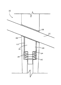
[0008] As shown in Figs. 1 - 11, a device 100 according to an exemplary
embodiment
of the present invention comprises an implant 102 and an intramedullary nail
104
including a locking mechanism 106 (e.g., a ratchet mechanism) permitting
limited
migration of the implant 102 through the nail 104 laterally while preventing
medial
migration. As shown in Fig. 1, an oblique opening 108 extending through the
nail 104 in
a plane substantially perpendicular to a longitudinal axis of the nail 104.
The opening
108 is sized to receive the implant 102 therethrough. A channel 110 extending
through
a portion of the nail 104 along the longitudinal axis opens to the opening 108
houses the
locking mechanism 106. In the embodiment shown, the channel 110 and the
locking
mechanism 106 extend distally of the opening 108 so that the locking mechanism
106
engages a distal side of the implant 102 when the implant 102 is inserted
through the
oblique opening 108. Those skilled in the art will understand that the channel
110 and
the locking mechanism 106 may alternatively be located on the proximal side of
the
implant 102. The locking mechanism 106 includes a biasing member 140 (e.g., a
spring) engaging a pawl member 105 to urge the pawl member 105 into contact
with the
implant 102.
[0009] As shown in Fig. 2, a distal facing surface of a locking engaging
portion of the
implant 102 which, when assembled in a desired configuration, overlaps with
the
channel 110 includes features for engaging corresponding structures of the
pawl
member 105. Specifically, the implant 102 comprises a shaft 112 extending from
a
proximal end 114 to a distal end (not shown) coupled to the proximal end of a
blade or
6
CA 02742215 2011-04-29
WO 2010/053628
PCT/US2009/058019
other bone engaging structure (not shown). As would be understood by those
skilled in
the art, the bone engaging structure may be formed as a helical blade
extending distally
from the distal end of the shaft 112. It will be understood by those of skill
in the art
however, that the bone engaging structure may be any other fixation means such
as, for
example, a lag screw.
[00010] The locking engaging portion of the shaft 112 includes a plurality of
abutting
structures 116 spaced from one another along a portion of a length of the
shaft 112.
Each of the abutting structures 116 includes a ramped surface 117 extending
from a
position adjacent to a radially inner end of the abutting structure 116
immediately distal
thereto and angling gradually outward to an abutting surface 119. As would be
understood by those of skill in the art, the abutting surfaces 119 of the
abutting
structures 116 may extend substantially perpendicular to a longitudinal axis
of the
implant 102.
[00011] As shown in Fig. 3a, the pawl member 105 includes an implant engaging
surface 130 angled to substantially align with an angle of the opening 108. A
pawl 126
including a proximally facing abutting surface 127 extends out from the
surface 130 so
that, when in an operational position, the abutting surface 127 of the pawl
126 engages
an abutting surface 119 of one of the abutting structures 116 of the implant
102. Thus,
engagement between the pawl 126 and the abutting structure 116 of the shaft
112
prevents movement of the implant 102 medially relative to the nail 104.
However, the
implant 102 may slide laterally as the angled distal surface of the pawl 126
and the
angled surfaces 117 of the abutting structures 116 allow the shaft 112 to
slide laterally
over the pawl member 105. A protrusion 128 including a ramped surface 134
extends
outward from the implant engaging surface 130 by a distance greater than the
pawl 126
so that, during insertion of the implant 102 through the opening 108, contact
between
the shaft 112 and the protrusion 128 moves the locking engaging portion of the
shaft
112 out of contact with the pawl 126 until the implant 102 has been advanced
to a
7
CA 02742215 2011-04-29
WO 2010/053628
PCT/US2009/058019
desired position in the bone. When in the desired position, the implant 102 is
rotated
about its longitudinal axis to a locking orientation in which the protrusion
128 aligns with
and enters a groove 118 formed in the shaft 112. At this point, the abutting
structures
116 of the shaft 112 and the pawl 126 are aligned with one another so that, as
the
implant 102 moves toward the pawl member 105 due to the insertion of the
protrusion
128 into the groove 118, the pawl 126 engages one of the abutting structures
116
corresponding to the desired medial-most position of the implant 102. As
described
above, engagement between the abutting surface 127 of the pawl 126 and the
abutting
surface 119 of the abutting structure 116 prevents further medial migration of
the
implant 102. This contact between the pawl member 126 and the corresponding
abutting structure 116 is maintained by the biasing member 140 which urges the
pawl
member 105 toward the shaft 112 at all times.
[0012] To ensure that the locking mechanism 106 does not move beyond the first
and
the second configuration, the locking mechanism 106 may also include an
elongated
hole 124 extending laterally through the locking mechanism 106, distally of
the shoulder
122, for receiving a pin (not shown) which fixes the locking mechanism 106 to
the
intramedullary nail 104. Thus, the intramedullary nail 104 also includes a
hole 136
extending laterally therethrough, distally of the oblique opening 108 such
that the
position of the hole 136 corresponds to a position of the elongated hole 124.
The hole
136 may be substantially circular such that the intramedullary nail 104
remains
stationary while the locking mechanism 106 moves relative to the
intramedullary nail
104 along the longitudinal axis. It will be understood by those of skill in
the art that the
pin inserted through the holes 124, 136 fixes the locking mechanism 106 to the
intramedullary nail 104 such that the locking mechanism 106 and the
intramedullary nail
104 may not rotate relative to one another, but may move between the first
configuration and the second configuration along the longitudinal axis of the
intramedullary nail 104.
8
CA 02742215 2011-04-29
WO 2010/053628
PCT/US2009/058019
[0013] In an alternative embodiment, as shown in Figs. 3b - 3c, a locking
mechanism
106' may include a pawl member 105' formed with a recess 124' rather than an
elongate
hole for fixing the locking mechanism 106' within an intramedullary nail 104'.
The
recess 124' may be fixed within the intramedullary nail 104' via a pin 137'
that is inserted
into the intramedullary nail 104' and the recess 124'. The locking mechanism
106' is
substantially similar to the locking mechanism 106 and may be used in the
device 100
in substantially the same manner. The recess 124' may be formed on an outer
surface
125' of the pawl member 105' and may include a first portion 142', a second
portion 144'
and a third portion 146'. The first portion 142' extends longitudinally along
a portion of
the outer surface 125' from an edge 156' of the pawl member 105' to a proximal
end
148' of the first portion 142'. The second portion 144' extends substantially
horizontally
along a portion of the outer surface 125' from the proximal end 148' of the
first portion
142' to an opposite end 150'. The third portion 146' extends from the end 150'
longitudinally along the outer surface 140' in a distal direction. The first
portion 142', the
second portion 144' and the 146' are connected such that they form a single
continuous
recess 124'.
[0014] The intramedullary nail 104' includes a hole 136' extending laterally
through
one side of the intramedullary nail 104', distally of an oblique opening 108'
such that a
positioning of the hole 136' corresponds to position of the recess 124'. The
hole 136' is
adapted and configured to receive the pin 137' thereth rough. A length of the
pin 137'
may be slightly longer than a thickness of the intramedullary nail 104'. The
thickness is
determined by a distance from an outer surface 109' of the intramedullary nail
104' to a
channel 110' of the intramedullary nail 104', which extends longitudinally
therethrough.
Thus, when the pin 137' is inserted through the hole 136' such that a proximal
end 152'
of the pin 137' is flush with the outer surface 109', a distal end 154' of the
pin 137'
extends into the channel 110' for engaging the recess 124'. The hole 136' may
be
substantially circular such that the intramedullary nail 104 remains
substantially
stationary while the locking mechanism 106' moves relative to the
intramedullary mail
9
CA 02742215 2011-04-29
WO 2010/053628
PCT/US2009/058019
104' along the longitudinal axis.
[0015] To fix the locking mechanism 106' within the intramedullary nail 104',
a biasing
member 140' of the locking mechanism 106' may be inserted into the channel
110'
along with the pawl member 105' such that the biasing member 140' urges the
pawl
member 105' into a position of contact with an implant (not shown) that is
inserted into
the opening 108'. The pawl member 105' is inserted distally into the channel
110' until
the distal end 154' of the pin 137' that is inserted into the hole 136'
engages the first
portion 142' of the recess 124' via the edge 156' of the first portion 142'.
The pawl
member 105' is pressed further distally against the urging of the biasing
member 140'
such that the first portion 142' slides along the pin 137' until the proximal
end 148' of the
first portion 142' is in contact with the pin 137. The pawl member 105' may
then be
rotated about a longitudinal axis thereof such that the second portion 144'
slides along
the pin 137' until the pin 137' is contacting the opposite end 150' of the
second portion
144'. Upon reaching the opposite end 150', the pawl member 105' may be
released, the
biasing member 140' urging the pawl member 105' in a proximal direction such
that the
third portion 146' slides along the pin 137' until the pin 137' engages a
distal end 158' of
the third portion 146'. Thus, it will be understood by those of skill in the
art that once the
locking mechanism 106' is fixed within the intramedullary nail 104', the
locking
mechanism 106' is movable along the longitudinal axis to engage the implant,
as
described in regard to the device 100. Longitudinal movement of the locking
mechanism 106' results in sliding of the third portion 146' longitudinally
along the distal
end 154' of the pin 137'.
[0016] In a first configuration, shown in Figs. 4 - 5, when no implant 102 is
present in
the opening 108, the implant engaging surface 130 substantially aligns with a
wall of the
oblique opening 108 while the pawl 126 and the protrusion 128 extend into the
oblique
opening 108. Then, as an implant 102 is inserted into the opening 108, contact
between the implant 102 and the ramped surface 134 forces the pawl member 105
into
CA 02742215 2011-04-29
WO 2010/053628
PCT/US2009/058019
the channel 110 to a second configuration in which the protrusion 128 is moved
into the
channel 110 to a second configuration shown in Figs. 6 and 7 to allow the
implant 102
to be advanced medially through the opening 108. The pawl member 105 is
constrained so that it does not move further into the opening 108 than desired
(i.e.,
beyond a desired first configuration), by a pin 135 passing through an opening
136 in
the intramedullary nail 104, and through an elongated opening 124 in the pawl
member
105. As described above, when the implant 102 has been inserted to a desired
position
in the bone, the implant 102 is rotated about its axis until the groove 118
aligns with the
protrusion 128. At this point the biasing member 140 moves the pawl member 105
back
to the first configuration with the protrusion 128 received within the slot
118 and the
pawl 126 engaging one of the abutting structures 116 of the implant 102
corresponding
to the desired maximum insertion of the implant 102 into the bone. Thereafter,
as
forces are applied to the implant 102 (e.g., as weight is placed on the bone),
the implant
102 may move laterally as ramped surfaces 117 slide over the pawl 126. The
abutting
surface 119 engages the pawl 126 preventing any further movement medially. In
addition, as each abutting surface 119 moves laterally past the pawl 126, a
new medial-
most position of the implant 102 is defined.
[0017] The intramedullary nail 104 may further include a shoulder 138 within
the
channel 110 positioned below the oblique opening 108. A reduced diameter shaft
120
extends from an end of the pawl member 105 to a shoulder 122 at an end of an
upper
portion of the pawl member 105. The biasing member 140 is received between the
shoulder 122 and the shoulder 138 of the channel 110 to urge the pawl member
105
toward the opening 108. A diameter of a portion of the channel 110 closer to
the
opening 108 than the shoulder 138 is larger than a diameter of the portion of
the
channel 110 extending past the shoulder 138 away from the opening 108. It will
be
understood by those of skill in the art that the diameters of these portions
the channel
110 correspond to the diameters of the proximal end 118 of the pawl member 105
and
the shaft 120, respectively.
11
CA 02742215 2011-04-29
WO 2010/053628
PCT/US2009/058019
[0018] In use, the intramedullary nail 104 is inserted into an intramedullary
canal (e.g.,
of a femur) with a central axis of the oblique opening 108 substantially
aligned with a
central axis of the femoral neck. It will be understood by those of skill in
the art that the
intramedullary nail 104 may be inserted into the bone using any accepted
insertion
method. For example, a guidewire may be inserted into the medullary canal of
the
longitudinal shaft and the intramedullary nail 104 slid therealong. Thus, it
will also be
understood by those of skill in the art that the intramedullary nail 104 and
the locking
mechanism 106 housed therewithin may also include a guide wire lumen along the
longitudinal axis thereof. Once the intramedullary nail 104 has been
appropriately
positioned, the implant 102 is inserted through the bone into the oblique
opening 108 to
a desired position and the implant 102 is rotated to return the locking
mechanism 106 to
the first configuration preventing further medial movement as described above.
[0019] If it becomes necessary to remove the implant 102 for any reason,
however,
the implant 102 may be rotated about the central axis of the oblique opening
108, as
shown in Fig. 11 to move the protrusion 128 out of the groove 118 and force
the pawl
member 105 back to the first configuration. At this point the locking
mechanism 106 is
disengaged from the abutting structures 116 of the implant 102 and the implant
102
may be slid entirely out of the opening 108 even after the protrusion 128 is
located
distally beyond the distal end of the groove 118. If the bone engaging
structure of the
implant 102 is formed as a helical blade, those skilled in the art will
understand that this
structure may be rotatably coupled to the shaft 112 of the implant 102 so that
the
engagement between the locking mechanism 106 and the abutting structures 116
of the
implant 102 is maintained. Thus, any rotation of the helical blade during
insertion would
not require a corresponding rotation of the shaft 112. However, it will be
understood by
those of skill in the art that the bone engaging structure of the implant 102
may be any
known capable of securing the femoral head and neck to the shaft through
engagement
of an intramedullary nail 104.
12
CA 02742215 2011-04-29
WO 2010/053628
PCT/US2009/058019
[0020] As shown in Figs. 12- 18, a device 200, according to another embodiment
of
the present invention comprises an implant 202 and an intramedullary nail 204
with a
locking mechanism 206 housed therewithin. The device 200, as shown in Fig. 12,
is
substantially similar to the device 100 described above including a biasing
member 240
(e.g., a spring) moving the locking mechanism 206 within a channel 210 of the
nail 204
along a longitudinal axis of the nail 204. The locking mechanism 206 also
moves
between first and second configurations in which a pawl 226 is brought into
and out of
the opening 208 to engage and disengage abutting structures 216 of the implant
202.
[0021] However, the pawl member 205 of the locking mechanism 206 does not
include a protrusion similar to the protrusion 128 for engaging the implant
202 and
moving the pawl member 205. The implant 202 may be substantially similar to
the
implant 102 except that no groove similar to the groove 118 is provided.
Rather, a shaft
212 of the implant 202 may include a plurality of notches 218 extending
longitudinally
along a portion of a length thereof and separated from one another around the
circumference of the shaft 212 by a distance corresponding to a separation of
a pair of
notch engaging wings 228 extending from an implant engaging surface 230 of the
pawl
member 205. Thus a first one of the wings 228 is received within a
corresponding one
of the notches 218. When the wings 228 are received in the notches 218, the
abutting
structures 216 of the implant 202 are aligned with the pawl 226 of the pawl
member
205. Engagement between the wings 228 and the notches 218 prevents the shaft
212
from rotating within the opening 208. As shown in Fig. 14, the locking
mechanism 206
may be substantially similar to the locking mechanism 106 with the pawl 226
extending
from the implant engaging surface 230 and engaging the abutting structures 216
to
prevent medial movement of the implant 202 beyond a defined medial-most
position.
[0022] The locking mechanism 206 includes a laterally facing hole 246 which is
aligned with a corresponding opening 250 adjacent to the lateral end of the
opening 208
13
CA 02742215 2011-04-29
WO 2010/053628
PCT/US2009/058019
so that a tool may be inserted therethrough to engage the pawl member 205 and
move
it manually between from the first configuration, shown in Figs. 15 - 16, to
the second
configuration, shown in Figs. 17- 18. The hole 246 may include a ramped
surface 248
so that when a pin 252 is inserted into the hole 246 via the hole 250, the pin
252
slidingly engages the ramped surface 248 pushing the pawl member 205 further
into the
channel 210 disengaging the locking mechanism 206 from the abutting structures
216
to permit insertion and/or withdrawal of the implant 202 from the opening 208
to the
second configuration. It will be understood by those of skill in the art that
the ramped
surface 248 enables the size of the hole 246 to be minimized so that the hole
248 does
not need to extend into an elongated hole 224 of the locking mechanism 206
which
engages a pin (not shown) in the same manner as the pin 135 of the device 100
to
prevent the pawl member 205 from moving into the opening 208 beyond the first
configuration.
[0023] The device 200 may be employed in substantially the same manner as the
device 100 as described above. However, when inserting the implant 202 through
the
nail 204, the pin 252 is inserted into the hole 246 of the locking mechanism
206 via the
hole 250 to move the locking mechanism 206 to the second configuration. The
implant
202 is then inserted to the desired position in substantially the same manner
described
above and the pin 252 is removed to allow the pawl member 205 to return to the
first
configuration through the bias of the biasing member 240 to lock the locking
mechanism
206 to the abutting structures 216 and prevent further medial movement of the
implant
202. As with the device 100, the shape of the abutting structures 216 allows
the implant
202 to move laterally over the pawl 226.
[0024] As shown in Figs. 19 - 27, a device 300 according to another embodiment
of
the invention comprises an implant 302 and an intramedullary nail 304 with a
locking
mechanism 306 housed therewithin. The device 300, as shown in Figs. 19 - 20 is
substantially similar to the devices 100, 200 described above except as
specifically
14
CA 02742215 2011-04-29
WO 2010/053628
PCT/US2009/058019
indicated below. The implant 302 is also substantially similar to the implant
102,
including a shaft 312 with a plurality of abutting structures 316 and a
longitudinal groove
318. Similarly to the intramedullary nail 104, the intramedullary nail 304
includes an
oblique opening 308 for receiving the implant 302. However, a channel 310 of
the
intramedullary nail 304 in which the locking mechanism 306 is housed extends
proximally from the oblique opening 308 toward a proximal end of the
intramedullary
nail 304.
[0025] As shown in Figs. 21 - 22 a pawl member 305 of the locking mechanism
306 is
further comprised of a first element 318 and a second element 320. The first
element
318 may be coupled to the second element 320 such that the first element 318
and the
second element 320 are movable relative to one another both along and about a
longitudinal axis. As shown in Figs. 23- 24, the first element 318 includes a
head
portion 360, a shaft 362 and a ball 372 at a distal end 366 of the shaft 362
configured to
engage a correspondingly shaped recess in the second element 320. A diameter
of the
ball 372 may be larger than a diameter of the shaft portion 362.
[0026] The head portion 360 extends proximally from a proximal end 364 of the
shaft
362 and includes threading 368 around an outer surface thereof. The head
portion 360
further includes a driving structure 376 at a proximal end 370 thereof
configured to
receive a driving tool. For example, the driving structure 376 may be a
hexagonal
recess configured to receive a hexagonally shaped bit of a driving tool. It
will be
understood by those of skill in the art, however, that the driving structure
376 may take
any of a variety of shapes and sizes so long it is configured to receive a
tool capable of
rotating the first element 318 relative to the second element 320 and the
intramedullary
nail 304. An annular groove 322 formed in a distally facing surface at a
distal end 374
of the head portion 360 receives a proximal end 344 of a biasing member 340
(e.g., a
spring). The biasing member 340 may extend around the shaft 362 of the first
element
318. The first element 318 may also include a lumen 378 extending
longitudinally
CA 02742215 2011-04-29
WO 2010/053628
PCT/US2009/058019
therethrough, to accommodate instruments such as reaming rods or guidewires,
etc.
[0027] As shown in Figs. 25 - 26, the second element 320 extends from a
proximal
end 380 to a distal end 382 and includes a space 346 in a central portion
thereof sized
and shaped to accommodate the ball 372 of the first element 318 to form a ball
and
socket joint. The proximal end 380 includes a hole 384 that extends into the
space 346
to accommodate the shaft portion 362 when the ball 372 is received within the
space
346. The second element 320 may further include an opening 348 along a portion
of an
outer surface 386 of the second element 320 such that the ball 372 may be
snapped
into the space 346 via the opening 348. The opening 348 should be smaller than
a
diameter of the ball 372 so that the second element 320 must be slightly
deformed to
snap the ball 372 thereinto and the ball 372 may not become easily disengaged
therefrom.
[0028] The distal end 382 includes a first protrusion 326 for engaging the
abutting
structures 316 and a second protrusion 328 for engaging the longitudinal
groove 318.
An angled surface 325 of the first protrusion 326 may be formed substantially
parallel to
the angle of ramped surfaces 317 of each of the abutting structures 316 to
minimize
resistance to the proximal sliding of the abutting structures 316 over the
protrusion 326.
As with the prior embodiments, contact between an abutting surface 327 of the
protrusion 326 and an abutting surface 319 of any of the abutting structures
316
prevents the implant 302 from moving medially beyond an initially set medial-
most
position. The second protrusion 328 is sized and shaped to be received within
the
longitudinal groove 318 such that the longitudinal groove 318 may slide
therealong.
The first and the second protrusions 326, 328 may be positioned on opposite
sides of
one another relative to the longitudinal axis of the implant 302 such that
engagement of
the first protrusion 326 with the plurality of notches 316 and engagement of
the second
protrusion 328 with the longitudinal groove 318 prevents rotation of the shaft
312 of the
implant 302 about a longitudinal axis of the opening 308. The proximal end 380
of the
16
CA 02742215 2011-04-29
WO 2010/053628
PCT/US2009/058019
second element 320 may also include a groove 338 surrounding the opening 384
for
receiving a distal end 342 of the biasing member 340 so that the biasing
member 340
urges the second element 320 into contact with the implant 302.
[0029] The second element 320 further includes a longitudinal element 388
extending
from the outer surface 386 along at least a portion of a length of the second
element
320. As shown in Fig. 27, the longitudinal element 388 may be configured to be
slidable
within a longitudinal slot 390 within the channel 310 of the intramedullary
nail 304 such
that the second element 320 and the intramedullary nail 304 are movable
relative to one
another along the longitudinal axis, but incapable of rotating relative to one
another
about the longitudinal axis.
[0030] The implant 302 may be inserted into the oblique opening 308 of the
intramedullary nail 304 until the implant 302 is in a desired position
relative to the nail
304 and the bone. Once a desired position had been reached, the assembled
locking
mechanism 306 may be inserted into the channel 310 of the intramedullary nail
304 by
aligning the longitudinal element 388 with the longitudinal slot 390 such that
the locking
mechanism 306 may be slid longitudinally through the nail 304. The driving
tool may
then be inserted into the driving means 376 to drive the locking mechanism 306
a
desired distance into the channel 310 by rotating the first element 318
relative to the
second element 320 as would be understood by those skilled in the art. Thus,
the
channel 310 may include a threading (not shown) corresponding to the threading
366 of
the first element 318 such that the first element 318 and the channel 310 may
engage
one another. As the first element 318 rotates about the longitudinal axis, the
first
element 318 pushes the second element 320 further into the channel 310. The
locking
mechanism 306 may be driven into the channel 310 until the distal end 382 of
the
second element 320 contacts the shaft 312 of the implant 302.
[0031] The implant 302 should be positioned such that upon contact of the
locking
17
CA 02742215 2011-04-29
WO 2010/053628
PCT/US2009/058019
mechanism 306 with the shaft 312, the first protrusion 326 engages one of the
abutting
structures 316 corresponding to the desired medial-most position of the
implant 302 and
the second protrusion 328 engages the longitudinal groove 318. As with the
previously
described embodiments, after the implant 302 has been engaged by the locking
mechanism 306, the implant 302 may move laterally relative to the opening 308
but is
prevented from moving medially by contact between the abutting surface 327 of
the
protrusion 326 and the abutting surface 319 of the corresponding abutting
structure 316
of the implant with the biasing member 340 operating to maintain the required
contact
between the protrusion 326 and the corresponding abutting structure 316.
[0032] As shown in Figs. 28 - 34, a device 400 according to a further
embodiment of
the invention may be substantially similar to the device 300, but in addition
to being
comprised of an implant 402 and intramedullary nail 404, a ratchet mechanism
thereof
comprises first and second portions 406 and 492, respectively, on opposite
sides of the
implant 402 from one another. As shown in Fig. 28, the implant 402 includes a
shaft
412 including a plurality of abutting structures 416 distributed along a
portion of a length
of the shaft 412. Similarly to the implants 102, 202 and 302, each of the
abutting
structures 416 is angled toward a proximal end 414 of the shaft 412 with a
distal facing
abutting surface 419 which, in a first configuration, engages a pawl of the
second
portion 492 of the locking mechanism to prevent movement of the implant 402
medially
after an initial position of the implant 402 is set (e.g., upon implantation)
while allowing
lateral migration of the implant 402.
[0033] The intramedullary nail 404 may be substantially similar to the
intramedullary
nail 304, except that a channel 410 extends across the oblique opening 408
from a
proximal end 494 proximal of the opening 408 to a distal end 496 distal of the
oblique
opening 408. The first portion 406 of the locking mechanism is housed in the
portion of
the channel 410 extending proximal of the oblique opening 408 while the second
portion
492 is housed in the portion of the channel 410 distal of the oblique opening
408.
18
CA 02742215 2011-04-29
WO 2010/053628
PCT/US2009/058019
[0034] Similarly to the locking mechanism 306, the first portion 406 includes
a first
element 418 couplable to a second element 420 with a biasing member 440 held
therebetween in a groove 422 of the first element 418 and a groove 438 of the
second
element. The first element 418 and the second element 420 may be coupled to
one
another via ball 472 of the first element 418 which is insertable into a space
446 of the
second element 420, as shown in Fig. 33. The second element 420, however,
includes
an elongated protrusion 428 extending from a distal end 482 of an outer
surface 486 of
the second element 420 radially outside a circumference of the opening 408.
The
elongated protrusion 428 is longer than a diameter of the oblique opening 408
such
that, when the locking mechanism 306 is moved longitudinally through the
channel 410
from a first configuration to a second configuration, the elongated protrusion
428
crosses the opening 408 to operate the second portion 492 of the locking
mechanism
causing the second portion 492 to pivot. Specifically, the distal end 482
remains
proximal to the opening 408 at all times while the protrusion 428 extends
along and
outside the opening 408 to reach the second portion 492. In the first
configuration, as
shown in Figs. 29 - 30, the first portion 406 of the locking mechanism is
positioned
within the channel 410 with the elongated protrusion 428 separated from the
second
portion 492. As shown in Figs. 31 and 32, when moved into the second
configuration,
the first portion 406 of the locking mechanism moves distally though the
channel 410
moving the elongated protrusion 428 distally past the oblique opening 408 to
pivot the
second portion 492 so that a pawl 426 protruding from an implant facing
surface 430 of
the second portion 492 engages the abutting structure 416 corresponding to the
desired
medial-most position of the implant 402.
[0035] As shown in Fig. 34, the second portion 492 is sized and shaped to fit
within
the portion of the channel 410 extending distally of the opening 408. A
proximal surface
430 thereof may be angled to substantially align with a surface of the oblique
opening
408 when in the first configuration. The second portion 492 is rotatably
mounted in the
19
CA 02742215 2011-04-29
WO 2010/053628
PCT/US2009/058019
channel 410 including, for example, a hole 424 for receiving a pin (not shown)
inserted
via a corresponding hole 436 in the intramedullary nail 404. The second
portion 492
rotates about the pin when contacted by the protrusion 428 so that the pawl
426 pivots
into the opening 408 to engage the abutting structures of the implant 402. To
bias the
second portion 492 toward the first configuration in which the pawl 426
remains outside
the oblique opening 408, the device 400 further comprises a biasing member
lumen 498
within the intramedullary nail 405 and a biasing member 500. The biasing
member 500
may be housed within the lumen 498 such that a proximal end 502 of the biasing
member 500 abuts the distal end 431 of the second portion 492 while a distal
end 504
of the biasing member 500 abuts a distal end 506 of the lumen 498. Thus, the
second
portion 492 is biased toward the first configuration at all times except when
the
elongated protrusion 428 presses the implant facing surface 430 of the second
portion
492 to the second configuration.
[0036] The device 400 may be used in substantially the same manner as the
devices
100, 200 and 300. Upon positioning of the intramedullary nail 404 within the
femoral
shaft, the implant 402 may be inserted through the oblique opening 408 of the
intramedullary nail while the first and second portions 406, 492,
respectively, of the
locking mechanism are in the first configuration ¨ i.e., with neither the
elongated
protrusion 428 nor the pawl 426 extending into the opening 408. After the
implant 402
has been inserted through the opening 408 to a desired position in the bone,
the first
portion 406 is moved into the second configuration in the same manner
described
above for the device 300 to move the elongated protrusion 428 distally until
it presses
against the implant facing surface 430 of the second portion 492, pivoting the
second
portion 492 and moving the pawl 426 into the oblique opening 408 to engage the
abutting structure 416 corresponding to the desired position of the implant
402 and
defining a medial-most position for the implant 402. As described above, the
geometry
of the abutting structures 416 is selected to permit lateral migration of the
implant 402
through the opening 408.
CA 02742215 2011-04-29
WO 2010/053628
PCT/US2009/058019
[0037] As shown in Figs. 35 - 41, a device 600 according to yet another
embodiment
of the invention may be substantially similar to the device 300 except as
specifically
described below. As shown in Fig. 35, the device 600 comprises an implant 602,
an
intramedullary nail 604 and a locking mechanism 606. As shown in Fig. 36, the
implant
602 includes a shaft 612 with a single recessed and tapered surface 616 as
opposed to
the plurality of abutting surfaces as described above in regard to implant
302. The
tapered surface 616 extends from a proximal end 614 to a distal end 615 with a
taper of
the surface 618 increasing from the proximal end 614 to the distal end 615 so
that a
length of a wall 614' at the proximal end 614 is less than a length of a wall
615' at the
distal end 615. The tapered surface 616 is adapted and configured to receive a
portion
of the lock mechanism 606. The intramedullary nail 604 may be substantially
similar to
the intramedullary nail 304, including an oblique opening 608 for receiving
the implant
602 and a channel 310 for housing the locking mechanism 606 therein,
proximally of the
oblique opening 608 toward a proximal end of the intramedullary nail 604.
[0038] As shown in Figs. 37 - 38, the locking mechanism 606 may be
substantially
similar to the locking mechanism 306 of the device 300. Similarly, the lock
mechanism
606 includes a first element 618 couplable to a second element 620 with a
biasing
member 640 (e.g., a spring) held therebetween. In addition to the biasing
member 640,
the lock mechanism 606 further includes a canted plate 692 held between a
distal end
642 of the of the biasing member 640 and a proximal end 680 of the second
element
620 selectively preventing movement of the second element 620 toward the first
element 618. Specifically, the canted plate 692 includes an opening 698
therethrough
closely matching in size and shape an outer surface of a shaft 662 of the
first element
618 so that, when the canted plate 692 is angled away from a plane
substantially
perpendicular to a longitudinal axis of the shaft 662, frictional engagement
between a
perimeter of the opening 698 and the outer surface of the shaft 6662 prevents
relative
movement between the first element 618 and the second element 620. The first
21
CA 02742215 2011-04-29
WO 2010/053628
PCT/US2009/058019
element 618 is substantially similar to the first element 318, including a
head portion
660 at a proximal end of the shaft 662 and a coupling element 672 at a distal
end 666
thereof configured to engage a correspondingly shaped recess in the second
element
620. A threading of the head portion 660 may engage an inner surface of the
intramedullary nail 604 in the same manner described above.
[0039] The second element 620 may be substantially similar to the second
element
320, extending from a proximal end 680 to a distal end 682 and including a
space 646
in a central portion thereof for slidably accommodating the coupling element
672 of the
first element 618 to permit relative movement therebetween along a
longitudinal axis of
the intrannedullary nail 604. In place of the first and second protrusions of
the previous
embodiments, the second element 620 includes a single elongate protrusion 626
engaging the tapered surface 616 of the implant 602. The elongate protrusion
626
extends from the distal end 682 of an outer surface 686 of the second element
620 and
tapers to a reduced thickness toward a distal tip 626 thereof. A taper of the
elongate
protrusion 626 may be selected so that the thin distal tip 626 may be received
within the
thinner distal end 615 of the tapered surface 616 with the increasing depth of
the
tapered surface 616 permitting the progressively thicker more proximal
portions of the
protrusion 626 to enter into engagement with the tapered surface 616 as the
implant
602 is advanced distally through the nail 604. The tapered surface 616 may be
formed
so that, when the implant 602 has been advanced a desired distance through the
nail
604, the protrusion 626 is fully received against the tapered surface 616
adjacent to the
proximal end 614 thereof locking the implant 602 in a distal-most permitted
position.
Specifically, as the biasing member 640 moves the second element 620 distally
urging
the protrusion 626 further into engagement with the tapered surface 616, the
canted
plate 692 acts as a locking preventing the second element 620 from being moved
proximally back toward the first element 618. This maintains the thicker
proximal
portion of the protrusion 626 in engagement with the tapered surface 616
preventing
distal movement of the implant 602 relative to the nail 604 as the thickness
of the
22
CA 02742215 2011-04-29
WO 2010/053628
PCT/US2009/058019
proximal portion of the protrusion 626 exceeds a depth of the more distal
portion of the
tapered surface 616. A length of the elongate protrusion 626 is substantially
equal to or
greater than a diameter of the oblique opening 608 such that an entire width
of the
tapered surface 616 of the implant 602 may be engaged by a contacting surface
625 of
the elongate protrusion 626.
[0040] The implant 602 may be inserted through the opening 608 into a desired
position within the bone. During insertion of the implant 602, the locking
mechanism
606 is in a first position within the intramedullary nail 604 in which the
elongate
protrusion 626 does not extend into the opening 608. Once the implant 602 has
been
inserted through the opening 608 to the desired position, the locking
mechanism 606 is
driven distally through the channel 610 to a second position in which the
elongate
protrusion 626 contacts the implant 602 and the contacting surface 625 abuts
the
recessed and tapered portion 616. Thus, it will be understood by those of
skill in the art
that, when implanted to the desired depth within the bone, the tapered surface
616 of
the implant 602 extends across the opening 608 of the intramedullary nail 604.
[0041] Specifically, with the locking mechanism 606 in the second position,
the device
600 is in an initial implanted position, as shown in Fig. 39 with a distal
portion of the
contacting surface 625 abutting the tapered surface 616. Due to the increasing
taper of
the tapered surface 616 distally along the shaft 612 and the biasing member
640 which
biases the second element 620 of the locking mechanism 606 to move away from
the
first element 618 along a longitudinal axis of the intramedullary nail 604,
the implant 602
is permitted to migrate proximally through the opening 608 while maintaining
contact
between the contacting surface 625 and the tapered surface 616 toward a final
proximal-most position, as shown in Fig. 40. The final position is reached
after the
implant 602 has moved laterally through the opening 608 until a width of the
distal end
615 of the tapered surface 616 is contacted by the contacting surface 625. As
the
implant 602 moves laterally through the opening 608, the biasing member 640
pushes
23
CA 02742215 2011-04-29
WO 2010/053628
PCT/US2009/058019
the second portion 620 of the locking mechanism distally such that the
elongate
protrusion 626 maintains constant contact with the tapered surface 616.
[0042] At all times until and after the implant 602 reaches the final
implanted position,
the implant 602 is prevented from moving medially through the opening 608 via
the
canted plate 692 which locks the locking mechanism 606 preventing the second
portion
620 from moving proximally within the channel 610 toward the first element 618
which is
fixed within the intramedullary nail 604. As shown in Fig. 41, the canted
plate 692
includes a first portion 694 and a second portion 696 angled relative to one
another,
substantially perpendicularly of one another. As described above, the second
portion
696 includes an opening 698 extending therethrough with a proximal surface of
the
canted plate 692 engaging a distal end 642 of the biasing member 640 while a
distal
end 700 of the first portion 694 engages a proximal surface 680 of the second
element
620 with the shaft 662 of the first element 618 received through the opening
696, a
surface of the second portion 696 abutting the distal end 642 of the biasing
member
640, while an edge 700 of the first portion 694 abuts the proximal end 680 of
the second
portion 620. The opening 698 is only slightly larger than a perimeter of the
shaft 662
such when the implant 602 attempts to move medially through the oblique
opening 608,
the implant 602 pushes the second portion 620 in a direction P, angling the
second
portion 696 relative to the shaft 662 and bringing an inner surface 702 of the
opening
698 into contact with an outer surface 704 of the shaft 662 preventing the
shaft 662
from sliding therethrough and preventing the second portion 620 from moving in
the
direction P.
[0043] The device 600 may be used in substantially the same manner as
described
above in regard to the device 300. Once the intramedullary nail 604 has been
positioned in an intramedullary canal of a bone, the implant 602 may be
inserted
medially through the oblique opening 608 until the implant 602 is in the
desired position
in the bone. As the implant 602 is being inserted through the opening 608, the
locking
24
CA 02742215 2016-05-17
mechanism 606 is maintained in the first position with the elongate protrusion
626 held proximally above the opening 608, leaving a clear path for the
insertion of the implant 602 therethrough. After the implant 602 has reached a
desired distal-most position in the bone with the tapered surface 616
extending
across the opening 608 of the intramedullary nail 604, the locking mechanism
606 is driven distally into the intramedullary nail 604 until the elongate
protrusion 626 extends into the opening 608 with the contacting surface 625 in
engagement with the tapered surface 616 of the implant 602 in the second
position. Even after the implant 602 is within the opening 608 in the initial
implanted position, the implant 602 move proximally through the opening 608
while distal movement relative to the nail 604 is substantially prevented.
However, once the implant 602 has reached the final implanted position, the
implant 602 is prevented from further movement proximally and distally
relative
to the nail 604 as described above.
[0044] The present disclosure has been described in the foregoing
specification by means of non-restrictive illustrative embodiments provided as
examples. These illustrative embodiments may be modified at will. The scope of
the claims should not be limited by the embodiments set forth in the examples,
but should be given the broadest interpretation consistent with the
description
as a whole.
8385623.1 25