Note: Descriptions are shown in the official language in which they were submitted.
CA 02742272 2016-02-24
52923-29
PRODUCTS AND PROCESSES FOR MULTIPLEX NUCLEIC ACID IDENTIFICATION
Related Patent Application
This patent claims the benefit of U.S. Patent Application No. 61/109,885
filed on October 30, 2009, entitled PRODUCTS AND PROCESSES FOR MULTIPLEX
NUCLEIC
ACID IDENTIFICATION, naming Dirk Johannes Van den Boom et al. as inventors,
and designated
by Attorney Docket No. SEQ-6020-PV.
Field
The technology relates in part to nucleic acid identification procedures in
which multiple target
nucleic acids can be detected in one procedure. The technology also in part
relates to
identification of nucleic acid modifications.
Background
The detection of specific nucleic acids is an important tool for diagnostic
medicine and molecular
biology research. Nucleic acid assays currently play roles in identifying
infectious organisms such
as bacteria and viruses, in probing the expression of normal genes and
identifying mutant genes
such as oncogenes, in typing tissue for compatibility preceding tissue
transplantation, in matching
tissue or blood samples for forensic medicine, and for exploring homology
among genes from
different species, for example.
Summary
A challenge associated with nucleic acid identification procedures lies in the
ability to determine the
presence or absence of multiple target nucleic acids in a composition, which
is referred to as
"multiplexing." Certain multiplexing technologies do not allow for the
detection of a significant
number of target nucleic acids in a composition.
Methods described herein answer this challenge in part by combining extension
and solid phase
capture approaches with an identification readout specific for each target
nucleic acid. These
1
CA 02742272 2011-04-29
WO 2010/056513 PCT/US2009/062239
processes are highly accurate and are very rapid as a significant number of
target nucleic acids
can be detected in one assay or procedure.
Accordingly, provided herein is a method for determining the presence or
absence of a plurality of
target nucleic acids in a composition, which comprises: (a) preparing
amplicons of the target
nucleic acids by amplifying the target nucleic acids, or portions thereof,
under amplification
conditions; (b) contacting the amplicons in solution with a set of
oligonucleotides under
hybridization conditions, where: (i) each oligonucleotide in the set comprises
a hybridization
sequence capable of specifically hybridizing to one amplicon under the
hybridization conditions
when the amplicon is present in the solution, (ii) each oligonucleotide in the
set comprises a
distinguishable tag located 5' of the hybridization sequence, (iii) a feature
of the distinguishable tag
of one oligonucleotide detectably differs from the features of distinguishable
tags of the other
oligonucleotides in the set; and (iv) each distinguishable tag specifically
corresponds to a specific
amplicon (e.g., an allele) and thereby specifically corresponds to a specific
target nucleic acid; (c)
generating extended oligonucleotides that comprise a capture agent by
extending oligonucleotides
hybridized to the amplicons by one or more nucleotides, where one of the one
of more nucleotides
is a terminating nucleotide and one or more of the nucleotides added to the
oligonucleotides
comprises the capture agent; (d) contacting the extended oligonucleotides with
a solid phase under
conditions in which the capture agent interacts with the solid phase; (e)
releasing the
distinguishable tags from the extended oligonucleotides that have interacted
with the solid phase;
and (f) detecting the distinguishable tags released in (e); whereby the
presence or absence of each
target nucleic acid is determined by the presence or absence of the
corresponding distinguishable
tag.
In certain embodiments, the extension in (c) is performed once yielding one
extended
oligonucleotide. In some embodiments, the extension in (c) is performed
multiple times (e.g.,
under amplification conditions) yielding multiple copies of the extended
oligonucleotide.
In certain embodiments, a solution containing amplicons (e.g., amplicons
produced in (a)) is
treated with an agent that removes terminal phosphates from any nucleotides
not incorporated into
the amplicons. The terminal phosphate sometimes is removed by contacting the
amplicons with a
phosphatase, and in certain embodiments the phosphatase is alkaline
phosphatase (e.g., shrimp
alkaline phosphatase).
2
CA 02742272 2011-04-29
WO 2010/056513 PCT/US2009/062239
In some embodiments, the hybridization sequence in each oligonucleotide is
about 5 to about 50
nucleotides in length. In certain embodiments, terminal nucleotides in the
extended
oligonucleotides comprise the capture agent, and sometimes one or more non-
terminal nucleotides
in the extended oligonucleotides comprise the capture agent. In some
embodiments, the capture
agent comprises biotin, or alternatively avidin or streptavidin, in which case
the solid phase
comprises avidin or streptavidin, or biotin, respectively. The solid phase is
paramagnetic, is a flat
surface, a silicon chip, a bead and/or a sphere in some embodiments.
The distinguishable tag is distinguished in part by mass in certain
embodiments (i.e., a mass
distinguishable tag where a distinguishing feature is mass). The
distinguishable tag in some
embodiments consists of nucleotides, and sometimes the tag is about 5
nucleotides to about 50
nucleotides in length. The distinguishable tag in certain embodiments is a
nucleotide compomer,
which sometimes is about 5 nucleotides to about 35 nucleotides in length. In
some embodiments,
the distinguishable tag is a peptide, which sometimes is about 5 amino acids
to about 100 amino
acids in length. The distinguishable tag in certain embodiments is a
concatemer of organic
molecule units. In some embodiments, the tag is a trityl molecule concatemer.
The distinguishable
tag in certain embodiments is released by treatment with an endonuclease
(e.g., endonuclease V),
and in some embodiments, the distinguishable tag is linked to the
oligonucleotide by a
photocleavable linkage and is released by treatment with light. In certain
embodiments, the
distinguishable tag is linked by a ribonucleotide and released by treatment
with a ribonuclease, and
in certain embodiments, the distinguishable tag is linked to the
oligonucleotide by inosine and is
released by an agent that cleaves the inosine. A distinguishable tag sometimes
is linked to the
oligonucleotide by a linkage selected from the group consisting of
methylphosphonate,
phosphorothioate and phosphoroamidate, and is released by an agent that
cleaves the
methylphosphonate, phosphorothioate or phosphoroamidate. In embodiments where
the
distinguishable label is distinguished by mass, the mass of the
distinguishable label sometimes is
determined by mass spectrometry, including, without limitation, matrix-
assisted laser desorption
ionization (MALDI) mass spectrometry and electrospray (ES) mass spectrometry.
In certain embodiments, the presence or absence of about 50 or more target
nucleic acids is
detected by a method described herein. In some embodiments, about 100 or more,
150 or more,
200 or more, 250 or more, 300 or more, 325 or more, 350 or more, 375 or more,
400, or more, 425
or more, 450 or more, 475 or more or 500 or more target nucleic acids is
detected. The target
nucleic acids in certain embodiments are genomic DNA (e.g., human, microbial,
viral, fungal or
3
CA 02742272 2011-04-29
WO 2010/056513 PCT/US2009/062239
plant genomic DNA; any eukaryotic or prokaryotic nucleic acid (RNA and DNA)).
In some
embodiments, the oligonucleotides are RNA or DNA.
Also provided herein is a method for amplifying a plurality of target nucleic
acids. In certain
embodiments, provided is a method that comprises: (a) contacting the target
nucleic acids with a
set of first polynucleotides, where each first polynucleotide comprises (1) a
first complementary
sequence that hybridizes to the target nucleic acid and (2) a first tag
located 5' of the
complementary sequence; (b) preparing extended first polynucleotides by
extending the first
polynucleotide; (c) joining a second polynucleotide to the 3' end of the
extended first
polynucleotides, where the second polynucleotide comprises a second tag; (d)
contacting the
product of (c) with a primer and extending the primer, where the primer
hybridizes to the first tag or
second tag; and (e) amplifying the product of (c) with a set of primers under
amplification
conditions, where one primer in the set hybridizes to one of the tags and
another primer in the set
hybridizes to the complement of the other tag. In certain embodiments linear
amplification is
performed with one set of primers. In some embodiments, the second
polynucleotide comprises a
nucleotide sequence that hybridizes to the target nucleic acid. The nucleotide
sequence of the first
tag and the nucleotide sequence of the second tag are different in some
embodiments, and are
identical, or are complementary to one another, in other embodiments. In
certain embodiments,
the first tag and the second tag are included in each of the amplification
products produced in (e).
The amplification procedures described in the previous paragraph can be
utilized in multiplex
detection assays of the present technology. Accordingly, the process described
in the previous
paragraph can further comprise (f) contacting the amplicons in solution with a
set of
oligonucleotides under hybridization conditions, where: (1) each
oligonucleotide in the set
comprises a hybridization sequence capable of specifically hybridizing to one
amplicon under the
hybridization conditions when the amplicon is present in the solution, (2)
each oligonucleotide in
the set comprises a distinguishable tag located 5' of the hybridization
sequence, (3) a feature of the
distinguishable tag of one oligonucleotide detectably differs from the
features of distinguishable
tags of other oligonucleotides in the set; and (4) each distinguishable tag
specifically corresponds
to a specific amplicon and thereby specifically corresponds to a specific
target nucleic acid; (g)
generating extended oligonucleotides that comprise a capture agent by
extending oligonucleotides
hybridized to the amplicons by one or more nucleotides, where one of the one
of more nucleotides
is a terminating nucleotide and one or more of the nucleotides added to the
oligonucleotides
comprises the capture agent; (h) contacting the extended oligonucleotides with
a solid phase under
4
CA 02742272 2016-02-24
52923-29
conditions in which the capture agent interacts with the solid phase; (i)
releasing the
distinguishable tags from the extended oligonucleotides that have interacted
with the solid phase;
and (j) detecting the distinguishable tags released in (i); whereby the
presence or absence of each
target nucleic acid is determined by the presence or absence of the
corresponding distinguishable
tag. In certain embodiments, the extension in (g) is performed once yielding
one extended
oligonucleotide. In some embodiments, the extension in (g) is performed
multiple times (e.g.,
under amplification conditions) yielding multiple copies of the extended
oligonucleotide.
Also provided herein is a method for determining the presence or absence of a
plurality of target
nucleic acids in a composition, which comprises (a) contacting target nucleic
acids in solution with
a set of oligonucleotides under hybridization conditions, wherein (i) each
oligonucleotide in the set
comprises a hybridization sequence capable of specifically hybridizing to one
target nucleic acid
species under the hybridization conditions when the target nucleic acid
species is present in the
solution, (ii) each oligonucleotide in the set comprises a mass
distinguishable tag located 5' of the
hybridization sequence, (iii) the mass of the mass distinguishable tag of one
oligonucleotide
detectably differs from the masses of mass distinguishable tags of the other
oligonucleotides in the
set; and (iv) each mass distinguishable tag specifically corresponds to an
amplicon and thereby
specifically corresponds to a specific target nucleic acid; (b) generating
extended oligonucleotides
that comprise a capture agent by extending oligonucleotides hybridized to the
amplicons by one or
more nucleotides under amplification conditions, wherein one of the one of
more nucleotides is a
terminating nucleotide and one or more of the nucleotides added to the
oligonucleotides comprises
the capture agent; (c) contacting the extended oligonucleotides with a solid
phase under conditions
in which the capture agent interacts with the solid phase; (d) releasing the
mass distinguishable
tags from the extended oligonucleotides that have interacted with the solid
phase; and (e)
detecting the mass distinguishable tags released in (e) by mass spectrometry;
whereby the
presence or absence of each target nucleic acid is determined by the presence
or absence of the
corresponding mass distinguishable tag.
5
CA 2742272 2017-04-28
, 81625133
Also provided herein is a method for determining the presence or absence of a
plurality
of target nucleic acids in a composition, which comprises: a. preparing
amplicons of the
target nucleic acids by amplifying the target nucleic acids, or portions
thereof, under
amplification conditions; b. contacting the amplicons in solution with a set
of
oligonucleotides under hybridization conditions, wherein: (i) each
oligonucleotide in the
set comprises a hybridization sequence capable of specifically hybridizing to
one
amplicon under the hybridization conditions when the amplicon is present in
the solution,
(ii) each oligonucleotide in the set comprises a mass distinguishable tag
located 5' of the
hybridization sequence, wherein the mass distinguishable tag comprises a
plurality of
nucleotides and one of the plurality of nucleotides is an inosine, (iii) the
mass of the mass
distinguishable tag of one oligonucleotide detectably differs from the masses
of mass
distinguishable tags of the other oligonucleotides in the set; and (iv) each
mass
distinguishable tag specifically corresponds to a specific amplicon and
thereby
specifically corresponds to a specific target nucleic acid; c. generating
extended
oligonucleotides that comprise a capture agent by extending oligonucleotides
hybridized
to the amplicons by one or more nucleotides, wherein one of the one of more
nucleotides
is a terminating nucleotide and one or more of the nucleotides added to the
oligonucleotides comprises the capture agent; d. contacting the extended
oligonucleotides with a solid phase under conditions in which the capture
agent interacts
with the solid phase; e. contacting the extended oligonucleotides that have
interacted
with the solid phase with an endonuclease that specifically cleaves at a
position 3' to an
inosine, whereby mass distinguishable tags comprising the plurality of
nucleotides,
inosine and a nucleotide of the hybridization sequence 3' to the inosine are
released from
the extended oligonucleotides; and f. detecting the mass distinguishable tags
released in
(e) by mass spectrometry; whereby the presence or absence of each target
nucleic acid is
determined by the presence or absence of the corresponding mass
distinguishable tag.
Certain embodiments are described further in the following description, claims
and drawings.
Brief Description of the Drawings
The drawings illustrate certain non-limiting embodiments of the technology and
not
necessarily drawn to scale.
5a
CA 02742272 2011-04-29
WO 2010/056513 PCT/US2009/062239
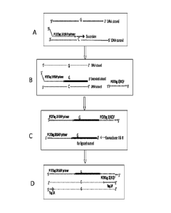
FIG. 1 shows amplification of a gene of interest using extension of a gene
specific primer with a
universal PCR tag and a subsequent single strand ligation to a second
universal tag followed by
exonuclease clean-up and amplification utilizing tag 1 and 2 (Approach 1).
FIG. 2 shows amplification of a gene of interest using a gene specific
biotinylated primer with a
universal tag 3 that is extended on a template then ligated downstream to a
gene specific
phosphorylated oligonucleotide tag 4 on the same strand. This product is
subsequently amplified
utilizing tag 3 and 4 (Concept2).
FIG. 3 shows the universal PCR products from both Approach 1 and 2 procedures
from FIGS. 1
and 2, which can be identified using a post-PCR reaction (goldPLEX, Sequenom).
FIG. 4 shows MALDI-TOF MS spectra for genotyping of a single nucleotide
polymorphism
(dbSNP# rs10063237) using a Approach 1 protocol.
FIG. 5A shows MALDI-TOF MS spectra for genotyping of rs1015731 using a
Approach 2 protocol.
FIG. 5B shows MALDI-TOF MS spectra for genotyping 12 targets (e.g., a 12plex
reaction) using a
Approach 2 protocol.
FIG. 5C shows MALDI-TOF MS spectra for genotyping a 19plex reaction using a
Approach 2
protocol.
FIG. 5D shows MALDI-TOF MS spectra for genotyping a 35plex reaction using a
Approach 2
protocol.
FIG 5E shows the genotypes acquired from MALDI-TOF MS spectra from FIG 50
(19plex) and FIG
5D (35plex).
FIG. 6 shows PCR amplification and post-PCR primer extension with allele-
specific extension
primers containing allele-specific mass tags.
6
CA 02742272 2011-04-29
WO 2010/056513 PCT/US2009/062239
FIG. 7 shows MALDI-TOF MS spectra for 35plex genotyping using post-PCR primer
extension with
allele-specific extension primers containing allele-specific mass tags as a
readout.
FIG. 8 shows MALDI-TOF MS spectra for genotyping of rs1000586 and rs10131894.
FIG. 9 shows oligonucleotides mass tags corresponding to a 70plex assay. All
oligos were diluted
to a final total concentration of 10 pmol and spotted on a 384 well chip.
Values for area, peak
height and signal-to-noise ratio were collected from Typer 3.4 (Sequenom).
FIG. 10 shows peak areas for oligonucleotides mass tags corresponding to
70plex assay sorted by
nucleotide composition. All oligos were diluted to a final total concentration
of 10 pmol and spotted
on a 384 well chip. Area values were collected from Typer 3.4 (Sequenom).
FIG. 11A shows a MALDI-TOF MS spectrum (zoomed views) of oligonucleotide tags
corresponding to a 100plex assay. FIG. 11B shows signal to noise ratios of
oligonucleotide tags
corresponding to a 100plex assay. All oligos were diluted to a final total
concentration of 10, 5, 2.5
or lpmol, with 8 replicates spotted on a 384 well chip. Values for signal-to-
noise ratio were
collected from Typer 3.4 (Sequenom). FIG. 11C shows a MALDI-TOF MS spectrum
(zoomed
views) of a 100plex assay after PCR amplification and post-PCR primer
extension with allele-
specific extension primers containing allele-specific mass tags.
FIG. 12 shows extension rates for a 5plex reaction. Comparing extension
oligonucleotides with or
without a deoxyinosine, and either standard ddNTPs or nucleotides containing a
biotin moiety.
Extension rates were calculated by dividing the area of extended product by
the total area of the
peak (extended product and unextended oligonucleotide) in Typer 3.4
(Sequenom). All
experiments compare six DNAs.
FIG. 13 shows extension rates for 7plex and 5plex reactions over two DNAs.
Results compare
extension by a single biotinylated ddNTP or a biotinylated dNTP and terminated
by an unmodified
ddNTP, and final amounts of biotinylated dNTP or ddNTP of 210 or 420 pmol
added to the
reaction. Extension rates were calculated by dividing the area of extended
product by the total
area (extended product and unextended oligonucleotide) in Typer 3.4. All
experiments include two
replicates of two Centre de'Etude du Polymorphisme Humain (CEPH) DNAs, NA07019
and
NA11036.
7
CA 02742272 2011-04-29
WO 2010/056513 PCT/US2009/062239
FIG. 14 shows a comparison of goldPLEX enzyme concentrations in an extension
reaction using a
70plex assay. All assays followed the same protocol except for the amount of
goldPLEX enzyme
used. All experiments include four replicates of the two CEPH DNAs NA06991 and
NA07019. The
results compare the signal-to-noise ratios of the extension products from
Typer 3.4 (Sequenom)..
FIG. 15 shows a comparison of goldPLEX buffer concentration in extension
reactions using a
70plex assay. All assays followed the same protocol except for the amount of
goldPLEX buffer
used. All experiments include four replicates of the two CEPH DNAs NA06991 and
NA07019. The
results compare the signal-to-noise ratios of the extension products from
Typer 3.4 (Sequenom).
FIG. 16, 17, 18 and 19 show a comparison of extension oligonucleotide
concentration in extension
reactions using a 70plex assay. All assays followed the same protocol except
for the amount of
extension oligonucleotide used. All experiments include four replicates of the
two CEPH DNAs
NA06991 and NA07019. The results compare the signal-to-noise ratios of the
extension products
from Typer 3.4 (Sequenom).
FIG. 20 and 21 show a comparison of biotinylated ddNTP concentration in
extension reactions
using a 70plex assay. All assays followed the same protocol except for the
amount of biotinylated
ddNTP used (value indicates final amount of each biotinylated nucleotide). All
experiments include
four replicates of the two CEPH DNAs NA06991 and NA07019. The results compare
the signal-to-
noise ratios of the extension products from Typer 3.4 (Sequenom).
FIG. 22 shows a comparison of Solulink and Dynabeads MyOne 01 magnetic
streptavidin beads
for capturing the extend products. A total amount of 10 pmol of each
oligonucleotide corresponding
to the two possible alleles for assay rs1000586 were bound to the magnetic
streptavidin beads, in
the presence of either water or varying quantities of biotinylated dNTPs
(total 10, 100 or 500 pmol).
The mass tags were then cleaved from the bound oligonucleotide with 10 U of
endonuclease V.
The results compare the area of the mass tag peaks from Typer 3.4 (Sequenom)
and are listed in
comparison with 10 pmol of an oligonucleotide which has a similar mass.
FIG. 23 shows analysis of the ability of endonuclease V to cleave an extension
product containing
a deoxyinosine nucleotide in different locations. The oligonucleotides were
identical aside from the
deoxyinosine being 10, 15, 20 or 25 bases from the 3' end of the
oligonucleotide. After binding the
8
CA 02742272 2011-04-29
WO 2010/056513 PCT/US2009/062239
oligonucleotide to the magnetic streptavidin beads, the supernatant was
collected, cleaned by a
nucleotide removal kit (Qiagen) and then cleaved by treatment with
endonuclease V (termed
unbound oligo). The beads were washed, and cleaved with endonuclease V, as
outlined in
protocol section (termed captured/cleaved). The results compare the area of
the peaks from Typer
3.4 (Sequenom), and are listed as a percentage of oligonucleotide cleaved by
endonuclease V
without being bound to magnetic streptavidin beads.
FIG. 24 shows a comparison of magnetic streptavidin beads and endonuclease V
concentration
using a 70plex assay. All assays were conducted using the same conditions
except for the amount
of magnetic streptavidin beads and endonuclease V. All experiments include
four replicates of the
CEPH DNA NA11036. The results compare the signal-to-noise ratio from Typer
3.4.
FIG. 25 and 26 show a comparison of magnetic streptavidin beads and
endonuclease V
concentration using a 70plex assay. All assays followed the same protocol
except for the amount
of magnetic streptavidin beads and endonuclease V. All experiments include
four replicates of the
two CEPH DNAs NA06991 and NA07019. The results compare the signal-to-noise
ratio from
Typer 3.4.
Detailed Description
Methods for determining the presence or absence of a plurality of target
nucleic acids in a
composition described herein find multiple uses by the person of ordinary
skill in the art (hereafter
referred to herein as the "person of ordinary skill"). Such methods can be
utilized, for example, to:
(a) rapidly determine whether a particular target sequence is present in a
sample; (b) perform
mixture analysis, e.g., identify a mixture and/or its composition or determine
the frequency of a
target sequence in a mixture (e.g., mixed communities, quasispecies); (c)
detect sequence
variations (e.g., mutations, single nucleotide polymorphisms) in a sample; (d)
perform haplotyping
determinations; (e) perform microorganism (e.g., pathogen) typing; (f) detect
the presence or
absence of a microorganism target sequence in a sample; (g) identify disease
markers; (h) detect
microsatellites; (i) identify short tandem repeats; (j) identify an organism
or organisms; (k) detect
allelic variations; (I) determine allelic frequency; (m) determine methylation
patterns; (n) perform
epigenetic determinations; (o) re-sequence a region of a biomolecule; (p)
perform analyses in
human clinical research and medicine (e.g. cancer marker detection, sequence
variation detection;
detection of sequence signatures favorable or unfavorable for a particular
drug administration), (q)
perform HLA typing; (r) perform forensics analyses; (s) perform vaccine
quality control analyses; (t)
9
CA 02742272 2011-04-29
WO 2010/056513 PCT/US2009/062239
monitor treatments; (u) perform vector identity analyses; (v) perform vaccine
or production strain
quality control and (w) test strain identity (x) plants. Such methods also may
be utilized, for
example, in a variety of fields, including, without limitation, in commercial,
education, medical,
agriculture, environmental, disease monitoring, military defense, and
forensics fields.
Target Nucleic Acids
As used herein, the term "nucleic acid" refers to an oligonucleotide or
polynucleotide, including,
without limitation, natural nucleic acids (e.g., deoxyribonucleic acid (DNA),
ribonucleic acid (RNA)),
synthetic nucleic acids, non-natural nucleic acids (e.g., peptide nucleic acid
(PNA)), unmodified
nucleic acids, modified nucleic acids (e.g., methylated DNA or RNA, labeled
DNA or RNA, DNA or
RNA having one or more modified nucleotides). Reference to a nucleic acid as a
"polynucleotide"
refers to two or more nucleotides or nucleotide analogs linked by a covalent
bond. Nucleic acids
may be any type of nucleic acid suitable for use with processes described
herein. A nucleic acid in
certain embodiments can be DNA (e.g., complementary DNA (cDNA), genomic DNA
(gDNA),
plasmids and vector DNA and the like), RNA (e.g., viral RNA, message RNA
(mRNA), short
inhibitory RNA (siRNA), ribosomal RNA (rRNA), tRNA and the like), and/or DNA
or RNA analogs
(e.g., containing base analogs, sugar analogs and/or a non-native backbone and
the like). A
nucleic acid can be in any form useful for conducting processes herein (e.g.,
linear, circular,
supercoiled, single-stranded, double-stranded and the like). A nucleic acid
may be, or may be
from, a plasmid, phage, autonomously replicating sequence (ARS), centromere,
artificial
chromosome, chromosome, a cell, a cell nucleus or cytoplasm of a cell in
certain embodiments. A
nucleic acid in some embodiments is from a single chromosome (e.g., a nucleic
acid sample may
be from one chromosome of a sample obtained from a diploid organism). In the
case of fetal
nucleic acid, the nucleic acid may be from the paternal allele, the maternal
allele or the maternal
and paternal allele.
The term "species," as used herein with reference to a target nucleic acid,
amplicon, primer,
sequence tag, polynucleotide or oligonucleotide, refers to one nucleic acid
having a nucleotide
sequence that differs by one or more nucleotides from the nucleotide sequence
of another nucleic
acid when the nucleotide sequences are aligned. Thus, a first nucleic acid
species differs from a
second nucleic acid species when the sequences of the two species, when
aligned, differ by one or
more nucleotides (e.g., about 1, 2, 3, 4, 5, 10, 15, 20, 25, 30, 35, 40, 45,
50, 55, 60, 65, 70, 75, 80,
85, 90, 95, 100 or more than 100 nucleotide differences). In certain
embodiments, the number of
CA 02742272 2011-04-29
WO 2010/056513 PCT/US2009/062239
nucleic acid species, such as target nucleic acid species, amplicon species or
extended
oligonucleotide species, includes, but is not limited to about 2 to about
10000 nucleic acid species,
about 2 to about 1000 nucleic acid species, about 2 to about 500 nucleic acid
species, or
sometimes about 2, 3, 4, 5, 6, 7, 8, 9, 10, 11, 12, 13, 14, 15, 16, 17, 18,
19, 20, 25, 30, 35, 40, 45,
50, 55, 60, 65, 70, 75, 80, 80, 85, 90, 95, 100, 125, 150, 175, 200, 225, 250,
275, 300, 325, 350,
375, 400, 425, 450, 475, 500, 600, 700, 800, 900,1000, 2000, 3000, 4000, 5000,
6000, 7000,
8000, 9000 or 10000 nucleic acid species.
As used herein, the term "nucleotides" refers to natural and non-natural
nucleotides. Nucleotides
include, but are not limited to, naturally occurring nucleoside mono-, di-,
and triphosphates:
deoxyadenosine mono-, di- and triphosphate; deoxyguanosine mono-, di- and
triphosphate;
deoxythymidine mono-, di- and triphosphate; deoxycytidine mono-, di- and
triphosphate;
deoxyuridine mono-, di- and triphosphate; and deoxyinosine mono-, di- and
triphosphate (referred
to herein as dA, dG, dT, dC, dU and dl, or A, G, T, C, U and l respectively).
Nucleotides also
include, but are not limited to, modified nucleotides and nucleotide analogs.
Modified nucleotides
and nucleotide analogs include, without limitation, deazapurine nucleotides,
e.g., 7-deaza-
deoxyguanosine (7-deaza-dG) and 7-deaza-deoxyadenosine (7-deaza-dA) mono-, di-
and
triphosphates, deutero-deoxythymidine (deutero-dT) mon-, di- and
triphosphates, methylated
nucleotides e.g., 5-methyldeoxycytidine triphosphate, 13C/15N
labelled nucleotides and
deoxyinosine mono-, di- and triphosphate. Modified nucleotides, isotopically
enriched nucleotides,
depleted nucleotides, tagged and labeled nucleotides and nucleotide analogs
can be obtained
using a variety of combinations of functionality and attachment positions.
The term "composition" as used herein with reference to nucleic acids refers
to a tangible item that
includes one or more nucleic acids. A composition sometimes is a sample
extracted from a
source, but also a composition of all samples at the source, and at times is
the source of one or
more nucleic acids.
A nucleic acid sample may be derived from one or more sources. A sample may be
collected from
an organism, mineral or geological site (e.g., soil, rock, mineral deposit,
fossil), or forensic site
(e.g., crime scene, contraband or suspected contraband), for example. Thus, a
source may be
environmental, such as geological, agricultural, combat theater or soil
sources, for example. A
source also may be from any type of organism such as any plant, fungus,
protistan, moneran, virus
or animal, including but not limited, human, non-human, mammal, reptile,
cattle, cat, dog, goat,
11
CA 02742272 2011-04-29
WO 2010/056513 PCT/US2009/062239
swine, pig, monkey, ape, gorilla, bull, cow, bear, horse, sheep, poultry,
mouse, rat, fish, dolphin,
whale, and shark, or any animal or organism that may have a detectable nucleic
acids. Sources
also can refer to different parts of an organism such as internal parts,
external parts, living or non-
living cells, tissue, fluid and the like. A sample therefore may be a
"biological sample," which refers
to any material obtained from a living source or formerly-living source, for
example, an animal such
as a human or other mammal, a plant, a bacterium, a fungus, a protist or a
virus. A source can be
in any form, including, without limitation, a solid material such as a tissue,
cells, a cell pellet, a cell
extract, or a biopsy, or a biological fluid such as urine, blood, saliva,
amniotic fluid, exudate from a
region of infection or inflammation, or a mouth wash containing buccal cells,
hair, cerebral spinal
fluid and synovial fluid and organs. A sample also may be isolated at a
different time point as
compared to another sample, where each of the samples are from the same or a
different source.
A nucleic acid may be from a nucleic acid library, such as a cDNA or RNA
library, for example. A
nucleic acid may be a result of nucleic acid purification or isolation and/or
amplification of nucleic
acid molecules from the sample. Nucleic acid provided for sequence analysis
processes described
herein may contain nucleic acid from one sample or from two or more samples
(e.g., from 1, 2, 3,
4, 5, 6, 7, 8, 9, 10, 11, 12, 13, 14, 15, 16, 17, 18, 19, 20, 25, 50, 75, 100,
200, 300, 400, 500, 600,
700, 800, 900 or 1000 or more samples).
Nucleic acids may be treated in a variety of manners. For example, a nucleic
acid may be reduced
in size (e.g., sheared, digested by nuclease or restriction enzyme, de-
phosphorylated, de-
methylated), increased in size (e.g., phosphorylated, reacted with a
methylation-specific reagent,
attached to a detectable label), treated with inhibitors of nucleic acid
cleavage and the like.
Nucleic acids may be provided for conducting methods described herein without
processing, in
certain embodiments. In some embodiments, nucleic acid is provided for
conducting methods
described herein after processing. For example, a nucleic acid may be
extracted, isolated, purified
or amplified from a sample. The term "isolated" as used herein refers to
nucleic acid removed from
its original environment (e.g., the natural environment if it is naturally
occurring, or a host cell if
expressed exogenously), and thus is altered "by the hand of man" from its
original environment.
An isolated nucleic acid generally is provided with fewer non-nucleic acid
components (e.g.,
protein, lipid) than the amount of components present in a source sample. A
composition
comprising isolated nucleic acid can be substantially isolated (e.g., about
90%, 91%, 92%, 93%,
94%, 95%, 96%, 97%, 98%, 99% or greater than 99% free of non-nucleic acid
components). The
term "purified" as used herein refers to nucleic acid provided that contains
fewer nucleic acid
12
CA 02742272 2011-04-29
WO 2010/056513 PCT/US2009/062239
species than in the sample source from which the nucleic acid is derived. A
composition
comprising nucleic acid may be substantially purified (e.g., about 90%, 91%,
92%, 93%, 94%,
95%, 96%, 97%, 98%, 99% or greater than 99% free of other nucleic acid
species).
Nucleic acids may be processed by a method that generates nucleic acid
fragments, in certain
embodiments, before providing nucleic acid for a process described herein. In
some
embodiments, nucleic acid subjected to fragmentation or cleavage may have a
nominal, average
or mean length of about 5 to about 10,000 base pairs, about 100 to about 1,00
base pairs, about
100 to about 500 base pairs, or about 10, 15, 20, 25, 30, 35, 40, 45, 50, 55,
60, 65, 70, 75, 80, 85,
90, 95, 100, 200, 300, 400, 500, 600, 700, 800, 900, 1000, 2000, 3000, 4000,
5000, 6000, 7000,
8000, 9000 or 10000 base pairs. Fragments can be generated by any suitable
method known in
the art, and the average, mean or nominal length of nucleic acid fragments can
be controlled by
selecting an appropriate fragment-generating procedure. In certain
embodiments, nucleic acid of a
relatively shorter length can be utilized to analyze sequences that contain
little sequence variation
and/or contain relatively large amounts of known nucleotide sequence
information. In some
embodiments, nucleic acid of a relatively longer length can be utilized to
analyze sequences that
contain greater sequence variation and/or contain relatively small amounts of
unknown nucleotide
sequence information.
As used herein, the term "target nucleic acid" refers to any nucleic acid
species of interest in a
sample. A target nucleic acid includes, without limitation, (i) a particular
allele amongst two or
more possible alleles, and (ii) a nucleic acid having, or not having, a
particular mutation, nucleotide
substitution, sequence variation, repeat sequence, marker or distinguishing
sequence. As used
herein, the term "different target nucleic acids" refers to nucleic acid
species that differ by one or
more features. Features include, without limitation, one or more methyl groups
or a methylation
state, one or more phosphates, one or more acetyl groups, and one or more
deletions, additions or
substitutions of one or more nucleotides. Examples of one or more deletions,
additions or
substitutions of one or more nucleotides include, without limitation, the
presence or absence of a
particular mutation, presence or absence of a nucleotide substitution (e.g.,
single nucleotide
polymorphism (SNP)), presence or absence of a repeat sequence (e.g., di-, tri-
, tetra-, penta-
nucleotide repeat), presence or absence of a marker (e.g., microsatellite) and
presence of absence
of a distinguishing sequence (e.g., a sequence that distinguishes one organism
from another (e.g.,
a sequence that distinguishes one viral strain from another viral strain)).
Different target nucleic
13
CA 02742272 2011-04-29
WO 2010/056513 PCT/US2009/062239
acids may be distinguished by any known method, for example, by mass, binding,
distinguishable
tags and the like, as described herein.
As used herein, the term "plurality of target nucleic acids" refers to more
than one target nucleic
acid. A plurality of target nucleic acids can be about 2 to about 10000
nucleic acid species, about
2 to about 1000 nucleic acid species, about 2 to about 500 nucleic acid
species, or sometimes
about 2, 3, 4, 5, 6, 7, 8, 9, 10, 11, 12, 13, 14, 15, 16, 17, 18, 19, 20, 25,
30, 35, 40, 45, 50, 55, 60,
65, 70, 75, 80, 80, 85, 90, 95, 100, 125, 150, 175, 200, 225, 250, 275, 300,
325, 350, 375, 400,
425, 450, 475, 500, 600, 700, 800, 900,1000, 2000, 3000, 4000, 5000, 6000,
7000, 8000, 9000 or
10000 nucleic acid species, in certain embodiments. Detection or
identification of nucleic acids
results in detection of the target and can indicate the presence or absence of
a particular mutation,
sequence variation (mutation or polymorphism). Within the plurality of target
nucleic acids, there
may be detection of the same or different target nucleic acids. The plurality
of target nucleic acids
may also be identified quantitatively as well as qualitatively in terms of
identification. Also refer to
multiplexing below.
Amplification and Extension
A nucleic acid (e.g., a target nucleic acid) can be amplified in certain
embodiments. As used
herein, the term "amplifying," and grammatical variants thereof, refers to a
process of generating
copies of a template nucleic acid. For example, nucleic acid template may be
subjected to a
process that linearly or exponentially generates two or more nucleic acid
amplicons (copies) having
the same or substantially the same nucleotide sequence as the nucleotide
sequence of the
template, or a portion of the template. Nucleic acid amplification often is
specific (e.g., amplicons
have the same or substantially the same sequence), and can be non-specific
(e.g., amplicons have
different sequences) in certain embodiments. Nucleic acid amplification
sometimes is beneficial
when the amount of target sequence present in a sample is low. By amplifying
the target
sequences and detecting the amplicon synthesized, sensitivity of an assay can
be improved, since
fewer target sequences are needed at the beginning of the assay for detection
of a target nucleic
acid. A target nucleic acid sometimes is not amplified prior to hybridizing an
extension
oligonucleotide, in certain embodiments.
Amplification conditions are known and can be selected for a particular
nucleic acid that will be
amplified. Amplification conditions include certain reagents some of which can
include, without
14
CA 02742272 2011-04-29
WO 2010/056513 PCT/US2009/062239
limitation, nucleotides (e.g., nucleotide triphosphates), modified
nucleotides, oligonucleotides (e.g.,
primer oligonucleotides for polymerase-based amplification and oligonucleotide
building blocks for
ligase-based amplification), one or more salts (e.g., magnesium-containing
salt), one or more
buffers, one or more polymerizing agents (e.g., ligase enzyme, polymerase
enzyme), one or more
nicking enzymes (e.g., an enzyme that cleaves one strand of a double-stranded
nucleic acid) and
one or more nucleases (e.g., exonuclease, endonuclease, RNase). Any polymerase
suitable for
amplification may be utilized, such as a polymerase with or without
exonuclease activity, DNA
polymerase and RNA polymerase, mutant forms of these enzymes, for example. Any
ligase
suitable for joining the 5 of one oligonucleotide to the 3' end of another
oligonucleotide can be
utilized. Amplification conditions also can include certain reaction
conditions, such as isothermal or
temperature cycle conditions. Methods for cycling temperature in an
amplification process are
known, such as by using a thermocycle device. Amplification conditions also
can, in some
embodiments, include an emulsion agent (e.g., oil) that can be utilized to
form multiple reaction
compartments within which single nucleic acid molecule species can be
amplified.
A strand of a single-stranded nucleic acid target can be amplified and one or
two strands of a
double-stranded nucleic acid target can be amplified. An amplification product
(amplicon), in some
embodiments, is about 10 nucleotides to about 10,000 nucleotides in length,
about 10 to about
1000 nucleotides in length, about 10 to about 500 nucleotides in length, 10 to
about 100
nucleotides in length, and sometimes about 10, 11, 12, 13, 14, 15, 16, 17, 18,
19, 20, 25, 30, 35,
40, 45, 50, 55, 60, 65, 70, 75, 80, 80, 85, 90, 95, 100, 125, 150, 175, 200,
225, 250, 275, 300, 325,
350, 375, 400, 425, 450, 475, 500, 600, 700, 800, 900 or 1000 nucleotides in
length.
Any suitable amplification technique and amplification conditions can be
selected for a particular
nucleic acid for amplification. Known amplification processes include, without
limitation,
polymerase chain reaction (PCR), extension and ligation, ligation
amplification (or ligase chain
reaction (LCR)) and amplification methods based on the use of Q-beta replicase
or template-
dependent polymerase (see US Patent Publication Number US20050287592). Also
useful are
strand displacement amplification (SDA), thermophilic SDA, nucleic acid
sequence based
amplification (3SR or NASBA) and transcription-associated amplification (TAA).
Reagents,
apparatus and hardware for conducting amplification processes are commercially
available, and
amplification conditions are known and can be selected for the target nucleic
acid at hand.
CA 02742272 2011-04-29
WO 2010/056513 PCT/US2009/062239
Polymerase-based amplification can be effected, in certain embodiments, by
employing universal
primers. In such processes, hybridization regions that hybridize to one or
more universal primers
are incorporated into a template nucleic acid. Such hybridization regions can
be incorporated into
(i) a primer that hybridizes to a target nucleic acid and is extended, and/or
(ii) an oligonucleotide
that is joined (e.g., ligated using a ligase enzyme) to a target nucleic acid
or a product of (i), for
example. Amplification processes that involve universal primers can provide an
advantage of
amplifying a plurality of target nucleic acids using only one or two
amplification primers, for
example.
Figure 1 shows certain embodiments of amplification processes. In certain
embodiments, only one
primer is utilized for amplification (e.g., Figure 1A). In certain
embodiments, two primers are
utilized. Under amplification conditions at least one primer has a
complementary distinguishable
tag. The gene specific extend primer has a 5' universal PCRTag1R (e.g., Figure
1A). It may be
extended on any nucleic acid, for example genomic DNA. The DNA or the PCR
Tag1R gene
specific extend primer may be biotinylated, to facilitate clean up of the
reaction. The extended
strand then is ligated by a single strand ligase to a universal phosphorylated
oligonucleotide, which
has a sequence that is the reverse complement of Tag2F (universal PCR primer;
Figure 1B). To
facilitate cleanup in the next step, the phosphorylated oligonucleotide can
include exonuclease
resistant nucleotides at its 3' end. During the exonuclease treatment, all non-
ligated extended
strands are degraded, whereas ligated products are protected and remain in the
reaction (e.g.,
Figure 1C). A universal PCR then is performed, using Tag1R and the Tag2F
primers, to amplify
multiple targets (e.g., Figure 1D).
Figure 2 also shows certain embodiments of amplification processes. In some
embodiments, a
method involving primer extension and ligation takes place in the same
reaction (e.g., Figure 2A).
Biotinylated PCRTag3R gene-specific primer is an extension primer. The
phosphorylated
oligonucleotide has a gene-specific sequence and binds about 40 bases (e.g., 4
to 100 or more)
away from the primer extension site, to the same strand of DNA. Thus a DNA
polymerase, such as
Stoffel polymerase, extends the strand, until it reaches the phosphorylated
oligonucleotide. A
ligase enzyme ligates the gene specific sequence of the phosphorylated
oligonucleotide to the
extended strand. The 3' end of phosphorylated oligonucleotide has PCRTag4(RC)F
as its
universal tag. The biotinylated extended strands then are bound to
streptavidin beads. This
approach facilitates cleanup of the reaction (e.g., Figure 2B). DNA, such as
genomic DNA, and the
gene specific phosphorylated oligonucleotides are washed away. A universal PCR
then is
16
CA 02742272 2011-04-29
WO 2010/056513 PCT/US2009/062239
performed, using Tag3R and Tag4F as primers, to amplify different genes of
interest (e.g., Figure
2C).
Certain nucleic acids can be extended in certain embodiments. The term
"extension," and
grammatical variants thereof, as used herein refers to elongating one strand
of a nucleic acid. For
example, an oligonucleotide that hybridizes to a target nucleic acid or an
amplicon generated from
a target nucleic acid can be extended in certain embodiments. An extension
reaction is conducted
under extension conditions, and a variety of such conditions are known and
selected for a
particular application. Extension conditions include certain reagents,
including without limitation,
one or more oligonucleotides, extension nucleotides (e.g., nucleotide
triphosphates (dNTPs)),
terminating nucleotides (e.g., one or more dideoxynucleotide triphosphates
(ddNTPs)), one or
more salts (e.g., magnesium-containing salt), one or more buffers (e.g., with
beta-NAD, Triton X-
100), and one or more polymerizing agents (e.g., DNA polymerase, RNA
polymerase). Extension
can be conducted under isothermal conditions or under non-isothermal
conditions (e.g.,
thermocycled conditions), in certain embodiments. One or more nucleic acid
species can be
extended in an extension reaction, and one or more molecules of each nucleic
acid species can be
extended. A nucleic acid can be extended by one or more nucleotides, and in
some embodiments,
the extension product is about 10 nucleotides to about 10,000 nucleotides in
length, about 10 to
about 1000 nucleotides in length, about 10 to about 500 nucleotides in length,
10 to about 100
nucleotides in length, and sometimes about 10, 11, 12, 13, 14, 15, 16, 17, 18,
19, 20, 25, 30, 35,
40, 45, 50, 55, 60, 65, 70, 75, 80, 80, 85, 90, 95, 100, 125, 150, 175, 200,
225, 250, 275, 300, 325,
350, 375, 400, 425, 450, 475, 500, 600, 700, 800, 900 or 1000 nucleotides in
length. Incorporation
of a terminating nucleotide (e.g., ddNTP), the hybridization location, or
other factors, can determine
the length to which the oligonucleotide is extended. In certain embodiments,
amplification and
extension processes are carried out in the same detection procedure.
Any suitable extension reaction can be selected and utilized. An extension
reaction can be
utilized, for example, to discriminate SNP alleles by the incorporation of
deoxynucleotides and/or
dideoxynucleotides to an extension oligonucleotide that hybridizes to a region
adjacent to the SNP
site in a target nucleic acid. The primer often is extended with a polymerase.
In some
embodiments, the oligonucleotide is extended by only one deoxynucleotide or
dideoxynucleotide
complementary to the SNP site. In some embodiments, an oligonucleotide may be
extended by
dNTP incorporation and terminated by a ddNTP, or terminated by ddNTP
incorporation without
dNTP extension in certain embodiments. One or more dNTP and/or ddNTP used
during the
17
CA 02742272 2011-04-29
WO 2010/056513 PCT/US2009/062239
extension reaction are labeled with a moiety allowing immobilization to a
solid support, such as
biotin, in some embodiments. Extension may be carried out using unmodified
extension
oligonucleotides and unmodified dideoxynucleotides, unmodified extension
oligonucleotides and
biotinylated dideoxynucleotides, extension oligonucleotides containing a
deoxyinosine and
unmodified dideoxynucleotides, extension oligonucleotides containing a
deoxyinosine and
biotinylated dideoxynucleotides, extension by biotinylated dideoxynucleotides,
or extension by
biotinylated deoxynucleotide and/or unmodified dideoxynucleotides, in some
embodiments
Any suitable type of nucleotides can be incorporated into an amplification
product or an extension
product. Nucleotides may be naturally occurring nucleotides, terminating
nucleotides, or non-
naturally occurring nucleotides (e.g., nucleotide analog or derivative), in
some embodiments.
Certain nucleotides can comprise a detectable label and/or a member of a
binding pair (e.g., the
other member of the binding pair may be linked to a solid phase), in some
embodiments.
A solution containing amplicons produced by an amplification process, or a
solution containing
extension products produced by an extension process, can be subjected to
further processing. For
example, a solution can be contacted with an agent that removes phosphate
moieties from free
nucleotides that have not been incorporated into an amplicon or extension
product. An example of
such an agent is a phosphatase (e.g., alkaline phosphatase). Amplicons and
extension products
also may be associated with a solid phase, may be washed, may be contacted
with an agent that
removes a terminal phosphate (e.g., exposure to a phosphatase), may be
contacted with an agent
that removes a terminal nucleotide (e.g., exonuclease), may be contacted with
an agent that
cleaves (e.g., endonuclease, ribonuclease), and the like.
The term "oligonucleotide" as used herein refers to two or more nucleotides or
nucleotide analogs
linked by a covalent bond. An oligonucleotide is of any convenient length, and
in some
embodiments is about 5 to about 200 nucleotides in length, about 5 to about
150 nucleotides in
length, about 5 to about 100 nucleotides in length, about 5 to about 75
nucleotides in length or
about 5 to about 50 nucleotides in length, and sometimes is about 5, 6, 7, 8,
9, 10, 11, 12, 13, 14,
15, 16, 17, 18, 19, 20, 25, 30, 35, 40, 45, 50, 55, 60, 65, 70, 75, 80, 80,
85, 90, 95, 100, 125, 150,
175, or 200 nucleotides in length. Oligonucleotides may include
deoxyribonucleic acid (DNA),
ribonucleic acid (RNA), naturally occurring and/or non-naturally occurring
nucleotides or
combinations thereof and any chemical or enzymatic modification thereof (e.g.
methylated DNA,
DNA of modified nucleotides). The length of an oligonucleotide sometimes is
shorter than the
18
CA 02742272 2011-04-29
WO 2010/056513 PCT/US2009/062239
length of an amplicon or target nucleic acid, but not necessarily shorter than
a primer or
polynucleotide used for amplification. An oligonucleotide often comprises a
nucleotide
subsequence or a hybridization sequence that is complementary, or
substantially complementary,
to an amplicon, target nucleic acid or complement thereof (e.g., about 95%,
96%, 97%, 98%, 99%
or greater than 99% identical to the amplicon or target nucleic acid
complement when aligned). An
oligonucleotide may contain a nucleotide subsequence not complementary to, or
not substantially
complementary to, an amplicon, target nucleic acid or complement thereof
(e.g., at the 3' or 5' end
of the nucleotide subsequence in the primer complementary to or substantially
complementary to
the amplicon). An oligonucleotide in certain embodiments, may contain a
detectable molecule
(e.g., a tag,. fluorophore, radioisotope, colormetric agent, particle, enzyme
and the like) and/or a
member of a binding pair, in certain embodiments.
The term "in solution" as used herein refers to a liquid, such as a liquid
containing one or more
nucleic acids, for example. Nucleic acids and other components in solution may
be dispersed
throughout, and a solution often comprises water (e.g., aqueous solution). A
solution may contain
any convenient number of oligonucleotide species, and there often are at least
the same number of
oligonucleotide species as there are amplicon species or target nucleic acid
species to be
detected.
The term "hybridization sequence" as used herein refers to a nucleotide
sequence in an
oligonucleotide capable of specifically hybridizing to an amplicon, target
nucleic acid or
complement thereof. The hybridization sequence is readily designed and
selected and can be of a
length suitable for hybridizing to an amplicon, target sequence or complement
thereof in solution
as described herein. In some embodiments, the hybridization sequence in each
oligonucleotide is
about 5 to about 200 nucleotides in length (e.g., about 5 to 10, about 10 to
15, about 15 to 20,
about 20 to 25, about 25 to 30, about 30 to 35, about 35 to 40, about 40 to
45, or about 45 to 50,
about 50 to 70, about 80 to 90, about 90 to 110, about 100 to 120, about 110
to 130, about 120 to
140, about 130 to 150, about 140 to 160, about 150 to 170, about 160 to 180,
about 170 to 190,
about 180 to 200 nucleotides in length).
The term "hybridization conditions" as used herein refers to conditions under
which two nucleic
acids having complementary nucleotide sequences can interact with one another.
Hybridization
conditions can be high stringency, medium stringency or low stringency, and
conditions for these
19
CA 02742272 2011-04-29
WO 2010/056513 PCT/US2009/062239
varying degrees of stringency are known. Hybridization conditions often are
selected that allow for
amplification and/or extension depending on the application of interest.
The term "specifically hybridizing to one amplicon or target nucleic acid" as
used herein refers to
hybridizing substantially to one amplicon species or target nucleic acid
species and not
substantially hybridizing to other amplicon species or target nucleic acid
species in the solution.
Specific hybridization rules out mismatches so that, for example, an
oligonucleotide may be
designed to hybridize specifically to a certain allele and only to that
allele. An oligonucleotide that
is homogenously matched or complementary to an allele will specifically
hybridize to that allele,
whereas if there is one or more base mismatches then no hybridization will
occur.
The term "hybridization location" as used herein refers to a specific location
on an amplicon or
target nucleic acid to which another nucleic acid hybridizes. In certain
embodiments, the terminus
of an oligonucleotide is adjacent to or substantially adjacent to a site on an
amplicon species or
target nucleic acid species that has a different sequence than another
amplicon species or target
nucleic acid species. The terminus of an oligonucleotide is "adjacent" to a
site when there are no
nucleotides between the site and the oligonucleotide terminus. The terminus of
an oligonucleotide
is "substantially adjacent" to a site when there are 1, 2, 3, 4, 5, 6, 7, 8, 9
or 10 nucleotides between
the site and the oligonucleotide terminus, in certain embodiments.
Capture Agents and Solid Phases
One or more capture agents may be utilized for the methods described herein.
There are several
different types of capture agents available for processes described herein,
including, without
limitation, members of a binding pair, for example. Examples of binding pairs,
include, without
limitation, (a) non-covalent binding pairs (e.g., antibody/antigen,
antibody/antibody,
antibody/antibody fragment, antibody/antibody receptor, antibody/protein A or
protein G,
hapten/anti-hapten, biotin/avidin, biotin/streptavidin, folic acid/folate
binding protein and vitamin
B12/intrinsic factor; and (b) covalent attachment pairs (e.g.,
sulfhydryl/maleimide,
sulfhydryl/haloacetyl derivative, amine/isotriocyanate, amine/succinimidyl
ester, and amine/sulfonyl
halides), and the like. In some embodiments, one member of a binding pair is
in association with
an extended oligonucleotide or amplification product and another member in
association with a
solid phase. The term "in association with" as used herein refers to an
interaction between at least
two units, where the two units are bound or linked to one another, for
example.
CA 02742272 2011-04-29
WO 2010/056513 PCT/US2009/062239
The term "solid support" or "solid phase" as used herein refers to an
insoluble material with which
nucleic acid can be associated. Examples of solid supports for use with
processes described
herein include, without limitation, arrays, beads (e.g., paramagnetic beads,
magnetic beads,
microbeads, nanobeads) and particles (e.g., microparticles, nanoparticles).
Particles or beads
having a nominal, average or mean diameter of about 1 nanometer to about 500
micrometers can
be utilized, such as those having a nominal, mean or average diameter, for
example, of about 10
nanometers to about 100 micrometers; about 100 nanometers to about 100
micrometers; about 1
micrometer to about 100 micrometers; about 10 micrometers to about 50
micrometers; about 1, 5,
10, 15, 20, 25, 30, 35, 40, 45, 50, 55, 60, 65, 70, 75, 80, 85, 90, 95, 100,
200, 300, 400, 500, 600,
700, 800 or 900 nanometers; or about 1, 5, 10, 15, 20, 25, 30, 35, 40, 45, 50,
55, 60, 65, 70, 75,
80, 85, 90, 95, 100, 200, 300, 400, 500 micrometers.
A solid support can comprise virtually any insoluble or solid material, and
often a solid support
composition is selected that is insoluble in water. For example, a solid
support can comprise or
consist essentially of silica gel, glass (e.g. controlled-pore glass (CPG)),
nylon, Sephadex ,
Sepharose , cellulose, a metal surface (e.g. steel, gold, silver, aluminum,
silicon and copper), a
magnetic material, a plastic material (e.g., polyethylene, polypropylene,
polyamide, polyester,
polyvinylidenedifluoride (PVDF)) and the like. Beads or particles may be
swellable (e.g., polymeric
beads such as Wang resin) or non-swellable (e.g., CPG). Commercially available
examples of
beads include without limitation Wang resin, Merrifield resin and Dynabeads
and SoluLink.
A solid support may be provided in a collection of solid supports. A solid
support collection
comprises two or more different solid support species. The term "solid support
species" as used
herein refers to a solid support in association with one particular solid
phase nucleic acid species
or a particular combination of different solid phase nucleic acid species. In
certain embodiments, a
solid support collection comprises 2 to 10,000 solid support species, 10 to
1,000 solid support
species or about 2, 3, 4, 5, 6, 7, 8, 9, 10, 15, 20, 25, 30, 35, 40, 45, 50,
55, 60, 65, 70, 75, 80, 85,
90, 95, 100, 200, 300, 400, 500, 600, 700, 800, 900, 1000, 2000, 3000, 4000,
5000, 6000, 7000,
8000, 9000 or 10000 unique solid support species. The solid supports (e.g.,
beads) in the
collection of solid supports may be homogeneous (e.g., all are Wang resin
beads) or
heterogeneous (e.g., some are Wang resin beads and some are magnetic beads).
Each solid
support species in a collection of solid supports sometimes is labelled with a
specific identification
tag. An identification tag for a particular solid support species sometimes is
a nucleic acid (e.g.,
21
CA 02742272 2011-04-29
WO 2010/056513 PCT/US2009/062239
"solid phase nucleic acid") having a unique sequence in certain embodiments.
An identification
tag can be any molecule that is detectable and distinguishable from
identification tags on other
solid support species.
Solid phase nucleic acid often is single-stranded and is of any type suitable
for hybridizing nucleic
acid (e.g., DNA, RNA, analogs thereof (e.g., peptide nucleic acid (PNA)),
chimeras thereof (e.g., a
single strand comprises RNA bases and DNA bases) and the like). Solid phase
nucleic acid is
associated with the solid support in any manner known by the person of
ordinary skill and suitable
for hybridization of solid phase nucleic acid to nucleic acid. Solid phase
nucleic acid may be in
association with a solid support by a covalent linkage or a non-covalent
interaction. Non-limiting
examples of non-covalent interactions include hydrophobic interactions (e.g.,
C18 coated solid
support and tritylated nucleic acid), polar interactions, and the like. Solid
phase nucleic acid may
be associated with a solid support by different methodology known to the
person of ordinary skill,
which include without limitation (i) sequentially synthesizing nucleic acid
directly on a solid support,
and (ii) synthesizing nucleic acid, providing the nucleic acid in solution
phase and linking the
nucleic acid to a solid support. Solid phase nucleic acid may be linked
covalently at various sites
in the nucleic acid to the solid support, such as (i) at a 1', 2', 3', 4' or
5' position of a sugar moiety
or (ii) a pyrimidine or purine base moiety, of a terminal or non-terminal
nucleotide of the nucleic
acid, for example. The 5' terminal nucleotide of the solid phase nucleic acid
is linked to the solid
support in certain embodiments.
After extended oligonucleotides are associated with a solid phase (i.e. post
capture), unextended
oligonucleotides and/or unwanted reaction components that do not bind often
are washed away or
degraded. Extended oligonucleotides may be treated by one or more procedures
prior to
detection. For example, extended oligonucleotides may be conditioned prior to
detection (e.g.,
homogenizing the type of cation and/or anion associated with captured nucleic
acid by ion
exchange). Extended oligonucleotides may be released from a solid phase prior
to detection in
certain embodiments.
Distinguishable Labels and Release
As used herein, the terms "distinguishable labels" and distinguishable tags"
refer to types of labels
or tags that can be distinguished from one another and used to identify the
nucleic acid to which
the tag is attached. A variety of types of labels and tags may be selected and
used for multiplex
22
CA 02742272 2011-04-29
WO 2010/056513 PCT/US2009/062239
methods provided herein. For example, oligonucleotides, amino acids, small
organic molecules,
light-emitting molecules, light-absorbing molecules, light-scattering
molecules, luminescent
molecules, isotopes, enzymes and the like may be used as distinguishable
labels or tags. In
certain embodiments, oligonucleotides, amino acids, and/ or small molecule
organic molecules of
varying lengths, varying mass-to-charge ratios, varying electrophoretic
mobility (e.g., capillary
electrophoresis mobility) and/or varying mass also can be used as
distinguishable labels or tags.
Accordingly, a fluorophore, radioisotope, colormetric agent, light emitting
agent, chemiluminescent
agent, light scattering agent, and the like, may be used as a label. The
choice of label may depend
on the sensitivity required, ease of conjugation with a nucleic acid,
stability requirements, and
available instrumentation. The term "distinguishable feature," as used herein
with respect to
distinguishable labels and tags, refers to any feature of one label or tag
that can be distinguished
from another label or tag (e.g., mass and others described herein).
For methods used herein, a particular target nucleic acid species, amplicon
species and/or
extended oligonucleotide species often is paired with a distinguishable
detectable label species,
such that the detection of a particular label or tag species directly
identifies the presence of a
particular target nucleic acid species, amplicon species and/or extended
oligonucleotide species in
a particular composition. Accordingly, one distinguishable feature of a label
species can be used,
for example, to identify one target nucleic acid species in a composition, as
that particular
distinguishable feature corresponds to the particular target nucleic acid.
Labels and tags may be
attached to a nucleic acid (e.g., oligonucleotide) by any known methods and in
any location (e.g.,
at the 5' of an oligonucleotide). Thus, reference to each particular label
species as "specifically
corresponding" to each particular target nucleic acid species, as used herein,
refers to one label
species being paired with one target species. When the presence of a label
species is detected,
then the presence of the target nucleic acid species associated with that
label species thereby is
detected, in certain embodiments.
The term "species," as used herein with reference to a distinguishable tag or
label (collectively,
"label"), refers to one label that that is detectably distinguishable from
another label. In certain
embodiments, the number of label species, includes, but is not limited to,
about 2 to about 10000
label species, about 2 to about 500,000 label species, about 2 to about
100,000, about 2 to about
50000, about 2 to about 10000, and about 2 to about 500 label species, or
sometimes about 2, 3,
4, 5, 6, 7, 8, 9, 10, 11, 12, 13, 14, 15, 16, 17, 18, 19, 20, 25, 30, 35, 40,
45, 50, 55, 60, 65, 70, 75,
80, 80, 85, 90, 95, 100, 125, 150, 175, 200, 225, 250, 275, 300, 325, 350,
375, 400, 425, 450, 475,
23
CA 02742272 2011-04-29
WO 2010/056513 PCT/US2009/062239
500, 600, 700, 800, 900, 1000, 2000, 3000, 4000, 5000, 6000, 7000, 8000, 9000,
10000, 20000,
30000, 40000, 50000, 60000, 70000, 80000, 90000,100000, 200000, 300000, 400000
or 500000
label species.
The term "mass distinguishable label" as used herein refers to a label that is
distinguished by mass
as a feature. A variety of mass distinguishable labels can be selected and
used, such as for
example a compomer, amino acid and/or a concatemer. Different lengths and/or
compositions of
nucleotide strings (e.g., nucleic acids; compomers), amino acid strings (e.g.,
peptides;
polypeptides; compomers) and/or concatemers can be distinguished by mass and
be used as
labels. Any number of units can be utilized in a mass distinguishable label,
and upper and lower
limits of such units depends in part on the mass window and resolution of the
system used to
detect and distinguish such labels. Thus, the length and composition of mass
distinguishable
labels can be selected based in part on the mass window and resolution of the
detector used to
detect and distinguish the labels.
The term "compomer" as used herein refers to the composition of a set of
monomeric units and not
the particular sequence of the monomeric units. For a nucleic acid, the term
"compomer" refers to
the base composition of the nucleic acid with the monomeric units being bases.
The number of
each type of base can be denoted by Bn (i.e.: AaCcGgTt, with AoCoGoTo
representing an "empty"
compomer or a compomer containing no bases). A natural compomer is a compomer
for which all
component monomeric units (e.g., bases for nucleic acids and amino acids for
polypeptides) are
greater than or equal to zero. In certain embodiments, at least one of a, c, g
or t equals 1 or more
(e.g., AoCoGiTo,PkiCoGiTo, A2C1G1 T2, A3C2G T5). For purposes of comparing
sequences to
determine sequence variations, in the methods provided herein, "unnatural"
compomers containing
negative numbers of monomeric units can be generated by an algorithm utilized
to process data.
For polypeptides, a compomer refers to the amino acid composition of a
polypeptide fragment, with
the number of each type of amino acid similarly denoted. A compomer species
can correspond to
multiple sequences. For example, the compomer A2G3 corresponds to the
sequences AGGAG,
GGGAA, AAGGG, GGAGA and others. In general, there is a unique compomer
corresponding to
a sequence, but more than one sequence can correspond to the same compomer. In
certain
embodiments, one compomer species is paired with (e.g., corresponds to) one
target nucleic acid
species, amplicon species and/or oligonucleotide species. Different compomer
species have
different base compositions, and distinguishable masses, in embodiments herein
(e.g., A0C0G5T0
and A0C5G0T0 are different and mass-distinguishable compomer species). In some
embodiments,
24
CA 02742272 2011-04-29
WO 2010/056513 PCT/US2009/062239
a set of compomer species differ by base composition and have the same length.
In certain
embodiments, a set of compomer species differ by base compositions and length.
A nucleotide compomer used as a mass distinguishable label can be of any
length for which all
compomer species can be detectably distinguished, for example about 1 to 15, 5
to 20, 1 to 30, 5
to 35, 10 to 30, 15 to 30, 20 to 35, 25 to 35, 30 to 40, 35 to 45, 40 to 50,
or 25 to 50, or sometimes
about 55, 60, 65, 70, 75, 80, 85, 90, 85 or 100, nucleotides in length. A
peptide or polypeptide
compomer used as a mass distinguishable label can be of any length for which
all compomer
species can be detectably distinguished, for example about 1 to 20, 10 to 30,
20 to 40, 30 to 50, 40
to 60, 50 to 70, 60 to 80, 70 to 90, or 80 to 100 amino acids in length. As
noted above, the limit to
the number of units in a compomer often is limited by the mass window and
resolution of the
detection method used to distinguish the compomer species.
The terms "concatemer" and "concatamer" are used herein synonymously
(collectively
"concatemer"), and refer to a molecule that contains two or more units linked
to one another (e.g.,
often linked in series; sometimes branched in certain embodiments). A
concatemer sometimes is a
nucleic acid and/or an artificial polymer in some embodiments. A concatemer
can include the
same type of units (e.g., a homoconcatemer) in some embodiments, and sometimes
a concatemer
can contain different types of units (e.g., a heteroconcatemer). A concatemer
can contain any type
of unit(s), including nucleotide units, amino acid units, small organic
molecule units (e.g., trityl),
particular nucleotide sequence units, particular amino acid sequence units,
and the like. A
homoconcatemer of three particular sequence units ABC is ABCABCABC, in an
embodiment. A
concatemer can contain any number of units so long as each concatemer species
can be
detectably distinguished from other species. For example, a trityl concatemer
species can contain
about 1, 2, 3, 4, 5, 6, 7, 8, 9, 10, 11, 12, 13, 14, 15, 16, 17, 18, 19, 20,
25, 30, 35, 40, 45, 50, 55,
60, 65, 70, 75, 80, 80, 85, 90, 95, 100, 125, 150, 175, 200, 225, 250, 275,
300, 325, 350, 375, 400,
425, 450, 475, 500, 600, 700, 800, 900 or 1000 trityl units, in some
embodiments.
A distinguishable label can be released from a nucleic acid product (e.g., an
extended
oligonucleotide) in certain embodiments. The linkage between the
distinguishable label and a
nucleic acid can be of any type that can be transcribed and cleaved, cleaved
and allow for
detection of the released label or labels (e.g., U.S. patent application
publication no.
US20050287533A1, entitled "Target-Specific Compomers and Methods of Use,"
naming Ehrich et
al.). Such linkages and methods for cleaving the linkages ("cleaving
conditions") are known. In
CA 02742272 2011-04-29
WO 2010/056513 PCT/US2009/062239
certain embodiments, a label can be separated from other portions of a
molecule to which it is
attached. In some embodiments, a label (e.g., a compomer) is cleaved from a
larger string of
nucleotides (e.g., extended oligonucleotides). Non-limiting examples of
linkages include linkages
that can be cleaved by a nuclease (e.g., ribonuclease, endonuclease); linkages
that can be
cleaved by a chemical; linkages that can be cleaved by physical treatment; and
photocleavable
linkers that can be cleaved by light (e.g., o-nitrobenzyl, 6-
nitroveratryloxycarbonyl, 2-nitrobenzyl
group). Photocleavable linkers provide an advantage when using a detection
system that emits
light (e.g., matrix-assisted laser desorption ionization (MALDI) mass
spectrometry involves the
laser emission of light), as cleavage and detection are combined and occur in
a single step.
In certain embodiments, a label can be part of a larger unit, and can be
separated from that unit
prior to detection. For example, in certain embodiments, a label is a set of
contiguous nucleotides
in a larger nucleotide sequence, and the label is cleaved from the larger
nucleotide sequence. In
such embodiments, the label often is located at one terminus of the nucleotide
sequence or the
nucleic acid in which it resides. In some embodiments, the label, or a
precursor thereof, resides in
a transcription cassette that includes a promoter sequence operatively linked
with the precursor
sequence that encodes the label. In the latter embodiments, the promoter
sometimes is a RNA
polymerase-recruiting promoter that generates an RNA that includes or consists
of the label. An
RNA that includes a label can be cleaved to release the label prior to
detection (e.g., with an
RNase).
Detection and Degree of Multiplexing
The term "detection" of a label as used herein refers to identification of a
label species. Any
suitable detection device can be used to distinguish label species in a
sample. Detection devices
suitable for detecting mass distinguishable labels, include, without
limitation, certain mass
spectrometers and gel electrophoresis devices. Examples of mass spectrometry
formats include,
without limitation, Matrix-Assisted Laser Desorption/lonization Time-of-Flight
(MALDI-TOF) Mass
Spectrometry (MS), MALDI orthogonal TOF MS (OTOF MS; two dimensional), Laser
Desorption
Mass Spectrometry (LDMS), Electrospray (ES) MS, Ion Cyclotron Resonance (ICR)
MS, and
Fourier Transform MS. Methods described herein are readily applicable to mass
spectrometry
formats in which analyte is volatized and ionized ("ionization MS," e.g.,
MALDI-TOF MS, LDMS,
ESMS, linear TOF, OTOF). Orthogonal ion extraction MALDI-TOF and axial MALDI-
TOF can give
rise to relatively high resolution, and thereby, relatively high levels of
multiplexing. Detection
26
CA 02742272 2011-04-29
WO 2010/056513 PCT/US2009/062239
devices suitable for detecting light-emitting, light absorbing and/or light-
scattering labels, include,
without limitation, certain light detectors and photodetectors (e.g., for
fluorescence,
chemiluminescence, absorbtion, and/or light scattering labels).
Methods provided herein allow for high-throughput detection or discovery of
target nucleic acid
species in a plurality of target nucleic acids. Multiplexing refers to the
simultaneous detection of
more than one target nucleic acid species. General methods for performing
multiplexed reactions
in conjunction with mass spectrometry, are known (see, e.g., U.S. Pat. Nos.
6,043,031, 5,547,835
and International PCT application No. WO 97/37041). Multiplexing provides an
advantage that a
plurality of target nucleic acid species (e.g., some having different sequence
variations) can be
identified in as few as a single mass spectrum, as compared to having to
perform a separate mass
spectrometry analysis for each individual target nucleic acid species. Methods
provided herein lend
themselves to high-throughput, highly-automated processes for analyzing
sequence variations with
high speed and accuracy, in some embodiments. In some embodiments, methods
herein may be
multiplexed at high levels in a single reaction. Multiplexing is applicable
when the genotype at a
polymorphic locus is not known, and in some embodiments, the genotype at a
locus is known.
In certain embodiments, the number of target nucleic acid species multiplexed
include, without
limitation, about 1-3, 3-5, 5-7, 7-9, 9- 11, 11- 13, 13- 15, 15- 17, 17- 19,
19-21, 21-23, 23-25, 25-
27, 27-29, 29-31, 31-33, 33- 35, 35-37, 37-39, 39-41, 41-43, 43-45, 45-47, 47-
49, 49-51, 51-53,
53-55, 55-57, 57-59, 59-61, 61-63, 63-65, 65-67, 67-69, 69-71, 71-73, 73-75,
75-77, 77-79, 79-81,
81-83, 83-85, 85-87, 87-89, 89-91, 91-93, 93-95, 95-97, 97-101, 101-103, 103-
105, 105-107, 107-
109, 109-111, 111-113, 113-115, 115-117, 117-119, 121-123, 123-125, 125-127,
127-129, 129-
131, 131-133, 133-135, 135-137, 137-139, 139-141, 141-143, 143-145, 145-147,
147-149, 149-
151, 151-153, 153-155, 155-157, 157-159, 159-161, 161-163, 163-165, 165-167,
167-169, 169-
171, 171-173, 173-175, 175-177, 177-179, 179-181, 181-183, 183-185, 185-187,
187-189, 189-
191, 191-193, 193-195, 195-197, 197-199, 199-201, 201-203, 203-205, 205-207,
207-209, 209-
211, 211-213, 213-215, 215-217, 217-219, 219-221, 221-223, 223-225, 225-227,
227-229, 229-
231, 231-233, 233-235, 235-237, 237-239, 239-241, 241-243, 243-245, 245-247,
247-249, 249-
251, 251-253, 253-255, 255-257, 257-259, 259-261, 261-263, 263-265, 265-267,
267-269, 269-
271, 271-273, 273-275, 275-277, 277-279, 279-281, 281-283, 283-285, 285-287,
287-289, 289-
291, 291-293, 293-295, 295-297, 297-299, 299-301, 301- 303, 303- 305, 305-
307, 307- 309, 309-
311, 311-313, 313-315, 315-317, 317-319, 319-321, 321-323, 323-325, 325-327,
327-329, 329-
331, 331-333, 333- 335, 335-337, 337-339, 339-341, 341-343, 343-345, 345-347,
347-349, 349-
27
CA 02742272 2011-04-29
WO 2010/056513 PCT/US2009/062239
351, 351-353, 353-355, 355-357, 357-359, 359-361, 361-363, 363-365, 365-367,
367-369, 369-
371, 371-373, 373-375, 375-377, 377-379, 379-381, 381-383, 383-385, 385-387,
387-389, 389-
391, 391-393, 393-395, 395-397, 397-401, 401-403, 403-405, 405-407, 407-409,
409-411, 411-
413, 413- 415, 415- 417, 417- 419, 419-421, 421-423, 423-425, 425-427, 427-
429, 429-431, 431-
433, 433-435, 435-437, 437-439, 439-441, 441-443, 443-445, 445-447, 447-449,
449-451, 451-
453, 453-455, 455-457, 457-459, 459-461, 461-463, 463-465, 465-467, 467-469,
469-471, 471-
473, 473-475, 475-477, 477-479, 479-481, 481-483, 483-485, 485-487, 487-489,
489-491, 491-
493, 493-495, 495-497, 497-501 or more.
Design methods for achieving resolved mass spectra with multiplexed assays can
include primer
and oligonucleotide design methods and reaction design methods. For primer and
oligonucleotide
design in multiplexed assays, the same general guidelines for primer design
applies for uniplexed
reactions, such as avoiding false priming and primer dimers, only more primers
are involved for
multiplex reactions. In addition, analyte peaks in the mass spectra for one
assay are sufficiently
resolved from a product of any assay with which that assay is multiplexed,
including pausing peaks
and any other by-product peaks. Also, analyte peaks optimally fall within a
user-specified mass
window, for example, within a range of 5,000-8,500 Da. Extension
oligonucleotides can be
designed with respect to target sequences of a given SNP strand, in some
embodiments. In such
embodiments, the length often is between limits that can be, for example, user-
specified (e.g., 17
to 24 bases or 17-26 bases) and often do not contain bases that are uncertain
in the target
sequence. Hybridization strength sometimes is gauged by calculating the
sequence-dependent
melting (or hybridization/dissociation) temperature, Tm. A particular primer
choice may be
disallowed, or penalized relative to other choices of primers, because of its
hairpin potential, false
priming potential, primer-dimer potential, low complexity regions, and
problematic subsequences
such as GGGG. Methods and software for designing extension oligonucleotides
(e.g., according to
these criteria) are known, and include, for example, SpectroDESIGNER
(Sequenom).
As used herein, the term "call rate" or "calling rate" refers to the number of
calls (e.g., genotypes
determined) obtained relative to the number of calls attempted to be obtained.
In other words, for
a 12-plex reaction, if 10 genotypes are ultimately determined from conducting
methods provided
herein, then 10 calls have been obtained with a call rate of 10/12. Different
events can lead to
failure of a particular attempted assay, and lead to a call rate lower than
100%. Occasionally, in
the case of a mix of dNTPs and ddNTPs for termination, inappropriate extension
products can
occur by pausing of a polymerase after incorporation of one non-terminating
nucleotide (i.e.,
28
CA 02742272 2011-04-29
WO 2010/056513 PCT/US2009/062239
dNTP), resulting in a prematurely terminated extension primer, for example.
The mass difference
between this falsely terminated and a correctly terminated primer mass
extension reaction at the
polymorphic site sometimes is too small to resolve consistently and can lead
to miscalls if an
inappropriate termination mix is used. The mass differences between a correct
termination and a
false termination (i.e., one caused by pausing) as well between a correct
termination and salt
adducts as well as a correct termination and an unspecific incorporation often
is maximized to
reduce the number of miscalls.
Multiplex assay accuracy may be determined by assessing the number of calls
obtained (e.g.,
correctly or accurately assessed) and/or the number of false positive and/or
false negative events
in one or more assays. Accuracy also may be assessed by comparison with the
accuracy of
corresponding uniplex assays for each of the targets assessed in the multiplex
assay. In certain
embodiments, one or more methods may be used to determine a call rate. For
example, a manual
method may be utilized in conjunction with an automated or computer method for
making calls, and
in some embodiments, the rates for each method may be summed to calculate an
overall call rate.
In certain embodiments, accuracy or call rates, when multiplexing two or more
target nucleic acids
(e.g., fifty or more target nucleic acids), can be about 99% or greater, 98%,
97%, 96%, 95%, 94%,
93%, 92%, 91%, 90%, 89%, 87-88%, 85-86%, 83-84%, 81-82%, 80%, 78-79% or 76-
77%, for
example.
In certain embodiments the error rate may be determined based on the call rate
or rate of
accuracy. For example, the error rate may be the number of calls made in
error. In some
embodiments, for example, the error rate may be 100% less the call rate or
rate of accuracy. The
error rate may also be referred to as the "fail rate." Identification of false
positives and/or false
negatives can readjust both the call and error rates. In certain embodiments
running more assays
can also help in identifying false positives and/or false negatives, thereby
adjusting the call and/or
error rates. In certain embodiments, error rates, when multiplexing two or
more target nucleic
acids (e.g., fifty or more target nucleic acids), can be about 1% or less, 2%,
3%, 4,%, 5%, 6%, 7%,
8%, 9%, 10%, 11%, 12%, 13%, 14%, 15%, 16%, 17%, 18%, 19%, 20%, 21%, 22%, 23%,
24% or
25%, for example.
Applications
Following are examples of non-limiting applications of multiplex technology
described herein.
29
CA 02742272 2011-04-29
WO 2010/056513 PCT/US2009/062239
1. Microbial Identification
Provided herein is a process or method for identifying genera, species,
strains, clones or subtypes
of microorganisms and viruses. The microorganism(s) and viruses are selected
from a variety of
organisms including, but not limited to, bacteria, fungi, protozoa, ciliates,
and viruses. The
microorganisms are not limited to a particular genus, species, strain, subtype
or serotype or any
other classification. The microorganisms and viruses can be identified by
determining sequence
variations in a target microorganism sequence relative to one or more
reference sequences or
samples. The reference sequence(s) can be obtained from, for example, other
microorganisms
from the same or different genus, species strain or serotype or any other
classification, or from a
host prokaryotic or eukaryotic organism or any mixed population.
Identification and typing of pathogens (e.g., bacterial or viral) is critical
in the clinical management
of infectious diseases. Precise identity of a microbe is used not only to
differentiate a disease state
from a healthy state, but is also fundamental to determining the source of the
infection and its
spread and whether and which antibiotics or other antimicrobial therapies are
most suitable for
treatment. In addition treatment can be monitored. Traditional methods of
pathogen typing have
used a variety of phenotypic features, including growth characteristics,
color, cell or colony
morphology, antibiotic susceptibility, staining, smell, serotyping,
biochemical typing and reactivity
with specific antibodies to identify microbes (e.g., bacteria). All of these
methods require culture of
the suspected pathogen, which suffers from a number of serious shortcomings,
including high
material and labor costs, danger of worker exposure, false positives due to
mishandling and false
negatives due to low numbers of viable cells or due to the fastidious culture
requirements of many
pathogens. In addition, culture methods require a relatively long time to
achieve diagnosis, and
because of the potentially life-threatening nature of such infections,
antimicrobial therapy is often
started before the results can be obtained. Some organisms cannot be
maintained in culture or
exhibit prohibitively slow growth rates (e.g., up to 6-8 weeks for
Mycobacterium tuberculosis).
In many cases, the pathogens are present in minor amounts and/or are very
similar to the
organisms that make up the normal flora, and can be indistinguishable from the
innocuous strains
by the methods cited above. In these cases, determination of the presence of
the pathogenic
strain can require the higher resolution afforded by the molecular typing
methods provided herein.
CA 02742272 2011-04-29
WO 2010/056513 PCT/US2009/062239
2. Detection of Sequence variations
Provided are improved methods for identifying the genomic basis of disease and
markers thereof.
The sequence variation candidates that can be identified by the methods
provided herein include
sequences containing sequence variations that are polymorphisms. Polymorphisms
include both
naturally occurring, somatic sequence variations and those arising from
mutation. Polymorphisms
include but are not limited to: sequence microvariants where one or more
nucleotides in a localized
region vary from individual to individual, insertions and deletions which can
vary in size from one
nucleotides to millions of bases, and microsatellite or nucleotide repeats
which vary by numbers of
repeats. Nucleotide repeats include homogeneous repeats such as dinucleotide,
trinucleotide,
tetranucleotide or larger repeats, where the same sequence in repeated
multiple times, and also
heteronucleotide repeats where sequence motifs are found to repeat. For a
given locus the number
of nucleotide repeats can vary depending on the individual.
A polymorphic marker or site is the locus at which divergence occurs. Such a
site can be as small
as one base pair (an SNP). Polymorphic markers include, but are not limited
to, restriction
fragment length polymorphisms (RFLPs), variable number of tandem repeats
(VNTR's),
hypervariable regions, minisatellites, dinucleotide repeats, trinucleotide
repeats, tetranucleotide
repeats and other repeating patterns, simple sequence repeats and insertional
elements, such as
Alu. Polymorphic forms also are manifested as different Mendelian alleles for
a gene.
Polymorphisms can be observed by differences in proteins, protein
modifications, RNA expression
modification, DNA and RNA methylation, regulatory factors that alter gene
expression and DNA
replication, and any other manifestation of alterations in genomic nucleic
acid or organelle nucleic
acids.
Furthermore, numerous genes have polymorphic regions. Since individuals have
any one of
several allelic variants of a polymorphic region, individuals can be
identified based on the type of
allelic variants of polymorphic regions of genes. This can be used, for
example, for forensic
purposes. In other situations, it is crucial to know the identity of allelic
variants that an individual
has. For example, allelic differences in certain genes, for example, major
histocompatibility
complex (MHC) genes, are involved in graft rejection or graft versus host
disease in bone marrow
transportation. Accordingly, it is highly desirable to develop rapid,
sensitive, and accurate methods
for determining the identity of allelic variants of polymorphic regions of
genes or genetic lesions. A
method or a kit as provided herein can be used to genotype a subject by
determining the identity of
31
CA 02742272 2011-04-29
WO 2010/056513 PCT/US2009/062239
one or more allelic variants of one or more polymorphic regions in one or more
genes or
chromosomes of the subject. Genotyping a subject using a method as provided
herein can be
used for forensic or identity testing purposes and the polymorphic regions can
be present in
mitochondrial genes or can be short tandem repeats.
Single nucleotide polymorphisms (SNPs) are generally biallelic systems, that
is, there are two
alleles that an individual can have for any particular marker. This means that
the information
content per SNP marker is relatively low when compared to microsatellite
markers, which can have
upwards of 10 alleles. SNPs also tend to be very population-specific; a marker
that is polymorphic
in one population can not be very polymorphic in another. SNPs, found
approximately every
kilobase (see Wang et al. (1998) Science 280:1077-1082), offer the potential
for generating very
high density genetic maps, which will be extremely useful for developing
haplotyping systems for
genes or regions of interest, and because of the nature of SNPS, they can in
fact be the
polymorphisms associated with the disease phenotypes under study. The low
mutation rate of
SNPs also makes them excellent markers for studying complex genetic traits.
Much of the focus of genomics has been on the identification of SNPs, which
are important for a
variety of reasons. They allow indirect testing (association of haplotypes)
and direct testing
(functional variants). They are the most abundant and stable genetic markers.
Common diseases
are best explained by common genetic alterations, and the natural variation in
the human
population aids in understanding disease, therapy and environmental
interactions.
3. Detecting the Presence of Viral or Bacterial Nucleic Acid Sequences
Indicative of an Infection
The methods provided herein can be used to determine the presence of viral or
bacterial nucleic
acid sequences indicative of an infection by identifying sequence variations
that are present in the
viral or bacterial nucleic acid sequences relative to one or more reference
sequences. The
reference sequence(s) can include, but are not limited to, sequences obtained
from an infectious
organism, related non-infectious organisms, or sequences from host organisms.
Viruses, bacteria, fungi and other infectious organisms contain distinct
nucleic acid sequences,
including sequence variants, which are different from the sequences contained
in the host cell. A
target DNA sequence can be part of a foreign genetic sequence such as the
genome of an
invading microorganism, including, for example, bacteria and their phages,
viruses, fungi,
32
CA 02742272 2011-04-29
WO 2010/056513 PCT/US2009/062239
protozoa, and the like. The processes provided herein are particularly
applicable for distinguishing
between different variants or strains of a microorganism (e.g., pathogenic,
less pathogenic,
resistant versus non-resistant and the like) in order, for example, to choose
an appropriate
therapeutic intervention. Examples of disease-causing viruses that infect
humans and animals and
that can be detected by a disclosed process include but are not limited to
Retroviridae (e.g., human
immunodeficiency viruses such as HIV-1 (also referred to as HTLV-III, LAV or
HTLV-III/LAV;
Ratner et al., Nature, 313:227-284(1985); Wain Hobson et al., Cell, 40:9-
17(1985), HIV-2
(Guyader et al., Nature, 328:662-669 (1987); European Patent Publication No. 0
269 520;
Chakrabarti et al., Nature, 328:543-547 (1987); European Patent Application
No. 0 655 501), and
other isolates such as HIV-LP (International Publication No. WO 94/00562);
Picornaviridae (e.g.,
polioviruses, hepatitis A virus, (Gust et al., Intervirology, 20:1-7 (1983));
enteroviruses, human
coxsackie viruses, rhinoviruses, echoviruses); Calcivirdae (e.g. strains that
cause gastroenteritis);
Togaviridae (e.g., equine encephalitis viruses, rubella viruses); Flaviridae
(e.g., dengue viruses,
encephalitis viruses, yellow fever viruses); Coronaviridae (e.g.,
coronaviruses); Rhabdoviridae
(e.g., vesicular stomatitis viruses, rabies viruses); Filoviridae (e.g., ebola
viruses); Paramyxoviridae
(e.g., parainfluenza viruses, mumps virus, measles virus, respiratory
syncytial virus);
Orthomyxoviridae (e.g., influenza viruses); Bungaviridae (e.g., Hantaan
viruses, bunga viruses,
phleboviruses and Nairo viruses); Arenaviridae (hemorrhagic fever viruses);
Reoviridae (e.g.,
reoviruses, orbiviruses and rotaviruses); Birnaviridae; Hepadnaviridae
(Hepatitis B virus);
Parvoviridae (parvoviruses); Parvoviridae (most adenoviruses); Papovaviridae
(papilloma viruses,
polyoma viruses); Adenoviridae (most adenoviruses); Herpesviridae (herpes
simplex virus type 1
(HSV-1) and HSV-2, varicella zoster virus, cytomegalovirus, herpes viruses;
Poxviridae (variola
viruses, vaccinia viruses, pox viruses); Iridoviridae (e.g., African swine
fever virus); and
unclassified viruses (e.g., the etiological agents of Spongiform
encephalopathies, the agent of delta
hepatitis (thought to be a defective satellite of hepatitis B virus), the
agents of non-A, non-B
hepatitis (class 1=internally transmitted; class 2=parenterally transmitted,
i.e., Hepatitis C); Norwalk
and related viruses, and astroviruses.
Examples of infectious bacteria include but are not limited to Helicobacter
pyloris, Borelia
burgdorferi, Legionella pneumophilia, Mycobacteria sp. (e.g. M. tuberculosis,
M. avium, M.
intracellulare, M. kansaii, M. gordonae), Salmonella, Staphylococcus aureus,
Neisseria
gonorrheae, Neisseria meningitidis, Listeria monocytogenes, Streptococcus
pyogenes (Group A
Streptococcus), Streptococcus agalactiae (Group B Streptococcus),
Streptococcus sp. (viridans
group), Streptococcus faecalis, Streptococcus bovis, Streptococcus sp.
(anaerobic species),
33
CA 02742272 2011-04-29
WO 2010/056513 PCT/US2009/062239
Streptococcus pneumoniae, pathogenic Campylobacter sp., Enterococcus sp.,
Haemophilus
influenzae, Bacillus anthracis, Corynebacterium diphtheriae, Corynebacterium
sp., Erysipelothrix
rhusiopathiae, Clostridium perfringens, Clostridium tetani, Escherichia coli,
Enterobacter
aerogenes, Klebsiella pneumoniae, PastureIla multocida, Bacteroides sp.,
Fusobacterium
nucleatum, Streptobacillus moniliformis, Treponema pallidium, Treponema
pertenue, Leptospira,
and Actinomyces israelli and any variants including antibiotic resistance
variants
Examples of infectious fungi include but are not limited to Cryptococcus
neoformans, Histoplasma
capsulatum, Coccidioides immitis, Blastomyces dermatitidis, Chlamydia
trachomatis, Candida
albicans. Other infectious organisms include protists such as Plasmodium
falciparum and
Toxoplasma gondii.
4. Antibiotic Profiling
Methods provided herein can improve the speed and accuracy of detection of
nucleotide changes
involved in drug resistance, including antibiotic resistance. Genetic loci
involved in resistance to
isoniazid, rifampin, streptomycin, fluoroquinolones, and ethionamide have been
identified [Heym et
al., Lancet 344:293 (1994) and Morris et al., J. Infect. Dis. 171:954 (1995)].
A combination of
isoniazid (inh) and rifampin (rif) along with pyrazinamide and ethambutol or
streptomycin, is
routinely used as the first line of attack against confirmed cases of M.
tuberculosis [Banerjee et al.,
Science 263:227 (1994)]. The increasing incidence of such resistant strains
necessitates the
development of rapid assays to detect them and thereby reduce the expense and
community
health hazards of pursuing ineffective, and possibly detrimental, treatments.
The identification of
some of the genetic loci involved in drug resistance has facilitated the
adoption of mutation
detection technologies for rapid screening of nucleotide changes that result
in drug resistance. In
addition, the technology facilitates treatment monitoring and tracking or
microbial population
structures as well as surveillance monitoring during treatment. In addition,
correlations and
surveillance monitoring of mixed populations can be performed.
5. Identifying Disease Markers
Provided herein are methods for the rapid and accurate identification of
sequence variations that
are genetic markers of disease, which can be used to diagnose or determine the
prognosis of a
disease. Diseases characterized by genetic markers can include, but are not
limited to,
34
CA 02742272 2011-04-29
WO 2010/056513 PCT/US2009/062239
atherosclerosis, obesity, diabetes, autoimmune disorders, and cancer. Diseases
in all organisms
have a genetic component, whether inherited or resulting from the body's
response to
environmental stresses, such as viruses and toxins. The ultimate goal of
ongoing genomic
research is to use this information to develop new ways to identify, treat and
potentially cure these
diseases. The first step has been to screen disease tissue and identify
genomic changes at the
level of individual samples. The identification of these "disease" markers is
dependent on the
ability to detect changes in genomic markers in order to identify errant genes
or sequence variants.
Genomic markers (all genetic loci including single nucleotide polymorphisms
(SNPs),
microsatellites and other noncoding genomic regions, tandem repeats, introns
and exons) can be
used for the identification of all organisms, including humans. These markers
provide a way to not
only identify populations but also allow stratification of populations
according to their response to
disease, drug treatment, resistance to environmental agents, and other
factors.
6. Haplotyping
The methods provided herein can be used to detect haplotypes. In any diploid
cell, there are two
haplotypes at any gene or other chromosomal segment that contain at least one
distinguishing
variance. In many well-studied genetic systems, haplotypes are more powerfully
correlated with
phenotypes than single nucleotide variations. Thus, the determination of
haplotypes is valuable for
understanding the genetic basis of a variety of phenotypes including disease
predisposition or
susceptibility, response to therapeutic interventions, and other phenotypes of
interest in medicine,
animal husbandry, and agriculture.
Haplotyping procedures as provided herein permit the selection of a portion of
sequence from one
of an individual's two homologous chromosomes and to genotype linked SNPs on
that portion of
sequence. The direct resolution of haplotypes can yield increased information
content, improving
the diagnosis of any linked disease genes or identifying linkages associated
with those diseases.
7. Microsatellites
Methods provided herein allow for rapid, unambiguous detection of
microsatellite sequence
variations. Microsatellites (sometimes referred to as variable number of
tandem repeats or
VNTRs) are short tandemly repeated nucleotide units of one to seven or more
bases, the most
prominent among them being di-, tri-, and tetranucleotide repeats.
Microsatellites are present every
CA 02742272 2011-04-29
WO 2010/056513 PCT/US2009/062239
100,000 bp in genomic DNA (J. L. Weber and P. E. Can, Am. J. Hum. Genet. 44,
388 (1989); J.
Weissenbach et al., Nature 359, 794 (1992)). CA dinucleotide repeats, for
example, make up about
0.5% of the human extra-mitochondrial genome; CT and AG repeats together make
up about
0.2%. CG repeats are rare, most probably due to the regulatory function of CpG
islands.
Microsatellites are highly polymorphic with respect to length and widely
distributed over the whole
genome with a main abundance in non-coding sequences, and their function
within the genome is
unknown. Microsatellites can be important in forensic applications, as a
population will maintain a
variety of microsatellites characteristic for that population and distinct
from other populations which
do not interbreed.
Many changes within microsatellites can be silent, but some can lead to
significant alterations in
gene products or expression levels. For example, trinucleotide repeats found
in the coding regions
of genes are affected in some tumors (C. T. Caskey et al., Science 256, 784
(1992) and alteration
of the microsatellites can result in a genetic instability that results in a
predisposition to cancer (P.
J. McKinnen, Hum. Genet. 1 75, 197 (1987); J. German et al., Clin. Genet. 35,
57 (1989)).
8. Short Tandem Repeats
The methods provided herein can be used to identify short tandem repeat (STR)
regions in some
target sequences of the human genome relative to, for example, reference
sequences in the
human genome that do not contain STR regions. STR regions are polymorphic
regions that are not
related to any disease or condition. Many loci in the human genome contain a
polymorphic short
tandem repeat (STR) region. STR loci contain short, repetitive sequence
elements of 3 to 7 base
pairs in length. It is estimated that there are 200,000 expected trimeric and
tetrameric STRs, which
are present as frequently as once every 15 kb in the human genome (see, e.g.,
International PCT
application No. WO 9213969 A1, Edwards et al., Nucl. Acids Res. 19:4791
(1991); Beckmann et
al. (1992) Genomics 12:627-631). Nearly half of these STR loci are
polymorphic, providing a rich
source of genetic markers. Variation in the number of repeat units at a
particular locus is
responsible for the observed sequence variations reminiscent of variable
nucleotide tandem repeat
(VNTR) loci (Nakamura et al. (1987) Science 235:1616-1622); and minisatellite
loci (Jeffreys et al.
(1985) Nature 314:67-73), which contain longer repeat units, and
microsatellite or dinucleotide
repeat loci (Luty et al. (1991) Nucleic Acids Res. 19:4308; Litt et al. (1990)
Nucleic Acids Res.
18:4301; Litt et al. (1990) Nucleic Acids Res. 18:5921; Luty et al. (1990) Am.
J. Hum. Genet.
46:776-783; Tautz (1989) Nucl. Acids Res. 17:6463-6471; Weber et al. (1989)
Am. J. Hum. Genet.
36
CA 02742272 2011-04-29
WO 2010/056513 PCT/US2009/062239
44:388-396; Beckmann et al. (1992) Genomics 12:627-631). VNTR typing is a very
established
tool in microbial typing e.g. M. tuberculosis (MIRU typing).
Examples of STR loci include, but are not limited to, pentanucleotide repeats
in the human CD4
locus (Edwards et al., Nucl. Acids Res. 19:4791 (1991)); tetranucleotide
repeats in the human
aromatase cytochrome P-450 gene (CYP19; Polymeropoulos et al., Nucl. Acids
Res. 19:195
(1991)); tetranucleotide repeats in the human coagulation factor XIII A
subunit gene (F13A1;
Polymeropoulos et al., Nucl. Acids Res. 19:4306 (1991)); tetranucleotide
repeats in the F13B locus
(Nishimura et al., Nucl. Acids Res. 20:1167 (1992)); tetranucleotide repeats
in the human c-les/fps,
proto-oncogene (FES; Polymeropoulos et al., Nucl. Acids Res. 19:4018 (1991));
tetranucleotide
repeats in the LFL gene (Zuliani et al., Nucl. Acids Res. 18:4958 (1990));
trinucleotide repeat
sequence variations at the human pancreatic phospholipase A-2 gene (PLA2;
Polymeropoulos et
al., Nucl. Acids Res. 18:7468 (1990)); tetranucleotide repeat sequence
variations in the VWF gene
(Ploos et al., Nucl. Acids Res. 18:4957 (1990)); and tetranucleotide repeats
in the human thyroid
peroxidase (hTP0) locus (Anker et al., Hum. Mol. Genet. 1:137 (1992)).
9. Organism Identification
Polymorphic STR loci and other polymorphic regions of genes are sequence
variations that are
extremely useful markers for human identification, paternity and maternity
testing, genetic
mapping, immigration and inheritance disputes, zygosity testing in twins,
tests for inbreeding in
humans, quality control of human cultured cells, identification of human
remains, and testing of
semen samples, blood stains, microbes and other material in forensic medicine.
Such loci also are
useful markers in commercial animal breeding and pedigree analysis and in
commercial plant
breeding. Traits of economic importance in plant crops and animals can be
identified through
linkage analysis using polymorphic DNA markers. Efficient and accurate methods
for determining
the identity of such loci are provided herein.
10. Detecting Allelic Variation
The methods provided herein allow for high-throughput, fast and accurate
detection of allelic
variants. Studies of allelic variation involve not only detection of a
specific sequence in a complex
background, but also the discrimination between sequences with few, or single,
nucleotide
differences. One method for the detection of allele-specific variants by PCR
is based upon the fact
37
CA 02742272 2011-04-29
WO 2010/056513 PCT/US2009/062239
that it is difficult for Taq polymerase to synthesize a DNA strand when there
is a mismatch between
the template strand and the 3' end of the primer. An allele-specific variant
can be detected by the
use of a primer that is perfectly matched with only one of the possible
alleles; the mismatch to the
other allele acts to prevent the extension of the primer, thereby preventing
the amplification of that
sequence. The methods herein also are applicable to association studies, copy
number variations,
detection of disease marker and SNP sets for typing and the like.
11. Determining Allelic Frequency
The methods herein described are valuable for identifying one or more genetic
markers whose
frequency changes within the population as a function of age, ethnic group,
sex or some other
criteria. For example, the age-dependent distribution of ApoE genotypes is
known in the art (see,
Schchter et al. (1994) Nature Genetics 6:29-32). The frequencies of sequence
variations known to
be associated at some level with disease can also be used to detect or monitor
progression of a
disease state. For example, the N291S polymorphism (N291S) of the Lipoprotein
Lipase gene,
which results in a substitution of a serine for an asparagine at amino acid
codon 291, leads to
reduced levels of high density lipoprotein cholesterol (HDL-C) that is
associated with an increased
risk of males for arteriosclerosis and in particular myocardial infarction
(see, Reymer et al. (1995)
Nature Genetics 10:28-34). In addition, determining changes in allelic
frequency can allow the
identification of previously unknown sequence variations and ultimately a gene
or pathway involved
in the onset and progression of disease.
12. Epigenetics
The methods provided herein can be used to study variations in a target
nucleic acid or protein
relative to a reference nucleic acid or protein that are not based on
sequence, e.g., the identity of
bases or amino acids that are the naturally occurring monomeric units of the
nucleic acid or
protein. For example, methods provided herein can be used to recognize
differences in sequence-
independent features such as methylation patterns, the presence of modified
bases or amino
acids, or differences in higher order structure between the target molecule
and the reference
molecule, to generate fragments that are cleaved at sequence-independent
sites. Epigenetics is
the study of the inheritance of information based on differences in gene
expression rather than
differences in gene sequence. Epigenetic changes refer to mitotically and/or
meiotically heritable
changes in gene function or changes in higher order nucleic acid structure
that cannot be
38
CA 02742272 2011-04-29
WO 2010/056513 PCT/US2009/062239
explained by changes in nucleic acid sequence. Examples of features that are
subject to
epigenetic variation or change include, but are not limited to, DNA
methylation patterns in animals,
histone modification and the Polycomb-trithorax group (Pc-G/tx) protein
complexes (see, e.g., Bird,
A., Genes Dev., 16:6-21 (2002)).
Epigenetic changes usually, although not necessarily, lead to changes in gene
expression that are
usually, although not necessarily, inheritable. For example, as discussed
further below, changes in
methylation patterns is an early event in cancer and other disease development
and progression.
In many cancers, certain genes are inappropriately switched off or switched on
due to aberrant
methylation. The ability of methylation patterns to repress or activate
transcription can be
inherited. The Pc-G/trx protein complexes, like methylation, can repress
transcription in a heritable
fashion. The Pc-G/trx multiprotein assembly is targeted to specific regions of
the genome where it
effectively freezes the embryonic gene expression status of a gene, whether
the gene is active or
inactive, and propagates that state stably through development. The ability of
the Pc-G/trx group
of proteins to target and bind to a genome affects only the level of
expression of the genes
contained in the genome, and not the properties of the gene products. The
methods provided
herein can be used with specific cleavage reagents or specific extension
reactions that identify
variations in a target sequence relative to a reference sequence that are
based on sequence-
independent changes, such as epigenetic changes.
13. Methylation Patterns
The methods provided herein can be used to detect sequence variations that are
epigenetic
changes in the target sequence, such as a change in methylation patterns in
the target sequence.
Analysis of cellular methylation is an emerging research discipline. The
covalent addition of methyl
groups to cytosine is primarily present at CpG dinucleotides
(microsatellites). Although the function
of CpG islands not located in promoter regions remains to be explored, CpG
islands in promoter
regions are of special interest because their methylation status regulates the
transcription and
expression of the associated gene. Methylation of promotor regions leads to
silencing of gene
expression. This silencing is permanent and continues through the process of
mitosis. Due to its
significant role in gene expression, DNA methylation has an impact on
developmental processes,
imprinting and X-chromosome inactivation as well as tumor genesis, aging, and
also suppression
of parasitic DNA. Methylation is thought to be involved in the cancerogenesis
of many widespread
tumors, such as lung, breast, and colon cancer, and in leukemia. There is also
a relation between
39
CA 02742272 2011-04-29
WO 2010/056513 PCT/US2009/062239
methylation and protein dysfunctions (long Q-T syndrome) or metabolic diseases
(transient
neonatal diabetes, type 2 diabetes).
Bisulfite treatment of genomic DNA can be utilized to analyze positions of
methylated cytosine
residues within the DNA. Treating nucleic acids with bisulfite deaminates
cytosine residues to
uracil residues, while methylated cytosine remains unmodified. Thus, by
comparing the sequence
of a target nucleic acid that is not treated with bisulfite with the sequence
of the nucleic acid that is
treated with bisulfite in the methods provided herein, the degree of
methylation in a nucleic acid as
well as the positions where cytosine is methylated can be deduced.
Methylation analysis via restriction endonuclease reaction is made possible by
using restriction
enzymes which have methylation-specific recognition sites, such as Hpall and
MSPI. The basic
principle is that certain enzymes are blocked by methylated cytosine in the
recognition sequence.
Once this differentiation is accomplished, subsequent analysis of the
resulting fragments can be
performed using the methods as provided herein.
These methods can be used together in combined bisulfite restriction analysis
(COBRA).
Treatment with bisulfite causes a loss in BstUl recognition site in amplified
PCR product, which
causes a new detectable fragment to appear on analysis compared to untreated
sample. Methods
provided herein can be used in conjunction with specific cleavage of
methylation sites to provide
rapid, reliable information on the methylation patterns in a target nucleic
acid sequence.
14. Resequencing
The dramatically growing amount of available genomic sequence information from
various
organisms increases the need for technologies allowing large-scale comparative
sequence
analysis to correlate sequence information to function, phenotype, or
identity. The application of
such technologies for comparative sequence analysis can be widespread,
including SNP discovery
and sequence-specific identification of pathogens. Therefore, resequencing and
high-throughput
mutation screening technologies are critical to the identification of
mutations underlying disease, as
well as the genetic variability underlying differential drug response.
Several approaches have been developed in order to satisfy these needs.
Current technology for
high-throughput DNA sequencing includes DNA sequencers using electrophoresis
and laser-
CA 02742272 2016-02-24
52923-29
induced fluorescence detection. Electrophoresis-based sequencing methods have
inherent
limitations for detecting heterozygotes and are compromised by GC
compressions. Thus a DNA
sequencing platform that produces digital data without using electrophoresis
will overcome these
problems. Matrix-assisted laser desorption/ionization time-of-flight mass
spectrometry (MALDI-
TOF MS) measures nucleic acid fragments with digital data output. Methods
provided herein allow
for high-throughput, high speed and high accuracy in the detection of sequence
identity and
sequence variations relative to a reference sequence. This approach makes it
possible to routinely
use MALDI-TOF MS sequencing for accurate mutation detection, such as screening
for founder
mutations in BRCA1 and BRCA2, which are linked to the development of breast
cancer.
15. Disease outbreak monitoring
In times of global transportation and travel outbreaks of pathogenic endemics
require close
monitoring to prevent their worldwide spread and enable control. DNA based
typing by high-
throughput technologies enable a rapid sample throughput in a comparatively
short time, as
required in an outbreak situation (e.g. monitoring in the hospital
environment, early warning
systems). Monitoring is dependent of the microbial marker region used, but can
facilitate
monitoring to the genus, species, strain or subtype specific level. Such
approaches can be useful
in biodefense, in clinical and pharmaceutical monitoring and metagenomics
applications (e.g.
analysis of gut flora). Such monitoring of treatment progress or failure is
described in U.S. Pat. No.
7,255,992, U.S. Pat. No. 7,217,510, U.S. Pat. No. 7,226,739 and U.S. Pat. No.
7,108,974.
16. Vaccine quality control and production clone quality control
Methods provided herein can be used to control the identity of recombinant
production clones (not
limited to vaccines), which can be vaccines or e.g. insulin or any other
production clone or
biological or medical product.
17. Microbial monitoring in pharmacology for production control and quality
Methods provided herein can be used to control the quality of pharmacological
products by, for
example, detecting the presence or absence of certain microorganism target
nucleic acids in such
products.
41
CA 02742272 2011-04-29
WO 2010/056513 PCT/US2009/062239
Examples
The examples set forth below illustrate, and do not limit, the technology.
Example 1: Pre-PCR Reaction
The presented process provides an alternative biochemistry to the regular PCR,
which usually has
two gene specific primers amplifying the same target. The process is suited
for the amplification of
target regions e.g. containing a SNP.
Approach 1: This method uses only one primer to extend, see Figure 1. The gene
specific extend
primer has a 5' universal PCRTag1R. It is extended on the genomic DNA. The DNA
or the PCR
Tag1R gene specific extend primer may be biotinylated, to facilitate clean up
of the reaction. The
extended strand is then ligated to a universal phosphorylated oligo, which has
sequence which is
reverse complement of Tag2F (universal PCR primer). To facilitate clean up in
the next step, the
phosphorylated oligo has exonuclease resistant nucleotides at its 3' end.
During the exonuclease
treatment, all non-ligated extend strands are digested, whereas ligated
products are protected and
remain in the reaction. A universal PCR is then performed using Tag1R and the
Tag2F primers, to
amplify multiple targets. An overview of concept-1 is outlined in Figure 1.
Approach 2: In this method, primer extension and ligation takes place in the
same reaction. Figure
2 shows the use of a biotinylated PCRTag3R gene specific primer as an
extension primer. The
phosphorylated oligo has a gene specific sequence and binds around 40 bases
away from the
primer extension site, to the same strand of DNA. Thus, Stoffel DNA polymerase
extends the
strand, until it reaches the phosphorylated oligo. Amp ligase (Epicentre)
ligates the gene specific
sequence of the phosphorylated oligo to the extended strand. The 3' end of
Phospho oligo has
PCRTag4(RC)F as its universal tag. The biotinylated extended strands are then
bound to
streptavidin beads. This facilitates clean up of the reaction. Genomic DNA and
the gene specific
phosphorylated oligos will get washed away. A universal PCR is then performed
using Tag3R and
Tag4F as primers, to amplify different genes of interest. An overview of
concept-2 is as shown in
Figure 2.
42
CA 02742272 2011-04-29
WO 2010/056513 PCT/US2009/062239
The universal PCR products from both the Approach 1 and 2 can be identified
using the post-PCR
reaction, as shown in Figure 3. SAP was used to clean up the PCR reaction.
Post-PCR reactions
were performed using gene specific oligos binding just before the SNP and the
single base
extended products were spotted on a chip array and analyzed on mass
spectrometry.
Alternatively the methods provided herein can be used for post-PCR read-out.
Example 2: Pre-PCR Reaction Materials from Example 1
Approach 1:
la) Extension: A 90 ul reaction was performed with 18 ng plasmid insert, 1X
Qiagen PCR buffer
with Mg, 2.82 mM of total MgC12,10 mM Tris,pH 9.5, 50 uM dNTPs, 0.5 uM 5' PCR
tagl R gene
specific extension primer, 5.76U Thermosequenase. The thermo cycling
conditions used were 2
minutes at 94 C followed by 45 cycles of 10 second denaturation at 94 C; 10
seconds annealing at
56 C; 20 seconds extension at 72 C.
lb) Ligation: 5 ul of extended product was ligated with 500 pmols of a phospho
oligo (reverse
complement of the Tag2F primer) which is exonuclease resistant at its
3'end.The extension
product and phosphooligo were denatured at 65 C/10 minutes, cooled before
volume made to 50
ul with 50 mM Tris-HCI, pH 7.8, 10 mM MgC12, 10 mM DTT, 1 mM ATP and 50 U T4
RNA Ligasel.
Incubation was carried out at 37 C/4 hours, 65 C/20minutes.
lc) Exonuclease treatment: 10 ul of the ligated product was denatured at 95
C/5minutes, cooled
and diluted with 0.5X exonuclease III buffer containing 20U exonuclease 1 and
100U exonuclease
III in a total volume of 20u1. The reaction was incubated at 37 C/4 hours, 80
C/20 minutes.
1d) Universal PCR: 2 ul of the exonuclease treated product was amplified with
0.4 uM each of M13
forward and reverse primers in a 25 ul reaction containing 1X Qiagen buffer
containing 1.5 mM
MgC12,200 uM dNTP and 0.625U Hot star DNA polymerase. The thermo cycling
conditions used
were 15 minutes at 94 C, followed by 45 cycles of 30 second denaturation at 94
C; 30 seconds
annealing at 55 C and one minute extension at 72 C.
The primers and PCR tag sequences used were:
Universal Tag 1R (rsl 0063237) = 5' GGAAACAGCTATGACCATG ¨
(GTAATTGTACTGTGAGTGGC) gene specific sequence 3',
Universal Tag2 (RC) F = 5'P-CATGTCGTTTTACAACGTCG*T*G*ddC 3'
(The * represents exonuclease resistant linkages between the nucleotides)
43
CA 02742272 2016-02-24
52923-29
Tag1R (M13R) = 5' GGAAACAGCTATGACCATG 3'
Tag2F (M13F) = 5' CACGACGTTGTAAAACGAC 3'
rs10063237_El (for post-PCR reaction): 5'TCAAAGAATTATATGGCTAAGG 3'
Results from Approach 1 can be seen in Figure 4.
Approach 2:
2a) Extension and Ligation: The 20 ul reaction was carried out with 16-35 ng
genomic DNA, 1X
Amp ligase buffer(Epicentre), 200 uM dNTP, 10 nM biotinylated extension
primer, 50 nM gene
specific phospho oligo , 1U Stoffel fragment DNA polymerase and 4U Amp ligase
(Epicentre). The
thermo cycling conditions used: 5 minutes at 94 C followed by 19 cycles of 30
second denaturation
at 94 C; 150 seconds annealing at 58.5 C, with a decrease in temperature by
0.2 C at every cycle;
45 seconds extension at 72 C. The extension and ligation reaction was treated
with 4Oug of
proteinase K at 60 C for 20 minutes.
2b) Bead Clean up: 15u1 of Dyna beads M-280 streptavidin beads were washed
three times with
1X binding buffer (5 mM Tris-HCI pH 7.5,1 M NaCI,0.5 mM EDTA).During all
washes, the beads
were bound to the magnet and the supernatant then discarded. Two extension
reactions were
pooled and diluted to get a 1X binding buffer concentration and then mixed
with the beads. The
beads were incubated at room temperature for 20 minutes, with gentle
agitation. The beads were
then washed 3 times with 1X wash buffer (10mM Tris, pH 81 mM EDTA) and 2 times
with water.
The beads were then treated with 0.1N NaOH at room temperature for 10 minutes.
The beads
were then washed 2 times with 1X wash buffer and 2 times with water. The beads
were finally
suspended in 15 ul water.
2c) Universal PCR: 2 ul beads were added to a 25u1 PCR reaction containing 1X
PCR Gold buffer
(Applied Biosystems), 250uM dNTP, 2.5 mM MgC12, and 0.4 uM each of Tag4F and
Tag3R
TM
primers, 1.25U AmpliTaq Gold DNA polymerase and 0.05% Tween 20. The thermo
cycling
conditions used were 12 minutes at 94 C followed by 60 cycles of 30 second
denaturation at 94 C;
30 seconds annealing at 68 C; 45 seconds extension at 72 C, with a final
extension of 72 C for 2
minutes.
The primers and Tag sequences used were:
Universal Tag 3R = 5' GAGCTGCTGCACCATATTCCTGAAC-gene specific sequence 3',
Universal Tag4 (RC) F = 5'P- gene specific sequence -
GCTCTGAAGGCGGTGTATGACATGG 3'
44
CA 02742272 2011-04-29
WO 2010/056513 PCT/US2009/062239
Tag3R = 5' GAGCTGCTGCACCATATTCCTGAAC 3'
Tag4F = 5' CCATGTCATACACCGCCTTCAGAGC 3'
Approach 2 gene specific extend primers, phospho oligos and post-PCR reaction
extension
primers are listed in Tables 1, 2 and 3 respectively. For Table 1, the PCR tag
region is underlined.
In Approach 2, 5'-Biotinylated and PCR-tagged gene specific-primer is extended
on genomic DNA
by Stoffel DNA polymerase and simultaneously ligated to a downstream gene
specific PCR-tagged
phospho oligo bound on the same strand, by Amp Ligase (Epicentre). Results
from Approach 2
are shown in Figures 5A-5.
45
TABLE 1: Extension primers used to extend genomic DNA in the extension
ligation reaction (non-hybridizing regions are underlined)
Primer Name 5'Biotin-primer seq
5'BiotinUF rs1000586 5'Biotin-
GAGCTGCTGCACCATATTCCTGAACTCTCAAACTCCAGAGTGGCC
5'BiotinU F rs10012004 5'Biotin-
GAGCTGCTGCACCATATTCCTGAACAGCAGTGCTTCACACACTTTAG
Jl
5'BiotinUF rs10014076 5'Biotin-
GAGCTGCTGCACCATATTCCTGAACGTCCTGATTTCTCCTCCAGAG
5'BiotinUF rs10027673 5'Biotin-
GAGCTGCTGCACCATATTCCTGAACCCCTCTTGCATAAAATGTTGCAG
5'BiotinUF rs10028716 5'Biotin-
GAGCTGCTGCACCATATTCCTGAACCATGAAGAGAAATAGTTCTGAGGTTTCC
5'BiotinNewU F rs10063237 5'Biotin-
GAGCTGCTGCACCATATTCCTGAACCTGATAGTAATTGTACTGTGAGTGGC
5'BiotinUF rs1007716 5'Biotin-
GAGCTGCTGCACCATATTCCTGAACCTAAAAACTTATAATTTTAATAGAGGGTGCATTGAAG
5'BiotinU F rs10131894 5'Biotin-
GAGCTGCTGCACCATATTCCTGAACACGTAAGCACACATCCCCAG
5'BiotinUF rs1014337 5'Biotin-
GAGCTGCTGCACCATATTCCTGAACGATTTCTATCCTCAAAAAGCTTATGGG
o
5'BiotinUF rs1015731 5'Biotin-
GAGCTGCTGCACCATATTCCTGAACGATGAATCATCTTACTCTTTAGTATGGTTGC
5'BiotinUF rs10164484 5'Biotin-
GAGCTGCTGCACCATATTCCTGAACCCTGCCCTTTAGACAGGAATC
1`)
n.)
5'BiotinUF rs10251765 5'Biotin-
GAGCTGCTGCACCATATTCCTGAACCATCTGCCTTGATCTCCCTTC
5'BiotinUF rs10265857 5'Biotin-
GAGCTGCTGCACCATATTCCTGAACCCITCATGCTCTICTICCTGC 0
oI
5'Biotin-GAGCTGCTGCACCATATTCCTGAACGCTATTT1TATAATATTTATTATTTT
5'BiotinU F rs1032426 AAATAATTCAAAATACAAAAGTAACAC
5'BiotinUF rs10495556 5'Biotin-
GAGCTGCTGCACCATATTCCTGAACCTAGACATTGGGAATACATAGGAGTG
5'BiotinUF rs10499226 5'Biotin-
GAGCTGCTGCACCATATTCCTGAACAACTTGTACCCAGATGCAGTC
5'BiotinUF rs10505007 5'Biotin-
GAGCTGCTGCACCATATTCCTGAACCTTCTAAGGCTTCAGGGATGAC
5'BiotinUF rs1063087 5'Biotin-
GAGCTGCTGCACCATATTCCTGAACGTACTTGAAAAGAAGCCCGG
5'BiotinUF rs10732346 5'Biotin-
GAGCTGCTGCACCATATTCCTGAACGATCTCTCTACCACCATCAGGG
5'BiotinNewU F rs10742993 5'Biotin-
GAGCTGCTGCACCATATTCCTGAACAGGAGTCACTACATTCAGGGATG
5'BiotinUF rs10882763 5'Biotin-
GAGCTGCTGCACCATATTCCTGAACGTGTCTCAGGTGAAAGTGACTC
5'BiotinNewU F rs10911946 5'Biotin-
GAGCTGCTGCACCATATTCCTGAACCTTCAGGATTATACTGGCAGTTGC
5'Bioin UF rs11033260 5'Biotin-
GAGCTGCTGCACCATATTCCTGAACGCTTTGAATGGTATCACCCTCAC
5'BiotinUF rs11240574 5'Biotin-
GAGCTGCTGCACCATATTCCTGAACAAACGCAGTCATCACTCTCC
ts.)
5'BiotinUF rs11599388 5'Biotin-
GAGCTGCTGCACCATATTCCTGAACGGGAGCGGGAATCTTAAATCC c.4
5'BiotinUF rs11634405 5'Biotin-
GAGCTGCTGCACCATATTCCTGAACGCAACAGGATTCGACTAAGGC
5'BiotinUF rs1222958 5'Biotin-
GAGCTGCTGCACCATATTCCTGAACCATGTATATAGTTTGGCTAGCAGTGAAAG
46
5'BiotinUF rs12334756 5'B iotin-
GAGCTGCTGCACCATATTCCTGAACGAATCCTACTCCTAAGGTGATGTTG
5'BiotinUF rs1266886 5'B iotin-
GAGCTGCTGCACCATATTCCTGAACCTTCATCAGCAAGCAACTACATTG
5'BiotinNewUF rs12825566 5'B iotin-
GAGCTGCTGCACCATATTCCTGAACGGGTCCAAAACTGCTCATGTC
ts.)
5'BiotinUF13023380 5'Biotin-
GAGCTGCTGCACCATATTCCTGAACTTTTTCCATGGCTTTTGGGC
5'BiotinUF rs1393257 5'B iotin-
GAGCTGCTGCACCATATTCCTGAACTGTACAGGCAGGTCTTAGAGATG
cri
cr,
5'BiotinUF rs1400130 5'B iotin-
GAGCTGCTGCACCATATTCCTGAACGTAGCCAATTCCTTCAGTGCAG
5'BiotinNewUF rs1490492 5'B iotin-
GAGCTGCTGCACCATATTCCTGAACAGGGCTTGTTTCAGCTTGAG
5'BiotinUF rs1567603 5'B iotin-
GAGCTGCTGCACCATATTCCTGAACCAAAAGTTTTGTTTAGGTGCCTTCC
TABLE 2: Gene-specific phospho oligos used to ligate the extended strand in
the extension ligation reaction (non-hybridizing regions
are underlined)
a
0
Primer name VP-Primer Sequence
5'P rs1000586
GGGGAGTGTAGGTTCTGGTACCCAGGCTCTGAAGGCGGTGTATGACATGG
CATCACCTATATCATTATTTACTAAATTATTTTTTCTTCAAACTGACTTAGGCTCTGAA
5'P rs10012004 GGCGGTGTATGACATGG
0
5'P rs10014076
CCCTTTTTTCCTAAAAGCCCCCAAACTTTTGGCTCTGAAGGCGGTGTATGACATGG
ol
5'P rs10027673
CTTTTGTGAGCTGGCTTTTGCTCATCTCGCTCTGAAGGCGGTGTATGACATGG
5'P rs10028716
CCTATTTGAGTTTTGCTTTTTTGTTTTGGTCTCGGCTCTGAAGGCGGTGTATGACATGG
5'P rs100632371ong
GATTTAGACAGAGTCTTACTCTGTCACCAGGGCTCTGAAGGCGGTGTATGACATGG
5'P rs1007716
CTATACTCTTGCTCGTGGAGTTAATCTCAGAGGGCTCTGAAGGCGGTGTATGACATGG
5'P rs10131894
CTCAGAAGTGTGGAACAGCTGCCCGCTCTGAAGGCGGTGTATGACATGG
5'P rs1014337
CTTGGGACTTCAGGTAGACTTAGTTTGAACATCGCTCTGAAGGCGGTGTATGACATGG
oLt
5'P rs1015731
CCATCTACATTAGCTTACCAGGGCTGCGCTCTGAAGGCGGTGTATGACATGG
5'P rs10164484
CTCTCTAATGTTCCAGAGAAACCCCAGGGCTCTGAAGGCGGTGTATGACATGG
5'P rs10251765
CGTTTTCTTATGTGTCTGGCCTCATCCGCTCTGAAGGCGGTGTATGACATGG
5'P rs10265857
GGAGCGCTCCATGAAACACAACAGGCTCTGAAGGCGGTGTATGACATGG
CE5
5'P rs1032426
GTTGACAGTTGATTTTGTAATGCCTCCACGCTCTGAAGGCGGTGTATGACATGG
5'P rs10495556
CGATGTGATCCTGTGTCAAATAATGACGGGCTCTGAAGGCGGTGTATGACATGG
5'P rs10499226
CTGAAGGGAATGGCTGGTTTTTAATTTGTAGTGGCTCTGAAGGCGGTGTATGACATGG
47
5'P rs10505007
GAAGGTGGGATTACGCCTAACTTTAGGGCTCTGAAGGCGGTGTATGACATGG
5'P rs1063087 GACTTCATGGCTGGCAGAAAGCTCTGAAGGCGGTGTATGACATGG
5'P rs10732346
CTGCATTTCTACTGGTAACATGCGCCGCTCTGAAGGCGGTGTATGACATGG
0
ts.)
5'PNew rs 10742993
CTATTCAGGIGTCACTITTATTATGATTATCTAAGGICAGTGGCTCTGAAGGCGGTGTATGACATGG
o
1--
o
--.
5'P rs10882763
CAGGTCCAGTTCTTGAGTTTCATCCTTTCGCTCTGAAGGCGGTGTATGACATGG
cri
5'P rs10911946Iong
CCTCTCTGTTTTGTTGAGAAATCCACTCTTGGTCGCTCTGAAGGCGGTGTATGACATGG
un
1-k
c..)
5'P rs11033260
GCAAAATGGGTATGGTTTAGCCAGAAACATGGCTCTGAAGGCGGTGTATGACATGG
5'P rs11240574 GGTGATGGACCCACTGCCTGGCTCTGAAGGCGGTGTATGACATGG
5'P rs11599388 GTGACCTGACACTGGTGGGATGGCTCTGAAGGCGGTGTATGACATGG
5'P rs11634405
GCTTTGTGTGCAAATCACCTATTTTCCTGGCTCTGAAGGCGGTGTATGACATGG
5'P rs1222958
GGTGAGAGAATATGAAAGCAAAACAGCAACCGCTCTGAAGGCGGTGTATGACATGG
5'P rs12334756
GGGCTATGTAGACACTTCAAAGGTGTTCGCTCTGAAGGCGGTGTATGACATGG
0
5'P rs1266886
GTTTGCTCTAGCTCAATGGCCTCTTAAGGCTCTGAAGGCGGTGTATGACATGG
o
N)
.--1
5'PNew rs12825566
CCAACACAGTCATCTGATCCCATCTCCGCTCTGAAGGCGGTGTATGACATGG
a,
N)
1..)
5'P rs13023380
GTAGGCAAGGCTGTTCTTTTTTGTGTTGGCTCTGAAGGCGGTGTATGACATGG
--.3
N)
5'P rs1393257
CCATATGCAGTTTTTGTTTTCCCAGTGCGCTCTGAAGGCGGTGTATGACATGG
n)
0
I-.
5'P rs1400130
CACCATAATAGTTTATCTGCTTCTACTAAAATTATTATTGGCGCTCTGAAGGCGGTGTATGACATGG
o1
5'PNew rs1490492
CCTCAGAATGAAATCATGCTTTTCTGCTAATTTGTAGGCTCTGAAGGCGGTGTATGACATGG
a,
1
5'P rs1567603
CCTTCAGACATACCTTGGGAAAATGTCAGGCTCTGAAGGCGGTGTATGACATGG
tv
ko
TABLE 3: Standard post-PCR primers used in the post-PCR assay for the
universal PCR readout
ro
n
TERM SNP ID UEP_DIR UEP MASS U EP SEQ 5'-3
EXT1 CALL EXT1 MASS EXT2 CALL EXT2 MASS 1-3
L1 goldPLEX rs10882763 F 4374.9 CCTTCTTCATCCCCC G
4662.1 T 4701.9
cr
L2 goldPLEX rs12334756 R 4515 GCCCATAAGCCAACA G
4762.2 A 4842.1 n.)
L3 goldPLEX rs1014337 F 4627 GTCCCAAGGGAGAGC G
4914.2 T 4954.1 o
o
L4 goldPLEX rs1063087 R 4875.2 GGTAAAGCCCCTCGAA C
5162.4 A 5202.3
CE5
L5 goldPLEX rs1000586 R 5027.3 CTCCCCACCTGACCCTG G
5274.5 A 5354.4 c,,
n.)
L6 goldPLEX rs1400130 R 5118.3 TTATGGTGTCTTTCCCC T
5389.5 C 5405.5 tv
r.,.)
L7 goldPLEX rs11634405 R 5237.4 CAAAGCAGGTGCACGAA G
5484.6 A 5564.5
L8 goldPLEX rs12825566 R 5311.5 ACTTCCTCCCTTCTTACT C
5598.7 A 5638.6
L9 goldPLEX rs10251765 F 5448.5 CCCTTTTGGCTTCCTGGG G
5735.7 T 5775.6
L10 goldPLEX rs11033260 F 5704.7 CCCATTTTGCGCCATTTAT A
5975.9 G 5991.9
48
L11 goldPLEX rs10495556 F 5827.8 GGATCACATCGTGTTAGAC c 6075
T 6154.9
L12 gold PLEX rs10027673 R 5867.8
ggAAGACGCTTATCATGGT G 6115 A 6194.9
M1 goldPLEX rs10131894 F 6037.9 ccctTGCATGCATGCGCACA C 6285.1
G 6325.1
M2 goldPLEX rs1393257 F 6239.1 ag
GCAATAGAGGGAGTATCA c 6486.3 T 6566.2 0
M3 goldPLEX rs10164484 F 6246.1 aaactTCTCCCTCAGCCTACC A 6517.3
G 6533.3 ts.)
M4 goldPLEX rs10499226 R 6373.2 CAGAAATACATTTGCCACTAT G 6620.4
C 6660.4 o
1--
M5 goldPLEX rs1007716 R 6446.2 gcGCTGTATCCTCAGAGAGTA G 6693.4
A 6773.3 o
--.
M6 goldPLEX rs10732346 R 6731.4 GGGAGAATGCATTTCTTTTTCC T 7002.6
c 7018.6 o
cri
M7 gold PLEX rs10014076 R 6831.5
GGATACTTCAAGAATAGTAGAG G 7078.7 A 7158.6 cr,,
un
M8 goldPLEX rs1266886 R 6840.4 cccacTCTATTCCCACGTCAGCC T
7111.7 c 7127.7 1-k
c..)
M9 gold PLEX rs11240574 F 6954.5 ttta
TTTTTCCATCACACGTATG C 7201.7 T 7281.6
M10 goldPLEX rs11599388 R 7233.7 tlicTAAATCCCCACCCGGCGCAG G
7480.9 A 7560.8
M11 goldPLEX rs1222958 F 7240.7 gCTCTCACCATTAACTATACAGCA A
7511.9 G 7527.9
M12 goldPLEX rs10742993 R 7327.8 gttgACAGTTCTCCAAGTCCAGAT T
7599 C 7615
H1 gold PLEX rs10505007 F 7398.8
ggattACAGATGCCTTCTTGGGTA A 7670 G 7686
H2 gold PLEX rs10063237 R 7722.1
CAATCAAAGAATTATATGGCTAAGG G 7969.2 A 8049.2
H3 gold PLEX rs10012004 F 7902.1
cccttTAACACCTATATGGGTTTTTG C 8149.3 T 8229.2
H4 gold PLEX rs13023380 F 7909.2
gcagcACAGCCTTGCCTACAATGACA A 8180.4 G 8196.4
H5 gold PLEX rs1490492 F 8098.3 ggg
CATTCTGAGGAAAATAATGTATG C 8345.5 T 8425.4 a
H6 gold PLEX rs10265857 R 8106.3
ggacGAGAGGTCTGAGAGTTTCTGAT T 8377.5 C 8393.5
H7 gold PLEX rs1567603 F 8265.4
acATAACTCTCAGATAATTAAAGTTGT C 8512.6 T 8592.5 o
tv
H8 goldPLEX rs1015731 R 8310.5 atgtTAACAGAAAGCACAATAAAAACA G
8557.7 A 8637.6 .--1
.1,
H9 goldPLEX rs10911946 F 8470.5 gg gag
GAGAGGAACCATAAGATATTAG C 8717.7 T 8797.6 tv
1..)
H10 goldPLEX rs10028716 R 8477.5 cctgg
TTTTGTCTTCCCTATTTACTGAT T 8748.7 c 8764.7 --.3
tv
H11 gold PLEX rs1032426 F 8672.7
ggacAAAAGTTCTGAATTATTTGGTTTG A 8943.9 G 8959.9 tv
0
I-.
1-
oI.
FP.
I
IV
li)
'TI
n
1-
cA
t,..)
,.=
--c-5
cN,
t..)
c.,.)
49
CA 02742272 2011-04-29
WO 2010/056513 PCT/US2009/062239
Example 3: Post-PCR Reaction after Examples 1 and 2
SAP/Post-PCR Reaction: 5 ul Univ PCR was dispensed in a 384 well plate and 2
ul SAP reaction
containing 0.6U SAP (shrimp alkaline phosphatase) were added with incubation
at 37 C for 40
minutes and finally inactivation of the enzyme at 85 C for 5 minutes.
Extension reagents were
added in 2 ul amounts containing 0.9 mM acyclic terminators and 1.353U post-
PCR enzyme. The
extension oligo mixture differed in concentration according to its mass: 0.5
uM of low mass: 4000-
5870 daltons, 1.0 uM of medium mass: 6000-7350 daltons and 1.5 uM of high
mass: 7400- 8700
daltons were added in a final volume of 9 ul. The cycling conditions used for
post-PCR reaction
were 94 C/30 sec and 40 cycles of an 11 temperature cycle (94 C/5 secs and 5
internal cycles of
(52 C/5 sec and 80 C/5sec) and final extension at 72 C/3 minutes.
MALDI-TOF MS: The extension reaction was diluted with 16 ul water and 6 mg
CLEAN Resin
(Sequenom) was added to desalt the reaction. It was rotated for 2 hours at
room temperature. 15
nl of the post-PCR reaction were dispensed robotically onto silicon chips
preloaded with matrix
(SpectroCHIP, Sequenom). Mass spectra were acquired using a Mass ARRAY Compact
Analyzer
(MALDI-TOF mass spectrometer, Sequenom).
Example 4: Post-PCR Reaction to Increase Multiplexing and Flexibility in SNP
Genotyping
The presented process provides a concept for an alternative goldPLEX primer
extension post-PCR
format to increase multiplexing and flexibility of SNP genotyping. It utilizes
allele specific extension
primers, with two extension primers per SNP designed to hybridize on the SNP
site. Each primer
contains a gene and allele specific 3' nucleotide for specific hybridization
to the SNP site of interest
and a varied defined 5' nucleotide sequence which corresponds to a mass tag.
The specificity of
the assay is determined by the match of the 3' end of the primer to the
template, which will only be
extended by DNA polymerase if corresponding to the specific SNP. An overview
of the process is
outlined in Figure 6.
The extension primers are extended by dNTP incorporation and terminated by a
ddNTP or
alternatively terminated by ddNTP incorporation without dNTP extension. One or
more dNTP
and/or ddNTP used during the extension reaction are labeled with a moiety
allowing immobilization
to a solid support, such as biotin.
CA 02742272 2011-04-29
WO 2010/056513 PCT/US2009/062239
The extension product is subsequently immobilized on a solid support, such as
streptavidin coated
beads, where only extended/terminated products will bind. Unextended primers
and unwanted
reaction components do not bind and are washed away.
The 5' nucleotide sequence or an alternative group which corresponds to a mass
tag is cleaved
from the extension product, leaving the 3' section of the extension product
bound to the solid
support. The cleavage can be achieved with a variety of methods including
enzymatic, chemical
and physical treatments. The possibility outlined in this example utilizes
Endonuclease V to cleave
a deoxyinosine within the primer. The reaction cleaves the second
phosphodiester bonds 3' to
deoxyinosine releasing an oligo nucleotide mass tag.
The 5' nucleotide sequence (mass tag) is then transferred to a chip array and
analyzed by mass
spectrometry (e.g. MALDI-TOF MS). The presence of a mass signal matching the
tag's mass
indicates an allele specific primer was extended and therefore the presence of
that specific allele.
Example 5: Endonuclease V Cleavage of Deoxyinosine
Prior to the extension reaction a 35plex PCR was carried out in a 5 pl
reaction volume using the
following reagents; 5 ng DNA, 1X PCR buffer, 500 pM each dNTP, 100 nM each PCR
primer (as
listed in Table 4), 3 mM MgC12, and 0.15 U Taq (Sequenom). Thermocycling was
carried out using
the following conditions: 7 minutes at 95 C; followed by 45 cycles of 20
seconds at 95 C, 30
seconds at 56 C and 1 minute at 72 C; and concludes with 3 minutes at 72 C.
The PCR reaction was treated with SAP (shrimp alkaline phosphatase) to
dephosphorylate
unincorporated dNTPs. A 2 pl mixture containing 0.6 U SAP was added to the PCR
product and
then subjected to 40 minutes at 37 C and 5 minutes at 85 C.
Extension reaction reagents were combined in a 3 pl volume, which was added to
the SAP treated
PCR product. The total extension reaction contained the following reagents; 1X
goldPLEX buffer,
17 pM each biotin ddNTP, 0.8 pM each extension primer (listed in Table 5) and
1X post-goldPLEX
enzyme.
Thermocycling was carried out using a 200 cycle program consisting of 2
minutes at 94 C; followed
by 40 cycles of 5 seconds at 94 C, followed by 5 cycles of 5 seconds at 52 C,
and 5 seconds at
51
CA 02742272 2011-04-29
WO 2010/056513 PCT/US2009/062239
72 C; and concludes with 3 minutes at 72 C. Extension primer sequences
containing the mass
tags and resulting masses of the cleaved products corresponding to specific
alleles are listed in
Table 5.
Solulink magnetic streptavidin beads were conditioned by washing three times
with 50 mM Tris-
HCI pH 7.5, 1M NaCI, 0.5 mM EDTA, pH 7.5. The extension reaction was then
combined with 300
pg conditioned beads. Beads were incubated at room temperature for 30 minutes
with gentle
agitation and then pelleted using a magnetic rack. The supernatant was
removed. Subsequently
the beads were washed 3 times with 50 mM Tris-HCI, 1M NaCI, 0.5 mM EDTA, pH
7.5 and 3 times
with water. For each wash step the beads were pelleted and the supernatant
removed.
The mass tags were cleaved from the extension product by addition of a
solution containing 30 U
Endonuclease V and 0.4x buffer 4(N EB) and incubation at 37 C for 1 hour.
After incubation the
magnetic beads were pelleted using a magnetic rack and the supernatant
containing the mass tag
products was removed.
Desalting was achieved by the addition of 6 mg CLEAN Resin (Sequenom). 15 nl
of the cleavage
reactions were dispensed robotically onto silicon chips preloaded with matrix
(SpectroCHIP,
Sequenom). Mass spectra were acquired using a MassARRAY Compact Analyser
(MALDI-TOF
mass spectrometer (Sequenom). Figure 7 shows MALDI-TOF MS spectra for 35plex
genotyping
using the post-PCR readout as presented herein.
52
TABLE 4: PCR primers used in this study
0
SNP ID Forward Primer Reverse Primer
w
o
rs 11155591 ACGTTGGATGAAAGGCTGATCCAGGTCATC ACGTTGGATGTT CT CTT CAAACCTCCCATC
o
,
rs 12554258 ACGTTGGATGTTGAGACACGGCACAGCGG ACGTTGGATGTTTTCCTCTTCCTACCCCTC
=
un
o,
rs 12162441 ACGTTGGATGAAGGTAGGCCTTTAGGAGAG ACGTTGGATGTGGCAACACACGACTGTACT
un
1-,
rs 11658800 ACGTTGGATGATGCACAATCGTCCTACTCC ACGTTGGATGTGCTTCCCAGGTCACTATTG
c..)
rs 13194159 ACGTTGGATGTGAGCCAGGGATATCCTAAC ACGTTGGATGTCCATGAGTGCAGGACTACG
rs 1007716 ACGTTGGATGTAATAGAGGGTGCATTGAAG
ACGTTGGATGCTCCACGAGCAAGAGTATAG
rs 11637827 ACGTTGGATGAAAGAGAGAGAGATCCCTG
ACGTTGGATGATCCCATACGGCCAAGAAGA
rs 13188128 ACGTTGGATGCACTAATAAAGGCAGCCTGT ACGTTGGATGATGAGTAACGCTTGGTGCTG
rs 1545444 AC GTTGGATGGGCTCTGATCC CTTTTTTTAG
ACGTTGGATGTGGTAGCCTCAAGAATGCTC
rs 1544928 ACGTTGGATGGCTTTTCCTCTTCTTTGGTAG
ACGTTGGATGGAATGTGTAAAACAAACCAG a
rs 11190684 ACGTTGGATGTCTCAGTTCCAACTCATGCC ACGTTGGATGTGAGCCATGTAGAGACTCAG
0
rs 12147286 ACGTTGGATGAGAATGTGCCAAAGAGCAG
ACGTTGGATGTCTGCATCCCTTAGGTTCAC n)
.--1
.1,
rs 11256200 ACGTTGGATGCCTTATTGGATTCTATGTCCC AC GTTGGATGACCAAGCACTGTACTTTTC
N)
tv
rs 1124181 ACGTTGGATGACTTGGCGAGTCCCCATTTC
ACGTTGGATGTTAATATAGTCCCCAGCCAC ...3
n)
rs 1392592 ACGTTGGATGTCTTGTCTCTTACCTCTCAG
ACGTTGGATGCTGTGCTGACTGAGTAGATG tv
0
rs 1507157 ACGTTGGATGTGAGGATTAAAGGATCTGGG
ACGTTGGATGATCTTTGAAGGCTCCTCTGG
I-.
1
rs 1569907 ACGTTGGATGGAGG CT CCT CTACACAAAAG ACGTTG GATGG CAT
GTCCCTATGAGATCAG 0
A.
1
rs 1339007 ACGTTGGATGTTGCTCTAAGGTGGATGCTG
ACGTTGGATGTTAGGCACCCCAAGTTTCAG n)
ko
rs 1175500 AC GTTGGATG GTTTACAACCTGTGGCAGAC
ACGTTGGATGTGTAGCATGTCAGCCATCAG
rs 11797485 ACGTTGGATGGAAAGTGACCCATCAAGCAG ACGTTGGATGGTAGTTGCTTGTGGTTACCG
rs 1475270 AC GTTGGATG CTATGGGGAACT GAATAAGTG
ACGTTGGATGGAGCAATTCATTTGT CT CC
rs 12631412 ACGTTGGATGCAAACTATTGACTGGTCATGG ACGTTGGATGTTTTGTTGTTTGGGCATTGG
rs 1456076 ACGTTGGATGGCAGAGGTTTGAGAAAAGAG
ACGTTGGATGGTTCCCATCCAGTAATGGAG
rs 12958106 ACGTTGGATGGTATATGCCTGTATGTGGTC ACGTTGGATGCCAACAGTTTTTCTTTAAGGG
00
n
rs 1436633 ACGTTGGATGGAGGGAAAGACCTGCTTCTA
ACGTTGGATGAGAAGCTCCGAGAAAAGGTG 1-3
rs 1587543 ACGTTGGATGGAGAAGGCTTTCCAGAATTTG
ACGTTGGATGTATAGCCATTACTGGGCTTG c7)
n.)
rs 10027673 ACGTTGGATGCAAAAGCCAGCTCACAAAAG ACGTTGGATGCCCTCTTGCATAAAATGTTGC
o
o
rs 12750459 ACGTTGGATGTTTTGGGCCCCTCCATATTC ACGTTG GATG CTCCATGCAAGGCTGTG GC
o
,
o
rs 13144228 ACGTTGGATGTGGATATGCTGAATTTGAGG ACGTTGGATGCGTTATCAAGGACTTTGTGC
o
t...)
ts.)
rs 11131052 ACGTTGGATGCTTTTGTCCATGTTTGGCAG ACGTTGGATGGAGGTTATCTTATTGTAACGC
w
o
rs 1495805 ACGTTGGATGAGGACAGTTGTCGTGAGATG
ACGTTGGATGAGACTGTCCTTTCCCAGGAT
rs 1664131 ACGTTGGATGCTGAGGCTGGGTAACTTATC
ACGTTGGATGTCATCAGAAGCAGATGCTGG
53
rs1527448 ACGTTGGATGGCCCTTGGCACATAGTACTG ACGTTGGATGCCATACGTTCAAGGATTGGG
rs 11062992 ACGTTGGATGTTGGTTATAGAGCGTCCCTG ACGTTGGATGAGGTGTGCAAGTGTCAGAAG
rs12518099 ACGTTGGATGACCCCTTACTCCAATAAGTC ACGTTGGATGGTATATCATGTCCAGTGAAG
0
w
o
1¨,
o
TABLE 5: Extension primers and mass tags released after cleavage*
,
=
(11
01
(11
I¨,
Co.)
SNP ID extension primer sequence mass tag
sequence mass
rs11155591 a CCACCGCCTCCI CCTCCCATCTCCACCCTCTA
CCACCGCCTCCIC 3802.49
rs11155591_g CCACCGCCTACICCTCCCATCTCCACCCTCTG
CCACCGCCTACIC 3826.52
rs12554258_c CCACAGCCTAC I CTTCCTACCCCTCCAG CCGC CCACAGCCTAC
I C 3850.54
rs12554258_t CCACAGCATACICTTCCTACCCCTCCAGCCGT CCACAGCATAC
IC 3874.57
rs12162441_c CAACAGCACAAITTGCTATCCCCACAATTACC
CAACAGCACAAIT 3922.62
a
rs12162441_t CAACAGAACAAITTGCTATCCCCACAATTACT
CAACAGAACAAIT 3946.64
rs11658800_c CAAAAGAACAAITGAAACTGCAGACTCTTCCC
CAAAAGAACAAIT 3970.67 0
iv
...3
rs11658800_t CAAAAGAAAAAITGAAACTGCAGACTCTTCCT
CAAAAGAAAAAIT 3994.69
N)
rs13194159_c AATAAGAAGAAI CGTCTGATTGGCTTTAGTTC AATAAGAAGAAI
C 4010.69 iv
...3
rs13194159 t GATAAGAAGAAI CGTCTGATTGGCTTTAGTTT
GATAAGAAGAAIC 4026.69 iv
N)
rs1007716_c AATAGCGAGAAIGCTGTATCCTCAGAGAGTAC
AATAGCGAGAAIG 4042.69 0
I¨.
I¨.
rs1007716_t AATAGCGAGAG I GCTGTATCCTCAGAGAGTAT AATAG CGAGAG
I G 4058.69 1
o
rs11637827_a CCACCCCCG CCC ITT CT CCCACAGTAAACTT CCA
CCACCCCCGCCCIT 4091.68 A.
1
iv
rs11637827_g CCACCACCGCCCITTCTCCCACAGTAAACTTCCG CCACCACCGCCC
IT 4115.70 l0
rs13188128 c CCACCGCACTACI CT CTTCTGCTTCATATTTCAC CCACCGCACTAC
I C 4139.73
rs13188128_g CCACAGCACTACICTCTTCTGCTTCATATTTCAG CCACAGCACTAC
I C 4163.75
rs1545444_a CAACAGCACCACITTCATTATTTCACTCAAGCGA CAACAGCAC
CAC I T 4187.78
rs1545444_g CAACAGCAACACITTCATTATTTCACTCAAGCGG CAACAGCAACAC
I T 4211.80
rs1544928_a CAACAGCTACAAIAAACAAACCAGAAAGTCACTA
CAACAGCTACAAI A 4235.83
00
rs1544928_g CAACAGATACAAIAAACAAACCAGAAAGTCACTG
CAACAGATACAAI A 4259.85 n
1-i
rs11190684_c CAAAAGATACAAIATGTAGAGACTCAGTCTCTTC
CAAAAGATACAAI A 4283.88
rs11190684_g CAAAAGATAGAAIATGTAGAGACTCAGTCTCTTG
CAAAAGATAGAAIA 4323.90 n.)
rs12147286_c CAAAAGAGAGAAITGCAAATTAGATTTGTCAGGC
CAAAAGAGAGAAIT 4339.90
,
rs12147286 t CAGAAGAGAGAAI TG CAAATTAGATTTGTCAG GT
CAGAAGAGAGAAIT 4355.90
er,
n.)
rs11256200_a CAGAAGAGAGAGITATGTCTTATTCTTCTTCACCA CAGAAGAGAGAG
I T 4371.90 ts.)
w
rs11256200_g CAGGAGAGAGAGITATGTCTTATTCTTCTTCACCG
CAGGAGAGAGAGIT 4387.90
rs1124181_c CCACCCACCGCCCITAGTCCCCAGCCACTATAAAAC
CCACCCACCGCCCIT 4404.89
rs1124181_g CCACCCGCCGCCCITAGTCCCCAGCCACTATAAAAG
CCACCCGCCGCCCIT 4420.89
54
rs1392592_c CCACCCGCCGCTCITTCCCAAAGTTGAGGGACTTAC CCACCCGCCGCTCIT
4435.90
rs1392592_t CCACTCGCCGCT C I TTCCCAAAG TT GAGGGACTTAT
CCACTCGCCGCTCIT 4450.91
rs1507157_c CCACGCGCCCTACIAAGGCTCCTCTGGGGCACAAGC CCACGCGCCCTACIA
4468.94
0
rs1507157_t CAACGCGCACTACIAAGGCTCCTCTGGGGCACAAGT CAACGCGCACTACIA
4516.99 w
o
rs1569907_a CAACAAGCACTAC I GGGTTTTGTTGTGCCAG TAGAA CAACAAGCACTACIG
4541.01
o
,
rs1569907_g CAACAAGCAATACI GGGTTTTGTTGTGCCAGTAGAG CAACAAGCAATACIG
4565.04 o
CA
01
rs1339007_c CAAGAAGAAATAAICTGCCAATTAATCATCAACTCTC CAAGAAGAAATAAIC
4613.09 CA
I..,
c..)
rs1339007_t AAAGAAGAAATAAICTGCCAATTAATCATCAACTCTT AAAGAAGAAATAAIC
4637.11
rs1175500 a GAAGAAGACATAAIATGTCAGCCATCAGCCTCTCACA GAAGAAGACATAAIA
4653.11
rs1175500_g GAAGAAGACATAGIATGTCAGCCATCAGCCTCTCACG GAAGAAGACATAG IA
4669.11
rs11797485_c GAAGAGGACGTAGIGCTCTTATATCTCATATGAACAC GAAGAGGACGTAG I G
4717.11
rs11797485_g GAGGAGGACGTAG I G CTCTTATAT CT CATATGAACAG
GAGGAGGACGTAG I G 4733.11
rs1475270_c CCACGCTCCTCTACIACTTTTCATGGTTATTCTCAGTC CCACGCTCCTCTACIA
4748.12
rs1475270 t CCGCGCTCCTCTACIACTTTTCATGGTTATTCTCAGTT CCGCGCTCCTCTACIA
4764.12 a
rs12631412_c CCACGCGCACCAACITGTTTTGTTTGTTTTGTTTTTTC CCACGCGCACCAAC I T
4782.15 0
iv
rs12631412_t CCACGCGCGCCAAC I T GTTTTGTTTGTTTTGTTTTTTT
CCACGCGCGCCAACIT 4798.15 ...3
.1,
rs1456076_c CCACGCGAGTCAACICCATCCAGTAATGGAGTACAGTC CCACGC GAGTCAAC IC
4822.17 iv
iv
...3
rs1456076_g C CAC GAGAGTCAAC I C CATC CAGTAATGGAGTACAGTG
CCACGAGAGTCAAC IC 4846.20 iv
rs12958106_a CCACGAGAGTCAACIAGTTTTTCTTTAAGGGGAGTAGA CCACGAGAGTCAAC IA
4870.22 iv
0
I-.
rs12958106_g CAACGAGAGTAAACIAGTTTTTCTTTAAGGGGAGTAGG CAACGAGAGTAAAC I A
4918.27
I
rs1436633_c CAAAGAGAATAAAC I GGACAAAGATGAG TGCGTATATC
CAAAGAGAATAAAC I G 4942.30 0
A.
1
rs1436633_t CAAAGAGAATAAAAIGGACAAAGATGAGTGCGTATATT CAAAGAGAATAAAAIG
4966.32 iv
l0
rs1587543_a CAAAGAGAATAGAAI GGCTTGGGG TCCCCATTAAAGC GA
CAAAGAGAATAGAAIG 4982.32
rs1587543_g CAGAGAGAATAGAAIGGCTTGGGGTCCCCATTAAAGCGG CAGAGAGAATAGAAI G
4998.32
rs10027673_c AAGAGCGAGAGAGAITACTAAAGACGCTTATCATGGTC AAGAGCGAGAGAGAIT
5014.32
rs10027673_t AGGAGCGAGAGAGAITACTAAAGACGCTTATCATGGTT AGGAGCGAGAGAGAIT 5030.32
rs12750459_c CGGAGAGAGAGGAG I TGCAAGGCTGTG GCTGGACAAGAC
CGGAGAGAGAGGAG I T 5046.32
rs12750459 t CGGAGAGGGAGGAGITGCAAGGCTGTGGCTGGACAAGAT CGGAGAGGGAGGAG IT
5062.31 00
n
rs13144228_c CCCGCTCCGCCAGTCIATTCTATATTAGAACAACTCTCTTC CCCGCTCCGCCAGTCIA
5078.31 1-3
rs13144228_t CCACGCGCGCCAGTCIATTCTATATTAGAACAACTCTCTTT
CCACGCGCGCCAGTC IA 5127.35
n.)
rs111 31052_c CCACGCG CGACAGAC I TAACGCATATGCACATGCACACATC CCACGC GCGACAGAC
I T 5151.38
o
rs11131052_t C CAC GC GAGACAGAC I TAAC GCATATGCACATGCACACATT
CCACGCGAGACAGAC IT 5175.40 ,
o
er,
rs1495805_c CAACGCGAGACAGACI TG TCCTTT CCCAGGATG CT CAAAGC CAACG CGAGACAGAC
I T 5199.43 n.)
ts.)
rs1495805_t CAACGCGAGACAGAAITGTCCTTTCCCAGGATGCTCAAAGT CAACGCGAGACAGAAIT
5223.45 w
rs1664131_g CAACGAGAGACAGTAIAGCAGATGCTGGCCCCATGCTTCAG CAACGAGAGACAGTAI A
5247.48
rs1664131_t CAACGAGAGAAAGTAIAGCAGATGCTGGCCCCATGCTTCAT CAACGAGAGAAAGTAI A
5271.50
rs1527448_c CAAGGAGAGAAAGAAITAATAGTACAACAGCTATCAATTAC CAAGGAGAGAAAGAAIT
5311.53
rs1527448_t CAAGGAGAGAGAGAAITAATAGTACAACAGCTATCAATTAT CAAGGAGAGAGAGAAIT
5327.53
rs11062992_a CAAGGAGAGAGAGAG I TGTGCAAGTGTCAGAAGATGAACAA CAAGGAGAGAGAGAG IT
5343.53
rs11062992_g C GAG GAGAGAGAGAG ITGTGCAAGTGTCAGAAGATGAACAG C GAG GAGAGAGAGAG
IT 5359.53
rs12518099_c CCACCTACCACCAGTCIGAAGAAATAAGAAACATTGAGACAC CCACCTACCACCAGTC I
G 5375.52
rs12518099_t CCACATACCACCAGT CI GAAGAAATAAGAAACATTGAGACAT CCACATACCACCAGTC
I G 5399.55
* SNP specific nucleotides are underlined, mass tags are underlined and "l"
refers to deoxyinosine.
0
0
0
56
CA 02742272 2011-04-29
WO 2010/056513 PCT/US2009/062239
Example 6: RNAse A Cleavage of Ribonucleotide
Materials and Methods
Prior to the extension reaction a 2-plex PCR was carried out in a 5 pl
reaction volume using the
following reagents; 2 ng DNA, 1.25X HotStar Taq buffer, 500 pM each dNTP, 100
nM each PCR
primer (as listed in Table 1), 3.5 mM MgC12, and 0.15 U HotStar Taq (Qiagen).
Thermocycling was
carried out using the following conditions: 15 minutes at 95 C; followed by 45
cycles of 20 seconds
at 95 C, 30 seconds at 56 C and 1 minute at 72 C; and concludes with 3 minutes
at 72 C.
The PCR reaction was treated with SAP (shrimp alkaline phosphatase) to
dephosphorylate
unincorporated dNTPs. A 2 pl mixture containing 0.3 U SAP was added to the PCR
product and
then subjected to 40 minutes at 37 C and 5 minutes at 85 C.
TABLE 6: PCR primers used
SNP ID forward primer reverse primer
rs 1000586 ACGTTGGATGTACCAGAACCTACACTCCCC AC GTTG
GATGTCTCAAACTCCAGAGTGGCC
rs 1 0 131894 AC G TTGGATGAC G TAAGCACACATC C C CAG AC
GTTGGATGAGCTGTTCCACACTTCTGAG
Extension reaction reagents were combined in a 2 pl volume, which was added to
the SAP treated
PCR product. The extension reaction contained the following reagents; 21 pM
each biotin ddNTP,
1 pM each extension primer including a ribonucleotide for subsequent RNase A
cleavage (listed in
Table 7) and 1.25 U Thermo Sequenase. Thermocycling was carried out using the
following
cycling conditions: 2 minutes at 94 C; followed by 100 cycles of 5 seconds at
94 C, 5 seconds at
52 C, and 5 seconds at 72 C; and concludes with 3 minutes at 72 C. Removal of
unbound
nucleotides was carried out using the QIAquick Nucleotide Removal Kit (Qiagen)
as recommended
by the manufacturer.
The eluted extension reaction was then combined with 30 pg prepared Dynabeads
M-280
Streptavidin beads (Dynal) (washed three times with 5 mM Tris-HCI pH 7.5, 1M
NaCI, 0.5 mM
EDTA). Beads were incubated at room temperature for 15 minutes with gentle
agitation and then
pelleted using a magnetic rack. The supernatant was removed. Subsequently the
beads were
washed 6 times with 5 mM Tris-HCI pH 7.5, 1 M NaCI, 0.5 mM EDTA. For each wash
step the
beads were pelleted and the supernatant removed.
The mass tags were cleaved from the extension product by addition of RNase A
and incubation at
37 C for 1 hour. After incubation the magnetic beads were pelleted using a
magnetic rack and the
57
CA 02742272 2011-04-29
WO 2010/056513 PCT/US2009/062239
supernatant containing the mass tag products was removed. Desalting was
achieved by the
addition of 6 mg CLEAN Resin (Sequenom).
15 nl of the cleavage reactions were dispensed robotically onto silicon chips
preloaded with matrix
(SpectroCHIP, Sequenom). Mass spectra were acquired using a MassARRAY Compact
Analyser
(MALDI-TOF mass spectrometer, Sequenom).
Extension primer sequences containing the mass tags and resulting masses of
the cleaved
products corresponding to specific alleles are listed in Table 7. Example
spectra are shown in
Figure 8. For each of the two SNPs both homozygous as well as a heterozygous
sample are
displayed and show a clear distinction of the corresponding mass tags.
TABLE 7: Extension primers and mass tags released after cleavage
assay name extension primer sequence mass tag sequence
mass
rs1000586_C TTTCTCCCCACCTGACCCTGC TTTCTCCCC 2697.73
rs 1000586 T TTTT CT CCCCACCTGACCCTGT TTTTCTCC CC 3001.93
rs 10131894_C TTATTCCCAGGUGCATGCATGCGCACAC TTATTCCCAGGU 3694.37
rs 10131894_G TTATTTCCCAGGUGCATGCATGCGCACAG TTATTTCCCAGGU
3998.57
In Table 7, ribonucleotides are highlighted in bold, SNP specific nucleotides
are underlined and
mass tags are underlined. In Figure 8, MALDI-TOF MS spectra are shown for
genotyping of
rs1000586 and rs10131894.
Example 7: Mass Tag Design
Mass Tags were designed to be at least 16 Daltons apart to avoid any overlap
with potential salt
adducts, and so a double charge of any mass signal would not interfere with a
mass tag signal.
The calculation of the mass tags must take into account the deoxyinosine and
the nucleotide 3' to
the deoxyinosine.
Nucleotide mass tags: MALDI-TOF flight behavior was examined for
oligonucleotides which
correspond to the mass tags used in a 70plex (Figures 9 and 10) and 100plex
assay (Figure 11A
and B).
58
CA 02742272 2011-04-29
WO 2010/056513 PCT/US2009/062239
All oligonucleotides corresponding to a 70plex assay were called by the
standard Sequenom Typer
3.4 software using the three parameters; area, peak height and signal-to-noise
ratio at a
comparable level (Figure 9). Using oligonucleotides representing a 70plex
assay, the area value of
each peak correlates to the sequence composition of that oligo. The higher
percentage of
guanidine and cytosine nucleotides results in larger area values; whereas the
percentage of
adenosine corresponds with lower area values (Figure 10). Using
oligonucleotides representing a
100plex assay we examined the effects of oligonucleotide concentration (10, 5,
2.5 and 1 pmol
final concentration per oligonucleotide) on signal-to-noise ratio (Figure
11B). The lower
oligonucleotide concentrations of 2.5 and 1 pmol gave consistently higher
signal-to-noise ratio
values than oligonucleotides concentrations of 10 and 5 pmol. This observation
was confirmed by
manual observation of the peaks seen in Typer 3.4. However, the four
oligonucleotides
concentrations gave comparable area values (data not shown).
Example 8: Extension Primer Design and dNTP/ddNTP Incorporation
Extension primers were designed using Sequenom's Assay Design software
utilizing the following
parameters SBE Mass Extend/goldPLEX extension, primer lengths between 20 and
35 bases (and
corresponding mass window), and a minimum peak separation of 10 Daltons for
analytes (the
minimum possible) and 0 Da!tons for mass extend primers.
Extension oligonucleotide and ddNTP role in extension reaction: To investigate
the effects of
extension oligonucleotide (with/without deoxyinosine nucleotide) and ddNTP
composition
(with/without biotin moiety) upon primer extension, we investigated extension
rates of a 5plex
(Figure 12). Assays generally show the best extension rates using unmodified
extension
oligonucleotides and ddNTPs. Extension oligonucleotides containing a
deoxyinosine showed no
significant reduction in extension rate. However, when using a ddNTP including
a biotin moiety a
reduction in extension rate was seen in all assays, when using either type of
extension
oligonucleotide.
Biotinylated dNTP/ddNTP extension: To compare the effects of extending by a
single biotinylated
ddNTP or a biotinylated dNTP and terminated by an unmodified ddNTP, we
compared extension
rates in a 7plex and 5plex. The 7plex was extended by a biotinylated ddCTP or
biotinylated dCTP
and a ddATP, ddUTP, or ddGTP. The 5plex was extended by a biotinylated ddUTP
or biotinylated
59
CA 02742272 2011-04-29
WO 2010/056513 PCT/US2009/062239
dUTP and a ddATP, ddCTP, or ddGTP. The experiment also compared two
concentrations of
biotinylated dNTP or ddNTP, either 210 or 420 pmol.
In both plexes, and in all individual assays extension rates when extended by
a biotinylated dNTP
and terminated by an unmodified ddNTP were significantly decreased when
compared to
extending by a single biotinylated ddNTPs (Figure 13).
These results indicated that extension with a single biotinylated ddNTPs gives
greater extension
efficiency.
PCR Amplification
Prior to the extension reaction a PCR was carried out in a 5 pl reaction
volume using the following
reagents; 5 ng DNA, 1X PCR buffer, 500 pM each dNTP, 100 nM each PCR primer, 3
mM MgCl2,
and 0.15 U Taq (Sequenom).
Thermocycling was carried out using the following conditions: 7 minutes at 95
C; followed by 45
cycles of 20 seconds at 95 C, 30 seconds at 56 C and 1 minute at 72 C; and
concludes with 3
minutes at 72 C.
SAP Treatment
The PCR reaction was treated with SAP (shrimp alkaline phosphatase) to
dephosphorylate
unincorporated dNTPs. A 2 pl mixture containing 0.6 U SAP was added to the PCR
product and
then subjected to 40 minutes at 37 C and 5 minutes at 85 C in a
Thermocycler.
Extension Reaction
Extension reaction reagents were combined in a 3 pl volume, which was added to
the SAP treated
PCR product. The total extension reaction contained the following reagents; 1
X goldPLEX buffer,
0.2 pl of 250 pM stock each biotinylated ddNTP (50 pmol final), 0.8 pl of 2.5
pM solution each
extension primer (2 pmol final) (IDT), and 0.05 pl goldPLEX enzyme (Sequenom).
CA 02742272 2011-04-29
WO 2010/056513 PCT/US2009/062239
Thermocycling was carried out using a 300 cycle program consisting of: 2
minutes at 94 C;
followed by 60 cycles of; 5 seconds at 94 C followed by 5 cycles of 5 seconds
at 52 C and 5
seconds at 80 C; and concludes with 3 minutes at 72 C.
Capture
For conditioning magnetic streptavidin beads were washed two times with 100 pl
of 50 mM Tris-
HCI, 1M NaCI, 0.5 mM EDTA, pH 7.5. The extension reaction was combined with 50
pg (5 pl)
conditioned beads. Beads were incubated at room temperature for 1 hour with
gentle agitation and
then pelleted using a magnetic rack. The supernatant was removed. Subsequently
the beads
were washed 3 times with 100 pl of 50 mM Tris-HCI, 1 M NaCI, 0.5 mM EDTA, pH
7.5 and 3 times
with 100 pl of water. For each wash step the beads were pelleted and the
supernatant removed.
MALDI-TOF
Desalting was achieved by the addition of 6 mg CLEAN Resin (Sequenom). 15 nl
of the cleavage
reactions was dispensed robotically onto silicon chips preloaded with matrix
(SpectroCHIP,
Sequenom). Mass spectra were acquired using a MassARRAY Compact Analyser
(MALDI-TOF
mass spectrometer).
Example 9: Enzyme, Buffer, Oligonucleotide and Biotin ddNTP Titration
Enzyme Titration: The amount of post-PCR enzyme used in the extension reaction
was examined.
The standard PCR, extension, and immobilization/cleavage conditions (as
outlined in the protocol
in Example 8) were used except for the enzyme. The amount of enzyme used
resulted in no
difference in either manual calls or signal-to-noise ratio values for
individual assays (Figure 14).
Buffer Titration: The amount of goldPLEX buffer used in the extension reaction
was examined. The
standard PCR, extension, and immobilization/cleavage conditions (as outlined
in the protocol in
example 8) were used except for adjusting the amount of buffer. The amount of
buffer used
resulted in no difference in either manual calls or signal-to-noise ratio
values for individual assays
(Figure 15).
Oligonucleotide Titration: The amount of oligonucleotide used in the extension
reaction was
examined. The standard PCR, extension, and immobilization/cleavage conditions
(as outlined in
the protocol section) were used except for adjusting the amount of
oligonucleotide.
61
CA 02742272 2011-04-29
WO 2010/056513 PCT/US2009/062239
In the initial experiment (Figure 16) final amounts of 15 pmol, 10 pmol and 5
pmol of each
oligonucleotide were tested. The 10 and 15 pmol amounts gave similar results,
but 5 pmol gave
significantly more manual and software genotype calls. This can be seen by
observing signal-to-
noise ratio values (Figure 9), where poorly performing assays showing an
increased signal-to-
noise ratio when using lower amounts of oligonucleotide.
In follow-up experiments final amounts of 5 pmol, 2.5 pmol and 1 pmol of each
oligonucleotide
were tested (Figure 17). The results for all three amounts gave similar
results as assessed by
signal-to-noise ratio and manual genotype calls. However, three individual
assays, for which peaks
were clearly seen when concentrations of 2.5 or 1 pmol were used, were
difficult to call due to low
intensity when a final concentration of 5 pmol was used. When using two 70plex
assays
comparing final amounts of 2 pmol, 1 pmol and 0.5 pmol of each oligonucleotide
the same amount
of manual calls were seen for all concentrations. However, greater signal-to-
noise ratios were seen
when more oligonucleotide was used (Figures 18 and 19).
These results show the optimal amount of each oligonucleotide to be 2 pmol
when using a 70plex
assay. However, similar results were seen with final amounts of each
oligonucleotide ranging from
0.5 to 5 pmol.
Biotinylated ddNTP concentration: The amount of biotinylated ddNTP used in the
extension
reaction was examined. The standard PCR, extension, and
immobilization/cleavage conditions (as
outlined in the protocol in Example 8) were used except for adjusting the
amount of biotinylated
ddNTP.
In the initial experiment final amounts of 100, 200, 300 and 400 pmol of each
biotinylated ddNTP in
each extension reaction were tested. Manual calls and signal-to-noise ratio
(Figure 20), show
similar results were seen with all test amounts of biotinylated ddNTP.
To further investigate the amount of biotinylated ddNTP needed in each
extension reaction, an
experiment compared 50 and 100 pmol of each biotinylated ddNTP in an
alternative 70plex assay.
These assays again show no difference in manual calls or signal-to-noise ratio
(Figure 21). This
indicates 50 pmol of each biotinylated ddNTP is sufficient to get an optimal
extension reaction
when using a 70plex assay.
62
CA 02742272 2011-04-29
WO 2010/056513 PCT/US2009/062239
Example 10: Capture and Cleavage Optimization
Immobilization and Oligonucleotide Cleavage: Binding capacity of magnetic
streptavidin beads.
Comparison of Solulink and Dynabeads MyOne C1 magnetic streptavidin beads to
capture
biotinylated oligonucleotide followed the capture protocol as described in
Example 8. The
experiment uses two oligonucleotides which correspond to extension products
for the two possible
alleles for an assay designed for SNP rs1000586. The oligonucleotides contain
a deoxyinosine
nucleotide and 3' biotinylated nucleotide. The oligonucleotides are bound to
the magnetic
streptavidin in the presence of either water or varying quantities of
biotinylated dNTPs, and are
cleaved by treatment with endonuclease V.
Dynabeads MyOne C1 magnetic streptavidin beads show no reduction in area in
the presence of
10 or 100 pmol biotinylated ddNTP. However, a large decrease in signal is seen
with the addition
of 500 pmol of biotinylated ddNTP.
Solulink magnetic beads show no reduction in signal in the presence of up to
and including 500
pmol of biotinylated dNTP. This indicates that unincorporated biotinylated
ddNTP from an
extension reaction would not cause a decrease in final signal if it does not
total greater than 500
pmol.
These results in combination with experiments not outlined in this report
indicate Solulink beads
have a greater tolerance to biotinylated small molecules inhibiting the
binding of biotinylated
extension product. This is probably due to the greater binding capacity of the
beads, which is
reported to be 2500 vs. 500 pmol biotin oligos/mg (Figure 22).
Cleavage
The mass tags were cleaved from the extension product by addition of a
solution containing 12 U
Endonuclease V (NEB) and 10 mM Magnesium Acetate (Sigma) and incubation at 37
0C for 4
hours in a Thermomixer R (Eppendorf) shaking at 1500 rpm. After incubation the
magnetic beads
were pelleted using a magnetic rack and the supernatant was removed.
63
CA 02742272 2011-04-29
WO 2010/056513 PCT/US2009/062239
Effect of deoxyinosine position on cleavage properties: This experiment was
designed to analyze
the ability of endonuclease V to cleave an extension product containing a
deoxyinosine nucleotide
in different locations. Four oligonucleotides were designed to simulate an
extension product
(contained a 3' biotin and a deoxyinosine nucleotide), which only differed in
the location of the
deoxyinosine nucleotide. The deoxyinosine was placed 10, 15, 20 and 25 base
pairs from the 3'
nucleotide containing the biotin moiety.
The mass tag signal seen after cleavage of the supernatant from the binding
step (unbound oligo)
indicates a similar quantity of oligonucleotide was bound onto the magnetic
streptavidin beads for
all oligonucleotides. However, after cleaving the oligonucleotides bound to
the magnetic
streptavidin beads a clear pattern is seen. The larger the distance of
deoxyinosine to the 3' end of
the oligonucleotide the greater the signal and presumably the cleavage. These
results led to
design all extension oligonucleotides so the deoxyinosine is at least 20
nucleotides from the
putative 3' end of the extension product (Figure 23).
Bead and Endonucleas V titration: The quantity of Solulink magnetic
streptavidin beads to
efficiently capture biotinylated extension products, and endonuclease V to
cleave captured product
to release mass tags was evaluated in a series of experiments using 70plex
assays.
The initial experiment compared 10, 20 and 30 pl of Solulink magnetic
streptavidin beads and 10,
20 and 30 units of endonuclease V. Signal-to-noise ratios show similar results
with all
combinations tested except when using 20 and 30 pl of magnetic beads in
combination with 10
units of endonuclease V (Figure 24). Identical results were seen when calling
genotypes manually
comparing 30 pl of beads and 30 U endonuclease V with 10 pl of beads and 10 U
endonuclease V.
To follow up these results an experiment compared the following conditions; 10
pl beads/10 U
endonuclease V; 5 pl beads/10 U endonuclease V, 10 pl beads/5 U endonuclease
V, and 5 pl
beads/5 U endonuclease V. When examining either manual genotype calls or
signal-to-noise ratio
similar results were seen when using either 10 or 5 pl of magnetic beads
(Figure 25). However,
when using 5 U endonuclease V there was a significant reduction in both manual
calls and signal-
to-noise ratio when compared to 10 U endonuclease V.
To confirm these results an additional experiment compared the following
conditions; 10 pl
beads/12 U endonuclease V; 5 pl beads/6 U endonuclease V, 5 pl beads/12 U
endonuclease V,
64
CA 02742272 2011-04-29
WO 2010/056513 PCT/US2009/062239
and 5 pl beads/18 U endonuclease V. When comparing both manual genotype calls
and signal-to-
noise ratios, similar results were seen when comparing 10 or 5 pl of Solulink
magnetic beads
(Figure 26). When comparing different quantities of endonuclease V, similar
results were seen with
12 and 18 U endonuclease V. However, when using 6 U of endonuclease V a
reduction in signal
was observed (Figure 26).
Example 11: Alternative Oligonucleotide Cleavage Mechanism
Ribonucleotide: Initial experiments used extension oligonucleotides which
included a
ribonucleotide. After extension and subsequent capture on magnetic
streptavidin beads the mass
tags are released by RNase A cleavage of the ribonucleotide. The method is
outlined in the
following section. The assays were developed for the SNPs rs1000586 and
rs10131894 in
combination. The 2plex reaction worked well and the genotypes are clearly seen
(Figure 8). A
challenge to overcome in the future is cleavage of the ribonucleotides-
containing oligonucleotides
due to freeze thawing.
Photocleavable: To explore an alternative to cleavage of deoxyinosine with
endonuclease V
oligonucleotides containing a photocleavable linker were tested (IDT). The
linker contains a 10-
atom spacer arm which can be cleaved with exposure to UV light in the 300-350
nm spectral
range.
Methylphosphonate: As a further alternative to using cleavage of deoxyinosine
with endonuclease
V, oligonucleotides containing a methylphosphonate modification were examined.
The
oligonucleotides contain a modification of the phosphate backbone at a single
position, where
oxygen is substituted with a methyl group. This results in a neutrally charged
backbone which can
be cleaved by Sodium hydroxide (NaOH), or potassium hydroxide (KOH) and heat.
A series of
experiments showed that the oligonucleotides can be cleaved by addition of as
little as 50 mM of
NaOH or 200 mM KOH and heating at 70 C for one hour.
dSpacer, Phosphorothioate/Phosphoramidite: Three alternative cleavage
mechanisms that have
not been explored in detail are the replacement of a nucleotide with a 1', 2'-
Dideoxyribose
(dSpacer) and the backbone modifications creating either a phosphorothioate or
phosphoramidite.
A phosphorothioate modification replaces a bridging oxygen with a sulphur.
This enables the
backbone to be cleaved with treatment with either 30/50mM aqueous sliver
nitrate solution
(with/without dithiothreitol) or 50mM iodine in aqueous acetone. A
phosphoramidite modification
CA 02742272 2016-02-24
=
52923-29
replaces a bridging oxygen with a amide group. The resulting P-N bond can be
cleaved with
treatment with 80% CH3COOH or during the MALDI-TOF procedure.
Citation of patents, patent applications, publications and documents herein is
not an admission
that they are pertinent prior art, nor does it constitute any admission as to
the contents or date
of these publications or documents.
Modifications may be made to the foregoing without departing from the basic
aspects of the
technology. Although the technology has been described in substantial detail
with reference to one
or more specific embodiments, those of ordinary skill in the art will
recognize that changes may be
made to the embodiments specifically disclosed in this application, yet these
modifications and
improvements are within the scope of the invention as defined by the claims.
The technology illustratively described herein suitably may be practiced in
the absence of any
element(s) not specifically disclosed herein. Thus, for example, in each
instance herein any of the
terms "comprising," "consisting essentially of," and "consisting of' may be
replaced with either of
the other two terms. The terms and expressions which have been employed are
used as terms of
description and not of limitation, and use of such terms and expressions do
not exclude any
equivalents of the features shown and described or portions thereof, and
various modifications are
possible within the scope of the technology claimed. The term "a" or "an" can
refer to one of or a
plurality of the elements it modifies (e.g., "a reagent" can mean one or more
reagents) unless it is
contextually clear either one of the elements or more than one of the elements
is described. The
term "about" as used herein refers to a value within 10% of the underlying
parameter (i.e., plus or
minus 10%), and use of the term "about" at the beginning of a string of values
modifies each of the
values (i.e., "about 1, 2 and 3" is about 1, about 2 and about 3). For
example, a weight of "about
100 grams" can include weights between 90 grams and 110 grams. Thus, it should
be understood
that although the present technology has been specifically disclosed by
representative
embodiments and optional features, modification and variation of the concepts
herein disclosed
may be resorted to by those skilled in the art, and such modifications and
variations are considered
within the scope of this technology.
Embodiments of the technology are set forth in the claims that follow.
66
CA 02742272 2016-02-24
52923-29
SEQUENCE LISTING IN ELECTRONIC FORM
In accordance with Section 111(1) of the Patent Rules, this
description contains a sequence listing in electronic form in ASCII
text format (file: 52923-29 Seq 09-JUN-11 vl.text).
A copy of the sequence listing in electronic form is available from
the Canadian Intellectual Property Office.
66a