Note: Descriptions are shown in the official language in which they were submitted.
3404OPCT CA 02742598 2011-05-03
WO 2010/054004 PCT/US2009/063296
NUCLEIC ACID EXTRACTION ON CURVED GLASS SURFACES
CROSS-REFERENCE TO RELATED APPLICATION
[1] This application claims the benefit of U.S. Provisional Application
No. 61/111,079, filed November 4, 2008, which is incorporated herein by
reference in its entirety.
BACKGROUND OF THE INVENTION
[2] Rapid analysis of nucleic acids from biological samples has been advanced
by the development of microfluidic technologies capable of extracting nucleic
acids from
cell lysates and other sources. Rapid extraction methodologies can be combined
with
amplification techniques such as polymerase chain reaction (PCR) to provide
useful
quantities of nucleic acids from minute samples of blood, tissue, cultured
cells, or other
biological materials. These microfluidic technologies have been widely adopted
in
biomedical research laboratories, permitting, for example, high-throughput
screening of
cloned DNA "libraries" from cultured bacteria or other host cells.
[3] Commonly used methods for extracting DNA on such a small scale exploit
the tendency for DNA to bind to materials such as silica gel, silica
membranes, porous
glass, or diatomaceous earth. One such system provides a microcentrifuge tube
containing the DNA-binding media (known as a "spin column"). The sample is
loaded
into the tube and spun in a centrifuge, whereby the DNA is captured and the
liquid phase
containing contaminants passes through to the bottom of the tube. Such a
procedure is
disclosed in, for example, U.S. Patent No. 6,821,757 to Sauer et al. Although
spin
column technology has been widely adopted by the research community, removal
of
contaminants is inefficient and the resulting DNA is often of low quality for
use in
downstream applications such as PCR. Moreover, the need to pipette multiple
reagents
into open tubes results in a significant risk of sample contamination. Such
methods are
time consuming when performed manually and very expensive to automate.
[4] The successful use of rapid DNA extraction techniques in research has led
to an interest in developing devices and processes through which this
technology can be
-1-
3404OPCT CA 02742598 2011-05-03
WO 2010/054004 PCT/US2009/063296
used in medical applications such as point-of-care diagnosis or testing of
blood
components. Recent progress toward more simple and compact devices has been
reviewed by Malic et al., Recent Patents on Engineering 1:71-88, 2007. Despite
these
recent advances, there remains a need in the art for devices and processes by
which
high-quality DNA and RNA can be rapidly and economically extracted from
biological
samples.
SUMMARY OF THE INVENTION
[5] The present invention provides processes, devices, assemblies, and kits
that are useful for the extraction of nucleic acids, including DNA and RNA,
from liquid
samples.
[6] One aspect of the invention provides a process for extracting nucleic acid
from a biological sample. The process comprises the steps of (a) providing a
device
comprising an inner surface, an outer surface, a first port, and a second
port, wherein the
inner surface is composed of unmodified, smooth glass and defines a tubular
lumen
providing fluid communication between the first port and second port, wherein
the lumen
is circular, oval, or elliptical in cross-section, and wherein the lumen is
essentially free of
nucleic acid-specific binding sites; (b) introducing a nucleic acid-containing
sample into
the lumen of the device via one of the first and second ports; (c) allowing
nucleic acid in
the sample to bind to the unmodified smooth glass surface; and (d) washing the
bound
nucleic acid to elute contaminants. Within one embodiment, the process further
comprises eluting bound nucleic acid from the unmodified smooth glass surface
following the washing step. Within other embodiments, the lumen is a linear
lumen with
a longitudinal axis. Within a related embodiment, at least a portion of the
lumen is
tapered along the longitudinal axis. Within another embodiment, the lumen is
serpentine.
Within a related embodiment, the lumen is helical. Within another embodiment,
the outer
surface comprises a longitudinal ridge. Within an additional embodiment, the
device
comprises an inner element within the lumen, the inner element comprising an
unmodified, smooth glass surface that is convex in cross-section. Within a
further
embodiment, the process further comprises lysing a cell sample to prepare the
nucleic
acid-containing sample. Within yet another embodiment, the nucleic acid-
containing
sample comprises a chaotropic salt. Within additional embodiments, the nucleic
-2-
3404OPCT CA 02742598 2011-05-03
WO 2010/054004 PCT/US2009/063296
acid-containing sample contains animal nucleic acid, human nucleic acid, or
microbial
nucleic acid. Within another embodiment, the nucleic acid is DNA. Within an
additional
embodiment, and the nucleic acid is fragmented prior to the introducing step.
Within
another embodiment, the bound nucleic acid is eluted with a buffer containing
a
fluorescent compound that exhibits a change in fluorescence intensity in the
presence of
nucleic acids. Within a further embodiment, flow of liquid through at least a
portion of
the lumen is turbulent. Within additional embodiments, the process comprises
the
additional step of amplifying the eluted nucleic acid. The amplifying step may
comprise
isothermal amplification. Within another embodiment, the washing step
comprises
introducing a wash reagent into the lumen of the device via said one of the
first and
second ports, allowing the wash reagent to contact the bound nucleic acid, and
removing
the wash reagent from the lumen via said one of the first and second ports.
Within a
further embodiment, the sample is introduced into the lumen and eluted nucleic
acid is
removed from the lumen via the same port.
[7] Within a second aspect of the invention there is provided an assembly
comprising (a) a device comprising an inner surface, an outer surface, a first
port, and a
second port, wherein the inner surface is composed of unmodified, smooth glass
and
defines a tubular lumen providing fluid communication between the first port
and second
port, wherein the lumen is circular, oval, or elliptical in cross-section, and
wherein the
lumen is essentially free of nucleic acid-specific binding sites; and (b) a
pump in fluid
communication with the lumen of the device. Within one embodiment, the pump is
connected to the second port of the device. Within a related embodiment, the
pump is
connected to the second port of the device via a manifold. Within a further
embodiment,
the assembly comprises fluid distribution control means in fluid communication
with the
pump.
[8] Within a third aspect of the invention there is provided an assembly
comprising (a) a plurality of devices, wherein each device comprises an inner
surface, an
outer surface, a first port, and a second port, wherein the inner surface is
composed of
unmodified, smooth glass and defines a tubular lumen providing fluid
communication
between the first port and second port, wherein the lumen is circular, oval,
or elliptical in
cross-section, and wherein the lumen is essentially free of nucleic acid-
specific binding
-3-
3404OPCT CA 02742598 2011-05-03
WO 2010/054004 PCT/US2009/063296
sites; (b) a manifold comprising a plurality of connectors, each connector
adapted to
receive one of the devices and provide a fluid pathway into the lumen thereof
via one of
the ports; and (c) a pump in fluid communication with the manifold, wherein
each of the
plurality of devices is coupled to a connector of the manifold.
[9] Within a fourth aspect of the invention there is provided a kit comprising
(a) a device comprising an inner surface, an outer surface, a first port, and
a second port,
wherein the inner surface is composed of unmodified, smooth glass and defines
a tubular
lumen providing fluid communication between the first port and second port,
wherein the
lumen is circular, oval, or elliptical in cross-section, and wherein the lumen
is essentially
free of nucleic acid-specific binding sites; and (b) a buffer in a sealed
container. The
buffer may be a lysis buffer, a wash buffer, or an elution buffer. Within one
embodiment,
the buffer is an elution buffer. Within a related embodiment, the buffer is an
elution
buffer that comprises a fluorescent compound that exhibits a change in
fluorescence
intensity in the presence of nucleic acids, such as a bis-benzimidine
compound.
[10] These and other aspects of the invention will become evident upon
reference to the following detailed description of the invention and the
attached drawings.
[11] All references cited herein are incorporated by reference in their
entirety.
Numeric ranges recited herein include the endpoints.
BRIEF DESCRIPTION OF THE DRAWINGS
[12] Fig. 1 illustrates an arrangement comprising a nucleic acid extraction
device and a pump.
[13] Fig. 2 illustrates an arrangement comprising a plurality of nucleic acid
extraction devices, a manifold, and a pump.
[14] Fig. 3 illustrates an Archimedean spiral.
[15] Fig. 4 illustrates a Fermat's spiral.
[16] Fig. 5 illustrates the results of amplification of DNA recovered from a
curved glass surface.
-4-
3404OPCT CA 02742598 2011-05-03
WO 2010/054004 PCT/US2009/063296
[17] Figs. 6A and 6B illustrates a portion of a nucleic acid extraction
device.
[18] Figs. 7A and 7B illustrate a portion of a nucleic acid extraction device.
[19] Figs. 8A and 8B illustrate a nucleic acid extraction device comprising an
end cap.
DESCRIPTION OF THE INVENTION
[20] The present invention provides for the extraction of nucleic acids,
including deoxyribonucleic acids (DNA) and ribonucleic acids (RNA), from
biological
samples. As used herein, the term "biological sample" means a sample
containing cells
or cell components and includes any sample, liquid or solid, that contains
nucleic acids.
Suitable biological samples that can be used within the invention include,
without
limitation, cell cultures, culture broths, cell suspensions, tissue samples,
cell lysates,
cleared cell lysates, whole blood, serum, buffy coat, urine, feces,
cerebrospinal fluid,
semen, saliva, wound exudate, viruses, mitochondria, and chloroplasts. In one
embodiment, the sample is blood or a blood product (e.g., platelets) and the
nucleic acids
that are extracted are those from contaminant bacterial pathogens in the blood
or blood
product.
[21] DNA produced through the present invention has been found to be of high
quality for downstream applications (e.g., amplification). In comparison to
porous glass
surfaces, the smooth glass surfaces used in the invention are easy to wash
free of
enzymes, metals (e.g., heme), and other protein contaminants that can
interfere with
PCR-based assays. PCR yields were improved and variability decreased. The
devices of
the invention also allow the extracted nucleic acids to be concentrated. For
example,
DNA captured from a 0.5-mL sample can be concentrated in 0.1 mL of elution
buffer by
sweeping the buffer through the lumen of the device. This concentration effect
is
valuable for dilute samples or pathogen detection with improved sensitivity.
[22] In contrast to the spin columns that are currently in widespread use, the
present invention incorporates nucleic acid extraction devices that can be
closed off from
the outside environment. The invention thus provides systems in which the
contents of
the extraction device are essentially isolated from the environment, although
these
systems comprise provisions (e.g., sealable ports or fittings) that allow for
introduction of
-5-
3404OPCT CA 02742598 2011-05-03
WO 2010/054004 PCT/US2009/063296
samples and reagents, and removal of waste products, washes, and extracted
nucleic
acids. For many applications such closed systems are preferred because they
are
inherently resistant to contamination.
[23] Devices used within the present invention have significantly lower
surface
area:volume ratios than known devices employing porous silica substrates, yet
efficiently
extract DNA from liquid samples. Porous silica substrates in cylindrical
devices such as
spin columns have a glass surface area of hundreds of mm2 per L of void
volume. For
example, a 0.6 mm x 5 mm diameter cylinder packed with 10- m porous silica
beads will
have a glass area of approximately 3684 mm2 and a void volume of 5.641 L,
resulting in
a surface area:void volume ratio of 653 mm2/ L. In contrast, devices of the
present
invention have surface area:void volume ratios of from 0.1 mm2/ L to 20 mm2/
L, more
commonly from 0.25 mm2/ L to 10 mm2/ L, and usually from 0.5 mm2/ L to 5 mm2/
L.
Typical Pasteur pipettes, which can be used within the invention, have surface
area:volume ratios of about 0.57 mm2/ L in the larger end and 4 mm2/ L in the
smaller
end.
[24] Nucleic acid extraction devices used within the present invention
comprise
first and second ports through which a nucleic acid-containing sample can be
introduced,
and through which contaminants and the extracted nucleic acid can be removed.
The
devices further comprises a tubular lumen defined by the inner surface of the
device,
wherein the inner surface is composed of unmodified, smooth glass. The lumen,
which is
circular, oval, or elliptical in cross-section, is essentially free of nucleic
acid-specific
binding sites and is in fluid communication with the two ports. Within the
practice of the
invention, nucleic acids are bound to the inner surface of the device. In
addition, the
device is designed to enable a bolus of liquid to move through the device
without an air
bubble penetrating the leading edge and becoming entrained in the bolus. The
device can
be sized to optimize performance with different types of samples. Parameters
to be
considered in optimizing performance include the diameter and length of the
lumen. For
example, the volume of the lumen can be selected based on the volume of the
sample. A
wider diameter lumen may improve flow rate with more viscous samples.
[25] Those skilled in the art will recognize that, in view of the fabrication
methods involved, the inner surface of the device may exhibit irregularities
in shape.
-6-
3404OPCT CA 02742598 2011-05-03
WO 2010/054004 PCT/US2009/063296
Such irregularities may arise, for example, as artifacts of the fabrication
process (e.g.,
tolerance variations). It is generally desirable to minimize such
irregularities to the extent
practicable.
[26] In one embodiment of the invention the lumen is a linear lumen. Within
this embodiment, the device will commonly comprise a straight tube with a
central
lumen. The diameter of the lumen can be essentially constant throughout its
length. In
the alternative, the lumen can be tapered along its longitudinal axis. The
entire lumen can
be tapered, or the taper restricted to a small section of the lumen. A device
exemplifying
the latter arrangement is a Pasteur pipette.
[27] In other embodiments of the invention the lumen is curved along its
central axis. A variety of curved conformations are contemplated.
Representative curved
lumens include, without limitation, those having a C or S shape, and more
extensive
serpentine lumens comprising a plurality of bends, spirals, and helical coils.
A high ratio
of lumen volume to overall device volume can be obtained by curving the lumen
through
three dimensions. The invention thus includes lumens comprising, for example,
a
plurality of serpentine channels arrayed in parallel planes or a plurality of
coaxial helical
channels. Devices of this type are conveniently constructed from readily
available forms
of glass tubing, such as capillaries, gas chromatography columns, condenser
tubes, and
the like.
[28] In a basic embodiment, the device consists of an inner surface, an outer
surface, a first port, and a second port, the inner surface defining the lumen
that provides
fluid communication between the first port and second port. In other
embodiments the
device comprises an inner element within the lumen, the inner element
comprising an
unmodified, smooth glass surface that is convex in cross-section. Such devices
can
comprise a plurality of essentially concentric binding elements, such as tubes
or rods,
thereby providing a plurality of unmodified, smooth glass binding surfaces in
the lumen
of the device. Figs. 6A and 6B illustrate examples of such devices in which
concentric
glass tubes 130 and 140 define two lumens 150. The outer lumen has both
concave and
convex walls, while the inner lumen has a concave wall. Figs. 7A and 7B
illustrate
another embodiment that comprises, in addition to concentric tubes 130 and
140, a central
glass rod 160. Within this embodiment, both inner and outer lumens 150 have
both a
-7-
3404OPCT CA 02742598 2011-05-03
WO 2010/054004 PCT/US2009/063296
concave and a convex wall. Such configurations of tubes and/or rods can be
stabilized
through the use of retention elements as disclosed below. As shown in Figs. 8A
and 8B,
this arrangement can be further stabilized by providing an end cap 170 distal
to the
retention element. The retention element and end cap will be configured to
allow fluid
flow therethrough to all glass surfaces within the device.
[29] When glass tubes are utilized within the present invention, the ends of
the
tube can provide the inlet and outlet ports, with the intermediate portion
defining the
lumen. The ends of the tube (inlet and outlet ports) can be fitted with
endcaps or other
fittings through which reagents are added and withdrawn, as disclosed in more
detail
below. Such fittings can also seal the device. Such devices can further
comprise a
protective housing, guard, handle, or the like to facilitate handling and
protect the tube
from breakage. These elements are conveniently constructed from polymeric
materials.
Those skilled in the art will recognize that a glass tube can be fitted to a
housing whereby
inlet and outlet ports are formed as openings through the surface of the
housing to
provide fluid access to the glass tube.
[30] In one embodiment, the shape and proportions of at least a portion of the
lumen are selected to provide for turbulent flow of liquids passing
therethrough.
Turbulent flow can facilitate the mixing of liquids passing through the lumen.
Whether
flow is turbulent or laminar can be characterized by its Reynolds number (Re).
The
Reynolds number can be described as the ratio of inertial forces over viscous
forces,
where viscous forces can be thought of as a resistance to velocity and
inertial forces can
be thought of as a resistance to change in velocity.
Re = (p x Vs x L) / (u), where:
p = fluid density (kg/m3)
Vs= mean fluid velocity (m/s)
L = characteristic length (m), which for pipes is Dh = hydraulic diameter (m)
Dh = (4 x Area) / (perimeter), i.e., area and perimeter of pipe cross section.
u = absolute viscosity (s N/m2)
When Re is below 2300, the flow is considered laminar, and when Re is above
4000 the flow is considered turbulent. Anything between these two values is
considered a transition region.
-8-
3404OPCT CA 02742598 2011-05-03
WO 2010/054004 PCT/US2009/063296
[31] A typical Pasteur pipette is of varying diameter, having two uniform
diameter sections at either end connected by a tapered portion. For
simplicity, the
Reynolds number in the two uniform diameter sections is calculated below. The
narrow
section has a diameter of 0.9 mm, and the larger section has a diameter of 5
mm. Flow
rates will generally not exceed 600 L/second, and will typically be
approximately
60 L/second. Using the above equation and the values:
L = 0.0009 m (small section) or 0.005 m (large section)
Vs = 0.94 m/s (small section, high flow); 0.094 m/s (small section,
low flow); 0.03 m/s (large section, high flow); and 0.003 m/s (large section,
low
flow)
p(water) = 1000 kg/m3
u(water) = 1/1000 sN/m2
Re = 1000 x 0.094 x 0.0009 x 1000 = 84.9, at a flow rate 60 L/second in the
small section; and Re = 1000 x 0.003 x 0.005 x 1000 = 15.3, at a flow rate of
60 L/second in the a large section. At a flow rate of 600 L/second, Re = 849
in the small section and Re = 153 in the large section. Thus, devices having
the
above-disclosed dimensions can accommodate flow rates in excess of
1625 L/second before Re approaches the transition region.
[32] Within one embodiment of the invention, the lumen is serpentine in shape.
As used herein, "serpentine" lumens include planar lumens that bend in two
dimensions
as well as three-dimensional pathways having the form of a helix and variants
thereof.
Such three-dimensional structures can be circumferentially flattened along at
least one
side to reduce overall device volume. A serpentine shape allows for exposure
of the
sample to a large surface area of glass, while keeping the cross-section of
the lumen and
the overall device small. Limiting the lumen cross-section dimensions
contributes to the
prevention of air bubbles slipping past the leading edge of a liquid bolus
within the
lumen. The serpentine design also allows this combination of high surface area
(glass-liquid interface) and small cross-section to exist within a compact
footprint. As
discussed above, serpentine (including helical) lumens include those with
circular
cross-sections and other configurations.
-9-
3404OPCT CA 02742598 2011-05-03
WO 2010/054004 PCT/US2009/063296
[33] Devices of the present invention comprise an inner surface composed of
unmodified, smooth glass. This surface is effective for binding nucleic acids,
including
DNA and RNA. As used herein, an "unmodified smooth glass surface" means a
glass
surface having a smoothness corresponding to that of a standard microscope
slide,
Pasteur pipette, glass capillary, or the like, wherein the surface has not
been etched or
otherwise altered to increase its surface area, and wherein it has not been
modified to
specifically bind nucleic acids as disclosed below. Specifically excluded from
"smooth
glass" is porous glass that is known in the art to capture nucleic acids,
commonly in bead,
frit, or membrane form. Such porous glass commonly has pores sized within the
range of
0.1 pm to 300 pm. Suitable glass materials for use within the present
invention include
soda lime glass (e.g., Erie Electroverre Glass; Erie Scientific Company,
Portsmouth, New
Hampshire), borosilicate glass (e.g., Corning 0211, PYREX 7740; Corning
Incorporated,
Corning, New York), zinc titania glass (Corning), and silica glass (e.g.,
VYCOR 7913;
Corning Incorporated). Suitable for use within the invention is glass tubing,
which is
readily available in a variety of sizes. Of particular interest are Pasteur
pipettes, which
are inexpensive, provide a good surface:volume ration, and include a large
diameter
region within the lumen to facilitate mixing of reagents. As discussed above,
glass
capillaries, chromatography columns, condenser tubes, syringes, rods, and the
like having
smooth glass surfaces can also be employed. The lumen is essentially free of
nucleic
acid-specific binding sites, such as charged surfaces or binding sites
provided by
immobilized oligonucleotides, minor groove binding agents, intercalating
agents, or the
like. A lumen that is "essentially free of nucleic acid-specific binding
sites" is one that
does not contain an amount of such sites sufficient to give a statistically
significant
increase in nucleic acid binding as compared to glass.
[34] In its simplest form, the device used within the invention is a glass
tube
with a port at each end. Those skilled in the art will recognize that other
configurations
can be employed, and that glass tubes of various shapes can be incorporated
into larger
and more complex devices. These other devices can be configured to, for
example,
facilitate automated handling, increase durability by protecting fragile glass
elements, or
connect to other devices used for upstream or downstream handling of samples.
The
remainder of the body of such a device is preferably made from materials that
exhibit low
auto-fluorescence and very low binding of nucleic acids. The materials should
also be
-10-
3404OPCT CA 02742598 2011-05-03
WO 2010/054004 PCT/US2009/063296
impervious to reagents with which they may come into contact during use (e.g.,
ethanol).
Rigid or semi-rigid, organic polymeric materials are preferred. Representative
such
materials include acrylic (a high molecular weight rigid material),
polycarbonate,
polypropylene (a low surface energy thin film), cellulose acetate,
polyethylene
terephthalate (PET), polyvinylchloride, and high density polyethylene (HDPE),
but not
polystyrene. Suitable adhesive materials for bonding polymeric materials
include,
without limitation, 300LSE adhesive film (3M); 467 acrylic adhesive film (3M
Company,
St. Paul, MN.); 8141 acrylic adhesive film (3M Company); and Transil silicone
adhesive
film. Outgassing of certain adhesives after device manufacture may reduce DNA
yield;
vacuum degassing can be used to alleviate this issue.
[35] The device further comprises ports through which liquids can be
introduced into or removed from the lumen. Thus, the ports provide openings
through the
surface of the device and are in fluid communication with the lumen. In the
simplest
configuration, the inlet and outlet ports are provided as openings in the
device, such as
openings at tube ends. Such openings are conveniently circular in shape,
although shape
is a matter of routine design choice. Devices in which the ports are provided
by the ends
of glass tubing can be inserted directly into a manifold or other retention
element as
disclosed in more detail below. The inlet and outlet ports can further
comprise additional
components, allowing the sample and other reagents to be introduced into the
device by
various means. For example, Peek tubing stubs can be attached to the device to
allow
manual input. Manual addition allows the various buffers to be optimized for
volume,
incubation time, and flow rate. In the alternative, standard 1-ml
polypropylene syringes
or a programmable peristaltic pump can be used with tubing and Luer-lock
adaptors.
Within another embodiment, the inlet and outlet ports are provided by small
diameter
holes sized to accept a needle (e.g., a blunt tip, 22G needle) inserted into
the hole.
Connections to the needles are made using Luer-lock fittings. In another
embodiment,
each of the inlet and outlet ports comprises an elastomeric septum or cap that
can be
pierced with a needle or cannula, thus providing a device that is sealed until
the time of
use. Ports can be further sealed against leaks by the inclusion of O-rings,
gaskets, or the
like.
-11-
3404OPCT CA 02742598 2011-05-03
WO 2010/054004 PCT/US2009/063296
[36] Fig. 1 illustrates an assembly of the invention comprising device 100 and
pump 300. Second port 120 of device 100 is inserted into retention element
200.
Retention element 200 is constructed by known methods, such as injection
molding.
Retention element 200 is coupled to pump 300 and provides for fluid
communication
with the lumen of device 100. In this arrangement, pump 300 can apply suction
and draw
liquids into device 100 via first port 110. In the alternative, liquids can be
delivered into
the lumen of device 100 via second port 120. In the illustrated embodiment,
retention
element 200 is designed to retain device 100 in a stable position relative to
pump 300.
Those skilled in the art will recognize that retention element 200 can be
configured in a
variety of alternative ways. For example, retention element 200 can be
constructed from
flexible or rigid tubing, and device 100 can be held in a fixed position using
a clamp or
the like. In an illustrative example, 0.25" W. polyurethane (e.g., TYGON)
tubing forms a
tight seal with a conventional Pasteur pipette having a 0.27" o.d. larger end.
This size
tubing also tightly mates with the tip of a 1-ml syringe or a hand-held
pipettor. Such
retention elements are readily prepared using thin-wall (e.g., 1/32 inch)
tubing cut in
3/8 inch lengths.
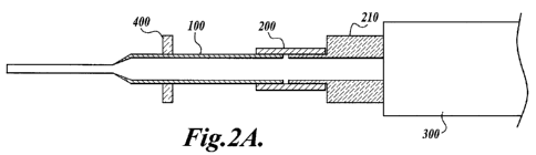
[37] The arrangement of Fig. 1 is readily modified as shown in Figs. 2A and 2B
to provide for simultaneous use of a plurality of devices 100. In this latter
arrangement,
shown as assembly 600, devices 100 are connected to manifold 210 via retention
elements 200, which are constructed from thin-wall polyurethane tubing.
Manifold 210 is
in turn coupled to pump 300 and provides fluid communication between pump 300
and
devices 100. Such multi-device assemblies can be configured so that the
plurality of
devices 100 are positioned to correspond to wells of standard multi-well
plates, such as
96-well plates. In the illustrated assembly, eight devices 100 are held in
position by
alignment plate 400 to align with a row of eight wells in a 96-well plate. In
such an
arrangement, the assembly can draw fluids from and expel fluids into one or a
series of
such plates. Samples and reagents (e.g., wash and elution buffers) can be
arrayed in
different rows of a single plate, and either the plate or the assembly is
moved to insert the
ends of the devices into the appropriate wells. This process can be carried
out manually
or automated. Multi-well plates are available in a range of well volumes
(e.g., 200 L,
0.5 mL, 1.0 mL, 2.0 mL) to provide a flexible system and facilitate
concentration of
nucleic acids from dilute samples. As will be recognized by those of ordinary
skill in the
-12-
3404OPCT CA 02742598 2011-05-03
WO 2010/054004 PCT/US2009/063296
art, other vessels, such as tubes (e.g., microcentrifuge tubes), plates, or
dishes can also be
used. Tubes can be arranged in a multi-well plate format. When glass tubes are
used, the
interior of the tube provides a further smooth glass surface that can be used
for nucleic
acid capture. In this arrangement, nucleic acid eluted from the glass surfaces
of the
device and the tube can be collected in the device and transferred to another
vessel, or can
be collected in the tube. For such multi-device assemblies, each device in the
assembly
can be run individually, or all devices in the assembly can be run
simultaneously.
[38] Fig. 2B shows an assembly further comprising a handling plate 500 to
which the remainder of the assembly is fixed. Handling plate 500 further
stabilizes the
components of assembly 600 and allows three-dimensional rotation of the entire
assembly. In a typical nucleic acid extraction procedure, a nucleic acid-
containing
sample in binding/lysis buffer is drawn into devices 100 by pump 300, and
nucleic acid is
allowed to bind to the inner walls of the devices. With the liquid in the
devices, assembly
600 is optionally tipped to the side and rotated to maximize contact between
sample and
glass in the upper (wide) section of devices 100. The liquid is then expelled,
and a first
wash buffer is drawn into the devices. The buffer is pumped up and down within
the
lumens of the devices by the action of pump 300. The buffer is then expelled,
and the
wash is repeated as required. After the final wash, a stream of air is passed
through
devices 100 to dry bound nucleic acid. Depending on the type of pump 300, air
drying
may be facilitated by disconnecting devices 100 from pump 300 (with or without
manifold 210) and connecting them to an air stream provided by other means.
Finally,
the nucleic acid is eluted from devices 100 and transferred into a 96-well
plate, a set of
tubes, or the like. Pump 300 can also be used to pre-wash or pre-treat the
interior
surfaces of devices 100.
[39] Additional automation can be provided by connecting these assemblies to
a valve mechanism connected to a microprocessor-controlled, multi-channel pump
and
fluid distribution control means as disclosed in more detail below. Such
assemblies can
be combined with standard laboratory robotic systems to provide for fully
automated
sample handling.
[40] The device will commonly take the form of a length of tubing, wherein the
outer cross-section is the same shape as the cross-section of the lumen. This
form of the
-13-
3404OPCT CA 02742598 2011-05-03
WO 2010/054004 PCT/US2009/063296
device is inexpensive, easy to store and handle, and provides considerable
flexibility in
use.
[41] Within one embodiment, the outer surface of the device comprises at least
one longitudinal ridge. A ridged device can be used to disrupt tissue during
sample
collection and/or mix samples prior to introduction into the lumen of the
device. In a
typical application, a nucleic-acid containing material is placed in a tube
with buffer, the
ridged device is inserted into the tube and spun to mix the sample, and the
sample is
drawn into the device.
[42] In another embodiment, a tubular device as disclosed above is contained
within a larger structure as disclosed briefly supra. Such an arrangement is
particularly
advantageous when using a device with a serpentine lumen to protect the glass
from
breakage and facilitate handling. For example, a spiral-shaped capillary tube
can be
enclosed within a card-like or block-like body prepared from adhesive, resin,
epoxy, or
the like. The term "spiral" is used herein for its ordinary meaning, that is a
planar curve
winding in a continuous and gradually widening form about a central point.
Examples of
suitable spirals include Archimedean spirals (Fig. 3) and Fermat's spirals
(Fig. 4),
although other shapes can be employed. See, for example, Wikipedia
(en.wikipedia.org/wiki/Spiral). Glass tubing (e.g., capillary tubes) can be
bent into the
desired spiral shape by heating a straight glass capillary tube to its
softening point and
winding it onto a reel with sidewalls designed to keep the tube aligned. The
spiral can be
constructed as a single-plane structure or in multiple planes (i.e., two or
more spirals
sitting flat on top of each other). The ends of the spiral are bent to face
and protrude
upwards slightly from the plane of the spiral to provide the first and second
ports. The
ends are then covered, and the body material (e.g., adhesive, resin, or epoxy)
is poured or
sprayed onto the spiral to provide strength and ease of handling. A mold can
be used to
create the desired shape, which may include alignment holes, slots, or
protrusions to
facilitate mating the device to a holder or manifold. After the material has
hardened, the
tube ends (ports) are uncovered. In a typical embodiment, the resulting
structure is in the
form of a flat disc with first and second ports on its upper surface. The
ports can be
provided with additional components as disclosed in more detail supra. A
viewing
window may be provided by leaving a hole in the body material.
-14-
3404OPCT CA 02742598 2011-05-03
WO 2010/054004 PCT/US2009/063296
[43] Alternative methods of construction will be evident to those of ordinary
skill in the art. For example, laminated plastic construction can be employed
essentially
as disclosed by Reed et al., U.S. 20090215125 Al. Briefly, individual
polymeric layers
are cut to shape using known methods such as laser cutting, CNC drag knife
cutting, and
die cutting. Adhesive layers are prepared to go between the layers of dry
plastic. The
adhesive layer will ordinarily be a pressure-sensitive adhesive available in a
thin film that
can be cut using the same method used for the plastic. Adhesives may be used
in an
Adhesive-Carrier-Adhesive (ACA) format where the carrier is preferred to be
the same
material as used in the other layers of the device. Other methods of applying
liquid
adhesives, such as screen printing, may also be employed. The several layers
are
registered to each other and pressed together. Features to assist in
registration, such as
alignment holes, are advantageously incorporated into the final design.
Pressure and
temperature during the cure cycle are adhesive-dependent; selection of
suitable conditions
is within the level of ordinary skill in the art. In the alternative, the
device can be
assembled through the use of a compression seal as disclosed in 20090215125
Al.
Lamination can incorporate molded elements as disclosed supra.
[44] The invention also provides an assembly comprising a device as disclosed
herein and a pump in fluid communication with the lumen of the device. The
term
"pump" is used herein to include both manually operated (e.g., syringes and
multi-channel pipettors) and powered (e.g., electric) devices. The assembly is
configured
so that the pump can deliver fluids into the lumen and remove them from the
lumen via
one or both of the ports. The pump is selected for its ability to meet the
following
criteria: (1) ability to accurately dispense volumes in the range of 20 L to
at least
1000 L, and preferably up to 2.5 mL; (2) ability to effectively pump air as
well as
liquids; and (3) ability to operate in reverse. Syringe-type or bellows-type
pumps satisfy
these criteria and allow the device to be operated in the manner of a
conventional pipette,
wherein one of the first and second ports is used for the introduction and
removal of all
reagents. When liquids are moved through both ports, it is advantageous to use
a pump
that also provides a low or zero dead volume to minimize cross contamination
of reagents
and has wetted surfaces made of materials compatible with the various reagents
used
(e.g., chaotropic salts and ethanol). Peristaltic pumps offer a good working
combination
of all of these traits, but do not offer the most accurate volume dispensing
of all pump
-15-
3404OPCT CA 02742598 2011-05-03
WO 2010/054004 PCT/US2009/063296
options. Peristaltic pumps are advantageously used when larger volumes of
liquids are
handled. Computer-controlled multi-channel peristaltic pumps (e.g., ISMATEC
12-channel pumps; Ismatec SA, Glattbrugg, Switzerland) will accommodate
multiple
devices simultaneously and can be programmed to start/stop/change flow rate or
reverse
direction of flow. When employing other pump styles, multiple pumps may be
required
for particular functions, although such an arrangement will complicate the
overall fluid
management system.
[45] The assemblies of the present invention may further include fluid
distribution control means in fluid communication with the pump. The fluid
distribution
control means comprises one or more valves that allow for a plurality of
fluids to be
sequentially pumped through the device, typically in the form of a valve-
manifold block.
It is preferred that manifold inputs and the exit pass through sterile filters
to protect the
valve-manifold assembly from contamination, and that the exit line have a
check valve to
prevent backflow from the pump tubing into the manifold. An exemplary fluid
distribution control means is a model V-1241-DC six-position, seven-port
rotary selector
valve manufactured by Upchurch Scientific, Oak Harbor, Washington. This
selector
valve allows the introduction of air gaps between reagents. The fluid
distribution control
means may further comprise a programmable computer, either external to the
valve
mechanism or fully integrated therewith. In certain embodiments of the
invention, the
programmable computer is a desktop or laptop personal computer. In other
embodiments, the programmable computer is a dedicated microprocessor device.
In an
exemplary system, control of fluid distribution is achieved using the above-
disclosed
selector valve in combination with a multi-channel peristaltic pump using an
application
written in Visual Basic for Microsoft Excel and running on a personal
computer. Both
the valve mechanism and the pump feature RS232 control interfaces. These
components
are addressed using Excel through the USB port of the computer and a USB-to-
Serial
converter. As will be understood by those skilled in the art, custom firmware
software
may also be employed.
[46] Liquid reagents are conveniently stored in septum-sealed vials equipped
with a sterile filter vent. The vials may be connected to the fluid
distribution control
-16-
3404OPCT CA 02742598 2011-05-03
WO 2010/054004 PCT/US2009/063296
means using a standard Luer-type needle inserted through the septum and
connected to
manifold inputs via microbore tubing.
[47] After fabrication, the device is preferably treated with ethylene oxide
or
gamma sterilization to remove pathogens. Reagents for use with the device
preferably
pass a 2-micron cellulose filter on entry to remove contaminants. Other
methods of
removing contaminants, including contaminants that may interfere with nucleic
acid
amplification, are disclosed by Reed et al., WO 2008002882. The reagent ports
on the
device may provide an interface to yellow and blue pipette tips. A needle-
septum
interface can be provided.
[48] Liquid samples are ordinarily introduced into the device at flow rate of
approximately 0.1 mUminute to approximately 5.0 mUminute, although, as
disclosed
above, considerably higher flow rates can be used. The actual flow rate is
design-dependent, taking into consideration the total volume of the fluid
pathway and the
configuration of the lumen.
[49] Dilute or concentrated samples can be prepared for input into the device.
Lysis and digestion of intact cells releases DNA or RNA from residual proteins
(for
example histones). In the alternative, solid samples (e.g., bacterial spores
or dried blood
on cloth) or semisolid samples (e.g., mouse tails or sputum/stool) can be
homogenized
and lysed before input to the device to provide a homogeneous and non-viscous
sample
that will flow through the lumen of the device. More viscous samples, such as
blood, can
also be used.
[50] Nucleic acids are bound to the glass surface(s) of the device in the
presence of a salt (e.g., KCl) at a concentration of at least 0.5 M to about 2
M or more
depending on solubility, or a chaotrope (e.g., guanidine HCl or guanidine
thiocyanate) at
a concentration of at least 1 M to about 6 M or the limit of solubility.
Binding of nucleic
acids is ordinarily done at a pH of approximately 5 to 8, preferably about 6.
The lumen is
then washed using buffered solutions of decreasing salt concentration. As salt
concentration decreases, ethanol is added to the wash solution to retain the
nucleic acid
on the glass and to remove contaminants that may interfere with downstream
processes
-17-
3404OPCT CA 02742598 2011-05-03
WO 2010/054004 PCT/US2009/063296
such as nucleic acid amplification. Washing is carried out at pH 6 - 9,
commonly pH 6 -
8. Nucleic acids are eluted from the device with a low-salt solution at basic
pH,
commonly pH 8 - 9.
[51] In general, when cells are present within the biological sample they are
lysed to provide a cell lysate from which the nucleic acids are extracted. A
variety of
methods of cell lysis are known in the art and are suitable for use within the
invention.
Examples of cell lysis methods include enzymatic treatment (using, for
example,
proteinase K, pronase, or subtilisin), mechanical disruption (e.g., by
sonication,
application of high pressure, use of a piezobuzzer device, or bead beating),
or chemical
treatment. Beads used for mechanical disruption should be made of a substance
that does
not bind nucleic acids under the disruption conditions. Suitable substances
include
acrylic, polycarbonate, polypropylene, cellulose acetate, polyethylene
terephthalate,
polyvinylchloride, and high density polyethylene. Lysing cells in the sample
by treating
them with a chaotropic salt solution is particularly advantageous. Methods and
reagents
for lysing cells using chaotropic salts are known in the art, and reagents can
be purchased
from commercial suppliers. Specific reagent compositions and reaction
conditions will
be determined in part by the type of cell to be lysed, and such determination
is within the
level of ordinary skill in the art. Suitable chaotropic salts include
guanidinium
thiocyanate, guanidine hydrochloride, sodium iodide, and sodium perchlorate.
Guanidine
hydrochloride, which is preferred for lysing blood cells, is used at
concentrations of 1M
to 10M, commonly 1M to 5M, usually 1M to 3M. Higher concentrations of sodium
iodide are required, approaching the saturation point of the salt (12M).
Sodium
perchlorate can be used at intermediate concentrations, commonly around 5M.
Neutral
salts such as potassium chloride and sodium acetate can also be used to obtain
binding of
nucleic acids to glass surfaces, and may be used in place of chaotropic salts
when cell
lysis is not required or is achieved by other means (e.g., in the case of
bacterial cell lysis).
When using neutral salts, the ionic strength of the buffer should be at least
0.25M. An
exemplary lysis buffer is a 2M solution of guanidinium thiocyanate (GuSCN)
buffer at
pH 6.4. Lysis in a chaotropic salt solution also removes histone proteins from
genomic
DNA and inactivates nucleases. Lysis buffers will generally also contain one
or more
buffering agents to maintain a near-neutral to slightly acidic pH. A suitable
buffering
agent is sodium citrate. One or more detergents may also be included. Suitable
-18-
3404OPCT CA 02742598 2011-05-03
WO 2010/054004 PCT/US2009/063296
detergents include, for example, polyoxyethylenesorbitan monolaurate (TWEEN
20),
t-octylphenoxypolyethoxyethanol (TRITON X-100), sodium dodecyl sulfate (SDS),
NP-40, CTAB, CHAPS, and sarkosyl. Alcohol, commonly ethanol, is included in
the
lysis and wash solutions, with the actual concentration selected to compensate
for the
lowered salt concentration in the washes. In the absence of salt, alcohol is
included at a
concentration of at least 50%, with 70% alcohol preferred in the final wash.
If salt is
included in the reagents, alcohol concentration will ordinarily range between
10% and
80%, often between 10% and 60%, usually between 20% and 50%. Optimization of
buffers is within the level of ordinary skill in the art. Lysis is generally
carried out
between room temperature and about 95 C, depending on the cell type. Blood
cells are
conveniently lysed at room temperature. It is generally preferred that the use
of silica
particles in cell lysis be avoided, since silica particles may bind nucleic
acids and reduce
the efficiency of the extraction process. Although not necessary, DNA may be
sheared
prior to loading the lysate into the extraction device. Methods for shearing
DNA are
known in the art.
[52] The nucleic acid-containing sample is introduced into the device via one
of the ports. Nucleic acid is captured on the glass surface(s) in the presence
of a salt or
chaotropic salt as disclosed above. Satisfactory binding of nucleic acids to
glass is
achieved at room temperature (15 -30 C, commonly about 20 C), although the
extraction
process can be run at higher temperatures, such as up to 37-42 C or up to 56
C, although
higher temperatures may reduce recovery of nucleic acids. The sample may be
allowed
to stand in the device for a period of time, and the sample solution may be
pumped back
and forth through the lumen. Wash buffers are then pumped into one port, such
as by use
of a peristaltic pump, a syringe, or a pipetter. Selection of wash buffers
will depend in
part on the composition of the sample loading solution. In general, salt
concentration will
be reduced during the washing process, and pH will be increased slightly. If
the lysis
buffer contains a chaotropic salt, the initial wash will commonly also contain
that salt at
the same or somewhat lower concentration (e.g., 1-3M GuSCN). The final wash
should
reduce the ethanol concentration to below 50%, preferably to about 10%-20%, to
minimize inhibition of nucleic acid amplification in downstream processing.
The alcohol
content of wash solutions will ordinarily range between 20% and 80%. Wash
solutions
containing at least 50% ethanol, preferably about 70% ethanol, have been found
to
-19-
3404OPCT CA 02742598 2011-05-03
WO 2010/054004 PCT/US2009/063296
improve nucleic acid capture. Complete removal of the final wash from the
lumen of the
device is also needed in certain embodiments. Methods for this removal of the
final wash
include drying by passaging air over the surfaces of the lumen utilizing an
air pump for
one to three minutes. After washing, the nucleic acid is eluted from the
device with a low
salt buffer at higher pH than the final wash. Elution buffers are typically
low ionic
strength, buffered solutions at pH > 8.0, although nucleic acid can be eluted
from the
device with water. Elution can be carried out at ambient temperature up to
about 56 C.
[53] The design of the device permits fluids, including both liquids and
gasses,
to be passed through the device from one port to the other. In this way
buffers can be
pumped back and forth through the lumen to increase washing and elution
efficiency, and
air can be pumped through between washes to remove residual buffer. The device
can be
configured in a variety of ways with respect to introduction and removal of
reagents. In
one arrangement, reagents are introduced into the lumen of the device via one
of the ports
and removed via the other port. In a second arrangement, one port serves as
both inlet
and outlet for reagents, and the second port is connected to a pump that
provides suction
and pressure. This second arrangement avoids contacting the pump and fluid
distribution
control means with the reagents, and is particularly advantageous if using
reagents that
are corrosives or strong solvents. A third arrangement combines the first and
second
arrangements so that some fluids are passed completely through the device from
one port
to the other and other fluids are introduced and removed via the same port.
For example,
the nucleic acid containing sample can be introduced into the lumen via the
first port and
removed via the second port, and wash and elution reagents are introduced and
removed
via the second port using suction and air pressure applied through the first
port. Those
skilled in the art will recognize that many variations on these basic
arrangements are
possible.
[54] As will be understood by those skilled in the art, actual working volumes
will be determined by the size of the device, including lumen volume, as well
as routine
experimental design. For small-volume devices comparable to Pasteur pipettes,
volumes
will ordinarily range from about 20 pL to about 500 L, although larger
volumes up to
1 mL or as much as 2.5 mL can be used. Samples can be concentrated by reducing
the
volume of the elution buffer.
-20-
3404OPCT CA 02742598 2011-05-03
WO 2010/054004 PCT/US2009/063296
[55] Quantitation of extracted nucleic acids is facilitated by the inclusion
of a
fluorescent compound within the elution buffer, thereby providing a rapid
quality check
on the extraction process while the extracted nucleic acids are still within
the device.
Thus, within one embodiment of the invention the nucleic acids are contacted
with a
fluorescent compound having a fluorescence intensity dependent on the
concentration of
nucleic acids, and the fluorescence of the fluorescent compound is measured.
Fluorescent
compounds having a fluorescence intensity dependent on the concentration of
nucleic
acids are fluorescent compounds that exhibit a conformation-dependent change
in
fluorescence intensity in the presence of nucleic acids. Useful fluorescent
compounds
include those compounds whose intensity increases in the presence of nucleic
acids.
Representative fluorescent compounds include fluorogenic minor groove binder
agents
such as bis-benzimide compounds and intercalating fluorogenic agents such as
ethidium
bromide, and commercially available fluorescent dyes (e.g., SYBR Green;
Invitrogen
Corp.). Fluorescent compounds can be introduced into the device in the elution
buffer or
immobilized in the lumen. Methods for immobilizing the fluorescent compound in
the
lumen and useful fluorescent compounds are described in Reed et al., U.S.
Application
Publication No. 20060166223 Al. The device of the invention allows for the
interrogation of the lumen by fluorescence by having at least a portion of the
lumen
suitable for transmitting excitation energy to the fluorescent compounds in
the lumen and
for transmitting fluorescence emission intensity from the compounds in the
lumen.
[56] Although in principal any fluorogenic DNA-binding dye can be used in
the invention, it is preferred to use a dye that is compatible with downstream
processes
such as PCR. A preferred dye is a bis-benzimidine (BB) dye disclosed by Reed
et al.,
U.S. Patent Application Publication No. 20060166223 Al, which gives a strong
fluorescent signal (detection at 460 nm, 40 nm filter slit width) when excited
at 360 nm
(40 nm slit width). The BB dye is selective for dsDNA but can also detect RNA.
A
popular green fluorescent dye, SYBR green (Invitrogen Corp.) is often used in
so called
"real time" PCR or quantitative PCR. Much like the BB dye, SYBR green can be
used to
both quantitate the extracted DNA before amplification and monitor the gene-
specific
increase during PCR. The use of fluorogenic DNA dyes or DNA probes in
isothermal
nucleic acid tests such as NASBA is also known.
-21-
3404OPCT CA 02742598 2011-05-03
WO 2010/054004 PCT/US2009/063296
[57] The preferred bis-benzimidine dye, although not as sensitive as some
DNA-binding dyes, has been found to be well suited for measuring genomic DNA
content of a sample after extraction from DNA-rich whole blood. The minor
groove-binding BB dye emits blue fluorescence in the presence of double
stranded DNA,
and can be added directly to PCR amplification buffer. In contrast, strong
binding DNA
dyes such as PICOGREEN (Invitrogen) may inhibit PCR.
[58] Preliminary evidence indicates that the BB dye can be used in existing
PCR assays if the PCR primer extension is carried out at higher annealing
temperature
(61.5 C vs. 60 C). Inclusion of the BB dye directly in the elution buffer
therefore allows
DNA to be measured before, during, and after gene-specific amplification. The
higher
primer extension temperature required with addition of BB dye may be
advantageous in
PCR assays (acting as a PCR enhancer). Much like the MGB TaqMan system (U.S.
Patent No. 6,727,356), A/T rich primer/target interactions are stabilized by
the BB in the
PCR mix, and increased duplex stability allows shorter (more specific) DNA
probes to be
used. The blue emitting MGB dye will likely not interfere with the green to
red
fluorescence wavelengths that are widely used with 2-color fluorogenic DNA
probes.
[59] RNA-selective dyes such as Ribogreen (see Molecular Probes Handbook
of Fluorescent Probes and Research Products, 9th edition, Chapter 8) can also
be used in
the device or elution buffer. RNA-selective dyes may have advantages for real
time RNA
assays such as NASBA. The caveats disclosed above about inhibition of the
gene-specific DNA or RNA tests also apply to RNA detecting fluorogenic dyes.
[60] If desired, the device can be re-used following removal of residual
nucleic
acids and/or reagents by washing. In many cases, satisfactory washing can be
achieved
by running several (typically 5-10) channel volumes of distilled sterile water
through the
lumen. In a preferred method, the device is first washed with 5-10 channel
volumes of
distilled sterile water, followed by a wash with 2-3 channel volumes of 70%
EtOH, which
is followed by another 2-3 channel volume wash with distilled sterile water.
Wash
solutions can be pumped through the device using a pump (e.g., a peristaltic
pump),
syringe, or the like. The cleaning protocol can be carried out in through a
manifold using
an automated pump.
-22-
3404OPCT CA 02742598 2011-05-03
WO 2010/054004 PCT/US2009/063296
[61] Bound nucleic acid can be stored in the device and used in later testing,
including confirmation of test results. The device is rinsed with an ethanol-
rich rinse and
dried. Storage is at room temperature for up to several days or in a freezer
for longer
periods.
[62] The invention also provides a kit comprising a nucleic acid extraction
device as disclosed above and a buffer in a sealed container. The buffer can
be a lysis
buffer, a wash buffer, or an elution buffer as generally disclosed herein.
Ordinarily, the
device will be packaged with more than one buffer, commonly a complete set of
buffers
for extracting nucleic acid from a biological sample. For some applications,
the elution
buffer will comprise a fluorescent compound that exhibits a change in
fluorescence
intensity in the presence of nucleic acids. A typical kit comprises these
components in a
single package, together with a set of printed instructions for use.
[63] The present invention has multiple applications in laboratory research,
human and veterinary medicine, public health and sanitation, forensics,
anthropological
studies, environmental monitoring, and industry. Such applications include,
without
limitation, bacterial and viral detection and typing, microbial drug
resistance screening,
viral load assays, genotyping, infection control and pathogen screening (of,
e.g., blood,
tissue, food, cosmetics, water, soil, and air), pharmacogenomics, detection of
cell-free
DNA in plasma, white cell counting, and other fields where preparation and
analysis of
DNA from biological samples is of interest. As disclosed above, nucleic acids
extracted
using the devices and methods of the invention are readily used in a variety
of
downstream processes, including amplification, hybridization, blotting, and
combinations
thereof. The devices and methods of the invention can be employed within point-
of-care
diagnostic assays to identify disease pathogens, and can be utilized in
genetic screening.
These devices and methods can also be used within veterinary medicine for the
diagnosis
and treatment of animals, including livestock and companion animals such as
dogs, cats,
horses, cattle, sheep, goats, pigs, etc.
[64] Nucleic acids can be extracted from a wide variety of sources. For
research and medical applications, suitable sources include, without
limitation, sputum,
saliva, throat swabs, oral rinses, nasopharyngeal swabs, nasopharyngeal
aspirates, nasal
swabs, nasal washes, mucus, bronchial aspirations, bronchoalveolar lavage
fluid, pleural
-23-
3404OPCT CA 02742598 2011-05-03
WO 2010/054004 PCT/US2009/063296
fluid, endotracheal aspirates, cerbrospinal fluid, feces, urine, blood,
plasma, serum, cord
blood, blood components (e.g., platelet concentrates), blood cultures,
peripheral blood
mononuclear cells, peripheral blood leukocytes, plasma lysates, leukocyte
lysates, buffy
coat leukocytes, anal swabs, rectal swabs, vaginal swabs, endocervical swabs,
semen,
biopsy samples, lymphoid tissue (e.g., tonsil, lymph node), bone marrow, other
tissue
samples, bacterial isolates, vitreous fluid, amniotic fluid, breast milk, and
cell culture
supernatants. Other starting materials for extraction of nucleic acids include
water
samples, air samples, soil samples, cosmetics, foods and food ingredients,
medical
supplies and equipment, and the like.
[65] Processes and assemblies of the present invention can be used for
extraction and analysis of fragmented DNA. DNA can be fragmented by a variety
of
methods known in the art, such as nuclease digestion (including digestion with
restriction
endonucleases and DNases), sonication, heat, mechanical disruption (such as by
shearing
or vortexing), and chemical treatment. Applicable chemical treatments include,
for
example, use of metal ions such as iron (Zhang et al., Nucl. Acids Res.
29(13):e66, 2001),
oxidizing agents such as bisulfite (Ehrich et al., Nucl. Acids Res. 35(5):e29,
2007), and
antibiotics and drugs such as bleomycin (Chen et al., Nucl. Acids Res.
36(11):3781-3790,
2008). A preparation of fragmented DNA can contain fragments of a range of
sizes or
may be relatively limited in size range. Those skilled in the art will
recognize that the
actual size of fragments will be determined by such factors as the
fragmentation method
selected and the conditions used (e.g., time of treatment).
[66] Nucleic acids prepared according to the present invention can be
amplified
by methods known in the art, including polymerase chain reaction (PCR) (see,
e.g.,
Mullis, U.S. Patent No. 4,683,202) and isothermal amplification methods. Real-
time
polymerase chain reaction (RT-PCR) is commonly used. See, for example,
Cockerill,
Arch. Pathol. Lab. Med. 127:1112-1120, 2002; and Cockerill and Uhl,
"Applications and
challenges of real-time PCR for the clinical microbiology laboratory," pp. 3-
27 in Reischl
et al, eds., Rapid cycle real-time PCR methods and applications, Springer-
Verlag, Berlin,
2002. For a review of the use of RT-PCR in clinical microbiology, see Espy et
al., Clin.
Microbiol. Rev. 19:165-256, 2006. Instrumentation and chemistry for carrying
out PCR
are commercially available. Instruments include thermal cyclers (e.g.,
ABI7000, 7300,
-24-
3404OPCT CA 02742598 2011-05-03
WO 2010/054004 PCT/US2009/063296
7500, 7700, and 7900, Applied Biosystems, Foster City, CA; LIGHTCYCLER, Roche
Applied Science, Indianapolis, IN; SMARTCYCLER, Cepheid, Sunnyvale, CA;
ICYCLER, Bio-Rad Laboratories, Inc., Hercules, CA; ROBOCYCLER and MX3000P,
Stratagene, La Jolla, CA), detection systems for use with fluorescent probes
(e.g., MYIQ
and CHROMO4, Bio-Rad Laboratories, Inc.), nucleic acid analyzers (e.g., Rotor-
Gene
6000, Corbett Life Science, Concorde, NSW, Australia), and amplification and
detection
systems (e.g., BD PROBETEC ET, Becton Dickinson, Franklin Lakes, NJ). Other
PCR
technologies include fluorescent dyes for quantitative PCR (e.g., SYBR,
Invitrogen
Corp.) and fluorogenic probes, including FRET (fluorescent resonance energy
transfer)
hybridization probes (Walker, Science 296:557-559, 2002), TAQMAN probes
(Applied
Biosystems, Foster City, CA; see, Kutyavin et al., Nucl. Acids. Res. 28:655-
661, 2000),
ECLIPSE probes (Nanogen, Bothell WA), and molecular beacons (U.S. Patent
Nos. 5,925,517 and 6,150,097. Isothermal amplification methods known in the
art
include nucleic acid sequence-based amplification (NASBA) (Malek et al., U.S.
Patent
No. 5,130,238; Compton, Nature 350:91-92, 1991; Deiman et al., Mol.
Biotechnol.
20:163-179, 2002), branched DNA (Alter et al., J. Viral Hepat. 2:121-132,
1995; Erice et
al., J. Clin. Microbiol. 38:2837-2845, 2000), transcription mediated
amplification (Hill,
Expert. Rev. Mol. Diagn. 1:445-455, 2001), strand displacement amplification
(Walker,
PCR Methods and Applications 3:1-6, 1993; Spargo et al., Mol. Cell Probes
10:247-256,
1996), helicase-dependent amplification (Vincent et al., EMBO Rep. 5:795-800,
2004),
loop-mediated isothermal amplification (Notomi et al., Nucl. Acids Res.
28:E63, 2000),
INVADER assay (Olivier et al., Nucl. Acids Res. 30:e53, 2002; Ledford et al.,
J. Mol.
Diagn. 2:97-104, 2000), cycling probe technology (Duck et al., BioTechniques
9:142-148, 1990; Cloney et al., Mol. Cell Probes 13:191-197, 1999), rolling
circle
amplification (Fire and Xu, Proc. Nat. Acad. Sci. USA 92:4641-4645, 1995; Liu
et al., J.
Am. Chem. Soc. 118:1587-1594, 1996), and Q-beta replicase (Shah et al., J.
Clin.
Microbiol. 32:2718-2724, 1994; Shah et al., J. Clin. Microbiol. 33:1435-1441,
1995).
For a review of isothermal amplification methods, see Gill and Ghaemi,
Nucleosides
Nucleotides Nucleic Acids 27:224-243, 2008.
[67] NASBA depends on the concerted action of three enzymes to amplify
target nucleic acid sequences. While able to amplify double-stranded DNA,
NASBA is
particularly suited for amplification of RNA. Target RNA enters the cycle by
binding to
-25-
3404OPCT CA 02742598 2011-05-03
WO 2010/054004 PCT/US2009/063296
a first primer, which is then extended by reverse transcriptase to form a
DNA/RNA
hybrid. The RNA strand is removed by the action of RNase H to yield a single-
stranded
cDNA. This cDNA can bind to a second primer (which includes a T7 RNA
polymerase
promoter sequence) and then form a double-stranded intermediate by the action
of the
reverse transcriptase activity. The intermediate is then copied by the action
of T7 RNA
polymerase into multiple single-stranded RNA copies (10-1000 copies per copy
of
template). These RNA copies can then enter the cycle and continue generating
more
copies in a self-sustained manner. Based on the NASBA mechanism, two products
can
be detected: a double-stranded DNA intermediate and a single-stranded RNA
product.
[68] NASBA is conveniently used with the devices of the present invention
since it is isothermal (i.e. temperature cycling is not required). A
denaturation step is not
necessary except when a DNA target is chosen. Two considerations when running
NASBA in the devices of the present invention are heat transfer and protein
adsorption.
The reaction temperature should be within the range of 30 C to 50 C, usually
at least
37 C, and preferably 42 C where primer binding is more specific. Room
temperature
does not support NASBA, so the channel temperature must be raised efficiently
or the
reaction will not work. In addition, proteins such as the NASBA enzymes
readily stick to
glass and some organic polymeric materials, inactivating them and stopping the
NASBA
cycle. Two methods to address this are (1) to preadsorb the glass with a
carrier such as
serum albumin, or (2) to add enough serum albumin to the NASBA reaction
mixture to
minimize loss of enzymes.
[69] Additional methods of nucleic acid amplification are known in the art and
can be applied to DNA prepared according to the present invention. Examples of
such
methods include ligase chain reaction (Wu and Wallace, Genomics 4:560-569,
1989;
Barany, Proc. Natl. Acad. Sci. USA 88:189-193, 1991), polymerase ligase chain
reaction
(Garany, PCR Methods and Applic. 1:5-16, 1991), gap ligase chain reaction
(Segev, WO
90/01069), repair chain reaction (Backman et al., U.S. Patent No. 5,792,607),
and rolling
circle amplification (RCA) (Lisby, Mol. Biotechnol. 12:75-99, 1999).
[70] As will be understood by those of ordinary skill in the art, nucleic
acids
prepared according to the present invention can also be detected and/or
analyzed without
amplification using methods known in the art. Suitable methods include,
without
-26-
3404OPCT CA 02742598 2011-05-03
WO 2010/054004 PCT/US2009/063296
limitation, hybridization, which can be coupled to fluorescence or
immunoassay,
including hybridization to oligonucleotide-nanoparticle conjugates (Park et
al., U.S.
Patent No. 7,169,556) and DNA barcodes (Mirkin et al., U.S. Application
Publication
No. 20060040286 Al); microarray technology, which can be used for expression
profiling by hybridization, diagnostics, gene identification, polymorphism
analysis, and
nucleic acid sequencing; hybridization protection assay (Arnold et al., Clin.
Chem.
35:1588-1594, 1989); dual kinetic assay (e.g., APTIMA COMBO 2 assay, Gen-Probe
Incorporated); and sequencing, including microsequencing (e.g., MICROSEQ 500
16s
rDNA bacterial identification kit, Applied Biosystems). Methods of detecting
polymorphisms include massively parallel shotgun sequencing (Nature 437:326-
327,
2005), which can detect previously unknown features of cell-free nucleic acids
such as
plasma mRNA distributions and/or methylation and histone modification of
plasma DNA
(Fan et al., Proc. Natl. Acad. Sci. USA 105:16266-16271, 2005) Those of
ordinary skill
in the art will further recognize that these and other methods can be used in
combination
with nucleic acid amplification.
[71] As noted above, extracted nucleic acids can be used within methods for
detecting pathogens, including bacteria, viruses, fungi, and parasites. In
addition,
extracted nucleic acids can be analyzed to characterize drug resistance and
drug
sensitivity of infectious agents (e.g., methicillin or other antibiotic
resistance in
Staphylocccus aureus). Many such methods are known in the art, and a number of
such
tests have been approved by the U.S. Food and Drug Administration for human
diagnostic use and are commercially available. For example, Table 1 is a list
of
FDA-approved tests for Chlamydia. Additional tests are listed in Table 2.
Other
pathogens of interest for which nucleic acid-based tests are known include
bloodborne
pathogens, Coccidioides immitis, Cryptococcus, Gardnerella vaginalis,
Haemophilus
spp., Histoplasma capsulatum, influenza virus, Mycoplasma spp., Salmonella
spp.,
Shigella spp., and Trichomonas vaginalis. Methods for the detection of
microbial
contaminants, including bacteria, viruses, fungi, and parasites, in samples of
foods and
other products using PCR are disclosed by, for example, Romick et al., U.S.
Patent
No. 6,468,743 B 1. The use of PCR in testing water samples for Enterococcus
species is
disclosed by Frahm and Obst, J. Microbiol. Methods 52:123-131, 2003.
-27-
CA 02742598 2011-05-03
WO 2010/054004 PCT/US2009/063296
d~ d N O O 00 O -
O M -- M M M N N ' N
N
O O O O 0 0 N N
01 Q\ 01 cd
64 64 :'8-1
W bbO bOA bbO bi) bbOi bO bD bb bO bbO
4~- 4,d 4 4-d w Z2 42
I
i i i U
W o 0 0 0 D o rn 0, 0, C 0~
C) H
04 "D N M O_ in N d vn m N 00
00
A O 00
N C -
N-
~ a a a
ago wuwuwu
O Q~ O 0 r:~ C O
U Op.., a a ~p.., a a~ W~ W~ w a
UZ Z UG~ZZUo Uri U~Z rya
Oa'uCw7 Cw7 ~uC7C7P4V) P4 P U)
w O W
W > U a
H H W H H
C40') V) O O OQU
HHH C7
C40') < H U U C)
U O O Z H
Q H x d x p U U a a x H
U 'T"
a H H H a Q - H H
uQaQ~W
U Up
a4 En aax x P'UU~
aazaHv,~a ZZU UC; U Z Z .Z HO ww0 0wOw~Uc'
UC7U<~ QU C7C7~ G4H 0.~UC'7Had
-28-
3404OPCT CA 02742598 2011-05-03
WO 2010/054004 PCT/US2009/063296
Table 2
Test References/Products
General bacterial Dreier et al., J. Clin. Microbiol. 42:4759-4764, 2004.
contamination of platelet Mohammadi et al., J. Clin. Microbiol. 41:4796-4798,
concentrates 2003
Bacillus anthracis Bell et al., J. Clin. Microbiol. 40:2897-2902, 2002;
Oggioni et al. J. Clin. Microbiol. 40:3956-3963, 2002;
Ellerbrok et al., FEMS Microbiol. Lett. 214:51-59, 2002.
Bartonella henselae Zeaiter et al. J. Clin Microbiol. 41:919-925, 2003.
Bordetella pertussis Reischl et al., J. Clin. Microbiol. 39: 1963-1966, 2001;
Anderson et al., Clin. Microbiol. Infect. 9:746-749,
2003.
Borrelia burgdorferi Makinen et al., " Geno species- specific melting
temperature of the recA PCR product for the detection of
Borellia burgdorferi sensu lato and differentiation of
Borrelia garinii from Borrelia afzelii and Borrelia
burgdorferi sensu stricto," pp. 139-147 in Reischl et al.,
eds., Rapid cycle real-time PCR methods and
applications, Springer-Verlag, Berlin, 2002
Borrelia garinii Pietila et al., J. Clin. Microbiol. 38:2756-2759, 2000.
Borrelia afzelii Pietila et al., J. Clin. Microbiol. 38:2756-2759, 2000.
Campylobacter Popovic-Uroic et al., Lab Medicine 22:533-539, 1991;
Tenover, J. Clin. Microbiol. 28:1284-1287, 1990.
Chlamydia Gaydos et al., J. Clin. Microbiol. 41:304-309, 2003;
Ikeda-Dantsuji et al., J. Med. Microbiol. 54:357-360,
2005
Chlamydophila pneumoniae Apfalter et al., J. Clin Microbiol. 41:592-600, 2003;
Tondella et al., .J. Clin Microbiol. 40:575-583, 2002.
Clostridium difficile Belanger et al., J. Clin. Microbiol. 41:730-734, 2003.
Ehrlichia chaffeensis Loftis et al., J. Clin. Microbiol. 41:3870-3872, 2003.
Enterococcus Species E. faecalis/OE PNA FISH assay, AdvanDx, Inc.,
Woburn, MA; see, Sloan et al., J. Clin. Microbiol.
42:2636-2643, 2004.
Escherichia coli Frahm and Obst, J. Microbiol. Methods 52:123-131,
2003
Histoplasma capsulatum Hall et al., J. Clin. Microbiol. 30:3003-3004, 1992.
Legionella pneumophila Wellinghausen et al., "Rapid detection and
simultaneious differentiation of Legionella spp. and L.
pheumophila in potable water samples and respiratory
specimens by LightCycler PCR," pp. 45-57 in Reischl et
al. eds., Rapid cycle real-time PCR methods and
applications, Springer-Verlag, Berlin, 2002; Welti et al.,
Diagn. Microbiol. Infect. Dis. 45:85-95, 2003.
-29-
3404OPCT CA 02742598 2011-05-03
WO 2010/054004 PCT/US2009/063296
Test References/Products
Legionella spp. Herpers et al., J. Clin. Microbiol. 41:4815-4816, 2003;
Reischl et al., J. Clin. Microbiol. 40:3814-3817, 2002.
Listeria monocytogenes Okwumabua et al., Res. Microbiol. 143:183-189, 1992.
Mycobacterium Spp. Hall et al., J. Clin. Microbiol. 41:1447-1453, 2003;
Lumb et al., Pathology 25:313-315, 1993
Mycobacterium e.g., AMPLICOR MTB, Roche Molecular Diagnostics,
tuberculosis Pleasanton, CA., See, e.g., Stevens et al., J. Clin.
Microbiol. 40:3986-3992, 2002; Garcia-Quintanilla et
al., J. Clin. Microbiol. 40:4646-4651, 2002;
Bruijnesteijn et al., J. Clin. Microbiol. 42:2644-2650,
2004; Sedlacek et al., J. Clin. Microbiol. 42:3284-3287,
2004.
Ethambutol resistance in M. Wada et al., J. Clin. Microbiol. 42:5277-5285,
2004.
tuberculosis
Isoniazid resistance in M. van Doorn et al., J. Clin. Microbiol. 41:4630-4635,
tuberculosis 2003;
Rifampin resistance in M. Edwards et al., J. Clin Microbiol. 39:3350-3352,
2001;
tuberculosis Piatek et al., Nat. Biotechnol. 16:359-363, 1998.
Mycobacterum ulcerans Rondini et al., J. Clin. Microbiol. 41:4231-4237, 2003.
Mycoplasma pneumoniae Welti et al., Diagn. Microbiol. Infect. Dis. 45:85-95,
2003; Ursi et al., J. Microbiol. Methods 55:149-153,
2003.
Neisseria gonorrhoeae BD PROBETEC ET, Becton Dickinson, Franklin Lakes,
NJ;
APTIMA COMBO 2 assay, Gen-Probe Incorporated,
San Diego, CA.
Gaydos et al., ibid.
Neisseria meningitides Guiver et al., FEMS Immunol. Med. Microbiol. 28:173-
179, 2000; Corless et al., J. Clin. Microbiol. 39:1553-
1558,2001.
Penicillin resistance in N. Stefanelli et al. J. Clin. Microbiol. 41:4666-
4670, 2003.
meningitides
Staphylococcus aureus S. aureus PNA FISH assay, Advandx, Inc., Woburn, MA
Fluoroquinolone resistance Lapierre et al., J. Clin. Microbiol. 41:3246-3251,
2003.
in S. aureus
Methicillin Resistant e.g., XPERT MRSA (Cepheid, Sunnyvale, CA); See,
Staphylococcus aureus e.g., Reischl et al., J. Clin. Microbiol. 38:2429-2433,
2000; Tan et al., J. Clin. Microbiol. 39:4529-4531, 2002;
Fang and Hedin, J. Clin. Microbiol. 41:2894-2899,
2003; Francois et al., J. Clin. Microbiol. 41:254-260,
2003; Ramakrishnan et al., U.S. Application Publication
No. 20060057613 Al).
Streptococcus pneumoniae Greiner et al., J. Clin. Microbiol. 39:3129-3134,
2001.
Penicillin resistance in S. Kearns et al. J. Clin. Microbiol. 40:682-684,
2002.
-30-
3404OPCT CA 02742598 2011-05-03
WO 2010/054004 PCT/US2009/063296
Test References/Products
pneumoniae
Group A Streptococcus Uhl et al., J. Clin. Microbiol. 41:242-249, 2003.
Group B Streptococcus CEPHEID SMART GBS ASSAY (Cepheid, Sunnyvale,
CA);
Bergeron et al., N. Engl. J. Med. 343:175-179, 2000; Ke
et al., "Rapid detection of group B streptoccocci using
the LightCycler instrument," pp. 107-114 in Reischl et
al, eds., Rapid cycle Real-time PCR methods and
applications, Springer-Verlag, Berlin, 2002.
Tropheryma whipplei Fenollar et al. J. Clin. Microbiol. 40:1119-1120, 2002.
Yersinia pestis Tomaso et al., FEMS Immunol. Med. Microbiol. 38:117-
126,2003.
Fluoroquinolone resistance Lindler et al., J. Clin. Microbiol. 39:3649-3655,
2001.
in Y. pestis
[72] Tests for detection and diagnosis of viruses are also known in the art.
Examples of such tests are shown in Table 3.
Table 3
Test References/Products
Adenovirus Houng et al., Diagn. Microbiol. Infect. Dis. 42:227-236,
2002; Heim et al., J. Med. Virol. 70:228-239, 2003; Faix
et al., Clin. Infect. Dis. 38:391-397, 2004; Lankester et
al., Clin. Infect. Dis. 38:1521-1525, 2004.
B19 virus Koppelman et al., Transfusion 44:97-103, 2004.
BK virus Whiley et al., J. Clin. Microbiol. 39:4357-4361, 2001.
Cytomegalovirus Machida et al., J. Clin. Microbiol. 38:2536-2542, 2000;
Nitsche et al., J. Clin. Microbiol. 38:2734-2737, 2000;
Tanaka et al., J. Med. Virol. 60:455-462, 2000; Gault et
al., J. Clin. Microbiol. 39:772-775, 2001; Ando et al.,
Jpn. J. Ophthalmol. 46:254-260, 2002; Aberle et al., J.
Clin. Virol. 25 (Suppl. 1):579-585; Cortez et al., J.
Infect. Dis. 188:967-972, 2003; Hermann et al., J. Clin.
Microbiol. 42:1909-1914, 2004; Hall, U.S. Patent
No. 7,354,708.
Enterovirus Read et al., J. Clin. Microbiol. 39:3056-3059, 2001;
Corless et al., J. Med. Virol. 67:555-562, 2002; Kares et
al., J. Clin. Virol. 29:99-104, 2004.
Epstein-Barr Virus Lo et al., Clin. Cancer Res. 7:1856-1859, 2001; van
Esser et al., Br. J. Haematol. 113:814-821, 2001; Patel et
al., J. Virol. Methods 109:227-233, 2003; Balandraud et
al., Arthritis Rheum. 48:1223-1228, 2003; Jebbink et al.,
-31-
3404OPCT CA 02742598 2011-05-03
WO 2010/054004 PCT/US2009/063296
Test References/Products
J. Mol. Diagn. 5:15-20, 2003.
Hepatitis A virus Costa-Mattioli et al., J. Viral Hepat. 9:101-106, 2002;
Rezende et al., Hepatology 38:613-618, 2003.
Hepatitis B Virus Abe et al., J. Clin. Microbiol. 37:2899-2903, 1999; Ide
et al., Am. J. Gastroenterol. 98:2048-2051, 2003; Aliyu
et al., J. Clin. Virol. 30:191-195, 2004; Candotti et al., J.
Virol. Methods 118:39-47, 2004;
Hepatitis C Virus VERSANT HCV RNA 3.0 Assay (Bayer Healthcare,
Tarrytown NY), COBAS AMPLICOR HCV TEST
(Roche Molecular Diagnostics); Enomoto et al., J.
Gastroenterol. Hepatol. 16:904-909, 2001; Schroter et
al., J. Clin. Microbiol. 39:765-768, 2001; Bullock et al.,
Clin. Chem. 48:2147-2154, 2002; Candotti et al., ibid.;
Law et al., U.S. Application Publication
No. 20070207455.
Hepatitis D Virus Yamashiro et al., J. Infect. Dis. 189:1151-1157, 2004
Hepatitis E Virus Orru et al., J. Virol. Methods 118:77-82, 2004
Herpes simplex virus Espy et al., J. Clin. Microbiol. 38:3116-3118, 2000;
Kessler et al., J. Clin, Microbiol. 38:2638-2642, 2000;
Aberle and Puchhammer-Stockl, J. Clin. Virol.
25(Suppl. 1):S79-S85, 2002; Kimura et al., J. Med.
Virol. 67:349-353, 2002.
Human herpes virus Aslanukov et al., U.S. Application Publication
subtypes No. 20060252032 Al.
HIV-1 Ito et al., J. Clin. Microbiol. 41:2126-2131, 2003;
Palmer et al., J. Clin. Microbiol. 41:4531-4536, 2003;
Candotti et al., ibid.; Gibellini et al., J. Virol. Methods
115:183-189, 2004;
HIV-2 Schutten et al., J. Virol. Methods 88:81-87, 2000; Ruelle
et al., J. Virol. Methods 117:67-74, 2004
Human Papillomavirus King, U.S. Application Publication No. 20080187919
Al; Hudson et al., U.S. Application Publication
No. 20070111200 Al.
JC virus Whiley et al., ibid.
Influenza Virus van Elden et al., J. Clin. Microbiol. 39:196-200, 2001;
Smith et al., J. Clin. Virol. 28:51-58, 2003; Boivan et al.,
J. Infect. Dis. 188:578-580, 2003; Ward et al., J. Clin.
Virol. 29:179-188, 2004.
Metapneumovirus Cote et al., J. Clin. Microbiol. 41:3631-3635, 2003;
Maertzdorf et al., J. Clin. Microbiol. 42:981-986, 2004.
Orthopoxvirus Espy et al., J. Clin. Microbiol. 40:1985-1988, 2002; Sofi
Ibrahim et al., J. Clin. Microbiol. 41:3835-3839, 2003;
Nitsche et al., J. Clin. Microbiol. 42:1207-1213, 2004.
Parainfluenza Virus Templeton et al., J. Clin. Microbiol. 42:1564-1569,
2004; Templeton et al., J. Clin. Virol. 29:320-322, 2004.
Respiratory Syncytial Virus Borg et al., Eur. Respir. J. 21:944-951, 2003;
Gueudin et
-32-
3404OPCT CA 02742598 2011-05-03
WO 2010/054004 PCT/US2009/063296
Test References/Products
al., J. Virol. Methods 109:39-45, 2003; Mentel et al., J.
Med. Microbiol. 52:893-896, 2003; Boivan et al., J.
Clin. Microbiol. 42:45-51, 2004.
Respiratory syncytial virus Guedin et al., J. Virol. Methods 109:39-45, 2003.
Severe acute respiratory Poon et al., Clin. Chem. 50:67-72, 2004; Drosten et
al.,
syndrome coronavirus J. Clin. Microbiol. 42:2043-2047, 2004.
(SARS-CoV)
Varicella zoster virus Espy et al., J. Clin. Microbiol. 38:3187-3189, 2000;
Furuta et al., J. Clin. Microbiol. 39:2856-2859, 2001;
Weidmann et al., J. Clin. Microbiol. 41:1565-1568,
2003; Tipples et al., J. Virol. Methods 113:113-116,
2003.
West Nile virus Lanciotti et al., J. Clin. Microbiol. 38:4066-4071, 2000
[73] Examples of tests for detection and diagnosis of fungal pathogens are
shown in Table 4.
Table 4
Test References/Products
Aspergillus Loeffler et al., J. Clin. Microbiol. 40:2240-2243, 2002;
Kawazu et al., J. Clin. Microbiol. 42:2733-2741, 2004
Blastomyces dermatitidis ACCUPROBE Blastomyces Dermatitidis Culture
Identification Test, Gen-Probe Incorporated, San Diego,
CA
Candida Hsu et al., J. Med. Microbiol. 52:1071-1076, 2003;
Maaroufi et al., J. Clin. Microbiol. 42:3159-3163, 2004
Coccidioides Bialek et al., J. Clin. Microbiol. 42:778-783, 2004
Conidiobolus Imhof et al., Eur. U. Clin. Microbiol. Infect. Dis. 22:558-
560,2003
Cryptococcus Bialek et al., Clin. Diagn. Lab. Innumol. 9:461-469, 2002;
Hsu et al., ibid.
Histoplasma Imhof et al., ibid.; Martagon-Villamil et al., J. Clin.
Microbiol. 41:1295-1298, 2003
Paracoccidioides Marques et al., Mol. Genet. Genomics 271:667-677, 2004
Pneumocystis Larsen et al., J. Clin. Microbiol. 40:490-494, 2002;
Meliani et al., J. Eukaryot. Microbiol. 50(Suppl):651,
2003
Stachybotrys Cruz-Perez et al., Mol. Cell. Probes 15:129-138, 2001
-33-
3404OPCT CA 02742598 2011-05-03
WO 2010/054004 PCT/US2009/063296
[74] Examples of known tests for detection and diagnosis of parasites are
shown in Table 5.
Table 5
Test References
Babesia Krause et al., J. Clin. Microbiol. 34:2791-2794, 1996
Cryptosporidium Jiang et al., Appl. Environ. Microbiol. 71:1135-1141,
2005
Encephalitozoon Wolk et al., J. Clin. Microbiol. 40:3922-3928, 2002
Entamoeba Blessmann et al., J. Clin. Microbiol. 40:4413-4417, 2002
Enterocyozoon Menotti et al., J. Infect. Dis. 187:1469-1474, 2003
Giardia Verweij et al., J. Clin. Microbiol 42:1220-1223, 2004
Leishmania Bossolasco et al., J. Clin. Microbiol. 41:5080-5084,
2003Schulz et al., J. Clin. Microbiol. 41:1529-1535,
2003.
Plasmodium Lee et al., J. Clin. Microbiol. 40:4343-4345, 2002;
Farcas et al., J. Clin. Microbiol. 42:636-638, 2004
Toxoplasma Costa et al., J. Clin. Microbiol. 38:2929-2932, 2000;
Menotti et al. J. Clin. Microbiol. 41:5313-5316, 2003
Trichomonas Hardick et al., J. Clin. Microbiol. 41:5619-5622, 2003
Trypanosoma cruzi Cummings and Tarleton, Mol. Biochem. Parasitol.
129:53-59, 2003
[75] DNA prepared according to the present invention can also be used in
genotyping, such as in prenatal screening, prediction of disease
predisposition (e.g.,
hypertension, osteoporosis, early onset Alzheimer's, type I diabetes, and
cardiovascular
disease), toxicology, drug efficacy studies, and metabolic studies. Examples
include tests
for celiac disease, cystic fibrosis, HLA-B27, narcolepsy, and Tay-Sachs
disease (Kimball
Genetics Inc., Denver, CO). Tests to predict drug efficacy or dosing include,
for
example, ACE inhibitor responder assays, screening for DNA polymorphisms in
CYP2D6 & CYP2C 19 genes affecting rates of drug metabolism, screening for
genes
affecting tamoxifen metabolism, and genetic screening for irinotecan dosing.
Genotyping
of single nucleotide polymorphisms (SNPs) is disclosed by Hsu et al., Clin.
Chem.
47:1373-1377, 2001 using a PCR-based assay and by Bao et al., Nucl. Acids Res.
33(2):el5, 2005 using a microarray platform. SNPs may be diagnostic of complex
-34-
3404OPCT CA 02742598 2011-05-03
WO 2010/054004 PCT/US2009/063296
genetic disorders, drug responses, and other genetic traits. Tests used to
guide cancer
treatment include tests for BRCA-1, BRCA-2, and Her-2/Neu, including
expression
levels thereof. Min et al. (Cancer Research 58:4581-4584, 1998) disclose
methods of
screening sentinel lymph nodes for expression of tumor markers by RT-PCR.
Identification of other cancer markers using nucleic acid technology is under
investigation. Additional genetic tests are shown in Table 6.
Table 6
Test References/Products
Alpha hemoglobin University of Washington Medical Center, Seattle, WA
(www.labmed. washington. edu)
a-thalassemia University of Washington Medical Center, Seattle, WA
(www.labmed. washington. edu)
Beta hemoglobin University of Washington Medical Center, Seattle, WA
(www.labmed. washington. edu)
BRCA1 & 2 Abbaszadegan et al., Genet. Test. 1:171-180, 1997-98;
Neuhausen and Ostrander, Genet. Test. 1:75-83, 1997
COL1A1 (osteoporosis risk) Ralston et al., PLoS Med. 3:e90, 2006.
Cystic fibrosis University of Washington Medical Center, Seattle, WA
(www.labmed.washington.edu);
INPLEX CF test, Third Wave Technologies, Inc.,
Madison, WI; Accola, U.S. Patent No. 7,312,033
Factor V Leiden Mutations Roche Molecular Diagnostics, Pleasanton, CA; Nauck
et
al., Clin. Biochem. 33:213-216, 2000.
INFINITI System Assay for Factor V, AutoGenomics,
Inc., Carlsbad, CA
Factor II Mutations Roche Molecular Diagnostics, Pleasanton, CA; Nauck et
al., Clin. Biochem. 33:213-216, 2000.
INFINITI Factor II assay, AutoGenomics, Inc.,
Carlsbad, CA
Fragile X University of Washington Medical Center, Seattle, WA
(www.labmed. washington. edu)
Friedreich ataxia University of Washington Medical Center, Seattle, WA
(www.labmed. washington. edu)
Growth hormone Kwitek et al., WO 2006/124664
secretagogue receptor
polymorphisms (obesity
risk)
hemochromatosis Hemochromatosis DNA Test, Kimball Genetics Inc.,
Denver, CO.
Hereditary hearing loss University of Washington Medical Center, Seattle, WA
(www.labmed. washington. edu)
-35-
3404OPCT CA 02742598 2011-05-03
WO 2010/054004 PCT/US2009/063296
Test References/Products
Huntington disease screen University of Washington Medical Center, Seattle, WA
(www.labmed.washington.edu)
Myotonic dystrophy University of Washington Medical Center, Seattle, WA
(www.labmed. washington. edu)
Spinla do bulbar muscular University of Washington Medical Center, Seattle, WA
atrophy (www.labmed.washington.edu)
Spinal cerebellar ataxia University of Washington Medical Center, Seattle, WA
(www.labmed. washington. edu)
Drug metabolism genes, INVADER UGT1A1 molecular assay (Third Wave
e.g., UDP Technologies, Inc.); Dorn, U.S. Application Publication
glucuronosyltransferase No. 20080032305 Al.
1A1 alleles
p53 mutations see U.S. Patent No. 5,843,654
rheumatoid arthritis: Black et al. Ann. Intern. Med. 129:716-718, 1998; van
prediction of drug response Ede et al., Arthritis Rheum. 44:2525-2530, 2001
& toxicity
Warfarin sensitivity INFINITI Warfarin Assay and INFINITI Warfarin XP
Assay (AutoGenomics, Inc., Carlsbad, CA); ESENSOR
Warfarin Sensitivity Test (Osmetech Molecular
Diagnostics, Pasadena, CA)
Prediction of anti-cancer Hayden et al., U.S. Application Publication
drug sensitivity No. 20080160533 Al; Muray et al., WO 2008/082643;
Semizarov et al., WO 2008/082673
[76] The present invention can also be used to detect cell-free DNA in plasma.
Increased concentrations of cell-free genomic DNA are symptomatic of systemic
lupus
erythematosus, pulmonary embolism, and malignancy. Fetal DNA in maternal
plasma or
serum may be used for determination of gender and rhesus status, detection of
certain
haemoglobinopathies, and determination of fetal HLA status for potential cord
blood
donation. See, for example, Reed et al., Bone Marrow Transplantation 29:527-
529,
2002. Abnormally high concentrations of circulating fetal DNA have been
associated
with trisomy 21 in the fetus (Lo et al., Clin. Chem. 45:1747-1751, 1999) and
preeclampsia (Levine et al., Am. J. Obstet. Gynecol. 190:707-713, 2004).
Methods for
measuring fetal DNA in maternal plasma and serum are known in the art. See,
for
example, Lo et al., Lancet 350:485-487, 1997 and Lo et al., Am. J. Hum. Genet.
62:768-775, 1998. A particularly valuable application is the use of fetal DNA
genotyping
to determine fetal Rhesus D status using maternal plasma (Muller et al.,
Transfusion 48:
2292-2301, 2008).
-36-
3404OPCT CA 02742598 2011-05-03
WO 2010/054004 PCT/US2009/063296
[77] DNA prepared according to the present invention can also be used for
quantitation of residual white blood cells or WBC fragments in platelet
concentrates by
RT-PCR. See, for example, Lee et al., Transfusion 42:87-93, 2002; Mohammadi et
al.,
Transfusion 44:1314-1318, 2004; and Dijkstra-Tiekstra et al., Vox Sanguinis
87:250-256,
2004.
[78] The present invention is also applicable to veterinary medicine,
including
disease screening and diagnosis. For example, horses imported into Australia
must be
tested for equine influenza by PCR. Equine influenza can be transmitted to
dogs
(Crawford et al., Science 310:482-485, 2005).
[79] The invention is further illustrated by the following non-limiting
examples.
EXAMPLES
Example 1
[80] The feasibility of using smooth, curved glass surfaces for the
purification
of DNA was tested using the inner surface of a Pasteur pipette. A blood lysate
was
prepared by mixing 10 l Proteinase K (10mg/ml), 200 l whole blood, and 200
l of
lysis buffer (6M guanidine HC1, 20mM EDTA, 50mM citric acid pH 6.0, 10%
Tween-20, 3% Triton X-100). After 15 minutes, 200 l of 100% ethanol was
added. The
lysate was then drawn up into the Pasteur pipette and allowed to sit for about
15 minutes.
The lysate was then expelled. The pipette was then washed three times with
Wash 1 (2M
guanidine HC1, 7mM EDTA, 17mM citric acid pH 6.0, 33% ethanol), and four times
with
Wash 2 (20mM Tris pH 7.0, 70% ethanol). Excess ethanol was dried away under
vacuum for 30 minutes. Bound DNA was eluted off the glass surface in three
successive
elutions, each using 200 l TE (10mM Tris 1mM EDTA pH 8.0). 2 l of each
eluate was
quantitated using a commercially available assay (PICOGREEN assay;
Invitrogen).
[81] Purifications were performed in triplicate and compared to a device
comprising flat-glass nucleic acid capture surfaces (S-channel card B0023; see
Reed et
al., U.S. Application Publication No. 20090215125 Al), also in triplicate. The
results of
the quantitation are shown in Table 7.
-37-
3404OPCT CA 02742598 2011-05-03
WO 2010/054004 PCT/US2009/063296
Table 7
Total Yield (ng)
Device Sample First Elution Second Elution Third Elution
1 10.8 2.7 1.3
B0023 2 8.0 4.1 3.4
3 9.2 2.8 2.1
1 34.3 26.6 41.5
Pipette 2 4.8 6.5 6.6
3 22.8 15.8 19.2
[82] In this experiment, the S-channel card recovered only about 9 ng of DNA
from 200 l of blood in the first elution. In contrast, the pipette isolated
more DNA
(samples 1 and 3). Sample 2 dropped out from the quantitation. The reason for
this is
unknown. Although the total surface areas of the pipette and the S-channel
card were not
determined, it appears that the pipette may be more efficient in purifying
DNA.
[83] To test the functionality of the isolated DNA, PCR was performed using
primers for human GAPDH. PCR reactions (50- l volume) were run in a mixture
containing 10mM Tris pH8.0, 50mM KC1, 3mM MgC12, 200 M dNTPs, 1 M of each
primer, 0.2 unit Taq polymerase (New England Biolabs), and 5 l undiluted
sample. The
primers were G3001 (GAGATCCCTCCAAAATCAAG; SEQ ID NO:1) and G3002
(CAAAGTTGTCATGGATGACC; SEQ ID NO:2). The thermocyle profile was
1 minute at 94 C, 1 minute at 54 C, and 1 minute at 72 C for a total of 35
cycles. 7.5 L
of each reaction was mixed with 2 l of sample buffer (New England Biolabs)
and run on
a 2% agarose gel in 1X TAE (40mM Tris-acetate pH 8.3, 1 mm EDTA) and 2 g/ml
ethidium bromide. Bands were visualized under short wave UV light and
photographed.
[84] The gel analysis of the PCR products is shown in Fig. 5. The lane marked
"M" contains electrophoretic mobility markers. The "(-)" and "(+)" lanes are
PCR
controls representing, respectively, a no-template-added control and a
positive control
with the addition of 10 ng of human DNA (Sigma-Aldrich). B0023 refers to S-
channel
purified DNA. The next nine lanes are pipette-isolated DNAs from the first,
second, and
third elutions. All DNAs isolated from the Pastuer pipettes were amplified
very
efficiently.
-38-
3404OPCT CA 02742598 2011-05-03
WO 2010/054004 PCT/US2009/063296
[85] These results demonstrate that a smooth, curved glass surface is a
suitable
isolation medium for DNA from a complex biological sample (blood). DNA can be
isolated in good yield and can be amplified very efficiently in PCR.
Example 2
[86] Glass Pasteur pipettes and glass slides (1" x 3" and 2" x 3") were
compared for their ability to bind DNA. Buffers used were as disclosed in
Example 1.
The sample in all cases was DNA (either 500 ng or 1000 ng) in 0.6 mL binding
buffer
(0.2m1 Lysis Buffer + 0.2m1 water + 0.2m1 alcohol + DNA). DNA samples were
layered
onto the glass slides and allowed to sit for 30 minutes. Slides were then
washed 3X with
wash 1 and 6X with wash 2. Washed slides were allowed to air dry overnight.
Bound
DNA was eluted in three 0.2-ml aliquots of TE buffer.
[87] For the pipettes, 0.6 mL of sample was drawn into the pipette, and the
top
of the pipette was sealed to hold the sample in place. The wide part of the
pipette was
filled to about 1.8 cm above the tapered part of the lumen up into the wider
part of the
lumen. The liquid was also located 6 cm into the narrow part of the lumen.
After
30 minutes, the binding mixture was expelled, and the pipette was washed by
drawing up
into the pipette 3X wash 1, and 6X wash 2. The pipettes were allowed to air
dry
overnight. To elute the bound DNA, 0.2m1 TE was drawn into the pipette to
rinse off the
inner surface, then expelled. The pipette was allowed to drain for a bit to
collect the film
of TE that formed over the inner surface. The elution was repeated two more
times.
[88] Surface are of the pipette was estimated using the exterior diameter of
the
wide end of 0.696 cm and exterior diameter of the narrow end of 0.123 cm.
Surface area
was calculated from the formula: Surface area = 2 X Pi X radius X height (or
Pi X
diameter X height). For calculation purposes, half of the taper was included
in the
large-diameter section and half in the small-diameter section. The area
covered by the
liquid in the wide end of the pipette and in the narrow end were calculated
and added for
the total area covered by the liquid (binding mix). The calculated area was
6.2 cm2,
although the actual interior surface area would be expected to be somewhat
less.
[89] DNA yields were normalized to the surface area of either the slide or
pipette. Results are shown in Table 8.
-39-
3404OPCT CA 02742598 2011-05-03
WO 2010/054004 PCT/US2009/063296
Table 8
Device Input DNA Yield Std. Dev. Area Ratio
(ng) (ng) (cm2) (ng/cm2)
1x3 500 108.4 9.5 19.4 5.6
2x3 500 291.7 71.2 38.7 7.5
Pipette 500 71.8 1.7 6.2 11.6
1x3 1000 217.2 6.4 19.4 11.2
2x3 1000 583.7 82.7 38.7 15.1
Pipette 1000 133.2 2.5 6.2 21.5
[90] Results indicated the pipettes were about twice as effective as the glass
slides in isolating DNA when normalized to the surface area. As noted above,
the interior
surface area of the pipette was believed to be overestimated, so the actual
binding
capacity was probably greater. The percent yield was lower in the pipettes,
but the
efficiency was higher due to the smaller surface area.
Example 3
[91] Twenty L Proteinase K is mixed with 200 pL whole blood. 200 pL lysis
reagent (28.7 g guanidine hydrochloride, 25 mL 0.1M sodium citrate pH 6.5, 2.5
mL
0.2M EDTA, 1 mL TRITON X-100, 3 mL TWEEN-20) is added. The solution is mixed
well and incubated at 56 C for 15 minutes. The solution is then cooled, and
200 L
ethanol is added. The contents of the tube are mixed, and the tube is
centrifuged to spin
down the condensate.
[92] Using a syringe connected to one port, the entire sample is slowly loaded
into the extraction device. The sample is run through the device, and the
lumen is then
filled with wash buffer 1 (lysis buffer without detergents diluted with equal
volumes of
water and 100% ethanol). The buffer is removed, and the wash is repeated. The
lumen is
then filled with wash buffer 2 (prepared by mixing 50 parts wash 2 concentrate
(10 mL
1M Tris, 5 mL 0.5M EDTA, and 2.93 g NaCl adjusted to pH 7.4 with 5N HC1) with
30 parts water and 20 parts 100% ethanol), and the buffer is allowed to sit
for 30 seconds
to 12 minutes, then removed completely. This wash is repeated twice. The
device is then
-40-
3404OPCT CA 02742598 2011-05-03
WO 2010/054004 PCT/US2009/063296
rocked slightly back and forth to collect any adherent drops of wash 2, which
are
removed with a syringe.
[93] To elute the bound DNA, 75 - 100 L of TE (10mM Tris pH 8.0, 1mM
EDTA) is loaded into the device and slowly swept through the lumen to its
distal end,
then back. This eluate is collected for quantitation.
Example 4
[94] To purify RNA from blood, commercially available buffers (Qiagen, Inc.)
are utilized. Five volumes of an erythrocyte lysis solution (Buffer EL) are
added to a
sample of whole blood. This solution lyses red blood cells and leaves the
RNA-containing white cells intact. White cells are then pelleted by
centrifugation. After
one additional wash to remove red cell contaminants, the white cells are lysed
in buffer
RLT (which contains guanidine thiocyanate). Pure ethanol is added to the
lysate, which
is then injected into a tubular extraction device. The device is left to stand
for 20 minutes
to allow the RNA to adsorb to the glass. After adsorption, the lumen is rinsed
with buffer
RW 1 and buffer RPE (which contains ethanol). RNA is eluted from the lumen
with
sterile water.
[95] From the foregoing, it will be appreciated that, although specific
embodiments of the invention have been described herein for purposes of
illustration,
various modifications may be made without deviating from the spirit and scope
of the
invention. Accordingly, the invention is not limited except as by the appended
claims.
-41-