Note: Descriptions are shown in the official language in which they were submitted.
CA 02742726 2011-05-04
WO 2010/053363 PCT/NL2009/050669
Title: Means and methods for investigating nucleic acid sequences
The invention relates to the fields of biology, molecular biology,
biotechnology and medicine.
Nucleic acid sequences are investigated in a wide variety of applications.
For instance, for diagnosis of infection with a pathogen, a sample of an
individual is often screened for the presence of pathogen nucleic acid.
Furthermore, nucleic acid sequence investigation is often performed for the
diagnosis of genetic disorders, such as for instance Prader-Willi syndrome,
Angelman syndrome and Duchenne muscular dystrophy. Widely used methods
for detection of deletions or duplications of chromosomal sequences are
quantitative multiplex PCR and quantitative Southern blotting. Drawbacks of
these methods are that they are time-consuming and that results are difficult
to interpret.
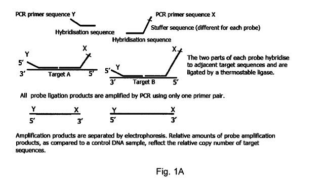
One particularly suitable technique for investigation of nucleic acid
sequences is multiplex ligation dependent probe amplification (MLPA). This
technique is based on hybridisation of probes to target nucleic acids, where
after probes are amplified. In currently used MLPA assays, each MLPA probe
set consists of two half probes. These two half probes contain a target-
specific
sequence and a primer binding site sequence to which a nucleic acid
amplification primer (preferably a PCR primer) can bind. One half probe is
typically shorter in length then the other. The other half probe is longer due
to
a non-hybridizing stuffer sequence. The stuffer sequence of each probe set is
unique in length, resulting in different lengths of amplification products
(typically between 130 and 480 base pairs) that can be separated by
electrophoresis. In an MLPA assay, typically a plurality of probe sets is
used.
The two half probes of each probe set are typically added to denatured sample
nucleic acid and hybridized immediately adjacent to each other on their target
sequence. Subsequently, the resulting nucleic acid is subjected to a ligation
reaction. Usually a ligase is used which ligates only half probes that are
perfectly matched with their target sequence (such as for instance the
1
CA 02742726 2011-05-04
WO 2010/053363 PCT/NL2009/050669
thermostable Ligase-65). A mismatch of a half probe at the ligation site
prevents ligation and amplification. Thereby no amplification products of the
probe will be detected. This allows MLPA to discriminate sequences that only
differ in a single nucleotide. Sequences from pseudogenes or related genes can
therefore be distinguished. Ligated half probes (which are also referred to as
"ligated probes") are amplified, preferably by PCR, using primers capable of
specifically binding the primer binding site sequences of the probes. The
amplification products of each ligated probe are separated and analyzed, for
instance by electrophoresis. Preferably, amplification products are
represented
graphically by separate peaks. Each peak is the product of an amplified MLPA
ligated probe and a relative difference in peak intensity (height or surface)
between a control sample and a sample of interest indicates copy number
variation. Figure 1A schematically outlines an MLPA reaction.
MLPA is particularly suitable for detecting nucleic acid (pseudo)gene
variants, (pseudo)gene-specific nucleotides and/or copy number variation.
MLPA has been employed in several studies, e.g. for the diagnosis of Prader-
Willi or Angelman syndromes, for prenatal diagnosis of chromosomal
aberrations in fetuses, and for the detection of exon deletions and/or
duplications in the Duchenne muscular dystrophy gene. Overall, the
conclusion was that MLPA could replace the existing methods used for
screening of chromosomal abnormalities due to its relative simplicity,
reproducibility and speed.
In an MLPA assay, targeted nucleic acid which is gene-specific or
pseudogene- specific is preferably present at the ligation site of the half
probes.
When a gene-specific or pseudogene-specific nucleotide is present at (or
within
three nucleotides from) a ligation site, this will ensure that only perfectly
matched half probes are ligated to each other. A mismatch of a half probe at
the ligation site prevents ligation and amplification, whereas a perfect match
2
CA 02742726 2011-05-04
WO 2010/053363 PCT/NL2009/050669
of the half probe at the ligation site allows ligation and amplification. As
said
before, this allows MLPA to discriminate between sequences that only differ in
a single nucleotide. Mismatches at four to six nucleotides away from the
ligation site have been reported to have little effect on the ligation step.
Hence, the half probes are preferably designed such that the half probe
whose 3' end hybridizes at a target sequence (called herein a "left probe" or
a
"left half probe") is complementary to a gene-specific sequence or pseudogene-
specific sequence of the target sequence. This gene-specific or pseudogene-
specific sequence of the target sequence comprises at least one but preferably
more nucleotides that make the probe specific for a given gene or pseudogene.
Preferably, at least one of the 3' end nucleotides of said left half probe is
complementary to at least one gene-specific nucleotide and/or at least one
pseudogene- specific nucleotide of the target sequence, so that the
(pseudo)gene-specific nucleotide(s) or a single nucleotide polymorphism within
a given (pseudo)gene is present at (or within three nucleotides from) the
ligation site of said left half probe. In this case, said left half probe and
the
probe whose 5' end hybridizes at a target sequence (called herein a "right
probe" or a "right half probe") are ligated to each other only when the
sequence
of the left half probe perfectly matches its target sequence.
As used herein the term "gene-specific nucleotide" or "gene-specific
sequence" means a nucleotide or sequence, respectively, which is present in
said gene but not present at the corresponding location in at least one other
related gene or pseudogene. The term "pseudogene-specific nucleotide" or
"pseudogene-specific sequence" means a nucleotide or sequence, respectively,
which is present in said pseudogene but not present at the corresponding
location in at least one other related gene or pseudogene. Hence, at least one
other (pseudo)gene comprises another nucleotide or sequence at that location.
The presence of a (pseudo)gene-specific nucleotide or (pseudo)gene-specific
sequence in a (pseudo)gene thus distinguishes said (pseudo)gene from at least
3
CA 02742726 2011-05-04
WO 2010/053363 PCT/NL2009/050669
one other (pseudo)gene, even in case when the other (pseudo)gene has a high
overall homology with said (pseudo)gene.
A pseudogene is defined herein as a nucleic acid sequence which does
not encode a wild type, functional, protein. The term "pseudogene"
encompasses nucleic acid sequences which do not encode protein at all.
Additionally, the term "pseudogene" encompasses gene alleles which comprise
a modification, for instance an insertion or deletion so that they encode a
protein or a part of a protein with significantly impaired, or lost, function
as
compared to a wild type protein of the same kind. Such allele for instance
encodes a truncated protein as a result of a frame shift caused by an
insertion
and/or deletion of at least one nucleotide, or caused by a premature stop
codon.
Since ligases only ligate half probes which are adjacent to each other,
half probes need to be designed which are capable of hybridizing immediately
adjacent to each other on their target sequence. This is not always
convenient,
because the hybridization location of a left half probe on a target nucleic
acid is
often determined by a (pseudo)gene-specific site of the target nucleic acid
(as
explained above). In such case, the sequence of the corresponding right half
probe is determined as well, since the right half probe should be capable of
hybridizing to a region of said target nucleic acid which is immediately
adjacent to said (pseudo)gene-specific nucleotide. However, such region may
comprise sequences which are very commonly present in the nucleic acid
sequences of a sample. As a result, a right half probe having a sequence which
is complementary to such common sequence will hybridize at many different
sites of the nucleic acids present in a sample. In such case, it would be more
attractive to design a right half probe with a sequence which is more specific
for a given site of interest of a target nucleic acid. However, if the left
half
probe and the right half probe do not hybridize to adjacent regions of a
target
nucleic acid, the commonly used ligases will not be capable of performing the
ligation reaction. Patent application WO 01/61033 in the name of Schouten
4
CA 02742726 2011-05-04
WO 2010/053363 PCT/NL2009/050669
discloses a solution to this problem by adding a short third probe to the
reaction mixture, which third probe will fill the gap between the left half
probe
and the right half probe. Such third probe is designed to hybridize to a
region
of a target nucleic acid which lies between the left and the right half
probes.
After hybridization of such third probe, the left half probe is connected to
the
right half probe via the third probe and ligation has become possible. The
third
half probe does not need to be perfectly complementary to the region of the
target nucleic acid which lies between the left and the right half probes, as
long as the third probe connects the left half probe and the right half probe
so
that a ligase reaction can occur. Moreover, since the third probe is small, it
will
hybridize more easily to the target nucleic acid as compared to the left and
right half probes. Hence, mismatches between the third probe and the target
nucleic acid are allowed. This way, one and the same third probe is suitable
for
connecting left and right half probes of different probe sets.
Instead of using a third probe, WO 01/61033 also discloses an
embodiment wherein the 3' end of a left half probe is extended after
hybridization of the half probes to the target sequence, so that the gap
between
the left half probe and the right half probe is filled. The resulting extended
left
half probe is adjacent to the right half probe and a ligase reaction has
become
possible.
In order to be capable of distinguishing between amplificates of different
probe sets, currently used MLPA probe sets are designed such that the
resulting amplificates have a different length. Differences in ligated probe
length are typically realized by using a non-hybridizing stuffer sequence in
one
of the half probes. The stuffer sequence of the half probes of each probe set
is
unique in length, resulting in different lengths of amplification products
that
can be separated by electrophoresis. Typically, in order to be capable of
discriminating between the different amplification products, the difference in
length between different ligated probes is at least 5 nucleotides. Since a
usual
5
CA 02742726 2011-05-04
WO 2010/053363 PCT/NL2009/050669
MLPA assay involves the use of many different probe sets in order to be
capable of detecting a wide variety of (pseudo)gene variants, this means that
long probes have to be generated. This is especially the case when complex
loci
carrying many (pseudo)gene-specific nucleotides are investigated for proper
genotyping and/or additional single nucleotide polymorphisms are investigated
for detection of subtle genetic variation within a specific genotype, as well
as
the presence of pseudogenes and single nucleotides in these pseudogenes. Such
investigation requires the use of many different probe sets. This is
inconvenient if probes are chemically synthesized, because a drawback of
synthetic probes is the lower quality in comparison with cloned probes, due to
contamination with incompletely synthesized probes. These incompletely
synthesized probes lack or gain one nucleotide, which results in stutter peaks
and split peaks. A method to remove these contaminants is to purify the
synthesized probes, for instance by polyacrylamid gel electrophoresis (PAGE).
If short and long probes are chemically synthesized, a higher proportion of
longer probes is more likely to be affected by the incomplete
oligonucleotides,
causing a limitation of synthetic probe size. The upper limit of synthetic
probes
is typically about 100 base pairs.
On the other hand, the use of synthetic probes is preferred because they
are easy to obtain and cost-effective whereas generating a probe by cloning in
bacteriophage vectors is a time-consuming process and more expensive.
Hence, although good results have been obtained with currently used
MLPA assays, it is desirable to provide alternatives and improvements,
especially if complex (pseudo)gene loci are investigated which involves the
use
of many probe sets.
It is an object of the present invention to provide alternative and
improved MLPA methods and MLPA-like methods.
6
CA 02742726 2011-05-04
WO 2010/053363 PCT/NL2009/050669
Accordingly, the present invention provides MLPA assays and MLPA-
like assays wherein at least one probe set is used which comprises a first
nucleic acid probe ("left probe" or "left probe part"), a second nucleic acid
probe
("right probe" or "right probe part") and a third nucleic acid probe ("third
probe" or "middle probe" or "middle probe part"), wherein at least one third
probe is complementary to a target nucleic acid region comprising a
(pseudo)gene-specific nucleotide or (pseudo)gene-specific sequence.
The present invention provides a different approach as compared to the
prior art. MLPA methods and MLPA-like methods are now provided wherein
at least one third probe, but preferably a plurality of third probes, is used
in
order to detect at least one (pseudo)gene-specific nucleotide of a target
nucleic
acid. Hence, an additional probe is used in at least one of the probe sets,
which
is specific for a (pseudo)gene-specific target nucleic acid. As used herein,
an
MLPA-like method is defined as a method comprising the steps of
hybridisation of at least two probes to a target nucleic acid and ligation of
at
least two probes. Preferably, said MLPA-like method comprises amplification
of ligated probes as well.
MLPA methods and MLPA-like methods according to the present
invention have several advantages as compared to current methods. For
instance, if the left probe and the third probe of a probe set are both
complementary to target nucleic acid regions comprising (pseudo)gene-specific
nucleotides and/or additional single nucleotide polymorphism(s), two different
(pseudo)gene-specific target nucleotides or two SNP's or a combination of one
(pseudo)gene specific target nucleotide and one SNP are screened using one
probe set. It has become possible to use one probe set in order to screen for
at
least two (pseudo)gene variations which are located within a region of about
150 nucleotides of a target nucleic acid. Contrary, in a currently used MLPA
assay two separate probe sets are needed for screening for two variants in a
target nucleic acid. This is illustrated by the following example. If a target
(pseudo)gene contains a (pseudo)gene variant at location A and at location B,
7
CA 02742726 2011-05-04
WO 2010/053363 PCT/NL2009/050669
an individual may comprise the following alleles: a-b, a-B, A-b and A-B. In
order to determine whether allele a-B is present in a sample of said
individual,
a currently used MLPA assay would need a probe set specific for the "a" and/or
"A" (pseudo)gene variant and a probe set specific for the "B" and/or "b"
(pseudo)gene variant. If both the probe set specific for "a" and the probe set
specific for "B" provide a positive result, it is concluded that allele a-B is
present in said individual. With a MLPA method according to the present
invention, however, only one probe set is needed wherein the left probe is
specific for the "a" (pseudo)gene variant and the third probe is specific for
the
"B" (pseudo)gene variant. If an amplification product is obtained, it is
immediately concluded that allele a-B is present in said individual. If allele
a-
B is not present, said probe set according to the invention will not yield an
amplification product. Hence, it has become possible to more specifically
screen
for a given allele.
Moreover, a method of the invention provides an additional advantage
when two (pseudo)gene variations are located close to each other. If the
(pseudo)gene variants at location A and at location B are close to each other,
the use of two different probe sets according to conventional MLPA techniques
is inconvenient or even not possible at all, because the two probe sets will
hinder each other in view of their close proximity. This will result in less
efficient hybridization of the two probe sets, resulting in a lower signal as
compared to a method according to the invention, wherein two (pseudo)gene
variants can be detected using only one probe set. Hence, a method according
to the invention is more sensitive when (pseudo)gene variants are located
close
to each other (in practice, this effect will be most profound when the
(pseudo)gene variants are located between 20-100 nucleotides from each
other). Having two probes to detect a variant at the same position (such as in
currently used MLPA assays) will result in a change in signal intensity,
depending on the presence of the (pseudo)gene variant and the binding of the
probe. The use of more than two probes for one position is not advised. Figure
8
CA 02742726 2011-05-04
WO 2010/053363 PCT/NL2009/050669
1B schematically outlines an MLPA reaction according to the invention in
which a probe set consisting of three probes is used for detecting two SNPs.
Figure 1C shows a non-limiting example of two specific probe sets according to
the invention for detecting two SNPs.
As another example, in case that an individual is heterozygous for the
above mentioned (pseudo)gene, the individual for instance contains alleles a-B
and A-b. A conventional MLPA assay would use four probe sets (one specific
for "a", one specific for "A", one specific for "b" and one specific for "B").
Four
positive results would be obtained, because all four probe sets would
hybridize
and result in an amplification product. However, in such case it would still
be
unknown whether the individual comprises the alleles a-b and A-B, or the
alleles a-B and A-b. With a method according to the present invention,
however, it has become possible to directly identify the alleles of said
individual. For instance, a first probe set of the invention is used
comprising a
left probe specific for "a" and a third probe specific for "b", together with
a
second probe set of the invention comprising a left probe specific for "a" and
a
third probe specific for "B" and a third probe set of the invention comprising
a
left probe specific for "A" and a third probe specific for "b" and a fourth
probe
set of the invention comprising a left probe specific for "A" and a third
probe
specific for "B". Two of these probe sets according to the present invention
will
yield an amplification product, namely the second probe set of the invention
comprising a left probe specific for "a" and a third probe specific for "B"
and the
third probe set of the invention comprising a left probe specific for "A" and
a
third probe specific for "b". The first and fourth probe sets according to the
present invention will not yield (significant) amplification product. This
way, it
is immediately apparent which alleles are present in said individual. This,
too,
is an advantage as compared to currently used methods, especially when
complex loci with many (pseudo)gene-specific nucleotides and additional single
nucleotide polymorphisms within a given (pseudo)gene are investigated,
9
CA 02742726 2011-05-04
WO 2010/053363 PCT/NL2009/050669
because in such case many different combinations of such (pseudo)gene
variants need to be screened for.
Another advantage of a method according to the present invention is the
fact that more variations in length of the ligated probes are obtained. Since
at
least one probe set of the invention, but preferably a plurality of probe sets
of
the invention, comprise a third probe it has become possible to design the
probe sets such that variations in length of the resulting ligated probes are
obtained. This obviates the need of stuffer sequences. As a result, the
individual probes of a probe set according to the invention can be kept
shorter,
which is particularly advantageous when chemically synthesized probes are
used because chemical production of long probes is cumbersome, as explained
above. Hence, a method according to the invention allows for the use of probe
sets with relatively short probes, while the resulting ligated probes are long
enough to allow for many size variations. Thus, the present invention allows
the use of synthetic probes, which are easy to obtain and cost-effective, even
when complex loci are investigated, and offers greater flexibility to adapt
the
assay in case of cross-reactivity or unclear results.
For instance, if 20 (pseudo)gene variants are investigated, probes with a
stuffer sequence with a length varying from 4 to 100 nucleotides would need to
be used in a conventional MLPA assay in order to be capable of distinguishing
the resulting amplification products by size. Since the probe sequences
hybridizing to a target sequence are typically about 30 nucleotides, and since
the primer binding sequences of the probes are typically about 15-25
nucleotides, this would mean that probe sets with probes with a length varying
from 45-125 nucleotides would need to be synthesized. When the probes are
chemically synthesized, it is hardly possible to obtain reliable probe sets
with
these lengths. With a method according to the invention, however, differences
of length between the various amplificates need not to be obtained by use of
CA 02742726 2011-05-04
WO 2010/053363 PCT/NL2009/050669
stuffer sequences in the probe sets. Instead, at least one third probe is
used,
preferably a plurality of third probes is used. By varying combinations of
three
probes, optionally in combination with probe sets consisting of two probes,
the
overall length differences of the ligated probes vary considerably whereas
probe sets can be used with chemically synthesized probes with convenient
lengths. Of course, this does not mean that the use of stuffer sequences is
excluded. But the skilled person does no longer have to rely on these stuffer
sequences only for length variations. If stuffer sequences are used in a
method
according to the invention, it is preferred to keep these sequences as short
as
possible.
Accordingly, the present invention provides a method for screening for
the presence of at least one target nucleic acid sequence in a sample,
comprising the steps of:
a) adding to said sample at least two different probe sets, each probe set
comprising:
- a first nucleic acid probe ("left probe"), said first probe
comprising a first nucleic acid sequence complementary to a first
region of said target nucleic acid sequence, and
- a second nucleic acid probe ("right probe"), said second probe
comprising a second nucleic acid sequence complementary to a
second region of said target nucleic acid sequence,
wherein at least one of said probe sets comprises a third nucleic acid
probe, said third probe comprising a third nucleic acid sequence
complementary to a third region of said target nucleic acid sequence,
and
wherein, if said third probe is present in said probe set, said first and
said third region of said target nucleic acid are located essentially
adjacent to each other and said third and said second region of said
target nucleic acid are located essentially adjacent to each other, and
11
CA 02742726 2011-05-04
WO 2010/053363 PCT/NL2009/050669
wherein, if said third probe is not present in said probe set, said first
and said second region of said target nucleic acid are located essentially
adjacent to each other,
b) allowing hybridization of said at least two different probe sets to
complementary nucleic acid of said sample,
c) subjecting nucleic acid of said sample to a ligation reaction, and
d) determining whether said at least one target nucleic acid sequence is
present in said sample,
wherein at least one third nucleic acid probe is complementary to a target
nucleic acid region comprising a (pseudo)gene variation.
The advantage of probe sets comprising at least three probes according
to the present invention is that at least two different SNPs can be detected
with one probe set. For instance, in a probe set comprising three probes two
sites for ligation are present. A left probe and middle probe are ligated, and
a
middle probe and right probe are ligated. At each ligation site a SNP can be
detected. Thus it is possible to design two probes of the same probe set in
such
a way that they are used to detect two SNPs. In that case, using MLPA and a
probe set comprising three probes according to the invention, a product will
only be obtained when both SNPs are present in a sample, because only then
ligation can occur at both ligation sites.
With conventional MLPA probesets consisting of two probes only one
SNP can be detected, because only one site for ligation is present. Additional
third probe parts in conventional MLPA, as described in WO 01/61033, are
occasionally used to bridge the two half probes. Such an additional third
probe
part is not SNP-specific. Therefore, the advantages of probe sets comprising
at
least three probes according to the present invention are not obtained when
using such additional third probe part for bridging purposes in conventional
MLPA.
12
CA 02742726 2011-05-04
WO 2010/053363 PCT/NL2009/050669
Therefore, in a preferred embodiment of the invention a probe set
comprises three nucleic acid probes wherein each of at least two nucleic acid
probes are specific for a different (pseudo)gene variation. Preferably, a
first (or
a second) nucleic acid probe of a probe set according to the invention is
complementary to a target nucleic acid region comprising a gene-specific
nucleotide and/or a pseudogene-specific nucleotide and/or a gene-specific
sequence and/or a pseudogene-specific sequence and/or a polymorphism within
a given gene or pseudogene, and a third nucleic acid probe of the same
probeset is complementary to another target nucleic acid region comprising a
gene-specific nucleotide and/or a pseudogene- specific nucleotide and/or a
gene-
specific sequence and/or a pseudogene-specific sequence and/or a
polymorphism within a given gene or pseudogene. Said polymorphism
preferably comprises an SNP.
Preferably, ligated probes are amplified. Accordingly, the present
invention provides a method for screening for the presence of at least one
target nucleic acid sequence in a sample, comprising the steps of:
a) adding to said sample at least two different probe sets, each probe set
comprising:
- a first nucleic acid probe ("left probe"), said first probe
comprising a first nucleic acid sequence complementary to a first
region of said target nucleic acid sequence and, located 5'
thereof, a non-complementary nucleic acid sequence comprising
a first primer binding site, and
- a second nucleic acid probe ("right probe"), said second probe
comprising a second nucleic acid sequence complementary to a
second region of said target nucleic acid sequence and, located 3'
thereof, a non-complementary nucleic acid sequence comprising
a second primer binding site,
13
CA 02742726 2011-05-04
WO 2010/053363 PCT/NL2009/050669
wherein at least one of said probe sets comprises a third nucleic acid
probe, said third probe comprising a third nucleic acid sequence
complementary to a third region of said target nucleic acid sequence,
and
wherein, if said third probe is present in said probe set, said first and
said third region of said target nucleic acid are located essentially
adjacent to each other and said third and said second region of said
target nucleic acid are located essentially adjacent to each other, and
wherein, if said third probe is not present in said probe set, said first
and said second region of said target nucleic acid are located essentially
adjacent to each other,
b) allowing hybridization of said at least two different probe sets to
complementary nucleic acid of said sample,
c) subjecting nucleic acid of said sample to a ligation reaction,
d) subjecting nucleic acid of said sample to a nucleic acid amplification
reaction, using at least one primer capable of specifically binding said
first primer binding site and at least one primer capable of specifically
binding said second primer binding site, and
e) determining whether amplified nucleic acid is present, thereby
determining whether said at least one target nucleic acid sequence is
present in said sample,
wherein at least one third nucleic acid probe is complementary to a
target nucleic acid region comprising a (pseudo)gene variation.
As used herein, the term "(pseudo)gene variation" encompasses a
(pseudo)gene-specific nucleotide and/or a (pseudo)gene-specific sequence. In
one embodiment, said (pseudo)gene variation comprises an additional
polymorphism within a given (pseudo)gene. Said additional polymorphism
preferably comprises an SNP.
14
CA 02742726 2011-05-04
WO 2010/053363 PCT/NL2009/050669
Hence, the present invention uses probe sets, wherein at least one probe
set, but preferably a plurality of probe sets, comprises three probes. The
probes
comprise sequences which are complementary to a region of a target nucleic
acid of interest. As used herein, the term "complementary" means that said
probe sequence comprises at least 70%, preferably at least 80%, more
preferably at least 85%, more preferably at least 90%, most preferably at
least
95% sequence identity to said region or to the complement of said region. The
term "% sequence identity" is defined herein as the percentage of residues in
a
nucleotide sequence that is identical with the residues in a reference
sequence
after aligning the two sequences and introducing gaps, if necessary, to
achieve
the maximum percent identity. Methods and computer programs for the
alignment are well known in the art. One computer program which may be
used or adapted for purposes of determining whether a candidate sequence
falls within this definition is Autoassembler 2.0 (ABI Prism, Perkin Elmer).
The first and second probes of each probe set also comprise a primer
binding site, so that the resulting ligated probes can be amplified.
Preferably,
the primer binding sites of the first nucleic acid probes of each probe set is
designed such that the same primer can bind. This allows the use of the same
primer for binding the primer binding sites of the first probes in step d).
Likewise, it is preferred that the primer binding sites of the second nucleic
acid probes of each probe set is designed such that the same primer can bind.
Most preferably, the probe sets are designed such that a first primer is
capable
of specifically binding the primer binding sites of the first nucleic acid
probes
of each probe set and a second primer is capable of specifically binding the
primer binding sites of the second nucleic acid probes of each probe set. This
embodiment allows the use of only one primer pair in step d). This is,
however,
not necessary: it is also possible to use different primers for different
probe
sets. The number of different primers is, however, kept as low as possible.
CA 02742726 2011-05-04
WO 2010/053363 PCT/NL2009/050669
One preferred embodiment therefore provides a method according to the
invention, wherein the first primer binding sites of the first nucleic acid
probes
of each probe set is capable of specifically binding the same primer and/or
wherein the second primer binding sites of the second nucleic acid probes of
each probe set is capable of specifically binding the same primer. Preferably,
the first nucleic acid probes and/or the second nucleic acid probes of each
probe
set comprise essentially identical primer binding sequences. Further provided
is therefore a method according to the invention, wherein the non-
complementary nucleic acid sequences of said first nucleic acid probes
comprise essentially identical first primer binding sites and/or wherein the
non-complementary nucleic acid sequences of said second nucleic acid probes
comprise essentially identical second primer binding sites. Using essentially
identical primer binding sequences ensures that the same primer can bind
different probes. The term "essentially identical primer binding sequences" is
defined herein as primer binding sequences which comprise at least 80%,
preferably at least 85%, more preferably at least 90%, most preferably at
least
95% sequence identity to each other.
As already described, a method according to the invention is particularly
suitable for investigating a nucleic acid sequence having various (pseudo)gene
specific nucleotides and/or (pseudo)gene variants, such as complex loci. It is
therefore preferred to use a plurality of third probes, so that many
(pseudo)gene variant combinations are investigated. A method according to the
invention is therefore preferably provided wherein at least two, preferably at
least five, more preferably at least ten different third nucleic acid probes
are
used. As illustrated in the Examples, a plurality of probe sets comprising
different third probes according to the invention allows for screening of
complex gene loci such as the KIR locus. Not all third probes need to be
specific
for a genetic variation of a target nucleic acid. It is also possible to use a
combination of variant-specific third probes and third probes which are not
16
CA 02742726 2011-05-04
WO 2010/053363 PCT/NL2009/050669
specific for a (pseudo)gene variation. Likewise, not all first probes need to
be
specific for a variant of a target nucleic acid. It is also possible to use a
combination of variant-specific first probes and first probes which are not
specific for a (pseudo)gene variation. Any of these combinations is for
instance
used to vary the length of the resulting ligated probes to a larger extent. In
one
preferred embodiment of the invention, therefore, at least 50%, preferably at
least 70%, more preferably at least 80%, most preferably at least 90% of the
third nucleic acid probes is complementary to a target nucleic acid region
comprising a (pseudo)gene variation. In one embodiment, all third probes are
complementary to a target nucleic acid region comprising a (pseudo)gene
variant. Preferably, the second probes ("right probes") are not designed to
contain (pseudo)gene variant-specific sequences, although the use of variant-
specific right probes in a method according to the invention is not excluded.
Preferably, at least 50%, preferably at least 70%, more preferably at
least 80%, most preferably at least 90% of the third nucleic acid probes that
are complementary to a target nucleic acid region comprising a (pseudo)gene
variation are combined with a first nucleic acid probe or a second nucleic
acid
probe that is complementary to another target nucleic acid region comprising a
(pseudo)gene variation in order to be capable of screening for many variants
with one MLPA assay or MLPA-like assay. In one embodiment, all third
probes that are combined with a first nucleic acid probe or a second nucleic
acid probe that is complementary to a target nucleic acid region comprising a
(pseudo)gene variation are complementary to a target nucleic acid region
comprising a (pseudo)gene variant. Of course, these probes are preferably
specific for different variants.
In one preferred embodiment, a (pseudo)gene variant-specific sequence
of a third probe is at least located within the last three nucleotides or the
first
three nucleotides of the third probe. This means that the last three
nucleotides
and/or the first three nucleotides comprise at least one nucleotide which is
17
CA 02742726 2011-05-04
WO 2010/053363 PCT/NL2009/050669
specific for a (pseudo)gene variation of a target nucleic acid. In this
embodiment, said (pseudo)gene variation is present at a ligation site of the
third probe, so that ligation is only possible when the sequence of the third
probe is exactly complementary to said (pseudo)gene variation. This enhances
the specificity of the MLPA method, as explained before. Preferably, the last
three nucleotides and/or the first three nucleotides of said third probe
comprise
one nucleotide which is specific for a (pseudo)gene variant of a target
nucleotide.
The probe sets according to the present invention preferably have a
length between 90 and 300 nucleotides. Cloned probes can be as long as 500
nucleotides. Preferably, however, chemically synthesized probes are used
because they are rapidly synthesized, easy to obtain and cost-effective. In
order
to be capable of synthetically producing the probes according to the present
invention, a method according to the invention is preferably provided wherein
third nucleic acid probes with a length of between 20 and 100 nucleotides are
used. Most preferably, third nucleic acid probes with a length of between 19
and 110 nucleotides are used. Since at least one probe set of the invention,
but
preferably a plurality of probe sets according to the invention, is used which
comprise three nucleic acid probes, sufficient variations in length and
specificity of the resulting ligated probes is ensured so that many
(pseudo)gene
variations can be investigated simultaneously.
These length variations of the resulting ligated probes obviate the need
of stuffer sequences, as explained before. It is therefore possible to design
the
probe sets such that the parts of the first and/or second probe which are not
complementary to a target nucleic acid have about the same length. According
to this embodiment, the length of the non-complementary sequences of all first
probes is about the same in each probe set, and/or the length of the non-
complementary sequences of all second probes is about the same in each probe
set. These lengths are about the same when they do not differ from each other
18
CA 02742726 2011-05-04
WO 2010/053363 PCT/NL2009/050669
by more than 10 nucleotides. Preferably, they do not differ from each other by
more than 6 nucleotides, most preferably they do not differ from each other by
more than 4 nucleotides. This, too, facilitates synthetic production of the
probes. Further provided is therefore a method according to the invention,
wherein the difference in length of said non-complementary nucleic acid
sequences of said first nucleic acid probes of said at least two different
probe
sets and/or the difference in length of said non-complementary nucleic acid
sequences of said second nucleic acid probes of said at least two different
probe
sets is less than 6, preferably less than 4 nucleic acids.
Besides the analysis of (pseudo)gene-specific nucleotides and additional
single nucleotide polymorphisms, an MLPA technique or MLPA-like technique
is particularly suitable for relative (pseudo)gene copy number determination.
If multiple copies of a (pseudo)gene of interest (or any other target nucleic
acid
of interest) are present in sample nucleic acid molecules, each copy will, in
principle, be bound by the specific probes which is detectable. When the
probes
are amplified, more amplification product will be present when multiple copies
were present in the original sample nucleic acid as compared to a situation
wherein only one copy is present. Analysis of the amount of amplification
product thus provides information about the copy number of a target nucleic
acid of interest. This is often done by graphically representing amplified
products by separate peaks. Each peak is the product of an amplified MLPA
ligated probe and a relative difference in peak intensity (height or surface)
between a control sample and a sample of interest indicates copy number
variation. When a complex locus is investigated, multiple copies of a
(pseudo)gene of interest can be present in highly polymorphic regions. In such
case, when (pseudo)gene copy number is to be determined, many different
combinations of (pseudo)gene variants need to be taken into account. This
involves the use of a wide variety of different probe sets, to ensure that
each
combination of (pseudo)gene variants can be detected. In one embodiment
19
CA 02742726 2011-05-04
WO 2010/053363 PCT/NL2009/050669
according to the present invention, however, when the relative copy number of
a nucleic acid of interest is to be estimated, an improved approach is
provided.
According to this embodiment, at least one probe is used with degenerate bases
at one or more positions. This means that a mixture of probes is used wherein
different nucleotides can be present at one or more positions. Hence a mixture
of probes is used, which probes have the same sequence, except for the fact
that some probes have a certain nucleotide at a given position X and some
probes have another nucleotide at said position X. Such degenerate bases are
commonly represented by the IUB nucleotide codes as depicted in figure 2. The
use of probes with degenerate bases allows for an efficient estimation of copy
number of a nucleic acid of interest, even in highly polymorphic regions.
Further provided is therefore a method for determining the copy number of a
nucleic acid of interest, wherein at least one probe set is used which
comprises
a probe with (a) degenerate base(s) at one or more positions. Preferably, at
most 20 probe positions have such multiple alternatives, in order to retain
specificity of the probes for a given target region of interest. A use of at
least
one probe set for determining the copy number of a nucleic acid of interest,
wherein at least one probe set comprises a probe with (a) degenerate base(s)
at
one or more positions, is also provided herewith. In one preferred embodiment,
at least one probe set comprising a probe with (a) degenerate base(s) is used
in
a MLPA method or MLPA-like method according to the present invention.
Further provided is therefore a method according to the invention, wherein at
least one probe set is used which comprises a probe with (a) degenerate
base(s)
at one or more positions.
Alternatively, or additionally, a probe set is used which comprises an
alternative base which alternative base is capable of binding at least two
bases
selected from the group consisting of A, T, G, C and U. Preferably, said
alternative base is capable of binding at least three, most preferably at
least
four, bases selected from the group consisting of A, T, G, C and U. Such
alternative base is suitable as an alternative for degenerate bases. It is, of
CA 02742726 2011-05-04
WO 2010/053363 PCT/NL2009/050669
course, also possible to combine such alternative base with degenerate bases.
In a particularly preferred embodiment said alternative base is deoxyinosine
triphosphate (dITP) or a functional equivalent thereof, which is capable of
binding A and T and G and C and U. Further provided is therefore a method
for determining the copy number of a nucleic acid of interest, wherein at
least
one probe set is used which comprises an alternative base which is capable of
binding at least two, preferably at least three, more preferably at least four
bases selected from the group consisting of A, T, G, C and U. As said before,
said alternative base preferably comprises deoxyinosine triphosphate (dITP) or
a functional equivalent thereof. A use of at least one probe set for
determining
the copy number of a nucleic acid of interest, wherein at least one probe set
comprises an alternative base which is capable of binding at least two,
preferably at least three, more preferably at least four bases selected from
the
group consisting of A, T, G, C and U, is also provided herewith. In one
preferred embodiment, at least one probe set comprising such alternative
base(s) is used in a MLPA method or MLPA-like method according to the
present invention. Further provided is therefore a method according to the
invention, wherein at least one probe set is used which comprises an
alternative base which is capable of binding at least two, preferably at least
three, more preferably at least four bases selected from the group consisting
of
A, T, G, C and U. As said before, said alternative base preferably comprises
deoxyinosine triphosphate (dITP) or a functional equivalent thereof.
The present invention provides alternative and improved methods for
screening for the presence of at least one target nucleic acid sequence in a
sample, wherein at least one third probe is used which is complementary to a
target nucleic acid region comprising a (pseudo)gene variation. A use of a
probe set comprising at least three nucleic acid probes, wherein at least one
third probe is complementary to a target nucleic acid region comprising a gene
variant and/or a pseudogene variant, for screening for the presence of at
least
21
CA 02742726 2011-05-04
WO 2010/053363 PCT/NL2009/050669
one target nucleic acid sequence in a sample is therefore also provided.
Preferably, a plurality of probe sets according to the present invention is
used.
Further provided is therefore a use of a plurality of probe sets for screening
for
the presence of at least one target nucleic acid sequence in a sample, wherein
each of said probe sets comprises:
- a first nucleic acid probe, said first probe comprising
a first nucleic acid sequence complementary to a first
region of said target nucleic acid sequence and, located 5'
thereof, a non-complementary nucleic acid sequence
comprising a first primer binding site, and
- a second nucleic acid probe, said second probe comprising
a second nucleic acid sequence complementary to a second
region of said target nucleic acid sequence and, located 3'
thereof, a non-complementary nucleic acid sequence
comprising a second primer binding site,
wherein at least one of said probe sets comprises a third nucleic acid
probe, said third probe comprising a third nucleic acid sequence
complementary to a third region of said target nucleic acid sequence,
and
wherein, if said third probe is present in said probe set, said first and
said third region of said target nucleic acid are located essentially
adjacent to each other and said third and said second region of said
target nucleic acid are located essentially adjacent to each other, and
wherein, if said third probe is not present in said probe set, said first
and said second region of said target nucleic acid are located essentially
adjacent to each other, and
wherein at least one third nucleic acid probe is complementary to a
target nucleic acid region comprising a gene-specific nucleotide and/or a
pseudogene- specific nucleotide and/or a gene-specific sequence and/or a
pseudogene-specific sequence and/or an additional polymorphism within
22
CA 02742726 2011-05-04
WO 2010/053363 PCT/NL2009/050669
a given gene or pseudogene, said polymorphism preferably comprising
an SNP.
A method according to the present invention is particularly suitable for
analysis of (pseudo)gene variation and (pseudo)gene copy number
determination in complex loci such as the gene encoding complement factors
(e.g. Factor H and FH-like genes, C4A and C4B within the HLA-class III
region), chemokines and their receptor alleles (e.g. CCL3L1, CCL4L1, CCR5 or
CCR5delta32), HLA-class I and II, SIRPs and LILRs.
In one preferred embodiment, a method according to the invention is
used in order to investigate the killer cell immunoglobulin-like receptor
(KIR)
locus. KIRs are expressed by natural killer (NK) cells and a subset of T
cells.
NK cells are cells of the lymphoid lineage, but display no antigen-specific
receptors. Their main function is to monitor host cells for the presence of
MHC
class I molecules and this is important for e.g. distinguishing healthy cells
from virus-infected or tumors cells. Interaction between NK cells and MHC
class I molecules is mediated by KIRs. The KIR locus in humans is polygenic
and highly polymorphic, so that accurate and efficient characterization of an
individual's KIR (pseudo)gene profile is cumbersome. In the determination of
the KIR (pseudo)gene profile and their role in many diseases an efficient and
reliable method for KIR genotyping is, however, important. Until now, KIR
genotyping is based upon the polymerase chain reaction sequence-specific
primer (PCR-SSP) (Sun et al, 2004), multiplex PCR (Vilches et al, 2007) and
PCR-sequence specific oligonucleotide probes (PCR-SSOP) (Crum et al, 2000).
For the PCR-SSP high-quality genomic DNA is required and multiple
reactions are needed to generate a complete KIR profile of an individual.
Multiple copies of KIR2DL4 and KIR3DL1 /S1 in individuals have been
reported with PCR-SSOP (Williams et al, 2003). Detection of the multiple gene
copies was possible because the gene copies of these genes consisted of
different alleles. However, multiple gene copies of highly homologous or
23
CA 02742726 2011-05-04
WO 2010/053363 PCT/NL2009/050669
identical sequences are not distinguishable with this molecular detection
system or cloning methods when individuals are homozygous for a gene
(Williams et al, 2003).
As shown in the Examples, a method according to the present invention
is particularly suitable for investigating the KIR locus of individuals. Even
though this locus is highly polymorphic, (pseudo)gene variants and copy
number variations are efficiently detected with methods according to the
present invention. One preferred embodiment therefore provides a method or
use according to the invention, wherein said target nucleic acid sequence is
present in a KIR locus. Preferably, copy number variation of at least one KIR
gene and/or at least one KIR pseudogene is determined. Figure 3A and B
provides KIR-specific probes which provide particularly good results. These
probes are therefore preferred when a KIR locus is investigated. Figure 3C and
D provides an extended list of KIR-specific probes which provide even better
results than the probes listed in figure 3A and B. Therefore, these probes are
even more preferred when a KIR locus is investigated. Further provided is
thus a method and/or a use according to the invention, wherein at least one
probe depicted in figure 3A, 3B, 3C or 3D, preferably in figure 3C or 3D, is
used. Preferably, at least two probes depicted in figure 3 are used. In
another
preferred embodiment at least four probes, more preferably at least six probes
depicted in figure 3A, 3B, 3C or 3D are used.
In a particularly preferred embodiment, a probe set of figure 3 is used.
Said probe set preferably comprises three probes. A probe set of figure 3 is
formed by two or three individual probes depicted in figure 3 which have the
same number, followed by the letter A, B, C, D, E, G, K, L, M or N. For
instance, probe set 408 is formed by probes 408A, 408B and 408C. Optionally,
four different probes with the same number are given for a probe set of figure
3. In that case, a left, a middle and a right probe is selected from said four
probes. Further provided is therefore a method and/or a use according to the
24
CA 02742726 2011-05-04
WO 2010/053363 PCT/NL2009/050669
invention, wherein at least one probe set depicted in figure 3A selected from
the group consisting of probe set 408, probe set 507, probe set 419, probe set
528, probe set 413, probe set 416, probe set 415 and probe set 418 is used. In
a
particularly preferred embodiment at least one probe set depicted in figure 3A
selected from the group consisting of probe set 408, probe set 507, probe set
528, probe set 413, probe set 416 and probe set 415 is used. These probe sets
contain a third probe which is specific for a (pseudo)gene variant of the KIR
locus. Also provided is a method and/or a use according to the invention,
wherein at least one probe set depicted in figure 3B selected from the group
consisting of probe set 409, probe set 506, probe set 507, probe set 538,
probe
set 417 and probe set 517 is used. In a particularly preferred embodiment at
least one probe set depicted in figure 3B selected from the group consisting
of
probe set 409, probe set 506, probe set 507, probe set 538, probe set 417 and
probe set 517 is used. These probe sets also contain a third probe which is
specific for a (pseudo)gene variant of the KIR locus. Also provided is a
method
and/or a use according to the invention, wherein at least one probe set
depicted
in figure 3C selected from the group consisting of probe set 415, probe set
703,
probe set 413, probe set 419, probe set 702, probe set 711, probe set 408,
probe
set 507, probe set 710, probe set 528, probe set 418 and probe set 416 is
used.
In a particularly preferred embodiment at least one probe set depicted in
figure 3C selected from the group consisting of probe set 415, probe set 703,
probe set 413, probe set 419, probe set 702, probe set 711, probe set 408,
probe
set 507, probe set 710, probe set 528, probe set 418 and probe set 416 is
used.
These probe sets also contain a third probe which is specific for a
(pseudo)gene
variant of the KIR locus. Also provided is a method and/or a use according to
the invention, wherein at least one probe set depicted in figure 3D selected
from the group consisting of probe set 506, probe set 417, probe set 517,
probe
set 409, probe set 507, probe set 710, probe set 709, probeset 708, probe set
704
and probe set 538 is used. In a particularly preferred embodiment at least one
probe set depicted in figure 3D selected from the group consisting of probe
set
CA 02742726 2011-05-04
WO 2010/053363 PCT/NL2009/050669
506, probe set 417, probe set 517, probe set 409, probe set 507, probe set
710,
probe set 709, probeset 708, probe set 704 and probe set 538 is used. These
probe sets also contain a third probe which is specific for a (pseudo)gene
variant of the KIR locus.
It is preferred to use at least two probe sets selected from figure 3, so
that various KIR (pseudo)gene variants are screened for with good results.
More preferably, at least three probe sets selected from figure 3 are used.
Even
more preferably, at least four, more preferably at least five, most preferably
at
least six probe sets selected from figure 3 are used. Said at least two,
three,
four, five or six probe sets are preferably selected from the group consisting
of
probe set 408, probe set 507, probe set 528, probe set 413, probe set 416,
probe
set 415, probe set 418, probe set 419, probe set 409, probe set 506, probe set
538, probe set 417, probe set 517, probe set 703, probe set 702, probe set
711,
probe set 710, probe set709 and probe set 704 since these probe sets contain a
third probe which is specific for a (pseudo)gene variant of the KIR locus. In
one
embodiment, all probe sets depicted in figure 3A, and/or 3B, and/or 3C, and/or
3D are used. In a preferred embodiment all probe sets depicted in figure 3C
and/or figure 3D are used.
It is of course also possible to modify a sequence of at least one probe
depicted in figure 3 to some extent. This is for instance done for
optimalization
purposes. Further provided is therefore a method and/or a use according to the
invention, wherein at least one probe is used which has at least 70%,
preferably at least 80%, more preferably at least 85%, more preferably at
least
90%, most preferably at least 95% sequence identity to a probe depicted in
figure 3. Preferably, at least two, more preferably at least four, most
preferably
at least six probes are used which have at least 70%, preferably at least 80%,
more preferably at least 85%, more preferably at least 90%, most preferably at
least 95% sequence identity to a probe depicted in figure 3. In one
embodiment,
a method or use according to the invention is provided wherein at least 20
26
CA 02742726 2011-05-04
WO 2010/053363 PCT/NL2009/050669
probes are used, said at least 20 probes having at least 70%, preferably at
least
80%, more preferably at least 85%, more preferably at least 90%, most
preferably at least 95% sequence identity to the probes depicted in figure 3.
A
minimum of two specific probes per (pseudo)gene is preferred to determine
copy number variation (CNV).
Preferably, probe sets are used which are based on the probe sets
depicted in figure 3A, 3B, 3C or 3D, preferably based on the probe sets
depicted in figure 3C and/or 3D. Said probe set preferably comprises three
probes. One or more of the probes of such probe set may be modified to some
extent, as described above. Further provided is therefore a method and/or a
use according to the invention, wherein at least one probe set is used which
has at least 70%, preferably at least 80%, more preferably at least 85%, more
preferably at least 90%, most preferably at least 95% sequence identity to a
probe set as depicted in figure 3. This means that the probes of said probe
set
have at least 70% sequence identity to the corresponding probes of at least
one
probe set of figure 3. Preferably, a probe set is used which has at least 70%,
preferably at least 80%, more preferably at least 85%, more preferably at
least
90%, most preferably at least 95% sequence identity to a probe set depicted in
figure 3 selected from the group consisting of probe set 408, probe set 507,
probe set 419, probe set 528, probe set 413, probe set 416, probe set 415,
probe
set 418, probe set 419, probe set 409, probe set 506, probe set 538, probe set
417, probe set 517, probe set 703, probe set 702, probe set 711, probe set
710,
probe set 709 and probe set 704 since these probe sets contain a third probe
specific for a KIR nucleic acid sequence. Preferably at least two, more
preferably at least three, more preferably at least four, more preferably at
least five, most preferably at least six of such probe sets are used, so that
various KIR (pseudo)gene variants are screened for with good results.
27
CA 02742726 2011-05-04
WO 2010/053363 PCT/NL2009/050669
Novel probes and probe sets which are particularly suitable for
(pseudo)gene variant analysis and (pseudo)gene copy number determination of
the KIR locus are also provided. These probes and probe sets are listed in
figure 3A, B, C and D, as described above. Further provided are therefore
probes and probe sets as depicted in figure 3A, 3B, 3C or 3D, as well as
probes
and probe sets which have at least 70%, preferably at least 80%, more
preferably at least 85%, more preferably at least 90%, most preferably at
least
95% sequence identity to a probe or probe set depicted in figure 3A, 3B, 3C or
3D. A mixture of nucleic acids, wherein said nucleic acids comprise at least
two
probe sets according to the invention is also provided. Preferably, said
mixture
comprises at least four, more preferably at least six probe sets according to
the
invention. As said before, such probe sets have at least 70% sequence identity
to a probe or probe set depicted in figure 3A, 3B, 3C or 3D. One embodiment
provides a mixture of nucleic acids comprising at least two, preferably at
least
four, more preferably at least six probe sets as depicted in figure 3A, 3B, 3C
or
3D.
Further provided is a kit for detecting the presence of at least one target
nucleic acid sequence in a sample, comprising a probe set or a mixture of
nucleic acids according to the invention. Said at least one target nucleic
acid
sequence preferably comprises a nucleic acid sequence present in a KIR locus.
A kit according to the invention preferably further comprises a PCR primer set
comprising at least 70%, preferably at least 80%, more preferably at least
85%,
more preferably at least 90%, most preferably at least 95% sequence identity
to nucleic acid sequences 5'-GGGTTCCCTAAGGGTTGGA and
TCTAGATTGGATCTTGCTGGCAC-3', or the complements thereof. These
primers are particularly suitable for amplifying probe sets depicted in figure
3.
KIR polymorphisms have been associated with disease. Association
between KIR polymorphisms and subtypes of leukemia were investigated by
Zhang et al. (Zhang et al. 2009). The presence of KIR2DS4 was demonstrated
28
CA 02742726 2011-05-04
WO 2010/053363 PCT/NL2009/050669
to be predisposing to chronic myelogenous leukemia (CML) and the absence of
KIR2DS3 was predisposing to acute lymphoblastic leukemia (ALL). KIR2DS4
is present in haplotype A, whereas KIR2DS3 is present in haplotype B.
Presence of KIR2DS4 and absence of KIR2DS3 are predisposing to leukemia
subtypes. Thus, characteristics of haplotype A are predisposing to leukemia
subtypes. The present invention provides probes that are particularly well
suitable for detecting KIR genes, including KIR2DS4 and KIR2DS3. Thus,
with probes according to the present invention selected from figure 3A, 3B, 3C
and/or 3D the presence and/or absence of KIR2DS4 and KIR2DS3 in a sample
is particularly well determined. Preferably probesets 540A/540C, and/or
513B/513D and/or 504A/504B, and/or 708K/708L/708M/708N as depicted in
figure 3C and/or 3D are used to detect KIR2DS3 and/or KIR2DS4
polymorphisms. With probes selected from figure 3 predisposition to leukemia
subtypes is thus particularly well determined.
Therefore, in one embodiment the invention provides a method for
determining predisposition to leukemia of an individual comprising
determining the presence or absence of KIR2DS4 and/or KIR2DS3 in a nucleic
acid sample of said individual with at least one probeset listed in figure 3A,
3B, 3C and/or 3D, wherein the presence of KIR2DS4 is indicative for a
predisposition for chronic myelogenous leukemia and the absence of KIR2DS3
is indicative for a predisposition for acute lymphoblastic leukemia. In a
preferred embodiment probe set 540A/540C, and/or 513B/513D and/or probe
set 504A/504B, and/or 708K/708L/708M/708N as depicted in figure 3C and/or
3D are used for determining the presence or absence of KIR polymorphisms.
As used herein, the term "nucleic acid sample" means a sample comprising
nucleic acid. Said sample may of course further comprise other components,
such as for instance proteins. Preferably, nucleic acid is at least partly
isolated
from said sample before being subjected to a method according to the present
invention.
29
CA 02742726 2011-05-04
WO 2010/053363 PCT/NL2009/050669
Association between KIR polymorphisms and inflammatory bowel
disease (IBD) and/or Crohn's disease have been established as well
(Hollenbach et al 2009). The KIR2DL2/KIR2DL3 heterozygous genotype
predisposes or protects from Crohn's disease depending on the presence of
their HLA-C ligands. KIR2DL2/KIR2DL3 heterozygosity in combination with
C1 predisposes to Crohn's disease whereas KIR2DL2/KIR2DL3 heterozygosity
in combination with C2 protects from IBD and/or Crohn's disease.
KIR2DL2/KIR2DL3 heterozygosity in combination with C1/C2 heterozygosity
has an intermediate effect on predisposition (Hollenbach et al 2009). Non-
limiting examples for determining the presence or absence of C1 and/or C2 are
detecting nucleic acid sequence(s) encoding C1 and/or C2 protein using for
instance a nucleic acid amplification reaction or detecting C1 and/or C2
protein
using for instance Western blot analysis.
The present invention provides probes that are particularly suitable for
detecting KIR genes, including KIR2DL2 and KIR2DL3. Thus, with probes
according to the present invention selected from figure 3A, 3B, 3C and/or 3D
KIR2DL2/KIR2DL3 heterozygosity in a sample is particularly well
determined. Preferably probeset 415B/415C/415D and/or 417A/417B/417C
and/or probeset 420A/420B, and/or 706A/706B as depicted in figure 3C and/or
3D are used to detect KIR2DL3 and/or KIR2DL2 polymorphisms. With probes
selected from figure 3 predisposition to Crohn's disease is thus particularly
well determined.
Therefore, in one embodiment the invention provides a method for
determining predisposition to IBD and/or Crohn's disease of an individual
comprising determining the presence or absence of KIR2DL2 and/or KIR2DL3
in a nucleic acid sample of said individual with at least one probeset listed
in
figure 3A, 3B, 3C and/or 3D, and determining the presence of absence of HLA
C1 and/or C2 ligand in a sample of said individual, wherein KIR2DL2,
KIR2DL3 heterozygosity in combination with C1 homozygosity is indicative for
a predisposition for Crohn's disease, and KIR2DL2, KIR2DL3 heterozygosity
CA 02742726 2011-05-04
WO 2010/053363 PCT/NL2009/050669
in combination with C2 homozygosity is indicative for protection for Crohn's
disease. In a preferred embodiment probe set 415B/415C/415D and/or
417A/417B/417C and/or probe set 420A/420B and/or 706A/706B as depicted in
figure 3C and/or 3D are used for determining the presence or absence of KIR
polymorphisms.
Copy number variation of KIR2DL3, KIR3DL1 and KIR3DS1 is
correlated to the course of disease in chronic infection, such as retroviral
infection, herpes virus infection, and hepatitis virus infection, more in
particular HIV, CMV, EBV, HSV, HBV and HCV (Martin et al 2007 and
Khakoo et al 2004). A higher copy number of KIR3DL1 and/or KIR3DS1 in an
individual is indicative for an improved course of the disease and/or response
to treatment of chronic infection as compared with a low copy number of
KIR3DL1 and/or KIR3DS1 in an individual and a low copy number of
KIR2DL3 in an individual is indicative for an improved course of the disease
and/or response to treatment of chronic infection as compared with a high copy
number of KIR2DL3 in an individual. Thus, a higher copy number of KIR3DL1
and/or KIR3DS1 in an individual is indicative for an increased survival in
chronic infection and a lower copy number of KIR2DL3 in an individual is
indicative for increased survival in chronic infection.
The present invention provides probes that are particularly well suitable
for determining copy number variation of KIR genes, including KIR3DL1 and
KIR3DS1. Thus, with probes according to the present invention selected from
figure 3A, 3B, 3C and/or 3D the copy number of KIR3DL1 and KIR3DS1 and
KIR2DL3 in a sample is particularly well determined. Preferably probe sets
409A/409B/409C, and/or 711A/711B/711C/711D and/or 418A/418B/418D,
and/or 709C/709D/709E/709G and/or probe set 415B/415C/415D and/or
417A/417B/417C as depicted in figure 3C and/or 3D are used to estimate the
copy number of KIR3DL1 and/or KIR3DS1 and/or KIR 2DL3. With probes
31
CA 02742726 2011-05-04
WO 2010/053363 PCT/NL2009/050669
selected from figure 3 susceptibility of an individual to course of disease
and/or
response to treatment in chronic infection is thus particularly well
determined.
Therefore the invention provides method for determining susceptibility
of an individual to course of disease and/or response to treatment in chronic
infection, preferably retroviral infection, herpes virus infection, and
hepatitis
virus infection, comprising determining the copy number of KIR2DL3,
KIR3DL1 and/or KIR3DS1 in a nucleic acid sample of said individual with at
least one probeset listed in figure 3A or 3B or 3C or 3D, wherein a high
KIR3DL1 and/or KIR3DS1 copy number in an individual is indicative for an
improved course of disease and/or response to treatment of chronic infection
as
compared with a low copy number of KIR3DL1 and/or KIR3DS1 in an
individual and a low KIR2DL3 copy number in an individual is indicative for
an improved course of disease and/or response to treatment of chronic
infection
as compared with a high copy number of KIR2DL3 in an individual. Preferably
said chronic infection comprises HIV, CMV, EBV, HSV, HBV and HCV. In a
preferred embodiment probeset 409A/409B/709D/409C, and/or
711A/711B/711C/711D and/or 418A/418B/418D, and/or 709C/709E/709G and/or
probe set 415B/415C/415D and/or 417A/417B/417C as depicted in figure 3C
and/or 3D are used for determining the copy number of KIR genes.
The presence of KIR2DS4 in a donor is correlated to transplantation-
related outcome measures, such as mortality, graft-versus-host, graft-versus-
tumor and grafted organ survival in recipients after transplantation. The
presence of KIR2DS4 in a donor is indicative for reduced mortality, reduced
graft-versus-host, increased graft-versus-tumor and increased grafted organ
survival in recipients after transplantation as compared to the absence of
KIR2DS4 in a donor. The present invention provides probes that are
particularly well suitable for determining copy number variation of KIR genes,
including KIR3DL1 and KIR3DS1. Thus, with probes according to the present
invention selected from figure 3A, 3B, 3C and/or 3D the copy number of
32
CA 02742726 2011-05-04
WO 2010/053363 PCT/NL2009/050669
KIR2DS4 in a sample is particularly well determined. Preferably probe sets
504A/504B, and/or 708K/708L/708M/708N as depicted in figure 3C and/or 3D
are used to the presence or absence of KIR2DS4. With probes selected from
figure 3 predisposition to transplantation-related outcome measures is thus
particularly well determined
Therefore the invention provides a method for determining
predisposition to transplantation-related outcome measures, such as mortality,
graft-versus-host, graft-versus-tumor and grafted organ survival of a
recipient
after transplantation, comprising determining the presence or absence of
KIR2DS4 in a nucleic acid sample of a donor for said recipient with at least
one probeset listed in figure 3A or 3B or 3C or 3D, wherein the presence of
KIR2DS4 in said donor is indicative for a reduced mortality, a reduced graft-
versus-host reaction, an increased graft-versus-tumor reaction and an
increased grafted organ survival in said recipient as compared to the
mortality, graft-versus-host reaction, graft-versus-tumor reaction and grafted
organ survival of a recipient with a donor wherein KIR2DS4 is absent. In a
preferred embodiment probeset 504A/504B, and/or 708K/708L/708M/708N as
depicted in figure 3C and/or 3D are used for determining the presence or
absence of KIR polymorphisms.
A correlation has been established between the copy number of
KIR2DL2 and KIR2DS2 and rheumatoid arthritis (RA) with extra-articular
manifestations and rheumatoid vasculitis. A higher copy number of KIR2DL2
and/or KIR2DS2 in an individual was demonstrated to be predisposing for
rheumatoid arthritis with extra-articular manifestations and rheumatoid
vasculitis (Majorczyk et al 2007, Yen et al 2001). Additionally, rheumatoid
arthritis patients positive for KIR2DL3 and negative for KIR2DS3 had earlier
disease diagnosis (Majorczyk et al 2007).
The present invention provides probes that are particularly well suitable
for determining the presence or absence and copy number variation of KIR
33
CA 02742726 2011-05-04
WO 2010/053363 PCT/NL2009/050669
genes, including KIR2DL2, KIR2DS2, KIR2DL3 and KIR2DS3. Thus, with
probes according to the present invention selected from figure 3A, 3B, 3C
and/or 3D the presence or absence and copy number of KIR2DL2, KIR2DS2,
KIR2DL3 and KIR2DS3in a sample is particularly well determined. Preferably
probe sets 420A/420B, and/or 706A/706B and/or probe set 703A/703B/703C,
and/or 544A/544B as depicted in figure 3C and/or 3D are used to estimate the
copy number of KIR2DL2 and/or KIR2DS2. Preferably probe sets
415B/415C/415D and/or 417A/417B/417C and/or probe set 513B/513D and/or
540A/540C as depicted in figure 3C and/or 3D are used to estimate the copy
number of KIR2DL3 and/or KIR2DS3. With probes selected from figure 3
susceptibility of an individual to rheumatoid arthritis (RA) with extra-
articular manifestations and rheumatoid vasculitis is thus particularly well
determined.
Therefore in one embodiment the invention provides a method for
determining predisposition to rheumatoid arthritis with extra-articular
manifestations and rheumatoid vasculitis of an individual comprising
determining the copy number of KIR2DS2 and/or KIR2DL2 in a nucleic acid
sample of said individual with at least one probeset listed in figure 3A, 3B,
3C
and/or 3D, wherein a high copy number of KIR2DS2 and/or KIRDL2 in said
individual is indicative for a predisposition for rheumatoid arthritis with
extra-articular manifestations and rheumatoid vasculitis as compared with a
low copy number of KIR2DL2 and/or KIR2DS2 in an individual. In a preferred
embodiment probeset 420A/420B, and/or 706A/706B and/or probe set
703A/703B/703C, and/or 544A/544B as depicted in figure 3C and/or 3D are
used for determining the copy number of KIR genes.
Finally, a correlation has been found between the presence or absence or
copy number of KIR genes and predisposition to autoinflammation, such as
HLA-B27-related enthesitis-related arthropathy and reactive arthritis,
psoriasis, in individuals. For instance, KIR3DL2 is increased in
34
CA 02742726 2011-05-04
WO 2010/053363 PCT/NL2009/050669
spondylarthritides and juvenile enthesitis-related arthritis (Chan et al 2005,
Brown 2009). The present invention provides probes that are particularly well
suitable for determining the presence or absence and copy number variation of
KIR genes. Thus with probes selected from figure 3 susceptibility of an
individual to autoinflammation, such as HLA-B27-related enthesitis-related
arthropathy and reactive arthritis, psoriasis is particularly well determined.
Therefore, in one embodiment the invention provides a method for
determining predisposition to autoinflammation, preferably HLA-B27-related
enthesitis-related arthropathy and reactive arthritis, psoriasis, in
individuals
comprising a) determining the presence or absence and/or copy number of a
KIR gene indicative for said disorder in a nucleic acid sample of said
individual
with at least one probeset listed in figure 3A or 3B or 3C or 3D, and b)
correlating the result obtained in step a) with presence or absence of said
predisposition.
In another embodiment the invention provides a method for determining
predisposition to spondylarthritides and/or juvenile enthesitis-related
arthritis
of an individual comprising determining the copy number of KIR3DL2 in a
nucleic acid sample of said individual with at least one probeset listed in
figure
3A, 3B, 3C and/or 3D, wherein a high copy number of KIR3DL2 in said
individual is indicative for a predisposition for spondylarthritides and/or
juvenile enthesitis-related arthritis as compared with a low copy number of
KIR3DL2 in an individual. In a preferred embodiment probeset 404A/404B,
and/or 538A/538B/538D as depicted in figure 3C and/or 3D are used for
determining the copy number of KIR genes.
The invention is further explained in the following examples. These
examples do not limit the scope of the invention, but merely serve to clarify
the
invention.
CA 02742726 2011-05-04
WO 2010/053363 PCT/NL2009/050669
Brief description of the drawings
Figure 1. A) Schematic outline of a conventional MLPA reaction. The figure
is adapted from www.mpla.com. Figure 1B) illustrates the use of two ligation
sites in one probe set, to detect two SNP's at the same time with one probe
set
(a Tri-Lig probe) on a specific target sequence. If the correct SNP's are
present
at both ligation sites, the three probe parts will become ligated together to
result in one PCR product, as shown at the bottom left. If an incorrect SNP is
present on one or both ligation sites, no PCR product will be formed, as shown
at the bottom right. Figure 1C) illustrates the use of two ligation sites in
one
Tri-Lig probe, to detect one particular gene, KIR3DL1*024N, in the
background of all other KIR3DL1 WT alleles at the first ligation site, and all
other KIR genes at the second ligation site. The la probe detects all WT
KIR3DL1 alleles (1a) whereas the lb probe only detects the KIR3DL1*024N
allele (1b), due to a different SNP at the first ligation site. The partial
KIR
gene sequences 2 to 12 are not detected by the la and lb probes, because these
probes are only specific for KIR3DL1 genes at the second ligation site due to
a
different SNP at the second ligation site.
Figure 2. IUB nucleotide codes of degenerate bases
Figure 3 KIR-specific probe sets. A) KIR probe mix 1. Bold nucleotides
represent probes that are part of a probe set consisting of three probes used
for
detection of two SNPs, B) KIR probe mix 2. Bold nucleotides represent probes
that are part of a probeset consisting of three probes used for detection of
two
SNPs, C) extended KIR probe mix 1. Bold nucleotides represent primer
binding sites. KIR genes in which two SNPs are detected using one probe set
according to the invention, consisting of three probes are depicted in figure
13,
D) extended KIR probe mix 2. Bold nucleotides represent primer binding sites.
36
CA 02742726 2011-05-04
WO 2010/053363 PCT/NL2009/050669
KIR genes in which two SNPs are detected using one probe set of this probe
mix, consisting of three probes are depicted in figure 13, E) control probe
mix.
Figure 4. The KIR protein structures. Depicted as large ovals are the
extracellular Ig-like domains, as squares the ITIMs and as small light grey
circles the charged residues on the cytoplasmic tail (IPD KlRdatabase).
Inhibitory KIRs and activating KIRs are indicated by a "+" and "-",
respectively.
Figure 5. Exon structure of KIR3DL1. Exons are depicted with black boxes
and introns with lines and are draw approximately to scale (Vilches et al,
2002).
Figure 6. The organization of KIR locus. a: Framework genes KIR3DL3,
KIR2DL4 and KIR3DL2 are in black and are found at the beginning, near the
middle and at the end of the locus. The pseudogenes KIR2DP1 and KIR3DP1
(which is also a framework gene) in white and black, respectively, and the
regions between the framework genes are variable and these KIR genes are in
grey, with activating KIRs with black letters and inhibitory KIRs in white. b:
One example of haplotype A. c: An example of haplotype B (Parham et al,
2003).
Figure 7. The pedigrees of 12 families from the KIR reference panel I (the
families 1347 and 1349 are depicted in figure 11 and 12, respectively). The
four
numbers on top of the pedigree is the CEPH family number and the numbers
in the shapes is the individual number, these numbers correspond with the
numbers in table 4. The letters below the shape indicates the haplotypes and
can be found in the legend next to the pedigree.
37
CA 02742726 2011-05-04
WO 2010/053363 PCT/NL2009/050669
Figure 8. Electropherogram of probe set 1. The peak patterns of the probes
on two donors: 8080 (top) and 5911 (bottom). All 17 KIR probe peaks are
present on donor 8080 and 10 KIR probe peaks on donor 5911. In all donors
the nine control probes (Ctr2-10) and the probes on the four framework genes:
KIR3DL3, KIR3DP1, KIR3DL2, and KIR2DL4 (indicated with the black
arrows) generated a signal. Electropherogram of probes set 2 were similar for
these two probe groups (data not shown).
Figure 9. Comparison of peak intensities of the probe 2DS2 (black arrows)
between a true positive for KIR2DS2 (top) and a false positive (bottom).
Figure 10. The peak profiles of the probes 2DL5 (left arrows) and 2DL5A
(right arrows). Top: a sample which is positive for KIR2DL5 indicated by the
presence of the peak from probe 2DL5 and the peak from 2DL5A cannot be
distinguished in the presence of KIR2DL5A or 3DP1*004. Bottom: this sample
is negative for KIR2DL5 indicated by the absence of the probe 2DL5 and the
peak of 2DL5A indicates the presence of KIR3DP1*004.
Figure 11. The pedigree of family 1347.
A) Left: The numbers of the individuals in top left pedigree correspond with
the numbers of the DNA samples in the table. At the bottom the haplotype is
denoted in letters and the legend for the haplotype is displayed below
(www.ihwg.org). The CNV of some of the genes where quantified different by
each of the two probe sets, the number before `/' is for probe set 1 and after
for
probe set 2.
131) Interpretation based on SSP-PCR data from CEPH-IHWG and the
conventional KIR haplotype model (see also
http://www.ncbi.nlm.nih.gov/projects/gv/mhc/xslcgi.fcgi?id=1347&cmd=kirped
&locus_group=1).
38
CA 02742726 2011-05-04
WO 2010/053363 PCT/NL2009/050669
B2) Novel haplotype model based on SSP-PCR data obtained from CEPH-
IHWG
(http://www.ncbi.nlm.nih.gov/projects/gv/mhc/xslcgi.fcgi?id=1347&cmd=kirped
&locus_group=1).
B3) Copy number variation of KIR genes, determined using SSP-PCR data
obtained from CEPH-IHWG based on the conventional KIR haplotype model
(table 1) and the novel KIR haplotype model (table 2) and copy number
variation of KIR genes, determined by KIR-MLPA using the extended probe
sets 1 and 2 and the novel KIR haplotype model (table 3).
Figure 12. The pedigree of family 1349.
A) Left: The numbers of the individuals in top left pedigree correspond with
the numbers of the DNA samples in the table. At the bottom the haplotype is
denoted in letters and the legend for the haplotype is displayed below
(www.ihwg.org). The CNV of some of the genes where quantified different by
each of the two probe sets, the number before `/' is for probe set 1 and after
for
probe set 2.
131) Interpretation based on SSP-PCR data from CEPH-IHWG and the
conventional KIR haplotype model (see also
http://www.ncbi.nlm.nih.gov/projects/gv/mhc/xslcgi.fcgi?id=1347&cmd=kirped
&locus_group=1).
B2) Novel haplotype model based on SSP-PCR data obtained from CEPH-
IHWG
(http://www.ncbi.nlm.nih.gov/projects/gv/mhc/xslcgi.fcgi?id=1347&cmd=kirped
&locus_group=l).
B3) Copy number variation of KIR genes, determined using SSP-PCR data
obtained from CEPH-IHWG based on the conventional KIR haplotype model
(table 1) and the novel KIR haplotype model (table 2) and copy number
variation of KIR genes, determined by KIR-MLPA using the extended probe
sets 1 and 2 and the novel KIR haplotype model (table 3).
39
CA 02742726 2011-05-04
WO 2010/053363 PCT/NL2009/050669
Figure 13. Detection of KIR alleles and KIR copy number variation.
CA 02742726 2011-05-04
WO 2010/053363 PCT/NL2009/050669
Examples
Example 1
This Example presents a new method for KIR genotyping.
KIRs are expressed by natural killer (NK) cells and a subset of T cells. NK
cells are cells of the lymphoid lineage, but display no antigen-specific
receptors. Their main function is to monitor host cells for the presence of
MHC
class I molecules and this is important for e.g. distinguishing healthy cells
from virus-infected or tumors cells. A low expression of MHC class I molecules
on host cells, which may for instance occur during viral infections as a
result of
virus-mediated down regulation to prevent presentation of viral peptides to
CD8 T cells, stimulate NK cells to launch cytotoxic attack. This phenomenon is
also known as the "missing self' theory.
NK cells express a variety of receptors that mediate interactions with
MHC class I molecules, including members of the KIRs and CD94/NKG
receptor multigene families. Interaction between MHC class I molecules and
these receptors regulates NK cytotoxicity generally through the generation of
inhibitory signals. The composition between KIR and CD94/NKG families of
humans and mice differs considerably, with KIRs constituting the most in
genetic and gene number variation in man.
KIRs were first discovered in their role in fighting virus infections by
natural killer cells, but they are also expressed by a subset of T cells. The
KIR
gene cluster is located at chromosome 19q13.4 within the leukocyte receptor
complex (LCR) and spans a region of about 150 kb. Up to 15 genes plus two
pseudogenes have been identified to date. Characteristic of the KIR gene
cluster is the variable gene content and an extensive degree of allelic gene
variants. The gene content between unrelated individuals can differ
considerably in the amount of KIR (pseudo)genes present, but also in the
numbers of activating and inhibitory (pseudo)genes. Contractions and
41
CA 02742726 2011-05-04
WO 2010/053363 PCT/NL2009/050669
expansions by non-reciprocal recombination are the major mechanism behind
KIR diversification. KIRs can be divided into two haplotypes, A and B in which
haplotype B has a greater variety in gene content and contains more activating
KIR genes. Studies of different ethnic populations show significant
differences
in the distribution of these two haplotypes. The selective pressures, such as
exposure to different pathogens and rapidly evolving MHC class I molecules
appear to be the forces behind such a gene diversification. A functional
analog
is the Ly49 gene family in mice, but KIRs and Ly49 are structurally distinct
proteins. KIRs have been identified in different primate species, but they are
species-specific and differ in gene content among various species. These
findings provide evidence for a rapid evolution and expansion of this gene
family.
Another level of relevant variation is the level of expression of KIRs by
individual NK cells. Each NK cell expresses only a subset of its KIR gene
repertoire and the presence of HLA ligands seems to influence the frequency of
NK cells expressing the cognate ligand. A higher frequency of NK cells
expressing inhibitory KIRs in individuals have been found, when their cognate
HLA ligand is present. The ligands of some KIRs, in particular those with
activating potential remain to be determined.
Some of these activating KIRs seem to have lower affinity for their
cognate HLA class I ligands in comparison with their related inhibitory
receptors.
KIRs have been associated with several diseases, but due to the genetic
diversity between and in populations and the differences in KIR expression by
NK cells, a clear understanding of their role has yet to be defined. KIRs have
been reported to play a role in allogeneic hematopoietic stem cell
transplantation (HSCT), which is used in the treatment of leukemia. It was
suggested that an intentional mismatch between donor KIR and recipient HLA
ligands would allow for a graft anti-tumor effect. KIR3DS1 and KIR3DL1 have
been reported to be associated with slower progression to AIDS and several
42
CA 02742726 2011-05-04
WO 2010/053363 PCT/NL2009/050669
other virus infections, such as Hepatitis C virus (HCV), human
cytomegalovirus (CMV). Also the protozoan infection with Plasmodium
falciparum implicated roles for KIRs in malaria. In autoimmune and
inflammatory conditions, certain KIRs and cognate ligand potentially results
in higher susceptibility or protection of the host.
The KIR gene cluster
The KIR acronym originally stood for killer cell-inhibitory receptor, because
the first KIR discovered had an inhibiting effect on NK cells. To date, KIR is
an abbreviation for Killer-cell Immunoglobulin-like Receptor, as this family
includes both inhibitory and activating receptors. The HUGO Genome
Nomenclature Committee (HGNC) is responsible for the naming of KIR genes.
Currently KIR gene family consists of 15 genes and 2 pseudogenes, listed in
Table 1 (Marsh et al, 2002). KIR genes are named after the protein structure
they encode. The "D" denotes "Domain" and the number 2 or 3 before it
indicates the number of extracellular Ig-like domains. "L" indicates a "Long"
cytoplasmic tail and "S" indicates a "Short" cytoplasmic tail and the "P"
indicates a "pseudogene". The number behind the letter L or S denotes the
gene encoding for this structure. Thus KIR2DL1 encodes for a structure with
two Ig-like domains and a long cytoplamic tail. KIR2DL5A and KIR2DL5B are
exceptions; they were initially identified as one gene KIR2DL5. However these
two structurally similar variants are discovered to be located on different
regions of the KIR gene cluster and can be inherited separately (Gomez-Lozano
et al, 2002).
The KIRs that possess long cytoplasmic tails transduce inhibitory signals to
the NK cell, owing to the two immunoreceptor tyrosine-based inhibitory motifs
(ITIMs) (figure 4). Binding of these receptors with HLA class I molecules
leads
to phosporylation of the tyrosine residues within the ITIM. Tyrosine
phosphatase (SHP-1) is then recruited and activated by the ITIM and prevents
or inhibits phosporylation events which are associated with cellular
activation.
43
CA 02742726 2011-05-04
WO 2010/053363 PCT/NL2009/050669
NK-cell mediated cytotoxicity and cytokine secretion inhibition are the main
downstream effects. Short cytoplasmic tails lack the ITIM and possess a basic
charged amino acid, such as lysine in the transmembrane domain. This
positively charged amino acid residue allows association with an adaptor
molecule, such as DAP12. DAP12 has one immunoreceptor tyrosine-based
activation motif (ITAM). When the tyrosine residues in the ITAM are
phosporylated a docking site for SH2 domain of ZAP70 and Syk tyrosine
kinase is generated. The action of these kinases triggers a downstream
transduction cascade that promotes NK-mediated cytolysis (Middleton et al,
2005). KIR2DL4 is unique among KIRs, as it possesses a long cytoplasmic tail
with a charged amino acid arginine in the transmembrane region. KIR2DL4
might therefore be capable of eliciting both activating as well as inhibitory
signals.
Exon and intron structure
The KIR3DL1 and KIR3DL2, with three extracellular Ig-like domains
represent the prototypical KIR from which all the others can be derived. KIR
genes are organized in nine exons, the order of these exons corresponding to
the different functional regions of the protein (figure 5). The first two
exons
encode the signal peptide, exons 3, 4 and 5 encode the Ig-like domain, DO, D1
and D2, respectively. Exon 6 encodes the stem or linker that connects the D2
domain with the transmembrane region that is encoded by exon 7. Exons 8 and
9 encode the cytoplasmic tail. Type 1 KIRs have two Ig-like domains D1 and
D2, KIR2DL1-3 and KIR2DS1-5. The protein products of type 1 lack the DO
domain because exon 3 is a pseudo-exon. This exon is spliced out of the RNA
transcript, possibly due to a three-base-pair deletion. Type 2 KIRs have the
DO
and D2 domains, KIR2DL4-5, exon 4 is absent in these KIR genes, resulting in
a protein without D1 domain.
In KIR2DP1 exon 3 is a pseudoexon and exon 4 has an early stop codon.
If KIR2DP1 would be transcribed this could result in a KIR protein with only a
44
CA 02742726 2011-05-04
WO 2010/053363 PCT/NL2009/050669
single Ig (D2) domain. In KIR3DP1 exon 2 is missing due to a deletion. The
exons encoding for the stalk, TM and cytoplasmic regions are also absent. The
three exons coding for the Ig-like domains are intact, however the leader
sequence is missing. No transcripts have been found for KIR2DP1 (Trowsdale
et al, 2001) and KIR3DP1, the latest one is normally silent, but a
recombination of KIR2DL5A and KIR3DP1 have been found to be transcribed
and is predicted to be secreted rather than anchored to the cell membrane
(Gomez-Lozano, 2005).
Genotypes
Uhrberg et al. (Uhrberg et al, 1997) identified that the KIR locus in humans
appeared to be polygenic and polymorphic. Individuals have a variable KIR
gene content, achieved through differences in number of total KIR genes and
differences in the amount of activating and inhibitory KIR genes. The
mechanism behind the KIR diversification is non-reciprocal recombinations
between non-allelic genes leading to expansion and contractions of the KIR
locus. Also reciprocal crossing over events are postulated to contribute to
the
diversity. The KIR locus can be separated into two parts with KIR3DL3 on the
centromeric end and the central KIR3DP1 on one half, and KIR2DL4 in the
central and KIR3DL2 on the telomeric end on the other half. Inside these two
parts of KIR locus, genes are located that are in much stronger linkage
disequilibrium, supporting a homologous recombination event (Uhrberg 2005).
Studies worldwide using genomic DNA to determine the presence or
absence of KIR genes in populations have contributed to an extensive amount
of KIR-genotype profiling data. These studies show a difference in frequency
of
KIR genes in populations of different ethnic backgrounds and can be found on
www.allelefrequencies.net. The methods used for KIR genotyping are
polymerase chain reaction with sequence- specific primers (PCR-SSP),
sequence-specific oligonucleotide probes, PCR (PCR-SSOP), multiplex PCR,
automated sequencing and mass spectrometry.
CA 02742726 2011-05-04
WO 2010/053363 PCT/NL2009/050669
Haplotypes
KIR genes can be divided in the haplotypes A and B (Carrington et al, 2003).
Both haplotypes contain the framework genes KIR3DL3, KIR3DP1, KIR2DL4
and KIR3DL2. These genes are conserved and are virtually present in every
individual. Haplotype A is uniform in terms of gene content and is composed of
five inhibitory genes (KIR3DL3, KIR2DL3, KIR2DL1, KIR2DL4KIR3DL1 and
KIR3DL2, and only one activating KIR2DS4, as shown in figure 6. However
the central framework gene KIR2DL4 may have an activating function. On the
other hand, there are haplotypes A that possess null variants of both KIR2DS4
and KIR2DL4 that are not expressed on the cell surface and technically these
haplotypes contain virtually no functional activating KIR.
Haplotype B is more variable than haplotype A and is characterized by one or
more of the following genes: KIR2DS2, KIR2DL2, KIR2DL5, KIR2DS3,
KIR3DS1, KIR2DL5A, KIR2DS5 and KIR2DS1, conversely haplotype A is
characterized by the absence of these genes. The frequency of both haplotypes
is relatively even among populations of different ethnic background. It is
possible that some haplotypes cannot be placed in these two categories, as the
definition of haplotypes varies between authors and hybrids of haplotypes are
possible (Vilches et al, 2002). Distinction between A and B haplotypes is
useful
in biological and medical settings, as haplotype B have more genes that encode
for activating KIR than haplotype A. The haplotypes have been constructed by
family segregation analysis, genomic sequencing and gene-order analysis
(Shilling et al, 2002). Figure 6 depicts the organization of a KIR locus.
Gene variation
Adding another level of genetic diversity to the KIR family is the extensive
degree of gene variations, which are exhibited by all KIR genes. Allelic
diversity is generated by substitutions of nucleotides, recombination or gene
46
CA 02742726 2011-05-04
WO 2010/053363 PCT/NL2009/050669
conversion and point mutations. Activating KIRs and inhibitory KIRs share a
high sequence homology. Activating KIRs are believed to be derived from
inhibitory KIRs by alterations in sequence, creating a charged residue
upstream of a stop codon and an elimination of ITIMs. Due to their younger
evolution, allelic diversity of activating KIRs is quite limited when compared
to
inhibitory KIRs, but the variation of activating receptors across ethnic
populations is more extensive.
Currently a total of 335 KIR alleles have been identified and can be
found at the website: http://www.ebi.ac.uk/ipd/kir (table 2). KIR allele
sequences are denoted by an asterisk after the gene name. Differences in the
encoded protein sequences are distinguished by the first three digits, the
next
two digits are used to denote alleles that differ by synonymous differences
within the coding sequence (i.e. not resulting in amino acid substitutions)
and
the last two digits are used for alleles that have differences in the
noncoding
region, such as introns and promoters. Thus, 3DL1*009 and 3DL1*010 are
alleles that encode different protein products and 3DL1*00101 and
3DL1*00102 are alleles that encode the same protein product, but these alleles
differ by a synonymous DNA substitution within the coding region (Marsh et
al, 2002).
Expression and HLA
The ligands for inhibitory KIRs are MHC class I molecules, which are
constitutively expressed by most healthy cells, but can be down-regulated in
tumors and infected cells allowing killing by NK cells. Interaction of MHC
with
inhibitory receptors ensures tolerance of NK cells towards self. MHC class I
molecules are encoded by human leukocyte antigen (HLA) genes that are
located at chromosome 6p21.3 and are polymorphic and display significant
variations. KIR genes and HLA genes segregate independently during meiosis,
because they are located on different chromosomes. This can lead to
interesting HLA and KIR combinations inherited by one individual, but to
47
CA 02742726 2011-05-04
WO 2010/053363 PCT/NL2009/050669
obtain a functional interaction between receptor and the cognate ligand, they
need to be expressed together. This raises the question whether a correlation
exists between the genes encoding KIR and HLA. The ligand specificity for
activating KIRs is not well defined. The ligands of some activating KIRs have
not been identified yet. The activating receptors of KIR2DS2 and KIR2DS1
were reported to have a lower affinity of binding to HLA-C than those of their
closely related inhibitory receptors. It is also possible that non-HLA ligands
exist for these activating KIRs. The KIRs with a defined cognate ligand are
presented in table 3.
The KIR surface protein repertoire in an individual is mainly determined by
the KIR genes. Hence, a lack of expression is more likely caused by the lack
of
that gene than by a down-regulation. KIR genes are expressed by NK cells in a
clonal manner, each individual NK cell within a person possesses a different
combination of KIRs, with a subset of the total KIR gene repertoire being
expressed on each individual. KIR2DL4 is one notable exception; this gene is
ubiquitously expressed on NK cells. The frequency of each expressed KIR may
differ between individuals, but is stable over time. For example the gene
KIR2DL1 may be expressed on 50% of the NK cell population of individual A,
while in individual B the expression of KIR2DL1 is found to be 14% of its NK
cell population. One explanation for this difference could be that particular
alleles of a gene are expressed more frequently due to the presence of
multiple
copies of a gene.
This Example presents a new method for KIR genotyping with multiplex
ligation dependent probe amplification (MLPA). With this method a rapid and
convenient way of KIR genotyping is performed and also the relative number
of copies of the KIR genes is quantified. Copy number variation (CNV)
accounts for a substantial amount of genetic variation, resulting in
significant
48
CA 02742726 2011-05-04
WO 2010/053363 PCT/NL2009/050669
phenotypic variations in e.g. transcript levels and therefore are of
functional
relevance.
We developed two synthetic MLPA probe sets for the typing of 16 out of
the 17 KIR genes KIR2DL1-5, KIR2DS1-5, KIR3DL1-3, KIR3DS1, KIR3DP1
and KIR2DP1. The probes for the KIR genes were designed for different loci to
detect most of the alleles. Probesets 1 and 2 are listed in figure 3A and 3B.
The
specificity of the probes was validated by comparison of the samples for the
KIR genotypes obtained with PCR-SSOP and PCR-SSP methods, and the
ability of the probes to quantify relative gene copy numbers was examined
with 12 families, each consisting of two parents and two offspring, which have
been genotyped for most KIR alleles.
Materials & Methods
DNA selection/isolation
DNA from unrelated randomly selected Caucasian donors was obtained for
this study to test the peak profile of the probes. For the validation of the
probes five SSP-PCR KIR typed genomic DNA samples and 11 EBV
transformed B cell lines from the 10th International Histocompatibility
Workshop were used (Cook et al, 2003), JVM, T7507, OLGA, SAVC, JBUSH,
BM16, LBUF, AMALA, BM90, TAB089 and KAS116. The KIR Reference Panel
I from the IHWG containing 48 samples from 12 Centre de'Etude du
Polymorphism Humain (CEPH) families - including 2 parents and 2 children
(table 4: KIR typing of the 48 samples and figure 7: the pedigrees) - also
served this purpose, but its main purpose was to determine the ability of copy
number quantification of the probes. Genomic DNA and the DNA from the Cell
lines were isolated with Qiagen (blood kit) according to the manufacturer's
instructions.
49
CA 02742726 2011-05-04
WO 2010/053363 PCT/NL2009/050669
Probe design
Probes were designed according to general instructions
(www.mlpa.com/protocols.htm). All the probes were manufactured by
Invitrogen (Carsblad, CA). The sizes of the probes after ligation ("ligated
probes") are spaced four to five nucleotides apart, to separate each
amplification product on the sequence type gels, amplification product size
ranged from 95 to 223 nucleotides. All MLPA probes contain a PCR primer
sequence, which is recognized by a universal primer pair. PCR primer
sequences were: forward 5'-GGGTTCCCTAAGGGTTGG-3' and reverse 5'-
TCTAGATTGGATCTTGCTGGCAC-3'.
The KIR probes were designed to identify and discriminate between the 17
KIR genes listed in table 1, with exception of KIR2DL5B. No specific probe
could be designed for this gene. The probe for KIR2DL5 now, detects both
KIR2DL5A and KIR2DL5B genes. In addition probes on alternative sequences
and intron sequences were designed, using basic local alignment sequence tool
searches and the IPD/KIR Database, http://www.ebi.ac.uk/ipd/kir. The sizes of
the KIR probes can be found in tables 5 and 6.
The targets of the nine control probes are on conserved genes in the human
genome, FGF3, BCAS4, LMNA, PARK2, MSH6, GALT, SPG4, IL-4 and NF2.
These target genes were tested to show no considerable variation between
donors in a previous MLPA study at Sanquin. Control 1 and 10 were initially
88 bp and 130 bp respectively, but have been elongated to 180 bp and 223 bp to
distribute the control probes more evenly among the KIR probes. Table 7
shows the list of the genes and the sizes of the control probes.
Competitor probes are designed where the signal of the probe was off-scale to
be detected by the capillary electrophoresis apparatus and are listed in table
8.
CA 02742726 2011-05-04
WO 2010/053363 PCT/NL2009/050669
MLPA reaction
All DNA samples were diluted to 20 ng/ l with water and 5 l was denatured
at 98 C for 5 minutes in 200 l tubes in a Biometra T-1 Thermoblock with
heated lid.
MLPA reagents (EK kit 5) were obtained from MRC-Holland (Amsterdam, The
Netherlands). SALSA MLPA buffer (2 l) and 1-1Ofmol of each MLPA probe in
a probe mixture (1 l) were added and incubated for 1 minute 95 C, followed by
16 hours at 60 C in a total volume of 10 l. Ligation of the hybridized probes
was performed by reducing the temperature to 54 C, before adding 32 l
Ligase-65 mix (3 l ligase buffer A, ligase buffer B, 1 l Ligase-65 and 25 l
water) and incubated for 15 min. After inactivating the enzyme at 98 C for 5
min, 10 l of the ligase mix was diluted with 4 l PCR Buffer and 26 l water
at 4 C in 200 l tubes. For the PCR reaction, 10 l of polymerase mix (0.5 l
polymerase, 2 l SALSA enzyme dilution buffer, 2 l SALSA PCR-primers and
5.5 l water) was added at 60 C. PCR amplification of the ligated MLPA
probes was performed for 36 cycles (30 sec 95 C, 30 sec 60 C, 60 sec 72 C)
followed by an incubation for 20 min at 72 C.
Electrophoresis
l l PCR product is added in new tubes containing 0.4 l Promega Rox size
standard 60-400bp + 8.6 l High Definition buffer. The products are separated
by Applied Biostystems Genetic Analyzer 3130XL capillary electrophoresis
according to its molecular weight and the resulting electropherogram show
specific peaks that correspond to each probe.
Analysis
Data were visualized with Genemapper v3.6 and normalized with Soft genetics
Genemarker v1.6, using internal control probe normalization
51
CA 02742726 2011-05-04
WO 2010/053363 PCT/NL2009/050669
(http://www.softgenetics.com/papers/MLPA). Finally these data was exported
to an Excel file.
52
CA 02742726 2011-05-04
WO 2010/053363 PCT/NL2009/050669
Results
Detection of probe signal
All the MLPA probes were initially tested on randomly chosen donors. We first
examined if the probes would generate a signal and if these signals
corresponded with the expected size of each probe. The control probe peaks and
the probe peaks for the four framework genes, KIR2DL4, KIR2DL3, KIR3DL3
and KIR3DP1, occurred in all samples, as expected. KIR gene content
variation between individuals was observed when different samples were
compared, figure 8. The probe intensity is denoted by arbitrary units (AU) on
the y-axis and the probe size is expressed on the x-axis in basepairs (bp). We
used the peak height to quantify the data, while others may suggest probe
area.
Secondly, the intensity of the probe signal was examined. The peak patterns
were visualized with Genemapper, to observe the peak intensities before
normalization. Genemarker is used to normalize the data and correct this for
the decay of larger probes, but does not indicate where signals are off-scale.
It
is preferred to have a probe signal between 500-6000 AU in order to obtain a
more reliable DQ value. Moreover fluorescent peaks with a signal less than
500 AU may not always be detected when more probes are added to the
reaction. Fluorescent peaks above 6000 AU can be off-scale to be detected by
the sequencer and decrease the signal of other probes relatively. Several
suggestions are described to enhance or lower probe intensity, the nucleotide
composition next to the PCR primer tag sites and/or the GC content of a probe
are a few factors that can be of influence (www.mlpa.com/protocols.htm). In
general competitors are used for reduction of probe signals and a higher probe
concentration for an increase in signal. Competitors are oligonucleotides that
are identical to a part of the MLPA probe without the forward or reverse
primer sequence, depending whether the left or right part is chosen.
53
CA 02742726 2011-05-04
WO 2010/053363 PCT/NL2009/050669
Competitors compete with the MLPA probe for the same target, however no
amplification of these ligated probes will occur, since they lack a primer
sequence. The result is that less probe amplification product will be detected
and lower peak intensity is obtained.
Competitors were designed for control probes 2, 3, 4, 7 and 9 and in the first
place also for the KIR probes 2DL4, 3DL3 (probe set 1) and 3DL2 (probe set 2)
These probes had a length of 96 bp, 100 bp and 108 bp, respectively. However
we observed a decrease in peak intensity, more or less corresponding with an
increase in probe size. Longer synthetic probes are more likely to contain a
higher proportion of incomplete oligonucleotides. Therefore it seemed to be an
option to elongate the length of probes with high peak intensities and to
shorten this for probes with low peak intensities. Probe 2DL4 was redesigned
to 170 bp and 3DL3 to 154 bp and lower peak intensities were the result. The
peak generated by probe 3DL3 (100 bp) was not affected by its competitor and
was apparently a product of the probe 2DS3 (108 bp), because when this probe
was removed from the probe set 1, the off-scale signal reduced to normal.
Furthermore competitors with a length of 30 bp had less effect than those with
a length of 50 bp, in which case a higher dosage was needed to reduce the
probe signal (data not shown).
For probes that failed to generate a signal or for which the signal was
insufficient, the followings have been performed; a three- to ten-fold
concentration of these probes was used and probes that have a high overlap in
sequence were not included in one probe set. Placing two cytosine nucleotides
after the forward primer should increase the probes signal and a tyrosine base
should decrease this, reported in the MLPA design protocol. However in our
experiment, several probes were redesigned to contain two cytosines after the
forward primer and this did not produce the same results. Probes that still
failed to generate a signal after the aforementioned proceedings and testing
on
lager number of donors were replaced by probes on the reverse strand of the
target gene or by probes that have a different target location on that gene.
54
CA 02742726 2011-05-04
WO 2010/053363 PCT/NL2009/050669
The frequencies of each KIR gene probe peak on the tested samples were
compared with the KIR gene frequencies in Caucasian population available on
www.allelefrequencies.net (table 9). Probes with observed frequencies that
were contradicted by the population frequencies were assumed to give false
negative or false positive results and were replaced by new designs. These
were assumed to be caused by gene variation at the ligation sites of the
probe.
The list of the alleles that can be detected by the KIR probes and the
coverage
of the total KIR alleles by the probes are shown in table 10.
Other factors interfering with peak intensities
Probe quality
We experienced differences probe quality by probes that were manufactured at
different companies. The nine control probes were initially ordered from
Biolegio (www.biolegio.com) which had also supplied these for the C4 MLPA
project previously done here. All the KIR MLPA probes were ordered at
Invitrogen (www.invitrogen.com). The control probe set was separated in two
mixes, control probes 1 (IL-4), 2 (FGF3), 3 (BCAS4), 4 (LMNA), 5 (PARK2) and
7 (MSH6) in one and the control probes 8 (GALT), 9 (SPG4) and Ctrl 10 (NF2)
in the other. The concentration needed for each control probe varied and
ranged from 0.5fmol to 6fmol and also different concentrations of competitors
were needed.
The control probes used for the KIR MLPA were ordered from Invitrogen.
Only 1fmol is needed for each control, with the exception of control probe 5
(3
fmol) in order to obtain the same peak intensity as mention above and the
probes do not need to be separated into two mixes. Due to the better probe
quality, time is saved in producing the probe sets.
CA 02742726 2011-05-04
WO 2010/053363 PCT/NL2009/050669
Template DNA amount
A MLPA reaction with 50ng of DNA was performed and compared with 100ng
that is used throughout this study. MLPA reactions using a DNA amount of
20ng have been reported by Schouten et al. (Schouten et al, 2002). When the
peak profiles were compared, no striking differences between these two
reactions were observed. The DQ of the nine control probes were calculated for
each sample and a sample with 100ng DNA was taken as reference. Seven out
of eight samples containing 50ng of DNA showed a DQ value outside [0.8-1.2]
for more than three control probes, ranging from [0.3-1.5] within one sample.
While all the eight samples of 100ng DNA had DQ within the acceptable range
[0.8-1.2] for all the nine control probes, with exception of one sample that
had
two control probe DQ value outside this range. Here we conclude that MLPA
reactions with different amounts of DNA cannot be compared with each other,
because the DQ values of the same sample did not yield the same score with
the different DNA amounts.
Next the samples of 50ng of DNA were compared among, by taking a sample of
50ng DNA as reference. The observation was that three of the eight samples
had more than three control probes with a DQ value out of the range of [0.8-
1.2]. When the nine control DQ values of one sample were analyzed, values
between [0.5-1.7] were found. Therefore MLPA reactions carried out with 50ng
of DNA were considered to be unreliable, as the DQ values of the probes
showed a great variation between the samples and within one sample, which
was not observed with the samples that contained 100ng of DNA. The
requirement of higher amounts of DNA for this study could be explained by the
fact that we are using a completely synthetic probe set in contrast with the
probe sets used by Schouten et al (Schouten et al, 2002). Moreover most
studies that were carried out with little amount of DNA often only analyzed
chromosomal abnormalities, such as recombination or mutations and did not
quantify copy numbers.
56
CA 02742726 2011-05-04
WO 2010/053363 PCT/NL2009/050669
Reproducibility
Samples of different runs were not always comparable, when the DQ of the
control probes were calculated. The explanation is that the experimental
conditions may vary with each run, due to human acting or differences in
probe signal reproducibility. Therefore, samples within the same run are
preferably normalized and analyzed first before comparing the data with
samples of a different run. Reference samples with a more or less established
relative gene copy numbers, are preferably included in each experiment to act
as reference.
Validation with KIR typed DNA samples
The specificity of the KIR probes was verified by testing 11 EBV-transformed
cell lines, which were KIR-genotyped by the 10th International
Histocompatiblity Workshop (IHW) (Cook et al, 2003). The cell lines were KIR-
genotyped using PCR-SPP and PCR-SSOP and were carried out in three
separated laboratories. The cell lines were not genotyped for the genes
KIR2DL5A, KIR3DL3, KIR2DP1 and KIR3DP1 and also contained no negative
controls for the genes KIR2DL1, KIR2DL4, KIR3DL1, KIR3DL2 and
KIR2DS4.
In addition, DNA samples from 5 individuals were genotyped by PCR-SSP for
further verification. These 5 samples were also genotyped for the genes
KIR3DL3 and KIR3DP1 and found to contain true negative genotypic results
for KIR2DL1 and KIR2DP1. The results of the verification of the two probe
sets are shown in tables 11-14.
Probe set 1
KIR genotyping with probe set 1 was found to be consistent with the 10th IHW
on 10 of the cell lines for the probes 2DL1-5, 2DS1, 2DS3-5, 3DL1-2 and 3DS1.
57
CA 02742726 2011-05-04
WO 2010/053363 PCT/NL2009/050669
All cell lines were typed positive for the genes KIR2DP1, KIR3DP1 and
KIR3DL3, the first has a frequency between 94-100% (table 9) and the last two
are framework genes that are always present. Typing of the 5 individuals
yielded the same results as with the PCR-SSP, except for the probe 2DS2.
Probes for 2DL5A (same probe in probe set 2)
Most studies on KIR genotyping detect the presence of KIR2DL5 and do not
differentiate this gene between the two genes KIR2DL5A and KIR2DL5B.
These two genes show a nucleotide sequence difference of only 1%. We were
unable to design a probe for KIR2DL5B, because a specific ligation site to
discriminate KIR2DL5B from KIR2DL5A and the other KIR genes was not
found. The probes that were designed for KIR2DL5A also detect the allele
KIR3DP1*004 (table 10), because this allele contains no other difference in
the
sequence within the probe's range, thus the probe sets do not contain specific
probes for the selective detection of KIR2DL5A. In fact, KIR3DP1 *004 is non-
expressed, and forms a hybrid of the promoter of KIR2DL5A and the coding
region of KIR3DP1. When probe 2DL5A generates a signal in the MLPA, this
could indicate the presence of both KIR2DL5A and KIR3DP1*004 or either
2KIRDL5A or KIR3DP1 *004 alone. However, probe 2DL5 detects the same
KIR2DL5A alleles as probe 2DL5A. When probe 2DL5 is not binding and probe
2DL5A is, the absence of KIR2DL5A and the presence of KIR3DP1*004 is
demonstrated. This is clearly demonstrated by the cell lines JVM, SAVC,
JBUSH, BM16, TAB089, KAS116 and the individuals 33_8025 and 33_8588
(figure 10).
Probe set 2
Probe set 2 contains a smaller proportion of probes. A higher proportion of
the
probes had overlapping sequences and seven out of the ten KIR probes needed
a 10-fold higher concentration than the others to obtain peak intensities
above
500 AU.
58
CA 02742726 2011-05-04
WO 2010/053363 PCT/NL2009/050669
Probe 2DS5 and 3DS1
Probes 2DS5 and 3DS1 bound to all samples including to those genotyped
negative for KIR2DS5 and KIR3DS1, indicating unspecific ligation of the
probes. Probes 2DL5 and 3DS1 were not based on primer sequences used
before, the probe search tool on the KIR database and BLAST results showed
no match with other KIR genes and these probes were considered to be specific
for KIR2DS5 and KIR3DS1. No explanation could be found, why these probes
gave false positive results. These probes were excluded from probe set 2.
Probe 2DS1
Three out of the six negative cell lines for KIR2DS1 were typed positive by
this
probe, while the two negatives from the PCR-SSP-typed individuals were
correctly typed. Probe 2DS1 target is on an intron and only little information
about intron sequences is available. The fact that other KIR genes may possess
the same sequence at this position, cannot be excluded and therefore this
probe is not included in the probe set.
Probe 3DP1
The probe 3DP1 in probe set 2 detects a deletion of exon 2, this allele of
KIR3DP1 is designated as KIR3DP1*003 and has a frequency of 0.72 in the
Caucasian population. Sample 33_8588 of the PCR-SSP typed individuals was
typed negative for KIR3DP1 by the MLPA probe and positive by PCR-SSP
(table 14). The conflicting typing results between these two methods can be
explained by the presence of exon 2 in this sample.
Cell line LBUF
Both probe sets have genotyped this cell line positive for KIR2DL3 and
negative for KIR2DL5 and KIR2DS. In addition, probe set 1, typed LBUF
negative for KIR2DS1, KIR2DS5 and KIR3DS1 (table 11 and 13). It is
reasonable to assume that the cell line LBUF that was tested, was not the
59
CA 02742726 2011-05-04
WO 2010/053363 PCT/NL2009/050669
same as published before by the 10th IHW. LBUF had been KIR-genotyped by
Hsu et al. 2002 (Hsu et al, 2002) and their typing was consistent with ours.
Moreover, LBUF and the other cell lines was KIR-genotyped with the standard
PCR-SSP method and these results confirmed our findings with MLPA,
including the positive typing results of the genes KIR3DL3, KIR2DP1 and
KIR3DP1 on all 11 cell lines.
Quantification of gene copy numbers
For the verification of gene copy number quantification, samples with a well-
defined number of copies of KIR genes were needed. Since these are not
available, we used the KIR reference panel I for this purpose, comprising 12
families of two parents and two children each. These 48 reference samples
have been KIR-genotyped by 15 different laboratory groups utilizing PCR-SSP
and PCR-SSOP. The Centre de'Etude du Polymorphism Humain (CEPH),
Foundation Jean Dausset, Paris, France (www.cephb.fr), had prepared
lymphoblastoid cell lines (LCLs) of these families. The International
Histocompatibility Working Group (IHWG) Cell and DNA Bank has made this
panel available for commercial use (www.ihwg.org).
All the samples have been identified for the presence or absence of 16 of
the KIR genes and for two variants of KIR3DP1, (KIR3DP1 *003 and
KIR3DPIv) and two variants of KIR2DS4 (KIRID alias KIR2DS4*003 and
KIR2DS4) (table 4). Whereas, KIR3DP1 of the KIR reference panel I is
characterized by the absence of exon 2 and the KIR3DP1 v indicates the
remaining KIR3DP1 alleles. KIRID contains a 22-bp deletion in Ig-like domain
D2, causing a frame shift and early stop codon which lead to a truncated
protein product (Hsu et al, 2002).
The haplotypes of these six families were also available as shown in figure 7.
In addition this figure shows the pedigrees of the 12 families. Because of the
information about the haplotypes, we could assume that some samples exhibit
at least two copies of KIR genes. The inheritance patterns of these copy
CA 02742726 2011-05-04
WO 2010/053363 PCT/NL2009/050669
numbers was deduced from the pedigree information. The reference panel has
at the same time been utilized as an extra verification step for the
specificity of
the probes.
Specificity in KIR genotyping
With both probe sets difficulties were experienced with generating reliable
data of the MLPA experiments with the KIR reference panel, presumably this
is caused by the lower quality of the DNA samples, as this did not occur with
the genomic DNA samples of the previous experiments. The DQ values of the
control probes had a higher frequency outside the proposed normal range [0.8-
1.2]. Therefore, data of a number of samples is missing and these samples
should be tested in the future.
Probe set 1
16 probes: 2DL1-5A, 2DS1, and 2DS3-5, 3DL1-3, 3DS1, 2DP1 and 3DP1 were
tested and the majority of the probes genotyped the KIR reference panel
accordingly to what has been reported, except there were some differences
with probes 2DP1 and 2DL5. These samples were correctly typed by probe set
2.
Probe set 2
The probes: 2DL1-5A, 2DS2, 2DS4, 3DL1-3, 3DS1, 2DP1 and 3DP1, in total 14
probes were tested on the reference panel. Probe 3DP1 was designed for
KIR3P1*003 (denoted as 3DP1 in table4) and its specificity for this allele was
confirmed with the reference panel. Probe 2DL2 typed approximately 58%
false positive and probe 2DL1 typed three of the four negative of the panel to
be positive and, therefore, no further testing has been done with these two
probes. Probe 2DS2 typed around 15% incorrectly as negative, although in a
previous run which was rejected because of the DQ values of the controls,
these two samples were typed positive. These samples need to be revised
61
CA 02742726 2011-05-04
WO 2010/053363 PCT/NL2009/050669
before a conclusion about probe 2DS2 can be drawn. Probe 2DS4 gave one false
negative result (sample 1333-8281). Only 80% of the KIR2DS4 alleles can be
detected by this probe because of a gene variant that is 4 bases away from the
ligation site in 1 out of 9 alleles. The right part of this probe will be
redesigned
with an UIB code on this position.
Quantification of CNV
Probes that have been demonstrated to be accurate in KIR genotyping in both
probe sets have been analyzed for their ability in copy number quantification.
Relative quantification of CNV with one probe is simply not reliable because
gene variations near the ligation site of the probe may influence the outcome
in DQ value. This is especially true for KIR sequences, because they show a
high level of gene variation, while demonstrating a homology up to 99%.
Certain probes discriminate the different KIR genes only by one nucleotide
difference at their ligation site. A gene variant near the ligation site of
the
target gene may lead to a lower probe signal. Alternatively, a gene variant at
one of the other KIR genes might cause a probe to recognize this gene as its
target, thus enhancing the probe signal. Therefore only the KIR genes of the
families with the reported haplotype and the complete MLPA data of the two
probes are analyzed for copy numbers.
The DQ values of the control probes of both probe sets on each sample
were compared to check if the MLPA data are reliable. The nine control probes
should generate the same DQ values as these control probes are the same in
both probe sets and are tested on the same sample. Samples with less than
seven comparable control probe DQ values between the two probe sets were
excluded. Next, the DQ values of the KIR probes were evaluated. We
interpreted the following; DQ values of 0.3< as 0 copies of that gene, DQ [0.4-
0.7] = 1 copy, DQ [0.8-1.2] = 2 copies, DQ [1.3-1.7] = 3 copies, DQ [1.8-2.2]
= 4
copies, DQ [2.3-2.7] = 5 copies, etc. The borderline values, such as a DQ of
0.7
are questionable and when the second probe obviously quantified 1 copy of this
62
CA 02742726 2011-05-04
WO 2010/053363 PCT/NL2009/050669
gene, 0.7 was considered as 1 copy, the same approach is applied with other
borderline values.
Figures 11A and 12A show the pedigrees of the families 1347 and 1349,
respectively and the legends for the haplotype are displayed below. The copy
numbers of the KIR genes are listed in the figures 11A and 12A next to the
pedigrees.
A difference in the quantification of the exact copy numbers was observed with
the probes for KIR3DP1 in samples: 1347-8445, 1347-8436 and 1349-8398.
Probe set 1 seems to detect more copies of this gene than probe set 2, which
is
in agreement with their design. Probe 3DP1 (1) detects all the KIR3DP1(v)
alleles and probe 3DP1 (2) detects only KIR3DP1*003 denoted in the legend as
3DP1, which exhibit the exon 2 deletion. The probes 2DL3 and 2DL4 in probe
set 1 detected fewer copies numbers than their counterparts in probe set 2.
Probe 2DL3 and probe 2DL4 might have problems with the presence of gene
variants at their target sequence, whereas these probes in probe set 2 have no
gene variants in the probe target sequence and give a coverage of 100% (table
10). The probes for KIR3DL1 quantified the members of family 1349
differently. The probe in probe set 1 covers different alleles than the probe
in
probe set 2, the coverage rate are 78% and 41% respectively due to gene
variants present at their target sequence more then 10 bases away from the
ligation site, that might influence the binding efficiency and thereby the
peakhights. Also here adding IUB codes in the probe sequence will overcome
the problem of misinterpretation of copy number differences between
individuals.
Despite the differences in copy number quantification of a number of probes,
the overall inheritance pattern of the gene copies was in agreement with the
inheritance of the haplotypes. For example the four framework genes
KIR3DL3, KIR3DP1, KIR2DL4 and KIR3DL2 were present in all samples and
63
CA 02742726 2011-05-04
WO 2010/053363 PCT/NL2009/050669
at least 2 copies of each of these genes have been found. This indicates that
these genes are present in at least one copy at each allele and are inherited
from both parents. Examination of family 1347 revealed that the father,
haplotype a/b (sample 8440) has three copies of gene KIR2DL5 on one allele,
haplotype b and one on the other, haplotype a and has past haplotype b, with
the three copies to the child (sample 8436) and the allele haplotype a, with
one
copy to the other child (sample 8412). For the family 1349, one copy of
KIR2DS4 is believed to reside on one allele, haplotype c and two on the other,
haplotype d of the mother (sample 8399). Because both children, haplotype b/c
and haplotype a/c (sample 8393 and 8636), respectively, inherited the allele
with two copies from their mother as they have both the haplotype c and one
child (sample 8636) inherited one copy of this gene from its father, haplotype
a.
Also when the inheritance patterns of the remaining copy numbers of genes
were analyzed, no inconsistency with the inheritance patterns of the
haplotypes could be found. The rest of the families with fully reported
haplotypes should be tested again to obtain complete data of all the members
within one family, before the inheritance patterns and copy numbers can be
analyzed.
Discussion
Before the present invention, the main problem in designing synthetic MLPA
probes for KIR genotyping was to design probes specific enough for the target
gene, but still sensitive enough to detect most of the alleles present in the
population. KIR genes have very high level of homology (85-99%) in the
sequences of both exons and introns and show an extensive degree of gene
variation.
The MLPA is a good method, because it can discriminate target sequences that
only differ one nucleotide at the ligation site. The present inventors
designed
synthetic MLPA probes consisting of three probe parts which added a second
64
CA 02742726 2011-05-04
WO 2010/053363 PCT/NL2009/050669
ligation site, so that an extra discrimination point was provided. In addition
these three-part probes made it possible to elongate the ligated probe size,
the
longest probe tested in this study was 223 bp (Ctr 10). Due to the better
quality of the probes and three-part probes, the number of probes in a
synthetic MLPA probe set according to the invention is less restricted by the
size of the ligated probes.
This study has demonstrated that the MLPA with two synthetic probe sets is
reliable in KIR genotyping, as these two probe sets have been well validated
by
three independent approaches. The two probes sets complement each other in
the detection and coverage of the KIR alleles, which yielded in no false
negatives any more in all the samples used for verification. Even after
exclusion of the probes that may have generated false positives from the probe
sets, all 16 KIR genes can still be consistently detected for their presence
or
absence. This makes the MLPA methods used in this Example in a qualitative
sense comparable to the PCR-SSP and PCR-SSOP methods. However time and
work is saved with the performed Example, as only two reactions are needed to
generate a complete KIR-genotype profile.
In summary, probe set 1 contains the probes 2DL1-5, 2DS1, and 2DS3-5,
3DL1-3, 3DS1, 2DP1 and 3DP1, in total 15 probes. Probe set 2 contains the
probes 2DL3-5, 2DS2-4, 3DL1-3, 2DP1 and 3DP1, in total 11 probes. Together
these two probe sets are accurate for the typing of 16 KIR genes and for
quantifying relative copy numbers of at least 9 KIR genes.
CA 02742726 2011-05-04
WO 2010/053363 PCT/NL2009/050669
Example 2
This Example presents additional probes for KIR genotyping and copy number
variation analysis with multiplex ligation dependent probe amplification
(MLPA). Here, probes are presented for all 17 KIR genes KIR2DL1-5,
KIR2DS1-5, KIR3DL1-3, KIR3DS1, KIR3DP1 and KIR2DP1, including
KIR2DL5a and KIR2DL5b, KIR3DP1 v and several null alleles. The extended
probesets 1 and 2 are listed in figure 3C and 3D, respectively. As in example
1,
the specificity of the probes was validated by comparison of the samples for
the
KIR genotypes obtained with PCR-SSOP and PCR-SSP methods, and the
ability of the probes to quantify relative gene copy numbers was examined
with 12 families, each consisting of two parents and two offspring, which have
been genotyped for most KIR alleles.
Materials & Methods
For DNA selection/isolation, probe design, MLPA reaction, electrophoresis and
analysis according to materials & methods of example 1 with the exception
that no competitors were used and data were normalized with Soft genetics
Genemarker v1.85, using internal control probe normalization
(http://www.softgenetics.com/papers/MLPA) and synthetic references.
Results
Extended probesets
With the extended probesets 1 and 2 all KIR genes and several KIR gene
variants were detected.
The extended probe set 1 depicted in figure 3C detects the same genes as probe
set 1 of example 1 but additional probes are added and therefore additional
KIR gene variants are now detected. Additional probes that are added are
2DL5B, 2DL4N (2DL4*007,008,009,011), 3DL1*024N.
66
CA 02742726 2011-05-04
WO 2010/053363 PCT/NL2009/050669
The extended probe set 2 as depicted in figure 3D detects the same genes as
probe set 2 of example 1 but additional probes are added and therefore
additional KIR gene variants are now detected. Additional probes that are
added are 2DL5B, 3DS1*049N and 2DS4N (2DS4*004, *006, *007,*008 and
*009). KIR2DS4N is also called KIR1D.
Probe 3DP1
The probe 3DP1 in extended probe set 2 detects a deletion of exon 2, this
allele
of KIR3DP1 is designated as KIR3DP1*003, KIR3DP1*005 or KIR3DP1*006.
Probes for 2DL5A and 2DL5B
With the extended probesets 1 and 2 KIR2DL5A and 2DL5B are now also
detected. The probes that were designed for KIR2DL5A and KIR2DL5B also
detect the alleles KIR3DP1 variants (table 10, KIR3DP1 v). When probe 2DL5A
or 2DL5B generates a signal in the MLPA, this could indicate the presence of
both KIR2DL5A and KIR3DP1 v or KIR2DL5B and KIR3DP1 v respectively.
Alternatively, when probe 2DL5A or 2DL5B generate a signal in the MLPA
the presence of either KIRDL5A or KIR3DP1 v alone (with probe 2DL5A) or
KIR2DL5B or KIR3DP1v alone (with probe 2DL5B) is indicated. Thus with
these probes 2DL5A and 2DL5B more than one KIR gene is detected.
Therefore, these probes are not suitable to determine copy number variation
(see figure 13).
Copy number variation (CNV)
For all KIR alleles except KIR3DP1 variants (KIR3DPlv), KIR2DL5A and
2DL5B copy number variation is determined with extended probesets 1 and 2
(figure 13).
67
CA 02742726 2011-05-04
WO 2010/053363 PCT/NL2009/050669
Quantification of CNV
A difference in the quantification of the exact copy numbers as compared to
example 1 was elaborated by studies with the extended probesets.
Optimization of the probe set initially used in Figure 1 1A, has now resulted
in
a 100%-perfect match with the validated KIR data in the in example 1
genotyped pedigrees. None of the MLPA probes gave a false-positive or false-
negative signal in the 10th ICW families tested as exemplified by the analysis
of families 1347 and 1349 (Figures 11B & 12B). Thus, both probe set 1 and/or 2
and extended probe sets 1 and/or 2 are suitable for detection of KIR genes and
for determination of relative copy number variation, but extended probe sets 1
and/or 2, as depicted in figure 3C and 3D, are preferred.
Specificity and quantification for KIR haplotyping
From the MLPA data within pedigrees haplotyping can be inferred.
First of all, the framework genes KIR3DL3 and KIR3DP1 for the first block in
both haplotypes A and B (figure 6) and KIR2DL4 and KIR3DL2 are present in
a fixed copy number of 2 genes. However, KIR3DP1 may be present as so-
called KIR3DP1 v variant (see also figure 7, grey boxes represent the
framework KIR genes in both haplotypes A and B). In case of haplotype B the
presence of KIR genes may vary widely (figure 6), making this haplotype an
important contribution to the variation within the KIR gene cluster.
In family 1347, we have deduced, using the extended probesets, from the
pedigree a correct and complete KIR haplotype analysis (figure 11B).
At the single gene level the MLPA results offers insight into the patterns of
inheritance. The sibs inherited from their parents different KIR haplotypes,
which - for instance - resulted in the variation in KIR2DL5 gene content.
Thus, both sibs have 2 of these genes, containing 2 KIR2DL5 genes from the
father (who carries 4 KIR2DL5 genes in total) and one null-haplotype from the
mother. From the present data from the literature or the current MLPA data,
68
CA 02742726 2011-05-04
WO 2010/053363 PCT/NL2009/050669
it cannot yet be distinguished whether the two KIR2DL5 genes that both sibs
have inherited, are the same alleles, or whether the KIR2DL5 are located in
the first or second block of the so-called B haplotype (see also figure 6).
At the haplotype level, patterns of inheritance are deduced for the remaining
non-framework KIR genes in this pedigree, e.g. KIR2DL3, KIR2DS2,
KIR2DL2, KIR2DP1, and KIR2DL1 genes in the first block of haplotype B,
generally located in between the framework genes KIR3DL3 and KIR3DP1
genes (see also figure 6).
In case of the first block of haplotype B, the results are explained by the
inheritance of a KIR2DL3-KIR2DP1-KIR2DL1 haplotype from the father and
the KIR2DS2-KIR2DL2-KIR2DP1-KIR2DL1 haplotypic block from the mother.
In case of the second block of haplotype B, it is clear that the KIR3DS1-
KIR2DS3-KIR2DS1 haplotype has been inherited from the father and the
KIR3DL1-KIR2DS4 from the mother. Yet, one sib (8436) must have lost a
KIR3DL1 gene according to our MLPA analysis. Sib 8436 has the normal
3DL1 present in our MLPA, though sib 8412 has inherited a 3DL1N variant
gene in stead of the normal 3DL1 gene. This is just by normal inheritance so
not an exception.
SSP-PCR can not discriminate between 3DL1 variants (also not between 3DS1
variant genes nor 2DL4 variant genes).
At the haplotype level, patterns of inheritance are similarly deduced for the
pedigree of family 1349 (Figure 12B). Apart from the framework KIR genes in
this pedigree, the non-framework genes form the haplotype B that are
inherited "en bloc".
In case of these two sibs, 1349-8393 and -8636, the KIR variation can be well
explained by inheriting different KIR haplotypes from both parents.
69
CA 02742726 2011-05-04
WO 2010/053363 PCT/NL2009/050669
With respect to the first block of haplotype B, the results are explained by
the
inheritance of one of his two similar KIR2DL3-KIR2DP1-KIR2DL1 alleles from
the father and one from the mother (while this female also carried a smaller
KIR2DL3-KIR2DP1 haplotypic block).
In case of the second block of haplotype B, it is clear that the father
carries a
KIR3DL1-KIR2DS4 combination on one allele and a separate KIR2DS3-
KIR2DS4- KIR2DS1 haplotypic on the other allele that were differently
inherited by the two sibs, whereas the mother carries two identical KIR3DL1-
KIR2DS4 alleles.
In figures 11 and 12 the standard SSP PCR results are compared with our
MLPA data with the extended probe sets 1 & 2 for the pedigrees in the CEPH
families 1347 and 1349.
Two KIR haplotype models have been described (see for instance: H.Li, PLoS
Genetics, 2008, 4, 11:e1000254; M.Uhrberg, Eur.J.Imm.Highlights, 2005,
35:10-15; M.Carington, The KIR Gene Cluster, 2003; K.Hsu, Imm.Reviews,
2002, 190:40-52). The conventional KIR haplotype model assumes that there
are two haplotypes A and B. Both haplotypes A and B contain the framework
genes 3DL3, 3DP1, 2DL4, and 3DL2. Then there are the KIR genes 2DP1,
2DL1 and 2DS4 that are common for both haplotypes, but only the haplotype
A contains 2DL3, 3DL1 and 2DS4. Haplotype B is more variable and can
contain the KIR genes 2DS1, 2DS2, 2DS3, 2DS4, 2DS5, 3DS1, 2DL2 and 2DL5
(apart form the aforementioned framework genes). In more than 96% of the
worldwide global population the A haplotype at KIR gene cluster contains the
KIR genes 3DL3, 2DL3, 2DP1, 2DL1, 3DP1, 2DL4, 3DL1, 2DS4 and 3DL2 (see
also: www.allelfrequencies.net).
The novel KIR haplotype model assumes that haplotype A and B are present
on the two different chromosomes. Therefore any individual can represent an
AA, AB or BB genotype. Based on the genes that are present in the DNA
CA 02742726 2011-05-04
WO 2010/053363 PCT/NL2009/050669
sample of that individual, one can conclude which haplotypes are present and
the positive genes from the assay can be divided over both haplotypes
according to the rules that certain KIR genes are present only in one of the
haplotypes A or B, essentially as was mentioned above.
For the SSP PCR data the two haplotype models are shown to interpret
possible CNV results, resp. the conventional KIR haplotype model in fig. 11B1
and 12B1 and the novel KIR haplotype model in fig. 11B2 and 12B2. Fig. 11B3
and 12B3 show the results of our MLPA data with the extended probe sets 1 &
2 compared with both the SSP PCR data according to the conventional KIR
haplotype model and with the novel KIR haplotype model.
In conventional KIR haplotype model in figures 11B1 and 12B1 the KIR gene
region is described by framework genes (3DL3, 3DP1, 2DL4 and 3DL2), genes
that can be present in both A and B haplotypes (2DP1, 2DL1 and 2DS4) and
haplotype-specific genes. The KIR genes 2DL3, 3DL1 and 2DS4 are specific for
haplotype A. while the KIR genes 2DL5, 2DS1, 2DS2, 2DS3, 2DS5, 3DS1 and
2DL2 are specific for haplotype B. The haplotype A is constant to a high
degree. In more than 96% of the global population haplotype A consists of
3DL3, 2DL3, 2DP1, 2DL1, 3DP1, 2DL4, 3DL1, 2DS4 and 3DL2 (
www.allelefrequencies.net ). Haplotype B is more variable and carries more
activating KIR genes.
Figures 11B2 and 12B2 show the interpretation for the respective families
based on the novel KIR haplotype model and SSP-PCR data from CEPH-
IHWG.
Figures 11B3 and 12B3 show the copy number variation for the respective
families. In table 3 Copy number variation of KIR genes by MLPA is
determined by 2 probes for each gene, except for the N-variant genes (single
71
CA 02742726 2011-05-04
WO 2010/053363 PCT/NL2009/050669
probe detection by definition), including those genes marked by an asterisk.
For the 3DP1v gene variant a combination of 3 probes has been designed. CNV
can be deduced from a comparison between the results for the probes for 2DL5,
2DL5a and 2DL5b.
The 2DS4N KIR probe is designed to detect the KIR-2DS4 deletion-variant
genes *003 to *009, while SSP-PCR only detects 2DS4 variant *003
(designated 1D).
In figure 12B3 KIR3DP1 variants are detected using MLPA (table 3), whereas
KIR3DP1 variants are not detected when SSP-PCR is used. SSP-PCR of
KIR3DP1v results in a band of 1672 bp that is obtained from the 3DP1 gene.
Because this is a large fragment which are known to be difficult to detect.
Therefore, a DNA sample can be positive for KIR3DP1v when MLPA is used
but appear to be negative for KIR3DP1v when SSP-PCR is used.
Conclusion
Extended probe set 1 contains the probes 2DL1-5, 2DS1-5, 3DL1-3, 3DS1,
2DP1 and 3DP1, in total 20 probes. Extended probe set 2 contains the probes
2DL1-5, 2DS1-5, 3DL1-3, 3DS1, 2DP1 and 3DP1, in total 20 probes. Together
these two probe sets are accurate for the typing of all 17 KIR genes, and 7
variant KIR gene variants (i.e. 2DL5a, 2DL5b, 3DP1v, and the null-variants
2DL4N, 3DL1N, 3DS1N, and 2DS4N), and for quantifying relative copy
numbers of at least all 17 different KIR genes, and 4 null-variant (2DL4N,
3DL1N, 3DS1N, and 2DS4N) (see Figure 13).
72
CA 02742726 2011-05-04
WO 2010/053363 PCT/NL2009/050669
Example 3
The advantage of probe sets comprising three probe parts according to the
present invention is that at least two different SNPs can be detected with one
probe set. For instance, in a probeset consisting of three probe parts two
sites
for ligation are preferably present. A left probe part and middle probe part
are
ligated and additionally a middle probe part and right probe part are ligated.
At each ligation site a SNP can be detected. With conventional MLPA probe
sets, consisting of two half probes, only one SNP can be detected per probe
set,
because only one site for ligation is present.
In this Example detection of the Null allele of KIR3DL1 with a probeset
consisting of three probes (one left probe part, one middle probe part and one
right probe part) is described. This example is illustrated in figure 1C.
Materials & Methods
The null allele, called KIR3DL1*024N, is discriminated from KIR3DL1 using
three probes of the invention. Partial probes (probe numbers as depicted in
figure 3C) used in this example are:
711A - KIR3DL1 WT Left probe part: 5'-PO4
GGTTCCCTAAGGGTTGGACCCCTCACGCCTCGTTGGACA-3'
711D - KIR3DL1*024N Left probe part: 5'-PO4-
GGGTTCCCTAAGGGTTGGACAAGGACCCCTCACGCCTCGTTGGAC-3'
711B - KIR3DL1 Middle probe part: 5'-PO4-
GATCCATGATGGGGTCTCCAAGGCCAATTTCTCCATCGGTCCCATGATGC
T-3'
711C - KIR3DL1 Right probe part: 5'-PO4-
GCCCTTGCAGGGACCTACAGATGCTACGGTTCTGGTCTAGATTGGATCTT
GCTGGCAC-3'
73
CA 02742726 2011-05-04
WO 2010/053363 PCT/NL2009/050669
For DNA selection/isolation, probe design, MLPA reaction, electrophoresis and
analysis see materials & methods of example 1.
With these partial probes 2 probe sets can be formed. Those two probe sets
consist of different left probe parts, but share the middle and right probe
parts.
Results and discussion
The final base of middle probe part 711B is a thymine. This thymine is
specific
for KIR3DL1 genes while all other KIR genes have a different base at this
position. Therefore, with probe part 711B KIR3DL1 is discriminated from
other KIR genes. Ligation between the middle probe part (711B) and right
probe part (711C) will only occur when KIR3DL1 genes are present.
The final base of left probe part 711A is an adenine. This base is present in
wildtype KIR3DL1 gene but deleted in the KIR3DL1 null allele,
KIR3DL1*024N. Thus, probe part 711A containing an adenine at the final
base position is specific for the wildtype KIR3DL1 gene and ligation between
the 711A left probe part and the middle probe part (711B) will only occur if
the
KIR3DL1 wildtype gene is present. In left probe part 711D the final adenine is
removed. Thus, probe part 711D is specific for null allele KIR3DL1*024N and
ligation between the 711D left probe part and the middle probe part (711B)
will only occur if KIR3DL1*024N is present.
Thus these two probe sets each detect 2 SNPs, namely those SNPs that are
specific for KIR3DL1 wildtype gene and null allele KIR3DL1*024N because
both the left probe part and the middle probe part are SNP-specific.
74
CA 02742726 2011-05-04
WO 2010/053363 PCT/NL2009/050669
Table 1. KIR genes and proteins names, adapted from KIR Nomenclature report
2002
(Marsh et al, 2002).
Gene Protein Aliases
symbol symbol
KIR2DL1 KIR2DL1 cl-42, nkatl, 47.11, p58.1, CD158a
KIR2DL2 KIR2DL2 cl-43, nkat6, CD158b1
KIR2DL3 KIR2DL3 cl-6, nkat2, nkat2a, nkat2b, p58, CD158b2
KIR2DL4 KIR2DL4 103AS, 15.212, CD158d
KIR2DL5A KIR2DL5A KIR2DL5.1, CD158f
KIR2DL5B KIR2DL5B KIR2DL5.2, KIR2DL5.3, KIR2DL5.4
KIR2DS1 KIR2DS1 EB6ActI, EB6ActII, CD158h
KIR2DS2 KIR2DS2 cl-49, nkat5, 183ActI, CD158j
KIR2DS3 KIR2DS3 nkat7
KIR2DS4 KIR2DS4 cl-39, KKA3, nkat8, CD158i
KIR2DS5 KIR2DS5 nkat9, CD158g
KIR2DP1 KIR2DP1 KIRZ, KIRY, KIR15, KIR2DL6
KIR3DL1 KIR3DL1 cl-2, NKB1, cl-11, nkat3, NKB1B, AMB11, KIR,
CD158e1
KIR3DL2 KIR3DL2 cl-5, nkat4, nkat4a, nkat4b, CD158k
KIR3DL3 KIR3DL3 KIRC1, KIR3DL7, KIR44, CD158z
KIR3DS1 KIR3DS1 nkatl0, CD158e2
KIR3DP1 KIR3DP1 KIRX, KIR48, KIR2DS6, KIR3DS2P, CD158c
CA 02742726 2011-05-04
WO 2010/053363 PCT/NL2009/050669
Table 2. Number of currently known alleles for each KIR gene and the
different protein products they encode (IPD KIR database,
htt-D://www.ebi.ac.uk/i-pd/kir).
Gene 2DLI 2DL2 2DL¾ 2DL4 2DL5 2DS1 2DS2 2DS3
Alleles 25 11 9 25 21 12 12 9
Proteins 28 7 8 12 11 8 6 3
Gene 2DS4 2DS5 3DL1 3DS1 3DL2 3DL3 2DP1 3DP1
Alleles 20 12 52 14 45 55 5 8
Proteins 13 9 46 12 40 31 0 0
76
CA 02742726 2011-05-04
WO 2010/053363 PCT/NL2009/050669
Table 3. KIRs and their cognate ligands (Carrington et al, 2003; Middleton et
al, 2005; Du et al, 2007). The ligands of the other KIRs are unknown or
uncertain.
Inhibitory KIRs Ligands Activating KIRs Ligands
HLA-C group 2, HLA-C group 2
2DL 1 allotypes 2D S 1 allotypes
Cwl,4,5,6,17,18 Cwl,4,5,6,17,18
HLA-C group 1, HLA group 1,
2DL2 and 2DL3 allotypes 2DS2 allotypes
Cw 1, 3, 7, 8,13,14 Cw 1, 3, 7, 8,13,14
2DL4 HLA-G 2DS4 HLA-C
3DL1 HLA-B, Bw4 3DS1 HLA-B, Bw4
3DL2 HLA-A3 and All
allotypes
77
CA 02742726 2011-05-04
WO 2010/053363 PCT/NL2009/050669
Table 4. The KIR Reference Panel I from the IHWG
(http://www.ihwg.org/cellbank/dna/refpan_nkkir_table.html). 2DS4
indicates all alleles except KIR2DS4*003 and 1 D indicates only KIR2DS4*003.
3DP1 indicates KIR3DPI*003 (deletion of
exon 2) only and 3DP1v indicates all alleles except KIR3DP1 "003
3DL : C : `1d1:: DL `ldP:: 51i ` DP::333P ` DL 4 DL DS `1d1:: C3 `1d5IMS `lL
::>>?:? ` DL
>:>:.' >> >::>::> >:>:>:>: >:>:> >:>:>:>: >:>:>:>
.... ...... FpmttY :: ::: .........
...............................
3:::::2::i::i::>?t:>:::i::>: k::>:......
1 IHWO1003 1331-8233 1 0 0 1 1 1 1 0 1 1 0 0 0 0 0 0 1 1
2 IHW01010 1331-8240 1 0 0 1 1 1 1 0 1 1 0 0 0 0 0 1 1 1
3 IHW01016 1331-8240 1 0 0 1 1 1 1 0 1 1 0 0 0 0 0 0 1 1
4 IHW01017 1331-8549 1 0 0 1 1 1 1 0 1 1 0 0 0 0 0 1 1 1
IHW01018 1332-1133 1 0 0 1 1 1 1 0 1 1 0 0 0 0 0 1 1 1
6 I H W 01021 1332-8252 1 0 0 1 1 1 1 0 1 1 1 1 0 1 1 0 1 1
7 1HW01029 1332-8260 1 0 0 1 1 1 1 0 1 1 0 0 0 0 0 1 1 1
8 I H W 01031 1332-8262 1 0 0 1 1 1 1 0 1 1 1 1 0 1 1 1 0 1
.................
..................
9 1 :2Y.ii 00 1333-8276 1 1 1 0 1 1 1 1 1 1 0 1 1 0 0 0 1 1
.................
..................
.................
W .W.01.044 1333-8280 1 1 1 1 1 1 1 0 1 1 0 1 1 0 0 0 1 1
..................
.................
11 1 E#I1{o.1.o4.;5 1333-8281 1 1 1 1 1 1 1 0 1 1 1 1 1 0 1 0 1 1
...............
.................
..................
12 11?k}P~iYitY4Fi 1333-8282 1 1 1 1 1 1 1 1 1 1 0 0 0 0 0 0 1 1
.................
13 1HW01052 1341-8313 1 1 1 0 0 0 0 1 1 1 0 0 0 0 0 1 1 1
14 1HW01053 1341-8314 1 1 1 0 0 0 0 1 1 1 0 0 0 0 0 0 1 1
1HW01056 1341-8317 1 1 1 1 1 1 1 1 1 1 0 0 0 0 0 1 1 1
16 IHW01060 1341-8346 1 1 1 0 0 0 0 1 1 1 0 0 0 0 0 1 1 1
17 1HW01069 1344-8348 1 0 0 1 1 1 1 0 1 1 0 0 0 0 0 0 1 1
18 1HW01074 1344-8353 1 0 0 1 1 1 1 0 1 1 0 0 0 0 0 0 1 1
19 1HW01077 1344-8356 1 0 0 1 1 1 1 0 1 1 0 0 0 0 0 0 1 1
1HW01078 1344-8370 1 0 0 1 1 1 1 0 1 1 0 0 0 0 0 0 1 1
21 IHW01080 1346-8357 1 1 1 1 1 1 1 1 1 1 0 0 0 0 0 1 1 1
22 1HW01085 1346-8362 1 1 1 1 1 1 1 1 1 1 0 0 0 0 0 1 0 1
23 1HW01088 1346-8365 1 0 0 1 1 1 1 0 1 1 0 0 0 0 0 1 1 1
24 IHW01091 1346-8438 1 1 1 1 1 1 1 1 1 1 0 0 0 0 0 1 1 1
..................
.................
]} .WØ 0"M 1347-8412 1 0 0 1 1 1 1 0 1 1 1 1 1 0 1 1 0 1
..................
.................
26 11? WO-1 tot 1347-8436 1 1 1 0 1 1 1 1 1 1 1 1 1 0 1 1 0 1
..................
.................
..................
27 11<k51LY`E:'E 1347-8440 1 1 1 1 1 1 1 0 1 0 1 1 1 0 1 0 0 1
.................
..................
.................
28 ]}#x!V¾t:tQ$ 1347-8445 1 1 1 1 1 1 1 1 1 1 0 0 0 0 0 1 0 1
..................
.................
29 11 ttA(t<I<I<I 1349-8393 1 0 0 1 1 1 1 0 1 1 1 1 1 0 1 0 1 1
..................
...............
..................
11^kM.N..`E:` 14 1349-8398 1 0 0 1 1 1 1 0 1 1 1 1 1 0 1 0 1 1
................
.............
31 11 kfAlAt:t:t7 1349-8399 1 0 0 1 1 1 1 0 1 1 0 0 0 0 0 0 1 1
.................
..................
.................
3211?ttA(O<I22 1349-8636 1 0 0 1 1 1 1 0 1 1 0 0 0 0 0 0 1 1
..................
33 HW01124 1362-8563 1 1 1 1 1 1 1 1 1 1 0 0 0 0 0 0 1 1
34 HW01130 1362-8569 1 1 1 1 1 1 1 1 1 1 0 0 0 0 0 0 1 1
HW01131 1362-8570 1 0 0 1 1 1 1 0 1 1 0 0 0 0 0 0 1 1
36 H W 01135 1362-8574 1 0 0 1 1 1 1 0 1 1 1 1 0 1 1 0 1 1
..................
37 11 kfAlUt:t 4U 1408-1011 1 1 1 1 1 1 1 1 1 1 1 1 0 1 1 1 0 1
.................
..................
.................
38 11? WO-1 t?At 1408-1012 1 0 0 1 1 1 1 0 1 1 1 1 0 1 1 1 0 1
..................
.................
..................
39 ltk#1:147 1408-1014 1 1 1 1 1 1 1 1 1 1 1 1 0 1 1 1 0 1
.................
..................
11 tf!llOt:t4: 1408-1016 1 1 1 1 1 1 1 1 1 1 1 1 0 1 1 1 0 1
.................
41 HW01160 1413-1083 1 1 1 1 1 1 1 1 1 1 0 0 0 0 0 0 1 1
42 1HW01161 1413-1084 1 1 1 0 1 0 0 1 1 1 0 0 0 0 1 0 1 1
43 1HW01162 1413-1085 1 0 0 1 1 1 1 0 1 1 0 0 0 0 1 0 1 1
44 I H W 01166 1413-1089 1 1 1 1 1 1 1 0 1 1 1 1 1 0 1 0 1 1
IHW 01175 1416-1188 1 1 1 1 1 1 1 1 1 1 1 1 1 0 0 0 1 1
46 IHWO1181 1416-1194 1 0 0 1 1 1 1 0 1 1 0 0 0 0 0 0 1 1
47 IHW 01182 1416-1195 1 0 0 1 1 1 1 0 1 1 1 1 0 1 1 0 1 1
48 IHW 01184 1416-1197 1 1 1 1 1 1 1 1 1 1 1 1 1 0 0 0 1 1
Note:
"1" = presence of KIR gene
"0" = absence of KIR gene
shaded cells (N = 16) represent four informative families selected for the
Phase II reference panel
5
78
CA 02742726 2011-05-04
WO 2010/053363 PCT/NL2009/050669
Table 5. The 17 KIR probes that have been designed and tested for probe set
1. The size of the complete MLPA probe and the size of the separate probe
parts and the concentration used are listed in this table.
Code Probe Size [bp] Probe Part Size [bp] Concentration (fmol)
420A 2DL2 96 Left 48 1
420B Right 48
512A 3DL3 100 Left 50 1
512B Right 50
540A 2DS3 108 Left 54 10
540B Right 54
404A 3DL2 112 Left 56 1
404B Right 56
405A 2DP1 121 Left 65 1
405B Right 56
406A 3DP1 125 Left 66 1
406B Right 59
504A 2DS4 137 Left 61 1
504B Right 76
408A 2DL5 142 Left 57 1
408B Middle 32
408C Right 53
514A 3DL1 149 Left 74 1
514B Right 75
526A 2DS2 154 Left 57 1
526B Middle 34
526C Right 63
507A 2DL5A 165 Left 66 1
507B Middle 32
507C Right 67
419A 2DL4 170 Left 59 1
419B Middle 54
419C Right 57
528A 2DS5 185 Left 67 1
528B Middle 47
528C Right 71
413A 2DL1 189 Left 72 1
413B Middle 64
413C Right 53
416A 2DS1 195 Left 78 10
416B Middle 67
416C Right 50
415A 2DL3 213 Left 75 10
415B Middle 69
415C Right 69
418A 3DS1 218 Left 81 10
418B Middle 64
418C Right 73
79
CA 02742726 2011-05-04
WO 2010/053363 PCT/NL2009/050669
Table 6. The 17 KIR probes that have been designed and tested for probe set
2. The size of the complete MLPA probe and the size of the separate probe
parts and the concentration used are listed in this table.
Code Probe Size [bp] Probe Part Size [bp] Concentration (fmol)
543A 2DS1 96 Left 48 10
543B Right 48
544A 2DS2 100 Left 50 1
544B Right 50
537A 2DL5 108 Left 54 1
537B Right 54
513D 2DS3 112 Left 52 10
513B Right 60
518A 3DP1 121 Left 61 1
518B Right 60
542A 2DP1 125 Left 60 1
542B Right 65
541A 3DS1 134 Left 67 10
541B Right 67
524A 2DS4 137 Left 66 10
524B Right 71
545A 2DS5 144 Left 68 10
545B Right 76
409A 3DL1 149 Left 60 10
409B Middle 34
409C Right 55
506A 3DL3 154 Left 54 10
506B Middle 48
506C Right 52
507A 2DL5A 165 Left 66 1
507B Middle 32
507C Right 67
539A 2DL2 170 Left 60 1
539B Middle 46
539C Right 64
525A 2DL1 190 Left 64 10
525B Middle 62
525C Right 64
538A 3DL2 r 195 Left 70 1
538B Middle 60
538C Right 65
417A 2DL3 213 Left 75 10
417B Middle 69
417C Right 69
517A 2DL4 218 Left 73 10
517B Middle 68
517C Right 77
CA 02742726 2011-05-04
WO 2010/053363 PCT/NL2009/050669
Table 7. The control probes used in the two probes sets. The size of the
complete MLPA probe and the size of the separate probe parts and the
concentration used for the probe sets are listed in this table.
Code Probe (Gene) Size [bp] Probe part Size [bp] Concentration (fmol)
201 Ctrl 2 (FGF3) 92 Left 45 1
Right 47
202 Ctrl 3 (BCAS4) 104 Left 52 1
Right 52
203 Ctrl 4 (LMNA) 116 Left 58 1
Right 58
204 Ctrl 5 (PARK2) 130 Left 44 3
Middle 41
Right 45
205 Ctrl 7 (MSH6) 160 Left 59 1
Middle 42
Ri ht 59
206 Ctrl 8 (GALT) 175 Left 58 1
Middle 59
Ri ht 58
207 Ctrl 9 (SPG4) 180 Left 60 1
Middle 60
Ri ht 60
210 Ctrl 1 (IL-4) 208 Left 73 1
Middle 69
Ri ht 66
209 Ctrl 10 NF2 223 Left 78 1
Middle 69
Right 76
Table 8. The competitors of the control probes. The size of the competitor,
the
part of the control probes used and concentration used for the probe sets are
listed in this table.
code gene length [bp] probe parl Concentration (fmol)
201X Ctrl 2 (FGF3) 30 Left 10
202X Ctrl 3 (BCAS4) 30 Left 10
203X Ctrl 4 (LMNA) 30 Left 3
205X Ctrl 7 (MSH6) 50 Left 0
207X Ctrl 9 (SPG4) 50 Left 1
81
CA 02742726 2011-05-04
WO 2010/053363 PCT/NL2009/050669
Table 9. KIR gene frequencies in the Caucasian population. The
frequencies are derived from several studies performed worldwide in the
Caucasian population and are available on www.allelfrequencies.net.
KIR2DL1 KIR2DL2 KIR2DL3 KIR2DL4 KIR2DL5 KIR2DS1 KIR2DS2 KIR2DS3
88-100% 39-63% 57-94% 100% 36-61% 27-49% 25-63% 19-42%
KIR2DS4 KIR2DS5 KIR3DL1 KIR3DL2 KIR3DL3 KIR3DS1 KIR2DP1 KIR3DP1
87-98% 21-46% 76-98% 99-100% 99-100% 26-50% 94-100% 97-100%
82
CA 02742726 2011-05-04
WO 2010/053363 PCT/NL2009/050669
Table 10. KIR alleles detected by the probes and the coverage of the total KIR
alleles, except for 3DPIv, by probe sets 1
and 2, as depicted in figure 3A and 3B. All KIR alleles including 3DPIvare
also detected by extended probe sets 1 and 2,
as depicted in figure 3C and 3D Coverage lower then 100% are caused by gene
variants that are present in the target
sequence to which the probes bind. The alleles shown here that can be detected
by the probes are generated with the
primer or probe blast tool on the IPD KIR database. The percentage of the
total KIR alleles that can be covered by the
probes is calculated by dividing the number of alleles for each probe by the
number of total alleles that is reported on the
website. Certain alleles are underlined where the coverage of both probe sets
is not 100% due to gene variants present in
the target sequence.
Probe set 1 Probe set 2 Probe set
1+2
PROBE ALLELES COVERAGE PROBE ALLELES COVERAGE COVERAGE
2DL1 2DL1*001 2DL1*00402 100% 2DL1 2DL1*001 2DL1*00402 100% 100%
2DL1*002 2DL1*005 2DL1*002 2DL1*005
2DL1*00301 2DL1*006 2DL1*00301 2DL1*006
2DL1*0030201 2DL1*007 2DL1*0030201 2DL1*007
2DL1*0030202 2DL1*008 2DL1*0030202 2DL1*008
2DL1*00303 2DL1*009 2DL1*00303 2DL1*009
2DLI*0040101 2DLI*010 2DLI*0040101 2DLI*010
2DL1*0040102 2DL1*0040102
2DL2 2DL2*001 2DL2*004 100% 2DL2 2DL2*001 2DL2*003 80% 100%
2DL2*002 2DL2*005 2DL2*002 2DL2*005
2DL2*003
2DL3 2DL3*001 2DL3*004 86% 2DL3 2DL3*001 2DL3*005 100% 100%
2DL3*002 2DL3*005 2DL3*002 2DL3*006
2DL3*003 2DL3*006 2DL3*003 2DL3*007
2DL3*004
2DL4 2DL4*00101 2DL4*00501 54% 2DL4 2DL4*00101 2DL4*00601 100% 100%
2DL4*00102 2DL4*00601 2DL4*00102 2DL4*00602
2DL4*00105 2DL4*00602 2DL4*0010301 2DL4*007
2DL4*00201 2DL4*007 2DL4*0010302 2DL4*0080101
2DL4*00202 2DL4*0080101 2DL4*00104 2DL4*0080102
2DL4*003 2DL4*0080201 2DL4*00105 2DL4*0080103
2DL4*004 2DL4*01 I 2DL4*00201 2DL4*0080104
2DL4 *00202 2DL4*0080201
2DL4 *00203 2DL4*0080202
2DL4*003 2DL4*009
2DL4*004 2DL4*010
2DL4*00501 2DL4*01 I
2DL4 *00502 2DL4*012
2DL5 2DL5A*0010101 2DL5B*003 100% 2DL5 2DL5A*0010101 2DL5B*00601 54% 100%
2DL5A *00 10 102 2DL5B*004 2DL5A *00 10 102 2DL5B*007
2DL5A*0050101 2DL5B*00601 2DL5B*003 2DL5B*00801
2DL5A*0050102 2DL5B*007 2DL5B*004
2DL58*0020101 2DL58*00801
2DL5B*0020102 2DL5B*009
2DL5B*0020103
2DL5A 2DL5A*0010101 2DL5A*0050101 100% 2DL5A Same probe as in probe set 1.
100% 100%
2DL5A *00 10 102 2DL5A*0050102
3DP1*004 14%
3DP 1 v
2DS1 No match found in the KIR 2DS1 No match found in the KIR
database. BLAST result in match database. Probe designed on
with KIR2DSIvalias KIR2DSI*002 intron 6.
2DS2 2DS2*0010101 2DS2*002 90% 2DS2 No match found in the KIR 90%
2DS2*0010102 2DS2*003 database. Probe designed on
2DS2*0010103 2DS2*004 intron 2 and 3.
2DS2"00102 2DS2*005
2DS2"00103
2DS3 2DS3"00101 2DS3*002 100% 2DS3 2DS3"00101 2DS3*002 100% 100%
2DS3"00102 2DS3*003N 2DS3"00102 2DS3*003N
2DS3"00103 2DS3*004 2DS3"00103 2DS3*004
2DS3"00104 2DS3"00104
83
CA 02742726 2011-05-04
WO 2010/053363 PCT/NL2009/050669
Probe set 1 Probe set 2 Probe set
1+2
PROBE ALLELES COVERAGE PROBE ALLELES COVERAGE COVERAGE
2DS4 2DS4*0010101 2DS4*003 100% 2DS4 2DS4*0010101 2DS4*003 80% 100%
2DS4*0010102 2DS4*004 2DS4*0010102 2DS4*006
2DS4*0010103 2DS4*006 2DS4*0010103 2DS4*007
2DS4*00102 2DS4*007 2DS4*00102 2DS4*009
2DS4*00103 2DS4*009
2DS5 2DS5*001 2DS5*004 100% 2DS5 2DS5*001 2DS5*004 100% 100%
2DS5*0020101 2DS5*005 2DS5*0020101 2DS5*005
2DS5"0020102 2DS5*006 2DS5"0020102 2DS5*006
2DS5"0020103 2DS5*007 2DS5"0020103 2DS5*007
2DS5*003 2DS5*008 2DS5*003 2DS5*008
3DL1 3DLI*00101 3DL1*027 78% 3DL1 3DLI*00101 3DL1*021 41% 88%
3DL1*00102 3DL1*028 3DL1*002 3DL1*022
3DL1*002 3DL1*029 3DL1*00401 3DL1"023
3DL1*00401 3DL1*030 3DL1*00402 3DLI*024N
3DL1*00402 3DL1*031 3DL1*00501 3DL1*025
3DL1*00501 3DL1*032 3DL1*00502 3DL1*026
3DL1*00502 3DL1*033 3DL1*006 3DL1*027
3DL1"007 3DL1*034 3DL1*007 3DL1*028
3DL1*008 3DL1*035 3DL1*008 3DL1*029
3DL1*009 3DL1*036 3DL1*009 3DL1*030
3DL1*01501 3DL1*037
3DL1*01502 3DL1*038
3DLI*016 3DL1*039
3DL1*01701 3DL1*040
3DL1*01702 3DL1*041
3DL1*018 3DL1*042
3DLI*024N 3DL1*043
3DL1*025 3DL1*044
3DL1*026 3DL1*057
3DL2 3DL2*00101 3DL2*00902 47% 3DL2 3DL2*00101 3DL2*010 45% 61%
3DL2*002 3DL2"013 3DL2*002 3DL2"011
3DL2"00301 3DL2"014 3DL2*00301 3DL2"012
3DL2*004 3DL2*016 3DL2*004 3DL2*013
3DL2*005 3DL2"017 3DL2*005 3DL2"015
3DL2"0070101 3DL2"018 3DL2*006 3DL2"016
3DL2"0070102 3DL2*019 3DL2*0070101 3DL2*020
3DL2*008 3DL2"020 3DL2"0070102 3DL2*021
3DL2*00901 3DL2*021 3DL2*008
10
84
CA 02742726 2011-05-04
WO 2010/053363 PCT/NL2009/050669
Probe set 1 Probe set 2 Probe set
1+2
PROBE ALLELES COVERAGE PROBE ALLELES COVERAGE COVERAGE
3DL3 3DL3*00101 3DL3"01102 75% 3DL3 3DL3*00101 3DL3*01303 100% 100%
3DL3*00102 3DL3*012 3DL3"00102 3DL3"01304
3DL3*00103 3DL3*01301 3DL3"00103 3DL3"01305
3DL3*00201 3DL3*01303 3DL3*00201 3DL3"01306
3DL3*00203 3DL3*01304 3DL3*00202 3DL3"01307
3DL3*00204 3DL3*01401 3DL3*00203 3DL3*01401
3DL3*00205 3DL3*01403 3DL3*00204 3DL3"01402
3DL3*00207 3DL3*01405 3DL3*00205 3DL3"01403
3DL3*0030101 3DL3*015 3DL3*00206 3DL3"01404
3DL3*0030102 3DL3*016 3DL3*00207 3DL3"01405
3DL3*00401 3DL3*017 3DL3*0030101 3DL3*015
3DL3*00402 3DL3*018 3DL3*0030102 3DL3*016
3DL3*005 3DL3*020 3DL3*00401 3DL3*017
3DL3*00601 3DL3*021 3DL3*00402 3DL3*018
3DL3*00602 3DL3*022 3DL3*005 3DL3*019
3DL3*00801 3DL3*023 3DL3*00601 3DL3*020
3DL3*00802 3DL3*024 3DL3*00602 3DL3*021
3DL3*00901 3DL3*025 3DL3*007 3DL3*022
3DL3*00902 3DL3*026 3DL3*00801 3DL3*023
3DL3*010 3DL3*028 3DL3*00802 3DL3*024
3DL3"01101 3DL3*00901 3DL3*025
3DL3*00902 3DL3*026
3DL3*010 3DL3*027
3DL3"01101 3DL3*028
3DL3"01102 3DL3*029
3DL3*012 3DL3*030
3DL3*01301 3DL3*031
3DL3"01302
3DS1 3DSI*010 3DS1*046 71% 3DS1 3DSI*010 3DS1*045 71% 86%
3DS1*01301 3DSI*047 3DS1"011 3DS1*046
3DS 1 *01302 3DS 1 *048 3DS 1 *012 3DS 1 *047
3DS1*014 3DS I *049N 3DS1"01301 3DS1*048
3DS1*045 3DS1*055 3DS1*01302 3DS I *049N
2DP1 2DPI*00101 2DP1*0020102 100% 2DP1 2DPI*00101 2DP1*0020102 100% 100%
2DP 1 *00102 2DP 1 *003 2DP 1 *00102 2DP 1 *003
2DPI*0020101 2DP1*0020101
3DP1 3DP1 *001 3DP1 *004 100% 3DP1 No match found on the KIR 100%
3DP1 *002 3DP1 *005 database.
3DP1 *00301 3DP1 *006 Detects deletion of exon 2.
3DP1*00302
CA 02742726 2011-05-04
WO 2010/053363 PCT/NL2009/050669
Table 11. Verification of KIR MLPA probe set 1 on 11 cell lines KIR-genotyped
by the 10th IHW.
KIR genotyped Cell lines by the 1011 IHW, results of probes sett.
CODE NAME 2DL1 2DL2 2DL3 2DL4 2DL5 2DL5A 21)S1 2DS2 2D53 2DS4 2D55 3DL1 3DL2
3DL3 3DS1 2DP1 3DP1
231 JVM 1 1 1 1 0 4 0 1 0 1 0 1 4 0 4 4
240 T7507 1 1 1 1 1 4 1 1 1 1 0 1 1 4 1 4 4
343 OLGA 1 0 1 1 1 4 1 2 0 1 1 1 1 4 1 4 4
423 SAVC 1 0 1 1 0 4 0 2 0 1 0 1 4 0 4 4
712 JBUSH 1 0 1 1 0 4 0 2 0 1 0 1 1 4 0 4 4
723 BM 16 1 0 1 1 0 4 0 2 0 1 0 1 1 4 0 4 4
773 LBUF 1 2 1 3 4 3 1 3 1 3 1 4 3 4 4
931 AMALA 1 1 1 1 1 4 1 1 0 1 1 1 1 4 1 4 4
1042 BM90 1 1 1 1 1 4 1 1 1 1 1 1 1 4 1 4 4
1102 TA8069 1 0 1 1 0 4 0 2 0 1 0 1 1 4 0 4 4
122 KAS116 1 0 1 1 0 4 0 2 0 1 0 1 4 0 4 4
0=negative by MLPA and 10th IHW
1=positive by MPLA and 10th H4'u negative by two labofatories and positive
typed by one
2= positvie by MLPA and negative by 10th iHW
3= negative by MLPA and positive by 10th ;HW
4= not typed by 10th 1H;'1r but positve by MLPA
Table 12. Verification of KIR MLPA probe set 1 on 5 PCR-SSP KIR typed
samples.
PCR-SSP KIR typed DNA, results of probe set 1.
sample 2DL1 2DL2 2DL3 2DL4 2DL5 2DL5A 2DS1 2DS2 2DS3 2DS4 2DS5 3DL1 3DL2 3DL3
3DS1 2DP1 3DP1
75F1 1 0 1 1 1 3 1 2 1 1 1 1 1 1 1 1 1
1 0 1 1 0 3 0 2 0 1 0 1 1 1 0 1 1
7 1 0 1 1 1 3 1 2 0 1 1 1 1 1 1 1 1
0 1 0 1 0 3 0 1 0 1 0 1 1 1 0 0 1
3"s 0 7 1 1 0 1 1 3 1 1 1 0 1 0 1 1 1 1 1
0=negative by MLPA and SSP
1=positive by MPLA and SSP
2= positvie by MLPA and negative by SSP
3=positive by MLPA not typed by SSP
86
CA 02742726 2011-05-04
WO 2010/053363 PCT/NL2009/050669
Table 13. Verification of KIR MLPA probe set 2 on 11 cell lines KIR-genotyped
by the 10th IHW.
KIR genotyped Cell lines by the 1O~r, IHW, results of probe sett.
CODE NAME 2DL1 20L2 2DL3 2DL4 2DL5 2DL5A 2DS1 2DS2 2DS3 2DS4 2DS5 3DL1 3DL2
3DL3 3DSI 2DPI 3DPI
231 JVh1 1 1 1 1 0 4 0 1 0 1 2 1 1 4 2 4 4
240 T7507 1 1 1 1 4 1 1 1 2 1 1 4 1 4 4
343 OLGA 0 1 1 1 4 1 O 0 2 1 1 4 1 4 4
423 SAVC 0 1 1 0 4 0 0 0 2 1 1 4 2 4 4
712 JBDSH 1 0 1 1 0 4 0 0 0 1 2 1 1 4 2 4 4
723 BM16 1 0 1 1 0 4 2 0 0 1 2 1 1 4 2 4 4
773 LBUF 1 2 1 3 4 1 1 3 1 1 1 1 4 1 4 4
931 AMALA 1 1 1 1 4 1 1 0 1 1 1 4 1 4 4
1042 B tai 90 1 1 1 1 4 1 1 1 1 1 1 4 1 4 4
1102 TABO&9 0 1 1 0 4 2 0 0 2 1 1 4 2 4 4
122 KAS116 1 0 1 1 0 4 2 0 0 1 2 1 1 4 2 4 4
0=negative by MLPA and 10th IHW
1=positive by MPLA and 10th IHW negative by two laboratories and positive
typed by one
2=positvie by MLPA and negative by 10th IHW
3= necative by MLPA and positive by 10th IHW
4= not typed by 1Oth ENW but posltve by MLPA
Table 14. Verification of KIR MLPA probe set 21 on 5 PCR-SSP KIR typed
samples.
PCR-SSP KIR typed patients, results of probe set 2.
sample 2DL1 2DL2 2DL3 2DL4 2DL5 2DL5A 2DS1 2DS2 2DS3 2DS4 2DS5 3DL1 3DL2 3DL3
3DS1 2DP1 3DP1
33 7536 1 0 1 1 1 4 1 0 1 1 1 1 1 1 1
33 8025 1 0 1 1 0 4 0 0 0 1 2 1 1 1 2 1 1
33 8037 1 0 1 1 1 4 1 0 0 1 1 1
1 1
I
33 8588 2 1 0 1 0 4 0 1 0 1 2 1 1 1 2 0 3
33 9097 1 1 0 1 1 4 1 1 1 0 1 2 1 1 1 1
0=negative by MLPA and SSP
1=positive by MPLA and SSP
2=positive by MLPA and negative by SS P
3=negative by MLPA and positive by SSP
4=positive by MLPA not 'typed by SSP
87
CA 02742726 2011-05-04
WO 2010/053363 PCT/NL2009/050669
References
Brown MA. Genetics and the pathogenesis of ankylosing spondylitis. Curr
Opin Rheumatol. 2009; 21:318-23.
Carrington M, Noramn P. The KIR gene cluster 2003
Chan AT, Kollnberger SD, Wedderburn LR, Bowness P. Expansion and
enhanced survival of natural killer cells expressing the killer immunoglobulin-
like receptor KIR3DL2 in spondylarthritis. Arthritis Rheum. 2005;52:3586-95.
Cook MA, Norman PJ, Curran MD, Maxwell LD, Briggs DC, Middleton D,
Vaughan RW. A multi-laboratory characterization of the KIR genotypes of the
10th International Histocompatibility Workshop cell lines. Human Immunology
2003: 64, 567-571
Crum KA, Logue S.E, Curran MD, Middleton D. Development of a PCR-SSOP
approach capable of defining the natural killer cell inhibitory receptor (KIR)
gene sequence repertoire. Tissue Antigens 2000: 56: 313-326.
Du Z, Gjertson DW, Reed EF, Rajalingam R. Receptor-ligand analyses define
minimal killer cell Ig-like receptor (KIR) in humans. Immunogenetics
2007:59:1-15
Gomez-Lozano N, Gardiner CM, Parham P, Vilches C. Some human KIR
haplotypes contain two KIR2DL5 genes: KIR2DL5A and KIR2DL5B.
Immunogenetics 2002: 54 (5): 314-9
Gomez-Lozano N, Estefania E, Williams F, Halfpenny I, Middleton D, Solis R,
Vilches C. The silent KIR3DP1 gene (CD158c) is transcribed and might encode
a secreted receptor in a minority of humans, in whom the KIR3DP1, KIR2DL4
and KIR3DL1/KIR3DS1 genes are duplicated. European Journal Immunology
2005: 35(1):16-24
88
CA 02742726 2011-05-04
WO 2010/053363 PCT/NL2009/050669
Hollenbach JA, Ladner MB, Saeteurn K, Taylor KD, Mei L, Haritunians T,
McGovern DPB, Erlich HA, Rotter JI, Trachtenberg EA. Susceptibility to
Crohn's disease is mediated by KIR2DL2/KIR2DL3 heterozygosity and the
HLA-C ligand. Immunogenetics 2009: 61(10): 663-671
Hsu KC, Liu XR, Selvakumar A, Mickelson E, O'Reilly RJ, Dupont B. Killer Ig-
like receptor haplotype analysis by gene content: evidence for genomic
diversity
with a minimum of six basic framework haplotypes, each with multiple subsets.
Journal of Immunology 2002: 1; 169(9):5118-29
Hsu KC, Chida S, Geraghty DE, Dupont B. The killer cell immunoglobulin-like
receptor (KIR) genomic region: gene-order, haplotypes and allelic
polymorphism. Immunol Rev. 2002 Dec;190:40-52.
Khakoo SI, Thio CL, Martin MP, Brooks CR, Gao X, Astemborski J, et al. HLA
and NK cell inhibitory receptor genes in resolving hepatitis C virus
infection.
Science 2004; 305: 872-4.
Li H, Pascal V, Martin MP, Carrington M, Anderson SK. Genetic control of
variegated KIR gene expression: polymorphisms of the bi-directional KIR3DL1
promoter are associated with distinct frequencies of gene expression. PLoS
Genet. 2008 Nov;4(11):e1000254.
Majorczyk E, Pawlik A, Luszczek W, Nowak I, Wisniewski A, Jasek M,
Kusnierczyk P. Associations of killer cell immunoglobulin-like receptor genes
with complications of rheumatoid arthritis. Genes Immun. 2007; 8:678-83.
Marsh S, Parham P, Dupont B, Geraghty D, Trowsdale J, Middelton D, Vilches
C, Carrington M, Witt C, Guethlein L, Shilling H, Garcia C, Hsu K, Wain H.
Killer-cell Immunoglobulin-like Receptor (KIR) Nomenclature Report. Human
Immunology 2002: 64, 648-654.
89
CA 02742726 2011-05-04
WO 2010/053363 PCT/NL2009/050669
MartinMP,Qi Y, Gao X, Yamada E, Martin JN, Pereyra F, et al. Innate
partnership of HLA-B and KIR3DL1 subtypes against HIV-1. Nat Genet 2007;
39:733-40.
Middleton D, Williams F, Halfpenny IA. KIR genes. Transplant Immunology
2005: 14(3-4):135-42
Parham P, McQueen KL. Alloreactive killer cells: hindrance and help for
haematopoietic transplants. Nature reviews Immunology 3 2003: doi:
10. 1038/nri999
Shilling HG, Guethlein LA, Cheng NW, Gardiner CM, Rodriguez R, Tyan D,
Parham P. Allelic polymorphism synergizes with variable gene content to
individualize human KIR genotype. Journal of Immunology 2002:
1:168(5):2307-15
Schouten JP, McElgunn CJ, Waaijer R, Zwijnenburg D, Diepvens F, Pals G.
Relative quantification of 40 nucleic acid sequences by multiplex ligation-
dependent probe amplification. Nucleic Acid Research. 2002: 15:30(12):e57
Sun JY, Gaidulis L, Miller MM, Goto RM, Rodriguez R, Forman SJ, Senitzer
D. Development of a multiplex PCR-SSP method for Killer-cell
immunoglobulin-like receptor genotyping. Tissue Antigens 2004: 64: 462-468.
Trowsdale J, Barten R, Haude A, Stewart CA, Beck S, Wilson MJ. The
genomic context of natural killer receptor extended gene families. 2001.
Immunological Reviews volume 181: 20-38
Urhberg M, Valiante NM, Shum BP, Shilling HG, Lienert-Weidenbach K,
Corliss B, Tyan D, Lanier LL, Parham P. Immunity volume 1997: 7, 753-763
Uhrberg M. The KIR gene family: life in the fast lane of evolution. European
Journal of Immunology 2005: 35:10-15
CA 02742726 2011-05-04
WO 2010/053363 PCT/NL2009/050669
Vilches C, Parham P. KIR: diverse, rapidly evolving receptors of innate and
adaptive immunity. Annual Reviews Immunology 2002: 20:217-51
Vilches C, Castano J, Gomez-Lozano N, Estefania E. facilitation of KIR
genotyping by a PCR-SSP method that amplifies short DNA fragments. 2007.
Tissue Antigens 70, 415-422.
Williams F, Maxwell LD, Halfpenny IA, Meenagh A, Sleator C, Curran MD,
Middleton D. Multiple copies of KIR 3DL/S1 and KIR 2DL4 genes identified in
a number of individuals. Human Immunology 2003: 64, 729-732.
Yen JH, Moore BE, Nakajima T, Scholl D, Schaid DJ, Weyand CM, Goronzy
JJ. Major histocompatibility complex class I-recognizing receptors are disease
risk genes in rheumatoid arthritis. J Exp Med. 2001; 193:1159-67.
Zhang Y, Wang B, Shihui Y, Liu S, Liu M, Shen C, Teng Y, Qi J. Killer cell
immunoglobulin-like receptor gene polymorphisms in patients with leukemia:
Possible association with susceptibility to the disease. Leuk Res 2009,
doi 10.1016/j .leukres.2009.04.022.
91