Note: Descriptions are shown in the official language in which they were submitted.
CA 02742777 2011-05-05
WO 2010/051635 PCT/CA2009/001606
-1-
TITLE: Methods Of Improving The Therapeutic Efficacy And Utility Of
Antibody Fragments
[0001] The present disclosure relates to the field of therapeutic
antibody fragments.
BACKGROUND OF THE DISCLOSURE
[0002] A number of small recombinant antibody (Ab) fragments (rAbs)
including monovalent fragments, such as Fab, scFv, VHH and multivalent
fragments, such as diabodies, triabodies and minibodies have been
engineered for various applications (reviewed in [1]). These rAb fragments
retain the target specificity of the full length monoclonal Abs (mAbs), can be
produced more economically than mAbs, and possess unique properties that
are suitable for specific diagnostic and therapeutic applications. Such
applications include those where Fc-mediated effector functions are not
required or are undesirable, for example, for use in in vivo imaging. For
imaging, radiolabeled rAb fragments exhibit rapid tumor localization and
diffusion and better imaging contrast due to their shorter in vivo half-life,
and
thus result in shorter exposure of non-specific tissues in comparison to their
mAb counterparts (reviewed in [2]). Consequently, rAb fragments are being
used as alternatives to mAbs for various applications such as in vivo tumor-
and-clot imaging applications and in vitro immunoassays, and are expected to
capture a significant share of the approximately $6 billion (US) per year
diagnostic market[1].
[0003] However, compared to full length Abs and in situations where
Fc-mediated effects are desired, the classic monovalent rAb fragments have
three major therapeutic limitations: 1) a shorter in vivo half-life due to
rapid
elimination by first pass renal clearance because their MW is below the
filtration barrier (approximately 65 kDa) of the kidney glomeruli, and because
there is no interaction with the neonatal receptors (FcRns) that bind to Fc
regions to regulate IgG catabolism, 2) reduced apparent affinity due the lack
of avidity, and 3) the inability to recruit Fc-mediated effector functions
such as
phagocytosis, complement dependent cell cytotoxicity (CDC) and Ab
CA 02742777 2011-05-05
WO 2010/051635 PCT/CA2009/001606
-2-
dependent cellular cytotoxicity (ADCC) (Figure 1a) [3]. Thus, in situations
where longer in vivo half-lives, increased apparent affinity and Fc-mediated
effector functions are desired, small rAb fragments have limited therapeutic
applications.
[0004] Epitope tagging is a technique in which a short antigenic amino
acid sequence is added to a protein of interest, often at the amino or carboxy-
terminus, by recombinant DNA methods. The antigenic tag is used in a variety
of in vitro applications for easy detection, characterization and purification
of
the tagged protein with a mAb against the peptide tag [reviewed in [4].
Combining epitope-tagged rAb fragments with an anti-epitope tag IgG in the
proper ratios should result in the non-covalent formation of bivalent rAb-anti-
epitope tag IgG complex. The utility of combining epitope-tagged rAb
fragments with an anti-epitope tag IgG to increase the rAb reactivity in an
ELISA has been previously described [5]. However, the potential in vivo
benefits of using this technology therapeutically has not yet been presented
in
the literature.
[0005] Accordingly, there is a need for: (1) an efficient and inexpensive
means of producing antibody fragments that are specific for antigenic targets
in mammals, and particularly in humans; and (2) methods of improving the
therapeutic utility and efficacy of the antibody fragments produced in (1) in
situations where activation of downstream immune system functions is
desired.
SUMMARY OF THE DISCLOSURE
[0006] The present inventors have discovered a method of improving
the therapeutic efficacy and utility of antibody fragments by employing anti-
epitope-tagging technologies. The epitope-tagged antibody fragments of the
methods described herein exhibited an increased in vivo persistence and the
ability to recruit downstream immune system functions to the target antigen
specified by the antibody fragment. The present inventors demonstrated that
the therapeutic efficacy and utility was achieved by the non-covalent binding
between epitope-tagged rAb fragments (e.g. 6xHis-tagged scFv and Fab) and
CA 02742777 2011-05-05
WO 2010/051635 PCT/CA2009/001606
-3-
an anti-epitope tag IgG (e.g. anti-Penta-His) that resulted in the formation
of a
bivalent rAb-IgG complex.
[0007] Accordingly, one aspect of the present disclosure is a method of
enhancing efficacy of an antibody fragment comprising administering an
effective amount of the antibody fragment linked to an epitope to an animal in
need thereof, wherein a complex forms between the antibody fragment linked
to the epitope and an antibody that binds to the epitope. The present
disclosure also includes use of an antibody fragment linked to an epitope to
enhance the efficacy of the antibody fragment in an animal in need thereof,
wherein upon use a complex forms between the antibody fragment linked to
the epitope and an antibody that binds to the epitope.
[0008] The antibody that binds to the epitope linked to the antibody
fragment can either be co-administered or used with the antibody fragment
linked to the epitope or the antibody that binds to the epitope may already be
present in the animal in vivo. For example, the anti-epitope antibody may
already present in the animal via previous immunizations with the epitope or
through standard vaccine protocols.
[0009] The methods and uses of the disclosure described herein result
in an enhanced efficacy of the antibody fragment including an increased
therapeutic effect of the antibody fragment; an increased persistence or half-
life and/or stability of the antibody fragment; an increased immune response;
activation of downstream immune system functions; increased recruitment of
FcR-mediated effector functions; recruitment of the complement system
and/or increasing phagocytosis; enhanced avidity of the antibody fragment;
and enhanced protective efficacy of the antibody fragment against its target
antigen including enhanced protection against infection from pathogens such
as bacteria, viruses, protozoans and/or yeasts, the toxins of pathogens,
and/or cancers, for example, as evidenced by prolonged survival.
[0010] A further aspect of the present disclosure relates to the
generation of a number of epitope-tagged antibody fragments that recognize
different target antigens, and the corresponding generation of one or a few
CA 02742777 2011-05-05
WO 2010/051635 PCT/CA2009/001606
-4-
anti-epitope antibodies that recognize the epitope tag of the antibody
fragments.
[0011] In another aspect, the disclosure provides a pharmaceutical
composition comprising an effective amount of the antibody fragment linked to
an epitope with a pharmaceutically acceptable carrier or diluent, adjuvant or
mixtures thereof. The composition may also comprise an antibody that binds
to the epitope.
[0012] Other features and advantages of the present disclosure will
become apparent from the following detailed description. It should be
understood, however, that the detailed description and the specific examples
while indicating preferred embodiments of the disclosure are given by way of
illustration only, since various changes and modifications within the spirit
and
scope of the disclosure will become apparent to those skilled in the art from
this detailed description.
BRIEF DESCRIPTION OF THE DRAWINGS
[0013] The disclosure will now be described in relation to the drawings
in which:
[0014] Figure 1 (a) is a diagram showing IgG and three major types of
monovalent Abs used in research together with their respective molecular
weights (kDa) and serum half life ([3 phase), (modified from Holliger and
Hudson, 2005 [1]). Figure 1 (b) is a diagram of a bivalent rAb-anti-epitope
tag
IgG complex as illustrated with a rAb scFv. Monovalent rAb and anti-epitope
tag IgG molecules when mixed (2:1) result in the formation of a bivalent rAb-
anti-epitope tag IgG complex.
[0015] Figure 2 is a diagram showing the proposed FcR-mediated
effector function associated with administration of bivalent rAb-IgG
complexes. (a) Antibody dependent cell cytotoxicity (ADCC; cell lysis), (b)
Phagocytosis, antigen presentation and T cell activation and, (c) Immune
activation via cytokine release e.g. increased FcR and MHC expression
(modified in part from Desjarlais et al. 2007 [6]).
CA 02742777 2011-05-05
WO 2010/051635 PCT/CA2009/001606
-5-
[0016] Figure 3 summarizes data from ELISA experiments. (a) ELISA
data of the binding specificity of the B5-1 scFv clone to immobilized heat-
killed
S. typhimurium, heat-killed S. enteridititis (107 cfu/well) and S. typhimurium
LPS (10 ug/well) in monovalent format. (b) ELISA data demonstrating the
increased binding affinity of the scFv-anti-epitope tag IgG complex to heat-
killed S. typhimurium in comparison to binding of scFv alone. The anti-Penta-
His and anti-c-myc IgGs were compared at various concentrations in complex
with a constant scFv concentration (10 nM). Tukey's HSD analysis of Penta-
HisTM vs c-Myc resulted in significant differences at P-values < 0.01 for each
concentration of the anti-tag IgG, respectively. (c) ELISA data demonstrating
the Clq binding ability of the c-Myc, Penta-HisTM and QCRL-1 anti-tag IgG1
Abs. Anti-tag Abs were immobilized on the ELISA plate (10 pg/mL), purified
mouse Clq was incubated at 2 pg/mL and detected with goat-anti-Clq
(1:2000) and then with anti-goat-HRP. A Dunnett's test was used to compare
each anti-tag IgG with the control (i.e. no Ab) and resulted in significant
differences at P < 0.001 for each anti-tag IgG. (d) ELISA data demonstrating
C 1 q recruitment of the anti-tag IgG-scFv complex when bound to wells coated
with heat-killed S. typhimurium (107/well). Clq binding was detected with
goat-anti-Clq and anti-goat-HRP, as above. A Dunnett's test to compare
each specific anti-tag IgG with the control (i.e. no Ab) resulted in
significant
differences at P < 0.001. All ELISAs were performed in triplicate with
background values to non-coated wells subtracted.
[0017] Figure 4 summarizes in vitro data comparing Ab-dependent
phagocytosis of S. typhimurium (a-c) and S. enteriditis (d) by J774 MO cells.
(a) Treatment with the B5-1-anti-c-Myc IgG complex and B5-1-anti-QCRL-1
non-complex. (b) Treatment with the B5-1-anti-Penta-His complex (c)
Treatment with boiled anti-tag mAb (anti-c-Myc) in association with B5-1 and
in intact anti-c-Myc in complex with a non-specific T1#10 (d) Phagocytosis of
the non-specific bacterium S. enteriditis. In (a-d) treated means were
separated using a Tukey's test. Significant differences between treatment
groups, P < 0.05, are indicated by the letter designations a-d.
CA 02742777 2011-05-05
WO 2010/051635 PCT/CA2009/001606
-6-
[0018] Figure 5 summarizes in vitro data examining the effects of FcR
blocking, anti-epitope tag IgG affinity and the presence of complement on
phagocytosis. (a) Blocking of J774 MO cell-mediated phagocytosis of the B5-
1-anti-Penta-His and B5-1-anti-c-Myc complex treated cells with the anti-FcR
mAb 2.4G2. A Dunnett's test was used to compare each treatment with the
control and resulted in P < 0.001 and < 0.001 for the B5-1 anti-c-Myc and B5-
1-anti-Penta-His treatments, respectively. (b) Treatment with the B5-1-anti-c-
Myc and B5-1-anti-Penta-His complexes was compared at various anti-tag
IgG1. A Dunnett's test was used to compare of the B5-1-anti-Penta-His vs.
the B5-1-anti-c-Myc treatments and resulted in P values of > 0.05, < 0.05, <
0.01, < 0.01, < 0.01, < 0.001 for the 167, 83, 41.7, 21, 10.5 and 5.25 nM anti-
tag IgG concentrations, respectively. (c) Phagocytosis of S. typhimurium in
the presence of whole murine complement serum, HI-complement or no
complement. A Dunnett's test of no complement vs. HI-complement and no
complement vs. complement treatments, and resulted in P values of > 0.05
and < 0.05 for the B5-1-anti-c-Myc treatment and > 0.05 and <_ 0.06 for the
B5-1-anti-Penta-His treatment.
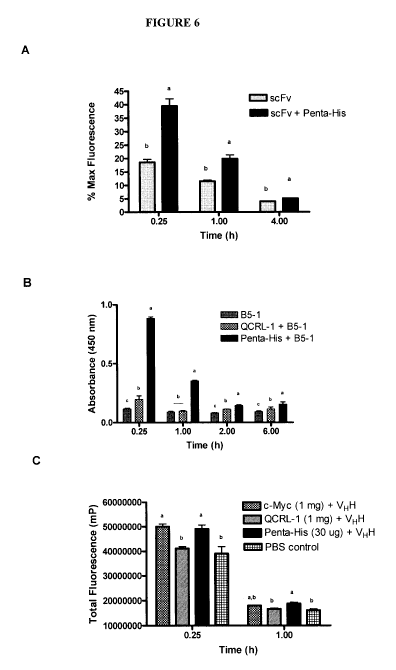
[0019] Figure 6 summarizes in vivo rAb clearance data. RAb-IgG
complexes improved rAb in vivo serum persistence. (a) In vivo experiment #1.
RAbs were labeled with FITC, then rAb-IgG complexes were allowed to
preform in vitro before intravenous administration to CD-1 female mice.
Serum persistence of TWO scFv through fluorescence analysis is shown.
Mean % maximum fluorescence (y-axis) is given versus time (x-axis) from
sera of treated mice, with standard error bars. Dark bars represent TWO
scFv-anti-Penta-His IgG complexes; light bars, TWO alone. Each treatment
and time point involved 5 mice. The rAb:IgG ratio was 20:1; 200 pg scFv and
0 or 50 pg IgG were used per mouse. (b) In vivo experiment #2. Administered
rAb persistence and binding-specificity was maintained in vivo. The binding of
B5-1 in pooled mouse serum from the 2:1 assay to immobilized S.
Typhimurium LPS (10 pg/mL) was tested by ELISA. Data from pooled mouse
serum diluted 1/3 is presented as an average of three replicates plus SEM.
Background binding to non-coated wells was not subtracted. (c) In vivo
CA 02742777 2011-05-05
WO 2010/051635 PCT/CA2009/001606
-7-
experiment #3. Total fluorescence values (mP) of the mouse serum. Data
from each of the five mice were pooled per time point. Maximum
fluorescence values of the injected dose were not measured and thus these
data are presented as total fluorescence (mP) values. In (a-c), treatments are
given in the upper right of each graph. Tukey's analysis was performed for
analysis of difference between treatments. Significant differences between
treatment groups, P values < 0.05, are indicated by the letter designations a-
c.
[0020] Figure 7 summarizes the binding properties of anti-tag IgGs to
epitope-tagged rAbs. Binding of Penta-His, 9E10, and QCRL-1 to the epitope-
tagged scFv (60 nM; Graph A) and Fab (60 nM; Graph B) was determined by
ELISA; both scFv and Fab are specific for P. aeruginosa O6ad. Data
represent the background-subtracted means of triplicates SD. Statistical
differences (P<0.0001) among the three means at each concentration of anti-
tag IgG were analyzed by One-Way ANOVA and are indicated by $.
[0021] Figure 8 summarizes the antigen binding ability of epitope-
tagged rAbs following complex formation with anti-tag IgGs. A and B, Binding
of P. aeruginosa O6ad-specific scFv treatments to heat-killed O6ad (1 x 10⁸
CFU/ml) and LPSosad (1 .g/ml), respectively. C and D, Binding of P.
aeruginosa O6ad-specific Fab treatments to heat-killed O6ad (1 x 10⁸
CFU/ml) and LPSosad (1 g/ml), respectively. Treatment legend is given in the
upper left of each graph. Each rAb-IgG complex treatment was prepared by
mixing scFv or Fab with Penta-His, 9E10, or QCRL-1 prior to conducting the
ELISA. Each sequential treatment (e.g., scFv, Penta-His) was comprised of
scFv or Fab added to the ELISA plate to interact with bound antigen following
by washing of the plate before one of the three anti-tag IgGs was added. scFv
or Fab applied together or sequentially with QCRL-1 are non-specific controls.
Data represent the background-subtracted means of triplicates SD. Symbols
above bars represent significant differences ($ = P<0.0001, * = P<0.001, and
# = P<0.01) between pairs of treatment means (e.g. scFv + Penta-His vs.
CA 02742777 2011-05-05
WO 2010/051635 PCT/CA2009/001606
-8-
scFv, Penta-His) at each concentration of rAb, as analyzed by One-Way
ANOVA.
[0022] Figure 9 summarizes data of Clq deposition by anti-tag IgG
when applied alone or complexed with epitope-tagged rAbs. A, Clq
deposition by each anti-tag IgG (66.67 nM) when applied alone. B, Clq
deposition by scFv-anti-tag IgG complexes using heat-killed P. aeruginosa
O6ad (1 x 10⁸ CFU/ml) and LPS06ad (10 g/ml) as coating antigens. C, Clq
deposition by Fab-anti-tag IgG complexes using heat-killed P. aeruginosa
O6ad (1 x 10⁸ CFU/ml) and LPS06ad (10 g/ml) as coating antigens. Data
represent the background-subtracted means of triplicates SD. Symbol ($)
above bars represent significant differences at $ = P<0.0001 among all three
treatment means within each antigen, as analyzed by One-Way ANOVA.
[0023] Figure 10 summarizes data showing rAb-anti-tag IgG complex-
mediated phagocytosis of P. aeruginosa O6ad (1 x 10⁶ CFU) by macrophage
J774.1A (1 x 10⁵ cells). A, scFv-IgG complex-mediated phagocytosis; B, Fab-
IgG complex-mediated phagocytosis. Incubations of P. aeruginosa O6ad cells
with J774.1A cells in the absence of antibodies or in the presence of rAb
alone, anti-tag IgG alone, mixture of rAbs and QCRL-1, or anti-O6ad IgG were
used as controls. The antibody concentrations used were 335 nM for rAbs
and 167.5 nM for anti-tag IgGs. The phagocytosed population of bacteria was
calculated according to the formula % phagocytosed bacteria =
(phagocytosed bacterial number at the end of 30 min incubation/initial
bacterial number at the beginning of 30 min incubation) x 100%. Data
represent the means SE from a single experiment performed at least in
triplicate. The different letters a-d indicate statistical differences
(P<0.001)
among the nine treatments, as analyzed by One-Way ANOVA.
[0024] Figure 11 summarizes data showing rAb-anti-tag IgG complex-
mediated phagocytosis of P. aeruginosa O6ad (1 x 10⁶ CFU) by macrophage
J774.1A cells (1 x 10⁵ cells) in the presence of 1.25% murine complement. A,
scFv-anti-tag IgG-mediated phagocytosis. B, Fab-anti-tag IgG-mediated
phagocytosis. Incubations of the bacteria with J774.1A cells in the absence of
CA 02742777 2011-05-05
WO 2010/051635 PCT/CA2009/001606
-9-
antibodies or in the presence of rAbs plus QCRL-1 were used as controls.
The antibody concentrations used were 335 nM for rAbs and 167.5 nM for
anti-tag IgGs. The phagocytosed population of bacteria was calculated
according to the formula % phagocytosed bacteria = (phagocytosed bacterial
number at the end of 30 min incubation/initial bacterial number at the
beginning of 30 min incubation) x 100%. Data represent the means SE from
a single experiment performed at least in triplicate. Statistical differences
within a treatment are analyzed by One-Way ANOVA and indicated by
P<0.001.
[0025] Figure 12 summarizes data showing inhibition of rAb-anti-tag
IgG complex-mediated phagocytosis of P. aeruginosa O6ad by anti-
FcyRIIB/III mAb 2.4G2. Phagocytosis inhibition was carried out by 1 h pre-
incubation of J774.1A cells with mAb 2.4G2 at concentrations 50 and 100
times greater than that of anti-tag IgG (167.5 nM) prior to addition of
antibody-
opsonized bacteria. The blocked population of bacteria was calculated
according to the formula % of blocked phagocytosis = [(phagocytosed
bacterial number without 2.4G2 - phagocytosed bacterial number with
2.4G2)/phagocytosed bacterial number without 2.4G2] x 100%. Data
represent the means SE from a single experiment performed at least in
triplicate. Statistical differences within a treatment were analyzed by One-
Way
ANOVA and are indicated by $ = P<0.0001 and * = P<0.001.
[0026] Figure 13 summarizes data showing the dose response of rAb-
anti-tag IgG complex-mediated phagocytosis of P. aeruginosa O6ad (1 x 10⁶
CFU) by macrophage J774.1A cells (1 x 10⁵). A, scFv-anti-tag IgG complex-
mediated phagocytosis; B, Fab-anti-tag IgG complex-mediated phagocytosis.
The phagocytosed population of bacteria was calculated according to the
formula % phagocytosed bacteria = (phagocytosed bacterial number at the
end of 30 min incubation/initial bacterial number at the beginning of 30 min
incubation) x 100%. Data represent the means SE from a single experiment.
Each experiment was performed at least in triplicate.
CA 02742777 2011-05-05
WO 2010/051635 PCT/CA2009/001606
-10-
[0027] Figure 14 summarizes data showing in vivo serum persistence
of Fab. Fab in mouse sera was analyzed by ELISA following i. v.
administration at 30 g/mouse alone or with an anti-tag IgG (50 .ig/mouse).
Fab in pooled mouse serum (n = 5/time point) was quantified by ELISA using
heat-killed P. aeruginosa O6ad cells (108 cells/ml) as a coating antigen; data
represent the means t SE of three replicates.
[0028] Figure 15 summarizes data showing survival of P. aeruginosa
O6ad-infected leukopenic mice following treatment with anti-O6ad antibodies.
Mice (n = 10-11/group) were treated i.v. with to-hS20 or scFv-Penta-His
complexes (32 g/mouse for scFv and 80 g/mouse for mAb) at time zero; 15
min later mice were inoculated i.v. with a LD80_90 of live P. aeruginosa O6ad
(103 CFU/mouse). Animals receiving the same volume of PBS, scFv alone, or
scFv plus QCRL-1 were used as controls. Mortality was recorded daily for 7
days.
[0029] Figure 16 summarizes data showing rAb-anti-tag IgG complex-
mediated phagocytosis of non-specific P. aeruginosa PAO1 and 010 (1 x 10⁶
CFU) by macrophage J774.1A (1 x 10⁵ cells). A and C, scFv-anti-tag IgG
complex-mediated phagocytosis against PAO1 and 010, respectively; B and
D, Fab-anti-tag IgG complex-mediated phagocytosis against PAO1 and 010,
respectively. Incubations of the bacteria with J774.1A cells in the absence of
antibodies or in the presence of rAb alone, anti-tag IgG alone, or mixture of
rAbs plus QCRL-1 were used as controls. The antibody concentrations used
were 335 nM for rAbs and 167.5 nM for anti-tag IgGs. The phagocytosed
population of bacteria was calculated according to the formula %
phagocytosed bacteria = (phagocytosed bacterial number at the end of 30
min incubation/initial bacterial number at the beginning of 30 min incubation)
x
100%. Data represent the means SE from a single experiment performed at
least in triplicate.
DETAILED DESCRIPTION OF THE DISCLOSURE
I. Methods and Uses For Improving Therapeutic Efficacy of Antibody
Fragments
CA 02742777 2011-05-05
WO 2010/051635 PCT/CA2009/001606
-11-
[0030] As described above, the present inventors discovered a method
of improving the therapeutic efficacy and utility of antibody fragments by
employing anti-epitope-tagging technologies, which resulted in antibody
fragments that exhibited an increased in vivo persistence, enhanced antigen
binding avidity, the ability to recruit downstream immune system functions to
the target antigen specified by the antibody fragment, enhanced in vivo
protective efficacy against infection from pathogens such as bacteria,
viruses,
protozoans and/or yeasts, and/or the toxins of pathogens, and/or cancers, for
example as evidenced by prolonged survival. The method of enhanced
therapeutic efficacy, utility and potency involves non-covalent interactions
between the epitope-tagged antibody fragments and anti-epitope tagged
antibodies, which results in the formation of a complex between the epitope-
tagged antibody fragment and the anti-epitope antibody. Specifically, the
present inventors demonstrated that increased therapeutic utility was
achieved by the non-covalent binding between epitope-tagged rAb fragments
(e.g. 6xHis-tagged scFv) and an anti-epitope tag IgG (e.g. anti-Penta-His)
that
resulted in the formation of a bivalent rAb-IgG complex.
[0031] The inventors used two different murine anti-epitope tag IgG1
Abs (i.e. anti-c-Myc and anti-Penta-His) in combination with an epitope-tagged
(i.e. c-Myc and 6xHis) murine anti-Salmonella enterica serovar typhimurium
(S. typhimurium) scFv, to examine both in vivo persistence in mice and FcR-
mediated complement recruitment and phagocytosis of S. typhimurium by the
murine macrophage (MO)-like cell line J774. When compared to the
monovalent scFv controls, the data showed that bivalent rAb-IgG complexes
recruited Fc-mediated effector functions as demonstrated by the binding of
human complement Clq by ELISA and by greater phagocytosis of S.
typhimurium by J774 MO cells following treatment with the B5-1-anti-tag IgG
complexes (Figures 4 and 5). Increased in vivo serum persistence of rAb
fragments was demonstrated by data showing greater quantities of epitope-
tagged scFvs (i.e. B5-1 and T1#10) and VHH at various time points following
i.v. administration to CD1 mice (Figure 6).
CA 02742777 2011-05-05
WO 2010/051635 PCT/CA2009/001606
-12-
[0032] The inventors also used murine anti-epitope tag IgG1 Abs (i.e.
anti-5xHis IgG (Penta-His) and anti-c-myc IgG (9E10)) in combination with c-
myc- and 6xHis-tagged Fab and scFv, which were directed against
Pseudomonas aeruginosa O6ad Iipopolysaccharide (LPS) to examine their in
vitro antigen binding ability, in vivo serum persistence, ability to mediate
effector functions including complement fixation, complement-dependent
cytotoxicity (CDC) and bacterial opsonization for phagocytosis, and their
protective efficacy against bacterial infection. The data showed that
complexes with the anti-tag IgGs significantly improved the antigen binding
avidity of both the Fab and scFv (Figures 7-8), extended the serum
persistence of the Fab (Figure 14), effectively recruited Fc-dependent
effector
functions including complement deposition and opsonization of the target
bacteria by macrophages in vitro (Figures 9-13), and enhanced in vivo
protective efficacy of the anti-O6ad scFv against infection with P. aeruginosa
as demonstrated by prolonged animal survival (Figure 15).
[0033] In summary, the present inventors demonstrated that 1) terminal
epitope tags expressed on antibody fragments specifically recruited functional
Fc regions, supplied by full-length anti-epitope tag IgGs, to antigens
targeted
by the epitope-tagged rAb fragments; 2) an epitope-tagged antibody fragment
in complex with an anti-epitope tag IgG increased in vivo persistence of the
antibody fragment; 3) an epitope-tagged antibody fragment in complex with
an anti-epitope tag IgG enhanced target-binding avidity of the antibody
fragment; and 4) complex formation of the epitope-tagged antibody fragment
with anti-epitope tag IgG enhanced protective efficacy of the antibody
fragment against bacterial infection and/or prolonged survival.
[0034] The present disclosure addresses the need for improving the
therapeutic utility, efficacy and potency of antibody fragments, which are
produced more efficiently and at a lower expense as compared to full length
antibodies, but which are suitable for use in situations where activation of
downstream immune system functions including Fc-mediated effector
functions are desired. The antibody fragments linked to epitopes described
CA 02742777 2011-05-05
WO 2010/051635 PCT/CA2009/001606
-13-
herein may be developed and produced quickly in simple microbial bioreactor
systems such as bacteria and yeast. The antibodies comprising sites that
bind to the epitopes described herein (anti-epitope antibodies) are full-
length
or near full-length antibodies that may be produced in more complex
dedicated bioreactor systems such as mammalian cells, and plants, when
large-scale production is warranted. The methods and uses described herein
provides the biopharmaceutical industry substantial flexibility to adapt to
antibody specificity and capacity needs by prioritizing full-length anti-
epitope
antibody production in more complex bioreactors, and epitope-tagged
antibody fragments in simpler bioreactors.
[0035] Accordingly, one aspect of the present disclosure includes a
method of enhancing the efficacy of an antibody fragment comprising
administering an effective amount of the antibody fragment linked to an
epitope to an animal in need thereof wherein a complex forms between the
antibody fragment linked to the epitope and an antibody that binds to the
epitope. In one embodiment, the antibody fragment is covalently linked to an
epitope. The present disclosure also includes the use of an antibody fragment
linked to an epitope to enhance the efficacy of the antibody fragment in an
animal in need thereof wherein upon use a complex forms between the
antibody fragment linked to the epitope and an antibody that binds to the
epitope. The present disclosure also includes the use of an antibody fragment
linked to an epitope in the manufacture of a medicament to enhance the
efficacy of the antibody fragment in an animal in need thereof wherein upon
use a complex forms between the antibody fragment linked to the epitope and
an antibody that binds to the epitope.
[0036] As used herein the term "enhancing efficacy" in reference to an
antibody fragment linked to an epitope includes without limitation increasing
the therapeutic effect of the antibody fragment, increasing the half-life of
the
antibody fragment, increasing persistence of the antibody fragment, including
for example, increasing serum persistence of the antibody fragment,
CA 02742777 2011-05-05
WO 2010/051635 PCT/CA2009/001606
-14-
increasing potency of the antibody fragment and/or increasing the utility of
the
antibody fragment.
[0037] The term "antibody fragment" as used herein includes any
fragment that is capable of binding to a target antigen and that is linked to
an
epitope. An antibody fragment may be a small fragment or derivative of a
larger antibody, however derived, that recognizes a target antigen, including
but not limited to, scFv antibodies, disulphide stabilized scFv fragments and
V. single domain antibodies, such as those of the camelid family, VH, V,,, F,
ScAb, HcAb and Fab. A VHH antibody is the single heavy chain variable
domain of a heavy chain antibody of the type that can be found in Camelid
mammals which are naturally devoid of light chains. A Fab antibody is the Fv
(variable) domain of an antibody. The techniques for preparing and using
various antibody-based constructs and fragments are well known by those of
skill in the art.
[0038] In one embodiment, the antibody fragment is scFv. An scFv
(single-chain Fv) antibody is a genetically engineered monospecific binding
protein that has a specific affinity for an antigen target. An scFv is a
derivative
of the Fv portion of an antibody molecule, or other receptor molecule of the
Ig
superfamily. It comprises one heavy and one light chain variable region (VH
and VL, respectively) of an antibody, joined by a flexible peptide linker. The
scFv antibody fragment contains all of the information required to determine
antigen specificity and none of the constant region domain that activates
downstream effector functions. An scFv antibody fragment is generally in the
size range of 25 to 30 kD, and therefore is small enough to be synthesized
efficiently in a bacterial or yeast expression system. Because of their small
size, scFv antibody fragments have a short circulating half-life as compared
to
full-size antibodies, or other antibody derivatives (i.e. diabodies,
minibodies).
[0039] In another embodiment, the antibody fragment is Fab. Fab is the
Fv (variable) domain of an antibody. Fab fragments are disulphide-linked
papain-cleavage fragments derived from whole antibodies. A Fab antibody
fragment comprises one constant and one variable domain of each of the
CA 02742777 2011-05-05
WO 2010/051635 PCT/CA2009/001606
-15-
heavy and the light chain of antibody and is a region on the antibody that
binds to antigens. A Fab antibody fragment is generally in the size range of
50
kDa, and therefore is small enough to be synthesized efficiently in a
bacterial
[7] or yeast expression system. Fab antibody fragments may also be made
recombinantly in mammalian cell bioreactors or plants. Fab antibody
fragments are intended to be included herein as useful epitope-tagged
antibody fragments, which may be so-produced and a suitable peptide
epitope could be covalently linked.
[0040] The specificity of the antibody fragment will be selected based
on the antigen that one wishes to target in the animal. The target antigen can
be selected from any antigen to which one wishes to generate an immune
response including, but not limited to, cellular antigens, humoral antigens,
viral antigens, bacterial antigens, tumour antigens (to treat cancer),
pathogens
including, for example, bacteria, viruses, protozoans and/or yeasts,
autoimmune antibodies, allergens, pathogenic protein complexes such as
prion and amyloid plaques, and toxins. The antibody fragment can be
generated using techniques known in the art or can be a known antibody that
is readily available. The CDR regions of many antibodies are readily available
which facilitates the recombinant production of antibody fragments.
[0041] The term "epitope" as used herein means an antigenic
determinant that may be bound by an antibody, and may be a peptide derived
from a toxin, pathogen, virus, bacteria, tumour antigen or autoantigen, and
includes an antigen for which an animal has been previously immunized. For
example, infants and children are immunized and/or vaccinated with one or
more of the following immunizations and/or vaccines: Diphtheria, tetanus,
acellular pertussis and inactivated polio virus vaccine (DTaP-IPV);
Haemophilus influenzae type b conjugate vaccine (Hib); Measles, mumps and
rubella vaccine (MMR); Varicella vaccine (Var); Hepatitis B vaccine (HB);
Pneumococcal conjugate vaccine - 7-valent (Pneu-C-7); Pneumococcal
polysaccharide - 23-valent (Pneu-P-23); Meningococcal C conjugate vaccine
(Men-C); Diphtheria, tetanus, acellular pertussis vaccine - adult/adolescent
CA 02742777 2011-05-05
WO 2010/051635 PCT/CA2009/001606
-16-
formulation (Tdap); Diphtheria, tetanus vaccine (Td); Influenza vaccine (Inf);
IPV Inactivated polio virus. In addition, adults with specific risk
indications may
also be immunized and/or vaccinated with one or more of the following
immunizations and/or vaccines: Influenza; Pneumococcal polysaccharide;
Hepatitis A and B; Bacille Calmette- Guerin (BCG); Cholera; Japanese
encephalitis; Poliomyelitis; Meningococcal conjugate; Meningococcal
polysaccharide; Rabies, pre-exposure use; Typhoid; Yellow fever; Smallpox.
[0042] In another embodiment, the epitope may be a peptide epitope
contrived and made completely unique from any amino acid sequences found
in nature. In another embodiment, the epitope may be a small molecule
hapten such as fluorescein. This embodiment would require the
corresponding anti-hapten mAb to either be coadministered or raised in the
animal by immunization.
[0043] The epitope is of no specific length, and can be any size
upwards of 3 amino acid residues in length, including the size of a full-
length
protein such as glutathione S transferase (GST) and maltose binding protein
(MBP). Smaller epitopes are preferred, for example epitopes between 8 to 50
amino acids in length, as these can result in smaller epitope-tagged antibody
fragments that are easier to produce, purify and use as compared to larger
recombinant proteins. Additionally, smaller epitopes are less likely to
interfere
with the target antigen binding function of the antibody fragment. Commonly
used epitope tags include glutathione-S-transferase (GST), c-Myc, poly-
histidine (6X-His), penta-histidine (Penta-His), FLAG , green fluorescent
protein (GFP), maltose binding protein (MBP), influenza A virus haemaglutinin
(HA tag; YPYDVPDYA (SEQ ID NO: 1)), (3-galactosidase ((3-gal), GAL4,
human MRP, V5 epitope from the simian virus, polyoma virus T antigen
epitopes, and the KT3 viral epitope or portions thereof of these proteins.
Some epitopes including c-Myc, QCRL-1 and poly-His epitopes are listed in
Table 1, and provide examples of various epitopes and their known antibodies
which bind to the epitopes, which can be used in the present disclosure. In
another embodiment, the epitope may be any epitope that is experimentally
CA 02742777 2011-05-05
WO 2010/051635 PCT/CA2009/001606
-17-
determined to raise a monoclonal response in an animal that results in a high-
affinity antibody for a defined peptide epitope. "A portion" of the above
named
proteins is any sequence of 3 amino acids or longer.
[0044] The phrase "antibody fragment linked to an epitope" as used
herein may be used interchangeably with "epitope-tagged antibody fragment"
in the present disclosure. As used herein the term "linked" includes an
epitope
attached to the antibody fragment using techniques known in the art, including
for example recominant DNA techniques such as fusion protein technology or
by chemical means such as cross-linking. The method used to link the
antibody fragment to an epitope must be capable of linking the antibody
fragment and epitope without interfering with the ability of the antibody
fragment to bind to its target antigen. In one embodiment, the antibody
fragment is covalently linked to an epitope.
[0045] In one embodiment, the antibody fragment is linked to an
epitope using recombinant DNA techniques. In such a case a DNA sequence
encoding the antibody fragment is fused to a DNA sequence encoding the
epitope, resulting in a chimeric DNA molecule. The chimeric DNA sequence
is transfected into a host cell that expresses the fusion protein. The fusion
protein can be recovered from the cell culture and purified using techniques
known in the art. In another embodiment, the nucleotide sequence encoding
the epitope could be fused to the DNA sequence encoding the antibody
fragment at carboxy-terminus, or distant from the antigen-binding site of the
epitope-tagged antibody. In a further embodiment, epitope tagged antibody
fragments may be screened out from recombinant antibody libraries, which
typically have epitope tags genetically engineered so as to be present on any
rAb.
[0046] In another embodiment, the antibody may be linked to an
epitope via chemical cross-linking using techniques well known in the art.
There are several hundred crosslinkers available that can conjugate two
proteins. (See for example "Chemistry of Protein Conjugation and
Crosslinking". 1991, Shans Wong, CRC Press, Ann Arbor). The crosslinker is
CA 02742777 2011-05-05
WO 2010/051635 PCT/CA2009/001606
-18-
generally chosen based on the reactive functional groups available or inserted
on the antibody fragment, and/or the epitope. In addition, if there are no
reactive groups, a photoactivatible crosslinker can be used. In certain
instances, it may be desirable to include a spacer between the antibody
fragment, and epitope. Crosslinking agents known to the art include the
homobifunctional agents: glutaraldehyde, dimethyladipimidate and
bis(diazobenzidine) and the heterobifunctional agents: m-maleimidobenzoyl-
N-hydroxysuccinimide and sulfo-m-ma leimidobenzoyl-N-hydroxysuccinimide.
[0047] As used herein, the phrase "effective amount" means an amount
effective, at dosages and for periods of time necessary to achieve the desired
result. Effective amounts of the antibody fragment linked to an epitope may
vary according to factors such as the disease state, age, sex, weight of the
animal. Dosage regime may be adjusted to provide the optimum therapeutic
response. For example, several divided doses may be administered daily or
the dose may be proportionally reduced as indicated by the exigencies of the
therapeutic situation.
[0048] The term "animal" as used herein refers to any member of the
animal kingdom, preferably a mammal, more preferably a human being.
[0049] The term "administered" as used herein means that the antibody
fragment linked to an epitope may be administered or used either as a protein
conjugate or as a chimeric nucleic acid construct. In the latter instance the
antibody fragment linked to the epitope will be expressed in vivo in a DNA-
based therapy. The form of administration or use will depend on the nature
and location of the target antigen. Suitable forms of administration include
systemic (subcutaneous, intravenous, intramuscular), oral administration,
inhalation, transdermal administration, topical application (such as topical
cream or ointment, etc.) or by other methods known in the art. Other modes
of administration are described in Section II under "Pharmaceutical
Compositions".
[0050] The administration or use of the antibody fragment linked to the
epitope results in the formation of a complex between the antibody fragment
CA 02742777 2011-05-05
WO 2010/051635 PCT/CA2009/001606
-19-
linked to the epitope (epitope-tagged antibody fragment) and an antibody that
binds to the epitope (anti-epitope antibody). As used herein the term
"complex" refers to the non-covalent interaction between the epitope-tagged
antibody fragment, and the anti-epitope antibody. In one embodiment, a
bivalent rAb-IgG complex forms between an anti-epitope IgG1 antibody (IgG)
and an epitope-tagged antibody fragment (rAb). This is shown schematically
in Figure 1 B. In another embodiment, the antibody fragment forms a complex
with the antibody in a 20:1 ratio. In a further embodiment, the antibody
fragment forms a complex with the antibody in a 2:1 ratio.
[0051] The term "antibody that binds to the epitope" as used herein
may be used interchangeably with "anti-epitope antibody" and includes full-
length or near full-length antibodies that can enhance the efficacy of the
antibody fragment. In one embodiment, the anti-epitope antibody will
comprise an Fc region. The antibodies may be wild-type antibodies and
natural variants thereof, and molecularly-engineered antibodies, polyclonal
and monoclonal antibodies, IgG, IgM, IgA, IgE or IgD antibodies, humanized
antibodies, crosslinked antibodies, heterospecific antibodies, bispecific
antibodies, crosslinked heterobispecific antibodies, chimeric antibodies,
minibodies, diabodies, triabodies, HCAb, Dab, Scab, VH1 VL, Fv and Fab. In
another embodiment, the antibody includes IgG1, IgG2a/c, IgG2b, and/or
IgG3. In a further embodiment, the anti-epitope antibody may be a full-length
polyclonal or monoclonal antibody selected from the group consisting of IgG,
IgM, IgA, IgE and IgD.
[0052] In one embodiment, the anti-epitope antibody includes all
isotypes of IgG and IgM antibodies, including for example monoclonal
antibodies, as these can be produced industrially. In another embodiment,
the anti-epitope antibody includes a full-length polyclonal or monoclonal
antibody capable of activating all downstream effector functions. A polyclonal
or monoclonal antibody useful in the present disclosure can be obtained as a
cell line from the American Type Culture Collection of monoclonal antibodies,
from commercially available sources, from polyclonal sources (i.e. animal- or
CA 02742777 2011-05-05
WO 2010/051635 PCT/CA2009/001606
-20-
serum-derived), or produced through recombinant DNA technology in a
bioreactor (hybridoma, plant, etc.). In one embodiment, for human therapy the
anti-epitope antibody is human or humanized. In another embodiment, for
animal therapy, the anti-epitope antibody is from the same animal.
[0053] Examples of anti-epitope antibodies with a binding domain that
may be useful in the present disclosure include antibodies specific for c-Myc
and QCRL-1 epitopes, which have an affinity (Kd) for these epitopes of less
than 200 nM, and are listed in Table 1. Other anti-epitope antibodies useful
in
the present disclosure, and the epitopes that they recognize, are also listed
in
Table 1. It is understood that other anti-epitope antibodies may be known, or
can be generated, which may have an affinity (Kd) for their epitope that is
lower or higher than 200 nM, and these may useful in the methods of the
disclosure disclosed herein.
[0054] Anti-epitope antibodies suitable for the methods and uses of the
present disclosure may be readily selected by persons skilled in the art
depending on the therapeutic effect sought (i.e. increased persistence or half-
life, and/or activation of downstream immune system functions and/or
improved or enhanced protective efficacy against infection from pathogens
such as bacteria, viruses, protozoans and/or yeasts, the toxins of pathogens,
and/or cancers, for example, as evidenced by prolonged survival). For
example, the antibody may be selected such that it comprises a functional Fc
region or derivative thereof, and may therefore be able to activate
downstream immune system functions. The Fc region of immunoglobulin
antibodies are known to trigger ADCC, the complement pathway and
opsonization. "Derived from" includes a natural variant of a wild-type Fc
sequence, a genetically- or biochemically- engineered variant thereof, or an
entirely artificial amino acid sequence.
[0055] In addition, if the anti-epitope antibody is intended to be used for
neutralizing a toxin, such as botulinum toxin or the toxins of a snake venom,
an anti-epitope antibody that increases the half-life of epitope-tagged
antibody
fragment, is sufficient for use herein. This anti-epitope antibody could be a
CA 02742777 2011-05-05
WO 2010/051635 PCT/CA2009/001606
-21-
full-length antibody, for example, a full-length antibody produced from a
plant
and scaled up in production. Other potential anti-epitope antibodies useful
herein for binding to the epitope-tagged antibody fragment include scFv, VHH,
VH, VL, Fab, F, ScAb, HcAb, diabodies, triabodies and minibodies. These
provide the advantage that they can be produced in simple bacterial or yeast
bioreactors, as opposed to full-length antibodies. However, it is understood
by those skilled in the art that the smaller the anti-epitope antibody, the
lesser
can likely be its ability to enhance the efficacy of the epitope-tagged
antibody
fragment. Accordingly, there is a trade-off between ease of manufacture of
the anti-epitope antibody and ability to enhance the efficacy of the epitope-
tagged antibody fragment.
[0056] Furthermore, if it is desired that the anti-epitope antibody also
activates downstream effector functions, in addition to increasing the
persistence or half-life and/or stability of the epitope-tagged antibody
fragment
(i.e., the antibody fragment is intended to be used to bind to a pathogen,
such
as a virus or a bacterium) the anti-epitope antibody may be produced by a
mammalian system, such as a cell-line bioreactor, so as to allow for complete
downstream immune system function. In this regard, full-length polyclonal or
monoclonal antibodies are preferred. Furthermore, if the anti-epitope antibody
is from a mammalian system, or if it has human specific or compatible
glycosylations, it can activate downstream immune system function against
the target antigen of the epitope-tagged antibody fragment, thus transforming
the tagged antibody into a therapeutic antibody that can be used in situations
where an FcR-mediated effector functions are desired.
[0057] In one embodiment, the anti-epitope antibody is administered or
used prior to, at the same time, or after the epitope-tagged antibody
fragment.
In another embodiment, the anti-epitope antibody is already present in the
animal.
[0058] As used herein "already present in the animal" includes an
antibody that is generated by immunizing the animal with an epitope and/or
using the epitope in the animal. Alternatively, the phrase also includes an
CA 02742777 2011-05-05
WO 2010/051635 PCT/CA2009/001606
-22-
antibody that is generated by an immunization previously administered to the
animal and/or previously used in the animal, wherein the immunization
comprised the epitope. For example, previously administered immunizations
may include: Diphtheria, tetanus, acellular pertussis and inactivated polio
virus vaccine (DTaP-IPV); Haemophilus influenzae type b conjugate vaccine
(Hib); Measles, mumps and rubella vaccine (MMR); Varicella vaccine (Var);
Hepatitis B vaccine (HB); Pneumococcal conjugate vaccine - 7-valent (Pneu-
C-7); Pneumococcal polysaccharide - 23-valent (Pneu-P-23); Meningococcal
C conjugate vaccine (Men-C); Diphtheria, tetanus, acellular pertussis vaccine
- adult/adolescent formulation (Tdap); Diphtheria, tetanus vaccine (Td);
Influenza vaccine (Inf); IPV Inactivated polio virus; Influenza; Pneumococcal
polysaccharide; Hepatitis A and B; Bacille Calmette- Guerin (BCG); Cholera;
Japanese encephalitis; Poliomyelitis; Meningococcal conjugate;
Meningococcal polysaccharide; Rabies, pre-exposure use; Typhoid; Yellow
fever; and Smallpox. In one embodiment, the antibody already present in the
animal is a monoclonal antibody, IgG, or IgG1.
[0059] A further aspect of the present disclosure is a method of
enhancing the efficacy of an antibody fragment comprising: a) immunizing an
animal with an epitope; and b) administering an effective amount of the
antibody fragment linked to the epitope to the animal in need thereof; wherein
the efficacy of the administered antibody fragment is enhanced. The present
disclosure also includes a use to enhance efficacy of an antibody fragment in
an animal in need thereof comprising: a) use of an epitope to immunize the
animal; and b) use of the antibody fragment linked to the epitope in the
animal
to enhance efficacy of the antibody fragment. The present disclosure also
includes a use to enhance efficacy of an antibody fragment in an animal in
need thereof comprising: a) use of an epitope to immunize the animal; and b)
use of the antibody fragment linked to the epitope in the animal in the
manufacture of a medicament to enhance efficacy of the antibody fragment.
[0060] "Immunizing an animal" with an epitope and/or use of an epitope
in an animal allows the animal to produce native or natural antibodies that
are
CA 02742777 2011-05-05
WO 2010/051635 PCT/CA2009/001606
-23-
raised against the epitope (anti-epitope antibodies). Accordingly, immunizing
the animal or using the epitope in the animal negates the need for co-
administering the epitope-tagged antibody fragment with an anti-epitope
antibody but rather provides an anti-epitope antibody that is made by the
animal itself. In another embodiment, an epitope may be used in an
immunization protocol in the case of an animal requiring continual
immunotherapy, thus reducing the need for administration of the full-length
anti-epitope antibody in a long-term immune response therapy protocol.
[0061] In another embodiment, the formation of epitope-tagged
antibody fragments:anti-epitope antibody complexes provides a basis from
which oligoclonal or polyclonal antibody (pAb) therapeutics can be created,
including for example, administration of pAb repertoires that mimic the
natural
Ab response aimed towards multiple target antigens. pAb production using
the methods of present disclosure disclosed herein would require only one, or
alternatively a few, humanized anti-epitope antibodies (i.e. IgG molecules),
while multi-antigen specificities are supplied by antibody fragments linked to
different epitope tags. A facility producing the anti-epitope antibodies, in
combination with the rapid and inexpensive production of polyclonal epitope-
tagged antibody fragments, could provide speed and flexibility in pAb
development. In one embodiment, only one anti-epitope antibody may be
required to deliver several therapeutic epitope-tagged antibody fragments. In
another embodiment, a few anti-epitope antibody molecules could be tailored
for specific and multiple types of therapeutic outcomes.
[0062] For example, the epitope-tagged antibody fragments described
herein may differ from one another in regard to their affinity for various
antigenic targets - i.e. toxins, pathogens, idiotypes and autoantigen targets.
They are linked or tagged with an epitope recognized by an anti-epitope
antibody. The anti-epitope antibody recognizes and binds to the epitope of an
epitope-tagged antibody fragment. More than one anti-epitope antibody may
be generated, which may differ in their ability to recognize different epitope
CA 02742777 2011-05-05
WO 2010/051635 PCT/CA2009/001606
-24-
tags, and/or in their ability to activate downstream immune functions, to
thereby increase the flexibility of the disclosure described herein.
[0063] Accordingly, disclosed herein is the generation of a number of
epitope-tagged antibody fragments that recognize different antigenic targets,
and the corresponding generation of one or a few anti-epitope antibodies that
recognize the epitope tag of the antibody fragments. By changing the tagged
antibody fragment that is combined with an anti-epitope antibody, a large
number of different therapeutic antibody fragments with different specific
affinities towards antigens, or different abilities to activate downstream
immune functions, can be generated. Because a relatively large number of
epitope-tagged antibody fragments required can be produced quickly and
inexpensively, and only one or a few complex anti-epitope antibodies are
required, this approach provides the therapeutic antibody industry substantial
flexibility to adapt to antibody specificity and capacity needs.
[0064] In one embodiment, the present disclosure includes methods
and uses of a plurality of epitope-tagged antibody fragments, which differ in
their capability of binding different antigens, but are all tagged with the
same
epitope. Accordingly, these antibody fragments may be used in conjunction
with one anti-epitope antibody which binds to the epitope tag on the antibody
fragments.
[0065] In another embodiment, more than one epitope tag may be
linked to the antibody fragment. For example, two or three copies of the same
epitope can be linked along antibody fragment to provide for increased
binding of the anti-epitope antibody. Alternatively, two or more different
epitopes can be linked to one antibody fragment. For example, two different
epitopes could be spaced on the antibody fragment to provide for an epitope-
tagged antibody fragment that is recognized by two different anti-epitope
antibodies. For example, c-Myc and polyhistidine epitopes may be combined
on one antibody fragment.
[0066] In a further embodiment, an anti-epitope antibody is
monospecific for an epitope. However, in another embodiment, the anti-
CA 02742777 2011-05-05
WO 2010/051635 PCT/CA2009/001606
-25-
epitope antibody is bispecific for two different epitopes. This embodiment of
the anti-epitope antibody could be used with epitope-tagged antibody
fragments that comprise either, or both, of the two epitope tags recognized by
the bispecific anti-epitope antibodies.
[0067] In another aspect of the method of this disclosure, a plurality of
epitope-tagged antibodies are provided, from which one or more is selected
for administration to the animal or use in the animal in combination with an
anti-epitope antibody.
[0068] In some situations it may be beneficial to select, for
administration, two or more epitope-tagged antibody fragments that each
have a specific affinity for the same antigen- i.e. two tagged antibody
fragments that each have a slightly different binding affinity for a
particular
antigen. In other situations it may be beneficial to administer two or more
epitope-tagged antibody fragments that have a specific affinity for different
but
related antigens. For example, the different antigens may be two different
proteins on the membrane surface of the same pathogen or, a toxin produced
by a pathogen, and the pathogen itself. In some situations, the epitope on the
two epitope-tagged antibody fragments may be the same (i.e., so they are
recognized by the anti-epitope antibody), or they may be different (i.e., so
that
they are recognized by different anti-epitope antibodies).
[0069] Once the appropriate tagged antibody fragment, or combination
of tagged antibody fragments, is selected, it can be combined with the
appropriate anti-epitope antibody, to form a complex as described herein.
The anti-epitope antibody can be an antibody that can only function to
stabilize the tagged antibody fragment (i.e., used for incomplete
immunotherapy), or it can be an antibody that also activates downstream
immune system functions. More than one anti-epitope antibody can be used,
and they can have different specific affinities for the epitope, or recognize
different epitopes, altogether. The anti-epitope antibody may be a natural
antibody that is made by the animal, as a result of immunization with the
epitope.
CA 02742777 2011-05-05
WO 2010/051635 PCT/CA2009/001606
-26-
[0070] In one embodiment, the epitope-tagged antibody fragment and
anti-epitope antibody are combined before administration to the animal or use
in the animal. It is preferred that, if a complex as described herein is to be
generated by mixing an anti-epitope antibody and an epitope-tagged antibody
fragment together before administration to an animal or used in an animal,
that such administration or use occurs immediately or within 30 minutes of
mixing. In another embodiment, anti-epitope antibody and epitope-tagged
antibody fragment are coadministered or used separately in the animal. In yet
another embodiment, anti-epitope antibody and epitope-tagged antibody
fragment are administered or used sequentially. In the latter embodiment,
anti-epitope antibody may be administered or used either before, or after,
epitope-tagged antibody fragment. It is also preferred in this embodiment to
administer or use anti-epitope antibody first. In yet another embodiment,
epitope-tagged antibody is administered or used and it combines with natural
anti-epitope antibody produced by the animal.
[0071] Another embodiment of the present disclosure is a method of
increasing the therapeutic effect of an antibody fragment comprising
administering an effective amount of the antibody fragment linked to an
epitope to an animal in need thereof, wherein a complex forms between the
antibody fragment linked to the epitope and an antibody that binds to the
epitope. The present disclosure also includes the use of an antibody fragment
linked to an epitope to increase the therapeutic effect of the antibody
fragment
in an animal in need thereof wherein upon use a complex forms between the
antibody fragment linked to the epitope and an antibody that binds to the
epitope. The present disclosure also includes the use of an antibody fragment
linked to an epitope in the manufacture of a medicament to increase the
therapeutic effect of the antibody fragment in an animal in need thereof
wherein upon use a complex forms between the antibody fragment linked to
the epitope and an antibody that binds to the epitope.
[0072] The term "increasing or increases the therapeutic effect" as
used herein includes without limitation increasing the persistence or half-
life of
CA 02742777 2011-05-05
WO 2010/051635 PCT/CA2009/001606
-27-
the antibody fragment, including, for example, increasing serum persistence
or serum half-life of the antibody fragment and/or stability of the antibody
fragment, increasing the immune response of the antibody fragment, for
example, by activating downstream immune system functions, such as the
ability to recruit FcR-mediated effector functions, and includes for example
recruiting the complement system, enhanced avidity of the antibody fragment
and/or increasing phagocytosis, and enhancing protective efficacy against
infection from pathogens such as bacteria, viruses, protozoans and/or yeasts,
the toxins of pathogens and/or cancer, for example, as evidenced by
prolonged survival.
[0073] Another embodiment of the present disclosure is a method of
increasing persistence and/or stability of an antibody fragment comprising
administering an effective amount of the antibody fragment linked to an
epitope to an animal in need thereof wherein a complex forms between the
antibody fragment linked to the epitope and an antibody that binds to the
epitope. The present disclosure also includes the use of an antibody fragment
linked to an epitope to increase the persistence and/or stability of the
antibody
fragment in an animal in need thereof wherein upon use a complex forms
between the antibody fragment linked to the epitope and an antibody that
binds to the epitope. The present disclosure also includes the use of an
antibody fragment linked to an epitope in the manufacture of a medicament to
increase the persistence and/or stability of the antibody fragment in an
animal
in need thereof wherein upon use a complex forms between the antibody
fragment linked to the epitope and an antibody that binds to the epitope.
[0074] As used herein "persistence and/or stability" includes increased
in vivo persistence, increased serum persistence or increased serum half-life
(including increased in vivo serum persistence or increased in vivo serum
half-life), and/or a reduction in the rate of degradation or clearance of the
antibody fragment described herein, so that it remains capable of, or
available
for, binding to its target antigen for a longer period of time.
CA 02742777 2011-05-05
WO 2010/051635 PCT/CA2009/001606
-28-
[0075] The antibody fragments disclosed herein are small proteins and
therefore may be unstable, for example in serum, meaning that the antibody
fragments may be degraded or cleared from the serum more rapidly than is
desired for therapeutic applications. Accordingly, by employing the methods of
the present disclosure, the antibody fragments are not cleared as rapidly,
exhibit an increased persistence or half-life and/or stability and are thus
are
useful for exerting therapeutic effects for longer periods of time. In other
words, the persistence or half-life of the epitope-tagged antibody fragment
may be improved, resulting in an increased persistence or half-life of the
antibody fragment in the animal in need thereof. As is apparent, the longer
that the antibody fragment is able to bind to its target antigen, the longer
can
be its therapeutic effectiveness.
[0076] Increased persistence, half-life and/or stability may be measured
using techniques known in the art, for example, by labeling the antibody
fragment with a fluorescein label, and then comparing the amount of label
remaining in the serum. If after a certain period of time, for example one
hour,
or one day, there is a statistically significant higher amount of fluorescein
in
the serum as compared to controls, the antibody fragment exhibits an
increased half-life and/or stability. As is apparent to one of skill in the
art, a
stable epitope-tagged antibody fragment can have a longer persistence or
half-life in the serum, peritoneum or other tissue to which it is
administered, as
compared to controls. Whether there is a statistically significant difference
higher amount of fluorescein tag may be determined by analyzing fluorescent
readings with GraphPad Prism (GraphPad Software Inc.) using 1 way ANOVA
analysis of variance, followed by Dunnett's multiple comparison test, in which
each experimental group is compared to the control values at the
corresponding time point. A difference is statistically significant if it has
a P
value of <0.05.
[0077] Another embodiment of the present disclosure is a method of
increasing the immune response of an antibody fragment comprising
administering an effective amount of the antibody fragment linked to an
CA 02742777 2011-05-05
WO 2010/051635 PCT/CA2009/001606
-29-
epitope to an animal in need thereof wherein a complex forms between the
antibody fragment linked to the epitope and an antibody that binds to the
epitope. The present disclosure also includes the use of an antibody fragment
linked to an epitope to increase the immune response of the antibody
fragment in an animal in need thereof wherein upon use a complex forms
between the antibody fragment linked to the epitope and an antibody that
binds to the epitope. The present disclosure also includes the use of an
antibody fragment linked to an epitope in the manufacture of a medicament to
increase the immune response of the antibody fragment in an animal in need
thereof wherein upon use a complex forms between the antibody fragment
linked to the epitope and an antibody that binds to the epitope.
[0078] The terms "increasing or increase the immune response"
"eliciting an immune response" or "inducing an immune response" as used
herein includes increasing the immunotherapeutic potential of the antibody
fragment including initiating, triggering, causing, enhancing, improving or
augmenting any response of the immune system, for example, of either a
humoral or cell-mediate nature. The initiation or enhancement of an immune
response can be assessed using assays known to those skilled in the art
including, but not limited to, antibody assays (for example ELISA assays),
antigen specific cytotoxicity assays and the production of cytokines (for
example ELISPOT assays). In one embodiment, the methods of the present
disclosure trigger or enhance a cellular immune response, antibody
dependent cell cytotoxicity (ADCC; cell lysis), phagocytosis, antigen
presentation and T cell activation, and/or immune activation via cytokine
release, for example, increased FcR and MHC expression.
[0079] Another embodiment of the present disclosure is a method of
activating downstream immune system functions comprising administering an
effective amount of the antibody fragment linked to an epitope to an animal in
need thereof wherein a complex forms between the antibody fragment linked
to the epitope and an antibody that binds to the epitope. The present
disclosure also includes the use of an antibody fragment linked to an epitope
CA 02742777 2011-05-05
WO 2010/051635 PCT/CA2009/001606
-30-
to activate downstream immune system functions in an animal in need thereof
wherein upon use a complex forms between the antibody fragment linked to
the epitope and an antibody that binds to the epitope. The present disclosure
also includes the use of an antibody fragment linked to an epitope in the
manufacture of a medicament to activate downstream immune system
functions in an animal in need thereof wherein upon use a complex forms
between the antibody fragment linked to the epitope and an antibody that
binds to the epitope.
[0080] The term "activating or activate downstream immune system
functions" as used herein includes for example one or more of: (a) activating
FcR-mediated effector functions; (a) activating the complement cascade for
complement dependent cytotoxicity (CDC); (b) directing the immune system
through Fc receptor function in antibody-dependent cell-mediated cytotoxicity
(ADCC); (c) increasing avidity of the antibody fragment; and (d) opsonization.
[0081] A further embodiment of the present disclosure is a method of
recruiting FcR-mediated effector functions comprising administering an
effective amount of the antibody fragment linked to an epitope to an animal in
need thereof wherein a complex forms between the antibody fragment linked
to the epitope and an antibody that binds to the epitope. The present
disclosure also includes the use of the antibody fragment linked to an epitope
to recruit FcR-mediated effector functions in an animal in need thereof
wherein upon use a complex forms between the antibody fragment linked to
the epitope and an antibody that binds to the epitope. The present disclosure
also includes the use of the antibody fragment linked to an epitope in the
manufacture of a medicament to recruit FcR-mediated effector functions in an
animal in need thereof wherein upon use a complex forms between the
antibody fragment linked to the epitope and an antibody that binds to the
epitope.
[0082] The term "recruiting or recruit FcR-mediated effector functions"
as used herein means the ability to recruit the complement system, and/or
CA 02742777 2011-05-05
WO 2010/051635 PCT/CA2009/001606
-31-
increase phagocytosis to the target antigens specified by the epitope-tagged
antibody fragment, including, for example, opsonic phagocytosis.
[0083] Another embodiment of the present disclosure includes a
method of recruiting the complement system and/or increasing phagocytosis
comprising administering an effective amount of the antibody fragment linked
to an epitope to an animal in need thereof wherein a complex forms between
the antibody fragment linked to the epitope and an antibody that binds to the
epitope. The present disclosure also includes the use of an antibody fragment
linked to an epitope to recruit the complement system and/or increase
phagocytosis in an animal in need thereof wherein upon use a complex forms
between the antibody fragment linked to the epitope and an antibody that
binds to the epitope. The present disclosure also includes the use of an
antibody fragment linked to an epitope in the manufacture of a medicament to
recruit the complement system and/or increase phagocytosis in an animal in
need thereof wherein upon use a complex forms between the antibody
fragment linked to the epitope and an antibody that binds to the epitope.
[0084] As used herein "complement system" refers to a complement
cascade of proteins or protein fragments, including for example, serum
proteins, serosal proteins and cell membrane receptors, which help clear
pathogens from an organism. The complement system includes the classical
complement pathway, the alternative complement pathway, and the
mannose-binding lectin pathway, which would all be known to those skilled in
the art. Recruitment of the complement system can be assessed using
assays known to those skilled in the art, including, but not limited to,
antibody
assays (for example ELISA assays, immunofluorescence assays,
radioimmunoassays and radioassays involving radiolabeled complement
proteins, surface plasmon resonance assays). For example, recruitment of
the classical complement system may be determined by assessing
recruitment of the first complement protein C1q, for example by determining
the ability of the complexes described herein to deposit complement protein
Clq or C1q complex.
CA 02742777 2011-05-05
WO 2010/051635 PCT/CA2009/001606
-32-
[0085] As used herein "increasing phagocytosis" means an increase in
the ingesting, taking in and/or engulfing of particles including for example,
pathogens, such as bacteria, viruses, parasites, protozoans and/or yeasts,
and cellular and/or foreign debris by phagocytes, including for example,
macrophages. Phagocytosis occurs after a particle has bound to receptors
that are present on the surface of phagocytes. A number of receptors are
present on phagocytes, including for example, opsonins. Phagocytosis can
be assessed using assays known to those skilled in the art, including, but not
limited to, antibody assays (for example ELISA assays, microscopy assays,
macrophage lysis and bacterial colony forming unit enumeration assays, etc.).
[0086] Another embodiment of the present disclosure includes a
method of enhancing protective efficacy against infection comprising
administering an effective amount of the antibody fragment linked to an
epitope to an animal in need thereof wherein a complex forms between the
antibody fragment linked to the epitope and an antibody that binds to the
epitope. The present disclosure also includes the use of an antibody fragment
linked to an epitope to enhance protective efficacy against infection in an
animal in need thereof wherein upon use a complex forms between the
antibody fragment linked to the epitope and an antibody that binds to the
epitope. The present disclosure also includes the use of an antibody fragment
linked to an epitope in the manufacture of a medicament to enhance
protective efficacy against infection in an animal in need thereof wherein
upon
use a complex forms between the antibody fragment linked to the epitope and
an antibody that binds to the epitope.
[0087] As used herein "enhancing protective efficacy against infection"
includes increased protection against infection and/or increased protection
efficiency against infection and/or increased protection capacity against
infection, which may be assessed by determining survival of infected animals
and/or prolonged survival of infected animals after treatment with the
complexes described herein. As used herein "infection" includes infection from
CA 02742777 2011-05-05
WO 2010/051635 PCT/CA2009/001606
-33-
pathogens, which includes for example infection from bacteria, viruses,
protozoans and/or yeasts.
II. Pharmaceutical Compositions
[0088] In another aspect, the disclosure includes a pharmaceutical
composition in a biologically compatible form suitable for use or
administration
in vivo. The term "biologically compatible form suitable for administration in
vivo" means a form of the substance to be administered in which any toxic
effects are outweighed by the therapeutic effects. Accordingly, the disclosure
provides a pharmaceutical composition comprising an effective amount of the
epitope-tagged antibody fragments and/or anti-epitope antibodies disclosed
herein with a pharmaceutically acceptable carrier or diluent, adjuvant or
mixtures thereof to an animal in need thereof.
[0089] The compositions containing the epitope-antibody fragments
and/or anti-epitope antibodies described herein can be prepared by known
methods for the preparation of pharmaceutically acceptable compositions
which can be administered to animals, such that an effective quantity of the
active substance is combined in a mixture with a pharmaceutically acceptable
vehicle, including for example, a carrier or diluent. Suitable vehicles are
described, for example, in Remington's Pharmaceutical Sciences (2003 - 20th
edition) and in The United States Pharmacopeia: The National Formulary
(USP 24 NF19) published in 1999. On this basis, the compositions include,
albeit not exclusively, solutions of the substances in association with one or
more pharmaceutically acceptable vehicles, carriers or diluents, and
contained in buffered solutions with a suitable pH and iso-osmotic with the
physiological fluids.
[0090] Pharmaceutical compositions include, without limitation,
lyophilized powders or aqueous or non-aqueous sterile injectable solutions or
suspensions, which may further contain antioxidants, buffers, bacteriostats
and solutes that render the compositions substantially compatible with the
tissues or the blood of an intended recipient. Other components that may be
present in such compositions include water, surfactants (such as Tween),
CA 02742777 2011-05-05
WO 2010/051635 PCT/CA2009/001606
-34-
alcohols, polyols, glycerin and vegetable oils, for example. Extemporaneous
injection solutions and suspensions may be prepared from sterile powders,
granules, tablets, or concentrated solutions or suspensions. The
pharmaceutical composition may be supplied, for example but not by way of
limitation, as a lyophilized powder which is reconstituted with sterile water
or
saline prior to administration to the animal.
[0091] The compositions containing the epitope-tagged antibody
fragments and/or anti-epitope antibodies described in the present disclosure
may comprise a pharmaceutically acceptable carrier. Suitable
pharmaceutically acceptable carriers include essentially chemically inert and
nontoxic compositions that do not interfere with the effectiveness of the
biological activity of the pharmaceutical composition. Examples of suitable
pharmaceutical carriers include, but are not limited to, water, saline
solutions,
glycerol solutions, ethanol, N-(1 (2,3-dioleyloxy)propyl)N,N,N-
trimethylammonium chloride (DOTMA), diolesylphosphotidyl-ethanolamine
(DOPE), and liposomes. Such compositions should contain a therapeutically
effective amount of the compound, together with a suitable amount of carrier
so as to provide the form for direct administration to the animal.
[0092] The composition may be in the form of a pharmaceutically
acceptable salt which includes, without limitation, those formed with free
amino groups such as those derived from hydrochloric, phosphoric, acetic,
oxalic, tartaric acids, etc., and those formed with free carboxyl groups such
as
those derived from sodium, potassium, ammonium, calcium, ferric hydroxides,
isopropylamine, triethylamine, 2-ethylarnino ethanol, histidine, procaine,
etc.
[0093] Immunogenicity can be significantly improved if the immunizing
agent is regardless of administration format, co-immunized with an
immunostimulatory component, such as an adjuvant. Adjuvants enhance the
immunogenicity of an immunogen but are not necessarily immunogenic in of
themselves. Adjuvants may act by retaining the immunogen locally near the
site of administration to produce a depot effect facilitating a slow,
sustained
release of immunogen to cells of the immune system. Adjuvants can also
CA 02742777 2011-05-05
WO 2010/051635 PCT/CA2009/001606
-35-
attract cells of the immune system to an immunogen depot and stimulate such
cells to elicit immune response. As such, embodiments of this present
disclosure encompass compositions of epitope-tagged antibody fragments
and/or anti-epitope antibodies disclosed herein further comprising adjuvants.
[0094] Adjuvants have been used for many years to improve the host
immune responses to, for example, vaccines. Intrinsic adjuvants (such as
Iipopolysaccharides) normally are the components of killed or attenuated
bacteria used as vaccines. Extrinsic adjuvants are immunomodulators which
are typically non-covalently linked to antigens and are formulated to enhance
the host immune responses. Thus, adjuvants have been identified that
enhance the immune response to antigens delivered parenterally. Some of
these adjuvants are toxic, however, and can cause undesirable side-effects
making them unsuitable for use in humans and many animals. Indeed, only
aluminum hydroxide and aluminum phosphate (collectively commonly referred
to as alum) are routinely used as adjuvants in human and veterinary vaccines.
The efficacy of alum in increasing antibody responses to Diphtheria and
Tetanus toxoids is well established.
[0095] A wide range of extrinsic adjuvants can provoke potent immune
responses to immunogens. These include saponins complexed to membrane
protein antigens (immune stimulating complexes), pluronic polymers with
mineral oil, killed mycobacteria and mineral oil, Freund's complete adjuvant,
bacterial products such as muramyl dipeptide (MDP) and Iipopolysaccharide
(LPS), as well as lipid A, and liposomes.
[0096] In one aspect of the present disclosure, adjuvants useful in any
of the embodiments described herein are as follows. Adjuvants for parenteral
immunization include aluminum compounds (such as aluminum hydroxide,
aluminum phosphate, and aluminum hydroxy phosphate). The antigen can
be precipitated with, or adsorbed onto, the aluminum compound according to
standard protocols. Other adjuvants such as RIBI (ImmunoChem, Hamilton,
MT) can also be used in parenteral administration.
CA 02742777 2011-05-05
WO 2010/051635 PCT/CA2009/001606
-36-
[0097] Adjuvants for mucosal immunization include bacterial toxins
(e.g., the cholera toxin (CT), the E. coli heat-labile toxin (LT), the
Clostridium
difficile toxin A and the pertussis toxin (PT), or combinations, subunits,
toxoids, or mutants thereof). For example, a purified preparation of native
cholera toxin subunit B (CTB) can be of use. Fragments, homologs,
derivatives, and fusion to any of these toxins are also suitable, provided
that
they retain adjuvant activity. Preferably, a mutant having reduced toxicity is
used. Suitable mutants have been described (e.g., in WO 95/17211 (Arg-7-
Lys CT mutant), WO 96/6627 (Arg-192-Giy LT mutant), and WO 95/34323
(Arg-9-Lys and Glu-129-Gly PT mutant)). Additional LT mutants that can be
used in the methods and compositions disclosed herein include, for example
Ser-63-Lys, Ala-69-Gly, Glu-110-Asp, and Glu-112-Asp mutants. Other
adjuvants (such as a bacterial monophosphoryl lipid A (MPLA) of various
sources (e.g., E. coli, Salmonella minnesota, Salmonella typhimurium, or
Shigella flexneri, saponins, or polylactide glycolide (PLGA) microspheres) can
also be used in mucosal administration.
[0098] Adjuvants useful for both mucosal and parenteral immunization
include polyphosphazene (for example, WO 95/2415), DC-chol (3 b-(N-(N',N'-
dimethyl aminomethane)-carbamoyl) cholesterol (for example, U.S. Patent
No. 5,283,185 and WO 96/14831) and QS-21 (for example, WO 88/9336).
[0099] An animal may be immunized with an epitope disclosed in the
present disclosure by any conventional route as is known to one skilled in the
art. This may include, for example, immunization via a mucosal (e.g., ocular,
intranasal, oral, gastric, pulmonary, intestinal, rectal, vaginal, or urinary
tract)
surface, via the parenteral (e.g., subcutaneous, intradermal, intramuscular,
intravenous, or intraperitoneal) route or intranodally. Preferred routes
depend
upon the choice of the immunogen as will be apparent to one skilled in the
art.
The administration can be achieved in a single dose or repeated at intervals.
The appropriate dosage depends on various parameters understood by
skilled artisans such as the immunogen itself (i.e. peptide vs. nucleic acid
CA 02742777 2011-05-05
WO 2010/051635 PCT/CA2009/001606
-37-
(and more specifically type thereof), the route of administration and the
condition of the animal to be vaccinated (weight, age and the like).
[00100] The epitope-tagged antibody fragments and/or anti-epitope
antibodies of the present disclosure may be administered to an animal in a
variety of forms depending on the selected route of administration, as will be
understood by those skilled in the art. The epitope-tagged antibody fragments
and/or anti-epitope antibodies disclosed in the present disclosure may be
used or administered for example, by parenteral, intravenous, subcutaneous,
intramuscular, intracranial, intraorbital, ophthalmic, intraventricular,
intracapsular, intraspinal, intracisternal, intraperitoneal, intranasal,
transepithelial, intrapulmonary, aerosol, topical, transdermal, buccal, nasal,
rectal, intrathecal, sublingual, or oral administration, and the
pharmaceutical
compositions may be formulated accordingly. Parenteral administration may
occur by continuous infusion over a selected period of time.
[00101] Parenteral liquid administration, i.e., I.V. or intramuscular
injection, is the preferred means of administration of epitope-tagged antibody
fragment and/or anti-epitope antibody. It is anticipated that a cocktail or
separate doses, epitope-tagged antibody fragment and/or anti-epitope
antibody can be administered by this route. Formulations for topical, oral,
pulmonary and transnasal delivery are also envisioned, as these antibody
drugs can also be amenable to administration by alternate means, such as
when the pharmaceutical industry develops needle-free delivery systems.
Those skilled in the art are aware of general methods for administration to
animals of drugs of this type- i.e., antibodies, or smaller binding proteins.
[00102] For parenteral administration, a therapeutically effective amount
of the epitope-tagged antibody fragment and/or anti-epitope antibody can be
administered as injectable dosages of a solution or suspension of the
substance in a physiologically acceptable diluent with a pharmaceutical
carrier
that can be a sterile liquid such as water oils, saline, glycerol, or ethanol.
Additionally, auxiliary substances, such as wetting or emulsifying agents,
surfactants, pH buffering substances and the like can be present in
CA 02742777 2011-05-05
WO 2010/051635 PCT/CA2009/001606
-38-
compositions. Other components of pharmaceutical compositions are those
of petroleum, animal, vegetable, or synthetic origin, for example, peanut oil,
soybean oil, and mineral oil. In general, glycols such as propylene glycol or
polyethylene glycol are preferred liquid carriers, particularly for injectable
solutions. Antibodies can be administered in the form of a depot injection or
implant preparation which can be formulated in such a manner as to permit a
sustained release of the active ingredient.
[00103] Oral formulations include a therapeutically effective amount of
the epitope-tagged antibody fragment and/or anti-epitope antibody and
excipients, such as pharmaceutical grades of mannitol, lactose, starch,
magnesium stearate, sodium saccharine, cellulose, and magnesium
carbonate. These compositions take the form of solutions, suspensions,
tablets, pills, capsules, delayed-, sustained- or extended- release
formulations
or powders.
[00104] A topical formulation typically contains a therapeutically effective
amount of the epitope-tagged antibody fragment and/or anti-epitope antibody
in a carrier such as a cream base. Various formulations for topical use
include drops, tinctures, lotions, creams, solutions and ointments containing
the antibodies and various supports and vehicles.
[00105] The dosage administered is dependent on the affinity of anti-
epitope antibody for epitope-tagged antibody fragment. In one embodiment,
the objective of administration is to provide a situation where the initial
(T=0)
blood concentration of anti-epitope antibody is equal to or greater than the
dissociation constant (Kd) of the anti-epitope antibody's affinity for the
epitope.
Ill. Methods of Making the Antibody Fragments and Antibodies of the
Present Disclosure
[00106] In one embodiment, the epitope-tagged antibody fragment is
capable of being produced in a biologically active form (i.e., functionally)
in
simple bioreactors, such as either or both of, a prokaryotic expression system
CA 02742777 2011-05-05
WO 2010/051635 PCT/CA2009/001606
-39-
(e.g., bacterial cells) or a simply eukaryotic expression system (e.g., yeast
cells). However, this may not always be the case. For example, Fab
fragments, which are disulphide-linked papain-cleavage fragments derived
from whole antibodies, can be made recombinantly in mammalian cell
bioreactors or plants, and are intended to be included herein as useful
epitope-tagged antibody fragments. These could be so-produced and a
suitable peptide epitope could be covalently linked, or alternatively Fab can
be also be made in bacteria [7]. Although an epitope-tagged antibody could
be made in more complex systems such as mammalian cell cultures and
plants, this is not necessary, as it is intended that the industry adapt this
technology and thereby dedicate more complex bioreactors, such as
mammalian cell production facilities and plants, for large-scale production of
anti-epitope antibodies.
[00107] For example, tagged scFv antibody fragments useful in the
present disclosure, can be made by means of recombinant antibody
technology whereby native or naive antibody libraries, built on phage-display,
cell-display or ribosome-display platforms, are panned for antibody fragments
that bind to target antigens [8-10]. These libraries can include the epitope
tags engineered onto each antibody fragment that is selected from them, or
the epitope tags can be added afterwards by means of recombinant DNA
technology, using synthetic oligonucleotides and PCR, or by cloning directly
into bacterial or yeast expression systems, using techniques known to those
skilled in the art.
[00108] It is one advantage of the present disclosure that epitope-tagged
antibody fragment does not have to be made by a mammalian cell bioreactor,
although it can be. Bacterial and yeast expression systems (i.e., bacterial or
yeast bioreactors) offer many advantages over mammalian expression
systems (i.e., mammalian bioreactors) in terms of ease of manipulation, high
yields and reduced cost. Many publications regarding the production of
antibody fragments in E coli exist, as this system is quickly adaptable to
producing large amounts of a recombinant protein (see, for example [11, 12]).
CA 02742777 2011-05-05
WO 2010/051635 PCT/CA2009/001606
-40-
In some instances, a preferred approach is to use periplasmic expression
systems for the production of epitope-tagged antibody fragments. Other
bacteria, such as Lactobacillus zeae, have also been used [13]. Yeasts and
other lower eukaryotes such as Candida boidinii, Saccharomyces cerevisiae
and Pichia pastoris are attractive and cost-effective bioproduction systems
for
industrial scale processes [12].
[00109] Bacterial and yeast cells are grown with agitation in fermenters.
Typical sizes for production fermenters are 60,000 to 200,000 liters, although
products based on genetic engineering tend to be produced in small amounts
and are suited to much smaller bioreactors. E. coli is easily accessible for
genetic modifications, requires simple inexpensive media for rapid growth and
can easily be cultured in fermenters permitting large-scale production of
proteins of interest. Several g/L can be obtained in fermentation processes
[14]. Antibody fragment production in E. coli can be accomplished either by
secretion of the fragments in to the culture medium and/or periplasmic space,
or by preparation of inclusion bodies with subsequent in vitro folding.
Recently, improved disulfide bond formation in the cytoplasm, using mutants
and over-expression of disulfide-bond isomerase allows E. coli to carry out
post-translational modifications involving disulfide bonds.
[00110] Bacteria and yeast can be grown in liquid media that range from
simple to complex, and which include sources of C, N, P, and S, as well as
micronutrients. Many types of culture media have been developed; and are
known to those of skill in the art (see for example BBL or DIFCO catalogs and
manuals for formulations). Complex media comprises constituents that are
not completely defined, and are often made from inexpensive organic
materials such as brewing and dairy industry wastes and the like. Synthetic
media is comprised of completes that are all known and measured. A
standard bacterial growth medium, such as Luria Broth, contains tryptone
(tryptic digest of casein), yeast extract and salt. In bacterial systems
producing an antibody or fragment, the use of a drug in the medium to ensure
maintenance of a linked selectable marker is usually required.
CA 02742777 2011-05-05
WO 2010/051635 PCT/CA2009/001606
-41-
[00111] Generally, yeast can grow on media such as yeast extract,
peptone and dextrose. Antibody production may require a drug for selectable
marker maintenance as well. Some yeast hosts producing a transgene-
encoded protein are auxotrophic in nature, requiring growth on defined media
called "dropout media." Dropout media lacks a known component required for
growth of the original auxotrophic host, which is produced by the activity of
a
gene product encoded on a plasmid that also contains the desired transgene.
[00112] Most yeast and bacteria are generally grown at temperatures
between 30 C and 37 C, with agitation to provide aeration (see [12] and [14]).
Primary expression hosts are E. coli and the yeast Pichia pastoris. Growth in
most bioreactors requires: (1) strain development; (2) media development
and optimization; (3) overall process development, and, (4) scale-up.
Downstream processing, for the recovery and purification of protein products,
can require: (1) production of frozen cell pellets; (2) cell disruption; (3)
inclusion body preparations: (4) product harvest and concentration by micro-
and ultrafiltration; (5) chromatographic purification (ion-exchange, affinity,
size
exclusion); (6) protein refolding, and, (7) freeze drying. All of these steps
and
processes are known to those of skill in the art.
[00113] An anti-epitope antibody can usually be of a size and
biochemical complexity that can require it to be produced in a complex
bioreactor, such as a plant or mammalian cell bioreactor, or a transgenic
animal such as a cow or goat. Mammalian cell lines were first used as
producers of biopharmaceuticals for an inactivated polio virus vaccine [15].
Mammalian cell culture, and especially Chinese Hamster Ovary and murine
myeloma SP2/0 and NSO cell cultures, for the production of
biopharmaceuticals, are now the industrial standards. Currently, large-scale
production using mammalian cell culture uses a platform of suspension cell
cultures in suspension-tank reactors [16, 17], from which full-length
monoclonal antibodies are purified by evolving downstream processing
technologies [18].
CA 02742777 2011-05-05
WO 2010/051635 PCT/CA2009/001606
-42-
[00114] The above generally describes the present disclosure. A more
complete understanding can be obtained by reference to the following specific
examples. These examples are described solely for the purpose of illustration
and are not intended to limit the scope of the disclosure. Changes in form
and substitution of equivalents are contemplated as circumstances might
suggest or render expedient. Although specific terms have been employed
herein, such terms are intended in a descriptive sense and not for purposes of
limitation.
[00115] The following non-limiting examples are illustrative of the
present disclosure:
EXAMPLES
Example 1
1.0 Summary
[00116] The present inventors determined that an epitope-tagged
antibody (rAb):anti-epitope antibody (mAb) complex increased the rAb
circulating in vivo concentration or persistence of the rAb fragment and
recruited Fc-mediated effector functions such as phagocytosis (as supplied by
Fc-region on the anti-epitope tag mAb) of the antigenic target specified by
the
rAb. The rAb-anti-epitope tag IgG protein complex has a higher apparent MW,
and increased valency and an association with a functional Fc-region when
compared to the monovalent rAb fragments (Figure 2).
[00117] The present inventors used two different specific murine anti-tag
IgG1 Abs (i.e. anti-c-Myc and anti-Penta-His) and one non-specific anti-
epitope tag IgG1 (i.e. anti-QCRL-1) in combination with a c-Myc and 6xhis-
tagged murine anti-S. typhimurium scFv (B5-1), to examine in vivo
persistence in CD1 mice and FcR-mediated phagocytosis of S. typhimurium
by the murine M1-like cell line J774. The data demonstrated that bivalent
rAb-IgG complexes increased the in vivo persistence of rAbs in mice and
recruited Fc-mediated effector functions such as Clq binding and
phagocytosis by J774 MO cells. Bivalent rAb-IgG complexes may also be
CA 02742777 2011-05-05
WO 2010/051635 PCT/CA2009/001606
-43-
used to extend the potential of rAb technologies against any antigenic target
for complete functional immunotherapies including the easy preparation of
polyclonal Ab (pAb) cocktails [19] for increased therapeutic potency. The
potential of this therapeutic application is discussed.
2.0 Materials and Methods
2.1. Materials and Reagents.
[00118] E.coli serotype 0111:84 LPS, recombinant mouse INF-y, and
mouse complement sera were obtained from Sigma-Aldrich Canada Ltd.
(Oakville, ON, Canada). Human complement Clq and goat anti-human Clq
were obtained from Quidel Corporation (San Diego, CA, USA). Swine anti-
goat Ab labeled with horseradish peroxidase (HRP), mouse and human
absorbed, was obtained from Cedarlane Laboratories Ltd (Hornby, ON,
Canada). Goat-anti-mouse-Immunoglobulin (1g)-HRP (GAM-HRP) and goat-
anti-mouse Ig-alkaline phosphatase (GAM-AP), as well as the NBT-BCIP and
TMB-ELISA substrates and protein G resin were obtained from Pierce
Biotechnology (Rockford, IL, USA). HisTrapTM columns, which were used for
immobilized metal affinity chromatography (IMAC), were obtained from GE
Healthcare Life Sciences (Uppsala, Sweden).
2.2. Bacterial strains and Abs.
[00119] Salmonella enterica serovar Typhimurium (strain pT 104 SA98-
3200; S. typhimurium) and Salmonella enterica serovar enteriditis (strain pT3
SAOO-4419 09+; S. enteriditis) were kindly provided by the Health Canada
Salmonella typing laboratory (Research Park, University of Guelph, Guelph,
ON). Both strains were grown in brain heart infusion media and grown on
Salmonella-Shigella agar plates. E. coli HB2151 was used for soluble scFv
expression (MRC, Cambridge, UK). Anti-Penta-HisTM IgG1 murine mAb (anti-
Penta-His) was obtained from QlAgen Inc. (Mississaugua, ON). The anti-
QCRL-1 IgG1 murine mAb (anti-QCRL-1) [20] was used as a negative anti-
tag IgG1 control and was kindly provided by Toxin Alert (Toronto, ON
Canada). The TWO scFv is a peptide binding scFv (i.e. to
CA 02742777 2011-05-05
WO 2010/051635 PCT/CA2009/001606
-44-
FDTGAFDPDWPAC peptide) that contains both c-terminal c-Myc and 6xHis
tags [21]. TWO scFv was used in the first in vivo experiment and as a non-
specific scFv binder to S. typhimurium in the phagocytosis assays. The AFA1
VHH binds to non-small cell lung carcinoma cell line A549 and contain c-
terminal c-Myc and 6xHis tags [22] and was used in the third in vivo
experiment. Both the TWO scFv and AFA1 VHH were purified by IMAC as
described [21].
2.3. Hybridoma cell lines.
[00120] The J774.A1 cell line (ATCC No. TIB-67) is an adherent MO-like
cell line derived from BALB/c mice with reticulum cell sarcoma, and is active
in Ab-dependent phagocytosis [23]. The 2.4G2 cell line (ATCC No. HB-197)
is a rat hybridoma cell line that expresses an anti-FcyRIIB/III mAb [24]. The
1-9E10.2 (9E10) murine hybridoma cell line (ATCC no. CRL-1729) expresses
an anti-c-Myc epitope tag IgG1 mAb (anti-c-Myc) [25]. All cell lines were
maintained in Dulbecco Minimum Essential Medium (DMEM) containing 4 mM
L-glutamine and 1.5 g sodium bicarbonate/L supplemented with 10% fetal
bovine serum (FBS), and were grown at 37 C in 5% CO2. Monoclonal IgGs
were purified from the supernatant of the 2.4G2 and 9E10 cell lines (1-L) by
protein G affinity chromatography. The J774.1 macrophage-like cells were
resuspended, enumerated and used directly in the phagocytosis assays.
2.4. B5-1 scFv.
[00121] The scFv clone B5-1 (B5-1) binds to S. enterica serotype
Paratyphi B LPS and has both c-Myc and penta-histidine c-terminal epitope
tags [26]. The modified pUC18 plasmid containing this scFv construct was
transformed into chemically competent E. coli HB2151 for soluble Ab
expression. Briefly, a 1 L culture was grown at 37 C in LB broth containing 75
lag/ml carbenicillin and 0.01% glucose to an OD600 = 0.9. The culture was
induced with 1mM IPTG and grown for 16 h at 30 C. Culture supernatant
was filtered (0.45 pm) and loaded onto a HisTrapTM column. B5-1 was purified
by IMAC as previously described [21] to approximately 95% purity as
analyzed by SDS-PAGE stained with Coomassie brilliant blue.
CA 02742777 2011-05-05
WO 2010/051635 PCT/CA2009/001606
-45-
2.5. ELISAs.
[00122] All coating antigens, e.g. heat-killed S. typhimurium and heat-
killed S. enteriditis (107 cfu/well), S. typhimurium LPS (10 pg/mL), and the
three anti-tag IgGs (i.e. anti-c-Myc, anti-Penta-His and anti-QCRL-1; 100
pg/mL), were dissolved in PBS and used to coat plates at 4 C for 16 h. All
ELISA plates were blocked with 3% skim milk in PBS (3% MPBS) for 1-2 h at
room temperature (RT) and all subsequent steps were done at RT and in 3%
MPBS. For the monovalent scFv ELISAs, scFv was added at various
concentration for 1 h, the wells were washed (3 x PBS-0.1 % Tween (PBST)),
and anti-Penta-His mAb was added at approximately 80 ng/mL for 1 h. Wells
were washed (3 x PBST) and GAM-HRP (1:2500) was added for 1 h. In the
bivalent ELISA, i.e. anti-epitope tag ELISA, B5-1 and the respective anti-
epitope tag IgG (i.e. anti-c-Myc or anti-Penta-His) were premixed at various
concentrations for 15 min at RT and added to the coated wells for 1 h. Wells
were washed (3 x PBST) and GAM-HRP (1:2500) added for 1 h as described
above. In the Clq binding ELISAs, human Clq (2 pg/mL) was added for 1 h.
Wells were washed (3 x PBST) and goat anti-Clq was added (1:2000) for 1 h
followed by another wash (3 x PBST) and the addition of swine anti-goat-HRP
(1:2000) for 1 h. All wells were washed (3 x PBST) prior to the addition of
TMB substrate. All wells were stopped with 1.5 M H2SO4 and read at 450 nm.
A450 nm of background binding to non-coated wells was subtracted from the
A450 nm values of sample wells.
2.6. Phagocytosis assays.
[00123] Approximately 2 x 10⁵ J774 macrophage cells/well were seeded
into each well of a sterile 48-well microtiter plate containing DMEM (1
mL/well)
amended with 10% FBS and 100 U/well IFN-y, and incubated for 16 h at 37 C
in 5% CO2. Media was replaced with DMEM (1 mUwell) containing 100
U/well IFN-y and 1 pg/mL E. coli LPS and incubated for 1.5-2 h. IFN-y and
IFN-y + E. co/i LPS were used to `prime' and `trigger' the MO cells,
respectively, thus up regulating FcR expression [27]. Wells were washed (1 x
PBS) and the premixed Ab and bacterial treatments were added to the wells
CA 02742777 2011-05-05
WO 2010/051635 PCT/CA2009/001606
-46-
(400 pl/well). Treatments were pre-mixed in bulk in the following manner: 1)
B5-1 and the anti-tag IgG1 mAbs were combined in DMEM and rocked for 30
min at RT to allow the formation of B5-1-IgG1 complex; 2) The scFv-IgGl
complex was mixed with bacterial cells (106 cfu/well) and incubated for 30 min
at RT to allow opsonization to occur. The Ab and bacterial treatments were
added to the MO coated wells (400 pl/well) and incubated at 37 C in 5% CO2
for 25 min to allow phagocytosis. Wells were washed (3 x sterile PBS) and
MO cells lysed for 30 min at RT with sterile ddH2O containing 0.05% Triton-X-
100 (500 pl/well). MO cell lysis was confirmed by microscopic analysis. For
assays in which bacterial viability was examined after phagocytosis (i.e. 30
and 90 min after 25 min of phagocytosis), the wells were washed (3 x PBS) to
remove extracellular bacteria, as described above, prior to the addition of 1
mL DMEM/well and incubated at 37 C in 5% CO2. After incubation (30 and 90
min) the wells were washed (3 x PBS) and lysed as described above. Cell
lysates from each well were collected and placed on ice. Dilutions of the cell
lysates from each well were prepared in BHI media and 3 x 10 pl spots from
the respective well were plated on either Shigella-Salmonella agar or BHI
agar plates. Plates were incubated at 37 C for 16 h and bacterial colonies
were counted. All treatments were replicated 3 to 6 times and all experiments
were repeated 1-3 times. Bacteria used in all assays were grown the same
day, seeded from an overnight culture, to mid-log phase, pelleted, and
washed (2 x PBS). In all assays the bacterial cell to MO cell multiplicity of
infection was 10:1.
[00124] In the FcR blocking assay, 2.4G2 mAb was added to the
`triggered' MO cells for 1 h at 4 C, followed by washing (1x PBS) and the
addition of the B5-1-anti-epitope tag IgG complex and bacterial treatments for
25 min as described above. In the assay with murine complement sera,
1.25% complement sera/well was added to the B5-1 and anti-tag IgG complex
bulk premix (step 1, as above). The heat inactivated murine complement
serum (HI-complement) treatment was heated at 56 C for 45 min prior to its
addition in the Ab bulk premix. All assays were conducted with an anti-tag
IgG to scFv molar ratio of 1:2.
CA 02742777 2011-05-05
WO 2010/051635 PCT/CA2009/001606
-47-
2.7. In vivo scFv clearance assays.
[00125] In the first in vivo scFv clearance experiment, a comparison of
the in vivo persistence of the T1#10 scFv and the T1#10-Penta-His IgG
complex were examined after 15 min, 1 hr and 4 hrs. In the second in vivo
experiment the B5-1 scFv was used so that comparisons could be made to
the in vitro phagocytosis data. Here the B5-1-Penta-His IgG complex, the B5-
1-non-specific anti-tag IgG (i.e. anti-QCRL-1), and B5-1 treatments were
compared over longer time periods (i.e from 15 min to 6 h). In the third in
vivo
experiment, both the effects of anti-epitope tag IgG affinity on epitope-
tagged
rAb persistence was assessed, and a different epitope-tagged rAb format
(VHH) was used. The anti-epitope tag IgG1 Abs were administered at different
concentrations (i.e. anti-c-Myc at 1mg/mouse and anti-Penta-His at 30
pg/mouse), which were based on the anti-epitope-tag IgG1 dissociation
constants, while the quantity of administered rAb was held constant (i.e. 100
pg/mouse). All rAbs, i.e. T1#10 scFv, B5-1 scFv and VHH, were labeled with
fluorescein isothiocyante (FITC) using the EZ-Label Kit (Pierce Biotechnology,
Rockford, IL, USA) as per the manufactured instructions at a FITC to rAb ratio
range of approximately 2:1 and 10:1. The FITC-conjugated scFv was
analyzed by SDS-PAGE and/or MALDI-TOF following conjugation. Female
CD1 mice (25-30 g; Charles River Laboratories, Wilmington, MA, USA) were
bleed one day prior to the day of the treatment injections. On the day of
treatment, the mice were injected (i.v.) with 50 pg anti-tag IgG (in vivo
experiments #1 and #2) that was premixed with the FITC-labeled scFv at
either a 20:1 (in vivo experiment #1) or a 2:1 molar ratio of scFv to anti-
epitope tag IgG (in vivo experiments #2 and #3). In the first and third in
vivo
experiments, the mice were grouped according to treatment with 5 replicate
mice per treatment, and each mouse was bled at 3 times (i.e. 15, 60 and 240
min after injection) by saphenous vein bleeds (approximately 100-200 pl
blood collected per time). Therefore, the experimental design consisted of 2
treatments x 5 replicate mice bled 2 or 3 times for a total of 10 mice. In the
second in vivo assay, mice were grouped by treatment and by time point with
4 replicate mice per each treatment at each time point (i.e. 15 min, 1, 2, or
6 h
CA 02742777 2011-05-05
WO 2010/051635 PCT/CA2009/001606
-48-
after injection) and blood collected by terminal bleeds (approximately 2 mL
blood collected per mouse). Therefore, the experimental design for the
second in vivo experiment consisted of 3 treatments x 4 replicate mice x 4
time points for a total of 64 mice.
[00126] Serum was collected and analyzed in black 96-well cliniplates
(Thermo Fisher Scientific, Vantaa, Finland; cat. no. 9502867) for total
fluorescence on the 2100 EnVisionTM Multi-Label Plate Reader (Perkin-Elmer,
Boston, MA, USA). Briefly, the mouse serum (5 pl) and PBS (200 pl) was
added to wells of a black 96-well plate and serially diluted. Serum from each
mouse was analyzed and replicate data was pooled for each treatment. The
total fluorescence of the pre-immune serum was measured and was
subtracted from the treatment data (i.e. background fluorescence). Data is
presented as a percentage of the injected dose, or maximum fluorescence.
Total fluorescence measurements of the original injected dose were
determined by diluting a volume of the original treatment preparation that was
equivalent to the injection volume (100-150 pl) into a volume of PBS
equivalent to the total blood volume of a mouse (approximately 2 ml), and
then 5 pl of this diluted mixture was measured for total fluorescence as
described above. Background fluorescence of the pre-immune serum,
described above, was also subtracted from fluorescent values of treatments.
Furthermore, ELISA was performed to compare scFv persistence and efficacy
in the control and treatment groups in experiment #2, where B5-1 in vivo
persistence and functional binding to S. typhimurium LPS (10 pg/mL) was
determined. Equal volumes of mouse sera were pooled from each of the
treatment groups per time point, and a 1/3 dilution of the pooled serum was
prepared in 3% MPBS. The diluted serum was added to coated ELISA wells,
serially diluted to 1/24, and incubated for 2 h at RT. B5-1, B5-1-anti-Penta-
His IgG and B5-1-anti-QCRL-1 IgG binding were detected for 6xHis tagged
B5-1 binding using anti-Penta-His and GAM-HRP as previously described. All
experimental protocols in which animals were used was reviewed and
approved by the University of Guelph Animal Care Committee (Guelph, ON,
Canada).
CA 02742777 2011-05-05
WO 2010/051635 PCT/CA2009/001606
-49-
2.8. Statistical analysis.
[00127] Three to six replicates were performed for each experiment and
each experiment was repeated either one or three times. Summary results
are represented as means SEM. A one-way analysis of variance was
conducted and when data were found to be significantly different means were
separated using either a Tukey's Honestly Significant Difference (HSD) test,
for analysis of difference between categories, or Dunnett's multiple
comparisons (two-sided) for analysis of the difference between treatments
and the control. P values < 0.05 were considered significant. For all analysis
Prism 4.0 (GraphPad Software, San Diego, CA) and XLSTAT software
(Addinsoft, New York, NY, USA) were used.
3.0 Results
3.1. Antigen binding of the B5-1 scFv alone or in complex with an anti-
epitope tag IgG
[00128] As determined by ELISA, the B5-1 scFv (monovalent format)
bound to both heat-killed S. typhimurium and S. typhimurium LPS in a dose
dependent manner, whereas it did not bind to heat-killed S. enteriditis
(Figure
3a). When present in the bivalent format, i.e. premixing B5-1 with either the
anti-c-Myc or anti-Penta-His mAbs, the apparent binding affinity of the B5-1
to
S. typhimurium LPS was higher than when presented in the monovalent
format (Figure 3b). Regardless of the anti-epitope tag mAb used, reducing the
anti-epitope tag mAb concentration, while maintaining the B5-1 concentration
constant, resulted in a dose-dependent reduction of detectable B5-1 binding
to the S. typhimurium LPS. Based on the rate of change in absorbance the
B5-1 anti-Penta-His complex bound better to S. typhimurium than the B5-1
anti-c-Myc complex (Figure 3b). These differences reflect the equilibrium
dissociation constants (Kd) of the anti-c-Myc and anti-Penta-His anti-tag Abs
for their epitope tag targets, i.e. anti-c-Myc has low Kd (refer to section
3.5).
3.2. Ability of the anti-tag IgG1 Abs to bind Clq
CA 02742777 2011-05-05
WO 2010/051635 PCT/CA2009/001606
-50-
[00129] Each of the three anti-epitope tag IgG1 Abs resulted in a
significant deposition of human Clq compared to the no Ab control (Figure
3c), indicating that all three anti-epitope tag IgG1 Abs have functional Fc
regions for Clq recruitment. Binding of Clq to the B5-1-anti-epitope tag IgG1
complex which was also bound to immobilized target antigen, i.e. S.
typhimurium LPS, was measured. When compared to the negative control,
i.e. anti-QCRL-1, there was significantly more deposition of Clq on the ELISA
plate when the specific anti-epitope tag IgG1, i.e. anti-c-Myc and anti-Penta-
His, were bound to B5-1. Since the QCRL-1 epitope tag is not present on the
B5-1 scFv, binding of the anti-QCRL-1 mAb to the B5-1 did not occur and thus
little Clq deposition was detected (Figure 3d). In both experiments, the anti-
Penta-His mAb bound less Clq than the anti-c-Myc mAb. It has been shown
that IgG1 variants exist that have different capacities to bind Clq [28]; this
may explain the difference in Clq binding to the anti-c-Myc and the anti-
Penta-His mAbs.
3.3 J774 phagocytosis of S. typhimurium mediated by the scFv-anti-
epitope tag IgGI complexes
[00130] Phagocytosis of S. typhimurium by J774 cells was significantly
greater in the presence of the B5-1-anti-epitope tag IgG1 complexes than B5-
1 alone (Fig. 4a and 4b). Furthermore, the extent of phagocytosis mediated by
both the B5-1-anti-c-Myc and B5-1-anti-Penta-His complexes were similar.
The extent of phagocytosis was B5-1-anti-Penta-His complex = B5-1-anti-c-
Myc complex > B5-1 = B5-1-anti-QCRL-1 (i.e. no complex) (Fig. 4a and 4b).
These data indicate that specific binding of the B5-1-anti-epitope tag IgG1
complex to the target organism allows for increase in phagocytosis via FcR-
mediated phagocytosis. It is not clear why B5-1 alone was the second best
treatment. Perhaps the B5-1 scFvs formed dimeric/multimeric scFv-bacterial
complexes resulting in non-FcR-mediated opsonization, e.g. scavenger
receptors [29]. Controls including a non-specific scFv (i.e. T1#10, that binds
to an unrelated peptide) in a T1#10-anti-c-Myc complex, and a B5-1-heat-
inactivated anti-tag IgG1 (i.e anti-c-Myc) complex were examined for
CA 02742777 2011-05-05
WO 2010/051635 PCT/CA2009/001606
-51 -
phagocytosis of S. typhimurium. Furthermore, B5-1-anti-c-Myc and B5-1-anti-
Penta-His complexes were examined for their ability to phagocytose the non-
specific bacterium S. enteriditis. These controls resulted in significantly
less
phagocytosis indicating that both an antigen specific rAb fragment and an
intact, i.e. non denatured, anti-epitope tag IgG are required to achieve the
best bacterial phagocytosis via the scFv-anti-epitope tag complex (Figure 4c
and 4d).
3.4 Blocking FcR-mediated phagocytosis with the 2.4G2 anti-FcR mAb
[00131] Further support for the importance of the FcR-mediated
interactions in bacterial phagocytosis with the B5-1-anti-c-Myc or B5-lanti-
Pent-His complexes, were provided using an anti-FcR mAb blocking assay.
Incubation of the J774 MO cells with the 2.4G2 anti-FcR mAb, in
concentrations that exceeded that of the respective anti-epitope tag mAb
concentration (i.e. used in the B5-1-anti-epitope tag complex) inhibited
bacterial phagocytosis in a dose-dependent manner (50x data is presented;
Figure 5a), indicating that 2.4G2 mAb blocked binding of both complexes to
the Fc region, thus restricting recruitment of phagocytosis.
3.5. Effects of anti-epitope tag IgGI affinity on bacterial phagocytosis
[00132] The equilibrium dissociation constant (Kd) that the anti-epitope
tag Ab has for its epitope tag, and thus the epitope tagged scFv, has a
significant effect on J774-mediated phagocytosis of S. typhimurium.
Therefore, the B5-1-anti-Penta-His (Kd for anti-Penta-His is approximately 10
nM) [30] complex resulted in significantly more J774-mediated phagocytosis
than did the B5-1-anti-c-Myc complex treatments (Kd for anti-c-Myc is
approximately 560 nM; Figure 5b) [31]. Furthermore, significantly more
binding to S. typhimurium LPS occurred with the B5-1-anti-Penta-His complex
as determined by ELISA (Figure 3b). Thus, the higher Kd value of anti-c-Myc
(and thus lower binding affinity for its target epitope) compared to anti-
Penta-
His, means that higher Ab concentrations are required to form the B5-1-anti-c-
Myc complex compared to the concentrations required to form the B5-1-anti-
Penta-His complex. Therefore, the ability to form a scFv-anti-tag IgG complex
CA 02742777 2011-05-05
WO 2010/051635 PCT/CA2009/001606
-52-
at lower anti-tag Ab concentrations has a significant effect on the number of
antigenic targets that are bound, and thus phagocytosed by the murine J774
MO cells.
3.6. Phagocytosis in the presence of complement
[00133] J774-mediated phagocytosis was evaluated with the B5-1-anti-
epitope tag mAb treatments in the absence and presence of 1.25% whole
murine complement and 1.25% HI-complement. Bacterial phagocytosis was
significantly greater in the presence of murine complement and the B5-1-anti-
c-Myc treatment, compared to the no complement and HI-complement
controls. Phagocytosis also increased with the B5-1-anti-Penta-His complex
plus murine complement, compared to B5-1-anti-Penta-His complex without
complement; however, this difference was significant P value = 0.06 (Figure
5c).
3.7. In vivo rAb clearance
[00134] The first in vivo rAb clearance experiment compared differences
in persistence times of TWO (tagged with both c-Myc and 6xHis) and T1#10-
Penta-His complex treatments. Regardless of the sampling time, the T1#10-
anti-Penta-His complex resulted in significantly greater persistence of T1#10
when compared to the T1#10 treatment (i.e. control) (Figure 6a)
[00135] The second in vivo rAb clearance experiment compared three
treatments, the B5-1-anti-Penta-His complex, B5-1-non-specific anti-tag IgG1
(i.e. anti-QCRL-1 mAb; no complex forms), and B5-1 at 15 min, 1, 2, and 6 h.
The B5-1-anti-Penta-His treatment had significantly higher B5-1 persistence
compared to the B5-1-anti-QCRL-1 and B5-1 control groups at the 15 min, 1,
2 and 6 h sampling times as measured by ELISA (Figure 6b). These results
also indicated that a lower ratio of anti-tag mAb:rAb can be used, as the
Penta-His mAb:B5-1 scFv ratio was 2:1, and that the rAb maintains its specific
antigen-binding through in vivo passage as pooled sera were applied to
specific S. Typhimurium LPS coated onto microtitre plate wells for ELISA.
CA 02742777 2011-05-05
WO 2010/051635 PCT/CA2009/001606
-53-
[00136] In the third in vivo persistence assay, different quantities of the
anti-c-Myc (1 mg) and anti-Penta-His (0.03 mg) Abs were examined for their
ability to increase the circulation of a constant quantity of a c-Myc and
6xhis-
tagged VHH. At 15 min, both the VHH-anti-Penta-His and VHH-anti-cMyc
complex were equally effective at maintaining the VHH concentrations at
significantly greater concentrations than the two controls (VHH alone and
VHH+ anti-QCRL-1). Futhermore, approximately 33 times less anti-Penta-His
than the anti-c-Myc was required. At 1 h, only the VHH-anti-Penta-His
complex had greater circulating VHH concentration when compared to the
other three treatments (Figure 6c). This data corroborates the affinity ELISA
(Figure 3a) and phagocytosis (Figure 5b) data which indicate that an anti-
epitope tag mAb with a higher affinity for its epitope tag (thus lower Kd,
i.e.
anti-Penta-His) results in formation of a more stable rAb-IgG complex thus
mediating greater antigen binding, phagocytosis and longer in vivo
persistence at lower anti-epitope tag concentrations.
[00137] Comparisons among the three in vivo clearance experiments
show many similarities. For example, the two scFv to anti-epitope tag mAb
ratios tested, i.e. 20:1 (in vivo experiment #1 with T1#10) and 2:1 (in vivo
experiment #2 with B5-1), gave similar results (i.e. approximately 40%, 20%
and 5% max fluorescence for the scFv-anti-Penta-His complex at 15 min and
1 h, and 4-6 h, respectively; Figures 6a and 6b) and suggest that a molar
excess of scFv to anti-tag IgG beyond 2:1 has no additional effect on reducing
in vivo rAb fragment clearance. Additionally, two different scFv fragments,
i.e.
TWO and B5-1, and a VHH fragment were tested and each Ab fragment had
similar and reduced clearance patterns when complexed with anti-Penta-His
or anti-cMyc, thus illustrating the consistency and applicability of this
technique. Furthermore, conjugation of the rAb fragments with FITC did not
interfere with the formation of the scFv-anti-epitope tag IgG complex by as
determined by ELISA (data not shown).
4.0 Discussion
CA 02742777 2011-05-05
WO 2010/051635 PCT/CA2009/001606
-54-
[00138] The present inventors demonstrated that: 1) terminal epitope
tags expressed on a rAb fragment can specifically recruit functional Fc
regions, supplied by full-length anti-epitope tag IgGs, to antigens targeted
by
the epitope-tagged rAb fragments and, 2) an epitope-tagged rAb in complex
with an anti-epitope tag IgG increases in vivo persistence of the rAb
fragment.
Proof that bivalent rAb-IgG recruited Fc-mediated effector functions was
demonstrated in vitro by the binding of human complement C1q by ELISA and
by greater phagocytosis of S. typhimurium by J774 MO cells following
treatment with the B5-1-anti-tag IgG complexes. Proof of increased in vivo
persistence of rAb when presented as a bivalent rAb-anti-epitope tag complex
was demonstrated by increased persistence and greater quantities of epitope-
tagged scFvs (i.e. B5-1 and T1#10) and VHH at various times following i.v.
administration to CD1 mice. Thus, use of a bivalent rAb-anti-epitope tag
complex is a valid method by which to improve the therapeutic efficacy and
utility of rAb fragments.
[00139] FcR-mediated effector functions, such as the ability to recruit
complement C1q and increase MO phagocytosis, were demonstrated. Binding
of murine complement Clq to the murine anti-epitope tag mAbs was not
shown due to the limited availability of this reagent; however, this binding
has
been well established as reviewed by Kinshore and Reid (2000) [32].
Additionally, mouse and human C1q a, b and c proteins, that make up the
C1q molecule, have approximately 70-80% homology and thus some degree
of cross-reactive binding should be expected as was demonstrated here with
the human Clq binding the to murine anti-epitope tag IgG1 Abs. Greater
bacterial phagocytosis was seen with the B5-1-anti-Penta-His and B5-1-anti-
c-Myc complexes in the presence of whole murine complement and is likely a
contribution of both the classical and alternative complement pathways, i.e.
the interaction of Clq with that anti-epitope tag IgG1, and C3-mediated
bacterial opsonization and subsequent binding to the C3 receptor on the J774
MO cells, respectively. However, the relative contributions of these two
factors are unknown. Regardless, the presence of murine complement acts
to significantly increase J774 bacterial phagocytosis beyond that obtained
CA 02742777 2011-05-05
WO 2010/051635 PCT/CA2009/001606
-55-
with the B5-1-anti-epitope-tag IgG treatment without complement. Blocking
the FcR with anti-FcR mAb (2.4G2) significantly reduced phagocytosis by the
MO cells which would be otherwise mediated by either the B5-1-anti-Penta-
His or B5-1-anti c-Myc complex, and thus provides further evidence for the
importance of Fc-mediated function associated with the bivalent rAb-IgG
complex treatments (Fig 5a). Other FcR-mediated effector functions, such T-
cell activation and cytokine release were not measured in this study but will
be
quantified in future experiment to determine the complete mechanisms of
action of the bivalent rAb-IgG1 complexes.
[00140] ADCC and CDC were not demonstrated in the J774
phagocytosis assays and this is likely a result of the model microorganism
used. S. typhimurium is a facultative intracellular pathogen that, by several
complex processes, can survive, proliferate and survive in macrophage cells
[33, 34]. To determine whether the B5-1-anti-c-Myc and B5-1-anti-Penta-His
complexes could initiate ADCC with the J774 cells, the present inventors
investigated intracellular S. typhimurium viability at 90 and 240 min
following
phagocytosis and found that intracellular bacterial viability did not change
after phagocytosis following treatment with either complex, i.e. S.
typhimurium
cell death did not occur (data not shown). Additionally, phagocytosis of S.
typhimurium in the presence of whole complement serum and the B5-1-anti-c-
Myc or B5-1-anti-Penta-His complex, i.e. to determine the effects of CDC,
increased bacterial phagocytosis approximately 2-fold; however, intracellular
S. typhimurium proliferation and MO cell lysis approximately 30 min following
phagocytosis was observed (data not shown). Thus, to fully appreciate the
full range of potential FcR-mediated effector functions that can be initiated
following treatment with bivalent rAb-IgG complex other antigenic targets such
as non-intracellular pathogens or tumor cells need to be tested. Furthermore,
it should be mentioned that the treatment of intracellular pathogens would
likely not be suitable therapeutic target for bivalent rAb-IgG complexes as
they
could, in some cases, exacerbate disease symptoms via the complement-
mediated lysis of cells containing viable pathogens.
CA 02742777 2011-05-05
WO 2010/051635 PCT/CA2009/001606
-56-
[00141] The present inventors have demonstrated that the in vivo
presence of a specific full-length anti-epitope tag Ab can increase the serum
persistence of epitope-tagged rAb fragments. The increased serum
persistence shown with the bivalent rAb-anti-eptiope tag IgG complexes is
likely a result of increased apparent MW. Furthermore, valency will contribute
to increase the serum persistence or half-life since Koff between the antigen
and bivalent scFv-anti-epitope tag complex will be reduced thus reducing
clearance based on low MW. Valency has been shown to have a dominant
effect over MW in accounting for superior retention times of small rapidly
cleared rAb molecules [35]. To determine whether valency will further
increase the serum persistence or half-life the target antigen with multiple
common epitopes (e.g. a tumor cell antigen) must be available for binding in
vivo. Clearly, valency and MW have been shown to increase persistence or
half-life since other technologies have been efficient at improving in vivo
half-
life or persistence of rAb fragments by increasing MW, such as by conjugation
with polyethylene glycol (PEGylation) [36], and (in addition to increasing MW)
by increasing avidity by mutimerization [36-38]. However, only a few of the
multivalent design formats, e.g. scFv2-Fc and scFv-CH3, have the ability to
recruit Fc-mediated effector functions [37, 38]. The advantage of using
bivalent rAb-IgG complexes is that they can easily be non-covalently
associated with any epitope-tagged monovalent rAb fragments to create a
bivalent complex with a functional Fc region.
[00142] Results indicated that the strength of binding (i.e. affinity
coupled with avidity) between the epitope tag and anti-epitope tag Ab is
important in mediating longer in vivo persistence and Fc effector functions.
For example, at a concentration 33 x lower than anti-c-Myc, anti-Penta-His (Kd
approximately 10 nM) was as efficient as anti-c-Myc (Kd approximately 560
nM) at increasing the levels of circulating VHH (Figure 6c). Additionally, the
B5-1-anti-Penta-His complex resulted in both significantly greater antigen
binding and phagocytosis, especially at lower anti-tag IgG concentrations,
than did the B5-1-anti-c-Myc complex (Figures 3b and 5b). These data could
be explained by the > 50-fold difference in affinity between the two anti-
CA 02742777 2011-05-05
WO 2010/051635 PCT/CA2009/001606
-57-
epitope tag Abs, suggesting that a high affinity interaction may be preferred
between the anti-epitope tag IgG and epitope-tagged rAb fragment for
potential therapeutic use.
[00143] Immunoglobulin isotypes have different affinities for different
FcRs and thus Ab isotype is another factor that needs consideration in
determing the both murine and human IgG that should be used in vivo.
Murine isotypes IgG2a/c and IgG2b are the most potent in stimulating FcR-
mediated effector functions (reviewed in [39]) and these may result in more
significant FcR-mediated effector functions than was observed with the IgG1
isotype used in the present studies. For targeted human therapy, the superior
effector function of IgG1 and IgG3 would make them primary isotype
candidates as anti-epitope tag IgGs [6].
[00144] In addition to the importance of anti-epitope tag IgG isotype and
affinity, the choice of the epitope tag and thus anti-epitope tag mAb used
also
requires consideration. Several different epitope tags are used in biological
research today (reviewed in [4]). However, both the c-Myc and 6xHis
epitopes have the potential disadvantages of targeting endogenous host
proteins. Thus, an epitope tag sequence that is completely foreign to that of
host proteins and one that could offer potential beneficial side effects may
be
desirable. For example, the HA tag (YPYDVPDYA (SEQ ID NO: 1)) from the
human influenza virus hemaglutinin protein [40] may be one possibility such
that non-specific cross-reactions may occur during an in vivo influenza
infection and, in theory, would be less likely to result in negative side
effects
compared to the non-specific targeting of host tissues associated with c-Myc
and 6xHis. Several other virus-derived epitopes have been used to label
proteins including the V5 epitope from the simian virus 5 [41], polyoma virus
T
antigen epitopes [42], and the KT3 viral epitope [43]. Furthermore, rAb
purification requires that the epitope tag used for affinity chromatography
will
ensure high quality and large quantities for therapeutic application. Kappa
light chain scFv and Fab can be purified by protein L affinity chromatography,
and thus may not require additional epitope tags beyond that targeted by the
CA 02742777 2011-05-05
WO 2010/051635 PCT/CA2009/001606
-58-
anti-epitope tag mAb. Conversely, VHH purification relies solely on epitope
tags. Thus, the type of rAb fragment(s) that is best suited for the
therapeutic
application of this technology may be determined by the tags required for
purification because fewer tags may reduce possible in vivo cross-reactivity.
In summary, selection of an epitope tag may be accomplished by determining
tags that have efficient purification qualities, high affinity for the anti-
epitope
tag IgG, and are void of non-specific in vivo cross-reactivity.
[00145] The non-covalent formation of bivalent rAb-IgG complexes
provides an easy format from which oligoclonal or polyclonal Ab (pAb)
therapeutics can be created. Most research to date has focused on
human(ized) mAb therapy directed at single antigenic epitopes. However,
increasing therapeutic Ab potency should include the development and
administration of pAb repertoires that mimic the natural pAb response and
target multiple antigens [44]. The in vivo benefits of pAb therapies over mAb
therapies have been demonstrated. For example, a pAb mixture consisting of
three mAbs had a protective index of 20,000-fold greater than the LD50 of
BoNT/A, whereas individually, each mAb had a protective index of only
approximately 20 times that of the LD50 doses [45]. Other combinations of
three or more mAbs have been shown to increase the potency of tetanus
toxin and HIV virus neutralisation by 200 and 10-fold, respectively [46, 47].
The development of pAb therapeutics has been examined via recombinant
methods [48-52], and via the development of transgenic animals with human
Ig loci [53]. The Xenomouse transgenic system [54] has generated human
Abs; however, these are focused towards single epitopes (i.e. mAbs) [55].
Homozygous heavy chain knockout transgenic cows has been described
[56]and together with light-chain and prion protein knockouts [57], could form
new platform for human pAbs production in the future. However, pAb
production via the non-covalent formation of bivalent rAb-IgG complexes,
would require only one (or few) human(ized) anti-epitope tag IgG molecules,
while multi-antigen specificities are supplied by different epitope-tagged rAb
fragments. A facility producing the anti-epitope tag IgG(s), in combination
with
the rapid and inexpensive production of polyclonal rAb repertoires, could
CA 02742777 2011-05-05
WO 2010/051635 PCT/CA2009/001606
-59-
provide speed and flexibility in pAb development. Therefore, only one anti-
epitope tag mAb may be required to deliver several therapeutic rAb
fragments. Thus a few anti-epitope tag IgG molecules could be tailored for
specific and multiple types of therapy therapeutic outcomes (e.g. for
enhanced immune engagement with FcyRs and complement (reviewed in [6,
39]). The use of bivalent rAb-IgG complexes in pAb therapeutic development
is promising.
[00146] Therapeutic use of bivalent rAb-IgG complexes improves the
therapeutic utility of monovalent rAbs. The present inventors have
successfully shown that bivalent rAb-IgG complexes increased the in vivo
persistence of rAb fragments and recruited Fc-mediated effector functions to
target the antigens specified by the rAb.
Example 2
1.0 Summary
[00147] Recombinant antibody fragments (rAb) are being increasingly
exploited as diagnostic reagents and therapeutic drugs. However, their
therapeutic applications are often compromised by their short serum half-lives
and inability to mediate Fc-dependent effector functions. Here, the efficacy
to
improve the therapeutic potency of rAbs via the formation of a bivalent rAb-
mAb complex through non-covalent binding of an epitope-tagged rAb with an
anti-epitope tag mAb was demonstrated. The epitope-tagged rAb provided
target specificity, while the anti-epitope tag mAb prolonged rAb serum
persistence or half-life and also triggered immune effector functions via its
Fc
region. This was shown using c-myc- and 6xHis-tagged Fab and scFv both
directed against Pseudomonas aeruginosa O6ad in combination with two
different murine anti-epitope tag IgGs, anti-5xHis IgG (Penta-His) and anti-c-
myc IgG (9E10), at a molar ratio of 2 to 1 (rAb to mAb). The data showed that
complexes with the anti-tag IgGs significantly improved the antigen binding
avidity of both the Fab and scFv by up to 260-fold, extended the serum
persistence of the Fab in mice approximately by 2-fold, and effectively
mediated complement deposition as well as opsonic phagocytosis of the
CA 02742777 2011-05-05
WO 2010/051635 PCT/CA2009/001606
-60-
target bacteria by murine J774.1A macrophages in vitro. These results
demonstrated that the combination of an epitope-tagged rAb with an anti-
epitope tag IgG is a simple and effective strategy for enhancing the
therapeutic potency of rAbs by simultaneously improving their antigen binding
ability and pharmacokinetic properties as well as conferring on the rAbs the
ability to recruit Fc-dependent effector functions.
1.1 Introduction
[00148] Since the successful expression of functional antigen-binding
antibody fragments in Escherichia coli [58], a number of rAbs with a variety
of
formats and unique properties have been generated. These include classic
antibody fragments, i.e., F(ab)'2, Fab, scFv, Fv, and VHH, and their
derivatives,
i.e., multivalent and multispecific antibody fragments, and rAb-based fusion
proteins [1, 59]. These rAbs retain the target specificity of their parent
mAbs,
and can be produced rapidly and economically in large quantities using
prokaryotic or lower eukaryotic expression systems and engineered with
desired properties for applications in diagnosis and therapeutics.
[00149] For some applications, such as viral and toxin neutralization,
receptor blockade, cytokine inactivation, drug delivery, and tumor imaging, Fc-
mediated functions are undesirable and thus, rAbs with antigen binding
specificity are adequate and even preferred. Interest in use of rAbs in these
situations is partially due to the lack of the Fc region, as its presence can
lead
to undesirable side-effects through activation of immune effectors, and more
importantly due to their small size (15-90 kDa vs. 150 kDa for mAbs). These
two features offer the advantages of lower immunogenicity when derived from
heterologous antibodies, efficient target-specific accumulation that avoids
non-specific exposure to normal tissues, and rapid clearance from the
circulation that is beneficial for fast clearance of toxins from the body [60,
61]
and for tumor imaging with reduced background signals [1, 62, 63]. In several
studies on tumor imaging, rAbs have been shown to perform as well and, in
some cases, even better than mAbs [64, 65]. In addition, small rAbs are best-
suited for intracellular applications [66].
CA 02742777 2011-05-05
WO 2010/051635 PCT/CA2009/001606
-61-
[00150] For some applications, it is desirable for rAbs to have a
prolonged serum half-life and the ability to mediate Fc-dependent effector
functions, i.e., antibody-dependent opsonization of target organisms and
immune cell- and complement-dependent cytotoxicities. In this respect, the
therapeutic applications of rAbs are compromised by following limitations: 1)
low antigen binding avidity due to the monovalent binding, 2) a short
circulating half-life (hours vs. 2-3 weeks for mAbs) due to rapid renal
clearance because of their small size (15-90 kDa vs. glomerular cutoff < 70
kDa) and the lack of recycling mechanism mediated by a neonatal Fc receptor
(FcRn), and 3) inability to mediate effector functions due to the lack of a Fc
region.
[00151] In this example, rAbs were complexed with an intact mAb to
improve their therapeutic potential by increasing their serum persistence or
half-lives and by conferring on them the ability to recruit Fc-mediated
effector
functions. This was achieved by addition of a short antigenic epitope tag
(i.e.,
c-myc and 6xHis) to C-terminus of a rAb, thus facilitating the non-covalent
coupling of the epitope-tagged rAb with an intact anti-epitope tag mAb,
resulting in the formation of a bivalent rAb-mAb complex. The epitope-tagged
rAb conferred antigen-binding specificity and affinity, while the anti-tag mAb
prolonged rAb serum persistence or half-life by increasing its size as well as
by providing the FcRn-dependent recycling mechanism, and also by providing
a functional Fc region to trigger immune effector functions against targets,
thereby improving the therapeutic potency of the rAbs. By using this
technique, enhancement of antigen binding activity has already been
described for scFvs [5] (Example 1). However, the potential applications of
this technique in improvement of therapeutic potency of Fab and its in vivo
applications against infections have not yet been addressed.
[00152] To test the efficacy of this strategy for improving the therapeutic
potential of rAbs, both c-myc- and 6xHis-tagged Fab and scFv fragments
were produced, both of which were specific for P. aeruginosa O6ad
Iipopolysaccharide (LPS); then, the Fab and scFv were tested in combination
CA 02742777 2011-05-05
WO 2010/051635 PCT/CA2009/001606
-62-
with murine anti-tag mAbs, i.e., Penta-His or 9E10, for their in vitro antigen
binding ability, in vivo serum persistence, and ability to mediate effector
functions including complement fixation, complement-dependent cytotoxicity
(CDC), as well as bacterial opsonization for phagocytosis by murine J774.1A
macrophages. The results showed that regardless of the rAb formats used,
complexes with either Penta-His or 9E10 significantly enhanced their target-
binding avidity, extended their in vivo serum persistence in mice, and
effectively recruited Fc-dependent effector functions including complement
deposition and opsonization of the target bacteria by macrophages in vitro.
These results demonstrated that complex formation with an anti-tag mAb is an
efficient strategy for improvement of the therapeutic potency of epitope-
tagged rAbs.
2.0 Materials and Methods
2.1 Materials
[00153] All chemicals were purchased from Sigma-Aldrich Canada Ltd.
(Oakville, ON, Canada) unless otherwise stated. LPSs of P. aeruginosa
serotype 010 and Escherichia coli serotype 0111:B4 were obtained from
Sigma-Aldrich, while those from P. aeruginosa serotypes O6ad and PAO1
were isolated using the Tri-Reagent method as previously described [67]. A
murine mAb QCRL-1 (IgGi) was a kind gift from Dr. Susan Cole (Queen's
University, Kingston, ON, Canada) and a murine mAb Penta-His (anti-5xHis
IgG1) [30] was purchased from QlAgen Inc. (Mississauga, ON, Canada).
2.2 Bacterial strains
[00154] P. aeruginosa serotype O6ad was kindly provided by Dr. John
R. Schreiber (Tufts University, Boston, MA, USA), while P. aeruginosa
serotypes PAO1 (ATCC BAA-47 TM) and 010 (ATCC 33357) were purchased
from American Type Culture Collection (ATCC) (Manassas, Virginia, USA). All
P. aeruginosa bacteria were cultured in tryptic soy broth (TSB) (Fisher
Scientific, Mississauga, ON, Canada) at 37 C with shaking (230 rpm),
harvested during log-phase growth by centrifugation (5000 rpm, 5 min, RT),
CA 02742777 2011-05-05
WO 2010/051635 PCT/CA2009/001606
-63-
and resuspended in Dulbecco's Modified Eagle's Medium (DMEM, containing
4 mM L-glutamine and 1.5 g sodium bicarbonate/L) (HyClone, Logan, Utah,
USA) at the required concentrations. Bacteria for the opsonophagocytic
experiments were prepared as described above, while those for Enzyme-
Linked Immunosorbent Assay (ELISA) were prepared in PBS (pH 7.4), heat-
killed at 60 C for 1 h, and stored at -20 C until later use.
2.3 Cell lines
[00155] Mammalian cell lines, including the murine macrophage J774.1A
(ATCC TIB-67), rat/mouse hybridoma 2.4G2 (ATCC HB-197), and murine
hybridoma 1-9E10.2 (9E10) (ATCC CRL-1729), were obtained from the
ATCC. The 2.4G2 cells express a rat anti-mouse mAb 2.4G2 (IgG2b) specific
for FccyRIIB/III and possibly FcyRl [24, 68], while the 9E10 cells express a
murine mAb 9E10 (anti-c-myc IgGi) specific for a peptide immunogen derived
from the human c-myc proto-oncoprotein [25]. All cell lines were stored in
liquid nitrogen in DMEM supplemented with 10% dimethylsulfoxide (DMSO).
All cells were cultured in DMEM supplemented with 10% fetal bovine serum
(FBS) at 37 C in a humidity chamber with 5% C02. All experiments were
performed in a humidity chamber with 5% C02 unless stated otherwise.
2.4 Antibody cloning, production, and purification
[00156] The human anti-P. aeruginosa O6ad LPS to-hS20 (IgGi) was
purified from transgenic tobacco according to McLean et al. [69]. The 2.4G2
and 9E1 0 mAbs were purified from mammalian cell culture supernatants using
protein G affinity chromatography with an Akta FPLC system (EY Laboratory
Inc. CA, USA) as previously described in Example 1.
[00157] To produce the Fab of the human anti-P. aeruginosa O6ad LPS
to-hS20 [69], the kappa light (L) chain and Fd fragment of the gamma H chain
were amplified by polymerase chain reaction (PCR) from pMM7 and pMM3,
respectively, using forward primers (5'-
GTATCTCTCGAGAAAAGAGAGGCTGAAGCTGACGTGGTTATGACACAAA
CT-3' (SEQ ID NO: 2) and 5'-
CA 02742777 2011-05-05
WO 2010/051635 PCT/CA2009/001606
-64-
TATCTCTCGAGAAAAGAGAGGCTGAAGCTCAAGTTCAACTTGTTGAAAGT
G-3' (SEQ ID NO: 3), respectively) and reverse primers (5'-
TCCTGTTCTAGATTATCAACACTCTCCTCTATTGAAACTCTT -3' (SEQ ID
NO: 4) and 5'-TCCTGTTCTAGATGTGTTTTGTCGCATGACTT-3' (SEQ ID
NO: 5), respectively). These PCR products were digested with Xhol and Xbal;
the L product was subcloned into pPICZaA (Invitrogen, Carlsbad, CA); the H
product, into pPICZaA-APmel, generated by site-directed mutagenesis to
destroy the Pmel site using the QuickChange Site-Directed Mutagenesis Kit
(Stratagene), resulting in pPICZaA-L and pPICZaA-APmel-Fd, respectively.
The L chain expression cassette was PCR-amplified from pPICZaA-L with
forward primer 5'-CATGAGATCGGATCCAACAT-3' (SEQ ID NO: 6) and
reverse primer 5'-AAAAAGAAACGAGGCGGTCT-3' (SEQ ID NO: 7), digested
with BamHl and subsequently cloned into BamHl-digested pPICZaA-APmel-
Fd to generate the pPICZaA-Fab expression plasmid.
[00158] The anti-P. aeruginosa O6ad scFv coding sequence was
synthesized at the PBI/NRC DNA/Peptide Synthesis Laboratory, National
Research Council of Canada, (Saskatoon, SK). The DNA coding sequences
of the VH and the VL of the IgGi were joined by a (GIy4Ser)3 linker. This
coding
sequence was amplified by PCR using a forward primer (5'-
GTATCTCTCGAGAAAAGAGAGGCTGAAGCTGACGTGGTTATGACACAAA
CT-3' (SEQ ID NO: 8)) and a reverse primer (5'-
TCCTGTTCTAGAGAAACAGTAA CCAATGTTCC-3' (SEQ ID NO: 9)),
digested with Xhol and Xbal and then subcloned into Xhol and Xbal double-
digested pPICZaA, resulting in the pPICZaA-scFv expression plasmid.
[00159] The pPICZaA-Fab and pPICZaA-scFv plasmids were linearized
with Pmel prior to introduction into Pichia pastoris X-33 (Invitrogen) by
chemical transformation and screening with Zeocin (100 g/ml, Invitrogen).
Note that the gamma sequences of both the Fab and the scFv have 6 X His
and c-myc motifs at their carboxyl termini.
CA 02742777 2011-05-05
WO 2010/051635 PCT/CA2009/001606
-65-
[00160] Both the anti-P. aeruginosa O6ad Fab and scFv were expressed
[70] and purified by immobilized metal affinity chromatography (IMAC) [21].
Since the anti-O6ad Fab fraction contained approximately 40% of
glycosylated yeast a-factor signal sequence-linked Fab, it was further treated
with concanavalin (Con) A-agarose to remove a-factor-linked Fab [71].
[00161] The purity of all antibodies and fragments was analyzed by
sodium dodecyl sulfate (SDS)-polyacrylamide gel electrophoresis (PAGE) and
Western immunoblotting [69], using Penta-His IgG (Qiagen, Mississauga, ON)
and goat anti-mouse IgG conjugated with alkaline phosphatase (AP) (Pierce,
Rockford, IL), both at 1:2000 dilutions in PBST as primary and secondary
antibodies, respectively.
2.5 Antigenic binding assays
[00162] The binding abilities of anti-tag IgGs, including Penta-His and
9E10, to the epitope-tagged anti-O6ad scFv and Fab was tested by ELISA.
The QCRL-1, an irrelevant anti-tag mAb, was used as a negative control. In
brief, microtiter polystyrene plates (Costar, Corning, NY) were coated with
either the anti-O6ad scFv or Fab (120 nM, 100 l/well) and treated with serial
dilutions of anti-tag IgG with an initial concentration of 60 nM, which were
detected with a goat anti-mouse IgG conjugated with
horseradish peroxidase (HRP) (Pierce) at 1:2000 dilutions. Plates were
developed with 1-StepTM Turbo 3, 3', 5, 5' tetramethylbenzidine (TMB)
substrate (Pierce), followed by termination with 1.5 M H2SO4 and optical
density (OD) measurement at 450 nm. Wells coated with PBS were used as
controls for background readings. All samples were run in triplicate and ODs
were corrected by subtracting background readings.
[00163] The binding abilities of the anti-O6ad scFv and Fab to heat-killed
P. aeruginosa O6ad bacteria (1 x 10⁸ CFU/ml) or its corresponding LPS
(LPS06ad) (1 g/ml) with or without forming immunocomplexes with the anti-
tag IgG was determined by ELISAs, as described previously (Example 1). The
ELISAs were also performed using other heat-killed P. aeruginosa strains
CA 02742777 2011-05-05
WO 2010/051635 PCT/CA2009/001606
-66-
PAO1 and 010 and their corresponding LPSs to determine the binding
specificity.
2.6 Clq deposition assays
[00164] The capacity of the anti-O6ad scFv/Fab-anti-tag IgG complexes
to recruit the first complement protein Clq was tested by ELISAs in the same
manner as described (Example 1), except heat-killed P. aeruginosa O6ad
(108 CFU/ml) or LPS06aa (10 g/ml) was used as coating antigens.
2.7 Opsonic phagocytosis assays
[00165] The ability of the anti-O6ad scFv/Fab-anti-tag IgG complexes to
opsonize P. aeruginosa O6ad for uptake by murine macrophages J774.1A
was assessed according to the method in Example 1. In this assay, the
QCRL-1 was used as a negative control and to-hS20 (tobacco-derived human
anti-O6ad IgGi) [69] as a positive control. The opsonic phagocytosis assay
was also carried out using other P. aeruginosa strains PAO1 and 010 to test
the target specificity. Further, to determine whether the rAb-IgG complex-
mediated opsonic phagocytosis was complement-dependent, the assay was
also done in the presence of normal or heat-inactivated murine complement at
a final concentration of 1.25%. For the dose response study, the assay was
carried out in the same manner as above using the anti-O6ad scFv and Fab
at the concentrations ranging from 13.09 nM to 837.5 nM with a molar ratio of
rAb to anti-tag IgG at 2 to 1. All experiments were performed in at least
duplicate and all treatments were repeated at least three times. The uptaken
population of bacteria was calculated according to the formulae: % uptaken
bacteria = (uptaken bacterial number at the end of 30 min incubation/initial
bacterial number at the beginning of 30 min incubation) x 100%.
[00166] To assess whether the rAb-anti-tag IgG complexes mediate the
bacterial phagocytosis through interaction with FcyRs present on
macrophages, the opsonic phagocytosis assay was also carried out by pre-
treatment of the macrophages with the anti-FcyRIIB/III mAb 2.4G2 at the
concentrations 50 and 100 times greater than that of the anti-tag IgG applied,
CA 02742777 2011-05-05
WO 2010/051635 PCT/CA2009/001606
-67-
as stated previously (Example 1). Incubation of 2.4G2 untreated-
macrophages with bacteria alone or antibody-opsonized bacteria, and
incubation of 2.4G2-treated macrophages with bacteria alone were used as
controls. Experiment was performed in at least duplicate and each treatment
was replicated at least three times. The blocked population of bacteria was
calculated according to the formulae: % of blocked phagocytosis = [(uptaken
bacterial number without 2.4G2 - uptaken bacterial number with
2.4G2)/uptaken bacterial number without 2.4G2] x 100%.
2.8 In vivo serum persistence experiment
[00167] The in vivo serum persistence experiment was performed using
CD1 female mice (Charles River Laboratories, Saint-Constant, Quebec,
Canada) at 9-10 weeks of age, which were housed under conventional, open-
top cage husbandry (Central Animal Facility, Ontario Veterinary College,
University of Guelph). Since the in vivo serum persistence of a scFv was
investigated previously in combination with the anti-tag IgGs (Example 1), in
the current example only the anti-O6ad Fab was tested for in vivo persistence
following complex formation with anti-tag IgGs. The Penta-His was chosen
because of its high affinity to epitope-tagged rAbs. In brief, 50 microliters
of
pre-immune blood was collected via saphenous vein bleed on the day before
antibody administration for use as background controls for Fab quantification.
The anti-O6ad Fab was labeled with fluorescein isothiocyanate (FITC) using
the EZ-Label Kit (Pierce) at FITC:Fab ratios of 8:1. The FITC-labeled Fab was
mixed with Penta-His at final concentrations of 0.3 and 0.5 mg/ml,
respectively, and then incubated for 1 h at RT before administrated into mice.
At time point 0, groups of five CD1 female mice were injected intravenously
(i.v.) with 100 l of the antibody cocktail comprised of FITC-labeled Fab (30
g/mouse) and Penta-His (50 g/mouse). Control mice received the same
volume of FITC-labeled Fab, or the mixture of FITC-labeled Fab and QCRL-1
at the same protein concentrations. Following antibody administration, blood
samples were collected by terminal bleeds at 0.25, 0.5, 1, 2, 4, and 8 h time-
points, and the sera were obtained by centrifugation (6000 x g, 15 min, 4 C)
CA 02742777 2011-05-05
WO 2010/051635 PCT/CA2009/001606
-68-
and used for Fab quantification. All animal work was done in accordance with
the Guidelines for the Care and Use of Laboratory Animals (CCAC, Ottawa)
and protocols approved by the Animal Care Committee of the University of
Guelph.
[00168] The FITC-labeled Fabs in the collected sera were quantified by
ELISA using heat-killed P. aeruginosa O6ad cells (108 cells/ml) as a coating
antigen and the HRP-conjugated goat anti-human IgG (Pierce) as a detection
antibody, as well as by measuring fluorescence intensity using an EnVisionTM
Multi-Label Plate Reader (Perkin-Elmer, Boston, MA, USA), as described
(Example 1).
2.9 In vivo protection experiment
[00169] The anti-O6ad scFv was chosen as the rAb in combination with
anti-tag IgG for the in vivo protection study. The in vivo protection efficacy
of
the anti-O6ad scFv following the complex formation with Penta-His to prevent
P. aeruginosa O6ad infections was investigated using a leukopenic mouse
[72, 73] with the following modifications. Following establishment of
leucopenia, the antibody cocktails consisting of scFv alone, scFv plus Penta-
His, scFv plus QCRL-1, or to-hS20 alone in 80 l sterile PBS (pH 7.4) was
injected intravenously (i.v.) via tail vein at doses of 32 g/mouse for scFv
and
80 .tg/mouse for mAb; 15 min later after antibody administration, P.
aeruginosa O6ad cells at LD80_90 (103 bacteria/mouse in 50 l sterile PBS)
were administered i.v. via tail vein. Mice infected with bacteria and injected
with the same volume of PBS were used as negative controls. At least ten
mice were used for each treatment and mouse survival was monitored daily
for 7 days. The experiment was done blinded. All animal work was undertaken
in accordance with Guidelines for the Care and Use of Laboratory Animals
(CCAC, Ottawa) and with protocols approved by the Animal Care Committee
of the University of Guelph.
2.10 Statistical analyses
CA 02742777 2011-05-05
WO 2010/051635 PCT/CA2009/001606
-69-
[00170] Statistical analyses were performed using SigmaStat for
Windows software (SAS 8.2; SAS Institute, Cary, NC). All data are displayed
as mean SD/SE (standard deviation or standard error) and graphs were
generated using Excel software (MS Office). Comparisons between groups
were performed using a One-way Analysis of Variance (ANOVA) multiple
comparison test (SAS 8.2); probability values of less than 0.05 were
considered significant.
3.0 Results
3.1 Complex with anti-tag IgGs enhanced antigen binding ability of rAbs
[00171] Both Penta-His and 9E10 bound to the epitope-tagged anti-
O6ad scFv and Fab in a concentration-dependent manner (Fig. 7).
Regardless of the rAb format used, the binding of Penta-His to the epitope-
tagged rAbs was much greater than that of 9E10, especially at concentrations
less than 7.5 nM (P<0.0001). At concentrations greater than 0.47 nM and less
than 1.88 nM, Penta-His still bound the scFv (Fig. 7A) and Fab (Fig. 7B),
whereas 9E10 had no binding to either fragment (Fig. 7). No significant
difference was obtained in binding between Penta-His and 9E10 when the
concentrations applied were higher than 30 nM, indicating that maximum
binding was reached. As expected, QCRL-1, an irrelevant anti-tag mAb, had
no binding to either the scFv or Fab at all tested antibody concentrations
since
the epitope recognized by QCRL-1 is not present on these fragments (Fig. 7).
These data demonstrate that both Penta-His and 9E10 have high affinity for
both epitope-tagged anti-O6ad scFv and Fab, with the Penta-His having the
highest affinity. This result is consistent with the fact that Penta-His has a
lower equilibrium dissociation constant (Kd) than 9E10 (10 nM for Penta-His
vs. 560 nM for 9E10) [30, 31].
[00172] To determine if the formation of bivalent rAb-IgG complexes
would enhance the antigen binding ability of rAbs, their binding to specific
antigens was tested by ELISA. In this experiment, each rAb-IgG complex
treatment (e.g., scFv + Penta-His; see Fig. 8) is prepared by mixing the anti-
O6ad scFv or Fab with Penta-His, 9E10, or QCRL-1 prior to conducting the
CA 02742777 2011-05-05
WO 2010/051635 PCT/CA2009/001606
-70-
ELISA. Each sequential treatment (e.g., scFv, Penta-His; see Fig. 8) was
comprised of the scFv or Fab added to the ELISA plate to interact with the
attached antigen followed by washing of the plates before addition of one of
the three anti-tag IgGs. The scFv or Fab applied together or sequentially with
QCRL-1 were used as non-specific controls (i.e., no rAb-IgG complex should
form since rAb has no tag recognized by QCRL-1). ELISA results showed that
antigen binding ability of both the anti-O6ad scFv (Fig. 8A and 8B) and Fab
(Fig. 8C and 8D) to either heat-killed P. aeruginosa O6ad (1 x 10⁸ CFU/ml) or
LPSosaa (1 g/ml) was significantly improved following complex formation with
either Penta-His or 9E10 at a molar ratio of 2 to 1 (rAb to anti-tag IgG). At
rAb
concentrations of 0.94, 3.75, 15, and 60 nM, the binding of the scFv to heat-
killed bacteria was 9.8-, 20.8-, 15.6-, and 3.4-fold higher (Fig. 8A) and
239.67-
1 263.8-, 29.9-, and 14-fold higher to LPSosaa (Fig. 8B), respectively, in the
rAb-IgG complex format with Penta-His than in the monomeric rAb format.
Similar results were obtained when the scFv was complexed with 9E10; the
binding to heat-killed bacteria was improved by 9.3-, 49-, 17.2-, and 7.1-fold
(Fig. 8A), and to LPSosaa by 20.2-, 110.3-, 42.6-, and 17.6-fold (Fig. 8B),
respectively, at the same rAb concentrations as above. Similarly, at rAb
concentrations of 0.94, 3.75, 15, and 60 nM, the binding of the Fab to heat-
killed bacteria was increased by 14.9-, 9.9-, 3.7-, and 1.3-fold (Fig. 8C) and
by
38.9-, 15.8-, 6.3-, and 2.8-fold to LPSosaa in the rAb-IgG complex format with
Penta-His than in the monomeric rAb format (Fig. 8C). The antigen binding
ability of the Fab was also enhanced when complexed with 9E10, as shown
by increased binding to heat-killed bacteria by 182.4-, 56.9-, 5.8-, and 1.6-
fold
(Fig. 8C) and to LPSosaa by 32.7-, 22.9-, 16.6-, and 4-fold (Fig. 8D),
respectively, at the same rAb concentrations as above. No enhancement in
antigen binding was observed when the scFv or Fab was mixed with QCRL-1,
regardless of antigens used and antibody concentrations applied (Fig. 8).
Further, complexes with anti-tag IgGs did not result in an increase in non-
specific reactivity of the scFv or Fab with either P. aeruginosa PAO1 or 010
or with their corresponding LPSs (data not shown). These data demonstrated
CA 02742777 2011-05-05
WO 2010/051635 PCT/CA2009/001606
-71 -
that the complex formation of epitope-tagged rAbs with anti-tag IgGs is an
effective strategy to improve the binding capacity of rAbs to their targets.
[00173] The scFv-Penta-His complex displayed higher binding ability to
heat-killed P. aeruginosa O6ad than did the scFv-9E10 complex at scFv
concentrations greater than 3.75 nM (P<0.0001, Fig. 8A) and to LPS06ad at
scFv concentrations greater than 15 nM (P<0.0001, Fig. 8B). Similarly, the
Fab-Penta-His complex was a better binder than the Fab-9E10 complex to
heat-killed bacteria (P<0.0001, Fig. 8C) and to LPS06ad (P<0.0001, Fig. 8D) at
Fab concentrations greater than 0.23 nM and 0.94 nM, respectively. Taken
together, these data indicate that Penta-His has a higher affinity than 9E10
for
the epitope-tagged rAbs, which agrees with the previous description [30, 31].
3.2 rAb-IgG Complexes were able to deposit Clq
[00174] The ability of the anti-tag IgGs to recruit C1q, the first
component of the complement cascade, was evaluated by ELISA. All three
anti-tag IgGs, namely Penta-His, 9E10, and QCRL-1, were able to deposit
human C1q via their Fc region at the concentration of 66.67 nM (Fig. 9A).
9E10 and QCRL-1 had similar ability to deposit Clq and their ability was 1.4-
fold greater than that of Penta-His (P<0.0001, Fig. 9A). The differences in
ability to deposit Clq might be related to the gamma chain variations among
these murine IgGis, which is supported by the fact that IgGs variants have
different capacities to bind C1q [28].
[00175] Clq deposition by the anti-tag IgGs (66.67 nM) was also
examined following complex formation with the epitope-tagged anti-O6ad
scFv (133.34 nM) or Fab (133.34 nM) at a molar ratio of 2 to 1 (rAb to anti-
tag
IgG) by ELISA using heat-killed P. aeruginosa O6ad (1 x 10⁸ CFU/ml) or
LPS06ad (10 g/ml) as coating antigens. Regardless of the coating antigens
used, both scFv-Penta-His and scFv-9E10 complexes showed greater ability
to deposit C1q as compared with the negative control scFv plus QCRL-1 (Fig.
9B). Similar results were obtained when the anti-tag IgGs were complexed
with the Fab. The levels of C1q deposited by both Fab-Penta-His and Fab-
9E10 complexes were much higher than that by the negative control Fab plus
CA 02742777 2011-05-05
WO 2010/051635 PCT/CA2009/001606
-72-
QCRL-1 (Fig. 9C). Regardless of the rAbs used, rAb-9E10 complex showed a
greater ability to deposit Clq than rAb-Penta-His IgG complex (P<0.01, Fig.
9B and 9C), especially when LPS06ad was used as a coating antigen. This
further confirms the different capacity of the Fc components of Penta-His and
9E10 to bind C1 q. These data demonstrated that the combination of epitope-
tagged rAbs with anti-tag IgGs is effective at recruiting complement protein
C1q to target antigens through a Fc region provide by the anti-tag IgGs; this
interaction does not interfere with rAb antigen-binding specificity.
3.3 rAb-IgG Complexes mediated bacterial phagocytosis
[00176] All four rAb-IgG complexes, including scFv-Penta-His, scFv-
9E10, Fab-Penta-His and Fab-9E10, were able to mediate opsonic
phagocytosis of target bacteria P. aeruginosa O6ad (1 x 10⁶ CFU) by murine
macrophage J774.1A (1 x 10⁵ cells) at protein concentrations of 335 nM for
rAbs and 167.5 nM for anti-tag IgGs. As shown in Figure 10, 42% and 24.8%
of the total bacterial cells were phagocytosed by J774.1A in the presence of
scFv-Penta-His and scFv-9E10 complexes, respectively (Fig. 10A), while 27%
and 18.8% were phagocytosed in the presence of Fab-Penta-His and Fab-
9E10 complexes, respectively (Fig. 10B). As expected, the presence of the
positive control to-hS20 resulted in ca. 50% phagocytosis. By contrast, less
than 0.6% of bacteria were phagocytosed by J774.1A without antibodies,
while the presence of anti-O6ad scFv (Fig. 10A) and Fab (Fig. 10B) alone
resulted in 12.64% and 4.45% phagocytosis, respectively, which were
however significantly lower than the percentages when the scFv or Fab was
complexed with the anti-tag IgGs (P<0.0001). Therefore, the enhanced
bacterial uptake in the presence of rAb-IgG complexes most likely results from
the interaction between Fc region and Fcy receptor (FcyR). As expected, in
the absence of the anti-O6ad scFv and Fab, none of the anti-tag IgGs
including Penta-His, 9E10, and QCRL-1 showed significant opsonic
phagocytosis activity (P>0.05), as compared to the control without antibodies.
Moreover, when compared with the percentage of bacteria that underwent
phagocytosis in the presence of the anti-O6ad scFv or Fab, the addition of
CA 02742777 2011-05-05
WO 2010/051635 PCT/CA2009/001606
-73-
QCRL-1 did not result in significant bacterial uptake (P>0.05). Furthermore,
regardless of the rAb-IgG complexes used, the presence of 1.25%
complement significantly enhanced bacterial uptake in all cases by
approximately 1.5-fold, as compared with treatments in the absence of active
complement (Fig. 11A and 11 B) These data demonstrated that complex
formation with anti-tag IgGs are able to effectively recruit Fc-mediated
bacterial phagocytic function to epitope-tagged rAbs. As noted, rAb-Penta-His
complexes had a better overall opsonic activity than rAb-9E10 complexes
(Fig. 10, Fig. 11), indicating the importance of the affinity of anti-tag IgG
to the
epitope-tagged rAb in mediating Fc-dependent effector functions. Target
specificity studies showed that none of the tested rAb-IgG complexes
mediated the phagocytosis against non-specific P. aeruginosa strains PAO1
or 010 (Figure 16), strongly suggesting that rAb-IgG-mediated phagocytosis
was P. aeruginosa O6ad-specific.
3.4 2.4G2 partially blocked rAb-IgG-mediated phagocytosis
[00177] To confirm that rAb-IgG complexes stimulated the bacterial
phagocytic mechanism of macrophages through interaction with FcyRs
present on their surface, the phagocytosis assay was carried out by pre-
treatment of the IFNy-primed J774.1A macrophages with mAb 2.4G2 raised
against mouse FcyRllB/lll [24, 68] to occupy FcyRs prior to addition of rAb-
IgG-opsonized P. aeruginosa O6ad cells. Regardless of rAbs and anti-tag
IgGs used, the opsonic activity of all four rAb-IgG complexes, including scFv-
Penta-His, scFv-9E10, Fab-Penta-His, and Fab-9E10, was significantly
attenuated by 1 h of pre-incubation of J774.1A cells with 2.4G2 in a
concentration-dependent manner (P<0.0001, Fig. 12A); however, their activity
was only partially inhibited by 50-65 % and 75-83% using 2.4G2 at
concentrations of 50 and 100 times greater than that of the anti-tag IgG
(167.5
nM), respectively (Fig. 12A). Longer (2 h) pre-incubation of macrophages with
2.4G2 did not enhance the inhibitory effects on rAb-IgG complex-mediated
phagocytosis (data not shown). The incomplete inhibition of phagocytic
activity by 2.4G2 may indicate the involvement of FcyRs other than FcyRIIB/III
CA 02742777 2011-05-05
WO 2010/051635 PCT/CA2009/001606
-74-
in Fc-mediated bacterial phagocytosis since three separate FcyRs have been
found on mouse macrophages [74]. These data demonstrated that rAb-IgG
complexes mediated phagocytosis of the target bacteria through interaction
with FcyRs expressed on macrophages and showed that more than one type
of FcyRs may be involved in this process.
3.5 rAb-IgG mediated phagocytosis in a dose-dependent manner
[00178] The ability of rAb-IgG complexes to mediate bacterial
phagocytosis was also evaluated at various antibody concentrations at a
constant 2:1 molar ratio of rAb to anti-tag IgG. All of the tested rAb-IgG
complexes, including scFv-Penta-His, scFv-9E10, Fab-Penta-His, and Fab-
9E10, mediated phagocytosis of P. aeruginosa O6ad by J774.1A in a dose-
dependent manner, as indicated by enhanced bacterial uptake with increasing
antibody concentrations (Fig. 13). Even at the lowest anti-tag IgG
concentration (13.09 nM), the rAb-IgG complexes showed significant activity
to mediate bacterial phagocytosis. For example, when compared to the
control without antibodies, the presence of scFv-Penta-His and scFv-9E10
significantly increased bacterial uptake by 10.6-fold and 4.6-fold (P<0.05,
Fig.
13A), respectively, while both Fab-Penta-His and Fab-9E10 significantly
enhanced the bacterial uptake by 3.5-fold (P<0.001, Fig. 13B). In addition,
the
ability of scFv-Penta-His to mediate bacterial phagocytosis was ca. 2-fold
higher than that of scFv-9E10 at anti-tag IgG concentrations greater than
13.09 nM and less than 209.38 nM (P<0.05, Fig. 13A). Similarly, Fab-Penta-
His showed at least 1.2-fold higher activity to mediate phagocytosis than Fab-
9E10 at all of anti-tag IgG concentrations tested (P<0.05, Fig. 13B). Overall,
regardless of the antibody concentrations used, rAb-Penta-His displayed
better opsonic ability than did rAb-9E10, which further reflects the high
affinity
of Penta-His for the epitope-tagged rAbs.
3.6 Complex formation with anti-tag IgG prolonged Fab serum
persistence
[00179] In this example, only the Fab was studied for serum persistence
in combination with Penta-His, since the in vivo serum persistence of a scFv,
CA 02742777 2011-05-05
WO 2010/051635 PCT/CA2009/001606
-75-
specific for Salmonella enterica serovar Typhimurium, was already shown to
be improved following complex formation with Penta-His in Example 1.
Furthermore, Penta-His but not 9E10 was chosen because of its high affinity
to the epitope-tagged rAbs. As was measured by ELISA and shown in Figure
14, significantly more 6xHis-tagged Fab remained in serum throughout the
course of this experiment when treated with the anti-Penta-His mAb. Both the
Fab-alone and Fab-anti-QCRL-1 mAb (negative control) treatments had very
similar Fab serum persistences. Furthermore, a rapid drop at 30 min and a
slow rise at 1 h in the amounts of the administrated Fab occurred in all
cases.
This is probably a result of the rapid redistribution of the Fab into tissues
within 30 min and then followed by the recirculation into the blood at 1 h
after
administration.
[00180] When compared with QCRL-1, the complex formation with
Penta-His prolonged the serum persistence of the Fab by 4.3 fold, as
determined by ELISA (Figure 14). Furthermore, the area under the plasma
concentration time curve (AUC) was 4.1- fold greater, as determined by
ELISA when the Fab was coadministered with Penta-His than with QCRL-,
suggesting that the complex formation with Penta-His improves the
bioavailability of the administered Fab. Results from these measurements
clearly showed the same trends and demonstrated a significant improvement
in the serum persistence of the Fab when it forms an immmunocomplex with
an anti-tag IgG. The in vivo serum persistence experiments indicate that the
complex formation with anti-tag IgGs is an effective strategy to extend the
serum persistence of epitope-tagged rAbs by increasing their MW and
possibly also by providing access to the FcRn-mediated recycling mechanism.
3.7 Complex formation with anti-tag IgG enhanced protection efficacy of
scFv against P. aeruginosa infection
[00181] Pre-treatment of the P. aeruginosa-infected mice with a single
i.v. dose of the anti-O6ad scFv or its parent IgG to-hS20 (32 g/mouse for
scFv and 80 g/mouse for mAb) prolonged mouse survival as more mice
remained alive at the end of the experiment, as compared to the controls that
CA 02742777 2011-05-05
WO 2010/051635 PCT/CA2009/001606
-76-
were treated with PBS (Fig. 15). Seventy-two hours post-infection with P.
aeruginosa 06ad bacteria at LD80_90 (103 CFU/mouse), most of the control
mice (i.e., 9 of 10 for PBS) were dead, whereas significantly less mice had
died when treated with a specific antibody or antibody cocktail (i.e., 1 of 10
for
to-hS20, 5 of 11 for scFv, 5 of 10 for scFv plus QCRL-1, and 0 of 10 for scFv-
Penta-His). After seven days, 8 of 10, 4 of 11, 3 of 10, and 5 of 10 mice that
received single i.v. doses of to-hS20, scFv, scFv plus QCRL-1, and scFv-
Penta-His, respectively, were still alive. As noted, more mice survived when
treated with scFv-Penta-His complex than did those received scFv alone or
scFv plus QCRL-1, suggesting the complex formation with Penta-His
enhanced the in vivo protection efficacy of the scFv, probably as a result of
prolonged scFv serum persistence as well as Fc-mediated bacterial
elimination mechanism following complex formation with Penta-His. However,
the protection capacity of scFv-Penta-His complexes was much less as
compared with that of whole IgG to-hS20, with 50% mice alive when treated
with scFv-Penta-His and 80% with to-hS20 at the end of the experiment. The
inefficient protection might result from inefficiency of murine IgGi in
mediating
Fc-associated effector functions [75-77].
4.0 Discussion
[00182] Recombinant antibody fragments are gaining favour as a new
class of therapeutics; however, their potential application in clinic is often
compromised by their rapid clearance through the kidneys and their inability
to
recruit Fc-mediated effector functions. In the current example, it was
demonstrated that by forming complexes with anti-tag IgGs, the therapeutic
potency of the epitope-tagged rAbs was markedly improved in antigen
binding, in vivo serum persistence, and ability to mediate Fc-dependent
effector functions. This strategy is based on the epitope tagging technique,
in
which an anti-epitope tag IgG non-covalently binds a rAb tagged with the
specific epitope, thereby forming a trimeric rAb-mAb complex. Therefore, this
strategy confers on the complex the bivalency for increased antigen binding
CA 02742777 2011-05-05
WO 2010/051635 PCT/CA2009/001606
-77-
avidity, larger MW for longer in vivo half-life, and an intact Fc region for
recruitment of immune effector functions.
[00183] Following complex formation with anti-tag IgGs, the antigen
binding of both anti-P. aeruginosa O6ad LPS scFv and Fab, which were
tagged with both 6xHis and c-myc epitopes, was increased by up to 260-fold
depending on the antigens, rAbs, anti-tag IgGs, and antibody concentrations
tested (Fig. 8). The enhanced antigen binding ability was likely the
consequence of increased binding valency, which improves binding avidity
following the non-covalent link of two rAbs to one single anti-tag mAb. This
approach has been used for screening rAbs with low antigen binding affinity
from a phage displaying library, which may be missed by conventional
selection methods, and also for increasing the sensitivity of immunoassays to
detect the reactivity of rAbs with target antigens [5]. Additionally, the
increased binding valency is crucial for effective tumor targeting and therapy
by increasing the retention time on targets, thereby subsequently improving
the tissue-specific accumulation of the rAbs [35]. Therefore, the formation of
a
bivalent rAb-mAb complex is beneficial for circulating tumor therapy and
pathogen elimination by enhancing the target-binding avidity of the rAb but
not suitable for the solid tumor targeting or therapy because of slow tissue
penetration of the complex [78].
[00184] The Fab displayed significantly prolonged serum residence in
mice when coadministered with Penta-His (Fig. 14). This strategy also
promoted the serum persistence of a VHH and a scFv (Example 1). The slow
clearance and improved bioavailability of the Fab is believed to be a result
of
the increase in apparent MW [79] following complex formation with the anti-
tag IgG. Many pharmacokinetic studies have successfully prolonged in vivo
half-lives of antibody molecules by increasing the apparent MW via
PEGylation [80, 81], linkage to albumin [82, 83], or polysialylation [84]. The
increase in apparent MW can reduce renal clearance [79, 85], thereby
effectively extending rAb-IgG complex persistence as a monomeric
immunocomplex in the circulation, which has been proven to effectively avoid
CA 02742777 2011-05-05
WO 2010/051635 PCT/CA2009/001606
-78-
immune clearance mechanisms [86, 87]. Prolonged serum residence may
also result from an FcRn-mediated recycling effect, which in part provides IgG
with longer serum persistence [88, 89]. In summary, the increased
persistence in the systemic circulation would greatly contribute to the
therapeutic efficacy of target-specific rAbs.
[00185] By complex formation with anti-tag IgG, Fc-mediated effector
functions such as complement fixation and phagocyte-dependent bacterial
phagocytosis were effectively recruited to targets specified by both scFv and
Fab. The in vitro binding data showed that all tested rAb-IgG complexes were
able to deposit complement Clq to the bacterial surface (Fig. 9B and 9C) via
the Fc region provided by the anti-tag IgGs. The enhancement (ca. 1.5-fold) of
the rAb-IgG complex-mediated bacterial phagocytosis in the presence of
1.25% whole murine complement (Fig. 11) further confirms the recruitment of
complement to the target bacteria. Since complement alone in the absence of
antibodies yielded minor bacterial uptake, the enhanced phagocytic activity in
the presence of complement likely resulted from the recruitment of
complement by rAb-IgG-antigen immune complex and also from C3-mediated
bacterial opsonization without assistance of antibodies [90].
[00186] All tested rAb-IgG complexes exhibited high opsonic activity for
uptake of P. aeruginosa O6ad bacteria by J774.1A macrophages (Fig. 10).
This opsonic activity was not complement dependent; however, the presence
of 1.25% complement greatly enhanced the bacterial uptake (Fig. 11), which
may result from simultaneous stimulation of macrophages by both FcyRs and
complement receptors [90]. Furthermore, a significant reduction in rAb-IgG-
mediated bacterial uptake following FcR blocking with anti-Fc1RIIB/III mAb
2.4G2 (Fig. 12) confirms the involvement of the Fc region provided by the
anti-tag IgGs in the phagocytic process.
[00187] The in vivo experiment showed that coadministration with Penta-
His enhanced the protection efficacy of the anti-O6ad scFv against infection
with LD80_90 (103 CFU/mouse) P. aeruginosa O6ad in a leukopenic mouse
model, as indicated by prolonged animal survival and more animals alive at
CA 02742777 2011-05-05
WO 2010/051635 PCT/CA2009/001606
-79-
the end of the experiment as compared with the controls treated with scFv
alone or scFv plus QCRL-1 (Fig. 15). The fact that this experiment was
performed blinded (i.e., the identity of each antibody treatment was not known
by the experimenter while performing the experiment) strengthens the data.
However, scFv-Penta-His was significantly less protective than was the intact
mAb to-hS20. The difference in the protective efficacies may result from
differences in antigen binding avidity, pharmacokinetic properties, and Fc-
mediated effector functions. Coadministration of the scFv with Penta-His does
not warrant every single Penta-His binds two scFvs and instead, all of the
following components should exist in the circulation: bivalent scFv-Penta-His,
monovalent scFv-Penta-His, scFv, and Penta-His. Therefore, the actual
amount of bivalent scFv-Penta-His administered was less than that of te-
hS20. In addition, the scFv might dissociate from scFv-Penta-His complex in
circulation once injected to reach the equilibrium because of the clearance of
free scFv through kidneys. More importantly, the less protective ability of
the
scFv-Penta-His complex might be also related to differences in isotype: anti-
Penta-His is murine IgGi and to-hS20 is a human IgG1. Despite this, the in
vivo experiment demonstrated that the immunocomplexing strategy enhanced
the protection efficacy of the rAbs.
[00188] Regardless of the rAb format used, Penta-His was more efficient
than 9E10 at increasing antigen binding of rAbs (greater than 1.2-fold) (Fig.
8), prolonging their serum half-lives (Fig. 14) (Example 1), and recruiting
phagocytic activity (1.2-fold greater in the case of Fab and 2-fold in the
case
of scFv) (Fig. 10). The superior ability of Penta-His to improve the
therapeutic
potential of rAbs is assumed to be related to its high binding affinity to the
target epitope tag (6xHis) added to rAbs (Kd ca. 10 nM for Penta-His vs. Kd
ca. 560 nM for 9E10) [30, 31]. This fact is supported by in vitro binding
experiments which showed that the binding of Penta-His to rAbs was up to
10-fold greater than that of 9E10, especially at lower antibody concentrations
(Fig. 7). This result is consistent with a previous pharmacokinetic
investigation
showing that a fluoresecin-ethanolamine (FL-EA) conjugate, which had high
binding to endogenous anti-FL antibodies, persisted longer in circulation than
CA 02742777 2011-05-05
WO 2010/051635 PCT/CA2009/001606
-80-
did its lower binding counterpart, eosin Y-EA (EY-EA) [85]. Based on these
observations, it is concluded that the binding affinity of an anti-tag mAb to
its
target epitope tag is a critical factor in determining the efficiency of the
epitope
tagging technique in therapeutic applications. In addition, the difference in
the
target-binding ability between Penta-His and 9E10 also may depend on the
spatial availability of the 6xHis and c-myc epitope tags of the rAbs to the
anti-
tag IgGs. In fact, on both of the anti-O6ad scFv and Fab, the 6xHis tag is
located at the very end of the C-terminus and thus, is more readily bound by
the anti-tag IgG when compared to the c-myc tag, which is located between
the gamma heavy chain C-terminus and 6xHis tag. In this respect, to increase
the binding capacity, the multiple epitope tags could be used since several
groups have shown this to be an effective strategy to increase signal strength
in detection applications [91-93]. However, for therapeutic applications, the
addition of multiple epitope tags may trigger the formation of polymeric
immune complexes that can be rapidly cleared from the systemic circulation
[86, 94-96].
[00189] In addition to the coadministration of the epitope-tagged rAbs
with the anti-tag mAbs, recipients can be actively immunized with the
immunogenic epitope tag conjugated to a suitable carrier protein (i.e., KLH)
to
raise their own serum titers of anti-tag antibodies prior application of
epitope-
tagged rAbs. This immunization would be an economic way to apply the
epitope tag technique to therapeutic applications, since only the rAb would
have to be injected and the anti-tag IgG would already be in circulation.
Several studies have demonstrated the efficiency of endogenous IgGs as
drug carriers for the improvement of pharmacokinetics of small drugs and for
systemic drug delivery to target tissues [85, 94, 97, 98]. For example, by
binding to endogenous IgG in mice, the half-lives of both CpG
oligodeoxynucleotides (CpG-ODNs) [94] and a bispecific diabody [97] were
increased by 100-300 fold and 6-fold, respectively. More importantly, when
immunocomplexed with endogenous IgG, CpG-ODNs displayed enhanced
antitumor activity [94] and the diabody was able to recruit complement, induce
mononuclear phagocyte respiratory burst and phagocytosis, as well as
CA 02742777 2011-05-05
WO 2010/051635 PCT/CA2009/001606
-81 -
promote synergistic cytotoxicity towards colon carcinoma cells in conjunction
with CD8+ T cells [97]. Therefore, by either coadministration or active
immunization, the rAb-anti-tag IgG platform technology described herein is
useful for improving therapeutic potential of rAbs of virtually any antigenic
target. To be therapeutically useful, epitope tags added to rAbs must be
carefully chosen. Both the 6xHis and c-myc epitope tags are very effective
tags; however, they have the potential to target endogenous host proteins and
induce autoimmune side effects. Therefore, to avoid potential non-specific
cross-reaction with endogenous proteins, epitope tags that are not
homologous to host proteins and are highly antigenic may also be used.
[00190] Regarding the capability to enhance therapeutic potency of
rAbs, the strategy disclosed herein has several advantages over other
currently used approaches. The strategy disclosed herein not only prolonged
the serum persistence of rAbs by increasing the apparent MW but also
enhanced the antigen binding avidity by increasing their binding valency via
the non-covalent link of two rAbs to one single anti-tag mAb, while the
antigen
binding affinity of individual rAb is not altered. In comparison, the commonly
used approaches for improving the pharmacokinetic profiles of antibody
molecules, such as PEGylation [81, 99], polysialylation [84, 100], and
coupling
to albumin [83, 101, 102] often cause a substantial reduction in the antigen
binding activity even at a low modification ratio [36, 80, 103]. Moreover, in
the
case of whole antibodies, the effector functions of complement fixation and
FcR binding are also substantially impaired as a result of modification [79].
Most importantly, the immunocomplexing technique recruited the desired
effector functions to rAbs via providing a functional Fc region by the anti-
tag
IgG, leading to the elimination of target cells via ADCP, ADCC, or CDC [97]
and further provided protection against bacterial infection (Fig. 15). Other
strategies, such as fusion with the Fc region [104-106] and engineering into
bispecific fragments with a second binding site capable of retargeting
effector
cells [102, 107], could recruit certain effector functions and subsequently
lead
to target cell-killing [59, 108, 109]. However, these techniques usually
involve
multiple cloning steps or chemical coupling, require mammalian cells for
CA 02742777 2011-05-05
WO 2010/051635 PCT/CA2009/001606
-82-
production, and are also time-consuming. Furthermore, unwanted mispairing
of the heavy and light chains associated with bispecific antibody production
has been problemic and often compromises large scale production [110-112],
thereby greatly impeding their clinical application. By contrast, the
technology
described herein has the advantage of simplicity. It is achieved by genetic
fusion of a short tag motif of choice to rAbs with desired target specificity
and
by mixing the rAbs with an anti-tag mAb of choice prior to its desired
applications. It avoids reliance on mammalian cell culture required for rAb-Fc
fusions, and the need for chemical conjugation and additional purification
required for PEGylated or polysialylated proteins [103, 113]. Furthermore,
epitope-tagged rAbs are produced economically at high yields in bacteria or
yeast. Finally, the addition of a tag provides the additional advantage of
high
quality purification of the tagged rAb in large quantities via affinity
chromatography [114].
[00191] Another attractive application of the immunocomplexing
technique disclosed herein is the generation of polyclonal antibodies (pAb),
which may be prepared by mixing one anti-tag mAb with several different rAbs
with specificities to different antigenic epitopes on the same antigen.
Alternatively, a few anti-tag mAbs of different isotypes may be used in a
single
preparation of pAb to achieve optimal therapeutic efficacy by triggering all
possible Fc-mediated effector functions [19]. Several preclinical studies have
shown that the combination of a few mAbs that target different epitopes on the
same antigen is substantially more efficient than using one mAb to neutralize
a toxin [45] or virus [115, 116], increase clearance of inflammatory cytokines
[86], or treat cancer [117-119]. In particular, a mixture of mAbs specific to
nine
different epitopes on the extracellular domain of HER-2 had more effective
anti-tumor activity than each individual mAb both in vitro and in vivo in a
mouse model [118]. They induced different mechanisms of growth inhibition,
leading to synergistic cell death when used together [118]. Simultaneous
binding to several antigenic epitopes on the same antigen makes mAbs
readily trigger complement activation and FcyR engagement [6].
CA 02742777 2011-05-05
WO 2010/051635 PCT/CA2009/001606
-83-
[00192] Taken together, the data demonstrated that overall activity of
rAbs is promoted by non-covalent binding to the anti-tag IgGs. This
immunocomplexing provides a new way to extend the activity of rAbs by
combining the affinity and specificity of the rAbs with the bivalency,
pharmacokinetics, and effector functions of an intact mAb. This strategy is
broadly applicable to improve the therapeutic potential of many other rAbs
with different specificities.
[00193] While the present disclosure has been described with reference
to what are presently considered to be the preferred examples, it is to be
understood that the disclosure is not limited to the disclosed examples. To
the
contrary, the disclosure is intended to cover various modifications and
equivalent arrangements included within the spirit and scope of the appended
claims.
[00194] All publications, patents and patent applications are herein
incorporated by reference in their entirety to the same extent as if each
individual publication, patent or patent application was specifically and
individually indicated to be incorporated by reference in its entirety.
CA 02742777 2011-05-05
WO 2010/051635 PCT/CA2009/001606
-84-
Table 1: Non-exhaustive list of epitopes and known full-length Abs that bind
to
the epitopes
Epitope Amino Acid Sequence Antibody that binds Source Reference
Name to the epitope
c-MYC1 EQKLISEEDL (SEQ ID MAb 9E10 Evan et al.,
NO: 10) 1985 [12]
(amino acids 410-419 of
human c-myc protein)
POLY HIS H>5 Anti-His MAb QlAgen
FLAG DYKDDDDK (SEQ ID Anti-FLAG M1, M2 Sigma/Kodak Thomas et
NO: 11) and M5 MAbs al., 1988
[121]
V5 GKPIPNPLLGLDST Anti-V5 Antibody
(SEQ ID NO: 12)
QCRL-1 SSYSGDI (SEQ ID NO: Anti-QCRL-1 MAb Hipfner et
13) al., 1997
(amino acids 918-924 of [120]
human MRP)
CA 02742777 2011-05-05
WO 2010/051635 PCT/CA2009/001606
-85-
FULL CITATIONS FOR REFERENCES REFERRED TO IN THE
SPECIFICATION
1. Holliger, P. and P.J. Hudson; Engineered antibody fragments and the
rise of single domains. Nat Biotechnol, 2005. 23(9): p. 1126-36.
2. Wu, A.M. and P.D. Senter, Arming antibodies: prospects and
challenges for immunoconjugates. Nat Biotech, 2005. 23(9): p. 1137-1146.
3. Liu, X., L.M. Pop, and E.S. Vitetta, Engineering therapeutic monoclonal
antibodies. Immunol Rev, 2008. 222(1): p. 9-27.
4. Maue, R.A., Understanding ion channel biology using epitope tags:
Progress, pitfalls, and promise. J Cell Physiol, 2007. 213(3): p. 618-625.
5. Wang, X., et al., Enhancement of scFv fragment reactivity with target
antigens in binding assays following mixing with anti-tag monoclonal
antibodies. J Immunol Methods, 2004. 294(1-2): p. 23-35.
6. Desjarlais, J.R., et al., Optimizing engagement of the immune system
by anti-tumor antibodies: an engineer's perspective. Drug Discov Today,
2007. 12(21-22): p. 898-910.
7. Muyldermans, S., Single domain camel antibodies: current status. J
Biotechnol, 2001. 74(4): p. 277-302.
8. Coomber, D.W., Panning of antibody phage-display libraries. Standard
protocols. Methods Mol Biol, 2002. 178: p. 133-45.
9. Amstutz, P., et al., In vitro display technologies: novel developments
and applications. Curr Opin Biotechnol, 2001. 12(4): p. 400-5.
10. Hanes, J., L. Jermutus, and A. Pluckthun, Selecting and evolving
functional proteins in vitro by ribosome display. Methods Enzymol, 2000.
328: p. 404-30.
11. Pluckthun, A., Escherichia coli producing recombinant antibodies.
Bioprocess Technol, 1994. 19: p. 233-52.
12. Joosten, V., et al., The production of antibody fragments and antibody
fusion proteins by yeasts and filamentous fungi. Microb Cell Fact, 2003.
2(1): p. 1.
13. Kruger, C., et al., In situ delivery of passive immunity by lactobacilli
producing single-chain antibodies. Nat Biotechnol, 2002. 20(7): p. 702-6.
14. Harrison, J.S. and E. Keshavarz-Moore, Production of antibody
fragments in Escherichia coli. Ann N Y Acad Sci, 1996. 782: p. 143-58.
CA 02742777 2011-05-05
WO 2010/051635 PCT/CA2009/001606
-86-
15. Enders, J.F., T.H. Weller, and F.C. Robbins, Cultivation of the Lansing
Strain of Poliomyelitis Virus in Cultures of Various Human Embryonic
Tissues. Science, 1949. 109(2822): p. 85-87.
16. Chu, L. and D.K. Robinson, Industrial choices for protein production by
large-scale cell culture. Curr Opin Biotechnol, 2001. 12(2): p. 180-7.
17. Cacciuttolo and etal, Large scale production of a monoclonal 1gM in a
hybridoma suspension culture. Pharm Technol, 1998. 22: p. 44-58.
18. Kalyanpur, M., Downstream processing in the biotechnology industry.
Mol Biotechnol, 2002. 22(1): p. 87-98.
19. Logtenberg, T., Antibody cocktails: next-generation biopharmaceuticals
with improved potency. Trends Biotechnol, 2007. 25(9): p. 390-4.
20. Hipfner, D.R., et al., Location of a protease-hypersensitive region in the
multidrug resistance protein (MRP) by mapping of the epitope of MRP-
specific monoclonal antibody QCRL-1. Cancer Res, 1996. 56(14): p. 3307-
3314.
21. Weisser, N.E., K.C. Almquist, and J.C. Hall, A rAb screening method
for improving the probability of identifying peptide mimotopes of
carbohydrate antigens. Vaccine, 2007. 25(23): p. 4611-22.
22. Zhang, J., et al., A pentavalent single-domain antibody approach to
tumor antigen discovery and the development of novel proteomics
reagents. J Mol Biol, 2004. 341(1): p. 161-169.
23. Ralph, P. and I. Nakoinz, Phagocytosis and cytolysis by a macrophage
tumour and its cloned cell line. Nature, 1975. 257(5525): p. 393-394.
24. Unkeless, J.C., Characterization of a monoclonal antibody directed
against mouse macrophage and lymphocyte Fc receptors. J Exp Med,
1979. 150(3): p. 580-96.
25. Evan, G.I., et al., Isolation of monoclonal antibodies specific for human
c-myc proto-oncogene product. Mol Cell Biol, 1985. 5(12): p. 3610-6.
26. Deng, S., et al., Basis for selection of improved carbohydrate-
binding single-chain antibodies from synthetic gene libraries. Proc Natl
Acad Sci USA, 1995. 92(11): p. 4992-4996.
27. Coligan, J.E., Current protocols in immunology. Vol. 14.2. 2001,
New York: Greene Pub. Associates and Wiley-Interscience.
28. Idusogie, E.E., et al., Mapping of the Clq binding site on rituxan,
a chimeric antibody with a human IgG1 Fc. J Immunol, 2000. 164(8): p.
4178-84.
CA 02742777 2011-05-05
WO 2010/051635 PCT/CA2009/001606
-87-
29. Underhill, D.M. and A. Ozinsky, Phogosytosis of microbes:
Complexity in Action. Ann Rev Immunol, 2002. 20(1): p. 825-852.
30. Muller, K.M., et al., Tandem immobilized metal-ion affinity
chromatography/immunoaffinity purification of His-tagged proteins--
evaluation of two anti-His-tag monoclonal antibodies. Anal Biochem, 1998.
259(1): p. 54-61.
31. Hilpert, K., et al., Anti-c-myc antibody 9E10: epitope key
positions and variability characterized using peptide spot synthesis on
cellulose. Protein Eng, 2001. 14(10): p. 803-6.
32. Kishore, U. and K.B.M. Reid, C1q: Structure, function, and
receptors. Immunopharmacology, 2000. 49(1-2): p. 159-170.
33. Haraga, A., M.B. Ohlson, and S.I. Miller, Salmonellae interplay
with host cells. Nat Rev Micro, 2008. 6(1): p. 53-66.
34. Prost, L.R., S. Sanowar, and S.I. Miller, Salmonella sensing of
anti-microbial mechanisms to promote survival within macrophages.
Immunol Rev, 2007. 219(1): p. 55-65.
35. Adams, G.P., et al., Avidity-mediated enhancement of in vivo
tumor targeting by single-chain Fv dimers. Clin Cancer Res, 2006. 12(5):
p. 1599-605.
36. Kubetzko, S., et al., PEGylation and multimerization of the anti-
p 185HER-2 single chain Fv fragment 4D5: effects on tumor targeting. J
Biol Chem, 2006. 281(46): p. 35186-201.
37. Natsume, A., et al., Fucose removal from complex-type
oligosaccharide enhances the antibody-dependent cellular cytotoxicity of
single-gene-encoded bispecific antibody comprising of two single-chain
antibodies linked to the antibody constant region. J Biochem, 2006.
140(3): p. 359-368.
38. Shahied, L.S., et al., Bispecific minibodies targeting HER2/neu
and CD16 exhibit improved tumor lysis when placed in a divalent tumor
antigen binding format. J Biol Chem, 2004. 279(52): p. 53907-53914.
39. Nimmerjahn, F. and J.V. Ravetch, Antibodies, Fc receptors and
cancer. Curr Opin Immunol, 2007. 19(2): p. 239-245.
40. Wilson, I.A., et al., The structure of an antigenic determinant in a
protein. Cell, 1984. 37(3): p. 767-778.
41. Southern, J.A., et al., Identification of an epitope on the P and V
proteins of simian virus 5 that distinguishes between two isolates with
different biological characteristics. J Gen Virol, 1991. 72(7): p. 1551-1557.
CA 02742777 2011-05-05
WO 2010/051635 PCT/CA2009/001606
-88-
42. Grussenmeyer, T., et al., Complexes of polyoma virus medium T
antigen and cellular proteins. Proc Natl Acad Sci USA, 1985. 82(23): p.
7952-7954.
43. MacArthur, H. and G. Walter, Monoclonal antibodies specific for
the carboxy terminus of simian virus 40 large T antigen. J Virol, 1984.
52(2): p. 483-491.
44. Glassy, M.C. and M.E. McKnight, Requirements for human
antibody cocktails for oncology. Expert Opin Biol Ther, 2005. 5(10): p.
1333-1338.
45. Nowakowski, A., et at., Potent neutralization of botulinum
neurotoxin by recombinant oligoclonal antibody. Proc Natl Acad Sci U S A,
2002. 99(17): p. 11346-50.
46. Volk, W.A., et al., Neutralization of tetanus toxin by distinct
monoclonal antibodies binding to multiple epitopes on the toxin molecule.
Infect Immun, 1984. 45(3): p. 604-609.
47. Zwick, M.B., et at., Neutralization synergy of human
immunodeficiency virus type 1 primary isolates by cocktails of broadly
neutralizing antibodies. J Virol, 2001. 75(24): p. 12198-12208.
48. Den, W., et at., A bidirectional phage display vector for the
selection and mass transfer of polyclonal antibody libraries. J Immunol
Methods, 1999. 222(1-2): p. 45-57.
49. Santora, K.E., et at., Generation of a polyclonal Fab phage
display library to the human breast carcinoma cell line BT-20. Comb Chem
High Throughput Screen, 2000. 3(1): p. 51-57.
50. Sharon, J., et at., Recombinant polyclonal antibody libraries.
Comb Chem High Throughput Screen, 2000. 3(3): p. 185-96.
51. Chen, L., et al., Polyclonal Fab phage display libraries with a
high percentage of diverse clones to Cryptosporidium parvum
glycoproteins. Int J Parasitol, 2003. 33(3): p. 281-291.
52. Williams, B.R. and J. Sharon, Polyclonal anti-colorectal cancer
Fab phage display library selected in one round using density gradient
centrifugation to separate antigen-bound and free phage. Immunology
Lett, 2002. 81(2): p. 141-148.
53. Lonberg, N., Human antibodies from transgenic animals. Nat
Biotech, 2005. 23(9): p. 1117-1125.
CA 02742777 2011-05-05
WO 2010/051635 PCT/CA2009/001606
-89-
54. Green, L.L., et al., Antigen-specific human monoclonal
antibodies from mice engineered with human Ig heavy and light chain
YACs. Nat Genet, 1994. 7(1): p. 13-21.
55. Jakobovits, A., et al., From XenoMouse technology to
panitumumab, the first fully human antibody product from transgenic mice.
Nat Biotech, 2007. 25(10): p. 1134-1143.
56. Kuroiwa, Y., et al., Sequential targeting of the genes encoding
immunoglobulin-mu and prion protein in cattle. Nat Genet, 2004. 36(7): p.
775-780.
57. Richt, J.A., et al., Production of cattle lacking prion protein. Nat
Biotech, 2007. 25(1): p. 132-138.
58. Skerra, A. and A. Pluckthun, Assembly of a functional
immunoglobulin Fv fragment in Escherichia coll. Science, 1988.
240(4855): p. 1038-41.
59. Muller, D. and R.E. Kontermann, Recombinant bispecific
antibodies for cellular cancer immunotherapy. Curr Opin Mol Ther, 2007.
9(4): p. 319-26.
60. Maynard, J.A., et al., Protection against anthrax toxin by
recombinant antibody fragments correlates with antigen affinity. Nat
Biotechnol, 2002. 20(6): p. 597-601.
61. Mozdzanowska, K., J. Feng, and W. Gerhard, Virus-neutralizing
activity mediated by the Fab fragment of a hemagglutinin-specific antibody
is sufficient for the resolution of influenza virus infection in SCID mice. J
Virol, 2003. 77(15): p. 8322-8.
62. Behr, T.M., D.M. Goldenberg, and W. Becker, Reducing the
renal uptake of radiolabeled antibody fragments and peptides for diagnosis
and therapy: present status, future prospects and limitations. Eur J Nucl
Med, 1998. 25(2): p. 201-12.
63. Kuss-Reichel, K., Grauer, L., Karavodin, L.M., Knott, C.,
Krusemeier, M., and Kay, N.E., Will immunogenicity limit the use, efficacy,
and future development of therapeutic monoclonal antibodies. Clin Diagn
Lab Immunol, 1994. 1: p. 365-372.
64. Kenanova, V., et al., Radioiodinated versus radiometal-labeled
anti-carcinoembryonic antigen single-chain Fv-Fc antibody fragments:
optimal pharmacokinetics for therapy. Cancer Res, 2007. 67(2): p. 718-26.
65. Wu, A.M., et al., Anti-carcinoembryonic antigen (CEA) diabody
for rapid tumor targeting and imaging. Tumor Targeting, 1999. 4: p. 47-58.
CA 02742777 2011-05-05
WO 2010/051635 PCT/CA2009/001606
-90-
66. Lo, A.S., Q. Zhu, and W.A. Marasco, Intracellular antibodies
(intrabodies) and their therapeutic potential. Handb Exp Pharmacol,
2008(181): p. 343-73.
67. Yi, E.C. and M. Hackett, Rapid isolation method for
lipopolysaccharide and lipid A from gram-negative bacteria. Analyst, 2000.
125(4): p. 651-6.
68. Balogh, P., J.G. Tew, and A.K. Szakal, Simultaneous blockade
of Fc gamma receptors and indirect labeling of mouse lymphocytes by the
selective detection of allotype-restricted epitopes on the kappa chain of rat
monoclonal antibodies. Cytometry, 2002. 47(2): p. 107-10.
69. McLean, M.D., et al., A human anti-Pseudomonas aeruginosa
serotype O6ad immunoglobulin G I expressed in transgenic tobacco is
capable of recruiting immune system effector function in vitro. Antimicrob
Agents Chemother, 2007. 51(9): p. 3322-8.
70. Palczewska, M., P. Groves, and J. Kuznicki, Use of Pichia
pastoris for the expression, purification, and characterization of rat
calretinin "EF-hand" domains. Protein Expr Purif, 1999. 17(3): p. 465-76.
71. Palczewska, M., G. Batta, and P. Groves, Concanavalin A-
agarose removes mannan impurities from an extracellularly expressed
Pichia pastoris recombinant protein. Cell Mol Biol Lett, 2003. 8(3): p. 783-
92.
72. Pier, G.B., et al., In vitro and in vivo activity of polyclonal and
monoclonal human immunoglobulins G, M, and A against Pseudomonas
aeruginosa lipopolysaccharide. Infect Immun, 1989. 57(1): p. 174-9.
73. Schreiber, J.R., et al., Anti-idiotype-induced, lipopolysaccharide-
specific antibody response to Pseudomonas aeruginosa. II. Isotype and
functional activity of the anti-idiotype-induced antibodies. J Immunol, 1991.
146(1): p. 188-93.
74. Vincendeau, P., M. Daeron, and S. Daulouede, Identification of
antibody classes and Fc receptors responsible for phagocytosis of
Trypanosoma musculi by mouse macrophages. Infect Immun, 1986. 53(3):
p. 600-5.
75. Azeredo da Silveira, S., et al., Complement activation selectively
potentiates the pathogenicity of the IgG2b and IgG3 isotypes of a high
affinity anti-erythrocyte autoantibody. J Exp Med, 2002. 195(6): p. 665-72.
76. Michaelsen, T.E., et al., The four mouse IgG isotypes differ
extensively in bactericidal and opsonophagocytic activity when reacting
with the P1.16 epitope on the outer membrane PorA protein of Neisseria
meningitidis. Scand J Immunol, 2004. 59(1): p. 34-9.
CA 02742777 2011-05-05
WO 2010/051635 PCT/CA2009/001606
-91 -
77. Ralph, P., et al., All classes of murine IgG antibody mediate
macrophage phagocytosis and lysis of erythrocytes. J Immunol, 1980.
125(5): p. 1885-8.
78. Jain, R.K. and L.T. Baxter, Mechanisms of heterogeneous
distribution of monoclonal antibodies and other macromolecules in tumors:
significance of elevated interstitial pressure. Cancer Res, 1988. 48(24 Pt
1): p. 7022-32.
79. Caliceti, P. and F.M. Veronese, Pharmacokinetic and
biodistribution properties of poly(ethylene glycol)-protein conjugates. Adv
Drug Deliv Rev, 2003. 55(10): p. 1261-77.
80. Chapman, A.P., PEGylated antibodies and antibody fragments
for improved therapy: a review. Adv Drug Deliv Rev 2002. 54: p. 531-545.
81. Fishburn, C.S., The Pharmacology of PEGylation: Balancing PD
with PK to Generate Novel Therapeutics. Journal of Pharmaceutical
Sciences, 2007. (www.interscience.wiley.com): p. 1-17.
82. Chaudhury, C., et al., Albumin binding to FcRn: distinct from the
FcRn-IgG interaction. Biochemistry, 2006. 45(15): p. 4983-90.
83. Muller, D., et al., Improved pharmacokinetics of recombinant
bispecific antibody molecules by fusion to human serum albumin. J Biol
Chem, 2007. 282(17): p. 12650-60.
84. Gregoriadis, G., et al., Improving the therapeutic efficacy of
peptides and proteins: a role for polysialic acids. Int J Pharm, 2005. 300(1-
2): p. 125-30.
85. Rehlaender, B.N. and M.J. Cho, Anti-drug antibodies as drug
carriers. I. For small molecules. Pharm Res, 2001. 18(6): p. 745-52.
86. Montero-Julian, F.A., et al., Pharmacokinetic study of anti-
interleukin-6 (IL-6) therapy with monoclonal antibodies: enhancement of
IL-6 clearance by cocktails of anti-IL-6 antibodies. Blood, 1995. 85(4): p.
917-24.
87. Skogh, T., et al., Physicochemical properties and blood
clearance of human serum albumin conjugated to different extents with
dinitrophenyl groups. Int Arch Allergy Appl Immunol, 1983. 70(3): p. 238-
44.
88. Junghans, R.P. and C.L. Anderson, The protection receptor for
IgG catabolism is the beta2-microglobulin-containing neonatal intestinal
transport receptor. Proc Natl Acad Sci U S A, 1996. 93(11): p. 5512-6.
CA 02742777 2011-05-05
WO 2010/051635 PCT/CA2009/001606
-92-
89. Lencer, W.I. and R.S. Blumberg, A passionate kiss, then run:
exocytosis and recycling of IgG by FcRn. Trends Cell Biol, 2005. 15(1): p.
5-9.
90. Vasta, G.R., et al., C-type lectins and galectins mediate innate
and adaptive immune functions: their roles in the complement activation
pathway. Dev Comp Immunol, 1999. 23(4-5): p. 401-20.
91. Hernan, R., K. Heuermann, and B. Brizzard, Multiple epitope
tagging of expressed proteins for enhanced detection. Biotechniques,
2000. 28(4): p. 789-93.
92. Nakajima, K. and Y. Yaoita, Construction of multiple-epitope tag
sequence by PCR for sensitive Western blot analysis. Nucleic Acids Res,
1997. 25(11): p. 2231-2.
93. Zhang, L., R. Hernan, and B. Brizzard, Multiple tandem epitope
tagging for enhanced detection of protein expressed in mammalian cells.
Mol Biotechnol, 2001. 19(3): p. 313-21.
94. Palma, E. and M.J. Cho, Improved systemic pharmacokinetics,
biodistribution, and antitumor activity of CpG oligodeoxynucleotides
complexed to endogenous antibodies in vivo. J Control Release, 2007.
120(1-2): p. 95-103.
95. Schumaker, V.N., G. Green, and R.L. Wilder, A theory of
bivalent antibody-bivalent hapten interactions. Immunochemistry, 1973.
10(8): p. 521-8.
96. Wilder, R.L., G. Green, and V.N. Schumaker, Bivalenthapten-
antibody interactions--Ill. Formation of an unusual polymer by IGA (MOPC
315) upon binding a bivalent hapten. Immunochemistry, 1975. 12(1): p.
54-60.
97. Holliger, P., et al., Retargeting serum immunoglobulin with
bispecific diabodies. Nat Biotechnol, 1997. 15(7): p. 632-6.
98. Rehlaender, B.N. and M.J. Cho, Antibodies as drug carriers. ll.
For proteins. Pharm Res, 2001. 18(6): p. 753-60.
99. Yang, K., et al., Tailoring structure-function and pharmacokinetic
properties of single-chain Fv proteins by site-specific PEGylation. Protein
Eng, 2003. 16(10): p. 761-70.
100. Constantinou, A., et al., Modulation of antibody
pharmacokinetics by chemical polysialylation. Bioconjug Chem, 2008.
19(3): p. 643-50.
CA 02742777 2011-05-05
WO 2010/051635 PCT/CA2009/001606
-93-
101. Holt, L.J., et al., Anti-serum albumin domain antibodies for
extending the half-lives of short lived drugs. Protein Eng Des Sel, 2008.
21(5): p. 283-8.
102. Muller, D., et al., Improved pharmacokinetics of recombinant
bispecific antibody molecules by fusion to human serum albumin. J Bio
Chem, 2007. 282(17): p. 12650-12660.
103. Kubetzko, S., C.A. Sarkar, and A. Pluckthun, Protein PEGylation
decreases observed target association rates via a dual blocking
mechanism. Mol Pharmacol, 2005. 68(5): p. 1439-54.
104. Lu, D., et al., Simultaneous blockade of both the epidermal
growth factor receptor and the insulin-like growth factor receptor signaling
pathways in cancer cells with a fully human recombinant bispecific
antibody. J Biol Chem, 2004. 279(4): p. 2856-65.
105. Powers, D.B., et al., Expression of single-chain Fv-Fc fusions in
Pichia pastoris. J Immunol Methods, 2001. 251(1-2): p. 123-35.
106. Rossi, E.A., et al., Novel designs of multivalent anti-CD20
humanized antibodies as improved lymphoma therapeutics. Cancer Res,
2008. 68(20): p. 8384-92.
107. Germain, C., et al., Redirecting NK cells mediated tumor cell
lysis by a new recombinant bifunctional protein. Protein Eng Des Sel,
2008. 21(11): p. 665-72.
108. Marvin, J.S. and Z. Zhu, Bispecific antibodies for dual-modality
cancer therapy: killing two signaling cascades with one stone. Curr Opin
Drug Discov Devel, 2006. 9(2): p. 184-93.
109. Nieri, P., et al., Antibodies for therapeutic uses and the evolution
of biotechniques. Curr Med Chem, 2009. 16(6): p. 753-79.
110. Suresh, M.R., A.C. Cuello, and C. Milstein, Bispecific
monoclonal antibodies from hybrid hybridomas. Methods Enzymol, 1986.
121: p. 210-28.
111. Schoonooghe, S., et al., Efficient production of human bivalent
and trivalent anti-MUCI Fab-scFv antibodies in Pichia pastoris. BMC
Biotechnol, 2009. 9: p. 70.
112. Schoonjans, R., et al., Fab chains as an efficient
heterodimerization scaffold for the production of recombinant bispecific
and trispecific antibody derivatives. J Immunol, 2000. 165(12): p. 7050-7.
113. Bailon, P., et al., Rational design of a potent, long-lasting form of
interferon: a 40 kDa branched polyethylene glycol-conjugated interferon
CA 02742777 2011-05-05
WO 2010/051635 PCT/CA2009/001606
-94-
alpha-2a for the treatment of hepatitis C. Bioconjug Chem, 2001. 12(2): p.
195-202.
114. Brizzard, B., Epitope tagging. Biotechniques, 2008. 44(5): p.
693-5.
115. lorio, R.M. and M.A. Bratt, Neutralization of Newcastle disease
virus by monoclonal antibodies to the hemagglutinin-neuraminidase
glycoprotein: requirement for antibodies to four sites for complete
neutralization. J Virol, 1984. 51(2): p. 445-51.
116. Martinez, I. and J.A. Melero, Enhanced neutralization of human
respiratory syncytial virus by mixtures of monoclonal antibodies to the
attachment (G) glycoprotein. J Gen Virol, 1998. 79 ( Pt 9): p. 2215-20.
117. Nahta, R., M.C. Hung, and F.J. Esteva, The HER-2-targeting
antibodies trastuzumab and pertuzumab synergistically inhibit the survival
of breast cancer cells. Cancer Res, 2004. 64(7): p. 2343-6.
118. Spiridon, C. I., et al., Targeting multiple Her-2 epitopes with
monoclonal antibodies results in improved antigrowth activity of a human
breast cancer cell line in vitro and in vivo. Clin Cancer Res, 2002. 8(6): p.
1720-30.
119. Tonra, J.R., et al., Synergistic antitumor effects of combined
epidermal growth factor receptor and vascular endothelial growth factor
receptor-2 targeted therapy. Clin Cancer Res, 2006. 12(7 Pt 1): p. 2197-
207.
120. Hipfner, D.R., et al., Membrane topology of the multidrug
resistance protein (MRP). A study of glycosylation-site mutants reveals an
extracytosolic NH2 terminus. J Biol Chem, 1997. 272(38): p. 23623-30.
121. Thomas P. Hoppl, Kathryn S. Prickett1, Virginia L. Pricel,
Randell T. Libby2, Carl J. March1, Douglas Pat Cerrettil, David L. Urdall
& Paul J. Conlon1 A Short Polypeptide Marker Sequence Useful for
Recombinant Protein Identification and Purification. Bio/Technology 6,
1204 - 1210 (1988).