Note: Descriptions are shown in the official language in which they were submitted.
CA 02742871 2013-08-26
54498-6
METHODS AND COMPOSITIONS FOR REGULATING IRON
HOMEOSTASIS BY MODULATION OF BMP-6
RELATED APPLICATIONS
[0001] This application claims priority from -U.S. Provisional Patent
Applications
61/114,290, filed on November 13, 2008; and 61/141,155, filed on December 29,
2008.
TECHNICAL FIELD OF THE INVENTION
[0002] The present invention relates generally to therapy, prevention
and
amelioration of iron homeostasis disorders. The invention is more specifically
related
to methods of using BMP-6 and BMP-6 protein-specific reagents, such as
antibodies
for altering scrum iron, serum hemoglobin and/or hematocrit levels in humans.
Such
antibodies are useful in pharmaceutical compositions for the prevention and
treatment
of hemochromatosis and anemia of inflammation.
BACKGROUND OF THE INVENTION
[0003] Iron is an essential element required for growth and survival
of almost
every organism. Red blood cells (RBC) contain hemoglobin (Hb), a red, iron-
rich
protein that carries oxygen from the lungs to all of the body's rnuscles and
organs
where it reacts to provide the energy the body needs for its normal
activities. When
the number of red blood cells or the amount of hemoglobin they contain fall
below
normal, the body receives less oxygen and generates less energy than it needs
to
function properly. This condition in general is referred to as anemia. A
common cause
for anemia among infants and children is an iron deficiency. As many as 20% of
children in the United States and 80% of children in developing countries will
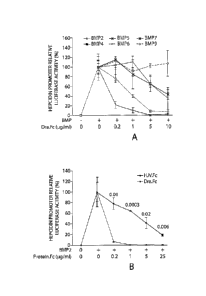
become
anemic at some point by the age of 18 years. Martin, P. L., et al. The
Anemias,
Principles and Practices of Pediatrics, 1657 (2d ed., Lippincott 1994).
[0004] In mammals, the iron balance is primarily regulated at the
level of
duodenal absorption of dietary iron. In humans, hereditary hemochromatosis
(HH) is
a common autosomal recessive genetic disease caused by hyperabsorption of
dietary
iron leading to an iron overload in plasma and multiple organs, including in
particular
1
CA 02742871 2011-05-03
WO 2010/056981
PCT/US2009/064369
the pancreas, liver, and skin, and resulting in damages in these organs and
tissues due
to the iron deposits.
[0005] Juvenile hemochromatosis is an iron overload disorder caused by
mutations in the gene encoding the major iron regulatory hormone hepcidin
(HAW)
and hemojuvelin (HFE2). (Roetto, A., et al.. 2003. Nut. Genet. 33:21-22;
Papanikolaou, G., et al. 2004. Nut. Genet. 36:77-82.) It has been shown that
hemojuvelin is a bone morphogenetic protein (BMP) co-receptor and that
hemojuvelin-mediated BMP signals regulate hepcidin expression and iron
metabolism. (Babitt, J.L., et al. 2006. Nat. Genet. 38:531-539; Babitt, J.L.,
et al..
2007. J Clin Invest. 117:1933-1939.) However, the endogenous BMP regulator(s)
of
hepcidin in vivo is unknown.
[0006] BMPs arc members of the TGF-13 superfamily, which is comprised of
over
40 ligands. (Shi, Y., and Massague, J. 2003. Ce11.113:685-700.) These growth
factors
mediate diverse biological processes including cell proliferation,
differentiation,
apoptosis, and patterning. BMP/TGF-13 superfamily ligands initiate an
intracellular
signaling cascade by binding to a complex of type I and type II serine
threonine
kinase receptors. The activated receptor complex phosphorylates intracellular
Smad
proteins, which then translocate to the nucleus to modulate gene expression.
[0007] Recently, a role for the BMP signaling pathway in regulating the
major
iron regulatory hormone hepcidin has been discovered. (Babitt, J.L., et al.
2006. Nat.
Genet. 38:531-539; Babitt, J.L., H et al. J Clin Invest. 117:1933-1939; Wang,
R.H., et
al. 2005. Cell Metab. 2:399-409.) Secreted by the liver, hepcidin inhibits
intestinal
iron absorption and macrophage iron release by decreasing cell surface
expression of
the iron exporter ferroportin. (Nemeth, E., et al. 2004. Science. 306:2090-
2093).
Hepcidin is upregulated by iron administration (Pigeon, C., et a/.2001. J.
Biol. Chem.
276:7811-7819, Nicolas, G., et al. 2002. J. Clin. Invest.110:1037-1044;
Nemeth, E., et
al. 2004. J. Clin. Invest. 113: 1271-1276.) and inhibited by anemia. (Nicolas,
G., et al.
2002. J. Clin. Invest.110:1037-1044) Hepcidin deficiency and unchecked
ferroportin
activity arc the common pathogenic mechanisms underlying the genetic iron
overload
disorder hereditary hemochromatosis due to mutations in HAMP itself, HFE2,
HFE,
TFR2 (encoding transferrin receptor type 2), and rare mutations of SCLA0A1
(encoding ferroportin). (Pietrangelo, A. 2006. Biochim Biophys Acta 1763:700-
710)
Hepcidin is also upregulated by inflammatory cytokines, such as IL-6, and
hepcidin
2
CA 02742871 2011-05-03
WO 2010/056981
PCT/US2009/064369
excess is implicated in the pathogenesis of anemia of inflammation (Pigeon,
C., et
a/.2001. J. Biol. Chenz. 276:7811-7819, Nicolas, G., et al. 2002. J. Clin.
Invest.110:1037-1044; Nemeth, E., et al. 2004. J. Clin. Invest. 113: 1271-
1276;
Nemeth, E., et al. 2003. Blood. 101 :246 1-2463; Weiss, G. and Goodnough, L.T.
2005. N. Engl. J. Med. 352:1011-1023; Andrews NC. 2008. Blood. 1122 19-30.)
[0008] Reduction of hepatic BMP signaling by a liver-specific conditional
knockout of the common BMP/TGF-13 intracellular mediator Smad4 (Wang, R.H., et
al. 2005. Cell Metab.2:399-409.), or by mutations in HFE2 (Papanikolaou, G.,
et
a/.2004. Nut. Genet. 36:77-82; Babitt, J.L., et al. 2006. Nat. Genet. 38:531-
539;
Huang, F.W., et al. J. Clin. Invest. 115:2187-2191; Niederkofler, V., Salie,
R., Arber,
S. 2005. J Clin Invest. 115:2180-6), which encodes the BMP co-receptor
hemojuvelin,
are associated with inappropriately low hepcidin expression and iron overload.
BMP
signals positively increase hepcidin expression at the transcriptional level
in vitro.
(Babitt, J.L., et al. 2006. Nat. Genet. 38:531-539; Babitt, J.L., H et al. J
Clin Invest.
117:1933-1939; Wang, R.H., et al. 2005. Cell Metab.2:399-409; Truksa, J., et
al.
2006. Proc. Natl. Acad. Sci. USA. 103:10289-10293; Verga Falzacappa, M.V., et
al.
2008. J Mot Med. 86:531-40.). Iron administration in vivo increases hepatic
BMP
signaling (Yu, P.B., et al. 2008. Nut Chem Biol. 4:33-41). BMP administration
in vivo
increases hepcidin expression and reduces serum iron. (Babitt, J.L., H et al.
J Clin
Invest. 117:1933-1939). Conversely, administration of soluble hemojuvelin
fused to
the Fc portion of human immunoglobulin Fc (HIV.Fc) in vivo, which selectively
inhibits BMP-2, BMP-4, BMP-5, and BMP-6, but not BMP-7 or BMP-9, inhibits
hepcidin expression, increases ferroportin expression, mobilizes
reticuloendothelial
cell iron stores, and increases serum iron in vivo. (Babitt, J.L., et al.
2007. J Clin
Invest. 117: 1933-1939.) Administration of the non-selective small molecule
BMP
inhibitor Dorsomorphin also inhibits hepcidin expression and increases serum
iron in
vivo. (Yu, P.B., et al. 2008. Nut Chem Biol. 4:33-41).
[0009] Hemojuvelin (also known as RGMc) is a member of the Repulsive
Guidance Molecules family of proteins, including RGMa and DRAGON (RGMb),
which share 50-60% amino acid identity. (Samad, T.A., et al. 2004. J.
Neurosci.
24:2027-2036.). Like Hemojuvelin, RGMa (Babitt, J.L., et al. 2005. J. Biol.
Chem.280:29820-29827) and DRAGON(Samad, T.A., et al. 2005. J. Biol. Chem.
280:14122- 14129) also function as co-receptors for the BMP signaling pathway.
3
CA 02742871 2011-05-03
WO 2010/056981
PCT/US2009/064369
[0010] There is a need for a cost-effective and efficient method for
regulating
hepcidin expression and iron metabolism.
SUMMARY OF THE INVENTION
[0011] The present invention provides novel methods for modulating BMP-6
for
treating disorders of iron overload due to hepcidin deficiency or anemia of
inflammation due to hepcidin excess.
[0012] The invention relates to modulators of the BMP signaling pathway
that
have a role in treating disorders of iron overload due to hepcidin deficiency
or anemia
of inflammation due to hepcidin excess.
[0013] The present invention relates to a method for regulating iron
homeostasis
in a subject, said method comprising administering to said subject an
effective amount
of a pharmaceutical composition sufficient for modulating BMP-6 signaling at a
level
sufficient to alter iron homeostasis, hemoglobin levels and/or hcmatocrit
levels in the
subject. In some aspects, administering the composition reduces BM P-6
signaling. In
some aspects, the composition comprises a reagent capable of binding BMP-6. In
some embodiments, the reagent binds BMP-6 at residues TQSQDVARVSSASDY
(SEQ ID NO:3). In some aspects, the reagent is an antibody. In some aspects,
the
antibody competitively inhibits BMP-6 binding by soluble human hemojuvelin
protein. In certain specific embodiments, the soluble human hemojuvelin
protein is
HJV.Fc or HJV.His. In other aspects, the antibody inhibits BMP-6 activity by
binding BMP-6 at a domain independent of the domain at which soluble human
hemojuvelin protein binds BMP-6. In certain specific embodiments, the soluble
human hemojuvelin protein is HJV.Fc or HJV.His.
[0014] The present invention relates to methods wherein administering the
compound reduces hemojuvelin-mediated induction of hepcidin expression. In
some
aspects, the reagent is administered in an amount sufficient to inhibit an
interaction
between hemojuvelin and BMP-6. In some aspects, the reagent preferably
inhibits
expression or activity of human BMP-6 over BMP-2, BMP-4, BMP-5, BMP-7 or
BMP-9. In some aspects, the reagent binds BMP-6 with at least 5-fold greater
affinity
that BMP-7.
4
CA 02742871 2011-05-03
WO 2010/056981
PCT/US2009/064369
[0015] The invention provides methods for the administration of the
compositions
of the instant invention which result in increased serum iron levels or
increased serum
transferrin saturation in the subject. The invention also provides methods for
the
administration of the compositions of the instant invention which result in
increased
hemoglobin or hematocrit levels.
[0016] The invention provides methods for the administration of the
compositions
of the instant invention which result in increased BMP-6 signaling. In some
aspects,
the composition comprises a reagent capable of increasing serum BMP-6 levels.
In
some aspects, the reagent increases BMP-6 expression levels.
[0017] The invention provides methods for treating a subject who has one or
more
symptoms of hereditary hemochromatosis, the symptoms selected from the group
consisting of: increased scrum iron level, increased scrum transferrin
saturation,
reduced hepcidin expression, reduced spleen iron store, increased ferroportin
expression and tissue iron overload.
[0018] The invention provides methods for administration of the composition
reduces expression level of BMP-6. In some aspects the composition comprises a
reagent capable of inhibiting BMP-6 gene expression, wherein the reagent is
antisense
DNA, siRNA, interfering RNA, microRNA (miRNA) or antisense RNA, and wherein
the reduction in expression of BMP-6 is sufficient to increase serum iron
level or
serum transferrin saturation in the subject.
[0019] The invention provides an isolated monoclonal antibody which
specifically
binds to human BMP-6, wherein said human BMP-6 consists of the amino acid
sequence set forth in SEQ ID NO: 1 and the binding inhibits the iron-
regulating
activity of BMP-6 and a composition comprising the isolated monoclonal
antibody,
comprising an amount of the antibody sufficient to increase serum iron level
or serum
transferrin saturation in a subject. In some embodiments, the isolated
monoclonal
antibody competitively inhibits BMP-6 binding by soluble human hemojuvelin
protein to reduce binding to BMP-6 by 25%-100%. In certain specific
embodiments,
the soluble human hemojuvelin protein is HJV.Fc or HJV.His. In more specific
embodiments, the isolated monoclonal antibody competitively inhibits BMP-6
binding by anti-BMP-6 antibodies selected from the group consisting of R & D
Systems monoclonal antibody to human BMP-6, MAB507 (clone 74219), R & D
CA 02742871 2013-08-26
54498-6
systems polyclonal antibody to human BMP-6, AF507 (lot CXL04A), and Santa Cruz
polyclonal antibody by 25%-100%. In other embodiments, the isolated monoclonal
antibody binds BMP-6 at a domain distinct from the domain at which HJV.Fc
binds.
[0020] The invention also provides anti-BMP-6 antibodies such as human
antibody, consisting of a chimerized antibody, a humanized antibody, a fully
human
antibody, a single chain Fv fragment, a F(ab')2fragment, an Fd, a domain
antibody
(dAb), a diabody, a maxibody, and a nanobody. In some aspects, the isolated
monoclonal antibody binds both human BMP-6 and murine BMP-6.
[0021] The invention also provides isolated nucleic acid molecules
comprising a
nucleotide sequence that encodes anti-BMP-6 antibody and expression vector
comprising the nucleic acid molecule operably linked to a regulatory control
sequence.
[0022] The invention also provides a method for using a host cell of
comprising
the vector of claim or a nucleic acid molecule to produce an antibody,
comprising
culturing the host cell of claim under suitable conditions such that the
nucleic acid is
expressed to produce the antibody, is provided.
[0023] The invention also provides for treating a subject who has one
or more
symptoms of hereditary hemochromatosis by administration of (a) pharmaceutical
composition sufficient for modulating BMP-6 signaling at a level sufficient to
alter
iron homeostasis, hemoglobin levels and/or hematocrit levels and (b) an
erythropoiesis stimulator, in therapeutically effective amounts. Exemplary
erythropoiesis stimulators include erythropoietin, erythropoietin agonist
variants, and
peptides or antibodies that bind and activate erythropoietin receptor.
Erythropoiesis
stimulators include, but are not limited to, epoetin alfa, epoetin beta,
epoetin delta,
epoetin omega, epoetin iota, epoetin zeta, and analogs thereof, mimetic
peptides,
mimetic antibodies and HIF inhibitors (see U.S. Patent Publication No.
2005/0020487). In
particular, erythropoietin includes, but is not limited to, erythropoietin
molecules or
variants or analogs thereof as disclosed in the following patents or patent
applications: U.S. Pat. Nos. 4,703,008;
5,441,868; 5,547,933; 5,618,698; 5,621,080; 5,756,349; 5,955,422 and
5,856,298; and WO 91/05867; WO 95/05465; WO 00/24893 and WO 01/81405. In
6
CA 02742871 2011-05-03
WO 2010/056981
PCT/US2009/064369
certain exemplary embodiments, the erythropoiesis stimulator is selected from
the
group consisting of human erythropoietin and darbepoetin alfa.
[0024] The invention also provides a method for diagnosing a BMP-6-related
disorder, the method comprising: (a) contacting a biological sample from a
human
suspected of having said disorder with an antibody that specifically binds to
BMP-6
under conditions suitable for binding of the antibody to human BMP-6; and (b)
quantitating the BMP-6 bound to the antibody, wherein the amount of BMP-6 in
said
sample, as quantitated in (b), above or below a normal level indicates the
presence of
a BMP-6-related disorder.
[0025] The invention also provides a method for monitoring a treatment in
which
a BMP-6 antagonist is administered, the method comprising: (a) contacting a
biological sample, from a human that has been administered a BMP-6 antagonist,
with
an antibody that specifically binds to BMP-6 under conditions suitable for
binding of
the antibody to human BMP-6; and (b) quantitating the BMP-6 bound to the
antibody,
wherein a change in the amount of serum BMP-6 level, as quantitated in (b), is
indicative of the efficacy of the BMP-6 antagonist. In some aspects, the
antagonist is
an antibody or a small molecule.
[0026] The invention also provides a method of treating a mammal with an
elevated level of iron or anemia through the administration of a
pharmaceutical
composition that modulates BMP-6 signaling.
[0027] The invention also provides a kit for treating a disorder associated
with
iron homeostasis, comprising an article of manufacture comprising a vial or
prefilled
syringe comprising anti-BMP-6 antibodies.
[0028] The invention also provides a method for screening compounds that
binds
to human BMP-6 comprising contacting a candidate compound with a composition
comprising bioactive BMP-6, and detecting a complex between the candidate
compound and human BMP-6 in the composition, wherein detection of a complex
indicates that the candidate compound binds to human BMP-6, and further
wherein
the candidate compound inhibits a binding of BMP-6 with HJV.Fc by at least
25%.
[0029] The invention also provides a method of generating an antibody to
human
BMP-6 comprising contacting an immunoglobulin producing cell with a
polypeptide
7
CA 02742871 2011-05-03
WO 2010/056981
PCT/US2009/064369
comprising a BMP-6 sequence of SEQ ID NO:1 or variant thereof, and isolating
an
immunoglobulin produced by said cell.
100301 The invention also provides a composition for the treatment of an
iron
deficiency disorder comprising an antibody that specifically binds to BMP-6
and
HJV.Fc. In one embodiment, the antibody competitively inhibits BMP-6 binding
by
soluble human hemojuvelin conjugate (HJV.Fc) protein. In another embodiment,
the
antibody competitively inhibits BMP-6 binding by soluble human hemojuvelin
conjugate (HJV.Fc) protein. In another embodiment, the antibody binds to a
domain
on BMP-6 distinct from the domain to which HJV.Fc binds.
[0031] In another specific embodiment, the antibody is in an amount
sufficient to
reduce HJV.Fc binding to BMP-6 by 25%-100%. Preferably, the antibody is
selected
from the group consisting of RandD Systems monoclonal antibody, RandD systems
polyclonal antibody, and Santa Cruz polyclonal antibody. In another specific
embodiment, the antibody is a human antibody. In other embodiments, said
antibody
is selected from the group consisting of a chimerized antibody, a humanized
antibody,
a fully human antibody, a single chain Fv fragment, a F(ab)2 fragment, an Fd,
a
domain antibody (dAb), a diabody, a maxibody, and a nanobody.
[0032] The invention also provides an isolated nucleic acid molecule
comprising a
nucleotide sequence that encodes the antibody that specifically binds to BMP-
6. The
invention also provides an expression vector comprising this nucleic acid
molecule
operably linked to a regulatory control sequence. The invention also provides
a host
cell comprising the vector of claim 31 or a nucleic acid molecule of claim 30.
[0033] The invention also provides a method for using the host cell of
claim 32 to
produce an antibody, comprising culturing the host cell of claim under
suitable
conditions such that the nucleic acid is expressed to produce the antibody.
[0034] The invention also provides a method for diagnosing a BMP-6-related
disorder, the method comprising: contacting a biological sample from a human
suspected of having said disorder with an antibody that specifically binds to
BMP-6
under conditions suitable for binding of the antibody to human BMP-6; and
quantitating the BMP-6 bound to the antibody, wherein the amount of BMP-6 in
said
sample, as quantitated in (b), above or below a normal level indicates the
presence of
a BMP-6-related disorder.
8
81632106
[0035] The invention also provides a method for monitoring a treatment in
which a BMP-6
antagonist is administered, the method comprising: contacting a biological
sample, from a human that
has been administered a BMP-6 antagonist, an antibody that specifically binds
to BMP-6 under
conditions suitable for binding of the antibody to human BMP-6; and
quantitating the BMP-6 bound to
the antibody, wherein a change in the amount of serum BMP-6 level, as
quantitated in (b), is indicative
of the efficacy of the BMP-6 antagonist. In a specific embodiment, the
antagonist is an antibody. In
another specific embodiment, the antagonist is a small molecule.
=
[0036] The invention also provides an antibody that specifically binds to
human BMP-6,
wherein in the presence of a concentration of a peptide comprising the amino
acid sequence of
TQSQDVARVSSASDY (SEQ ID NO:3) the antibody is competed away from specifically
binding to
human BMP-6.
100371 The invention also provides an antibody specifically binds to any 5
consecutive amino
acids of TQSQDVARVSSASDY (SEQ ID NO:3). In specific embodiments, the antibody
specifically
binds to and 6, 7, 8, 9 or 10 consecutive amino acids of TQSQDVARVSSASDY (SEQ
ID NO:3).
[0037a] The present invention as claimed relates to:
- a pharmaceutical composition for use in increasing serum iron levels in a
subject in
need thereof, wherein the composition comprises a neutralizing human anti-BMP-
6 monoclonal
antibody, or a fragment of the anti-BMP-6 monoclonal antibody, wherein the
antibody or fragment
preferentially binds mature BMP-6 over BMP-2, BMP-4, BMP-5, BMP-7, or BMP-9,
wherein the
composition further comprises a pharmaceutically acceptable carrier, diluent
or excipient, and wherein
said composition is for administration in an amount effective to increase
serum iron levels in the subject;
- a pharmaceutical composition for use in reducing hepcidin expression or
activity in a
subject having elevated hepatic hepcidin expression or low serum iron levels,
wherein the composition
comprises a neutralizing human anti-BMP-6 monoclonal antibody, or a fragment
of the anti-BMP-6
monoclonal antibody, wherein the antibody or fragment binds mature BMP-6 or a
fragment thereof over
BMP-2, BMP-4, BMP-5, BMP-7, or BMP-9, wherein the antibody competitively
inhibits BMP-6 binding
to soluble human hemojuvelin protein, wherein the soluble human hemojuvelin
protein is HJV.Fc or
HJV.His, wherein the composition further comprises a pharmaceutically
acceptable canier, diluent or
excipient, and wherein said composition is for administration in an amount
sufficient for modulating
BMP-6 signaling at a level sufficient to reduce hepcidin expression or
activity in the subject;
9
CA 2742871 2018-05-31
= 81632106
- a pharmaceutical composition for use in increasing serum transferrin
saturation
in a subject having elevated hepatic hepcidin expression or low serum iron
levels, wherein the
composition comprises a neutralizing human anti-BMP-6 monoclonal antibody, or
a fragment of
the anti-BMP-6 monoclonal antibody, wherein the antibody or fragment
preferentially binds
mature BMP-6 or a fragment thereof over BMP-2, BMP-4, BMP-5, BMP-7, or BMP-9,
wherein
the composition further comprises a pharmaceutically acceptable carrier,
diluent or excipient,
and wherein said composition is for administration in an amount effective to
increase serum
transferrin saturation in the subject; and
- a composition comprising a neutralizing human anti-BMP-6 monoclonal
antibody, or a fragment of the anti-BMP-6 monoclonal antibody, wherein the
antibody or
fragment preferentially binds mature BMP-6 or a fragment thereof over BMP-2,
BMP-4, BMP-5,
BMP-7, or BMP-9, wherein the antibody competitively inhibits BMP-6 binding to
soluble
human hemojuvelin protein, wherein the soluble human hemojuvelin protein is
HJV.Fc or
HJV.His, wherein the antibody specifically binds BMP-6 with higher affinity
than BMP-7, and
wherein the composition further comprises a pharmaceutically acceptable
carrier, diluent or
excipient.
[0038] The present invention and other objects, features, and advantages
of the present
invention will become further apparent in the following Detailed Description
of the Invention
and the accompanying Figures and embodiments.
BRIEF DESCRIPTION OF THE FIGURES
[0039] Figures 1A-1D show evidence that DRAGON.Fc selectively inhibits
BMP
induction of hepcidin expression.
[0040] Figures 2A-2G show evidence that DRAGON.Fc administration in mice
does
not affect hepcidin expression or iron metabolism.
100411 Figures 3A-3D show evidence that specific neutralizing BMP-6
antibody inhibits
hepatic hepcidin expression and increases serum iron and transferrin
saturation in vivo.
100421 Figures 4A-4H show evidence that Bmp6 null mice exhibit reduced
hepatic
hepcidin expression, increased spleen ferroportin expression, increased serum
iron and
transferrin saturation, increased liver iron content and reduced spleen iron
content.
9a
CA 2742871 2018-05-31
CA 02742871 2011-05-03
WO 2010/056981
PCT/US2009/064369
[0043] Figures 5A-5C show evidence that BMP-6 administration in mice
increases hepcidin mRNA expression and reduces serum iron.
[0044] Figure 6 is a Western blot for BMP-6.
[0045] Figure 7 is a Western blot for HJV.
[0046] Figure 8 is a Western blot of hBMP-6.
DETAILED DESCRIPTION OF THE INVENTION
[0047] The inventors surprisingly have found that BMP-6 is an important
regulator of hcpcidin expression and iron metabolism. Compared with soluble
hemojuvelin (HJV.Fc), the homologous DRAGON.Fc fusion protein is a more potent
inhibitor of hepcidin promoter activation by BMP-2 and BMP-4, but a less
potent
inhibitor of BMP-6 in vitro. In vivo, DRAGON.Fc has no effect on hepcidin
expression and iron metabolism, while HJV.Fc or a specific neutralizing BMP-6
antibody inhibit hepcidin expression and increase serum iron. Further, Bmp6
null
mice have a phenotype that resembles hereditary hemochromatosis with reduced
hepcidin expression, increased ferroportin expression, increased serum iron
and
transferrin saturation, reduced spleen iron stores, and tissue iron overload.
The
inventors show that BMP-6 administration in mice increases hepcidin expression
and
reduces serum iron. Taken together, these data support a key role for BMP-6 as
an
endogenous regulator of hcpcidin expression and iron metabolism in vivo.
[0048] Administration of specific neutralizing BMP-6 antibody resulted in
increased serum iron and transferrin saturation indicating effects on hepcidin
expression and iron metabolism. Inhibition of endogenous BMP-6 by siRNA or
neutralizing antibody inhibits hemojuvelin-mediated induction of hepcidin
expression.
BMP-6 likely is a ligand for hemojuvelin.
[0049] Further, addition of exogenous BMP-6 was found to increase hepcidin
expression and cause a dose-dependent reduction in serum iron and serum
transferrin
saturation.
CA 02742871 2011-05-03
WO 2010/056981
PCT/US2009/064369
[0050] The amino acid sequence of pro-BMP-6 is shown in Table 1
below:
Table 1. Amino acid sequence of human pro-BMP-6 (428 amino acids; SEQ ID
NO:1)
20 30 40 50 60
DCSRQGPQRP RSGLAPPQPP ALRQQEEQQQ QQQLPRGEPP PGRLKSAPLF MLDLYNALSA
70 80 90 100 110 120
DNDEDGASEG ERQQSWPHEA ASSSQRRQPP PGAAHPLNRK SLLAPGSGSG GASPLTSAQD
130 140 150 160 170 180
SAFLNDADMV MSFVNLVEYD KEFSPRQRHH KEFKFNLSQI PEGEVVTAAE FRIYKDCVMG
190 200 210 220 230 240
SEKNQTFLIS IYQVLQEHQH RDSDLFLLDT RVVWASEEGW LEFDITATSN LWVVTPQHNM
250 260 270 280 290 300
GLQLSVVTRD GVHVHPRAAG LVGRDGPYDK QPFMVAFFKV SEVHVRTTRS ASSRRRQQSR
310 320 330 340 350 360
NRSTQSQLVA RVSSASDYNS SELKTACRKH ELYVSEQDLG WQDWIIAPKG YAANYCDGEC
370 380 390 400 410 420
SFPLNAHMNA TNHAIVQTLV HLMNPEYVPK PCCAPTKLNA ISVLYFDDNS NVILKKYRNM
VVRACGCH
[0051] BMP-6 is made up of amino acids 297-428 of the pro-BMP-6
sequence
shown in Table 1. BMP-6 is shown in Table 2 below.
Table 2. Amino acid sequence of human BMP-6 (132 amino acids; SEQ ID NO:2)
10 20 30 40 50 60
QQSRNRSTQS QDVARVSSAS DYNSSELKTA CRKHELYVSF QDLGWQDWII APKGYAANYC
70 80 90 100 110 120
DGECSFPLNA HMNATNHAIV QTLVHLMNPE YVPKPCCAPT KLNAISVLYF DDNSNVILKK
130
YRNMVVRACG CH
[0052] Unless otherwise defined, scientific and technical terms used in
connection
with the present invention shall have the meanings that are commonly
understood by
those of ordinary skill in the art. Further, unless otherwise required by
context,
singular terms shall include pluralities and plural terms shall include the
singular.
Generally, nomenclatures utilized in connection with, and techniques of, cell
and
tissue culture, molecular biology, and protein and oligo- or polynucleotide
chemistry
and hybridization described herein are those well known and commonly used in
the
11
CA 02742871 2011-05-03
WO 2010/056981
PCT/US2009/064369
art. Standard techniques are used for recombinant DNA, oligonucleotide
synthesis,
and tissue culture and transformation (e.g., electroporation, lipofection).
Enzymatic
reactions and purification techniques are performed according to
manufacturer's
specifications or as commonly accomplished in the art or as described herein.
The
practice of the present invention will employ, unless indicated specifically
to the
contrary, conventional methods of virology, immunology, microbiology,
molecular
biology and recombinant DNA techniques within the skill of the art, many of
which
are described below for the purpose of illustration. Such techniques are
explained
fully in the literature. See, e.g., Sambrook, et al. Molecular Cloning: A
Laboratory
Manual (2nd Edition, 1989); Maniatis et al. Molecular Cloning: A Laboratory
Manual
(1982); DNA Cloning: A Practical Approach, vol. I & 11 (D. Glover, ed.);
Oligonucleotide Synthesis (N. Gait, ed., 1984); Nucleic Acid Hybridization (B.
Names & S. Higgins, eds., 1985); Transcription and Translation (B. Names & S.
Higgins, eds., 1984); Animal Cell Culture (R. Freshney, ed., 1986); Perbal, A
Practical Guide to Molecular Cloning (1984).
[0053] The nomenclatures utilized in connection with, and the laboratory
procedures and techniques of, analytical chemistry, synthetic organic
chemistry, and
medicinal and pharmaceutical chemistry described herein are those well known
and
commonly used in the art. Standard techniques are used for chemical syntheses,
chemical analyses, pharmaceutical preparation, formulation, and delivery, and
treatment of patients.
[0054] The following definitions are useful in understanding the present
invention:
[0055] The term "antibody" (Ab) as used herein includes monoclonal
antibodies,
polyclonal antibodies, multispecific antibodies (e.g., bispecific antibodies),
and
antibody fragments, so long as they exhibit the desired biological activity.
The term
"immunoglobulin" (Ig) is used interchangeably with "antibody" herein.
[0056] An "isolated antibody" is one that has been separated and/or
recovered
from a component of its natural environment. Contaminant components of its
natural
environment are materials that would interfere with diagnostic or therapeutic
uses for
the antibody, and may include enzymes, hormones, and other proteinaceous or
nonproteinaceous solutes. In preferred embodiments, the antibody is purified:
(1) to
12
CA 02742871 2011-05-03
WO 2010/056981
PCT/US2009/064369
greater than 95% by weight of antibody as determined by the Lowry method, and
most preferably more than 99% by weight; (2) to a degree sufficient to obtain
at least
15 residues of N-terminal or internal amino acid sequence by use of a spinning
cup
sequenator; or (3) to homogeneity by SDS-PAGE under reducing or non-reducing
conditions using Coomassie blue or, preferably, silver stain. Isolated
antibody
includes the antibody in situ within recombinant cells since at least one
component of
the antibody's natural environment will not be present. Ordinarily, however,
isolated
antibody will be prepared by at least one purification step.
[0057] The basic four-chain antibody unit is a heterotetrameric
glycoprotein
composed of two identical light (L) chains and two identical heavy (H) chains.
An
IgM antibody consists of 5 of the basic heterotetramer unit along with an
additional
polypeptide called J chain, and therefore contain 10 antigen binding sites,
while
secreted IgA antibodies can polymerize to form polyvalent assemblages
comprising 2-
of the basic 4-chain units along with J chain. In the case of IgGs, the 4-
chain unit is
generally about 150,000 daltons. Each L chain is linked to an H chain by one
covalent
disulfide bond, while the two H chains are linked to each other by one or more
disulfide bonds depending on the H chain isotypc. Each H and L chain also has
regularly spaced intrachain disulfide bridges. Each H chain has at the N-
terminus, a
variable domain (VH) followed by three constant domains (CH) for each of the a
and y
chains and four CH domains for 11 and c isotypes. Each L chain has at the N-
terminus,
a variable domain (VL) followed by a constant domain (CL) at its other end.
The VL is
aligned with the VH and the CL is aligned with the first constant domain of
the heavy
chain (CH1). Particular amino acid residues are believed to form an interface
between
the light chain and heavy chain variable domains. The pairing of a VH and VL
together
forms a single antigen-binding site. For the structure and properties of the
different
classes of antibodies, see, e.g., Basic and Clinical Immunology, 8th edition,
Daniel P.
Stites, Abba I. Ten and Tristram G. Parslow (eds.), Appleton & Lange, Norwalk,
Conn., 1994, page 71, and Chapter 6.
[0058] The L chain from any vertebrate species can be assigned to one of
two
clearly distinct types, called kappa (K) and lambda (k), based on the amino
acid
sequences of their constant domains (CL). Depending on the amino acid sequence
of
the constant domain of their heavy chains (CH), immunoglobulins can be
assigned to
different classes or isotypes. There are five classes of immunoglobulins: IgA,
IgD,
13
CA 02742871 2011-05-03
WO 2010/056981
PCT/US2009/064369
IgE, IgG, and IgM, having heavy chains designated alpha (a), delta (6),
epsilon (c),
gamma (7) and mu ( ), respectively. The 7 and a classes are further divided
into
subclasses on the basis of relatively minor differences in CH sequence and
function,
e.g., humans express the following subclasses: IgGl, IgG2, IgG3, IgG4, IgAl,
and
IgA2.
[0059] The term "variable" refers to the fact that certain segments of the
V
domains differ extensively in sequence among antibodies. The V domain mediates
antigen binding and defines specificity of a particular antibody for its
particular
antigen. However, the variability is not evenly distributed across the 110-
amino acid
span of the variable domains. Instead, the V regions consist of relatively
invariant
stretches called framework regions (FRs) of 15-30 amino acids separated by
shorter
regions of extreme variability called "hypervariable regions" that are each 9-
12 amino
acids long. The variable domains of native heavy and light chains each
comprise four
FRs, largely adopting a 13-sheet configuration, connected by three
hypervariable
regions, which form loops connecting, and in some cases forming part of, the
13-shcct
structure. The hypervariable regions in each chain are held together in close
proximity
by the FRs and, with the hypervariable regions from the other chain,
contribute to the
formation of the antigen-binding site of antibodies (see Kabat et al.,
Sequences of
Proteins of Immunological Interest, 5th Ed. Public Health Service, National
Institutes
of Health, Bethesda, Md. (1991)). The constant domains are not involved
directly in
binding an antibody to an antigen, but exhibit various effector functions,
such as
participation of the antibody in antibody dependent cellular cytotoxicity
(ADCC).
[0060] The term "hypervariable region" when used herein refers to the amino
acid
residues of an antibody that are responsible for antigen binding. The
hypervariable
region generally comprises amino acid residues from a "complementarity
determining
region" or "CDR" (e.g., around about residues 24-34 (L1), 50-56 (L2) and 89-97
(L3)
in the VL, and around about 1-35 (H1), 50-65 (H2) and 95-102 (H3) in the VH;
Kabat
et al., Sequences of Proteins of Immunological Interest, 5th Ed. Public Health
Service, National Institutes of Health, Bethesda, Md. (1991)) and/or those
residues
from a "hypervariable loop" (e.g., residues 26-32 (L1), 50-52 (L2) and 91-96
(L3) in
the VL, and 26-32 (H1), 53-55 (H2) and 96-101 (H3) in the VH ; Chothia and
Lesk, J.
Mol. Biol. 196:901-917 (1987)).
14
CA 02742871 2011-05-03
WO 2010/056981
PCT/US2009/064369
[0061] The term -monoclonal antibody" as used herein refers to an antibody
obtained from a population of substantially homogeneous antibodies, i.e., the
individual antibodies comprising the population are identical except for
possible
naturally occurring mutations that may be present in minor amounts. Monoclonal
antibodies are highly specific, being directed against a single antigenic
site.
Furthermore, in contrast to polyclonal antibody preparations that include
different
antibodies directed against different determinants (epitopes), each monoclonal
antibody is directed against a single determinant on the antigen. In addition
to their
specificity, the monoclonal antibodies are advantageous in that they may be
synthesized uncontaminated by other antibodies. The modifier "monoclonal" is
not to
be construed as requiring production of the antibody by any particular method.
For
example, the monoclonal antibodies useful in the present invention may be
prepared
by the hybridoma methodology first described by Kohler et al., Nature, 256:495
(1975), or may be made using recombinant DNA methods in bacterial, eulcaryotic
animal or plant cells (see, e.g., U.S. Pat. No. 4,816,567). The "monoclonal
antibodies"
may also be isolated from phage antibody libraries using the techniques
described in
Clackson et al., Nature, 352:624-628 (1991) and Marks et al., J. Mol. Biol.,
222:581-
597 (1991), for example.
[0062] The monoclonal antibodies herein include "chimeric" antibodies in
which
a portion of the heavy and/or light chain is identical with or homologous to
corresponding sequences in antibodies derived from a particular species or
belonging
to a particular antibody class or subclass, while the remainder of the
chain(s) is
identical with or homologous to corresponding sequences in antibodies derived
from
another species or belonging to another antibody class or subclass, as well as
fragments of such antibodies, so long as they exhibit the desired biological
activity
(see U.S. Pat. No. 4,816,567; and Morrison et al., Proc. Natl. Acad. Sci. USA,
81:6851-6855 (1984)). The present invention provides variable domainantigen-
binding dequences derived from human antibodies. Accordingly, chimeric
antibodies
of primary interest hjerein include antibodies having one or more human
antigen
binding sequences (e.g., CDRs) and containing one or more sequences derived
from a
non-human antibody, e.g., an FR or C region sequence. In addition, chimeric
antibodies of primary interest herein include those comprising a human
variable
domain antigen binding sequence of one antibody class or subclass and another
CA 02742871 2011-05-03
WO 2010/056981
PCT/US2009/064369
sequence, e.g., FR or C region sequence, derived from another antibody class
or
subclass. Chimeric antibodies of interest herein also include those containing
variable
domain antigen-binding sequences related to those described herein or derived
from a
different species, such as a non-human primate (e.g., Old World Monkey, Ape,
etc).
Chimeric antibodies also include primatized and humanized antibodies.
[0063] Furthermore, chimeric antibodies may comprise residues that are not
found
in the recipient antibody or in the donor antibody. These modifications are
made to
further refine antibody performance. For further details, see Jones et al.,
Nature
321:522-525 (1986); Riechmann et al., Nature 332:323-329 (1988); and Presta,
Curr.
Op. Struct. Biol. 2:593-596 (1992).
[0064] A "humanized antibody" is generally considered to be a human
antibody
that has one or more amino acid residues introduced into it from a source that
is non-
human. These non-human amino acid residues are often referred to as "import"
residues, which are typically taken from an "import" variable domain.
Humanization
is traditionally performed following the method of Winter and co-workers
(Jones et
al., Nature, 321:522-525 (1986); Reichmann et al., Nature, 332:323-327 (1988);
Verhoeyen et al., Science, 239:1534-1536 (1988)), by substituting import
hypervariable region sequences for the corresponding sequences of a human
antibody.
Accordingly, such "humanized" antibodies are chimeric antibodies (U.S. Pat.
No.
4,816,567) wherein substantially less than an intact human variable domain has
been
substituted by the corresponding sequence from a non-human species.
[0065] A "human antibody" is an antibody containing only sequences present
in
an antibody naturally produced by a human. However, as used herein, human
antibodies may comprise residues or modifications not found in a naturally
occurring
human antibody, including those modifications and variant sequences described
herein. These are typically made to further refine or enhance antibody
performance.
[0066] An "intact" antibody is one that comprises an antigen-binding site
as well
as a CL and at least heavy chain constant domains, CH 1, CH 2 and CH 3. The
constant
domains may be native sequence constant domains (e.g., human native sequence
constant domains) or amino acid sequence variant thereof Preferably, the
intact
antibody has one or more effector functions.
16
CA 02742871 2011-05-03
WO 2010/056981
PCT/US2009/064369
[0067] An -antibody fragment" comprises a portion of an intact antibody,
preferably the antigen binding or variable region of the intact antibody.
Examples of
antibody fragments include Fab, Fab', F(ab')2, and Fv fragments; diabodies;
linear
antibodies (see U.S. Pat. No. 5,641,870; Zapata et al., Protein Eng. 8(10):
1057-1062
[1995]); single-chain antibody molecules; and multispecific antibodies formed
from
antibody fragments.
[0068] The phrase "functional fragment or analog" of an antibody is a
compound
having qualitative biological activity in common with a full-length antibody.
For
example, a functional fragment or analog of an anti-IgE antibody is one that
can bind
to an IgE immunoglobulin in such a manner so as to prevent or substantially
reduce
the ability of such molecule from having the ability to bind to the high
affinity
receptor, FcER1.
[0069] Papain digestion of antibodies produces two identical antigen-
binding
fragments, called "Fab" fragments, and a residual "Fc" fragment, a designation
reflecting the ability to crystallize readily. The Fab fragment consists of an
entire L
chain along with the variable region domain of the H chain (VH), and the first
constant
domain of one heavy chain (CH 1). Each Fab fragment is monovalent with respect
to
antigen binding, i.e., it has a single antigen-binding site. Pepsin treatment
of an
antibody yields a single large F(ab')2 fragment that roughly corresponds to
two
disulfide linked Fab fragments having divalent antigen-binding activity and is
still
capable of cross-linking antigen. Fab' fragments differ from Fab fragments by
having
additional few residues at the carboxy terminus of the CH1 domain including
one or
more cysteines from the antibody hinge region. Fab'-SH is the designation
herein for
Fab' in which the cysteine residue(s) of the constant domains bear a free
thiol group.
F(ab')2 antibody fragments originally were produced as pairs of Fab' fragments
that
have hinge cysteines between them. Other chemical couplings of antibody
fragments
are also known.
[0070] The "Fc" fragment comprises the carboxy-terminal portions of both H
chains held together by disulfides. The effector functions of antibodies are
determined
by sequences in the Fc region, which region is also the part recognized by Fc
receptors (FcR) found on certain types of cells.
17
CA 02742871 2011-05-03
WO 2010/056981
PCT/US2009/064369
[0071] "Fv" is the minimum antibody fragment that contains a complete
antigen-
recognition and -binding site. This fragment consists of a dimer of one heavy-
and one
light-chain variable region domain in tight, non-covalent association. From
the
folding of these two domains emanate six hypervariable loops (three loops each
from
the H and L chain) that contribute the amino acid residues for antigen binding
and
confer antigen binding specificity to the antibody. However, even a single
variable
domain (or half of an Fv comprising only three CDRs specific for an antigen)
has the
ability to recognize and bind antigen, although at a lower affinity than the
entire
binding site.
[0072] "Single-chain Fv" also abbreviated as "sFv" or "scFv" are antibody
fragments that comprise the VH and VL antibody domains connected into a single
polypeptide chain. Preferably, the sFv polypeptide further comprises a
polypeptide
linker between the VH and VL domains that enables the sFv to form the desired
structure for antigen binding. For a review of sFv, see Pluckthun in The
Pharmacology of Monoclonal Antibodies, vol. 113, Rosenburg and Moore eds.,
Springer-Verlag, New York, pp. 269-315 (1994); Borrebaeck 1995, infra.
[0073] The term "diabodies" refers to small antibody fragments prepared by
constructing sFv fragments (see preceding paragraph) with short linkers (about
5-10
residues) between the VH and VL domains such that inter-chain but not intra-
chain
pairing of the V domains is achieved, resulting in a bivalent fragment, i.e.,
fragment
having two antigen-binding sites. Bispecific diabodies are heterodimers of two
"crossover" sFv fragments in which the VH and VL domains of the two antibodies
are
present on different polypeptide chains. Diabodics arc described more fully
in, for
example, EP 404,097; WO 93/11161; and Hollinger et al., Proc. Natl. Acad. Sci.
USA, 90:6444-6448 (1993).
[0074] As used herein, an antibody that "internalizes" is one that is taken
up by
(i.e., enters) the cell upon binding to an antigen on a mammalian cell (e.g.,
a cell
surface polypeptide or receptor). The internalizing antibody will of course
include
antibody fragments, human or chimeric antibody, and antibody conjugates. For
certain therapeutic applications, internalization in vivo is contemplated. The
number
of antibody molecules internalized will be sufficient or adequate to kill a
cell or
inhibit its growth, especially an infected cell. Depending on the potency of
the
antibody or antibody conjugate, in some instances, the uptake of a single
antibody
18
CA 02742871 2011-05-03
WO 2010/056981
PCT/US2009/064369
molecule into the cell is sufficient to kill the target cell to which the
antibody binds.
For example, certain toxins are highly potent in killing such that
internalization of one
molecule of the toxin conjugated to the antibody is sufficient to kill the
infected cell.
[0075] As used herein, an antibody is said to be "immunospecific,"
"specific for"
or to "specifically bind" an antigen if it reacts at a detectable level with
the antigen,
preferably with an affinity constant, Ka, of greater than or equal to about
104 M-1, or
greater than or equal to about 105 M-1, greater than or equal to about 106 M-
1, greater
than or equal to about 107 M-1, or greater than or equal to 108 M-1. Affinity
of an
antibody for its cognate antigen is also commonly expressed as a dissociation
constant
KD, and in certain embodiments, anti-BMP-6 antibody specifically binds to BMP-
6 if
it binds with a KD of less than or equal to 10-4 M, less than or equal to
about 10-5 M,
less than or equal to about 10-6 M, less than or equal to 10-7 M, or less than
or equal to
10-8 M. Affinities of antibodies can be readily determined using conventional
techniques, for example, those described by Scatchard et al. (Ann. N.Y. Acad.
Sci.
USA 51:660 (1949)).
[0076] Binding properties of an antibody to antigens, cells or tissues
thereof may
generally be determined and assessed using immunodetection methods including,
for
example, immunofluorescence-based assays, such as immuno-histochemistry (IHC)
and/or fluorescence-activated cell sorting (FACS).
[0077] An antibody having a "biological characteristic" of a designated
antibody
is one that possesses one or more of the biological characteristics of that
antibody
which distinguish it from other antibodies. For example, in certain
embodiments, an
antibody with a biological characteristic of a designated antibody will bind
the same
epitope as that bound by the designated antibody and/or have a common effector
function as the designated antibody.
[0078] The term "antagonist" antibody is used in the broadest sense, and
includes
an antibody that partially or fully blocks, inhibits, or neutralizes a
biological activity
of an epitope, polypeptide, or cell that it specifically binds. Methods for
identifying
antagonist antibodies may comprise contacting a polypeptide or cell
specifically
bound by a candidate antagonist antibody with the candidate antagonist
antibody and
measuring a detectable change in one or more biological activities normally
associated with the polypeptide or cell.
19
CA 02742871 2011-05-03
WO 2010/056981
PCT/US2009/064369
[0079] Antibody "effector functions" refer to those biological activities
attributable to the Fc region (a native sequence Fc region or amino acid
sequence
variant Fc region) of an antibody, and vary with the antibody isotype.
Examples of
antibody effector functions include: Clq binding and complement dependent
cytotoxicity; Fc receptor binding; antibody-dependent cell-mediated
cytotoxicity
(ADCC); phagocytosis; down regulation of cell surface receptors (e.g., B cell
receptor); and B cell activation.
[0080] "Fc receptor" or "FcR" describes a receptor that binds to the Fc
region of
an antibody. In certain embodiments, the FcR is a native sequence human FcR.
Moreover, a preferred FcR is one that binds an IgG antibody (a gamma receptor)
and
includes receptors of the FcyRI, FcyRII, and FcyR1II subclasses, including
allelic
variants and alternatively spliced forms of these receptors. FCyRII receptors
include
FcyRIIA (an "activating receptor") and FcyRIIB (an "inhibiting receptor"),
which
have similar amino acid sequences that differ primarily in the cytoplasmic
domains
thereof. Activating receptor FcyRIIA contains an immunoreceptor tyrosine-based
activation motif (ITAM) in its cytoplasmic domain. Inhibiting receptor FcyRIIB
contains an immunoreceptor tyrosine-based inhibition motif (ITIM) in its
cytoplasmic
domain. (see review M. in Daeron, Annu. Rev. Immunol. 15:203-234 (1997)). FcRs
are reviewed in Ravetch and Kinet, Annu. Rev. Immunol 9:457-92 (1991); Capel
et
al., Immunomethods 4:25-34 (1994); and de Haas et al., J. Lab. Clin. Med.
126:330-
41 (1995). Other FcRs, including those to be identified in the future, are
encompassed
by the term "FcR" herein. The term also includes the neonatal receptor, FcRn,
which
is responsible for the transfer of maternal IgGs to the fetus (Guyer et al.,
J. Immunol.
117:587 (1976) and Kim et al., J. Immunol. 24:249 (1994)).
[0081] "Human effector cells" are leukocytes that express one or more FcRs
and
perform effector functions. Preferably, the cells express at least FcyRIII and
perform
ADCC effector function. Examples of human leukocytes that mediate ADCC include
PBMC, NK cells, monocytes, cytotoxic T cells and neutrophils; with PBMCs and
NK
cells being preferred. The effector cells may be isolated from a native
source, e.g.,
from blood.
[0082] A "mammal" for purposes of treating n infection, refers to any
mammal,
including humans, domestic and farm animals, and zoo, sports, or pet animals,
such as
CA 02742871 2011-05-03
WO 2010/056981
PCT/US2009/064369
dogs, cats, cattle, horses, sheep, pigs, goats, rabbits, etc. Preferably, the
mammal is
human.
[0083] "Treating" or "treatment" or "alleviation" refers to both
therapeutic
treatment and prophylactic or preventative measures; wherein the object is to
prevent
or slow down (lessen) the targeted pathologic condition or disorder. Those in
need of
treatment include those already with the disorder as well as those prone to
have the
disorder or those in whom the disorder is to be prevented. A subject or mammal
is
successfully "treated" for an infection if, after receiving a therapeutic
amount of an
antibody according to the methods of the present invention, the patient shows
observable and/or measurable reduction in or absence of one or more of the
following: reduction in the number of infected cells or absence of the
infected cells;
reduction in the percent of total cells that are infected; and/or relief to
some extent,
one or more of the symptoms associated with the specific infection; reduced
morbidity and mortality, and improvement in quality of life issues. The above
parameters for assessing successful treatment and improvement in the disease
are
readily measurable by routine procedures familiar to a physician.
[0084] The term "therapeutically effective amount" refers to an amount of
an
antibody or a drug effective to "treat" a disease or disorder in a subject or
mammal.
See preceding definition of "treating."
[0085] "Chronic" administration refers to administration of the agent(s) in
a
continuous mode as opposed to an acute mode, so as to maintain the initial
therapeutic
effect (activity) for an extended period of time. "Intermittent"
administration is
treatment that is not consecutively done without interruption, but rather is
cyclic in
nature.
[0086] Administration -in combination with" one or more further therapeutic
agents includes simultaneous (concurrent) and consecutive administration in
any
order. In one embodiment of the invention, a combination therapy using a
pharmaceutical composition sufficient for modulating BMP-6 signaling at a
level
sufficient to alter iron homeostasis, hemoglobin levels and/or hematocrit
levels and an
erythropoiesis stimulator is used. This combination is useful for treating a
subject who
has one or more symptoms of hereditary hemochromatosis. In various
embodiments,
erythropoiesis stimulators can be used to improve treatment of a patient with
anemia.
21
CA 02742871 2013-08-26
54498-6
In particular, patients who are hypo-responsive to, including unresponsive to,
erythropoiesis stimulator therapy, such as erythropoietin or analogs thereof
(Epoetin
alfa, Epoetin beta, darbepoetin alfa), among others, will benefit from co-
treatment
with a hepcidin activity antagonist or hepcidin expression inhibitor.
[0087] As used herein, "erythropoiesis stimulator" means a chemical
compound
that directly or indirectly causes activation of the erythropoietin receptor,
for example,
by binding to and causing dimerization of the receptor or by stimulating
endogenous
erythropoietin expression. Erythropoiesis =stimulators include erythropoietin
and
variants, analogs, or derivatives thereof that bind to and activate
erythropoietin
receptor; antibodies that bind to erythropoietin receptor and activate the
receptor; or
peptides that bind to and activate erythropoietin receptor; or small organic
chemical
compounds, optionally less than about 1000 Daltons in molecular weight, that
bind to
and activate erythropoietin receptor. Erythropoiesis stimulators include, but
are not
limited to, epoetin alfa, epoetin beta, epoetin delta, epoetin omega, epoetin
iota,
epoetin zeta, and analogs thereof, pegylated erythropoietin, carbamylated
erythropoietin, mimetic peptides (including EMP l/hematide), mimetic
antibodies and
HIF inhibitors (see U.S. Patent Publication No. 2005/0020487).
Exemplary erythropoiesis
stimulators include erythropoietin, darbepoetin, erythropoietin agonist
variants, and
peptides or antibodies that bind and activate erythropoietin receptor (and
include
compounds reported in U.S. Patent Application Publication Nos. 2003/0215444
and
2006/0040858)
as well as erythropoietin molecules or variants or analogs thereof as
disclosed in the following patents of patent applications,
U.S. Pat. Nos. 4,703,008; 5,441,868;
5,547,933; 5,618,698; 5,621,080; 5,756,349; 5,767,078; 5,773,569; 5,955,422;
5,830,851; 5,856,298; 5,986,047; 6,030,086; 6,310,078; 6,391,633; 6,583,272;
6,586,398; 6,900,292; 6,750,369; 7,030,226; 7,084,245; 7,217,689; PCT
publication
nos. WO 91/05867; WO 95/05465; WO 99/66054; WO 00/24893; WO 01/81405;
WO 00/61637; WO 01/36489; WO 02/014356; WO 02/19963; WO 02/20034; WO =
02/49673; WO 02/085940; WO 03/029291; WO 2003/055526; WO 2003/084477;
W01003/094858; WO 2004/002417; =WO 2004/002424; WO 2004/009627; WO
2004/024761; WO 2004/033651; WO 2004/035603; WO 2004/043382; WO
22
CA 02742871 2013-08-26
54498-6
2004/101600; WO 2004/101606; W02004/101611; WO 2004/106373; WO
2004/018667; WO 2005/001025; WO 2005/001136; WO 2005/021579; WO
2005/025606; WO 2005/032460; WO 2005/051327; WO 2005/063808; WO ,
2005/063809; WO 2005/070451; WO 2005/081687; WO 2005/084711; WO
2005/103076; WO 2005/100403; WO 2005/092369; WO 2006/50959; WO
2006/02646; WO 2006/29094; and US publication nos. US 2002/0155998; US
2003/0077753; US 2003/0082749; US 2003/0143202; US 2004/0009902; US
2004/0071694; US 2004/0091961; US 2004/0143857; US 2004/0157293; US
2004/0175379; US 2004/0175824; US 2004/0229318;-1JS 2004/0248815; US
2004/0266690; US 2005/0019914; US 2005/0026834; US 2005/0096461; US
2005/0107297; US 2005/0107591; US 2005/0124045; US 2005/0124564; US
2005/0137329; US 2005/0142642; US 2005/0143292; US 2005/0153879; US
2005/0158822; US 2005/0158832; US 2005/0170457; US 2005/0181359; US
2005/0181482; US 2005/0192211; US 2005/0202538; US 2005/0227289; US
2005/0244409; US 2006/0088906; US 2006/0111279.
1j0088] Exemplary sequences, manufacture, purification and use of
recombinant
human erythropoietin are described in a number of patent publications,
including but
not limited to Lin U.S. Pat. No. 4,703,008 and Lai et al. U.S. Pat. No.
4,667,016.
Darbepoetin is a
hyperglycosylated erythropoietin analog having five changes in the amino acid
sequence of rHuEPO which provide for two additional carbohydrate chains. More
specifically, darbepoetin alfa contains two additional N-linked carbohydrate
chains at
amino acid residues 30 and 88. Exemplary sequences, manufacture, purification
and
use of darbepoetin and other erythropoietin analogs are described in a number
of
patent publications, including Strickland et al., 91/05867, Elliott et al., WO
95/05465,
Egrie et al., WO 00/24893, and Egrie et al. WO 01/81405.
Derivatives of naturally occurring or
analog polypeptides include those which have been chemically modified, for
example,
to attach water soluble polymers (e.g., pegylated), radionuclides, or other
diagnostic
or targeting or therapeutic moieties.
[0089] The term "erytlu-opoietic activity" means activity to stimulate
erythropoiesis as demonstrated in an in vivo assay, for example, the exhypoxic
polycythemic mouse assay. See, e.g., Cotes and Bangham, Nature 191:1065
(1961).
23
CA 02742871 2011-05-03
WO 2010/056981
PCT/US2009/064369
[0090] -Carriers" as used herein include pharmaceutically acceptable
carriers,
excipients, or stabilizers that are nontoxic to the cell or mammal being
exposed
thereto at the dosages and concentrations employed. Often the physiologically
acceptable carrier is an aqueous pH buffered solution. Examples of
physiologically
acceptable carriers include buffers such as phosphate, citrate, and other
organic acids;
antioxidants including ascorbic acid; low molecular weight (less than about 10
residues) polypeptide; proteins, such as serum albumin, gelatin, or
immunoglobulins;
hydrophilic polymers such as polyvinylpyrrolidone; amino acids such as
glycine,
glutamine, asparagine, arginine or lysine; monosaccharides, disaccharides, and
other
carbohydrates including glucose, mannose, or dextrins; chelating agents such
as
EDTA; sugar alcohols such as mannitol or sorbitol; salt-forming counterions
such as
sodium; and/or nonionic surfactants such as TWEENTm polyethylene glycol (PEG),
and PLURONICSTm.
[0091] "Label" as used herein refers to a detectable compound or
composition
that is conjugated directly or indirectly to the antibody so as to generate a
"labeled"
antibody. The label may be detectable by itself (e.g., radioisotope labels or
fluorescent
labels) or, in the case of an enzymatic label, may catalyze chemical
alteration of a
substrate compound or composition that is detectable.
[0092] The term "epitope tagged" as used herein refers to a chimeric
polypeptide
comprising a polypeptide fused to a "tag polypeptide." The tag polypeptide has
enough residues to provide an epitope against which an antibody can be made,
yet is
short enough such that it does not interfere with activity of the polypeptide
to which it
is fused. The tag polypeptide is also preferably fairly unique so that the
antibody does
not substantially cross-react with other epitopes. Suitable tag polypeptides
generally
have at least six amino acid residues and usually between about 8 and 50 amino
acid
residues (preferably, between about 10 and 20 amino acid residues).
[0093] A "small molecule" is defined herein to have a molecular weight
below
about 500 Daltons.
[0094] The terms "nucleic acid" and "polynucleotide" are used
interchangeably
herein to refer to single- or double-stranded RNA, DNA, PNA, or mixed
polymers.
Polynucleotides may include genomic sequences, extra-genomic and plasmid
24
CA 02742871 2011-05-03
WO 2010/056981
PCT/US2009/064369
sequences, and smaller engineered gene segments that express, or may be
adapted to
express polypeptides.
[0095] An "isolated nucleic acid" is a nucleic acid that is substantially
separated
from other genome DNA sequences as well as proteins or complexes such as
ribosomes and polymerases, which naturally accompany a native sequence. The
term
embraces a nucleic acid sequence that has been removed from its naturally
occurring
environment, and includes recombinant or cloned DNA isolates and chemically
synthesized analogues or analogues biologically synthesized by heterologous
systems.
A substantially pure nucleic acid includes isolated forms of the nucleic acid.
Of
course, this refers to the nucleic acid as originally isolated and does not
exclude genes
or sequences later added to the isolated nucleic acid by the hand of man.
[0096] The term "polypeptide" is used in its conventional meaning, i.e., as
a
sequence of amino acids. The polypeptides are not limited to a specific length
of the
product. Peptides, oligopeptides, and proteins are included within the
definition of
polypeptide, and such terms may be used interchangeably herein unless
specifically
indicated otherwise. This term also does not refer to or exclude post-
expression
modifications of the polypeptide, for example, glycosylations, acetylations,
phosphorylations and the like, as well as other modifications known in the
art, both
naturally occurring and non-naturally occurring. A polypeptide may be an
entire
protein, or a subsequence thereof Particular polypeptides of interest in the
context of
this invention are amino acid subsequences comprising CDRs and being capable
of
binding an antigen or Influenza A-infected cell.
[0097] An "isolated polypeptide" is one that has been identified and
separated
and/or recovered from a component of its natural environment. In preferred
embodiments, the isolated polypeptide will be purified (1) to greater than 95%
by
weight of polypeptide as determined by the Lowry method, and most preferably
more
than 99% by weight, (2) to a degree sufficient to obtain at least 15 residues
of N-
terminal or internal amino acid sequence by use of a spinning cup sequenator,
or (3)
to homogeneity by SDS-PAGE under reducing or non-reducing conditions using
Coomassie blue or, preferably, silver stain. Isolated polypeptide includes the
polypeptide in situ within recombinant cells since at least one component of
the
polypeptide's natural environment will not be present. Ordinarily, however,
isolated
polypeptide will be prepared by at least one purification step.
CA 02742871 2011-05-03
WO 2010/056981
PCT/US2009/064369
[0098] A "native sequence" polynucleotide is one that has the same
nucleotide
sequence as a polynucleotide derived from nature. A "native sequence"
polypeptide is
one that has the same amino acid sequence as a polypeptide (e.g., antibody)
derived
from nature (e.g., from any species). Such native sequence polynucleotides and
polypeptides can be isolated from nature or can be produced by recombinant or
synthetic means.
[0099] A polynucleotide "variant," as the term is used herein, is a
polynucleotide
that typically differs from a polynucleotide specifically disclosed herein in
one or
more substitutions, deletions, additions and/or insertions. Such variants may
be
naturally occurring or may be synthetically generated, for example, by
modifying one
or more of the polynucleotide sequences of the invention and evaluating one or
more
biological activities of the encoded polypeptide as described herein and/or
using any
of a number of techniques well known in the art.
[00100] A polypeptide "variant," as the term is used herein, is a
polypeptide that
typically differs from a polypeptide specifically disclosed herein in one or
more
substitutions, deletions, additions and/or insertions. Such variants may be
naturally
occurring or may be synthetically generated, for example, by modifying one or
more
of the above polypeptide sequences of the invention and evaluating one or more
biological activities of the polypeptide as described herein and/or using any
of a
number of techniques well known in the art.
[00101] Modifications may be made in the structure of the polynucleotides and
polypeptides of the present invention and still obtain a functional molecule
that
encodes a variant or derivative polypeptide with desirable characteristics.
When it is
desired to alter the amino acid sequence of a polypeptide to create an
equivalent, or
even an improved, variant or portion of a polypeptide of the invention, one
skilled in
the art will typically change one or more of the codons of the encoding DNA
sequence.
[00102] For example, certain amino acids may be substituted for other amino
acids
in a protein structure without appreciable loss of its ability to bind other
polypeptides
(e.g., antigens) or cells. Since it is the binding capacity and nature of a
protein that
defines that protein's biological functional activity, certain amino acid
sequence
substitutions can be made in a protein sequence, and, of course, its
underlying DNA
26
CA 02742871 2011-05-03
WO 2010/056981
PCT/US2009/064369
coding sequence, and nevertheless obtain a protein with like properties. It is
thus
contemplated that various changes may be made in the peptide sequences of the
disclosed compositions, or corresponding DNA sequences that encode said
peptides
without appreciable loss of their biological utility or activity.
[00103] In many instances, a polypeptide variant will contain one or more
conservative substitutions. A "conservative substitution" is one in which an
amino
acid is substituted for another amino acid that has similar properties, such
that one
skilled in the art of peptide chemistry would expect the secondary structure
and
hydropathic nature of the polypeptide to be substantially unchanged.
[00104] It is known in the art that certain amino acids may be substituted by
other
amino acids having a similar hydropathic index or score and still result in a
protein
with similar biological activity, i.e. still obtain a biological functionally
equivalent
protein. In making such changes, the substitution of amino acids whose
hydropathic
indices are within 2 is preferred, those within 1 are particularly
preferred, and those
within 0.5 are even more particularly preferred. It is also understood in the
art that
the substitution of like amino acids can be made effectively on the basis of
hydrophilicity. U. S. Patent 4,554,101 states that the greatest local average
hydrophilicity of a protein, as governed by the hydrophilicity of its adjacent
amino
acids, correlates with a biological property of the protein.
[00105] As outlined above, amino acid substitutions are generally therefore
based
on the relative similarity of the amino acid side-chain substituents, for
example, their
hydrophobicity, hydrophilicity, charge, size, and the like. Exemplary
substitutions
that take various of the foregoing characteristics into consideration are well
known to
those of skill in the art and include: arginine and lysine; glutamate and
aspartate;
serine and threonine; glutamine and asparagine; and valine, leucine and
isoleucine.
[00106] Amino acid substitutions may further be made on the basis of
similarity in
polarity, charge, solubility, hydrophobicity, hydrophilicity and/or the
amphipathic
nature of the residues. For example, negatively charged amino acids include
aspartic
acid and glutamic acid; positively charged amino acids include lysine and
arginine;
and amino acids with uncharged polar head groups having similar hydrophilicity
values include leucine, isoleucine and valine; glycine and alanine; asparagine
and
glutamine; and serine, threonine, phenylalanine and tyrosine. Other groups of
amino
27
CA 02742871 2011-05-03
WO 2010/056981
PCT/US2009/064369
acids that may represent conservative changes include: (1) ala, pro, gly, glu,
asp, gln,
asn, ser, thr; (2) cys, ser, tyr, thr; (3) val, ile, leu, met, ala, phe; (4)
lys, arg, his; and
(5) phe, tyr, trp, his. A variant may also, or alternatively, contain
nonconservative
changes. In a preferred embodiment, variant polypeptides differ from a native
sequence by substitution, deletion or addition of five amino acids or fewer.
Variants
may also (or alternatively) be modified by, for example, the deletion or
addition of
amino acids that have minimal influence on the immunogenicity, secondary
structure
and hydropathic nature of the polypeptide.
[00107] Polypeptides may comprise a signal (or leader) sequence at the N-
terminal
end of the protein, which co-translationally or post-translationally directs
transfer of
the protein. The polypeptide may also be conjugated to a linker or other
sequence for
ease of synthesis, purification or identification of the polypeptide (e.g.,
poly-His), or
to enhance binding of the polypeptide to a solid support. For example, a
polypeptide
may be conjugated to an immunoglobulin Fc region.
[00108] Optimal alignment of sequences for comparison may be conducted using
the Megalign program in the Lasergene suite of bioinformatics software
(DNASTAR,
Inc., Madison, WI), using default parameters. This program embodies several
alignment schemes described in the following references: Dayhoff, M.O. (1978)
A
model of evolutionary change in proteins ¨ Matrices for detecting distant
relationships. In Dayhoff, M.O. (ed.) Atlas of Protein Sequence and Structure,
National Biomedical Research Foundation, Washington DC Vol. 5, Suppl. 3, pp.
345-
358; Hein J. (1990) Unified Approach to Alignment and Phylogenes pp. 626-645
Methods in Enzymology vol. 183, Academic Press, Inc., San Diego, CA; Higgins,
D.G. and Sharp, P.M. (1989) CABIOS 5:151-153; Myers, E.W. and Muller W. (1988)
CABIOS 4:11-17; Robinson, E.D. (1971) Comb. Theor 11:105; Santou, N. Nes, M.
(1987) Mol. Biol. Evol. 4:406-425; Sneath, P.H.A. and Sokal, R.R. (1973)
Numerical
Taxonomy ¨ the Principles and Practice of Nutnerical Taxonomy, Freeman Press,
San
Francisco, CA; Wilbur, W.J. and Lipman, D.J. (1983) Proc. Natl. Acad., Sci.
USA
80:726-730.
[00109] Alternatively, optimal alignment of sequences for comparison may be
conducted by the local identity algorithm of Smith and Waterman (1981) Add.
APL.
Math 2:482, by the identity alignment algorithm of Needleman and Wunsch
(1970)1
Mol. Biol. 48:443, by the search for similarity methods of Pearson and Lipman
(1988)
28
CA 02742871 2011-05-03
WO 2010/056981
PCT/US2009/064369
Proc. Natl. Acad. Sci. USA 85: 2444, by computerized implementations of these
algorithms (GAP, BESTFIT, BLAST, FASTA, and TFASTA in the Wisconsin
Genetics Software Package, Genetics Computer Group (GCG), 575 Science Dr.,
Madison, WI), or by inspection.
[00110] One preferred example of algorithms that are suitable for determining
percent sequence identity and sequence similarity are the BLAST and BLAST 2.0
algorithms, which are described in Altschul et al. (1977) NucL Acids Res.
25:3389-
3402 and Altschul et al. (1990) Mol. Biol. 215:403-410, respectively. BLAST
and
BLAST 2.0 can be used, for example with the parameters described herein, to
determine percent sequence identity for the polynucleotides and polypeptides
of the
invention. Software for performing BLAST analyses is publicly available
through the
National Center for Biotechnology Information.
[00111] "Homology" refers to the percentage of residues in the
polynucleotide or
polypeptide sequence variant that are identical to the non-variant sequence
after
aligning the sequences and introducing gaps, if necessary, to achieve the
maximum
percent homology. In particular embodiments, polynucleotide and polypeptide
variants have at least 70%, at least 75%, at least 80%, at least 90%, at least
95%, at
least 98%, or at least 99% polynucleotide or polypeptide homology with a
polynucleotide or polypeptide described herein.
[00112] "Vector" includes shuttle and expression vectors. Typically, the
plasmid
construct will also include an origin of replication (e.g., the Co1E1 origin
of
replication) and a selectable marker (e.g., ampicillin or tetracycline
resistance), for
replication and selection, respectively, of the plasmids in bacteria. An
"expression
vector" refers to a vector that contains the necessary control sequences or
regulatory
elements for expression of the antibodies including antibody fragment of the
invention, in bacterial or eukaryotic cells. Suitable vectors are disclosed
below.
[00113] The present invention includes human BMP-6 antibodies comprising a
polypeptide of the present invention, including those polypeptides encoded by
a
polynucleotide sequence corresponding to Bmp6, and fragments and variants
thereof.
In particular embodiments, the antibodies of the present invention bind to the
BMP-6
protein. In certain embodiments, the present invention provides BMP-6
antibodies
29
CA 02742871 2011-05-03
WO 2010/056981
PCT/US2009/064369
that bind to epitopes within BMP-6 that are only present in the native
conformation,
i.e., as expressed in cells.
[00114] As will be understood by the skilled artisan, general description of
antibodies herein and methods of preparing and using the same also apply to
individual antibody polypeptide constituents and antibody fragments.
[00115] The antibodies of the present invention may be polyclonal or
monoclonal
antibodies. However, in preferred embodiments, they are monoclonal. In
particular
embodiments, antibodies of the present invention are fully human antibodies.
Methods of producing polyclonal and monoclonal antibodies are known in the art
and
described generally, e.g., in U.S. Patent No. 6,824,780. Typically, the
antibodies of
the present invention are produced recombinantly, using vectors and methods
available in the art, as described further below. Human antibodies may also be
generated by in vitro activated B cells (see U.S. Pat. Nos. 5,567,610 and
5,229,275).
[00116] Human antibodies may also be produced in transgenic animals (e.g.,
mice)
that are capable of producing a full repertoire of human antibodies in the
absence of
endogenous immunoglobulin production. For example, it has been described that
the
homozygous deletion of the antibody heavy-chain joining region (JH) gene in
chimeric
and germ-line mutant mice results in complete inhibition of endogenous
antibody
production. Transfer of the human germ-line immunoglobulin gene array into
such
germ-line mutant mice results in the production of human antibodies upon
antigen
challenge. See, e.g., Jakobovits et al., Proc. Natl. Acad. Sci. USA, 90:2551
(1993);
Jakobovits et al., Nature, 362:255-258 (1993); Bruggemann et al., Year in
Immuno.,
7:33 (1993); U.S. Pat. Nos. 5,545,806, 5,569,825, 5,591,669 (all of GenPharm);
U.S.
Pat. No. 5,545,807; and WO 97/17852. Such animals may be genetically
engineered
to produce human antibodies comprising a polypeptide of the present invention.
[0100] In certain embodiments, antibodies of the present invention are
chimeric
antibodies that comprise sequences derived from both human and non-human
sources.
In particular embodiments, these chimeric antibodies are humanized or
PRIMATIZEDTm. In practice, humanized antibodies are typically human antibodies
in which some hypervariable region residues and possibly some FR residues are
substituted by residues from analogous sites in rodent antibodies.
CA 02742871 2011-05-03
WO 2010/056981
PCT/US2009/064369
[0101] In the context of the present invention, chimeric antibodies also
include fully
human antibodies wherein the human hypervariable region or one or more CDRs
are
retained, but one or more other regions of sequence have been replaced by
corresponding sequences from a non-human animal.
[0102] The choice of non-human sequences, both light and heavy, to be used in
making the chimeric antibodies is important to reduce antigcnicity and human
anti-
non-human antibody responses when the antibody is intended for human
therapeutic
use. It is further important that chimeric antibodies retain high binding
affinity for the
antigen and other favorable biological properties. To achieve this goal,
according to a
preferred method, chimeric antibodies are prepared by a process of analysis of
the
parental sequences and various conceptual chimeric products using three-
dimensional
models of the parental human and non-human sequences. Three-dimensional
immunoglobulin models are commonly available and are familiar to those skilled
in
the art. Computer programs are available which illustrate and display probable
three-
dimensional conformational structures of selected candidate immunoglobulin
sequences. Inspection of these displays permits analysis of the likely role of
the
residues in the functioning of the candidate immunoglobulin sequence, i.e.,
the
analysis of residues that influence the ability of the candidate
immunoglobulin to bind
its antigen. In this way, FR residues can be selected and combined from the
recipient
and import sequences so that the desired antibody characteristic, such as
increased
affinity for the target antigen(s), is achieved. In general, the hypervariable
region
residues are directly and most substantially involved in influencing antigen
binding.
[0103] As noted above, antibodies (or immunoglobulins) can bc divided into
five
different classes, based on differences in the amino acid sequences in the
constant
region of the heavy chains. All immunoglobulins within a given class have very
similar heavy chain constant regions. These differences can be detected by
sequence
studies or more commonly by serological means (i.e. by the use of antibodies
directed
to these differences). Antibodies, or fragments thereof, of the present
invention may
be any class, and may, therefore, have a y, [t, a, 6, or c heavy chain. A
chain may be
yl, y2, y3, or y4; and an a chain may be al or a2.
[0104] In a preferred embodiment, an antibody of the present invention, or
fragment
thereof, is an IgG. IgG is considered the most versatile immunoglobulin,
because it is
capable of carrying out all of the functions of immunoglobulin molecules. IgG
is the
31
CA 02742871 2011-05-03
WO 2010/056981
PCT/US2009/064369
major Ig in serum, and the only class of Ig that crosses the placenta. IgG
also fixes
complement, although the IgG4 subclass does not. Macrophages, monocytes, PMN's
and some lymphocytes have Fc receptors for the Fc region of IgG. Not all
subclasses
bind equally well; IgG2 and IgG4 do not bind to Fc receptors. A consequence of
binding to the Fc receptors on PMN's, monocytes and macrophages is that the
cell can
now internalize the antigen better. IgG is an opsonin that enhances
phagocytosis.
Binding of IgG to Fc receptors on other types of cells results in the
activation of other
functions. Antibodies of the present invention may be of any IgG subclass.
[0105] In another preferred embodiment, an antibody, or fragment thereof, of
the
present invention is an IgE. IgE is the least common serum Ig since it binds
very
tightly to Fc receptors on basophils and mast cells even before interacting
with
antigen. As a consequence of its binding to basophils an mast cells, IgE is
involved in
allergic reactions. Binding of the allergen to the IgE on the cells results in
the release
of various pharmacological mediators that result in allergic symptoms. IgE
also plays
a role in parasitic helminth diseases. Eosinophils have Fc receptors for IgE
and
binding of eosinophils to IgE-coated helminths results in killing of the
parasite. IgE
does not fix complement.
[0106] In various embodiments, antibodies of the present invention, and
fragments
thereof, comprise a variable light chain that is either lc or k. The 2. chain
may be any
of subtype, including, e.g., XJ, 22, 23, and k4.
[0107] As noted above, the present invention further provides antibody
fragments
comprising a polypeptide of the present invention. In certain circumstances
there are
advantages of using antibody fragments, rather than whole antibodies. For
example,
the smaller size of the fragments allows for rapid clearance, and may lead to
improved
access to certain tissues, such as solid tumors. Examples of antibody
fragments
include: Fab, Fab', F(ab')2 and Fv fragments; diabodies; linear antibodies;
single-
chain antibodies; and multispecific antibodies formed from antibody fragments.
[0108] Various techniques have been developed for the production of antibody
fragments. Traditionally, these fragments were derived via proteolytic
digestion of
intact antibodies (see, e.g., Morimoto et al., Journal of Biochemical and
Biophysical
Methods 24:107-117 (1992); and Brennan et al., Science, 229:81 (1985)).
However,
these fragments can now be produced directly by recombinant host cells. Fab,
Fv and
32
CA 02742871 2011-05-03
WO 2010/056981
PCT/US2009/064369
ScFv antibody fragments can all be expressed in and secreted from E. coli,
thus
allowing the facile production of large amounts of these fragments. Fab'-SH
fragments can be directly recovered from E. coli and chemically coupled to
form
F(ab')2 fragments (Carter et al., Bio/Technology 10:163-167 (1992)). According
to
another approach, F(ab')2 fragments can be isolated directly from recombinant
host
cell culture. Fab and F(ab')2 fragment with increased in vivo half-life
comprising a
salvage receptor binding epitope residues are described in U.S. Pat. No.
5,869,046.
Other techniques for the production of antibody fragments will be apparent to
the
skilled practitioner.
[0109] In other embodiments, the antibody of choice is a single chain Fv
fragment
(scFv). See WO 93/16185; U.S. Pat. Nos. 5,571,894; and 5,587,458. Fv and sFy
are
the only species with intact combining sites that are devoid of constant
regions. Thus,
they are suitable for reduced nonspecific binding during in vivo use. sFy
fusion
proteins may be constructed to yield fusion of an effector protein at either
the amino
or the carboxy terminus of an sFv. See Antibody Engineering, ed. Borrebaeck,
supra.
The antibody fragment may also be a "linear antibody", e.g., as described in
U.S. Pat.
No. 5,641,870 for example. Such linear antibody fragments may be monospecific
or
bispecific. Antibodies of the present invention further include single chain
antibodies.
[0110] Amino acid sequence modification(s) of the antibodies described herein
are
contemplated. For example, it may be desirable to improve the binding affinity
and/or
other biological properties of the antibody. Amino acid sequence variants of
the
antibody may be prepared by introducing appropriate nucleotide changes into a
polynucleotide that encodes the antibody, or a chain thereof, or by peptide
synthesis.
Such modifications include, for example, deletions from, and/or insertions
into and/or
substitutions of, residues within the amino acid sequences of the antibody.
Any
combination of deletion, insertion, and substitution may be made to arrive at
the final
antibody, provided that the final construct possesses the desired
characteristics. The
amino acid changes also may alter post-translational processes of the
antibody, such
as changing the number or position of glycosylation sites. Any of the
variations and
modifications described above for polypeptides of the present invention may be
included in antibodies of the present invention. A useful method for
identification of
certain residues or regions of an antibody that are preferred locations for
mutagenesis
33
CA 02742871 2013-08-26
54498-6
is called "alanine scanning mutagenesis" as described by Cunningham and Wells
in
Science, 244:1081-1085 (1989).
[0111] Methods of identifying antibodies or specific binding agents which bind
BMP-
6 and/or which cross-block soluble HJV.Fc or exemplary antibodies described
herein,
and/or which inhibit BMP-6 activity are also provided. Such methods may
utilize the
composition of highly purified, bioactive, human BMP-6 (either chemically
synthesized or produced in bacteria or non-mammalian cells) provided herein.
[0112] Antibodies or specific binding agents may be screened for binding
affinity by
methods known in the art. For example, gel-shifl assays, Western blots,
radiolabeled
competition assay, co-fractionation by chromatography, co-precipitation, cross
linking, ELISA, and the like may be used, which are described in, for example,
Current Protocols in Molecular Biology (1999) John Wiley & Sons, NY.
[0113] To initially screen for antibodies or specific binding agents which
bind to the
desired epitope on the= target antigen, a routine cross-blocking assay such as
that
described in Antibodies, A Laboratory Manual, Cold Spring Harbor Laboratory,
Ed
Harlow and David Lane (1988), can be performed. Routine competitive binding
assays may also be used, in which the unknown antibody is characterized by its
ability
= to inhibit binding of target to a target-specific antibody of the
invention. Intact
antigen, fragments thereof such as the extracellular domain, or linear
epitopes can be
used. Epitope mapping is described in Champe et al., J. Biol. Chem. 270: 1388-
1394
(1995).
[0114] In one variation of an in vitro binding assay, the invention provides a
method
comprising (a) contacting an immobilized BMP-6 with a candidate antibody or
specific binding agent and (b) detecting binding of the candidate antibody or
specific
binding agent to the BMP-6. In an alternative embodiment, the candidate
antibody or
specific binding agent is immobilized and binding of BMP-6 is detected.
Immobilization is accomplished using any of the methods well known in the art,
including covalent bonding to a support, a bead, or a chromatographic resin,
as well
as non-covalent, high affinity interaction such as antibody binding, or use of
streptavidin/biotin binding wherein the immobilized compound includes a biotin
moiety. Detection of binding can be accomplished (i) using a radioactive label
on the
= 34
CA 02742871 2011-05-03
WO 2010/056981
PCT/US2009/064369
compound that is not immobilized, (ii) using a fluorescent label on the non-
immobilized compound, (iii) using an antibody immunospecific for the non-
immobilized compound, (iv) using a label on the non-immobilized compound that
excites a fluorescent support to which the immobilized compound is attached,
as well
as other techniques well known and routinely practiced in the art.
[0115] In some embodiments, antibodies or specific binding agents that inhibit
or
neutralize human BMP-6 activity may be identified by contacting BMP-6 with the
antibody (or specific binding agent), comparing BMP-6 activity in the presence
and
absence of the test antibody (or specific binding agent), and determining
whether the
presence of the antibody (or specific binding agent) decreases activity of the
BMP-6.
The biological activity of a particular antibody, or specific binding agent,
or
combination of antibodies or specific binding agents, may be evaluated in vivo
using a
suitable animal model, including any of those described herein.
[0116] In particular embodiments, an antibody of the present invention is an
antagonist antibody, which partially or fully blocks or inhibits a biological
activity of
a polypeptide or cell to which it specifically or preferentially binds. In
other
embodiments, an antibody of the present invention is a growth inhibitory
antibody,
which partially or fully blocks or inhibits the growth of an infected cell to
which it
binds. In another embodiment, an antibody of the present invention induces
apoptosis. In yet another embodiment, an antibody of the present invention
induces
or promotes antibody-dependent cell-mediated cytotoxicity or complement
dependent
cytotoxicity.
[0117] Hemojuvelin (also known as RGMc) is a member of the Repulsive Guidance
Molecules family of proteins, including RGMa and DRAGON (RGMb), which share
50-60% amino acid identity. (Samad, T.A., et al. 2004. J. Neurosci. 24:2027-
2036).
Like Hemojuvelin, RGMa (Babitt, J.L., et al. 2005. J. Biol. Chem.280:29820-
29827)
and DRAGON(Samad, T.A., et al. 2005. J. Biol. Chem. 280:14122- 14129) also
function as co-receptors for the BMP signaling pathway.
[0118] Other BMP inhibitors may function as potential therapy for anemia due
to
hepcidin excess. Whether purified soluble DRAGON fused to the Fc portion of
human immunoglobulin Fc (DRAGON.Fc) inhibited BMP induction of hepcidin
expression in a manner similar to HJV.Fc (Babitt, J.L., et al. 2007. J Clin
Invest.
CA 02742871 2011-05-03
WO 2010/056981
PCT/US2009/064369
117:1933-1939) was tested. Hep3B cells were transfected with a hepcidin
promoter
firefly luciferase reporter and a control Renilla luciferase vector. Cells
were
stimulated with various BMP ligands, either alone or in combination with
increasing
concentrations of DRAGON.Fc. As shown in Figure 1A, DRAGON.Fc significantly
inhibited hepcidin promoter induction in response to BMP-2 and BMP-4, but was
less
effective in inhibiting BMP-5, BMP-6, and BMP-7 and did not inhibit BMP-9. In
comparison with HJV.Fc, DRAGON.Fc was significantly more potent against BMP-2
(Fig 1B) and BMP-4 (Fig 1C), but was less potent against BMP-6 (Fig ID).
DRAGON.Fc also inhibited endogenous hepcidin mRNA expression in hepatoma-
derived HepG2 cells, where basal hepcidin expression is dependent in part on
endogenous BMP-2, BMP-4, and BMP-6 ligands (data not shown; Babitt, J.L., et
al.
2007. J Clin Invest. 117:1933-1939).
[0119] Whether administration of DRAGON.Fc in mice affected hepcidin
expression
and iron metabolism was tested. DRAGON.Fc had no effect on hepatic hepcidin
expression as measured by quantitative real-time RT-PCR (Fig 2A), splenic
ferroportin expression as measured by Western blot (Fig 2B), serum iron (Fig
2C),
scrum transferring saturation (Fig 2D), liver iron content (Fig 2E), or spleen
iron
content (Fig 2F) compared with mock treated control mice. An equivalent dose
of
HJV.Fc reduced hepatic hepcidin expression (Fig 2A), increased splenic
ferroportin
expression (Fig 2B), increased serum iron (Fig 2C) and transferrin saturation
(Fig
2D), increased liver iron content (Fig 2E) and reduced spleen iron content
(Fig 2F)
compared with mock treated control mice. To confirm that sufficient active
DRAGON.Fc was present in the serum of mice injected with DRAGON.Fc protein to
inhibit BMP-2, we tested the ability of serum from these mice to inhibit BMP-2
induction of hepcidin promoter activity as measured by luciferase assay. Serum
from
mock treated control mice inhibited BMP-2 induction of hepcidin promoter
activity
by about 30% (Fig. 2G, compare bars 3 to 2), likely due to the presence of
known
secreted BMP inhibitors such as Noggin (23). Serum from DRAGON.Fc treated mice
was significantly more potent compared with scrum from mock treated control
mice,
inhibiting BMP-2 induction of hepcidin promoter activity by over 70% (Fig 2G,
bar
4).
[0120] Since DRAGON.Fc had no effect on hepcidin expression and iron
metabolism
in vivo despite its higher potency in vitro as an inhibitor of BMP-2 and BMP-4
36
CA 02742871 2011-05-03
WO 2010/056981
PCT/US2009/064369
compared with HJV.Fc, while DRAGON.Fc was less potent at inhibiting BMP-6
compared with HJV.Fc, BMP-6 may be an important endogenous regulator of
hepcidin expression and iron metabolism. Whether administration of a specific
neutralizing BMP-6 antibody affected hepcidin expression and iron metabolism
in
mice was tested. As shown in Figure 3A, the BMP-6 neutralizing antibody
selectively
inhibited BMP-6 activation of the hepcidin promoter luciferase reporter in
Hep3B
cells, but had no significant effect on BMP-2, BMP-4, BMP-5, or BMP-9. The BMP-
6 antibody exhibited some inhibitory activity against BMP-7 at higher
concentrations,
but significantly less compared with BMP-6 (Fig 3A). As shown in Figure 3B,
mice
treated with BMP-6 antibody for three days had significantly reduced hepatic
hepcidin expression by about 50% compared with mock treated control mice as
measured by quantitative real-time RT-PCR. Additionally, BMP-6 antibody
treated
mice had significantly increased serum iron and transferrin saturation
compared with
mock treated controls (Fig. 3C and D).
[0121] The importance of endogenous BMP-6 in regulating hepcidin expression
and
iron metabolism, was confirmed by testing in 8 week-old Bmp6 null mice, which
were
previously generated by Solloway et al. (Solloway MJ, et al. 1998. Dev Genet.
22:321-39). These Bmp6 null mice were described to have some mild delays in
bone
formation during development, but no other overt defects were Compared with
wildtype control mice, Bmp6 null mice exhibited reduced hepatic hepcidin
expression
by approximately 10-fold as measured by quantitative real-time RTPCR (Fig 4A),
and
increased splenic ferroportin expression by 2.3-fold as measured by Western
blot
(Fig. 4B and C). Additionally, Bmp6 null mice had significantly increased
serum iron
with serum transferrin saturation approaching 100% (Figs 4D and E). Liver iron
content was increased over 20-fold (Fig. 4F and H), while spleen iron content
was
reduced by 4-fold (Fig 4G) in Bmp6 null mice compared with wildtype controls.
The
degree of iron overload in the liver of 8 week-old Bmp6 null mice appears
comparable
to that reported in Hfe2-I mice at a similar age. (Huang, F.W., et al. J.
Clin. Invest.
115:2187-2191; Niederkofler, V., Salie, R., Arber, S. 2005. J Clin Invest.
115:2180-
6). Thus, Bmp6 null mice have a phenotype that resembles juvenile
hemochromatosis
due to loss of the BMP co-receptor hemojuvelin.
[0122] Next, the ability of exogenous BMP-6 to regulate hepcidin expression
and iron
metabolism in vivo was tested. Mice were injected with a single dose of
exogenous
37
CA 02742871 2011-05-03
WO 2010/056981
PCT/US2009/064369
BMP-6 ligand at 250 and 1000 ,t,g/kg 1P. BMP-6 administration significantly
increased hepatic hepcidin expression, as measured by quantitative real-time
RT-
PCR. BMP-6 administration also caused a dose dependent reduction in serum iron
(Fig 5B) and serum transfen-in saturation (Fig 5C).
[0123] These results demonstrate that exogenous BMP-6 administration in vivo
positively regulates hcpcidin expression and reduces scrum iron, while
knockout of
the Batp6 gene or selective inhibition of endogenous BMP-6 using a BMP-6
antibody
inhibits hepcidin expression, increase serum iron, and ultimately results in a
phenotype resembling hereditary hemochromatosis due to mutations in the BMP co-
receptor Hfe2. These data support the importance of BMP-6 as a major
endogenous
regulator of hepcidin expression.
[0124] Numerous BMP ligands have been shown to regulate hcpcidin in vitro,
including BMP-2, BMP-4, BMP-5, BMP-6, BMP-7, and BMP-9 (Babitt, J.L., et al.
2006. Nat. Genet. 38:531-539; Babitt, J.L., et al. 2007. J Clin Invest.
117:1933-1939;
Wang, R.H., et al. 2005. Cell Afetab.2:399-409; Truksa, J., et al. 2006. Proc.
Natl.
Acad. Sci. USA. 103:10289-10293). Messenger RNA for all of these ligands,
excluding BMP-7, is expressed endogenously in human liver (Xia Y, et al. 2008.
Blood. 1115 195-204). Although it previously has been shown that hemojuvelin
binds
directly to BMP-2 and BMP-4 ligands (Babitt, J.L., et al. 2006. Nat. Genet.
38:531-
539), a soluble version of hemojuvelin, HJV.Fc, inhibits BMP-6 activation of
hepcidin activity even more potently than its ability to inhibit BMP-2 and BMP-
4
activity (Babitt, J.L., et al. 2007. J Clin Invest. 117:1933-1939).
Additionally,
inhibition of endogenous BMP-6 by siRNA or a neutralizing antibody inhibits
hemojuvelin-mediated induction of hepcidin expression (Xia Y, et al. 2008.
Blood.
1115 195-204). These data suggest that BMP-6 is a ligand for hemojuvelin.
[0125] Further support for a role for BMP-6 in iron metabolism comes from a
recent
study reporting that Binp6 transcripts were increased in response to an iron
enriched
diet and reduced in response to an iron poor diet (Kautz, L., et al. 2008.
Blood.
112:1503-9). Bmp2 transcripts were up-regulated lightly under extreme iron
overload
in that study and Binp4 was not altered (Id.). Although hemojuvelin does bind
to both
BMP-2 and BMP-4, the inability of DRAGON.Fc, which selectively inhibits BMP-2
and BMP-4, to inhibit hepcidin expression and modulate systemic iron balance
in our
study suggests that BMP-2 and BMP-4 ligands may be less important in this
context.
38
CA 02742871 2011-05-03
WO 2010/056981
PCT/US2009/064369
Tests by the inventors also did not find any changes in Bmp2 and Bmp4
transcript
levels in the liver of Bmp6 null mice despite significant iron overload (data
not
shown). However, the data does not definitively rule out any possible role for
other
BMP ligands, including BMP-2 and BMP-4, in iron metabolism.
[0126] The data clearly suggests that selective BMP-6 inhibitors may be
effective
agents for treating anemia of inflammation due to hepcidin excess. The lack of
any
other notable phenotype in Bmp6 null mice suggest that a more selective
inhibitor
may be better tolerated with fewer off-target effects. Additionally, BMP-6-
like
agonists may be an alternative treatment strategy for managing iron overload
disorders in patients resistant to current therapies. Although no human
patients with
BMP-6 mutations have yet been described, the data also suggests that BMP-6
mutations or BMP-6 gene variants may function as another cause of hereditary
hemochromatosis or a modifier of disease penetrance.
[0127] Without further elaboration, it is believed that one skilled in the art
can, using
the preceding description, utilize the present invention to its fullest
extent. The
following examples are illustrative only, and not limiting of the remainder of
the
disclosure in any way whatsoever.
EXAMPLES
Example 1. Preparation of cDNA
[0128] cDNA encoding codon optimized DRAGON.Fc was generated by GenScript
Corporation (Piscataway, NJ 08854), based on the human DRAGON protein sequence
upstream of the predicted GPI anchor (UniProtKB/Swiss-Prot accession Q6NW40,
amino acids 1-409) and the human IgGlFc sequence (from the Signal pIg plus
vector
(R&D Systems) and GenBank AF150959).
Example 2. Production and purification of DRAGON.Fc and HJV.Fc.
[0129] cDNA encoding DRAGON.Fc was transfected using 293fectin (Invitrogen)
into Freestyle 293-F cells (Invitrogen) according to the manufacturer's
instructions.
Transfected cells were cultured in GIBCO Freestyle 293 Expression medium
(Invitrogen) shaking at 110 RPM in a humidified 8% CO2 incubator at 37 C.
Seven
days after transfection, cells were pelleted by centrifugation and DRAGON.Fc
was
purified from the media via one-step Protein A affinity chromatography using
Hi-
39
CA 02742871 2011-05-03
WO 2010/056981
PCT/US2009/064369
Trap rProtein A FF columns (Amersham Biosciences) as described in Babbitt,
J.L., et
al. 2005. J. Biol. Chem.280:29820-29827. HJV.Fc was produced as described in
Babitt, J.L., et al. 2007. J Clin Invest. 117:1933-9. To determine purity and
to
quantify protein concentration, DRAGON.Fe and HJV.Fc were subjected to
reducing
SDS-PAGE followed by Bio-safe Coomassie blue staining (Bio-Rad) as well as
Western blot with anti-HJV antibody (Babitt, J.L., et al. 2006. Nat. Genet.
38:531-
539), anti-DRAGON antibody (Samad, T.A., et al. 2004. J. Neurosci. 24:2027-
2036),
and goat anti-human Fc antibody (Jackson ImmunoResearch Laboratories) as
described. (Babitt, J.L., et al. 2006. Nat. Genet. 38:531-539; Samad, T.A., et
al. 2004.
J. Neurosci. 24:2027-2036). Protein concentration was also quantified by the
bovine
serum albumin protein assay (Pierce).
Example 3. Production of BMP-6.
[0130] Purified recombinant human BMP-6 was prepared as previously described.
(Simic, P., et a1.2006. J Blot Chem. 281:25509-21). Lyophilized BMP-6 was
dissolved in 20 mM sodium acetate, 5% mannitol solution, pH 4.0 for animal
injections.
Example 4. Lueiferase Assay.
[0131] Hepcidin promoter luciferase reporter assays in hepatoma-derived Hep3B
cells
were carried out using the Dual-Luciferase Reporter Assay System (Promega) as
previously described (Babitt, J.L., et al. 2006. Nat. Genet. 38:531-539;
Babitt, J.L., et
al. 2007. J Clin Invest. 117:1933-9) with the following modifications. Hep3B
cells
transfected with the hepcidin promoter luciferase reporter (Babitt, J.L., et
al. 2006.
Nat. Genet. 38:531-539) and control Renilla luciferase vector (pRL-TK) were
serum
starved in a-MEM with L-glutamine (Invitrogen) supplemented with 1% FBS for 6
hours, followed by stimulation with 25 ng/mL BMP-2 kindly provided by Vicki
Rosen, Harvard School of Dental Medicine), BMP-4, BMP-6, or BMP-7, 50 ng/mL
BMP-5 or 5 ng/mL BMP-9 (R&D Systems) either alone or with 0.2-25 itig/mL of
DRAGON.Fc, HJV.Fc or anti-BMP-6 antibody for 16 hrs. Relative concentrations
of
BMP ligands were chosen to elicit similar degrees of hepcidin promoter
luciferase
activity, as previously described (Babitt, J.L., et al. 2007. J Clin Invest.
117:1933-9).
CA 02742871 2011-05-03
WO 2010/056981
PCT/US2009/064369
Example 5. Animals.
[0132] All animal protocols were approved by the Institutional Animal Care and
Use
Committee at the Massachusetts General Hospital and the Institutional Animal
Care
Committee and the Ministry of Science and Technology at the University of
Zagreb
School of Medicine. Eight-week-old 129S6/SvEvTac mice (Taconic) were housed in
the Massachusetts General Hospital rodent facility and fed on the Prolab 5P75
Tsopro
RMH 3000 diet with 380 parts per million iron. Binp6 null mice on a mixed
129Sv/C57 background (Solloway MJ, et al. 1998. Dev Genet. 22:321-39) kindly
provided by Elizabeth J. Robertson, were housed at the University of Zagreb
School
of Medicine and maintained on standard GLP diet (4RF21, Mucedola, Italy) with
180
mg/kg iron.
[0133] For DRAGON.Fc and HJV.Fc experiments, 8-week-old 129S6/SvEvTac mice
(Taconic) received an intraperitoneal injection of DRAGON.Fc at doses of 5
or10
mg/kg, H.TV.Fc at doses of 5 or 7 mg/kg, or an equal volume of isotonic saline
three
times per week for three weeks. For BMP-6 antibody injection experiments, 8-
week-
old 129S6/SvEvTac mice received an intraperitoneal injection of monoclonal
anti-
human BMP-6 antibody (R&D Systems) at 10 mg/kg or isotonic saline daily for
three
days. Twelve hours after the last injection, mice were sacrificed and blood
and livers
were harvested for measurement of iron parameters and hepcidin expression.
[0134] For BMP-6 injection experiments, 8-week-old 129S6/SvEvTac mice received
an intraperitoneal injection of BMP-6 at 250 or 1000 mcg/kg or an equal volume
of
vehicle alone (20 mM sodium acetate, 5% mannitol solution, pH 4.0). Six hours
after
injection, mice were sacrificed and blood, livers, and spleen were harvested
for
measurement of iron parameters and hepcidin expression.
[0135] For Bnzpo null mouse experiments, five 8-week-old Bnzpo null mice and
five
wildtype control mice were sacrificed and blood, livers, and spleen were
harvested for
measurements of iron parameters and hepcidin expression.
Example 6. Quantitative real-time RT-PCR.
[0136] Total RNA was isolated from mouse livers using the RNeasy kit (QIAGEN)
according to the manufacturer's instructions. Real-time quantification of
Hampl
mRNA transcripts relative to RPL19 was performed using 2-step quantitative
real-time
RT-PCR as previously described. (Babitt, J.L., et al. 2006. Nat. Genet. 38:531-
539;
41
CA 02742871 2011-05-03
WO 2010/056981
PCT/US2009/064369
Babitt, J.L., et al. 2007. J Clin Invest. 117:1933-9; Xia, Y., et al. 2007. J
Biol Chem.
282:18129-40). Real time quantification of Bnzp2, Bmp4, and Brnp6 mRNA from
livers of Brnp6 null versus wildtype mice was also performed using previously
described primers (Kautz, L., et aL 2008. Blood. 112:1503-9).
Example 7. Western blot.
[0137] For ferroportin assays, spleen membrane preparations were prepared as
previously described. Protein concentrations were determined by BCA assay
(Pierce).
After solubilization in lx Laemmli buffer for 30 minutes at room temperature,
20 tg
of protein per sample were resolved by SDS-PAGE using pre-cast NuPAGE Novex 4-
12% Bis-Tris gels (Invitrogen) and transferred onto PDVF membranes (liquid
transfer
method). The blots were saturated with 10% non-fat milk in tris buffered
saline (TBS)
containing 0.1% Twcen (TBS-T) and probed overnight at 4 C with 2.5 g/ml
ferroportin antibody (diluted in TBS-T with 5% non-fat milk). Knutson, M.D.,
et al.
2005. Proc Natl Acad Sci USA .102:1324-8. Following wash with TBS-T, the blots
were incubated with 1:5000 diluted peroxidase-coupled goat anti-rabbit IgG
(Sigma)
for 1 hour. Detection was performed with the enhanced chemiluminescence ECLB
method (Perkin Elmer). Blots were stripped and re-probed for 13-actin
expression as a
loading control as described in Babitt, J.L., et al. 2006. Nat. Genet. 38:531-
539.
Chemiluminescence was quantitated using IPLab Spectrum software version 3.9.5
r2
(Scanalytics).
Example 8. Serum and tissue iron measurements.
[0138] Serum was collected and analyzed for iron concentration and unsaturated
iron-
binding capacity as previously described. Total iron binding capacity and
transferrin
saturation were calculated as previously described. Quantitative measures of
nonheme
iron was performed on liver and spleen tissue as previously described. (See
Babitt,
J.L., et aL 2007. J Clin Invest. 117:1933-9).
Example 9. Histology.
[0139] Livers from Brnp6 null and wildtype mice were fixed in 2%
paraformaldehyde
followed by 2% ethanol and embedded in paraffin. Sections at 5 pm were
deparaffinized in Xylene and hydrated to distilled water. Sections were then
placed in
staining solution with equal volumes of 2% potassium ferrocyanide (Electron
Microscopy Sciences, Hatfield, PA) and 2% hydrochloric acid, for 60 min at
room
42
CA 02742871 2011-05-03
WO 2010/056981
PCT/US2009/064369
temperature. The sections were then rinsed in distilled water, counterstained
in
Safranin 0.2% (Electron Microscopy Sciences, Hatfield, PA) for 2 min, and
washed in
1% acetic acid, before being dehydrated in 95% alcohol, absolute alcohol,
cleared in
Xylene, and mounted in DPX.
Example 10. Statistics.
[0140] A two-tailed Student's t test with P < 0.05 was used to determine
statistical
significance.
Example 11. DRAGON.Fc selectively inhibits BMP induction of hepcidin
expression.
[0141] Figure 1. Hep3B cells were transfected with a hepcidin promoter
luciferase
reporter and control Renilla luciferase vector (pRL-TK). Forty-eight hours
after
transfection, cells were incubated in the absence or presence of BMP-2, BMP-4,
BMP-5, BMP-6, BMP-7, or BMP-9 ligands, either alone or in combination with 0.2
to
25 iitg/mL purified DRAGON.Fc (Dra.Fc) or HJV.Fc for 16 hours as shown. Cell
lysates were assayed for luciferase activity and relative luciferase activity
was
calculated as the ratio of firefly to Renilla luciferase to control for
transfection
efficiency. Results are reported as the mean +/- s.d. of the percent decrease
in relative
luciferase activity for cells treated with BMP ligands in combination with
DRAGON.Fc or HJV.Fc compared with cells treated with BMP ligands alone, n = 2
to 4 per group. Exact P-values are shown. (1A) Effects of DRAGON.Fc on BMP-2,
BMP-4, BMP-5, BMP-6, BMP-7, and BMP-9 ligands. (1B-1D). Head to head
comparison of DRAGON.Fc and HJV.Fc for inhibiting BMP-2 (1B), BMP-4 (IC)
and BMP-6 (1D) arc shown.
Example 12. DRAGON.Fc administration in mice does not affect hepcidin
expression or iron metabolism.
[0142] Figure 2. Eight week-old male 129S6/SvEvTac mice received an
intraperitoneal injection of purified soluble DRAGON.Fc (Dra.Fc) at 5 (n = 3)
or 10
mg/kg (n = 4) or an equal volume of isotonic saline (Con, n = 7) three times
weekly
for three weeks. As a positive control, another group of mice received an
intraperitoneal injection of an equivalent amount of HJV.Fc at 5 (n = 3) or 7
mg/kg (n
= 4) or an equal volume of isotonic saline (Con, n = 7) three times weekly for
three
weeks. Results for both DRAGON.Fc and HJV.Fc doses were similar and were
43
CA 02742871 2011-05-03
WO 2010/056981
PCT/US2009/064369
therefore combined into one group. (A) Total liver RNA was isolated and
analyzed by
quantitative real-time RT-PCR for hepcidin mRNA relative to RPL19 mRNA as an
internal control. (B) Spleen membrane preparations were analyzed for
ferroportin
(FPN) expression by Western blot. Blots were stripped and probed with anti-p-
actin
antibody as a loading control. Chemiluminescence was quantitated by IPLab
Spectrum software for ferroportin relative to p-actin expression. (C and D)
Measurement of serum iron (C) and transferring saturation (D). (E and F)
quantitation of liver (E) and spleen (F) tissue iron content. (G) Hep3B cells
were
transfected with a hepcidin promoter luciferase reporter and pRL-TK as in
Figure 1.
Forty eight hours after transfection, cells were incubated in the absence or
presence of
1 ng/mL BMP-2, either alone or in combination with 20% pooled serum from mock
treated control mice (Con, n = 4) or from DRAGON.Fc treated mice (Dra.Fc, n =
4)
for sixteen hours. Relative luciferase activity was calculated as in Figure 1.
Results
are reported as the mean +I- s.d. Exact P values are shown.
Example 13. Specific neutralizing BMP-6 antibody inhibits hepatic hepcidin
expression and increases serum iron and transferrin saturation in vivo.
[0143] Figure 3. (A) Hep3B cells were transfected with a hepcidin promoter
luciferase reporter and control pRL-TK as in Figure 1. Forty-eight hours after
transfection, cells were incubated in the absence or presence of BMP-2, BMP-4,
BMP-5, BMP-6, BMP-7, or BMP-9 ligands, either alone or in combination with 0.2
to
25 iug/mL neutralizing BMP-6 antibody as shown (n = 2 per group). Relative
luciferase activity was calculated as in Figure 1. (B-D) Eight week-old male
129S6/SvEvTac mice received an intraperitoncal injection of neutralizing BMP-6
antibody at 10 mg,/kg (a-BMP-6, n = 4) or an equal volume of isotonic saline
(Control, n= 4) daily for three days. (B) Total liver RNA was isolated and
analyzed by
quantitative real-time RT-PCR for hepcidin mRNA relative to RPL19 mRNA as an
internal control. (C and D) Measurement of serum iron (C) and transferrin
saturation
(D). Results are expressed as the mean +I- s.d. Exact P values are shown.
44
CA 02742871 2011-05-03
WO 2010/056981
PCT/US2009/064369
Example 14. Bmp6 null mice exhibit reduced hepatic hepcidin expression,
increased spleen ferroportin expression, increased serum iron and transferrin
saturation, increased liver iron content and reduced spleen iron content.
[0144] Figure 4. Eight-week-old male Bmp6 null mice (n = 5) and strain matched
wildtype control mice (WT, n = 5) were analyzed for (A) hepcidin mRNA
expression
relative to RPL19 mRNA expression by quantitative real-time RT-PCR, (B and C)
ferroportin expression relative to p-actin expression by Western blot (C)
followed by
quantitation using TPLab Spectrum software (B), (D) serum iron, (E) serum
transferrin
saturation, (F) liver iron content, (G) and spleen iron content as described
in Figure 2.
(H) Perls Prussian blue staining of tissue iron in wildtype (WT) and a Bmp6
null
mouse livers. Original magnification x 10. Results are expressed as mean +/-
s.d.
Exact P values are shown.
Example 15. BMP-6 administration in mice increases hepcidin mRNA
expression and reduces serum iron.
[0145] Figure 5. Eight-week-old male 129S6/SvEvTac mice received an
intraperitoneal injection of BMP-6 at 250 lug/kg (n = 6) or 1000 ug/kg (n = 7)
or an
equal volume of vehicle alone (n = 6). Six hours after injection, blood and
livers were
harvested. (A) Total liver RNA was isolated and analyzed by quantitative real-
time
RT PCR for hepcidin mRNA relative to RPL19 mRNA as an internal control. (B and
C) Measurement of serum iron (B) and transferrin saturation (C). Results are
reported
as the mean +/- s.d. Exact P values are shown.
Example 16. Preparation of Brucella abortus for Intraperitoneal Injection Used
In
Mouse Models of Anemia
[0146] Brucella abortus (BA) is used to induce an inflammatory anemia when
injected intraperitoneally (IP) into mice. Resultant anemia usually
established within
7 days, characterized by 2-3 g/dL drop in hemoglobin levels. The anemia
typically
lasts 28 days post injection. After 28 days the mice start to recover and see
hemoglobin levels start to rise. This protocol describes how to 1) wash and
separate
BA particles from phenolized buffer and 2) prepare BA for IP delivery of 5x1Os
particles/mouse in a volume of 200 uL for 10 mice.
CA 02742871 2011-05-03
WO 2010/056981
PCT/US2009/064369
[0147] A 5x10⁹ stock is washed and prepared in the following manner. First, 60
mL
bottles are removed from refrigerator and mix completely. 500 ml of BA is then
transferred into 500 mL centrifuge bottle. These are then centrifuges at
10,000 rpm
for 15 mins using an Ultracentrifuge. The supernatant is removed and resuspend
in
100 mL PBS. Suspension is now 5x10⁹particles/mL. Aliquot and freeze at -80 C,
typically at 1 mL aliquots.
[0148] 5x10⁸ particles for injection are prepared in the following manner
(example
for 10 mice). Starting concentration needs to be 2.5x10⁹ particles/mL since
200
4/mouse will be injected. Dilute stock 2-fold using PBS. For example, 10 mice
x
0.200 uL = 2 mL + 20% overage = 2.2 mL of 2.5x10⁹ particles/mL needed. 1.1 mL
BA stock + 1.1 mL PBS.
[0149] Materials
[0150] BA: Brucella abortus Ring Test Antigen (strain 1119-3) in 60 mL bottles
can
be purchased from U.S. Department of Agriculture, Animal and Plant Health
Inspection Service, National Veterinary Services Laboratories, Ames, Iowa.
Brucellosis ring test antigen contains a suspension of killed, stained B.
abortus strain
1119-3 cells in phenolized buffer. The concentration of each 60 mL bottle is
approximately i09 particles/mL. Antigen stored at 4 C.
[0151] Diluent: DPBS (Gibco) used washing agent, carrier and control.
[0152] Results
[0153] Antibodies that specifically bind to BMP-6 are administered to mice
subject to
the intraperitoneal injection of Brucella abortus. Antibodies that diminish or
abolish
symptoms of inflammatory anemia in mice from intraperitoneal injection of
Brucella
abortus are appropriate to be used to treat anemia in mammals.
Example 17. Binding on multiple sites on BMP-6 inhibits hepatic hepcidin
expression and increases serum iron and transferrin saturation.
[0154] A pulldown assay was performed to ascertain whether HJV and the BMP-6
antibody used in Example 13, above, bind BMP-6 at the same site.
[0155] hBMP6 (1 lag) +/- hHJV.His (7.1 lag for 5x or 35.5 lag for 25x molar
ratio to
mAb) was incubated in 500 ul saline solution overnight at 4 C. Then 2.5 jtg
mAb
(mAb507, R&D Systems) was added for 1 hr, before pull-down with Protein A
beads.
46
CA 02742871 2011-05-03
WO 2010/056981
PCT/US2009/064369
Samples were then run on SDS-PAGE gel 4-12%, and transferred to PVDF
membrane. The Western blot analysis shown in Figure 6, was performed with a
rabbit polyclonal anti-BMP6 antibody as primary antibody, and goat anti-rabbit
IgG-
HRP as secondary Ab. The blot was developed with ECL reagent, exposed for 3
minutes. The BMP6 protein band is indicated by the arrow and is present only
in
samples containing BMP6 (Lanes 2, 3, 4, 5, 8 and 9) but not present in samples
without BMP6 (Lanes 6, 7 and 10).
[0156] Lanes:
[0157] 1. See Blue Plus 2 Prestained MW ladder
[0158] 2. Pre-IP aliquot of hBMP6 + mAb mixture
[0159] 3. IP of hBMP6 + mAb
[0160] 4. IP of hBMP6 + mAb + 5x hHJV.His
[0161] 5. IP of hBMP6 + mAb + 25x hHJV.His
[0162] 6. IP of mAb only
[0163] 7. Blank
[0164] 8. Pre-IP aliquot of hBMP6 + mAb + 5x hHJV.His
[0165] 9. Pre-IP aliquot of hBMP6 + mAb + 25x hHJV.His
[0166] 10. Pre-IP aliquot of mAb
[0167] Results:
[0168] Anti-BMP6 mAb (R&D Systems) pulled down hBMP6, as shown in Figure 6,
lane 3. Addition of molar excess hHJV.His did not inhibit pull down as shown
in
Figure 6, lanes 4 and 5. Based on this we recognized that hHJV.His and anti-
BMP6
may be binding to different sites on hBMP6. Next the blot was checked for the
presence of hHJV.his to see if it was also pulled down.
[0169] Figure 7 shows the same blot as shown in Figure 6, after stripping, and
being
reprobed with mAb 24C8-C10 (specific for HJV) to visualize hHJV.His. As shown
in
Figure 7, anti-BMP6 mAb (R&D Systems) pulled down hHJV.His protein in the
presence of hBMP6 (lanes 4 and 5). Thus, we concluded that hHJV.his and anti-
BMP6 bind to non-overlapping sites on hBMP6, a non-obvious result.
47
CA 02742871 2013-08-26
54498-6
[0170] Example 18. hBMP-6 Peptide for Use in Raising Antibodies to BMP-6.
[0171] Antibodies have been made against the hBMP6 peptide
TQSQDVARVSSASDY.
[0172] A Westem blot analysis assay was performed to demonstrate that it is
possible to generate antibodies to a specific peptide domain in mature hBMP6
defined
as TQSQDVARVSSASDY (SEQ ID NO:3).
[0173] Goat polyclonal anti-hBMP6 antibodies (R and D Systems AF507)
specifically detect bovine serum albumin (BSA) conjugated to hBMP6 peptide by
a
Cysteine residue (BSA-C-TQSQDVARVSSASDY (SEQ ID NO:4)) (Figure 8, lane
1).
[0174] Competition with 500x molar excess of the unconjugated hBMP6 peptide
eliminated the binding of the polyclonal antibody to BSA-C7TQSQDVARVSSASDY
(SEQ ID NO:4) (Figure 8, lane 2).
[0175] This indicates it is possible to generate antibodies to a specific
peptide domain
in mature hBMP6 defined as TQSQDVARVSSASDY (SEQ ID NO:3).
References
[0176]
[0177] 1. Roetto, A., et al. 2003. Nat. Genet. 33:21-22.
[0178] 2. Papanikolaou, G., et a/.2004. Nat. Genet. 36:77-82.
[0179] 3. Babitt, J.L., et al. 2006. Nat. Genet. 38:531-539.
[0180] 4. Babitt, J.L., et al. 2007. J Clin Invest. 117:1933-1939.
[0181] 5. Shi, Y., and Massague, J. 2003. Cell .113,685-700.
[0182] 6. Wang, R.H., et al. 2005. Cell Metab.2:399-409.
[0183] 7. Pigeon, C., et al.2001. J. Biol. Chem. 276:7811-7819.
[0184] 8. Nemeth, E., et al. 2004. Science. 306:2090-2093.
[0185] 9. Nicolas, G., et al. 2002. J. Clin. Invest.110:1037-1044
[0186] 10. Nemeth, E., et al. 2004. J. Clin. Invest. 113: 1271-1276.
48
CA 02742871 2013-08-26
54498-6
[0187] 11. Pietrangelo, A. 2006. 1763:700-710.
[0188] 12. Nemeth, E., et al. 2003. Blood. 101 :246 1-2463.
[0189] 13. Weiss, G. and Goodnough, L.T. 2005. N.Engl. J. Med. 352:1011-1023.
[0190] 14. Andrews NC. 2008. Blood. 1122 19-30.
[0191] 15. Huang, F.W., et al. J. Clin. Invest. 11.5:2187-2191.
[0192] 16. Niederkofler, V., Salie, R., Arber, S. 2005. J Clin Invest.
115:2180-6.
[0193] 17. Truksa, J., et al. 2006. Proc. Natl. Acad. Sci. USA. 103:10289-
10293.
[0194] 18. Verga Falzacappa, M.V., et al. 2008. J Mol Med. 86:531-40.
[0195] 19. Yu, P.B., et al. 2008. Nat Chem Biol. 4:33-41.
[0196] 20. Samad, T.A., et al. 2004. J. Neurosci. 24:2027-2036.
[0197] 21. Babitt, J.L., et al. 2005. J. Biol. Chem.280:29820-29827.
[0198] 22. Samad, T.A., et al. 2005. J. Biol. Chem. 280:14122- 14129.
[0199] 23. Balemans, W., Van Hul, W. 2002. Dev Biol. 250:23 1-50.
[0200] 24. Solloway MJ, et al. 1998. Dev Genet. 22:321-39.
[0201] 25. Xia Y, et al. 2008. Blood. 1115 195-204.
[0202] 26. Kautz, L., et al. 2008. Blood. 112:1503-9.
[0203] 27. Simic, P., et a/.2006. J Biol Chem. 281:25509-21.
[0204] 28. Xia, Y., et al. 2007. J Biol Chem. 282:18129-40.
[0205] 29. Knutson, M.D., et al. 2005. Proc Natl Acad Sci U S A .102:1324-8.
[0206] 30. Andriopoulos, B. et al 2009. Nat Genet. 41(4):482-7.
102071 PCT application No. PCTI/US08/059753, filed on April 9,2008;
US Patent Application No. 11/884,509, filed on August 16,2007; US Patent
Application No. 11/195,205, filed on August 2, 2005; and US Patent Application
No.
10/419,296, filed on April 17, 2003.
49
CA 02742871 2013-08-26
54498-6
[0208]
[0209] Although the foregoing invention has been described in some detail by
way of
illustration and example for purposes of clarity of understanding, it will be
readily
apparent to those of ordinary skill in the art in light of the teachings of
this invention
that certain changes and modifications may be made thereto without departing
from
the scope of the appended claims.
= SEQUENCE LISTING IN ELECTRONIC FORM
In accordance with Section 111(1) of the Patent Rules, this
description contains a sequence listing in electronic form in ASCII
text format (file: 52571-35 Seq 07-JUL-11 vl.txt).
A copy of the sequence listing in electronic form is available from
the Canadian Intellectual Property Office.
= The sequences in the sequence listing in electronic form are
reproduced in the following table.
SEQUENCE TABLE
<110> Lin, Herbert
Babitt, Jodie
<120> Methods and Compositions for Regulating Iron Homeostasis by
Modulation of BMP-6
<130> 37149-506001US =
=
<140> US 12/618,319
<141> 2009-11-13
<150> US 61/114290
<151> 2008-11-13
<150> US 61/141155
<151> 200B-12-29
<160> 4
<170> PatentIn version 3.5
CA 02742871 2011-07-27
<210> 1
<211> 428
<212> PRT
<213> Homo sapiens
<220>
<221> PROPEP
<222> (1)..(428)
<223> Human pro-BMP-6
<400> 1
Asp Cys Ser Arg Gln Gly Pro Gln Arg Pro Arg Ser Gly Leu Ala Pro
10 15
Pro Gin Pro Pro Ala Leu Arg Gln Gln Glu Glu Gln Gln Gln Gln Gln
20 25 30
Gln Leu Pro Arg Gly Glu Pro Pro Pro Gly Arg Leu Lys Ser Ala Pro
35 40 45
Leu Phe Met Leu Asp Leu Tyr Asn Ala Leu Ser Ala Asp Asn Asp Glu
50 55 60
Asp Gly Ala Ser Glu Gly Glu Arg Gln Gln Ser Trp Pro His Glu Ala
65 70 75 80
Ala Ser Ser Ser Gln Arg Arg Gln Pro Pro Pro Gly Ala Ala His Pro
85 90 95
Leu Asn Arg Lys Ser Leu Leu Ala Pro Gly Ser Gly Ser Gly Gly Ala
100 105 110
Ser Pro Leu Thr Ser Ala Gln Asp Ser Ala Phe Leu Asn Asp Ala Asp
115 120 125
Met Val Met Ser Phe Val Asn Leu Val Glu Tyr Asp Lys Glu Phe Ser
130 135 140
Pro Arg Gln Arg His His Lys Glu Phe Lys Phe Asn Leu Ser Gln Ile
145 150 155 160
Pro Glu Gly Glu Val Val Thr Ala Ala Glu Phe Arg Ile Tyr Lys Asp
165 170 175
Cys Val Met Gly Ser Phe Lys Asn Gin Thr Phe Leu Ile Ser Ile Tyr
180 185 190
Gln Val Leu Gln Glu His Gln His Arg Asp Ser Asp Leu Phe Leu Leu
195 200 205
Asp Thr Arg Val Val Trp Ala Ser Glu Glu Gly Trp Leu Glu Phe Asp
210 215 220
Ile Thr Ala Thr Ser Asn Leu Trp Val Val Thr Pro Gln His Asn Met
225 230 235 240
Gly Leu Gln Leu Ser Val Val Thr Arg Asp Gly Val His Val His Pro
245 250 255
Arg Ala Ala Gly Leu Val Gly Arg Asp Gly Pro Tyr Asp Lys Gln Pro
260 265 270
Phe Met Val Ala Phe Phe Lys Val Ser Glu Val His Val Arg Thr Thr
275 280 285
Arg Ser Ala Ser Ser Arg Arg Arg Gln Gln Ser Arg Asn Arg Ser Thr
290 295 300
Gln Ser Gin Asp Val Ala Arg Val Ser Ser Ala Ser Asp Tyr Asn Ser
305 310 315 320
Ser Glu Leu Lys Thr Ala Cys Arg Lys His Glu Leu Tyr Val Ser Phe
325 330 335
Gin Asp Leu Gly Trp Gln Asp Trp Ile Ile Ala Pro Lys Gly Tyr Ala
340 345 350
Ala Asn Tyr Cys Asp Gly Glu Cys Ser Phe Pro Leu Asn Ala His Met
355 360 365
50a
CA 02742871 2011-07-27
Asn Ala Thr Asn His Ala Ile Val Gln Thr Leu Val His Leu Met Asn
370 375 380
Pro Glu Tyr Val Pro Lys Pro Cys Cys Ala Pro Thr Lys Leu Asn Ala
385 390 395 400
Ile Ser Val Leu Tyr Phe Asp Asp Asn Ser Asn Val Ile Leu Lys Lys
405 410 415
Tyr Arg Asn Met Val Val Arg Ala Sys Gly Cys His
420 425
<210> 2
<211> 132
<212> PRT
<213> Homo sapiens
<220>
<221> mat peptide
<222> (1)..(132)
<223> Human BMP-6 (hBMP-6)
<400> 2
Gln Gln Ser Arg Asn Arg Ser Thr Gln Ser Gln Asp Val Ala Arg Val
1 5 lo 15
Ser Ser Ala Ser Asp Tyr Asn Ser Ser Glu Leu Lys Thr Ala Cys Arg
20 25 30
Lys His Clu Leu Tyr Val Ser Phe Gin Asp Leu Sly Trp Gin Asp Trp
35 40 45
Ile Ile Ala Pro Lys Gly Tyr Ala Ala Asn Tyr Cys Asp Gly Glu Cys
50 55 60
Ser Phe Pro Leu Asn Ala His Met Asn Ala Thr Asn His Ala Ile Val
65 70 75 80
Gln Thr Lea Val His Leu Met Asn Pro Glu Tyr Val Pro Lys Pro Cys
85 90 95
Cys Ala Pro Thr Lys Leu Asn Ala Ile Ser Val Leu Tyr Phe Asp Asp
100 105 110
Asn Ser Asn Val Ile Leu Lys Lys Tyr Arg Asn Met Val Val Arg Ala
115 120 125
Cys Gly Cys His
130
<210> 3
<211> 15
<212> PRT
<213> Homo sapiens
<220>
<221> PEPTIDE
<222> (1;..(15)
<223> h-BMP6 peptide fragment
<400> 3
Thr Gln Ser Gln Asp Val Ala Arg Val Ser Ser Ala Ser Asp Tyr
1 5 10 15
50b
CA 02742871 2011-07-27
<210> 4
<211> lb
<212> PRT
<213> Artificial Sequence
<220>
<223> Chemically Synthesized
<220>
<221> MISC_FEATURE
<222> (1)..(1)
<223> bovine serum albumin (BSA) conjugated via Cysteine
<220>
<221> PEPTIDE
<222> (2)..(16)
<223> h-BMP6 peptide fragment
<400> 4
Cys Thr Gln Ser Gln Asp Val Ala Arg Val Ser Ser Ala Ser Asp Tyr
1 5 10 15
50c