Note: Descriptions are shown in the official language in which they were submitted.
CA 02742913 2011-05-04
WO 2010/052660 PCT/IB2009/054914
Coupled Antenna Impedance Spectroscopy
Cross Reference to Related Applications
This application claims priority to the US Provisional Patent Application
having
the title "Non Invasive Glucometry Method and Apparatus", filed on Nov. 6,
2008,
having application serial no. 61/111,795, which is incorporated herein by
reference.
Background of Invention
The present invention relates to the molecular spectroscopy of matter, and
more
particular the spectroscopy of fluid or tissues in which essentially
continuous monitoring
can occur without physical sampling, which is removal, of a portion of the
fluid or tissue.
Even more particularly, the invention relates to the molecular spectroscopy of
living
tissue for the purpose of determining the concentration of glucose and other
small
molecules therein.
Prior methods of dielectric or RF spectroscopy have shown correlations between
the acquired signals and the blood glucose concentrations.
However, these prior methods suffer a number of recognized deficiencies, in
particular electrode polarization, which leads to a loss in signal to noise
ratio and other
compromises in performance that greatly affect the commercial viability of the
methods.
Further, such methods appear to measure only the electrolyte imbalances in
skin tissue
that results from hypo or hyperglycemic events.
Accordingly, it is a first object of the invention to overcome the
deficiencies of
the prior art methods to provides a non invasive means for blood glucose
measurement
with a higher signal to noise ratio.
It is a further object of the invention to provide a means for more direct
measurement of glucose in tissue that is deeper than the skin and therefore
more
representative of the availability of glucose at cell membranes.
1
CA 02742913 2011-05-04
WO 2010/052660 PCT/IB2009/054914
It is a further object of the invention that the means for direct measurement
of
glucose in tissue is non-invasive and continuous.
It is a further object of the invention to that this means for more non-
invasive and
continuous direct measurement of glucose in tissue provides for sufficiently
deep
penetration to be tissue selective.
It is a further object of the invention that the means for direct measurement
of
glucose in tissue is not dependent on skin contact reproducibility
6. Higher SNR and wider spectral range for glucose and other molecules of
interest
to Summary of Invention
In the present invention, the first object is achieved by providing a process
for
molecular spectroscopy of a media to determine the concentration of at least
one
molecular species therein, the process comprising the steps of providing a
pair of coiled
antennas as electrodes for dielectric spectroscopy measurements, placing the
pair of
1.5 coiled antenna in signal communication through the media, powering at
least one of
coiled antennas at a first frequency, scanning a frequency range during said
step of
powering from the first frequency to at least a second frequency, the
difference between
the first and second frequency representing a first frequency range, acquiring
one or more
signals from at least one of the coiled antennas during said step of scanning
to determine
20 the value thereof, integrating the value of the one or more signals in said
step of
acquiring, the integration occurring over at least a portion of the first
frequency range,
calculating the concentration of the molecular species from the integrated
value of the
one or more signals.
Other objects of the invention are achieved by providing s device for the in-
vivo
25 molecular spectroscopy, the device comprising at least one pair of coiled
antennas and
configured for placement in signal communication with the other antennas in
the pair
through a first dielectric medium comprising at least a portion of a living
organism, a
variable frequency power generator in signal communication to each of the
antennas in
2
CA 02742913 2011-05-04
WO 2010/052660 PCT/IB2009/054914
said pair, a signal detector in communication to each of the antennas in said
pair for
collecting transmitted and reflected signals between each of the antennas over
the
generated frequency range, a computation means to determine a plurality of
signal
propagation constants from the detected signals and calculate the
concentration of at least
one molecular species there from, wherein the pair of coiled antennas have a
first
resonance below about 100 MHz and the concentration of the molecular species
is
calculated by integration of one or more of the plurality of signal
propagation constants
over a frequency range from a first lower frequency to a second upper
frequency wherein
the second upper frequency is less than about I GHz.
Another object of the invention is achieved by providing a process for to
calibrate
a device for molecular spectroscopy of a media to determine the concentration
of at least
one molecular species therein, the process comprising the steps of providing
at least one
sample media through which a plurality of different concentrations of the
molecular
species is at least one of known and determinable by independent means of the
molecular
spectroscopy process, providing a pair of coiled antennas as electrodes for
dielectric
spectroscopy measurements, placing the pair of coiled antennas in signal
communication
through the sample media, powering at least one of coiled antennas at a first
frequency,
scanning a frequency range during said step of powering from the first
frequency to at
least a second frequency, the difference between the first and second
frequency
representing a first frequency range, repeating said step of scanning of the
sample media
at plurality of times each corresponding to the different concentrations of
the molecular
species that is at least one of known and determinable by independent means of
the
molecular spectroscopy process, acquiring one or more signals from at least
one of the
coiled antennas during said steps of repeated scanning to determine the value
of a
plurality of signal propagation parameters, calculating a first correlation
product of each
of the signal propagation parameters with at least a first subset of the known
or
determined concentrations of the molecular species, calculating at second
correlation
product of each of the signal propagation parameters with at least a second
subset of the
known or determined concentrations of the molecular species, the second subset
being
larger than the first subset, comparing the first and second correlation
products over the
first frequency range, identify at least one signal propagation parameter
having a
3
CA 02742913 2011-05-04
WO 2010/052660 PCT/IB2009/054914
selecting regions within the first frequency range wherein the absolute value
of the
correlation product is greater than about 0.75 over a continuous second
frequency range
having a width of at least about 50 MHz, calculating the integrated. value of
each signal
propagation parameter identified in the previous step over the continuous
second
frequency associated therewith with provide at least one Q-band parameters,
calculating
the correlation of the at least one Q-band parameter to the known or
determined
concentrations of the molecular species to provide a calibration equation.
The above and other objects, effects, features, and advantages of the present
invention will become more apparent from the following description of the
embodiments
thereof taken in conjunction with the accompanying drawings.
Brief Description of the Drawings
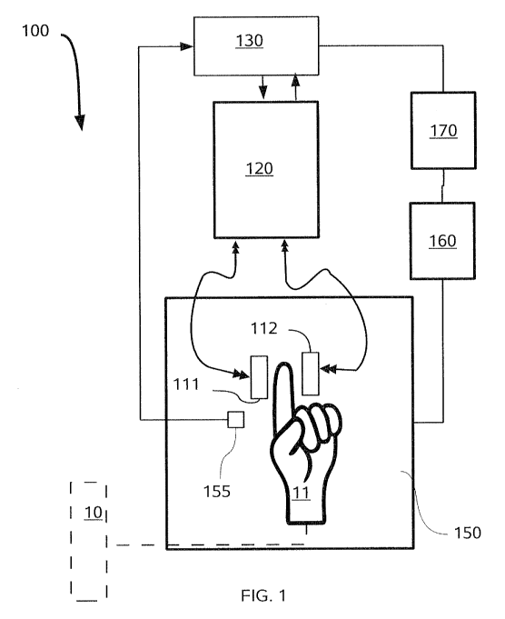
FIG. 1 is a block diagram of an apparatus for conducting the inventive method.
FIG. 2A is a plan view of a preferred embodiment of the antennas shown in FIG.
1,
1.5 whereas FIG. 213 is a fragmented view of an enlarged portion of the
antenna in FIG. 2A.
FIG. 3A is a sectional view of a first embodiment of an antenna supporting
mold. FIG.
3B is an enlarged orthogonal section through the mold of FIG. 3A.. FIG. 3C is
an
enlarged orthogonal view through the mold of FIG. 3A and 3B. FIG. 3D is a
sectional
plan view of another embodiment of an antenna supporting mold.
FIG. 4A is a perspective view of the antenna supporting mold of FIG. 3D, with
the test
subjects hand inserted showing the external connection to the antenna.
FIG. 4B is a second perspective view of the antenna supporting mold of FIG. 4A
with the
subject's hand and fingers removed to show the interior pockets.
FIG. 5 is a plot of the calculated electric field penetration of the antenna
of FIG. 2 in
tissue.
4
CA 02742913 2011-05-04
WO 2010/052660 PCT/IB2009/054914
FIG. 6A is a first perspective view from above a more preferred antenna
supporting mold
that deploys a plurality of antennas on each side of the hand as shown in FIG.
6A and 613,
whereas FIG. 6B is a second perspective view thereof as seen facing the hand
supporting
pocket therein.
FIG. 7A and 7B are plan views of opposite sides of the subjects hand to show
the
optimum placement of a set of 4 of more preferred generally rectangular
antennas.
FIG. 8A cross section elevation through a signal pair of the more preferred
antenna of the
FIG. 6 and 7.
FIG. 8B is a plan view of the winding pattern of the coiled antenna of FIG.
8A.
1.0 FIG. 8C is a fragmented view of an enlarged portion of the antenna in FIG.
8B.
FIG. 9 is the equivalent circuit used to analyze the results of the frequency
scan with the
antennas of FIG. 1 and 2.
FIG. 10A and l0B compare the spectral response of the Sj 1 and S12 parameters
over the
frequency spectrum of 300kHz to 800 MHz. with and without the subject's finger
inserted in the antenna supporting mold of FIG. 3.
FIG. 1 I is a cross-section elevation of an embodiment of an antenna system
that can
effectively deploy 2 pairs of coupled electrodes of different length to sample
roughly the
same projected area of the specimen or tissue.
FIG. 12A is a cross-section elevation of a different embodiment of an antennas
that can
be deployed with an identical antenna to effective deploy 2 pairs of coupled
electrodes of
different length to sample roughly the same projected area of the specimen or
tissue.
FIG. 12B is a plan view of the coupled antennas in FIG. 12A.
FIG. 13A is a cross-section elevation of a further embodiment of an antennas
that can be
deployed with an identical antenna to effective deploy 2 pairs of coupled
electrodes of
different length to sample roughly the same projected area of the specimen or
tissue.
5
CA 02742913 2011-05-04
WO 2010/052660 PCT/IB2009/054914
FIG. 13B is a plan view of the coupled antennas in FIG. 13A.
FIG. 14 is an example of the function 1~4 (1)
FIG. 15 is another antennas transmission spectra of S 12 using the antenna
configuration
in FIG. 8A and 813.
FIG. 16 is flowchart illustrating the steps in a process of calibration of the
device
disclosed herein to non-invasively and continuous monitor blood glucose.
FIG. 17 illustrates an observed correlation of temperature dependence of the
integrated
intensity of selected model circuit parameters, integrated over specific
narrow frequency
ranges.
FIG. 18A compares the predicted versus actual blood glucose concentration of a
subject
using the Q-band parameters in TABLE 1..
FIG. 18B is Clark grid plot of the data such as in FIG. 18A from a plurality
of test
subjects.
FIG. 19A compares the predicted versus actual blood glucose concentration
using the Q-
band parameters in TABLE 2.
FIG. 19B is Clark grid plot of the data such as in FIG. 19A from a plurality
of test
subjects.
FIG. 20 is flowchart illustrating the steps of using the device disclosed.
herein to non-
invasively and continuously monitor blood glucose to after the steps of
calibration of
FIG. 16.
Detailed Description
Referring to FIGS. I through 20, wherein like reference numerals refer to like
components in the various views, there is illustrated therein a new and
improved device
and method of Coupled Antenna Impedance Spectroscopy.
6
CA 02742913 2011-05-04
WO 2010/052660 PCT/IB2009/054914
One embodiment of the inventive apparatus 100 for Coupled Antenna Impedance
Spectroscopy is shown in FIG. I and can be deployed for either in vivo
detection or in
vitro samples. Apparatus 100 deploys a pair of coiled or patch antennas 11 I
and 112 on
opposing sides of a test tube 10 (for in vitro measurement) or a limb 11, such
as a finger,
for in vivo measurements. It should be appreciated that in place of a test
tube, a
continuously flowing dielectric media can be sampled, such as a pipe in a
process stream.
The antennas 111 and 112 are energized via a vector network analyzer (VNA)
120. The
vector network analyzer 120 is in signal communication with a general purpose
computer
130 or microprocessor to perform calculations and calibration processes
described in
further detail below. The same or a different computer or microprocessor can
control the
VNA 120. Further, it is also highly preferred that a thermometer 155 or other
means can
be provided to measure the sample or body temperatures, such as a thermocouple
or a
non-contact infrared thermometer, which is also in signal communication with
the
computer or microprocessor 130.
It should be appreciated that high quality cables and connectors should be
used to
connect the pair of coiled or patch antennas III and 112 to the VNA 120 to
minimize
signal to noise and variability with subject or sample movement.
In initial experiments, the temperature was controlled by placing the antennas
11.1
and 112 along with the sample in a temperature controlled box or low
temperature oven
150, having a fan and heaters (not shown) in signal communication with a relay
box 160.
The relay box 160 was connected to a control box 170. The control box 170 was
in signal
communication with the same computer 130 used for control and data acquisition
of the
VNA 120 signals, as well the temperature measurements from thermocouple 1.55,
placed
at or near the skin of limb or finger 11.
The antenna configuration, shown in part in FIG. 1-4 and 6-8, among others,
when used in vivo is preferably deployed non-invasively. Further, the antennas
are
intended to be energized at a frequency range of about 50 KHz. to I GHz, but
more
preferably from about 200 K.Hz to 900 KHz. as discussed further below, this
results in
relatively deep penetration of the electric field, providing what is believed
to be a more
accurate measurement than prior methods of dielectric spectroscopy, as well as
a means
7
CA 02742913 2011-05-04
WO 2010/052660 PCT/IB2009/054914
for tissue selective measurement of blood glucose. A superior means for the
measurement
of blood glucose concentrations, and is of great benefit to diabetic patients
that require
relatively accurate monitoring of blood glucose through the day to manage
their food.
consumption and administration of insulin.
Further, in contrast to prior art methods of dielectric spectroscopy, the
method
disclosed herein is believed to be capable of providing a higher SNR and wider
spectral
range for glucose and other molecules of interest.
Prior attempts to measure glucose in the human body by non-invasive dielectric
spectroscopy are complicated by two factors the inventive method is believed
to
overcome. First, the conductivity of biological systems creates electrode
polarization with
capacitive antennas. The electrode polarization effect results from the
accumulation of
charge on electrode surfaces and the formation of electrical double layers and
can
overwhelm the characteristic signal. Various methods have been proposed to
correct for
this effect, such as are described by Feldman et al.: Time Domain Dielectric
Spectroscopy of Biological Systems, IEEE Transactions on Dielectrics and
Electrical
Insulation Vol.. 10, No. 5; October 2003, which is incorporated herein by
reference.
Further, according to A. Caduff et al. in "Non-invasive glucose monitoring in
patients with diabetes: A novel system based on impedance spectroscopy ",
Biosensors
and Bioelectronics 22 (2006) 598-604, which is incorporated herein by
reference, among
others, have noted that dielectric spectroscopy does not measure blood glucose
directly,
but rather the effect of hyper and hypoglycemic excursions that lead to
changes in the
electrolyte balance in blood, cells and interstitial fluid (ISF), and is thus
an indirect
measurement. This occurs in part because the electric field of prior art
capacitive sensors
only penetrates the skin and the closest underlying tissues to a depth of
about 1-2 mm.
In contrast, the inventive technique disclosed herein is believed capable of
producing more accurate and reproducible results because it not only avoids
electrode
polarization, but also probes much deeper tissues.
FIG. 2 illustrates in plan view the configuration of the coiled or patch.
antennas
111 or 112 having a generally spiral configuration. In this spiral
configuration there are
8
CA 02742913 2011-05-04
WO 2010/052660 PCT/IB2009/054914
multiple wraps or winding of a continuous line or conductive stripe around
itself in the
same plane with at least four or more turns at ends or corners such that the
overall shape
can be square, rectangular, round, oval or any combination. Further, the
topography or
shape of the patch antenna deployed herein can be in the form of a loop, coil,
spiral or
serpentine configuration, as well as combinations of the above. Typically, as
illustrated in
FIG. 2A and 2B, the stripe or ribbon portion of the coiled antennas 111 or 112
has a
width (W) of about 100 microns, a center to center (C-C) between adjacent
lines of about
200 microns and generally at least about of turns so that a section across the
entire
antenna will bisect about 40 of these lines. The antennas can be printed on
general
1.0 purpose printed circuit boards, or flexible film such as Kapton and the
like, shown as
801 in FIG. 8A . Currently, such antennas are fabricated on a PCB material
designated
TMMA 10/1 available from Rogers Corporation, which has a dielectric constant,
F , of
about 10.8 and a minimum thickness of 0.38 mm. As shown in FIG. 2A, for the
generally
square patch antenna, the wrapping starts around a square with a width (wl) of
about 200
microns. Thus the total antenna length is about 70 cm.
The penetration depth of a patch antenna depends both on frequency and.
antenna
configuration. However, for in vivo application penetration depth is primary
limited by
absorption of electromagnetic radiation by water molecules, and is thus also
frequency
dependent. Generally, the losses of any given antenna increases as the
frequency exceeds
400 MHz, as has been reported in "A. 31.5 GHz Patch Antenna Design for Medical
Implants ", Ahmed et al., International Journal of Antennas and Propagation,
Volume
2008, which is incorporated herein by reference. It has also been reported by
Kim et al.
"Implanted Antennas Inside a Human Body: Simulations, Designs, and
Characterizations ", IEEE TRANSACTIONS ON MICROWAVE THEORYAND
TECHNIQUES, VOL. 52, NO. 8, AUGUST 2004, that for a particular antenna
energized
at 400 MHz, a transmitted communication signals can penetrate 20 cm. Over the
frequency range 30 MHz -800 MHz the penetration range corresponding to a loss
of 70
dB was in the range of about 5-10 cm. It should be noted that such losses have
been of
interest to those designing patch antennas for the wireless communication
between
implanted medical devices and external monitors or control systems.
9
CA 02742913 2011-05-04
WO 2010/052660 PCT/IB2009/054914
The penetration range of the antenna 111 and 112 in FIG. 2 have been modeled
assuming different properties for underlying tissue, which indicate a useful
penetration
range of at least about 3-5 cm at the very low frequencies of about 300 KHz to
about
400M14.z. Hence, the patch antennas Ill and 112 can be employed on opposite
sides of a
limb or organ, more directly measure glucose concentrations.
One result of such a simulation of the electromagnetic field penetration
within the
tissue for the antennas of FIG. 2 is shown in FIG. 5. FIG. 5 is a perspective
view of the
calculated potential field variation of intensity in the x-y plane is plotted
in units of volts,
the voltage corresponding to the intensity level of the cross=hatching pattern
per the
1.0 legend bar to the right. The EM field was calculated at 2MHz. At this
frequency the skin
dielectric constant was taken er = 900 and conductivity rr = 0.12 S/rn. In
this
configuration the patch antenna 111 was connected to the core of the coaxial
cable. The
dashed lines grid lines are 5 mm apart with the 1 cm wide square electrode
being
disposed in the x-z plane having the general outer dimensions shown by the
rectangle
labeled 111 .'.While the intensity is a maximum of about 1.4V within. 3-5 mm
from the
electrode, the power only drops to about 0.4 V within about 1-2 cm. Thus the
general
penetration depth of this antenna is in the range 3-5 cm at this very low
frequency.
Measurement of glucose are then made by the process of first placing the
antennas 1.11 and 112 on skin, the antennas are then sequentially energized in
by the
VNA 120 in the frequency scanning mode, with both the transmitted and
reflected power
measured as the frequency range of each antenna is swept. The frequency sweep
speed
has an impact on the SIN ratio in the measurements, with the higher speed
resulting in a
lower is S/N ratio. In the current mode of the operation VNA spectrum sampling
rate is
about 30 sec. of 800 MHz. During this process raw data are acquired to
calculate four
signal propagation parameters which vary at least somewhat with frequency for
determining the concentration of the molecular species of interest.
While the patch antenna structure 111 and 112 have a penetration depth and
intensity that is highly dependent on its structure, as well as the signal
interaction with the
dielectric medium being probed, this depth is much greater than the prior art
methods, so
it is not necessary to place the antennas directly on the skin. Thus, in a
more preferred
1.0
CA 02742913 2011-05-04
WO 2010/052660 PCT/IB2009/054914
method of using the inventive antenna structure, a molded carrier or support
301 contains
and encases the antennas 111 and 112. As the supporting mold 301 is also
sculpted or
cast to shape of the finger 1 1, or other appendage, to reproducibly surround
the limb or
organ portion being probed the placement of the antennas 111 and 112 provides
a
reproducible spacing from the subject's skin, as the mold 301 fits snugly
around the
finger. Variations of such antenna supporting molds 301 are shown in FIG. 3A-
D, with
the actual mold used to generate the experimental data shown in FIG. I OA and
FIG. IOB.
The antenna supporting mold 301 is optionally made of gypsum or another
material that
is reasonably transparent, that is having low signal attenuation in the range
of about 10 to
900 MHz. It is more preferably to use a plastic cast forming compound, such as
ORFIT
Classic, which is available from ORFIT Industries of Wijnegem, Belgium.
In such supporting structure each antennas is wound in a common plane so that
antennas in the pair can be placed with their respective common planes
parallel and
spaced apart. However, depending on the portion of the organism that is
sampled, the
antennas in the at least one pair can be placed adjacent to each other.
In further contrast to the prior art, it was further discovered that it is
undesirable to
place the antennas in direct contact with the skin. As the tissue areas with
higher electric
field have more influence on the S-parameters than with the weaker electric
field, electric
field for antennas placed on skin is maximum at the skin layer. Therefore, the
skin layer
may have a dominant influence on S-parameters. The outer skin layer is a
source of
systematic error for VNA data since it is influenced by the varying
environmental
conditions such as temperature and humidity. Therefore, it is desirable to
reduce its
influence on the measurement. One way to do it is to separate antennas from
skin by
some layer of dielectric material. Another, but less desirable approach
includes creation
of holder that maintains constant environmental conditions (incubator).
Although the current spacing away from the skin (as shown by the thickness of
spacer 802 in FIG. 8A) is by about I mm, it is expected that a dielectric
spacer with a
thickness of 300 m to about 4-5 mm will be sufficient. The spacing media can
be the
above cast forming compound from ORFIT, or a comparable dielectric medium.
11
CA 02742913 2011-05-04
WO 2010/052660 PCT/IB2009/054914
Thus, spacing of the antennas away from the skin appears to achieve a better
correlation between actual blood glucose, such as measured by the YSI method,
and then
inventive system for several reasons. This is potentially due to the
insensitivity to the skin
conditions, that is contact, moisture, pressure and the like, but also may
reflect
representative sampling of the tissue. It is believed that prior methods of
dielectric
spectroscopy that place the antenna on the skin sample largely the
interstitial tissue, while
the inventive method is more capable of sampling a larger portion of the
arterial and
venous blood of the patient/subject.
In one embodiment, the antenna supporting mold 301 in FIG. 3A-C surrounds a
single finger, placing a comparably sized antenna 1.11 and 112 on opposite
sides of the
finger as shown in the section in FIG. 3C. In contrast, the antenna supporting
mold 301 in
FIG. 3D, surrounds and immobilizes all the fingers, like a rigid glove, but
still disposes
the comparably sized antenna 111 and 112 on opposite sides of the finger as
shown in the
section in FIG. 3C
FIG. 4A shows an exterior perspective view the antenna supporting mold 301 of
FIG. 3D, which is adapted (as shown in FIG. 4B) to retain each finger in its
own pocket it
is adapted to receive the entire hand. The cable 401 connects the antenna 111.
to the VNA
at external connector 302. It is believed that by more completely immobilizing
the fingers
during measurements the antenna position is less likely to move or creep when
the entire
hand is in the mold, which will thus improve accuracy and the precision of
measurements. Thus, the antenna supporting mold 301 is preferably custom cast
for each
subject or patient, but may also be provided in a range of generic sizes such
as for gloves.
Further, the thermocouple 155 may optionally also be encased into the mold
and/or
deployed at the internal surface of the mold to measure the skin temperature.
It should be appreciated that in addition to the antenna pair being deployed
on
opposite sides of body portion or appendage, the pair can also be placed
adjacent to each
other on the same side of the skin or appendage. Accordingly, it is expected
that the patch
antennas deployed in the inventive method will yield more reproducible and
systemic
results when properly calibrated for the subject/patient.
12
CA 02742913 2011-05-04
WO 2010/052660 PCT/IB2009/054914
Further, alternative positions or appendages for placement of the antenna are
optionally the patient's ear lobe, forearm, wrist, head or leg. In additional
it may be
preferable to place the inventive antenna system either across the abdominal
cavity, as for
example to more accurately measure blood glucose within an organ such as the
pancreas,
as well as on adjacent locations or in closer proximity to larger blood veins
or arteries.
Thus, for example depending on the body portion used, a particular
configuration might
be more preferred for patient that desires or requires more continuous
monitoring. it
should be appreciated from the following discussion that the optimum antennas
configurations for different portions of the body may be different from what
is currently
the preferred configuration for making continuous measurement from the hands
and
finger as illustrated in FIG.6-8 , as for example with respect to the size and
number of the
antennas deployed. In the embodiment of FIG. 6-8, the supporting mold encased
a
sufficient portions of the patients/subjects hand to place 2 pairs of antennas
in signal
communication.
In the frequency scan described above with a single antenna pair the vector
network analyzer (VNA) 120 yields four main signal propagation parameters:
S22, S11
that represent reflection coefficients and S21, S12 that represent
transmission coefficients.
In the models that follow, each S-parameter is a function of time and
frequency,
where
Sij =S(cv.,T1) (1.1) and
T - time
w=2,rf
f - frequency
The reflection and transmission coefficients S;j can be transformed to four
impedance parameters Y11, Y12, Y21, and Y22 by the following formulas:
13
CA 02742913 2011-05-04
WO 2010/052660 PCT/IB2009/054914
S = Sa.1 S12
S21 s22
11 0
I=
0 1
V= I-S
I+S (1 2)
Y =Y
Zo
Y = Y1 Y2
Y21 Y22
Z = Y-1
Where Z. 50[f)] is the reference impedance.
It is possible to model the antennas of FIG. 2 and the intervening sample or
dielectric medium by the electric circuit shown in FIG. 9, from which we
extract the
additional parameters by the following formulas:
_ IM(Y11) (1.3)
Re(Y1)
R1 =Re 1. (1.4)
Y1 +Y21
C1=ImY'+Y21,where co=27cf (1.5)
Co
R2 =Re (1.6)
Y22 +Y2
C2 = Im Y22 +Y2 , where co = 27rf (1.7)
I
R=Re(Z),where Z=-- (1.8)
Yl
Im(z)
L where Z and w=2rf (1.9)
co Y21
As shown for selected parameters S11 and S12 in FIG.IOA and IOB, when no
sample is present between the antennas, strong resonances are seen. wherein
the
14
CA 02742913 2011-05-04
WO 2010/052660 PCT/IB2009/054914
transmission or reflection coefficients are particularly high at specific
frequencies that
vary periodically from about 0 to 800 MHz.
In comparison FIG. 1 OA and FIG. 10B also show parameters S I] and S22 when
the
test subjects finger is inserted in the probe region between the antennas. The
resonance
and spectral characteristics change dramatically due to the interaction with
the molecule
species in the tissue.
Although most of the cross variation in the spectral intensity of any of the
Sij
parameters is dominated by the antennas resonance pattern, it has been
discovered
through extensive statistical analysis that small portions of the spectra will
correlate very
1.0 well a patient's blood glucose concentrations.
It has also been discovered that more accurate and reproducible measurements
of
blood glucose can be obtaining using 4 antennas in a slightly different finger
and hand
mold, which is now illustrated in FIG. 6A and 6B. The antennas supporting mold
301. has
four external connectors 302 to the antennas.
It was also discovered that further improvements were obtained when the
antennas had a rectangular shape as shown in FIG. 8 and are oriented around
the hand as
shown in FIG. 7. It is thus currently preferred that the antenna 111/112 have
an aspect
ratio of 2:1. It should also be noted that superior results were obtained when
the longer
axis of the rectangular antenna is oriented perpendicular to the fingers, as
shown in FIG.
7A and 7B.
The antennas pairs 111a1I 12a and I I lb/I l2b in FIG. 8A and 8B have external
dimensions of about I cm by 2 cm with the flat antenna coil having a width of
about 75
j.m being spaced apart from the adjacent winding by about 125 microns (for a
200
micron center to center spacing) to provide a total antennas length of about
130 cm,
having about 20 to 25 turns. The individual antennas are simultaneously
labeled I (11 la),
2(112a), 3(1.1 lb) and 4 (112b) to comport with the mathematical treatments
that follow.
It should now be appreciated that the deployment of four antennas permitted
the
measurement and analysis of at least 10 basic S-parameters, that is 4
reflection
coefficients (S11, S22, S33, S44) and 6 transmission coefficients (S12,
S13,S14, S23, S24, 532,
CA 02742913 2011-05-04
WO 2010/052660 PCT/IB2009/054914
S34). It should be understood that for reflection and transmission coefficient
or parameter,
the experimental amplitude and phase may be used together or separately in the
data
analysis and extraction that follow. It should also be noted that transmission
coefficient
S13 and S24 refer to adjacent antennas on the same side of the hand in which
the
electromagnetic radiation extends through the tissue, but is not transmitted
perpendicular
to the plane of the coiled antenna. In contrast, transmission coefficient S12
and S34 refer to
antennas on the direct opposite side of the hand in which the electromagnetic
radiation
extends through the tissue and is transmitted perpendicular to the plane of
the coiled
antenna. However, transmission coefficient S14 and S23 refer to antennas on
the opposite
side of the hand that are not directly opposite each other, in which the
electromagnetic
radiation extends through the tissue and is not transmitted perpendicular to
the plane of
the coiled antenna.
Not wishing to be bound by theory, it is currently believed that the
resonances
characteristics of the novel antenna designs have several distinct advantages
over prior
methods of dielectric spectroscopy to measure or estimate the in-vivo
availability glucose
in a patient. it is also believed that the antennas designs have distinct
advantages in
measuring glucose and other molecule in in-vitro.
Deploying antennas that generate deeply penetrating electromagnetic in the
most
desired range of about 100 to 800 MHz (0.1 to 0.8 GHz) provided more
opportunity for
the discovery of particular narrow frequency bands that gave good correlations
with
blood glucose and where also relatively insensitive to sources of error that
have hindered
the advance of earlier approaches to non- invasive measurements of blood
glucose.
This was particularly the case when the resonant characteristics of antennas
111/112 are tuned for the media of interest such that the loss in transmission
is generally
less than -50db, but more preferably less than about -30dB between about 1.00
MHz and
800 MHz, but more preferably between about] MHz to 500 Mhz.
For detecting glucose in living tissue using the inventive method we have
discovered it is preferable that the coiled antenna have a first resonance
below about 100
MI-1z, but more preferable below about 50 MHz.
16
CA 02742913 2011-05-04
WO 2010/052660 PCT/IB2009/054914
It has also been discovered that it is preferable that the coiled antennas
also
provide a characteristic zone of flat transmission coefficients in the media
of interest over
a range of about 200 MHz in which the transmission varies by less than about
NO, but
more preferably less than about 20 dB, and the loss in transmission also less
than -50db,
but more preferably less than about -30dB This range is typically up scale,
higher
wavelength that the first resonant frequency.
As the frequency of the first resonance of microwave antennas is inversely
proportion to the antenna length, meeting this requirement posed a particular
challenge to
a conflicting need to make the antennas as small as possible for patient
convenience and
obtaining local measurements. However, both these requirements could be met by
keeping the antennas width and spacing as narrow as possible and using
multiple folds to
obtain a long length, as for example the antenna I I 1 in FIG. 8 has at least
15 to 20 turns.
This is in dramatic contrast to the prior art work in dielectric spectroscopy
in which the
antennas are in fact tuned to have a first resonance above 1GHz to measure the
shift in
resonant frequency that is was observed by McClung (MS Thesis M. J. McClung,
titled
"Calibration Methodology for a Microwave Non-Invasive Glucose Sensor", Baylor
University, Department of Electrical and Computer Engineering 2008). The
antennas
disclosed therein has only 3 widely spaced turns, and the receiving antenna is
a pair of
strips placed on the same side of the thumb as this short coiled antenna.
Therefore, the
frequency shift measured by McClung is not actually in transmission, but is in
fact a
reflection coefficient.
The apparatus and method disclosed herein is expected to be more accurate than
other methods of dielectric spectroscopy for several reasons. First, the
prominent
resonance peaks provide a stronger interaction with the dielectric relaxation
properties of
glucose and are less affected by the absorption from. other molecules. This
method thus
appears to overcome electrode polarization effects noted in the prior art.
Further, the
inventive method is likely to be more representative of the bio-availability
of glucose, as
the measurement is more than skin deep.
17
CA 02742913 2011-05-04
WO 2010/052660 PCT/IB2009/054914
Further, such deeper sampling of tissue by the inventive method is likely to
produce more temporally stable results, being less sensitive to skin
temperature and other
skin conditions such as dirt, contamination and moisture and the like.
As the dielectric spectra is from a resonant system. that will inherently vary
with
the electrode placement and physiology of each user or patient, it is not
possible to
precisely define universal lines or ranges of the spectra that are applicable
to all patient's
or test subjects.
However, it has been discovered that for each user/patient with a particular
antenna combination disposed in signal communication across a particular body
part or
organ it is possible to identify the spectral ranges that correlate well with
actual blood
glucose concentration.
Accordingly, another aspect of the invention is a process for discovering such
portions of the spectrum for each patient for use as a means of continuously
and non-
invasively accurately determining the blood glucose levels.
Yet another aspect of the invention are methods to develop the most robust
means
of continuously and non-invasively accurately determining the blood glucose
levels.
It should be appreciated that such methods require the collection of data from
a
patient/subject equipped with the inventive antenna combination over a period
that is
sufficiently long to record a range actual blood glucose levels that is at
least close to
those likely to occur in real world conditions.
In the simplest mode of deployment such a device can warn the patient to
better
control dangerous excursions through the time administration of a source of
glucose,
generally by eating a healthy meal, or insulin.
In a more advanced mode of deployment such a device is anticipated to guide a
patient to better control the blood glucose level within a narrower range to
minimize the
longer term and generally debilitating effects of diabetes, such as diabetic
retinopathy, a
proneness to infections and the like.
18
CA 02742913 2011-05-04
WO 2010/052660 PCT/IB2009/054914
It is also anticipated that the potential for accurate and. continuous
measurement
will enable integration into an artificial pancreas that in a closed feedback
loop to a pump
that can continuously provide insulin in response to the blood glucose levels.
According, the currently preferred methods of such embodiments are disclosed
in
the experimental description in the paragraphs that follow.
Experimental Method and Results
Using the antennas and antennas supporting molds of FIG. 6-8, VNA spectra were
collected in continuous mode using a commercial data acquisition system and
program
(LabView). Typically VNA spectra consist of 1600 measurement points spanned
linearly
from IMHz till 800 MHz. A four ports model VNA (such as for example from
Agilent
Technologies, Santa Clara, CA) can provide sixteen spectra Sjj ,(1j = 1.,-.,4}
of which
four spectra S= j corresponds to the reflection parameters , while the others
twelve
SE,j, % * J corresponds to the transmission parameters. Since Sj,j have to be
equal to 53,2
for reciprocal media such as human tissue, there are ten parameters altogether
Sep, t I
1.5 corresponding to the upper triangular matrix S~j.
The acquisition time of sixteen VNA spectra is about 30 sec. The SNR of the
VNA in transmission mode is about -120 dB, while the signal level in
transmission mode
is in the range -30 dB to -70 dB, depending on frequency.
Each of VNA spectra Sh, collected in continuous mode with a sample time r can
be organized into the N x M matrix
S.s w , t.) {m - 1 ,2, ... M and n - 1,2,.... NJ (2.0)
Where M = 1600 is the number of frequency points and N is the number of
collected spectra.
19
CA 02742913 2011-05-04
WO 2010/052660 PCT/IB2009/054914
Data cross-correlation analysis
Fixing a frequencyk in Se 1s,) , we obtain a function of time
fk(t) = S (Wk, 0) (indexes i, j are omitted) ; thus we can consider a
correlation
function
p(k,1) = corr(ff t),f(t) ), k,I = 1,...,M (2.1)
As follows from the Equation 2.1, p(k,1) is the matrix of size M X M with
values ofp(k,.1) from -1 to 1.
Correlelogram Analysis
Assume that we have a target function 9(t): (such as glucose concentration)
measured at same set of the sampling times tk, k = 1,2 .. , K . This set can
be a subset
of the set of all times tom, _ ,2, =..
From two functions g(t)) and Scr1(w,t), we can build a correlation product
over
time.
rt1 w) = Corr( S,,1(&, t), ,g(t)' ), k, l (2.2)
The correlation product is the correlation function of the measured reflection
and
transmission coefficients Sid to at least a portion of the measured blood
glucose
concentration. In the case of determining the concentration of other molecules
of interest,
the concentration of the other molecules would be used. The function rz1(w)`
reflects
degree of similarity between the data St,1 ,t) and the target function at
given frequency.
As definition (2.1) suggests, the module of the functions r,.3 is less than or
equal to one.
This correlation product when derived using the glucose values g(t) that vary
widely, as can be obtained in an oral glucose tolerance test (OGTT), provides
a means to
identify spectral ranges in which the measured reflection or transmission
coefficient S=1
correlated highly with the actual glucose concentration.
CA 02742913 2011-05-04
WO 2010/052660 PCT/IB2009/054914
FIG. 1.4 represents a typical plot of the correlation product function r24 M.
The
solid curve in this figure shows correlation versus frequency with all the
values of
glucose concentration obtained during the OGTT, while the partially dashed
curve shows
the result of the same calculation using half the data. In this experiment
this data was
from before the large rise and fall of blood glucose induced by the OGTT. It
is currently
believed preferable to generate these curves from an OGTT using the broad peak
in
glucose as the partial data set, to determine if it will also predict the
periods before and
after when the blood glucose level are more stable. From definition (2.1) it
follows that if
S_j are the smooth functions of frequency, then rzl are smooth functions as
well.
1.0 However, it is also believed possible to perform the above calculation
using different
combinations of partial data sets.
The example above shows that the behavior of the function r24 (w) is
relatively
smooth in the high frequency range of approximately from 350 MHz till 800 MHz,
while
the variation of this function is more considerable at low frequencies (below
200 MHz ).
By relatively smooth we mean a smaller oy jla(to) than in the region of about
10 MHz
to about 200 MHz where the narrow frequency bands corresponds to the antenna
radiation pattern. It should now be appreciated that the most preferred
antennas though
having multiple resonances below 200 MHz frequency range, such that the
response
above 200 MHz is still relatively flat. FIG. 12 illustrates this desirable
spectral response
for S12 for the antennas of FIG. 8 when deployed around the palm of the test
subjects that
is just below the fingers, as illustrated in FIG. 7A and 7B.
It is more preferable to deploy the frequency range where the behavior of the
functions raj is smooth to define the frequency or Q-bands, where the values
of Jr,, I is
more than some threshold value.
While experiments to date have used antennas of essentially identical sizes
and
patterns (as is limited by fabrication technology), when using either a total
of 2 or 4
antennas, it may also be desirable to deploy 2 different pairs of 2 identical
antennas,
wherein the resonace characteristics of each antenna pair is different. Three
different
embodiments of this aspect of the invention are illustrated in FIG. 11-13.
This will allow
the expansion of the optimum usefull spectral range, such as where the
transmission is
21
CA 02742913 2011-05-04
WO 2010/052660 PCT/IB2009/054914
relatively flat and sufficiently high, where one pair of the antennas is
optimized for a first
spectral range that has at least a portion below higher optimized spectral
range of the
other antenna pair. The antenna pairs can occupy the same area by changing the
spacing
on one antennas coil, keeping the line width identical or varying in different
portions. In
such a case, it may also be preferable to provide some overlap in a spectral
range where
usefull information cn be obtained for an aditional cross-correlation or
selection of the Q-
bands.
Different pairs of antennas can be arranged in several ways in addition to
configuration illustrated in FIG. 7. For example antennas I 1 la and I I lb in
the top half of
antenna supporting mold 301 are superimposed laterally as shown in FIG. 11,
but spaced
apart slightly on different side of a portions of a PCB or flexible carrier
tape 801, in
which the spacing is the tape or PCB thickness, as are the other antennas of
the pair 112a
and I I2b. Thus, antennas in pair I I la/I 12a can be longer to have a shorter
or lower first
resonance frequency while the antennas in pair 11. lb/I.12b can be shorter to
have longer
or larger first resonance frequency. The difference in spacing between
antennas pairs
I I l a/1 ].2a and I I I b/1 12b through the sample or tissue is twice the
thickness of the PCB
or flexible carrier tape 801.
Alternatively, as shown in FIG. 12A and 12B, antenna pairs I I Ia/112a and
I 1 lb/I 12b can also have the same spacing through the sample or tissue
byproviding
adjacent wrapped conductive traces on the same board or tape 801, with the
shorter
antenna I I lb terminating first and having an external contact (shown in FIG.
11-13 as
vertical lines) through a via in the PCB or tape 801.The shorter antennas I I
Ib in the plan
view in FIG. 12B is shown in a dashed line.
In the embodiment shown in FIG. 13, Antenna I I la is longer, as it uses a
portion
of the inner and shorter antenna I I Ib via a switch 1301, so there is only
one spiral trace
with the switch 1.301 being in the spiral trace.
In addition to expanding the useful] spectral range, having such overlapping
plural
antenna pairs also provide a different penetration depth in the tissue for
each pair to
permit a continous comparsion of the both glucose in tissue closer to the skin
against
22
CA 02742913 2011-05-04
WO 2010/052660 PCT/IB2009/054914
what might be much deeper venous and arterial tissue. As the glucose in tissue
closer to
the skin is more likely to represent intersticial tissue, this may provide
greater
predictability of trends in glucose in the pateient/test subject, as well as
for greater
accuracy of measurement.
Thus, after the acquisition of the different sets of signal propagation
parameters
S j, the entire calibration process can be carried out fully automatically by
a
microprocessor or other computing means by first aquiring the data, that is Sj
(gym, t" J,
then calculating at least 2 sets of r;1 via the equation below using a
complete and partial
set of independently measured blood glusoce values. Further, the comparsion of
of these
at least two sets can be an automated process as described below.
Extracting the Q-bands
The final predictive equation for blood glucose concentration requires the
indentification of frequency interval or bands of the spectral response of any
Sij
parameterin which model function and the measured glucose concentration are
well
correlated. This can be exprtessed mathematically as the set of all
frequencies bands
I such that the inequality (2.3) holds are called Q-bands.
V 60 -3 k, zj , Fri, ( ) I c (2.3)
Where c is a threshold value. That is, a set of Q-bands are selected where
absolute
value of rid is greater than or equal. to a threshold value, C, from some band
width
represented by Wk to col.This correlation threshold, C, is preferably at least
about 0.75.
Ideally such Q-bands should not overlap with each other. Thus, within each Q-
band the
correlation ofS¾:5 and the target function g(t) is more than the treshold
value. Fig. 14
shows example of three Q-bands where the correlation with the target function
(partial
glucose data) and S24 are more than 0.75, as higlighted by the broken circles
1401.
For each Q-band [ k,~= (indexes iJ here corresponds to the indexes of the
S,f, one can extract a feature function by averaging Sr, (ws, t) over the
interval k, j.
(S (w, t)) (2.5)
23
CA 02742913 2011-05-04
WO 2010/052660 PCT/IB2009/054914
The definition (2.3) insures that correlatin of the fkt;rj.(t) and the target
function
g(t)will be not less that the threshold value c.
The above equations thus provide an algorithm for generating feature functions
from the set of Q-bands that are highly correlated with blood glucose
concentration of the
patient, g(t).
Thus, a preferred mode of using the dual antenna appartus 100 is to preform
the
previously described set of calculations on each pateint during an initial
OGTT, or similar
diagnostic processure that provides an opportunity to collect spectral data
during a
reasonably large excursion in blood glucose concentration when the actual
glucose
concentration is known very accurately by an independent method. This provides
a set of
candidate S;j parameters, each at one or more selected Q-bands, to derive a
predictive
formula for calculating the pateint blood glucose concentration continuesly.
Such sets
may range from 10 to 30 potential Q-bands. The analysis to date of about a
dozen
individuals has revealed a general trend of idnetifying about 1 to 4 Q-bands
for 7 to 10 of
the Sij paramters.
A final predictive equation can be derived from the feature functions of
equation
(2.5) by a wide variety of known regression techniques for each of the feature
functions,
which are found by integrating the value of the Q-band parameters selected as
candidates
in the previous set of 10 to 30 Q-bands.
The correlation coefficient for each of the feature function corresponding to
specific Sij parameters over the Q-band frquency ranges can then be compared
so that
only the most highly correlated feature functions are used in the final
predictive equation.
However, it has also been found preferable to use additonal criteria for
selecting a limited
set of Sij parameters to derive and select the feature functions used in the
final predictive
formula. Among these criteria it is preferred to compare the temporal
stability of the Q-
band over a time period when the blood glucose is quite stable. Thus, in this
case rather
than scanning over the entire band width used to discover Q-bands, just the
narrow Q-
band would be repeatedly scanned. Such scans can be much faster than 30
seconds, and
can be repeaded as needed to compare their tempral stability as well as the
signal to noise
24
CA 02742913 2011-05-04
WO 2010/052660 PCT/IB2009/054914
ratio. In this manner, the Q-bands used to derive the final predictive
equation can be
selected based on their having the highest signal to noise ratio.
It is additionally preferable to also or alternatively select the Sid /Q-band
parameters that are relatively insensitve to external effects, for example
temperature and
well as precise positioning in the antenna holder 301. The exploration of a
correlation
with temperature is easily perfomed for each Q-band if there is sufficient
temperature
excursion either during or after the initial data collection step when the
device 100 also
includes a thermocoupe of non-contact IR thermometer.
FIG. 17 illustrates an observed correlations of temperature dependence of the
integrated intensity of selected model circuit parameters, integrated over
specific narrow
frequency ranges showing a strong correlation with temperature without a
strong
dependence on blood glucose. As opposed to prior art methods of dielectric
spectroscopy
based glucometry, if it should be necessary to empirically correct for
temperature
variation, it is likely that the need for temperature sensors to estimate the
actual
temperature of the sampled skin depth can be avoided, as the measurement
system itself
provides a means to measure the actual temperature of the tissue within the
depth being
sampled. Thus, inventive method is likely to provide an improved means to
calibrate and
correct for temperature variation of the subject and the environmental effects
thereon.
As to the sensitivity of candidatate Q-band to position of the holder device
on the
patient/test subject, it has been discovered that more reproducible results
are obtained by
first acquiring the spectra over the candidate Q -bands repeated times in
sequence and
then calculating the stardard diviation of each Sij value as integrated over
the Q-band.
width to select Q-bands of lower stardard deviation.
Ideally, a limited selection of all possible Sij and associated band regions
parameters are selected for regression analysis. While various forms of
Chemometrics
techniques for multivariable regression can be performed on a plurality of Sia
parameters,
as the objective of the present invention is to provide a diagnostic tool, it
is currently
preferred that a single S;j parameter be derived by linear regression that
provides a good
fit to the measured glucose values in the ranges of clinical importance. Thus,
another
CA 02742913 2011-05-04
WO 2010/052660 PCT/IB2009/054914
criteria for selecting the most appropriate Q-band is based on the lowest
error in the
regression analysis.
The flow chart in FIG. 16 summarizes the above measurement and calibration
steps, including the other criteria for Q-band selection described more fully
below.
Another aspect of the calibration process is to select the optimum S,j
parameter
that correlates best and most robustly with the measured blood glucose
concentration as
measured by convention methods in either the hospital or clinical setting, or
those
routinely used by diabetic patients.
Part of such optimization is insuring that particular parameter is robust with
respect to a minimum noise and errors that occur repeated removal and
insertion of the
hand in the antenna supporting mold 301, or alternatively with respect to any
other
fixture that holds and support the pair of at least 2 antennas if deployed to
measure blood
glucose on another organ or part of the body than the hand.
Clinical trials have been conducted using the technqiues described above. The
predicted blood glucose level from the trial is compared in the Clark grid in
FIG. 18 and
19. The most productive Q-bands identified in a relatively small subset of
patients/subjects are listed in Tables 1 and 2 below. In these tables the
first column
identifies the signal parameter, which is intensity, when not reffered to
specifically with
the subscript "ph" for phase. The second column is the channel count range for
this band,
while the third column is the equivalent frequency range in MHz. for the Q-
band. The
fourth column is the correlation coeficient with the actual blood glucose
measurement, as
made by the YSI method. Tabel I contains an extra column between the fifth and
last
column showing the standard deviation in re-positioning error. The fifth
column is the
correlation coefficient with temperature. The column to the farthest right in
the table is
the signal to noise ratio of the Q-band that was calculated as the STD of the
reposition
error divided by the signal amplitude.
Table I refers to tests taken when the subjects were subjected to an OGTT to
produce a hyperglycemic state, with the glucose concenstration ranging from
about 100
to 350 mg/dl. Table 2 refers to tests taken when the subjects were
administered a very
26
CA 02742913 2011-05-04
WO 2010/052660 PCT/IB2009/054914
controlled dose of insulin to lower the insulin levels to the hypoglycemic
state, with the
blood glucose levels ranging from 50 to 175 mg/dl. The predicative result of
the 10 "best"
Q-bands in the table were then averaged after linear (uni-variant) regression
to provide
the final linear predictive equation as described above, and are plotted as
the solid line
"Regression and Prediction" against the blood glucose measured by YSI , which
is the
wider partially broken line.
TABLE I
MCalibratiu
vs c1CCa1btat on vs Deviation 5tgta1
Type POs" Frequency Qucose Temperature Peposidon N se
S11db [263,268] [156.17,158.57]MHz -91.0066 -55.7116 stdr.3 3.783294
512 [959, 969] [491.33,496.14]M -92.6765 -58.7426 stdr 5.1 5.118336
512 [1061,11141 [540.44, 565.97] MHz -91.9049 -63.3541 stdr.1 2.688731
S12 [1221,1263] [617.49, 637.72]MHz -91.1643 -67.4358 stdr 5 3.150891
513 [146,146] [99.82 99.82]MHz 92.72564 59.04019 stdrcg 3.968204
544db [661,6611 [347.8.2 347 82]MHz -92.147 -67.2053 stdr 3.2 5.384147
Suit E3,31 [30.94 30.961 MHz 91.26106 74.28393 stdrd 5431) 9 5.145149
Si4ph [146,146] [99.82, 99.827 MHz 93.05493 60.93371 stdr65883.6 4.360244
S22ph [854, 8581 [440.74 442.691 MHz 91.16346 74.646(8 stdr=22 4.178983
SZ3ph [1518, 1537] [760.51, 769.667 MHz 93.21846 71.18909 stdr [058970.7
2.982021
S34ph [679,685] [366.49, 359.38]Milt 92.97443 71.50172 stdr-5134. 1 2.594565
S34ph [142146] [97.9,99.82]MM -95.6516 -58.746 stdr446575.3 3.933949
S44ph [1287,1288] [649.27, 649 761 MHz -91.7352 -62.9503 stdr=f1.7 7.108362
The average of the data from the Q-bands in the above Table 1 is plotted
against
the actual glucose concentration in FIG. 18A, of which the corresponding Clark
grid is
shown in FIG. 18B for a group of patients.
27
CA 02742913 2011-05-04
WO 2010/052660 PCT/IB2009/054914
TABLE 2
CLCalibration CX Calibration vs Signal
Type Position Frequency vs Glucose Temperature Noise
S 11db [662, 664] [348.31,349.27] MHz 85.10891 5171584 5.633072
S13 [2'51, 2791 115039, 163.87]MHz 85.00375 44.3D454 4.421911
S22db [791, 800] [410.43, 414.76] MHz 89.91409 55.29072 5.216331
S22db [1284,12841 [647.83, 647.831MHz -85.4515 -21.6692 4.059745
S13ph [248,254] [148.94,151.831MHz -86.5356 -55.13 5.396657
S22ph [316,3161 1181.69,181.691M1k 45.8894 -54.4095 4.201929
S24ph [121,1271 [87.79, 9068] MHz 89.9217 55.68879 4.28766
S24ph 1401,4061 [222.62, 225.031 MHz 87.39871 41.29046 4.411985
The average of the data from the Q-bands in the above Table 2 is plotted
against
the actual glucose concentration in FIG. 19A of which the corresponding Clark
grid is
shown in FIG. 19B.
FIG. 20 is flowchart illustrating the steps of using the device disclosed
herein to
monitor blood glucose to non-invasively and continuous after the steps if
calibration of
FIG. 16.
While the invention has been described in connection with a preferred
embodiment, it is not intended to limit the scope of the invention to the
particular form
set forth, but on the contrary, it is intended to cover such alternatives,
modifications, and
equivalents as may be within the spirit and scope of the invention as defined
by the
appended claims.
28