Note: Descriptions are shown in the official language in which they were submitted.
CA 02743140 2011-05-09
WO 2010/056771 PCT/US2009/064069
LOW PROFILE ELECTRODE ASSEMBLY
REFERENCE TO PRIORITY DOCUMENT
[0001] This application claims the benefit of priority under 35 U.S.C. 119(e)
of U.S. Provisional Patent Application Serial Number 61/113,228, filed
November 11, 2008; U.S. Provisional Patent Application Serial
Number 61/160,204, filed March 13, 2009; U.S. Provisional Patent Application
Serial Number 61/179,654, filed May 19, 2009; U.S. Provisional Patent
Application Serial Number 61/232,756, filed August 10, 2009; and U.S.
Provisional Patent Application Serial Number 61/253,683, filed October 21,
2009.
Priority of the aforementioned filing dates is hereby claimed, and the
disclosures
of the Provisional Patent Applications are hereby incorporated by reference in
their entirety by reference thereto.
BACKGROUND
[0002] Energy transmission to tissues can be used to treat a variety of
medical conditions. Electrodes can be used to deliver energy to tissues and
cells
for the purpose of sensing, mapping, ablating, and/or stimulating muscles
and/or
nerves. Stimulation of muscles and/or nerves can be used to trigger signals to
the brain or directly to a specified muscle cell/group. When the treatment
requires removing or destroying a target tissue, thermal ablation therapy can
be
used to heat a target tissue with a surgical instrument such as a needle or
probe
electrode coupled to an energy source that heats the probe tip, the target
tissue,
or both. The goal for most ablation procedures is to achieve cell death
quickly,
precisely and with minimal or no collateral damage.
[0003] In the case of thermal ablation therapy for terminating destructive
cardiac conductive pathways, energy can be delivered to the aberrant cells
using
minimally-invasive techniques such as an electrode-tip catheter. Pulmonary
vein
isolation via radio frequency catheter ablation has been demonstrated to be an
1
CA 02743140 2011-05-09
WO 2010/056771 PCT/US2009/064069
effective treatment for some patients experiencing atrial fibrillation (AF).
The
cornerstone of the AF ablation procedures is electrical isolation of
relatively large
pulmonary vein antra. Ablation of large confluent areas or lines of ablation
with
older generation AF ablation devices is accomplished by point to point
manipulation and RF application with the single electrode tip. The single
electrode catheter technique is extremely time-consuming, complex and fraught
by subjectivity because each individual transmission of energy yields ablation
at
only a single point. Furthermore, efficient and complete mapping of the
electrical
activity in target tissues often requires the placement of multiple catheters
in the
left atrium, the use of a 3D-mapping, and/or steering system.
[0004] Newer larger electrode arrays for "one shot" ablation have been used
to improve catheter ablation treatments. These ablation systems provide full
contact to tissues having a complex 3-D anatomy and an overall larger lesion
area. But known devices incorporate electrodes that are bulky, stiff and
limited in
their ability to be packed efficiently and effectively into the small space of
the
treatment catheter. The stiffness of these devices limits conformability
against
the tissue resulting in the need for additional repositioning and overlapping
patterns to ensure uninterrupted lines of ablation.
SUMMARY
[0005] There is a need for devices that incorporate flexible electrodes that
are readily conformable, foldable and have a very low profile for minimally
invasive procedures and a large electrode surface area that may be used for
ablating, mapping, sensing, and/or stimulating tissue areas.
[0006] In one aspect, there is disclosed a tissue electrode assembly that
includes a membrane configured to form an expandable, conformable body that
is deployable in a patient. The assembly further includes a flexible circuit
positioned on a surface of the membrane and comprising at least one base
substrate layer, at least one insulating layer and at least one planar
conducting
layer. An electrically-conductive electrode covers at least a portion of the
flexible
2
CA 02743140 2011-05-09
WO 2010/056771 PCT/US2009/064069
circuit and a portion of the surface of the membrane not covered by the
flexible
circuit, wherein the electrically-conductive electrode is foldable upon itself
with
the membrane to a delivery conformation having a diameter suitable for
minimally-invasive delivery of the assembly to the patient.
[0007] More details of the devices, systems and methods are set forth in the
accompanying drawings and the description below. Other features and
advantages will be apparent from the description and drawings, and from the
claims.
BRIEF DESCRIPTION OF THE DRAWINGS
[0008] These and other aspects will now be described in detail with
reference to the following drawings. Generally speaking the figures are not to
scale in absolute terms or comparatively but are intended to be illustrative
of
claimed features. Also, relative placement of features and elements may be
modified for the purpose of illustrative clarity.
[0009] Figures 1A-1 B show enlarged, cross-sectional schematic views of an
embodiment of an electrode assembly.
[0010] Figure 1 C illustrates an embodiment of a flex circuit for an electrode
device.
[0011] Figure 1D illustrates an embodiment of an electrode assembly
including a membrane, flex circuit and electrodes.
[0012] Figures 2A-2E illustrate cross-sectional views of various
embodiments of an electrode assembly.
[0013] Figure 2F illustrates a cross-sectional view of an existing flex
circuit.
[0014] Figures 3A-3C illustrate top views of various embodiments of a flex
circuit.
3
CA 02743140 2011-05-09
WO 2010/056771 PCT/US2009/064069
[0015] Figures 4A-4C illustrate cross-sectional views of an embodiment of
an electrode assembly in different folding configurations.
[0016] Figures 5A-5F illustrate various exemplary electrode patterns and
electrode shapes.
[0017] Figures 6A-6B illustrate groupings of multiple smaller electrodes
creating a larger electrode.
[0018] Figure 6C illustrates an embodiment of an electrode that includes a
small mapping electrode.
[0019] Figures 7A-7E illustrate various embodiments of electrodes and a flex
circuit having mapping electrodes and temperature sensors.
[0020] Figure 8 illustrates an embodiment of the flex circuitry wiring.
[0021] Figures 9A-9B illustrate various electrode configurations and
activation mechanisms.
[0022] Figure 10 illustrates an embodiment of electrode activation using an
electrode sleeve.
[0023] Figure 11 illustrates another embodiment of electrode activation using
an electrode sleeve.
[0024] Figure 12 shows an embodiment of an electrode pattern that can be
used for ablation.
[0025] Figures 13A-13B illustrate embodiments of a flex circuit at the
electrodes.
[0026] Figures 14A-14B illustrate embodiments of an electrode assembly
having a cylindrical electrode element and an electrode sheath.
4
CA 02743140 2011-05-09
WO 2010/056771 PCT/US2009/064069
[0027] Figures 15A-15B illustrate embodiments of an electrode assembly
having a cylindrical electrode element within a sheath.
[0028] Figures 16A-16B illustrate embodiments of an electrode assembly
having a cylindrical electrode element.
[0029] Figures 17A-17G illustrate embodiments of an electrode assembly
having an expandable electrode structure.
[0030] Figures 18A-18M illustrate embodiments of an electrode assembly
having an expandable electrode structure.
[0031] Figures 19A-19F illustrate embodiments of an electrode assembly
having an expandable electrode structure that can be deployed asymmetrically
and/or can be of various shapes.
[0032] Figures 20A-20C illustrate embodiments of an electrode assembly
having an expandable electrode structure that can be deployed into various
shapes.
[0033] Figures 21A-21 E illustrate the tissue conformability of embodiments
of the expandable electrode structure.
[0034] Figures 22A-22C illustrate embodiments of electrode deposition onto
a deployable membrane.
[0035] Figures 23A-23H illustrate embodiments of flex circuit routing through
an electrode device and electrode deposition onto a deployable membrane.
[0036] Figures 24A-24B illustrate folding of an embodiment of a deployable
membrane having flex circuits attached thereto.
[0037] Figures 25A-25C illustrate embodiments of a catheter having features
to improve flexibility and torque control.
CA 02743140 2011-05-09
WO 2010/056771 PCT/US2009/064069
[0038] Figures 26A-26C illustrate embodiments of a steerable catheter
having a membrane mounted thereto.
[0039] Figures 27A-27C illustrate embodiments of a steerable catheter
having a membrane mounted thereto and steerable elements mounted to the
membrane.
[0040] Figures 28A-28D illustrate an embodiment of an expandable
electrode structure having mapping and ablation electrodes deposited thereon.
[0041] Figures 29A-29C illustrate embodiments of an electrode assembly
integrated with additional expandable structures that can be used for mapping
and/or anchoring.
[0042] Figure 30 illustrates an embodiment of an electrode assembly
integrated with a mapping catheter.
[0043] Figures 31A-31 B illustrate an embodiment of a linear mapping
electrode catheter.
[0044] Figures 32A-32B illustrate an embodiment of a self-expanding
mapping electrode structure.
[0045] Figures 33A-33D illustrate embodiments of a mapping electrode
structure.
[0046] Figures 34A-34F illustrate embodiments of a flex circuit that can be
used for a mapping electrode structure.
[0047] Figure 35 illustrates an embodiment of an electrode support structure.
[0048] Figures 36A-36B illustrate an embodiment of an electrode system for
use near a heat sink.
[0049] Figures 37A-37D illustrate embodiments of irrigation holes positioned
near one or more electrodes.
6
CA 02743140 2011-05-09
WO 2010/056771 PCT/US2009/064069
[0050] Figures 38A-38G illustrate embodiments of a visualization system for
use with an electrode assembly.
[0051] Figures 39A-39E illustrate various embodiments of radiopaque
marker systems.
[0052] Figures 40A-40C illustrate various embodiments of radiopaque
marker systems.
[0053] Figures 41A-41 B illustrates embodiments for sensing tissue contact
via impedance measurements.
[0054] Figures 41 C-41 D illustrate various embodiments of micro-switches
that can be used to activate electrodes.
[0055] Figure 42 illustrates an embodiment of a tissue contact assessment
mechanism that can be incorporated into the electrode assembly.
[0056] Figure 43 illustrates another embodiment of a tissue contact
assessment mechanism that can be incorporated into the electrode assembly.
[0057] Figures 44A-44F illustrate various embodiments of an anchoring
system to create ablation lines.
[0058] Figures 45A-45B illustrate embodiments of an anchoring system for
use with an electrode assembly.
[0059] Figures 46A-46B illustrate embodiments of a suction tip anchoring
and electrode assembly.
[0060] Figure 47 illustrates an embodiment of a suction tip anchoring and
electrode assembly.
[0061] Figures 48A-48B illustrate an embodiment of a two arm suction tip
anchoring and electrode assembly.
7
CA 02743140 2011-05-09
WO 2010/056771 PCT/US2009/064069
[0062] Figures 49A-49D illustrate an embodiment of a suction tip anchoring
and electrode assembly for creating continuous energy transmission lines.
[0063] Figure 50 illustrates an embodiment of a suction anchoring and
electrode assembly.
[0064] Figures 51A-51C illustrate an embodiment of a suction anchoring and
electrode assembly for creating continuous energy transmission lines.
[0065] Figures 52A-52D illustrate an embodiment of an electrode system
including an inner suction catheter and an outer electrode catheter.
[0066] Figures 53A-53E illustrate an embodiment of a suction electrode
catheter having an expandable region.
[0067] Figures 54A-54D illustrate an embodiment of a suction electrode
catheter having more than one expandable regions.
[0068] Figures 55A-55C illustrate an embodiment of a suction electrode
catheter having more than one expandable regions.
[0069] Figures 56A-56E illustrate various embodiments of a rapid exchange
electrode sheath and anchoring catheter.
[0070] Figures 57A-57C illustrate a sheathing device that can be used to
sheath an electrode assembly for minimally-invasive delivery.
[0071] Figures 58A-58E illustrate a method of sheathing the electrode
assembly for minimally-invasive delivery.
[0072] Figure 59 illustrates a device that can be used to assemble the
electrode assembly.
[0073] Figures 60A-60D show examples of a flexible membrane disposed
around an expandable structure that is self-expanding.
8
CA 02743140 2011-05-09
WO 2010/056771 PCT/US2009/064069
DETAILED DESCRIPTION
[0074] The use of minimally-invasive electrode devices, especially those for
use in regions of the body having somewhat complicated 3-D anatomy, has been
hindered by the conformability, flexibility and overall profile of the device
as well
as the electrode stimulation, ablation, and mapping effectiveness. Disclosed
herein are devices having electrode assemblies that incorporate one or more
flexible electrodes deposited over one or more flex circuits positioned on a
deployable, flexible membrane. The flexible electrodes can be used to sense,
map, ablate, or stimulate muscles and/or nerves. Energy transmission through
the electrodes can be accomplished over large surfaces such as selected
portions of tissue such as the lining within an organ or at a particular
structure in
its entirety, for example, a discrete tumor mass. Stimulation of muscles
and/or
nerves can be used to trigger signals to the brain or directly to a specified
muscle
cell/group. The electrode assemblies can also be used as temporary implants
for
the purpose of providing thermal energy for a specified period of time, such
as
can be needed for stimulation of nerves and/or muscles. It should be
appreciated
that the electrodes and electrode assemblies described herein can be used for
a
variety of functions as is known in the art, including but not limited to,
ablation,
mapping, sensing, and/or stimulating a variety of cells types and tissue
types.
When an electrode is described herein to perform a particular function, such
as
ablation, it should not be construed to mean the electrode is incapable of
performing another electrode function such as mapping, sensing or stimulating.
[0075] The electrode assemblies described herein are readily conformable,
foldable and have the capability to adapt from a very low profile for
insertion and
introduction during minimally-invasive procedures, offering a large working
electrode surface to selectively apply energy over a large surface area. The
electrode assemblies described herein allow for superior apposition to the
target
site and limit the number of catheter manipulations required. Further, the
apparatus and electrode assemblies described herein can greatly reduced
9
CA 02743140 2011-05-09
WO 2010/056771 PCT/US2009/064069
procedure times and reduce the necessary skill level required to achieve
successful results.
[0076] Although the devices, assemblies and methods of the present
disclosure are at times described in terms of mapping, ablating or sensing
tissue
which creates aberrant electrical signals in the heart, it should be
appreciated
that the devices described herein can be used to treat a variety of conditions
through sensing, mapping, ablation and/or stimulation in a variety of
anatomical
locations and that other indications are considered herein. The devices,
assemblies and methods described herein are not limited to treating cardiac
conditions or any other particular indication and can be used for any
treatment in
which an energy delivery system is indicated, and in particular, a minimally-
invasive treatment.
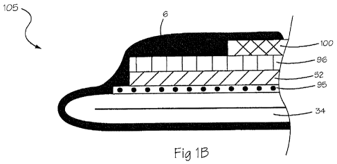
[0077] Figures 1A-1 B show enlarged, cross-sectional schematic views of an
embodiment of an electrode assembly 105. The electrode assembly 105 can
include a flexible membrane 34, one or more flex circuits 89 and one or more
electrodes 6. The flex circuit 89 can include a base substrate 52, a
conducting
layer 96 and a dielectric layer 100. As shown in Figure 1 C, the flex circuit
89 can
diverge from one or more main branches 17 into multiple distal branches 87,
such as at least three, each having one or more conductive traces 16 (not
shown) which each lead to one or more conductive pads 59 (not shown). The
flex circuit 89 as shown in Figure 1 C can be wrapped around an expandable
membrane, such as a balloon (see Figure 23G or 23H), so that the main
branches 17 come together at the shaft. In an embodiment, each conductive
trace 16 can include at least two conductive pads 59. The conductive pad 59
can be a region of the conductive trace 16 that has an exposed, non-insulated
portion of the conducting layer 96. The electrode 6 can be electrically
coupled to
the flex circuit 89 via the conductive pad 59 (not shown) of the conductive
layer
96. The base substrate 52 can also have a wider surface for better adhesion of
the flex circuit 89 to the membrane 34. With a larger base substrate surface,
the
conductive pad 59 can have a larger surface to electrically connect to the
CA 02743140 2011-05-09
WO 2010/056771 PCT/US2009/064069
electrode 6. It should be appreciated that the embodiment of the electrode
assembly shown in Figures 1A-1C is exemplary and that variations in the
structure, shape, and relative positions of the components are possible.
[0078] Each electrode 6 can be a thin, electro-conductive film that covers at
least a portion of the flex circuit 89 and a portion of the outer surface of
the
membrane 34. Figure 1 D illustrates a portion of a membrane 34 supporting a
one distal branch of the flex circuit 87. The figure shows two electrodes 6
that
overlap separate conductive pads 59 of the flex circuit 89, the corresponding
conductive traces 16, and a portion of the flex circuit distal branch 87. The
electrode 6 can have a surface area or diameter that is significantly larger
than
the conductive pad 59. Because the electrode 6 has a larger surface area it
also
covers a portion of the membrane 34 not covered by the conductive pad 59 or
the flex circuit distal branch 87.
[0079] The electrode assembly 105 can be deployed to deliver energy to a
target tissue. When deployed, each electrode 6 on the membrane 34, both alone
and in combination, can cover a relatively large surface area of the membrane
34
with which to contact target tissues. Despite the large overall surface area
of the
electrodes 6 and the components of the flex circuit 89 covering the flexible
membrane 34, the electrode assembly 105 can be compactly folded into a small
diameter such that the electrode assembly 105 can be delivered, for example,
through small access channels for minimally-invasive delivery.
Flexible electronics
[0080] The electrode devices described herein incorporate flexible
electronics that are foldable to a very low profile for minimally-invasive
delivery in
contrast to a relatively stiff and bulky electrode assembly. Upon reaching the
target tissue, the electrode devices described herein can unfold to reveal a
very
large surface area electrode assembly that can be readily conformable with the
target tissues.
11
CA 02743140 2011-05-09
WO 2010/056771 PCT/US2009/064069
Flex circuit
[0081] As mentioned above, the electrode assembly 105 of the devices
described herein can include one or more branching flex circuits 89. The flex
circuit 89 can include a base substrate 52, a conducting layer 96 and a
dielectric
layer 100 as will be discussed in more detail below. Still with respect to
Figure 1 D, the flex circuit 89 can include one or more main proximal branches
17
(not shown) that can divide into multiple conductive distal branches 87. Each
distal branch can contain multiple conductive traces 16, each having one or
more
conductive pads 59. The conductive pad 59 has an electrically-conductive
region
formed by exposure of the conducting layer 96 upon removal of a portion of the
overlying insulating dielectric layer 100. The exposed portion of conductive
layer
96 can contact the conductive film electrode 6.The conductive pad 59 can be a
region of the conductive trace 16 that has a larger surface area due to a
larger
base substrate layer 52 and insulating dielectric layer 100 (not shown). The
method of conductive trace 16 termination is created as known in the art.
These
regions of wider and larger surface areas can be used for better adherence to
the
membrane.
[0082] As shown in Figure 1 C, the distal branches 87 of the flex circuit 89
can form a pattern of distal branches 87 that can spread out across the
membrane 34. The branching pattern can vary and includes a fractal,
self-repeating pattern or other symmetrical pattern, as well as an
unsymmetrical
pattern. The flex circuit 89 can include branches that are sinusoidal in shape
so
that some elongations between electrodes can be achieved. Multiple flex
circuits
89 can be used to accommodate for the quantity and location of the multiple
electrodes 6. Some elements of the flex circuitry 89 can have bridging
elements
88 that facilitate handling during manufacturing (see Figure 3C).
[0083] As shown in Figures 2A-2E, the flex circuit 89 and multiple conductive
traces 16 can be constructed using laminations of various materials, but
generally includes a base substrate 52, an electrically conductive layer 96
and an
12
CA 02743140 2011-05-09
WO 2010/056771 PCT/US2009/064069
electrically insulating layer 100. In an embodiment, the multiple conductive
traces 16 include a bottom insulating substrate layer 52, a middle conductive
layer 96 and a top insulating dielectric layer 100. The dielectric or top
insulating
layer 100 can be removed as is known in the art to expose a small region of
the
conductive layer 96. For example, a laser can be used to remove the dielectric
layer 100 by etching as will be discussed in more detail below. In other
embodiments, an adhesive layer can be used between the layers described
above. In other embodiments, multiple layers of conductivity and/or dielectric
and/or adhesive can be included.
[0084] The materials used in the laminations of the flex circuit 89 can vary.
The base substrate layer 52 and the electrically insulating layer 100 can be a
material such as, but not limited to, a thin flexible plastic substrate,
polyimide,
polyester, PET (polyethylene terephthalate), PEEK (polyaryletheretherketone),
PTFE (polytetrafluoroethylene), PEN (polyethylene naphthalate), LCP (liquid
crystal polymer), PIC (photoimageable coverlay), thin epoxy glass, polyimide
glass, acrylic adhesive or other material or combinations thereof. In an
embodiment, the substrate or bottom insulating layer 52 and the dielectric or
top
insulating layer 100 can be the same materials. In another embodiment, the
substrate and the dielectric layers are different materials. For example, the
substrate can be polyimide and the dielectric can be polyimide glass or
similar
material.
[0085] The conductor or conductive layer 96 can be a material such as, but
not limited to, a metal or metal foil of copper, gold, silver, tin, nickel,
steel,
cupronickel (copper-nickel alloy), KOVAR (nickel-cobalt ferrous alloy) or
other
material. In an embodiment, more than one conductive material can be used in
the conductive layer 96. In an embodiment, a conductive layer 96 of copper can
be plated with a thin layer of an additional conductive material at the
conductive
pad 59. In an embodiment, the thin layer of additional conductive material can
be gold. The flex circuit and its components can be manufactured using
techniques as known in the art.
13
CA 02743140 2011-05-09
WO 2010/056771 PCT/US2009/064069
[0086] Still with respect to Figures 2A-2E, the flex circuit 89 and associated
conductive traces 16 and conductive pads 59 can be coupled to the membrane
34 by a variety of techniques known in the art to affix a metallic or polymer,
shaped member onto another surface as are known in the art. For example, an
adhesive film 95 or other material can be used to adhere the bottom layer of
the
flex circuit 89 to the membrane 34 as will be discussed in more detail below.
The
adhesive film 95 can be conductive or non-conductive. For example, an
adhesive 95 that is conductive can be laid over portions of the electrodes to
adhere to the exposed conductive layer 96. Adhesive 95 that is not conductive
can be used to bond the rest of the components to the membrane 34, for
example to secure an end region of the flex circuit 89 to the membrane 34. The
flex circuit 89 can be direct formed into the membrane 34 as will be discussed
in
detail below.
[0087] Although the conductive layer 96 can be relatively narrow, it can also
have a surface that is somewhat planar, as opposed to having a cylindrical
geometry. The planar surface of the conductive layer 96 can have a width and
thickness that is optimized for carrying current to the electrodes 6. Further,
the
plurality of conductive traces 16 can be grouped to create a planar surface
width
optimized to bond the flex circuit 89 to the membrane 34. The flex circuit 89
can
also include one or more holes 53 incorporated through the base substrate 52
and the insulating layer 100 to allow for adhesive to wick through to improve
adhesion of the flex circuit 89 to the membrane 34 (see Figure 1 D).
[0088] Figures 2A-2E illustrate various lamination configurations of the flex
circuit and electrode assembly 105. The lamination configurations are
exemplary
and variations are possible. Figure 2A shows an adhesive layer 95 that is
electrically non-conductive adjacent to the electrode 6 and covers a portion
of the
membrane 34 and the flex circuit distal branch 89. The conductive section of
the
conductive layer 96 contacts the electrode 6. An adhesive layer 95 can also be
applied over the top of the flex circuit distal branch 87 near an end where it
contacts the electrode 6 to secure the end of the distal branch 87 to the
14
CA 02743140 2011-05-09
WO 2010/056771 PCT/US2009/064069
membrane 34. The adhesive applied over this portion can be conductive to
increase the surface area of the electrode 6. In other embodiments, the
electrode
6 itself can also serve as an adhesive for portions of the flex circuit 89
when
conductivity is desired.
[0089] Figure 2B shows a conductive trace 16 bonded to a membrane 34
using an adhesive 95. An exposed portion of the conductive layer 96, such as
where the insulating layer 100 has been removed, can face away from the
membrane 34 surface such that it does not come in direct contact with the
membrane 34. Since the conductive layer 96 faces away from the membrane
34, a non-conductive adhesive can be applied. The electrode 6 covers the
exposed portion of the conductive layer 96 as well as a portion of the
membrane
34 and flex circuit distal branch 87. Figure 2C shows the distal branch 87 of
a
flex circuit 89 that is adhered to a region of an inner surface of the
membrane 34
as well as the outer surface of the membrane 34. The flex circuit distal
branch
87 pierces through the membrane surface. In an embodiment, an adhesive layer
95 is not used to fix the flex circuit 89 to the inner surface of the membrane
34.
The adhesive in this case can be non-conductive as the conductive layer 96
faces away from the membrane 34. Figures 2D-2E shows the flex circuit 89
directly coupled to a membrane structure 34. Figure 2D shows a membrane 34
encapsulating of the base substrate 52 of the flex circuit 89. The exposed
conductive layer 96 is covered by the electrode 6 which also covers part of
the
membrane. Figure 2E shows an electrode 6 embedded within the membrane 34
and the conductive layer 96 of the flex circuit 89 covering a portion of the
electrode such that the electrode 6 and exposed conductive layer 96 are in
contact.
[0090] The flexible and thin components of the flex circuit 89 contribute to
the low profile and low bulk of the electrode assembly 105 such that the
circuit
can fold to a very small profile for minimally-invasive delivery. The flex
circuit 89
can be affixed to the membrane 34 such that the membrane 34 and electrodes 6
undergo preferential folding, for example between or across the flex circuits
89.
CA 02743140 2011-05-09
WO 2010/056771 PCT/US2009/064069
The folding can occur in an organized, controlled and repetitive manner. The
flex
circuit 89 can aid in better packing as it straightens out during folding and
encourages the membrane to do the same. Figure 2F shows an embodiment of
an existing flex circuit with multiple layers of conductivity, adhesive and
dielectric
layers.
[0091] Figures 3A-3B show two embodiments of a flex circuit that can be
used to power the electrodes described herein. The embodiments of 3A and 3B
are exemplary and are not intended to be limiting. Figure 3A shows a flex
circuit
89 that includes an array of distal branches 87 extending from a proximal main
flex circuit lead 17 toward the distal end. The distal branches 87 can split
forming a Y-junction. This allows the flex circuit 89 to continue at various
angles
from the main flex circuit lead 17 and can be used to wrap a membrane 34, for
example an expandable balloon-shaped membrane, at different latitudes along
the surface. The distal branch 87 which can contain multiple conductive traces
16 can be electrically insulated through the length of the flex circuit 89 and
the
conductive layer 96 exposed at specific points on the flex circuit 89, for
example
at a conductive pad 59 surrounded by an area of enlarged width or diameter
substrate layer 52 and dielectric 100 (not shown). The substrate layers 52 are
shown including holes 53 through the substrate 52 and insulating dielectric
layer
100 (not shown) to facilitate attachment with, for example an adhesive. The
embodiment of the flex circuit 89 illustrated in Figure 3A can power four
electrodes (not shown) via the four conductive pads 59. The embodiment is
shown as including two temperature sensors 90, but fewer or more than two
temperature sensors 90 can be included. The temperature sensor also requires
a conductive pad 59 for power. The conductive traces for the temperature
sensors 90 can also be used to simultaneously power a mapping electrode (not
shown). In an embodiment five flex circuits 89 can be used to power twenty
ablation electrodes, ten mapping electrodes and ten temperature sensors 90.
[0092] Figure 3B shows a different embodiment of the flex circuit 89 in which
all the flex circuits are integrated into a single piece that can be split
into all the
16
CA 02743140 2011-05-09
WO 2010/056771 PCT/US2009/064069
distal branches 87 needed to power the electrodes 6. The flex circuit 89 in
this
embodiment is a single unit that is split into a number of branches. These
branches 87 can be connected to one another via a small bridge 88 on the
substrate at various points throughout the length of the flex circuit 89 (see
Figure 3C). The flex circuit 89 can be rolled up into a small profile to
insert the
flex circuit 89 into a catheter for assembly. Since the flex circuit 89 can be
split
into branches 87, these cuts help facilitate the flexing and bending necessary
for
assembly and during use. The flex circuit 89 can be placed inside a catheter
and
at the distal end; each branch 87 can peel away at the distal end to form a Y-
junction as shown in Figure 3A. The flex circuit 89 can then be attached to
the
membrane 34 at the various desired positions. The flex circuit 89 can also
include staggered conductive pads 59. Staggering the position of the
conductive
pads 59 can aid in providing a low profile to reduce a stack up of the regions
of
enlarged width or diameter substrate 52. The distal end region of the branches
87 can contain an extra amount of length to be used as sacrificial tabs 102.
These sacrificial tabs 102 can be used to provide for consistent tensioning of
the
flex circuit branches 87 during assembly. The tabs 102 can be mounted to an
assembly fixture (see Figure 59) to ensure the locations of each tab 102 and
each branch 87 of the flex circuit 89 is properly positioned relative to the
membrane 34 and/or shaft 57.
Electrodes
[0093] One or more electrodes 6 can contact specified non-insulated
sections of a conductive trace 16 of the flex circuit 89, the conductive pad
59, as
well as a portion of the deployable membrane 34 and insulated portion of the
flex
circuit 89. The electrodes 6 can be a thin film material that can be
repeatedly
folded such that the electrode 6 and membrane 34 can be compacted into a
small diameter for minimally-invasive delivery. The conductive material of the
electrode 6 has a relatively large surface area compared to the conductive pad
59 it contacts, which provides for a large overall electrode area.
17
CA 02743140 2011-05-09
WO 2010/056771 PCT/US2009/064069
[0094] Despite this large surface area, the electrodes 6 do not significantly
increase the stiffness of the membrane 34 and can fold with the membrane 34.
Figures 4A-4C show an embodiment of the interface bond where the membrane
34 is manufactured separately from the flex circuit 89 and the electrode 6.
The
electrode 6 can be deposited such that it contacts specified non-insulated
section
of the conductive layer 96 and a portion of the membrane 34. Figure 4A shows a
slight curvature in the membrane 34 and how the electrode 6 can follow this
curvature. Figure 4B shows the electrode 6 folded away from the membrane 34
whereas Figure 4C shows the electrode 6 folded inwards and possibly contacting
itself. Despite the large surface area covered, the thin electrode 6 and the
membrane 34 can still be folded (see Figures 4B and 4C). The electrode 6 can
fold and flex to substantially the same extent as the membrane 34, even a
region
of the membrane 34 not covered by an electrode layer, such that the electrode
6
does not impede the flexibility of the membrane 34 or the electrode assembly
105. It should be appreciated that the electrode 6 can fold upon itself along
with
the membrane 34, although folding can also occur between the electrodes 6.
The ability to fold can allow for a lower device profile. Thus, the electrode
can
assume substantially the same curvature as the membrane and conform, along
the length and all contact points at the electrode(s) to the shape and
configuration of the membrane. Accordingly, in use, the membrane and
electrode may adapt the surface configuration of any resilient tissue abutted
by
the membrane and the application of energy across at least a portion of the
surface area of the electrode(s) is defined by the orientation and selective
placement and fixation of the membrane.
[0095] The materials used to create the electrodes 6 can vary. The
electrodes 6 can be a thin film of an electro-conductive or optical ink. The
ink
can be polymer-based for better adhesion to the membrane. The electrode
material can be a biocompatible, low resistance metal such as silver, silver
flake,
gold, and platinum or combinations thereof. An example of an electro-
conductive
ink is provided by Engineered Conductive Materials, LLC (ECM) which is a
polyurathene-based silver loaded ink. Another example may be obtained from
18
CA 02743140 2011-05-09
WO 2010/056771 PCT/US2009/064069
Creative Materials Inc., which manufactures conductive inks, films, as well as
radiopaque inks. As mentioned above, the electrodes 6 can be applied to the
membrane 34 and flex circuit 89 using an adhesive. Alternatively, the
electrode
material can have adhesive properties or be an adhesive-loaded with conductive
particles such as silver flakes such that the electrodes 6 can adhere the
components of the flex circuit 89 to the membrane 34. If an additional
adhesive
layer is used to adhere the electrode 6 to the membrane 34 and flex circuit
89,
the adhesive layer can include a conductive or non-conductive material. The
electrodes formed with electro-conductive or optical ink or thin metal film
can be
visualized under fluoroscopy to provide a general sense of the shape of the
membrane and location of the electrode. To enhance visualization under
fluoroscopy, radiopaque additives can be included in the electrode material or
radiopaque markers laid out next to, on top or below the electrodes as will be
discussed in more detail below.
[0096] The electrode material can be deposited using a variety of techniques
known in the art including, but not limited to, printing, pad printing, screen
printing, silk screening, flexography, gravure, offset lithography, inkjet,
painting,
spraying, soldering, bonding deposited using touch-less technologies or
otherwise transferred onto the surface of the membrane 34. In an embodiment,
the electrode 6 can be formed by depositing an electrically conductive coating
or
layer by spraying a preselected onto the designated surface region.
Alternatively, the electrode can be formed by depositing the electrically-
conductive material onto a region of the membrane 34 by vacuum deposition or
printing the electrically conductive material on the designated surface
region.
This provides an electrically conductive coating of a desired thickness and a
relatively uniform electrode through the desired area. Printing processes can
include pad printing, screen printing and the like. Touch-free technologies
such
as positive material deposition of ink such as from a syringe or similar
device can
also be used to transfer conductive film or ink onto the membrane or
substrates
that are sensitive to pressure.
19
CA 02743140 2011-05-09
WO 2010/056771 PCT/US2009/064069
[0097] The electrodes can also be made using thin, conductive adhesive film
or gel that can be cut to the shape of the electrodes and serve as an adhesive
for
the flex circuit when conductivity is desired. The conductive adhesive gel can
be
mixed with conductive particles for conductivity and disposed over the
substrate
and UV cured.
[0098] Each region of electrically conductive material can be deposited over
and electrically connected to a specified conductive pad 59 of the flex
circuit 89
and coupled to the surface of the membrane 34. The electrodes can be formed
by using a mask (chemical or mechanical) over the membrane during the
deposition process, which can deposit electrode material over the membrane and
the mask alike. Once the deposition process is completed, the masking can be
removed as is known in the art. An alternate technique can be used where
automated robotic systems are programmed to precisely and accurately spray
only the desired electrode surfaces without the presence of a mask. This
technique can have multiple movable axes such as the Engineering Fluid
Dispensing Inc. dispensing robots (East Providence, RI).
[0099] The flex circuit 89 components can be bonded before, during or after
deposition of the electrodes 6 to the membrane 34, for example, using an
adhesive or thermal bond or the like as described above. The electrically
conductive layer 96 of the flex circuit distal branches 87 can be exposed by
etching away a portion of the dielectric layer 100.
[0100] The shape and pattern of electrodes 6 created can vary. The surface
area, shape and pattern of the electrodes 6 can influence the amount of energy
applied and the ablation line created. Figures 5A-5F illustrate various
electrode
patterns and electrode shapes considered herein including, but not limited to,
circular, rectangular, octagonal, polygonal etc. The shape and pattern of the
electrodes 6 deposited on the membrane 34 can be selected depending upon the
intended application of the electrode assembly. A square electrode, for
example,
can be better suited for interpolation based on image projection analysis,
such as
CA 02743140 2011-05-09
WO 2010/056771 PCT/US2009/064069
to reproduce the shape of deployable membrane 34 in a mapping and
identification software algorithm. One or more rows of electrodes 6 can be
used.
Each row of electrodes 6 can have the same shape or can vary in shape and
size. Spacing between the electrodes 6 within the same row or spacing between
the rows can alter the depth and quality of lesion created. The rows of
electrodes can have electrodes that line up or can be staggered as shown in
Figure 5D. The electrodes 6 can also be deposited in a variety of locations on
the
deployable membrane 34 as will be described in more detail below.
[0101] Figure 12 shows an embodiment of a pattern of electrodes 6. The
pattern shown in Figure 12 is exemplary and variations in the pattern are
possible. Current 92 can be passed between adjacent electrodes 6 and/or
overlap an electrode 6 to reach the next electrode 6 to create the desired
ablation line. Each of the electrodes 6 can be created as a solid pattern, a
set of
concentric circles or other geometric shape, or a set of lines intersecting or
not.
The surface area, shape and internal pattern of the electrodes can influence
the
density of the current and burn line created. These features can also affect
the
amount of current and power required as well as duty cycle and/or pulse wave
modulation. There can be more than one row of electrodes 6 to allow the user
to
actively select which region to use for creating the ablation lesion and avoid
having to exactly position the device and or manipulate to create the proper
ablation line. The ablation line can be created in a desirable location using
techniques that are easy and fast and without the need for tedious
repositioning.
[0102] Referring again to Figure 12, the pattern of multiple electrodes 6
deposited on the membrane 34 can collectively create a large electrode array
of
energy-transmitting elements. This electrode array can form a variety of
patterns
across the membrane 34 and has an energy-transmitting surface area. The
electrode array pattern and energy-transmitting surface area can vary. In an
embodiment, the energy-transmitting surface area covers at least about 10% of
the membrane surface area to be selectively activated. In an embodiment, the
energy-transmitting surface area can cover about 25% of the membrane surface
21
CA 02743140 2011-05-09
WO 2010/056771 PCT/US2009/064069
area. In another embodiment, the energy-transmitting surface area can cover
approximately 50% of the membrane surface area. The energy-transmitting
surface area can be a factor of the physical surface area of each individual
electrode within the energy-transmitting array as well as the projection of
the
expected ablation surface area based on the electrode pattern spacing. The
preferred energy-transmitting surface area percentage can also vary depending
upon the indication being treated. For example, for the treatment of atrial
fibrillation the energy-transmitting surface area can cover at least 25% of
the
membrane surface to be selectively activated. In another embodiment, the
energy-transmitting surface area can cover greater than 40% of the membrane
surface to be selectively activated. These percentages are provided for
example
and can vary. The large energy-transmitting surface area allows the membrane
surface to selectively ablate more tissue simultaneously without the need for
repositioning. Generally, the lesion site can be slightly larger than the
energy-transmitting surface area.
[0103] Each electrode 6 can also be a grouping of multiple smaller
electrodes 51 such as the embodiments shown in Figures 6A-6B. Each of the
smaller electrodes 51 can be connected by the conductive traces 16 of the flex
circuit 89 as shown in Figure 6B to form a larger electrode 6. Alternatively,
the
smaller electrodes 51 can be independently activated for mapping electrical
signals as may be needed in some indications such as the treatment of atrial
fibrillation. The traces 16 can be created as a sinusoidal line, for example,
to
allow for some elongation of the expandable element so that the individual
electrodes can spread farther apart and the electrodes become substantially
larger. As shown in Figure 6B, traces 16 allow for uniform elongation in all
directions. Alternatively, the traces 16 can allow for elongation in specified
directions. The surface area, shape and pattern of the electrodes can
influence
the amount of energy transmitted to the target tissue. Measurement with
smaller
electrodes 51 can provide higher resolution and accuracy of the signal
location,
which can be useful for example in mapping aberrant signals. Figure 6C
illustrates an embodiment of an electrode 6 that includes a small electrode 51
22
CA 02743140 2011-05-09
WO 2010/056771 PCT/US2009/064069
located at the center of the larger electrode 6. Each of the electrodes is
connected to their individual traces 16. This embodiment can be used to
confirm
conduction block such as during treatment of atrial fibrillation by comparing
conductivity before and after ablation or by moving the electrode structure
further
into the pulmonary vein for measurements.
[0104] The electrode 6 can be a thin, flexible film that is deposited over a
portion of the flex circuit 89 as well as a portion of the membrane 34. As
discussed briefly above and shown as an example in Figures 7A-7E, each of the
electrodes 6 has dimensions that exceed those of the conductive pad 59 or the
conductive trace 16 of the flex circuit 89 such that the electrode 6 covers an
area
of the membrane 34 on which the flex circuit 89 is mounted. Figure 7A shows
the substrate layer 52 of the flex circuit 89 following and outlining the
conductive
traces 16. The electrodes 6 can extend beyond the substrate layer 52 onto the
underlying membrane 34 to provide a large surface for the electrode 6 to
contact
the tissue. This is in contrast to many devices known in the art which use the
small, non-insulated portion of the flex circuit itself as the electrode
element.
Larger surface area and bigger overall electrodes 6 allow the electrode
assembly
105 of the devices described herein to transmit energy deeper and with less
risk
of gaps in the energy transmission line. To increase the durability of the
electrodes 6, the substrate layer 52 can be extended over portions of the
electrodes 6. This can restrict elongation on sections of the membrane where
the
electrodes 6 are located and can ensure, for example predictable ablation
lesion
size and quality. Figure 7B shows the substrate layer 52 can extend to outline
the shape of the electrodes 6 to be deposited. Figure 7C shows the substrate
layer 52 can have finger-like extensions or struts that can extend to the edge
of
the electrodes 6. A combination of any of the above can also be used.
[0105] The dimensions of the electrode 6 can vary. In an embodiment, each
electrode 6 can be between about 0.015 to .050 mm in thickness. In an
embodiment, each electrode 6 is less than 0.025 mm in thickness. In an
embodiment, each electrode 6 can have an overall surface area of between 3
23
CA 02743140 2011-05-09
WO 2010/056771 PCT/US2009/064069
and 36 mm2. In an embodiment, each electrode 6 can have a size that is
approximately about 2 mm round. In comparison, each conductive trace 16 can
be between about 0.05 mm and 0.10 mm in width and between about 0.02 and
0.05 mm in thickness. Each conductive pad 59 can be between about 0.05 and
0.70 mm in width and between about 0.02 and 0.05 mm in thickness. In an
embodiment, each conductive pad 59 can have an overall surface area of
between about 0.002 and 0.450 mm2. In an embodiment, the conductive pad 59
can be approximately 0.5 mm round. It should be appreciated that the
aforementioned dimensions are exemplary and that variations are possible.
[0106] The relative dimensions of the electrode 6 and portions of the flex
circuit 89, such as the conductive pad 59, can also vary. In an embodiment,
the
surface area of each electrode 6 as it relates to the surface area of its
associated
conductive pad 59 can be described in terms of a ratio and can be at least
about
14:1. In another embodiment, the ratio of electrode width to conductor width
can
be about 13:1. The relative dimensions between the electrode assembly
components can also vary depending upon the indication being treated. For
example, atrial fibrillation-specific devices the ratio of surface area of
electrode 6
to surface area of conductive pad 59 can be at least about 44:1. The
conductive
pad 59 can be approximately 0.5 mm round and the electrode can be a minimum
of approximately 3 x 3 mm or 3.4 mm round for a 44:1 ratio. For an electrode
having an area of 4 mm round, the ratio can be approximately 62:1. For an
electrode having an area of 5 mm round, the ratio can be approximately 95:1.
For an electrode having an area of 3 x 5 mm, the ratio can be approximately
74:1. For an electrode having an area of 5 x 5 mm, the ratio can be
approximately 123:1. In another embodiment, the ratio of electrode width to
conductor width on the flex circuit can be approximately 35:1. The conductor
width can be 0.07 mm and a minimum width of the electrode can be 3 mm for a 3
x 3 mm electrode. In another embodiment, the electrode can have a surface
area of at least about 9 mm2 (3.4 mm round) and an electrical conductor of
between about .025 to .050 mm maximum thickness. This combination results in
a flexible electrode that has a large surface area yet is connected to a very
thin
24
CA 02743140 2011-05-09
WO 2010/056771 PCT/US2009/064069
conductive trace. It should be appreciated that the aforementioned relative
dimensions are exemplary and that variations are possible.
[0107] The energy transmitted by the electrodes 6 can vary. The energy can
include radiofrequency (RF) energy, for example in a monopolar or bipolar
energy configuration, microwave, high voltage, or irreversible electroporation
(IRE). Microwave and RF energy can use the application of thermal energy for
cell necrosis while IRE can use high voltage electrical pulses to create cell
permeability leading to cell death. Voltage energy can be delivered in very
high
voltage dosage in short bursts. Use of bipolar RF energy prevents the current
from traveling through the bloodstream and reduces the risk of charring and
thrombus. Bipolar energy also removes the effect of blood flow on the energy
delivery compared to monopolar and generally provides more consistent results.
The electrode assembly 105 can be used exclusively in the bipolar
configuration
without using the monopolar configuration to minimize or eliminate current
transfer through the bloodstream. The energy applied during an energy
transmission period can be in the form of high energy and low energy cycles
(on/off) or alternating high and low temperatures. In use, the pattern of
multiple
electrodes facilitate combining the individual processes of mapping,
detecting,
stimulating, ablating, sensing or measuring the tissue in contact with the
electrode, and physical and electrical characteristics of the tissue. Each of
these
functions can be performed at different electrodes and also in a step-wise
fashion
at different electrodes or subsets thereof, based on a particular clinical
indication.
For example, a step may be comprised of sensing or mapping electrical signals
in underlying tissue followed by stimulation or ablation at selected points or
groups of points. Because the ablation affects the physical and electrical
properties of the tissue, a separate or second set of electrodes can sense the
results of a first ablation step applied through a first set of electrodes.
Any
sequence of sensing, mapping, ablating, detecting, stimulating scanning or
measuring at discrete sets or subsets of electrodes is thereby enabled to
permit
maximum flexibility in diagnosis and treatment of each patient.
CA 02743140 2011-05-09
WO 2010/056771 PCT/US2009/064069
[0108] Figure 8 illustrates an embodiment of the flex circuitry wiring for the
electrodes 6. Each electrode 6 can be connected to an RF amplifier 48. Each
electrode 6 can be individually turned on and off for monopolar or bipolar
energy
transmission. For monopolar, the electrodes 6 can be connected via a
monopolar bus line 14 to a patient return electrode 13 and can be individually
or
simultaneously activated by switches 37. For bipolar, the electrodes 6 can be
connected via a bipolar bus line 73 and can be individually or simultaneously
activated by switches 37. Variations in the manner of connection between the
electrodes are possible. As will be discussed in more detail below,
temperature
sensors 90 can be included in the electrode assembly 105 and can share an RF
conductive trace with an adjacent electrode 6. This allows for dual use of the
conductors which reduces the overall bulk and profile of the device. It also
eliminates the need for an additional assembly junction on the membrane during
manufacturing and allows for a narrower flex circuit and lower profile. It
should
be appreciated that the electrodes 6 can also be used for mapping as will be
discussed in more detail below.
[0109] The electrodes 6 can include a variety of activation mechanisms.
Multiple electrodes 6 can be individually connected to a single flex circuit
89 and
can be individually controlled and individually activated for a more precise
energy
transmission via an electronic control box. Alternatively, the electrodes 6
can
have a physical moveable activation means, such as a conductive wire, which
can be electrically connected to an array of electrodes in series. Figures 9A-
9B,
for example, show a conductive trace 16 that can be a moveable wire housed
within a lumen 33. The trace 16 can contact individual electrodes 6 located in
series and activate them individually or in unison. This can allow a user to
create
a burn pattern precisely where needed without having to move the membrane 34
to a different position. Figure 10 shows another embodiment of a selective
activation mechanism which includes an electrode sleeve 10. A conductive trace
16 can be advanced distally or withdrawn proximally within a lumen of the
electrode sleeve 10. The distal end of the conductive trace 16 can have a
region
of exposed conductive layer 96 covered by an electrode 6 that can selectively
26
CA 02743140 2011-05-09
WO 2010/056771 PCT/US2009/064069
contact the tissue to be ablated through the openings 32 of the electrode
sleeve
10. This configuration can allow the user to position the electrode device
once
and tune the position of the electrodes 6 with the least amount of
manipulation.
This minimizes the amount of risk of trauma and injury to the patient as well
as
reduces the time of the procedure. Figure 11 shows an embodiment in which the
electrode sleeve 10 having moveable traces 16 is mounted to a surface of a
membrane 34 such as a balloon.
[0110] The electrodes 6 described herein can have low thermal mass or
inertia for quick heating and quick heat dissipation. This low thermal mass
provides a more consistent and predictable temperature and energy delivery as
well as a more accurate measurement of temperature and better user control of
the energy. One or more temperature sensors 90 can be mounted directly on the
flex circuit 89 adjacent or over the electrodes 6 to provide feedback during
use of
tissue temperature such that power, current, duty cycle can be modulated and
maintained within a specified temperature or temperature range. The
temperature sensors 90 considered herein can include surface mount
thermistors, thermocouples or other resistance temperature detectors or
platinum
resistance thermometers. The temperature sensor 90 can be bonded to the
conductive trace 16, for example, with an adhesive.
[0111] The number and pattern of temperature sensors 90 included in each
flex circuit 89 can vary. Figure 12 shows an embodiment of an electrode 6 and
temperature sensor 90 pattern where the temperature sensor is located between
two electrodes 6, between four electrodes 6 or in contact with one electrode
6.
Figures 13A-13B show other embodiments of an electrode assembly including a
distal branch 87 and branching conductive traces 16 of a flex circuit 89
contacting multiple electrodes 6 and a temperature sensor 90. Each electrode 6
can be connected to one conductive trace 16 stemming from the distal branch
87. The temperature sensor 90 can share the conductive trace 16 with the
electrode 6 and be positioned near where the electrode 6 is touching the
tissue.
For example, a temperature sensor 90 can have a common ground and each
27
CA 02743140 2011-05-09
WO 2010/056771 PCT/US2009/064069
end can be connected to one of the electrodes 6 and switched/multiplexed with
RF power. The dual usage for the trace 16 between temperature sensors 90 and
electrodes 6 reduces the overall profile of the electrode assembly 105. Fewer
connections results in less material and less bulk of the device, better
packing
and ease of manufacture.
[0112] The location, distribution throughout the flex circuit 89 and number of
temperature sensors 90 incorporated with the electrode assembly 105 can vary.
In an embodiment, the temperature sensors 90 can be adjacent, directly
covering, or in between the electrodes 6. Figure 7A shows a temperature sensor
90 located in between two electrodes 6. In a non-limiting example, the
temperature sensor 90 can be generally less than 1 mm away from the electrode
6. The trace connected to the temperature sensor 90 can be shared with the
trace 16 for the adjacent electrode 6. Figures 7D and 7E shows an embodiment
of an electrode assembly 105 where the temperature sensor 90 is located at the
center of an electrode 6 instead of between two electrodes. The temperature
sensor 90 may be electrically isolated from the electrode 6. One or more
temperature sensors 90 can be used per pair of electrodes 6. In an embodiment,
at least 10 temperature sensors 90 can be included for temperature control.
Deployable Membrane
[0113] The electrode assembly 105 also includes a deployable, flexible
membrane 34 to which the flex circuit 89 and electrodes 6 can be coupled.
When deployed, the membrane 34 can deliver energy through the large surface
area of the electrodes 6 to a target tissue. The deployed membrane 34 and
electrodes 6 can ablate tissue over a large zone or area in a variety of
patterns,
for example circumferential, curved and linear patterns, as will be discussed
in
more detail below. Despite the large overall surface area of the membrane 34
covered by the electrodes 6 and the flex circuit 89, the membrane 34 can be
readily conformable to the target tissue to be ablated and also compactly
folded
28
CA 02743140 2011-05-09
WO 2010/056771 PCT/US2009/064069
into a small diameter such that the electrode assembly 105 can be delivered,
for
example, through small access channels for minimally-invasive delivery.
[0114] The structure of the membrane 34 can vary including, but not limited
to a membrane sheet, cylinder, tube, inflatable, expandable, or fillable
structure,
such as a balloon, or braided mesh and the like. In an embodiment, the
electrode assembly includes a deployable membrane that is formed into a linear
structure or a cylindrical tube such as a cylindrical electrode element 34 as
shown in Figures 16A-16B. The cylindrical membrane 34 can have multiple
electrodes 6 deposited along its length in varying patterns. The membrane 34
can be steered and manipulated, for example to ablate two anatomical regions
simultaneously. The membrane 34 can include sections of varying flexibility
and
stiffness for the ability to steer. The distal end of the membrane 34 can be
manipulated with a guidewire 40 for proper placement, for example into a
vessel
such as the pulmonary vein 80 for the treatment of atrial fibrillation. A
region of
the membrane 34, for example a middle region, can be highly flexible such that
by pushing a handle (not shown) distally the middle region of the membrane 34
can bend and be directed toward another anatomical region, for example such as
inserted into an adjacent vessel (Fig. 16B). This can be useful, for example,
when ablating a region between the two pulmonary veins that can have highly
irregular anatomy that is difficult to access. The membrane 34 can also be
inflated or expanded to contact the vessel wall 83 and anchor the device in
place
as will be discussed in more detail below. The cylindrical electrode element
34,
which is located on the electrode catheter 71, can be advanced through a
sheath
65, such as a transseptal sheath (see Figures 15A-15B). The user can control
the distal end of the electrode catheter 76 via a pull tether 70 at the
proximal end
of the electrode catheter 76. Pull tether 70 can be concentric and housed
within
the electrode catheter 76 in some portion more proximal than what would be
protruding from the transseptal sheath 65.
[0115] In one embodiment, the electrode catheter 71 can be housed within
an electrode sheath 76 as shown in Figures 14A-14B. In an embodiment, one or
29
CA 02743140 2011-05-09
WO 2010/056771 PCT/US2009/064069
more electrodes 6 can be positioned on an outer surface along the length of
the
electrode sheath 76. The electrode catheter 71 can be used in conjunction to
electrode sheath 76 to transmit thermal energy in multiple locations. In
another
embodiment, the electrode sheath 76 can slide over a steerable guide catheter
47 anchored in place, for example using an anchoring basket 50 or a suction
tip
18 at the end of an anchoring catheter 15 to anchor onto neighboring tissue
such
as the myocardium 80. The steerable guide catheter 47 can be used to position
the electrode sheath 76 to produce the desired treatment lines 81. It should
be
appreciated that the electrode sheath 76, the electrode catheter 71 and
steerable
guide catheter 47 can be incorporated into a single catheter configuration.
[0116] The membrane 34 of the electrode assembly 105 can have an
expandable structure (self-expanding or otherwise) that can place the
electrodes
in full contact with tissues. The membrane 34 of the electrode assembly 105
can
have an expandable structure that is closed or fluid-tight, such as a balloon
or a
cylindrical tube. The membrane 34 of the electrode assembly 105 can also span
or have an expandable structure that is open, such as a woven, braided, stent
or
basket-like expandable structure as shown in Figure 17A-17D. In an
embodiment, the expandable structure 93 can radially expand to an open state
(self-expanding or user-operated). The expandable structure 93 can be
surrounded by the electrode assembly 105 such that the flexible, outer
membrane 34, flex circuit 89 and electrodes 6 are disposed thereon. The
expandable structure 93 can be attached to a catheter 57 via distal support
elements 44. In one embodiment the flexible membrane 34 can surround the
expandable structure 93 while attached by sutures at the intersections of the
distal support elements 44 and the expandable structure 93. In another
embodiment, the membrane 34 may be weaved through some or all the loops of
the expandable structure 93 while allowing sufficient material for elongation
and
movement of the expandable structure 93. The electrodes (not shown) can also
be mounted over a single wire or over the intersection of wires or both. The
expandable structure 93 can be flexible and conform to a variety of anatomical
shapes. Figure 17A shows the expandable structure 93 in a relatively elongated
CA 02743140 2011-05-09
WO 2010/056771 PCT/US2009/064069
state with a lower profile more suitable for insertion and removal through a
small
access channel or sheath. Figure 17B shows the expandable structure 93 in its
fully expanded state that can be used or is suitable for energy transmission.
A
guidewire (not shown) can be used when ablating, for example around the
pulmonary vein. When the guidewire is retracted, the distal end of the
expandable structure 93 can be used to ablate tissue. Figure 17C and 17D show
close-up views of an embodiment of the woven loops of the expandable structure
93. The expandable structure 93 can be a shape memory material such as
Nitinol.
[0117] In another embodiment, shown in Figures 17E-17G, a catheter 57
can have one or more electrodes disposed on an expandable structure. The
configuration of the expandable element can vary including a flat wire or
coil.
Once deployed the diameter of the electrode 6 can be larger than the diameter
of
the catheter body 57. This promotes optimum contact with the tissue 83 to be
ablated or mapped. Additionally, these "spring" electrodes can be constructed
for self-adjustment within their range of movement to conform to a variety of
anatomies. A pressure or movement sensitive mechanism can be incorporated
at each electrode in order to provide feedback to the user about tissue
contact
prior to device activation. A flexible membrane 34 can also be placed over
these
spring elements with the electrodes disposed on the membrane.
[0118] The flexible membrane 54 can be disposed around an expandable
structure 98 that is self-expanding such as a braid, coil or the like, as
shown in
Figures 60A-60D. Electrodes 6 may be arranged over the tubular thin walled
membrane 54. A sheath 31 can cover the electrodes and support structure for a
low profile delivery. Once inside the desired location, the sheath can be
pulled
back, exposing the structure 98 and the electrodes 6. The membrane 54 can be
attached to one or both ends of the support structure 98. An exemplary benefit
of
this approach is that the device does not occlude the anatomy during ablation.
The structure is open through its longitudinal length and thus allows for the
flow
of fluid or gas. This eliminates a concern especially when treating blood
vessels.
31
CA 02743140 2011-05-09
WO 2010/056771 PCT/US2009/064069
The membrane can also include holes, slits, or ports which allow for
additional
fluid or gas passage to minimally interfere with anatomical flows.
[0119] Figures 60A and 60B show an embodiment of this design. The
structure 98 is directly attached to the catheter shaft 57 which creates a
funnel
shape at the junction of the shaft and the structure. This facilitates
sheathing and
unsheathing of the structure. Figure 60C shows another embodiment in which a
coupling element 60 connects the shaft 57 and the structure 98, which allows
for
full expansion of the support structure 98 at the distal and proximal end and
thus
fully expansion of the electrode-carrying membrane 54. A depiction of the flow
of
blood is indicated with arrows in Figure 60C. Figure 60D illustrates a thin
wall
membrane 54 with electrodes 6 supported by a coil support structure 98. This
embodiment allows for a very small profile in that a coil can be sheathed into
an
essentially linear structure. To prevent distortion of the electrodes, the
membrane
54 in this particular embodiment may be attached at only the proximal end or
otherwise contain compliant sections not directly affecting the electrodes
during
sheathing.
[0120] The electrode assembly can include a perfusion balloon and catheter
configuration in which blood flow is not restricted by the presence of the
device.
The assembly can include a large inner lumen allows the use of a guidewire and
is large enough to also accommodate for flow of fluid, such as blood. Figure
18G
illustrates one such embodiment. The flow of blood indicated by arrows can
enter
the guidewire lumen and exit a hole 100 that can be located just proximal to
the
membrane 34 on the shaft 57.
[0121] The membrane 34 of the electrode assembly 105 can also have a
closed, expandable structure, such as a balloon as shown in Figures 18A-18M.
The membrane 34 can have an expandable structure that is fluid-tight such that
it
can be filled with a liquid or gas to expand or inflate it. The membrane can
be
expanded using a variety of techniques such as by filling with a gas or
liquid,
such as saline, a radiopaque dye, cooling fluid, blood-safe gas and the like.
The
32
CA 02743140 2011-05-09
WO 2010/056771 PCT/US2009/064069
expandable structure can also be self-expanding. The membrane 34 can be
covered by multiple electrodes 6 and can be coupled near its proximal region
to a
distal end of a catheter 57. The distal and proximal regions of the membrane
structure 34 shown in Figures 18A-18C protrude outwards forming smaller
domes, which can provide convenience for manufacturing. Figures 18D-18M
illustrate other embodiments of an electrode assembly 105 where the membrane
34 has a continuous smooth surface and no protrusions or domed regions at its
distal and proximal end regions. The distal end of the membrane 34 can be flat
or
as shown in Figure 18F and 18G drawn into itself creating a concave surface at
its distal end.
[0122] Figures 181-18M show various views of an embodiment of the
deployable membrane 34 of the electrode assembly 105 that has a fluid-tight
expandable structure. The deployable membrane 34 can have multiple
electrodes 6 electrically connected via one or more flex circuits 89. As shown
in
Figure 181, each flex circuit 89 can be routed through the shaft 57 and can
exit or
emerge from the inner diameter of the membrane 34 at a distal end region and
split into two at a Y-junction. This allows a single flex circuit 89 to be
positioned
at different latitude positions on a membrane 34. Figure 18J shows an
embodiment of the conductive pad 59 that can be used to electrically connect
the
electrodes 6. Figure 18K shows an embodiment of a mapping electrode 51 that
is smaller and in between the larger electrodes 6. Figure 18L shows an
embodiment of the distal end region of the membrane 34 that can be drawn into
itself creating a concave surface.
[0123] The flex circuit 89 can wrap around the membrane 34 to power the
electrodes as shown in Figure 18J. The flex circuit 89 can extend to the
proximal
end of the membrane 34 and/or into the distal end of the shaft 57 as shown in
Figure 18M. Extending the flex circuit to the joint where the shaft 57 and the
membrane 34 meet can increase the robustness and ease of manufacturing the
electrode assembly 105. The flex circuit main leads can be inserted within the
inner diameter of the shaft and bonded in place. This can be beneficial for
33
CA 02743140 2011-05-09
WO 2010/056771 PCT/US2009/064069
preventing de-lamination of the flex circuit main leads 17, such as during the
sheathing process. These sections of the flex circuit 89 can power another set
of
electrodes located at or near the proximal end of the membrane 34. With a
toroidal-shaped, closed membrane 34, the location of the electrodes 6 face
away
from the distal portion of the membrane 34, such that they face in a proximal
direction towards the shaft 57 of the assembly 105. This configuration can be
useful in reaching target tissues that are located directly through an access
point,
such as for example the septum once a catheter crosses the septum to enter the
left atrium.
[0124] The shape of the expandable membrane 34 can vary including, but
not limited to, cylindrical, spherical, toroid, doughnut-like, conical,
branched,
pronged and other geometries. As shown in Figures 18D-18M, the expandable
membrane 34 has a toroid shape. This shape provides for better
maneuverability of the distal tip due to the relatively short longitudinal
length of
the structure. Figure 18H illustrates the swiveling action the toroid shaped
membrane structure can achieve. Because the longitudinal length of the
membrane structure on the catheter shaft is relatively short, the membrane
structure can move relative to the shaft without bending the shaft. When the
membrane structure is used in a semi-inflated state, this allows for greater
motion or swiveling of the membrane structure on the shaft. Further, a smaller
membrane structure 34 can be used, which although it may be subject to more
frequent manipulation of the electrode assembly 105 during the procedure it
can
allow for easier manipulation especially in smaller and/or tight anatomies.
Electrode assemblies having small membrane structures 34, such as shown in
Figures 22A to 22B and Figures 26A to 26C, can be useful for touch-ups during
the procedure or during follow-up procedures.
[0125] The deployable membrane 34 can have an expandable structure that
is symmetrical such as shown in Figure 19A. The electrodes 6 can be evenly
distributed across the membrane 34 where they can be connected to their
individual conductive traces 16 stemming from the distal flex circuit branches
87.
34
CA 02743140 2011-05-09
WO 2010/056771 PCT/US2009/064069
The distal branches 87 connect to the main flex circuit leads 17 (not shown)
which can be routed through the catheter 57 such that they can connect at a
proximal region for example at a handle. The deployable membrane 34 can also
have an asymmetrical configuration as shown in Figures 19B and 19C. An
asymmetrical structure can reduce bulk and can allow for easier manipulation
and positioning of electrodes. Figure 19C shows the asymmetrical membrane
structure conforming to tissue 83 such as the pulmonary vein. In atrial
fibrillation
applications, for example, the deployable membrane 34 having an asymmetrical
structure can involve two or more different applications of energy and
rotations of
the membrane 34 to completely isolate the pulmonary vein. The asymmetrical
structure can allow for better rotation control and placement of the
electrodes 6
against the tissue 83.
[0126] The electrode assembly 105 can include an enclosed membrane 34
and can be of any shape or form, one of which can be a cylindrical shaped
balloon. The membrane 34 can further be shaped to maintain a curved position
or include one or more pull wires. Figures 19D-19F show alternative
embodiments for a membrane 34 for an ablation assembly 105 including one that
has a flat distal end surface and one that is more conical. It should be
appreciated that other variations of the membrane shape can be possible. The
length of the membrane 34 can be shorter or longer and the shape can be
straight or contain any amount of curvature. The electrode assembly 105 can
include a flex circuit 89 which powers one or more electrodes 6. The
electrodes 6
can be laid out in an asymmetrical pattern of electrodes 6 on the inside of
the
curve versus the outside of the curvature. The distal end of the membrane 34
can also include a single large electrode 6 as shown in Figure 19F. Fiber
optic
scopes 38 can be included to the electrode assembly 105 as well as shown in
Figure 19D.
[0127] The shape of the membrane 34 can be changed or adjusted before,
during or after use. Figures 20A-20C show an embodiment of an electrode
assembly 105 having a deployable membrane 34 that can be expanded into a
CA 02743140 2011-05-09
WO 2010/056771 PCT/US2009/064069
balloon shape. The deployable membrane 34 is coupled at an outer surface of
its proximal region to support arms 44 extending from the distal end of the
steerable catheter 57. The membrane is coupled at its distal region to a shaft
46
that extends through and translatable relative to the steerable catheter 57.
The
shaft 46 can translate from a proximal position in which the membrane 34 is
folded distal to the catheter 57 and shaft 46. The shaft 46 can also translate
to a
distal position in which the membrane 34 expands into an enlarged structure
and
the exposes the most distal electrodes suitable for energy transmission as
seen
in Figure 20B. The shape of the membrane 34 can be varied depending upon
the position of the shaft 46 relative to the catheter 57. For example, the
membrane 34 can have a fully rounded configuration as shown in Figure 20B or
a distally flattened configuration such as shown in Figure 20A or a distally
concave configuration as shown in Figure 20C or anywhere in between. This
allows for positioning and exposure of the electrodes as needed to fully
contact
the target tissue.
[0128] The membrane 34 and electrode assembly 105 can conform to
three-dimensional anatomy for optimum tissue contact and energy transmission.
Good apposition of the membrane allows for better contact of the electrodes 6
to
the surface of the tissue. The membrane 34 having an expandable structure as
described above can be expanded to a variety of conformations, shapes and
have a range of diameters over a relatively low range of pressures. In an
embodiment the membrane can be radially expanded such that it fits and
conforms within two regions of the anatomy simultaneously (for example see
Figure 16B). In another embodiment, the membrane 34 can have a large distal
diameter (for example Figures 18A-18M) and/or can be tapered, or funnel-
shaped (for example Figures 20A-20C). This allows for a better conformation to
circumferential geometries, for example regions near the ostia of a pulmonary
vein.
[0129] Figures 21A-21 E illustrate how a membrane 34 having an
expandable balloon-type structure can conform to tissue 83 having a variety of
36
CA 02743140 2011-05-09
WO 2010/056771 PCT/US2009/064069
anatomical shapes. The membrane 34 can be semi- or non-compliant, but can
still conform to target tissues depending the degree to which it is filled. In
an
embodiment, the deployable membrane 34 can be non-compliant and have an
expandable, closed structure that is only partially filled. Partial filling of
a
non-compliant, closed structure can allow it to properly conform to the target
tissue despite the non-compliant properties of the membrane material itself.
In
another embodiment, the deployable membrane 34 has an expandable, closed
structure and a relatively short longitudinal length. In this embodiment,
partial
filling of the structure such as with a fluid, gas or liquid results in a
conformability
and swiveling steerability. The membrane 34 can have an expandable, closed
structure that is branched or can split into two branches at its distal end
upon
expansion. In the expanded state, electrodes 6 on each of the branches can be
in contact with the tissue 83 during energy transmission (see Figure 21 E).
The
pronged or two-leg shape can aid in reaching irregular surfaces between, for
example two vessels such as the carina between the pulmonary vein 80.
[0130] As described above, the electrodes 6 can be deposited on the
membrane 34 as well as on a portion of the flex circuit 89. The membrane 34
can include multiple electrodes 6 or the membrane 34 can have a single
electrode 6 covering a portion or the entire membrane 34. For example,
Figure 22A shows a membrane 34 having multiple electrodes 6. Figure 22B
shows a single electrode 6 covering a distal portion of the membrane 34.
Figure 22C shows a single electrode 6 that surrounds the entire outer surface
of
the membrane 34. Further, the membrane 34 can be impregnated with
conductive material which then can become the electrode. It should be
appreciated that the size of the membrane 34, in particular an enclosed
membrane such as the balloon shape shown in Figures 22A-22C, can be of any
size and shape. A small balloon size can be used for treatment of small
anatomical sites or for touch-up / follow-up secondary treatments. Also, once
brought into contact with tissue, the step-wise combination of all functional
capabilities of the device, including sensing, mapping, stimulating, ablation,
and
measuring, as described herein can selectively be applied to the target
tissue.
37
CA 02743140 2011-05-09
WO 2010/056771 PCT/US2009/064069
[0131] In addition to the variation in the number of electrodes 6 deposited on
the membrane 34, the location and pattern of electrode deposition can vary as
well. For example, as shown in Figures 18A-18C the electrodes 6 can be
positioned on a section of the membrane structure 34 having the largest
diameter
upon expansion. The distal domed region can include electrodes 6 for the
purpose of mapping, sensing, stimulating and/or ablation. Figures 18D-18M
illustrate other embodiments of the membrane 34 having electrodes 6 positioned
circumferentially from the largest diameter section of the membrane structure
34
to the flat region at the distal end. As another example, in the treatment of
atrial
fibrillation the electrodes can be positioned on the membrane structure to
optimize contact with the antrum of the ostium. The electrodes 6 can also be
placed at the proximal end of the membrane 34 as shown in Figure 18M to ablate
or map structures in anatomical locations such as the septum as described
above.
[0132] The materials of the membranes 34 described herein can vary.
Generally, the membrane material is thin, readily foldable into a low profile
and
refoldable after expansion. The materials can be elastic, inelastic, stretchy,
non-stretchy, compliant, semi-compliant, or non-compliant. In an embodiment,
the membrane 34 has an expandable structure and can be constructed of
materials such as those materials used in the construction of balloon
catheters
known in the art, including, but not limited to polyvinyl chloride (PVC),
polyethylene (PE), cross-linked polyethylene, polyolefins, polyolefin
copolymer
(POC), polyethylene terephthalate (PET), nylon, polymer blends, polyester,
polyimide, polyamides, polyurethane, silicone, polydimethylsiloxane (PDMS) and
the like or combinations thereof. The membrane 34 can be constructed of
relatively inelastic polymers such as PE, POC, PET, polyimide or a nylon
material or combinations thereof. The membrane 34 can be constructed of
relatively compliant, elastomeric materials including, but not limited to, a
silicone,
latex, or mylar elastomer. The membrane 34 can be embedded with other
materials such as for example, metal, Kevlar or nylon fibers. The membrane 34
can be constructed of a thin, non-extensible polymer film such as polyester or
38
CA 02743140 2011-05-09
WO 2010/056771 PCT/US2009/064069
other flexible thermoplastic or thermosetting polymer film. In one embodiment
the flexible membrane 34 can be 0.001" to 0.002" in thickness to provide
sufficient burst strength and allow for foldability.
Low profile folding and delivery conformation
[0133] The electrode assemblies and devices described herein incorporate a
design and structure that are optimized for low bulk and low profile folding.
The
electrode assemblies and devices described herein can be used, for example in
minimally-invasive delivery of energy transmission to tissues. The
construction of
the electrode devices, such as the routing of the flex circuit main leads
through
the device, also can contribute to the low bulk and low profile of the device.
[0134] A deployable membrane 34 having an expandable structure can be
mounted at a distal end of a catheter 57 configured for percutaneous delivery
(see Figures 23A-23H). The flex circuit main leads 17 of the flex circuit 89
can
extend from a handle (not shown) and be routed through an inner lumen of the
catheter 57. The flex circuit main leads 17 can emerge out of the inner lumen
of
the catheter 57 as well as the inner diameter of the deployable membrane 34 at
a
distal end region as shown in Figures 23A and 23B. Alternatively, the flex
circuit
main leads 17 can emerge from a proximal end region as shown in Figure 23C-
23H. The flex circuit main leads 17 can be kept together until they emerge out
of
the catheter 57 where they may branch out into their respective distal
branches
87. The distal branches 87 can immediately branch out into multiple conductive
traces 16, which can be attached to an outer surface of the membrane 34. Other
configurations of the flex circuit main leads 17 and distal branches 87 are
possible, the distal branch 87 can continue to the distal tip of the balloon
for
example. During manufacturing, the membrane 34 can be mounted on a
temporary mandrel support having inflation ports to maintain a constant
expanded state during assembly. The flex circuit can have branches with
sacrificial tabs 102 (see Figures 3A and 3B) that can be mated to an assembly
fixture for consistent tensioning of all branches of the flex circuit 89
during
39
CA 02743140 2011-05-09
WO 2010/056771 PCT/US2009/064069
assembly. Adhesive can be applied to the inner surface of the flex circuit
that will
be in contact with the membrane 34. This can be accomplished through the use
of a robotic system that can apply precise volume of adhesive and precise
locations on the flex circuit. The main leads 17 of the flex circuit 89 can
exit at or
near the distal end of the shaft 57 or the proximal end of the flexible
membrane
34 and extend distally (see Figures 23C-23H). Electrodes 6 can be positioned
at
or near the distal end of the membrane 34. They can be positioned as two
distal-
most electrodes for each branch of the flex circuit as shown in Figures 23G-
23H.
It should be appreciated that the flex circuit 89 and the electrodes 6 can
vary in
the power configuration and layout. For example, the end of each flex circuit
89
can terminate with one large electrode 6 instead of two smaller electrodes 6.
[0135] Folding of the deployable membrane 34 can occur distal to the end of
the catheter 57. A shaft 46 (see Figures 20A-20C) can be withdrawn in a
proximal direction to fold the membrane 34 distal to the end of the shaft 46
and
catheter 57. The folds of the membrane 34 do not therefore contribute to bulk
and overall diameter of the catheter 57. Alternatively, in other embodiments
and
membrane shapes, the shaft 46 can be extended fully distally while elongating
the membrane 34 (in particular an elastic membrane) to minimize bunching of
the
membrane material. An outer sheath (not shown) can additionally be used to
cover the folded up membrane 34 providing the electrode assembly 105 with a
smooth outer surface for better delivery, for example through the vasculature.
The deployable membrane 34, electrodes 6 and flex circuits 89 can all fold up
such that they fit inside a specified sheath size appropriate for the
anatomical site
to the treated. This allows for a smaller diameter of catheter shaft and, in
turn, a
very low profile delivery configuration of the device, which minimizes the
trauma
and risk of complications.
[0136] As shown in Figures 24A-24B, the membrane 34 can fold
preferentially, for example along, between or across the flex circuits 89 and
electrodes 6 when deflated or in the unexpanded state. The folding can occur
in
an organized, controlled, predictable and repetitive manner. The flex circuit
89
CA 02743140 2011-05-09
WO 2010/056771 PCT/US2009/064069
can also act to influence folding along a preferential folding line and
provide for or
aid in better packing of the electrode assembly into a low profile delivery
configuration.
Catheter
[0137] As described above, the electrode assemblies described herein can
be mounted onto a catheter configured for percutaneous delivery. Control of
the
movement of catheters in general can be somewhat difficult due to the
elongated, tubular structure. To provide sufficient control over the movement,
the catheters described herein can be somewhat rigid, but not so rigid as to
prevent navigation of the catheter through the body to arrive at a precise
location.
Further, the catheter should not be so rigid that it could cause damage to
portions of the body being treated or through which it is passed. The
catheters
described herein can be manufactured of a variety of materials known in the
art
of percutaneous catheters including extruded polyether block amid (PEBAX) or
other polymeric materials such as polyurethane, polycarbonate, nylon, FEP,
PTFE, LDPE, and HDPE or combinations thereof. The catheters described
herein can be reinforced as known in the art such as with a braided or coiled
layer of stainless steel to increase torsional rigidity. Other reinforcement
materials and constructions can be used both metallic and polymer based. The
catheters can also be formed to a desired shape, such as a curved tip, for
ease
of placement into the proper orientation. One typical method of shaping a
catheter is through thermal re-forming of an extruded catheter which can be
done
pre- or post-assembly of the catheter. The catheter needs to be of sufficient
length and adequate diameter to reach the target tissue through known access
points without causing trauma to the tissue during introduction and tracking
through the anatomy.
[0138] The catheters described herein can include a laser cut region 3 in a
variety of patterns, such as an interlocking zigzag pattern or similar, to
optimize
flexibility while resisting compression and tension (see Figure 25A). Figure
25B
41
CA 02743140 2011-05-09
WO 2010/056771 PCT/US2009/064069
shows a close-up of a laser cut region 3 having teeth lined up at every row.
Figure 25C shows a close-up of a laser cut region 3 of the catheter having
teeth
lined up at every other row. This pattern can be more resistant to, tension or
compression compared to the pattern of Figure 25B. This laser cut region 3 can
be added to any of the catheters described herein such as the guide catheter
or
electrode catheter or other catheter to increase the ease of use and improve
manipulation of the electrode assembly 105. The laser cut region 3 can be
constructed of metallic or polymeric material and an addition to the catheter
or
part of the catheter structure.
[0139] The catheters described herein can be steerable in multiple directions
and can be held at various degrees of bend during the procedure as shown in
Fig. 26A-26C. Generally steerable shafts or sheaths allow for motion at the
distal end of the catheter itself. External elements distal to the shaft or
sheath tip
can move indirectly. Furthermore, a steerable shaft located within a steerable
sheath can result in a loss of function as the shaft is constrained within the
sheath. Embodiments described herein allow for steering of the distal most
element of the shaft, for example a membrane attached to the shaft.
[0140] In an embodiment, the support arms 44 can be used to aid in
maneuvering the catheter shaft 57 in a distal and proximal direction. As shown
in
Figures 20A-20C, the membrane 34 was coupled to the catheter shaft 57 using
one or more support elements 44 extending from the distal end of the catheter
57
to provide better control of positioning and orientation of the electrode
assembly
105 against the tissue. The support elements 44 can be a shape memory
material such as Nitinol and can have radiopaque visual orientation markers 49
in the form of a specific shape or element on the support elements 44 or the
materials may in themselves be radiopaque. These can be used to identify the
orientation of the device as will be described in more detail below.
[0141] Figures 27A-27C show various embodiments of a steerable or
deflectable catheter 57 having a membrane 34 mounted on its distal end. The
42
CA 02743140 2011-05-09
WO 2010/056771 PCT/US2009/064069
embodiments of Figures 27A-27C are examples and other embodiments are
possible. Steering elements 56 can be placed on the membrane 34 to allow for
precise control and placement of the membrane 34. These elements 56 can be
attached to the membrane 34, directly or indirectly, anywhere distal to the
junction of the membrane 34 and catheter shaft 57. The use of the steering
elements 56 allows for easier use of the device especially in more tortuous
anatomies. The elements 56 can be used in a pulling configuration and/or have
the ability to push. The ability to steer at the membrane 34 eliminates any
constriction an outer sheath or a traditional steerable shaft (not shown) may
have
on the full range of motion. The ability to steer distal to the junction
enhances the
overall maneuverability of the device. Multiple steering elements 56 spaced
equally or not can be used to allow for the desired degree of maneuverability.
Figures 27A and 27B show the use of three steering elements 56. In an
embodiment, when one of the steering elements 56 is pulled (Figure 27B), only
the membrane 34 is deflected. The catheter 57 remains unchanged or flexes just
slightly. Figure 27C shows a membrane 34 with four steering elements 56
mounted on a steerable catheter 57. In this embodiment, when the steering
element 56 is pulled, the membrane 34 and the distal end of the catheter 57
can
both flex. In an alternative embodiment, only the membrane 34 can flex.
[0142] The catheter shaft can also include an anchoring system for stability
and orientation. For example, suction can be applied through the shaft to
stabilize the device over a specific region on the tissue. The catheter shaft
can
also be used to inflate the expandable membrane structure with a gas or fluid.
This will be described in more detail below.
Assessment and Control of Energy Transmission to Tissue
[0143] Excessive energy applied to tissues can cause collateral damage
such as charring and clotting. Conversely, the lack of good apposition of
electrodes to target tissues can result in sub-optimal transmission of energy
particularly in anatomical areas having complex three-dimensional geometries.
43
CA 02743140 2011-05-09
WO 2010/056771 PCT/US2009/064069
As such, assessing the progress of energy transmission as well as adjusting
and
controlling the energy delivered can be used, particularly without the need
for
removing the device is beneficial. The devices described herein can include
other features to assess and control the energy transmission in real-time. For
example, the devices described herein can include temperature sensors,
mapping electrodes, irrigation mechanisms, visualization systems, radiopaque
markers, fiber optics, pressure sensors, heat dissipation pattern recognition
software, impedance measurement software, anchoring mechanisms and other
control mechanisms as will be described in more detail below.
[0144] It should also be appreciated that a variety of elements are described
herein and that they can be used individually or in a variety of combinations.
Features described herein in the context with or respect to one device,
assembly
or system can be implemented separately or in any suitable sub-combination
with other devices, assembly or systems.
Sensing Electrodes
[0145] The devices described herein can include one or more electrodes
that can be used for a variety of functions, including but not limited to
ablation,
sensing, measuring, stimulating and/or mapping. Mapping electrodes can be
used, for example, to sense intrinsic cardiac activity and measure the
conduction
block during and/or after ablation for the treatment of atrial fibrillation.
In an
embodiment in which atrial fibrillation is being treated, mapping electrodes
can
be incorporated to measure EKG during the procedure without the need to
introduce a separate device. The variety of electrodes can be deposited using
the same or similar techniques and materials as described above.
[0146] In an embodiment, the electrode assembly includes a combination of
mapping and ablation electrodes. The mapping electrodes can be interspersed
between the ablation electrodes on the electrode assembly. For example, a
small mapping electrode 51 can be positioned in the middle of a larger
ablation
electrode 6. Each of the ablation 6 and mapping electrodes 51 can be connected
44
CA 02743140 2011-05-09
WO 2010/056771 PCT/US2009/064069
to their own individual trace 16. The configuration of mapping electrodes 51
can
allow for confirmation of conduction block by comparing conductivity before
and
after ablation. Further, the proper number of these mapping electrodes 51 can
help identify the direction of electrical signals to confirm proper conduction
block.
In an embodiment, at least 10 small electrodes can be dedicated for mapping.
In
another embodiment, at least 20 small electrodes can be dedicated for mapping.
The mapping electrodes can be evenly spaced and arranged in a pattern similar
to the ablation electrodes. In addition, the larger ablation electrodes can
also
provide mapping capabilities but the smaller mapping electrodes provide a more
accurate measurement.
[0147] One or more mapping electrodes 51 can be incorporated with the flex
circuit 89. As shown in Figure 7A-7E, mapping electrodes 51 can be connected
to the flex circuit 89 via a conductive pad 59. The mapping electrode 51 can
be
located on top of or in between two electrodes 6, such as ablation electrodes,
and remain electrically isolated from the electrodes 6. Each of the ablation
electrode 6 and the mapping electrode 51 can have their individual conductive
traces 16. The mapping electrode 51 can be about the same size as its
conductive pad 59 or can be laid over both the conductive pad 59 and the
temperature sensor 90, if in proximity. The temperature sensor 90 and
corresponding conductive traces 16 can be insulated from the mapping electrode
by a non-conductive adhesive for example. As shown in Figure 7E, the mapping
electrode 51 can be positioned more distally on the flex circuit such that
less
advancement of a catheter is needed for measurement of an electrical signal
when measuring inside the pulmonary vein.
[0148] In an embodiment, a mapping electrode 51 can be positioned on an
expandable membrane 34 having ablation electrodes 6. Figures 28A-28D show
embodiments of an expandable, closed membrane structure engaged in the
pulmonary vein 80. The membrane 34 can include multiple electrodes 6
deposited thereon. Some electrodes 6 can be deposited on a region of the
membrane that has a larger diameter. This region of the membrane 34 can be
CA 02743140 2011-05-09
WO 2010/056771 PCT/US2009/064069
more proximal and, for example, contact the antrum of the pulmonary vein 80 to
create an energy transmission line on the tissue 83. The smaller mapping
electrodes 51 can be deposited near a distal region of the membrane 34 for
mapping electrical activity originating from the veins. A guidewire 40 is
shown
and can be used for better positioning of the membrane 34. Figure 28B shows
an alternate embodiment in which the guidewire lumen is retracted proximally
to
decrease the size of the mapping section of the membrane 34. This can allow
for
mapping in smaller anatomical regions.
[0149] In another embodiment, the mapping electrodes can be positioned on
an expandable membrane 34 having the mapping electrodes 51 between the
ablation electrodes 6. Figures 28C-28D illustrate an electrode assembly 105
which is partially deflated prior to introduction into the pulmonary vein 80.
Once
inside the pulmonary vein 80, the electrode assembly 105 can be re-inflated if
necessary to ensure good tissue contact of the mapping electrodes 51. A
guidewire 40 is shown and can be used for better positioning of the
membrane 34.
[0150] In an embodiment, folding of the electrode assembly 105 and
deflation of the expandable membrane 34 exposes the mapping electrodes 51
(see Figures 24A-24B). The electrode assembly 105 can fold preferentially
when the expandable membrane 34 is deflated. The deflated assembly with
exposed mapping electrodes 51 can be inserted into the pulmonary veins and
used to map the electrical signals. Once mapping is performed, the membrane
34 of the electrode assembly 105 can be re-expanded or re-inflated allowing
for
the ablation electrodes 6 to be used at their full size. During deflation, the
membrane 34 can begin to fold at areas of the membrane 34 not covered by the
flex circuit or areas adjacent to the flex circuits 89. The electrodes 6 can
also
fold in this process as the electrodes 6 are flexible and carry similar
mechanical
properties as the bare membrane 34. Figure 24A shows an expanded
membrane 34 ready for ablation. The flex circuits 89 are shown to contain one
mapping electrode 51 each, although there can be one or more mapping
46
CA 02743140 2011-05-09
WO 2010/056771 PCT/US2009/064069
electrodes 51 per flex circuit 89. Figure 24B shows the membrane beginning to
fold, initially at the sections not covered by a flex circuit such that the
flex circuits
89 remain exposed. It is important to note that the membrane may not be fully
deflated for this procedure. Also, re-inflation of the membrane once inside
the
pulmonary vein is possible to ensure the mapping electrodes 51 are in full
contact with the tissue.
[0151] The mapping electrodes 51 can also be positioned on a device
separate from the ablation assembly such as a second expandable structure as
shown in Figures 29A-29C. Figure 29A shows an example of a two balloon
integrated ablation and mapping system having a separate balloon for mapping
69. This second balloon 69 can have a separate inflation hole 68. The
guidewire
lumen can be located on one side of the balloon 69 to allow for better control
of
the balloon 69 position. The second balloon 69 can also be used to anchor the
electrode assembly during use. Figures 29B and 29C show a proximal ablation
balloon 34 coupled to a distal mapping balloon 69. The two balloons can be
part
of a single catheter or can be separate devices. Each of the balloons can
include
electrodes for ablation and/or mapping, or electrodes to perform other
functions
such as sensing or stimulating. A guidewire 40 can be used for example to
center the mapping balloon 69 for better positioning of the mapping
electrodes 51.
[0152] In an embodiment, the mapping electrode structure can be a tubular
structure such as a mapping catheter 45 as shown in Figure 30. The catheter 45
can serve as a guidewire for the ablation assembly as well as provide mapping
information. The distal end of the mapping catheter 45 can wrap around an
inside surface of the pulmonary vein 80 and measure electrical signals.
Figures 31A-31 B also show a linear mapping electrode catheter 45. The
mapping catheter 45 can be used also as a guidewire and can be the same
diameter and length of a standard guidewire. In an embodiment, the mapping
catheter 45 can be between about 0.035" and 0.038". In an embodiment, the
diameter of the mapping catheter 45 does not exceed 0.035" and can be
47
CA 02743140 2011-05-09
WO 2010/056771 PCT/US2009/064069
interchanged with a conventional 0.035" guidewire. The mapping catheter 45 can
be manufactured of a flexible outer shell with an inner diameter that allows
for a
core element (not shown) to be inserted which will determine the shape, size,
and stiffness of the catheter. As shown in Figure 31A, the core can create a
loop
shape on the catheter 45 where the mapping electrodes 51 can be located. The
loop as shown in Figure 31A can be off-center or centered. The loop shape of
the
catheter 45 can be adjustable in size and can conform to the pulmonary vein
for
mapping. A section distal to the electrodes 51 can be atraumatic and behave
like
a standard guidewire tip and terminate as a standard guidewire, for example a
J-
tip as shown. The distal end can be closed such that it does not allow the
core to
protrude beyond the tip. A steerable element (not shown) can be included to
manipulate the distal end of the catheter.
[0153] The mapping electrodes 51 can be deposited using the same or
similar techniques and materials as the electrodes described above. The
electrodes 51 can be formed with electro-conductive ink which can be painted,
printed, sprayed, deposited or otherwise transferred onto the catheter as
described above with respect to the ablation electrodes. The ink can include
radiopaque additives for visualization under fluoroscopy or a radiopaque ink
pattern can be included adjacent to or on top or below the electrodes. A thin,
conductive film and/or conductive adhesive gel can be cut into strips and
wrapped around the catheter to serve as the mapping electrodes 51. Use of a
conductive adhesive film or gel can also serve to secure the end of the flex
circuit. The conductive adhesive can be created by mixing conductive
particles,
such as silver flakes, into a flexible adhesive.
[0154] During mapping, the catheter 45 can be extended distal to the
expanded membrane 34 as shown in Figure 31A. If not in use, the shaped
section of the mapping catheter 45 can be retracted into or proximal to the
expanded membrane 34 as shown in Figure 31 B. A mapping wire can be the
same diameter of a guidewire. In an embodiment, the proximal handle end of the
48
CA 02743140 2011-05-09
WO 2010/056771 PCT/US2009/064069
mapping wire can be detachable to allow for other devices to be inserted over
the
mapping wire.
[0155] In another embodiment, the mapping electrode structure can include
a scaffold or braided, self-expanding structure 98 that can be pushed distal
to an
expanded membrane 34 and electrode assembly 105 as shown in Figure 32A-
32B. The mapping structure 98 can be covered by a membrane 54 and can
include one or more mapping electrodes 51. In its retracted configuration as
shown in Figure 32A, the mapping structure 98 can be elongated, narrow and
positioned within the guidewire lumen. The mapping structure 98 can be
attached to a moving element 55. The lumen can remain open for a guidewire 40
to travel through. When mapping is performed, the mapping structure 98 can be
pushed distal to the expanded membrane 34 and can self-expand (see Figure
32B). The mapping structure 98 can have a tapered or funnel-shaped structure
near where it attaches to the moving element 55. The funnel shape can allow
for
easier retraction of the mapping structure 98. Mapping electrodes 51 can be
mounted on the expanding portion of the mapping structure 98 in a variety of
patterns, such as a single or multiple rows.
[0156] In another embodiment, the mapping electrode structure includes a
mapping wire (see Figures 33A-33B). A pre-shaped core 74 can be used with a
coil 75 wound tightly around it. The flex circuit main lead 17 of the flex
circuit 89
can be wrapped and bonded over the surface of the coil 75. Multiple flex
circuit
main leads 17 can be used in the flex circuit 89 and the conductive layer 96
can
be located at specific intervals. The mapping electrodes 51 can be formed
circumferentially around the wire using conductive ink at each of the
conductive
sections 96 as described above. Figures 33C and 33D illustrate another
embodiment of a mapping wire. In this embodiment, a pre-shaped core 74 can
be used and a flex circuit 89 wrapped over it. An insulated coil 75 of a
non-conductive material can be wound around the inner assembly, tightly at the
proximal end and varying at the distal end. The sections that are not tightly
wound can correspond to the conductive sections 96 of the lead 17. A
conductive
49
CA 02743140 2011-05-09
WO 2010/056771 PCT/US2009/064069
filler material 26, such as an adhesive, epoxy, or the like, can be used to
fill the
gaps between the flex circuit main lead 17 up to the surface of the coil 75.
The
mapping electrodes 51 can be formed circumferentially around the coil using
conductive ink at each of the conductive sections 96.
[0157] Figures 34A-34F show various embodiments of a flex circuit 89 that
can be used for the mapping wire. Conductive traces 16 on a flex circuit 89
can
end in an L-shape. The proximal end of the lead 17 can be routed to the handle
(not shown). The short L-arm of the trace 16 can be exposed and provide the
conductive pad 59 for the electrodes. The flex circuit can be wrapped over the
inner assembly of the mapping wire so that the conductive section forms a loop
around the core and connects to itself as shown in Figure 34B. The loops then
can become the electrodes 51 themselves or the electrodes 51 are formed using
the same or similar conductive material as described above. Figures 34C and
34D show two embodiments of the termination of the conductive section. In a
first
embodiment, the tabs at the end can be bonded or secured in place via adhesive
or an outer bonding layer without disturbing the conductive pad. In another
embodiment a self-locking mechanism can be used. Figure 34E shows straight
traces 16 on a flex circuit 89 with conductive tips 59 ending at different
locations
relative to the edge of the flex circuit 89. The flex circuit 89 can be
wrapped over
the inner assembly of the mapping wire with each conductive section ending at
specific locations on the length of the catheter. Alternatively, as shown in
Figure 34F, the traces can be wound around the inner assembly similar to a
coil.
At each conductive section, electrodes 51 can be laid circumferentially around
the inner assembly.
[0158] The devices and electrode assemblies described herein can also
include one or more pairs of pacing and capture electrodes 91 to verify that
the
lesion created was effective in obtaining action potential block through the
ablation line. As shown in Figure 12, the large electrodes 6 can be used to
create the ablation lesion lines for the treatment of atrial fibrillation, for
example,
as current 92 is passed between the adjacent electrodes 6. Current 92 can also
CA 02743140 2011-05-09
WO 2010/056771 PCT/US2009/064069
skip over one electrode to reach the next to create the desired line. The
pattern
of electrodes 6 can be designed to create segments of interconnecting lines,
for
example to isolate the pulmonary veins and other areas in the heart. Multiple
applications of energy can be applied by the electrodes 6 to adjacent or
overlapping tissue regions. Pacing and capture electrodes 91 can be used, for
example during creation of a lesion with the RF power on or in between
delivery
of energy. In an embodiment, two or more sets of pacing and capture electrodes
91 can be included. One set of electrodes 91 can deliver the pacing action
potential and the other set of electrodes 91 can be located behind the lesion
line
to be created to sense or "capture" the action potential delivered. When the
ablation line is complete and there are no open electrical gaps in the tissue,
a
single pair of these pacing and capture electrodes 91 (one pacing, one
capturing)
may be used to verify the action potential block. Whereas during creation of
the
first portion of the lesion line at the start of ablation energy application
the action
potential can travel around the lesion line to reach the capturing electrode.
In
this scenario, a larger number of (e.g. more than two) pacing and capture
electrodes 91 can be used to identify the direction from which the action
potential
came. The pacing and capture electrodes 91 can be used to identify whether the
action potential came through the lesion line or around it thus identifying
where
additional energy transmission may be necessary. The multiple pacing and
capture electrodes 91 can detect direction of the action potential by
identifying
which electrode detected the action potential first, second, third, fourth and
so on.
With this feature, the user can verify signal blockage after each segment of
the
lesion instead of waiting until the overall lesion is created.
Control of Energy Transmission
[0159] The electrode assemblies described herein are conformable such
that they provide good contact with the target tissues, especially tissues
having
complex three-dimensional geometries. Mechanisms can be incorporated into
the devices described herein that improve contact of the electrode assembly
with
the target tissues. In an embodiment a support structure such as a balloon can
51
CA 02743140 2011-05-09
WO 2010/056771 PCT/US2009/064069
be used to press the electrode assembly against the target tissue (see
Figure 35). In this embodiment, a distal and relatively small expandable
electrode structure 34 that includes electrodes 6 on its outer surface is
positioned
against the target tissue. A larger proximal support structure 39 can assist
in
positioning the electrode structure 34 by pushing the smaller electrode
structure
34 against the tissue. A guidewire or guiding rod 85 is shown that can also be
used to assist in positioning.
[0160] Heat and current can dissipate quickly away from a region to be
treated if for example a heat sink is present such as a near-by pool of blood
such
as a large artery, vein, or the coronary sinus. This results in sections of
the
tissue not getting sufficient energy transmission and the failure of a
conduction
block. Because of the poor heat transfer of energy through gas compared to a
liquid such as blood, a fluid-tight structure filled with a blood-safe gas,
such as
carbon dioxide or helium, can be provided near the location of energy
delivery.
As shown in Figures 36A-36B, a gas inflated balloon 94 can be placed in the
coronary sinus for example and used such that current 2 can pass from the
electrodes 6 on the electrode structure 43 to a reference electrode 6 on the
gas-inflated balloon 94. The tissue between can then be appropriately ablated.
The gas-filled structure can also be used for temperature measurement and
feedback.
[0161] As described above, the flex circuit 89 can have temperature sensors
90 mounted on conductive traces 16 near, on or between electrodes 6 in contact
with the tissues. The temperature sensors 90 provide feed back to the user as
to
the temperature of the target and surrounding tissues such that the device
and/or
the user can modulate energy supply and charring or excessive coagulation can
be avoided. Controlling the temperature, for example using irrigation near the
tissue treatment site, is another way in which charring can be prevented. As
shown in Figures 37A-37C, irrigation holes 7 can be positioned near one or
more
of the electrodes 6 to deliver a cooling fluid to the region and maintain
consistent,
predictable pattern of energy transmission. The fluid can include a non-
52
CA 02743140 2011-05-09
WO 2010/056771 PCT/US2009/064069
conductive irrigation medium. The figures show irrigation holes 7 for three
electrodes 6, but it should be appreciated that more or less than three
electrodes
6 can have irrigation holes. In an embodiment, all electrodes 6 have one or
more
irrigation holes 7. The irrigation holes 7 can be contacting or adjacent to an
electrode, for example surrounding the border of the electrode 6. In another
embodiment such as shown in Figure 37B, the irrigation holes 7 can be placed
directly on the surface of the electrode 6 near the edge of the electrode 6.
It
should be appreciated that the irrigation hole may be placed anywhere on the
electrode 6. Figure 37C shows yet another embodiment with irrigation holes 7
located in between two electrodes 6 so adjacent electrodes 6 share a set of
irrigation holes 7. It should be appreciated that the configuration of
irrigation
holes 7 to electrodes 6 can vary and that the configurations provided in the
figures are for example only. Figure 37D shows a single irrigation hole 7
located
at the center of each electrode (only six holes are shown). In one embodiment,
these irrigation holes 7 can match with holes placed on the flex circuit
conductive
pad 59 (see Figure 3B). In one embodiment the flow rate of the irrigation
fluid
can vary and be controlled to a desired level. The irrigation flow rate can be
set
to a minimum level to maintain pressure within a closed membrane, such as a
balloon for example, while positioning or orienting the catheter.
Alternatively,
cooling fluid can be circulated within a closed membrane without the use of
irrigation holes.
[0162] The devices and electrode assemblies described herein can
incorporate a variety of mechanisms that allow the user to assess the extent,
orientation and quality of treatment line as well as the orientation of the
electrode
assembly itself in real-time during a procedure without the need to remove the
device. For example, energy transmission can be visualized and assessed
through the deployable membrane of the device such as, for example, using
incorporated fiber optics or a camera on a chip. Figures 38A-38G show a
balloon 34 having electrodes 6 mounted on its surface as well as a fiber optic
scope 38 to visualize the tissue as it is being ablated. The scope 38 can be
53
CA 02743140 2011-05-09
WO 2010/056771 PCT/US2009/064069
positioned on an interior of the expandable structure 34 as shown in the
figures
or an exterior surface of the expandable structure 34.
[0163] In an embodiment, more than one fiber optic scope 38 can be used in
the electrode assembly 105 (see Figures 38D-38G). The fiber optic scopes 38
can be wrapped helically around an inner shaft 12 with a flexible shaft 57 or
be
placed adjacent to the inner shaft 12 to achieve a desired field of view
(FOV).
The scope(s) 38 can also be fitted with angular viewing optics to achieve a
different view. For example, Figure 38D illustrates the scope 38 wrapped
around
the inner shaft 12 to achieve the FOV shown. The same scope 38 in Figure 38E
goes straight through the shaft 12 but to achieve the same FOV, an angular
viewing optic can be used. In one embodiment, the fiber optics scope 38 can be
movable along the axial length within the membrane 34. This is can aid in the
confirmation of good apposition to the tissue when the electrode assembly 105
is
already in place. Figure 38G shows a close-up view of four scopes 38 wrapped
helically around an inner shaft. Radiopaque markers can also be used to aid in
determining the orientation of the electrode apparatus during use. Figure 20A
shows radiopaque visual orientation markers 49 coupled to the support arms 44.
The orientation markers 49 can have a variety of specific shapes that can be
used, for example by a software projection algorithm from the fluoroscope
output.
Mapping data can be combined with orientation data from the markers 49 to
visualize and allow the user to select which electrodes 6 to activate and use
for
the desired energy transmission. A user interface can display the orientation
of
the device, for example on a screen on the RF generator, and this image can
also be superimposed on a fluoroscopic view.
[0164] Figures 3B, 39A-39E, and 40A-40B show various embodiments of
radiopaque patterns that can be used with an expandable membrane structure
34 for the visualization and orientation of the placement of the electrodes 6
onto
the tissue as well as the overall shape of the expandable membrane structure
34.
In an embodiment, the radiopaque markers 58 can be thin lines or "spines"
along
the longitudinal axis either between the electrodes 6 as shown in Figure 39A
or
54
CA 02743140 2011-05-09
WO 2010/056771 PCT/US2009/064069
directly across the center of the electrodes as shown in Figures 39B or 39C.
These spines of radiopaque markers 58 provide an indication of distance
between electrodes 6 and overall shape of the balloon 34 against the tissue.
In
another embodiment, radiopaque markers 58 can be incorporated into the flex
circuits that are used to connect each electrode 6. Layers of denser,
radiopaque
material such as gold can be added to the conductive pads of the flex circuit
89
for visualization. The denser material can also be placed at the distal branch
of
the flex circuit to create the thin spines. In this embodiment a thin layer of
additional material can be used such that the surface or thickness of the
electrodes is not altered and an overall low profile of the device maintained.
[0165] In another embodiment the radiopaque markers 58 can form lines
angled across the electrodes 6 giving the user a sense of whether the
electrode
6 is, for example on the anterior or posterior side (see Figure 39B). In
another
embodiment, the radiopaque markers 58 can be in the shape of an "X" across
the electrode 6 allowing for the center and edges of the electrodes 6 to be
pinpointed (see Figure 39C). An outline of the electrode 6 can also be traced
with
radiopaque materials. In other embodiments, the radiopaque markers 58 can
include dots around or directly on top of the edges of the electrodes 6 such
that
they outline the shape of each electrode (see Figures 39D and 39E). Other
configurations, shapes, sizes of the radiopaque markers are possible.
[0166] The radiopaque markers can be placed on an electrode assembly at
circumferentially asymmetrical intervals along the membrane 34. If the
deployable membrane of the electrode assembly has an expandable structure
such as a balloon, the radiopaque markers can be placed at adjacent quadrants
of the balloon or between specified electrodes that are not evenly spaced
apart.
The markers can be the same or have varying shapes and sizes. Alternatively,
the markers can create a distinguishing pattern over the surface of the
membrane. In an example, a first quadrant marker can be one dot, a second
quadrant marker can have two dots, and a third quadrant marker can have three
CA 02743140 2011-05-09
WO 2010/056771 PCT/US2009/064069
dots and so on. The markers can include matching markers mounted on the
shaft at the same spacing.
[0167] As shown in Figures 40A-40C, a radiopaque marker system can be
incorporated on the membrane 34 of an electrode assembly. In an embodiment,
two dissimilar markers 58 can be placed at just over 90 degrees apart
(quadrants
1 and 2) and three electrode widths apart. Matching markers 58 to those on the
membrane 34 can be located on the distal end of the shaft 57. Under
fluoroscopy, the user can determine the orientation of the electrode structure
34
based on the location of the markers 58. The use of dissimilar markers 58 as
shown, or varying numbers of dots on each consecutive quadrant as described
above, allows a user to determine the orientation of the membrane 34 and
determine the target energy transmission location.
[0168] Figure 3B illustrates the integration of a radiopaque marker system
58 directly onto the flex circuit 89. A set of markers 58 is shown on two
separate
branches 87 of the flex circuit 89, for example 1 line and 2 dots. The
spacing,
number, shape and size of the markers 58 can play an important role in
defining
the geometry and orientation of the device as well as ease of use or the
marker.
The branches 87 of the flex circuit 89 can be located at unique latitudes on
the
membrane 34, in particular an embodiment of a membrane 34 similar to those of
Figures 18A-18M. The marker system 58 can then lie at unique positions on the
membrane 34. If the markers are spaced out in adjacent quadrants, for example,
and are of different shape and/or number, the user can readily recognize a
particular marker as quadrant I. Additionally, the temperature sensors 90 and
electrodes themselves can serve as radiopaque markers which provide an
indication of overall shape of the expandable membrane 34.
[0169] Other mechanisms can be included in the devices or electrode
assemblies described herein that allow a user to assess orientation and
quality of
energy transmission without the removal or repositioning of the device. For
example, sensors located at or near the electrodes can be incorporated to
detect
56
CA 02743140 2011-05-09
WO 2010/056771 PCT/US2009/064069
tissue contact with the electrodes or the amount of pressure exerted on the
tissue during a procedure. Because the amount of contact and pressure can
have a dramatic influence on the depth and quality of the lesion being
created, it
can be important to assess in real-time the extent of contact made with the
tissue
and the degree of pressure being exerted. The depth of energy penetration and
the ability to detect tissue contact with the electrodes during transmission
allows
a user to avoid thrombus formation and inadvertent charring of the tissue.
[0170] Tissue contact can be measured using a variety of techniques. In an
embodiment, software can be programmed such that no significant hardware
need be implemented. For example, the measurement of electrocardiograms
through the electrodes on the membrane. Signals obtained by the
electrocardiogram allow a user to determine whether the electrode is in
contact
or not. Algorithms can be employed to determine partial contact as well.
[0171] Another method to determine tissue contact with the electrode is to
incorporate heat dissipation pattern recognition into the software. A short
burst
of RF heating can be applied to the electrodes and based on the behavior of
heat
dissipation the software can recognize whether the electrode is in contact
with
tissue or is in contact with only blood, for example. A faster dissipation of
the
heat applied would indicate contact with flowing blood instead of tissue,
which
would retain the heat longer.
[0172] Yet another method to detect tissue contact with the electrode is
through impedance measurements. Contact with tissue can show a change in
impedance as compared to blood. The amount of contact force may also be
assessed through impedance measurements. This allows for proper
determination of not only electrode-tissue contact but amount of force in
which
they are in contact, which could more accurately predict the depth of the
energy
transmission to be performed. A number of variables (frequency and amplitude)
can be adjusted to achieve the desirable threshold and accuracy to determine
the difference between tissue and flowing blood. The detection of tissue
contact
57
CA 02743140 2011-05-09
WO 2010/056771 PCT/US2009/064069
can be performed together with the mapping, sensing, measuring, stimulation
and ablation steps described herein either step-wise or concurrently.
[0173] Figures 41A-41 B illustrate another sensing mechanism using
impedance measurements. The flex circuit 89 can contain two conductive traces
16 having non-insulated conductive pads 59 near their distal end and located
near or adjacent to the electrodes (not shown), which are in proximity to one
another. Impedance can be measured between the two conductive pads 59. In
an example, when both conductive pads 59 are in contact with tissue, the
impedance measurement will be generally high. When only one conductive pad
59 is in contact with tissue or both ends are not in contact, the impedance
measurement will be generally lower. Figure 41 B shows a similar method that
allows for larger conductive pads 59. This may allow for partial tissue
detection
based on a larger range of impedance measurements.
[0174] Pressure sensors are known in the art and can be incorporated into
the flex circuit. An example is a piezoresistive pressure sensor which can be
covered with gel, silicon or another material. Examples of these sensors
include
GE NPD-240, GE NovaSensor P1602 and Silicon Microstructures SM5102,
EPCOS ASB1200V and T5300, and Intersema MS7212. The sensor can be
placed on the flex circuits near or at the electrodes.
[0175] Micro-switches can be located at each electrode, for example with
additional hardware and/or software integration. Figures 41 C and 41 D
illustrate
an example of an electrode 6 broken down into 3 separate conductive patches
6a, 6b, and 6c. Each conductive patch 6a, 6b, and 6c can have a corresponding
micro-switch that is physically activated when tissue is in contact with the
electrode. The switch and conductive patch are connected when in contact with
the tissue. Once all three patches 6a, 6b, and 6c are connected the electrode
6
can be activated. The flex circuit 89 can be arranged differently between the
two
figures which may define the overall flexibility and foldability of the
assembly.
58
CA 02743140 2011-05-09
WO 2010/056771 PCT/US2009/064069
[0176] In another embodiment shown in Figure 42, an electrode element 71
can incorporate radiopaque, longitudinal "arms" 60 that protrude out when the
appropriate amount of pressure is being applied by the electrode element 71
against the tissue 83. If there is no pressure exerted against the tissue 83
or not
enough pressure being exerted, the electrode element 71 has a slender profile
with no protrusion of the arms. If too much pressure is being exerted, the
arms
60 splay such that they can point backward. A specific shape of the arms can
be
an indicator of proper contact pressure. Figure 43 shows an electrode element
71 that includes an expandable element 62 such as a balloon that can be
controlled by a valve 61 or other fluid-control mechanism. When the
appropriate
amount of pressure is being exerted by the electrode element 71 on the tissue
83, the valve 61 allows the expandable element 62 to be inflated via an
inflation
lumen 36. Electrodes (not shown) can be placed on the distal tip of the
electrode
element 71 for activation when the expandable element 62 reaches the proper
size. The expandable element 62 can be inflated with a radiopaque dye or
radiopaque dye can be injected into the bloodstream for visualization.
Electrode Assembly Anchors
[0177] The devices described herein can incorporate various structural
elements that provide further assistance in the manipulation and repositioning
of
the electrode assembly without the need for removing the device and
reorienting
the device. For example, the electrode apparatus can be independently
translatable over an anchor catheter or guide element that is fixed in place
at or
near the target tissue. The anchor can provide a stable reference point and
act
as an efficient, quick and controlled repositioning device that the electrode
assembly can slidably or rotatably move over, for example to contact the
ablation
pattern region just created. This allows a user to perform additional energy
transmissions, for example in areas that did not result in full trans-mural
ablation.
Or a user can map and verify the effectiveness of the therapy, for example in
areas of the tissue that are thicker or require a higher dosage of energy or
several passes of energy transmission.
59
CA 02743140 2011-05-09
WO 2010/056771 PCT/US2009/064069
[0178] The configuration of the anchor device can vary including, but not
limited to, a suction catheter, an expandable member such as a balloon or
basket, or suction pods that incorporate electrodes and suction mechanisms
simultaneously. In an embodiment where cells outside the pulmonary vein are to
be treated, for example in atrial fibrillation, an expandable element can be
inserted within the pulmonary vein.
[0179] Figures 44A-44F show an embodiment of a membrane 34 that
includes an anchoring basket 50. The membrane 34 is shown as having a
balloon structure, but the membrane 34 can have another shape and
configuration as described above such as a single catheter that extends to an
anchoring basket 50. Figure 44A shows a guide 47 (catheter or wire) that can
be
anchored at the distal end by deploying the anchoring basket 50. The guide 47
can be deployed along the desired line 81. Once the guide 47 is in place and
optionally a visualization balloon and scope assembly (not shown) advanced
over the guide to confirm correct placement and tissue contact, the membrane
34
can be retracted (or advanced) while activating electrodes to achieve the
desired
linear lesion 81 (Figure 44B). After the first linear lesion is made 81, the
guide 47
can be rotated around the anchor 50 and re-oriented to create a secondary
lesion (Figures 44C-44D). Alternatively, a fully or partially circumferential
lesion
81 can be created around the antrum of the pulmonary vein or in combination
with the linear lesions described above (Fig. 44E). This can be done by
maintaining the membrane 34 position relative to the guide 47, and rotating
the
membrane 34 around the axis of the anchor. Once the desired lesion set is
completed, conduction can be tested for example by monitoring electrical
potentials via mapping electrodes 51 located on anchor 50 deployed within the
pulmonary vein (Figure 44F) as discussed above.
[0180] As shown in Figures 45A and 45B, the anchor can also have an
expandable structure such as a balloon. The anchor 42 can have a variety of
shapes. In this embodiment, the anchor 42 can be deployed, for example in the
pulmonary vein 80 for anchoring and positioning of an element 43. A guidewire
CA 02743140 2011-05-09
WO 2010/056771 PCT/US2009/064069
40 can be introduced in the pulmonary vein 80 to assist in the location of the
anchor 42. The electrode element 43 is shown having electrodes 6 on its outer
surface and a fiber optic scope 38 that can be rotated for visualization
around the
circumference of the electrode element 43.
[0181] Controlled repositioning mechanisms using suction can also be used
such that some portion of the anchor is in contact with the tissue while
another
portion is being repositioned. In an embodiment, suction tip catheters can be
used to anchor the electrode assembly. The suction tip can be deployed within
the pulmonary vein. A suction tip 1 can also be used for controlled
repositioning
of the electrode element. For example, one or more suction regions can be
alternately turned on or off to allow a user to guide and move the device,
such as
an electrode catheter as shown in Figures 48A-48B, 49A-49D, 50, 51A-51C, ,
and 52A-52D. Suction can be incorporated with an optional inflatable element
to
improve energy transmission achieved in addition to anchoring such as shown in
Figures 44A-44F, 47, 53A-53E, 54A-54D and 55A-55C.
[0182] An anchoring catheter 15 can have a suction tip 18 to anchor on the
myocardium wall of the pulmonary vein 80 to be used in conjunction with a
separate electrode sheath 76 (see Figure 14A-14B). Alternatively, an electrode
sheath 76 can be a single catheter that extends to an anchoring basket distal
end
50 or terminates in a suction tip 18. Figures 46A-46B and 47, show close-up
views of the electrode element having an aspiration lumen 4 and a distal
region
that has an elliptical, rounded or funnel-shaped suctioning tip 1. The suction
tip 1
allows the electrode element to locate and anchor onto an area of the
myocardium 83 as well as transmit energy in the same region using electrodes
6.
The tissue 83 can be pulled inside the suction tip 1 for anchoring and energy
transmission. As shown in Figure 46A, the electrodes 6 can be used in a
bipolar
configuration allowing the current 2 to move from one side of the suction tip
1 to
the other. Current 2 can pass through the tissue 83 in a pattern similar to
lines 2.
Alternatively, the electrodes 6 of the electrode element can be used in a
monopolar RF energy delivery. The electrodes 6 can be on the inside surface of
61
CA 02743140 2011-05-09
WO 2010/056771 PCT/US2009/064069
the suction tip 1 to contact the tissue 83 directly or through a fluid layer
such as
saline. Irrigation holes 7 and irrigation lumens 8 can be included to reduce
the
chance of clotting and charring at the electrode site as well as prevent
excessive
heat build-up. The irrigation holes 7 can be placed on the inside and or
outside
of the suction tip 1. As shown in Figure 47, the catheter 71 can be a catheter
having a flexible and torque-able shaft that can be laser cut in a puzzle like
pattern 3 out of metal or hard polymer. The main flex circuit lead 17 can
connect
the electrode 6 to the proximal end.
[0183] Figure 48A shows a steerable sheath 9 and a two arm catheter 63
extending from the distal end of the sheath 9. The two arm catheter 63 can
include two suction tips 1 each of which can have electrodes to allow RF
energy
transmission between the two suction tips 1 of the catheter 63. The two
suction
tips 1 can have a funnel shape each disposed with an electrode 6. The suction
tips 1 allow the electrode to be anchored independently. One suction tip 1 of
the
catheter can anchor onto the tissue, for example by activating the suction,
and
the other suction tip 1 arm moved to the next target tissue region. Movement
can
occur by moving the suction tip 1 guided for example by the pre-determined
spacing between the tips 1 and a tension wire 20 that can be controlled by the
user (see Figure 48B). The tension wire 20 can be pulled to bring the two tips
1
towards one another. Release or relaxation of the tension wire 20 can allow
for
the two suction tips 1 to spread apart such as due to a spring force in the
material of the tips 1 and/or catheter 63.
[0184] Once the catheter 63 is positioned ablation can be initiated. The
suction tips 1 can include one or more electrodes and one or more temperature
sensors. The two suction tips 1 can be spread apart and suction turned on
through both tips 1 before energy is applied. Alternatively, the suction can
be
turned on for a first tip 1 and then turned on for the second tip 1 before
energy is
applied. To continue the energy pattern one of the suction tips is turned off
and
is positioned in another location, for example by rotation or changing the
distance
between the tips using the tension wire 20. To achieve the desired position,
the
62
CA 02743140 2011-05-09
WO 2010/056771 PCT/US2009/064069
user can alternately turn on and off either of the tips 1 and orient the
catheter 63
as desired. When creating a particular pattern, the use can keep suction
active
on one of the suction tips 1 and inactive on the tip or tips being moved. The
main
body of the sheath 9 or the catheter 63 can have great flexibility and torque-
ability. The sheath 9 or the catheter 63 can include a laser cut pattern 3 or
have
a braided shaft that allows for the catheter to maintain one-to-one torque
control,
such as after taking out the slack, while providing flexibility/bendability
and
enhance the ease of positioning of the electrodes.
[0185] In another embodiment, the catheter can include suction pods and
two control arms. Figures 49A-49D show a schematic representation of the
suction catheter having two proximal control arms 21, 22. The control arms 21,
22 can be positioned next to each other as shown in Figure 49A. Motion of the
control arms 21, 22 can allow for the catheter to be anchored and positioned
as
the user desires in a deliberate and repeatable manner. The user can position
the catheter in proximity to the region of treatment and turn the suction on
through one of the suction holes. Figure 49A shows both suction holes turned
off
24 (shown as a white circles). The suction hole can be turned on 23 (shown as
a
darkened circle) to anchor to the tissue. The other suction hole can remain
turned off 24, for example to allow its associated control arm 22 to be
advanced
distally (see Figure 49B). Once positioned, the suction hole is turned on 23
while
the other suction hole is turned off 24 and the associated control arm 21
moved
in similar fashion (see Figures 49C and 49D). The control arms 21, 22 can also
be moved in a proximal direction using a similar on-off alternating suction
mechanism.
[0186] The two control arms 21, 22 can also be concentric or in apposition to
each other (e.g., as opposed to linearly displaced) with the inner tip
extended
distal to the outer. In the concentric embodiment, the inner tip can move
distally
while the outer tip is anchored. Then the distal tip suction can be turned on
and
the outer tip is moved until just proximal to the distal tip. The catheter can
rotate
around the suction pods (i.e. control arms with suction holes) to achieve
lateral
63
CA 02743140 2011-05-09
WO 2010/056771 PCT/US2009/064069
motion and/or energy transmission. The suction pods can be made out of
conductive material or coated with such to act as the electrodes 6. RF current
can be passed between each of the suction pods/electrodes to perform the
ablation, sensing, stimulating and/or mapping. There can be two or more
suction
pods/electrodes per catheter.
[0187] As shown in Figure 50, the catheter 63 can include suction holes 5 or
pods without the use of multi-tipped configuration described above. The
catheter
63 can incorporate multiple suction holes 5 and electrodes 6 can be placed
adjacent to or near the suction holes 5 to anchor the electrodes 6 to the
tissue
83. Movement of the catheter 63 and suction holes 5 along the tissue 83 can
occur without the use of cables or tension wires for movement. A long
continuous energy transmission line along the tissue 83 can be created.
[0188] Figures 51A-51 C show a closer view of the suction catheter 63
creating a long continuous energy transmission line along the tissue 83 and
the
manipulation of the distal tip of the suction catheter 63. The catheter 63 can
be
moved over the tissue 83 without losing initial position. The catheter 63
manipulation sequence can vary. In an embodiment, both suction holes 5a, 5b
can be turned on such that the catheter 63 is anchored onto the tissue 83
(Figure 51A). The suction in the distal hole 5a can be turned off and a pull
wire
20 withdrawn proximally to bend the catheter 63 and cause a backward motion
(Figure 51 B). Suction can then be turned on in the distal hole 5a and turned
off
in the proximal hole 5b to allow the catheter 63 to straighten out (Figure 51
C).
The suction can then be turned on in the proximal hole 5b and energy
transmission initiated. This process can be repeated to create an energy
transmission line in a first direction (e.g. proximally). Suction can also be
activated in the opposite manner such that the catheter is moved forward (e.g.
distally). The catheter 63 can include a laser cut pattern 3, for example
between
each suction hole 5a, 5b that increases flexibility and allows for lateral
movement
of the catheter 63.
64
CA 02743140 2011-05-09
WO 2010/056771 PCT/US2009/064069
[0189] In an alternate design, suction can be turned on to maintain the
position but not for anchoring the catheter 63 for movement. In this
embodiment,
the push element 97 can be used as an alternative to suction forces to oppose
the pull force provided by the pull wire 20 to bring the distal tip closer to
the
proximal tip as shown in Figure 51 B. The push element 97 can also be used to
straighten the catheter 63 or to orient it using the flexible laser cut
pattern 3.
[0190] Figures 52A-52D illustrate another example of an electrode system
including a concentric inner suction catheter 4 and an outer electrode
catheter
71. The inner catheter 4 can be movable and steerable and can extend beyond
the distal tip of the electrode catheter 71. Once the suction tip is firmly
anchored
onto the tissue 83, the electrode catheter 71 can be manipulated to be in
contact
against the tissue 83. Electrodes 6 can be mounted on the electrode catheter
71
and can also be mounted at the tip of the suction catheter 4. After an energy
transmission line is created the electrode catheter 71 can pivot or swivel
around
the suction catheter tip 4 and transmit energy to the opposite side without
losing
its initial position. An irrigation mechanism can be included and used in
conjunction with the electrode system to keep the tissue cool during the
procedure as described above.
[0191] Figures 53A-53E illustrate another embodiment of a suction catheter
that includes an expandable portion. The electrode catheter 27 can include a
single aspiration lumen and a moveable inner shaft 29. The electrode catheter
27 has an internal lumen with multiple openings which the movable inner shaft
29
can translate over and cover. In this manner, the inner shaft 29 can
selectively
control the amount of suction by covering the specific sections of the
catheter 27.
In this embodiment, no separate suction lumen need be connected to each of the
aspiration ends all the way back to the handle. The catheter 27 can be
contained
within an outer sheath 31 for ease in delivery (see Figure 53D). The main body
of the electrode catheter 27 (between the outer sheath 31 and the retractable
shaft 29) can be made of a flexible or super-elastic material such as Nitinol
or
other material. Also, shown in this embodiment is a mechanism that allows for
CA 02743140 2011-05-09
WO 2010/056771 PCT/US2009/064069
the passage of a cooling fluid onto the surface of the catheter through holes
7 to
cool the electrodes 6 and the surrounding tissue 83. Saline 30 can be used for
irrigation through the holes 7 also as described in more detail above.
[0192] Figures 54A-54D show another embodiment of a suction electrode
catheter that includes an expandable portion. In this embodiment, the catheter
electrode system includes inflatable elements 34 having electrodes 6 disposed
thereon, such as on the surface of the inflatable elements 34. The inflatable
elements 34 can be an inflatable balloon with a corresponding inflation
lumen(s)
36. A suction lumen 4 and corresponding suction holes 5 can form multiple
suction pods 67 disposed along the length of the catheter at various intervals
that
stabilize the catheter and assure good contact with the target tissue to be
ablated, for example a moving target tissue such as the myocardium. The
catheter between each suction pod 67 can include a laser cut pattern 3 for
increased flexibility in positioning of the electrodes, as described herein.
Figures 54B-54D show the various stages of the catheter from pre-inflation to
fully inflated and engaged with the tissue.
[0193] Figures 55A-55C show another embodiment of a suction electrode
catheter that includes an expandable portion. The catheter electrode system
can
include expandable elements 43 having electrodes 6 disposed thereon. The
linear electrode catheter 71 can use a combination of expandable elements 43
and aspiration to anchor the device and transmit energy to the target tissue.
The
expandable elements 43 can be flexible membranes or balloons having
electrodes 6 of electro-conductive ink deposited thereon as described above.
The expandable elements 43 can be shaped to create an opening to the tissue
when inflated and allow for aspiration and anchoring. An aspiration lumen 4
can
connect each of the expandable elements 43 and can be controlled at the handle
(not shown). A retractable shaft can be used to control suction of the
individual
suction pods. In another embodiment, each suction pod can be individually
controlled via separate aspiration lumens. The aspiration holes 5 create a gap
between the aspiration lumen 4 and the tissue. This separation allows for the
66
CA 02743140 2011-05-09
WO 2010/056771 PCT/US2009/064069
tissue to be drawn into the opening of the expandable element 43 and be in
full
contact with the electrodes 6 without blocking flow to the aspiration lumen 4
itself.
The distal end of catheter 71 can be flexible between each suction pod or may
include a laser cut pattern 3 and can be manipulated for best apposition to
the
tissue. Irrigation holes (not shown) can also be included at each suction pod
to
allow for saline to flow through and prevent clotting of blood in the suction
pods.
An electrode assembly 105 that includes one or more suctions elements can be
used to treat the internal space of an organ target tissue via electrodes
positioned inside or outside the organ. For example, for treatment of atrial
fibrillation within the left atrium, the electrode assembly can produce
endocardial
or epicardial ablation lesion lines.
[0194] Figures 56A-56E illustrate various embodiments of a rapid exchange
electrode sheath 77 that can be positioned over an anchoring catheter 11 that
is
fixed to the tissue via suction holes 5a, 5b, 5c as described in embodiments
above. In this embodiment, the electrode catheter 77 can have one or more
rings 64 near the distal end through which the anchoring catheter 11 can
extend.
These rings 64 as well as the proximal portion of the electrode catheter 77
can
be oriented such that they do not obstruct the suction holes 5a, 5b, 5c as
shown
in Figure 56A. It should be appreciated that although only three suction holes
are depicted in the figure, more or fewer suction holes are considered herein.
Figure 56B shows the electrodes 6 coupled to an anterior portion of one or
more
of the rings 64 of the catheter 77 to minimize interference with the suction
holes
5a, 5b, and 5c. An expandable element 66 can be included that has an inside
reflecting surface 79 to allow for vision through a fiberscope 78 with an
angle of
view 82 towards the tissue. The reflecting surface 79 can have holes (not
shown) that allow for a mechanism such as a water jet to contact the tissue
and
provide a clear field of view for the fiberscope 78. Although a reflecting
surface
79 and water jet are depicted, it should be appreciated that vision can be
accomplished with the use of only the fiberscope 78. Figure 56C shows an
electrode catheter 77 having a distal curved tip 86 that can press in a
downward
67
CA 02743140 2011-05-09
WO 2010/056771 PCT/US2009/064069
direction on the anchoring catheter 11. This mechanism helps to keep the
suction hole(s) 5a, 5b, 5c against the tissue and to provide for a better
anchoring.
[0195] Figure 56D shows a guiding wire 85 extending through the anchoring
catheter 11 that can be used to orient the anchoring catheter 11 to an optimal
or
better place for the suction holes 5a, 5b, and 5c to press against the tissue.
As
mentioned in previous embodiments, the anchoring catheter 11 can be flexible
with little torque resistance to enhance its ability to orient the suction
holes 5a,
5b, and 5c against the surface of the tissue at a variety of angles. The
anchoring
catheter 11 can also include a retractable hollow shaft 84 to provide more
rigidity
and torque control for placing the suction holes 5a, 5b, 5c against the
tissue. In
an example, a user can orient the wire 85 to obtain contact and anchoring of
the
most distal suction hole 5c against the tissue. The user can pull back and
rotate
the shaft 84 in combination with manipulating the wire 85 to orient the second
most distal suction hole 5b to contact and engage with the tissue. The next
most
proximal hole 5a can be similarly oriented and the shaft retracted to allow
for all
the suction holes 5a, 5b, 5c to be actively anchored against the tissue. Once
the
anchoring catheter 11 is properly oriented and stable, the electrode catheter
77
can be advanced and retracted over the suction holes 5 without loosing
adhesion
against the tissue. This provides for a quicker and more efficient energy
transmission, for example for the purpose of ablation and mapping. Figure 56E
shows the electrode catheter 77 movement relative to the anchoring catheter 11
against the tissue 83.
Methods of Manufacture and Materials
[0196] Various techniques can be employed in the manufacture of the
devices described herein. In an embodiment, the flex circuit 89 can be
constructed to optimize for an overall low profile of the electrode assembly
105.
The flex circuit 89 can have temperature sensors 90 that can be powered
through one of the conductive traces 16 of the flex circuit 89. This
eliminates the
need for an additional assembly junction on the membrane 64. The temperature
68
CA 02743140 2011-05-09
WO 2010/056771 PCT/US2009/064069
sensors 90 can share a conductive trace 16 with a mapping electrode 51.
Sharing the conductive traces 16 allows for narrower flex circuits 89 and an
overall lower profile of the electrode assembly 105. A single flex circuit 89
can
split into at least two branches 87 to reduce the number of parts and ease of
assembly. There can be only one flex circuit 89 that splits into all the
branches
87 of the flex circuit 89 needed to power the electrodes 6. The distal end of
the
flex circuit branches 87 can contain sacrificial tabs 102 that allow for
proper
positioning of the branches of the flex circuit 89 during assembly.
[0197] The flex circuit main leads 17 of the flex circuits 89 can be routed
from the proximal end (near a handle or actuator) of a catheter shaft 57
through
the catheter lumen to the distal end. The flex circuit main leads 17 can
divide
into two or more branches 87 and can be folded over the membrane 34 from
either a proximal region or a distal region of the membrane 34. The membrane
34 can be mounted on a temporary mandrel support with inflation ports to
maintain a constant expanded state during assembly. The flex circuit
sacrificial
tabs 102 can be mated to an assembly fixture for consistent tensioning of all
branches of the flex circuit. The fixture can be designed to hold the membrane
34 and the flex circuit 89 in a pre-determined position relative to the other.
For a
streamlined bond of the flex circuit 89 to the membrane 34, the flex circuit
branches 87 can be pressed firmly against the membrane 34 surface while an
agent, such as adhesive, is applied and cured. This can minimize the profile
due
to, for example, an excessive amount of agent applied. Adhesive can be applied
to the underneath surface or bottom substrate layer of the flex circuit 89,
which
will be in contact with the membrane 34. This can be accomplished through the
use of a robotic system, which can apply precise amounts of adhesive at
appropriate locations on the flex circuit 89.
[0198] As shown in Figure 59, the assembly fixture can include a centering
and inflation pin 106 and the fixture base 107. The flex circuit 89 can be
inserted
through a central slot 108 in the fixture base 107 and the branches 87
directed to
their respective radial pattern slots 109. The membrane 34, a toroid-shaped
69
CA 02743140 2011-05-09
WO 2010/056771 PCT/US2009/064069
balloon in this example, can be mounted on the centering and inflation pin 106
and the pin is inserted through the center slot 108 of the fixture base 107
and
secured in place. A regulated, low pressure air supply can be used to inflate
the
membrane 34 to the desired level once on the fixture 107. The sacrificial tabs
102 of the flex circuit 89 can be mated to the radially-spaced slots 109 of
the
perimeter of the fixture base 107, maintaining a consistent position of the
flex
circuit 89 relative to the expandable membrane 34. Once the flex circuit 89
and
the membrane 34 are properly located and secured, the agent can be applied
and cured.
[0199] The electrodes 6 can be sprayed onto the flex circuit 89 and
membrane 34 while still mounted on the temporary support mandrel. The
electrodes 6 can cover each conductive pad 59 for electrical connection to the
flex circuit trace 16 and a relatively large portion of the surrounding
membrane 34
surface and over the insulated portions of the flex circuit 89 itself. The
electrodes
6 can be formed by using a mask over the membrane 34 during the deposition
process, which can spray over the membrane and the mask alike. Once the ink
is cured, the mask can be removed. An alternate technique is to use automated
robotic systems which may be programmed to precisely and accurately spray
only the desired electrode surfaces without the presence of a mask.
[0200] The electrodes 6 can be formed before or after the flex circuit is
bonded to the base membrane structure. Figure 2A shows an electrode 6
deposited onto the membrane 34 first. The trace 16 of the flex circuit 89 can
be
laid over the membrane 34 with the conductive pad 59 positioned directly over
the electrode 6. An electrically conductive adhesive layer 95 can be laid over
portions of the electrode 6 to adhere to the exposed conductive layer 96.
Non-conductive adhesive 95 can be used to bond to the rest of the membrane 34
and trace 16. Figure 2B shows that the trace 16 can be first bonded to the
membrane 34 using an adhesive which does not need to be conductive. The
conductive pad 59 can face outward from the membrane 34 surface such that it
is not in direct contact with the membrane 34. The electrode 6 can then be
laid
CA 02743140 2011-05-09
WO 2010/056771 PCT/US2009/064069
over the conductive pad 59, the adjacent insulated flex circuit 89 portion,
and the
membrane 34.
[0201] Figure 2C shows the trace 16 of the flex circuit 89 traveling from
inside the membrane 34 through the membrane surface. The electrode 6 can
alternatively be placed first, in which case the exposed conductive pad 59 of
the
trace 16 can face inwards to be in contact with the electrode 6. Figure 2D
shows
the flex circuit 89 manufactured at the same time as the membrane 34. As
shown, a layer of membrane 34 material can be the inner-most layer, followed
by
placement of the flex circuit 89 and traces 16 with the exposed conductive pad
59 facing out. The conductive pad 59 of the trace 16 can be masked to deposit
the remaining layers of membrane material to encapsulate the flex circuit 89.
Lastly, the electrode 6 can be laid over the exposed conductive pad 59 of the
trace 16 and the membrane 34. The electrode 6 in this embodiment can also be
a polymer impregnated with conductive material. Figure 2E shows an
embodiment where the electrode 6 is manufactured at the same time as the
membrane 34. The electrode 6 can be embedded with the membrane 34 layer
and the electrode material may be impregnated with the membrane material to
enhance adherence. The trace 16 can then be placed over the electrode 6 with
the exposed conductive pad 59 in contact with the electrode 6.
Methods of Use
[0202] As described previously, the devices and method described herein
are not limited to use for atrial fibrillation. It should be appreciated that
the
following is for example only and that other indications are considered
herein.
[0203] The devices described herein can be used for the ablation of the
myocardium, for example for the treatment of atrial fibrillation. The
pulmonary
veins, which are known to cause irregular signals, can be electrically
isolated
from the rest of the atrium. Aberrant tissue on other areas of the atrium that
can
cause irregular electrical signals can be found and ablated. The electrode
assemblies described herein can conform to the different anatomical sites
within
71
CA 02743140 2011-05-09
WO 2010/056771 PCT/US2009/064069
the atrium to electrically eliminate these abnormal signals. In an embodiment,
the electrode assembly for use in treating atrial fibrillation includes a
balloon
shaped membrane in the shape of a sphere or a toroid allowing for large
diameter to be positioned against the antrum of the pulmonary vein for
circumferential lesions.
[0204] In an embodiment, the electrode assembly 105 can be sheathed
using a sheathing fixture 103 and introduced into a sheath that is placed at
the
appropriate entry point, the femoral vein for example (see Figures 57A-57C).
The
sheathing fixture 103 can be a block with a predefined internal diameter for
the
electrode assembly 105. The fixture 103 can be manufactured as two halves that
are slidable and interlockable to each other as shown in Figure 57A. A
sheathing
tube 104 can be used in conjunction with the sheathing fixture 103 in that the
tube 104 can slide into the sheathing fixture 103 until it reaches a hard stop
as
shown in Figure 57B and 57C. The inner diameter of the tube 104 can match that
of the fixture 103.
[0205] To sheath the ablation assembly 105, the catheter can be placed
within the sheathing fixture 103 such that the assembly 105 is outside of the
fixture 103 at one end as shown in Figure 58A-59E. The shaft 57 can also be
placed with the two halves of the sheathing fixture 103 still separated. The
assembly 105 can be pulled into the inner portion of the sheathing fixture
103.
The tube 104 can be inserted into the fixture 103 until it reaches a hard
stop. The
shaft 57 and the electrode assembly 105 can be pushed into the tube 104 and
seated within the tube 104. Once the assembly 105 and shaft 57 are securely
sheathed into the tube 104, the fixture 103 can be removed from the assembly
105 by separating the two halves of the sheathing fixture 103. The sheathing
tube 104 can be to introduce the assembly 105 into a sheath that is placed to
reach the desired target tissue. The assembly 105 is then pushed out of the
sheathing tube 104 and travels within the introducer to reach the target site.
The
sheathing tube 104 remains proximal to the assembly and does not travel within
the introducer sheath.
72
CA 02743140 2011-05-09
WO 2010/056771 PCT/US2009/064069
[0206] The assembly 105 can be delivered to the left atrium and the
membrane expanded and placed at the antrum of one of the pulmonary veins.
The overall shape of the membrane can be visualized using the electrodes
themselves as the conductive metallic material of the electrodes can provide
visualization under fluoroscopy. The radiopaque markers can be used to
determine exact location of each electrode based on the marker orientation.
The
mapping electrodes can be used to measure initial electrical signals and can
later
confirm electrical conduction block post ablation. The user can select which
electrodes to turn on, which ones to leave off, and which ones to set to a
higher
or lower power setting based on their contact with the tissue. The various
methods of contact detection as described above or a fiber optic can be used
to
confirm contact of the electrodes with the tissue. The device is then set to
the
appropriate power and temperature settings, irrigation turned on to the
desired
level, and energy transmission initiated. The mapping electrodes can be used
now to determine successful conduction block. Once conduction block is
achieved, the catheter and moved over to the next target location, another
pulmonary vein or atrial wall, for ablation.
[0207] It should be appreciated that variations of the disclosed devices,
assemblies, and methods can exist. It should also be appreciated that a
variety
of elements described herein can be used individually or in a variety of
combinations. Features described herein in the context with or respect to one
exemplary device or assembly can be implemented separately or in any suitable
sub-combination with other exemplary devices or systems.
[0208] It is to be understood that this subject matter described herein is not
limited to particular embodiments described, as such may of course vary. It is
also to be understood that the terminology used herein is for the purpose of
describing particular embodiments only, and is not intended to be limiting.
Unless defined otherwise, all technical terms used herein have the same
meaning as commonly understood by one skilled in the art to which this subject
matter belongs.
73
CA 02743140 2011-05-09
WO 2010/056771 PCT/US2009/064069
[0209] While this specification contains many specifics, these should not be
construed as limitations on the scope of what is claimed or of what may be
claimed, but rather as descriptions of features specific to particular
embodiments.
Certain features that are described in this specification in the context of
separate
embodiments can also be implemented in combination in a single embodiment.
Conversely, various features that are described in the context of a single
embodiment can also be implemented in multiple embodiments separately or in
any suitable sub-combination. Moreover, although features may be described
above as acting in certain combinations and even initially claimed as such,
one
or more features from a claimed combination can in some cases be excised from
the combination, and the claimed combination may be directed to a sub-
combination or a variation of a sub-combination. Similarly, while operations
are
depicted in the drawings in a particular order, this should not be understood
as
requiring that such operations be performed in the particular order shown or
in
sequential order, or that all illustrated operations be performed, to achieve
desirable results. Only a few examples and implementations are disclosed.
Variations, modifications and enhancements to the described examples and
implementations and other implementations may be made based on what is
disclosed.
74