Note: Descriptions are shown in the official language in which they were submitted.
CA 02743485 2011-05-11
WO 2010/056681 PCT/US2009/063918
THYROID ANALYTE DETECTION AND MEASUREMENT
BY
W. JEAN DODDS
FERDIE S. ONGCHANGCO
RELATED APPLICATIONS
This application is concerned with and relates to the disclosure of, and is
also
a Continuation-in-Part of Application Serial No. 12/269,866 entitled
DETECTION AND MEASUREMENT OF THYROID HORMONE
AUTOANTIBODIES (W. Jean Dodds and Ferdie S. Ongchangco) filed
November 12, 2008, and Application Serial No. 12/430,038 entitled
DETECTION AND MEASUREMENT OF THYROID ANALYTE PROFILE (W.
Jean Dodds and Ferdie S. Ongchangco) filed April 24, 2009, and claims the
benefit of and priority to Application Serial No. 61/156,843 entitled
DETECTION AND MEASUREMENT OF THYROID HORMONE
AUTOANTIBODIES USING EQUILIBRIUM DIALYSIS (W. Jean Dodds and
Ferdie S. Ongchangco) filed March 2, 2009. The contents of these
applications are incorporated by reference herein.
BACKGROUND
This disclosure is directed to the qualitative and quantitative detection of
thyroid autoantibodies in non-human species.
The laboratory diagnosis of autoimmune thyroid disease is determined by
demonstrating elevated levels of autoantibodies directed against thyroid
hormones and related proteins in serum or plasma. Measurement of thyroid
autoantibodies in serum by radioimmunoassay (RIA) is currently an important
clinical diagnostic and research tool to determine whether an individual is
affected with autoimmune thyroid disease, one of the most common
endocrine disorders of humans and domestic animals.
Thus, as physicians and veterinary clinicians have become increasingly aware
of the prevalence of thyroid disorders, the demand for practical and
1
CA 02743485 2011-05-11
WO 2010/056681 PCT/US2009/063918
inexpensive screening tests for thyroid dysfunction has arisen. Currently the
RIA procedures require equipment that needs labor intensive operation,
potentially toxic reagents, sophisticated technology, and skilled
technologists.
These tests are labor-intensive assays which increase the actual and retail
cost of the assay.
There is a need within the field for sensitive assays which are quantitative,
specific, safe and easy to perform, and have increased efficiency.
No simple, safe immunological screening assay for the autoimmune type of
thyroid disorders has been available in the form of sophisticated quantitative
assays of thyroid hormones.
Radioimmunoassay methods are presently used to measure thyroid-
autoantibodies in humans and the non-human species.
A disadvantage of the above assay methods is their dependency on the use
of radioisotopes, which are no longer considered safe for users or the
environment.
SUMMARY
A solution to these problems is provided in this disclosure.
The present disclosure provides a non-radioisotopic method of detecting
thyroid analytes comprising detecting T3, Free T3, T4, Free T4 and
thyroglobulin autoantibody in a sample of biological fluid such as blood serum
or plasma or saliva from a non-human species. Each one of these analytes in
an assay profile includes non-radioisotopic measurement of T3, Free T3, T4,
Free T4 and thyroglobulin autoantibody in the sample from the non-human
species. Additionally, a non-radioisotopic method detects T3AA and T4AA
thyroid autoantibodies in a sample of biological fluid such as blood serum or
plasma or saliva from a non-human species such as the canine species.
The non-radioisotopic method can additionally detect T3AA thyroid
autoantibody in a sample from a non-human species which comprises binding
antibody or autoantibody, precipitating or binding the antigen-antibody or
2
CA 02743485 2011-05-11
WO 2010/056681 PCT/US2009/063918
antigen-autoantibody complex, removing the supernatant or surrounding fluid
of the precipitated or bound antigen-antibody or antigen-autoantibody
complex; and measuring the thyroid activity of the bound complex, precipitate,
supernatant or surrounding fluid, where the thyroid analyte is T3 or Free T3.
The non-radioisotopic method can additionally detect T4AA thyroid
autoantibody in a sample from a non-human species which comprises binding
antibody or autoantibody, precipitating or binding the antigen-antibody or
antigen-autoantibody complex, removing the supernatant or surrounding fluid
of the precipitated or bound antigen-antibody or antigen-autoantibody
complex; and measuring the thyroid activity of the bound complex, precipitate,
supernatant or surrounding fluid, where the thyroid analyte is T4 or Free T4.
The disclosure includes a non-radioisotopic method of detecting a thyroid
analyte comprising detecting Free T3 and for Free T4 in a sample of biological
fluid such as blood serum or plasma or saliva from a non-human species.
The Free T3 and for Free T4 analyte in an assay includes non-radioisotopic
measurement of Free T3 and for Free T4, by applying chemiluminesence,
selectively being acridium ester as the label and paramagnetic particles as a
solid phase.
The present disclosure provides an assay for determining T3AA or T4AA
thyroid-autoantibodies in non-human species which is easy to perform, safe,
efficient, and accurate using non-radioisotopic and non-radioimmune
detection methods.
For example, in one assay configuration a serum sample in a sample of
biological fluid such as serum or plasma or saliva from a non-human species
is contacted with a thyroid antibody, thus allowing the thyroid antigen
present
in the sample to bind to the antibody and form an anitgen:antibody complex.
The complex is detected with a non-radioisotopic method such as a
chemiluminesce assay (CLA) or an electroluminescence assay (ELA).
Antigen or antibody is added to the aliquot of the serum or plasma or saliva
sample and then is treated in a manner that causes precipitation or substrate
binding or electrophoretic migration of any thyroid antibody present in the
3
CA 02743485 2011-05-11
WO 2010/056681 PCT/US2009/063918
sample. The resulting precipitate or substrate bound complex is separated
from the supernatant or surrounding fluid by centrifugation or migration, and
then the remaining supernatant or surrounding fluid is contacted a second
time with a thyroid antibody and the amount of thyroid antigen: antibody
complex is measured.
Alternatively, the amount of the precipitate or substrate bound complex is
measured in like manner.
The difference between the thyroid antibody: antigen complex level in treated
serum samples is quantitated, and represents the amount of thyroid antibody
or autoantibody present in the individual serum or plasma or saliva sample for
the non-human species.
Other features and advantages of the disclosure will be apparent from the
following description of the embodiments thereof, and from the claims.
DRAWINGS
Figures la to l d are first comparative representations of T4 and Free T4 for
the non- RIA disclosed (Hemolife TM) system relative to comparative RIA
Antech TM T4 and Free T4, while the subject canine specimens are on
thyroxine supplement medication.
Figures 2a to 2d are second comparative representations of T4 and Free T4
for the disclosed (Hemolife TM) system relative to comparative Antech TM T4
and Free T4, while the subject canine specimens are on thyroxine supplement
medication.
Figures 3a to 3d are first comparative representations of T4 and Free T4 for
the disclosed (Hemolife TM) system relative to comparative Antech TM T4 and
Free T4, while the subject canine specimens are not on thyroxine medication.
Figures 4a to 4d are second comparative representations of T4 and Free T4
for the disclosed (Hemolife TM) system relative to comparative Antech TM T4
and Free T4, while the subject canine specimens are not on thyroxine
medication.
4
CA 02743485 2011-05-11
WO 2010/056681 PCT/US2009/063918
DESCRIPTION
The following embodiments according to the disclosure are given as an
example only, without being limiting in any way.
The system and method includes a non-radioisotopic method of detecting
thyroid analytes comprising detecting T3, Free T3, T4, Free T4 and
thyroglobulin autoantibody in a canine sample.
Each one of these analytes in an assay profile includes non- radio isotopic
measurement of T3, Free T3, T4, Free T4 and thyroglobulin autoantibody in
the sample from the canine species.
Immunological and physical reaction conditions for the disclosed methods are
for instance conditions with respect to temperature, concentration, solvent,
time of contact, and pH under which the immunological or physical reaction
such as the formation of an antibody antigen - autoantibody complex
occurs. Those skilled in the art are familiar with the parameters under which
such complexes form. The temperature cannot be so high or the pH too
extreme as to inactivate the reactant. The solvent is typically a selected
buffer or other carrier for the reactants. The reaction products, including
the
intermediate reaction products of this disclosure, are soluble in the reaction
solvent.
This disclosure addresses disadvantages of prior art systems. This disclosure
also relates to an assay system which avoids the need of radio immune assay
systems.
Thyroid dysfunction caused by autoimmune thyroiditis which leads to
hypothyroidism is the most common endocrine disorder of canines. The
heritable form of canine autoimmune, lymphocytic thyroiditis is very prevalent
and present in at least 50 breeds of purebred dogs and their crossbreeds or
mixed breeds. An estimated 90% of thyroid disease in those dogs is due to
the autoimmune form of the disorder.
There is a need, therefore, for sensitive and specific diagnostic tests for
the
thyroid hormones and also for the thyroid autoantibodies, the presence of
CA 02743485 2011-05-11
WO 2010/056681 PCT/US2009/063918
which is the hallmark of heritable autoimmune thyroiditis. Diagnosis of
autoimmune thyroiditis is important for clinical identification, management
and
treatment of affected individual animals, as well as for genetic screening in
purebred animal populations to improve the overall health and longevity of
affected breeds.
In humans, sensitive assays for thyroid autoantibodies exist for measuring
thyroglobulin (anti-TG) and thyroid peroxidase (anti-TPO), and for antibodies
to the thyroid stimulating hormone receptor (Anti-TSHR). Most of these
specific human assays for autoimmune thyroiditis use radioisotopes, although
direct chemiluminescent techniques are also available. Autoantibodies to T3
(anti-T3) and T4 (anti-T4) are not measured in humans.
By contrast, autoimmune thyroiditis in dogs is diagnosed by measuring anti-
T3 and anti-T4 (also known as T3AA and T4AA autoantibodies) as well as
thyroglobulin autoantibody (TgAA) in serum. No clinical diagnostic tests are
available for anti-TPO and anti-TSHR in dogs or other animals because these
autoantibodies either have not been detected in animals with thyroiditis (anti-
TSHR) or are present infrequently or in low levels in affected individuals
(anti-
TPO). This is a major difference between diagnosing human and canine
autoimmune thyroid disease.
While measurement of anti-TG is commercially available in dogs and uses a
non-radioisotopic electroimmunosorbent assay (ELISA) method, until the
present disclosure, no non-radioisotopic test for anti-T3 and anti-T4
antibodies
(T3AA and T4AA, respectively) is known or available. There is a need for
non-radioisotopic assays for measuring T3AA and T4AA with high sensitivity,
and this has not been known before the present disclosure.
In one aspect of the disclosure, there is a method of detecting non-radio
isotopic T3AA thyroid autoantibodies in a sample from a non-human species,
particularly a canine, which comprises precipitating the antigen-antigen or
antibody-autoantibody complex. The precipitated antigen-antigen or
antibody-autoantibody complex is separated from the supernatant or
6
CA 02743485 2011-05-11
WO 2010/056681 PCT/US2009/063918
surrounding fluid; and the thyroid activity of the precipitate or supernatant
or
surrounding fluid is measured, where the thyroid analyte is T3 or Free T3.
In another aspect of the disclosure, there is a method of detecting non-radio
isotopic T4AA thyroid autoantibodies in a sample from a non-human species,
particularly a canine, which comprises precipitating the antigen-antigen or
antibody-autoantibody complex. The precipitated antigen-antigen or
antibody-autoantibody complex is separated from the supernatant or
surrounding fluid; and the thyroid activity of the precipitate or supernatant
or
surrounding fluid is measured, where the thyroid analyte is T4 or Free T4.
The non-radioisotopic method includes detecting respectively T3AA or T4AA
thyroid autoantibody in a sample comprising measuring respectively Free T3
or Free T4, and wherein the sample is from a canine species which comprises
1. binding one of an antigen, antibody or autoantibody from the
sample to a non-radioisotopic labeled
(i) antibody when the sample is an antigen; or
(ii) antigen when the sample is an antibody or autoantibody,
2. precipitating or binding the non-radioisotopic labeled antigen-
antibody or non-radioisotopic labeled antigen-autoantibody complex,
3. removing supernatant or surrounding fluid of the precipitated or
bound non-radioisotopic labeled antigen-antibody or non-radioisotopic labeled
antigen-autoantibody complex; and
4. removing supernatant or surrounding fluid of the precipitated or
bound non-radioisotopic labeled antigen-antibody or non-radioisotopic labeled
antigen-autoantibody complex; and detecting respectively Free T3 or Free T4
of the bound complex, precipitate, or supernatant or surrounding fluid, the
detection and the amount of respectively Free T3 or Free T4 detection before
or after precipitation or binding being related to the quantity of the non-
human
respectively T3AA or T4AA autoantibody in the sample.
7
CA 02743485 2011-05-11
WO 2010/056681 PCT/US2009/063918
In a further sense there is use of a non-radioisotopic first particle or a
first
chemical or substance to precipitate or bind the antigen-antigen or antibody-
autoantibody complex.
Either polyethylene glycol or charcoal or binding compound, second chemical,
or second particle for effecting separation of bound antigen or antibody from
unbound antigen or antibody prior to the step of removing.
Respectively the Free T3 or Free T4 is measured in either the supernatant or
surrounding fluid after precipitation or binding or removal of the antigen-
autoantibodies complex.
Respectively the Free T3 or Free T4 is measured in the antigen-
autoantibodies complex itself in the precipitate or detachable bound complex,
the measuring being by fluorescence, chemical or other tagging, or measuring
the mass.
The system and method can employ direct chemiluminescence, and include a
bioluminescent detector and microparticles as a solid phase.
Detection is effected by employing direct chemiluminescence, and includes a
bioluminescent detector and microparticles as a solid phase.
The detection can be more sensitive than 1 picogram per mL.
There are the steps of:
A. (1) providing one of a non-radioisotopic labeled antigen or antibody;
(2) contacting the respective non-radioisotopic labeled antigen or
antibody with the sample in solution to form a non-radioisotopic labeled
antigen-antibody or an antigen-autoanti body complex;
(3) providing an agent for precipitating or binding the non-
radioisotopic labeled antigen-antibody or the antigen-autoantibody complex;
(4) mixing the solution containing the non-radioisotopic labeled
antigen-antibody or an antigen-autoantibody complex with the precipitating or
binding agent to produce a precipitate or bound non-radioisotopic labeled
antigen-antibody or antibody-autoantibody complex, and a supernatant, the
8
CA 02743485 2011-05-11
WO 2010/056681 PCT/US2009/063918
supernatant or surrounding fluid containing uncomplexed non-radioisotopic
labeled non-radioisotopic labeled antigen or antibody and the precipitate or
bound radioisotopic labeled antigen-antibody or antibody-autoantibody
complex containing the non-radioisotopic labeled antigen antibody or antigen-
autoantibody complex and uncomplexed non-radioisotopic labeled antigen or
antibody; and
(5) measuring the quantity of non-radioisotopic label in the precipitate or
bound complex in a manner substantially independent of the amount of
uncomplexed non-radioisotopic labeled antigen or antibody in the precipitate
or bound complex by
(a) measuring the quantity of the label in the precipitate or bound complex;
(b) determining the quantity of the uncomplexed non-radioisotopic labeled
antigen or antibody present in the precipitate or bound complex.
Respectively T3AA or T4AA autoantibody can be thyroid autoantibody, and
the antigen can be thyroid hormone.
The sample the quantity of non-radioisotopic label is measured in the
precipitate or bound complex or in the supernatant or surrounding fluid in a
manner substantially independent of the amount of uncomplexed non-
radioisotopic labeled antigen in the precipitate or bound complex. The
quantity of the label in the precipitate or bound complex or in the
supernatant
or surrounding fluid is measured.
The method also includes determining the quantity of the uncomplexed non-
radioisotopic labeled respectively either antigen or antibody present by
precipitation or by binding, the steps including:
B. (1) providing a control sample,
(2) providing a non-radioisotopic unlabelled respectively either
antigen or antibody,
9
CA 02743485 2011-05-11
WO 2010/056681 PCT/US2009/063918
(3) contacting the control in solution with the non-radioisotopic
unlabelled respectively either antigen or antibody to form an unlabelled
antigen-antibody complex,
(4) contacting the control in solution containing the unlabelled
antigen--antibody complex with the non-radioisotopic labeled respectively
either antigen or antibody,
(5) mixing the control in solution containing the unlabelled antigen-
antibody complex with the precipitating or binding agent to cause a
precipitate
or bound complex to form, the precipitate or bound complex containing the
unlabelled antigen-antibody complex, the unlabelled respectively either
antigen or antibody, and the non-radioisotopic labeled respectively either
antigen or antibody, and
(6) measuring the quantity of non-radioisotopic labeled respectively
Free T3 or Free T4 in the precipitate or bound antigen-antibody complex or in
the supernatant or surrounding fluid; and
The quantity of the non-human respectively T3AA or T4AA autoantibody in
the sample is determined by subtracting the measured respectively Free T3 or
Free T4 result of the control from the measured respectively Free T3 or Free
T4result of the sample and relating the difference in quantity of the non-
radioisotopic labeled respectively Free T3 or Free T4 in the precipitates or
bound antigen-antibody complexes to the quantity of the T3AA autoantibody
in the sample;
The quantity of the unlabelled respectively Free T3 or Free T4 respectively
either antigen or antibody contacted with the control in step (iii) is
sufficient to
preclude essentially the non-radioisotopic labeled respectively either antigen
or antibody contacted in step (iv) from forming a non-radioisotopic labeled
antigen-antibody complex.
Detection can be effected by detecting with a sensitivity more sensitive than
1
picogram per mL and upto about at least about I femtogram per mL.
CA 02743485 2011-05-11
WO 2010/056681 PCT/US2009/063918
The method can also comprise dialyzing a serum sample using a dialysis cell
to remove respectively T3AA or T4AA thyroid autoantibody, measuring
respectively Free T3 or Free T4 in the dialyzate, subtracting the quantity of
respectively Free T3 or Free T4 after dialysis from that present before
dialysis
thereby to obtain respectively the amount of T3AA or T4AA thyroid
autoantibody present in the non-dialyzable fraction of the serum sample, the
method being without the use of radioisotopes.
Non-radioisotopic detection of autoimmune thyroid autoantibodies is effected.
A chemical or substance can bind or precipitate the antigen: antibody
complex. This can be either charcoal or polyethylene glycol or other
substances or particles.
Detection of either the supernatant or surrounding fluid after precipitation
or
binding or removal of the antigen-antigen or antibody-autoantibody complex is
made.
Alternatively, the antigen-antigen or antibody-autoantibody complex itself is
measured in the precipitate or detachable bound complex. The measuring
can be by fluorescence, or chemical or other tagging, or measuring of the
mass.
In some cases the quantity of the unlabelled antigen is at least as great as
or
greater than the quantity of the non-radioisotopic labeled antigen.
The sample can be serum, and the autoantibody can be thyroid autoantibody,
and the antigen can be thyroid hormone.
The precipitate or bound complex formed in step (4) can be washed at least
twice with a washing agent to dissolve the uncomplexed labeled antigen
without dissolving the non-radioisotopic labeled antigen-antigen or antibody-
autoantibody complex. The precipitate or bound complex formed in step
(5)(b)(v) can be washed at least twice with a washing agent to dissolve the
uncomplexed labeled antigen without dissolving the non-radioisotopic labeled
antigen-antigen or anti body-autoantibody complex.
11
CA 02743485 2011-05-11
WO 2010/056681 PCT/US2009/063918
The washing would reduce the amount of the uncomplexed labeled antigen to
less than 5% of the total amount of label in the precipitate or bound complex.
The supernatant or surrounding fluid after precipitation or binding and
removal
of the precipitate or bound complex can be detected, or the precipitate or
detachable bound complex, itself is measured. The measuring of the
precipitate or detectable bound complex is by fluorescence, or non-
radioisotopic tagging, or measuring the mass of the bound complex.
TECHNOLOGY
A system is used that employs direct CLA technology and can employ
different bioluminescent detectors such as oxyluciferin, luminol, isoluminol
and acridium ester , and different microparticles such as latex, polystyrene,
gold or paramagnetic materials as a solid phase.
An example is described with acridium ester (AE) as the label and
paramagnetic particles (PMP) as a solid phase. This chemiluminescence
technology procedure requires an additional signal amplification or additional
substrate using base and acid reagents, and the result is a rapid emission of
light and minimal background noise. The random access immunoassay
system has a throughput of up to about 180 tests per hour in batch or random
access mode.
CLA is a chemical reaction that emits energy in the form of light. When used
in combination with immunoassay technology, the light produced by the
reaction indicates the amount of analyte in a sample. Direct CLA reactions
directly measure the light energy without the use of added steps or amplifying
molecules. The assays use AE as the CLA label, which uses the addition of a
catalyst or substrate to initiate the chemiluminescence reaction.
Direct CLA using AE is automated and provides many benefits, such as long
reagent shelf-life, fast reaction time, and assay sensitivity. The assays use
the dimethyl form of AE since its stability allows long reagent shelf-life.
AE is oxidized by hydrogen peroxide and the light emission is maximized by
changing the environment from acidic to basic. Oxidation of AE occurs
12
CA 02743485 2011-05-11
WO 2010/056681 PCT/US2009/063918
rapidly, with peak light emission within one second. The rapid reaction time
and low background make direct CLA with AE faster than RIA or ELA
methods.
ASSAY REACTION FORMATS
The assay system directly measures the amount of light that the
chemiluminescent reaction emits. The system uses a variety of formats to
detect antigens as well as antibodies. The system applies the immunoassay
binding principles of antibodies using any one of several different formats:
= sandwich format
= competitive format
= antibody-capture format
Antibody binding principles are known and are established on the basis that
antibodies are proteins that are produced by the immune system in response
to an antigen. Antibodies are ideal for use in immunoassays because they
can be produced to bind to specific antigens. In immunoassays, the antigen
is the analyte that is being measured.
AE can be covalently bound to an antigen or antibody without altering the
ability of the autoantibodies to bind to an antigen or antibody, respectively.
PMP are iron oxide crystals that are attracted to a magnetic field. In the
assays, PMP coated with antibodies or antigen provide a solid phase reactive
surface. Coated PMP provide approximately 50 times the reactive surface
area of coated tubes or beads.
During incubation, coated PMP bind to the target antigen or antibody. When
exposed to a magnetic field, the PMP bound to antigen or antibody are drawn
toward the magnets. While the magnets hold the PMP in place, sample and
reagent not bound to the coated PMP are washed away.
Acid and base reagents are added to initiate the CLA reaction. The emission
of light is measured in relative light units (RLUs). Once the light produced
13
CA 02743485 2011-05-11
WO 2010/056681 PCT/US2009/063918
from the oxidation of AE is quantified, the system calculates the
concentration
of antigen.
In a sandwich format, the analyte-specific antigen concentration in the sample
and the light emission has a direct relationship. If more analyte-specific
antigen molecules are present in the sample, then more AE is present, and
light emission is therefore greater.
If the sample has a low concentration of analyte-specific antigen, most
binding
sites on the antibody are bound to AE-labeled antigen. This results in an
elevated reading of RLUs from the oxidation of AE.
If the sample has a high concentration of analyte-specific antigen, most
binding sites on the antibody are bound to antigen from the sample, and few
sites are bound to AE-labeled antigen. This results in a lower reading of
RLUs from the oxidation of the AE.
In a competitive assay with AE-labeled antigen or antibody, the concentration
of antigen or antibody in the sample and the light emission have an inverse
relationship.
Antigen bound to PMP competes with analyte-specific antigen in the sample
for limited binding sites on AE-labeled antibody. If more analyte-specific
antigen is present in the sample, then less PMP-labeled antigen is bound.
Alternatively, if less analyte-specific antigen is present in the sample then
more PMP-labeled antigen is bound.
The antibody-capture format is used when the substance being measured in
the sample is an antibody. The assay uses a reagent containing an additional
antibody that is specifically directed against the antibody in the sample.
In this example of an antibody-capture assay, the sample concentration and
the light emission have a direct relationship. If more antibody is present,
then
more AE is present, and therefore the light emission is higher.
In general, the disclosure features a method for determining the quantity of
an
autoantibody in a sample, the method having the steps of: (1) providing a
non-radioisotopic labeled antigen; (2) contacting the labeled antigen with the
14
CA 02743485 2011-05-11
WO 2010/056681 PCT/US2009/063918
sample in solution to form a labeled antigen-antigen or antibody-autoantibody
complex; (3) providing an agent for precipitating or binding the complex; (4)
mixing the solution containing the labeled antigen-antigen or antibody-
autoantibody complex with the precipitating or binding agent to produce a
precipitate or bound complex and a supernatant or surrounding fluid, the
supernatant or surrounding fluid containing labeled antigen and the
precipitate
containing the labeled antigen-antigen or antibody-autoantibody complex
possibly contaminated with uncomplexed non-radioisotopic labeled antigen;
and (5) measuring the quantity of non-radioisotopic label in the precipitate
or
bound complex or the supernatant and surrounding fluid in a manner
substantially independent of the amount of any contaminating uncomplexed
non-radioisotopic labeled antigen in the precipitate.
The disclosure can include the steps of: (a) measuring the quantity of the non-
radioisotopic label in the precipitate or bound complex; (b) determining the
quantity of the non-radioisotopic label in the precipitate or bound complex
not
attributable to the non-radioisotopic labeled antigen-antigen or antibody-
autoantibody complex; and (c) determining the quantity of the antibody or
autoantibody in the sample by subtracting the result of step (b) from the
result
of step (a). In step (b), the quantity of the uncomplexed non-radioisotopic
labeled antigen present in the precipitate or bound complex is determined by
(i) providing a control sample that is identical to the sample; (ii) providing
an
unlabelled antigen to the antibody or autoantibody; (iii) contacting the
control
sample in solution with the unlabelled antigen to form an unlabelled antigen-
antigen or antibody-autoantibody complex; (iv) contacting the solution
containing the unlabelled antigen-antigen or antibody-autoantibody complex
with the non-radioisotopic labeled antigen to the autoantibodies, the quantity
of the non-radioisotopic labeled antigen added being the same as the quantity
added in step (2); (v) mixing the solution containing the unlabelled antigen-
antibody-autoanti bodies complex with the same quantity of the precipitating
or
binding agent used in step (4) to cause a precipitate or bound complex to
form, the precipitate containing the unlabelled antigen-anti body-
autoantibodies complex, possibly contaminated with unlabelled antigen, and
CA 02743485 2011-05-11
WO 2010/056681 PCT/US2009/063918
possibly contaminated with non-radioisotopic labeled antigen, the non-
radioisotopic labeled antigen being present in the same quantity as in the
precipitate or bound complex formed in step (4); and (vi) providing a
measurement of the quantity of label in the precipitate or bound complex;
wherein the quantity of the unlabelled antigen contacted with the control
sample in step (iii) is sufficient to preclude substantially all the non-
radioisotopic labeled antigen contacted in step (iv) from forming a non-
radioisotopic labeled antigen-antigen or antibody-autoantibody complex.
The disclosure can include the steps of: (a) providing a control sample that
is
identical to the sample; (b) providing an unlabelled antigen to the antibody
or
autoantibody; (c) contacting the control sample in solution with the
unlabelled
antigen to form an unlabelled antigen-antigen or antibody-autoantibody
complex; (d) contacting the solution containing the unlabelled antigen-antigen
or antibody-autoantibody complex with labeled antigen to the antibody or
autoantibody, the quantity of the non-radioisotopic labeled antigen added
being the same as the quantity added in step (2); (e) mixing the solution
containing the unlabelled antigen-antigen or anti body-autoantibody complex
with the same quantity of the precipitating or binding agent used in step (4)
to
produce a precipitate or bound complex and a supernatant or surrounding
fluid, the precipitate or bound complex containing the unlabelled antigen-
antigen or antibody-autoantibody complex, unlabelled antigen, and non-
radioisotopic labeled antigen, the non-radioisotopic labeled antigen being
present in the same quantity as in the precipitate or bound complex formed in
step (4); (f) providing a measurement of the quantity of the label in the
supernatant or surrounding fluid produced in step (e); (g) providing a
measurement of the quantity of the label in the supernatant produced in step
(4); and (h) determining the quantity of the antibody or autoantibody in the
precipitate or bond complex by subtracting the result of step (g) from the
result of step (f); wherein the quantity of the unlabelled antigen contacted
with
the control sample in step (iii) is sufficient to preclude substantially all
the non-
radioisotopic labeled antigen contacted in step (iv) from forming a labeled
antigen-antigen or anti body-autoantibody complex.
16
CA 02743485 2011-05-11
WO 2010/056681 PCT/US2009/063918
The sample is serum; and the autoantibodies are thyroid autoantibodies. In a
particular embodiment where the autoantibodies are thyroid autoantibodies,
the antigen is thyroid hormone, and the amount of the non-radioisotopic
labeled thyroid antigen contacted with the serum in step (2) is between 0.2-15
micrograms of labeled non-radioisotopic thyroid per deciliter of serum.
In another aspect, there is a method for determining the quantity of
autoantibodies in a body fluid or tissue, the method having the steps of: (1)
providing a controlled amount of non-radioisotopic labeled antigen to the
antibody or autoantibody, the controlled amount not substantially exceeding
the amount of natural antigen present in the body fluid; (2) contacting the
non-
radioisotopic labeled antigen with the body fluid to form a labeled antigen-
antibody or-antigen-autoantibody complex; (3) providing an agent for
precipitating or binding the complex; (4) mixing the solution containing the
complex with the precipitating or binding agent to produce a precipitate or
bound complex and a supernatant or surrounding fluid, the precipitate or
bound complex containing non-radioisotopic labeled antigen-anti body-
autoantibodies complex; and (5) measuring the quantity of label in the
precipitate or bound complex or the supernatant or surrounding fluid.
In another aspect, the disclosure features a method for determining the
quantity of an autoantibody in a sample, such as body fluid or tissue extract,
the method having the steps of: (1) providing a non-radioisotopic labeled
antigen to the antibody or autoantibodies; (2) contacting the labeled non-
radioisotopic antigen with the sample in solution to form a labeled antigen-
antibody-autoanti bodies complex; (3) providing an agent for precipitating or
binding the complex; (4) mixing the solution containing the non-radioisotopic
labeled antigen-antigen or antibody-autoantibody complex with the
precipitating or binding agent to produce a precipitate or bound complex and a
supernatant or surrounding fluid, the precipitate or bound complex containing
the non-radioisotopic labeled antigen-antigen or antibody-autoantibody
complex and uncomplexed non-radioisotopic labeled antigen; (5) washing the
precipitate or bound complex at least twice with a washing agent to remove
uncomplexed non-radioisotopic labeled antigen without dissolving non-
17
CA 02743485 2011-05-11
WO 2010/056681 PCT/US2009/063918
radioisotopic labeled antigen-antibody or antibody-autoantibody complex, the
supernatant or surrounding fluid from the first washing being combined with
the supernatant or surrounding fluid produced in step (4); and (6) measuring
the quantity of label in the precipitate or bond complex in the combined
supernatants or surrounding fluids.
In different embodiments, the washing reduces the amount of non-
radioisotopic labeled antigen in the precipitate to less than 5% of the total
amount of label in the precipitate or bound complex; the autoantibodies are
thyroid autoantibodies; the washing agent is 7-25% polyethylene glycol or
other suitable fluids; the sample is serum; the antigen is thyroid hormone;
and
the amount of the non-radioisotopic labeled thyroid contacted with the serum
in step (2) is between 0.2-15 micrograms of non-radioisotopic labeled thyroid
antigen per deciliter of serum.
In another aspect, the disclosure features a method for determining the
quantity of an autoantibodies in a body fluid or tissue, the method having the
steps of: (1) providing a non-radioisotopic labeled antigen to the antibody or
autoantibody; (2) contacting the non-radioisotopic labeled antigen with the
body fluid and incubating the resultant solution for a period sufficient to
allow
substantially all naturally present antigen to dissociate from the antibody or
autoantibody and to form a non-radioisotopic labeled antigen-antigen or
antibody-autoantibody complex; (3) providing an agent for precipitating or
binding the complex; (4) mixing the solution containing the non-radioisotopic
labeled antigen-antigen or antibody-antigen-autoantibody complex with the
precipitating or binding agent to produce a precipitate or bound complex and a
supernatant or surrounding fluid, the supernatant or surrounding fluid
containing uncomplexed non-radioisotopic labeled antigen and the precipitate
or bound complex containing the non-radioisotopic labeled antigen-antigen or
antibody-autoantibody complex and uncomplexed non-radioisotopic labeled
antigen; and (5) measuring the quantity of label in the precipitate or bound
complex or the supernatant or the surrounding fluid.
18
CA 02743485 2011-05-11
WO 2010/056681 PCT/US2009/063918
In different embodiments, the body fluid is serum, and the autoantibodies are
thyroid autoantibodies, the antigen is thyroid hormone, and the incubation
period is 15-90 minutes.
In another aspect, the disclosure features a method of diagnosing thyroid
disease in a non-human being prior to their being clinically diagnosed as
having thyroid disease, the method having the steps of: (1) providing a serum
sample of the non-human, the serum sample containing hormone or other
protein autoantibodies (e.g., autoantibodies to thyroid hormone); (2)
providing
non-radioisotopic labeled hormone or other protein (e.g., thyroid hormone);
(3)
contacting the non-radioisotopic labeled hormone or other protein with the
serum to form a non-radioisotopic labeled hormone or other protein-hormone
or other protein autoantibody complex; (4) providing an agent for
precipitating
or binding the complex; (5) mixing the solution containing the complex with
the precipitating or binding agent to produce a precipitate or bound complex,
and a supernatant or surrounding fluid, the precipitate or bound complex
containing the non-radioisotopic labeled complex; (6) measuring the quantity
of label in the precipitate or bound complex, the quantity indicating the
quantity of the hormone autoantibodies in the serum; (7) comparing the
quantity of hormone autoantibodies in the serum to a pre-determined
threshold level; and (8) diagnosing the thyroid disease if the quantity of the
autoantibodies in the serum is higher than the pre-determined threshold level.
DETECTION OF THYROID HORMONE AUTOANTIBODIES
In the radioimmunoassay (RIA) method, the patient's serum is incubated with
radiolabeled triiodothyronine (T3) or thyroxine (T4) or Free T3 or Free T4 and
barbital buffer containing inhibitors, such as 8-anili-no-1-naphthalene-
sulfonic
acid or salicylates, which act to prevent thyroid hormones from binding to
their
binding proteins. This step is typically followed either by precipitation of
gamma globulin or by absorption of free radioactive thyroid hormone.
The present disclosure uses a non-radioimmunoassay technique as
described.
19
CA 02743485 2011-05-11
WO 2010/056681 PCT/US2009/063918
T3 and T4 Autoantibodies Non-Radioisotopic Procedure
The following is a method for the detection of antithyroid hormone
autoantibodies.
Measure the amount of T3 or Free T3 and T4 or Free T4 in the serum of
patient (unknown) specimens, preferably in duplicate. These are the pre-
treatment serum samples.
Pipette another aliquot of sample into a test tube labeled with the specimen
accession number and T3 or Free T3.
Add an aliquot of the T3 or Free T3 antibody.
Repeat steps (2) and (3) adding another aliquot of specimen labeled T4 or
Free T4, and add an aliquot of T4 or Free T4 antibody.
Cover the specimen tubes and mix in Vortex or other mixer.
Incubate all specimen tubes in a water bath or heating block at a temperature
range of 25-50 degrees C for an incubation range of 15 -90 minutes.
Remove specimens from the water bath or heating block, add a precipitating
or binding agent such as polyethylene glycol (PEG) or charcoal or other
substance or particle, vortex for 5 minutes, and incubate at room temperature
for 15-90 minutes.
Centrifuge the mixture at a speed range of 1500-4500 rpm at a temperature
range of 2-10 degrees C for a time range of 10-30 minutes.
Aspirate the supernatant fluids, and add to test tubes labeled with the
specimen accession number and analyte measured.
Re-suspend the precipitates or bound antigen: antibody complexes in distilled
water or other eluting agent, mix, and then measure the T3 or Free T3 and T4
or Free T4 is the accessioned treated specimens, as was done in Step (1).
Add distilled water or other fluid to the supernatants to dilute them from 2-5
times, mix, and then measure the T3 or Free T3 and T4 or Free T4 is the
accessioned treated specimens, as was done in Step (1).
CA 02743485 2011-05-11
WO 2010/056681 PCT/US2009/063918
Calculate the average amount of T3 or Free T3 and T4 or Free T4 in the
duplicate pre-treatment patient specimens.
Calculate the average amount of T3 or Free T3 and T4 or Free T4 in the
duplicate post-treatment specimens after treatment outlined in Steps (2) -
(11).
Subtract the results obtained in Step (13) from those in Step (12) to obtain
the
amount of T3 autoantibody and T4 autoantibody, and record in Relative
Antibody Units (RAU).
Repeat Steps (2)-(14) using known Control specimens from healthy
individuals and known Thyroiditis specimens from patients with documented
autoimmune thyroiditis.
Standards and Controls: Non-human sera obtained from healthy (normal)
individuals is pooled for the pooled as negative control. Normal sera are
defined as having T4 < 3.0 micrograms/deciliter and T3 < 200
nanograms/deciliter. Positive control patient sera with elevated thyroid
autoantibodies is also pooled, when it is available. Controls are aliquotted
and frozen, and are thawed and used once. The autoantibody levels in the
positive control specimens will tend to decrease over time despite freezing of
the specimens.
EXAMPLE RESULTS: T3 AND T4 AUTOANTIBODY NON-RADIOISOPTIC
PROCEDURE
The results of the disclosed system for measurement in comparison to other
measuring systems are set out.
In the following tables, Table 1 compares the current normal ranges for
healthy dogs established by two reference laboratories, namely, Michigan
State University Diagnostic Center for Population and Animal Health and
Antech Diagnostics, using RIA techniques, with those established by direct
CLA of the present disclosure, measured at Hemopet/Hemolife.
21
CA 02743485 2011-05-11
WO 2010/056681 PCT/US2009/063918
Table 2 compares the established background autoantibody cut-off levels for
healthy normal dogs (negative autoantibody control cut-off levels) at Michigan
State University, Antech Diagnostics, and Hemopet/Hemolife.
Table 3 lists examples of negative autoantibody cut-off data from four healthy
normal dogs and the mean results for these four animals listed in RAU.
Table 4 compares results for T3 and T4 autoantibody (T3AA and T4AA,
respectively) expressed in RAU from six dogs with autoimmune thyroiditis
measured at Antech Diagnostics with RIA and at Hemopet/Hemolife with the
CLA method of the present disclosure. All six samples are positive for both
T3AA and T4AA except sample # 5 which is positive for T3AA but negative
(below negative control cut-off level) for T4AA.
Table 1. Normal Canine Thyroid Analytes Measured By Radioimmunoassay
(RIA) and Chemiluminescence (CLA)
ANALYTES RIA RIAs CLA
(Michigan State (Antech (Present Disclosure;
University; S.I. Diagnostics; Standard Units)
units) Standard Units)
T3 1-2.5 nmol/L 45-150 ng/dL 30-70 ng/dL
Free T3 4.5-12 pmol/L 1.7-5.3 pg/mL 1.6-3.5 pg/mL
T4 15-67 nmol/L 1-4 pg/dL 0.8-3.8 pg/dL
Free T4 8-26 pmol/L 0.4-2.06 ng/dL 0.6-2.5 ng/dL
Table 2 . Negative Autoantibody Normal Control Dogs
Canine Normals Using RIA and CLA Disclosed T3AA T4AA
Methods (RAU) (RAU)
NORMAL NORMAL
RIA Method Michigan State University < 10 < 20
Diagnostic Center
RIA method Antech Diagnostics < 2. 0 < 2. 0
22
CA 02743485 2011-05-11
WO 2010/056681 PCT/US2009/063918
CLA Disclosed Hemopet/Hemolife < 1. 4 < 0. 9
Method
Table 3. Example Data from Normal Control Dogs Using CLA Disclosed
Method
Canine Normal Samples T3AA (RAU) T4AA (RAU)
1. 1.7 0.9
2. 1.2 0.9
3. 1.4 0.3
4. 0.7 1.1
Mean Result 1.3 0.8
Table 4. Example Data from Dogs with Autoimmune Thyroiditis (Positive T3
and/or T4 Autoantibody)
Example Thyroiditis Data Using RIA Method versus CLA Disclosed Method
Canine Thyroditis Antech RIA Hemolife Antech RIA Hemolife
Serum Samples T3AA RAU) CLA T3AA T4AA (RAU) CLA T4AA
(RAU) (RAU)
2.5 2.4 1.2 0.8
6 3.8 2.9 2.3 1.6
7 8.0 5.3 7.2 4.4
8 2.6 1.9 3.2 2.9
9 4.6 3.5 5.5 3.0
4.2 5.8 2.2 1.0
RIA = radioimmunoassay; CLA = chemiluminescence; RAU = relative
antibody units.
23
CA 02743485 2011-05-11
WO 2010/056681 PCT/US2009/063918
The present disclosure achieves these assays with a direct chemiluminesce
technique having a sensitivity of 1 femtogram (10-15 g) per mL. This is
significantly more sensitive than RIA assays that have a sensitivity of 1
picogram (10"12 g) per mL. The non-RIA assays of the present disclosure also
provide an assay system and reagent technique with improved safety and
shelf-life.
The above Tables 3 and 4 are examples of measuring circulating T3 and T4
autoantibodies.
In another embodiment of the disclosure, a dialysis cell is used for part of
the
thyroid evaluation. A dialysis cell can be used to separate by dialysis the
T3AA or T4AA from their respective T3AA or T4AA fractions without
radioisotopes.
A vial for containing a first fluid has an open end or mouth portion. A
dialysis
chamber which can be disposable is located in the vial through the open end
portion. The dialysis chamber includes an elongated hollow member for
containing a second fluid.
The hollow member is open at one end to permit open communication
between the interior of the hollow member and the ambient atmosphere. The
walls of the hollow member can be formed of a substantially rigid, fluid
impervious material. There can be elongated openings such as slots between
spaced apart rib portions which are part of the hollow member. This provides
a communication path between the interior of the hollow member and a
medium contained in the vial when the hollow member is inserted into the vial.
A dialysis membrane can be supported on and by the rib portions of the
hollow member in the communication path between the medium contained in
the vial and the interior of the hollow member.
In yet a different form of the disclosure, the dialysis cell includes a first
fluid-
containing compartment effectively sealed from exposure to ambient
conditions. There is a second fluid-containing compartment having walls
defining a first opening which allows communications between the first and
24
CA 02743485 2011-05-11
WO 2010/056681 PCT/US2009/063918
second compartments. There is also a second opening for open
communication between the second compartment and ambient conditions.
A semi-permeable membrane of a poly-cellulose material covers the first
opening and seals the membrane to the second compartment walls in an
essentially fluid tight manner to substantially effectively prevent transfer
of
fluids between the compartments except through the membrane. The second
compartment is formed so that evaporative loss of a given fluid from the
second compartment through the second opening is substantially equal to
osmotic gain of the given fluid from the first compartment into the second
compartment through the membrane. The membrane seal can be self sealed
from communication with the first and second compartments.
In one example, after dialysis, the amount of free T4 and Free T3 in the
dialysate is measured using the dialysis cell. The difference in quantity of
free
T4 and free T3 before and after dialysis is directly proportional to the
amount
of circulating T4AA and T3AA (autoantibody) in the non-dialyzable fraction of
the original serum sample.
A non-radioisotopic method detects T3AA thyroid autoantibody in a sample
from a non-human species. A serum sample is dialyzed using a dialysis cell
to remove T3AA thyroid autoantibody. The free T3 in the dialyzate is
measured, and the quantity of free T3 after dialysis is subtracted from that
present before dialysis. The amount of T3AA thyroid autoantibody present in
the non-dialyzable fraction of the serum sample is obtained without the use of
radioisotopes.
A non-radioisotopic method detects T4AA thyroid autoantibody in a sample
from a non-human species. A serum sample is dialyzed using a dialysis cell
to remove T4AA thyroid autoantibody. The free T4 in the dialyzate is
measured, and the quantity of free T4 after dialysis is subtracted from that
present before dialysis. The amount of T4AA thyroid autoantibody present in
the non-dialyzable fraction of the serum sample is obtained without the use of
radioisotopes.
CA 02743485 2011-05-11
WO 2010/056681 PCT/US2009/063918
The results of the non-radioisotopic assays relative to comparative other
assays are further described and illustrated.
As hypothyroidism is the most common endocrine disorder of dogs and is
most often caused by heritable autoimmune thyroditis, accurate diagnosis is
important not only for genetic screening of purebred dog families but also to
identify to treat clinically and behaviorally affected dogs. Accurate
measurement and diagnosis of this condition is complex and fraught with
inaccurate test methodology. Unlike the parallel condition in people termed
Hashimoto's lymphocytic thyroditis the accurate and specific diagnostic
assays available in human medicine are either not available or work
inadequately in dogs. This is usually due to the fact that antihuman thyroid
analyte reagents must be used and do not work in dogs because their
differing blood concentrations of the protein bound and free fractions of
these
trace hormones.
Traditionally canine thyroid assays use radio immunoassay (RIA) technology
available in humans although other methods such as electroimmunoassay
(ELISA) and chemiluminesence (CLA) have been available for assays of T4
(the protein bound thyroxin or tetraiodothyroxine fraction) and free T4 (the
much smaller free fraction of thyroxin or tetraiodothyroxine fraction).
In today's environmentally conscious climate use of non radioisotopic
methods for laboratory diagnostics is preferred. While these non radioisotopic
methods are available and accurate for humans, there are also no non
radioisotopic thyroid hormone assays for free T3 (the protein bound fraction
of
triidothyronine) and free T3 ( the free fraction of triidothyronine).
A second difficulty with accurately measuring thyroid function in dogs is the
generally poor precision and coefficient of variation in measuring free T4.
Typically, in dogs, this is measured by one step or two step analog RIA
methodology or RIA following equilibrium dialysis (ED).
ELISA methodology for canine free T4 is available and some of these assays
have acceptable inter- and intra- assay variability. However, none of the RIA
26
CA 02743485 2011-05-11
WO 2010/056681 PCT/US2009/063918
or ELISA methods for measuring canine free T4 provides acceptable
correlation with canine total T4.
In fact, the r-squared value of these assays of canine T4 vs. Canine Free T4
varies widely from a range of 0.20 to 0.65, which is unacceptably low. R-
squared is a statistical measure of how well a regression line approximates
real data points; an r-squared of 1.0 (100%) indicates a perfect fit. Any r-
squared value below 0.8 (80%) is considered poor correlation with regard to
the diagnostic specificity of the test.
The present disclosure uses CLA methodology with acridium ester and
produces a value of canine T4 vs. canine Free T4 consistently above about
0.8, except for the high end spectrum for dogs taking thyroxine
supplemenation, where precision is much less important clinically.
In the disclosed assay system the r-squared value of T4 for dogs relative to
Free T4 is between about 0.75 and about 0.99.
Figures 1-4 show correlative data from RIA vs. the CLA method of the
disclosure. This compares T4 vs. FT4 by RIA with T4 vs. FT4 by CLA both in
dogs not receiving thyroxine supplementation and in dogs taking thyroxine
twice daily. The data for the CLA method of the present disclosure shows
good correlation at the lower, middle, and high end of the figure graph plots,
except for Figure 2a, as explained above. By contrast, the data graph plots
for all the RIA Figures show lower [Figures 1b, 2b, 3b] to poor [Figure 4b]
correlations.
In these plots, the normal range for the disclosed (Hemolife TM) system is
0.80
- 3.80 for T4 and for Free T4 is 0.60 - 2.50. The Antech TM system is based
on an RIA technology and the normal range values are 1.0 - 4.0 for T4 and
0.45 - 2 .06 for Free T4.
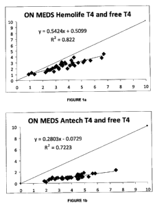
In Figures 1 a to 1 d the first comparative representations of T4 and Free T4
for
the disclosed (Hemolife TM) system are shown compared to Antech TM T4 and
Free T4, while the subject canine specimens are on thyroxine medication.
27
CA 02743485 2011-05-11
WO 2010/056681 PCT/US2009/063918
The top diagonal straight line in Figures la and lb represents the ideal
correlation (1.0; 100%). In Figure 1a, the disclosed non-RIA system shows an
r-squared value of 0.822. By comparison the Antech RIA system [Figure 1 b]
shows an r-squared value of 0.7223. The diversion from the perfect fit line in
the disclosed non RIA (Hemolife TM) system is within acceptable limits for
clinical diagnostic assays [r =0.9066; bias =1.06; n=40], whereas the
diversion
from the perfect fit line in the RIA (Antech TM) system is much greater [r=
0.8499; bias =2.84; n=40].
Figure 1c relative to Figure l d illustrates this comparative difference in
diversion between T4 and FT4. In the disclosed non-RIA system of Figure 1c,
the diversion relative to the top line data is minimal, as the two plotted
data
point lines run in parallel. In Figure 1d, the RIA system shows a wider
difference between T4 and FT4, and the diversion between the two plotted
data point lines is marked and non-parallel. These are significant differences
in being able to accurately diagnose the adequacy of thyroxine dosage in
animals being treated for thyroid disease.
In Figures 2a to 2d the first comparative representations of T4 and Free T4
for
the disclosed (Hemolife TM) system are shown compared to Antech TM T4 and
Free T4, for the high end of the therapeutic curve, while the subject canine
specimens are on thyroxine medication.
The top diagonal straight line in Figures 2a and 2b represent the ideal
correlation (1.0; 100%). In Figure 2a the disclosed non-RIA system shows
0.5778. Similarly, the Antech TM RIA system shows 0.6255 [Figure 2b]. The
diversion from the perfect fit line in the disclosed non-RIA (Hemolife TM)
system has more acceptable bias than in the RIA (Antech TM) system [r
=0.7601; bias =1.35; n=25, and r=0.7909; bias=3.39; n= 25, respectively], but
both systems perform poorly with respect to r-squared at the high end of the
therapeutic curve.
Figure 2c relative to Figure 2d illustrates this comparative difference in
diversion between T4 and FT4. In the disclosed non-RIA system of Figure 2c,
the diversion relative to the top line data is minimal, as the two plotted
data
28
CA 02743485 2011-05-11
WO 2010/056681 PCT/US2009/063918
point lines run in parallel. In Figure 2d, the RIA system shows a much wider
difference between T4 and FT4, and the diversion between the two plotted
data point lines is marked. These are significant differences in being able to
accurately diagnose the adequacy of thyroxine dosage in animals being
treated for thyroid disease because the Free T4 reads so much lower than the
T4.
In Figures 3a to 3d the first comparative representations of T4 and Free T4
for
the disclosed (Hemolife TM) system are shown compared to Antech TM T4 and
Free T4, while the subject canine specimens are not taking thyroxine
medication.
The top diagonal straight line in Figures 3a and 3b represents the ideal
correlation (1.0; 100%). In Figure 3a, the disclosed non-RIA system shows an
r-squared value of 0.816. By comparison the Antech RIA system [Figure 3b]
shows an r-squared value of 0.6443. The diversion from the perfect fit line in
the disclosed non RIA (Hemolife TM) system is within acceptable limits for
clinical diagnostic assays [r =0.9033; bias =0.43; n= 40], whereas the
diversion from the perfect fit line in the RIA (Antech TM) system is much
greater [r= 0.8027; bias =1.28; n=40].
Figure 3c relative to Figure 3d illustrates this comparative difference in
diversion between T4 and FT4. In the disclosed non-RIA system of Figure 3c,
the diversion relative to the top line data is minimal, as the two plotted
data
point lines start together and run in close parallel. In Figure 3d, the RIA
system shows a much wider difference between T4 and FT4, and the
diversion between the two plotted data point lines is marked and non-parallel.
These are significant differences in being able to accurately diagnose the
presence of thyroid disease.
In Figures 4a to 4d the first comparative representations of T4 and Free T4
for
the disclosed (Hemolife TM) system are shown compared to Antech TM T4 and
Free T4, for the lower end of the reference ranges while the subject canine
specimens are not taking thyroxine medication.
29
CA 02743485 2011-05-11
WO 2010/056681 PCT/US2009/063918
The top diagonal straight line in Figures 4a and 4b represents the ideal
correlation (1.0; 100%). In Figure 4a, the disclosed non-RIA system shows an
r-squared value of 0.8028. By comparison the Antech RIA system [Figure 4b]
shows a very low r-squared value of 0.3935. The diversion from the perfect fit
line in the disclosed non RIA (Hemolife TM) system is within acceptable limits
for clinical diagnostic assays [r =0.8960; bias =0.29; n= 25], whereas the
diversion from the perfect fit line in the RIA (Antech TM) system is much
greater [r= 0.6273; bias =0.98; n=25].
Figure 4c relative to Figure 4d illustrates this comparative difference in
diversion between T4 and FT4. In the disclosed non-RIA system of Figure 4c,
the diversion relative to the top line data is minimal, as the two plotted
data
point lines start together and run in close parallel. In Figure 4d, the RIA
system shows a much wider difference between T4 and FT4, and the
diversion between the two plotted data point lines is marked and non-parallel.
These are significant differences in being able to accurately diagnose the
presence of thyroid disease.
The Thyroid 5 profile measurement of the disclosure uses non-RIA assays
together to accurately form a predictive comprehensive profile to diagnose
thyroiditis and hypothyroidism in dogs. This profile is composed of T4, Free
T4, T3, Free T3 and TGAA. The TGAA analyte is included for genetic
screening of breeds at risk for heritable autoimmune thyroiditis. In its
preferred embodiment, the TGAA analyte is measured with the confirmatory
assay method which removes any non-specific binding that could falsely
elevate the result.
Since a percentage of thyroiditis cases have high circulating T3AA and/or
T4AA but normal TGAA, the measurement of T3AA and T4AA by non-RIA
methodology as disclosed is added on, whenever the results of the Thyroid 5
profile or prior results indicate the need to include these two additional non-
RIA tests. This is more effective in terms of cost and assay performance turn-
around-time than currently available commercial RIA profiles that include
TGAA, and optionally TSH.
CA 02743485 2011-05-11
WO 2010/056681 PCT/US2009/063918
OTHER EMBODIMENTS
Other embodiments are within the scope of the disclosure.
For example, other precipitating agents can be used, such as either
ammonium sulfate or hydrochloric acid in ethanol. Moreover, other antibodies
can be detected by the methods of this disclosure. For example, antigenic
determinants for islet cell autoantibodies can be isolated from islets of
Langerhans, labeled radioisotopically or non-radioisotopically, and then used
to assay for islet cell autoantibodies in serum.
In addition to dogs, the assay of the subject disclosure can quantitatively
determine levels in many other domestic and laboratory animal species
including but not limited to non-human primates, horse, pig, mouse, rat,
guinea pig, cow and cat. Previously, accurate measurements of thyroid
hormones were not possible for many of these species. The assay can thus
be used to screen valuable racing and working horse stock as well as
pleasure horses for the presence of thyroid autoantibody.
The method of the disclosure is particularly useful in screening assays which
may be performed in a general laboratory or a clinical setting more
efficiently
and without the need of highly trained staff, which are needed because the
available sophisticated quantitative assays are performed only in large
biomedical and commercial laboratories. The assay of the subject disclosure
may be performed simply in both veterinary hospitals and veterinary
laboratories to demonstrate the presence of thyroid autoantibodies in serum,
thus assisting in the laboratory diagnosis of thyroid disease.
In each of the methods discussed above, the autoantibodies which are initially
contacted with the sample may be attached to an immunological or physical
reaction surface. An immunological reaction surface is a surface which is
insoluble in the reacting medium and on which immunological reactions take
place. Typically they are glass, paper, or plastic, such as polystyrene or
polyacrylate. The surface may be the interior surface of a test tube, the well
of a micro titer plate or some other container suitable for an immunological
31
CA 02743485 2011-05-11
WO 2010/056681 PCT/US2009/063918
reaction. Physical reaction surfaces include glass or other types of beads or
the walls of a test tube or other surface.
Other appropriate surfaces on which immunological or physical reactions can
take place and which can be used, e.g. glass or plastic beads or rods, or
paper strips. An immunological or physical reaction surface is one to which
the antigens and antibodies adhere.
Immunological and physical reaction conditions for the disclosed methods are
for instance conditions with respect to temperature, concentration, solvent,
time of contact, and pH under which the immunological or physical reaction
such as the formation of an antibody-antigen-autoantibody complex occurs.
Those skilled in the art are familiar with the parameters under which such
complexes form. The temperature cannot be so high or the pH too extreme
as to inactivate the reactant. The solvent is typically a selected buffer or
other
carrier for the reactants. The reaction products, including the intermediate
reaction products of this disclosure, are soluble in the reaction solvent.
In each of the methods disclosed above, the detectable marker is preferably
an enzyme, but those skilled in the art to which the subject disclosure
pertains
would readily understand that other detectable markers may also be used.
These include, but are not limited to, luminescent probes, radioisotopes,
chromophores, fluorophores, or heavy metals. Enzymes are horseradish
peroxidase and alkaline phosphatase, although other enzymes known to
those skilled in the art can also be used in the subject disclosure.
The color detectors are most convenient for utilizing the antithyroid
autoantibody of the disclosure, but the disclosure is not so limited. Other
detection systems including radioisotopic, luminescent, or electrochemical
labels can also be employed.
The samples which can be analyzed using the methods of the subject
disclosure can be obtained from any vertebrate species in which one is
interested in determining the content of thyroid autoantibodies in the sample.
32
CA 02743485 2011-05-11
WO 2010/056681 PCT/US2009/063918
The sample which is analyzed using the subject disclosure is preferably a
biological fluid. Numerous other biological fluids from any vertebrate species
can be used in the assay. In embodiments of the disclosure, however, the
biological fluid comprises serum.
Numerous types of assays can be used in the disclosure as long as the
configuration of the assay allows the autoantibodies to recognize the
antibody: antigen complex, although the embodiment of the subject disclosure
comprises using the modified precipitation or binding assay which allows the
autoantibodies of the subject disclosure to recognize the antibody antigen.
Those skilled in the art would readily understand that any conventional
immunoassay which would allow the recognition of the antibody: antigen can
be used in the disclosure to both quantitatively and qualitatively detect
thyroid
autoantibodies in non-human species. Such other assays includes regular
precipitation assays wherein an antigen is precipitated or bound between the
bound autoantibodies on a solid carrier and non-radioisotopic labeled
autoantibodies, reverse precipitation assays, in which a non-radioisotopic
labeled autoantibodies are reacted with the antigen prior to contact with the
bound autoantibodies, and a simultaneous precipitation or binding assay, in
which the antibodies and the antigen are reacted simultaneously.
The process of this disclosure utilizes antibodies in new qualitative and
quantitative tests to permit immunologic measurement of thyroid
autoantibodies. The process is particularly useful in screening assays which
may be performed in a general laboratory or clinical setting without the need
of expensive equipment or a highly trained staff. Further the direct assay
system, such as the CLA, of the disclosure permits for assaying small
molecules such as hormones with a high level of specificity and sensitivity
and
efficiency. The disclosed system provides for a fast and efficient through put
in a laboratory or clinical setting.
The assay of the subject disclosure solves a long-standing problem which has
not been recognized by those working in the area of thyroid autoantibodies.
The problem relates to the need for an assay which can be used to
33
CA 02743485 2011-05-11
WO 2010/056681 PCT/US2009/063918
qualitatively and quantitatively detect thyroid autoantibodies antigen in
multiple species without using radioisotopes and without the need to create or
purchase an assay which is specific for each individual species, for example,
rat, rabbit, guinea pig, mouse, etc. It is impractical in the research area to
have individual thyroid assays for each species that may need thyroid function
testing.
The diagnostic systems of the subject disclosure unexpectedly solve this long-
standing problem which has previously been unrecognized in the thyroid
hormone diagnostic field. This assay will be of particular use in work where
clinicians can evaluate thyroid autoantibodies with a fast and efficient assay
useful for each of these species.
The assay of the subject disclosure is more sensitive than previous RIA
assays for thyroid function, and is safer (non-radioisotopic reagents), thus
providing a definite advantage over the previously used conventional assays.
Those skilled in the art will recognize the foregoing outline as a description
of
a modified procedure. The generalized outline omits certain of the specific
steps such as serial dilution and washing with appropriate buffers which are
standard in the procedure. Although specific buffers and other thyroid assay
reagents agents are described, and specific dilutions are employed to
illustrate the disclosure, these are only illustrative and many equivalents
are
possible.
The method is employed to determine whether non-humans are at risk to
disease caused by inherited or acquired thyroid disease, or for genetically
transmitting thyroid disease. It can also be used to measure thyroid levels in
non-human individuals experiencing or at risk to develop conditions such as
thrombotic states, cancers, or other autoimmune diseases and acute and
chronic inflammatory disorders.
The assays of the disclosure have greatly improved sensitivity and
specificity,
and can be used to detect both antibodies and autoantibodies produced in
small amounts in response to exposure to antigens, e.g.,non-human thyroid
hormone antibodies and autoantibodies.
34
CA 02743485 2011-05-11
WO 2010/056681 PCT/US2009/063918
Further although the exemplary assays relate to blood samples, it is clear
that
other biological fluids could be used.
The disclosure is not limited to the embodiments described as examples.
Many different variations in size and scope and features are possible. A
person of ordinary skill in the art will recognize that many further
combinations
and permutations of the present disclosure are possible. For instance,
instead of the direct chemiluminescent technique, the system can operate
with other non-radio immunoassays such as an immunofluorescent technique.
The disclosure embraces all such alterations, combinations, modifications,
and variations that fall within the spirit and scope of the appended claims.
The disclosure includes any and all embodiments of the following claims.