Note: Descriptions are shown in the official language in which they were submitted.
81627520
AMNION DERIVED ADHERENT CELLS
[0001] This application claims benefit of United States Provisional Patent
Application No.
61/116,248, filed November 19, 2009.
1. FIELD
[0002] Provided herein are novel angiogenic cells from amnion, and
populations of such
cells, referred to herein as "amnion derived adherent cells" (AMDACs). Amnion
derived
adherent cells are distinct from previously-described placental stem cells,
including tissue
culture plastic adherent placental stem cells.
2. BACKGROUND
[0003] Cell compositions, e.g., stem cell compositions, have become an
attractive therapy
for a number of physiological deficiencies, e.g., bone marrow replacement. A
need exists for
additional populations of cells, e.g., stem cells or progenitor cells that
have angiogenic
potential and/or properties.
3. SUMMARY
[00041 In one aspect, provided herein is an isolated amnion derived
adherent cell, also
referred to herein as an AMDAC, wherein said cell is adherent to tissue
culture plastic, and
wherein said cell is OCT-4- (negative for OCT-4, also known as POU5F1 or
octamer binding
protein 4), or as determined by reverse transcriptase-polymerase chain
reaction (RT-PCR),
e.g., as compared to an appropriate control cell line, such as an embryonal
carcinoma-derived
stern cell line (e.g., NTERA-2, e.g., available from the American Type Culture
Collection,
ATCC Number CRL-1973), as determined by RT-PCR for 30 cycles. In a specific
embodiment, the cells are OCT-4, as determined by RT-PCR, and VEGFR1/Flt-1+
(vascular
endothelial growth factor receptor 1) and/or VEGFR2/ICDR+ (vascular
endothelial growth
factor receptor 2, also known as lcinase insert domain receptor), as
determined by
immunolocalization. In another specific embodiment, the cells are OCT-4, as
determined by
RT-PCR, and CD49P- (integrin-a6+), as determined by immunolocalization. In a
specific
embodiment, said cell is OCT-4, as determined by RT-PCR, and HLA-G-, as
determined by
RT-PCR. In another specific embodiment, said cell is OCT-4, as determined by
RT-PCR,
and CD904; CD105+, or CD11T as determined by immunolocalization. In a more
specific
embodiment, said OCT-4- cell is CD90+, CD105+, and CD11T. In another specific
embodiment, the cell is OCT-4, and does not express SOX2, e.g., as determined
by RT-PCR
for 30 cycles.
1
CA 2743566 2017-08-11
CA 02743566 2011-05-12
WO 2010/059828
PCT/US2009/065152
[0005] In another embodiment, said OCT-4- cell is one or more of CD29+,
CD73+, ABC-
p+, and CD38-, as determined by immunolocalization.
[0006] In another specific embodiment, said OCT-4- cell is additionally one
or more of
CD9 CD10 CD44 CD54 CD98% (angiopoietin receptor), TEM-7-' (tumor
endothelial marker 7), CD31-, CD34-, CD45-, CD133-, CD143- (angiotensin-I-
converting
enzyme, ACE), CD146- (melanoma cell adhesion molecule), CXCR4- (chemokine (C-X-
C
motif) receptor 4) as determined by immunolocalization. In a more specific
embodiment,
said cell is CD9', CD1O, CD44% CD54', TEM-7',
CD31-, CD34-, CD45-,
CD133-, CD143-, CD146-, and CXCR4- as determined by immunolocalization. In
another
more specific embodiment, the amnion derived adherent cell provided herein is
OCT-4, as
determined by RT-PCR; VEGFRI/F1t-1+ and/or VEGFR2/KDR+, as determined by
immunolocalization; and one or more, or all, of CD31-, CD34-, CD45-, CD133-,
and/or Tie-
2- as determined by immunolocalization. In a specific embodiment, the amnion
derived
adherent cell, or a population of amnion derived adherent cells, expresses at
least 2 log less
PCR-amplified mRNA for OCT-4 at, e.g., >20 cycles, than an NTERA-2 cell, or
population
of NTERA-2 cells having an equivalent number of cells. In another specific
embodiment,
said OCT-4- cell is additionally VE-cadherin- (CD144-) as determined by
immunolocalization. In another specific embodiment, said OCT-4- cell is
additionally
positive for CD105 and CD200-' as determined by immunolocalization. In another
specific
embodiment, said OCT-4- cell does not express CD34 as detected by
immunolocalization
after exposure to 1 to 100 ng/mL VEGF (vascular endothelial growth factor) for
4 to 21 days.
[0007] In another aspect, provided herein is an isolated amnion derived
adherent cell,
wherein said cell is adherent to tissue culture plastic, and wherein said cell
is OCT-4- and
SOX-T, as determined by RT-PCR; and CD90+, CD105+, and CD117-, as determined
by
flow cytometry. In a specific embodiments, the OCT-4, SOX-2- cell is
additionally HLA-G-
or CD271-, as determined by flow cytometry. In a more specific embodiment,
said cell is
OCT-4- and SOX-2, as determined by RT-PCR; and CD90 CD105 CD117-, CD271- and
HLA-G-, as determined by flow cytometry.
[0008] In another aspect, provided herein is an isolated amnion derived
adherent cell,
wherein said cell is adherent to tissue culture plastic, and wherein said cell
is positive for
CD309 (also known as VEGFR2/KDR+).
[0009] In another aspect, provided herein is an isolated amnion derived
adherent cell,
wherein said cell is adherent to tissue culture plastic, and wherein said cell
is OCT-4 , as
determined by RT-PCR, and one or more of VEGFR2/KDR+, CD9+, CD54+, CD105+,
2
CA 02743566 2011-05-12
WO 2010/059828
PCT/US2009/065152
CD200 or VE-cadherin-, as determined by immunolocalization. in a specific
embodiment,
said cell is OCT-4, as determined by RT-PCR at, e.g., >20 cycles, and
VEGFR21KDR' ,
CD9+, CD54+, CD105+, CD200-, and VE-cadherin-, as determined by
immunolocalization.
In another specific embodiment, the cell does not express CD34, as detected by
immunolocalization, after exposure to 1 to 100 ng/mL VEGF for 4 to 21 days.
[0010] In another
embodiment, the amnion derived adherent cell is OCT-4, CD49r,
HLA-G-, CD90+, CD105+, and CD 117-. In a more specific embodiment, said cell
is one or
more of CD9', CD10, CD44 CD54 CD98% CD31-, CD34-
, CD45-,
CD133-, CD143-, CD146- (melanoma cell adhesion molecule), or CXCR4-, as
determined
by immunolocalization or flow cytometry. In a more specific embodiment, said
cell is CD9
CD44% CD54, CD98% Tie-2', CD31 ,
CD34 , CD45 , CD133 , CD143 ,
CD146-, and CXCR4- as determined by immunolocalization. In another specific
embodiment, said cell is additionally VEGFR1/Flt-r and/or VEGFR2/KDR% as
determined
by immunolocalization; and one or more of CD31-, CD34-, CD45-, CD133-, and/or
Tie-2- as
determined by immunolocalization. In another specific embodiment, said cell is
additionally
VEGFR1/F1t-1+, VEGFR2/KDR+, CD31-, CD34-, CD45-, CD133-, and Tie-2- as
determined
by immunolocalization.
[0011] In another
embodiment, provided herein is an isolated amnion derived adherent
cell, wherein said cell does not express mRNA for FGF4, IFNG, CXCL10, ANGPT4,
ANGPTL3, FGA, LEP, PRL, PROK1, TNMD, FLT3, XLKD1, CDH5, LECT1, PLG, TERT,
SOX2, NANOG, MMP-13, DLX5, or BGLAP, as determined by RT-PCR, e.g., for 30
cycles.
In another embodiment, provided herein is an isolated amnion derived adherent
cell, wherein
said cell does not constitutively express one or more of invariant chain, HLA-
DR-DP-DQ,
CD6, CD271, as determined by flow cytometry.
[0012] Further
provided herein is an isolated population of cells comprising an amnion
derived adherent cell. In a specific embodiment, at least about 50%, 60%, 70%,
80%, 90%,
95%, 98% or 99% of cells in said population are amnion derived adherent cells.
In one
embodiment, said cell is OCT-4, as determined by RT-PCR, and VEGFR1/F1t-1+
and/or
VEGFR2/KDR+ as determined by immunolocalization, and wherein said isolated
population
of cells is not an amnion. In another embodiment, provided herein is an
isolated population
of cells comprising an amnion derived adherent cell that is OCT-4- and HLA-G-,
as
determined by RT-PCR, and VEGFR1/Flt-1-' or VEGFR2/KDR as determined by
immunolocalization; and wherein said isolated population of cells is not an
amnion. In a
specific embodiment, at least about 50%, 60%, 70%, 80%, 90%, 95%, 98% or 99%
of cells in
3
CA 02743566 2011-05-12
WO 2010/059828
PCT/US2009/065152
said population are said amnion derived adherent cells. In another embodiment,
provided
herein is an isolated population of cells comprising an amnion derived
adherent cell, wherein
said cell is adherent to tissue culture plastic, wherein said cell is OCT-4,
as determined by
RT-PCR, VEGFR1/Flt-1 and VEGFR2/KDR as determined by immunolocalization,
wherein said cell is additionally one or more of CD9-', CD10% CD44', CD54%
CD98%
TEM-7', CD31-, CD34-, CD45-, CD133-, CD143-, CD146-, or CXCR4-, as determined
by
immunolocalization, or HLA-G- as determined by RT-PCR for, e.g., >20 cycles,
and wherein
said isolated population of cells is not an amnion. In a specific embodiment,
at least about
50%, 60%, 70%, 80%, 90%, 95%, 98% or 99% of cells in said population are said
amnion
derived adherent cells. In another embodiment, provided herein is an isolated
population of
cells comprising an amnion derived adherent cell, wherein said cell is
adherent to tissue
culture plastic, wherein said cell is OCT-4, as determined by RT-PCR,
VEGFRI/Flt-1+
and/or VEGFR2/KDR as determined by immunolocalization, wherein said cell does
not
express CD34 as detected by immunolocalization after exposure to 1 to 100
ng/mL VEGF for
4 to 21 days, and wherein said isolated population of cells is not an amnion.
In a specific
embodiment, at least about 50%, 60%, 70%, 80%, 90%, 95%, 98% or 99% of cells
in said
population are said amnion derived adherent cells. In another embodiment, any
of the above
populations of cells comprising amnion derived adherent cells forms sprouts or
tube-like
structures when cultured in the presence of angiogenic factors such as
vascular endothelial
growth factor (VEGF), epithelial growth factor (EGF), platelet derived growth
factor (PDGF)
or basic fibroblast growth factor (bFGF), e.g., on a substrate such as
MATRIGELTm.
[0013] In another
embodiment, provided herein is a population of cells, wherein at least
about 50%, 60%, 70%, 80%, 90%, 95% or 98% of cells in said isolated population
of cells
are amnion derived adherent cells that are OCT-4, as determined by RT-PCR, and
positive
for VEGFR2/KDR, CD9, CD54, CD105, or CD200. In a specific embodiment, at least
about
50%, 60%, 70%, 80%, 90%, 95% or 98% of cells in said isolated population of
cells are
amnion derived adherent cells that are OCT-4, as determined by RT-PCR, and
VEGFR2/KDR% CD9 , CD54% CD105 and CD200, as determined by immunolocalization.
In a more specific embodiment, said amnion derived adherent cells do not
express CD34, as
detected by immunolocalization, after exposure to 1 to 100 ng/mL VEGF for 4 to
21 days. In
a specific embodiment, said amnion derived adherent cells are adherent to
tissue culture
plastic. In another specific embodiment, said population of cells forms
sprouts or tube-like
structures when cultured in the presence of an angiogenic factor such as
vascular endothelial
4
CA 02743566 2011-05-12
WO 2010/059828 PCT/US2009/065152
growth factor (VEGF), epithelial growth factor (EGF), platelet derived growth
factor (PDGF)
or basic fibroblast growth factor (bFGF), e.g., on a substrate such as
MATRIGELTm.
[0014] In another embodiment, provided herein is a population of cells,
e.g., human cells,
wherein at least about 50%, 60%, 70%, 80%, 90%, 95% or 98% of cells in said
isolated
population of cells are amnion derived adherent cells that express RNA for one
or more, or
all, of ACTA2, ADAMTS1, AMOT, ANG, ANGPT1, ANGPT2, ANGPTL1, ANGPTL2,
ANGPTL4, BAH, CD44, CD200, CEACAM1, CHGA, COL15A1, COL18A1, COL4A1,
COL4A2, COL4A3, CSF3, CTGF, CXCL12, CXCL2, DNMT3B, ECGF1, EDG1, EDIL3,
ENPP2, EPHB2, FBLN5, F2, FGF1, FGF2, FIGF, FLT4, FN1, FST, FOXC2, GRN, HGF,
HEY1, HSPG2, IFNB1, IL8, IL12A, ITGA4, ITGAV, ITGB3, MDK, MMP2, MYOZ2,
NRP1, NRP2, PDGFB, PDGFRA, PDGFRB, PECAM1, PF4, PGK1, PROX1, PTN,
SEMA3F, SERPINB5, SERPINC1, SERPINF1, TIMP2, TIMP3, TGFA, TGFB1, THBS1,
THBS2, TIE1, TIE2/TEK, TNF, 'TNNI1, TNFSF15, VASH1, VEGF, VEGFB, VEGFC,
VEGFR1/FLT1, or VEGFR2/KDR.
[0015] In another embodiment, provided herein is a population of cells,
e.g., a population
of amnion derived adherent cells, or a population of cells wherein at least
about 50%, 60%,
70%, 80%, 90%, 95% or 98% of cells in said isolated population of cells are
amnion derived
adherent cells that express one or more of, or all of, CD49d, Connexin-43, HLA-
ABC, Beta
2-microglobulin, CD349, CD318, PDL1, CD106, Galectin-1, ADAM 17 precursor (A
disintegrin and metalloproteinase domain 17) (TNF-alpha converting enzyme)
(TNF-alpha
convertase), Angiotensinogen precursor, Filamin A (Alpha-filamin) (Filamin 1)
(Endothelial
actin-binding protein) (ABP-280) (Nonmuscle filamin), Alpha-actinin 1 (Alpha-
actinin
cytoskeletal isoform) (Non-muscle alpha-actinin 1) (F-actin cross linking
protein), Low-
density lipoprotein receptor-related protein 2 precursor (Megalin)
(Glycoprotein 330) (gp330),
Macrophage scavenger receptor types T and TI (Macrophage acetylated LDL
receptor 1 and II),
Activin receptor type JIB precursor (ACTR-IIB), Wnt-9 protein, Glial
fibrillary acidic protein,
astrocyte (GFAP), Myosin-binding protein C, cardiac-type (Cardiac MyBP-C) (C-
protein,
cardiac muscle isoform), or Myosin heavy chain, nonmuscle type A (Cellular
myosin heavy
chain, type A) (Nonmuscle myosin heavy chain-A) (NMMHC-A).
[0016] In another aspect, provided herein is a population of cells, e.g., a
population of
amnion derived adherent cells, or a population of cells wherein at least about
50%, 60%, 70%,
80%, 90%, 95% or 98% of cells in said isolated population of cells are amnion
derived
adherent cells that secrete one or more, or all, of VEGF, HGF, IL-8, MCP-3,
FGF2, follistatin,
CA 02743566 2011-05-12
WO 2010/059828 PCT/US2009/065152
G-CSF, EGF, ENA-78, GRO, 1L-6, MCP-1, PDGF-BB, T1MP-2, uPAR, or galectin-1,
e.g.,
into culture medium in which the cell, or cells, are grown.
[0017] In another embodiment, provided herein is a population of cells,
e.g., a population
of amnion derived adherent cells, or a population of cells wherein at least
about 50%, 60%,
70%, 80%, 90%, 95% or 98% of cells in said isolated population of cells are
amnion derived
adherent cells that express angiogenic micro RNAs (miRNAs) at a higher level
than bone
marrow-derived mesenchymal stem cells, wherein said miRNAs comprise one or
more, or all
of, miR-17-3p, miR-18a, miR-18b, miR-19b, miR-92, and/or miR-296. In another
embodiment, provided herein is a population of cells, e.g., a population of
amnion derived
adherent cells, or a population of cells wherein at least about 50%, 60%, 70%,
80%, 90%,
95% or 98% of cells in said isolated population of cells are amnion derived
adherent cells that
express one or more of, or all of, angiogenic micro RNAs (miRNAs) at a lower
level than
bone marrow-derived mesenchymal stem cells, wherein said miRNAs comprise one
or more,
or all of, miR-20a, miR-20b, miR-221, miR-222, miR-15b, and/or miR-16. In
certain
embodiments, AMDACs, or populations of AMDACs, express one or more, or all,
the
angiogenic miRNAs miR-17-3p, miR-18a, miR-18b, miR-19b, miR-92, miR-20a, miR-
20b,
miR-296, miR-221, miR-222, miR-15b, and/or miR-16.
[0018] In another specific embodiment, provided herein is an amnion derived
angiogenic
cell, or population of amnion derived angiogenic cells, that express increased
levels of
CD202b, IL-8 and/or VEGF under hypoxic conditions (e.g., less than about 5%
02) compared
to normoxic conditions (e.g., about 20% or about 21% 02).
[0019] In another specific embodiment, the isolated population of amnion
derived
adherent cells additionally comprises a second type of cell. In specific
embodiments, the
AMDACs comprise at least 10%, 15%, 20%, 25%, 30%, 35%, 40%, 45%, 50%, 55%,
60%,
65%, 70%, 80%, 85%, 90% , 95% or at least 98% of cells in said population. In
a specific
embodiment, the second type of cell is contained within or isolated from
placental blood,
umbilical cord blood, crude bone marrow or other tissues. In a more specific
embodiment,
said second type of cell is embryonic stem cell, a blood cell, a stem cell
isolated from
peripheral blood, a stem cell isolated from placental blood, a stem cell
isolated from placental
perfusate, a stem cell isolated from placental tissue, a stem cell isolated
from umbilical cord
blood, an umbilical cord stem cell, a bone marrow-derived mesenchymal stem
cell, a
mesenchymal stromal cell, a hematopoietic stem cell, a somatic stem cell, a
chondrocyte, a
fibroblast, a muscle cell, an endothelial cell, an endothelial progenitor
cell, a pericyte, a
myocyte, a cardiomyocyte, a myoblast, an angioblast, or a cardiomyoblast. In
another
6
CA 02743566 2011-05-12
WO 2010/059828 PCT/US2009/065152
specific embodiment, said second type of cell is a hematopoietic stem or
progenitor cell, e.g.,
a CD34+ cell. In another more specific embodiment, said second type of cell
comprises at
least 10%, 15%, 20%, 25%, 30%, 35%, 40%, 45%, 50%, 55%, 60%, 65%, 70%, 80%,
85%,
90%, 95% or at least 98% of cells in said population.
[0020] In another specific embodiment, any of the above cells are, or have
been,
proliferated in culture. In another specific embodiment, any of the above
cells is from a
culture of said cells that has been passaged at least 1, 2, 3, 4, 5, 6, 7, 8,
9, 10, 11, 12, 13, 14,
15, 16, 17, 18, 19, or 20 times, or more. In another specific embodiment, any
of the above
cells is from a culture that has doubled in culture at least 1, 2, 3, 4, 5, 6,
7, 8, 9, 10, 11, 12, 13,
14, 15, 16, 17, 18, 19, 20, 21, 22, 23, 24, 25, 26, 27, 28, 29, 30, 31, 32,
33, 34, 35, 36, 37, 38,
39, 40, 41, 42, 43, 44, 45, 46, 47, 48, 49 or at least 50 times, or more.
[0021] In another aspect, provided herein is a composition, e.g., a
pharmaceutical
composition, comprising any of the amnion derived adherent cells, or
populations of cells
comprising the amnion derived adherent cells, provided herein. In a specific
embodiment,
the composition is a matrix or scaffold, e.g., a natural tissue matrix or
scaffold, for example, a
permanent or degradable decellularized tissue matrix or scaffold; or synthetic
matrix or
scaffold. In a more specific embodiment, said matrix or scaffold is shaped in
the form of a
tube or other three-dimensional form of an organoid. In another more specific
embodiment,
said matrix is a decellularized tissue matrix. In another specific embodiment,
the
composition comprises one or more of the isolated amnion derived adherent
cells provided
herein, or population of cells comprising the amnion derived adherent cells,
in a
physiologically-acceptable solution, e.g., a saline solution, culture medium
or the like.
[0022] In another aspect, provided herein is a method of treating an
individual having a
disease or disorder of the circulatory system, comprising administering one or
more of the
amnion derived adherent cells described herein to said individual in an amount
and for a time
sufficient for detectable improvement of one or more symptoms of said disease
or disorder.
In another embodiment, provided herein is a method of treating an individual
having a
disease or disorder of the circulatory system, comprising administering amnion
derived
adherent cells to said individual in an amount and for a time sufficient for
detectable
improvement of one or more indicia of cardiac function, wherein said indicia
of cardiac
function are chest cardiac output (CO), cardiac index (CI), pulmonary artery
wedge pressure
(PAWP), cardiac index (CI), % fractional shortening (%FS), ejection fraction
(EF), left
ventricular ejection fraction (LVEF); left ventricular end diastolic diameter
(LVEDD), left
ventricular end systolic diameter (LVESD), contractility (dP/dt), a decrease
in atrial or
7
CA 02743566 2011-05-12
WO 2010/059828 PCT/US2009/065152
ventricular functioning, an increase in pumping efficiency, a decrease in the
rate of loss of
pumping efficiency, a decrease in loss of hemodynamic functioning, or decrease
in
complications associated with cardiomyopathy, as compared to the individual
prior to
administration of amnion derived adherent cells.
[0023] In a specific embodiment, said disease or disorder is myocardial
infarction. In
another specific embodiment, said disease or disorder is cardiomyopathy. In
other specific
embodiments, said disease or disorder is aneurysm, angina, aortic stenosis,
aortitis,
arrhythmias, arteriosclerosis, arteritis, asymmetric septal hypertrophy (ASH),
atherosclerosis,
atrial fibrillation and flutter, bacterial endocarditis, Barlow's Syndrome
(mitral valve
prolapse), bradycardia, Buerger's Disease (thromboangiitis obliterans),
cardiomegaly, carditis,
carotid artery disease, coarctation of the aorta, congenital heart defects,
congestive heart
failure, coronary artery disease, Eisenmenger's Syndrome, embolism,
endocarditis,
erythromelalgia, fibrillation, fibromuscular dysplasia, heart block, heart
murmur,
hypertension, hypotension, idiopathic infantile arterial calcification,
Kawasaki Disease
(mucocutaneous lymph node syndrome, mucocutaneous lymph node disease,
infantile
polyarteritis), metabolic syndrome, microvascular angina, myocarditis,
paroxysmal atrial
tachycardia (PAT), periarteritis nodosa (polyarteritis, polyarteritis nodosa),
pericarditis,
peripheral vascular disease, critical limb ischemia, phlebitis, pulmonary
valve stenosis
(pulmonic stenosis), Raynaud's Disease, renal artery stenosis, renovascular
hypertension,
rheumatic heart disease, diabetic vasculopathy, septal defects, silent
ischemia, syndrome X,
tachycardia, Takayasu's Arteritis, Tetralogy of Fallot, transposition of the
great vessels,
tricuspid atresia, truncus arteriosus, valvular heart disease, varicose
ulcers, varicose veins,
vasculitis, ventricular septal defect, Wolff-Parkinson-White Syndrome,
endocardial cushion
defect, acute rheumatic fever, acute rheumatic pericarditis, acute rheumatic
endocarditis,
acute rheumatic myocarditis, chronic rheumatic heart diseases, diseases of the
mitral valve,
mitral stenosis, rheumatic mitral insufficiency, diseases of aortic valve,
diseases of other
endocardial structures, ischemic heart disease (acute and subacute), angina
pectoris, acute
pulmonary heart disease, pulmonary embolism, chronic pulmonary heart disease,
kyphoscoliotic heart disease, myocarditis, endocarditis, endomyocardial
fibrosis, endocardial
fibroelastosis, atrioventricular block, cardiac dysrhythmias, myocardial
degeneration,
cerebrovascular disease, a disease of arteries, arterioles and capillaries, or
a disease of veins
and lymphatic vessels.
[0024] In other specific embodiments, said disease or disorder is an
occlusion and
stenosis of precerebral arteries, or occlusion of cerebral arteries. In one
aspect, provided
8
81627520
herein is a method of treating an individual who has a disruption of the flow
of blood in or
around the individual's brain, e.g., who has a symptom or neurological deficit
attributable to
a disruption of the flow of blood in or around the individual's brain or
central nervous system
(CNS), comprising administering to said individual a therapeutically effective
amount or
isolated AMDACs. In certain embodiments, the disruption of flow of blood
results in anoxic
injury or hypoxic injury to the individual's brain or CNS.
[00251 In other specific embodiments, said disease or disorder is an
occlusion and
stenosis of peripheral arteries. In one aspect, provided herein is a method of
treating an
individual who has a disruption of the flow of blood in or around limb, e.g.,
who has a
symptom or vascular deficit attributable to a disruption of the flow of blood
in or around the
individual's peripheral vascular system, comprising administering to said
individual a
therapeutically effective amount of isolated AMDACs. In certain embodiments,
the
disruption of flow of blood results in anoxic injury or hypoxic injury to the
individual's limbs
and or extremities.
[00261 In another aspect, provided herein is a method of treating an
individual suffering
from a wound or trauma, comprising administering one or more of the amnion
derived
adherent cells described herein to said individual in an amount and for a time
sufficient for
detectable improvement of said wound or trauma.
[00271 In another specific embodiment of the method of treatment, said
cells are
administered to said individual by injection. In a more specific embodiment,
said injection is
injection into an ischemic area of the individual's heart. In another specific
embodiment of
the method of treatment, said cells are administered to said individual by
intravenous infusion.
In another specific embodiment of the method of treatment, said cells, or a
population of said
cells, or a population of cells comprising said cells, is administered to said
individual by
implantation in said individual of a matrix or scaffold comprising amnion
derived adherent
cells, as described above.
9
CA 2743566 2017-08-11
81627520
[0027a] This application as claimed relates to:
- an isolated population of cells comprising human amnion derived adherent
cells, wherein said amnion derived adherent cells are adherent to tissue
culture plastic, and
wherein said amnion derived adherent cells are OCT-4- (octamer binding protein
4) and }ILA-
Gas determinable by RT-PCR and CD49ff and CD90+ as determinable by flow
cytometry;
- a composition comprising the isolated population of cells as described
herein
and a pharmaceutically acceptable carrier;
- a permanent or degradable decellularized or synthetic matrix or scaffold
comprising the isolated population as described herein;
- use of the population of cells comprising amnion derived adherent cells
of as
described herein for treating an individual having a disease or disorder of
the circulatory
system, wherein the disease or disorder of the circulatory system is
myocardial infarction,
stroke, congestive heart failure, peripheral artery disease, hypoplastic left
heart syndrome,
diabetic ulcer, decubitus ulcer, venous ulcer, arterial ulcer, bum, non-union
fracture, tumor-
associated bone loss, or maxillofacial bone repair;
- use of the population of amnion derived adherent cells as described
herein for
treating an individual having a disruption of blood flow in or around the
central nervous
system; and
- use of the population of amnion derived adherent cells as described
herein for
treating an individual having a disruption of blood flow in or around a limb.
[0028] The isolated amnion derived adherent cells and cell populations
provided herein are
not the isolated placental stem cells or cell populations described, e.g., in
U.S. Patent
No. 7,255,879 or U.S. Patent Application Publication No. 2007/0275362. The
isolated amnion
derived adherent cells provided herein are also not endothelial progenitor
cells, amniotic
epithelial cells, trophoblasts, cytotrophoblasts, embryonic germ cells,
embryonic stem cells,
cells obtained from the inner cell mass of an embryo, or cells obtained from
the gonadal ridge
of an embryo.
9a
Date Recue/Date Received 2020-08-31
CA 02743566 2011-05-12
WO 2010/059828 PCT/US2009/065152
[0029] As used herein, the term "about" means, e.g., within 10% of a stated
figure or
value.
[0030] As used herein, the term "angiogenic," in reference to the amnion
derived
adherent cells described herein, means that the cells can form vessels or
vessel-like sprouts,
or that the cells can promote angiogenesis (e.g., the formation of vessels or
vessel-like
structures) in another population of cells, e.g., endothelial cells.
[0031] As used herein, the term "angiogenesis" refers to the process of
blood vessel
formation that includes, but is not limited to, endothelial cell activation,
migration,
proliferation, matrix remodeling and cell stabilization.
[0032] As used herein, the term "stem cell" defines the functional
properties of any given
cell population that can proliferate extensively, but not necessarily
infinitely, and contribute
to the formation of multiple tissues, either during embryological development
or post-natal
tissue replacement and repair.
[0033] As used herein, the term "progenitor cell" defines the functional
properties of any
given cell population that can proliferate extensively, but not necessarily
infinitely, and
contribute to the formation of a restricted set of multiple tissues in
comparison to a stem cell,
either during embryological development or post-natal tissue replacement and
repair.
[0034] As used herein, the term "derived" means isolated from or otherwise
purified. For
example, amnion derived adherent cells are isolated from amnion. The term
"derived"
encompasses cells that are cultured from cells isolated directly from a
tissue, e.g., the amnion,
and cells cultured or expanded from primary isolates.
[0035] As used herein, "immunolocalization" means the detection of a
compound, e.g., a
cellular marker, using an immune protein, e.g., an antibody or fragment
thereof in, for
example, flow cytometry, fluorescence-activated cell sorting, magnetic cell
sorting, in situ
hybridization, immunohistochemistry, or the like.
[0036] As used herein, the term "isolated cell" means a cell that is
substantially separated
from other, cells of the tissue, e.g., amnion or placenta, from which the cell
is derived. A cell
is "isolated" if at least about 20%, 30%, 40%, 50%, 60%, 70%, 80%, 90%, 95%,
or at least
99% of the cells with which the stem cell is naturally associated are removed
from the cell,
e.g., during collection and/or culture of the cell.
[0037] As used herein, the term "isolated population of cells" means a
population of cells
that is substantially separated from other cells of the tissue, e.g., amnion
or placenta, from
which the population of cells is derived. A cell is "isolated" if at least
about 20%, 30%, 40%,
50%, 60%, 70%, 80%, 90%, 95%, or at least 99% of the cells with which the
population of
CA 02743566 2011-05-12
WO 2010/059828 PCT/US2009/065152
cells, or cells from which the population of cells is derived, is naturally
associated are
removed from the cell, e.g., during collection and/or culture of amnion
derived adherent cells.
[0038] As used herein, a cell is "positive" for a particular marker when
that marker is
detectable above background, e.g., by immunolocalization, e.g., by flow
cytometry; or by
RT-PCR. For example, a cell is described as positive for, e.g., CD105 if CD105
is detectable
on the cell in an amount detectably greater than background (in comparison to,
e.g., an
isotype control). In the context of, e.g., antibody-mediated detection,
"positive," as an
indication a particular cell surface marker is present, means that the marker
is detectable
using an antibody, e.g., a fluorescently-labeled antibody, specific for that
marker; "positive"
also means that a cell bears that marker in a amount that produces a signal,
e.g., in a
cytometer, that is detectably above background. For example, a cell is
"CD105+" where the
cell is detectably labeled with an antibody specific to CD105, and the signal
from the
antibody is detectably higher than a control (e.g., background). Conversely,
"negative" in the
same context means that the cell surface marker is not detectable using an
antibody specific
for that marker compared to background. For example, a cell is "CD34-" where
the cell is
not detectably labeled with an antibody specific to CD34. Unless otherwise
noted herein,
cluster of differentiation ("CD") markers are detected using antibodies. For
example, OCT-4
can be determined to be present, and a cell is OCT-4 if mRNA for OCT-4 is
detectable
using RT-PCR, e.g., for 30 cycles.
4. BRIEF DESCRIPTION OF THE FIGURES
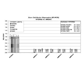
[0039] FIG. 1 shows expression of stem cell-related genes by amnion derived
adherent
cells and NTERA-2 cells.
[0040] FIG. 2 shows the expression of TEM-7 on the cell surface of amnion
derived
adherent cells (AMDACs).
[0041] FIG. 3 shows the secretion of selected angiogenic proteins by amnion
derived
adherent cells.
[0042] FIG. 4 shows the angiogenic effect of amnion derived adherent cells
conditioned
medium on Human Endothelial Cell (HUVEC) tube formation.
[0043] FIG. 5 shows the angiogenic effect of amnion derived adherent cells
conditioned
medium on human endothelial cell migration.
[0044] FIG. 6 shows the effect of amnion derived adherent cell-conditioned
medium on
Human Endothelial Cell proliferation.
[0045] FIG. 7 shows the uptake of acetylated LDL by HUVECs and amnion
derived
adherent cells.
11
CA 02743566 2011-05-12
WO 2010/059828 PCT/US2009/065152
[0046] FIG. 8 shows tube formation of HUVECs and amnion derived adherent
cells.
[0047] FIG. 9 shows the secretion of VEGF and IL-8 by amnion derived
adherent cells
under hypoxic and normoxic conditions.
[0048] FIG. 10 shows the expression of cellular marker Tie2 under normoxic
(about 21%
02) and hypoxic (less than about 5% 02) conditions. Y axis: percentage of
cells positive for
Tie2 by flow cytometry.
[0049] FIG. 11 shows the cardiomyocytic differentiation potency of amnion
derived
adherent cells, wherein AM refers to amnion derived adherent cells (AMDACs),
HD refers to
untreated hanging drop, HD ND refers to hanging drop exposed to inducing
conditions, and
HD ND + 5-AZA refers to induction in the presence or absence of 5-azacytidine.
CTRL
refers to the untreated hanging drop control.
[0050] FIG. 12 shows positive effect of AMDACs on angiogenesis in a chick
chorioallantois angiogenesis model. Lot 1, Lot 2, Lot 3: AMDACs from three
separate cell
preparations. bFGF: basic fibroblast growth factor (positive control).
MDAMB231:
Angiogenic breast cancer cell line (positive control). Y axis: Degree of blood
vessel
formation.
[0051] FIG. 13 shows positive effect of AMDAC-conditioned medium on
angiogenesis
in a chick chorioallantois angiogenesis model. Lot 1, Lot 2, Lot 3: AMDACs
from three
separate cell preparations. bFGF: basic fibroblast growth factor (positive
control).
MDAMB231: Angiogenic breast cancer cell line (positive control). Y axis:
Degree of blood
vessel formation.
[0052] FIGS. 14A, 14B: Hydrogen peroxide-generated reactive oxygen species
present
in cultures of astrocytes, co-cultures of astrocytes and bone marrow-derived
mesenchymal
stem cells (BM-MSCs), or co-cultures of astrocytes and AMDACs. 14A: AMDACs,
Lot 1;
14B: AMDACs, Lot 2. The conditions HA (human astrocytes) alone, astrocytes +
H202, and
astrocytes + BM-MSCs + H202 are the same for FIGS. 14A and 14B. RFU ROS
activity:
Relative fluorescence units for reactive oxygen species.
5. DETAILED DESCRIPTION
5.1 CHARACTERISTICS OF AMNION DERIVED ADHERENT CELLS
[0053] Provided herein are unique adherent, angiogenic cells, and
populations of such
cells, isolatable from the amnion, referred to herein as "amnion derived
adherent cells" or
AMDACs. Amnion derived adherent cells superficially resemble mesenchymal cells
in
appearance, having a generally fibroblastoid shape. The cells adhere to a cell
culture surface,
e.g., to tissue culture plastic.
12
CA 02743566 2011-05-12
WO 2010/059828 PCT/US2009/065152
[0054] AMDACs display cellular markers that distinguish them from other
amnion-
derived, or placenta-derived, cells. For example, in one embodiment, the
amnion derived
adherent cell is OCT-4- (octamer binding protein 4), as determined by RT-PCR.
In another
specific embodiment, the OCT-4- amnion derived adherent cell is CD49f-, as
determined by
immunolocalization. In another specific embodiment, said OCT-4- cell is HLA-G-
, as
determined by RT-PCR. In another specific embodiment, the OCT-4- cell is
VEGFR1/Flt-1+
(vascular endothelial growth factor receptor 1) and/or VEGFR2/KDR+ (vascular
endothelial
growth factor receptor 2), as determined by immunolocalization. In a specific
embodiment,
the OCT-4- amnion derived adherent cell, or a population of OCT-4- amnion
derived
adherent cells, expresses at least 2 log less PCR-amplified mRNA for OCT-4 at,
e.g., 20
cycles, than an NTERA-2 cell, or population of NTERA-2 cells having an
equivalent number
of cells and RNA amplification cycles. In another specific embodiment, said
OCT-4- cell is
CD90+, CD105+, or CD117. In a more specific embodiment, said OCT-4- cell is
CD90+,
CD105+, and CD117-. In a more specific embodiment, the cell is OCT-4- or HLA-G-
, and is
additionally CD49r, CD90+, CD105+, and CD117-. In a more specific embodiment,
the cell
is OCT-4, HLA-G-, CD49f, , CD90+, CD105+, and CD117-. In another specific
embodiment,
the OCT-4- cell does not express SOX2, e.g., as determined by RT-PCR for 30
cycles. In a
specific embodiment, therefore, the cell is OCT-4, CD49f, , CD90+, CD105+, and
CD 117-, as
determined by immunolocalization or flow cytometry, and S0X2-, as determined
by RT-PCR,
e.g., for 30 cycles.
[0055] In another embodiment, said OCT-4- cell is one or more of CD29',
CD73', ABC-
p+, and CD38 , as determined by immunolocalization.
[0056] In another specific embodiment, for example, an OCT-4- AMDAC can
additionally be one or more of CD9' , CD10 , CD44' , CD54 , CD98 , TEM-7'
(tumor
endothelial marker 7), CD31-, CD34-, CD45-, CD133-, CD143- (angiotensin-I-
converting
enzyme, ACE), CD146- (melanoma cell adhesion molecule), or CXCR4- (chemokine
(C-X-C
motif) receptor 4) as determined by immunolocalization, or HLA-G- as
determined by RT-
PCR. In a more specific embodiment, said cell is CD9+, CD10+, CD44+, CD54+,
CD98+, Tie-
2+, TEM-7+, CD31-, CD34-, CD45-, CD133-, CD143-, CD146-, and CXCR4- as
determined
by immunolocalization, and HLA-G- as determined by RT-PCR. In one embodiment,
the
amnion derived adherent cell provided herein is one or more of CD31-, CD34-,
CD45-,
and/or CD133-. In a specific embodiment, the amnion derived adherent cell is
OCT-4, as
determined by RT-PCR; VEGFRI/Flt-1+ and/or VEGFR2/KDR', as determined by
immunolocalization; and one or more, or all, of CD31-, CD34-, CD45-, and/or
CD133-.
13
CA 02743566 2011-05-12
WO 2010/059828
PCT/US2009/065152
[0057] In another specific embodiment, said cell is additionally VE-
cadherin- as
determined by immunolocalization. In another specific embodiment, said cell is
additionally
positive for CD105 and CD200+ as determined by immunolocalization. In another
specific
embodiment, said cell does not express CD34 as detected by immunolocalization
after
exposure to 1 to 100 ng/mL VEGF for 4 to 21 days. In more specific
embodiments, said cell
does not express CD34 as detected by immunolocalization after exposure to 25
to 75 ng/mL
VEGF for 4 to 21 days, or to 50 ng/mL VEGF for 4 to 21 days. In even more
specific
embodiments, said cell does not express CD34 as detected by immunolocalization
after
exposure to 1, 2.5, 5, 10, 25, 50, 75 or 100 ng/mL VEGF for 4 to 21 days. In
yet more
specific embodiments, said cell does not express CD34 as detected by
immunolocalization
after exposure to 1 to 100 ng/mL VEGF for 7 to 14, e.g., 7, days.
[0058] In specific embodiments, the amnion derived adherent cell is OCT-4,
as
determined by RT-PCR, and one or more of VE-cadherin-, VEGFR2/KDR , CD9 , CD54
,
CD105 and/or CD200' as determined by immunolocalization. In a specific
embodiment,
the amnion derived cell is OCT-4, as determined by RT-PCR, and VE-cadherin-,
VEGFR2/KDR+, CD9+, CD54+, CD105+, and CD200 as determined by
immunolocalization.
In another specific embodiment, said cells do not express CD34, as detected by
immunolocalization, e.g., after exposure to 1 to 100 ng/mL VEGF for 4 to 21
days.
[0059] In another embodiment, the amnion derived adherent cell is OCT-4,
CD49r,
HLA-G-, CD90+, CD105+, and CD117-. In a more specific embodiment, said cell is
one or
more of CD9+, CD10+, CD44+, CD54+, CD98+, Tie-2+, TEM-7+, CD31-, CD34-, CD45-,
CD133 , CD143 , CD146 , or CXCR4 , as determined by immunolocalization. In a
more
specific embodiment, said cell is CD9+, CD10+, CD44+, CD54+, CD98+, Tie-2, TEM-
7+,
CD31-, CD34-, CD45-, CD133-, CD143-, CD146-, and CXCR4- as determined by
immunolocalization. In another specific embodiment, said cell is additionally
VEGFR1/Flt-
1 and/or VEGFR2/KDR', as determined by immunolocalization; and one or more of
CD31-,
CD34-, CD45-, CD133-, and/or Tie-2- as determined by immunolocalization. In
another
specific embodiment, said cell is additionally VEGFR1/Flt-1 VEGFR2/KDR', CD31,
CD34-, CD45-, CD133-, and Tie-2- as determined by immunolocalization.
[0060] In another embodiment, the OCT-4- amnion derived adherent cells are
additionally one or more, or all, of CD9+, CD10+, CD44+, CD49r, CD54+, CD90+,
CD98+,
CD105 CD200, Tie-2', TEM-7', VEGFR1/Flt-1+, and/or VEGFR2/KDR+ (CD309'), as
determined by immunolocalization; or additionally one or more, or all, of CD31
, CD34 ,
CD38-, CD45-, CD117-, CD133-, CD143-, CD144-, CD146-, CD271-, CXCR4-, HLA-G-,
14
CA 02743566 2011-05-12
WO 2010/059828 PCT/US2009/065152
and/or VE-cadherin-, as determined by immunolocalization, or SOX2-, as
determined by RT-
PCR.
[0061] In certain embodiments, the isolated tissue culture plastic-adherent
amnion
derived adherent cells are CD49fl . In a specific embodiment, said CD49fH
cells are
additionally one or more, or all, of CD9-', CD10 CD44% CD54', CD90', CD98%
CD105%
CD200, Tie-2 VEGFR1/Flt-1, and/or VEGFR2/KDR-' (CD309'), as determined
by immunolocalization; or additionally one or more, or all, of CD31-, CD34-,
CD38-, CD45-,
CD117-, CD133-, CD143-, CD144-, CD146-, CD271-, CXCR4-, HLA-G-, OCT-4- and/or
VE-cadherin-, as determined by immunolocalization, or S0X2-, as determined by
RT-PCR.
[0062] In certain other embodiments, the isolated tissue culture plastic-
adherent amnion
derived adherent cells are HLA-G , CD90+, and CD117 . In a specific
embodiment, said
HLA-G-, CD90+, and CD117- cells arc additionally one or more, or all, of CD9+,
CD10+,
CD44 , CD49fl , CD54 , CD98 , CD105 , CD200 , Tie-2', TEM-7 ' , VEGFR1/F1t-1 ,
and/or
VEGFR2/KDR-' (CD309'), as determined by immunolocalization; or additionally
one or
more, or all, of CD3F, CD34-, CD38-, CD45-,CD133-, CD143-, CD144-, CD146-,
CD27F,
CXCR4-, OCT-4- and/or VE-cadherin-, as determined by immunolocalization, or
S0X2-, as
determined by RT-PCR.
[0063] In another embodiment, the isolated amnion derived adherent cells,
or population
of amnion derived angiogenic cells, do not constitutively express mRNA for
fibroblast
growth factor 4 (FGF4), interferon y (IFNG), chemokine (C-X-C motif) ligand 10
(CXCL10),
angiopoietin 4 (ANGPT4), angiopoietin-like 3 (ANGPTL3), fibrinogen a chain
(FGA), leptin
(LEP), prolactin (PRL), prokineticin 1 (PROK1), tenomodulin (TNMD), FMS-like
tyrosine
kinase 3 (FLT3), extracellular link domain containing 1 (XLKD1), cadherin 5,
type 2
(CDH5), leukocyte cell derived chemotaxin 1 (LECT1), plasminogen (PLG),
telomerase
reverse transcriptase (TERT), (sex determining region Y)-box 2 (S0X2), NANOG,
matrix
metalloprotease 13 (MMP-13), distal-less homeobox 5 (DLX5), and/or bone gamma-
carboxyglutamate (gla) protein (BGLAP), as determined by RT-PCR, e.g., for 30
cycles
under standard culture conditions. In other embodiments, isolated amnion
derived adherent
cells, or population of amnion derived angiogenic cells, express mRNA for
(ARNT2), nerve
growth factor (NGF), brain-derived neurotrophic factor (BDNF), glial-derived
neurotrophic
factor (GDNF), neurotrophin 3 (NT-3), NT-5, hypoxia-Inducible Factor la
(HIF1A),
hypoxia-inducible protein 2 (HIG2), heme oxygenase (decycling) 1 (HMOX1),
Extracellular
superoxide dismutase [Cu-Zn] (SOD3), catalase (CAT), transforming growth
factor 131
CA 02743566 2011-05-12
WO 2010/059828
PCT/US2009/065152
(TGFB1), transforming growth factor 01 receptor (TGFB1R), and hepatoycte
growth factor
receptor (HGFR/c-met)
[0064] In another aspect, provided herein are isolated populations of cells
comprising the
amnion derived adherent cells described herein. The populations of cells can
be
homogeneous populations, e.g., a population of cells, at least about 90%, 95%,
98% or 99%
of which are amnion derived adherent cells. The populations of cells can be
heterogeneous,
e.g., a population of cells wherein at most about 10%, 20%, 30%, 40%, 50%,
60%, 70% or
80% of the cells in the population are amnion derived adherent cells. The
isolated
populations of cells are not, however, tissue, i.e., amniotic membrane.
[0065] In one embodiment, provided herein is an isolated population of
cells comprising
AMDACs, e.g., a population of cells substantially homogeneous for AMDACs,
wherein said
AMDACs are adherent to tissue culture plastic, and wherein said AMDACs arc OCT-
4, as
determined by RT-PCR. In a specific embodiment, the AMDACs are CD49F or HLA-G
e.g., as determined by immunolocalization or RT-PCR. In another specific
embodiment, said
population of AMDACs is VEGFR1/Flt-1+ and/or VEGFR2/KDR+ as determined by
immunolocalization, wherein said isolated population of cells is not an amnion
or amniotic
membrane. In a more specific embodiment, the AMDACs are OCT-4, and/or HLA-G-
as
determined by RT-PCR, and VEGFR1/Flt-1+ and/or VEGFR2/KDR+ as determined by
immunolocalization. In a specific embodiment, at least about 50%, 60%, 70%,
80%, 90%,
95%, 98% or 99% of cells in said population are said amnion derived adherent
cells. In
another specific embodiment, said AMDACs are CD90', CD105 or CD11T. In a more
specific embodiment, said AMDACs are CD90 CD105 and CD117 . In a more specific
embodiment, the AMDACs are OCT-4, CD49r, CD90', CD105 and CD117-. In another
specific embodiment, the AMDACs do not express SOX2, e.g., as determined by RT-
PCR
for 30 cycles. In an even more specific embodiment, the population comprises
AMDACs,
wherein said AMDACs are OCT-4, HLA-G-, CD49f-, CD90+, CD105+, and CD117-, as
determined by immunolocalization or flow cytometry, and S0X2-, e.g., as
determined by
RT-PCR for 30 cycles
[0066] In another specific embodiment, said AMDACs in said population of
cells are
CD90', CD105 or CD117-, as determined by immunolocalization or flow cytometry.
In a
more specific embodiment, the AMDACs are CD90', CD105 and CD117-, as
determined
by immunolocalization or flow cytometry. In a more specific embodiment, the
AMDACs are
OCT-4 or HLA-G , e.g., as determined by RT-PCR, and are additionally CD49r,
CD90',
CD105-', and CD 117- as determined by immunolocalization or flow cytometry. In
a more
16
CA 02743566 2011-05-12
WO 2010/059828
PCT/US2009/065152
specific embodiment, the AMDACs in said population of cells are OCT-4, HLA-G-,
CD49r,
CD90 , CD105 , and CD 117-. In another specific embodiment, the AMDACs do not
express
SOX2, e.g., as determined by RT-PCR for 30 cycles. In a more specific
embodiment,
therefore, the cell is OCT-4, CD49f' , CD90+, CD105+, and CD 117-, as
determined by
immunolocalization or flow cytometry, and S0X2-, as determined by RT-PCR,
e.g., for 30
cycles. In an even more specific embodiment, the AMDACs are OCT-4- or HLA-G-,
and are
additionally CD49r, CD90+, CD105+, and CD117-. In a more specific embodiment,
the
AMDACs are OCT-4, HLA-G-, CD49r, CD90, CD105 and CD117-.
[0067] In another embodiment, the amnion derived adherent cells in said
population of
cells are adherent to tissue culture plastic, OCT-4- as determined by RT-PCR,
and
VEGFRI/Flt-1 and/or VEGFR2/KDIC as determined by immunolocalization, and are
additionally one or more of CD9', CD10% CD44', CD54', CD98% CD3F,
CD34-, CD45-, CD133-, CD143-, CD146-, or CXCR4-, as determined by
immunolocalization, or HLA-G- as determined by RT-PCR, and wherein said
isolated
population of cells is not an amnion. In another embodiment, provided herein
is an isolated
population of cells comprising an amnion derived adherent cell, wherein said
cell is adherent
to tissue culture plastic, wherein said cell is OCT-4- as determined by RT-
PCR, and
VEGFR1/Flt-1+ and/or VEGFR2/KDR+ as determined by immunolocalization, wherein
said
cell does not express CD34 as detected by immunolocalization after exposure to
1 to 100
ng/mL VEGF for 4 to 21 days, and wherein said isolated population of cells is
not an amnion.
In a specific embodiment of any of the above embodiments, at least about 50%,
60%, 70%,
80%, 90%, 95%, 98% or 99% of cells in said population are said amnion derived
adherent
cells.
[0068] In another embodiment, any of the above populations of cells
comprising amnion
derived adherent cells forms sprouts or tube-like structures when cultured in
the presence of
an extracellular matrix protein, e.g., like collagen type I and IV, or an
angiogenic factor, e.g.,
like vascular endothelial growth factor (VEGF), epithelial growth factor
(EGF), platelet
derived growth factor (PDGF) or basic fibroblast growth factor (bFGF), e.g.,
in or on a
substrate such as placental collagen, e.g., or MATRIGELTm for at least 4 days
and up to 14
days.
[0069] Amnion derived adherent cells, and populations of amnion derived
adherent cells,
display characteristic expression of proteins related to angiogenesis-related
or
cardiomyogenesis-related genes. In certain embodiments, provided herein is a
cell that
expresses, or a population of cells, wherein at least about 50%, 60%, 70%,
80%, 90%, 95%
17
CA 02743566 2011-05-12
WO 2010/059828 PCT/US2009/065152
or 98% of cells in said isolated population of cells are amnion derived
adherent cells that
express RNA for one or more of, or all of, ACTA2 (actin, alpha 2, smooth
muscle, aorta),
ADAMTS1 (ADAM metallopeptidase with thrombospondin type 1 motif, 1), AMOT
(angiomotin), ANG (angiogenin), ANGPT1 (angiopoietin 1), ANGPT2, ANGPTL1
(angiopoietin-like 1), ANGPTL2, ANGPTL4, BAI1 (brain-specific angiogenesis
inhibitor 1),
CD44, CD200, CEACAM1 (carcinoembryonic antigen-related cell adhesion molecule
1),
CHGA (chromogranin A), COL15A1 (collagen, type XV, alpha 1), COL18A1
(collagen, type
XVIII, alpha 1), COL4A1 (collagen, type IV, alpha 1), COL4A2 (collagen, type
IV, alpha 2),
COL4A3 (collagen, type IV, alpha 3), CSF3 (colony stimulating factor 3
(granulocyte),
CTGF (connective tissue growth factor), CXCL12 (chemokine (CXC motif) ligand
12
(stromal cell-derived factor 1)), CXCL2, DNMT3B (DNA (cytosine-5-)-
methyltransferase 3
beta), ECGF1 (thymidinc phosphorylasc), EDG1 (endothelial cell differentiation
gene 1),
EDIL3 (EGF-like repeats and discoidin 1-like domains 3), ENPP2 (ectonucleotide
pyrophosphatase/phosphodiesterase 2), EPHB2 (EPH receptor B2), FBLN5 (FIBULIN
5), F2
(coagulation factor II (thrombin)), FGF1 (acidic fibroblast growth factor),
FGF2 (basic
fibroblast growth factor), FIGF (c-fos induced growth factor (vascular
endothelial growth
factor D)), FLT4 (fms-related tyrosine kinase 4), FN1 (fibronectin 1), FST
(follistatin),
FOXC2 (forkhead box C2 (MFH-1, mesenchyme forkhead 1)), GRN (granulin), HGF
(hepatocyte growth factor), HEY1 (hairy/enhancer-of-split related with YRPW
motif 1),
HSPG2 (heparan sulfate proteoglycan 2), IFNB1 (interferon, beta 1,
fibroblast), IL8
(interleukin 8), IL12A, ITGA4 (integrin, alpha 4; CD49d), ITGAV (integrin,
alpha V),
ITGB3 (integrin, beta 3), MDK (midkine), MMP2 (matrix metalloprotease 2),
MYOZ2
(myozenin 2), NRP1 (neuropilin 1), NRP2, PDGFB (platelet-derived growth factor
13),
PDGFRA (platelet-derived growth factor receptor a), PDGFRB, PECAM1
(platelet/endothelial cell adhesion molecule), PF4 (platelet factor 4), PGK1
(phosphoglycerate kinase 1), PROX1 (prospero homeobox 1), PTN (pleiotrophin),
SEMA3F
(semophorin 3F), SERPINB5 (serpin peptidase inhibitor, clade B (ovalbumin),
member 5),
SERPINC1, SERPINF1, TIMP2 (tissue inhibitor of metalloproteinases 2), TIMP3,
TGFA
(transforming growth factor, alpha), TGFB I, THBS1 (thrombospondin 1), THBS2,
TIE1
(tyrosine kinase with immunoglobulin-like and EGF-like domains 1), TIE2/TEK,
TNF
(tumor necrosis factor), TNNI1 (troponin I, type 1), TNFSF15 (tumor necrosis
factor (ligand)
superfamily, member 15), VASH1 (vasohibin 1), VEGF (vascular endothelial
growth factor),
VEGFB, VEGFC, VEGFRI/FLT1 (vascular endothelial growth factor receptor 1),
and/or
VEGFR2/KDR.
18
CA 02743566 2011-05-12
WO 2010/059828 PCT/US2009/065152
[0070] When human cells are used, the gene designations throughout refer to
human
sequences, and, as is well known to persons of skill in the art,
representative sequences can
be found in literature, or in GenBank. Probes to the sequences can be
determined by
sequences that are publicly-available, or through commercial sources, e.g.,
specific
TAQMANO probes or TAQMANO Angiogenesis Array (Applied Biosystems, part no.
4378710).
[0071] Amnion derived adherent cells, and populations of amnion derived
adherent cells,
display characteristic expression of angiogenesis-related proteins. In certain
embodiments,
provided herein is a cell that expresses, or a population of cells, wherein at
least about 50%,
60%, 70%, 80%, 90%, 95% or 98% of cells in said isolated population of cells
are amnion
derived adherent cells that express CD49d, Connexin-43, HLA-ABC, Beta 2-
microglobulin,
CD349, CD318, PDL1, CD106, Galectin-1, ADAM 17 precursor (A disintegrin and
metalloproteinase domain 17) (TNF-alpha converting enzyme) (TNF-alpha
convertase),
Angiotensinogen precursor, Filamin A (Alpha-filamin) (Filamin 1) (Endothelial
actin-binding
protein) (ABP-280) (Nonmuscle filamin), Alpha-actinin 1 (Alpha-actinin
cytoskeletal
isoform) (Non-muscle alpha-actinin 1) (F-actin cross linking protein), Low-
density
lipoprotein receptor-related protein 2 precursor (Megalin) (Glycoprotein 330)
(gp330),
Macrophage scavenger receptor types I and II (Macrophage acetylated LDL
receptor I and II),
Activin receptor type JIB precursor (ACTR-IIB), Wnt-9 protein, Glial
fibrillary acidic protein,
astrocyte (GFAP), Myosin-binding protein C, cardiac-type (Cardiac MyBP-C) (C-
protein,
cardiac muscle isoform), and/or Myosin heavy chain, nonmuscle type A (Cellular
myosin
heavy chain, type A) (Nonmuscle myosin heavy chain-A) (NMMHC-A).
[0072] The amnion derived adherent cells provided herein further secrete
proteins that
promote angiogenesis, e.g., in endothelial cells, endothelial progenitor
cells, or the like. In
certain embodiments, the amnion derived adherent cell, population of amnion
derived
adherent cells, or population of cells comprising amnion derived adherent
cells, e.g., wherein
at least about 50%, 60%, 70%, 80%, 90%, 95% or 98% of cells in said isolated
population of
cells are amnion derived adherent cells, secrete one or more, or all, of VEGF,
HGF, IL-8,
MCP-3, FGF2, Follistatin, G-CSF, EGF, ENA-78, GRO, IL-6, MCP-1, PDGF-BB, TIMP-
2,
uPAR, Galectin-1, e.g., into culture medium in which the cell, or cells, are
grown.
[0073] In another embodiment, any of the above populations of cells
comprising amnion
derived adherent cells can cause the formation of sprouts or tube-like
structures in a
population of endothelial cells in contact with said amnion derived adherent
cells. In a
specific embodiment, the amnion-derived angiogenic cells are co-cultured with
human
19
CA 02743566 2011-05-12
WO 2010/059828 PCT/US2009/065152
endothelial cells, forming sprouts or tube-like structures, or supporting the
endothelial cell
sprouts, e.g., when cultured in the presence of extracellular matrix proteins
such as collagen
type I and IV, and/or angiogenic factors such as vascular endothelial growth
factor (VEGF),
epithelial growth factor (EGF), platelet derived growth factor (PDGF) or basic
fibroblast
growth factor (bFGF), e.g., in or on a substrate such as placental collagen or
MATRIGELTm
for at least 4 days and/or up to 14 days.
[0074] In another embodiment, any of the above populations of cells
comprising amnion
derived adherent cells secrete angiogenic factors such as vascular endothelial
growth factor
(VEGF), epithelial growth factor (EGF), platelet derived growth factor (PDGF),
basic
fibroblast growth factor (bFGF), or Interleukin-8 (IL-8) and thereby can
induce human
endothelial cells to form sprouts or tube-like structures when cultured in the
presence of
extracellular matrix proteins such as collagen type I and IV e.g., in or on a
substrate such as
placental collagen or MATRIGELTm.
[0075] In another embodiment, provided herein is a population of cells,
e.g., a population
of amnion derived adherent cells, or a population of cells wherein at least
about 50%, 60%,
70%, 80%, 90%, 95% or 98% of cells in said isolated population of cells are
amnion derived
adherent cells that express angiogenic micro RNAs (miRNAs) at a higher level
than bone
marrow-derived mesenchymal stem cells, wherein said miRNAs comprise one or
more, or all
of, miR-17-3p, miR-18a, miR-18b, miR-19b, miR-92, and/or miR-296. In another
embodiment, provided herein is a population of cells, e.g., a population of
amnion derived
adherent cells, or a population of cells wherein at least about 50%, 60%, 70%,
80%, 90%,
95% or 98% of cells in said isolated population of cells are amnion derived
adherent cells that
express one or more of, or all of, angiogenic micro RNAs (miRNAs) at a lower
level than
bone marrow-derived mesenchymal stem cells, wherein said miRNAs comprise one
or more,
or all of, miR-20a, miR-20b, miR-221, miR-222, miR-15b, and/or miR-16. In
certain
embodiments, AMDACs, or populations of AMDACs, express one or more, or all, of
the
angiogenic miRNAs miR-17-3p, miR-18a, miR-18b, miR-19b, miR-92, miR-20a, miR-
20b,
(members of the of the angiogenic miRNA cluster 17-92), miR-296, miR-221, miR-
222,
miR-15b, and/or miR-16.
[0076] Thus, in one embodiment, provided herein is an isolated amnion
derived adherent
cell, wherein said cell is adherent to tissue culture plastic, and wherein
said cell is OCT-4, as
determined by RT-PCR, and CD49t, HLA-G-, CD90', CD105 and CD117-, as
determined
by immunolocalization, and wherein said cell: (a) expresses one or more of
CD9, CD 10,
CD44, CD54, CD98, CD200, Tie-2, TEM-7, VEGFR1/Flt-1, or VEGFR2/KDR (CD309), as
CA 02743566 2011-05-12
WO 2010/059828 PCT/US2009/065152
determined by immunolocalization; (b) lacks expression of CD31, CD34, CD38,
CD45,
CD133, CD143, CD144, CD146, CD271, CXCR4, HLA-G, or VE-cadherin, as determined
by immunolocalization, or lacks expression of SOX2, as determined by RT-PCR;
(c) express
mRNA for ACTA2, ADAMTS1, AMOT, ANG, ANGPT1, ANGPT2, ANGPTL I,
ANGPTL2, ANGPTL4, BAIL CD44, CD200, CEACAM1, CHGA, COL15A1, COL18A1,
COL4A1, COL4A2, COL4A3, CSF3, CTGF, CXCL12, CXCL2, DNMT3B, ECGF1, EDG1,
EDIL3, ENPP2, EPHB2, FBLN5, F2, FGF1, FGF2, FIGF, FLT4, FN1, FST, FOXC2, GRN,
HGF, HEY1, HSPG2, IFNB1, IL8, IL12A, ITGA4, ITGAV, ITGB3, MDK, MMP2, MYOZ2,
NRP1, NRP2, PDGFB, PDGFRA, PDGFRB, PECAM1, PF4, PGK1, PROX1, PTN,
SEMA3F, SERPINB5, SERPINC1, SERPINF1, TIMP2, TIMP3, TGFA, TGFB1, THBS1,
THBS2, TIE1, TIE2/TEK, TNF, TNNI1, TNFSF15, VASHI, VEGF, VEGFB, VEGFC,
VEGFR1/FLT1, or VEGFR2/KDR; (d) expresses one or more of the proteins CD49d,
Connexin-43, HLA-ABC, Beta 2-microglobulin, CD349, CD318, PDL1, CD106,
Galectin-1,
ADAM 17, angiotensinogen precursor, filamin A, alpha-actinin 1, megalin,
macrophage
acetylated LDL receptor I and II, activin receptor type JIB precursor, Wnt-9
protein, glial
fibrillary acidic protein, astrocyte, myosin-binding protein C, or myosin
heavy chain,
nonmuscle type A; (e) secretes VEGF, HGF, IL-8, MCP-3, FGF2, Follistatin, G-
CSF, EGF,
ENA-78, GRO, IL-6, MCP-I, PDGF-BB, TIMP-2, uPAR, or galectin-1 into culture
medium
in which the cell grows; (f) expresses micro RNAs miR-17-3p, miR-18a, miR-18b,
miR-19b,
miR-92, or miR-296 at a higher level than an equivalent number of bone marrow-
derived
mesenchymal stem cells; (g) expresses micro RNAs miR-20a, miR-20b, miR-221,
miR-222,
miR-15b, or miR-16 at a lower level than an equivalent number of bone marrow-
derived
mesenchymal stem cells; (h) expresses miRNAs miR-17-3p, miR-18a, miR-18b, miR-
19b,
miR-92, miR-20a, miR-20b, miR-296, miR-221, miR-222, miR-15b, or miR-16;
and/or (i)
expresses increased levels of CD202b, IL-8 or VEGF when cultured in less than
about 5% 02,
compared to expression of CD202b, IL-8 or VEGF under 21% 02. In a specific
embodiment,
the isolated amnion derived adherent cell is OCT-4, as determined by RT-PCR,
and CD49r,
HLA-G-, CD90+, CD105+, and CD 117-, as determined by immunolocalization, and
(a)
expresses CD9, CD10, CD44, CD54, CD90, CD98, CD200, Tie-2, TEM-7, VEGFR1/Flt-
1,
and/or VEGFR2/KDR (CD309), as determined by immunolocalization; (b) lacks
expression
of CD31, CD34, CD38, CD45, CD133, CD143, CD144, CD146, CD271, CXCR4, HLA-G,
and/or VE-cadherin, as determined by immunolocalization, or lacks expression
of SOX2, as
determined by RT-PCR; (c) express mRNA for ACTA2, ADAMTS1, AMOT, ANG,
ANGPT1, ANGPT2, ANGPTL1, ANGPTL2, ANGPTL4, BAll, CD44, CD200, CEACAM1,
21
CA 02743566 2011-05-12
WO 2010/059828 PCT/US2009/065152
CHGA, COL15A1, COL18A1, COL4A1, COL4A2, COL4A3, CSF3, CTGF, CXCL12,
CXCL2, DNMT3B, ECGF1, EDG1, EDIL3, ENPP2, EPHB2, FBLN5, F2, FGF1, FGF2,
FIGF, FLT4, FN1, FST, FOXC2, GRN, HGF, HEY1, HSPG2, IFNB1, IL8, IL12A, ITGA4,
ITGAV, ITGB3, MDK, MMP2, MYOZ2, NRP1, NRP2, PDGFB, PDGFRA, PDGFRB,
PECAM1, PF4, PGK1, PROX1, PTN, SEMA3F, SERPINB5, SERPINC1, SERPINF1,
TIMP2, TIMP3, TGFA, TGFB1, THBS1, THBS2, TIE1, TIE2/TEK, TNF, TNNI1,
TNFSF15, VASH1, VEGF, VEGFB, VEGFC, VEGFR1/FLT1, and/or VEGFR2/KDR; (d)
expresses one or more of CD49d, Connexin-43, HLA-ABC, Beta 2-microglobulin,
CD349,
CD318, PDL1, CD106, Galectin-1, ADAM 17, angiotensinogen precursor, filamin A,
alpha-
actinin 1, megalin, macrophage acetylated LDL receptor I and II, activin
receptor type JIB
precursor, Wnt-9 protein, glial fibrillary acidic protein, astrocyte, myosin-
binding protein C,
and/or myosin heavy chain, nonmuscic type A; (c) secretes VEGF, HGF, IL-8, MCP-
3, FGF2,
Follistatin, G-CSF, EGF, ENA-78, GRO, IL-6, MCP-1, PDGF-BB, TIMP-2, uPAR,
and/or
Galectin-1, e.g., into culture medium in which the cell grows; (f) expresses
micro RNAs miR-
17-3p, miR-18a, miR-18b, miR-19b, miR-92, and/or miR-296 at a higher level
than an
equivalent number of bone marrow-derived mesenchymal stem cells; (g) expresses
micro
RNAs miR-20a, miR-20b, miR-221, miR-222, miR-15b, and/or miR-16 at a lower
level than
an equivalent number of bone marrow-derived mesenchymal stem cells; (h)
expresses
miRNAs miR-17-3p, miR-18a, miR-18b, miR-19b, miR-92, miR-20a, miR-20b, miR-
296,
miR-221, miR-222, miR-15b, and/or miR-16; and/or (i) expresses increased
levels of
CD202b, IL-8 and/or VEGF when cultured in less than about 5% 02, compared to
expression
of CD202b, IL-8 and/or VEGF under 21% 02. Further provided herein are
populations of
cells comprising AMDACs, e.g. populations of AMDACs, having one or more of the
above-
recited characteristics.
[0077] In another embodiment, any of the above populations of cells
comprising amnion
derived adherent cells secretes angiogenic factors. In specific embodiments,
the population
of cells secretes vascular endothelial growth factor (VEGF), epithelial growth
factor (EGF),
platelet derived growth factor (PDGF), basic fibroblast growth factor (bFGF),
and/or
interleukin-8 (IL-8). In other specific embodiments, the population of cells
comprising
amnion-derived angiogenic cells secretes one or more angiogenic factors and
thereby induces
human endothelial cells to migrate in an in vitro wound healing assay. In
other specific
embodiments, the population of cells comprising amnion derived adherent cells
induces
maturation, differentiation or proliferation of human endothelial cells,
endothelial progenitors,
myocytes or myoblasts.
22
CA 02743566 2011-05-12
WO 2010/059828 PCT/US2009/065152
[0078] In another embodiment, any of the above populations of cells
comprising amnion
derived adherent cells take up acetylated low density lipoprotein (LDL) when
cultured in the
presence of extracellular matrix proteins, e.g., collagen type I or IV, and/or
one or more
angiogenic factors, e.g., VEGF, EGF, PDGF, or bFGF, e.g., on a substrate such
as placental
collagen or MATRIGELTm.
[0079] In another embodiment, provided herein is a population of cells
comprising
amnion derived adherent cells, wherein said cells are adherent to tissue
culture plastic, and
wherein said cells are OCT-4, as determined by RT-PCR, and VEGFR2/KDR',
CD5e, CD105-', CD200-', or VE-cadherin-, as determined by immunolocalization.
In
specific embodiments, at least 10%, 20%, 30%, 40%, 50%, 60%, 70%, 80%, 90%,
95%, 98%
or 99% of the cells in said population of cells are amnion derived cells that
are OCT-4 , as
determined by RT-PCR, and VEGFR2/KDR', CD9', CD54 CD105 CD200, or VE-
cadherin-, as determined by immunolocalization. In another specific
embodiment, at least
10%, 20%, 30%, 40%, 50%, 60%, 70%, 80%, 90%, 95%, 98% or 99% of the cells in
said
population are amnion derived cells that are OCT-4, as determined by RT-PCR,
and
VEGFR2/KDR+, CD9+, CD54+, CD105+, CD200+, and VE-cadherin-, as determined by
immunolocalization. In another specific embodiment, said cells that are OCT-4,
as
determined by RT-PCR, and VEGFR2/KDR+, CD9+, CD54+, CD105+, CD200+, or VE-
cadherin-, as determined by immunolocalization, do not express CD34, as
detected by
immunolocalization, after exposure to 1 to 100 ng/mL VEGF for 4 to 21 days. In
another
specific embodiment, said cells are also VE-cadherin-.
[0080] The populations of cells provided herein, comprising amnion derived
adherent
cells, are able to form sprouts or tube-like structures resembling vessels or
vasculature. In
one embodiment, the populations of cells comprising amnion derived adherent
cells form
sprouts or tube-like structures when cultured in the presence of an angiogenic
moiety, e.g.,
VEGF, EGF, PDGF or bFGF. In a more specific embodiment, said amnion derived
cells that
are OCT-4, as determined by RT-PCR, and VEGFR2/KDR+, CD9+, CD54+, CD105+,
CD200+, or VE-cadherin-, as determined by immunolocalization, form sprouts or
tube-like
structures when said population of cells is cultured in the presence of
vascular endothelial
growth factor (VEGF).
[0081] The amnion derived adherent cells described herein display the above
characteristics, e.g., combinations of cell surface markers and/or gene
expression profiles,
and/or angiogenic potency and function, in primary culture, or during
proliferation in medium
suitable for the culture of stem cells. Such medium includes, for example,
medium
23
CA 02743566 2011-05-12
WO 2010/059828 PCT/US2009/065152
comprising 1 to 100% DMEM-LG (Gibco), 1 to 100% MCDB-201(Sigma), 1 to 10%
fetal
calf serum (FCS) (Hyclone Laboratories), 0.1 to 5x insulin-transferrin-
selenium (ITS, Sigma),
0.1 to 5x linolenic-acid-bovine-serum-albumin (LA-BSA, Sigma), i0 to 10-15M
dexamethasone (Sigma), 10-2to 10-1 M ascorbic acid 2-phosphate (Sigma), 1 to
50 ng/mL
epidermal growth factor (EGF), (R&D Systems), 1 to 50 ng/mL platelet derived-
growth
factor (PDGF-BB) (R&D Systems), and 100U penicillin/1000U streptomycin. In a
specific
embodiment, the medium comprises 60% DMEM-LG (Gibco), 40% MCDB-201(Sigma), 2%
fetal calf serum (FCS) (Hyclone Laboratories), lx insulin-transferrin-selenium
(ITS), lx
linolenic-acid-bovine-serum-albumin (LA-BSA), 10-9M dexamethasone (Sigma), 10-
4M
ascorbic acid 2-phosphate (Sigma), epidermal growth factor (EGF)10 ng/ml (R&D
Systems),
platelet derived-growth factor (PDGF-BB) 10 ng/ml (R&D Systems), and 100U
penicillin/1000U streptomycin Other suitable media arc described below.
[0082] The isolated populations of amnion derived adherent cells provided
herein can
comprise about, at least about, or no more than about, 1 x 10⁵, 5 x 10⁵, 1 x
106, 5 x 10⁶, 1 x
107, 5 x 10⁷, 1 x 10⁸, 5 x 10⁸, 1 x 10⁹, 5 x 10⁹, 1 x 101 , 5 x 101 , 1 x 10¹¹
or more amnion
derived adherent cells, e.g., in a container. In various embodiments, at least
10%, 20%, 30%,
40%, 50%, 60%, 70%, 80%, 90%, 95%, or 99% of the cells in the isolated cell
populations
provided herein are amnion derived adherent cells. That is, a population of
isolated amnion
derived adherent cells can comprise, e.g., as much as 1%, 5%, 10%, 20%, 30%,
40%, 50%,
60%, 70%, 80%, 90% non-stem cells.
[0083] The amnion derived adherent cells provided herein can be cultured on
a substrate.
In various embodiments, the substrate can be any surface on which culture
and/or selection of
amnion derived adherent cells, can be accomplished. Typically, the substrate
is plastic, e.g.,
tissue culture dish or multiwell plate plastic. Tissue culture plastic can be
treated, coated or
imprinted with a biomolecule or synthetic mimetic agent, e.g., CELLSTARTTm,
MESENCULTTm ACF-substrate, ornithine, or polylysine, or an extracellular
matrix protein,
e.g., collagen, laminin, fibronectin, vitronectin, or the like.
[0084] Amnion derived cells, e.g., the amnion derived adherent cells
provided herein, and
populations of such cells, can be isolated from one or more placentas. For
example, an
isolated population of the amnion derived cells provided herein can be a
population of
placental cells comprising such cells obtained from, or contained within,
disrupted amnion
tissue, e.g., tissue digestate (that is, the collection of cells obtained by
enzymatic digestion of
an amnion), wherein said population of cells is enriched for the amnion
derived cells, and
wherein the tissue is from a single placenta or from two or more placentas.
Isolated amnion
24
81627520
derived cells can be cultured and expanded to produce populations of such
cells. Populations
of placental cells comprising amnion derived adherent cells can also be
cultured and
expanded to produce populations of amnion derived adherent cells.
[00851 In certain embodiments, AMDACs displaying any of the above marker
and/or
gene expression characteristics have been passaged at least 1, 2, 3, 4, 5,
6,7, 8, 9, 10, 11, 12,
13, 14, 15, 16, 17, 18, 19 or 20 times, or more. In certain other embodiments,
AMDACs
displaying any of the above marker and/or gene expression characteristics have
been doubled
in culture at least 1, 2, 3, 4, 5, 6, 7, 8, 9, 10, 11, 12, 13, 14, 15, 16, 17,
18, 19, 20, 21, 22, 23,
24, 25, 26, 27, 28, 29, 30, 31, 32,33, 34,35, 36, 37, 38, 39, 40, 41, 42, 43,
44, 45, 46,47, 48,
49 or at least 50 times, or more.
5.2 POPULATIONS OF AMNION DERIVED ADHERENT CELLS
COMPRISING 011IER CELL TYPES
[00861 The isolated cell populations comprising amnion derived adherent
cells described
herein can comprise a second cell type, e.g., placental cells that are not
amnion derived
adherent cells, or, e.g., cells that are not placental cells. For example, an
isolated population
of amnion derived adherent cells can comprise, e.g., can be combined with, a
population of a
second type of cells, wherein said second type of cell are, e.g., embryonic
stem cells, blood
cells (e.g., placental blood, placental blood cells, umbilical cord blood,
umbilical cord blood
cells, peripheral blood, peripheral blood cells, nucleated cells from
placental blood, umbilical
cord blood, or peripheral blood, and the like), stern cells isolated from
blood (e.g., stem cells
isolated from placental blood, umbilical cord blood or peripheral blood),
placental stem cells
(e.g., the placental stem cells described in U.S. Patent No. 7,468,276, and in
U.S. Patent
Application Publication No. and 2007/0275362), nucleated cells from placental
perfusate, e.g., total
nucleated cells from placental perfusate; umbilical cord stem cells,
populations of blood-
derived nucleated cells, bone marrow-derived mesenchymal stromal cells, bone
marrow-
derived mesenchymal stem cells, bone marrow-derived hematopoietic stem cells,
crude bone
marrow, adult (somatic) stem cells, populations of stem cells contained within
tissue, cultured
cells, e.g., cultured stem cells, populations of fully-differentiated cells
(e.g., chondrocytes,
fibroblasts, amniotic cells, osteoblasts, muscle cells, cardiac cells, etc.),
pericytes, and the like.
In a specific embodiment, a population of cells comprising amnion derived
adherent cells
comprises placental stem cells or stem cells from umbilical cord. In certain
embodiments in
which the second type of cell is blood or blood cells, erythrocytes have been
removed from
the population of cells.
CA 2743566 2017-08-11
81627520
100871 In a specific embodiment, the second type of cell is a hematopoietic
stem cell.
Such hematopoietic stem cells can be, for example, contained within
unprocessed placental,
umbilical cord blood or peripheral blood; in total nucleated cells from
placental blood,
umbilical cord blood or peripheral blood; in an isolated population of CD34+
cells from
placental blood, umbilical cord blood or peripheral blood; in unprocessed bone
marrow; in
total nucleated cells from bone marrow; in an isolated population of CD34+
cells from bone
marrow, or the like.
[00881 In another embodiment, an isolated population of amnion derived
adherent cells is
combined with a plurality of adult or progenitor cells from the vascular
system. In various
embodiments, the cells are endothelial cells, endothelial progenitor cells,
myocytes,
cardiomyocytes, pericytes, angioblasts, myoblasts or cardiomyoblasts.
[0089] In a another embodiment, the second cell type is a non-embryonic
cell type
manipulated in culture in order to express markers of pluripotency and
functions associated
with embryonic stem cells
[00901 In specific embodiments of the above isolated populations of amnion
derived
adherent cells, either or both of the amnion derived adherent cells and cells
of a second type
are autologous, or are allogeneic, to an intended recipient of the cells.
[0091] Further provided herein is a composition comprising amnion derived
adherent
cells, and a plurality of stem cells other than the amnion derived adherent
cells. In a specific
embodiment, the composition comprises a stem cell that is obtained from a
placenta, i.e., a
placental stem cell, e.g., placental stem cells as described in U.S. Patent
Nos. 7,045,148;
7,255,879; and 7,311,905, and in U.S. Patent Application Publication No.
2007/0275362. In
specific embodiments, said placental stem cells are CD200+ and HLA-G+; CD73+,
CD105+,
and CD200+; CD200+ and OCT-4; CD73+, CD105+ and HLA-G+; CD73+ and CD10? and
facilitate the formation of one or more embryoid-like bodies in a population
of placental cells
comprising said stem cell when said population is cultured under conditions
that allow the
formation of an embryoid-like body; or OCT-4+ and facilitate the formation of
one or more
embryoid-like bodies in a population of placental cells comprising the stem
cell when said
population is cultured under conditions that allow formation of embryoid-like
bodies; or any
combination thereof. In a more specific embodiment, said CD200+, I-ILA-G+ stem
cells are
CD34-, CD38-, CD45-, CD73+ and CD105+. In another more specific embodiment,
said
CD73+, CD1051, and CD200+ stem cells are CD34-, CD38", CD45-, and HLA-G+. In
another
more specific embodiment, said CD200+, OCT-4 stem cells are CD34-, C038-, C045-
,
26
CA 2743566 2017-08-11
CA 02743566 2011-05-12
WO 2010/059828 PCT/US2009/065152
CD73', CD105 and HLA-G'. In another more specific embodiment, said di
CD73', CD105
and HLA-G stem cells are CD34-, CD45-, OCT-4' and CD200H In another more
specific
embodiment, said CD73+ and CD105+ stem cells are OCT-4+, CD34-, CD38- and CD45-
. In
another more specific embodiment, said OCT-4+ stem cells are CD73+, CD105+,
CD200+,
CD34-, CD38-, and CD45-. In another more specific embodiment, the placental
stem cells
are maternal in origin (that is, have the maternal genotype). In another more
specific
embodiment, the placental stem cells are fetal in origin (that is, have the
fetal genotype).
[0092] In another specific embodiment, the composition comprises amnion
derived
adherent cells, and embryonic stem cells. In another specific embodiment, the
composition
comprises amnion derived adherent cells and mesenchymal stromal or stem cells,
e.g., bone
marrow-derived mesenchymal stromal or stem cells. In another specific
embodiment, the
composition comprises bone marrow-derived hematopoietic stem cells. In another
specific
embodiment, the composition comprises amnion derived adherent cells and
hematopoietic
progenitor cells, e.g., hematopoietic progenitor cells from bone marrow, fetal
blood,
umbilical cord blood, placental blood, and/or peripheral blood. In another
specific
embodiment, the composition comprises amnion derived adherent cells and
somatic stem
cells. In a more specific embodiment, said somatic stem cell is a neural stem
cell, a hepatic
stem cell, a pancreatic stem cell, an endothelial stem cell, a cardiac stem
cell, or a muscle
stem cell.
[0093] In other specific embodiments, the second type of cells comprise
about, at least, or
no more than, 10%, 15%, 20%, 25%, 30%, 35%, 40%, 45%, or 50% of cells in said
population. In other specific embodiments, the AMDACs in said composition
comprise at
least 50%, 55%, 60%, 65%, 70%, 75%, 80%, 85% or 90% of cells in said
composition. In
other specific embodiments, the amnion derived adherent cells comprise about,
at least, or no
more than, 10%, 15%, 20%, 25%, 30%, 35%, 40%, or 45% of cells in said
population.
[0094] Cells in an isolated population of amnion derived adherent cells can
be combined
with a plurality of cells of another type, e.g., with a population of stem
cells, in a ratio of
about 100,000,000:1, 50,000,000:1, 20,000,000:1, 10,000,000:1, 5,000,000:1,
2,000,000:1,
1,000,000:1, 500,000:1, 200,000:1, 100,000:1, 50,000:1, 20,000:1, 10,000:1,
5,000:1, 2,000:1,
1,000:1, 500:1, 200:1, 100:1, 50:1, 20:1, 10:1, 5:1, 2:1, 1:1; 1:2; 1:5; 1:10;
1:100; 1:200;
1:500; 1:1,000; 1:2,000; 1:5,000; 1:10,000; 1:20,000; 1:50,000; 1:100,000;
1:500,000;
1:1,000,000; 1:2,000,000; 1:5,000,000; 1:10,000,000; 1:20,000,000;
1:50,000,000; or about
1:100,000,000, comparing numbers of total nucleated cells in each population.
Cells in an
27
CA 02743566 2011-05-12
WO 2010/059828 PCT/US2009/065152
isolated population of amnion derived adherent cells can be combined with a
plurality of cells
of a plurality of cell types, as well.
5.3 GROWTH IN CULTURE
[0095] The growth of the amnion derived adherent cells described herein, as
for any
mammalian cell, depends in part upon the particular medium selected for
growth. Under
optimum conditions, amnion derived adherent cells typically double in number
in
approximately 24 hours. During culture, the amnion derived adherent cells
described herein
adhere to a substrate in culture, e.g. the surface of a tissue culture
container (e.g., tissue
culture dish plastic, fibronectin-coated plastic, and the like) and form a
monolayer. Typically,
the cells establish in culture within 2-7 days after digestion of the amnion.
They proliferate at
approximately 0.4 to 1.2 population doublings per day and can undergo at least
30 to 50
population doublings. The cells display a mesenchymal/fibroblastic cell-like
phenotype
during subconfluence and expansion, and a cuboidal/cobblestone-like appearance
at
confluence, and proliferation in culture is strongly contact-inhibited.
Populations of amnion-
derived angiogenic cells can form embryoid bodies during expansion in culture.
5.4 METHODS OF OBTAINING AMNION-DERIVED ANGIOGENIC
CELLS
[0096] The amnion derived adherent cells, and populations of cells
comprising the
amnion derived adherent cells, can be produced, e.g., isolated from other
cells or cell
populations, for example, through particular methods of digestion of amnion
tissue,
optionally followed by assessment of the resulting cells or cell population
for the presence or
absence of markers, or combinations of markers, characteristics of amnion
derived adherent
cells, or by obtaining amnion cells and selecting on the basis of markers
characteristic of
amnion derived adherent cells.
[0097] The amnion derived adherent cells, and isolated populations of cells
comprising
the amnion derived adherent cells, provided herein can be produced by, e.g.,
digestion of
amnion tissue followed by selection for adherent cells. In one embodiment, for
instance,
isolated amnion derived adherent cells, or an isolated population of cells
comprising amnion
derived adherent cells, can be produced by (1) digesting amnion tissue with a
first enzyme to
dissociate cells from the epithelial layer of the amnion from cells from the
mesenchymal
layer of the amnion; (2) subsequently digesting the mesenchymal layer of the
amnion with a
second enzyme to form a single-cell suspension; (3) culturing cells in said
single-cell
suspension on a tissue culture surface, e.g., tissue culture plastic; and (4)
selecting cells that
28
CA 02743566 2011-05-12
WO 2010/059828
PCT/US2009/065152
adhere to said surface after a change of medium, thereby producing an isolated
population of
cells comprising amnion derived adherent cells. In a specific embodiment, said
first enzyme
is trypsin. In a more specific embodiment, said trypsin is used at a
concentration of 0.25%
trypsin (w/v), in 5-20, e.g., 10 milliliters solution per gram of amnion
tissue to be digested.
In another more specific embodiment, said digesting with trypsin is allowed to
proceed for
about 15 minutes at 37 C and is repeated up to three times. In another
specific embodiment,
said second enzyme is collagenase. In a more specific embodiment, said
collagenase is used
at a concentration between 50 and 500 U/L in 5 mL per gram of amnion tissue to
be digested.
In another more specific embodiment, said digesting with collagenase is
allowed to proceed
for about 45-60 minutes at 37 C. In another specific embodiment, the single-
cell suspension
formed after digestion with collagenase is filtered through, e.g., a 75 pl\/1¨
150 [ilVI filter
between step (2) and step (3). In another specific embodiment, said first
enzyme is trypsin,
and said second enzyme is collagenase.
[0098] An
isolated population of cells comprising amnion derived adherent cells can, in
another embodiment, be obtained by selecting cells from amnion, e.g., cells
obtained by
digesting amnion tissue as described elsewhere herein, that display one or
more
characteristics of an amnion derived adherent cell. In one embodiment, for
example, a cell
population is produced by a method comprising selecting amnion cells that are
(a) negative
for OCT-4, as determined by RT-PCR, and (b) positive for one or more of
VEGFR2/KDR,
CD9, CD54, CD105, CD200, as determined by immunolocalization; and isolating
said cells
from other cells to form a cell population. In a specific embodiment, said
amnion cells are
additionally VE-cadherin . In a specific embodiment, a cell population is
produced by
selecting placental cells that are (a) negative for OCT-4, as determined by RT-
PCR, and VE-
cadherin, as determined by immunolocalization, and (b) positive for each of
VEGFR2/KDR,
CD9, CD54, CD105, CD200, as determined by immunolocalization; and isolating
said cells
from other cells to form a cell population. In certain embodiments, selection
by
immunolocalization is performed before selection by RT-PCR. In another
specific
embodiment, said selecting comprises selecting cells that do not express
cellular marker
CD34 after culture for 4 to 21 days in the presence of 1 to 100 ng/mL VEGF.
[0099] In another
embodiment, for example, a cell population is produced by a method
comprising selecting amnion cells that are adherent to tissue culture plastic
and are OCT-4,
as determined by RT-PCR, and VEGFR1/Flt-1- and VEGFR2/KDR-, as determined by
immunolocalization, and isolating said cells from other cells to form a cell
population. In a
specific embodiment, a cell population is produced by a method comprising
selecting amnion
29
CA 02743566 2011-05-12
WO 2010/059828 PCT/US2009/065152
cells that are OCT-4, as determined by RT-PCR, and VEGFR1/Flt-1+, VEGFR2/KDR+,
and
HLA-G-, as determined by immunolocalization. In another specific embodiment,
said cell
population is produced by selecting amnion cells that are additionally one or
more, or all, of
CD9', CD10 CD44', CD54', CD98+, Tie-2+, TEM-7 , CD31, CD34-, CD45-, CD133-,
CD143-, CD146-, and/or CXCR4- (chemokine (C-X-C motif) receptor 4) as
determined by
immunolocalization, and isolating the cells from cells that do not display one
or more of these
characteristics. In another specific embodiment, said cell population is
produced by selecting
amnion cells that are additionally VE-cadherin- as determined by
immunolocalization, and
isolating the cells from cells that are VE-cadherin+. In another specific
embodiment, said cell
population is produced by selecting amnion cells that are additionally CD105+
and CD200+ as
determined by immunolocalization, and isolating the cells from cells that are
CD105 or
CD200-. In another specific embodiment, said cell does not express CD34 as
detected by
immunolocalization after exposure to 1 to 100 ng/mL VEGF for 4 to 21 days.
[0100] In the selection of cells, it is not necessary to test an entire
population of cells for
characteristics specific to amnion derived adherent cells. Instead, one or
more aliquots of
cells (e.g., about 0.5% - 2%) of a population of cells may be tested for such
characteristics,
and the results can be attributed to the remaining cells in the population.
[0101] Selected cells can be confirmed to be the amnion derived adherent
cells provided
herein by culturing a sample of the cells (e.g., about 104 to about 105 cells)
on a substrate, e.g.,
MATRIGELTm, for 4 to 14, e.g., 7, days in the presence of VEGF (e.g., about 50
ng/mL), and
visually inspecting the cells for the appearance of sprouts and/or cellular
networks.
[0102] Amnion derived adherent cells can be selected by the above markers
using any
method known in the art of cell selection. For example, the adherent cells can
be selected
using an antibody or antibodies to one or more cell surface markers, for
example, in
immunolocalization, e.g., flow cytometry or FACS. Selection can be
accomplished using
antibodies in conjunction with magnetic beads. Antibodies that are specific
for certain
markers are known in the art and are available commercially, e.g., antibodies
to CD9
(Abeam); CD54 (Abeam); CD105 (Abeam; BioDesign International, Saco, ME, etc.);
CD200
(Abeam) cytokeratin (SigmaAldrich). Antibodies to other markers are also
available
commercially, e.g., CD34, CD38 and CD45 are available from, e.g., StemCell
Technologies
or BioDesign International. Primers to OCT-4 sequences suitable for RT-PCR can
be
obtained commercially, e.g., from Millipore or Invitrogen, or can be readily
derived from the
human sequence in GenBank Accession No. DQ486513.
81627520
[0103] Detailed methods of obtaining placenta and amnion tissue, and
treating such tissue
in order to obtain amnion derived adherent cells, are provided below.
5.4.1 Cell Collection Composition
[0104] Generally, cells can be obtained from amnion from a mammalian
placenta, e.g., a
human placenta, using a physiologically-acceptable solution, e.g., a cell
collection
composition. Preferably, the cell collection composition prevents or
suppresses apoptosis,
prevents or suppresses cell death, lysis, decomposition and the like. A cell
collection
composition is described in detail in related U.S. Patent Application
Publication No.
2007/0190042, entitled "Improved Medium for Collecting Placental Stem Cells
and
Preserving Organs."
[01051 The cell collection composition can comprise any physiologically-
acceptable
solution suitable for the collection and/or culture of amnion derived adherent
cells, for
example, a saline solution (e.g., phosphate-buffered saline, Kreb's solution,
modified Kreb's
solution, Eagle's solution, 0.9% NaCI. etc.), a culture medium (e.g., DMEM,
H.DMEM, etc.),
and the like, with or without the addition of a buffering component, e.g., 4-
(2-hydroxyethyl)-
1-piperazineethanesulfonic acid (HEPES).
[0106] The cell collection composition can comprise one or more
components that tend to
preserve cells, e.g., amnion derived adherent cells, that is, prevent the
cells from dying, or
delay the death of the cells, reduce the number of cells in a population of
cells that die, or the
like, from the time of collection to the time of culturing. Such components
can be, e.g., an
apoptosis inhibitor (e.g., a caspase inhibitor or INK inhibitor); a
vasodilator (e.g., magnesium
sulfate, an antihypertensive drug, atrial natriuretic peptide (ANP),
adrenocorticotropin,
corticotropin-releasing hormone, sodium nitropnaside, hydralazine, adenosine
triphosphate,
adenosine, indomethacin or magnesium sulfate, a phosphodiesterase inhibitor,
etc.); a
necrosis inhibitor (e.g., 2-(1H-Indo1-3-y1)-3-pentylamino-maleimide,
pyrrolidine
dithiocarbamate, or clonazepam); a TNF-a inhibitor; and/or an oxygen-carrying
perfluorocarbon (e.g., perfluorooctyl bromide, perfluorodecyl bromide, etc.).
[0107] The cell collection composition can comprise one or more tissue-
degrading
enzymes, e.g., a metalloprotease, a serine protease, a neutral protease, an
RNase, or a DNase,
or the like. Such enzymes include, but are not limited to, collagenases (e.g.,
collagenase I, II,
HI or W, a collagenase from Clostridium histolyticum, etc.); dispase,
thermolysin, elastase,
trypsin, LIBJ3RASETM, hyaluronidase, and the like. The use of cell collection
compositions
comprising tissue-digesting enzymes is discussed in more detail below.
31
CA 2743566 2017-08-11
CA 02743566 2011-05-12
WO 2010/059828 PCT/US2009/065152
[0108] The cell collection composition can comprise a bacteriocidally or
bacteriostatically effective amount of an antibiotic. In certain non-limiting
embodiments, the
antibiotic is a macrolide (e.g., tobramycin), a cephalosporin (e.g.,
cephalexin, cephradine,
cefuroxime, cefprozil, cefaclor, cefixime or cefadroxil), a clarithromycin, an
erythromycin, a
penicillin (e.g., penicillin V) or a quinolone (e.g., ofloxacin, ciprofloxacin
or norfloxacin), a
tetracycline, a streptomycin, etc. In a particular embodiment, the antibiotic
is active against
Gram(+) and/or Gram(¨) bacteria, e.g., Psettdomonas aeruginosa, Staphylococcus
aureus,
and the like.
[0109] The cell collection composition can also comprise one or more of the
following
compounds: adenosine (about 1 mM to about 50 mM); D-glucose (about 20 mM to
about
100 mM); magnesium ions (about 1 mM to about 50 mM); a macromolecule of
molecular
weight greater than 20,000 daltons, in one embodiment, present in an amount
sufficient to
maintain endothelial integrity and cellular viability (e.g., a synthetic or
naturally occurring
colloid, a polysaccharide such as dextran or a polyethylene glycol present at
about 25 g/l to
about 100 g/l, or about 40 g/1 to about 60 g/l); an antioxidant (e.g.,
butylated hydroxyanisole,
butylated hydroxytoluene, glutathione, vitamin C or vitamin E present at about
25 JAM to
about 100 M); a reducing agent (e.g., N-acetylcysteine present at about 0.1
mM to about 5
mM); an agent that prevents calcium entry into cells (e.g., verapamil present
at about 2 M to
about 25 04); nitroglycerin (e.g., about 0.05 g/L to about 0.2 g/L); an
anticoagulant, in one
embodiment, present in an amount sufficient to help prevent clotting of
residual blood (e.g.,
heparin or hirudin present at a concentration of about 1000 units/I to about
100,000 units/I);
or an amiloride containing compound (e.g., amiloride, ethyl isopropyl
amiloride,
hexamethylene amiloride, dimethyl amiloride or isobutyl amiloride present at
about 1.0 IVI
to about 5 ).1,M).
[0110] The amnion derived adherent cells described herein can also be
collected, e.g.,
during and after digestion as described below, into a simple physiologically-
acceptable buffer,
e.g., phosphate-buffered saline, a 0.9% NaCl solution, cell culture medium, or
the like.
5.4.2 Collection and Handling of Placenta
[0111] Generally, a human placenta is recovered shortly after its expulsion
after birth, or
after, e.g., Caesarian section. In a preferred embodiment, the placenta is
recovered from a
patient after informed consent and after a complete medical history of the
patient is obtained
and is associated with the placenta. Preferably, the medical history continues
after delivery.
Such a medical history can be used to coordinate subsequent use of the
placenta or cells
32
CA 02743566 2011-05-12
WO 2010/059828 PCT/US2009/065152
harvested therefrom. For example, human placental cells, e.g., amnion derived
adherent cells,
can be used, in light of the medical history, for personalized medicine for
the infant, or a
close relative, associated with the placenta, or for parents, siblings, or
other relatives of the
infant.
[0112] Prior to recovery of amnion derived adherent cells, the umbilical
cord blood and
placental blood are removed. In certain embodiments, after delivery, the cord
blood in the
placenta is recovered. The placenta can be subjected to a conventional cord
blood recovery
process. Typically a needle or cannula is used, with the aid of gravity, to
exsanguinate the
placenta (see, e.g., Anderson, U.S. Patent No. 5,372,581; Hessel et al., U.S.
Patent No.
5,415,665). The needle or cannula is usually placed in the umbilical vein and
the placenta
can be gently massaged to aid in draining cord blood from the placenta. Such
cord blood
recovery may be performed commercially, e.g., LifeBank USA, Cedar Knolls,
N.J., ViaCord,
Cord Blood Registry and Cryocell. Preferably, the placenta is gravity drained
without further
manipulation so as to minimize tissue disruption during cord blood recovery.
[0113] Typically, a placenta is transported from the delivery or birthing
room to another
location, e.g., a laboratory, for recovery of cord blood and collection of
cells by, e.g.,
perfusion or tissue dissociation. The placenta is preferably transported in a
sterile, thermally
insulated transport device (maintaining the temperature of the placenta
between 20-28 C), for
example, by placing the placenta, with clamped proximal umbilical cord, in a
sterile zip-lock
plastic bag, which is then placed in an insulated container. In another
embodiment, the
placenta is transported in a cord blood collection kit substantially as
described in United
States Patent No. 7,147,626. Preferably, the placenta is delivered to the
laboratory four to
twenty-four hours following delivery. In certain embodiments, the proximal
umbilical cord is
clamped, preferably within 4-5 cm (centimeter) of the insertion into the
placental disc prior to
cord blood recovery. In other embodiments, the proximal umbilical cord is
clamped after
cord blood recovery but prior to further processing of the placenta.
[0114] The placenta, prior to cell collection, can be stored under sterile
conditions and at
a temperature of, e.g., 4 to 25 C (centigrade), e.g., at room temperature. The
placenta may be
stored for, e.g., a period of for zero to twenty-four hours, up to forty-eight
hours, or longer
than forty eight hours, prior to perfusing the placenta to remove any residual
cord blood. In
one embodiment, the placenta is harvested from between about zero hours to
about two hours
post-expulsion. The placenta can be stored in an anticoagulant solution at a
temperature of,
e.g., 4 to 25 C (centigrade). Suitable anticoagulant solutions are well known
in the art. For
example, a solution of sodium citrate, heparin or warfarin sodium can be used.
In a preferred
33
CA 02743566 2011-05-12
WO 2010/059828 PCT/US2009/065152
embodiment, the anticoagulant solution comprises a solution of heparin (e.g.,
1% w,/w in
1:1000 solution). The exsanguinated placenta is preferably stored for no more
than 36 hours
before cells are collected.
5.4.3 Physical Disruption and Enzymatic Digestion of Amnion Tissue
[0115] In one embodiment, the amnion is separated from the rest of the
placenta, e.g., by
blunt dissection, e.g., using the fingers. The amnion can be dissected, e.g.,
into parts or tissue
segments, prior to enzymatic digestion and adherent cell recovery. Amnion
derived adherent
cells can be obtained from a whole amnion, or from a small segment of amnion,
e.g., a
segment of amnion that is about 1, 2, 3, 4, 5, 6, 7, 8, 9, 10, 20, 30, 40, 50,
60, 70, 80, 90, 100,
200, 300, 400, 500, 600, 700, 800, 900 or about 1000 square millimeters in
area.
[0116] Amnion derived adherent cells can generally be collected from a
placental amnion
or a portion thereof, at any time within about the first three days post-
expulsion, but
preferably between about 0 hours and 48 hours after expulsion, or about 8
hours and about 18
hours post-expulsion.
[0117] In one embodiment, amnion derived adherent cells are extracted from
amnion
tissue by enzymatic digestion using one or more tissue-digesting enzymes. The
amnion, or a
portion thereof, may, e.g., be digested with one or more enzymes dissolved or
mixed into a
cell collection composition as described above.
[0118] In certain embodiments, the cell collection composition comprises
one or more
tissue-disruptive enzyme(s). Enzymatic digestion preferably uses a combination
of enzymes,
e.g., a combination of a matrix metalloprotease and a neutral protease, for
example, a
combination of dispase and collagenase, e.g., used in sequential order. When
more than one
protease is used, the proteases may be used at the same time to digest the
amnion tissue, or
may be used serially. In one embodiment, for example, the amnion tissue is
digested three
times with trypsin and then once with collagenase.
[0119] In one embodiment, amnion tissue is enzymatically digested with one
or more of a
matrix metalloprotease, a neutral protease, and a mucolytic enzyme. In a
specific
embodiment, the amnion tissue is digested with a combination of collagenase,
dispase, and
hyaluronidase. In another specific embodiment, the amnion tissue is digested
with a
combination of LIBERASETM (Boehringer Mannheim Corp., Indianapolis, Ind.) and
hyaluronidasc. Other enzymes that can bc used to disrupt amnion tissue include
papain,
deoxyribonucleases, serine proteases, such as trypsin, chymotrypsin, or
elastase. Serine
proteases may be inhibited by alpha 2 microglobulin in serum and therefore the
medium used
34
CA 02743566 2011-05-12
WO 2010/059828 PCT/US2009/065152
for digestion can, in certain embodiments, be serum-free. In certain other
embodiments,
EDTA and DNase are used in the digestion of amnion tissue, e.g., to increase
the efficiency
of cell recovery. In certain other embodiments, the digestate is diluted so as
to avoid trapping
cells within the viscous digest.
[0120] Typical concentrations for tissue digestion enzymes include, e.g.,
50-200 U/mL
for collagenase I and collagenase IV, 1-10 U/mL for dispase, and 10-100 U/mL
for elastase.
Proteases can be used in combination, that is, two or more proteases in the
same digestion
reaction, or can be used sequentially in order to isolate amnion derived
adherent cells. For
example, in one embodiment, amnion tissue, or part thereof, is digested first
with an
appropriate amount of trypsin, at a concentration of about 0.25%, for, e.g.,
15 minutes, at
37 C, followed by collagenase I at about 1 to about 2 mg/ml for, e.g., 45
minutes.
[0121] In one embodiment, amnion derived adherent cells can be obtained as
follows.
The amniotic membrane is cut into segments approximately 0.1" x 0.1" to about
5" x 5", e.g.,
2" x 2", in size. The epithelial monolayer is removed from the fetal side of
the amniotic
membrane by triple trypsinization as follows. The segments of amniotic
membrane are
placed into a container with warm (e.g., about 20 C to about 37 C) trypsin-
EDTA solution
(0.25%). The volume of trypsin can range from about 5 mL per gram of amnion to
about 50
mL per gram of amnion. The container is agitated for about 5 minutes to about
30 minutes,
e.g., 15 minutes, while maintaining the temperature constant. The segments of
amniotic
membrane are then separated from the trypsin solution by any appropriate
method, such as
manually removing the amnion segments, or by filtration. The trypsinization
step can be
repeated at least one more time.
[0122] Upon completion of the final trypsinization, the segments of
amniotic membrane
are placed back into the container filled with warm trypsin neutralization
solution, such as
phosphate-buffered saline (PBS)/10% FBS, PBS/5% FBS or PBS/3% FBS. The
container is
agitated for about 5 seconds to about 30 minutes, e.g., 5 minutes. The
segments of amniotic
membrane are then separated from the trypsin neutralization solution as
described above, and
the segments of amniotic membrane are placed into the container filled with
warm PBS, pH
7.2. The container is agitated for about 5 seconds to about 30 minutes, and
the amniotic
membrane segments are then separated from the PBS as described above.
[0123] The segments of amniotic membrane are then placed into the container
filled with
warm (e.g., about 20 C to about 37 C) digestion solution. The volume of
digestion solution
can range from about 5 mL per gram of amnion to about 50 mL per gram of
amnion.
Digestion solutions comprise digestion enzymes in an appropriate culture
medium, such as
CA 02743566 2011-05-12
WO 2010/059828 PCT/US2009/065152
DMEM. Typical digestion solutions include collagenase type I (about 50 U/mL to
about 500
U/mL); collagenase type I (about 50 U/mL to about 500 U/mL) plus dispase
(about 5 U/mL
to about 100 U/mL); and collagenase type I (about 50 U/mL to about 500 U/mL),
dispase
(about 2 U/mL to about 50 U/mL) and hyaluronidase (about 3 U/mL to about 10 Ul
mL).
The container is agitated at 37 C until amnion digestion is substantially
complete
(approximately 10 minutes to about 90 minutes). Warm PBS/5% FBS is then added
to the
container at a ratio of about 1 mL per gram of amniotic tissue to about 50 mL
per gram of
amniotic tissue. The container is agitated for about 2 minutes to about 5
minutes. The cell
suspension is then filtered to remove any un-digested tissue using a 40 [tm to
100 pm filter.
The cells are suspended in warm PBS (about 1 mL to about 500 mL), and then
centrifuged at
200 x g to about 400 x g for about 5 minutes to about 30 minutes, e.g. 300 x g
for about 15
minutes at 20 C. After centrifugation, the supernatant is removed and the
cells are
resuspended in a suitable culture medium. The cell suspension can be filtered
(40 pm to 70
[tm filter) to remove any remaining undigested tissue, yielding a single cell
suspension.
[0124] In this embodiment, cells in suspension are collected and cultured
as described
elsewhere herein to produce isolated amnion derived adherent cells, and
populations of such
cells. The remaining undigested amnion, in this embodiment, can be discarded.
The cells
released from the amnion tissue can be, e.g., collected, e.g., by
centrifugation, and cultured in
standard cell culture medium.
[0125] In any of the digestion protocols herein, the cell suspension
obtained by digestion
can be filtered, e.g., through a filter comprising pores from about 501..tm to
about 150 pm, e.g.,
from about 75 JAM to about 125 m. In a more specific embodiment, the cell
suspension can
be filtered through two or more filters, e.g., a 125 pm filter and a 75[tm
filter.
[0126] In conjunction with any of the methods described herein, AMDACs can
be
isolated from the cells released during digestion by selecting cells that
express one or more
characteristics of AMDACs, as described in Section 5.1, above.
[0127] AMDACs can also, for example, be isolated using a specific two-step
isolation
method comprising digestion with trypsin followed by digestion with
collagenase. Thus, in
another aspect, provided herein is a method of isolating amnion derived
adherent cells
comprising digesting an amniotic membrane or portion thereof with tryp sin
such that
epithelial cells are released from said amniotic membrane; removing the
amniotic membrane
or portion thereof from said epithelial cells; further digesting the amniotic
membrane or
portion thereof with collagenase such that amnion derived adherent cells are
released from
said amniotic membrane or portion thereof; and separating said amnion derived
adherent cells
36
CA 02743566 2011-05-12
WO 2010/059828 PCT/US2009/065152
from said amniotic membrane. In a specific embodiment, digestion of the
amniotic
membrane or portion thereof is repeated at least once. In another specific
embodiment,
digestion of the amniotic membrane or portion thereof with collagenase is
repeated at least
once. In another specific embodiment, the trypsin is at about 0.1%-1.0% (final
concentration). In a more specific embodiment, the trypsin is at about 0.25%
(final
concentration). In another specific embodiment, the collagenase is at about 50
U/mL to about
1000 U,/mL (final concentration). In a more specific embodiment, the
collagenase is at about
125 U/mL (final concentration). In another specific embodiment, the method of
isolation
additionally comprises culturing said amnion derived adherent cells in cell
culture and
separating said amnion derived adherent cells from non-adherent cells in said
culture to
produce an enriched population of amnion derived adherent cells. In more
specific
embodiments, at least 50%, 55%, 60%, 65%, 70%, 75%, 80%, 85%, 90%, 95%, 98% or
99%
of cells in said enriched population of amnion derived adherent cells are said
amnion derived
adherent cells.
[0128] In a more specific embodiment of the above methods, the amnion
derived
adherent cells are negative for OCT-4, as determined by RT-PCR, and one or
more of HLA-
G-', CD90', CD105 and CD11T, as determined by flow cytometry.
5.4.4 Isolation, Sorting, and Characterization of Amnion Derived Adherent
Cells
[0129] Cell pellets can be resuspended in fresh cell collection
composition, as described
above, or a medium suitable for cell maintenance, e.g., Dulbecco's Modified
Eagle's Medium
(DMEM); Iscove's Modified Dulbecco's Medium (IMDM), e.g. IMDM serum-free
medium
containing 2U/mL heparin and 2 mM EDTA (GibcoBRL, NY); a mixture of buffer
(e.g. PBS,
HBSS) with FBS (e.g. 2% v/v); or the like.
[0130] Amnion derived adherent cells that have been cultured, e.g., on a
surface, e.g., on
tissue culture plastic, with or without additional extracellular matrix
coating such as
fibronectin, can be passaged or isolated by differential adherence. For
example, a cell
suspension obtained from collagenase digestion of amnion tissue, performed as
described in
Section 5.4.3, above, can be cultured, e.g., for 3-7 days in culture medium on
tissue culture
plastic. During culture, a plurality of cells in the suspension adhere to the
culture surface,
and, after continued culture, give rise to amnion derived adherent cells.
Nonadherent cells,
which do not give rise to the amnion derived adherent cells, are removed
during medium
exchange.
37
CA 02743566 2011-05-12
WO 2010/059828 PCT/US2009/065152
[0131] The number and type of cells collected from amnion can be monitored,
for
example, by measuring changes in morphology and cell surface markers using
standard cell
detection techniques such as immunolocalization, e.g., flow cytometry, cell
sorting,
immunocytochemistry (e.g., staining with tissue specific or cell-marker
specific antibodies)
fluorescence activated cell sorting (FACS), magnetic activated cell sorting
(MACS), by
examination of the morphology of cells using light or confocal microscopy,
and/or by
measuring changes in gene expression using techniques well known in the art,
such as PCR
and gene expression profiling. These techniques can be used, too, to identify
cells that are
positive for one or more particular markers. For example, using one or more
antibodies to
CD34, one can determine, using the techniques above, whether a cell comprises
a detectable
amount of CD34; if so, the cell is CD34-.
[0132] Amnion-derived cells, e.g., cells that have been isolated by Ficoll
separation,
differential adherence, or a combination of both, can be sorted using a
fluorescence activated
cell sorter (FACS). Fluorescence activated cell sorting (FACS) is a well-known
method for
separating particles, including cells, based on the fluorescent properties of
the particles (see,
e.g., Kamarch, 1987, Methods Enzymol, 151:150-165). Laser excitation of
fluorescent
moieties in the individual particles results in a small electrical charge
allowing
electromagnetic separation of positive and negative particles from a mixture.
In one
embodiment, cell surface marker-specific antibodies or ligands are labeled
with distinct
fluorescent labels. Cells are processed through the cell sorter, allowing
separation of cells
based on their ability to bind to the antibodies used. FACS sorted particles
may be directly
deposited into individual wells of 96-well or 384-well plates to facilitate
separation and
cloning.
[0133] In one sorting scheme, cells from placenta, e.g., amnion derived
adherent cells,
can be sorted on the basis of expression of the markers CD49f, VEGFR2/KDR,
and/or Fit-
1NEGFR1. Preferably the cells are identified as being OCT-4, e.g., by
determining the
expression of OCT-4 by RT-PCR in a sample of the cells, wherein the cells are
OCT-4- if the
cells in the sample fail to show detectable production of mRNA for OCT-4 after
30 cycles.
For example, cells from amnion that are VEGFR2/KDR+ and VEGFR1/Flt-1+ can be
sorted
from cells that are one or more of VEGFR2/KDR-, and VEGFR1/Flt-1 CD9', CD54
CD105-', CD200 and/or VE-cadherin-. In a specific embodiment, amnion-derived,
tissue
culture plastic-adherent cells that are one or more of CD49e , VEGFR2/KDR-',
CD9, CD54E,
CD105 CD200 and/or VE-cadherin , or cells that are VEGFR2/KDR% CD9% CD54
CD105 CD200 and VE-cadherin-, are sorted away from cells not expressing one or
more
38
CA 02743566 2011-05-12
WO 2010/059828
PCT/US2009/065152
of such marker(s), and selected. In another specific embodiment, CD49t-' ,
VEGFR2/KDR+,
VEGFR1/F1t-1+ cells that are additionally one or more, or all, of CD31+,
CD34+, CD45+,
CD133-, and/or Tie-2 are sorted from cells that do not display one or more, or
any, of such
characteristics. In another specific embodiment, VEGFR2/KDR% VEGFR1/Flt-1
cells that
are additionally one or more, or all, of CD9-', CD10', CD44 CD5e, CD98', Tie-2
TEM-
7 CD31-, CD34-, CD45-, CD133-, CD143-, CD146-, and/or CXCR4-, are sorted from
cells
that do not display one or more, or any, of such characteristics.
[0134] Selection for amnion derived adherent cells can be performed on a
cell suspension
resulting from digestion, or on isolated cells collected from digestate, e.g.,
by centrifugation
or separation using flow cytometry. Selection by expressed markers can be
accomplished
alone or, e.g., in connection with procedures to select cells on the basis of
their adherence
properties in culture. For example, an adherence selection can be accomplished
before or
after sorting on the basis of marker expression.
[0135] With respect to antibody-mediated detection and sorting of placental
cells, any
antibody, specific for a particular marker, can be used, in combination with
any fluorophore
or other label suitable for the detection and sorting of cells (e.g.,
fluorescence-activated cell
sorting). Antibody/fluorophore combinations to specific markers include, but
are not limited
to, fluorescein isothiocyanate (FITC) conjugated monoclonal antibodies against
CD105
(available from R&D Systems Inc., Minneapolis, Minnesota); phycoerythrin (PE)
conjugated
monoclonal antibodies against CD200 (BD Biosciences Pharmingen); VEGFR2/KDR-
Biotin
(CD309, Abeam), and the like. Antibodies to any of the markers disclosed
herein can be
labeled with any standard label for antibodies that facilitates detection of
the antibodies,
including, e.g., horseradish peroxidase, alkaline phosphatase,f3-
galactosidase,
acetylcholinesterase streptavidin/biotin, avidin/biotin, umbelliferone,
fluorescein, fluorescein
isothiocyanate (FTTC), rhodamine, dichlorotriazinylamine fluorescein, dansyl
chloride or
phycoerythrin (PE), luminol, luciferase, luciferin, and aequorin, and examples
of suitable
-,
radioactive material include 1251 1311
, 35S or 3H.
[0136] Amnion derived adherent cells can be labeled with an antibody to a
single marker
and detected and/sorted based on the single marker, or can be simultaneously
labeled with
multiple antibodies to a plurality of different markers and sorted based on
the plurality of
markers.
[0137] In another embodiment, magnetic beads can be used to separate cells,
e.g., to
separate the amnion derived adherent cells described herein from other amnion
cells. The
cells may be sorted using a magnetic activated cell sorting (MACS) technique,
a method for
39
CA 02743566 2011-05-12
WO 2010/059828 PCT/US2009/065152
separating particles based on their ability to bind magnetic beads (0.5-100
[tm diameter). A
variety of useful modifications can be performed on the magnetic microspheres,
including
covalent addition of antibody that specifically recognizes a particular cell
surface molecule or
hapten. The beads are then mixed with the cells to allow binding. Cells are
then passed
through a magnetic field to separate out cells having the specific cell
surface marker. In one
embodiment, these cells can then isolated and re-mixed with magnetic beads
coupled to an
antibody against additional cell surface markers. The cells are again passed
through a
magnetic field, isolating cells that bound both the antibodies. Such cells can
then be diluted
into separate dishes, such as microtiter dishes for clonal isolation.
[0138] Amnion derived adherent cells can be assessed for viability,
proliferation potential,
and longevity using standard techniques known in the art, such as trypan blue
exclusion assay,
fluorescein diacctatc uptake assay, propidium iodide uptake assay (to assess
viability); and
thymidine uptake assay or MTT cell proliferation assay (to assess
proliferation). Longevity
may be determined by methods well known in the art, such as by determining the
maximum
number of population doubling in an extended culture.
[0139] Amnion derived adherent cells, can also be separated from other
placental cells
using other techniques known in the art, e.g., selective growth of desired
cells (positive
selection), selective destruction of unwanted cells (negative selection);
separation based upon
differential cell agglutinability in the mixed population as, for example,
with soybean
agglutinin; freeze-thaw procedures; filtration; conventional and zonal
centrifugation;
centrifugal elutriation (counter-streaming centrifugation); unit gravity
separation;
countercurrent distribution; electrophoresis; and the like.
5.5 CULTURE OF AMNION DERIVED ADHERENT CELLS
5.5.1 Culture Media
[0140] Isolated amnion derived adherent cells, or populations of such
cells, can be used
to initiate, or seed, cell cultures. Cells are generally transferred to
sterile tissue culture
vessels either uncoated or coated with extracellular matrix or biomolecules
such as laminin,
collagen (e.g., native or denatured), gelatin, fibronectin, omithine,
vitronectin, and
extracellular membrane protein (e.g., MATRIGELTm (BD Discovery Labware,
Bedford,
Mass.)).
[0141] AMDACs can, for example, be established in media suitable for the
culture of
stem cells, Establishment media can, for example, include EGM-2 medium
(Lonza), DMEM
+ 10% FBS, or medium comprising 60% DMEM-LG (Gibco), 40% MCDB-201(Sigma), 2%
CA 02743566 2011-05-12
WO 2010/059828 PCT/US2009/065152
fetal calf serum (FCS) (Hyclone Laboratories), 1X insulin-transferrin-selenium
(ITS), lx
lenolenic-acid-bovine-serum-albumin (LA-BSA), 10-9 M dexamethasone (Sigma), 10-
4M
ascorbic acid 2-phosphate (Sigma), epidermal growth factor (EGF) 10 ng/ml (R&D
Systems),
platelet derived-growth factor (PDGF-BB) 10 ng/ml (R&D Systems), and 100 U
penicillin/1000 U streptomycin (referred to herein as "standard medium").
[0142] Amnion derived adherent cells can be cultured in any medium, and
under any
conditions, recognized in the art as acceptable for the culture of cells,
e.g., adherent placental
stem cells. Preferably, the culture medium comprises serum. In various
embodiments, media
for the culture or subculture of AMDACs includes STEMPROO (Invitrogen), MSCM-
sf
(ScienCell, Carlsbad, CA), MESENCULTO-ACF medium (StemCell Technologies,
Vancouver, Canada), standard medium, standard medium lacking EGF, standard
medium
lacking PDGF, DMEM + 10% FBS, EGM-2 (Lonza), EGM-2MV (Lonza), 2%, 10% and
20% ES media, ES-SSR medium, or a-MEM-20%FBS. Medium acceptable for the
culture
of amnion derived adherent cells includes, e.g., DMEM, IMDM, DMEM (high or low
glucose), Eagle's basal medium, Ham's F10 medium (F10), Ham's F-12 medium
(F12),
Iscove's modified Dulbecco's medium, Mesenchymal Stem Cell Growth Medium
(MSCGM
Lonza), ADVANCESTEMTm Medium (Hyclone), KNOCKOUTTm DMEM (Invitrogen),
Leibovitz's L-15 medium, MCDB, DMEM/F12, RPMI 1640, advanced DMEM (Gibco),
DMEM/MCDB201 (Sigma), and CELL-GRO FREE, or the like. In various embodiments,
for example, DMEM-LG (Dulbecco's Modified Essential Medium, low glucose)/MCDB
201
(chick fibroblast basal medium) containing ITS (insulin-transferrin-selenium),
LA+BSA
(linoleic acid-bovine serum albumin), dextrose, L-ascorbic acid, PDGF, EGF,
IGF-1, and
penicillin/streptomycin; DMEM-HG (high glucose) comprising about 2 to about
20%, e.g.,
about 10%, fetal bovine serum (FBS; e.g. defined fetal bovine serum, Hyclone,
Logan Utah);
DMEM-HG comprising about 2 to about 20%, e.g., about 15%, FBS; IMDM (Iscove's
modified Dulbecco's medium) comprising about 2 to about 20%, e.g., about 10%,
FBS, about
2 to about 20%, e.g., about 10%, horse serum, and hydrocortisone; M199
comprising about 2
to about 20%, e.g., about 10%, FBS, EGF, and heparin; a-MEM (minimal essential
medium)
comprising about 2 to about 20%, e.g., about 10%, FBS, GLUTAMAXTm and
gentamicin;
DMEM comprising 10% FBS, GLUTAMAXTm and gentamicin; DMEM-LG comprising
about 2 to about 20%, e.g., about 15%, (v/v) fetal bovine serum (e.g., defined
fetal bovine
serum, Hyclone, Logan Utah), antibiotics/antimycotics (e.g., penicillin at
about 100
Units/milliliter, streptomycin at 100 micrograms/milliliter, and/or
amphotericin B at 0.25
micrograms/milliliter (Invitrogen, Carlsbad, Calif.)), and 0.001% (v/v)13-
mercaptoethanol
41
CA 02743566 2011-05-12
WO 2010/059828 PCT/US2009/065152
(Sigma, St. Louis Mo.); KNOCKOUT rm-DMEM basal medium supplemented with 2 to
20%
FBS, non-essential amino acid (Invitrogen), beta-mercaptoethanol, KNOCKOUTTm
basal
medium supplemented with KNOCKOUTTm Serum Replacement, alpha-MEM comprising 2
to 20% FBS, EBM2T1 basal medium supplemented with EGF, VEGF, bFGF, R3-IGF-1,
hydrocortisone, heparin, ascorbic acid, FBS, gentamicin), or the like.
[0143] The culture medium can be supplemented with one or more components
including,
for example, serum (e.g., FCS or FBS, e.g., about 2-20% (v/v); equine (horse)
serum (ES);
human serum (HS)); beta-mercaptoethanol (BME), preferably about 0.001% (v/v);
one or
more growth factors, for example, platelet-derived growth factor (PDGF),
epidermal growth
factor (EGF), basic fibroblast growth factor (bFGF), insulin-like growth
factor-1 (IGF-1),
leukemia inhibitory factor (LIF), vascular endothelial growth factor (VEGF),
and
crythropoictin (EPO); amino acids, including L-valinc; and one or more
antibiotic and/or
antimycotic agents to control microbial contamination, such as, for example,
penicillin G,
streptomycin sulfate, amphotericin B, gentamicin, and nystatin, either alone
or in
combination.
[0144] Amnion derived adherent cells (AMDACs) can be cultured in standard
tissue
culture conditions, e.g., in tissue culture dishes or multiwell plates. The
cells can also be
cultured using a hanging drop method. In this method, the cells are suspended
at about 1 x
104 cells per mL, in about 5 mL of medium, and one or more drops of the medium
are placed
on the inside of the lid of a tissue culture container, e.g., a 100 mL Petri
dish. The drops can
be, e.g., single drops, or multiple drops from, e.g., a multichannel pipetter.
The lid is
carefully inverted and placed on top of the bottom of the dish, which contains
a volume of
liquid, e.g., sterile PBS sufficient to maintain the moisture content in the
dish atmosphere,
and the cells are cultured. AMDACs can also be cultured in standard or high-
volume or
high-throughput culture systems, such asT-flasks, Coming HYPERFLASK , Cell
Factories
(Nunc), 1-, 2-, 4-, 10 or 40-Tray Cell stacks, and the like.
[0145] In one embodiment, amnion derived adherent cells are cultured in the
presence of
a compound that acts to maintain an undifferentiated phenotype in the cells.
In a specific
embodiment, the compound is a substituted 3,4-dihydropyridimol[4,5-
d]pyrimidine. In a
more specific embodiment, the compound is a compound having the following
chemical
structure:
42
CA 02743566 2011-05-12
WO 2010/059828 PCT/US2009/065152
H3C
0
N N
NisN N)XL
1110 CH3
0
CH3
The compound can be contacted with an amnion derived adherent cell, or
population of such
cells, at a concentration of, for example, between about 1 [NI to about 10
[tM.
5.5.2 Expansion and Proliferation of Amnion Derived Adherent Cells
[0146] Once an isolated amnion derived adherent cell, or isolated
population of such cells
(e.g., amnion derived adherent cells, or population of such cells separated
from at least 50%
of the amnion cells with which the cell or population of cells is normally
associated in vivo),
the cells can be proliferated and expanded in vitro. For example, a population
of adherent
cells or amnion derived adherent cells can be cultured in tissue culture
containers, e.g., dishes,
flasks, multiwell plates, or the like, for a sufficient time for the cells to
proliferate to 40-70%
confluence, that is, until the cells and their progeny occupy 40-70% of the
culturing surface
area of the tissue culture container.
[0147] Amnion derived adherent cells can be seeded in culture vessels at a
density that
allows cell growth. For example, the cells may be seeded at low density (e.g.,
about 400 to
about 6,000 cells/cm2) to high density (e.g., about 20,000 or more cells/cm).
In a preferred
embodiment, the cells are cultured at about 0% to about 5% by volume CO2 in
air. In some
preferred embodiments, the cells are cultured at about 0.1% to about 25% 02 in
air,
preferably about 5% to about 20% 02 in air. The cells are preferably cultured
at about 25 C
to about 40 C, preferably at about 37 C.
[0148] The cells are preferably cultured in an incubator. During culture,
the culture
medium can be static or can be agitated, for example, during culture using a
bioreactor.
Amnion derived adherent cells preferably are grown under low oxidative stress
(e.g., with
addition of glutathione, ascorbic acid, catalase, tocopherol, N-
acetylcysteine, or the like).
[0149] Although the amnion-derived angiogenic cells may be grown to
confluence, the
cells are preferably not grown to confluence. For example, once 40%-70%
confluence is
obtained, the cells may be passaged. For example, the cells can be
enzymatically treated, e.g.,
trypsinized, using techniques well-known in the art, to separate them from the
tissue culture
surface. After removing the cells by pipetting and counting the cells, about
20,000-100,000
43
81627520
cells, preferably about 50,000 cells, or about 400 to about 6,000 cells/cm2,
cart be passaged to
a new culture container containing fresh culture medium. Typically, the new
medium is the
same type of medium from which the cells were removed. The amnion derived
adherent cells
can be passaged at least 1, 2, 3, 4, 5, 6, 7, 8, 9, 10, 11, 12, 13, 14, 15,
16, 17, 18, 19 or 20
times, or more. AMDACs can be doubled in culture at least 1, 2, 3, 4, 5, 6, 7,
8, 9, 10, 11, 12,
13, 14, 15, 16, 17, 18, 19,20, 21, 22, 23, 24, 25, 26, 27, 28, 29, 30, 31, 32,
33, 34, 35, 36, 37,
38, 39, 40, 41, 42, 43, 44,45, 46, 47, 48,49 or at least 50 times, or more.
5.6 PRESERVATION OF AMNION DERIVED ADRERENT CELLS
[01501 Amnion derived adherent cells can be preserved, that is, placed
under conditions
that allow for long-term storage, or conditions that inhibit cell death by,
e.g., apoptosis or
necrosis, e.g., during collection or prior to production of the compositions
described herein,
e.g., using the methods described herein.
[01511 Amnion derived adherent cells can be preserved using, e.g., a
composition
comprising an apoptosis inhibitor, necrosis inhibitor and/or an oxygen-
carrying
pet-fluorocarbon, as described in U.S. Application Publication No.
2007/0190042. In one embodiment, a
method of preserving such cells, or a population of such cells, comprises
contacting said cells
or population of cells with a cell collection composition comprising an
inhibitor of apoptosis
and an oxygen-carrying perfluorocarbon, wherein said inhibitor of apoptosis is
present in an
amount and for a time sufficient to reduce or prevent apoptosis in the
population of cells, as
compared to a population of cells not contacted with the inhibitor of
apoptosis. In a specific
embodiment, said inhibitor of apoptosis is a caspase inhibitor. In another
specific
embodiment, said inhibitor of apoptosis is a INK inhibitor. In a more specific
embodiment,
said INK inhibitor does not modulate differentiation or proliferation of
amnion derived
adherent cells. In another embodiment, said cell collection composition
comprises said
inhibitor of apoptosis and said oxygen-carrying perfluorocarbon in separate
phases. In
another embodiment, said cell collection composition comprises said inhibitor
of apoptosis
and said oxygen-canying perfluorocarbon in an emulsion. In another embodiment,
the cell
collection composition additionally comprises an emulsifier, e.g., lecithin.
In another
embodiment, said apoptosis inhibitor and said perfluorocarbon are between
about 0 C and
about 25 C at the time of contacting the cells. In another more specific
embodiment, said
apoptosis inhibitor and said perfiuorocarbon are between about 2 C and 10 C,
or between
about 2 C and about 5 C, at the time of contacting the cells. In another more
specific
44
CA 2743566 2017-08-11
CA 02743566 2011-05-12
WO 2010/059828 PCT/US2009/065152
embodiment, said contacting is performed during transport of said population
of cells. In
another more specific embodiment, said contacting is performed during freezing
and thawing
of said population of cells.
[0152] Populations of amnion derived adherent cells can be preserved, e.g.,
by a method
comprising contacting a population of said cells with an inhibitor of
apoptosis and an organ-
preserving compound, wherein said inhibitor of apoptosis is present in an
amount and for a
time sufficient to reduce or prevent apoptosis in the population of cells, as
compared to a
population of cells not contacted with the inhibitor of apoptosis. In a
specific embodiment,
the organ-preserving compound is UW solution (described in U.S. Patent No.
4,798,824; also
known as ViaSpan; see also Southard et al., Transplantation 49(2):251-257
(1990)) or a
solution described in Stern et al.,U U.S. Patent No. 5,552,267. In another
embodiment, said
organ-preserving compound is hydroxyethyl starch, lactobionic acid, raffinose,
or a
combination thereof. In another embodiment, the cell collection composition
additionally
comprises an oxygen-carrying perfluorocarbon, either in two phases or as an
emulsion.
[0153] In another embodiment of the method, amnion derived adherent cells
are
contacted with a cell collection composition comprising an apoptosis inhibitor
and oxygen-
carrying perfluorocarbon, organ-preserving compound, or combination thereof,
during
perfusion. In another embodiment, the amnion derived adherent cells are
contacted with such
a cell collection composition during a process of tissue disruption, e.g.,
enzymatic digestion
of amnion tissue. In another embodiment, amnion derived adherent cells are
contacted with
said cell collection compound after collection by tissue disruption, e.g.,
enzymatic digestion
of amnion tissue.
[0154] Typically, during collection of amnion derived adherent cells,
enrichment and
isolation, it is preferable to minimize or eliminate cell stress due to
hypoxia and mechanical
stress. In another embodiment of the method, therefore, an amnion derived
adherent cell, or
population of cells comprising the amnion derived adherent cells, is exposed
to a hypoxic
condition during collection, enrichment or isolation for less than six hours
during said
preservation, wherein a hypoxic condition is a concentration of oxygen that
is, e.g., less than
normal atmospheric oxygen concentration; less than normal blood oxygen
concentration; or
the like. In a more specific embodiment, said cells or population of said
cells is exposed to
said hypoxic condition for less than two hours during said preservation. In
another more
specific embodiment, said cells or population of said cells is exposed to said
hypoxic
condition for less than one hour, or less than thirty minutes, or is not
exposed to a hypoxic
CA 02743566 2011-05-12
WO 2010/059828 PCT/US2009/065152
condition, during collection, enrichment or isolation. In another specific
embodiment, said
population of cells is not exposed to shear stress during collection,
enrichment or isolation.
[0155] Amnion derived adherent cells can be cryopreserved, in general or by
the specific
methods disclosed herein, e.g., in cryopreservation medium in small
containers, e.g.,
ampoules. Suitable cryopreservation medium includes, but is not limited to,
culture medium
including, e.g., growth medium, or cell freezing medium, for example
commercially available
cell freezing medium, e.g., cell freezing medium identified by SigmaAldrich
catalog numbers
C2695, C2639 (Cell Freezing Medium-Serum-free 1X, not containing DMSO) or
C6039
(Cell Freezing Medium-Glycoerol 1 X containing Minimum Essential Medium,
glycerol, calf
serum and bovine serum), Lonza PROFREEZETM 2x Medium, methylcellulose,
dextran,
human serum albumin, fetal bovine serum, fetal calf serum, or Plasmalyte.
Cryopreservation
medium preferably comprises DMSO (dimethylsulfoxide) or glycerol, at a
concentration of,
e.g., about 1% to about 20%, e.g., about 5% to 10% (v/v), optionally including
fetal bovine
serum or human serum. Cryopreservation medium may comprise additional agents,
for
example, methylcellulose and/or glycerol. Isolated amnion derived adherent
cells are
preferably cooled at about 1 C/min during cryopreservation. A preferred
cryopreservation
temperature is about -80 C to about -180 C, preferably about -125 C to about -
140 C.
Cryopreserved cells can be transferred to vapor phase of liquid nitrogen prior
to thawing for
use. In some embodiments, for example, once the ampoules have reached about -
80 C, they
are transferred to a liquid nitrogen storage area. Cryopreservation can also
be done using a
controlled-rate freezer. Cryopreserved cells preferably are thawed at a
temperature of about
25 C to about 40 C, preferably to a temperature of about 37 C.
5.7 PRODUCTION OF A BANK OF AMNION DERIVED ADHERENT
CELLS
[0156] Amnion derived adherent cells can be cultured in a number of
different ways to
produce a set of lots, e.g., a set of individually-administrable doses, of
such cells. Sets of lots
of angiogenic amniotic cells, obtained from a plurality of placentas, can be
arranged in a bank
of cells for, e.g., long-term storage. Generally, amnion derived adherent
cells are obtained
from an initial culture of cells to form a seed culture, which is expanded
under controlled
conditions to form populations of cells from approximately equivalent numbers
of doublings.
Lots are preferably derived from the tissue of a single placenta, but can be
derived from the
tissue of a plurality of placentas.
46
CA 02743566 2011-05-12
WO 2010/059828 PCT/US2009/065152
[0157] In one non-limiting embodiment, lots or doses of amnion derived
adherent cells
are obtained as follows. Amnion tissue is first disrupted, e.g., digested as
described in
Section 5.4.3, above using serial trypsin and collagenase digestions. Cells
from the
collagenase-digested tissue are cultured, e.g., for about 1-3 weeks,
preferably about 2 weeks.
After removal of non-adherent cells, high-density colonies that form are
collected, e.g., by
trypsinization. These cells are collected and resuspended in a convenient
volume of culture
medium, and defined as Passage 0 cells.
[0158] Passage 0 cells can then be used to seed expansion cultures.
Expansion cultures
can be any arrangement of separate cell culture apparatuses, e.g., a Cell
Factory by NUNCTm.
Cells in the Passage 0 culture can be subdivided to any degree so as to seed
expansion
cultures with, e.g., 1 x 10³, 2 x 10³, 3 x 10³, 4 x 10³, 5 x 10³, 6 x 10³, 7 x
103, 8 x 10³, 9 x
103, lx 104, 1 x 10⁴, 2 x 10⁴, 3 x 10⁴, 4 x 10⁴, 5 x 10⁴, 6 x 10⁴, 7 x 10⁴, 8
x 104, 9 x 10⁴, or 10
x 104 adherent cells. Preferably, from about 1 x 10³ to about 3 x 10⁴ Passage
0 cells are used
to seed each expansion culture. The number of expansion cultures can depend
upon the
number of Passage 0 cells, and may be greater or fewer in number depending
upon the
particular placenta(s) from which the adherent cells are obtained.
[0159] Expansion cultures can then be grown until the density of cells in
culture reaches a
certain value, e.g., about 1 x 10⁵ cells/cm2. Cells can either be collected
and cryopreserved at
this point, or passaged into new expansion cultures as described above. Cells
can be
passaged, e.g., 2, 3, 4 , 5, 6, 7, 8, 9, 10, 11, 12, 13, 14, 15, 16, 17, 18,
19 or 20 times prior to
use. A record of the cumulative number of population doublings is preferably
maintained
during expansion culture(s). The cells from a Passage 0 culture can be
expanded for 2, 3, 4, 5,
6, 7, 8, 9, 10, 12, 14, 16, 18, 20, 22, 24, 26, 28, 30, 32, 34, 36, 38 or 40
doublings, or up to 60
doublings. Preferably, however, the number of population doublings, prior to
dividing the
population of cells into individual doses, is between about 15 and about 30
doublings. The
cells can be culture continuously throughout the expansion process, or can be
frozen at one or
more points during expansion.
[0160] Cells to be used for individual doses can be frozen, e.g.,
cryopreserved for later
use. Individual doses can comprise, e.g., about 1 million to about 50 million
cells per mL,
and can comprise between about 106 and about 1010 cells in total.
[0161] In one embodiment, therefore, a cell bank comprising amnion derived
adherent
cells can be made by a method comprising: expanding primary culture amnion
derived
adherent cells from a human post-partum placenta for a first plurality of
population doublings;
cryopreserving the cells to form a Master Cell Bank; optionally expanding a
plurality of the
47
CA 02743566 2011-05-12
WO 2010/059828 PCT/US2009/065152
cells from the Master Cell Bank for a second plurality of population
doublings;
cryopreserving the expanded cells to form a Working Cell Bank; optionally
expanding a
plurality of the expanded amnion derived adherent cells from the Working Cell
Bank for a
third plurality of population doublings; and cryopreserving the resulting
expanded cells in
individual doses, wherein said individual doses collectively compose a cell
bank. The bank
can comprise doses, or lots, of solely amnion derived adherent cells, or can
comprise a
combination of lots of amnion derived adherent cells and lots or doses of
another kind of cell,
e.g., another kind of stem or progenitor cell. Preferably, each individual
dose comprises only
amnion derived adherent cells. In another specific embodiment, all of said
cells in said
primary culture are from the same placenta. In another specific embodiment,
said individual
doses comprise from about 104 to about 105 cells. In another specific
embodiment, said
individual doses comprise from about 105 to about 106 cells. In another
specific embodiment,
said individual doses comprise from about 106 to about 107 cells. In another
specific
embodiment, said individual doses comprise from about 107 to about 108 cells.
In another
specific embodiment, said individual doses comprise from about 108 to about
109 cells. In
another specific embodiment, said individual doses comprise from about 109 to
about 1010
cells.
[0162] In certain embodiments, amnion derived adherent cells can be thawed
from a
Working Cell Bank and cultured for a plurality of population doublings. When a
desired
number of cells is generated, or a desired number of population doublings has
taken place,
the adherent cells can be collected, e.g., by centrifugation, and resuspended
in a solution
comprising, e.g., dextran, e.g., 5% dextran. In certain embodiments, the
dextran is dextran-
40. In certain embodiments, the cells are collected a second time and
resuspended in a
solution comprising dextran and a cryopreservant, e.g., a 5% dextran (e.g.,
dextran-40)
solution comprising 10% HSA and 2%-20%, e.g., 5% DMSO, and cryopreserved. The
cryopreserved amnion derived adherent cells can be thawed, e.g., immediately
before use.
[0163] In a preferred embodiment, the donor from which the placenta is
obtained (e.g.,
the mother) is tested for at least one pathogen. In certain embodiments, if
the mother tests
positive for a tested pathogen, the entire lot from the placenta is discarded.
Such testing can
be performed at any time during production of lots of amnion derived adherent
cells,
including before or after establishment of Passage 0 cells, or during
expansion culture.
Pathogens for which the presence is tested can include, without limitation,
hepatitis A,
hepatitis B, hepatitis C, hepatitis D, hepatitis E, human immunodeficiency
virus (types I and
11), cytomegalovirus, herpesvirus, and the like.
48
CA 02743566 2011-05-12
WO 2010/059828 PCT/US2009/065152
5.8 USES OF AMNION DERIVED ADHERENT CELLS
[0164] Provided herein are compositions comprising amnion derived adherent
cells.
Examples of such compositions include pharmaceutical compositions (see Section
5.8.1,
below); matrices and scaffolds (see Section 5.8.2, below), and media
conditioned by amnion
derived adherent cells (see Section 5.8.3, below).
5.8.1 Compositions Comprising Amnion derived adherent cells
[0165] In certain embodiments, amnion derived adherent cells are contained
within, or
are components of, a pharmaceutical composition. The cells can be prepared in
a form that is
easily administrable to an individual, e.g., amnion derived adherent cells
that are contained
within a container that is suitable for medical use. Such a container can be,
for example, a
syringe, sterile plastic bag, flask, jar, or other container from which the
anion derived
angiogenic cell population can be easily dispensed. For example, the container
can be a
blood bag or other plastic, medically-acceptable bag suitable for the
intravenous
administration of a liquid to a recipient. The container, in certain
embodiments, is one that
allows for cryopreservation of the cells. The cells in the compositions, e.g.,
pharmaceutical
compositions, provided herein, can comprise amnion derived adherent cells
derived from a
single donor, or from multiple donors. The cells can be completely HLA-matched
to an
intended recipient, or partially or completely HLA-mismatched.
[0166] Thus, in one embodiment, amnion derived adherent cells in the
compositions
provided herein are administered to an individual in need thereof in the form
of a
composition comprising amnion derived adherent cells in a container. In
another specific
embodiment, the container is a bag, flask, or jar. In more specific
embodiment, said bag is a
sterile plastic bag. In a more specific embodiment, said bag is suitable for,
allows or
facilitates intravenous administration of said adherent cells, e.g., by
intravenous infusion,
bolus injection, or the like. The bag can comprise multiple lumens or compat
Intents that are
interconnected to allow mixing of the cells and one or more other solutions,
e.g., a drug, prior
to, or during, administration. In another specific embodiment, prior to
cryopreservation, the
solution comprising the amnion derived adherent cells comprises one or more
compounds
that facilitate cryopreservation of the cells. In another specific embodiment,
said amnion
derived adherent cells are contained within a physiologically-acceptable
aqueous solution. In
a more specific embodiment, said physiologically-acceptable aqueous solution
is a 0.9%
NaCl solution. In another specific embodiment, said amnion derived adherent
cells comprise
placental cells that are HLA-matched to a recipient of said cells. In another
specific
49
CA 02743566 2011-05-12
WO 2010/059828 PCT/US2009/065152
embodiment, said amnion derived adherent cells comprise cells that are at
least partially
HLA-mismatched to a recipient of said cells. In another specific embodiment,
said amnion
derived adherent cells are derived from a plurality of donors. In various
specific
embodiments, said container comprises about, at least, or at most 1 x 10⁶ said
cells, 5 x 10⁶
said cells, 1 x 10⁷ said stem cells, 5 x 10⁷ said cells, 1 x 10⁸ said cells, 5
x 108 said cells, 1 x
109 said cells, 5 x 10⁹ said cells, or 1 x 10¹⁰ said cells. In other specific
embodiments of any
of the foregoing cryopreserved populations, said cells have been passaged
about, at least, or
no more than 5 times, no more than 10 times, no more than 15 times, or no more
than 20
times. In another specific embodiment of any of the foregoing cryopreserved
cells, said cells
have been expanded within said container. In specific embodiments, a single
unit dose of
amnion derived adherent cells can comprise, in various embodiments, about, at
least, or no
more than lx 105, 5 x 10⁵, lx 106, 5 x 10⁶, lx 107, 5 x 10⁷, lx 108, 5 x 108,1
x 109, 5 x 10⁹,
1 x 10¹⁰, 5 x 10¹⁰, 1 x 10¹¹ or more amnion derived adherent cells.
[0167] In certain embodiments, the pharmaceutical compositions provided
herein
comprises populations of amnion derived adherent cells, that comprise 50%
viable cells or
more (that is, at least 50% of the cells in the population are functional or
living). Preferably,
at least 60% of the cells in the population are viable. More preferably, at
least 70%, 80%,
90%, 95%, or 99% of the cells in the population in the pharmaceutical
composition are viable.
5.8.2 Matrices Comprising Amnion Derived Adherent Cells
[0168] Further provided herein are compositions comprising matrices,
hydrogels,
scaffolds, and the like. Such compositions can be used in the place of, or in
addition to, such
cells in liquid suspension.
[0169] The matrix can be, e.g., a permanent or degradable decellularized
tissue, e.g., a
decellularized amniotic membrane, or a synthetic matrix. The matrix can be a
three-
dimensional scaffold. In a more specific embodiment, said matrix comprises
collagen,
gelatin, laminin, fibronectin, pectin, ornithine, or vitronectin. In another
more specific
embodiment, the matrix is an amniotic membrane or an amniotic membrane-derived
biomaterial. In another more specific embodiment, said matrix comprises an
extracellular
membrane protein. In another more specific embodiment, said matrix comprises a
synthetic
compound. In another more specific embodiment, said matrix comprises a
bioactive
compound. In another more specific embodiment, said bioactive compound is a
growth
factor, a cytokine, an antibody, or an organic molecule of less than 5,000
daltons.
81627520
[0170] The amnion derived adherent cells described herein can be seeded
onto a natural
matrix, e.g., a placental biomaterial such as an amniotic membrane material.
Such an
amniotic membrane material can be, e.g., amniotic membrane dissected directly
from a
mammalian placenta; fixed or heat-treated amniotic membrane, substantially dry
(i.e., <20%
H20) amniotic membrane, chorionic membrane, substantially dry chorionic
membrane,
substantially dry amniotic and chorionic membrane, and the like. Preferred
placental
biomaterials on which the amnion derived adherent cells provided herein can be
seeded are
described in Hariri, U.S. Application Publication No. 2004/0048796.
[0171] In another specific embodiment, the matrix is a composition
comprising an
extracellular matrix. In a more specific embodiment, said composition is
MATRIGELTm
(BD Biosciences).
[0172] The isolated amnion derived adherent cells described herein can be
suspended in a
hydrogel solution suitable for, e.g., injection. The hydrogel is, e.g., an
organic polymer
(natural or synthetic) that is cross-linked via covalent, ionic, or hydrogen
bonds to create a
three-dimensional open-lattice structure that entraps water molecules to form
a gel. Suitable
hydrogels for such compositions include self-assembling peptides, such as
RAD16. In one
embodiment, a hydrogel solution comprising the cells can be allowed to harden,
for instance
in a mold, to form a matrix having cells dispersed therein for implantation.
The amnion
derived adherent cells in such a matrix can also be cultured so that the cells
are mitotically
expanded, e.g., prior to implantation. Hydroget-forming materials include
polysaccharides
such as alginate and salts thereof, peptides, polyphosphazines, and
polyacrylates, which are
cross linked ionically, or block polymers such as polyethylene oxide-
polypropylene glycol
block copolymers which are crosslinked by temperature or pH, respectively. In
some
embodiments, the hydrogel or matrix is biodegradable.
[0173] In certain embodiments, the compositions comprising cells, provided
herein,
comprise an in situ polymerizable gel (see., e.g., U.S. Patent Application
Publication
2002/0022676; Anseth et al., J. Control Release, 78(1-3):199-209 (2002); Wang
etal.,
Bionzaterials, 24(22):3969-80 (2003). In some embodiments, the polymers are at
least
partially soluble in aqueous solutions, such as water, buffered salt
solutions, or aqueous
alcohol solutions, that have charged side groups, or a monovalent ionic salt
thereof.
Examples of polymers having acidic side groups that can be reacted with
cations are
poly(phosphazenes), poly(acrylic acids), poly(methacrylic acids), copolymers
of acrylic acid
and methacrylic acid, poly(vinyl acetate), and sulfonated polymers, such as
sulfonated
51
CA 2743566 2017-08-11
CA 02743566 2011-05-12
WO 2010/059828 PCT/US2009/065152
polystyrene. Copolymers having acidic side groups formed by reaction of
acrylic or
methacrylic acid and vinyl ether monomers or polymers can also be used.
Examples of acidic
groups are carboxylic acid groups, sulfonic acid groups, halogenated
(preferably fluorinated)
alcohol groups, phenolic OH groups, and acidic OH groups.
[0174] In a specific embodiment, the matrix is a felt, which can be
composed of a
multifilament yarn made from a bioabsorbable material, e.g., PGA, PLA, PCL
copolymers or
blends, or hyaluronic acid. The yarn is made into a felt using standard
textile processing
techniques consisting of crimping, cutting, carding and needling. In another
preferred
embodiment the cells of the invention are seeded onto foam scaffolds that may
be composite
structures. In addition, the three-dimensional framework may be molded into a
useful shape,
such as a specific structure in the body to be repaired, replaced, or
augmented. Other
examples of scaffolds that can be used include nonwoven mats, porous foams, or
self
assembling peptides. Nonwoven mats can be formed using fibers comprised of a
synthetic
absorbable copolymer of glycolic and lactic acids (e.g., PGA/PLA) (VICRYL,
Ethicon, Inc.,
Somerville, N.J.). Foams, composed of, e.g., poly(E-
caprolactone)/poly(glycolic acid)
(PCL/PGA) copolymer, formed by processes such as freeze-drying, or
lyophilization (see,
e.g., U.S. Pat. No. 6,355,699), can also be used as scaffolds.
[0175] The amnion derived adherent cells described herein can be seeded
onto a three-
dimensional framework or scaffold and implanted in vivo. Such a framework can
be
implanted in combination with any one or more growth factors, cells, drugs or
other
components that, e.g., stimulate tissue formation, e.g., bone formation or
formation of
vasculature.
[0176] The placental amnion derived adherent cells provided herein can, in
another
embodiment, be seeded onto foam scaffolds that may be composite structures.
Such foam
scaffolds can be molded into a useful shape, such as that of a portion of a
specific structure in
the body to be repaired, replaced or augmented. In some embodiments, the
framework is
treated, e.g., with 0.1M acetic acid followed by incubation in polylysine,
PBS, and/or
collagen, prior to inoculation of the cells in order to enhance cell
attachment. External
surfaces of a matrix may be modified to improve the attachment or growth of
cells and
differentiation of tissue, such as by plasma-coating the matrix, or addition
of one or more
proteins (e.g., collagens, elastic fibers, reticular fibers), glycoproteins,
glycosaminoglycans
(e.g., heparin sulfate, chondroitin-4-sulfate, chondroitin-6-sulfate, dermatan
sulfate, keratin
sulfate, etc.), a cellular matrix, and/or other materials such as, but not
limited to, gelatin,
alginates, agar, agarose, and plant gums, and the like.
52
CA 02743566 2011-05-12
WO 2010/059828 PCT/US2009/065152
[0177] In some embodiments, the matrix comprises, or is treated with,
materials that
render it non-thrombogenic. These treatments and materials may also promote
and sustain
endothelial growth, migration, and extracellular matrix deposition. Examples
of these
materials and treatments include but are not limited to natural materials such
as basement
membrane proteins such as laminin and Type IV collagen, synthetic materials
such as EPTFE,
and segmented polyurethaneurea silicones, such as PURSPANTM (The Polymer
Technology
Group, Inc., Berkeley, Calif.). The matrix can also comprise anti-thrombotic
agents such as
heparin; the scaffolds can also be treated to alter the surface charge (e.g.,
coating with plasma)
prior to seeding with the adherent cells provided herein.
[0178] The framework may be treated prior to inoculation of the amnion
derived adherent
cells provided herein in order to enhance cell attachment. For example, prior
to inoculation
with the cells of the invention, nylon matrices could be treated with 0.1
molar acetic acid and
incubated in polylysine, PBS, and/or collagen to coat the nylon. Polystyrene
can be similarly
treated using sulfuric acid.
[0179] In addition, the external surfaces of the three-dimensional
framework may be
modified to improve the attachment or growth of cells and differentiation of
tissue, such as by
plasma coating the framework or addition of one or more proteins (e.g.,
collagens, elastic
fibers, reticular fibers), glycoproteins, glycosaminoglycans (e.g., heparin
sulfate, chondroitin-
4-sulfate, chondroitin-6-sulfate, dermatan sulfate, keratin sulfate), a
cellular matrix, and/or
other materials such as, but not limited to, gelatin, alginates, agar,
agarose, or plant gums.
[0180] In some embodiments, the matrix comprises or is treated with
materials that
render the matrix non-thrombogenic, e.g., natural materials such as basement
membrane
proteins such as laminin and Type IV collagen, and synthetic materials such as
ePTFE or
segmented polyurethaneurea silicones, such as PURSPAN (The Polymer Technology
Group,
Inc., Berkeley, Calif.). Such materials can be further treated to render the
scaffold non-
thrombogenic, e.g., with heparin, and treatments that alter the surface charge
of the material,
such as plasma coating.
[0181] The therapeutic cell compositions comprising amnion derived adherent
cells can
also be provided in the form of a matrix-cell complex. Matrices can include
biocompatible
scaffolds, lattices, self-assembling structures and the like, whether
bioabsorbable or not,
liquid, gel, or solid. Such matrices are known in the arts of therapeutic cell
treatment,
surgical repair, tissue engineering, and wound healing. In certain
embodiments, the cells
adhere to the matrix. In other embodiments, the cells are entrapped or
contained within
matrix spaces. Most preferred are those matrix-cell complexes in which the
cells grow in
53
CA 02743566 2011-05-12
WO 2010/059828 PCT/US2009/065152
close association with the matrix and when used therapeutically, stimulate and
support
ingrowth of a recipient's cells, or stimulate or support angiogenesis. The
matrix-cell
compositions can be introduced into an individual's body in any way known in
the art,
including but not limited to implantation, injection, surgical attachment,
transplantation with
other tissue, injection, and the like. In some embodiments, the matrices form
in vivo, or in
situ. For example, in situ polymerizable gels can be used in accordance with
the invention.
Examples of such gels are known in the art.
[0182] In some embodiments, the cells provided herein are seeded onto such
three-
dimensional matrices, such as scaffolds and implanted in vivo, where the
seeded cells may
proliferate on or in the framework or help establish replacement tissue in
vivo with or without
cooperation of other cells. Growth of the amnion derived adherent cells or co-
cultures
thereof on the three-dimensional framework preferably results in the formation
of a three-
dimensional tissue, or foundation thereof, which can be utilized in vivo, for
example for
repair of damaged or diseased tissue. For example, the three-dimensional
scaffolds can be
used to form tubular structures, for example for use in repair of blood
vessels; or aspects of
the circulatory system or coronary structures. In accordance with one aspect
of the invention,
amnion derived adherent cells, or co-cultures thereof, are inoculated, or
seeded on a three-
dimensional framework or matrix, such as a scaffold, a foam or hydrogel. The
framework
may be configured into various shapes such as generally flat, generally
cylindrical or tubular,
or can be completely free-form as may be required or desired for the
corrective structure
under consideration. In some embodiments, the amnion derived adherent cells
grow on the
three dimensional structure, while in other embodiments, the cells only
survive, or even die,
but stimulate or promote ingrowth of new tissue or vascularization in a
recipient.
[0183] The cells of the invention can be grown freely in culture, removed
from the
culture and inoculated onto a three-dimensional framework. Inoculation of the
three-
dimensional framework with a concentration of cells, e.g., approximately 106
to 5 x 10⁷ cells
per milliliter, preferably results in the establishment of the three-
dimensional support in
relatively shorter periods of time. Moreover in some application it may be
preferably to use a
greater or lesser number of cells depending on the result desired.
[0184] In a specific embodiment, the matrix can be cut into a strip (e.g.,
rectangular in
shape) of which the width is approximately equal to the inner circumference of
a tubular
organ into which it will ultimately be inserted. The amnion derived adherent
cells can be
inoculated onto the scaffold and incubated by floating or suspending in liquid
media. At the
appropriate stage of confluence, the scaffold can be rolled up into a tube by
joining the long
54
CA 02743566 2011-05-12
WO 2010/059828 PCT/US2009/065152
edges together. The seam can then be closed by suturing the two edges together
using fibers
of a suitable material of an appropriate diameter. In order to prevent cells
from occluding the
lumen, one of the open ends of the tubular framework can be affixed to a
nozzle. Liquid
media can be forced through the nozzle from a source chamber connected to the
incubation
chamber to create a current through the interior of the tubular framework. The
other open
end can be affixed to an outflow aperture which leads into a collection
chamber from which
the media can be recirculated through the source chamber. The tube can be
detached from
the nozzle and outflow aperture when incubation is complete. See, e.g.,
International
Application No. WO 94/25584.
[0185] In general, two three-dimensional frameworks can be combined into a
tube in
accordance with the invention using any of the following methods. Two or more
flat
frameworks can be laid atop another and sutured together. The resulting two-
layer sheet can
then be rolled up, and, as described above, joined together and secured. In
certain
embodiments, one tubular scaffold that is to serve as the inner layer can be
inoculated with
amnion derived adherent cells and incubated. A second scaffold can be grown as
a flat strip
with width slightly larger than the outer circumference of the tubular
framework. After
appropriate growth is attained, the flat framework is wrapped around the
outside of the
tubular scaffold followed by closure of the seam of the two edges of the flat
framework and
securing the flat framework to the inner tube. In another embodiment, two or
more tubular
meshes of slightly differing diameters can be grown separately. The framework
with the
smaller diameter can be inserted inside the larger one and secured. For each
of these methods,
more layers can be added by reapplying the method to the double-layered tube.
The scaffolds
can be combined at any stage of growth of the amnion derived adherent cells,
and incubation
of the combined scaffolds can be continued when desirable.
[0186] In conjunction with the above, the cells and therapeutic
compositions provided
herein can be used in conjunction with implantable devices. For example the
amnion derived
adherent cells can be coadminstered with, for example, stents, artificial
valves, ventricular
assist devices, Guglielmi detachable coils and the like. As the devices may
constitute the
dominant therapy provided to an individual in need of such therapy, the cells
and the like
may be used as supportive or secondary therapy to assist in, stimulate, or
promote proper
healing in the area of the implanted device. The cells and therapeutic
compositions of the
invention may also be used to pretreat certain implantable devices, to
minimize problems
when they are used in vivo. Such pretreated devices, including coated devices,
may be better
CA 02743566 2011-05-12
WO 2010/059828 PCT/US2009/065152
tolerated by patients receiving them, with decrease risk of local or systemic
infection, or for
example, restenosis or further occlusion of blood vessels.
5.8.3 Media Conditioned by Amnion Derived Adherent Cells
[0187] Further provided herein is medium that has been conditioned by
amnion derived
adherent cells, that is, medium comprising one or more biomolecules secreted
or excreted by
the adherent cells. In various embodiments, the conditioned medium comprises
medium in
which the cells have grown for at least 1, 2, 3, 4, 5, 6, 7, 8, 9, 10, 11, 12,
13, 14 or more days,
or for at least 1, 2, 3, 4, 5, 6, 7, 8,9, 10, 11, 12, 13, 14, 15, 16, 17, 18,
19, or 20 population
doublings, or more. In other embodiments, the conditioned medium comprises
medium in
which amnion derived adherent cells have grown to at least 30%, 40%, 50%, 60%,
70%, 80%,
90% confluence, or up to 100% confluence. Such conditioned medium can be used
to
support the culture of a population of cells, e.g., stem cells, e.g.,
placental stem cells,
embryonic stem cells, embryonic germ cells, adult stem cells, or the like. In
another
embodiment, the conditioned medium comprises medium in which amnion derived
adherent
cells, and cells that are not amnion derived adherent cells, have been
cultured together.
[0188] The conditioned medium can comprise the adherent cells provided
herein. Thus,
provided herein is a cell culture comprising amnion derived adherent cells. In
a specific
embodiment, the conditioned medium comprises a plurality, e.g., a population,
of amnion
derived adherent cells.
5.9 MODIFIED AMNION DERIVED ADHERENT CELLS
5.9.1 Genetically Modified Amnion Derived Adherent Cells
[0189] In another aspect, the amnion derived adherent cells described
herein can be
genetically modified, e.g., to produce a nucleic acid or polypeptide of
interest, or to produce a
differentiated cell, e.g., an osteogenic cell, myocytic cell, pericytic cell,
or angiogenic cell,
that produces a nucleic acid or polypeptide of interest. For example, the
amnion derived
adherent cells can be modified to produce angiogenic factors, such as
proangiogenic
molecules, soluble factors and receptors or promigratory molecules such as
chemokines, e.g.,
stromal cell derived factor 1 (SDF-1) or chemokine receptors. Genetic
modification can be
accomplished, e.g., using virus-based vectors including, but not limited to,
non-integrating
replicating vectors, e.g., papilloma virus vectors, SV40 vectors, adenoviral
vectors;
integrating viral vectors, e.g., retrovirus vector or adeno-associated viral
vectors; or
replication-defective viral vectors. Other methods of introducing DNA into
cells include the
use of liposomes, electroporation, a particle gun, direct DNA injection, or
the like.
56
CA 02743566 2011-05-12
WO 2010/059828 PCT/US2009/065152
[0190] The adherent cells provided herein can be, e.g., transformed or
transfected with
DNA controlled by or in operative association with, one or more appropriate
expression
control elements, for example, promoter or enhancer sequences, transcription
terminators,
polyadenylation sites, internal ribosomal entry sites. Preferably, such a DNA
incorporates a
selectable marker. Following the introduction of the foreign DNA, engineered
adherent cells
can be, e.g., grown in enriched media and then switched to selective media. In
one
embodiment, the DNA used to engineer a amnion derived adherent cell comprises
a
nucleotide sequence encoding a polypeptide of interest, e.g., a cytokine,
growth factor,
differentiation agent, or therapeutic polypeptide.
[0191] The DNA used to engineer the adherent cell can comprise any promoter
known in
the art to drive expression of a nucleotide sequence in mammalian cells, e.g.,
human cells.
For example, promoters include, but arc not limited to, CMV promoter/enhancer,
SV40
promoter, papillomavirus promoter, Epstein-Barr virus promoter, elastin gene
promoter, and
the like. In a specific embodiment, the promoter is regulatable so that the
nucleotide
sequence is expressed only when desired. Promoters can be either inducible
(e.g., those
associated with metallothionein and heat shock proteins) or constitutive.
[0192] In another specific embodiment, the promoter is tissue-specific or
exhibits tissue
specificity. Examples of such promoters include but are not limited to myosin
light chain-2
gene control region (Shani, 1985, Nature 314:283) (skeletal muscle).
[0193] The amnion derived adherent cells disclosed herein may be engineered
or
otherwise selected to "knock out" or "knock down" expression of one or more
genes in such
cells. The expression of a gene native to a cell can be diminished by, for
example, inhibition
of expression by inactivating the gene completely by, e.g., homologous
recombination. In
one embodiment, for example, an exon encoding an important region of the
protein, or an
exon 5' to that region, is interrupted by a positive selectable marker, e.g.,
neo, preventing the
production of normal mRNA from the target gene and resulting in inactivation
of the gene. A
gene may also be inactivated by creating a deletion in part of a gene or by
deleting the entire
gene. By using a construct with two regions of homology to the target gene
that are far apart
in the genome, the sequences intervening the two regions can be deleted
(Mombaerts et al.,
1991, Proc. Nat. Acad. Sci. U.S.A. 88:3084). Antisense, morpholinos, DNAzymes,
small
interfering RNA, short hairpin RNA, and ribozyme molecules that inhibit
expression of the
target gene can also be used to reduce the level of target gene activity in
the adherent cells.
For example, antisense RNA molecules which inhibit the expression of major
histocompatibility gene complexes (HLA) have been shown to be most versatile
with respect
57
81627520
to immune responses. Triple helix molecules can be utilized in reducing the
level of target
gene activity. See, e.g., L. G. Davis etal. (eds), 1994, BASIC METHODS IN
MOT F.CULAR BIOLOGY, 2nd ed., Appleton & Lange, Norwalk, Conn.
[01941 In a specific embodiment, the amnion derived adherent cells
disclosed herein can
be genetically modified with a nucleic acid molecule comprising a nucleotide
sequence
encoding a polypeptide of interest, wherein expression of the polypeptide of
interest is
controllable by an exogenous factor, e.g., polypeptide, small organic
molecule, or the like.
The polypeptide of interest can be a therapeutic polypeptide. In a more
specific embodiment,
the polypeptide of interest is IL-12 or interleukin-1 receptor antagonist (IL-
1Ra). In another
more specific embodiment, the polypeptide of interest is a fusion of
interleukin-1 receptor
antagonist and dihydrofolate reductase (DHFR), and the exogenous factor is an
antifolate,
e.g., methotrexate. Such a construct is useful in the engineering of amnion
derived adherent
cells that express IL-1Ra, or a fusion of IL-1Ra and DHFR, upon contact with
methotrexate.
Such a construct can be used, e.g., in the treatment of rheumatoid arthritis.
In this
embodiment, the fusion of IL-1Ra and DHFR is translationally upregulated upon
exposure to
an antifolate such as methotrexate. Therefore, in another specific embodiment,
the nucleic
acid used to genetically engineer an amnion derived adherent cell can comprise
nucleotide
sequences encoding a first polypeptide and a second polypeptide, wherein said
first and
second polypeptides are expressed as a fusion protein that is translationally
upregulated in the
presence of an exogenous factor. The polypeptide can be expressed transiently
or long-term
(e.g., over the course of weeks or months). Such a nucleic acid molecule can
additionally
comprise a nucleotide sequence encoding a polypeptide that allows for positive
selection of
engineered cells, or allows for visualization of the engineered cells. In
another more specific
embodiment, the nucleotide sequence encodes a polypeptide that is, e.g.,
fluorescent under
appropriate visualization conditions, e.g., luciferase (Luc). In a more
specific embodiment,
such a nucleic acid molecule can comprise IL-1Ra-DHFR-IRES-Luc, where IL-1Ra
is
interleukin-1 receptor antagonist, TRES is an internal ribosomal entry site,
and DHFR is
dihydrofolate reductase.
5.9.2 Immortalized Amnion Derived Adherent Cell Lines
[01951 Mammalian amnion derived adherent cells can be conditionally
immortalized by
transfection with any Suitable vector containing a growth-promoting gene, that
is, a gene
encoding a protein that, under appropriate conditions, promotes growth of the
transfected cell,
58
CA 2743566 2017-08-11
CA 02743566 2011-05-12
WO 2010/059828 PCT/US2009/065152
such that the production and/or activity of the growth-promoting protein is
regulatable by an
external factor. In a preferred embodiment the growth-promoting gene is an
oncogene such
as, but not limited to, v-myc, N-myc, c-myc, p53, SV40 large T antigen,
polyoma large T
antigen, El a adenovirus or E7 protein of human papillomavirus. In another
embodiment,
amnion derived adherent cells can be immortalized using cre-lox recombination,
as
exemplified for a human pancreatic 13-cell line by Namshima, M., et al (Nature
Biotechnology, 2005, 23(10:1274-1282).
[0196] External regulation of the growth-promoting protein can be achieved
by placing
the growth-promoting gene under the control of an externally-regulatable
promoter, e.g., a
promoter the activity of which can be controlled by, for example, modifying
the temperature
of the transfected cells or the composition of the medium in contact with the
cells. in one
embodiment, a tetracycline (tet)-controlled gene expression system can be
employed (see
Gossen et al., Proc. Natl. Acad. Sci. USA 89:5547-5551, 1992; Hoshimaru et
al., Proc. Natl.
Acad. Sci. USA 93:1518-1523, 1996). In the absence of tet, a tet-controlled
transactivator
(tTA) within this vector strongly activates transcription from Phofv.1, a
minimal promoter
-
from human cytomegalovirus fused to tet operator sequences. tTA is a fusion
protein of the
repressor (tetR) of the transposon-10-derived tet resistance operon of
Escherichia coli and the
acidic domain of VP16 of herpes simplex virus. Low, non-toxic concentrations
of tet (e.g.,
0.01-1.0 g/mL) almost completely abolish transactivation by tTA.
[0197] In one embodiment, the vector further contains a gene encoding a
selectable
marker, e.g., a protein that confers drug resistance. The bacterial neomycin
resistance gene
(neoR) is one such marker that may be employed within the present methods.
Cells carrying
neoR may be selected by means known to those of ordinary skill in the art,
such as the
addition of, e.g., 100-200 ug/mL G418 to the growth medium.
[0198] Transfection can be achieved by any of a variety of means known to
those of
ordinary skill in the art including, but not limited to, retroviral infection.
In general, a cell
culture may be transfected by incubation with a mixture of conditioned medium
collected
from the producer cell line for the vector and DMEM/F12 containing N2
supplements. For
example, a placental cell culture prepared as described above may be infected
after, e.g., five
days in vitro by incubation for about 20 hours in one volume of conditioned
medium and two
volumes of DMEM/F12 containing N2 supplements. Transfected cells carrying a
selectable
marker may then be selected as described above.
[0199] Following transfection, cultures are passaged onto a surface that
permits
proliferation, e.g., allows at least 30% of the cells to double in a 24 hour
period. Preferably,
59
CA 02743566 2011-05-12
WO 2010/059828 PCT/US2009/065152
the substrate is a polyornithine/laminin substrate, consisting of tissue
culture plastic coated
with polyornithine (101g/mL) and/or laminin (10 pg/mL), a polylysine/laminin
substrate or a
surface treated with fibronectin. Cultures are then fed every 3-4 days with
growth medium,
which may or may not be supplemented with one or more proliferation-enhancing
factors.
Proliferation-enhancing factors may be added to the growth medium when
cultures are less
than 50% confluent.
[0200] The conditionally-immortalized amnion derived adherent cell lines
can be
passaged using standard techniques, such as by trypsinization, when 80-95%
confluent. Up
to approximately the twentieth passage, it is, in some embodiments, beneficial
to maintain
selection (by, for example, the addition of G418 for cells containing a
neomycin resistance
gene). Cells may also be frozen in liquid nitrogen for long-term storage.
[0201] Clonal cell lines can be isolated from a conditionally-immortalized
adherent cell
line prepared as described above. In general, such clonal cell lines may be
isolated using
standard techniques, such as by limit dilution or using cloning rings, and
expanded. Clonal
cell lines may generally be fed and passaged as described above.
[0202] Conditionally-immortalized human amnion derived adherent cells
lines, which
may, but need not, be clonal, may generally be induced to differentiate by
suppressing the
production and/or activity of the growth-promoting protein under culture
conditions that
facilitate differentiation. For example, if the gene encoding the growth-
promoting protein is
under the control of an externally-regulatable promoter, the conditions, e.g.,
temperature or
composition of medium, may be modified to suppress transcription of the growth-
promoting
gene. For the tetracycline-controlled gene expression system discussed above,
differentiation
can be achieved by the addition of tetracycline to suppress transcription of
the growth-
promoting gene. In general, 1 [tg/mL tetracycline for 4-5 days is sufficient
to initiate
differentiation. To promote further differentiation, additional agents may be
included in the
growth medium.
5.10 METHODS OF TREATMENT USING AMNION DERIVED
ADHERENT CELLS
5.10.1 Circulatory System Diseases
[0203] The amnion derived adherent cells, populations of such cells, and
populations of
cells comprising amnion derived adherent cells, provided herein, can be used
to treat
individuals exhibiting a variety of disease states or conditions that would
benefit from
angiogenesis. Examples of such disease states or conditions include myocardial
infarction,
stroke, congestive heart failure, peripheral artery disease, hypoplastic left
heart syndrome,
CA 02743566 2011-05-12
WO 2010/059828 PCT/US2009/065152
diabetic ulcer, decubitus ulcer, venous ulcer, arterial ulcer, burn, non-union
fracture, tumor-
associated bone loss, osteoarthritis and maxillofacial bone repair. The amnion
derived
adherent cells, and populations of such cells, can also be used to promote
angiogenesis in
individuals exhibiting traumatic tissue loss or to prevent scar formation, or
in individuals
having total joint replacement or dental prosthetics.
[0204] In a more specific embodiment, the amnion derived adherent cells,
and
populations of such cells, provided herein, can be used to treat an individual
having an
insufficiency of the circulatory system, e.g., and individual having
peripheral vascular disease
or coronary artery disease.
[0205] In one aspect, provided herein are methods for treating a patient
with a heart
disease or injury comprising administering a therapeutic cell composition to a
patient with a
disease or injury of the heart or circulatory system, and evaluating the
patient for
improvements in cardiac function, wherein said cell composition comprises
amnion derived
adherent cells as described herein. In one embodiment, the heart disease is a
cardiomyopathy.
In specific embodiments, the cardiomyopathy is either idiopathic or a
cardiomyopathy with a
known cause. In other specific embodiments, the cardiomyopathy is either
ischemic or
nonischemic in nature. In another embodiments, the disease of the heart or
circulatory
system comprises one or more of angioplasty, aneurysm, angina (angina
pectoris), aortic
stenosis, aortitis, arrhythmias, arteriosclerosis, arteritis, asymmetric
septal hypertrophy
(ASH), atherosclerosis, atrial fibrillation and flutter, bacterial
endocarditis, Barlow's
Syndrome (mitral valve prolapse), bradycardia, Buerger's Disease
(thromboangiitis
obliterans), cardiomegaly, cardiomyopathy, carditis, carotid artery disease,
coarctation of the
aorta, congenital heart diseases (congenital heart defects), congestive heart
failure (heart
failure), coronary artery disease, Eisenmenger's Syndrome, embolism,
endocarditis,
erythromelalgia, fibrillation, fibromuscular dysplasia, heart block, heart
murmur,
hypertension, hypotension, idiopathic infantile arterial calcification,
Kawasaki Disease
(mucocutaneous lymph node syndrome, mucocutaneous lymph node disease,
infantile
polyarteritis), metabolic syndrome, microvascular angina, myocardial
infarction (heart
attacks), myocarditis, paroxysmal atrial tachycardia (PAT), periarteritis
nodosa (polyarteritis,
polyarteritis nodosa), pericarditis, peripheral vascular disease, critical
limb ischemia, diabetic
vasculopathy, phlebitis, pulmonary valve stenosis (pulmonic stenosis),
Raynaud's Disease,
renal artery stenosis, renovascular hypertension, rheumatic heart disease,
septal defects, silent
ischemia, syndrome X, tachycardia, Takayasu's Arteritis, Tetralogy of Fallot,
transposition of
the great vessels, tricuspid atresia, truneus arteriosus, valvular heart
disease, varicose ulcers,
61
CA 02743566 2011-05-12
WO 2010/059828 PCT/US2009/065152
varicose veins, vasculitis, ventricular septal defect, Wolff-Parkinson-White
Syndrome, or
endocardial cushion defect.
[0206] In other embodiments, the disease of the heart or circulatory system
comprises
one or more of acute rheumatic fever, acute rheumatic pericarditis, acute
rheumatic
endocarditis, acute rheumatic myocarditis, chronic rheumatic heart diseases,
diseases of the
mitral valve, mitral stenosis, rheumatic mitral insufficiency, diseases of
aortic valve, diseases
of other endocardial structures, ischemic heart disease (acute and subacute),
angina pectoris,
diseases of pulmonary circulation (acute pulmonary heart disease, pulmonary
embolism,
chronic pulmonary heart disease), kyphoscoliotic heart disease, myocarditis,
endocarditis,
endomyocardial fibrosis, endocardial fibroelastosis, atrioventricular block,
cardiac
dysrhythmias, myocardial degeneration, diseases of the circulatory system
including
cerebrovascular disease, occlusion and stcnosis of precerebral arteries,
occlusion of cerebral
arteries, diseases of arteries, arterioles and capillaries (atherosclerosis,
aneurysm), or diseases
of veins and lymphatic vessels.
[0207] In one embodiment, treatment comprises treatment of a patient with a
cardiomyopathy with a therapeutic cell composition comprising amnion derived
adherent
cells, either with or without another cell type. In other preferred
embodiments, the patient
experiences benefits from the therapy, for example from the ability of the
cells to support the
growth of other cells, including stem cells or progenitor cells present in the
heart, from the
tissue ingrowth or vascularization of the tissue, and from the presence of
beneficial cellular
factors, chemokines, cytokines and the like, but the cells do not integrate or
multiply in the
patient. In another embodiment, the patient benefits from the therapeutic
treatment with the
cells, but the cells do not survive for a prolonged period in the patient. In
one embodiment,
the cells gradually decline in number, viability or biochemical activity, in
other embodiments,
the decline in cells may be preceded by a period of activity, for example
growth, division, or
biochemical activity. In other embodiments, senescent, nonviable or even dead
cells are able
to have a beneficial therapeutic effect.
[0208] Improvement in an individual having a disease or disorder of the
circulatory
system, wherein the individual is administered the amnion derived adherent
cells or
therapeutic compositions provided herein, can be assessed or demonstrated by
detectable
improvement in one or more symptoms of the disease or disorder of the
circulatory system.
[0209] In another embodiment, improvement in an individual having a disease
or disorder
of the circulatory system, wherein the individual is administered the amnion
derived adherent
cells or therapeutic compositions provided herein, can be assessed or
demonstrated by
62
CA 02743566 2011-05-12
WO 2010/059828 PCT/US2009/065152
detectable improvement in one or more, indicia of cardiac function, for
example,
demonstration of detectable improvement in one or more of chest cardiac output
(CO),
cardiac index (CI), pulmonary artery wedge pressures (PAWP), and cardiac index
(CI), %
fractional shortening (%FS), ejection fraction (EF), left ventricular ejection
fraction (LVEF);
left ventricular end diastolic diameter (LVEDD), left ventricular end systolic
diameter
(LVESD), contractility (e.g. dP/dt), pressure-volume loops, measurements of
cardiac work,
an increase in atrial or ventricular functioning; an increase in pumping
efficiency, a decrease
in the rate of loss of pumping efficiency, a decrease in loss of hemodynamic
functioning; and
a decrease in complications associated with cardiomyopathy, as compared to the
individual
prior to administration of amnion derived adherent cells.
[0210] Improvement in an individual receiving the therapeutic compositions
provided
herein can also be assessed by subjective metrics, e.g., the individual's self-
assessment about
his or her state of health following administration.
[0211] Success of administration of the cells is not, in certain
embodiments, based on
survival in the individual of the administered amnion derived adherent cells.
Success is,
instead, based on one or more metrics of improvement in cardiac or circulatory
health, as
noted above. Thus, the cells need not integrate and beat with the patient's
heart, or into blood
vessels.
[0212] In certain embodiments, the methods of treatment provided herein
comprise
inducing the therapeutic amnion derived adherent cells to differentiate along
mesenchymal
lineage, e.g., towards a cardiomyogenic, angiogenic or vasculogenic phenotype,
or into cells
such as myocytes, cardiomyocytes, endothelial cells, myocardial cells,
epicardial cells,
vascular endothelial cells, smooth muscle cells (e.g. vascular smooth muscle
cells).
[0213] Administration of amnion derived adherent cells, or therapeutic
compositions
comprising such cells, to an individual in need thereof, can be accomplished,
e.g., by
transplantation, implantation (e.g., of the cells themselves or the cells as
part of a matrix-cell
combination), injection (e.g., directly to the site of the disease or
condition, for example,
directly to an ischemic site in the heart of an individual who has had a
myocardial infarction),
infusion, delivery via catheter, or any other means known in the art for
providing cell therapy.
[0214] In one embodiment, the therapeutic cell compositions are provided to
an
individual in need thereof, for example, by injection into one or more sites
in the individual.
In a specific embodiment, the therapeutic cell compositions are provided by
intracardiac
injection, e.g., to an ischemic area in the heart. In other specific
embodiments, the cells are
63
CA 02743566 2011-05-12
WO 2010/059828 PCT/US2009/065152
injected onto the surface of the heart, into an adjacent area, or even to a
more remote area. In
preferred embodiments, the cells can home to the diseased or injured area.
[0215] An individual having a disease or condition of the coronary or
vascular systems
can be administered amnion derived adherent cells at any time the cells would
be
therapeutically beneficial. In certain embodiments, for example, the cells or
therapeutic
compositions of the invention are administered within 1, 2, 3, 4, 5, 6, 7, 8,
9, 10, 11, 12, 13,
14, 15, 16, 17, 18, 19, 20, 21, 22, 23, or 24 hours, or 1, 2, 3, 4, 5, 6, 7,
8, 9, 10, 11, 12, 13, 14,
15, 16, 17, 18, 19, 20, 21, 22, 23, 24, 25, 26, 27, 28, 29, or 30 days of the
myocardial
infarction. Administration proximal in time to a myocardial infarction, e.g.,
within 1-3 or 1-7
days, is preferable to administration distal in time, e.g., after 3 or 7 days
after a myocardial
infarction. In other embodiments, the cells or therapeutic compositions of the
invention are
administered within 1, 2, 3, 4, 5, 6, 7, 8,9, 10, 11, 12, 13, 14, 15, 16, 17,
18, 19, 20, 21, 22,
23, or 24 hours, or 1, 2, 3, 4, 5, 6, 7, 8, 9, 10, 11, 12, 13, 14, 15, 16, 17,
18, 19, 20, 21, 22, 23,
24, 25, 26, 27, 28, 29, or 30 days of initial diagnosis of the disease or
condition.
[0216] Also provided herein are kits for use in the treatment of myocardial
infarction.
The kits provide the therapeutic cell composition which can be prepared in a
pharmaceutically acceptable form, for example by mixing with a
pharmaceutically acceptable
carrier, and an applicator, along with instructions for use. Ideally the kit
can be used in the
field, for example in a physician's office, or by an emergency care provider
to be applied to a
patient diagnosed as having had a myocardial infarction or similar cardiac
event.
[0217] In specific embodiments of the methods of treatment provided herein,
the amnion
derived adherent cells are administered with stem cells (that is, stem cells
that are not amnion
derived adherent cells), myoblasts, myocytes, cardiomyoblasts, cardiomyocytes,
or
progenitors of myoblasts, myocytes, cardiomyoblasts, and/or cardiomyocytes.
[0218] In a specific embodiment, the methods of treatment provided herein
comprise
administering amnion derived adherent cells, e.g., a therapeutic composition
comprising the
cells, to a patient with a disease of the heart or circulatory system; and
evaluating the patient
for improvements in cardiac function, wherein the therapeutic cell composition
is
administered as a matrix-cell complex. In certain embodiments, the matrix is a
scaffold,
preferably bioabsorbable, comprising at least the cells.
[0219] To this end, provided herein are populations of amnion derived
adherent cells
incubated in the presence of one or more factors which stimulate stem or
progenitor cell
differentiation along a cardiogenic, angiogenic, hemangiogenic, or
vasculogenic pathway.
Such factors are known in the art; determination of suitable conditions for
differentiation can
64
CA 02743566 2011-05-12
WO 2010/059828 PCT/US2009/065152
be accomplished with routine experimentation. Such factors include, but are
not limited to
factors, such as growth factors, chemokines, cytokines, cellular products,
demethylating
agents, and other stimuli which are now known or later determined to stimulate
differentiation, for example of stem cells, along cardiogenic, angiogenic,
hemangiogenic, or
vasculogenic pathways or lineages.
[0220] Amnion derived adherent cells may be differentiated along
cardiogenic,
angiogenic, hemangiogenic, or vasculogenic pathways or lineages by culture of
the cells in
the presence of factors comprising at least one of a demethylation agent, a
BMP, FGF, Wnt
factor protein, Hedgehog, and/or anti-Wnt factors.
[0221] Inclusion of demethylation agents tends to allow the cells to
differentiate along
mesenchymal lines, toward a cardiomyogenic pathway. Differentiation can be
determined by,
for example, expression of at least one of cardiomyosin, skeletal myosin, or
GATA4; or by
the acquisition of a beating rhythm, spontaneous or otherwise induced; or by
the ability to
integrate at least partially into a patient's cardiac muscle without inducing
arrhythmias.
Demethylation agents that can be used to initiate such differentiation
include, but are not
limited to, 5-azacytidine, 5-aza-2'-deoxycytidine, dimethylsulfoxide,
chelerythrine chloride,
retinoic acid or salts thereof, 2-amino-4-(ethylthio)butyric acid,
procainamide, and procaine.
[0222] In certain embodiments herein, cells induced with one or more
factors as
identified above may become cardiomyogenic, angiogenic, hemangiogenic, or
vasculogenic
cells, or progenitors. Preferably at least some of the cells can integrate at
least partially into a
recipient's cardiovascular system, including but not limited to heart muscle,
vascular and
other structures of the heart, cardiac or peripheral blood vessels, and the
like. In certain other
embodiments, the differentiated amnion derived adherent cells differentiate
into cells
acquiring two or more of the indicia of cardiomyogenic cells or their
progenitors, and able to
partially or fully integrate into a recipient's heart or vasculature. In
specific embodiments,
the cells, which administered to an individual, result in no increase in
arrhythmias, heart
defects, blood vessel defects or other anomalies of the individual's
circulatory system or
health. In certain embodiments, the amnion derived adherent cells act to
promote the
differentiation of stem cells naturally present in the patient's cardiac
muscle, blood vessels,
blood and the like to themselves differentiate into for example,
cardiomyocytes, or at least
along cardiomyogenic, angiogenic, hemangiogenic, or vasculogenic lines.
[0223] Amnion derived adherent cells, and populations of such cells, can be
provided
therapeutically or prophylactically to an individual, e.g., an individual
having a disease,
disorder or condition of, or affecting, the heart or circulatory system. Such
diseases,
81627520
disorders or conditions can include congestive heart failure due to
atherosclerosis,
cardiomyopathy, or cardiac injury, e.g., an ischemic injury, such as from
myocardial
infarction or wound (acute or chronic).
[0224] In certain embodiments, the individual is administered a
therapeutically effective
amount of amnion derived adherent cells, e.g., in a population of cells that
comprise the
amnion derived adherent cells. In a specific embodiment, the population
comprises about
50% amnion derived adherent cells. In another specific embodiment, the
population is a
substantially homogeneous population of amnion derived adherent cells. In
other
embodiments the population comprises at least about 5%, 10%, 20%, 25%, 30%,
33%, 40%,
60%, 66%, 70%, 75%, 80%, or 90% amnion derived adherent cells.
10225) The amnion derived adherent cells may be administered to an
individual in the
form of a therapeutic composition comprising the cells and another therapeutic
agent, such as
insulin-like growth factor (IGP), platelet-derived growth factor (PDGF),
epidermal growth
factor (EGF), fibroblast growth factor (FGF), vascular endothelial growth
factor (VEGF),
hepatocyte growth factor (HGF), IL-8, an antithrombogenic agent (e.g.,
heparin, heparin
derivatives, urokinase, and PPack (dextrophenylalanine proline arginine
chloromethylketone);
antithrombin compounds, platelet receptor antagonists, anti-thrombin
antibodies, anti-platelet
receptor antibodies, aspirin, dipyridamole, protamine, hirudin, prostaglandin
inhibitors,
and/or platelet inhibitors), an antiapoptotic agent (e.g., EPO, EPO
derivatives and analogs,
and their salts, TPO, IGF-I, IGF-II, hepatocyte growth factor (HGF), or
caspase inhibitors),
an anti-inflammatory agent (e.g., P38 MAP kinase inhibitors, statins, IL-6 and
IL-I inhibitors,
Pemirolast, Tranilast, Remicade, Sirolimus, nonsteroidal anti-inflammatory
compounds, for
example, acetylsalicylic acid, ibuprofen, Tepoxalin, Tolmetin, or Suprofen),
an
imrnunosuppressive or immunomodulatory agent (e.g., calcineurin inhibitors,
for example
cyclosporine, Tacrolimus, mTOR inhibitors such as Sirolimus or Everolimus;
anti-
proliferatives such as azathioprine and mycophenolate mofetil;
corticosteroids, e.g.,
prednisolone or hydrocortisone; antibodies such as monoclonal anti-IL-2Ra
receptor
antibodies, Basiliximab, Daclizuma, polyclonal anti-T-cell antibodies such as
anti-thymocyte
globulin (ATG), anti-lymphocyte globulin (ALG), and the monoclonal anti-T cell
antibody
OKT3, or adherent placental stem cells as described in U.S. Patent No.
7,468,276, and U.S.
Patent Application Publication No. and 2007/0275362), and/or an antioxidant
(e.g., probucol;
vitamins A, C, and E, coenzyme Q-10, glutathione, L cysteine, N-
acetylcysteine, or
antioxidant derivative, analogs or salts of the foregoing). In certain
embodiments, therapeutic
66
CA 2743566 2017-08-11
CA 02743566 2011-05-12
WO 2010/059828 PCT/US2009/065152
compositions comprising the amnion derived adherent cells further comprise one
or more
additional cell types, e.g., adult cells (for example, fibroblasts or
endodermal cells), or stem
or progenitor cells. Such therapeutic agents and/or one or more additional
cells, can be
administered to an individual in need thereof individually or in combinations
or two or more
such compounds or agents.
[0226] In certain embodiments, the individual to be treated is a mammal. In
a specific
embodiment the individual to be treated is a human. In specific embodiments,
the individual
is a livestock animal or a domestic animal. In other specific embodiments, the
individual to
be treated is a horse, sheep, cow or steer, pig, dog or cat.
5.10.2 Stroke and Other Ischemic Disease
[0227] In certain embodiments, provided herein is a method of treating an
individual
having a disruption of blood flow, e.g., in or around the brain, or in the
peripheral vasculature,
comprising administering to the individual a therapeutically-effective amount
of AMDACs.
In certain specific embodiments, the ischemia is peripheral arterial disease
(PAD), e.g., is
critical limb ischemia (CLI). In certain other embodiments, the ischemia is an
ischemia of
the central nervous system (CNS). In certain other embodiments, the ischemia
is peripheral
arterial disease, ischemic vascular disease, ischemic heart disease, ischemic
brain disease, or
ischemic renal disease.
[0228] In a specific embodiment, said disruption of flow of blood is a
stroke. In a more
specific embodiment, said stroke is an ischemic stroke. In another more
specific embodiment,
said stroke is a hemorrhagic stroke, e.g., an intracranial cerebral hemorrhage
or spontaneous
subarachnoid hemorrhage. In another specific embodiment, said disruption is a
hematoma.
In more specific embodiments, the hematoma is a dural hematoma, a subdural
hematoma or a
subarachnoid hematoma. In another specific embodiment, said hematoma is caused
by
external force on the skull, e.g., a head injury. In another specific
embodiment, said
disruption is a Transient Ischemic Attack (TIA), e.g., recurrent TIA. In
another specific
embodiment, said disruption is a vasospasm, e.g., a vasospasm following a
hemorrhagic
stroke.
[0229] In another specific embodiment of the method, said therapeutically
effective
amount is a number of AMDACs that results in elimination of, a detectable
improvement in,
lessening of the severity of, or slowing of the progression of one or more
symptoms of, or
neurological deficits attributable to, a disruption of the flow of blood in or
around the brain or
CNS exhibited by said individual, e.g., anoxic injury or hypoxic injury. In
another specific
67
CA 02743566 2011-05-12
WO 2010/059828 PCT/US2009/065152
embodiment, said therapeutically effective amount of isolated AMDACs is
administered to
said individual prophylactically, e.g., to reduce or eliminate neurological
damage caused by a
second or subsequent disruption of flow of blood in or around the brain or CNS
following
said disruption of flow of blood.
[0230] In another specific embodiment, said symptom of disruption of blood
flow in or
around the brain, e.g., stroke, anoxic injury or hypoxic injury, is one or
more of hemiplegia
(paralysis of one side of the body); hemiparesis (weakness on one side of the
body); muscle
weakness of the face; numbness; reduction in sensation; altered sense of
smell, sense of taste,
hearing, or vision; loss of smell, taste, hearing, or vision; drooping of an
eyelid (ptosis);
detectable weakness of an ocular muscle; decreased gag reflex; decreased
ability to swallow;
decreased pupil reactivity to light; decreased sensation of the face;
decreased balance;
nystagmus; altered breathing rate; altered heart rate; weakness in
sternocleidomastoid muscle
with decreased ability or inability to turn the head to one side; weakness in
the tongue;
aphasia (inability to speak or understand language); apraxia (altered
voluntary movements); a
visual field defect; a memory deficit; hemineglect or hemispatial neglect
(deficit in attention
to the space on the side of the visual field opposite the lesion);
disorganized thinking;
confusion; development of hypersexual gestures; anosognosia (persistent denial
of the
existence of a deficit); difficulty walking; altered movement coordination;
vertigo;
disequilibrium; loss of consciousness; headache; and/or vomiting.
[0231] In another specific embodiment, the methods of treatment described
above
comprise administering a second therapeutic agent to said individual. In a
more specific
embodiment, said second therapeutic agent is a neuroprotective agent. In a
more specific
embodiment, said second therapeutic agent is NXY-059 (a disulfonyl derivative
of
phenylbutylnitrone: disodium 4-((tert-butylimino)-methyl)benzene-1,3-
disulfonate N-oxide,
or di sodium 4-((oxido-tert-butyl-azaniumylidene)methyl)benzene-1,3-
disulfonate; also
known as disufenton). In another more specific embodiment, the second
therapeutic agent is
a thrombolytic agent. In a more specific embodiment, said thrombolytic agent
is tissue
plasminogen activator (tPA). In embodiments in which the disruption of flow of
blood in or
around the brain is a hemorrhage, the second therapeutic agent can be an
antihypertensive
drug, e.g., a beta blocker or diuretic drug, a combination of a diuretic drug
and a potassium-
sparing diuretic drug, a combination of a beta blocker and a diuretic drug, a
combination of
an angiotensin-converting enzyme (ACE) inhibitor and a diuretic, an
angiotensin-II
antagonist and a diuretic drug, and/or a calcium channel blocker and an ACE
inhibitor. In
another more specific embodiment, the second therapeutic agent is a calcium
channel blocker,
68
CA 02743566 2011-05-12
WO 2010/059828
PCT/US2009/065152
glutamate antagonist, gamma aminobutyric acid (GABA) agonist, an antioxidant
or free
radical scavenger.
[0232] In another specific embodiment of the method of treatment, said
isolated
AMDACs are administered to said individual within 21-30, e.g., 21 days of
development of
one or more symptoms of a disruption of the flow of blood in or around the
brain of said
individual, e.g., within 21-30, e.g., 21 days of development of symptoms of
stroke, anoxic
injury or hypoxic injury. In another specific embodiment of the method of
treatment, said
isolated AMDACs are administered to said individual within 14 days of
development of one
or more symptoms of a disruption of the flow of blood in or around the brain
of said
individual. In another specific embodiment of the method of treatment, said
isolated
AMDACs are administered to said individual within 7 days of development of one
or more
symptoms of a disruption of the flow of blood in or around the brain of said
individual. In
another specific embodiment of the method of treatment, said isolated AMDACs
are
administered to said individual within 48 hours of development of one or more
symptoms of
a disruption of the flow of blood in or around the brain of said individual.
In another specific
embodiment, said isolated AMDACs are administered to said individual within 24
hours of
development of one or more symptoms of a disruption of the flow of blood in or
around the
brain of said individual. In another specific embodiment, said isolated AMDACs
are
administered to said individual within 12 hours of development of one or more
symptoms of
a disruption of the flow of blood in or around the brain of said individual.
In another specific
embodiment, said isolated AMDACs are administered to said individual within 3
hours of
development of one or more symptoms of a disruption of the flow of blood in or
around the
brain of said individual.
[0233] In a specific embodiment, said disruption of flow of blood is
critical limb
ischemia. In another more specific embodiment, said CLI is a severe blockage
in the arteries
of the lower extremities, which markedly reduces blood-flow. In another more
specific
embodiment said CLI is characterized by ischemic rest pain, severe pain in the
legs and feet
while a person is not moving, non-healing sores on the feet or legs, pain or
numbness in the
feet, shiny, smooth, dry skin of the legs or feet, thickening of the toenails,
absent or
diminished pulse in the legs or feet, open sores, skin infections or ulcers
that will not heal,
dry gangrene (dry, black skin) of the legs or feet. In another specific
embodiment, CLI can
lead to loss of digits and or whole limbs. In another specific embodiment of
the method, said
therapeutically effective amount is a number of AMDACs that results in
elimination of, a
detectable improvement in, lessening of the severity of, or slowing of the
progression of one
69
CA 02743566 2011-05-12
WO 2010/059828 PCT/US2009/065152
or more symptoms of, loss of limb function and or oxygen deprivation
(hypoxia/anoxia)
attributable to, a disruption of the flow of blood in or around the brain or
CNS exhibited by
said individual, e.g., anoxic injury or hypoxic injury. In another specific
embodiment, said
therapeutically effective amount of isolated AMDACs is administered to said
individual
prophylactically, e.g., to reduce or eliminate tissue damage caused by a
second or subsequent
disruption of flow of blood in or around the limb following said disruption of
flow of blood.
5.10.3 Dosages and Routes of Administration
[0234] Administration of AMDACs to an individual in need thereof can be by
any
medically-acceptable route relevant for the disease or condition to be
treated. In another
specific embodiment of the methods of treatment described above, said AMDACs
are
administered by bolus injection. In another specific embodiment, said isolated
AMDACs are
administered by intravenous infusion. In a specific embodiment, said
intravenous infusion is
intravenous infusion over about 1 to about 8 hours. In another specific
embodiment, said
isolated AMDACs are administered intracranially. In another specific
embodiment, said
isolated AMDACs are administered intramulscularly. In another specific
embodiment, said
isolated AMDACs are administered intraperitoneally. In another specific
embodiment, said
isolated AMDACs are administered intra-arterially. In a more specific
embodiment, said
isolated AMDACs are administered within an area of ischemia. In another more
specific
embodiment, said isolated AMDACs are administered to an area peripheral to an
ischemia.
In another specific embodiment of the method of treatment, said isolated
AMDACs are
administered intramuscularly, intradermally, or subcutaneously.
[0235] In another specific embodiment of the methods of treatment described
above, said
AMDACs are administered once to said individual. In another specific
embodiment, said
isolated AMDACs are administered to said individual in two or more separate
administrations. In another specific embodiment, said administering comprises
administering
between about 1 x iO4 and 1 x 1O5 isolated AMDACs, e.g., AMDACs per kilogram
of said
individual. In another specific embodiment, said administering comprises
administering
between about 1 x i05 and 1 x 10⁶ isolated AMDACs per kilogram of said
individual. In
another specific embodiment, said administering comprises administering
between about 1 x
106 and 1 x 1 07 isolated AMDACs per kilogram of said individual. In another
specific
embodiment, said administering comprises administering between about 1 x 1 07
and 1 x 10⁸
isolated placental cells per kilogram of said individual. In other specific
embodiments, said
administering comprises administering between about 1 x 10⁶ and about 2 x 10⁶
isolated
CA 02743566 2011-05-12
WO 2010/059828 PCT/US2009/065152
placental cells per kilogram of said individual; between about 2 x 10⁶ and
about 3 x 10⁶
isolated placental cells per kilogram of said individual; between about 3 x
106 and about 4 x
106 isolated placental cells per kilogram of said individual; between about 4
x 106 and about 5
x 106 isolated placental cells per kilogram of said individual; between about
5 x 10⁶ and about
6 x 10⁶ isolated placental cells per kilogram of said individual; between
about 6 x 10⁶ and
about 7 x 10⁶ isolated placental cells per kilogram of said individual;
between about 7 x 10⁶
and about 8 x 10⁶ isolated placental cells per kilogram of said individual;
between about 8 x
106 and about 9 x 10⁶ isolated placental cells per kilogram of said
individual; or between
about 9 x 10⁶ and about 1 x i07 isolated placental cells per kilogram of said
individual. In
another specific embodiment, said administering comprises administering
between about 1 x
1 07 and about 2 x i07 isolated placental cells per kilogram of said
individual to said
individual. In another specific embodiment, said administering comprises
administering
between about 1.3 x 10⁷ and about 1.5 x 1 07 isolated placental cells per
kilogram of said
individual to said individual. In another specific embodiment, said
administering comprises
administering up to about 3 x i07 isolated placental cells per kilogram of
said individual to
said individual. In a specific embodiment, said administering comprises
administering
between about 5 x 10⁶ and about 2 x i07 isolated placental cells to said
individual. In another
specific embodiment, said administering comprises administering about 150 x
106 isolated
placental cells in about 20 milliliters of solution to said individual.
[0236] In a specific embodiment, said administering comprises administering
between
about 5 x 10⁶ and about 2 x i07 isolated placental cells to said individual,
wherein said cells
are contained in a solution comprising 10% dextran, e.g., dextran-40, 5% human
serum
albumin, and optionally an immunosuppressant. In another specific embodiment,
said
administering comprises administering between about 5 x 1 07 and 3 x 1 09
isolated placental
cells intravenously. In more specific embodiments, said administering
comprises
administering about 9 x 10⁸ isolated placental cells or about 1.8 x 1 09
isolated placental cells
intravenously. In another specific embodiment, said administering comprises
administering
between about 5 x i07 and 1 x 10⁸ isolated placental cells intracranially. In
a more specific
embodiment, said administering comprises administering about 9 x 1 07 isolated
placental
cells intracranially.
5.11 DIFFERENTIATION OF AMNION DERIVED ADHERENT CELLS
[0237] The amnion derived adherent cells provided herein can be
differentiated. In one
embodiment, the cell has been differentiated sufficiently for said cell to
exhibit at least one
71
CA 02743566 2011-05-12
WO 2010/059828 PCT/US2009/065152
characteristic of an endothelial cell, a myogenic cell, or a pericytic cell,
e.g., by contacting
the cell with vascular endothelial growth factor (VEGF), or as described in
Sections 5.11.2,
6.3.3, or 6.3.4, below. In more specific embodiments, said characteristic of
an endothelial
cell, myogenic cell or pericytic cell is expression of one or more of CD9,
CD31, CD54,
CD102, NG2 (neural/glial antigen 2) or alpha smooth muscle actin, which is
increased
compared to an amniotic cell that is OCT-4, VEGFR2/KDR', CD9, CD54 CD105
CD200, and \7E-cadherin-. In other more specific embodiments, said
characteristic of an
endothelial cell, myogenic cell or pericytic cell is expression of one or more
of CD9, CD31,
CD54, CD102, NG2 (neural/glial antigen 2) or alpha smooth muscle actin, which
is increased
compared to an amniotic cell that is OCT-4, VEGFR2/KDR+, and VEGFR1/Flt-1+.
5.11.1 Induction of Angiogenesis
[0238] Angiogenesis from the amnion derived adherent cells provided herein
can be
accomplished as follows. The amnion derived adherent cells, are cultured,
e.g., in an
endothelial cell medium, e.g., EGMO-2 (Lonza) or a medium comprising 60% DMEM-
LG
(Gibco), 40% MCDB-201 (Sigma); 2% fetal calf serum (Hyclone Labs.); lx insulin-
transferrin-selenium (ITS); lx linoleic acid-bovine serum albumin (LA-BSA); 5
x 10-9 M
dexamethasone (Sigma); le M ascorbic acid 2-phosphate (Sigma); epidermal
growth factor
ng/mL (R&D Systems); and platelet-derived growth factor (PDGF-BB) 10 ng/mL
(R&D
Systems), to passage 3. The cells are then plated onto MATRIGELTm or a
substrate
comprising collagen-1, e.g., in 96-well plates at a density of, e.g., about
1.5 x 10⁴ cells per
well in the same medium or DMEM with FBS (0 ¨ 5% v/v) comprising vascular
endothelial
growth factor (VEGF) at, e.g., about 10 to 50 ng per milliliter. Medium can be
changed
about twice a week. Angiogenesis is evidenced by visual inspection of the
cells for sprouting
of vessel-like structures and tube formation, visible under a microscope at a
magnification of,
e.g., 50X to 100X.
5.11.2 Induction of Differentiation into Cardiac Cells
[0239] Myogenic (cardiogenic) differentiation of the amnion derived
adherent cells
provided herein can be accomplished, for example, by placing the cells in cell
culture
conditions that induce differentiation into cardiomyocytes. A preferred
cardiomyocytic
medium comprises DMEM/20% CBS supplemented with retinoic acid, 1 [tM; basic
fibroblast
growth factor, 10 ng/mL; and transforming growth factor beta-1, 2 ng/mL; and
epidermal
growth factor, 100 ng/mL. KnockOut Serum Replacement (Invitrogen, Carlsbad,
California)
may be used in lieu of CBS. Alternatively, the amnion derived adherent cells
are cultured in
72
CA 02743566 2011-05-12
WO 2010/059828 PCT/US2009/065152
DMEM/20% CBS supplemented with 1 to 100, e.g., 50 ng/mL Cardiotropin-1 for 24
hours.
In another embodiment, amnion derived adherent cells can be cultured 10-14
days in protein-
free medium for 5-7 days, then stimulated with human myocardium extract, e.g.,
produced by
homogenizing human myocardium in 1% HEPES buffer supplemented with 1% cord
blood
serum.
[0240] Differentiation can be confirmed by demonstration of cardiac actin
gene
expression, e.g., by RT/PCR, or by visible beating of the cell. An adherent
cell is considered
to have differentiated into a cardiac cell when the cell displays one or more
of these
characteristics.
6. EXAMPLES
6.1 EXAMPLE 1: ISOLATION AND EXPANSION OF ADHERENT
CELLS FROM AMNIOTIC MEMBRANE
[0241] This Example demonstrates the isolation and expansion of amnion
derived
adherent cells.
6.1.1 Isolation
[0242] Amnion derived adherent cells were isolated from amniotic membrane
as follows.
Amnionichorion were cut from the placenta, and amnion was manually separated
from the
chorion. The amnion was rinsed with sterile PBS to remove residual blood,
blood clots and
other material. Sterile gauze was used to remove additional blood, blood clots
or other
material that was not removed by rinsing, and the amnion was rinsed again with
PBS. Excess
PBS was removed from the membrane, and the amnion was cut with a scalpel into
2" by 2"
segments. For epithelial cell release, a processing vessel was set up by
connecting a sterile
jacketed glass processing vessel to a circulating 37 C water bath using tubing
and connectors,
and set on a stir plate. Trypsin (0.25%, 300 mL) was warmed to 37 C in the
processing
vessel; the amnion segments were added, and the amnion/trypsin suspension was
agitated,
e.g., at 100 RPM-150 RPM at 37 C for 15 minutes. A sterile screening system
was
assembled by placing a sterile receptacle on a sterile field next to the
processing vessel and
inserting a sterile 75 [(m to 125 [tm screen into the receptacle (Millipore,
Billerica, MA).
After agitating the amnion segments for 15 minutes, the contents of the
processing vessel
were transferred to the screen, and the amnion segments were transferred,
e.g., using sterile
tweezers back into the processing vessel; the trypsin solution containing the
epithelial cells
was discarded. The amnion segments were agitated again with 300 mL trypsin
solution
(0.25%) as described above. The screen was rinsed with approximately 100-150
mL of PBS,
73
CA 02743566 2011-05-12
WO 2010/059828
PCT/US2009/065152
and the PBS solution was discarded. After agitating the amnion segments for 15
minutes, the
contents of the processing vessel were transferred to the screen. The amnion
segments were
then transferred back into the processing vessel; the trypsin solution
containing the epithelial
cells was discarded. The amnion segments were agitated again with 300 mL
trypsin solution
(0.25%) as described above. The screen was rinsed with approximately 100-150
mL of PBS,
and the PBS solution was discarded. After agitating the amnion segments for 15
minutes, the
contents of the processing vessel were transferred to the screen. The amnion
segments were
then transferred back into the processing vessel, and the trypsin solution
containing the
epithelial cells was discarded. The amnion segments were agitated in PBS/5%
FBS (1:1 ratio
of amnion to PBS/5% FBS solution by volume) at 37 C for approximately 2-5
minutes to
neutralize the trypsin. A fresh sterile screen system was assembled. After
neutralizing the
trypsin, the contents of the processing vessel were transferred to the new
screen, and the
amnion segments were transferred back into the processing vessel. Room
temperature, sterile
PBS (400 mL) was added to the processing vessel, and the contents of the
processing vessel
were agitated for approximately 2-5 minutes. The screen was rinsed with
approximately 100-
150 mL of PBS. After agitation, the contents of the processing vessel were
transferred to the
screen; the processing flask was rinsed with PBS, and the PBS solution was
discarded. The
processing vessel was then filled with 300 mL of pre-warmed DMEM, and the
amnion
segments were transferred into the DMEM solution.
[0243] For
release of the amnion derived adherent cells, the treated amniotic membrane
was further treated with collagenase as follows. A sterile collagenase stock
solution (500
U/mL) was prepared by dissolving the appropriate amount of collagenase powder
(varied
with the activity of the collagenase lot received from the supplier) in DMEM.
The solution
was filtered through a 0.22 lam filter and dispensed into individual sterile
containers. CaC12
solution (0.5 mL, 600 mM) was added to each 100 mL dose, and the doses were
frozen.
Collagenase (100 mL) was added to the amnion segments in the processing
vessel, and the
processing vessel was agitated for 30-50 minutes, or until amnion digestion
was complete by
visual inspection. After amnion digestion was complete, 100 mL of pre-warmed
sterile
PBS/5% FBS was added to the processing vessel, and the processing vessel was
agitated for
an additional 2-3 minutes. Following agitation, the contents of the flask were
transferred to a
sterile 60 ttm screen, and the liquid was collected by vacuum filtration. The
processing
vessel was rinsed with 400 mL of PBS, and the PBS solution was sterile-
filtered. The filtered
cell suspension was then centrifuged at 300 x g for 15 minutes at 20 C, and
the cell pellets
were resuspended in pre-warmed PBS/2% FBS (approximately 10 mL total).
74
CA 02743566 2011-05-12
WO 2010/059828 PCT/US2009/065152
6.1.2 Establishment
[0244] Freshly isolated angiogenic amniotic cells were added to growth
medium
containing 60% DMEM-LG (Gibco); 40% MCBD-201 (Sigma); 2% FBS (Hyclone Labs),
lx
insulin-transferrin-selenium (ITS); 10 ng/mL linoleic acid-bovine serum
albumin (LA-BSA);
1 n-dexamethasone (Sigma); 100 JAM ascorbic acid 2-phosphate (Sigma); 10 ng/mL
epidermal growth factor (R & D Systems); and 10 ng/mL platelet-derived growth
factor
(PDGF-BB) (R & D Systems) and were plated in a T-Flask at a seeding density of
10,000
cells per cm2. The culture device(s) were then incubated at 37 C, 5% CO2 with
>90%
humidity. Cellular attachment, growth, and morphology were monitored daily.
Non-
adherent cells and debris were removed by medium exchange. Medium exchange was
performed twice per week. Adherent cells with typical fibroblastoid/spindle
shape
morphology appeared at several days after initial plating. When confluency
reached 40% -
70% (at 4 ¨ 11 days after initial plating), the cells were harvested by
trypsinization (0.25%
trypsin ¨ EDTA) for 5 minutes at room temperature (37 C). After neutralization
with PBS-
5%FBS, the cells were centrifuged at 200 ¨ 400 g for 5-15 minutes at room
temperature, and
then were resuspended in growth medium. At this point, an AMDAC line was
considered to
be successfully established at the initial passage. Initial passage amnion
derived adherent
cells were, in some cases, cryopreserved or expanded.
6.1.3 Culture Procedure
[0245] Amnion derived adherent cells were cultured in the growth medium
described
above and seeded at a density of 2000 ¨4000 per cm2 in an appropriate tissue
culture ¨
treated culture device(s). The culture device(s) were then incubated at 37 C,
5% CO2 with
>90% humidity. During culture, AMDACs would adhere and proliferate. Cellular
growth,
morphology, and confluency were monitored daily. Medium exchange was performed
twice
a week to replenish fresh nutrients if the culture extended to 5 days or more.
When
confluency reached 40% - 70% (at 3 ¨ 7 days after seeding), the cells were
harvested by
trypsinization (0.05% - 0.25% trypsin ¨ EDTA) for 5 minutes at room
temperature (37 C).
After neutralization with PBS-5%FBS, the cells were centrifuged at 200 ¨400 g
for 5-15
minutes at room temperature, then were resuspended in growth medium.
[0246] AMDACs isolated and cultured in this manner typically produced 33530
+/-
15090 colony-forming units (fibroblast) (CFU-F) out of 1 x 10⁶ cells plated.
CA 02743566 2011-05-12
WO 2010/059828 PCT/US2009/065152
6.2 EXAMPLE 2: PHENOTYPIC CHARACTERIZATION OF AMNION
DERIVED ADHERENT CELLS
6.2.1 Gene and Protein Expression Profiles
[0247] This Example describes phenotypic characterization of amnion derived
adherent
cells, including characteristic cell surface marker, mRNA, and proteomic
expression.
[0248] Sample preparation: Amnion derived adherent cells were obtained as
described in
Example 1. The cells at passage 6 were grown to approximately 70% confluence
in growth
medium as described in Example 1, above, trypsinized, and washed in PBS. NTERA-
2 cells
(American Type Culture Collection, ATCC Number CRL-1973) were grown in DMEM
containing 4.5 g/L glucose, 2 mM glutamine and 10% FBS. Nucleated cell counts
were
performed to obtain a minimum of 2 x 10⁶ to 1 x 10⁷ cells. The cells were then
lysed using a
Qiagen RNeasy kit (Qiagen, Valencia, CA), utilizing a QIAshredder, to obtain
the lysates.
The RNA isolation was then performed using a Qiagen RNeasy kit. RNA quantity
and
quality were determined using a Nanodrop ND1000 spectrophotometer, 25 ng/[tt
of
RNA/reaction. The cDNA reactions were prepared using an Applied Biosystems
(Foster City,
CA) High Capacity cDNA Archive Kit. Real time PCR reactions were performed
using
TAQMAN universal PCR master mixes from Applied Biosystems. Reactions were run
in
standard mode on an Applied Biosystems 7300 Real time PCR system for 40
cycles.
[0249] Sample analysis and results: Using the real time PCR methodology and
specific
TAQMAN gene expression probes and/or the TAQMAN human angiogenesis array
(Applied Biosystems), cells were characterized for expression of stem cell-
related,
angiogenic and cardiomyogenic markers. Results were expressed either as the
relative
expression of a gene of interest in comparison to the pertinent cell controls,
or the relative
expression (delta Ct) of the gene of interest in comparison to a ubiquitously
expressed
housekeeping gene (for example, GAPDH, 18S, or GUSB).
[0250] Amnion derived adherent cells expressed various, stem-cell related,
angiogenic
and cardiomyogenic genes and displayed a relative absence of OCT-4 expression
in
comparison to NTERA-2 cells. Table 1 summarizes the expression of selected
angiogenic,
cardiomyogenic, and stem cell genes.
Table 1: Gene expression profile of amnion derived adherent cells as
determined by RT-PCR.
AMDAC Marker Positive Negative mRNA
ACTA2 X X
ACTC1 X X
ADAMTS1 X X
76
CA 02743566 2011-05-12
WO 2010/059828
PCT/US2009/065152
AMOT X X
ANG X X
ANGPT1 X X
ANGPT2 X X
ANGPT4 X X
ANGPTL1 X X
ANGPTL2 X X
ANGPTL3 X X
ANGPTL4 X X
BAH X X
BG LAP X X
c-myc X X
CD31 X X
CD34 X X
CD44 X X
CD140a X X
CD140b X X
CD200 X X
CD202b X X
CD304 X X
CD309 X
(VEGFR2/KDR) X
CDH5 X X
CEACAM1 X X
CHGA X X
C0L15A1 X X
C0L18A1 X X
COL4A1 X X
COL4A2 X X
COL4A3 X X
Connexin-43 X X
CSF3 X X
CTGF X X
CXCL10 X X
CXCL12 X X
CXCL2 X X
DLX5 X X
DNMT3B X X
ECGF1 X X
EDG1 X X
EDIL3 X X
EN PP2 X X
EPHB2 X X
F2 X X
FBLN5 X X
FGA X X
FG F1 X X
FG F2 X X
77
CA 02743566 2011-05-12
WO 2010/059828
PCT/US2009/065152
FGF4 X X
FIGF X X
FLT3 X X
FLT4 X X
FN1 X X
FOXC2 X X
Follistatin X X
Galectin-1 X X
GRN X X
HEY1 X X
HGF X X
HLA-G X X
HSPG2 X X
IFNB1 X X
IFNG X X
IL-8 X X
IL-12A X X
ITGA4 X X
ITGAV X X
ITGB3 X X
KLF-4 X X
LECT1 X X
LEP X X
MDK X X
MMP-13 X X
MMP-2 X X
MYOZ2 X X
NAN OG X X
NESTIN X X
NRP2 X X
PDGFB X X
PF4 X X
PGK1 X X
PLG X X
POU5F1 (OCT-4) X X
PRL X X
PROK1 X X
PROX1 X X
PTN X X
SEMA3F X X
SERPINB5 X X
SERPINC1 X X
SERPINF1 X X
SOX2 X X
TERT X X
TGFA X X
TGFB1 X X
THBS1 X X
78
81627520
THBS2 X X
X X
TIMP2 X X
TIMP3 X X
TNF X X
TNFSF15 X X
TNMD X X
TNNC1 X X
TNNT2 X X
VASH1 X X
VEGF X X
VEGFB X X
VEGFC X X
VEGFR1/FLT-1 X X
XLKD1 X X
Column "mRNA" indicates that the presence or absence of triRNA for particular
markers
were determined in each instance.
[02511 In a separate experiment, AMDACs were additionally found to express
genes for
Aryl hydrocarbon receptor nuclear translocator 2 (ARNT2), nerve growth factor
(NGF),
brain-derived neurotrophic factor (BDNF), glial-derived neurotrophic factor
(GDNF),
neurotrophin 3 (NT-3), NT-5, hypoxia-Inducible Factor In (HIFI A), hypoxia-
inducible
protein 2 (H1G2), heme oxygenase (decycling) I (HMOX1), Extraceltular
superoxide
dismutase [Cu-Zn] (SOD3), catalase (CAT), transforming growth factor pI
(TGFB1),
transforming growth factor Al receptor (TGFB1R), and hepatoycte growth factor
receptor
(HGFR/c-met).
6.2.2 Flow Cvtometry for Evaluation of Angiogenic Potency of Amnion
Derived Adherent Cells
[0252] Flow cytometry was used as a method to quantify phenotypic markers
of amnion
derived adherent cells to define the identity of the cells. Cell samples were
obtained from
frozen stocks. Prior to thaw and during reagent preparation, cell vials were
maintained on dry
ice. Subsequently, samples were thawed rapidly using a 37 C water bath. Pre-
freeze cell
counts were used for calculations for initial post-thaw cell number-dependent
dilutions.
Briefly, cryovials were thawed in a 37 C water bath for approximately 30
seconds with
gentle agitation. Immediately following thawing, approximately 100-200 tiL of
cold (2 to
8 C) thawing solution (PBS with 2.5% albumin and 5% Gentran*40) was added to
the
cryovial and mixed. After gentle mixing, the total volume in the cryoviaLs was
transferred
into a 15 mL conical tube containing an equal volume of cold (2 to 8 C)
thawing solution.
The cells were centrifuged in a conical tube at 400 g for 5 minutes at room
temperature
*Trademark
79
CA 2743566 2017-08-11
CA 02743566 2011-05-12
WO 2010/059828 PCT/US2009/065152
before removing the supernatant. The residual volume was measured with a
pipette
(estimation); the residual volume and cell pellet were resuspended at room
temperature in 1%
FBS in PBS to achieve a cell concentration of 250 x 103 cells/100 FL buffer.
For example, 1
x 106 cells would be resuspended in 400 III- 1% FBS. The cell suspension was
placed into
pre-labeled 5 mL FACS tubes (Becton Dickinson (BD), Franklin Lakes, NJ). For
each
primary antibody isotype, 100 uL of cell suspension was aliquoted into one
isotype control
tube. Prior to phenotype analysis, the concentrations of all antibodies were
optimized to
achieve good signal to noise ratios and adequate detection of CD antigens
across a potential
four-log dynamic range. The volume of each isotype and sample antibody that
was used to
stain each sample was determined. To standardize the amount of antibody (in
ug) in the
isotype and sample tubes, the concentration of each antibody was calculated as
(1/actual
antibody concentration (iug/ L)) x (desired final quantity of antibody in lug
for 2.5 x 10⁵ cells)
= # pi of antibody added. A master mix of antibodies for both the isotype and
the sample
was made with the appropriate amount of antibody added to each tube. The cells
were
stained for 15-20 minutes at room temperature in the dark. After staining,
unbound antibody
in each sample was removed by centrifugation (400 g x 5 minutes) followed by
washing
using 2 mL 1% FBS PBS (room temperature) before resuspension in 150 !IL of
room
temperature 1% FBS PBS. The samples were then analyzed on Becton Dickinson
FACSCalibur, FACSCanto1 or BD FACSCanton flow cytometers prepared for use per
manufacturer's instructions. Multi-parametric flow cytometry data sets (side
scatter (S SC),
forward scatter (FSC) and integrated fluorescence profiles (FL)) were acquired
without
setting on-the-fly instrument compensation parameters. Compensation parameters
were
determined after acquisition using the FACSDiva software according to the
manufacturer's
instructions. These instrument settings were applied to each sample.
Fluorophore conjugates
used in these studies were Allophycocyanin (APC), AlexaFluor 647 (AF647),
Fluorescein
isothiocyanate (FITC), Phycoerythrin (PE) and Peridinin chlorophyll protein
(PerCP), all
from BD Biosciences. Table 2 summarizes the expression of selected cell-
surface markers,
including angiogenic markers.
CA 02743566 2011-05-12
WO 2010/059828
PCT/US2009/065152
Table 2: Cell surface marker expression in amnion derived adherent cells as
determined by
flow cytometry.
Immuno-
localization
AMDAC Marker Positive Negative Flow Cytometry
CD6 X X
CD9 X X
CD10 X X
CD31 X X
CD34 X X
CD44 X X
CD45 X X
CD49b X X
CD49c X X
CD49d X X
CD54 X X
CD68 X X
CD90 X X
CD98 X X
CD105 X X
CD117 X X
CD133 X X
CD143 X X
CD144 X
(VE-cadherin) X
CD146 X X
CD166 X X
CD184 X X
CD200 X X
CD202b X X
CD271 X X
CD304 X X
CD309 X
(VEGFR2/KDR) X
CD318 X X
CD349 X X
CytoK X X
HLA-ABC+ B2 X
Micro+ X
Invariant Chain+ X
HLA-DR-DP-DQ+ X
PDL-1 X X
VEGFR1/FLT-1 X X
X
Column "Immunolocalization Flow Cytometry" indicates that the presence or
absence of
particular markers were determined by immunolocalization, specifically flow
cytometry.
81
81627520
[0253] In another experiment, AMDAC cells were labeled with anti-human
CD49f
(Clone GoH3, phycoerythrin-conjugated; BD Pharmingen Part No. 555736), and
analyzed by
flow cytometry. Approximately 96% of the AMDACs labeled with anti-CD49f (that
is, were
CD49e).
[0254] In other experiments, AMDACs were additionally found by
immunolocalization
to express CD49a, CDI06, CD119, CD130, c-met (hepatocyte growth factor
receptor;
HGFR), CXC chemolcine receptor 1 (CXCR1), PDGFRA, and PDGFRB by
immunolocalization. AMDACs were also found, by immunolocalization, to lack
expression
of CD49e, CD62E, fibroblast growth factor receptor 3 (FGFR3), tumor necrosis
factor
receptor superfamily member 12A (TNFRSF12A), insulin-like growth factor 1
receptor
(IGF-1R), CXCR2, CXCR3, CXCR4, CXCR6, chemokine receptor I (CCR1), CCR2, CCR3,
CCR4, CCR5, CCR6, CCR7, CCR8, CCR9, epidermal growth factor receptor (EGF-R),
insulin receptor (CD220), interleukin receptor 4 (IL4-R; CD124),IL6-R (CD126),
TNF-Rl a
and lb (CD120a, b), and erbB2/Her2.
6.2.3 Immunohistochemistry (1HC)/Immunofluorochemistry (IFC) for
Evaluation of Angiogenic Potency of Amnion Derived Adherent Cells
[0255] Amnion derived adherent cells from passage 6 were grown to
approximately 70%
confluence on 4-well chamber slides and fixed with a 4% formalin solution for
30 minutes
each. After fixation, the slides were rinsed with PBS two times for 5 minutes.
The slides
Were then incubated with 10% normal serum from the same host as the secondary
antibody,
2x casein, and 0.3% Triton X100 in PBS, for 20 minutes at room temperature in
a humid
chamber. Excess serum was blotted off and the slides were incubated with
primary antibody
(goat polyclonal IgG (Santa Cruz; Santa Cruz, CA) in a humidified chamber.
Time and
temperature for incubations were determined by selecting the optimal
conditions for the
antibody being used. In general, incubation times were 1 to 2 hours at 37 C or
overnight at
4 C. The slides were then rinsed with PBS three times for 5 minutes each and
incubated for
20-30 minutes at room temperature in a humid chamber with fluorescent-
conjugated anti-
immunoglobulin secondary antibody directed against the host of the primary
antibody (rabbit
anti-goat antibody (Santa Cruz)). Thereafter, the slides were rinsed with PBS
three times for
minutes each, mounted with a coverslip utilizing DAPI VECTASH1ELDV (Vector
Labs)
mounting solution to counterstain nuclei. Cell staining was visualized
utilizing a Nikon
fluorescence microscope. All pictures were taken at equal exposure time
normalized against
*Trademark
82
CA 2743566 2017-08-11
CA 02743566 2011-05-12
WO 2010/059828 PCT/US2009/065152
the background of the corresponding isotype (goat IgG (Santa Cruz)). Table 3
summarizes
the results for the expression of angiogenic proteins by amnion derived
adherent cells.
Table 3: Angiogenic markers present or absent on amnion derived adherent
cells.
Immunolocalization
AMDAC Immuno fluorescence
Marker Positive Negative Immunohistochemistly
CD31 X X
CD34 X X
(VEGFR2/KDR) X X
Connexin-43 X X
Galectin-1 X X
TEM-7 X X
[0256] Amnion derived adherent cells expressed the angiogenic marker tumor
endothelial
marker 7 (TEM-7), one of the proteins shown in Table 3. See FIG. 2.
6.2.4 Membrane Proteomics for Evaluation of Angiogenic Potency of
Amnion Derived Adherent Cells
[0257] Membrane Protein Purification: Cells at passage 6 were grown to
approximately
70% confluence in growth medium, trypsinized, and washed in PBS. The cells
were then
incubated for 15 minutes with a solution containing protease inhibitor
cocktail (P8340, Sigma
Aldrich, St. Louis, MO) prior to cell lysis. The cells were then lysed by the
addition of a 10
mM HC1 solution (thus avoiding the use of detergents) and centrifuged for 10
minutes at 400
g to pellet and remove the nuclei. The post-nuclear supernatant was
transferred to an
ultracentrifugation tube and centrifuged using a WX80 ultracentrifuge with a T-
1270 rotor
(Thermo Fisher Scientific, Asheville, NC) at 100,000 g for 150 minutes
generating a
membrane protein pellet.
[0258] Generation, Immobilization and Digestion of Proteoliposomes: The
membrane
protein pellet was washed several times using Nanoxis buffer (10 mM Tris, 300
mM NaCl,
pH 8). The membrane protein pellet was suspended in 1.5 mL of Nanoxis buffer
and then
tip-sonicated using a VIBRACELLTM VC505 ultrasonic processor (Sonics &
Materials, Inc.,
Newtown, CT) for 20 minutes on ice. The size of the proteoliposomes was
determined by
staining with FM1-43 dye (Invitrogen, Carlsbad, CA) and visualization with
fluorescence
microscopy. The protein concentration of the proteoliposome suspension was
determined by
a BCA assay (Thermo Scientific). The proteoliposomes were then injected onto
an
LPITmFlow Cell (Nanoxis AB, Gothenburg, Sweden) using a standard pipette tip
and allowed
83
81627520
to immobilize for 1 hour. After immobilization, a series of washing steps were
carried out
and trypsin at 5 WinL (Princeton Separations, Adelphi, NJ) was injected
directly onto the
LPITM Flow Cell. The chip was incubated overnight at 37 C and the tryptic
peptides were
eluted from the LPITM chip and then desalted using a Sep-Pax*eartridge (Waters
Corporation,
Milford, MA).
[0259] LTQ Linear Ion Trap LC/A1S/M5 Analysis: Each tryptic digest sample
was
separated on a 0.2 mm x 150 mm 3pm 200 A MAGIC C18 column (Michrom
Bioresources,
Inc., Auburn, CA) that was interfaced directly to an axial desolvation vacuum-
assisted
nanocapillary electrospray ionization (ADVANCE) source (Michrom Bioresources,
Inc.)
using a 180 minute gradient (Buffer A: Water, 0.1% Formic Acid; Buffer B:
Acetonitrile,
0.1% Formic Acid). The ADVANCE source achieves a sensitivity that is
comparable to
traditional nanoESI while operating at a considerably higher flow rate of 3
AL/min. Eluted
peptides were analyzed on an LTQ linear ion trap mass spectrometer (Thermo
Fisher
Scientific, San Jose, CA) that employed ten data-dependent MS/MS scans
following each full
scan mass spectrum. Seven analytical replicate datasets were collected for
each biological
sample.
[0260] Bioinformatics: Seven RAW files corresponding to the 7 analytical
replicate
datasets that were collected for each cell line were searched as a single
search against the IPI
Human Database using an implementation of the SEQIJEST algorithm on a Sorcerer
SoloTm
workstation (Sage-N Research, San Jose, CA). A peptide mass tolerance of 1.2
amu was
specified, oxidation of methionine was specified as a differential
modification, and
carbamidomethylation was specified as a static modification. Scaffold software
implementation of the Trans-Proteomic Pipeline (TPP) was used to sort and
parse the
membrane proteomic data. Proteins were considered for analysis if they were
identified with
a peptide probability of 95%, protein probability of 95% and 1 unique peptide.
Comparisons
between membrane proteornic datasets were made using custom Perl scripts
developed in-
house.
[0261] Results: As shown in Table 4, amnion derived adherent cells
expressed various
angiogenic and cardiomyogenie markers.
Table 4: Cardiomyogenic or angiogenic markers expressed by amnion derived
adherent cells.
Immunolocalization
Membrane
AMDAC Marker Positive Negative ProteomIcs
*Trademark
84
CA 2743566 2017-08-11
CA 02743566 2011-05-12
WO 2010/059828 PCT/US2009/065152
Activin receptor
type IIB X X
ADAM 17 X X
Alpha-actinin 1 X X
Angiotensinogen X X
Filamin A X X
Macrophage
acetylated LDL
receptor I and II X X
Megalin X X
Myosin heavy
chain non
muscle type A
X X
Myosin-binding
protein C cardiac
type
X X
Wnt-9 X X
6.2.5 Secretome Profiling for Evaluation of Angiogenic Potency of Amnion
Derived adherent cells
[02621 Protein Arrays: Amnion derived adherent cells at passage 6 were
plated at equal
cell numbers in growth medium and conditioned media were collected after 4
days.
Simultaneous qualitative analysis of multiple angiogenic cytokines/growth
factors in cell-
conditioned media was performed using RayBiotech Angiogenesis Protein Arrays
(Norcross,
GA). In brief, protein arrays were incubated with 2 mL IX Blocking Buffer (Ray
Biotech) at
room temperature for 30 minutes (min) to block membranes. Subsequently, the
Blocking
Buffer was decanted and the membranes were incubated with 1 mL of sample
(growth
medium conditioned by the respective cells for 4 days) at room temperature for
1 to 2 hours.
The samples were then decanted and the membranes were washed 3 x 5 min with 2
mL of 1X
Wash Buffer I (Ray Biotech) at room temperature with shaking. Then, the
membranes were
washed 2 x 5 min with 2 mL of IX Wash Buffer II (Ray Biotech) at room
temperature with
shaking. Thereafter, 1 mL of diluted biotin-conjugated antibodies (Ray
Biotech) was added to
each membrane and incubated at room temperature for 1-2 hours and washed with
the Wash
Buffers as described above. Diluted HRP-conjugated streptavidin (2 mL ) was
then added to
each membrane and the membranes were incubated at room temperature for 2
hours. Finally,
the membranes were washed again, incubated with the ECLTm detection kit
(Amersham)
according to specifications and the results were visualized and analyzed using
the Kodak Gel
Logic 2200 Imaging System. The secretion of various angiogenic proteins by
AMDACs is
shown in FIG. 3.
CA 02743566 2011-05-12
WO 2010/059828 PCT/US2009/065152
[0263] ELISAs: Quantitative analysis of single angiogenic cytokines/growth
factors in
cell-conditioned media was performed using commercially available kits from
R&D Systems
(Minneapolis, MN). In brief, ELISA assays were performed according to
manufacturer's
instructions and the amount of the respective angiogenic growth factors in the
conditioned
media was normalized to 1 x 10⁶ cells. Amnion derived adherent cells (n = 6)
exhibited
approximately 4500 pg VEGF per million cells and approximately 17,200 pg IL-8
per million
cells.
Table 5: ELISA results for angiogenic markers
Secretome
Analysis ELISA,
AMDAC Marker Positive Negative Protein Arrays
ANG X X
EGF X X
ENA-78 X X
FGF2 X X
Follistatin X X
G-CSF X X
GRO X X
HGF X X
IL-6 X X
IL-8 X X
Leptin X X
MCP-1 X X
MCP-3 X X
PDGFB X X
PLGF X X
Rantes X X
TGFB1 X X
Thrombopoietin X X
TIMP1 X X
TIMP2 X X
u PAR X X
VEGF X X
VEGFD X X
[0264] In a separate experiment, AMDACs were confirmed to also secrete
angiopoietin-1,
angiopoietin-2, PECAM-1 (CD31; platelet endothelial cell adhesion molecule),
laminin and
fibroncctin.
86
CA 02743566 2011-05-12
WO 2010/059828 PCT/US2009/065152
6.2.6 AMDAC MicroRNA Expression Confirms Angiogenic Activity
[0265] This Example demonstrates that AMDACs express higher levels of
certain micro-
RNAs (miRNAs), and lower levels of certain other miRNAs, each of which
correlated with
angiogenic function, than bone marrow-derived mesenchymal stem cells.
[0266] It is known that pro-angiogenic miR-296 regulates angiogenic
function through
regulating levels of growth factor receptors. For example, miR-296 in
endothelial cells
contributes significantly to angiogenesis by directly targeting the hepatocyte
growth factor-
regulated tyrosine kinase substrate (HGS) mRNA, leading to decreased levels of
HGS and
thereby reducing HGS-mediated degradation of the growth factor receptors
VEGFR2 and
PDGFRb. See Wiirdinger et al., Cancer Cell 14:382-393 (2008). In addition, miR-
15b and
miR-16 have been shown to control the expression of VEGF, a key pro-angiogenic
factor
involved in angiogenesis, and that hypoxia-induced reduction of miR-15b and
miR-16
contributes to an increase in VEGF, a pro-angiogenic cytokine. See Kuelbacher
et al., Trends
in Pharmacological Sciences, 29(1):12-15 (2007).
[0267] AMDACs were prepared as described in Example 1, above. AMDACs and BM-
MSC cells (used as a comparator) were subjected to microRNA (miRNA)
preparation using a
MIRVANATM miRNA Isolation Kit (Ambion, Cat# 1560). 0.5 x 10⁶ to 1.5 x 10⁶
cells were
disrupted in a denaturing lysis buffer. Next, samples were subjected to acid-
phenol+chloroform extraction to isolate RNA highly enriched for small RNA
species. 100%
ethanol was added to bring the samples to 25% ethanol. When this
lysate/ethanol mixture
was passed through a glass fiber filter, large RNAs were immobilized, and
small RNA
species were collected in the filtrate. The ethanol concentration of the
filtrate was then
increased to 55%, and the mixture was passed through a second glass fiber
filter where the
small RNAs became immobilized. This RNA was washed, and eluted in a low ionic
strength
solution. The concentration and purity of the recovered small RNA was
determined by
measuring its absorbance at 260 and 280 nm.
[0268] AMDACs were found to express the following angiogenic miRNA: miR-17-
3p,
miR-18a, miR-18b, miR-19b, miR-92, miR-20a, miR-20b, (members of the of the
angiogenic
miRNA cluster 17-92), miR-296, miR-221, miR-222, miR-15b, miR-16. AMDACs were
also found to express higher levels of the following angiogenic miRNA when
compared to
bone marrow-derived mesenchymal stem cells (BM-MSCs): miR-17-3p, miR-18a, miR-
18b,
miR-19b, miR-92 (members of the of the angiogenic miRNA cluster 17-92), miR-
296. These
results correlate well with the observation that AMDACs express high levels of
VEGFR2/KDR (see above). Conversely, AMDACs were found to express lower levels
of
87
81627520
the following angiogenic miRNA when compared to BM-MSCs: miR-20a, miR-20b,
(members of the of the angiogenic miRNA cluster 17-92), miR-221, miR-222, miR-
15b,
miR-16. The reduced expression of miR-15b and miR-16 correlated with the
higher levels of
expression of VEGF seen in AMDACs.
6.3 EXAMPLE 3: FUNCTIONAL CHARACTERIZATION OF AMNION
DERIVED ADHERENT CELLS
[02691 This Example demonstrates different characteristics of AMDACs
associated with
angiogenesis and differentiation capability.
6.3.1 HUVEC Tube Formation for Evaluation of Angiogenic Potency of
Amnion Derived Adherent Cells
[0270] Human Umbilical Vein Endothelial Cells (HUVEC) were subcultured at
passage 3
or less in EGM-2 medium (Cambrex, East Rutherford, NJ) for 3 days, and
harvested at a
confluency of approximately 70%-80%. HUVEC were washed once with basal
medium/antibiotics (DMEM/F12 (Gibco)) and resuspended in the same medium at
the
desired concentration. HUVEC were used within 1 hour of preparation. Human
placental
collagen (HPC) was brought to a concentration of 1.5 mg/mL in 10 mM HC1 (pH
2.25), was
"neutralized" with buffer to pH 7.2 and kept on ice until used. The HPC was
combined with
the HUVEC suspension at a final cell concentration of 4000 cells/ 1. The
resulting
HUVEC/HPC suspension was immediately pipetted into 96-well plates at 3 111 per
well (plate
perimeter must be pre-filled with sterile PBS to avoid evaporation, n = 5 per
condition).
HUVEC drops were incubated at 37 C and 5% CO2 for 75-90 minutes without medium
addition to allow for collagen polymerization. Upon completion of "dry"
incubation, each
well was gently filled with 200 I of conditioned AMDAC medium (n = 5 cell
lines) or
control medium (e.g., DMEM/F12 as the negative control, and EGM-2 as the
positive control)
and incubated at 37 C and 5% CO2 for 20 hrs. Conditioned medium was prepared
by
incubating amnion derived adherent cells at passage 6 in growth medium for 4 ¨
6 hours;
after attachment and spreading, the medium was changed to DMEM/F12 for 24
hours. After
incubation, the medium was removed from the wells without disturbing the HUVEC
drops
and the wells were washed once with PBS. The HUVEC drops were then fixed for
10
seconds and stained for 1 minute using a Diff-Quiljeell staining kit and
subsequently rinsed 3
x times with sterile water. The stained drops were allowed to air dry and
images of each well
were acquired using the Zeiss SteReo Discovery V8 microscope. The images were
then
analyzed using the computer software package, "Imager and/or MatLali. Images
were
converted from color to 8-bit grayscale images and threshoIded to convert to a
black and
*Trademark
88
CA 2743566 2017-08-11
CA 02743566 2011-05-12
WO 2010/059828 PCT/US2009/065152
white image. The image was then analyzed using the particle analysis features,
which
provided pixel density data, including count (number of individual particles),
total area,
average size (of individual particles), and area fraction, which equates to
the amount
endothelial tube formation in the assay.
[0271] The conditioned medium exerted an angiogenic effect on endothelial
cells, as
demonstrated by the induction of proliferation tube formation (see FIG. 4).
6.3.2 HUVEC Migration Assay
[0272] This experiment demonstrated the angiogenic capacity of amnion
derived
adherent cells. HUVECs were grown to confluence in a fibronectin (FN)-coated
12-well
plate and the monolayer was "wounded" with a 1 mL plastic pipette tip to
create an acellular
line across the well. HUVEC migration was tested by incubating the "wounded"
cells with
serum-free conditioned medium (EBM2; Cambrex) obtained from 5 amnion derived
adherent
cell lines after 3 days of growth. EBM2 medium without cells was used as the
control. After
15 hours, the cell migration into the acellular area was recorded (n = 3)
using an inverted
microscope. The pictures were then analyzed using the computer software
package,
"ImageJ" and/or MatLab. Images were converted from color to 8-bit grayscale
images and
thresholded to convert to a black and white image. The image was then analyzed
using the
particle analysis features, which provided pixel density data, including count
(number of
individual particles), total area, average size (of individual particles), and
area fraction, which
equates to the amount endothelial migration in the assay. The degree of cell
migration was
scored against the size of the initially recorded wound line and the results
were normalized to
1x10⁶ cells.
[0273] The trophic factors secreted by amnion derived adherent cells
exerted angiogenic
effects on endothelial cells, as demonstrated by the induction of cell
migration (FIG. 5).
[0274] In a separate experiment, HUVECs were grown to sub-confluence in a
FN-coated
96-well plate, and induction of proliferation was tested by incubating the
cells with serum-
free conditioned medium from each of 5 amnion derived adherent cell lines (EBM-
2 medium,
3 days). EBM-2 medium was used as the negative control, and EGM-2 was used as
the
positive control. After 48 hours, the cell proliferation was scored by DNA
content
measurements using a Promega Cell Titer 96 AZ One Solution Cell Proliferation
Assay
(Promcga, Madison, WI). Error bars denote standard deviations of analytical
replicates (n=3)
and the results were normalized to 1 x 10⁶ cells.
89
CA 02743566 2011-05-12
WO 2010/059828 PCT/US2009/065152
[0275] The trophic factors secreted by amnion derived adherent cells
resulted in an
increase in DNA concentration, which is indicative of HUVEC proliferation. See
FIG. 6,
where "CM" is conditioned medium.
6.3.3 Acetylated Low Density Lipoprotein (AcLDL) Uptake for Evaluation
of Angiogenic Potency of Amnion Derived Adherent Cells
[0276] Endothelial cells and microglial cells in culture can be identified
by their ability to
take up fluorescent AcLDL. If the lysine residues of LDL's apoprotein are
acetylated, the
LDL complex no longer binds to the LDL receptor, and instead can be taken up
by
endothelial cells and macrophages in a highly cell specific manner.
[0277] Amnion derived adherent cells were grown either in growth medium
without
VEGF, or in EGM2-MV (Cambrex) with VEGF, to evaluate the angiogenic potency of
amnion derived adherent cells in general, as well as the effect of VEGF on the
differentiation
potential of amnion derived adherent cells. The cells were cultured in their
respective media
in 12-well plates for 4 to 7 days until they reached 70-80% confluence and
subsequently were
incubated with a 10pg/mL acetylated LDL (Invitrogen) overnight. The cells were
then
counterstained with Calccin AM (Invitrogen) and evaluated for their acctylated
LDL uptake
using fluorescence microscopy. HUVECs as control cells for acetylated LDL
uptake were
grown in EGM2-MV and analyzed as described above. Amnion derived adherent
cells
displayed minimal uptake of acetylated T,DT, under normal growth conditions,
but were
induced/differentiated to increase their uptake through stimulation with VEGF.
See FIG. 7.
6.3.4 Tube Formation for Evaluation of Angiogenic Potency of Amnion
Derived Adherent Cells
[0278] Amnion derived adherent cells were grown either in growth medium
without
VEGF or EGM2-MV with VEGF to evaluate the angiogenic potency of the cells in
general,
as well as the effect of VEGF on the differentiation potential of the cells.
HUVECs, as
control cells for tube formation, were grown in EGM2-MV. The cells were
cultured in the
respective media for 4 to 7 days until they reached 70-80% confluence. Cold (4
C)
MATRIGELTm solution (50 [LL; BD Biosciences) was dispensed into wells of a 12-
well plate
and the plate was incubated for 60 min at 37 C to allow the solution to gel.
The AMDACs
and HUVEC cells were trypsinized, resuspended in the appropriate media (with
and without
VEGF) and 100 pl of diluted cells (1 to 3 x 10⁴ cells) were added to each of
the
MATR1GELTm -containing wells. The cells on the polymerized MATRIGELTm, in the
presence or absence of 0.5 to 100 ng VEGF, were placed for 4 to 24 hours in a
5% CO2
CA 02743566 2011-05-12
WO 2010/059828 PCT/US2009/065152
incubator at 37 C. After incubation the cells were evaluated for signs of tube
formation using
standard light microscopy.
[0279] Amnion derived adherent cells displayed minimal tube formation in
the absence
of VEGF, but were induced/differentiated to form tube-like structures through
stimulation
with VEGF. See FIG. 8.
6.3.5 Hypoxi a Responsiveness for Evaluation of An giogeni c Potency of
Amnion Derived Adherent Cells
[0280] To evaluate the angiogenic functionality of endothelial cells and/or
endothelial
progenitors, cells can be assessed in regard to their capability to secrete
angiogenic growth
factors under hypoxic and normoxic conditions. Culture under hypoxic
conditions usually
induces an increased secretion of angiogenic growth factors by either
endothelial cells or
endothelial progenitor cells, which can be measured in the conditioned media.
Amnion
derived adherent cells were plated at equal cell numbers in their standard
growth medium and
grown to approximately 70-80% confluence. Subsequently, the cells were
switched to
serum-free medium (EBM-2) and incubated under normoxic (21% 02) or hypoxic (1%
02)
conditions for 48 h. The conditioned media were collected and the secretion of
angiogenic
growth factors was analyzed using commercially available ELISA kits from R&D
Systems.
The ELISA assays were performed according to manufacturer's instructions and
the amount
of the respective angiogenic growth factors (VEGF and 1L-8) in the conditioned
media was
normalized to 1 x 10⁶ cells.
[0281] Amnion derived adherent cells displayed elevated secretion of
various angiogenic
growth factors under hypoxic conditions. See FIG. 9.
[0282] In a separate experiment, AMDACs were plated at equal cell numbers
in standard
growth medium and grown to approximately 70-80% confluence. Subsequently, the
cells
were switched to serum-free medium (EBM-2) and incubated under normoxic (21%
02) or
hypoxic (1% 02) conditions for 48 h. The cells were subjected to
flowcytometric analyses
for the cellular marker CD202b (also known as Tie2, Tek, or angiopoietin-1
receptor), a
receptor involved in vascular development and angiogenesis. Conditioned media
were
collected, and the secretion of angiogenic growth factors was analyzed using
commercially
available ELISA kits (R&D Systems). Flow cytometric analyses were performed
according
as described above, and the ELISA assays were performed according to
manufacturer's
instructions. The amount of the respective angiogenic growth factors in the
conditioned
media was normalized to 1 x 10⁶ cells. AMDACs displayed elevated expression of
CD202b
under hypoxic conditions, as compared to normoxic conditions. See FIG. 10.
91
CA 02743566 2011-05-12
WO 2010/059828
PCT/US2009/065152
6.3.6 Cardiomyogenic Differentiation for Evaluation of Angiogenic Potency
of Amnion Derived Adherent Cells
[0283] To induce the differentiation of progenitor cells towards a
cardiomyocyte lineage,
a combination of hanging drop culture (HD, to halt proliferation of the cells
and start the
differentiation process) with subsequent treatment of the growth-arrested
cells with specific
factors was performed in several stages. Following hanging drop culture, the
cells were
induced with combinations of activin A, bone morphogenetic protein 4 (BMP4),
basic
fibroblast growth factor (bFGF, also known as FGF2), vascular endothelial
growth factor
(VEGF, also known as VEGFA) and dickkopf homolog 1 (DKK1) over a period of 16
days.
In brief, amnion derived adherent cells were grown to approximately 70%
confluence in
standard growth medium. The cells were then trypsinized, and washed in buffer
as
previously described. Drops of 20 ill containing 700 cells were suspended in
the relevant
medium and placed on the inner side of the lid of a 100 mm Petri dish using a
multi-channel
pipette. The lid was carefully inverted and placed on the top of the dish,
which contained 25
mL of sterile PBS to keep the drops from drying out. The hanging drop cultures
were
incubated for 48 h at 37 C in a 5% CO2 incubator. Subsequently, the aggregate
bodies of the
cells were re-seeded into culture plates coated with 0.1% gelatin containing
lineage specific
differentiation medium for further induction.
[0284] Stimulation steps proceeded as follows: Stage 1, 4 days BMP4 (0.5
ng/mL); Stage
2, 5 days BMP4 (10 ng/mL), bFGF (5 ng/mL), Activin A (3 ng/mL); Stage 3, 3
days VEGF
(10 ng/mL), DKK1 (150 ng/mL); Stage 4, 4 days VEGF (10 ng/mL), DKK1 (150
ng/mL),
bFGF (5 g/mL) (+/- 5-10 nM 5-aza-cytidinc). Subsequently, total RNA of the
treated cells
was prepared and qRT-PCR analyses for cardiomyogenic markers were performed as
previously described.
[0285] The results showed that amnion derived adherent cells can be
induced/differentiated to express various cardiomyocytic markers. See FIG. 11.
6.3.7 HUVEC Response to AMDAC-Conditioned Medium
[0286] AMDACs were cultured for 48 hours in growth medium containing 60%
DMEM-
LG (Gibco); 40% MCBD-201 (Sigma); 2% FBS (Hyclone Labs), lx insulin-
transferrin-
selenium (ITS); 10 ng/mL linolcic acid-bovine scrum albumin (LA-BSA); 1 n-
dexamethasone (Sigma); 100 1..tIVI ascorbic acid 2-phosphate (Sigma); 10 ng/mL
epidermal
growth factor (R & D Systems); and 10 ng/mL platelet-derived growth factor
(PDGF-BB) (R
& D Systems), and then cultured for an additional 48 hrs in serum-free media.
Conditioned
medium from AMDAC culture was collected and used to stimulate serum-starved
HUVECs
92
CA 02743566 2011-05-12
WO 2010/059828
PCT/US2009/065152
for 5, 15, and 30 minutes. The HUVECs were subsequently lysed and stained with
a BDim
CBA (Cytometric Bead Assay) Cell Signaling Flex Kit (BD Biosciences) for
phosphoproteins known to play a role in angiogenic pathway signaling. AMDACs
were
found to be strong activators of AKT-1 (which inhibits apoptotic processes),
AKT-2 (which
is an important signaling protein in the insulin signaling pathway, and ERK
1/2 cell
proliferation pathways in HUVECs. These results further demonstrate the
angiogenic
capability of AMDACs.
6.4 EXAMPLE 4: INDUCTION OF ANGIOGENESIS BY AMDACS
[0287] This Example demonstrates that AMDACs promote angiogenesis in an in
vivo
assay using chick chorioallantoic membrane (CAM).
[0288] Two separate CAM assays were conducted. In the first CAM assay,
intact cell
pellets from different preparations of AMDACs were evaluated. In the second
CAM assay,
supernatants of different AMDACs preparations were evaluated. Fibroblast
growth factor
(bFGF) was used as a positive control, and MDA-MB-231 human breast cancer
cells as a
reference (negative control). The endpoint of the study was to determine the
blood vessel
densities of all treatment and control groups.
6.4.1 CAM Assay Using Cells
[0289] Three AMDACs cell preparations, referred to herein at Lot 1, Lot 2
and Lot 3,
prepared as described above and cryopreserved, were used. AMDACs were thawed
for
dosing and the number of cells dosed on the CAM was determined.
[0290] Study Design: The study included 7 groups with 10 embryos in each
group. The
design of the study is described in Table 6.
Table 6: Study groups, chick chorioallantoic membrane angiogenesis assay.
Group # of Treatment End Point
No. Embryos
1 10 Vehicle control (40 1 of PBS/ Blood vessel density
MATR1GELrm mixture, 1:1 by volume) score
2 10 Positive control, treated with bFGF (100 Same as group 1
ng/CAM in 40 IA of DMEM/ MATRIGELTm
mixture, 1:1)
3 10 Medium control (40 IA of DMEM) Same as group 1
4 10 AMDACS, Lot 1 Same as group 1
10 AMDACS, Lot 2 Same as group 1
6 10 AMDACS, Lot 3 Same as group 1
93
81627520
7 10 MDA-MB-231 cells P34, Lot No. 092608 Same as group 1
[02911 CAM Assay Procedure: Fresh fertile eggs were incubated for 3 days
in a standard
egg incubator at 37 C for 3 days. On Day 3, eggs were cracked under sterile
conditions and
embryos were placed into twenty 100 mm plastic plates and cultivated at 37 C
in an embryo
incubator with a water reservoir on the bottom shelf. Air was continuously
bubbled into the
water reservoir using a small pump so that the humidity in the incubator was
kept constant.
On Day 6, a sterile silicon "0" ring was placed on each CAM, and then AMDACs
at a
density of 7.69 x10⁵ cells/40 jiL of mediumNIATRIGELTm mixture (1:1) were
delivered into
each "0" ring in a sterile hood. Table 7 represents the number of cells used
and the
amount of medium added to each cell preparation for dosing. Vehicle control
embryos
received 40 l.iL of vehicle (PBS/ MATRIGELTm, 1:1), positive controls received
100 ng/ml
bEGF in 40 ul of DMEM medium/ MATRIGELTm mixture (1:1), and medium controls
received 40 pl of DMEM medium alone. Embryos were returned to the incubator
after each
dosing was completed. On Day 8, embryos were removed from the incubator and
kept at
room temperature while blood vessel density was determined under each "0" ring
using an
image capturing system at a magnification of 100 X.
[02921 Blood vessel density was measured by an angiogenesis scoring system
that used
arithmetic numbers 0 to 5, or exponential numbers 1 to 32, to indicate the
number of blood
vessels present at the treatment sites on the CAM. Higher scoring numbers
represented
higher vessel density, while 0 represented no angiogenesis. The percent of
inhibition at each
dosing site was calculated using the score recorded for that site divided by
the mean score
obtained from control samples for each individual experiment. The percent of
inhibition for
each dose of a given compound was calculated by pooling all results obtained
for that dose
from 8-10 embryos.
[02931 Table 7: Amount of medium added to each cell preparation for
normalization of
the final cell suspension for dosing
Cell Line Pellet size Normalization with DMEM
Final Volume of Cell
and MATRIGELTm Suspension
AMDACs Lot 1 260 L 0 AL + 260 pL MATR1GELTm 520 pi,
AMDACs Lot 2 170 p1., 90 L + 260 AL MATR1GELTm 520 AL
AMDACs Lot 3 170 AL 90 pl + 260 pL MATRIGELTm 520 pi
MDA-MB-231 40 pL 220 pt + 260 pL MATR1GELTm 520 pL
All cells used at passage 6.
94
CA 2743566 2017-08-11
CA 02743566 2011-05-12
WO 2010/059828 PCT/US2009/065152
Results
[0294] .. The results of blood vessel density scores are presented in FIG. 12.
The results
clearly indicate that the blood vessel density scores of chick chorioallantoic
membranes
treated with each of the stem cell suspensions, or 100 ng/mL of bFGF, or
MDAMB231 breast
cancer cell suspensions were statistically significantly higher compared to
those of the
vehicle control CAMs (P < 0.001, Student's "t" test). The medium used for
culturing the
stem cells did not have any effect on the blood vessel density. Further, the
induction of blood
vessel density of AMDACs preparations showed some variation, but the
variations were not
statistically significant. This concludes that the induction potency of each
of the 5 stem cell
preparations was approximately the same.
6.4.2 CAM Assay Using AMDACs Cell Supernatants
[0295] .. Supernatant samples from MDA-MB-231 cells and from each of the
different
stem cell preparations described in the AMDACs CAM assay above were used in a
second
CAM assay. As with the AMDACs CAM assay, bFGF and MDA-MB-231 cells were used
as positive controls.
[0296] Study Design: The study included 7 groups with 10 embryos in each
group. The
design of the study is described in Table 8.
[0297] Table 8: Study Design ¨ CAM assay using cell supernatants
Group # of Treatment End Point
No. Embryos
1 10 Vehicle control (40 Al of Blood vessel density
PBS/MATRIGELTm mixture, 1:1 by score
volume)
2 10 Positive control, treated with bFGF Same as group 1
(100 ng/CAM in 40 IA of DMEM/
MATRIGELTm mixture, 1:1)
3 10 Medium control (40 ul of DMEM) Same as group 1
4 10 Supernatant of AMDACs Lot 1 Same as group 1
10 Supernatant of AMDACs Lot 2 Same as group 1
6 10 Supernatant of AMDACs Lot 3 Same as group 1
7 10 Supernatant of MDAMB231 cells Same as group 1
(P34)
AMDACs cells were used as Passage 6.
[0298] CAM Assay Procedure: The assay procedure was the same as described
above in
the AMDACs CAM assay. The only difference was that supernatant from each stem
cell
preparation or from MDA-MB-231 cells was used as test material. For dosing,
each
CA 02743566 2011-05-12
WO 2010/059828 PCT/US2009/065152
supernatant was mixed with MATRIGEL'm (1:1 by volume) and 40 iut of the
mixture was
dosed to each embryo.
Results
[0299] Blood vessel density scores (see FIG. 13) indicate that the
induction of blood
vessel formation by the supernatant of each stem cell preparation differed.
Supernatant
samples from the three lots of AMDACs showed significant effect on blood
vessel induction
with P < 0.01, P < 0.001, and P < 0.02 (Student's "t" test) respectively. As
expected, positive
control bFGF also showed potent induction of blood vessel formation as seen
above in CAM
assay no. 1 (P < 0.001, Student's "t" test). However, supernatant from MDA-MB-
231
human breast cancer cells did not show significant induction on blood vessel
formation
compared to the vehicle controls. As previously shown, culture medium alone
did not have
any effect.
6.5 EXAMPLE 5: AMDACS EXHIBIT NEUROPROTECTIVE EFFECT
[0300] This Example demonstrates that AMDACs have a neuroprotective effect
in low-
oxygen and low-glucose conditions using an oxygen-glucose deprivation (OGD)
insult assay,
and reduce reactive oxygen species. As such, these results indicate that
AMDACs would be
useful in treating ischemic conditions such as stroke or peripheral vascular
disease, and
would protect against reperfusion injuries resulting from ischemic conditions.
[0301] Human neurons (ScienCell, catalog # 1520) were cultured as per
manufacturer's
recommendations. Briefly, culture vessels were coated with Poly-L-Lysinc
(2.iug/mL) in
sterile distilled water for 1 hour at 37 C. The vessel was washed with double
distilled H20
three times. Neuron Medium (ScienCell) was added to vessel and equilibrated to
37 C in an
incubator. Neurons were thawed, and added directly into the vessels without
centrifugation.
During subsequent culture, medium was changed the day following culture
initiation, and
every other day thereafter. The neurons were typically ready for insult by day
4.
[0302] OGD medium (Dulbecco's Modified Eagle's Medium-Glucose Free) was
prepared by first warming the medium in a water bath, in part to reduce the
solubility of
oxygen in the liquid medium. 100% nitrogen was bubbled for 30 minutes through
the
medium using a 0.5ium diffusing stone to remove dissolved oxygen. HEPES buffer
was
added to a final concentration of 1 mM. Medium was added directly to the
neurons at the end
of the sparge. A small sample of the medium was aliquoted for confirmation of
oxygen
levels using a dip-type oxygen sensor. Oxygen levels were typically reduced to
0.9% to
about 5.0% oxygen.
96
CA 02743566 2011-05-12
WO 2010/059828 PCT/US2009/065152
[0303] A hypoxia chamber was prepared by placing the chamber in an
incubator at 37 C
for at least 4 hours (overnight preferred) prior to gassing. Medium in the
culture vessels was
removed and replaced with de-gassed medium, and the culture vessels were
placed in the
hypoxia chamber. The hypoxia chamber was then flushed with 95% N2/5% CO2 gas
through
the system at a rate of 20-25 Lpm for at least 5 minutes. The system was
incubated in the
incubator at 37 C for 4 hours, with degassing of the chamber once more after 1
hour.
[0304] At the conclusion of the insult procedure, OGD medium was aspirated
and warm
medium was added to the neurons. 24-28 hours later, AMDACs and neurons were
plated at
equal numbers at 100,000 cells each per well of a 6-well plate suspended in
Neuronal
Medium were added to the neurons and co-cultured for 6 days.
[0305] Photomicrographs were taken of random fields in a 6-well plate for
each condition.
Cells having a typical neuron morphology were identified, and neurite lengths
were recorded.
The average length of the neurites positively correlated to neuronal health,
and were longer in
co-cultures of neurons and AMDACs, indicating that the AMDACs were protecting
the cells
from the insult.
Reactive Oxygen Species Assay
[0306] AMDACs were determined to express superoxide dismutase, catalase,
and heme
oxygenase gene during hypoxia. The ability of AMDACs to scavenge reactive
oxygen
species, and to protect cells from such species, was determined in an assay
using hydrogen
peroxide as a reactive oxygen species generator.
[0307] Assay Description: Target cells (Astrocytes, ScienCell Research
Laboratories)
were seeded in 96-well black well plates pre-coated with poly-L-lysine at
6000/cm2. The
astrocytes are allowed to attach overnight in growth medium at 37 C with 5%
carbon dioxide.
The following day, the culture media was removed and the cells were incubated
with cell
permeable dye DCFH-DA (Dichlorofluorescin diacetate), which is a fluorogenic
probe.
Excess dye was removed by washing with Dulbecco's Phosphate Buffered Saline or
Hank's
Buffered Salt Solution. The cells were then insulted with reactive oxygen
species by addition
of 1000 ?AM hydrogen peroxide for 30-60 minutes. The hydrogen peroxide-
containing
medium was then removed, and replaced with serum-free, glucose-free growth
medium.
AMDACs (either cells designated as Lot 1 or Lot 2), or BM-MSCs, were added at
6000/cm2,
and the cells were cultured for another 24 hours. The cells were then read in
a standard
fluorescence plate reader at 480Ex and 530Em. The reactive oxygen species
content of the
medium was directly proportional to the levels of DCFH-DA in the cell cytosol.
The reactive
97
CA 02743566 2011-05-12
WO 2010/059828
PCT/US2009/065152
oxygen species content was measured by comparison to pre-determined DCF
standard curve.
All experiments were done with N=24.
[0308] For the assay, 1X DCFH-DA was prepared immediately prior to use by
diluting a
20 X DCFH-DA stock solution to lx in cell culture media without fetal bovine
serum, and
stirring to homogeneity. Hydrogen Peroxide (H202) dilutions were prepared in
DMEM or
DPBS as necessary. A standard curve was prepared as a 1:10 dilution series in
concentration
range 0 JAM to 10 1..tM by diluting 1mM DCF standard in cell culture media,
transferring 100
ml of DCF standard to a 96 well plate suitable for fluorescent measurement,
and adding 100
ml of cell lyses buffer. Fluorescence was read at 480Ex and 530Em.
[0309] Results: Both lots of AMDACs used significantly reduced the
concentration of
reactive oxygen species in the astrocyte co-cultures. See FIGS. 14A and 14B.
In contrast,
BM-MSCs failed to significantly reduce reactive oxygen species in the
astrocyte co-cultures.
6.6 METHODS OF TREATMENT USING AMNION DERIVED
ADHERENT CELLS
6.6.1 Treatment of Myocardial Infarction
[0310] A male individual in his middle '50s presents with chest pain
radiating to the left
arm for more than 20 minutes, shortness of breath, nausea, palpitations,
sweating. With
electrocardiogram results and a rise and fall of blood levels of creatine
kinase, a differential
diagnosis of myocardial infarction (transmural) of the anterior wall of the
heart is made.
After stabilization of the individual with nitroglycerin and streptokinase,
the individual is
administered 1 x 10⁸ to 5 x 10⁸ AMDACs in 0.9% saline directly to the affected
area using a
cardiac syringe with local anesthetic. The individual is monitored on an
emergency basis for
the next 72 hours. The individual is further monitored over the next three
months port-
treatment by electrocardiogram and/or dye visualization techniques to assess
the extent of
revascularization of the infarcted area. Therapeutic effectiveness is
established if
electrocardiogram results are discernably closer to normal than before
administration of the
AMDACs, or if the infarcted area, as visualized, is discernably
revascularized.
6.6.2 Treatment of Cardiomyopathy
[0311] An individual presents with breathlessness, swelling of the legs and
ankles, and
irregular heartbeats. After excluding other causes, and with a confirmatory
electrocardiogram, a diagnosis of cardiomyopathy is made. A sonogram confirms
that the
individual has congestive cardiomyopathy. The individual is administered 1 x
108 to 5 x 10⁸
AMDACs in 0.9% saline directly to the cardiac artery using a cardiac syringe
with local
98
CA 02743566 2011-05-12
WO 2010/059828 PCT/US2009/065152
anesthetic. The individual is monitored over the next three months for changes
in sonogram
readings indicating more normal blood flow, and for improvement in sensation
of
breathlessness and reduction in swelling of the legs and ankles. Therapeutic
effectiveness is
established for the individual if any of these sings show improvement during
the monitoring
period.
6.6.3 Treatment of Peripheral Vascular Disease
[0312] An individual presents with cold, tingling feet that turn red upon
dangling, and
pain, weakness and tiredness in the legs. After excluding diabetes, a
diagnosis of peripheral
artery disease is made. The individual is administered individual is
administered 1 x 10⁹ to 5
x 109 AMDACs intravenously in 450 mL 0.9% saline, and is monitored biweekly
for the next
three months. Therapeutic effectiveness is established if any of the symptoms
described
above improve during the monitoring period.
6.6.4 Treatment of Peripheral Vascular Disease
[0313] An individual presents with cold, tingling feet that turn red upon
dangling, and
pain, weakness and tiredness in the legs. After excluding diabetes, a
diagnosis of peripheral
artery disease is made. The individual is administered individual is
administered 1 x 10⁸ to 5
x 108 AMDACs intramuscularly in 5 mL 0.9% saline, and/or an equivalent amount
intravenously or intraarterially, locally between the digits of the foot, and
is monitored
biweekly for the next three months. Therapeutic effectiveness is established
if any of the
symptoms described above improve during the monitoring period.
6.6.5 Combination Treatment of Peripheral Vascular Disease
[0314] An individual presents with cold, tingling feet that turn red upon
dangling, and
pain, weakness and tiredness in the legs. After excluding diabetes, a
diagnosis of peripheral
artery disease is made. The individual is administered individual is
administered 1 x 10⁹ to 5
x 109 AMDACs intravenously in 450 mL 0.9% saline, and is monitored biweekly
for the next
three months. The individual is also prescribed Cilostazol, 100 mg, to be
taken twice daily.
Therapeutic effectiveness is established if any of the symptoms described
above improve
during the monitoring period.
6.6.6 Combination Therapy of Peripheral Vascular Disease
[0315] An individual presents with a cold, tingling right foot that turns
red upon dangling,
and pain, weakness and tiredness in the right leg. After excluding diabetes, a
diagnosis of
peripheral artery disease is made. The individual is administered individual
undergoes
angioplasty, and surgery to implant a stent in the femoral artery. The
individual is
99
81627520
subsequently administered 1 x 10⁹ to 5 x 10⁹ AMDACs intravenously in 450 mL
0.9% saline,
and is monitored biweekly for the next three months. Therapeutic effectiveness
is established
if any of the symptoms described above improve during the monitoring period.
6.6.7 Treatment of Stroke Using AMDACs
[0316] A 52 year old male presents with hemiplegia on the left side of the
body, and
partial aphasia. A diagnosis of ischemic stoke is made. After locating the
area of ischernia
using magnetic resonance imaging, the individual is prepared for surgery to
create an opening
in the skull on the affected side. Once the opening is made, 5 x 10⁷ to 1 x I
Og AMDACs in 1-
2 rnl., 0/9% saline solution are administered to the ischemic area. The
individual is monitored
over the next 7-14 days for signs of improvement in any symptom of the stroke,
particularly
hemiplegia or aphasia. Therapeutic effectiveness is established if any of the
symptoms
described above improve during the monitoring period.
6.6.8 Treatment of Stroke Using AMDACs
[0317] A 52 year old male presents with hemiplegia on the left side of the
body, and
partial aphasia. A diagnosis of ischemic stoke is made. After locating the
area of ischemia
using magnetic resonance imaging, the individual is prepared for surgery to
create an opening
in the skull on the affected side. Once the opening is made, 1 x 10⁹ to 5 x
109 AMDACs in
450 mL 0/9% saline solution are administered intravenously. The individual is
monitored
over the next 7-14 days for signs of improvement in any symptom of the stroke,
particularly
hemiplegia or aphasia. Therapeutic effectiveness is established if any of the
symptoms
described above improve during the monitoring period.
Equivalents:
[0318] The present invention is not to be limited in scope by the specific
embodiments
described herein. Indeed, various modifications of the invention in addition
to those
described will become apparent to those skilled in the art from the foregoing
description and
accompanying figures. Such modifications are intended to fall within the scope
of the
appended claims.
100
CA 2743566 2017-08-11