Note: Descriptions are shown in the official language in which they were submitted.
CA 02743669 2011-05-12
WO 2010/058023 PCT/EP2009/065705
High affinity T cell receptor and use thereof
FIELD OF THE INVENTION
The present invention is directed to a high affinity T cell receptor (TCR)
against a tumor-
associated antigen, an isolated nucleic acid molecule encoding same, a T cell
expressing
said TCR, and a pharmaceutical composition for use in the treatment of
diseases involving
malignant cells expressing said tumor-associated antigen.
BACKGROUND OF THE INVENTION
TCR's are members of the immunoglobulin superfamily and usually consist of two
subunits,
namely the a- and (3-subunits. These possess one N-terminal immunoglobulin
(1g)-variable
(V) domain, one Ig-constant (C) domain, a transmembrane/cell membrane-spanning
region,
and a short cytoplasmic tail at the C-terminal end. The variable domains of
both the TCR a-
chain and (3-chain have three hypervariable or complementarity determining
regions
(CDRs), whereas the variable region of the (3-chain has an additional area of
hypervariability (HV4) that does not normally contact antigen and therefore is
not
considered a CDR.
CDR3 is the main CDR responsible for recognizing processed antigen, although
CDRI of
the alpha chain has also been shown to interact with the N-terminal part of
the antigenic
peptide, whereas CDRI of the (3-chain interacts with the C-terminal part of
the peptide.
CDR2 is thought to recognize the MHC. CDR4 of the (3-chain is not thought to
participate
in antigen recognition, but has been shown to interact with superantigens. The
constant
domain of the TCR domain consists of short connecting sequences in which a
cysteine
residue forms disulfide bonds, which forms a link between the two chains.
CA 02743669 2011-05-12
WO 2010/058023 PCT/EP2009/065705
2
The affinity of TCR's for a specific antigen makes them valuable for several
therapeutic
approaches. For example, cancer patients, such as melanoma patients, can be
effectively
treated by using adoptive immunotherapy.
The adoptive transfer of lymphocytes in the setting of allogeneic stem cell
transplantation
(SCT) has demonstrated the power of the immune system for eradicating
hematological
malignancies (Kolb et al. 1995). It appears that SCT can also function to
eliminate solid
tumors, such as renal cell carcinomas (RCC) in some cases (reviewed in Kolb et
al. 2004
and Dudley and Rosenberg, 2003). In SCT recipients, the elimination of
malignant cells
may only occur after several months up to a year, due to the fact that
specific T cells must
be activated in vivo and must then expand to adequate numbers following the
development
of the new hematopoietic system in the transplant recipient. Alternatively,
after a period of
time (approximately 60 days) during which tolerance is established in the SCT
recipient, a
transfer of unprimed, unseparated lymphocytes can be made to speed up the
generation of
immune responses directed against tumor cells. Here again, the specific
lymphocytes
capable of attacking tumor cells must be activated and expanded from the low
frequency
precursor lymphocytes that are present among the unselected population of
lymphocytes
that are transferred. Donor lymphocyte infusions (DLI) of unselected
lymphocyte
populations after SCT work well for the elimination of chronic myelogenous
leukemia
(CML), which grows slowly, but are less effective in the eradication of acute
leukemia,
partly due to the fact that the growth of the malignant cells outpaces the
expansion capacity
of the immune cells. This same expansion differential in which immune cells
expand more
slowly than tumor cells, also impacts on the poor immune elimination of
rapidly
progressing solid tumors. A second handicap in the use of unselected mixed
lymphocyte
populations in DLI is that T cells may also be transferred that have the
capacity to attack
normal cells and tissues of the recipient, leading to graft-versus-host-
disease (GVHD), a
disease with high morbidity and mortality.
Recent studies have demonstrated that the adoptive transfer of selected T
cells with defined
peptide specificities can lead to major reductions in tumor burden in an
autologous setting,
particularly if patients have been pretreated with non-myeloablative regimens
(Dudley et al.
2002, 2003). This eliminates the need to perform SCT in the tumor patient, and
thereby also
bypasses the problem of GVHD.
CA 02743669 2011-05-12
WO 2010/058023 PCT/EP2009/065705
3
In order to extend the capacity to use adoptive cell therapy (ACT) to treat
patients with
more rapidly growing tumors, it is a goal to transfer enriched, peptide-
specific effector T
cells (both CD4 T helper cells and cytotoxic T lymphocytes) that have been
selected for
their ligand specificities to effectively attack tumor cells while avoiding
serious attack of
normal tissues. These cells are to be rapidly expanded to large numbers ex
vivo and then
used for ACT. Alternatively, the T cell receptors (TCR) of such ligand-
specific T cells can
be cloned and expressed as TCR-transgenes in activated lymphocytes, using
either recipient
peripheral blood lymphocytes or activated T cell clones with defined
specificities that grow
well and do not have the capacity to attack normal host tissues.
As examples, an expanded allospecific T cell clone that is specific for an MHC
molecule
not expressed by the recipient or an expanded T cell clone specific for a
virus, such as
cytomegalovirus or Epstein-Barr virus, could be used as recipient cells for
the transgenic
TCR. The availability of a panel of transgenic TCR vectors, recognizing
different MHC-
peptide ligands could be used to develop large numbers of pre-activated T
cells of both the
CD4 and CD8 subtypes, thereby allowing large numbers of effector lymphocytes
to be
rapidly prepared and transferred to patients whose tumors express the
corresponding TCR
ligands. This would save time in achieving the numbers of specific T cells
required to
control tumor growth, possibly leading to more effective tumor eradication of
rapidly
progressing tumors.
Because the determinants that specific T cells recognize on leukemia and
lymphomas, as
well as solid tumor cells, often represent self-peptides derived from over-
expressed proteins
that are presented by self-MHC molecules, the affinity of their T cell
receptors (TCR) is
low, since T cells bearing high affinity receptors have been eliminated
through the process
of negative selection which is applied to lymphocytes during their development
in the
thymus to prevent autoimmunity. More effective tumor cell recognition occurs
if the T cells
are generated from lymphocyte populations that have not been negatively
selected against
self-MHC-molecules during their development in the thymus.
WO 2006/031221 pertains to T cell receptors against tumor-associated antigens,
nucleic
acids encoding the same, vectors and cells comprising the nucleic acids
encoding the T cell
receptors, and methods of use thereof. Among others, it is disclosed that the
TCR subunits
CA 02743669 2011-05-12
WO 2010/058023 PCT/EP2009/065705
4
have the ability to form TCR that confer specificity to T cells for tumor
cells presenting
MART-I, NY-ESO-I, and melanoma-related gp100.
In the prior art, several scientific and patent documents are existing, which
describe TCR,
which are able to recognise and bind tyrosinase. Visseren et al. (Int. J.
Cancer (1997) 72,
1122-1128) describe the affinity and specificity of several tyrosinase-
specific TCR and
suggest to use these TCR as a specific treatment of melanoma patients.
Roszkowski et al. (J. Immunol. (2003) 170, 2582-2589 and Cancer Res. (2005)
65, 1570-
1576) the like are characterising tyrosinase-specific TCR.
US 5,906,936 is directed to cytotoxic T-cells which kill non-MHC-restricted
target cells and
not to cells, which comprise specific TCR sequences.
W097/32603 is directed to a method for producing non-human TCR and TCR
specific for
human HLA-restricted tumor antigens. Furthermore, the TCR-nucleic acids and
recombinant T-cells are described as well as the administration of TCR
recombinant T-cells
for the treatment of several diseases.
W02007/065957 describes an effector T-cell transfected with an antigen
specific TCR
coding RNA wherein the transfected T-cell recognizes the antigen in a complex
with the
MHC-molecule and binds the same. As a potential tumor antigen, MART-1 (Melan-
A),
tyrosinase and survivin are named.
W02008/039818 discloses MART-1 and tyrosinase-specific TCR sequences and
describes
the enhancement of antigen recognition by substitution in the CDR2 region.
The above prior art TCR sequences are all derived from autologous or
xenogeneic, but not
allogeneic, sources.
For example, TCR sequences are from peripheral blood or from tumor
infiltrating
lymphocytes of HLA-A2 positive melanoma patients. This means that all these
TCR are
HLA-A2 self-restricted TCRs, or, are HLA-DP4 restricted, NY-ESO-1 specific,
both
CA 02743669 2011-05-12
WO 2010/058023 PCT/EP2009/065705
derived from autologous sources. As an alternative, as disclosed in
W097/32603, the TCR
is derived from an HLA-A2 transgenic mouse, so in this case the sequence is
xenogeneic.
However, the available prior art documents do not show TCR sequences, which
are allo-
restricted and tyrosinase-specific.
Thus, there is still an important need to find means to generate T cells that
bear TCR with
high functional avidity that have the capacity to recognize specific ligands
on tumor cells.
Although adoptive transfer of T cells expressing transgenic T cell receptors
(TCR) with
anti-tumor function is a hopeful new therapy for patients with advanced
tumors, there is a
critical bottleneck in identifying high-avidity T cells with TCR specificities
needed to treat
different malignancies.
SUMMARY OF THE INVENTION
Therefore, it is an object of the present invention to provide TCR or
functional parts thereof,
such as CDR3 regions, which show high affinity against tumor-associated
antigens, in
particular tyrosinase. It is a further object of the invention to provide
pharmaceutical
compositions for use in adoptive cell therapy which allow an effective
treatment of diseases
involving malignant cells expressing tyrosinase, preferably melanomas,
gliomas,
glioblastomas, and/or rare tumors of ectodermal origin.
These objects are solved by the subject-matter of the independent claims.
Preferred
embodiments are indicated in the dependent claims.
TCR specific for the melanoma-associated antigen, tyrosinase, could be
isolated by the
inventors and it could be shown that TCR derived from the allo-restricted
clone were
superior in recognition of specific peptide and tumor cells after expression
as transgenes in
recipient lymphocytes. Therefore, TCR's and functional parts thereof, such as
CDR3 regions
could be identified, which find application in adoptive cell therapy for the
treatment of
several malignancies.
A number of T cell clones with specificity for various tumor-associated
antigens have been
reported over the years (see above). Most of these TCR are restricted by self-
MHC
molecules. The TCR sequences disclosed herein, however, are allo-restricted
and show
CA 02743669 2011-05-12
WO 2010/058023 PCT/EP2009/065705
6
high-avidity in recognition of their specific ligands. The TCR of the present
invention are
not self-MHC-restricted and therefore have higher structural affinity for
interactions with
MHC-peptide ligands that target tumor cells via common over-expressed self
proteins. As it
will be outlined in the Examples, the TCR of the present invention were
derived from a T
cell clone generated by priming CD8* T cells with autologous dendritic cells
from an HLA-
A2 negative donor co-expressing allogeneic HLA-*A0201 molecules and an
antigen. As a
result, the present TCR are of therapeutic use for the treatment of HLA-A2
positive patients.
In more detail, T cell responses against tumors are often directed against
self-MHC
molecules presenting peptides derived from over-expressed self-proteins. In
general, T cells
with high avidity for self-peptide/self-MHC ligands are eliminated by negative
selection to
prevent autoimmunity. The TCR affinity of remaining T cells specific for self-
ligands is
normally low, however high-avidity T cells are needed to effectively eradicate
tumors.
Because negative selection is limited to self-MHC molecules, T cells that
recognize
allogeneic MHC molecules have not undergone negative selection. However, as
described
in the present invention if peptides are presented by allogeneic MHC
molecules, it is
feasible to obtain high-avidity T cells specific for common tumor-associated
ligands derived
from over-expressed self-proteins.
DETAILED DESCRIPTION OF THE INVENTION
According to a first aspect, the present invention provides a nucleic acid
molecule coding
for the V(D)J regions of a TCR that recognizes a tumor antigen and comprising
the nucleic
acid sequence of SEQ ID NO: 1 coding for the a-chain and/or comprising the
nucleic acid
sequence of SEQ ID NO: 2 coding for the (3-chain of said TCR.
Therefore, a TCR of the present invention and a nucleic acid sequence encoding
the same
may comprise only one of the a-chain or (3-chain sequences as defined herein
(in
combination with a further a-chain or 0-chain, respectively) or may comprise
both chains.
The term "nucleic acid" as used herein with reference to nucleic acids refers
to a naturally-
occurring nucleic acid that is not immediately contiguous with both of the
sequences with
which it is immediately contiguous (one on the 5'end and one on the 3'end) in
the naturally-
CA 02743669 2011-05-12
WO 2010/058023 PCT/EP2009/065705
7
occurring genome of the cell from which it is derived. For example, a nucleic
acid can be,
without limitation, a recombinant DNA molecule of any length, provided one of
the nucleic
acid sequences normally found immediately flanking that recombinant DNA
molecule in a
naturally-occurring genome is removed or absent. Thus, a nucleic acid
includes, without
limitation, a recombinant DNA that exists as a separate molecule (e. g., a
cDNA or a
genomic DNA fragment produced by PCR or restriction endonuclease treatment)
independent of other sequences as well as recombinant DNA that is incorporated
into a
vector, an autonomously replicating plasmid, a virus (e. g., a retrovirus, or
adenovirus). In
addition, an isolated nucleic acid can include a recombinant DNA molecule that
is part of a
hybrid or fusion nucleic acid sequence.
Furthermore, the term "nucleic acid" as used herein also includes artificially
produced DNA
or RNA sequences, such as those sequences generated by DNA synthesis based on
in silico
information.
The nucleic acids of the invention can comprise natural nucleotides, modified
nucleotides,
analogs of nucleotides, or mixtures of the foregoing as long as they are
capable of causing
the expression of a polypeptide in vitro, and preferably, in a T cell. The
nucleic acids of the
invention are preferably RNA, and more preferably DNA.
Furthermore, the present invention also comprises derivatives of the above
described
nucleic acid molecules, wherein, related to the above SEQ ID NO: 1 and 2, the
sequence has
been altered by additions, deletions and/or substitutions and wherein the
tumor antigen
recognizing characteristics are maintained or improved. In other words, the
tunmor antigen
recognizing characteristics are at least maintained.
More precisely, such a derivative is coding for the a- or (3-chain, wherein
the chain has been
altered by one or more additions or deletions of from 1-15 amino acids, the
additions or
deletions being outside the CDR3 region of each chain, and/or by conservative
substitutions
of from 1-15 amino acids. It is noted in this connection that also the CDR3
region may be
altered, but to a lesser extent. The definition of those amendments is
indicated below for the
derivatives of fragments coding for the CDR3 region.
CA 02743669 2011-05-12
WO 2010/058023 PCT/EP2009/065705
8
Useful changes in the overall nucleic acid sequence in particular are related
to codon
optimization and the addition of epitope tags, which will be explained in
detail below. Such
codon optimization can include optimization of expression levels, optimization
of avidity
for target cells, or both.
In general, it should, however, be noted that the alterations should not
diminish or alter the
ability of the encoded polypeptide to form part of a TCR that recognizes tumor
associated
antigens in the context of an MHC, but should facilitate destruction of a
cancer cell, and
preferably facilitate the regression of a tumor, or other cancerous state.
For example, alterations can be made which lead to conservative substitutions
within the
expressed amino acid sequence. These variations can be made in complementarity
determining and non-complementarity determining regions of the amino acid
sequence of
the TCR chain that do not affect function. However, as noted above, additions
and deletions
should not be performed in the CDR3 region (for example an addition of epitope
tags).
The concept of "conservative amino acid substitutions" is understood by the
skilled artisan,
and preferably means that codons encoding positively-charged residues (H, K,
and R) are
substituted with codons encoding positively-charged residues, codons encoding
negatively-
charged residues (D and E) are substituted with codons encoding negatively-
charged
residues, codons encoding neutral polar residues (C, G, N, Q, S, T, and Y) are
substituted
with codons encoding neutral polar residues, and codons encoding neutral non-
polar
residues (A, F, I, L, M, P, V, and W) are substituted with codons encoding
neutral non-polar
residues. These variations can spontaneously occur, be introduced by random
mutagenesis,
or can be introduced by directed mutagenesis. Those changes can be made
without
destroying the essential characteristics of these polypeptides, which are to
recognize
antitumor antigens in the context of an MHC with high avidity so as to enable
the
destruction of cancer cells. The ordinarily skilled artisan can readily and
routinely screen
variant amino acids and/or the nucleic acids encoding them to determine if
these variations
substantially lessen or destroy the ligand binding capacity by methods known
in the art.
In a further embodiment, the present invention provides fragments of the above
nucleic acid
molecules, coding for a CDR3 region of a TCR recognizing a tumor antigen and
having the
nucleic acid sequence of SEQ ID NO: 3 or 4 or coding for the amino acid
sequences of SEQ
CA 02743669 2011-05-12
WO 2010/058023 PCT/EP2009/065705
9
ID NO: 5 or 6. Alterations in the CDR3 region will be performed according to
the
considerations described below.
The invention further provides derivatives wherein the CDR3 region has been
altered by one
or more additions and/or deletions of an overall number of from 1-5 amino
acids, but not
more than 1-3 contiguous amino acids and/or conservative substitutions of from
1-6 amino
acids and wherein the tumor antigen recognizing characteristics are maintained
or improved.
This means, more precisely, that additions or deletions may be performed to an
extent that
1-5 amino acids are added or deleted in the CDR3 region. If more then one
addition or
deletion is performed, the overall number of added or deleted amino acids may
not exceed 5
amino acids. Further, one single addition or deletion at one site may only be
in the range of
1-3 amino acids, i.e. 1-3 contiguous amino acids, since the ligand binding
capacity might be
deteriorated by performing larger additions/deletions.
A preferred derivative of the nucleic acid molecule encoding the a- or (3-
chain of said TCR
is one, wherein the original sequence of SEQ ID NO: 1 and 2 has been altered
by codon
optimization. A preferred example of such a derivative coding for the V(D)J
regions of a
TCR that recognizes a tumor antigen is the nucleic acid sequence of SEQ ID NO:
7 coding
for the a-chain and the nucleic acid sequence of SEQ ID NO: 8 coding for the
(3-chain of
said TCR.
Codon optimization is a generic technique to achieve optimal expression of a
foreign gene
in a cell system. Selection of optimum codons depends on codon usage of the
host genome
and the presence of several desirable and undesirable sequence motifs. It is
noted that codon
optimization will not lead to an altered amino acid sequence and, thus, will
not fall under
the definition of a conservative substitution as contained in this
application.
In a preferred embodiment, the tumor antigen is tyrosinase. Tyrosinase
expressing
malignancies still have a high incidence, for example, around 160,000 new
cases of
melanoma are diagnosed worldwide each year. According to a report issued by
WHO, about
48,000 melanoma related deaths occur worldwide per year. Thus, tyrosinase is a
suitable
tumor antigen which can serve as a target for tumor treatment.
CA 02743669 2011-05-12
WO 2010/058023 PCT/EP2009/065705
In a second aspect, the present invention is directed to a TCR, preferably a
soluble TCR,
encoded by a nucleic acid molecule as defined above or comprising the amino
acid
sequences of SEQ ID NO: 5 and/or 6.
Said TCR preferably is present in the form of a functional TCR a-and/or (3-
chain fusion
protein, comprising:
a) at least one epitope-tag, and
b) the amino acid sequence of an a and/or 0 chain of a TCR as defined above or
encoded by
a nucleic acid molecule as outlined above,
wherein said epitope-tag is selected from
i) an epitope-tag added to the N- and/or C-terminus of said a- and/or (3-
chain, or added into
the a- and/or (3-chain sequence, but outside the CDR3 region,
ii) an epitope-tag inserted into a constant region of said a- and/or (3-chain,
and
iii) an epitope-tag replacing a number of amino acids in a constant region of
said a-and/or f3-
chain.
Epitope tags are short stretches of amino acids to which a specific antibody
can be raised,
which in some embodiments allows one to specifically identify and track the
tagged protein
that has been added to a living organism or to cultured cells. Detection of
the tagged
molecule can be achieved using a number of different techniques. Examples of
such
techniques include: immunohistochemistry, immunoprecipitation, flow cytometry,
immunofluorescence microscopy, ELISA, immunoblotting ("Western"), and affinity
chromatography. Epitope tags add a known epitope (antibody binding site) on
the subject
protein, to provide binding of a known and often high-affinity antibody, and
thereby
allowing one to specifically identify and track the tagged protein that has
been added to a
living organism or to cultured cells.
In the context of the present invention, a "functional" T-cell receptor (TCR)
a- and/or f3-
chain fusion protein shall mean an a- and/or (3-chain fusion protein that,
although the chain
includes the epitope-tag and/or has a tag attached to it, maintains at least
substantial fusion
protein biological activity in the fusion. In the case of the a- and/or (3-
chain of a TCR, this
shall mean that both chains remain able to form a T-cell receptor (either with
a non-
modified a- and/or (3-chain or with another inventive fusion protein a- and/or
(3-chain)
which exerts its biological function, in particular binding to the specific
peptide-MHC
complex of said TCR, and/or functional signal transduction upon peptide
activation.
CA 02743669 2011-05-12
WO 2010/058023 PCT/EP2009/065705
11
Preferred is a functional T-cell receptor (TCR) a- and/or (3-chain fusion
protein according to
the present invention, wherein said epitope-tag has a length of between 6 to
15 amino acids,
preferably 9 to 11 amino acids.
Even more preferred is a functional T-cell receptor (TCR) a- and/or (3-chain
fusion protein
according to the present invention, wherein said T-cell receptor (TCR) a-
and/or (3-chain
fusion protein comprises two or more epitope-tags, either spaced apart or
directly in tandem.
Embodiments of the fusion protein can contain 2, 3, 4, 5 or even more epitope-
tags, as long
as the fusion protein maintains its biological activity/activities
("functional").
Preferred is a functional T-cell receptor (TCR) a- and/or (3-chain fusion
protein according to
the present invention, wherein said epitope-tag is selected from, but not
limited to, CD20 or
Her2/neu tags, or other conventional tags such as a myc-tag, FLAG-tag, T7-tag,
HA
(hemagglutinin)-tag, His-tag, S-tag, GST-tag, or GFP-tag. myc, T7, GST, GFP
tags are
epitopes derived from existing molecules. In contrast, FLAG is a synthetic
epitope tag
designed for high antigenicity (see, e.g., U.S. Pat. Nos. 4,703,004 and
4,851,341). The myc
tag can preferably be used because high quality reagents are available to be
used for its
detection. Epitope tags can of course have one or more additional functions,
beyond
recognition by an antibody. The sequences of these tags are described in the
literature and
well known to the person of skill in art.
In the functional T-cell receptor (TCR) a- and/or (3-chain fusion protein
according to the
present invention, said fusion protein may be for example selected from two
myc-tag
sequences that are attached to the N-terminus of an a-TCR-chain and/or 10
amino acids of a
protruding loop region in the (3-chain constant domain being exchanged for the
sequence of
two myc-tags.
In an embodiment of the present invention, the inventors inserted an amino
acid sequence
that corresponds to a part of the myc protein (myc-tag) at several reasonable
sites into the
structure of a T cell receptor and transduced this modified receptor into T
cells (see
examples below). By introducing a tag into the TCR structure, it is possible
to deplete the
modified cells by administering the tag-specific antibody to the patient.
CA 02743669 2011-05-12
WO 2010/058023 PCT/EP2009/065705
12
Those functional TCR fusion proteins may be used in a method for selecting a
host cell
population expressing a fusion protein selected from the group consisting of a
fusion protein
comprising a) at least one epitope-providing amino acid sequence (epitope-
tag), and b) the
amino acid sequence of an a- and/or (3-chain of a TCR as defined above,
wherein said
epitope-tag is selected from an epitope-tag added to the N- and/or C-terminus
of said a-
and/or (3-chain or added into the a- and/or (3-chain sequence, but outside the
CDR3 region,
an epitope-tag inserted into a constant region of said a- and/or (3-chain, and
an epitope-tag
replacing a number of amino acids in a constant region of said a- and/or (3-
chain; and a TCR
comprising at least one fusion protein as above on the surface of the host
cell; comprising
contacting host cells in a sample with a binding agent that immunologically
binds to the
epitope-tag, and selection of said host cells based on said binding.
The present invention further provides an immunoglobulin molecule, anticaline,
TCR y/8
chain having a CDR3 region as defined herein (or a derivative thereof)
inserted.
In a third aspect, the invention is directed to a T cell expressing a TCR as
defined herein or
a TCR comprising one of the CDR3 regions as defined above.
Furthermore, the invention provides a vector, preferably a plasmid, shuttle
vector,
phagemide, cosmid, expression vector, retroviral vector, adenoviral vector or
particle and/or
vector to be used in gene therapy, which comprises one or more of the nucleic
acids as
disclosed above.
In the context of the present invention, a "vector" shall mean a nucleic acid
molecule as
introduced into a host cell, thereby producing a transformed host cell. A
vector may include
nucleic acid sequences that permit it to replicate in a host cell, such as an
origin of
replication. A vector may also include one or more selectable marker genes and
other
genetic elements known to those of ordinary skill in the art. A vector
preferably is an
expression vector that includes a nucleic acid according to the present
invention operably
linked to sequences allowing for the expression of said nucleic acid.
A fourth aspect provides a cell, preferably a peripheral blood lymphocyte
(PBL) which has
been transformed with the above vector. The step of cloning the T cell
receptor (TCR) of the
CA 02743669 2011-05-12
WO 2010/058023 PCT/EP2009/065705
13
isolated T cells and/or expressing the TCR transgenes in PBMC can be done
according to
established methods such as those described in Engels et al., 2005.
In a fifth aspect, the present invention provides a pharmaceutical composition
which
comprises a TCR, a T cell or cell (PBL) as defined above and a
pharmaceutically acceptable
carrier.
Those active components of the present invention are preferably used in such a
pharmaceutical composition, in doses mixed with an acceptable carrier or
carrier material,
that the disease can be treated or at least alleviated. Such a composition can
(in addition to
the active component and the carrier) include filling material, salts, buffer,
stabilizers,
solubilizers and other materials, which are known state of the art.
The term "pharmaceutically acceptable" defines a non-toxic material, which
does not
interfere with effectiveness of the biological activity of the active
component. The choice of
the carrier is dependent on the application.
The pharmaceutical composition can contain additional components which enhance
the
activity of the active component or which supplement the treatment. Such
additional
components and/or factors can be part of the pharmaceutical composition to
achieve
synergistic effects or to minimize adverse or unwanted effects.
Techniques for the formulation or preparation and application/medication of
active
components of the present invention are published in "Remington's
Pharmaceutical
Sciences", Mack Publishing Co., Easton, PA, latest edition. An appropriate
application is a
parenteral application, for example intramuscular, subcutaneous, intramedular
injections as
well as intrathecal, direct intraventricular, intravenous, intranodal,
intraperitoneal or
intratumoral injections. The intravenous injection is the preferred treatment
of a patient.
According to a preferred embodiment, the pharmaceutical composition is an
infusion or an
injection.
An injectable composition is a pharmaceutically acceptable fluid composition
comprising at
least one active ingredient, e.g., an expanded T-cell population (for example
autologous or
CA 02743669 2011-05-12
WO 2010/058023 PCT/EP2009/065705
14
allogenic to the patient to be treated) expressing a TCR. The active
ingredient is usually
dissolved or suspended in a physiologically acceptable carrier, and the
composition can
additionally comprise minor amounts of one or more non-toxic auxiliary
substances, such as
emulsifying agents, preservatives, and pH buffering agents and the like. Such
injectable
compositions that are useful for use with the fusion proteins of this
disclosure are
conventional; appropriate formulations are well known to those of ordinary
skill in the art.
In a further aspect, the present invention is directed to a method of treating
a patient in need
of adoptive cell therapy, said method comprising administering to said patient
a
pharmaceutical composition as defined above to said patient. The patient to be
treated
preferably belongs to the group of HLA-A2 positive patients.
Preferably, said patient suffers from a disease involving malignant cells
expressing
tyrosinase, preferably melanoma, glioma, glioblastoma, and/or rare tumors of
ectodermal
origin.
The present invention now will be illustrated by the enclosed Figures and the
Examples. The
following examples further illustrate the invention but, of course, should not
be construed as
limiting its scope.
DESCRIPTION OF THE FIGURES
Fig. 1: Screening of clones obtained from limiting dilution cultures after DC
priming.
T cells were primed with dendritic cells expressing HLA-A2 and tyrosinase RNA.
After two
rounds of priming in vitro, cells were cloned by limiting dilution. 14 to 28
days later T cell
clones showing adequate growth in individual culture wells were identified by
light
microscopy. Aliquots of growing clones were obtained and tested in a standard
51Cr release
assay to measure their killing activity against two melanoma target cell
lines. Mel-A375
cells express HLA-A2 but not tyrosinase. Mel-93.04A12 cells express HLA-A2 and
tyrosinase, so they can form the ligands recognized by HLA-A2-restricted,
tyrosinase
peptide-specific T cells. If Mel-A375 cells are recognized by T cell clones,
this means the
clones are alloreactive and recognize HLA-A2 independent of tyrosinase peptide
(ie clone
T41 and T42). If the T cell clones only recognize Mel-93.04A12, then they
should have
CA 02743669 2011-05-12
WO 2010/058023 PCT/EP2009/065705
specificity for HLA-A2-tyrosinase peptide ligands (i.e. T58, T43). Percentage
specific lysis
mediated by various T cell clones, (listed on x-axis) is given for the two
target melanoma
cell lines. The arrow designates clone T58 which shows strong killing of Mel-
93.04A12 but
not of Mel-A375. This clone was selected for further characterization based on
its strong
growth capacity.
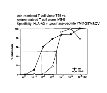
Fig. 2: Comparison of clones T58 and IVS-B
Fig. 2a: Cytotoxic activity directed against melanoma cell lines.
The killing capacity of clone T58 was compared with that of clone IVS-B,
derived from a
melanoma patient, using as target cells the T2 cell line pulsed with synthetic
tyrosinase-
peptide for the amino acid sequence YMDGTMSQV in different molar
concentrations,
listed on the x-axis. The % relative lysis is given on the y-axis. The
concentration of peptide
that corresponds to 50% relative lysis is indicated by the crossing lines and
shows that clone
T58 can recognize substantially lower concentrations of peptide in comparison
to clone
IVS-B.
Fig. 2b: Measurement of multimer binding and off-rates.
The two clones were incubated with multimers to determine the percentage of
positive cells
at time 0 h. Both clones bound multimer on 100% of the cells. Multimer was
washed out
and the clones were incubated in medium containing HLA-A2-specific antibody.
When
multimers are released from the cell surface, they are captured by the
antibody and can not
rebind to the cells. The percent multimer-positive cells were reanalyzed at 1
h and 2 h.
Fig. 2c: Interferon-gamma secretion after stimulation with melanoma cell
lines.
Clone T58 and IVS-B were co-cultured with the two melanoma cell lines used for
the initial
screening (described in Figure 1) and their secretion of IFN-y into the
culture medium was
assessed by standard ELISA after 24 hours. n.d.= not detectable. Data are
presented as
pg/ml on the y-axis.
Fig. 2d: Cytotoxic activity against melanoma cell lines.
The clones were compared for killing activity using a standard 51Cr-release
assay as
described in Figure 1. Data are given as percent specific lysis on the y-axis.
CA 02743669 2011-05-12
WO 2010/058023 PCT/EP2009/065705
16
Fig. 3: Recognition of primary melanoma tumor cells by clone T58 and IVS-B.
(a) HLA-A2 surface expression on primary tumor cells (passage 12) of an HLA-A2-
melanoma patient transfected with 50 gg HLA-A2 ivt-RNA and on established
melanoma
cell lines Mel-93.04A12 (HLA-A2 +tyrosinase+) and Mel-A375 (HLA-A2
+tyrosinase) was
measured by flow cytometry after staining with HLA-A2-specific monoclonal
antibody.
Each histogram shows the stained sample (filled curves) and the corresponding
control
sample (empty curves): control curves represent untransfected primary tumor
cells stained
with HLA-A2-specific monoclonal antibody (left histogram) or melanoma cell
lines stained
with isotype control antibody. HLA-A2 protein expression on RNA-transfected
primary
tumor cells was detected 10 h after electroporation. (b) The capacity of the
patient-derived T
cell clone (IVS-B), and T cell clone T58 to secrete IFN-y or (c) release
perforin in co-
culture with the melanoma cells shown above was measured in ELISPOT assays.
Fig. 4: Transfer of antigen specificity by TCR retroviral gene transfer. (a)
The human TCR-
deficient T cell line Jurkat769 was transduced with the TCR of the T cell
clone T58. TCR-
expression was detected using tyrosinase-peptide-specific HLA-multimers. TCR
expression
was only detected in Jurkat76 cells tranduced with TCR-T58 (right histogram)
and not in
untransduced Jurkat76 cells (left histogram). (b) PBL of a healthy donor were
retrovirally
transduced with TCR-T58. After 10 days, untransduced and TCR-transduced PBL
were
analysed for tyrosinase TCR-expression using specific HLA-multimers. Multimer
staining
is shown on the x-axis and CD8 staining on the y-axis. The percentage of
multimer+CD8+ T
cells is displayed in the upper right quadrant. (c) Functionality of TCR-
transduced PBL was
measured using a standard IFN-y release assay. T2 cells loaded with graded
amounts of
tyrosinase369_377 peptide (10-12 M - 10-5 M) were used as target cells at a
fixed effector to
target cell ratio of 1:1. Untransduced PBL served as a control and showed no
tyrosinase-
peptide specific IFN-y release (data not shown). Data are shown as pg/ml
cytokine after
subtration of secretion by untransduced PBL controls. (d) The capacity to
secrete IFN-y in
co-culture with melanoma cell lines SK-Mel-28 (HLA-A2-tyrosinase+), Mel-A375
(HLA-
A2+tyrosinase) , Me 1-624.38 (HLA-A2 +tyrosinase+) and Mel-93.04A12 (HLA-
A2+tyrosinase+) was assessed using a standard IFN-y release assay using an E:T
= 1:1; (n.d.
= not detectable).
Fig. 5: Transfer of specificity of T58 and IVS-B for HLA-A2 and tyrosinase-
peptide
YMDGTMSQV by TCR retroviral gene transfer. (a) PBL of a healthy donor were
CA 02743669 2011-05-12
WO 2010/058023 PCT/EP2009/065705
17
retrovirally transduced with the patient-derived TCR-IVS-B or the TCR-T58.
After 11 days,
untransduced and TCR-transduced PBL were analysed for tyrosinase TCR-
expression using
specific HLA-multimers. Multimer staining is shown on the x-axis and CD8
staining on the
y-axis. The percentage of multimer+CD8+ T cells is displayed in the upper
right quadrant.
(b) Functionality of TCR-transduced PBL was measured using a standard IFN-y
release
assay. T2 cells loaded with graded amounts of tyrosinase369_377 peptide (10-"
M - 10.5 M) or
with 10.5 M irrelevant influenza matrix protein8-66 were used as target cells
at a fixed
effector to target cell ratio of 1:1. Untransduced PBL served as a control and
showed no
tyrosinase-peptide specific IFN-y release (data not shown). Data are shown as
pg/ml
cytokine after substration of secretion by untransduced PBL controls (mean =
318 pg/ml;
range = 219-368 pg/ml) and adjustment for comparable numbers of multimer+
cells.
Fig. 6: Tyrosinase peptide-specific CTL recognition of tumor cell lines and
primary
melanoma tumor cells. Columns represent the amount of IFN-y (pg/ml) secreted
by self-
restricted Dl 15 CTL and allo-restricted T58 CTL in co-culture with a panel of
tumor cell
lines from left to right: MaCal (HLA-A2-tyrosinase-); SK-Mel-28 (HLA-A2-
tyrosinase+);
Mel-A375, RCC-26, PancTu 1, MaCal/A2, and UTS CC 1588 (all HLA-A2+tyrosinase-
);
Mel-624.38, Mel-93.04A12, SK-Mel-23, SK-Mel-29 and WM-266-4 (all HLA-
A2+tyrosinase+). T cells designates CTL without stimulating cells. The HLA-
A2+tyrosinase- tumor cell lines Mel-A375, RCC-26 and MaCal/A2 were exogenously
loaded with either 10-5 M irrelevant flu peptide or 10-5 M tyrosinase peptide
YMD and
IFN-y secretion was measured by ELISA and given as pg/ml.
Fig. 7: Transfer of antigen specificity by retroviral transfer of TCR-D115 and
TCR-T58.
PBL of a healthy donor were transduced with TCR-D115 or TCR-T58. Specificity
of
recognition was assessed by IFN-y release following co-culture with the tumor
cell lines
from left to right: MaCal (HLA-A2-tyrosinase-); SK-Mel-28 (HLA-A2-
tyrosinase+); Mel-
A375, RCC-26, PancTu 1, MaCal/A2, and UTS CC 1588 (all HLA-A2+tyrosinase-);
Mel-
624.38, Mel-93.04A12, SK-Mel-23, SK-Mel-29 and WM-266-4 (all HLA-
A2+tyrosinase+).
T designates CTL without stimulating cells. The HLA-A2+tyrosinase- tumor cell
lines Mel-
A375, RCC-26 and MaCal/A2 were exogenously loaded with either 10-5 M
irrelevant flu
peptide or 10-5 M tyrosinase peptide YMD and IFN-y secretion was measured by
ELISA
and given as pg/ml.
CA 02743669 2011-05-12
WO 2010/058023 PCT/EP2009/065705
18
Fig. 8: Transfer of antigen specificity by retroviral transfer of TCR-D115 and
TCR-T58. (A)
PBL of a healthy donor were transduced with TCR-D 115 or TCR-T58. Unsorted TCR-
transduced PBL were analyzed on day 10 for transgenic TCR-expression using
irrelevant
B7-pp65 and A2-pp65 multimers and specific A2-tyr multimers. Untransduced PBL
showed
no multimer binding (0.1 %, data not shown). Percentages of multimer+CD8+ T
cells are
displayed in the upper right quadrant. (B) and (C) show the IFN-y release of
unsorted TCR-
transduced PBL following stimulation with T2 cells loaded with graded amounts
of
tyrosinase peptide (10-12 M - 10-5 M) at a ratio of 2:1. In (B) the relative
IFN-y release is
displayed in percent and in (C) the specific IFN-y release is presented as
pg/ml. (D)
Functionality of unsorted TCR-transduced PBL was measured by IFN-y release
using
autologous HLA-A2+ PBMC loaded with tyrosinase peptide (10-11 M - 10-6 M) as
stimulating cells at ratio of 2:1. Untransduced PBL (A) showed no peptide-
specific IFN-y
release. (E) The HLA-A2+tyrosinase- tumor cell lines Mel-A375, RCC-26 and
MaCal/A2
were exogenously loaded with either 10-5 M irrelevant flu peptide (f) or 10-5
M tyrosinase
peptide YMD (t) and IFN-y secretion was measured by ELISA and given as pg/ml.
(F)
Specificity of recognition was assessed by IFN-y release following co-culture
with the
tumor cell lines from left to right: MaCal (HLA-A2-tyrosinase-); SK-Mel-28
(HLA-A2-
tyrosinase+); Mel-A375, RCC-26, PancTu 1, MaCal/A2, and UTS CC 1588 (all HLA-
A2+tyrosinase-); Mel-624.38, Mel-93.04A12, SK-Mel-23, SK-Mel-29 and WM-266-4
(all
HLA-A2+tyrosinase+). T designates CTL without stimulating cells.
Fig. 9: TCR transfer retains differences in cytokine profile. (A-D) On the
left hand side the
cytokine release of TCR-transduced PBL in co-culture with the melanoma lines
Mel-A375
(HLA-A2+tyrosinase-) and Mel-624.38 (HLA-A2+tyrosinase+) is depicted, on the
right
hand side the corresponding cytokine release after stimulation with T2 cells
loaded with
graded amounts of tyrosinase peptide (10-12 M - 10-5 M) is shown. Untransduced
PBL
(A) showed no peptide-specific cytokine release. The following cytokines were
measured:
IFN-y (A), IL-2 (B), TNF-a (C) and MIP-10 (D). The levels of cytokine
secretion for all
four cytokines were higher when PBL transduced with the allo-restricted TCR-
T58 were
used. Since untransduced PBL secreted very high levels of MIP-1(3 in response
to T2 cells
the peptide titration for this cytokine could not be evaluated.
CA 02743669 2011-05-12
WO 2010/058023 PCT/EP2009/065705
19
EXAMPLE 1
The inventors prepared stimulating dendritic cells (DC) from an HLA-A2-
negative healthy
donor that co-expressed allogeneic HLA-A*0201-molecules and tyrosinase protein
using
mature DC that were electroporated with in vitro transcribed (ivt)-RNA for
tyrosinase and
HLA-A2, as described' 2. These DC were used to prime purified, autologous CD8+
T cells
using two rounds of stimulation with freshly prepared DC. After these two
rounds of
priming, CD8+ T cells with T cell receptors (TCR) recognizing HLA-A2-
tyrosinase369_377-
peptide complexes were stained using a tyro sinase369_377/HLA-A*0201-
multimer3.
CD8+multimer+ cells were isolated by fluorescence activated cell sorting.
Sorted cells were
cloned in limiting dilution cultures and isolated clones showing HLA-
A2/tyrosinase-peptide
specificity were expanded using antigen-independent stimulation4. The T cell
clone T58 was
identified in an initial screen as having good functional activity (Figure 1).
Because T58 was isolated from an HLA-A*0201-negative donor it represents an
allo-
restricted T cell clone that did not undergo negative selection in vivo. The
activity of the
T58 clone was compared with the IVS-B clone that was isolated from a patient
with
metastatic melanomas. This clone recognizes exactly the same HLA-A2/tyrosinase
peptide
ligand as clone T58 but it is self-restricted since it was activated in vivo
in the patient who
was HLA-A*0201-positive. This patient-derived T cell clone represents an
example of T
cells that are available in the peripheral repertoire that have undergone
negative selection
against self-peptides/self-MHC-molecules in the thymus in vivo.
Side-by-side comparisons of clone T58 and clone IVS-B were made to demonstrate
the
superior properties of the allo-restricted T58 clone versus the self-
restricted IVS-B clone.
Functional T cell avidity for tyrosinase369-377 peptide recognition was
measured in a release assay using HLA-A2+ T2 cells pulsed with graded amounts
of exogenous peptide as
target cells. The peptide concentration needed for 50% relative lysis defined
the value of
half-maximum lysis6. The allo-restricted T cell clone T58 required
substantially less peptide
to be activated by peptide-pulsed T2 cells than clone IVS-B (6.0x10⁻¹⁰ M vs.
3.0x10.8 M)
(Fig. 2a).
CA 02743669 2011-05-12
WO 2010/058023 PCT/EP2009/065705
As an estimate of structural TCR-MHC/peptide binding affinity, loss of
multimer binding
was measured over time (i.e. HLA-multimer off-rate). A slower off-rate
indicates that TCR-
ligand interactions are more stable and of higher structural affinity. After
initial incubation
with multimer and washing, T cells were incubated for 1 h and 2 h without
multimers in the
presence of HLA-A2-specific antibody to prevent cellular re-association of
released
multimers. The melanoma patient-derived T cell clone IVS-B showed an
intermediate
multimer binding: all cells were multimer+ at 0 h and about 40% retained
multimers at 1 and
2 h (Fig. 2b). In contrast, clone T58 had a slower off-rate, showing 74%
positive binding at
1 h versus 41% for clone IVS-B and even at 2 h still had somewhat more
multimer+ cells
(55% vs. 40%).
Both T cell clones were analyzed in an IFN-y release assay for function and
specificity (Fig.
2c). The clones were co-cultured with two melanoma cell lines that express HLA-
A2
molecules but differ with respect to expression of tyrosinase protein: Mel-
93.04A12 co-
expresses both proteins (HLA-A2+tyrosinase+) but Mel-A375 fails to express
tyrosinase
protein (HLA-A2+tyrosinase) and therefore can not generate the MHC-peptide
ligand seen
by the T cell clones. Allo-restricted T cell clone T58 was induced to secrete
a high level of
IFN-y by the tyrosinase-expressing melanoma cell line, whereas only marginal
cytokine
secretion was seen with IVS-B cells (1,234 pg/ml vs. 106 pg/ml), demonstrating
the vastly
superior function of clone T5 8 in recognizing tumor cells expressing their
HLA-A2-
tyrosinase ligand. As expected, the clones showed no detectable IFN-y
secretion after
stimulation with Mel-A375 cells, demonstrating the specificity for HLA-A2 and
tyrosinase
expression for tumor cell recognition.
The killing capacity of allo-restricted clone T58 was also compared with clone
IVS-B using
a 51Cr-release assay (Fig. 2d). Again, clone T58 showed superior function (76%
vs. 24%
specific lysis).
Both clones were also tested for their capacity to recognize primary melanoma
cells. Since
primary HLA-A2+ melanoma cells were not available, we introduced ivt-RNA for
HLA-A2
into the tumor cells as for DC (Fig. 3a). Function was measured using ELISPOT
assays
detecting IFN-y secretion and perforin release to bypass high spontaneous
release of
radioactive label by primary tumor cells. Recognition of primary tumor cells
was shown to
be HLA-A2-restricted since primary tumor cells lacking HLA-A2 RNA were not
CA 02743669 2011-05-12
WO 2010/058023 PCT/EP2009/065705
21
recognized. Again, a strong difference was observed with poor recognition by
the patient
self-restricted IVS-B cells versus good recognition by allo-restricted T58
cells as assessed
with IFN-y secretion (Fig. 3b) and by perforin secretion (Fig. 3c).
To demonstrate that the superior functional avidity of allo-restricted T58
cells resided
directly in the TCR, separate recombinant retroviruses were created for TCR
alpha and beta
chains of clone T58 as described8. Human TCR-deficient Jurkat76 cells9 were co-
infected
with the a-chain and (3-chain retroviruses and transgenic TCR-expression was
measured by
multimer staining. TCR-T58 was expressed at a good level, demonstrating
adequate quality
of the separate retroviral supernatants (Fig. 4a). Next, activated peripheral
blood
lymphocytes (PBL) of a healthy HLA-A2- donor were transduced and analyzed with
multimers for tyrosinase-specific TCR-expression (Fig. 4b). Despite this low
frequency,
PBL transduced with TCR-T58 released high amounts of IFN-y following
stimulation with
T2 cells pulsed with graded amounts of tyrosinase-peptide (Fig. 4c). TCR-T58
transduced
PBL could also respond specifically to stimulation by melanoma cell lines that
expressed
HLA-A2 and tyrosinase (Fig. 4d). They did not respond to tumor cells that did
not express
HLA-A2 or tyrosinase, again demonstrating the specificity of HLA-A2-tyrosinase
ligands
for T58 recognition.
Bi-cistronic retroviral vectors were also prepared encoding the a-chain and (3-
chains of the
TCR of IVS-B cells and used to transduce activated PBL. In parallel, the same
activated
PBL were transduced with bi-cistronic retroviral vectors encoding the two
chains of TCR-
T58. PBL expressing the corresponding receptors were identified by co-staining
for CD8
and multimer and showed low numbers of positive cells. (Fig. 5a) Despite their
low
frequency, PBL transduced with TCR-T58 released high amounts of IFN-y
following
stimulation with T2 cells pulsed with graded amounts of tyrosinase-peptide.
PBL expressing
TCR-IVS-B secreted far less IFN-y. Tyrosinase peptide-specific cytokine
secretion was not
detected with untransduced PBL control cells. Data are shown as pg/ml cytokine
after
substraction of secretion by untransduced PBL controls (mean = 318; range =
219 - 369
pg/ml) (Fig. 5b).
Table 1 shows the genetic information regarding the use of VJ and VDJ gene
segments by
the alpha and beta chains of TCR-T58, respectively. The CDR3 regions,
according to
CA 02743669 2011-05-12
WO 2010/058023 PCT/EP2009/065705
22
IMGT, are presented as nucleotide sequences and amino acid sequences. Also
shown are the
codon optimized sequences for the full VJ and VDJ regions.
Materials and Methods
Cell lines
The human melanoma cell lines, Mel-A375 (HLA-A2+, tyrosinase ; CRL-1619,
American
Type Culture Collection (ATCC), Bethesda, MD), Mel-93.04A12 (HLA-A2+,
tyrosinase+,
gift of P. Schrier, Department of Immunohematology, Leiden University
Hospital, The
Netherlands), Mel-624.3810 (HLA-A2+, tyrosinase+, gift of M. C. Panelli,
National Institutes
of Health, Bethesda, MD), SK-Mel-28 (HLA-A2-, tyrosinase+; MTB-72, ATCC) as
well as
the lymphoid cell line T2 (CRL-1992, ATCC), and the human TCR-deficient
Jurkat769 T
cell line were cultured in RPMI 1640 medium supplemented with 12% fetal bovine
serum
(FBS), 2 mM L-glutamine and 1 mM sodium-pyruvate and non-essential amino
acids.
The HLA-A*0201-restricted tyrosinase369_377 peptide-specific melanoma patient-
derived
IVS-B T cell clone was cultured as described'.
Production of tyrosinase and HLA-A2 ivt-RNA
The plasmid pCDM8-HLA-A2 with HLA-A*0201 cDNA and the pZeoSV2+/huTyr with
tyrosinase cDNA were linearized and used as in vitro transcription templates
to produce
RNA with the aid of the mMESSAGE mMACHINE T7 kit (Ambion, Austin, TX)
according
to the manufacturer's instructions.
De novo priming of T cells with RNA-pulsed DC
Blood samples from healthy donors were collected after informed consent and
with
approval of the Institutional Review Board of the University Hospital of the
Ludwig-
Maximilians-University, Munich, Germany. Peripheral blood lymphocytes (PBL)
were
isolated by Ficoll density gradient centrifugation. PBL were resuspended in 15
ml very low
CA 02743669 2011-05-12
WO 2010/058023 PCT/EP2009/065705
23
endotoxin (VLE) RPMI 1640 medium (Biochrom, Berlin, Germany) supplemented with
1.5% human serum (DC medium) at 7.5x10⁷ cells per 75 cm culture flask and
incubated at
37 C and 5% CO2 for 1 h. Non-adherent cells were carefully removed by washing.
Mature
DC were prepared from adherent monocytes and transfected with ivt-RNA via
electroporation as previously described. DC of HLA-A2- donors were co-
transfected with
24 gg tyrosinase ivt-RNA and 48 gg HLA-A2 ivt-RNA. On the same day, autologous
CD8+
T lymphocytes were enriched from PBL via negative selection using a commercial
kit
according to the manufacturer's instructions (CD8+ T cell Isolation Kit II
(human),
Miltenyi, Bergisch Gladbach, Germany). Co-cultures were initiated 10 h after
DC
electroporation in 24-well plates (TPP, Trasadingen, Switzerland) by adding
1x10⁵ RNA-
pulsed DC to 1x10⁶ CD8+ T cells in RPMI 1640, supplemented with 10% heat-
inactivated
human serum, 4 mM L-glutamine, 12.5 mM HEPES, 50 gM (3-mercaptoethanol and 100
U/ml penicillin/streptomycin (T cell medium). IL-7 (5 ng/ml) (Promokine,
Heidelberg,
Germany) was added on day 0 and 50 U/ml IL-2 (Chiron Behring, Marburg,
Germany) was
added after 2 days and then on every 3rd subsequent day. Addition of IL-2 was
delayed to
decrease proliferation of non-specific CD8+ T cells4. The 2"d stimulation of
primed T cells
was made after seven days using freshly prepared RNA-pulsed DC.
HLA-multimer staining and sorting
Seven days after the 2"d stimulation of CD8-enriched T cells with RNA-pulsed
DC, HLA-
A2-restricted tyrosinase-specific T cells were detected by staining with a PE-
labeled HLA-
A*0201/htyr369_377 peptide/human (32m multimer", anti-CD8-APC antibody (clone
RPA-T8,
BD Pharmingen, Franklin Lakes, NJ) and propidium iodide (PI: 2 gg/ml). For
sorting, up to
5x10⁶ cells were incubated with 12 gg multimer in 100 gl PBS + 0.5% human
serum. CD8-
APC antibody was then added at 1/50 for an additional 25 min. After staining
cells were
washed twice and diluted in PBS + 0.5% human serum with PI for sorting. 20-
50x106 total
cells per priming culture were stained for sorting. PI-negative cells were
gated and
CD8+multimer+ T cells were sorted on a FACSAria cell sorter (BD Biosciences)
with a 70
m nozzle, at a rate of 15,000 events/s.
For HLA-multimer off-rate assays, cells were washed after multimer binding and
resuspended in FACS buffer containing saturating amounts of BB7.2 monoclonal
antibody
(ATCC) to capture detached multimers and prevent rebinding to T cells. After 1
or 2 h,
CA 02743669 2011-05-12
WO 2010/058023 PCT/EP2009/065705
24
samples were fixed in FACS buffer with 1% paraformaldehyde and analysed by
flow
cytometry7.
Culture of peptide-specific T clones
Multimer-sorted T cells were cloned by limiting dilution. Clones were plated
in 96-well
round-bottom plates (TPP) in 200 gl/well T cell medium. 50 IU/ml IL-2 was
supplemented
every 3 days with 5 ng/ml IL-7 and 10 ng/ml IL-15 (PeproTech Inc., Rocky Hill,
NJ) every
7 days. T cell clones were stimulated non-specifically with anti-CD3 antibody
(0.1 gg/ml;
OKT-3) and provided with 1x10⁵ feeder cells per 96-well, consisting of
irradiated (50 Gy)
PBL derived from a pool of five unrelated donors and 1x10⁴ irradiated (150 Gy)
EBV-
transformed allogeneic B-LCL every two weeks. Proliferating T cells were
transferred into
24-well plates (TPP) and cultured in 1.5 ml T cell medium plus cytokines.
1x10⁶ allogeneic
irradiated PBL and 1x10⁵ irradiated EBV-transformed allogeneic B-LCL were
added per
well as feeder cells in 24-well plates. Clonality was determined by TCR-beta-
chain receptor
analysis, as described'.
Peptide loading of T2 cells
For exogenous peptide pulsing, 1x10⁶ T2 cells were incubated at 37 C and 5%
CO2 for 2 h
with 10 gg/ml human (32-microglobulin (Calbiochem, San Diego, CA) and
titrating amounts,
ranging from 10-5 M to 10-12 M, of the tyrosinase peptide YMD (tyrosinase369-
377
YMDGTMSQV, Metabion, Martinsried, Germany). T2 cells pulsed with 10-5 M
influenza
peptide GIL (influenza matrix protein58_66 GILGFVTL, Metabion) served as
negative
control. After washing, peptide-loaded T2 cells were used as target cells in
cytotoxicity or
IFN-y-release assays.
IFN-y release assay
For investigation of specificity, T cell clones (2x 10³ cells in 100 l) were
incubated with the
respective melanoma cell lines or peptide-pulsed T2 cells (lx 104 cells in 100
l). Culture
supernatants were harvested after 24 h co-culture and assessed by a standard
ELISA using
the OptEIATM Human IFN-y Set (BD Biosciences Pharmingen).
CA 02743669 2011-05-12
WO 2010/058023 PCT/EP2009/065705
Cytotoxicity assay
Cytotoxic activity of T cell clones was analysed in a standard 4 h 51-chromium
release
assay. Melanoma cells or peptide-loaded T2 cells were used as target cells.
Briefly, 1x10⁶
target cells were labeled with 100 gCi Na25'Cr04 (ICN Biochemicals, Irvine,
CA) for 1-1.5
h. 51Cr-labeled target cells were cultured with T cells in 100 gl/well RPMI
1640 with 12%
F C S in V-bottom 96-well tissue culture plates (Greiner, Solingen, Germany).
For
determination of functional avidity lx104 T cells were added to lx103 peptide-
pulsed T2
cells loaded with titrated amounts of peptide, giving a constant E:T of 10:1.
After 4 h co-culture at 37 C, 50 gl of supernatant were collected and
radioactivity was
measured in a gamma counter. The percentage of specific lysis was calculated
as: 100 x
(experimental release - spontaneous release)/ (maximum release - spontaneous
release).
Spontaneous release was assessed by incubating target cells in the absence of
effector cells
and was generally less than 15%. For the calculation of percent relative
lysis, the maximum
percent specific lysis was set to the reference value of 100% and
corresponding values were
calculated corresponding to this reference. To determine half-maximum lysis,
percent
relative lysis was plotted against peptide concentration. The peptide
concentration at which
the curve crossed 50% relative lysis was taken as the value of half-maximum
lysis6.
ELISPOT
Antibody pre-coated PVDF plates (Mabtech AB, Nacka, Sweden) were incubated at
37 C in
CTL TestTM medium (Cellular Technology Ltd., Cleveland, Ohio) for 2 h to block
unspecific binding. For the IFN-y ELISPOT, plates were pre-coated with the IFN-
y-specific
capture antibody clone 1-D1K; for perforin ELISPOT plates were pre-coated with
the
perforin-specific capture antibody (clone Pf-80/164; Mabtech AB). Primed T
cells were
washed with CTL WashTM Supplement culture medium (Cellular Technology Ltd) and
1x10³ responder T cells were stimulated with 5x10³ melanoma cells in 150 gl
CTL TestTM
medium and 24 h later assessed in IFN-y ELISPOT or 48 h later in perforin
ELISPOT. After
washing with PBS/0.01% Tween and PBS alone, plates were incubated either with
a direct
streptavidin-alkaline phosphatase (ALP)-conjugated detection antibody (clone 7-
B6-1;
Mabtech AB) for IFN-y ELISPOT or with biotinylated detection antibody (clone
Pf-344;
CA 02743669 2011-05-12
WO 2010/058023 PCT/EP2009/065705
26
Mabtech AB) for perforin ELISPOT for 2 h at room temperature following a 1 h
incubation
with streptavidin-alkaline phosphatase (ALP). The plates were washed again and
a ready-to-
use BCIP/NBT-plus substrate solution (Mabtech AB) was added. Spots were
counted using
the AID reader system ELR03 with the software version 4.0 (AID Autoimmun
Diagnostika
GmbH, Strassberg, Germany).
Construction of retroviral vectors, production of virus supernatants and
transduction
of Jurkat76 T cells and PBL
For TCR identification of tumor-specific T cell clones, part of the TCRa- and
TCR(3-chain
sequences including the complementary determining region (CDR3) was amplified
by PCR
using a panel of TCRVa and TCRV(3 primers combined with the respective
constant region
primer as described 13. The TCRa and TCR(3 chain genes of T cell clones T58
and IVS-B
were amplified by PCR with gene specific primers and cloned into the
retroviral vector
MP71PRE8 via Notl and EcoRI restriction sites. Both chains of human TCR-T58
(Va7,
V(323) and TCR-IVS-B (Va3, V(314) were constructed as single-TCR gene vectors
or
double-TCR gene vectors (pMP71-T58a and pMP71-T580, pMP71-IVS-Ba and pMP71-
IVS-B0; pMP71-T58f3-P2A-T58a and pMP71-IVS-B(3-P2A-IVS-BU). Retroviral vector
plasmids were co-transfected into 293T cells with expression plasmids encoding
Moloney
MLV gag/pol and MLV-1OA1 env gene to produce amphotropic MLV-pseudotyped
retroviruses as described 14. The human TCR-deficient T cell line Jurkat76 and
PBL were
transduced as reported14. Jurkat76 cells (5 days after transduction) and PBL
(10 days after
transduction) were stained using PE-labeled HLA-A*0201/htyr369_377
peptide/human (32m
multimer and anti-CD8-FITC antibody. On day 13 an IFN-y release assay was
performed
using T2 cells loaded with graded amounts of tyrosinase369-377 peptide (10-12
M - 10.5 M) or
T2 cells pulsed with 10.5 M influenza matrix protein8-66 peptide and the tumor
cell lines
SK-Mel-28, Mel-A375, Mel-624.38 and Mel-93.04A12 as stimulating cells at an
E:T ratio
= 1:1. Control values for peptide-stimulated untransduced PBL were subtracted
from values
of transduced cells at each peptide concentration and then adjusted to
comparable numbers
of total TCR-transgenic cells.
CA 02743669 2011-05-12
WO 2010/058023 PCT/EP2009/065705
27
T58-TCR analysis
For the T-cell receptor analysis of the tyrosinase-specific clone T58, part of
the TCR alpha-
chain and beta-chain containing the CDR3 region was amplified by RT-PCR using
a panel
of TCR Va and TCR V(3 primers combined with a respective TCR constant region
primer.
Products were sequenced and assigned according to IMGT (Table 1; IMGT, the
international ImMunoGeneTics information system!, http://imgt.cines.fr).
Modifications of the TCR-sequence
Codon optimization of the VJ/VDJ-regions of both T58-TCR chains was done to
facilitate
TCR mRNA translation (Table 1). Antibody-tags, for example myc-tags'5 (Patent
Application number: 06014606.5-1212) or other modifications, for example a
CD20
epitope, can be introduced in any position, i.e. the N-terminus of the TCRa-
chain, that is
recognized by the depleting antibody and does not interfere with TCR-
functionality.
Table 1. TCR-CDR3 sequences and codon optimized VJ/VDJ regions of clone T58
Alpha-chain
VJ region* TRAV 1-2 AJ28
CDR3 region*
TGTGCTGTGACATACTCTGGGGCTGGGAGTTACCAACT
Nucleotide sequence
C (SEQ ID NO: 3)
Amino acid sequence C A V T Y S G A G S Y Q L (SEQ ID NO: 5)
ATGTGGGGCGTGTTTCTGCTGTACGTGTCCATGAAGAT
GGGCGGCACCACCGGCCAGAACATCGACCAGCCCACC
GAGATGACAGCCACCGAGGGCGCCATCGTGCAGATCA
ACTGCACCTACCAGACCAGCGGCTTCAACGGCCTGTTC
TGGTATCAGCAGCACGCCGGCGAGGCCCCTACCTTCCT
Codon optimized VJ GAGCTACAACGTGCTGGACGGCCTGGAAGAGAAGGGC
CGGTTCAGCAGCTTCCTGAGCCGGTCCAAGGGCTACAG
CTACCTGCTGCTGAAAGAACTGCAGATGAAGGACAGC
GCCAGCTACCTGTGCGCCGTGACCTACAGCGGAGCCG
GCAGCTACCAGCTGACCTTCGGCAAGGGCACCAAGCT
GTCCGTG (SEQ ID NO: 7)
CA 02743669 2011-05-12
WO 2010/058023 PCT/EP2009/065705
28
Beta-chain
VDJ region* TRBV13 BD I BJI -4
CDR3 region*
TGTGCCAGCAGTCAGAAACAGGGCTGGGAAAAACTG
Nucleotide sequence
(SEQ ID NO: 4)
Amino acid sequence C A S S Q K Q G W E K L (SEQ ID NO: 6)
ATGCTGTCCCCCGATCTGCCCGACAGCGCCTGGAACAC
CAGACTGCTGTGCCACGTGATGCTGTGTCTGCTGGGAG
CCGGATCTGTGGCCGCTGGCGTGATCCAGAGCCCCAG
ACACCTGATCAAAGAGAAGCGGGAGACAGCCACCCTG
AAGTGCTACCCCATCCCCCGGCACGACACCGTGTACTG
GTATCAGCAGGGACCAGGACAGGACCCCCAGTTCCTG
Codon optimized VDJ
ATCAGCTTCTACGAGAAGATGCAGAGCGACAAGGGCA
GCATCCCCGACAGATTCAGCGCCCAGCAGTTCAGCGA
CTACCACAGCGAGCTGAACATGAGCAGCCTGGAACTG
GGCGACTCTGCCCTGTACTTCTGCGCCAGCAGCCAGAA
GCAGGGCTGGGAGAAGCTGTTCTTCGGCAGCGGCACC
CAGCTGTCCGTGCTG (SEQ ID NO: 8)
TCR alpha-chain (VJ region), TCR beta-chain (VDJ region) and CDR3 lenghts are
designated according to IMGT (IMGT, the international ImMunoGeneTics
information
system , http://imgt.cines.fr)
Example 2
In Example 1, data are provided that compared two T cell clones that
specifically recognize
a peptide derived from tyrosinase (ie YMDGTMSQV hereafter referred to as YMD)
presented by HLA-A*0201 molecules. The T cell clone T58 was an allo-
restricted, peptide-
specific T cell clone derived from an HLA-A2-negative donor. The T cell clone
IVS-B was
derived from an HLA-A*0201-positive patient who suffered from metastatic
melanoma.
This melanoma expressed tyrosinase.
CA 02743669 2011-05-12
WO 2010/058023 PCT/EP2009/065705
29
In this Example, comparisons have been extended to include an example of a T
cell clone,
D1 15, which is also derived from an HLA-A*0201-positive individual and
recognizes the
same YMD peptide. However, in contrast to clone IVS-B, clone D115 was
generated in
vitro using responding T cells derived from the blood of a healthy individual.
Therefore,
there have been no potential negative impacts on this T cell clone from a
tumor environment
(ie melanoma) in vivo.
Figure 6 shows a comparison of the pattern of the target cell recognition of
the new clone
D115 and clone T58 which is the subject of this patent. As can be clearly
seen, both D115
and T5 8 show the same pattern of recognition, detected by secretion of
interferon-gamma
(y-axis), after co-cultivation with various tumor cell lines (x-axis and
figure legend). Neither
clone recognizes tumor cells that are HLA-A2-negative but express tyrosinase,
nor do they
recognize tumor cells that are HLA-A2-positive and tyrosinase negative. On the
other hand,
both T cell clones recognize several tumor cell lines that are both HLA-A2-
positive and
tyrosinase-positive. The role of the YMD peptide in this recognition is shown
by the finding
that HLA-A2-positive tumor cells that do not express tyrosinase from which the
YMD
peptide could be processed internally and transported to the cell surface by
HLA-A2
molecules for presentation, can be loaded with synthetic YMD peptide, leading
to their
recognition by D1 15 and T58. Thereby, both clones show the same specificity
for the YMD
peptide presented by HLA-A2 molecules. However, the efficiency of recognition
displayed
by clone T58 is far superior to clone D115, as seen by the levels of
interferon-gamma
secretion. This, for example, leads to negligible recognition of the melanoma
cell line SK-
Mel-29 by D1 15 but clear recognition by T58.
The TCR of clone D115 and T58 were expressed as recombinant proteins in
activated
recipient lymphocytes (Figure 7). When these TCR-transduced lymphocytes were
retested
with the same panel of target cells, they showed the same specificity pattern
as the original
T cell clones, demonstrating that the TCR recognition was responsible for the
results seen in
Figure 6. Again, in Figure 7 it is demonstrated that the TCR of clone T58
shows superior
recognition of the melanoma tumor cell lines that express HLA-A2 and
tyrosinase and the
YMD peptide-pulsed HLA-A2-positive tumor cells.
Figure 8A shows that the TCR-transduced lymphocytes show comparable levels of
expression of the respective recombinant TCRs, with each transduced population
having
CA 02743669 2011-05-12
WO 2010/058023 PCT/EP2009/065705
around 11% of T cells that bind a MHC multimer comprised of HLA-A2 molecules
presenting the YMD peptide. Such binding is not observed with control
multimers that
present other peptides derived from the pp65 protein of human cytomegalovirus.
When the two populations of TCR-transduced PBL are stimulated with HLA-A2-
positive
antigen-presenting cells (ie T2 cells) that are pulsed with different
concentrations of YMD
peptide (shown on the x-axis), it can be seen that the cells expressing TCR-
T58 release 50%
of their maximal levels of interferon-gamma (y-axis) at 100-fold lower peptide
concentrations. This peptide-sensitivity assay shows that the TCR-T58 has a
much higher
functional avidity when compared to TCR-D 115 (Figure 8B).
This difference is further exemplified by the strong difference in the maximum
levels of
interferon-gamma produced by the TCR-T58- versus TCR-D115-transduced
lymphocytes.
In the case of TCR-T58 cells, the maximum reaches 5000 pg/ml whereas this
results in only
around 2000 pg/ml for TCR-D115 in 24 hours. Furthermore, the amount of peptide
that
must be presented by T2 cells to cause release of 2000 pg/ml interferon-gamma
is 15,000-
fold lower for triggering of this level of response from TCR-T58-transduced
lymphocytes
compared with TCR-D 115 -transduced lymphocytes (Figure 8C).
Figure 8D shows another peptide-sensitivity assay, this time using peripheral
blood
mononuclear cells that have been pulsed with titrating amounts of YMD peptide
(x-axis).
Once again, the amounts of interferon-gamma released by lymphocytes expressing
TCR-
T5 8 are much greater compared with TCR-D 115. The arrows show that the first
detection of
cytokine secretion occurs with 1000-fold less peptide for TCR-T58 compared
with TCR-
D115.
Figures 8E and 8F demonstrate the specificity of the transduced lymphocyte
populations for
peptide-pulsed tumor cells (Figure 8E) or tumor cell lines expressing HLA-A2
and
tyrosinase (Figure 8F). In all cases, recognition is superior by lymphocytes
expressing TCR-
T58 compared to TCR-D115.
The superior secretion of cytokine is not limited to interferon-gamma. The
levels of
secretion of interleukin-2, TNF-alpha and MIP-lbeta are also superior for TCR-
T58. This is
CA 02743669 2011-05-12
WO 2010/058023 PCT/EP2009/065705
31
seen after stimulation of the TCR-transduced lymphocytes by tumor cells or by
peptide-
pulsed T2 cells (Figure 9).
Material and methods
Cell lines
The human melanoma cell lines, Mel-A375 (HLA-A2+, tyrosinase ; CRL-1619,
American
Type Culture Collection (ATCC)), Mel-93.04A12 (HLA-A2+, tyrosinase+; gift of
P. Schrier,
Department of Immunohematology, Leiden University Hospital, The Netherlands),
Mel-
624.381 and SK-Mel-23 (HLA-A2+, tyrosinase+; gift of M. C. Panelli, National
Institutes of
Health, Bethesda, MD), SK-Mel-28 (HLA-A2-, tyrosinase+; MTB-72, ATCC), SK-Mel-
29
(HLA-A2+, tyrosinase+, gift of P. Rieber, Institute of Immunology, Technical
University
Dresden, Germany), WM-266-4 (HLA-A2+, tyrosinase+; CRL-1676, ATCC) and primary
cultures of a human melanoma (passage 6-12) and MaCal (HLA-A2-, tyrosinase,
gift of R.
Wank, M.D. Munich, Germany), stable HLA-A*0201 transfectant of MaCal
(MaCal/A2)
(HLA-A2+, tyrosinase, gift of E. Noessner, Institute of Molecular Immunology,
Helmholtz
Zentrum Munchen, Germany), RCC-262 (HLA-A2+, tyrosinase ), PancTul (HLA-A2+,
tyrosinase, gift of P. Nelson, Department for Biological Chemistry University
Hospital
LMU Munich, Germany), UTS CC 1588 (HLA-A2+, tyrosinase, gift of M. Schmitz,
Institute of Immunology, Technical University Dresden, Germany) as well as the
lymphoid
cell line T2 (CRL-1992, ATCC) were cultured in RPMI 1640 medium supplemented
with
12% fetal bovine serum (FBS), 2 mM L-glutamine and 1 mM sodium-pyruvate and
non-
essential amino acids.
Peptide loading of T2 cells, PBMC and tumor cells
For exogenous peptide pulsing, 1x10⁶ T2 cells were incubated at 37 C and 5%
CO2 for 2 h
with 10 gg/ml human (32-micro globulin (Calbiochem) and titrating amounts,
ranging from
10.5 M to 10-11 M, of the tyrosinase peptide YMD (tyrosinase369_377 YMDGTMSQV,
Metabion). T2 cells pulsed with 10.5 M influenza peptide GIL (flu: influenza
matrix
protein58_66 GILGFVFTL, Metabion) served as the negative control. PBMC were
loaded
with tyrosinase peptide as for T2 cells with titrating amounts ranging from 10-
6 to 10-11 M.
Tumor cells were loaded with either 10-5 M flu peptide or 10-5 M tyrosinase
peptide YMD
CA 02743669 2011-05-12
WO 2010/058023 PCT/EP2009/065705
32
as described for T2 cells. After washing, peptide-loaded T2 cells, PBMC or
tumor cells
were used as stimulating cells in IFN-y release assays.
Cytokine assays
For investigation of specificity, CTL (2x 10³ cells in 100 l) were incubated
with various
tumor cell lines (lx 104 cells in 100 l), with or without peptide pulsing, as
described above.
Culture supernatants were harvested after 24 h co-culture and assessed by a
standard ELISA
using the OptEIATM Human IFN-y Set (BD Biosciences). Data represent mean
values with
corresponding mean deviations calculated from duplicate determinations. For
the
calculation of % relative IFN-y release, the maximum IFN-y release was set to
the reference
value of 100% and corresponding values were calculated corresponding to this
reference.
To investigate multiple cytokines simultaneously (IFN-y, IL-2, TNF-a and MIP-
1(3)
cytokine secretion in supernatants of co-culture of CTL with tumor cells and
with or without
tyrosinase peptide pulsed T2 cells (10-5 M) was measured using the multiplex
protein array
system technology (Bio-Rad Laboratories, Hercules, CA).
Retroviral TCR gene transfer
For TCR identification of tumor-specific CTL, regions of the TCRa- and TCR(3-
chains
encoding CDR3 were amplified by PCR using a panel of TCRVa and TCRV(3 primers
in
combination with respective constant region primers as described.3 The full
TCRa- and
TCR(3-chain genes of CTL clones T58 and D1 15 were amplified by PCR using cDNA
as
template. Primer sequences will be provided on request. The constant regions
of both TCR
chains were exchanged by the murine counterparts to increase the stability of
the TCR.4 The
TCR chains were linked by a 2A peptide linker (TCR(3-P2A-TCRa)5, codon-
optimized
(Geneart)6 and cloned into the retroviral vector MP71PRE via Notl and EcoRI
restriction
sites.5 Retroviral vector plasmids were co-transfected into 293T cells with
expression
plasmids encoding Moloney MLV gag/pol and MLV-1OA1 env gene, respectively, to
produce amphotropic MLV-pseudotyped retroviruses as described.5 Ten days after
the
second transduction, PBL were stained using PE-labeled A2-tyr multimer and
FITC-labeled
CD8-specific antibody. Multimers presenting peptides derived from
cytomegalovirus pp65
were used as controls: PE-labeled HLA-B7 pp65417-427 (B7-pp65) multimers
served as the
HLA control and HLA-A2 pp65495-503 multimers as a peptide-specificity control.
On day 15
CA 02743669 2011-05-12
WO 2010/058023 PCT/EP2009/065705
33
an IFN-y release assay was performed using T2 cells or autologous PBMC loaded
with
graded amounts of tyrosinase peptide (10-12 M - 10.5 M) and the tumor cell
lines MaCal,
SK-Mel-28, Mel-A375, RCC-26, PancTu 1, MaCal/A2, UTS CC 1588, Mel-624.38, Mel-
93.04A12, SK-Mel-23, SK-Mel-29 and WM-266-4 as stimulating cells at an E:T of
2:1.
CA 02743669 2011-05-12
WO 2010/058023 PCT/EP2009/065705
34
References
1. Liao, X., et at. Transfection of RNA encoding tumor antigens following
maturation
of dendritic cells leads to prolonged presentation of antigen and the
generation of high-
affinity tumor-reactive cytotoxic T lymphocytes. Mol Ther 9, 757-764 (2004).
2. Javorovic, M., et at. Inhibitory effect of RNA pool complexity on
stimulatory
capacity of RNA-pulsed dendritic cells. Jlmmunother 31, 52-62 (2008).
3. Altman, J.D., et at. Phenotypic analysis of antigen-specific T lymphocytes.
Science
274, 94-96 (1996).
4. Ho, W.Y., Nguyen, H.N., Wolff, M., Kuball, J. & Greenberg, P.D. In vitro
methods
for generating CD8+ T-cell clones for immunotherapy from the naive repertoire.
Jlmmunol
Methods 310, 40-52 (2006).
5. Wolfel, T., et at. Analysis of antigens recognized on human melanoma cells
by A2-
restricted cytolytic T lymphocytes (CTL). IntJCancer 55, 237-244 (1993).
6. Margulies, D.H. TCR avidity: it's not how strong you make it, it's how you
make it
strong. Nat Immunol 2, 669-670 (2001).
7. Palermo, B., et at. Qualitative difference between the cytotoxic T
lymphocyte
responses to melanocyte antigens in melanoma and vitiligo. Eur Jlmmunol 35,
3153-3162
(2005).
8. Engels, B., et at. Retroviral vectors for high-level transgene expression
in T
lymphocytes. Hum Gene Ther 14, 1155-1168 (2003).
9. Heemskerk, M.H., et at. Redirection of antileukemic reactivity of
peripheral T
lymphocytes using gene transfer of minor histocompatibility antigen HA-2-
specific T-cell
receptor complexes expressing a conserved alpha joining region. Blood 102,
3530-3540
(2003).
10. Rivoltini, L., et at. Quantitative correlation between HLA class I allele
expression
and recognition of melanoma cells by antigen-specific cytotoxic T lymphocytes.
Cancer Res
55, 3149-3157 (1995).
11. Wolff, M., et at. Quantitation of MHC tetramer-positive cells from whole
blood:
evaluation of a single-platform, six-parameter flow cytometric method.
Cytometry A 57,
120-130 (2004).
12. Zhou, D., et at. High throughput analysis of TCR-beta rearrangement and
gene
expression in single T cells. Lab Invest 86, 314-321 (2006).
CA 02743669 2011-05-12
WO 2010/058023 PCT/EP2009/065705
13. Steinle, A., Reinhardt, C., Jantzer, P. & Schendel, D.J. In vivo expansion
of HLA-
B35 alloreactive T cells sharing homologous T cell receptors: evidence for
maintenance of
an oligoclonally dominated allospecificity by persistent stimulation with an
autologous
MHC/peptide complex. JExp Med 181, 503-513 (1995).
14. Leisegang, M., et at. Enhanced functionality of T cell receptor-redirected
T cells is
defined by the transgene cassette. JMo1 Med 86, 573-583 (2008).
15. Kieback, E., Charo, J., Sommermeyer, D., Blankenstein, T. & Uckert, W. A
safeguard eliminates T cell receptor gene-modified autoreactive T cells after
adoptive
transfer. Proc Natl Acad Sci USA 105, 623-628 (2008).
16. Kolb, H.J., Schattenberg, A., Goldman, J.M., Hertenstein, B., Jacobsen,
H., Arcese W.,
Ljungman, P., Ferrant, A., Verdonck, L. Niederwieser, B. et al. 1995. Graft-
versus-leukemia effect
of donor lymphocyte transfusions in marrow grafted patients. Blood 86:2041.
17. Kolb, H. J., Schmid, C., Barrett, A. J. and Schendel, D. J. (2004). Graft-
versus-leukemia
reactions in allogeneic chimeras. Blood 103:767-776.
18. Dudley, M.E. and Rosenberg, S.A. (2003). Adoptive-cell-transfer therapy
for the treatment
of patients with cancer. Nature Reviews Cancer 3: 666-675.
19. Dudley, M. E., Wunderlich, J. R., Robbins, P. F., Yang, J. C., Hwu, P.,
Schwartzentruber, D.
J., Topalian, S. L., Sherry, R., Restifo, N. P., Hubicki, A. M., Robinson, M.
R., Raffeld, M., Duray,
P., Seipp, C. A., Rogers-Freezer, L., Morton, K. E., Mavroukakis, S. A.,
White, D. E., Rosenberg, S.
A. (2002). Cancer regression and autoimmunity in patients after clonal
repopulation with antitumor
lymphocytes. Science 298:850-854.
20. Engels, B., Nossner, E., Frankenberger, B., Blankenstein, Th., Schendel,
D.J., and W. Uckert.
2005. Redirecting human T lymphocytes towards renal cell carcinoma-specificity
by retroviral
transfer of T cell receptor genes. Human Gene Ther., 16(7):79.9-810
21. Rivoltini L, Barracchini KC, Viggiano V, et al. Quantitative correlation
between
HLA class I allele expression and recognition of melanoma cells by antigen-
specific
cytotoxic T lymphocytes. Cancer Res. 1995;55:3149-3157.
22. Schendel DJ, Gansbacher B, Oberneder R, et al. Tumor-specific lysis of
human renal
cell carcinomas by tumor-infiltrating lymphocytes. I. HLA-A2-restricted
recognition of
autologous and allogeneic tumor lines. J Immunol. 1993;151:4209-4220.
23. Steinle A, Reinhardt C, Jantzer P, Schendel DJ. In vivo expansion of HLA-
B35
alloreactive T cells sharing homologous T cell receptors: evidence for
maintenance of an
oligoclonally dominated allospecificity by persistent stimulation with an
autologous
MHC/peptide complex. J Exp Med. 1995;181:503-513.
CA 02743669 2011-05-12
WO 2010/058023 PCT/EP2009/065705
36
24. Cohen CJ, Zhao Y, Zheng Z, Rosenberg SA, Morgan RA. Enhanced antitumor
activity of murine-human hybrid T-cell receptor (TCR) in human lymphocytes is
associated
with improved pairing and TCR/CD3 stability. Cancer Res. 2006;66:8878-8886.
25. Leisegang M, Engels B, Meyerhuber P, et al. Enhanced functionality of T
cell
receptor-redirected T cells is defined by the transgene cassette. J Mol Med.
2008;86:573-
583.
26. Scholten KB, Kramer D, Kueter EW, et al. Codon modification of T cell
receptors
allows enhanced functional expression in transgenic human T cells. Clin
Immunol.
2006;119:135-145.