Note: Descriptions are shown in the official language in which they were submitted.
CA 02743959 2011-05-17
WO 2010/057317 PCT/CA2009/001708
LIPOSOMAL COMPOSITION FOR CONVECTION-ENHANCED DELIVERY TO THE CENTRAL NERVOUS
CENTRE
FIELD OF THE INVENTION
[0001] The present invention relates to liposomal formulations that are
deliverable by
convection-enhanced delivery and useful for the treatment of central nervous
system disorders.
BACKGROUND OF THE INVENTION
[0002] For patients with brain tumors, systemic delivery of therapeutics is
usually associated with
systemic side effects while achieving only marginal therapeutic concentrations
in the central nervous
system (CNS), and thus the efficacy of systemic treatment is limited. The
observed lack of efficacy is
primarily due to poor penetration of therapeutic agents across the blood-brain
barrier. Although the
blood-brain barrier may be disrupted at the core of the tumor allowing
systemically-delivered
chemotherapy agents access to the mostly inactive center of the tumor, the
barrier typically remains
intact at the growing tumor margin where the agent is needed most.
[0003] One approach to circumventing the blood brain barrier is direct
infusion of therapeutic agents
into the CNS. However, agents infused directly into the brain distribute
poorly by diffusion. High
concentration gradients are required to move even small molecule drugs
millimeters from the infusion
site, and such concentrations are often neurotoxic. A developing strategy to
overcome this problem is
a direct intracerebral infusion approach called convection-enhanced delivery
(CED). CED employs
positive pressure to generate a local pressure gradient for distributing
agents, including therapeutic
macromolecules, in the extracellular space. (Bobo, R.H., et al. (1994) Proc.
Natl. Acad. Sci. USA
91:2076-80.; Chen, M.Y., et al. (1999) J. Neurosurg. 90:315 20). CED provides
reproducible
distribution within a given target tissue and can produce homogeneous drug
concentrations
throughout the volume of distribution (Vd) (Croteau et al., 2005; Lonser et
al., 2002).
[0004] Chemotherapeutic agents delivered locally by CED have produced
favorable therapeutic
outcomes (Bruce et al., 2000; Degen et al., 2003; Kaiser et al. 2000).
However, most cytotoxic agents
delivered directly to the nervous system have the capacity to damage healthy
cells. Accordingly,
good candidates for CED administration into brain tumors must have the highest
possible therapeutic
index against tumor cells in comparison with healthy neuronal cells. While
liposonnal drug delivery
offers potential for avoiding the high peak drug concentrations that are often
associated with
pronounced toxicity, and preclinical studies of liposome-encapsulated
camptothecin drugs given via
CED have shown some improvement in the sustained release of the drug (Moog et
al, 2002; Saito et
al, 2006; Nobel et al, 2006), the use of PEGylated liposonnes was deemed
essential to mask tissue
binding site interactions and thereby increase tissue distribution volume.
(Saito et al, 2006).
[0005] Despite the success of PEGylation in overcoming liposome/tissue
interactions, it has recently
been demonstrated that PEGylated liposomes may generate unwanted and
potentially life-threatening
immune responses (Szebeni et al. (2007) J. Liposome Res. 17:107-117; Ishida
and Kiwada (2008)
1
DM_VAN/253729-17775/7468072.1
CA 027 43959 2011-05-17
WO 2010/057317
PCT/CA2009/001708
Int. J. Pharm. 354:56-62 Epub Nov. 9 2007). In addition to accelerated blood
clearance when
administered into the same subject twice, PEGylated liposomes may cause non-
IgE-mediated
hypersensitivity reactions, which include symptoms of cardiopulmonary distress
(e.g., dyspnea,
tachypnea, tachycardia, chest pain, hypertension, and hypotension) (Ishida and
Kiwada (2008),
supra; Moghimi et al. (2006) FASEB J. 20:2591-3 Epub Oct. 25, 2006).
[0006] What is needed, therefore, is an improved liposomal drug formulation
for convection-
enhanced delivery that provides increased tissue distribution volume, but
avoids the problematic
immunogencity associated with PEGylation.
SUMMARY OF THE INVENTION
[0007] The present inventors have surprisingly discovered that liposomes can
be highly convective in
tissues of the central nervous system when an anionic lipid component is
employed in the formulation
in lieu of PEGylation, as described and claimed herein. Moreover, the subject
formulations exhibit
pharmacokinetic profiles comparable to PEGylated formulations employed in the
art while avoiding
the problematic immunogencity associated with PEGylation. Accordingly,
provided herein are
improved compositions and methods of administering therapeutic drugs to
discrete tissue(s) of the
central nervous system (CNS), e.g., a localized CNS tumor, via convection-
enhanced delivery of
anionic liposome formulations.
[0008] In one aspect, described herein are methods for treating a CNS
disorder, e.g., a disorder
associated with the death and/or dysfunction of a particular neuronal
population in the CNS. The
methods involve administering a therapeutically effective amount of a
pharmaceutical composition to
a patient having a CNS disorder, wherein the pharmaceutical composition is
locally delivered to the
particular neuronal population by convection-enhanced delivery, and wherein
the pharmaceutical
composition comprises at least one therapeutic agent encapsulated in non-
PEGylated liposomes
comprising a mixture of a neutral saturated phospholipid and at least one
anionic saturated lipid, and
wherein the convection-enhanced delivery of the pharmaceutical composition
treats a patient having a
CNS disorder.
[0009] Therapeutic agents finding advantageous use in the subject invention
include, e.g.,
antineoplastic agents, radioiodinated compounds, toxins (including protein
toxins), cytotoxic agents
including cytostatic or cytolytic drugs, genetic and viral vectors, vaccines,
synthetic vectors, growth
factors, neurotrophic factors, antivirals, antibiotics, neurotransmitters,
cytokines, enzymes and agents
for targeted lesioning of specific sites.
[0010] CNS disorders that may be treated by the compositions and methods
provided herein
include, e.g., cancer, infection, head trauma, spinal cord injury, multiple
sclerosis, dementia with Lewy
bodies, ALS, lysosomal storage disorders, psychiatric disorders,
neurodegenerative disorders, stroke,
epilepsy, and other acute and chronic disorders of the CNS.
2
DM_VAN/253729-17775/7468072.1
CA 027 43959 2011-05-17
WO 2010/057317
PCT/CA2009/001708
[0011] In one embodiment, provided herein are methods for inhibiting the
growth of a CNS tumor,
reducing a CNS tumor, killing one or more CNS tumor cells, and/or treating a
patient having a CNS
tumor. The methods involve administering a therapeutically effective amount of
a pharmaceutical
composition to a patient having a CNS tumor, wherein the pharmaceutical
composition is locally
delivered to the CNS tumor by convection-enhanced delivery, and wherein the
pharmaceutical
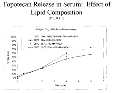
composition comprises at least one cytotoxic agent encapsulated in non-
PEGylated liposomes
comprising a mixture of a neutral saturated phospholipid and at least one
anionic saturated lipid, and
wherein the convection-enhanced delivery of the pharmaceutical composition
inhibits the growth of a
CNS tumor, reduces a CNS tumor, kills one or more of the CNS tumor cells
and/or treats a patient
having a CNS tumor.
[0012] In another embodiment, provided herein are methods for inhibiting or
reducing the number or
duration of seizures in a patient having epilepsy. The methods involve
administering a therapeutically
effective amount of a pharmaceutical composition to a patient having epilepsy,
wherein the
pharmaceutical composition is locally delivered to an aggregate of CNS neurons
exhibiting abnormal
or excessive hypersynchronous discharges by convection-enhanced delivery, and
wherein the
pharmaceutical composition comprises at least one therapeutic agent
encapsulated in non-PEGylated
liposomes comprising a mixture of a neutral saturated phospholipid and at
least one anionic saturated
lipid, and wherein the convection-enhanced delivery of the pharmaceutical
composition inhibits or
reduces the number or duration of seizures in a patient having epilepsy. In
one embodiment, the
therapeutic agent is a toxin, e.g., a peptide toxin. In one embodiment, the
peptide toxin is a
w-conotoxin, e.g., w-conotoxin MVIIA or w-conotoxin,GVIA. In another
embodiment,the toxin is a
botulinum toxin, e.g., a botulinum toxin serotype A such as BOTOXO or
DYSPORTO, a botulinum
toxin serotype B such as MYOBLOCO, etc. In another embodiment, the toxin is p-
conotoxin or a-
conantokin peptide.
[0013] In one embodiment, the pharmaceutical composition further comprises at
least one
diagnostic agent (sometimes referred to herein as a "tracing agent" or
"tracer") encapsulated in similar
non-PEGylated anionic liposomes, which allows for visualization of the
distribution of the therapeutic
agent during and after CED. In preferred embodiments, the non-PEGylated
liposomes encapsulating
the diagnostic agent are composed of the same lipids as the non-PEGylated
liposomes encapsulating
the therapeutic agent. Accordingly, in one embodiment, methods described
herein further comprise
the step of detecting the diagnostic agent.
[0014] As described herein, the non-PEGylated liposomes may contain a
therapeutic drug. In one
embodiment, the therapeutic drug is an insoluble therapeutic drug. In another
embodiment, the
therapeutic drug is a topoisomerase I inhibitor (e.g., a camptothecin and
derivatives thereof), which
includes but is not limited to topoisomerase I/11 inhibitors. For example, in
one embodiment, the
therapeutic drug is a camptothecin derivative selected from the group
consisting of
9-anninocamptothecin, 7-ethylcamptothecin, 10-hydroxycamptothecin, 9-
nitrocamptothecin, 10,11-
methlyenedioxycamptothecin, 9-amino-10,11-methylenedioxycamptothecin 9-chloro-
10,11-
3
DM_VAN/253729-17775/7468072.1
CA 027 43959 2011-05-17
WO 2010/057317
PCT/CA2009/001708
methylenedioxycamptothecin, irinotecan, topotecan, 7-(4-
methylpiperazinomethylene)-10,11-
ethylenedioxy-20(S)-camptothecin, 7-(4-rnethylpiperazinomethylene)-10,11-
rnethylenedioxy-20(S)-
camptothecin and 7-(2-(N-isopropylamino)ethyl)-(20S)-camptothecin. In another
embodiment, the
camptothecin derivative is selected from the group consisting of irinotecan,
topotecan, (7-(4-
methylpiperazinomethylene)-10,11-ethylenedioxy-20(S)-camptothecin, 7-(4-
methylpiperazinomethylene)-10,11-methylenedioxy-20(S)-camptothecin or 7-(2-(N-
isopropylamino)ethyl)-(20S)-camptothecin. In another embodiment, the
camptothecin is topotecan.
[0015] In another embodiment, the topoisomerase inhibitor is a topoisomerase
1/11 inhibitor, such as
6[[2-(dimethylamino)-ethyllamino]-3-hydroxy-7H-indeno[2,1-c]quinolin-7-one
dihydrochloride,
azotoxin or 3-methoxy-11H-pyrido[3',4'-4,5]pyrrolo[3,2-c]quinoline-1,4-dione.
[0016] In another embodiment, the therapeutic drug is a toxin, e.g., a protein
toxin, e.g.,
w-conotoxin, (e.g., w-conotoxin MVIIA or w-conotoxin,GVIA), a botulinum toxin
(e.g., a botulinum
toxin serotype A such as BOTOXO or DYSPORTO, a botulinum toxin serotype B such
as
MYOBLOCO) p-conotoxin, a-conantokin peptide, etc.
[0017] In one embodiment, the initial drug concentration is at least about 100
ug/mL, preferably at
least about 200 ug/mL, and more preferably at least about 300 ug/mL. In
another embodiment, the
initial drug concentration is about 2 mg/ml to about 5 mg/ml. In one
embodiment, the therapeutic
drug and/or diagnostic agent to lipid ratio is from about 0.1 to about 0.5. In
another embodiment, the
therapeutic drug and/or diagnostic agent to lipid ratio is about 0.1. In
another embodiment, the
therapeutic drug and/or diagnostic agent to lipid ratio is about 0.3. In
another embodiment, the
therapeutic drug and/or diagnostic agent to lipid ratio is about 0.5.
[0018] In one aspect, the non-PEGylated liposome contains a diagnostic agent.
In one
embodiment, the diagnostic agent is an MRI magnet. In another embodiment, the
diagnostic agent is
gadolinium chelate. In another embodiment, the diagnostic agent is selected
from the group
consisting of gadodiamide and rhodamine. In another embodiment, the diagnostic
agent is
gadodiamide.
[0019] The methods described herein comprise convection-enhanced delivery of a
liposomal
formulation comprising at least one therapeutic agent and/or at least one
diagnostic agent
encapsulated in non-PEGylated liposornes composed of a mixture of at least one
neutral saturated
phospholipid and at least one anionic saturated phospholipid. In one
embodiment, the neutral
saturated phospholipid is selected from the group consisting of derivatives of
phosphatidylcholine and
mixtures thereof, for example dipalmitoylphosphatidylcholine (DPPC),
distearoylphosphatidylcholine
(DSPC), dimyristoylphosphatidylcholine (DMPC), and mixtures thereof. Longer
chain saturated lipids,
e.g., C20and C22, may also be used. In one embodiment, the anionic saturated
phospholipid is
selected from a group consisting of derivatives of phosphatidylglycerol (e.g.,
distearoylphosphatidylglycerol (DSPG)), dipalmitoyl phosphatidyl glycerol
(DPPG),
phosphatidylserine, phosphatidylinositol, phosphatidic acid and mixtures
thereof.
4
DM_VAN/253729-17775/7468072.1
CA 02743959 2011-05-17
WO 2010/057317
PCT/CA2009/001708
[0020] The liposomal formulations described herein may also contain other
lipid components such
as sterols and derivatives (for example cholesterol (CHOL)) or sphingolipids
(for example
sphingomyelins and glycosphingolipids, in particular gangliosides). In
preferred embodiments, the
liposomal formulations will consist essentially of or consist of at least one
neutral saturated
phospholipid, at least one anionic saturated phospholipid and a stabilizer
such as, e.g., cholesterol.
[0021] In one embodiment, the non-PEGylated liposome is composed of a
combination of
distearoylphosphatidylcholine (DSPC) and distearoylphosphatidylglycerol
(DSPG). In one
embodiment, the non-PEGylated liposome comprises about 10 to about 95 mole
percent DSPC. In
one embodiment, the non-PEGylated liposome comprises about 5 to about 90 mole
percent DSPG.
In one embodiment, the non-PEGylated liposome further comprises cholesterol
(CHOL), e.g., about 5
to about 45 mole percent cholesterol. In a preferred embodiment, the liposome
comprises or consists
essentially of about 60 to about 90 mole percent DSPC, about 5 to about 10
mole percent cholesterol,
and about 5 to about 30 mole percent DSPG. In a preferred embodiment, the non-
PEGylated
liposome comprises or consists essentially of DSPC, DSPG, and CHOL at a 7:2:1
molar ratio. In
another embodiment, the non-PEGylated liposome comprises or consists
essentially of DSPC, DSPG
and CHOL at a 6:2:2 molar ratio. In another embodiment, the non-PEGylated
liposome comprises or
consists essentially of DSPC, DSPG and CHOL at a 5:2:3 molar ratio.
[0022] In one embodiment, convection-enhanced delivery (CED) of non-PEGylated
liposomal
formulations as described herein provides increased tissue distribution,
decreased toxicity and
increased in vivo half-life of the therapeutic drug as compared to the
respective tissue distribution,
toxicity, and in vivo half-life of the freely administered therapeutic drug.
[0023] In one aspect, the invention provides a cannula comprising a liposomal
formulation
described herein, e.g., a liposomal formulation comprising at least one
therapeutic agent
encapsulated in non-PEGylated liposomes composed of a mixture of at least one
neutral saturated
phospholipid and at least one anionic saturated phospholipid, and wherein the
formulation may be
delivered by convection-enhanced delivery (CED). In another embodiment, the
cannula further
comprises a liposomal formulation comprising a diagnostic agent encapsulated
in non-PEGylated
liposonnes composed of a mixture of at least one neutral saturated
phospholipid and at least one
anionic saturated phospholipid, and wherein the formulation may be delivered
by CED. In another
embodiment, the cannula comprises a liposomal formulation comprising a first
liposome containing a
therapeutic drug and a second liposome containing a diagnostic agent, wherein
neither the first nor
second liposome are PEGylated, wherein the first and second lipsomes are
composed of a mixture of
at least one neutral saturated phospholipid and at least one anionic saturated
phospholipid, and
wherein the formulation may be delivered by convection-enhanced delivery
(CED). The cannula is
compatible with convection-enhanced delivery to the CNS. In one embodiment,
the cannula is a
reflux-free step-design cannula.
[0024] In one aspect, the invention provides methods for producing the
liposomal formulations
described herein. In one aspect, the invention provides methods for producing
a medicament useful
DM_VAN/253729-17775/7468072.1
CA 02743959 2011-05-17
WO 2010/057317
PCT/CA2009/001708
for the treatment of a patient having cancer of the CNS, which medicament
comprises a liposomal
formulation described herein. In one embodiment, the method comprises
entrapping the therapeutic
drug or diagnostic agent within the liposomes by remote loading, for example,
via an ammonium
sulfate gradient.
[0025] Further objects, features and advantages of the apparatuses and methods
described herein
will become apparent from the following detailed description taken in
conjunction with the
accompanying figures showing illustrative embodiments of the invention.
BRIEF DESCRIPTION OF THE DRAWINGS
[0026] Figs. 1A - IF compare the effect of lipid composition, drug
concentration and drug:lipid ratio
on the release characteristics of topotecan from pegylated and non-pegylated
liposomal formulations.
[0027] Fig. 2 shows the pharmacokinetics of Ls-TPT Formulations and free
topotecan in Normal
brain tissue.
[0028] Fig. 3 shows the effect of sucrose on convectability of rhodamine
liposomes.
[0029] Fig. 4 shows the distribution volume (Vd) of rhodamine loaded liposomes
after a 20 pl
infusion into the striatum.
[0030] Fig. 5 shows survival of animals by treatment group.
[0031] Fig. 6 shows survival of animals by combined treatment group vs. group
2 (0.5 mg/mL dual
dosing).
[0032] Fig. 7 shows overall survival by U87 cell load at tumor implantation.
[0033] Fig. 8 shows survival of animals by combined treatment groups vs. group
2 (0.5 mg/mL dual
dosing) in animals with low U87MG Cell Load (6.8X10³).
[0034] Fig. 9 shows survival of animal by combined treatment groups vs. group
2 (0.5 mg/mL
dosing) in animals with high U87MG cell load (9.7X10⁵)
[0035] Fig. 10 shows volume of distribution of Ls-TPT-marina blue DHPE
coinfused with Ls-Gd-
rhodamine-PE in naive rodent brain tissue. For each formulation, n = 3 and 20
pL was infused in
each hemisphere.
[0036] Fig. 11 shows volume of distribution of Ls-TPT-marina blue DHPE
coinfused with Ls-Gd-
rhodamine-PE in U87MG xenograft rodent brain tissue. For each formulation, n =
4 and 20 pL was
infused in each hemisphere.
[0037] Fig. 12 shows survival of animals by treatment groups (euthanized
animals considered as
uncensored).
6
DM_VAN/253729-17775/7468072.1
CA 027 43959 2011-05-17
WO 2010/057317
PCT/CA2009/001708
[0038] Fig. 13 shows survival of animals by treatment groups (euthanized
animals considered as
censored.
DETAILED DESCRIPTION
[0039] Definitions
[0040] As used herein, "liposome" refers to a lipid bilayer membrane
containing an entrapped
aqueous volume. Liposomes may be unilamellar vesicles having a single membrane
bilayer or
multilamellar vesicles having multiple membrane bilayers separated from each
other by an aqueous
layer. Generally, the liposomal bilayer is composed of two lipid monolayers
having a hydrophobic
"tail" region and a hydrophilic "head" region. The structure of the membrane
bilayer is such that the
hydrophobic (non-polar) "tails" of the lipid monolayers orient toward the
center of the bilayer while the
hydrophilic (polar) "heads" orient toward either the entrapped aqueous volume
or the extraliposomal
aqueous environment. In one embodiment, a liposome of the invention includes a
targeting moiety,
e.g., an antibody or other ligand.
[0041] "Liposomal formulations" are understood to be those in which part or
all of the therapeutic
drug and/or diagnostic agent is encapsulated inside the liposomes. "Consisting
essentially of as
used herein in reference to liposomal formulations refers to liposomes having
the recited lipid
components only, and no additional lipid components.
[0042] "Phospholipid" is understood to mean an amphiphile derivative of
glycerol in which one of its
hydroxyl groups is esterified with phosphoric acid and the other two hydroxyls
are esterified with long-
chain fatty acids, which may be equal or different from each other.
[0043] A saturated phospholipid will be that whose fatty acids only have
simple (not multiple)
covalent carbon-carbon bonds.
[0044] A neutral phospholipid will generally be one in which another
phosphoric acid hydroxyl is
esterified by an alcohol substituted by a polar group (usually hydroxyl or
amine) and whose net
charge is zero at physiological pH.
[0045] An anionic phospholipid will generally be one in which another
phosphoric acid hydroxyl is
esterified by an alcohol substituted by a polar group and whose net charge is
negative at physiological
pH.
[0046] The meaning of the expression "charged saturated phospholipid", as well
as including
charged saturated phospholipids, also includes other amphiphile compounds
whose net charge is
different from zero. Such amphiphile compounds include, but are not limited
to, long chain
hydrocarbonate derivatives, substituted by a polar group (for example amine)
and derivatives of fatty
acids.
7
DM_VAN/253729-17775/7468072.1
CA 02743959 2011-05-17
WO 2010/057317
PCT/CA2009/001708
[0047] As used herein, "active agent" or "therapeutic agent" refers to any
molecule that may be
delivered to CNS target tissue in the form of a high molecular weight
neurotherapeutic, and when so
delivered, effects a desirable response in the target CNS tissue. Therapeutic
agents include but are
not limited to antineoplastic agents, radioiodinated compounds, toxins
(including protein toxins),
cytotoxic agents including cytostatic or cytolytic drugs, genetic and viral
vectors, vaccines, synthetic
vectors, growth factors, neurotrophic factors, antivirals, antibiotics,
neurotransmitters, cytokines,
enzymes and agents for targeted lesioning of specific sites. Therapeutic
agents include, but are not
limited to, nucleic acids, including nucleic acid analogs, proteins, including
antibodies, and small
molecule chemical compositions. Active agents include agents that exhibit
toxicity and unwanted
effects when administered systemically.
[0048] As used herein, a "CNS disorder" refers to a disorder of the central
nervous system of a
subject. The disorder may be associated with the death and/or dysfunction of a
particular neuronal
population in the CNS. The disorder may be associated with the aberrant growth
of cells within the
CNS. The aberrantly growing cells of the CNS may be native to the CNS or
derived from other
tissues. Included among CNS disorders are cancer, infection, head trauma,
spinal cord injury,
multiple sclerosis, dementia with Lewy bodies, ALS, lysosomal storage
disorders, psychiatric
disorders, neurodegenerative disorders, stroke, epilepsy, and other acute and
chronic disorders of the
CNS.
[0049] Gliomas are the most common primary tumors of the central nervous
system (CNS).
Glioblastoma multiforme (GBM) is the most frequent and the most malignant type
of glioma. There is
a much higher incidence of GBM in adults than in children. According to the
Central Brain Tumor
Registry of the United States statistical report, GBM accounts for about 20%
of all brain tumors in the
USA (CBTRUS, 1998-2002). Other tumors of the CNS include, but are not limited
to, other gliomas,
including astrocytoma, including fibrillary (diffuse) astrocytoma, pilocytic
astrocytoma, pleomorphic
xanthoastrocytoma, and brain stem glioma, oligodendroglioma, and ependynnoma
and related
paraventricular mass lesions, neuronal tumors, poorly differentiated
neoplasms, including
medulloblastoma, other parenchymal tumors, including primary brain lymphoma,
germ cell tumors,
and pineal parenchymal tumors, meningiomas, metastatic tumors, paraneoplastic
syndromes,
peripheral nerve sheath tumors, including schwannonna, neurofibroma, and
malignant peripheral
nerve sheath tumor (malignant schwannoma)
[0050] Epilepsy is the most common serious CNS disorder associated with the
dysfunciton of a
particular neuronal population in the CNS (Shorvon, S., Epidemiology,
classification, natural history,
and genetics of epilepsy, Lancet 1990 Jul. 14; 336(8707):93-6; McNamara J.,
The neurobiological
basis of epilepsy, Trends Neurosci 1992 October; 15(10):357-9). Severe,
penetrating head trauma is
associated with up to a 50% risk of leading to epilepsy. Other causes of
epilepsy include stroke,
infection and genetic susceptibility. A seizure is a neurological dysfunction
which results from
abnormal, excessive, hypersynchronous discharges from an aggregate of central
nervous system
neurons. A seizure can be manifested behaviorally (if motor systems are
involved) or
8
DM_VAN/253729-17775/7468072.1
CA 027 43959 2011-05-17
WO 2010/057317
PCT/CA2009/001708
electrographically. Epilepsy describes a condition in which a person has
recurrent seizures due to a
chronic, underlying process. Although there are various epilepsy syndromes in
which the clinical and
pathologic characteristics differ the common underlying etiology is neuronal
hyperexcitability. Thus,
epilepsy encompasses disorders of central nervous system (CNS)
hyperexcitability, characterized by
chronic, recurrent, paroxysmal changes in neurological function that can be
categorized according to
electroencephalographic and clinical presentation (Dichter M., Basic
mechanisms of epilepsy: targets
for therapeutic intervention, Epilepsia 1997; 38 Suppl 9:S2-6).
[0051] Epileptic seizures are broadly categorized into two groups: focal
(partial) and generalized
seizures. Focal seizures arise from abnormal activity of a limited group of
neurons in cortical or
subcortical regions of the brain. The underlying structural abnormality or
lesion can develop as a
result of birth injury, head trauma, tumor, abscess, infarction, vascular
malformation or genetic
disease (Dichter 1997, lbid). The location of the focal activity can be
identified by the clinical seizure
presentation or may be cryptic. Equivalently, the active focus may not involve
the lesion itself but may
arise in adjacent or distant (but connected) neuronal populations, supporting
the hypothesis of plastic
synaptic reorganization underlying focal hyperexcitability. (See e.g. Prince
D. A., Epileptogenic
neurons and circuits. In: Jasper's Basic Mechanisms of the Epilepsies, Third
Edition (1999), Delgado-
Escueta A. V., et al., editors), Advances in Neurology 79: 665-684).
[0052] Focal seizures are termed "simple" if there is no apparent change in
consciousness,
otherwise they are termed "complex". Complex focal seizures involve the
temporal lobe and limbic
system, and are the most common manifestation of epilepsy in adults. Focal
seizures that spread to
become bilateral electrographically, with concomitant loss of consciousness
and with or without motor
manifestations, are said to be secondarily generalized. Primary generalized
seizures initiate with
bilateral electrographic activity, loss of consciousness, and with or without
motor convulsions. Focal
epilepsy can involve almost any part of the brain and usually results from a
localized lesion of
functional abnormality. Current therapy for focal epilepsy includes use of an
EEG to localize
abnormal spiking waves originating in areas of organic brain disease that
predispose to focal epileptic
attacks, followed by surgical excision of the focus to prevent future attacks.
[0053] Liposomal Formulations
[0054] Liposomal formulations described herein, e.g., pharmaceutical
compositions comprising
such formulations, may be formed in a variety of ways, including by active or
passive loading
methodologies. For example, one or more therapeutic drug(s) and/or diagnostic
agent(s) may be
encapsulated using a transmembrane pH gradient loading technique. General
methods for loading
liposomes with therapeutic drugs through the use of a transmembrane potential
across the bilayers of
the liposomes are well known to those in the art (e.g., U.S. Patent Nos.
5,171,578; 5,077,056); and
5,192,549).
[0055] Briefly, for example, the lipids may be first dissolved in an organic
solvent, such as ethanol,
t-butanol, mixtures thereof, etc., and gently heated (e.g., 60 C - 70 C).
The lipid components used in
9
DM_VAN/253729-17775/7468072.1
CA 027 43959 2011-05-17
WO 2010/057317
PCT/CA2009/001708
forming the non-PEGylated liposomes may be selected from a variety of vesicle-
forming lipids,
typically including phospholipids and sterols (e.g., U.S. Patent Nos.
5,059,421 and 5,100,662). For
example, phospholipids derived from egg yolk, soybean or other vegetable or
animal tissue, such as
phosphatidylcholines, phosphatidylethanolamines, phosphatidic acid,
phosphatidylserines,
phosphatidylinositols, phosphatidylglycerols, sphingomyelins, etc.; mixtures
thereof such as egg yolk
phospholipid, soybean phospholipid, etc.; hydrogenation products thereof; and
synthetic
phospholipids such as dipalmitoylphosphatidlcholines,
distearoylphosphatidylcholines,
distearoylphosphatidylglycerols or the like may be used.
[0056] As described herein, the non-PEGylated anionic liposomes of the subject
invention are a
mixture of two or more non-PEGylated lipids, e.g., a neutral phospholipid and
an anionic phospholipid.
In one embodiment, the neutral phospholipid is chosen from the group composed
of derivatives of
phosphatidylcholine and their combinations, for example
dipalmitoylphosphatidylcholine (DPPC),
distearoylphosphatidylcholine (DSPC), dimyristoylphosphatidylcholine (DMPC)
and their
combinations. In one embodiment, the anionic phospholipid is selected from a
group composed of
derivatives of phosphatidylglycerol, dipalmitoyl phosphatidyl glycerol (DPPG),
phosphatidylserine,
phosphatidylinositol, phosphatidic acid and their combinations, for example,
distearoyl phosphatidyl
glycerol (DSPG) and a mixture of phosphatidylserine esters with different
saturated fatty acids (PS).
For stabilization of liposomes and other purposes, a sterol (e.g.,
cholesterol), a-tocopherol, dicetyl
phosphate, stearylannine or the like may also be added.
[0057] To the dissolved lipids, a pre-heated aqueous solution may be added
while vigorously
mixing. For example, a solution containing 150-300 mM buffer may be added.
Buffers that may be
used include, but are not limited to, ammonium sulphate, citrate, maleate and
glutamate. Following
mixing, the resulting multilamellar vesicles ("MLVs") may be heated and
extruded through an
extrusion device to convert the MLVs to unilamellar liposome vesicles. The
organic solvent used
initially to dissolve the lipids may be removed from the liposome preparation
by dialysis, diafiltration,
etc.
[0058] One or more therapeutic drugs and/or diagnostic agents may be entrapped
in the liposomes
using transmembrane pH gradient loading. By raising the pH of the solution
external to the liposomes,
a pH differential will exist across the liposome bilayer. Thus, a
transmembrane potential is created
across the liposome bilayer and the one or more therapeutic drug and/or
diagnostic agent is loaded
into the liposomes by means of the transmembrane potential.
[0059] Generally, the therapeutic drug and/or diagnostic agent to lipid ratio
is about 0.01 to about
0.5 (wt/wt). In one embodiment, therapeutic drug and/or diagnostic agent to
lipid ratio is about 0.1. In
another embodiment, the therapeutic drug and/or diagnostic agent to lipid
ratio is about 0.3. In one
embodiment, vesicles are prepared with a transmembrane ion gradient, and
incubated with a
therapeutic drug and/or diagnostic agent that is a weak acid or base under
conditions that result in
encapsulation of the therapeutic agent or diagnostic agent. In another
embodiment vesicles are
prepared in the presence of the therapeutic drug and/or diagnostic agent and
the unecapsulated
DM_VAN/253729-17775/7468072.1
CA 027 43959 2011-05-17
WO 2010/057317
PCT/CA2009/001708
material removed by dialysis, ion exchange chromatography, gel filtration
chromatography, or
diafiltration.
[0060] A preferred embodiment for loading is based upon U.S. Patent No.
5,192,549 and involves
removing ammonium from the external media. The result creates a transmembrane
ammonium
concentration gradient that induces a pH gradient. The drug is added to the
vesicles, and "remote"
loaded following incubation at elevated temperatures.
[0061] In a preferred embodiment, with an agent that is essentially
impermeable (e.g., a diagnostic
agent such as gadodiamide), the agent is present in the buffer that is used to
make the liposomes and
becomes passively encapsulated at the time of vesicle formation. This
preferred method also applies
to other zwitterionic drugs such as methotrexate. In contrast, weak bases (and
acids) can be remote
loaded into liposomes.
[0062] The liposomal formulations described herein may be used for convection-
enhanced delivery
to central nervous system regions, and CED can achieve high tissue
distribution volumes within the
CNS. Accordingly, the liposomal formulations may be used for the treatment of
CNS disorders. Such
CNS disorders include, but are not limited to CNS tumors such as, e.g.,
glioblastoma, and disorders
associated with dysfunction of neuronal cells such as , e.g., epilepsy.
[0063] Accordingly, a wide variety of therapeutic drugs used in the treatment
of CNS disorders may
be entrapped within the liposomal formulations described herein for use in
methods described herein.
Such therapeutic drugs include antitumor agents, toxins, biogenic agents
(e.g., dopamine, serotonin),
neurotrophic factors (e.g. GDNF, CDNF, MANF), etc.
[0064] In one embodiment, topoisomerase I inhibitors (including, but not
limited to topoisomerase
I/11 inhibitors) are comprised within the liposomal formulations described
herein. In one embodiment,
the topoisomerase inhibitor is camptothecan or a derivative thereof. For
example, in one embodiment,
the therapeutic drug is a camptothecin derivative selected from the group
consisting of 9-
aminocamptothecin, 7-ethylcamptothecin, 10-hydroxycamptothecin, 9-
nitrocamptothecin,
10,11-methlyenedioxycamptothecin, 9-amino-10,11-methylenedioxycamptothecin 9-
chloro-10,11-
methylenedioxycamptothecin, irinotecan, topotecan, 7-(4-
methylpiperazinomethylene)-10,11-
ethylenedioxy-20(S)-cannptothecin, 7-(4-methylpiperazinomethylene)-10,11-
methylenedioxy-20(S)-
camptothecin and 7-(2-(N-isopropylamino)ethyl)-(20S)-camptothecin. In another
embodiment, the
camptothecin derivative is selected from the group consisting of irinotecan,
topotecan, (7-(4-
methylpiperazinomethylene)-10,11-ethylenedioxy-20(S)-camptothecin, 7-(4-
nnethylpiperazinomethylene)-10,11-nnethylenedioxy-20(S)-camptothecin or 7-(2-
(N-
isopropylamino)ethyl)-(20S)-camptothecin. In another embodiment, the
camptothecin is topotecan. It
will be evident to those of ordinary skill in the art that, although certain
agents are described as
illustrative, numerous other agents are also suitable within the liposome
compositions of the present
invention.
11
DM_VAN/253729-17775/7468072.1
CA 027 43959 2011-05-17
WO 2010/057317
PCT/CA2009/001708
[0065] Also contemplated for use herein are toxins, e.g., protein toxins,
including p-conotoxins (e.g.,
p-conotoxin GIIIA, . p-conotoxin GIIIB, p-conotoxin GIIIC, p-conotoxin PIIIA,
p -conotoxin SmIlIA, p -
conotoxin KIIIA, etc.), w-conotoxins (e.g., w conotoxin GVIA (also referred to
herein as "w-conotoxin
G" and "w-CTX-G")), w-conotoxin MVIIA (also referred to herein as "w-conotoxin
M" and "w-CTX-M"),
botulinum toxins (e.g., botulinum toxin A (also referred to herein as BTX-A),
botulinum toxin B (also
referred to herein as "BTX-B", botulinum toxin C1, botulinum toxin D,
botulinum toxin E, botulinum
toxin F, etc.), conantokin peptides (e.g., conantokin G, conantokin T,
conantokin L, conantokin Sl,
conantokin 0c, conantokin Gm, conantokin Ca2, conantokin Cal, and conantokin
Qu), derivatives
thereof, and pharmaceutically acceptable salts thereof.
[0066] In one embodiment, conotoxins derived from the venom of Conus snails
can be delivered
using the subject formulations.. The active components of the venom are small
peptide toxins, usually
to 30 amino acid residues in length and typically highly constrained due to
their high density of
disulphide bonds. The venom components act on voltage-gated ion channels,
ligand-gated ion
channels, and G protein-coupled receptors. The pharmaceutical selectivity of
conotoxins is at least in
part determined by specific disulfide bond frameworks combined with
hypervariable amino acids
within disulfide loops. Due to the high potency and exquisite selectivity of
the conotoxin peptides,
several have been evaluated for the treatment of human disorders and one of
these w-conotoxin
MVIIA (ziconotide), an N-type calcium channel blocker, is currently used to
treat pain in human
patients by means of an implantable, programmable pump with a catheter
threaded into the
intrathecal space.
[0067] In certain embodiments of the present invention, the antiepileptic drug
formulation comprises
w -conotoxins such as w conotoxin GVIA, w -conotoxin MVIIA and w -conotoxin
CVID. See, e.g.,
Gasior et al. J. Pharmacol. Exp. Ther. 323:458-68 (2007). In alternative
embodiments, the
antiepileptic drug formulation comprises p-conotoxins such as p-conotoxin
GIIIA, p-conotoxin GIIIB,
p-conotoxin GIIIC, p-conotoxin PIIIA, p-conotoxin SmIlIA, p-conotoxin KIIIA.
See, e.g., Zhang et al., J.
Biol. Chem. 282:30699-30706 (2007). Other embodiments utilize derivatives or
pharmaceutically
acceptable salts of the conotoxins, as described herein.
[0068] Also contemplated for use herein are botulinum toxins derived from
Clostridium botulinum.
Seven immunologically distinct botulinum neurotoxins have been characterized,
these being
respectively botulinum neurotoxin serotypes A, B, C1, D, E, F and G each of
which is distinguished by
neutralization with type-specific antibodies. The different serotypes of
botulinum toxin vary in the
animal species that they affect and in the severity and duration of the
paralysis they evoke. For
example, it has been determined that botulinum toxin type A is 500 times more
potent, as measured
by the rate of paralysis produced in the rat, than is botulinum toxin type B.
Additionally, botulinum
toxin type B has been determined to be non-toxic in primates at a dose of 480
U/kg which is about 12
times the primate LD50 for botulinum toxin type A. Accordingly, non-type A
botulinum toxin serotypes
may have a lower potency and/or a shorter duration of activity as compared to
botulinum toxin type A.
12
DM_VAN/253729-17775/7468072.1
CA 02743959 2016-02-10
[00691 Although all the botulinum toxins serotypes apparently inhibit release
of the neurotransmitter
at the neuromuscular junction, they do so by affecting different
neurosecretory proteins and/or
cleaving these proteins at different sites. For example, botulinum types A and
E both cleave the 25
kiloDalton (k0) synaptosomal associated protein (SNAP-25), but they target
different amino acid
sequences within this protein. Botulinum toxin types B, 0, F and G act on
vesicle-associated protein
(VAMP, also called synaptobrevin), with each serotype cleaving the protein at
a different site. Finally,
botulinum toxin type C1 has been shown to cleave both syntaxin and SNAP-25.
These differences in
mechanism of action may affect the relative potency and/or duration of action
of the various botulinum
toxin serotypes.
[0070] In vitro studies have indicated that botulinum toxin inhibits potassium
induced release of
various neurotransmitters from primary cell cultures and brain synaptosome
preparations. Glutamate
is the neurotransmitter responsible for the bulk of synaptic excitation in the
brain, and it is believed to
be integral to the generation and spread of seizure discharges. It has been
reported that botulinum
toxin inhibits the evoked release of glutamate in primary cultures of spinal
cord neurons and that in
brain synaptosome preparations botulinum toxin inhibits the release of
glutamate and other
neurotransmitters.
[0071] In some embodiments of the present invention, the antiepileptic drug is
botulinum toxin A or
botulinum toxin B. in other embodiments, the toxin is a fragment or an analog
of botulinum toxin A or
botulinum toxin B that possesses biological activity of the parent toxins. In
other embodiments, the
toxins are modified to bind specifically to appropriate targets on brain
neurons. In some embodiments,
recombinant techniques are used to produce the clostridiat neurotoxins or
their fragments or analogs.
[0072] Also contemplated for use in the present invention are conantokins,
including those
described in U.S. Pat. Nos. 6,172,041 and 6,399,574.
[0073] Diagnostic agents may also be entrapped within liposomes as described
herein. Suitable
agents include a paramagnetic ion for use with MRI, referred to herein as 'MRI
magnets.". Suitable
metal ions include those having atomic numbers of 22-29 (inclusive), 42, 44
and 58-70 (inclusive) and
have oxidation states of +2 or +3. Examples of such metal ions are chromium
(111), manganese (11),
iron 01), iron (111), cobalt (II), nickel (11), copper (I1), praseodymium
(111), neodymium (III), samarium (II!),
gadolinium (Ill), terbium (III), dysprosium (111), holmium (III), erbium (111)
and ytterbium (Ill).
[0074] In embodiments wherein X-ray imaging (such as CT) is used to monitor
CED, the diagnostic
agent may comprise a radiopaque material. Suitable radiopaque materials are
well known and
include iodine compounds, barium compounds, gallium compounds, thallium
compounds, and the
like. Specific examples of radiopaque materials include barium, diatrizoate,
ethiociized oil, gallium
citrate, iocarmic acid, iocetamic acid, ioclamide, lodipamide, iodoxamic acid,
iogulamide, iohexol,
iopamidol, iopanoic acid, ioprocemic acid, iosefamic acid, ioseric acid,
iosulamicle meglumine,
'13
CA 02743959 2016-02-10
iosumetic acid, iotasul, iotetric acid, lothalamic acid, iotroxic acid,
ioxaglic acid, ioxotriroic acid,
= ipodate, meglumine, metrizamide, metrizoate, propyliodone, and thallous
chloride.
[0075] As described herein, the liposomal formulations are suitable for
convection-enhanced
delivery.
[0076] Convection-enhanced Delivery
[0077] Convection-enhanced delivery (CED) is a direct intracranial drug
delivery technique that
utilizes a bulk-flow mechanism to deliver and distribute macromolecules to
clinically significant
volumes of solid tissues. CED offers a greater volume of distribution than
simple diffusion and is
designed to direct a therapeutic drug to a specific target site. See, e.g.,
U.S. Patent No. 5,720,720.
Briefly, convection-enhanced
delivery (CED) is a method that circumvents the blood-brain barrier and allows
large molecular weight
substances, such as drug-loaded liposomes, to be administered uniformly and in
a controlled fashion
within a defined region of brain. (See for example, USSN 11/740,548).
CED may be used to administer a fluid pharmacological agent (e.g., a
liposomal (ormulation) to a solid tissue (e.g., a brain tumor) through direct
convective interstitial
infusion and over a predetermined time by inserting a catheter directly into
the tissue; and
administering the agent under pressure through the catheter into the
interstitial space at a
predetermined flow rate, e.g., from about 0.1,uLimin to about 12 pe/min.
[00781 As detailed herein, Applicants have discovered that CED may be
effectively used for the
delivery of therapeutic drugs and also optionally diagnostic agents
encapsulated in non-PEGylated
liposome formulations, where the formulations comprise or consist essentially
of a mixture of at least
one neutral saturated phospholipid and at least one anionic saturated lipid.
As described in the
Examples section, CED of a composition comprising at least one therapeutic
drug (e.g., topotecan)
and/or diagnostic agent encapsulated in a non-PEGylated liposome formulation
as described herein
increases the volume of distribution and dramatically improves the serum half-
life of the therapeutic
drug.
[00791 A suitable apparatus that may be used for administration of a liposomal
formulation (e.g., as
pharmaceutical compositions) may comprise a pump device that contains a
reservoir filled with the
liposomal formulation. The pump may be external to the body or implanted
within the body. The
pump may be connected to a catheter, which may be implanted into discrete
tissue(s) within the CNS.
The pump may be activated to release the liposomal formulation at a pressure
and flow rate that
causes the solute to convect within the specific tissue.
[0080] The duration and other parameters of the infusion may be adjusted to
distribute the
liposomal formulation throughout the discrete tissue(s) to areas adjacent to
the discrete tissue(s), e.g.,
not into the cerebrospinal fluid. Depending upon the size and shape of the
discrete tissue(s), it may
14
CA 02743959 2016-02-10
be necessary to use multiple implanted infusion catheters or to use an
infusion catheter with multiple
solution exit ports.
[0081] Using CED, a liposomal formulation may be distributed by slow infusion
into the interstitial
space under positive pressure through a fine carinula. Bulic flow driven by
hydrostatic pressure
derived from a pump may be used to distribute the liposomal formulation within
the extracellular
spaces of the CNS. Because the use of CED permits distribution of liposomal
formulations directly
within nervous tissues via the tip of a cannula, the blood-brain barrier is
bypassed and discrete
tissues in the central nervous system may be targeted, including discrete
tissue defined, e.g., as
cancerous or identified as for resection by a conventional presurgical
evaluation, and in different foci if
more than one focus are in need of treatment. Based on the properties of bulk
flow. CED may be
used to distribute liposomal formulations reliably, safely, and homogeneously
over a range of
volumes. See for example USSN 11,/740,508, Further, CED does not cause
structural or functional
damage to the infused tissue and provides greater control over the
distribution of the liposomal
formulation. Additionally, liposomal formulations may be distributed
homogeneously throughout a
distribution volume that is proportional to the infusion volume regardless of
the molecular weight of the
liposomes comprised in the liposomal formulations.
[0082] In one embodiment, an ultrafine delivery catheter (constructed of
polyurethane and fused
silica in a novel "step" design) may be permanently implanted with a
transcutaneous port. The novel
catheter design may be rapidly biointegrated and may be internally sealed and
filtered to prevent
bacterial ingress and capped for further safety. A liposomal formulation may
be infused as needed
through the port of this catheter system.
[0083] In one embodiment described herein, CED may be applied with a small
diameter catheter
permanently implanted in the brain region using an infusion pump. Liposomal
formulations to be
administered may be prepared as an aqueous isotonic solution, or other
appropriate formulation.
During the administration (e.g., infusion), the liposomal solution may flow
within the extracellular
space and cause minimal to no damage to the brain tissue.
[0084] In one embodiment, an ultraflne (0.2 mm OD at tip), minimally traumatic
catheter system
specially designed for transcutaneous CED delivery may be used. The catheter
system has a step
design, which may eliminate solution reflux along the sides of the catheter.
Such solution leakage is a
major problem with straight-sided catheters. The catheter system may be
constructed of polyurethane
and fused silica or Peek Optima so that it is highly biocompatible and does
not interfere with MR(
signals. Treatment of CNS disorders may require readministration of a
liposomal formulation at
varying intervals, e.gõ weekly intervals, monthly intervals, etc. For example,
see USSN 11/740,124.
The transcutaneous port may
remain capped during the interval period. Multiple catheter designs are
feasible so that it may be
possible to perfuse a larger area of discrete tissue(s) than is feasible with
a single catheter. It has
been found that the volume of distribution of liposorres after CED infusion is
linearly related to the
solution volume infused.
CA 02743959 2016-02-10
[00851 An especially preferred cannula is disclosed in Krauze et al., J
Neurosurg. November 2005
;103(5):923-9 as well as in U.S. Patent Application
Publication No, US 2007/0088295 A1 and United
States Patent Application Publication No. US 2006/0135945 M.
[0086] In one embodiment, CED comprises an infusion rate of between about 0.1
AL/min and about
flimin. In another embodiment, CED comprises an infusion rate of greater than
about 0.11i1.Jmin
to about 0.3AUmin, e.g., about 0.2 AL/min , more preferably greater than about
0.71iUmin, more
preferably greater than about 14/min, more preferably greater than about 1.2
p.Limin, more
preferably greater than about 1.5 AL/min, more preferably greater than about
1.7 AL/min, more
preferably greater than about 2 AUmin, more preferably greater than about 2.2
AL/min, more
preferably greater than about 2.5 AUmin, more preferably greater than about
2.7 AUmin, more
preferably greater than about 3 AUmin, and preferably less than about 12
AUmin, more preferably
less than about 10 AUmin.
[0087] In a preferred embodiment, CED comprises incremental increases in flow
rate, referred to as
"stepping" or up-titration, during delivery. Preferably, stepping comprises
infusion rates of between
about 0.11iUmin and about 10 AL/min.
[00881 In a preferred embodiment, stepping comprises infusion rates of greater
than about
0.5AUmin, more preferably greater than about 0.7AUrnin, more preferably
greater than about
1mUmin, more preferably greater than about 1.2 AL/min, more preferably greater
than about 1.5
AL/min, more preferably greater than about 1.7 AUmin, more preferably greater
than about 2 AUmin,
more preferably greater than about 2.2 AUmin, more preferably greater than
about 2.5 AL/min, more
preferably greater than about 2.7 ,Ll../rnin, more preferably greater than
about 3 AL/min, and preferably
less than about 12 AUmin, more preferably less than about 10 AUrnin.
[0088] Treatment methods herein also preferably comprise neurolmaging via a
diagnostic agent,
preferably MRI, for target localization and guided cannula placement.
Preferably a stereotactic holder
is used in conjunction with neuroimaging of a diagnostic agent to provide for
guided cannula
placement at or proximal to a target neuronal population. A tracing agent is
preferably detectable by
magnetic resonance imaging (MRI) or X-ray computed tomography. The
distribution of tracing agent
is monitored and used as an indirect measure of the distribution of high
molecular weight
neurotherapeutic. This monitoring is done to detect unwanted delivery of
infusate to non-target tissue
and to verify that the high molecular weight neurotherapeutic is reaching
target tissue and achieving
an effective concentration therein.
[0090] in one embodiment, the diagnostic agent is separate from the
therapeutic agent. The
diagnostic agent is distributed at a rate that correlates with that of the
therapeutic agent and thus is an
indirect indicator of therapeutic distribution. In a preferred embodiment, the
diagnostic agent and the
16
CA 02743959 2011-05-17
WO 2010/057317
PCT/CA2009/001708
therapeutic agent are separately administered but encapsulated by the same non-
PEGylated anionic
liposomal formulation, which confers highly similar distribution
characteristics. In another
embodiment, the diagnositic agent and the therapeutic agent are co-
administered.
[0091] Treatment methods herein also preferably comprise neuroimaging for
monitoring infusate
distribution. In a preferred embodiment, a treatment method comprises the use
of MRI for monitoring
distribution of an infused pharmaceutical composition of the invention,
wherein the pharmaceutical
composition comprises an MRI magnet.
EXAMPLES
[0092] Example 1: Comparison of PEGylated and non-PEGylated liposome
formulations for CED
[0093] Example 1.1: Materials and Methods
[0094] Example 1.1.1: DSPC/CHOL (60/40 mole ratio)
[0095] Weigh 26.1 mg DSPC (MW 790; lot # C3L006; actual wt. 26.2 mg) + 8.5 mg
cholesterol (MW
387; lot # CH1S003; actual wt. 8.8 mg).
[0096] Dissolve in 0.5 ml chloroform; add 75 pi 5 mg/ml RhPE in Et0H (0.2
mole% of phospholipid).
[0097] Dry down the sample under nitrogen while vortexing to form a thin film.
Finish drying under
vacuum for 1 hour.
[0098] Rehydrate the lipids at 60 C. in 1.5 ml HBS (5 mM HEPES-145 mM NaCI pH
7.0; 0.1192 g
HEPES [MW 238.3] + 0.8475 g NaCI [MW 58.45], pH adjusted with NaOH, volume
made up to 100
ml) to form MLVs.
[0099] Extrude at 60 C. through 2x100 nm filters to obtain LUVs (target size 1
00-1 20 nm).
[00100] Assay for phosphate ¨ dilute to 20 mM phospholipids.
[0100] Vial in 2.0 ml serum vials (previously depyrogenated).
[0101] Example 1.1.2: DSPC/CHOUPEG2000DSPE (59.5/40/0.5 mole ratio)
[0102] Weigh 25.8 mg DSPC (MW 790; lot # C3L006; actual wt. 25.6 mg) + 8.5 mg
cholesterol (MW
387; lot # CH1S003; actual wt. 8.7 mg) + 0.75 mg PEG2000DSPE (MW 2774; lot #
PPE2011809;
actual wt. 50 pi of 15 mg/ml solution in CHCI3; prepare 18 mg [actual 17.9 mg]
in 1.2 ml CHCI3).
[0103] Dissolve in 0.5 ml chloroform; add 75 I RhPE (0.2 mole% of
phospholipid).
[0104] Dry down the sample under nitrogen while vortexing to form a thin film.
Finish drying under
vacuum for 1 hour.
17
DM_VAN/253729-17775/7468072.1
CA 027 43959 2011-05-17
WO 2010/057317
PCT/CA2009/001708
[0105] Rehydrate the lipids at 60 C. in 1.5 ml HBS (5 mM HEPES-145 mM NaCI pH
7.0) to form
MLVs.
[0106] Extrude at 60 C. through 2x100 nm filters to obtain LUVs (target size
100-120 nm).
[0107] Assay for phosphate - dilute to 20 mM phospholipids.Vial in 2.0 ml
serum vials (previously
depyrogenated).
[0108] Example 1.1.3: DSPC/CHOUPEG2000DSPE (55/40/5 mole ratio)
[0109] Weigh 23.9 mg DSPC (MW 790; lot # C3L006; actual wt. 23.8 mg) + 8.5 mg
cholesterol (MW
387; lot # CH1S003; actual wt. 8.6 mg) + 7.5 mg PEG2000DSPE (MW 2774; lot #
PPE2011809; actual
wt. 5001.11of 15 mg/ml solution in CHCI3).
[0110] Dissolve in 0.5 ml chloroform; add 75 ill RhPE (0.2 mole% of
phospholipid).
[0111] Dry down the sample under nitrogen while vortexing to form a thin film.
Finish drying under
vacuum for 1 hour.
[0112] Rehydrate the lipids at 60 C. in 2.0 ml HBS (5 mM HEPES-145 mM NaCI pH
7.0) to form
MLVs.
[0113] Extrude at 60 C. through 2x100 nm filters to obtain LUVs (target size
100-120 nm).
[0114] Assay for phosphate - dilute to 20 mM phospholipids.
[0115] Vial in 2.0 ml serum vials (previously depyrogenated).
[0116] Example 1.1.4: DSPC/CHOL/NG-DOPE (55/40/5 mole ratio)
[0117] Weigh 23.9 mg DSPC (MW 790; lot # C3L006; actual wt. 24.2 mg) + 8.5 mg
cholesterol (MW
387; lot #CH1S003; actual wt. 8.9 mg) + 2.4 mg NG-DOPE (MW 880.13; lot #
050328L; actual wt. 2.4
mg).
[0118] Dissolve in 0.5 ml chloroform; add 751.1.1 RhPE (0.2 mole% of
phospholipid).
[0119] Dry down the sample under nitrogen while vortexing to form a thin film.
Finish drying under
vacuum for 1 hour.
[0120] Rehydrate the lipids at 60 C. in 2.0 ml HBS (5 mM HEPES-145 mM NaCI pH
7.0) to form
MLVs.
[0121] Extrude at 60 C. through 2x100 nm filters to obtain LUVs (target size
100-120 nm).
[0122] Assay for phosphate - dilute to 20 mM phospholipids.
18
DM_VAN/253729-17775/7468072.1
CA 027 43 95 9 2011-05-17
WO 2010/057317
PCT/CA2009/001708
[0123] Vial in 2.0 ml serum vials (previously depyrogenated).
[0124] Example 1.1.5: DSPC/PEG2000DSPE (99/1 mole ratio)
[0125] Weigh 25.8 mg DSPC (MW 790; lot #C3L006 ; actual wt. 26.1 mg) + 0.9 mg
PEG2000DSPE
(MW 2774; lot # PPE2011809; actual wt. 60 I of 15 mg/ml solution in CHCI3).
[0126] Dissolve in 0.5 ml chloroform; add 75 I RhPE (0.2 mole% of
phospholipid).
[0127] Dry down the sample under nitrogen while vortexing to form a thin film.
Finish drying under
vacuum for 1 hour.
[0128] Rehydrate the lipids at 60 C. in 1.5 ml HBS (5 mM HEPES-145 mM NaCI pH
7.0) to form
MLVs.
[0129] Extrude at 60 C. through 2x100 nm filters to obtain LUVs (target size
100-120 nm).
[0130] Assay for phosphate ¨ dilute to 20 mM phospholipids.
[0131] Vial in 2.0 ml serum vials (previously depyrogenated).
[0132] Example 1.1.6: DSPC/PEG2000DSPE (95/5 mole ratio)
[0133] Weigh 24.8 mg DSPC (MW 790; lot #C3L006; actual wt. 24.6 mg) + 4.6 mg
PEG2000DSPE
(MW 2774; lot #PPE2011809; actual wt. 307 41 of 15 mg/ml solution in CHCI3).
[0134] Dissolve in 0.5 ml chloroform; add 75 pl RhPE (0.2 mole% of
phospholipid).
[0135] Dry down the sample under nitrogen while vortexing to form a thin film.
Finish drying under
vacuum for 1 hour.
[0136] Rehydrate the lipids at 60 C. in 1.5 ml HBS (5 mM HEPES-145 mM NaCl pH
7.0) to form
MLVs.
[0137] Extrude at 60 C. through 2x100 nm filters to obtain LUVs (target size
100-120 nm).
[0138] Assay for phosphate ¨ dilute to 20 mM phospholipids.
[0139] Vial in 2.0 ml serum vials (previously depyrogenated).
[0140] Example 1.1.7: DSPC/DSPG (70/30 mole ratio)
[0141] Weigh 18.2 mg DSPC (MW 790; lot #C3L006; actual wt. 18.1 mg) + 7.4 mg
DSPG (MW 745;
lot #G3L006; actual wt. 7.6 mg)
[0142] Dissolve in 0.5 ml chloroform/Me0H (9/1, v/v); add 75 I RhPE (0.2
mole% of phospholipid).
19
DM_VAN/253729-17775/7468072.1
CA 027 43959 2011-05-17
WO 2010/057317
PCT/CA2009/001708
[0143] . Dry down the sample under nitrogen while vortexing to form a thin
film. Finish drying under
vacuum for 1 hour.
[0144] Rehydrate the lipids at 60 C. in 2.0 ml HBS (5 mM HEPES-145 mM NaCI pH
6.5) to form
MLVs.
[0145] Extrude at 60 C. through 2x100 nm filters to obtain LUVs (target size
100-120 nm).
[0146] Assay for phosphate ¨ dilute to 20 mM phospholipids.
[0147] Vial in 2.0 ml serum vials (previously depyrogenated).
[0148] Example 1.1.8: Phosphate Assay
[0149] Dilute samples 1/50 (20 JAI to 1.0 ml) with water to make concentration
¨0.4 mM.
[0150] Aliquot 3x200 I of each diluted sample.
[0151] Assay for phosphate as per ACM-010.
[0152] Examples 1.2: Results
[0153] See Figures 1A - 1F.
[0154] Example 2: Pharmacology Assessment of Nanoliposomal Compounds Delivered
Intracerebrally to the Rodent Brain
[0155] Example 2.1: Materials and Methods
[0156] Example 2.1.1: Test Articles
[0157] The experiments in this example were performed with research grade
material of both
liposomal-topotecan (Ls-TPT) and liposomal gadodiamide (Ls-GD). Topotecan
(TPT) for free
topotecan formulation and for Ls-TPT preparation was obtained from Hisun
Pharmaceuticals (Taizhou
City, Zhejiang, China). Ls-TPT was provided by Northern Lipids Inc (Burnaby,
BC, Canada). In brief,
liposomes were composed of distearoylphosphatidylcholine (DSPC),
distearoylphosphatidylglycerol
(DSPG), and cholesterol at a 7:2:1 molar ratio with 75 to 90 nm target size.
Topotecan was remotely
loaded (actively encapsulated) into liposomes in response to a transmembrane
pH gradient using
internal and external buffers consisting of ammonium sulfate 250 mM pH 5.5 and
histidine 5 mM/NaCI
145 mM pH 6.0 respectively. Topotecan concentrations of 0.67 and 2.0 mg/mL
with a 0.1 and 0.3
(w/w) drug:lipid ratio were respectively targeted assuming a 90-95% drug
encapsulation efficiency. A
constant total lipid concentration target of 6.7 mg/mL was maintained in both
formulations. The
manufacturing process is described in details in Example 2.1.2.
DM_VAN/253729-17775/7468072.1
CA 027 43959 2011-05-17
WO 2010/057317
PCT/CA2009/001708
[0158] Gadodiamide (GD) for Ls-GD preparation was obtained from Beijing SHLHT
Science & Trade
(Beijing, China). Ls-GD was prepared similarly to Ls-TPT, except that the
gadodiamide was passively
encapsulated in the liposomes. The internal buffer solution consisted of 520
mM gadodiamide, pH 3.5
instead of 250 mM ammonium sulfate, pH 5.5. Assuming an encapsulation
efficiency of 4-6%, a
gadodiamide to lipid ratio of 0.3 (w/w) and a particle size of 75 to 120 nm
were targeted. The final
formulation lipid and gadodiamide concentrations were 51.1 mg/mL and 17.0
mg/mL, respectively.
[0159] Unless otherwise stated, Ls-TPT test articles were stored frozen (-20
to -30 C). Dosing
solutions were prepared fresh on the day of dosing and kept at room
temperature. Appropriate
dilutions with 5 mM histidine, 145 mM NaCl pH 6.0, 300 mM sucrose of stock
solution (Ls-TPT and
free topotecan) were performed to yield the desired concentrations. Fresh
vials of the stock test
article solution were used on each dosing day.
[0160] Example 2.1.2: Liposome manufacturing process
[0161] The amount of lipid required for the batch was calculated and the lipid
powders were weighed
into weighing boats. A solvent solution consisting of t-butanol, ethanol and
water (45:45:10 vol/vol)
was prepared and heated to 70 C. While stirring, the lipid powders were added
to the solvent
solution. The solvent was maintained at 70 C and stirred until all the lipids
were dissolved (- 1 hour).
The concentration of lipids in solution at that point was 320 mg/mL. A 250 mM
solution of ammonium
sulphate was prepared (volume was nine times that of the lipid solvent
solution) and heated to 70 C.
After the ammonium sulphate had reached temperature, the lipid solution was
poured into the
ammonium sulphate solution while stirring to generate multilamellar vesicles
(MLVs). The MLVs were
maintained at 70 C and extruded through 4-stacked polycarbonate filters with
80 nm pores. Two
passes were required to generate large unilamellar vesicles (LUVs) of the
desired size (75 - 90 nm
mean diameter). The size of the liposomes was measured by QELS following each
pass through the
extruder. The LUVs were maintained at 70 C until they had been reduced to the
desired size and
were then diluted with histidine saline pH 6.0 buffer to a concentration of 5%
solvent as the LUVs
were unstable below their phase transition temperature of - 55 C in 10%
solvent. The LUVs were
then re-concentrated to -50 mg/mL total lipid by ultrafiltration and
subsequently diafiltered against 10
wash volumes of 10 mM histidine, 145 mM NaCI buffer to remove the solvent and
exchange the
external buffer from ammonium sulphate to pH 6.0 histidine buffer. This buffer
exchange resulted in
the generation of a transmembrane pH gradient that was used to load topotecan
into the preformed
liposomes. The total lipid concentration was then determined by phosphate
assay. After determining
the total amount of lipid, the amount of topotecan required to achieve a 0.1:1
or 0.3:1 (w/w) drug:lipid
ratio is calculated by multiplying the total mass of lipid by 0.1 and 0.3
respectively. To achieve a final
drug:lipid ratio of 0.1:1 or 0.3:1 (w/w) a loading efficiency of 90% was
assumed. After calculating the
total amount of topotecan required, the powder was weighed into a clean
bottle. The LUV suspension
was heated to 60 C and the topotecan powder added. The topotecan was allowed
to load for 60
minutes following drug addition to ensure optimal loading into the liposomes.
Following drug loading,
the un-encapsulated topotecan was removed by diafiltration employing 5-wash
volumes of a 5 mM
21
DM_VAN/253729-17775/7468072.1
CA 027 43959 2011-05-17
WO 2010/057317
PCT/CA2009/001708
histidine, 300 mM sucrose pH 6.0 buffer. This step also served to exchange the
external buffer from
sodium chloride solution to sucrose which acted as a cryo-protectant and
allowed the formulation to
be frozen without changing its physical characteristics. The estimated lipid
content at this stage was
8.3 mg/mL (for the 0.3:1 drug:lipid ratio). The formulation was heated to 50 C
and passed through a
0.2 pm syringe filter. The product was then vialed. The product was finally
frozen, completing the
manufacturing process.
[0162] Example 2.1.3: Animals and Grouping
[0163] Adult male Sprague-Dawley rats (Harlan, Indianapolis, IN) (batches
120806 and 010507)
weighing 250-350g were used.
[0164] For the formulation screening component of this example, the animals
were divided in 4
groups based on Ls-TPT formulations or free topotecan as outlined in Table 1.
Table 1. Group Assignments and Dosing for Formulation Screening
Group TPT GD Injection Sacrifice time Number
Planned Total
concentration concentration volume points of time number
to be number of
(pg/pL) (pg/pL) per rat (hours, days) points
euthanized at animals to
(pL) each time point be
used
F1 0.5 1.15 40 1h, 6h, 2d, 4d, 7d 5 3 15
F2 0.5 1.15 40 1h, 6h, 2d, 4d, 7d 5 3 15
F3 0.5 1.15 40 1h, 6h, 2d, 4d, 7d 5 3 15
F4 0.5 0 40 1h, 6h, 2d, 4d, 7d 5 3 15
Total rats 60
F1: DSPC/Chol 0.1 D:L ratio Ls-TPT at 0.5 mg/mL + Ls-GD at 1.15 mg/mL
F2: DSPC/DSPG/Chol 0.3 D:L ratio Ls-TPT at 0.5 mg/mL + Ls-GD at 1.15 mg/mL
F3: DSPC/DSPG/Chol 0.1 D:L ratio Ls-TPT at 0.5 mg/mL+ Ls-GD at 1.15 mg/mL
F4: Free topotecan at 0.5 mg/mL
DSPC/DSPG = distearoylphosphatidylcholine/distearoylphosphatidylglycerol
Chol = cholesterol
D:L ratio = drug:lipid ratio (w/w)
Ls-TPT = liposomal topotecan
Ls-GD = liposomal gadodiamide
[0165] Rats were assigned to groups based on body weight in a manner to
achieve comparable
group mean body weights and standard deviations. The groups were then to be
randomly assigned to
treatment and time point.
[0166] Example 2.1.4: Surgical Procedures
[0167] Rats were anesthetized with either isoflurane (5% for induction; 2.5 to
3.0% for maintenance
during surgery) inhalation or a combination of ketamine (60 mg/kg) and
xylazine (8mg/kg) via an
intraperitoneal injection. The skin over the cranium was shaved and the animal
mounted in a
stereotaxic frame with the head positioned by the use of ear bars and the
incisor bar. Aseptic
techniques were used for all surgical procedures. The skin was disinfected
with 70% alcohol followed
by betadine solution. A longitudinal incision was made in the skin on top of
the skull and blunt
dissection was used to remove connective tissue overlying the skull.
Craniectomy was performed
using a small electric dental drill with 1-mm diameter burr holes, 0.5 mm
anterior and 3 mm left and
22
DM_VAN/253729-17775/7468072.1
CA 02 7 43 95 9 2 011-05-1 7
WO 2010/057317
PCT/CA2009/001708
right from the bregma. A fused silica cannula (OD 168 pm, ID 102 pm)
(PolyMicro Technologies,
Phoenix, AZ) connected to an automated pump (BASi, Inc., West Lafayette, IN)
was used for CED
and was lowered to the dorso-ventral appropriate coordinates (-4.5 to -5 mm
with the tooth bar at -3.3
mm). Dorso-ventral coordinates were calculated from the pial surface. The
cannula was inserted into
a 27-gauge needle connected with a 10-pL Hamilton syringe and secured with
superglue on the
tubing. The test article was injected bilaterally at one site into each
striatum. A progressive infusion
rate increment was used in this study to achieve a 20 pL dose per hemisphere
with 0.2 pL/min (15
min) followed by 0.5 pL/min (10 min) and 0.8 pL/min (15 min). Following
infusion completion, the
cannula was left in place for 5 minutes to minimize outflow of infusate, and
then slowly withdrawn.
[0168] Following completion of the procedure, the rats were maintained in a
draft free environment,
and kept warm via heating lamp or water bottle or other appropriate warming
methods and monitored
during anesthesia recovery. Buprenorphine was administered subcutaneously on
an as needed basis.
Rats were allowed to recover in the procedure room prior to return to their
home cages.
[0169] Example 2.1.5: Tissue collection and processing
[0170] At designated time points animals were anesthetized with isoflurane
(2.5%), followed by
intracardiac perfusion with 0.9% saline.
[0171] A complete gross necropsy of all animals found dead or sacrificed
(scheduled and
unscheduled) during the study was performed on the carcass and
muscular/skeletal system, all
external surfaces and orifices, cranial cavity and external surface of the
brain, neck with associated
organs and tissues, thoracic, abdominal and pelvic cavities with their
associated organs and tissues.
[0172] The brains were removed, placed on ice and the striata dissected using
a dorsal approach
and the tissue frozen in liquid nitrogen. The tissue was subsequently
homogenized with an equal
volume of water (1:1 v/v) and then extracted with methanol and stored at -70 C
until shipment to the
Sponsor. Tail vein blood was collected (1.0 mL) for the formulation tissue and
plasma
pharmacokinetics component of this Example.
[0173] Example 2.1.6: HPLC
[0174] High performance liquid chromatography (HPLC) of total topotecan (free
and liposome-
encapsulated) in brain tissue and in plasma was performed by Northern Lipids
Inc. (Burnaby, BC,
Canada) using an isocratic reversed phase HPLC/UV method. Method details were
as follows.
Briefly, the animals (n=3) were sacrificed at 1 and 6 hours, 2, 4 and 7 days.
The brains were
removed, placed on ice, the striata dissected using a dorsal approach and the
tissue frozen in liquid
nitrogen. Equal volume of ice cold water (1:1 w/w) was added and the thawed
tissue was
homogenized (Biospec) mechanically for 2 minutes and frozen. The frozen
homogenate was shipped
to NLI for analysis. Two hundred pL of the thawed homogenate samples were
transferred to an
Eppendorf tube containing 800 pL of cold methanol (1:4) and centrifuged at
12,000 rpm for 2-5
23
DM_VAN/253729-17775/7468072.1
CA 027 43959 2011-05-17
WO 2010/057317
PCT/CA2009/001708
minutes. The supernatant solution, 200 pL, was placed in an autosampler vial
for immediate analysis
(or stored at -70 C until analysis up to 3 months) for analysis by high-
performance liquid
chromatography (HPLC) using a validated reversed phase HPLC methods by
Northern Lipids Inc,
Burnaby, BC, Canada. For TPT, standards were freshly prepared for the lactone
form utilizing
methanol:water:trifluoroacetic acid (40:60:0.02) and for the carboxylate form,
20 mM borate buffer:
Methanol (60:40). Analysis was conducted on a Waters 2690/5 Separation Module
and Empower
software HPLC system with a C18 reverse-phase silica column [Phenomenex Inc.
Luna C-18(2)
column, 250 mm x 4.6 mm inner diameter, 5 pm particle size, ambient
temperature] preceded by a
C18 security guard cartridge (Phenomenex Inc., 4 x 3.0mm). Samples were placed
in an autosampler
tray at 5 3 C, a sample injection volume of 30 to 50 pL was used, and the
column was eluted at a
flow rate of 1.0 mL/min with a mobile phase consisting of mobile phase A: 3%
triethylamine acetic
acid buffer, pH 5.5, (TEAA) and mobile phase B: acetonitrile:3% TEAA (50:50).
Gradient elution initial
78:22 A:B to 50:50 A:B in 5 min, held 3 min, back to initial in 0.5 min, total
run time 15 min.
Topotecan detected by a Waters 2475 Multi is fluorescence detector (excitation
380 nm, emission 520
nm). Typical retention time for topotecan carboxylate and lactone forms was
5.5 and 7.5 min,
respectively. The method has good sensitivity and linearity over the range of
0.8 ng/mL to 240 ng/mL.
The extraction method recovery factor for TPT was 0.9.
[0175] Example 2.1.7: Early death/unscheduled sacrifice
[0176] If an animal died on study, the time of death was estimated as closely
as possible and
recorded, and necropsy was performed as soon as possible. If the necropsy
could not be performed
immediately, the animal was refrigerated (not frozen) to minimize tissue
autolysis. The necropsy was
performed no later than 12 hours after death.
[0177] If an animal appeared in poor condition or in extremis, it could be
euthanized. If possible,
blood or other specimens were collected and analyzed as appropriate (e.g., for
clinical pathology
parameters) to help reveal the cause of malaise/morbidity.
[0178] Example 2.1.8: Statistical Methods
[0179] Descriptive statistics for continuous (N, mean and standard deviation)
and categorical (N, %)
data are presented both in tabular form and graphically, where appropriate.
Pharmacokinetic (PK)
parameters that included tissue half-life of the drug (t1/2), clearance (CL),
mean residence time (MRT)
in the brain, and area under the concentration versus time curve (AUC) were
all determined by non-
compartmental pharmacokinetics data analysis utilizing WinNonlin 5.0
(Pharsight Corporation,
Mountain View, CA, USA).
[0180] Example 2.1.9: Animal care
[0181] Each animal was identified by a numbered ear tag and by cage cards.
Upon arrival to the test
facility, study animals were allowed to acclimatize to their housing room for
a minimum of 3 days prior
24
DM_VAN/253729-17775/7468072.1
CA 02743959 2011-05-17
WO 2010/057317
PCT/CA2009/001708
to study procedures. Animals were observed during the routine husbandry by the
husbandry staff
within their cages at least once daily throughout the study. Each animal was
observed for changes in
general appearance of health. Any signs of illness were promptly reported to
the responsible
veterinarian and study director.
[0182] Body weights were measured prior to drug infusion and planned sacrifice
at 2, 4, 7, and 10
days.
[0183] Food consumption was assessed daily for each animal by the husbandry
staff, beginning on
the day prior to the surgery date until sacrifice. Food consumption was
assessed by visual
observation of the daily food left over. Evidence of fasting or dehydration
was reported to the
attending veterinarian and to a study personnel and the appropriate action was
taken.
[0184] Example 2.2: Results
[0185] Example 2.2.1: Deviations
[0186] Two animal batches (batches 120806 and 010507) were used for the study.
After the 6 hour,
2 day, 4 day and 7 day time points were performed and animals sacrificed,
cardiomegaly was
observed on gross necropsy at the time of transcardiac perfusion in a few
animals sacrificed at
different time points. In order to determine whether the cardiomegaly observed
was related to the test
article or the animal batch/strain, a different animal batch (batch 010507)
was used the for 1 hour time
point and 15 animals from the initial batch (batch 120806) were used as
control. The control group did
not have any surgical procedures or receive any test article.
[0187] Example 2.2.2: Formulation screening pharmacokinetics
[0188] The planned number of animals (60) for this study component was tested.
No significant
weight loss ( AO%) was observed between baseline and sacrifice for the time
points where weight
was assessed prior to sacrifice (2d, 4d, 7d). Four animals at the 1 hour time
point had to be replaced;
two died from anesthesia, one woke up during test article infusion
(formulation 3) and had to be
euthanized, and one animal stopped breathing during burr hole drilling. None
of the animals showed
findings at gross necropsy. No animals at the 6 hour time point had to be
replaced. One animal at the
2 day time point died from anesthesia and had to be replaced. One animal at
the 4 day time point had
to be replaced as it was mistakenly sacrificed as a control animal (3 days
after infusion of formulation
3). Three animals at the 7 day time point had to be replaced as they were
dosed with an incorrect
preparation of formulation 1 and could not therefore be included in the
analyses. Infusions were
uneventful except for one animal assigned to formulation 4 and 1 hour time
point in which leakage of
the infusion system was observed 27 minutes into the 40 minute infusion. As
described in Example
2.2.1, a total of 15 animals were used as control and did not undergo any
intervention. A summary of
animal disposition can be found in Table 3.
DM_VAN/253729-17775/7468072.1
CA 027 43959 2011-05-17
WO 2010/057317 PCT/CA2009/001708
Table 3. Animal Disposition Summary
Time point Control 1 hour 6 hour 2 day 4 day 7 day Total
number of
Group animals
F1 0 3 3 3 3 6 (3) 18 (3)
F2 0 3 3 3 3 3 15
F3 0 4(1) 3 3 _ 4(1) 3 17(2)
F4 0 3 3 3 3 3 15
NA 15 3(3) 0 1 (1) 0 0 19(4)
Total number of animals 0 4 0 1 1 3 9
replaced
Total number of animals 0 12 12 12 12 12 84
included in the
pharmacokinetic analyses 60
F1: DSPC/Chol 0.1 D:L ratio Ls-TPT at 0.5 mg/mL + Ls-GD at 1.15 mg/mL
F2: DSPC/DSPG/Chol 0.3 D:L ratio Ls-TPT at 0.5 mg/mL + Ls-GD at 1.15 mg/mL
F3: DSPC/DSPG/Chol 0.1 D:L ratio Ls-TPT at 0.5 mg/mL+ Ls-GD at 1.15 mg/mL
F4: Free topotecan at 0.5 mg/mL
DSPC/DSPG = distearoylphosphatidylcholine/distearoylphosphatidylglycerol
Chol = cholesterol
D:L ratio = drug:lipid ratio (w/w)
Ls-TPT = liposomal topotecan
Ls-GD = liposomal gadodiamide
( ) indicates number of animals that were replaced
[0189] Example 2.2.3: Brain tissue concentrations
[0190] Topotecan brain tissue concentrations were measurable at 1 and 6 hours
only in the free
topotecan group (formulation 4). In contrast, measurable brain tissue
concentrations were found
through 48 hours (formulation 1) or even 96 hours (formulations 2 and 3) in
the Ls-TPT groups. None
of the formulations had detectable levels at 7 days. At all time points,
formulation 2 had the highest
tissue concentrations, except at 96 hours where formulations 2 and 3 had very
low and similar
concentrations. The topotecan levels detected are assumed to reflect
encapsulated topotecan for
liposomal formulations 1, 2 and 3, particularly beyond 6 hours, given the
short half life of free
topotecan. Table 4 summarizes the brain tissue concentrations of topotecan by
formulation and time
point.
26
DM_VAN/253729-17775/7468072.1
CA 02 7 43 95 9 2 011-05-1 7
WO 2010/057317 PCT/CA2009/001708
Table 4 Topotecan Brain Tissue Concentrations by Formulation and Time Point
Formulation 1 hour 6 hours 48 hours 96 hours 168 hours
/Time Point
F1 Mean (mg/g of 0.0394 0.0150 0.0049 0 0
brain tissue) STD 0.0073 0.0115 0.0036
Mean pM) STD 86.06 32.85 10.76 0 0
16.01 25.20 7.90
F2 Mean (mg/g of 0.0670 0.0498 0.0143 0.0006 0
brain tissue) STD 0.0329 0.0200 0.0095 0.0007
Mean pM) STD 146.40 108.77 31.25 1.24 0
71.78 43.76 20.77 1.45
F3 Mean (mg/g of 0.0270 0.0170 0.0122 0.0003 0
brain tissue) STD 0.0213 0.0116 0.0085 0
Mean pM) STD 58.90 37.06 26.64 0.70 0
46.58 25.25 18.65 0
F4 Mean (mg/g of 0.0203 0.0071 0 0 0
brain tissue) STD 0.0181 0.0063
Mean pM) STD 44.29 15.57 0 0 0
39.41 13.80
F1: DSPC/Chol 0.1 D:L ratio Ls-TPT at 0.5 mg/mL + Ls-GD at 1.15 mg/mL
F2: DSPC/DSPG/Chol 0.3 D:L ratio Ls-TPT at 0.5 mg/mL + Ls-GD at 1.15 mg/mL
F3: DSPC/DSPG/Chol 0.1 D:L ratio Ls-TPT at 0.5 mg/mL+ Ls-GD at 1.15 mg/mL
F4: Free topotecan 0.5 mg/mL
DSPC/DSPG = distearoylphosphatidylcholine/distearoylphosphatidylglycerol
Chol = cholesterol
D:L ratio = drug:lipid ratio (w/w)
Ls-TPT = liposomal topotecan
Ls-GD = liposomal gadodiamide
[0191] Example 2.2.4 Concentration-time variables
[0192] As shown in Figure 2, the highest brain tissue concentrations were
achieved with the
DSPC/DSPG/Chol 0.3 D:L ratio nanoliposomal formulation of topotecan, while the
other two liposomal
formulations performed similarly to free topotecan. A brain tissue
concentration range of
1.24-146.4 pM over the first 96 hours was determined for the DSPC/DSPG/Chol
0.3 D:L ratio
nanoliposomal formulation. The pharmacokinetic (PK) parameters are listed in
Table 5 including
tissue t1/2 of the drug for each formulation, CL, MRT in the brain, and AUC.
Interpretation of PK
parameters are taken cautiously as there are insufficient concentration points
(at least 3
concentrations in terminal slope) to adequately calculate the regression
(WinNonlin Analysis, separate
attachment). Due to the limited number of data points (each data point
required sacrificing 3 animals),
meaningful PK variables could not be calculated with the exception of AUC. The
AUC(0-last) was
markedly larger for the DSPC/DSPG/Chol 0.3 D:L ratio formulation (153.8
pg=day/g) compared to
DSPC/Chol 0.1 and DSPC/DSPG/Chol 0.1 (38.27 and 68.21 pg=day/g, respectively),
and free
topotecan (5.5 pg=day/g). All the nanoliposomal formulations yielded half-
lives in the range of one
day while the half-life of free topotecan was much shorter. Based on these
results, the Ls-TPT
formulation 2 (DSPC/DSPG/Chol 0.3 D:L ratio) was selected for further study.
27
DM_VAN/253729-17775/7468072.1
CA 027 43959 2011-05-17
WO 2010/057317
PCT/CA2009/001708
Table 5. Pharmacokinetics of Ls-TPT Formulations and Free Topotecan in Normal
Brain Tissue
Formulation tu2 AU Cp-las() CL MRT,nf
(day) (pg.day/g) (g/day) (day)
DSPC/Chol 0.1 D:L ratio nLs-TPT 1.130 38.167 0.144 1.624
DSPC/DSPG/Chol 0.3 D:L ratio nLs- 0.852 153.791 0.072
1.063
TPT
DSPC/DSPG/Chol 0.1 D:L ratio nLs- 1.117 68.208 0.096
1.583
TPT
Free topotecan not 5.5 not available* not
available*
available*
"Insufficient concentration points to adequately calculate the regression.
[0193] Example 2.3: Discussion
[0194] The study provided in this Example 2 evaluated the pharmacokinetic
profiles in rat normal
brain tissue of a combined drug delivery approach comparing 3 novel Ls-TPT
formulations and free
topotecan delivered via intracerebral CED. Among the 3 nanoliposomal
formulations assessed,
formulation 2, DSPC/DSPG/Chol with drug to lipid ratio of 0.3 and a topotecan
concentration of 0.5
mg/mL, was determined to result in the most optimal intracerebral
pharmacokinetic profile with an
AUC(0-last) of 153.8 pg=day/g and a half-life of approximately one day. The
AUC and half-life of Ls-
TPT formulation 2 (DSPC/DSPG/Chol 0.3 D:L ratio) far exceeded that of free
topotecan indicating
longer drug release kinetics from the liposome, a desirable characteristic for
CED delivery. The better
pharmacokinetic profile observed for Ls-TPT formulation 2 is likely related to
better drug release
characteristics with slower release from liposomes of the active drug.
[0195] To put the pharmacokinetic profile of Ls-TPT formulation 2 in
perspective, the concentrations
of topotecan found in our study were compared with data from previous in vitro
studies. The
concentrations at 6, 48 and 96 hours (108.8, 31.25 and 1.24 pM respectively)
were well above the
50% inhibitory concentrations (IC50) of 2.4, 0.038, 0.28 and 0.02->4 ,uM after
exposure over 1, 24, 72
and 120 hours, respectively, of various malignant glioma cells lines
(Marchesini 1996, Pollina 1998,
Schmidt 2001). Hence, there is a solid basis to assume that Ls-TPT formulation
2 provides for
sufficient cytotoxic tissue concentrations of topotecan over at least 96 hours
in vivo.
[0196] Example 2.4: Conclusions
[0197] The Ls-TPT formulation DSPC/DSPG/Chol with drug to lipid ratio of 0.3
and a topotecan
concentration of 0.5 mg/mL was determined to result in the most optimal
intracerebral
pharmacokinetic profile.
[0198] Example 3: Convectability of Rhodamine Liposomes Delivered to the
Striata of Nude Rats by
CED
[0199] Example 3.1: Materials and Methods
28
DM_VAN/253729-17775/7468072.1
CA 02743959 2011-05-17
WO 2010/057317 PCT/CA2009/001708
[0200] Nine rats were used in this study. Rhodamine liposomes (DSPC/DSPG/
Chol, 70:20:10 mole
ratio) with 0.5 mole% rhodamine PE were delivered bilaterally to the rat
striatum by CED infusion.
Dilutions of rhodamine liposomes were prepared using histidine/saline buffer
and added sucrose to
achieve final sucrose concentrations of 3 mM 15 mM and 5 mM according to Table
7.
Table 7
rhodamine liposomes (uL) saline (uL) sucrose (uL) final
sucros final vol
300 96 4 3mM 400
300 80 20 15mM 400
300 0 100 75nnM 400
[0201] For CED, a silicon cannula was connected to the automated pump used for
convection-
enhanced delivery and was lowered to the appropriate ventral coordinates (AP=
+0.5 mm; ML=3.0
mm; DV = -4.5 to -5 mm with the tooth bar at -3.3 mm). The test article was
injected bilaterally at one
site into each striatum. The infusion rates used in this study to achieve a 20
pL dose per hemisphere
were 0.2 pL /min (15 min) + 0.5 pL /min (10 min) + 0.8 pL /min (15 min). Rats
were sacrificed
immediately following CED delivery. The brains were removed and divided into
left and right
hemispheres. Right hemispheres were frozen at -60 C in dry ice/isopentane and
stored at -80 C for
24h prior to histological analysis. The left hemispheres of each animal were
frozen at -80 C for
subsequent analysis by Northern Lipids, Inc. In some of the rats, the striatum
was removed from the
left hemisphere for analysis.
[0202] The right hemispheres of each rat were sectioned at 20 microns and
every 10th section
through the striatum was mounted onto slides. Sections were photographed and
NIH Image was used
to calculate the volume of distribution within the striatum. Rhodamine
fluorescence occurring outside
of the striatum was not included in the analysis. Histological slides were
sent to UCSF for Vd analysis.
Tissues obtained from the left hemispheres of each rat will be sent to
Northern Lipids for
determination of extraction efficiency.
[0203] Example 3.2: Results
[0204] Rhodamine fluorescence was detected in all rats receiving CED
infusions. In all rats, the label
distributed within the striatum. Some rats showed strong labeling in the
corpus callosum and the
internal capsule fiber tracks (data not shown). Table 8 indicates the volume
of distribution (Vd) for
individual rats at the sucrose concentrations of 3mM, 15 mM and 75 mM. Due to
technical difficulty
with the CED tubing, three rats were bilaterally infused with 40 uL of
rhodamine liposomes into each
striatum (shaded area) rather than 20 uL into each striatum. These rats were
not included in the
analysis. One animal in the 75 mM group died during surgery (at 5 min) and was
not included.
Infusion was continued on this animal, however, the liposomes were extruded
from the site following
the animal's death and did not distribute into the parenchyma.
29
DM_VAN/253729-17775/7468072.1
CA 027 43959 2011-05-17
WO 2010/057317
PCT/CA2009/001708
Table 8
RAT ID Vd (mm3) final sucrose concentration (mM) vol infused (uL)
8213 17.6 3 40
8219 13.3 15 40
8237 33.9 15 40
8206 13.4 3 20
8216 11.7 3 20
8233 23.3 15 20
8242 26.6 15 20
8205 12.8 75 20
[0205] Rhodamine fluorescence was detected in all rats receiving CED
regardless of sucrose
concentration with both 40 uL and the 20 uL infusion volumes (Table 9). The
mean volumes of
distribution ranged from 12.6 mm3 to 24.9 mm3 in all groups.
Table 9
vol rhodamine
final sucrose Vd rhodamine
lipsomes infusedSD SE
concentration (mM) fluorescence (mm3)
(uL/hemisphere)
40 3 17.6 na na 1
40 15 23.6
14.56639969 10.33077992 2
20 3 12.55
1.202081528 0.852540091 2
20 15 24.95
2.333452378 1.654930764 2
20 75 12.8 na na 1
[0206] The 15 mM final sucrose concentration demonstrated a two-fold greater
volume of distribution
compared to the 3 mM final sucrose concentration (Figure 3). Statistical
comparison of the sucrose
concentration groups was not determined in this study due to small group size.
[0207] The data demonstrate that varying sucrose concentrations in a liposomal
preparation does
not affect the ability of CED to distribute liposomes to the rat parenchyma.
The number of samples in
this study was not sufficient perform statistical analysis of the effect of
different sucrose
concentrations on the volume of distribution following CED delivery of
rhodamine liposomes to the rat
striatum.
[0208] Previous data (Figure 4) using rhodamine loaded liposomes of varying
lipid compositions
demonstrated distribution volumes similar to the range obtained in the present
study.
[0209] In the present study, the volumes of distribution ranged from 11.7 mm3
to 26.6 mm3 among
rats receiving 20 pL liposomes per hemisphere at all sucrose concentrations.
The data shown in
Figure 4 demonstrate Vds in similar ranges for formulations 1 and 6. Although
none of the liposomal
formulations in Figure 4 were identical to the formulation used in the present
study, the data suggest a
DM_VAN/253729-17775/7468072.1
CA 027 43959 2011-05-17
WO 2010/057317
PCT/CA2009/001708
high degree of variability in the procedure that is likely to be related to
technical aspects of the
infusion procedure. Moreover, the distribution of liposomes to adjacent
structures and fiber tracts
close to the striatum in rat may account for the within group differences
noted in both studies., since
distribution of liposomes outside of the striatal region was not included in
the Vd calculations.
Table 10 Distribution Volume of Various Formulations (see Figure 4)
Formulation 1 2 3 4 5 6 7
left right left right left right left
#1: 23.5 19.7 #2: na 21.3 #8: na 11.6 #7: na
#3: 16.4 31.7 #4: 23.9 25.6 #10: na 43.5 #9: na
#5: 26.1 29.6 #6: 36.8 37.9 #12: 28.2 31.2 #11: 16.8
average 22.0 27.0 30.3 28.3 28.2 28.8 16.8
sd 5.0 6.5 9.1 8.6 na 16.1 na
sem 3.6 4.6 6.5 6.1 na 11.5 na
[0210] This data is plotted in Figure 4. The numbers in the top row correspond
to bars 1-7 in Figure
4, respectively. Individual animals are represented as "#1-12". The actual
values represent the
distribution volumes obtained with the different formulations.
[0211] Example 4: Pharmacological Assessment of Nanoliposomal Compounds
Delivered
Intracerebrally to the Naïve Rodent Brain and Efficacy of Nanoliposomal
Compounds Delivered To
Intracranial Xenografted Tumors in the Adult Athymic Rat
[0212] Example 4.1: Materials
[0213] Example 4.1.1: Test Articles
[0214] GLP grade material of both Ls-TPT and Ls-GD were prepared as indicated
in Examples 1.1
and 1.2.
[0215] Example 4.1.2: Animals and Grouping
[0216] Adult male athymic rats rnu/rnu (Charles River Laboratories,
Wilmington, MA, batch
5226156/032607) 6-8 weeks of age weighing 200-300g were used. The animals were
divided in 4
groups as outlined in Table 11.
31
DM_VAN/253729-17775/7468072.1
CA 02743959 2011-05-17
WO 2010/057317 PCT/CA2009/001708
Table 11: Group Assignments and Dosing
G Planned number Treatment Number of Sacrifice
roup
of animals time points treatments (CED) time points
1 8 Day 8 1 life span or Day 60
2 8 Day 8, Day 12 2 life span or Day 60
3 8 Day 8, Day 12 2 life span or Day 60
4 8 0 life span or Day 60
Group 1: DSPC/DSPG/Chol D:L 0.3 Ls-TPT at 0.5 mg/mL + Ls-GD at 1.15 mg/mL
Group 2: DSPC/DSPG/Chol D:L 0.3 Ls-TPT at 0.5 mg/mL + Ls-GD at 1.15 mg/mL
Group 3: DSPC/DSPG/Chol D:L 0.3 Ls-TPT at 0.1 mg/mL + Ls-GD at 1.15 mg/mL
Group 4: Control (no surgical procedure or CED)
DSPC/DSPG = distearoylphosphatidylcholine/ distearoylphosphatidylglycerol
Choi = cholesterol Ls-TPT = liposomal topotecan
D:L ratio = drug to lipid ratio (w/w) Ls-GD = liposomal gadodiamide
[0217] Rats were assigned to groups based on body weight in a manner to
achieve comparable
group mean body weights and standard deviations. The groups were then randomly
assigned to
treatment regimen. Single treatment was planned 8 days post tumor implantation
and dual treatment
at 8 and 12 days post tumor implantation.
[0218] Example 4.1.3: Surgical Procedures and Treatment
[0219] Example 4.1.3.1: Intracranial Tumor Xenograft Implantation
[0220] Implantation of U87MG tumor cells (human glioblastoma cells; Perry
Scientific Inc, San
Diego, CA, lot W5051507U87MC) was performed unilaterally in the right striatum
using standard
stereotaxic procedures. Rats were anesthetized with isoflurane (2.5%) and the
skin over the cranium
was shaved. The rat was mounted in a stereotaxic frame with the head
positioned by the use of ear
bars and the incisor bar. Aseptic techniques were used for all surgical
procedures. The skin was
disinfected with Betadine solution. A longitudinal incision was performed in
the skin on top of the skull
and blunt dissection was used to remove connective tissue overlying the skull.
A small dental drill was
used to drill a burr hole burr hole 0.5 mm anterior and 3.0 mm lateral from
the bregma. Using a 30
gauge 25 pL Hamilton syringe, U87MG cells were stereotactically injected into
the striatum using the
appropriate dorso-ventral coordinates from pial surface (-4.5 to -5 mm with
the tooth bar at -3.3 mm).
A total volume of 10 pL containing approximately 5.0 X 10⁵ cells total was
injected in the right striatum
over a period of 10 minutes. The tumor implantation was done on 2 different
days because the
number of animals planned did not allow performing all interventions on one
single day. Therefore, 2
separate tumor suspensions were prepared.
[0221] Following inoculation, the skin was stapled. The rats were monitored
during anesthesia
recovery. Buprenorphine was administered subcutaneously (SC) before the end of
the procedure then
buprenorphine was administered SC on an as needed basis. Rats were monitored
twice daily
following tumor cell implantation. The survival time following implantation
was expected to be
approximately 0-60 days, wherein the animal was euthanized and the brain
harvested.
32
DM_VAN/253729-17775/7468072.1
CA 027 43959 2011-05-17
WO 2010/057317
PCT/CA2009/001708
[0222] Example 4.1.3.2: Treatment
[0223] Anesthesia was performed with isoflurane (2.5%). A stereotactic frame
with blunt ear bars
was used to perform CED through the previously performed burr hole. Only the
blood clots were
removed. Doses were administered via CED using a cannula placed at the tumor
implantation site in
the right striatum. A fused silica cannula (OD 168 pm, ID 102 pm) (PolyMicro
Technologies, Phoenix,
AZ) connected to an automated pump (BASi, Inc., West Lafayette, IN) was used
and was lowered to
the appropriate dorso-ventral coordinates (-4.5 to -5 mm with the tooth bar at
-3.3 mm). Dorso-ventral
coordinates were calculated from the pial surface. The cannula was inserted
into a 27-gauge needle
and secured with superglue on the tubing. The animals were to receive one dose
(20 pL) of the
Ls-TPT/Ls-GD formulation. A progressive infusion rate increment was used. The
infusion rates to be
used to administer the 20 pL volume were 0.2 pL /min for 15 min, 0.5 pL /min
for 10 min and 0.8
pL/min for 15 min. Following infusion completion the cannula was left in place
for 5 minutes to
minimize outflow of infusate, and then slowly withdrawn.
[0224] Following completion of the procedure, the rats were maintained in a
draft free environment,
and kept warm via heating lamp or water bottle or other appropriate warming
methods and monitored
during anesthesia recovery. Buprenorphine was administered subcutaneously on
an as needed basis.
Rats were allowed to recover in the procedure room prior to return to their
home cages.
[0225] Example 4.1.4: Euthanasia Criteria Before Day 60
[0226] If any one or a combination of symptoms (nasal/periorbital bleeding,
paresis, hunching,
inactivity or not feeding or grooming or weight loss >15% of baseline body
weight) was observed,
animals were treated with analgesics. In addition to buprenorphine, an NSAID
such as Meloxicam or
Ketorolac also was given. In the event the animals did not show signs of
improvement within 48 hours
they were euthanized as outlined in Example 4.1.5.
[0227] Example 4.1.5: Tissue Collection and Processing
[0228] At the end of their respective survival period or at 60 days, animals
were anesthetized with
isoflurane (2.5%) inhalation and then to undergo intracardiac perfusion with
PBS followed by 4%
paraformaldehyde.
[0229] A complete gross necropsy of all animals found dead or sacrificed
(scheduled and
unscheduled) during the study was performed on the carcass and
muscular/skeletal system, all
external surfaces and orifices, cranial cavity and external surface of the
brain, neck with associated
organs and tissues, thoracic, abdominal and pelvic cavities with their
associated organs and tissues.
[0230] Major organs were collected and stored in formalin 10%. Brains were
removed and placed in
4% paraformaldehyde overnight and then equilibrated in 30% sucrose. Brains
were then to be frozen
and stored at -70 C.
33
DM_VAN/253729-17775/7468072.1
CA 027 43959 2011-05-17
WO 2010/057317
PCT/CA2009/001708
[0231] Example 4.1.6: In-life Observations and Measurements
[0232] Clinical observations and measurements were performed at least once
daily throughout the
acclimation and study period. The clinical observations and measurements are
outlined in Table 12.
Table 12: Clinical Observations and Measurements
Monitoring Parameters Frequency
Activity Twice daily Monday through Friday.
Weekends and holidays if necessary
Excreta Twice daily Monday through Friday.
Weekends and holidays if necessary
Appearance Twice daily Monday through Friday.
Weekends and holidays if necessary
Grooming Twice daily Monday through Friday.
Weekends and holidays if necessary
Posture Twice daily Monday through Friday.
Weekends and holidays if necessary
Weight Twice weekly (following intracranial tumor implantation)
and one terminal unfasted body weight prior to necropsy
Food Consumption Weekly
Behavior Twice daily Monday through Friday.
Weekends and holidays if necessary
[0233] Example 4.1.7: Early Death/Unsubscribed Sacrifice
[0234] If an animal died on study, the time of death was estimated as closely
as possible and
recorded, and necropsy was performed as soon as possible. If the necropsy
could not be performed
immediately, the animal was refrigerated (not frozen) to minimize tissue
autolysis. The necropsy was
performed no later than 12 hours after death.
[0235] If an animal appeared in poor condition or in extremis, it could be
euthanized. If possible,
blood or other specimens were collected and analyzed as appropriate (e.g., for
clinical pathology
parameters) to help reveal the cause of malaise/morbidity.
[0236] Example 4.1.8: Statistical Methods
[0237] For survival analysis purposes animals were grouped by treatment arm.
In addition, animals
in the highest topotecan total dose group (group 2) were compared to all other
treatment arms
combined including the control group. The latter grouping was also performed
within the approximate
U87MG cell load groups as described below. Since the number of U87MG cells
implanted potentially
varied as animals were treated on 2 different days with preparation of 2
separate tumor cell
suspensions without pre-implantation cell count, treatment groups were
analyzed by tumor cell
suspensions and therefore indirectly by approximate U87MG cell load at tumor
implantation based on
34
DM_VAN/253729-17775/7468072.1
CA 027 43959 2011-05-17
WO 2010/057317
PCT/CA2009/001708
the post-implantation cell count (see Example 4.2.1). The Log-rank test was
used to compare survival
among the different groups.
[0238] Example 4.1.9: Animal Care
[0239] Each animal was identified by a numbered ear tag. Additionally, each
animal's cage was
identified by a cage card listing the animal identification number, study
number, group, and sex of the
animal.
[0240] The animals were housed individually in microisolator cages so they did
not disturb each
other's wounds. The room(s) in which the animals were kept were documented in
the study records.
No other species was housed in the same room(s). The rooms were well
ventilated (greater than 10
air changes per hour) with 100% fresh air (no air recirculation). A 12-hour
light/12-hour dark
photoperiod was maintained, except when room lights had to be turned on during
the dark cycle to
accommodate blood sampling or other study procedures. Room temperature was
maintained between
18 and 26 C.
[0241] Animals were to have ad libitum access to Prolab RMH 2500, except for
periods of fasting. No
contaminants were known to be present in the diet at levels that would
interfere with the results of this
study. Chlorinated, municipal tap water was available ad libitum to each
animal via water bottles.
Records of annual water quality testing are maintained in the PSI archives.
All study animals were
acclimatized to their designated housing for at least 3 days prior to study
procedures.
[0242] Example 4.2 Results
[0243] Example 4.2.1: Protocol Deviations
[0244] Post-implantation cell counts revealed that the actual numbers of U87MG
tumor cells
implanted were significantly higher than stipulated by the protocol. Also, the
tumor cell density differed
markedly between the two suspensions prepared. Specifically, the post-
implantation counts for the
two suspensions were 6.8 X 10⁵ and 9.7 X 10⁵ cells per 10 pL, as compared to
the protocol-specified
number of 5.0 X 10⁵. The observed differences are presumably attributable to
cell growth between
suspension preparation and cell count. Conceivably, the respective pre-
implantation counts may
therefore have been lower and less different, but it seems unlikely that they
were much closer to the
protocol-specified number. In order to account for these differences in the
analysis of the results,
treatment groups were analyzed by approximate U87MG cell load at tumor
implantation based on the
post-implantation cell count as described in Example 4.1.8.
[0245] Four of the tumor implanted animals only had a partial or no gross
necropsy as they were
found dead in their cage. Three of these animals had only the brain examined
while one did not have
any organ examined.
[0246] Example 4.2.2: Clinical Observations and Measurements
DM_VAN/253729-17775/7468072.1
CA 027 43959 2011-05-17
WO 2010/057317
PCT/CA2009/001708
[0247] Four animals, two assigned to group 1, one to group 3 and one to group
4 died before tumor
implantation probably related to anesthesia performed for the procedure.
Within the tumor implanted
groups (29 animals), four animals were found dead in their cage during the
course of the study. One
animal was assigned to group 1, one to group 2 and two to group 4. The 2
animals assigned to group
4 had the high tumor cell load implanted while the others had the low tumor
cell load. The other 25
animals were euthanized because they appeared in poor condition, the most
common signs being
weight loss A5% in the great majority of the animals, lethargy, hunched back
posture, motor deficits,
tremor and laborious breathing.
[0248] Example 4.2.3: Efficacy
[0249] Eight animals were treated in each group except the control group in
which only 5 animals
were treated as 4 animals died from anesthesia and the rest of the animals
were redistributed across
the treatment groups in order to have a total of 8 animals in each active
treatment group. Individual
survival for each animal and median survival for each treatment group are
outlined in Table 13.
Table 13: Individual, Treatment Group, and Overall Survival
Group Number of Individual survival (days) Median survival (days)
animals [95% Cl]
1 8 15, 15, 17, 17, 18, 20, 21, 22 17.5 [15-21]
2 8 15, 16, 18, 19, 23, 24, 24, 25 21.0 [16-24]
3 8 13, 14, 16, 17, 18, 19, 19, 19 17.5 [14-19]
4 5 13, 16, 17, 18, 20 17.0 [13-20]
Group 1, 3, 4 21 17.0 [16-19]
Total 29 18.0 [17-19]
Group 1: DSPC/DSPG/Chol D:L 0.3 Ls-TPT at 0.5 mg/mL + Ls-GD at 1.15 mg/mL
Group 2: DSPC/DSPG/Chol D:L 0.3 Ls-TPT at 0.5 mg/mL + Ls-GD at 1.15 mg/mL
Group 3: DSPC/DSPG/Chol D:L 0.3 Ls-TPT at 0.1 mg/mL + Ls-GD at 1.15 mg/mL
Group 4: Control (no surgical procedure or CED)
DSPC/DSPG = distearoylphosphatidylcholine/ distearoylphosphatidylglycerol
Choi = cholesterol Ls-TPT = liposomal topotecan
D:L ratio = drug to lipid ratio (w/w) Ls-GD = liposomal
gadodiamide
[0250] Survival by treatment group is shown in Figure 5 revealing a longer
survival for animals
treated in group 2 (0.5 mg/mL dual dosing) although not statistically
significant by Log-rank test (0.5
mg/mL dual dosing vs. control, p = 0.0724; 0.5 mg/mL dual dosing vs. 0.1 mg/mL
dual dose, p =
0.0593; and 0.5 mg/mL dual dosing vs. 0.5 mg/mL single dose, p = 0.0742).
Median survival for group
2 was 21 days.
[0251] Survival by combined treatment groups (1, 3, 4) compared to group 2
(0.5 mg/mL dual
dosing) is shown in Figure 6. A longer survival for animals treated in group 2
at the highest Ls-TPT
total topotecan dose compared to the combined groups is observed which was
statistically significant
by Log-rank test (p = 0.0112). Median survivals of 21 vs 17 days were observed
for group 2 and
combined group 1, 3 and 4, respectively.
36
DM_VAN/253729-17775/7468072.1
CA 02 7 43 95 9 2 011 - 05 -1 7
WO 2010/057317
PCT/CA2009/001708
[0252] Survival (all treatment groups combined) by low (6.8 X 10⁵ cells) and
high (9.7 X 10⁵ cells)
U87MG cell load is shown in Table 14 as individual survival for each animal
and median survival by
implant cell load group, and as overall survival plot in Figure 7. Survival
appears shorter for animals
that received a high cell load at tumor implantation with median survival of
16 days versus 19 days for
animals that received a low cell load, although (possibly due to small
numbers) the survival curves
converged towards the end of the survival period.
Table 14: Survival by U87MG Cell Load at Tumor Implantation
Number ofMedian survival
Group Individual survival (days)
animals (days)
Low U87MG cell load 16 15, 16, 17, 17, 18, 18d, 18, 19, 19.0
19, 19, 19, 20d, 20, 22, 24, 24
High U87MG cell load 13 13, 13, 14, 15, 15,
16, 16, 16.0
17, 17, 18, 21, 23, 24
Total 29 18.0
[0253] Survival by combined treatment groups (1, 3, 4) compared to group 2
(0.5 mg/mL dual
dosing) in animals with low U87MG cell load (6.8 X 10⁵ cells) is shown in
Figure 8. A longer survival
for animals treated in group 2 at the highest Ls-TPT total topotecan dose
compared to the combined
groups is observed although not statistically significant by Log-rank test (p
= 0.0646).
[0254] Survival by combined treatment groups (1, 3, 4) compared to group 2
(0.5 mg/mL dual
dosing) in animals with high U87MG cell load (9.7 X 10⁵cells) is shown in
Figure 9. Again, a longer
survival for animals treated in group 2 at the highest Ls-TPT total topotecan
dose compared to the
combined groups is observed although not statistically significant by Log-rank
test (p = 0.1176).
[0255] Example 4.3: Discussion
[0256] The studies disclosed in Example 4 evaluated the efficacy of a combined
drug delivery
approach using a novel Ls-TPT formulation delivered to an intracranial glioma
xenograft model in
athymic rats by intracerebral CED. This example used 2 dose levels: one
previously reported safe by
another group, 0.5 mg/mL (Saito 2006), and a lower one at 0.1 mg/mL. In
addition, 2 dosing regimens
were assessed: single dosing for 0.5 mg/mL and dual dosing 4 days apart for
both dose levels studied
as only single dosing has been studied thus far. Longer overall and median
survivals were observed
for the highest Ls-TPT total topotecan dose (0.5 mg/mL dual dosing) compared
to the other groups,
individually (not statistically significant) or combined (statistically
significant). A dose dependent effect
was also observed when comparing total dose accounting for dose levels and
number of dosing.
[0257] Example 4.4: Conclusions
37
DM_VAN/253729-17775/7468072.1
CA 027 43 95 9 2011-05-17
WO 2010/057317
PCT/CA2009/001708
[0258] The results of this exploratory efficacy study in a rat glioma model
using U87MG suggest that
Ls-TPT administered by CED results in survival advantage at the highest dose
level assessed (0.5
mg/mL dual dosing).
[0259] Example 5: Cytotoxicity of topotecan and liposomal topotecan on U87MG
cells
[0260] Example 5.1: Materials and Methods
[0261] Example 5.1.1: Test Articles:
[0262] Free topotecan formulations were obtained from GlaxoSmithKline
(Research Triangle Park,
NC) and Hisun Pharmaceuticals (Taizhou City, Zhejiang, China).
[0263] Topotecan for GLP-grade Ls-TPT formulation preparation was obtained
from Hisun
Pharmaceuticals (Taizhou City, Zhejiang, China). In brief, liposomes were
composed of
distearoylphosphatidylcholine (DSPC), distearoylphosphatidylglycerol (DSPG)
and cholesterol at a
7:2:1 molar ratio with 75 to 90 nm target size. Topotecan was remotely loaded
(actively encapsulated)
into liposomes in response to a transmembrane pH gradient using internal and
external buffers
consisting of ammonium sulfate 250 mM pH 5.5 and histidine 10 mM/NaCl 145 mM
pH 6.0
respectively. A topotecan concentration of 2.0 mg/mL and a 0.3 (w/w)
drug:lipid ratio were targeted
assuming a 90-95% drug encapsulation efficiency.
[0264] Gadodiamide for Ls-GD preparation was obtained from was obtained from
Estech Pharma,
Ansan-Si, Gyeonggi-Do, Korea. GLP-grade Ls-GD was prepared similarly topoCED,
except that the
GD was passively encapsulated in the nanoliposomes. Following removal of un-
encapsulated GD
and solvents by diafiltration, the final GD encapsulation was .90%. The target
GD content was 5.0
mg/mL 10 % and a particle size range of 75 to 120 nm.
[0265] Test articles of Ls-TPT were stored frozen (-20 to -30 C) and Ls-GD
were stored refrigerated
(2 to 8 C), respectively, and protected from light. Test article solutions
were prepared fresh on the
day of dosing and kept at room temperature. Appropriate dilutions of the test
article stock solution
with 5 mM histidine, 145 mM NaCI pH 6.0, 300 mM sucrose or 0.9% saline were
performed to yield
test solutions at appropriate concentrations at the desired test volumes.
[0266] Example 5.1.2: Cell Line and Culture
[0267] U87MG human glioblastoma cell line was used for all experiments (UCSF
culture facility, San
Francisco, CA). The cells were established in T175 Falcon flasks (BD
Bioscience, San Jose, CA). The
cells were maintained in complete minimal essential medium (CMEM), consisting
of Eagle's minimal
essential medium (MEM) supplemented with 10% fetal bovine serum, non-essential
amino acids and
antibiotics (streptomycin 100 pg/mL, penicillin 100 U/mL). All media
components were from UCSF cell
culture facility. Cultures were incubated at 37 C in a humidified chamber with
5% CO2. Once a 95%
confluence was achieved, cells were trypsinized briefly with 0.05% trypsin-
0.02%
38
DM_VAN/253729-17775/7468072.1
CA 027 43959 2011-05-17
WO 2010/057317
PCT/CA2009/001708
ethylenediaminetetra-acetic acid (UCSF culture facility, San Francisco, CA),
and cells were
centrifuged at 500 X g for 10 minutes. After the supernatant was aspirated,
the cells were
resuspended directly in 5 ml of a complete cell growth medium (with
antibiotics and 10% fetal bovine
serum). The cell count was done with trypan blue in a hematocyter (Hausser
Scientific, Horsham, PA).
The appropriate amount of complete cell growth medium was added to achieve a
final concentration
of 10,000 cells in 100 pL for transfer in each well of 96-well plates designed
for luminescence-based
cell viability assay (CellTiter-GloTm, Promega, Madison, WI). Cells were
allowed to attach for 24 hours
before any exposure to test article. The culture medium was removed from the
96-well plates just
before adding 100 pL of test article using a 12-multichannel pipettor. After
exposure to test article,
cytotoxic assays were conducted at 24, 48 and 72 hours. All time points of
each test article and
control were run in triplicates.
[0268] Example 5.1.3: Experimental Design
[0269] Table 15 outlines the different test article and concentrations
evaluated along with controls.
Table 15: Test articles and experimental design
Test Articles Concentrations (pM)
Free TPT (Hisun Pharmaceuticals) 0.01, 0.1, 1.0, 10
Free TPT (GlaxoSmithKline) 0.01, 0.1, 1.0, 10
Ls-TPT 0.01, 0.1, 1.0, 10
Ls-TPT and Ls-GD 0.01, 0.1, 1.0, 10 (200 for Ls-GD)
Ls-GD 200
Control U87MG in culture medium
Background control (culture medium only)
TPT = Topotecan
Ls-TPT liposomal topotecan
Ls-GD = liposomal gadodiamide
[0270] All calculations and dilutions of test articles were verified by a
second investigator. Test article
dilutions were performed with the culture medium used for U87MG culture.
[0271] Example 5.1.4: Viability Assay
[0272] The assay is based on quantification of the ATP present, as an
indicator of metabolically
active cells using a thermostable form of luciferease. The luciferase uses
luciferin, oxygen and ATP
as substrates in a reaction producing oxyluciferin and releasing energy in the
form of light. The
amount of light produced is proportional to the amount of ATP present,
reflecting the number of viable
cells. At the pre-determined time points 20 pL of CellTiter-Glo luminescent
cell viability assay reagent
(Promega, Madison, WI) was added to each well used for that time point. After
gently agitating the
plates, they were put back into the incubator for one hour. The plates'
luminescence was then read
39
DM_VAN/253729-17775/7468072.1
CA 027 43 95 9 2011-05-17
WO 2010/057317
PCT/CA2009/001708
using an FLx800 Multi-Detection Microplate Reader (Biotek, Winooski, VT). The
relative light units
(RLU) obtained for each well were converted into numbers of viable cells based
on a standard curve.
Cell survival fractions and IC50 values were derived from graphic
extrapolation (Gen 5 Data Analysis
Software, Biotek, Winooski, VT).
[0273] Example 5.1.5: Statistical Methods
[0274] All cytotoxic assays were run in triplicate, and mean values are being
reported at all
concentrations and time points. No other statistics were applied.
[0275] Example 5.2: Results
[0276] Cytotoxic activity and potency of different sources and formulations of
free topotecan
(GlaxoSmithKline and Hisun Pharmaceutical) and liposomal topotecan appear very
similar at
comparable concentrations (0.01, 0.1, 1.0 and 10 pM) and time points (24, 48
and 72 hours)
supporting the potential efficacy of Ls-TPT formulations. Ls-GD alone or co-
infused with Ls-TPT did
not appear to result in cytotoxicity even at the very high concentration of
200 pM and consequently
seems a good candidate surrogate imaging tracer for Ls-TPT. (data not shown).
The general
absence of difference between free topotecan and liposomal topotecan in this
study may be explained
by the in vitro nature of the environment resulting in a rapid release of the
topotecan from the
liposomes, with the pharmacokinetic advantages of the liposomal formulations
becoming more
apparent in vivo.
[0277] Example 6: Convection Profile and Tissue Distribution in Normal Brain
and Xenografted
U87MG Tumors of Different Formulations of Liposomal Topotecan and Liposomal
Gadodiamide
Administered by Intracerebral Convection-Enhanced Delivery to the Adult
Athymic Rat
[0278] Example 6.1
[0279] Example 6.1.1: Test Articles
[0280] GLP grade material Ls-TPT and Ls-Gd were prepared as described in
Examples 2.1.1 and
2.1.2. Gadodiamide for Ls-GD preparation was obtained from Estech Pharma,
Ansan-Si, Gyeonggi-
Do, Korea. Ls-GD was prepared similarly topoCED, except that the GD was
passively encapsulated in
the nanoliposomes. Following removal of un-encapsulated GD and solvents by
diafiltration, the final
GD encapsulation was _90%. The target GD content was 5.0 mg/mL 10 % and a
particle size
range of 75 to 120 nm.
[0281] Different fluorophores were used to label Ls-TPT and Ls-GD in order to
allow differential
microscopic fluorescence/luminescence: marina blue-DHPE (1,2-dehexadecanoyl-sn-
glycero-3-
phosphoethanolamine) (Invitrogen, Carlsbad, CA) for Ls-TPT and rhodamine-PE
(phosphoethanolamine) (lnvitrogen, Carlsbad, CA) for Ls-GD. Marina blue-DHPE
and rhodamine-PE
labeled liposomes were prepared similarly to Ls-TPT and Ls-GD respectively
with the fluorophores
DM_VAN/253729-17775/7468072 1
CA 02 7 43 95 9 2 0 11 - 05 -1 7
WO 2010/057317
PCT/CA2009/001708
added to the lipid powder at the same time as the solvent solution based on a
DSPC:DSPG:cholesterol:fluorophore molar ratio of 69.7:20:10:0.3.
[0282] Test articles of Ls-TPT were stored frozen (-20 to -30 C) and Ls-GD
were stored refrigerated
(2 to 8 C), respectively, and protected from light. Dosing solutions were
prepared fresh on the day of
dosing and kept at room temperature. Appropriate dilutions with 0.9% saline of
the test article stock
solution were performed to yield the desired concentrations. Fresh vials of
the stock test article
solutions were used on each dosing day. No control article was used in this
study.
[0283] Example 6.1.2: Animals and Grouping
[0284] Adult male athymic rats rnu/rnu (Taconic, Germantown, NY) (batches
071007 and 073107) 6-
8 weeks of age weighing 200-275g were used. The animals were divided in 4
groups as outlined in
Table 16.
Table 16: Group Assignments and Dosing
Group Drug Target Injection Ls-TPT Ls-GD Treatment Sacrifice Planned
to tissue volume concentration concentration time time
number
lipid (pL) (mg/mL) (mg/mL) points points of
ratio animals
(w/w)
1 0.1 Naïve 20 0.38 1.15 Day 1 Day 1 3
brain bilateral
tissue
2 0.3 Naïve 20 1.02 1.15 Day 1 Day 1 3
brain bilateral
tissue
3 0.1 U87MG 20 0.38 1.15 Day 10 Day 10 4
xenograft unilateral
4 0.3 U87MG 20 1.02 1.15 Day 10 Day 10 4
xenograft unilateral
Total - 14
Group 1: Naïve brain tissue ¨ DSPC/DSPG/Chol D:L 0.1 + Ls-TPT 0.38 mg/mL + Ls-
GD 1.15 mg/mL
Group 2: Naïve brain tissue ¨ DSPC/DSPG/Chol D:L 0.3 + Ls-TPT 1.02 mg/mL + Ls-
GD 1.15 mg/mL
Group 3: Tumor tissue ¨ DSPC/DSPG/Chol D:L 0.1 + Ls-TPT 0.38 mg/mL + Ls-GD
1.15 mg/mL
Group 4: Tumor tissue ¨ DSPC/DSPG/Chol D:L 0.3 + Ls-TPT 1.02 mg/mL + Ls-GD
1.15 mg/mL
DSPC/DSPG = distearoylphosphatidylcholine/ distearoylphosphatidylglycerol
Chol = cholesterol
D:L ratio = drug to lipid ratio (w/w)
Ls-TPT = liposomal topotecan
Ls-GD = liposomal gadodiamide
[0285] At the beginning of the study, rats were assigned to the formulation
and tissue groups based
on body weights in a manner so as to achieve comparable group mean body
weights and standard
deviations. Animals in both formulation groups were to receive CED infusions
on Day 1 if they were
assigned to the naïve brain tissue group, and on Day 10, if they were assigned
to the tumor tissue
group.
[0286] Example 6.1.3: Surgical Procedures and Treatment
41
DM_VAN/253729-17775/7468072.1
CA 027 43959 2011-05-17
WO 2010/057317
PCT/CA2009/001708
[0287] Example 6.1.3.1: Intracranial Tumor Xenograft Implantation
[0288] This procedure was performed for rats assigned to the tumor tissue
groups. Human
glioblastoma cells (U87MG) were obtained from frozen cell stock (Perry
Scientific Inc, San Diego, CA)
two weeks prior to the scheduled inoculation. Cells were harvested on the day
of tumor inoculation
surgery and adjusted to a density of 50,000 to 100,000 cells/pL. On the day of
inoculation (Day 0),
each rat was implanted with a total of 500,000 U87MG tumor cells unilaterally
into the right striatum
using 5-10 pL of suspension. A stereotactic technique and anesthesia with
isoflurane (2.5%) were
used. The rat was mounted in a stereotactic frame with the head positioned by
the use of ear bars
and an incisor bar. Aseptic techniques were used for all surgical procedures.
The skin was disinfected
with 70% alcohol followed by betadine solution. A longitudinal incision was
made in the skin on top of
the skull and blunt dissection was used to remove connective tissue overlying
the skull. A burr hole
was drilled 0.5 mm anterior and 3.0 mm lateral from the bregma. Using a 30
gauge 25 pL Hamilton
syringe, U87MG cells were stereotactically injected into the striatum using
the appropriate dorso-
ventral coordinates from pial surface (-4.5 to -5 mm with the tooth bar at -
3.3 mm). Depending on the
final cell concentration of the U87MG suspension, the volume of injection was
adjusted between 5
and 10 pL to ensure that a total of 500,000 25,000 cells be delivered over a
period of 10 minutes.
[0289] Following inoculation, the skin was stapled. The rats were monitored
during anesthesia
recovery. Buprenorphine was administered subcutaneously (SC) before the end of
the procedure then
buprenorphine was administered SC on an as needed basis.
[0290] Example 6.1.3.2: Treatment
[0291] The test articles were administered via CED infusion on study Day 1 in
rats assigned to the
naïve brain tissue groups, and on Day 10 in rats assigned to the tumor tissue
groups. Doses were
administered via CED bilaterally to the dorsolateral striatum of the rats in
the naive tissue groups, and
intratumorally in the rats in the tumor tissue groups using the same
coordinates that were used for the
tumor implantation. Rats were dosed in a systematic order that distributed the
time of dosing similarly
across all groups. Anesthesia was performed with either isoflurane (2.5%) or a
combination of
ketamine (90 mg/kg) and xylazine (12mg/kg) via an intraperitoneal injection. A
stereotactic frame with
blunt ear bars was used to perform CED. In rats assigned to the naïve brain
tissue groups, bilateral
burr holes were created as outlined in section 5.3.1. In rats assigned to the
tumor tissue groups, the
scalp incision was reopened to visualize the previously prepared burr hole.
Only the blood clots were
removed. A fused silica cannula (OD 168 pm, ID 102 pm) (PolyMicro
Technologies, Phoenix, AZ)
connected to an automated pump (BASi, Inc., West Lafayette, IN) was used for
CED and was lowered
to the appropriate dorso-ventral coordinates (-4.5 to -5 mm with the tooth bar
at -3.3 mm).
Dorso-ventral coordinates were calculated from the pial surface. The cannula
was inserted into a 27-
gauge needle and secured with superglue on the tubing. A progressive infusion
rate increment was
used. The infusion rates used in this study to achieve a total infusion volume
(Vi) of 20 pL per
treatment were 0.2 pL /min for 15 min, 0.5 pL /min for 10 min and 0.8 pL /min
for 15 min. Following
infusion the cannulae were left in place for 5 minutes to avoid infusate
outflow, and then slowly
42
DM_VAN/253729-17775/7468072.1
CA 0 2 7 4 3 9 5 9 2 0 1 1 - 0 5 - 1 7
WO 2010/057317
PCT/CA2009/001708
withdrawn. Following completion of the procedure, the rats were maintained in
a draft free
environment, and kept warm via heating lamp or water bottle or other
appropriate warming methods
and monitored during anesthesia recovery. Buprenorphine was administered
subcutaneously on an
as needed basis.
[0292] Example 6.1.3.3: Euthanasia
[0293] One hour following CED infusion of the test articles, on Day 1 (naive
brain tissue groups) or
Day 10 (tumor tissue groups) all rats in all groups were euthanized and the
brains removed for
histological analysis. For euthanization, animals were deeply anesthesized
with isofluorane (2.5%)
and then undergo intracardiac perfusion with 0.9% saline (100 mL) followed by
4% paraformaldehyde
(300 mL).
[0294] Example 6.1.3.4: Tissue Collection and Processing
[0295] A complete gross necropsy of all animals found dead or sacrificed
(scheduled and
unscheduled) during the study was performed on the carcass and
muscular/skeletal system, all
external surfaces and orifices, cranial cavity and external surface of the
brain, neck with associated
organs and tissues, thoracic, abdominal and pelvic cavities with their
associated organs and tissues.
[0296] The brain was removed, incubated in 4% paraformaldehyde for up to 24 h,
and then
equilibrated in 30% sucrose. Following sucrose equilibration, the tissue was
frozen at -60 C in a
mixture of dry ice and isopentane and stored at -70 C for subsequent
processing. The heart, lungs,
liver, kidneys, spleen (or portions of), when present, were also to be
collected and preserved. These
tissues were fixed in neutral-buffered 10% formalin. Formalin fixed organs
were then to be grossed
and processed to paraffin blocks for subsequent histopathological analyses if
required.
[0297] Example 6.1.3.5: Histopathological analyses and volume of distribution
assessment
[0298] Brains were cryosectioned at 20 microns and every fourth section was
collected onto glass
slides, and cover slipped with Fluoromount-G. The convection profiles and
tissue distribution of both
Ls-TPT and Ls-GD were determined by means of fluorescence microscopy, the
image captured using
a SPOT camera, SPOT software and a Macintosh G4 computer, and the volume of
distribution (Vd) of
both marina blue-DHPE and rhodamine-PE fluorophores in the sections was
calculated using
Macintosh-based image analysis system [ImageJ, National Institute of Health
(NIH), Bethesda, MD].
Region of interests (ROI) were drawn using NIH image software and distribution
data was transferred
to an excel spreadsheet. Distribution volumes (rnm3) were calculated by
multiplying the mean ROI
area (mm2) and the distribution distance (mm). Remaining sections were stored
at 4 C and could be
used for additional immunohistochemical analyses.
[0299] Example 6.1.3.6: In-life observations and measurements
43
DM_VAN/253729-17775/7468072.1
CA 027 43959 2011-05-17
WO 2010/057317
PCT/CA2009/001708
[0300] Clinical observations and measurements were performed at least once
daily throughout the
acclimation and study period. Recording of cage side observations were to
commence at least 3 days
prior to dosing and were to continue until termination. Each animal was
observed for changes in
general appearance and behavior.
[0301] Example 6.1.3.7: Early death/unscheduled sacrifice
[0302] Rats receiving intracerebral injections of U87MG tumor cells typically
have a life span of 17-
25 days, and they remain asymptomatic until shortly before death. Although
unlikely given the early
sacrifice at Day 10 in this study, if any one or combination of the symptoms
(nasal/periorbital bleeding,
paresis, hunching, inactivity or not feeding or grooming or weight loss >15%
of baseline body weight)
were observed, the animal could be euthanized. If possible, blood or other
specimens were collected
and analyzed as appropriate (e.g., for clinical pathology parameters) to help
reveal the cause of
malaise/morbidity.
[0303] If an animal died on study, the time of death was estimated as closely
as possible and
recorded, and necropsy was performed as soon as possible. If the necropsy
could not be performed
immediately, the animal was refrigerated (not frozen) to minimize tissue
autolysis. The necropsy
should be performed no later than 12 hours after death.
[0304] Example 6.1.3.8: Statistical Methods:
[0305] Descriptive statistics (mean and standard deviation) were used to
summarize the data and
present them graphically.
[0306] Example 6.1.4: Animal Care
[0307] Each animal was identified by a numbered ear tag. Additionally, each
animal's cage was
identified by a cage card listing the animal identification number, study
number, group, source, arrival
date, species/strain, date of birth and sex of the animal.
[0308] The animals were housed individually in isolator cages. The bedding
material was shaved
hardwood chips (Sanichips, Harlan, CA) and was changed weekly. Room
temperature was centrally
maintained at 18-26 C (64-79 F), with relative humidity at 30-70%. Temperature
and humidity were
continuously monitored and daily minimums and maximums recorded. A 12-hour
light/12-hour dark
cycle illumination period was maintained, except when room lights had to be
turned on (during the
dark cycle) to accommodate study procedures.
[0309] The rats were to have ad libitum access to irradiated Teklad Global 18%
Protein Rodent Diet
(Harlan, San Diego, CA, USA) and municipal tap water throughout the study
period. No contaminants
were known to be present in the diet or water at levels that would have a
deleterious effect on the
results of the study. Records of annual water quality testing are maintained
in the PSI archives.
44
DM_VAN/253729-17775/7468072.1
CA 027 43959 2011-05-17
WO 2010/057317
PCT/CA2009/001708
[0310] Upon arrival at the designated housing, all rats accepted for receipt
following an initial health
inspection were allowed to acclimatize to the housing environment (primary
enclosure and room) for a
minimum of 3 days prior to initiating any animal-related study procedures.
During the acclimatization
period, the general health of the rats was monitored daily. Only rats that
were visually appraised to be
in good clinical condition (i.e., within body weight specifications) were
enrolled in the study. Any rats
that appeared abnormal and exhibited signs of poor health (i.e., ruffled coat,
significantly low body
weight) were excluded from the study.
[0311] Example 6.2: Results
[0312] Example 6.2.1: Protocol Deviations
[0313] Although animals assigned to the tumor tissue groups had unilateral
tumor implantation, CED
of test articles was performed bilaterally (in left hemisphere naïve brain
tissue and in right hemisphere
tumor tissue). Brain specimen section thickness was changed from 20 pm to 30
pm for all animals
with every fifth brain section collected instead of every fourth in order to
increase the fluorescence
signal.
[0314] Example 6.2.2: Clinical Observations and Measurements
[0315] No animals had to be replaced in this study. No animals were found dead
and all had
scheduled sacrifice performed. The pre-sacrifice examination was normal in all
animals of both naïve
brain tissue and tumor tissue groups.
[0316] Example 6.2.3: Convection Profiles and Tissue Distribution
[0317] Fourteen animals were treated consistent with the treatment schedule
and planned number of
animals to be treated in each group. Individual volumes of distribution (Vd)
along with means and
standard deviations, and the correlation coefficients for the Vd of Ls-TPT-
marina blue DHPE and Ls-
Gd-rhodamine-PE for groups 1 and 2 (naïve brain tissue) are shown in Table 17
and displayed
graphically in Figure 10, and for groups 3 and 4 (tumor tissue) shown in Table
18 and displayed
graphically in Figure 11. The CORR procedure in Statistical Analysis System
(SAS) was used to
produce Pearson correlation coefficients.
DM_VAN/253729-17775/7468072.1
CA 027 43959 2011-05-17
WO 2010/057317
PCT/CA2009/001708
Table 17: Volumes of Distribution of Ls-TPT-marina blue DHPE and Ls-Gd-
rhodamine-PE in Naïve
Brain Tissue
Group Drug to Target Actual Animal
Ls-TPT-marina blue Ls-Gd-rhodamine-
lipid ratio tissue number ID DHPE Vd (mm3) PE Vd
(mm3)
(w/w) of
animals
Left Right Left Right
1 0.1:1 Naïve brain 3 7635 38.4 43.4 40.0
44.5
tissue 7636 36.6 37.5 35.7
35.6
7643 40.1 # 39.6 #
38.4 40.5 38.4 40.1
Mean SD 1.8 4.2 2.4 6.3
39.0 3.0 39.0 4.2
Correlation Coefficient (TPT and GD distributions) 0.95
2 0.3:1 Naïve brain 3 7644 47.3 40.4 47.5
38.7
tissue 7645 30.9 34.4 32.2
35.7
7646 38.0 39.8 39.7 41.9
38.7 38.2 39.8 38.8
Mean SD 6.7 2.7 6.2 2.5
38.5 5.6 39.3 5.3
Correlation Coefficient (TPT and GD distributions) 0.97
# For animal 7643 on the right hemisphere, no or minimal fluorescence signal
was seen with both Ls-
TPT and Ls-GD, possibly due to an infusion malfunction or operator error.
Group 1: Naïve brain tissue - DSPC/DSPG/Chol D:L 0.1 + Ls-TPT 0.38 mg/mL + Ls-
GD 1.15 mg/mL
Group 2: Naive brain tissue - DSPC/DSPG/Chol D:L 0.3 + Ls-TPT 1.02 mg/mL + Ls-
GD 1.15 mg/mL
Table 18: Volumes of Distribution of Ls-TPT-marina blue DHPE and Ls-Gd-
rhodamine-PE in Tumor
Implanted Animals
Group Drug Target Actual Animal Ls- Ls-Gd- Ls- Ls-
Gd-
to lipid tissue number of ID TPT- rhodamine-PE
TPT- rhodami
ratio animals marina Vd (mm3) marina
ne-PE
(w/w) blue blue Vd
DHPE DHPE
(mm3)
Vd Vd
(mm3) (mm3)
Left Left Right Right
3 0.1:1 U87M 4 7607 14.6 13.4 # #
G 7613 35.6 35.8 24.7 21.6
xenogr 7630 27.3 22.9 26.1 21.5
aft* 7633 # # 29.5 29.3
Mean 25.8 24.0 11.2 26.8
24.1
SD 10.6 2.5 4.5
Correlation Coefficient (TPT and GD 0.98 0.96
distributions)
4 0.3:1 U87M 4 7608 38.8 35.5 34.4 34.4
G 7617 18.7 19.6 40.9 42.2
xenogr 7619 12.1 14.2 25.6 29.1
aft* 7622 16.1 19.8 23.8 23.1
Mean 21.4 22.3 9.2 31.2
32.2
SD 11.9 8.0 8.1 _
Correlation Coefficient (TPT and GD 0.99 0.97
distributions) ,
[0318] The Vd values of Ls-TPT-marina blue DHPE for both formulations were in
a tight range in the
naïve brain tissue groups (means of 39.0 3.0 and 38.5 5.6 mm3 for D:L
0.1:1 and 0.3:1,
respectively), with corresponding Vd:Vi ratios of 1.9 2Ø In contrast, the Vd
values were markedly
46
DM_VAN/253729-17775/7468072.1
CA 027 43959 2011-05-17
WO 2010/057317
PCT/CA2009/001708
smaller and generally more variable in the tumor tissue groups, with means of
25.8 mm3 10.6 and
21.4 11.9 for the two Ls-TPT formulations in naïve brain tissue, and means
of 26.8 2.5 and
31.2 8.0 mm3 in tumor tissue. The corresponding Vd:Vi ratios were 1.1-1.3 in
naïve brain tissue and
1.3-1.6 in tumor tissue. Although there were minor differences between the two
Ls-TPT formulations
in the tumor tissue group, these were inconsistent (Vd values with D:L 0.1:1
nominally higher than
with D:L 0.3:1 in naïve brain tissue, but nominally lower in tumor tissue) and
not statistically
significant.
[0319] The results for Ls-Gd-rhodamine-PE were remarkably consistent with
those for Ls-TPT-
marina blue DHPE. Specifically, the mean Vd values were 39.0 4.2 and 39.3
5.3 mm3 in the naïve
brain tissue groups, with corresponding Vd:Vi ratios of 1.9-2Ø In the tumor
tissue groups, the mean
Vd values were 24.0 11.2 and 22.3 9.2 mm3 in naïve brain tissue, and 24.1
4.5 and 32.2 8.1
mm3 in tumor tissue. The corresponding Vd:Vi ratios were 1.1-1.2 in naïve
brain tissue and 1.2-1.6 in
tumor tissue.
[0320] Consistent with the individual distribution results, the correlation
between the mean Vd values
of Ls-TPT-marina blue DHPE and Ls-Gd-rhodamine-PE was excellent in all
treatment groups (range:
0.95 to 0.99), and there were no appreciable differences in the correlation
between tissue types
(naïve brain vs. tumor tissue).
[0321] In three animals all receiving the DSPC/DSPG/Chol D:L 0.1 formulation,
there was minimal to
no fluorescent signal observed with both Ls-TPT-marina blue DHPE and Ls-Gd-
rhodamine-PE in one
of the hemispheres. One of these animals was in the naïve brain tissue group
(rat #7643), the other
two animals were implanted with U87 tumor xenografts (tumor tissue group), and
one instance
occurred on the tumor xenograft side (rat # 7607) while the other instance
occurred on the non-
implanted side (rat #7633). All instances were possibly due to an infusion
malfunction or operator
error.
[0322] Example 6.3: Discussion
[0323] This Example 6 evaluated the convection profile and volumes of
distribution of two
formulations of a therapeutic nanoliposomal compound, Ls-TPT, and an imaging
tracer surrogate for
Ls-TPT, Ls-Gd, using different fluorophores to co-label these liposonnes in
order to demonstrate any
differential tissue distribution.
[0324] Both topotecan and gadodiamide encapsulated in non-PEGylated
DSPC/DSPG/Chol
liposomes (7:2:1 molar ratio) convected reliably and consistently in naïve rat
brain tissue. The
observed Vd:Vi ratios of 1.9-2.0 were consistent with expectations based on
previously published data
with a PEGylated liposomal formulation (Saito 2004). Importantly, the
distribution of Ls-TPT and
Ls-Gd was correlated very closely, and was not noticeably affected by the
drug:lipid ratio of the Ls-
TPT formulation. This seems to suggest that the liposomal carrier, independent
of its drug load,
determines the distribution characteristics of the compound.
47
DM_VAN/253729-17775/7468072.1
CA 027 43959 2011-05-17
WO 2010/057317
PCT/CA2009/001708
[0325] The distribution of Ls-TPT and Ls-Gd in tumor tissue was correlated as
closely as in naive
brain tissue, but the actual distribution volumes and corresponding Vd:Vi
ratios were markedly
smaller. This may in part be explained by alteration of CED kinetics due to
intratunnoral pressure and
microanatomy of tumor tissue. However, the distribution of both Ls-TPT and Ls-
GD in naive brain
tissue of the non-implanted hemisphere of tumor bearing rats was also impaired
as compared to
tumor-free animals. Therefore, it is proposed that increased intracranial
pressure with tissue
compression due to excessive tumor growth may be the most important factor
underlying the reduced
drug distribution in both tumor tissue and naïve brain tissue of tumor-
implanted animals. This is
supported by findings of massive tumor growth with hemispheric enlargement
ipsilateral to the tumor
xenograft and tumor protrusion through the cannula track with mass effects in
a previous study (see
Example 4).
[0326] In general, drug distribution was more variable in the tumor tissue
groups than in the naïve
brain tissue groups. Again, this may be explained with altered fluid dynamics
associated with
increased intracranial pressure. Minor distribution differences between the
two Ls-TPT formulations
in tumor-implanted animals were inconsistent (Vd values with D:L 0.1:1
nominally higher than with D:L
0.3:1 in naive brain tissue, but nominally lower in tumor tissue) and mirrored
by very similar
differences that were observed for Ls-GD between the two tumor-tissue groups.
This makes it
unlikely that the differences were related to the liposomal formulation.
[0327] Lack of or minimal fluorescence in either naïve brain or tumor tissue
of three animals was
likely due to pump malfunction or leakage of the infusate into the
subarachnoid space by suboptimal
positioning of the cannula.
[0328] Example 6.4: Conclusions
[0329] The study demonstrated that CED of Ls-TPT and Ls-GD led to reliable and
consistent drug
distribution in both naïve rat brain and tumor tissue. CED fluid dynamics
appear to be impacted by
intracranial pressure, with high intracranial pressure due to excessive tumor
growth leading to
impaired drug distribution. There were no relevant differences between the two
formulations of
Ls-TPT tested (D:L 0.1:1 and D:L 0.3:1), and both formulations co-convected
excellently with
co-administered Ls-GD, confirming the suitability of Ls-GD as a liposomal
tracer of Ls-TPT drug
distribution following CED.
[0330] Example 7: Pilot Toxicology Assessment of Liposomal Topotecan and
Liposomal
Gadodiamide Administered by Intracerebral Convection-Enhanced Delivery to the
Adult Athymic Rat
[0331] Example 7.1: Materials and Methods
[0332] Example 7.1.1: Test articles
[0333] GLP grade material of both Ls-TPT and Ls-GD were prepared as described
in Example 6.1.1.
48
DM_VAN/253729-17775/7468072.1
CA 027 43959 2011-05-17
WO 2010/057317
PCT/CA2009/001708
[0334] Example 7.1.2: Animals and Grouping
[0335] Adult male athymic rats (rnu/rnu) (Taconic, Germantown, NY) (batch
061207) weighing
200-270 g were used. The animals were divided in 2 groups based on
nanoliposomal topotecan
concentrations as outlined in Table 19.
Table 19: Group Assignments and Dosing
Injection Total
Ls-TPT Ls-GD Treatment Sacrifice
volume per number of
Group concentration concentration time time
hemisphereanimals to
(pg/pL) (pg/pL) points points
(pL) be used
1 1.0 1.15 20 Day 1 andDay 11 3
4
2 1.6 1.15 20 Day 1 andDay 11 3
4
Total animals 6
Ls-TPT = liposomal topotecan
Ls-GD = liposomal gadodiamide
[0336] Rats were assigned to groups based on body weight in a manner to
achieve comparable
group mean body weights and standard deviations.
[0337] Example 7.1.3: Surgical Procedures
[0338] On Day 1 and 4 of the study rats were to receive the test articles
administered stereotactically
into the striatum of each hemisphere using CED. The same coordinates were used
for both
treatments in this repeat dosing regimen. Rats were dosed in a systematic
order that distributed the
time of dosing similarly across both groups. Rats were anesthetized with
either isoflurane (5% for
induction; 2.5 to 3.0% for maintenance during surgery) inhalation or a
combination of ketamine (90
mg/kg) and xylazine (12 mg/kg) via an intraperitoneal injection. The skin over
the cranium was shaved
and the animal mounted in a stereotaxic frame with the head positioned by the
use of ear bars and
the incisor bar. Aseptic techniques were used for all surgical procedures. The
skin was disinfected
with 70% alcohol followed by betadine solution. A longitudinal incision was
made in the skin on top of
the skull and blunt dissection was used to remove connective tissue overlying
the skull. Craniectomy
was performed using a small electric dental drill with two 1-mm diameter burr
holes, 0.5 mm anterior
and 3.0 mm left and right from the bregnna. A fused silica cannula (OD 168 pm,
ID 102 pm)
(PolyMicro Technologies, Phoenix, AZ) connected to an automated pump (BASi,
Inc., West Lafayette,
IN) was used for CED in each hemisphere and was lowered to the appropriate
dorso-ventral
coordinates (-4.5 to -5 mm with the tooth bar at -3.3 mm). Dorso-ventral
coordinates were calculated
from the pial surface. The cannula was inserted into a 27-gauge needle and
secured with superglue
on the tubing. The test articles were administered bilaterally at one site
into each striatum. A
progressive infusion rate increment was used. The infusion rates to be used to
administer the 20 pL
volume per hemisphere were 0.2 pL/min for 15 min, 0.5 pL/min for 10 min and
0.8 pL/min for 15 min.
49
DM_VAN/253729-17775/7468072.1
CA 02743959 2011-05-17
WO 2010/057317
PCT/CA2009/001708
Following infusion completion the cannula was left in place for 5 minutes to
minimize outflow of
infusate, and then slowly withdrawn.
[0339] Following completion of the procedure, the rats were maintained in a
draft free environment,
and kept warm via heating lamp or water bottle or other appropriate warming
methods and monitored
during anesthesia recovery. Buprenorphine was administered subcutaneously on
an as needed basis.
Rats were allowed to recover in the procedure room prior to returning to their
home cages.
[0340] Example 7.1.4: Tissue Collection and Processing
[0341] Euthanasia was to take place on Day 11. Animals were anesthetized with
isoflurane (2.5%) or
CO2 inhalation. The animals were to have a transcardiac blood sample taken for
the determination of
topotecan plasma levels and other tests as appropriate. Subsequently, the
animals were to undergo
transcardiac perfusion with 100 mL heparinized saline followed by 300 mL 4%
parafornnaldehyde, and
necropsied immediately.
[0342] A complete gross necropsy of all animals found dead or sacrificed
(scheduled and
unscheduled) during the study was performed on the carcass and
muscular/skeletal system, all
external surfaces and orifices, cranial cavity and external surface of the
brain, neck with associated
organs and tissues, thoracic, abdominal and pelvic cavities with their
associated organs and tissues.
[0343] The brains were removed, equilibrated in 30% sucrose and subsequently
frozen at 60 C in a
mixture of dry ice and isopentane. Brains were stored at 70 C for subsequent
processing. The heart,
lungs, liver, kidneys, spleen (or portions of), when present, from any animal
that died or was
sacrificed, were collected and preserved. All of these tissues were fixed in
neutral-buffered 10%
formalin.
[0344] Formalin fixed organs were processed to paraffin blocks for subsequent
histopathological
analysis if required. All brains from each of the 2 groups in the study were
sectioned with a 30 pm
thickness and floating sections collected in phosphate buffered saline (PBS)
and sodium azide 0.2%.
Every fourth section was collected onto glass slides, fixed in 4%
paraformaldehyde and processed for
hematoxylin/eosin staining. Remaining sections were stored at 4 C and could be
used for additional
immunohistochemical analyses.
[0345] Blood samples for plasma topotecan and gadodiamide extraction and
measurement were
centrifuged to separate plasma. Four-hundred pL of plasma per animal were
obtained. Cold methanol
1.6 mL in 2.0 mL Eppendorf tubes was kept on ice and plasma was added to the
tubes and then
vortexed. The samples were to remain on ice until all the animals at the time
point were processed.
The tubes were sealed with parafilm to prevent the tops from opening
accidentally and were stored
frozen. Samples were shipped to Northern Lipids Inc. (Burbany, BC, Canada) on
dry ice. The
topotecan plasma levels were determined by high performance liquid
chromatography (HPLC) with
DM_VAN/253729-17775/7468072.1
CA 027 43959 2011-05-17
WO 2010/057317
PCT/CA2009/001708
fluorescence detection and gadodiannide plasma levels by inductively coupled
plasma mass
spectroscopy (ICP-MS).
[0346] Example 7.1.5: Early Death/Unscheduled Sacrifice
[0347] If an animal died on study, the time of death was estimated as closely
as possible and
recorded, and necropsy was performed as soon as possible. If the necropsy
could not be performed
immediately, the animal was refrigerated (not frozen) to minimize tissue
autolysis. The necropsy was
performed no later than 12 hours after death.
[0348] If an animal appeared in poor condition or in extremis, it could be
euthanized. If possible,
blood or other specimens were collected and analyzed as appropriate (e.g., for
clinical pathology
parameters) to help reveal the cause of malaise/morbidity.
[0349] Example 7.1.6: Animal Care
[0350] Each animal was identified by a numbered ear tag and by cage cards
specifying the animal
identification number, study number, species/strain, sex, date of birth,
source, and arrival date.
[0351] The animals were housed individually in isolator cages. The bedding
material was shaved
hardwood chips (Sanichips, Harlan, CA) and was changed weekly. Room
temperature was centrally
maintained at 18-26 C (64-79 F), with relative humidity at 30-70%. Temperature
and humidity were
continuously monitored and daily minimums and maximums recorded. A 12-hour
light/12-hour dark
cycle illumination period was maintained, except when room lights had to be
turned on (during the
dark cycle) to accommodate study procedures. The rats were to have ad libitum
access to irradiated
Teklad Global 18% Protein Rodent Diet (Harlan, San Diego, CA, USA) and
municipal tap water
throughout the study period. No contaminants were known to be present in the
diet or water at levels
that would have a deleterious effect on the results of the study. Records of
annual water quality
testing are maintained in the PSI archives.
[0352] Upon arrival at the designated housing, all rats accepted for receipt
following an initial health
inspection were allowed to acclimatize to the housing environment (primary
enclosure and room) for a
minimum of 3 days prior to initiating any animal-related study procedures.
During the acclimatization
period, the general health of the rats was monitored daily. Only rats that
were visually appraised to be
in good clinical condition (i.e., within body weight specifications) were
enrolled in the study. Any rats
that appeared abnormal and exhibited signs of poor health (i.e., ruffled coat,
significantly low body
weight) were excluded from the study.
[0353] Clinical observations and measurements were performed at least once
daily throughout the
acclimation and study period. Recording of cage side observations were to
commence at least 3 days
prior to the first dose and were to continue until termination. Each animal
was observed for changes in
general appearance and behavior. Rats were weighed and body weights recorded
the day after
arrival, prior to test article administration, and the day of necropsy.
51
DM_VAN/253729-17775/7468072.1
CA 027 43959 2011-05-17
WO 2010/057317
PCT/CA2009/001708
[0354] Example 7.2: Results
[0355] Example 7.2.1: Clinical Observations and Measurements
[0356] No animals had to be replaced in this study. No animals were found dead
and all had
scheduled sacrifice performed. The pre-sacrifice examination was normal in all
animals.
[0357] Example 7.2.2: Topotecan Plasma Level Measurements
[0358] Plasma extract measurements at Day 11 (7 days after the last treatment)
revealed both
topotecan (lactone form only detected) and gadodiamide levels were either
absent or below the lower
limit of quantification as shown in Table 20. Topotecan carboxylate form was
not observed or it was
very low and overlapped with interfering peaks from the plasma. A peak at the
retention time of
topotecan lactone form was observed for each sample with the highest peak
found in animal number
7214. Whether this peak was topotecan or from plasma blank could not be
determined.
Table 20: Plasma Topotecan and Gadodiamide Levels in Plasma Extract
Animal Study Treatment Topotecan Gadodiamide
Number Assignment (pg/mL) (pg/mL)
7201 Group 1 <0.0007 <0.04
7203 Group 1 <0.0007 <0.04
7205 Group 1 <0.0007 <0.04
7207 Group 2 <0.0007 <0.04
7211 Group 2 <0.0007 <0.04
7214 Group 2 <0.0007 <0.04
[0359] Example 7.3: Discussion
[0360] This study evaluated the safety and toxicity of 2 concentrations of Ls-
TPT co-infused with a
fixed concentration of Ls-GD in rat normal brain tissue delivered via
intracerebral CED. The
concentrations of 1.0 and 1.6 mg/mL of Ls-TPT were intermediate between the
safe (0.5 mg/mL) and
toxic (5.0 mg/mL) Ls TPT concentrations established previously.
[0361] Both concentrations appeared equally safe with no gross or microscopic
changes attributed to
the test article. Areas of acute hemorrhage were mostly localized along the
cannula tract and were
presumably related to the experimental procedure and drug delivery system.
Gross and microscopic
changes related to the delivery technique including cannula insertion and CED
have been described
previously and the changes observed in this study are consistent with the
delivery technique
employed (Lieberman 1995, Lonser 2002). A no observable adverse effect level
(NOAEL) was not
established in this study as none of the concentrations evaluated resulted in
toxicity attributable to the
test article.
52
DM_VAN/253729-17775/7468072.1
CA 027 43959 2011-05-17
WO 2010/057317
PCT/CA2009/001708
[0362] Plasma extract measurements 7 days after the last treatment revealed
that both topotecan
and gadodiamide levels were either absent or below the lower limit of
quantification. A minute peak at
the retention time of topotecan lactone form which was present in all samples
could not clearly be
attributed to topotecan or plasma. However, noticeable plasma levels would be
somewhat
unexpected given the loco-regional delivery method bypassing the blood brain
barrier and the time
between last treatment and sample collection. Brain tissue concentrations were
not measured in this
study because the brains were sectioned for histopathological analysis. It is
therefore impossible to
conclude whether the above peak in plasma was correlated with persisting brain
parenchymal levels.
In a separate study, no intracerebral topotecan was detected at 7 days after a
single treatment with
Ls-TPT in both hemispheres at a topotecan concentration of 0.5 pg/mL (Example
2).
[0363] Example 7.4: Conclusion
[0364] Ls-TPT at concentrations of 1.0 and 1.6 ring/mL co-infused with Ls-GD
appears safe with no
evidence of changes attributable to the test article in rat naïve brain
tissue. Topotecan and
gadodiamide plasma levels were below the lower level of quantitation for the
assay consistent with the
delivery method and drug properties.
[0365] Example 8: Convection-Enhanced Delivery of Liposomal Topotecan and
Liposomal
Gadodiamide To Intracranial Xenografted U87MG Tumors in the Adult Athymic Rat
[0366] Example 8.1: Materials and Methods
[0367] Example 8.1.1:Test articles
[0368] GLP grade material of both Ls-TPT and Ls-GD were prepared as described
in Example 6.1.1.
[0369] Example 8.1.2: Animals and Grouping
[0370] Adult male athymic rats rnu/rnu (Taconic, Germantown, NY, batch
04/30/2007-150501) 6-8
weeks of age weighing 200-275g were used. The animals were divided in 3 groups
as outlined in
Table 21.
53
DM_VAN/253729-17775/7468072.1
CA 02743 95 9 2011-05-17
WO 2010/057317 PCT/CA2009/001708
Table 21: Group Assignment and Dosing
Ls-TPT Total Injection Ls-GD Treatment Number of
Sacrifice Planned
Grovolume number
concentration dose concentration time treatment time
up per rat of
(mg/mL) (lig) (mg/mL) points s (CED) points
(pL) animals
Day 5 life span
,
1 0.5 10 20 1.15 Da 8 2 or Day 10
y
Day 5,
life span
2 1.0 20 20 1.15 D 8 2 or Day 10
ay
life span
3 0 or Day 10
Tot
al
Group 1: Ls-TPT 0.5 mg/mL + Ls-GD 1.15 mg/mL
Group 2: Ls-TPT 1.0 mg/mL + Ls-GD 1.15 mg/mL
Group 3: control (no treatment)
Ls-TPT = liposonnal topotecan
Ls-GD = liposomal gadodiamide
[0371] Tumor inoculation was performed over two days (n=15 rats/day). Five
animals of each
treatment group were inoculated with U87MG tumor cells on successive days The
actual treatment
allocation was to occur after tumor implantation on each of the two
implantation days, aiming at
comparable group mean body weights and standard deviations.
[0372] Example 8.1.3 Surgical Procedures and Treatment
[0373] Example 8.1.3.1: Intracranial Tumor Xenograft Implantation
[0374] Human glioblastoma cells (U87MG) were obtained from frozen cell stock
(Perry Scientific Inc,
San Diego, CA) two weeks prior to the scheduled inoculation. Cells were
harvested on the day of
tumor inoculation surgery and adjusted to a concentration of 50,000 to 100,000
cells/pL. On the day
of inoculation (Day 0), each rat was implanted with a total of 500,000 U87MG
tumor cells unilaterally
into the right striatum using a 5-10 pL of suspension. A stereotaxic technique
and anesthesia with
isoflurane (2.5%) were used. The rat was mounted in a stereotaxic frame with
the head positioned by
the use of ear bars and the incisor bar. Aseptic techniques were used for all
surgical procedures. The
skin was disinfected with 70% alcohol followed by betadine solution. A
longitudinal incision was made
in the skin on top of the skull and blunt dissection was used to remove
connective tissue overlying the
skull. A burr hole was drilled 0.5 mm anterior and 3.0 mm lateral from the
bregma. Using a 30 gauge
25 pL Hamilton syringe, U87MG cells were stereotaxically injected into the
striatum using the
appropriate dorso-ventral coordinates from pial surface (-4.5 to -5 mm with
the tooth bar at -3.3 mm).
Depending on the final cell concentration of the U87MG suspension, the volume
of injection was
adjusted between 5 and 10 pL to ensure that a total of 500,000 25,000 cells
be delivered over a
period of 10 minutes.
54
DM_VAN/253729-17775/7468072.1
CA 027 43959 2011-05-17
WO 2010/057317
PCT/CA2009/001708
[0375] Following inoculation, the skin was stapled. The rats were monitored
during anesthesia
recovery. Buprenorphine was administered subcutaneously (SC) before the end of
the procedure then
buprenorphine was administered SC on an as needed basis. Rats were monitored
twice daily
following tumor cell implantation. The survival time following implantation
was expected to be
approximately 0-60 days, wherein the animal was euthanized and the brain
harvested.
[0376] Example 8.1.3.2: Treatment
[0377] On both Day 5 and Day 8, active-treated rats (Groups 1 and 2) were to
receive the test
articles delivered to the intracerebral tumor by CED using the same
coordinates that were used for the
tumor implantation in the striatum. Control rats were to remain untreated and
were not to undergo
sham surgery. Rats were dosed in a systematic order that distributed the time
of dosing similarly
across all groups. Anesthesia was performed with either isoflurane (2.5%) or a
combination of
ketamine (90 mg/kg) and xylazine (12mg/kg) via an intraperitoneal injection. A
stereotaxic frame with
blunt ear bars was used to perform CED through the previously performed burr
hole. Only the blood
clots were removed. A fused silica cannula (OD 168 pm, ID 102 pm) (PolyMicro
Technologies,
Phoenix, AZ) connected to an automated pump (BASi, Inc., West Lafayette, IN)
was used for CED
and was lowered to the dorso-ventral appropriate coordinates (-4.5 to -5 mm
with the tooth bar at -3.3
mm). Dorso-ventral coordinates were calculated from the pial surface. The
cannula was inserted into
a 27-gauge needle and secured with superglue on the tubing. A progressive
infusion rate increment
was used. The infusion rates used in this study to achieve a 20 pL volume per
treatment were 0.2 pL
/min for 15 min, 0.5 pL /min for 10 min and 0.8 pL /min for 15 min. Following
infusion the cannula was
left in place for 5 minutes to avoid infusate outflow, and then slowly
withdrawn.
[0378] Following completion of the procedure, the rats were maintained in a
draft free environment,
and kept warm via heating lamp or water bottle or other appropriate warming
methods and monitored
during anesthesia recovery. Buprenorphine was administered subcutaneously on
an as needed basis.
Rats were allowed to recover in the procedure room prior to return to their
home cages.
[0379] Example 8.1.3.3: Euthanasia Criteria Before Day 60
[0380] If any one or a combination of symptoms (nasal/periorbital bleeding,
paresis, hunching,
inactivity or not feeding or grooming or weight loss >15% of baseline body
weight) was observed, the
animal could be euthanized. If possible, blood or other specimens were
collected and analyzed as
appropriate (e.g., for clinical pathology parameters) to help reveal the cause
of malaise/morbidity. For
euthanization, animals were be deeply anesthesized with isofluorane (2.5%) and
then to undergo
intracardiac perfusion with 0.9% saline (100 mL) followed by 4%
paraformaldehyde (300 mL).
[0381] Example 8.1.3.4: Tissue Collection and Processing
[0382] A complete gross necropsy of all animals found dead or sacrificed
(scheduled and
unscheduled) during the study was performed on the carcass and
muscular/skeletal system, all
DM_VAN/253729-17775/7468072.1
CA 02743959 2011-05-17
WO 2010/057317
PCT/CA2009/001708
external surfaces and orifices, cranial cavity and external surface of the
brain, neck with associated
organs and tissues, thoracic, abdominal and pelvic cavities with their
associated organs and tissues.
[0383] The brain was removed, incubated in 4% paraformaldehyde for up to 24 h,
and then
equilibrated in 30% sucrose. Following sucrose equilibration, the tissue was
frozen and stored at
-70 C until cryosectioning. The heart, lungs, liver, kidneys, spleen (or
portions of), when present, were
also to be collected and preserved. These tissues were fixed in neutral-
buffered 10% formalin.
Formalin fixed organs were then to be grossed and processed to paraffin blocks
for subsequent
histopathological analyses if required.
[0384] Example 8.1.3.5: Histopathological Analyses
[0385] At least three randomly selected brains from both survivors and non-
survivors at 60 days in
each of the 3 treatment groups were sectioned with a 20 pm thickness and every
fourth section was
collected onto glass slides, fixed in 4% paraformaldehyde and processed for
hematoxylin/eosin to
assess the size and histology of the tumor mass. Remaining sections were
stored at 4 C and could
be used for additional immunohistochemical analyses.
[0386] Example 8.1.3.6: In-Life Observations'and Measurements
[0387] Clinical observations and measurements were performed at least once
daily throughout the
acclimation and study period. Recording of cage side observations were to
commence at least 3 days
prior to the first dose and were to continue until termination. Each animal
was observed for changes in
general appearance and behavior. The clinical observations and measurements
are outlined in Table
22.
Table 22: Clinical Observations and Monitoring Parameters
Monitoring Parameters Frequency
Activity Twice daily Monday through Sunday
Excreta Twice daily Monday through Sunday
Appearance Twice daily Monday through Sunday
Grooming Twice daily Monday through Sunday
Posture Twice daily Monday through Sunday
Wei ht Twice weekly (following intracranial tumor
implantation) and one
terminal body weight prior to necropsy
Food Consumption Weekly
Behavior Twice daily Monday through Sunday
[0388] Example 8.1.3.7: Early Death/Unscheduled Sacrifice
[0389] If an animal died on study, the time of death was estimated as closely
as possible and
recorded, and necropsy was performed as soon as possible. If the necropsy
could not be performed
56
DM_VAN/253729-17775/7468072.1
CA 027 43959 2011-05-17
WO 2010/057317
PCT/CA2009/001708
immediately, the animal was refrigerated (not frozen) to minimize tissue
autolysis. The necropsy
should be performed no later than 12 hours after death.
[0390] If an animal appeared in poor condition or in extremis, it could be
euthanized. If possible,
blood or other specimens were collected and analyzed as appropriate (e.g., for
clinical pathology
parameters) to help reveal the cause of malaise/morbidity.
[0391] Example 8.1.3.8: Statistical Methods
[0392] For survival analysis purposes animals were grouped by treatment arm. A
Kaplan-Meier
survival analysis was performed using a log rank statistic for comparative
purposes. Median survival
times were presented based on the KM curve. Separate analyses of survival were
performed with
euthanized animals considered as either uncensored (dead) and censored
(alive).
[0393] Example 8.1.4: Animal Care
[0394] Each animal was identified by a numbered ear tag. Additionally, each
animal's cage was
identified by a cage card listing the animal identification number, study
number, group, source, arrival
date, species/strain, date of birth and sex of the animal.
[0395] The animals were housed individually in isolator cages. The bedding
material was shaved
hardwood chips (Sanichips, Harlan, CA) and was changed weekly. Room
temperature was centrally
maintained at 18-26 C (64-79 F), with relative humidity at 30-70%. Temperature
and humidity were
continuously monitored and daily minimums and maximums recorded. A 12-hour
light/12-hour dark
cycle illumination period was maintained, except when room lights had to be
turned on (during the
dark cycle) to accommodate study procedures.
[0396] The rats were to have ad libitum access to irradiated Teklad Global 18%
Protein Rodent Diet
(Harlan, San Diego, CA, USA) and municipal tap water throughout the study
period. No contaminants
were known to be present in the diet or water at levels that would have a
deleterious effect on the
results of the study. Records of annual water quality testing are maintained
in the PSI archives.
[0397] Upon arrival at the designated housing, all rats accepted for receipt
following an initial health
inspection were allowed to acclimatize to the housing environment (primary
enclosure and room) for a
minimum of 3 days prior to initiating any animal-related study procedures.
During the acclimatization
period, the general health of the rats was monitored daily. Only rats that
were visually appraised to be
in good clinical condition (i.e., within body weight specifications) were
enrolled in the study. Any rats
that appeared abnormal and exhibited signs of poor health (i.e., ruffled coat,
significantly low body
weight) were excluded from the study.
[0398] Example 8.2:Results
[0399] Example 8.2.1: Clinical Observations and Measurements
57
DM_VAN/253729-17775/7468072.1
CA 027 43959 2011-05-17
WO 2010/057317
PCT/CA2009/001708
[0400] No animals had to be replaced. Eight animals were found dead in their
cage (5 in group 1,
one in group 2 and 3 in group 3). Twenty-one animals had to be euthanized (5
in group 1, 9 in group 2
and 7 in group 3) because they appeared in poor conditions, the most common
signs being weight
loss A5%, lethargy, hunched back posture, and motor deficit (e.g. altered
righting reflex, laying on
one side).
[0401] Example 8.2.2: Efficacy
[0402] Ten animals were treated in each group as planned. The main efficacy
analysis considered
euthanized animals as uncensored (dead). Consequently the survival times shown
represent time to
death. Individual survival for each animal as well as median and mean survival
by treatment group are
shown in Table 23. Survival curves by treatment group are presented in Figure
12.
Table 23: Individual, Treatment Group, and Overall Survival* with Euthanized
Animals Considered as
Uncensored
Total numberMedian Mean Survival
Group Individual survival (days)
of animals survival (days) (days)
1 26d, 27d, 27d, 28d, 29d, 29.5
(95% Cl,31.5
1
0
30d, 31d, 33d, 36d, 48d 27.0-33.0)
30d, 30d, 31d, 32d, 33d, 33.0 (95% CI,
2 10 35.6
33d, 37d, 40d, 42d, 48d 31.0-40.0)
1 and 2 31.5 (95 /0 CI,
20 33.5
combined 30.0-36.0)
19d, 19d, 19d, 20d, 20d, 20.0 (95% CI,
3 10 20.2
20d, 20d, 21d, 21d, 23d 19.0-21.0)
Overall 30 29.5 29.1
*Time to death
Group 1: Ls-TPT 0.5 mg/mL + Ls-GD 1.15 mg/mL (Day 5 and Day 8)
Group 2: Ls-TPT 1.0 mg/mL + Ls-GD 1.15 mg/mL (Day 5 and Day 8)
Group 3: control (no treatment)
Ls-TPT = liposomal topotecan
Ls-GD = liposomal gadodiamide
[0403] The data reveal a longer survival for animals treated in group 2 at the
higher topotecan total
dose and concentration but also in group 1 at the lower topotecan total dose
and concentration
compared to controls (group 3) with median survivals of 33.0 (95% Cl, 31-40),
29.5 (95% CI, 27-33)
and 20.0 (95% Cl, 19-21) days, respectively. These differences were all
statistically significant when
compared to control (1.0 mg/mL vs. controls, p<0.0001 and 0.5 mg/mL vs.
controls, p<0.0001).
Median survival for the actively treated groups combined (groups 1 and 2) was
31.5 (95% Cl, 30-36)
days and also statistically significant when compared to controls, p<0.0001.
Although a
dose/concentration response trend is observed with a hazard ratio of 0.567
(95% Cl, 0.23-1.38), the
difference between the two actively treated groups does not reach the level of
statistical significance
(0.5 mg/mL vs 1.0 mg/mL, p=0.215).
58
DM_VAN/253729-17775/7468072.1
CA 027 43959 2011-05-17
WO 2010/057317 PCT/CA2009/001708
[0404] A secondary efficacy analysis was performed considering euthanized
animals as censored.
Median survival by treatment group is shown in Table 24, and survival curves
by treatment group are
presented in Figure 13.
Table 24: Treatment Group and Overall Survival with Euthanized Animals
Considered as Censored
Total number of Number of animals
Group Median survival (days)
animals euthanized
1 10 5 33.0 (95% Cl, 30.0-48.0)
2 10 9 48.0 (N/D)
1 and 2 combined 20 14 48.0 (95% Cl, 36.0-48.0)
3 10 6 23.0 (95% Cl, 20.0-23.0)
Total 30 20
Group 1: Ls-TPT 0.5 mg/rra.. + Ls-GD 1.15 mg/mL (Day 5 and Day 8)
Group 2: Ls-TPT 1.0 mg/mL + Ls-GD 1.15 mg/mL (Day 5 and Day 8)
Group 3: control (no treatment)
Ls-TPT = liposomal topotecan
Ls-GD = liposomal gadodiamide
N/D = not determined
[0405] This analysis is consistent with and supports the one considering
euthanized animals as
uncensored, revealing a longer survival for animals treated in group 2 at the
higher topotecan total
dose and concentration but also in group 1 at the lower topotecan total dose
and concentration
compared to controls (group 3) with median survivals of 48.0 (95% Cl, not
determined), 33.0 (95% Cl,
30.0-48.0) and 23.0 (95% Cl, 20.0-23.0) days, respectively. These differences
were also statistically
significant when compared to control (1.0 mg/mL vs. controls, p=0.0014 and 0.5
mg/mL vs. controls,
p=0.0014). Median survival for the actively treated groups combined (group 1
and 2) was 48.0 (95%
Cl, 36.0-48.0) days and also statistically significant when compared to
controls, p<0.0001.
[0406] This confirmatory efficacy study evaluated the efficacy of a combined
drug delivery approach
using a novel liposomal topotecan formulation at two concentrations delivered
to an intracranial
glioma xenograft model in athymic rats by intracerebral CED. Liposomes loaded
with gadodiamide
were co-administered as potential imaging tracer surrogate for liposomal
topotecan. The topotecan
concentrations selected for the study included 0.5 mg/mL which was tested in a
preceding exploratory
efficacy study (Example 4), and 1.0 mg/mL which was well within the non-toxic
range as defined in a
preceding pilot toxicology study (Example 7). Based on the findings of the
Example 4, a dual dosing
strategy was used in this study, and the start of treatment after tumor
xenograft implantation was
moved up to Day 5 (vs Day 8 in Example 4) in order to avoid excessive tumor
burden and
consequently optimize tumor coverage by the volume of distribution. Also, the
U87MG tumor cell load
at xenograft implantation was kept similar across all groups at 5X10⁵ tumor
cells in order to obtain a
similar tumor burden for all animals (the tumor cell load varied from 6.8 to
9.7X10⁵ in the study
disclosed in Example 4).
59
DM_VAN/253729-17775/7468072.1
CA 027 43959 2011-05-17
WO 2010/057317
PCT/CA2009/001708
[0407] Longer overall and median survivals were observed for both active
treated groups. As
compared to controls, the higher Ls-TPT concentration (1.0 mg/mL) resulted in
a highly statistically
significant increase in overall survival (p<0.0001), with a 65% and 76%
increase in median and mean
survival, respectively. The lower Ls-TPT concentration (0.5 mg/mL) also
produced a highly statistically
significant increase in overall survival when compared to controls (p<0.0001),
but the effect size was
slightly more moderate than with the higher Ls-TPT concentration and thus,
suggestive of a
dose/concentration dependent effect. The increase in median and mean survival
relative to controls
was 48% and 56%, respectively, with the lower Ls-TPT concentration. Similar
findings were observed
when the survival analysis was performed with euthanized animals considered as
censored which is a
more conservative assessment method preventing any potential overestimation of
the true effect size
of Ls-TPT while possibly underestimating that effect. The results of that
secondary efficacy analysis
were still statistically significant and strongly support the primary efficacy
analysis findings in which
euthanized animals are considered as uncensored.
[0408] The overall findings of the experiments described in this confirmatory
efficacy study differ
from those reported in study disclosed in Example 4. The longer median and
overall survivals
observed for animals receiving Ls-TPT at concentrations of 0.5 or 1.0 mg/mL
are consistent with the
use of a slightly lower and constant tumor cell load at xenograft
implantation, earlier treatment timing
after tumor xenograft implantation and dual treatment (Day 5 and 8). The
importance of the tumor cell
load for survival is indicated by the longer median survival of control
animals in this study (20 days)
which was very similar to the one reported by Saito et al. (Saito 2006) and
longer than in the study
disclosed in Example 4 (17 days).
[0409] Example 8.4: Conclusion
[0410] Ls-TPT administered by CED in a rat glioma model using U87MG results in
a clear and
consistent survival advantage as compared to untreated controls.
[0411] Example 9: Convection-Enhanced Delivery of Liposonnal w-Conotoxin To
Kindled Rats
[0412] Synthetic w -CTX-G (27 amino acids; MW, 3037), w -CTX-M (25 amino
acids; MW, 2639),
and carbannazepine are obtained from Sigma-Aldrich (St. Louis, Mo.). Each is
loaded into liposomes
composed of distearoylphosphatidylcholine (DSPC),
distearoylphosphatidylglycerol (DSPG), and
cholesterol (see, e.g., Example 2.1.2). The effects of liposomal w -CTX-G,
liposomal w -CTX-M,
native w -CTX-G, native w -CTX-M, and native carbamazepine on kindled rats are
detemined using
convection enhanced delivery and a protocol substantially similar to that
described in Gasior et al.
(2007) J. Pharmacology and Experimental Therapeutics 323:458-68.
[0413] Briefly, a cannula-bipolar stimulating electrode assembly is
chronically implanted into each rat
such that the electrode tip is placed into the basolateral nucleus of the
right amygdala at stereotaxic
coordinates (AP: -2.8 mm; ML: 5.0 mm; DV: -8.7 mm) measured from bregma
(Paxinos G and
Watson C (1998) The rat brain in stereotaxic coordinates, 4th ed. Academic
Press, Sydney). Dental
DM_VAN/253729-17775/7468072.1
CA 027 43959 2011-05-17
WO 2010/057317
PCT/CA2009/001708
acrylic cement (Lang Dental, Wheeling, Ill.) and stabilizing stainless steel
screws (Plastics One) are
used to secure the cannula-electrode assembly to the skull and at least ten
days are allowed for
recovery after the surgery. Kindling consists of three phases: (1) pre-
kindling determination of the AD
threshold; (2) kindling development, and (3) post-kindling redetermination of
the AD threshold (Pinel,
J. P., et al. (1976) Epilepsia 17:197-206; Freeman, F. G. and Jarvis, M. F.
(1981) Brain Res. Bull.
7:629-33; Gasior et al. (2007) J. Pharmacology and Experimental Therapeutics
323:458-68). During
kindling, rats are stimulated individually within a 29 cm diameter Plexiglas
cylinder with a custom
made stimulator (National Institutes of Health Research Services Branch,
Bethesda, Md.) via a swivel
attachment to allow free movement within the chamber.
[0414] The convection enhanced delivery system is substantially described in
Gasior et al. (2007),
supra). For convection enhanced delivery, each rat is restrained and the
infusion cannula is slowly
inserted into the brain through the guide cannula. The tip of the infusion
cannula extends to a depth
0.5 mm above the tips of the stimulating electrode wires and is maintained at
the appropriate depth by
a plastic stop at the top of the cannula. The rat is released and placed in a
plastic cylinder for the
entire infusion. All infusions are performed in conscious and unrestrained
animals. After infusion
cannula insertion, the brain tissue is allowed to seal around the cannula for
a few minutes before
initiation of the infusion. A progressive infusion rate increment is used. The
infusion rates used to
administer 20 pL volume per hemisphere are 0.2 pL/min for 15 min, 0.5 pL/min
for 10 min, and 0.8
pL/min for 15 min. Following infusion completion, the cannula is left in place
for 5 min to minimize the
outflow of infusate and then slowly withdrawn. The effects of test substances
on seizure sensitivity in
fully-kindled rats are assessed by establishing the AD threshold and measuring
the AD duration,
seizure stage and behavioral seizure duration. Following CED infusion of the
test substances,
animals are stimulated and kindling measures are determined 20 min post-
infusion as well as on the
subsequent days at 24 h, 48 h, 72 h, 96 h, 1 week, 2 weeks, 4 weeks, and 8
weeks post-infusion.
Each rat is observed for the occurrence of tremor (rhythmic oscillatory
movements of the limbs, head
and trunk) or other neurological signs during the test substance infusion, for
at least 1 hour after the
infusion, and before each subsequent stimulation session.
[0415] At the end of the studies characterizing toxin effects on kindling
measures, fully-kindled rats
are randomly selected for the locomotor activity testing with a VersaMax
Animal Activity Monitoring
System (AccuScan Instruments, Columbus, Ohio). Briefly, each rat is exposed to
a locomotor-activity
chamber (Gasior et al. (2007), supra) for 60 min on 5 successive days to allow
habituation. Horizontal
and vertical activity trends toward a stable baseline over the 5-day period;
the means of the activity
counts during the test session on the final two days of the habituation period
are taken as the baseline
for the infusion studies. On the day after the completion of the 5-day
habituation period, each rat
receives an infusion of a test substance. The parameters of the infusion and
other factors including
animal handling and external cues are identical to those in the kindled
seizure experiments. Horizontal
and vertical beam interruptions are determined in 60 min periods beginning 20
min post infusion and
on subsequent days at 24 h, 48 h, 72 h 96 h, 1 week, 2 weeks, 4 weeks, and 8
weeks post infusion.
61
DM_VAN/253729-17775/7468072.1
CA 027 43959 2011-05-17
WO 2010/057317
PCT/CA2009/001708
[0416] After the completion of testing, selected animals are perfused
transcardially with 4%
paraformaldehyde and the brains are removed for sectioning and cresyl violet
and silver staining to
assess cannula placement and evidence of neuronal damage. The effects of each
drug treatment are
expressed as a change (in percent) from baseline calculated using the
following formula: 100 X[(value
before treatment)-(value after treatment)]/(value before treatment). Treatment
effects with respect to
baseline for each rat are calculated separately and then averaged for a group.
Statistical analyses of
the data from the kindling and locomotor-activity testing are performed by one-
way (within a group)
and two-way (between groups) repeated measures analysis of variance (ANOVA)
after transformation
of the percentage change data using arcsine-root transformation. When
appropriate, post hoc
analysis is performed using Dunnett's test or Tukey's test. Tremor data are
expressed as frequencies
analyzed by the Fisher's exact probability test.
[0417] References
1. Stupp R, Mason WP, van den Bent MJ et al. Radiotherapy plus concomitant
and
adjuvant temozolomide for glioblastoma. N Engl J Med 352:987-996, 2005
2. Bobo RH, Laske DW, Akbasak A, et al. Convection-enhanced delivery of
macromolecules in the brain. Proc Natl Acad Sci USA 91:2076-2080, 1994
3. Lieberman DM, Laske DW, Morrison PF, et al. Convection-enhanced
distribution of
large molecules in gray matter during interstitial drug infusion. J Neurosurg
82:1021-1029,
1995
4. Morrison PF, Laske DW, Bobo H, et al. High-flow microinfusion: tissue
penetration
and pharmacodynamics. Am J Physiol 266:R292-305, 1994
5. Croteau D, Walbridge S, Morrison PF, Butman JA, Vortmeyer AO et al. Real-
time in
vivo imaging of the convective distribution of a low-molecular-weight tracer.
J Neurosurg 102:
90-97, 2005
6. Lonser RR, Walbridge S, Garmestani K, Butman JA, Walters HA et al.
Succesful and
safe perfusion of the primate brainstem: in vivo magnetic resonance imaging of
nnacromolecular distribution during infusion. J Neurosurg 97:905-913, 2002
7. Sampson JH, Brady ML, Petry NA, Croteau, D, Friedman AH et al.
Intracerebral
infusate distribution by convection-enhanced delivery in humans with malignant
gliomas:
descriptive effects of target anatomy and catheter positioning. Neurosurgery
60 [ONS Suppl
1]:89-98, 2007
8. Moog R, Burger AM, Brandi M et al. Change in pharmacokinetics and
pharmacodynamic behavior of gemcitabine in human tumor xenografts upon
entrapment in
vesicular phospholipid gels. Cancer Chemother Pharmacol 49:356-366, 2002
62
DM_VAN/253729-17775/7468072.1
CA 02743959 2016-02-10
9, Saito R, Krauze MT, Noble CO, Drummond DC, Kirpotin DB et al. Convection-
enhanced delivery of Ls-TPT enables an effective, continous, low-dose
chemotherapy against
malignant glioma xenograft model. Neuro-Oncol 8:205-214, 2006
10, Schmidt F, Rieger J, Wischhusen J, Naumann U, Weller M. Glioma cell
sensitivity to
topotecan: the role of p53 and topotecan-induced DNA damage. Eur J
Pharmacology 412:21-
25, 2001
11. Kaiser MG, Parse AT, Fine RL, Hall JS, Chakrabarti I et al. Tissue
distribution and
antitumor activity of topotecan delivered by intracerebral clysis in a rat
glioma model.
Neurosurg 47: 1391-1398, 2000
12. Po!line J, PlUnkett RJ, Ciesielski MJ, Lis A, Barone TA et al.
Intratumoral infusion of
topotecan prolongs survival in the nude rat intracranial U87 human glioma
model. J Neuro-
Onc 39:217-225, 1998
13. Lonser RR, Schiffman R, Robison RA, Butman JA, Quezado J et al. Image-
guided,
direct convective delivery of glucocerebrosidase for neuronopathic Gaucher
disease.
Neurology 68:254-261, 2007
14. Murad GJ, Walbridge 6, Morrison PF, Garmestani K, Degen JVV et al. Real-
time,
image-guided, convection-enhanced delivery of interleukin 13 bound to
pseudomonas
exotoxin. Clin Cancer Res 12(10):3145-3151, 2006
15. Saito R, Bringas JR, McKnight TR, Wendland MF, Mamot C et al.
Distribution of
liposomes into brain and rat brain tumor models by convection-enhanced
delivery monitored
with magnetic resonance imaging. Cancer Res 64:2572-79, 2004
16. Saito R, Krauze MT, Bringas JR, Noble C, McKnight TR et al. Gadolinium-
loaded
liposomes allow for real-time magnetic resonance imaging of convection-
enhanced delivery in
the primate brain. Exp Neurol 196(2):381-389, 2005
17. Bruce et al., Intracerebral clysis in a rat glioma model. Neurosurgery.
2000
Mar;46(3):683-91
18. Degen et al., Safety and efficacy of convection-enhanced delivery of
gemcitabine or carboplatin in a malignant glioma model in rats. J Neurosurg.
2003
' Nov;99(5):893-8 =
19. Noble et al., Novel nanoliposomal CPT-11 infused by convection-enhanced
delivery in intracranial tumors: pharmacology and efficacy. Cancer Res. 2006
Mar
1 ;66 (5 ):2801-6
63