Note: Descriptions are shown in the official language in which they were submitted.
CA 02744015 2011-05-17
WO 2010/059230 PCT/US2009/006219
SUBMUSCULAR FACIAL FIXATION (MYO-OSSEOUS FIXATION) USING
MICROINCISION MICROSCREW DEVICE, INJECTABLE GLUES AND
ADHESIVES, AND
METHODS AND INDICATIONS FOR USE
RELATED APPLICATIONS
[00011 This application claims priority to U.S. Provisional Application No.
61/117,069
filed November 21, 2008, the disclosure of which is herein incorporated by
reference in its
entirety.
FIELD OF THE INVENTION
[00021 The present invention is directed to a method and supporting devices to
accomplish soft tissue fixation in the face or other body regions for the
purpose of treating
movement disorders as well as accomplishing alteration of facial contour and
position relative to
supporting facial bone or elevating or contouring other body regioins. A
microincision
microscrew device and methods of using the device in surgical procedures is
described.
Described herein are methods of osseous bolting and osseous screws for soft
tissue elevation via
deep facial plate compression fixation; suspension of upper, mid, and lower
facial soft tissue
structures; and other body regions. Described herein also are injectable
materials that require
puncture and no incision to accomplish suspension of upper, mid, and lower
facial soft tissue
structures; and other body regions. Some embodiments of the present invention
involve either a
microincision or puncture as opposed to conventional larger tissue plan
dissections. This results
in rapid healing, reduced recovery period, and ease of use for the treated
subject and the surgeon
or physician. Some embodiments of the invention use the concept that the a
planar joint lies on
the undersurface of facial and other muscles consisting of fibro fatty tissue
which provides a low
resistance plane for movement. This plane is targeted for alteration by screw
or adhesive
#4847-8914-9445v4
CA 02744015 2011-05-17
WO 2010/059230 PCT/US2009/006219
material to elevate or proportionally alter, for example, cheek, jowl, and
brow over facial bones,
therefore creating a more vital and youthful appearance.
BACKGROUND
[00031 Facelift, brow lift, and mid facelift are all cosmetic procedures
practiced and
advocated by plastic surgeons for aesthetic improvement. These procedures
involve various
methods of subcutaneous tightening of the facial plane to achieve elevation of
critical structures
of the face such as the brow, jowl region, and cheek areas. Such elevations
produce a more
youthful and aesthetically pleasing appearance.
[00041 Previous methods of achieving such suspensions and soft tissue
elevations have
included the use of sling procedures with various sutures; tightening of the
superficial muscular
aponeurotic system (SMAS) (see Figure 1); resection of regions of the brow;
skin resections; as
well as various forms of tightening procedures using thermal cautery, or
transcutaneous cautery
such as Thermage or ultrasonic collagen contraction. Brow lifts are often done
by use of an
endoscope with internal peri-orbital orbital muscle cutting and large
dissection planes, often
requiring general anesthesia, extended operating room time, and an extended
post operative
recovery time.
[00051 Surgeons in the past have used suspension or hook-type implants screwed
to the
skull that use a hook mechanism or projection device to suspend soft tissue
from the point of
osseous tissue fixation (see Coapt Tecnologies, Inc). These prior methods are
inadequate because
#4847-8914- 9445v4 2
CA 02744015 2011-05-17
WO 2010/059230 _ PCT/US2009/006219
they have been noted to be notoriously irreproducible, and moreover, they
often do not produce
lasting results and/or cause tissue inflammatory reaction from the implanted
materials. Because
an incision is needed, these devices are also associated with varying degrees
of scarring. More
invasive procedures require more extensive tissue dissections, which results
in more
inflammation, scarring, longer healing time and increased pain and suffering.
[00061 Use of glues have been advocated for conventional facelift surgery and
other
forms of facial plastic surgery. However, using glue to actually replace
incision surgery has
never been contemplated before the present invention. In one embodiment of the
present
invention, glue or bioadhesives are used as an injectable delivered into a
critical anatomic tissue
plane, i.e., between bone and the undersurface of muscle, followed by external
manipulation. In
this implementation, the need for an incision is eliminated or radically
reduced.
SUMMARY OF THE INVENTION
[00071 The invention described herein is directed to novel methods of fixating
soft tissue
to bone structures wherein the methods do not involve a hook mechanism or
projection device to
suspend the soft tissue. Instead, the instant invention is directed to a bolt
or a screw-type device
that is placed directly under a deep facial plane, such as the gala of the
scalp, or the SMAS or
suborbicularis orbital fascia or other portion of the body where the patient
is in need of tissue
elevation, support, or contouring. Additionally, certain bioadhesives can be
used to accomplish
similar goals. Surprisingly, the methods of the instant invention provide a
large, highly
noticeable, consistent in degree improvement in facial contour elevation in
the cheeks, forehead,
brow and jowls using a rapidly-performed small incision method which requires
less tissue
#4847-8914- 9445v4 3
CA 02744015 2011-05-17
WO 2010/059230 PCT/US2009/006219
dissection, less work for the surgeon, less time and less of an ordeal for the
patient and less
inflammation, smaller post-operative healing time and less scarring.
Injectable fixation implants
using tissue adhesive administered without surgical incisions but rather
simple needle puncture
represents another embodiment of the invention. Certain embodiment of the
present invention
involve altering the anatomic origin, and/or structure of a muscle and
enveloping tendon and
fascial structure to achieve a surface contour change using specific small
incision or even no
incision fixation devices.
[0008] In one embodiment, the gala or SMAS or other forms of deep or
superficial facial
connective tissue is fixated to bone by direct placement of a screw and,
optionally a compression
plate over the screw (which is analogous to a washer), such that the
compression plate and screw
or bolt are used to create a flush fixation of the soft tissue to the osseous
tissue without free
suspension of the soft tissue as by a hook or projection device.
[0009] The methods described herein afford a very strong soft tissue fixation
point,
unlike the use of absorbable or non-absorbable hooks or suspension hooks,
currently used and
advertised by, for example, Coapt Technologies, Inc.
[0010] The methods described herein allow for improvements in surgical
procedures
designed to produce both functional and cosmetic improvements for patients.
These
improvements include, but are not limited to:
(1) Decreased size of incision resulting in faster recuperation period and
faster heal time
(reduced post-operative convalescence). Small incision surgery is a preferred
surgical method because the potential for unsightly scars or scar hypertrophy
is
reduced.
#4847-8914- 9445v4 4
CA 02744015 2011-05-17
WO 2010/059230 PCT/US2009/006219
(2) Reduction in post-operative edema because of a smaller surgical dissection
plan.
(3) Reduction or elimination of the need for intraoperative analgesia-
anesthesia because
of limited surgical dissection and the capability of producing anesthesia with
local
anesthesia such as lidocaine or Marcaine with hyaluronidase
(4) A reversible procedure because osseous bolt soft tissue fixation can be
removed.
(5) Reduced operative time.
(6) Reduced incision size requiring a minimal amount of suture placements. In
one
embodiment, the screws of the present invention require an incision size of
from
about 1 to about 15 mm per screw, preferably from about 1 to about 3 mm per
screw.
(7) Direct and immediate assessment of soft tissue placement with most facial
muscles
dynamic and not paralyzed by excessive placement of local anesthetic or the
complete facial paralysis associated with general anesthesia.
(8) Versatility in placement of tissue fixation with tailored fixation
possible for different
facial configurations and facial contouring as compared to prior procedures.
[00111 Certain embodiments of the invention are directed to a surgical method
for
elevating and/or contouring facial soft tissue in a human patient in need
thereof comprising
altering or reinforcing attachment of muscle to underlying skull or facial
bone by one or both of
placing a screw through microincisions or injecting a bioadhesive to the
undersurface of said
muscle, thereby fixating said muscle and overlying facial soft tissue to said
underlying skull or
facial bone; wherein said facial soft tissue is elevated and/or a contour, a
proportion, dynamic
facial wrinkles, static facial wrinkles, and a movement pattern of said
patient's face are
redefined. In certain embodiments, the screw comprises a compression plate or
washer. In
#4847-8914- 9445v4 5
CA 02744015 2011-05-17
WO 2010/059230 PCT/US2009/006219
certain embodiments, the screw is a self-tapping or self drilling screw. In
certain embodiments,
the head of said impaled screw is positioned close to flush continuity with
the osseous surface.
In certain embodiments, the head of said screw is convex or flat. In preferred
embodiments, the
compression plate is positioned close to flush continuity with the osseous
surface. In preferred
embodiments, the screw is placed into said underlying skull or facial bone
with a screw wrench.
In certain embodiments, the skull bones are selected from the group consisting
of maxillae,
frontal, mandible, nasal, zygomatic and temporal bones. In certain
embodiments, screws of the
instant invention may be comprised of a material selected from the group
consisting of, but not
limited to, hard silicon, porous polyethylene, ceramic, steel, titanium, gold,
platinum, resorbable
poly-L-lactic acid, polyglycolic acid, polymethyl methacrylate, and a
composite of these
materials
[00121 In certain embodiments, the soft tissue is selected from one or more
members of
the group consisting of facial muscle, gala, SMAS, subcutaneous facial tissue
and periosteum.
[00131 In certain preferred embodiments, the methods described herein comprise
multiple points of fixation.
[00141 In certain preferred embodiments, the compression plate is comprised of
a
material selected from the group consisting of hard silicon, porous
polyethylene, ceramic, steel,
titanium, gold, platinum, resorbable poly-L-lactic acid, polyglycolic acid,
polymethyl
methacrylate, and a composite of these materials. .
[00151 In certain embodiments, the compression plate may be comprised of an
absorbable material selected from the group consisting of porous polyethylene,
nylon, collagen,
cyanoacrylate glue and hyaluronidatev.
#4847-8914- 9445v4 6
CA 02744015 2011-05-17
WO 2010/059230 PCT/US2009/006219
[0016] In certain preferred embodiments the surgical methods are directed to
elevated or
contoured facial soft tissue that is the human brow or forehead.
[0017] In certain preferred embodiments the surgical methods are directed to
elevated or
contoured facial soft tissue that is the human midface or cheek region.
[0018] In certain embodiments the surgical methods are directed to elevated or
contoured
facial soft tissue that is the human jowl region.
[0019] In certain embodiments the surgical methods are directed to elevated or
contoured
facial soft tissue that is the human submental region or neck region.
[0020] In certain embodiments, the bioadhesive is a cyanoacrylate. In
preferred
embodiments, the cyanoacrylate or an injection amount thereof is selected on
the basis of its
inflammatory response in patients. In certain embodiments, the inflammatory
response is less
than about 5% of patients, preferably less than about 2% of patients, and most
preferably less
than about 0.5% of patients.
[0021] In certain embodiments, the cynaoacrylate is selected from the group
consisting of
methyl 2-cyanoacrylate, ethyl 2-cyanoacrylate, n-propyl 2-cyanoacrylate, iso-
propyl 2-
cyanoacrylate, n-butyl 2-cyanoacrylate, iso-butyl 2-cyanoacrylate, hexyl 2-
cyanoacrylate, n-octyl
2-cyanoacrylate, 2-octyl 2-cyanoacrylate, 2-methoxyethyl 2-cyanoacrylate, 2-
ethoxyethyl 2-
cyanoacrylate, 2-propoxyethyl 2-cyanoacrylate, and combinations thereof.
[0022] In certain embodiments, the bioadhesive is absorbable, for example
absorbable
cyanoacrylate.
[0023] In certain embodiments, a tip of the injection is in the fibrofatty
plane between
facial muscle and bone, thereby creating a new muscle to bone adhesion not
previously present.
#4847-8914- 9445v4 7
CA 02744015 2011-05-17
WO 2010/059230 PCT/US2009/006219
[0024] In certain embodiments, the invention is directed to a surgical method
for
elevating or contouring tissue within the waist region in a patient in need
thereof comprising the
one or both of placing a screw through microincisions in the subcutaneous
connective tissue,
fascia, and muscle into an underlying pelvic bone or injecting a bioadhesive
to an undersurface
of said tissue, thereby fixating said tissue to said underlying pelvic bone;
wherein said tissue is
elevated and/or contoured.
[0025] In certain embodiments, the invention is directed to a surgical method
for
elevating or contouring soft tissue within the upper arm region in a patient
in need thereof
comprising one or both of placing a screw through microincisions in the
subcutaneous
connective tissue, fascia, and muscle into an underlying humerous bone or
injecting a
bioadhesive to an undersurface of said tissue, thereby fixating said tissue to
said underlying
humerous bone; wherein said tissue is elevated and/or contoured.
[0026] In certain embodiments, the invention is directed to surgical method
for elevating
and/or contouring tissue within a breast region in a human patient in need
thereof comprising one
or both of placing a screw through microincisions in the subcutaneous
connective tissue, fascia,
and muscle into an underlying clavicle bone or injecting a bioadhesive to an
undersurface of said
tissue, thereby fixating said tissue to said underlying clavicle bone; wherein
said tissue is
elevated and/or contoured
[0027] In certain embodiments, the invention is directed to a surgical method
for
elevating and/or contouring facial tissue in a human patient afflicted with
facial paralysis
comprising one or both of placing a screw through microincisions in the
subcutaneous
connective tissue, fascia, and muscle into an underlying facial, orbital or
frontal bones or
#4847-8914- 9445v4 8
CA 02744015 2011-05-17
WO 2010/059230 PCT/US2009/006219
injecting a bioadhesive to an undersurface of said tissue, thereby fixating
said tissue to said
underlying facial, orbital or frontal bones; wherein said facial tissue is
elevated and/or contoured.
[00281 In certain embodiments, the invention is directed to a surgical method
for
elevating and/or contouring soft tissue in a brow region in a human patient
afflicted with
blepharospasm comprising one or both of placing a screw through microincisions
in the
subcutaneous connective tissue, fascia, and muscle into an underlying facial,
orbital or frontal
bones or injecting a bioadhesive to an undersurface of said tissue, thereby
fixating said tissue to
said underlying facial, orbital or frontal bones; wherein said soft tissue in
a brow region is
elevated and/or contoured.
[00291 In certain embodiments, the invention is directed to a surgical method
for
reducing or preventing abnormal facial movement in a human patient in need
thereof comprising
one or both of placing a screw through microincisions in the subcutaneous
connective tissue,
fascia, and muscle into an underlying skull or facial bone or injecting a
bioadhesive to an
undersurface of facial soft tissue, thereby fixating said facial soft tissue
to said underlying skull
or facial bone; wherein abnormal facial movement is reduced or prevented.
[00301 In certain embodiments, the invention is directed to a surgical
cosmetic method
for reducing or eliminating facial wrinkles or rhytides in a human patient in
need thereof
comprising one or both of placing a screw through microincisions in the
subcutaneous
connective tissue, fascia, and muscle into an underlying skull or facial bone
or injecting a
bioadhesive to an undersurface of facial soft tissue, thereby fixating said
facial soft tissue to said
underlying skull or facial bone; wherein facial wrinkles or rhytides are
reduced or eliminated.
#4847-8914- 9445v4 9
CA 02744015 2011-05-17
WO 2010/059230 PCT/US2009/006219
100311 In certain embodiments, the invention is directed to a screwdriver that
is attached
to an implantable self drilling device for the purpose of compressing soft
tissue directly to skull
table.
[00321 In certain embodiments, the invention is directed to a method
comprising fixating
a facial muscle to facial bone to decrease contractility of said muscle by
immobilizing said
muscle and causing a myo-osseous scar to thereby reduce or decrease dynamic
facial lines or
wrinkles. In certain embodiments, the method is directed to the reduction of
facial lines or
wrinkles associated with aging. In certain embodiments, said fixation is
accomplished with a
compression screw impaled through said facial muscle or the surrounding
fascia. In certain
preferred embodiments, said fixation is accomplished with a combination of a
screw and
bioadhesive accomplished via puncture or microincision. In certain preferred
embodiments, the
fixation is accomplished by inducing a scar by applying cautery, a lipolytic
agent,
radiofrequency, ultrasonic means, or laser energy to induce a scar within the
pre-periosteal fat to
fixate the undersurface of facial muscle to bone. In certain embodiments, the
fixation is
accomplished using bioadhesive. In certain embodiements, the bioadhesive
comprises
polymethyl methyacrylate, cyanoacrylate, a fibrinogen-thrombin combination,
albumin
gluteraldehyde combination, acrylic, or absorbable cyanoacrylate. In certain
embodiments, the
dynamic facial lines are crowsfeet, glabellar lines or forehead transverse
lines.
100331 In certain embodiments, the invention is directed to a device
comprising a
metallic or ceramic screw shaft and a compression head, which when applied
over the skin or
through an incisional site, engages said screw shaft into facial bone to
fixate facial muscle and.
decrease contractility of said muscle and induce sub-muscular scarring and
adhesion of said
#4847-8914- 9445v4 10
CA 02744015 2011-05-17
WO 2010/059230 PCT/US2009/006219
muscle to facial bone to reduce or decrease static and dynamic facial lines or
wrinkles. The
device may be used to reduce dynamic facial lines or wrinkles that are
crowsfeet, forehead lines
or glabellar lines. In certain preferred embodiments, the device may further
comprise
bioadhesive.
[0034] In certain embodiments, the invention is directed to a surgical method
for
elevating and/or contouring facial soft tissue in a human patient in need
thereof comprising
altering or reinforcing attachment of muscle to underlying skull or facial
bone by injecting a
bioadhesive to the undersurface of said muscle, thereby fixating said muscle
and overlying facial
soft tissue to said underlying skull or facial bone; wherein said facial soft
tissue is elevated
and/or a contour, a proportion, and a movement pattern of said patient's face
are redefined. In
certain preferred embodiments, the bioadhesive is selected from the group
consisting of
fibrinogen, fibrin, thrombin, bovine albumin, collagen, gluteraldehyde,
autogenous blood clot,
polyethylene spheres, hydroxyappetite, onyx, polyethylene glycol based
sealants, cyanoacrolyte,
human serum albumin, and combinations thereof. In certain preferred
embodiments, said
fibrinogen, fibrin, thrombin, bovine albumin, collagen, gluteraldehyde or
human serum albumin
are recombinantly produced.
[0035] In certain embodiments, the invention is directed to a method of
compacting and
reshaping human muscles comprising steps of: injecting a bioadhesive into a
muscle; allowing
spreading and curing of said bioadhesive in said muscle; externally
manipulating said muscle
during bioadhesive curing; creating a synthetic connective matrix within said
muscle; and
altering a shape and volume of said muscle, thereby improving superficial
contour, wrinkle
pattern, proportionality, and/or volume of a body region. In certain preferred
embodiments the
#4847-8914- 9445v4 11
CA 02744015 2011-05-17
WO 2010/059230 PCT/US2009/006219
body region is the face, the neck, the buttock, the arm, or the abdomen. In
certain embodiments,
the bioadhesive is selected from the group consisting of fibrinogen, fibrin,
thrombin, bovine
albumin, collagen, gluteraldehyde, autogenous blood clot, polyethylene
spheres,
hydroxyappetite, onyx, polyethylene glycol based sealants, cyanoacrolyte,
human serum
albumin, and combinations thereof. In certain embodiments, said fibrinogen,
fibrin, thrombin,
bovine albumin, collagen, gluteraldehyde or human serum albumin are
recombinantly produced.
[00361 In certain embodiments, the invention is.directed to method of altering
the viable
contractility of muscles to treat movement disorders comprising steps of:
injecting a bioadhesive
into a muscle;externally manipulating said muscle during bioadhesive curing;
accomplishing an
internal tethering of fibers within said muscle of fibers within said muscles
to each other and to a
synthetic connective matrix; and altering a shape and volume of said muscle,
thereby decreasing
contractility of the muscle to relieve symptoms of the movement disease. In
certain
embodiments, the movement disease is selected from the group consisting of
dystonia, cerebral
palsy, painful spasmodic disorder, spasticity, development movement disease,
scoliosis, chronic
cramping, internal organ smooth muscle spasms, bladder spasticity, external
bladder spasm
associated with prostate hypertrophy, intentesinal spasms, esophageal spasms,
and stomach
spasms. In certain embodiments, the bioadhesive is selected from the group
consisting of
fibrinogen, fibrin, thrombin, bovine albumin, collagen, gluteraldehyde,
autogenous blood clot,
polyethylene spheres, hydroxyappetite, onyx, polyethylene glycol based
sealants, cyanoacrolyte,
human serum albumin, and combinations thereof. In certain embodiments, the
fibrinogen,
fibrin, thrombin, bovine albumin, collagen, gluteraldehyde or human serum
albumin are
recombinantly produced.
#4847-8914- 9445v4 12
CA 02744015 2011-05-17
WO 2010/059230 PCT/US2009/006219
[0037] In certain embodiments, the invention is directed to a method of
altering the
viable contractility muscles to alter a surface contour or wrinkle pattern of
a human body region
comprising steps of : injecting a thermal cautery into the undersurface of a
muscle;effecting fat
coagulations and an inflammatory bioadhesion between muscle, bone, or muscle
to muscle ; and
altering a shape and volume of said muscle, thereby decreasing contractility
of the muscle to
alter a surface contour or wrinkle pattern.
[0038] In any of the embodiments involving injection of bioadhesive, the
invention
contemplates repeated injections.
DESCRIPTION OF THE FIGURES
[0039] Figure 1: Typical approach to face lift using skin resection and SMAS
tightening.
The conventional facelift requires deep and superficial planes of dissection
which are inherently
more traumatic causing a greater degree of post-operative inflammation, and
the potential for
injury to critical vascular and nervous structures. Facial nerve injury is a
serious complication of
the conventional facelift.
[0040] Figure 2: Diagram of the human skull. Red indicates facial muscle
attachment
points to the skull. Facial muscle is part of the SMAS, facial muscle is in
turn attached to the
deep skin.
[0041] Figure 3: Improvement in facial contouring and cosmetic facial
appearance.
Patient is a 72 year old female with a history of parotid cancer. Patient
underwent radical
resection 20 years before pictures were taken. In order to cure her cancer,
she needed to have the
entire facial nerve removed on the right side. Subsequently, the face became
paralyzed with
#4847-8914- 9445v4 13
CA 02744015 2011-05-17
WO 2010/059230 PCT/US2009/006219
complete absence of facial tone leading to disfiguring brow ptosis (drooping)
on the right side
(Figure 3a). Patient underwent three implantable bone soft tissue compression
implants on the
right side of the brow fixating the brow and its atrophied muscle to the bone
(Figures 3b and 3c).
Figures 3b and 3c demonstrate the technique of compression implantation and
the device, with
impalement of soft tissue, SMAS (with orbicularis and frontal muscles in this
example), and
compressing this soft tissue later. Cosmetic facial appearance was felt to be
markedly improved
by the patient, her family and the surgeon (Figures 3d and 3e).
[0042] Figure 4: Treatment of progressive involuntary facial movement disease
(blepharospasm) - improvement of facial contouring and cosmetic appearance.
Patient is a 69
year old engineer who was diagnosed with progressive involuntary facial
movement disease
(blepharospasm) causing involuntary eyelid closure and loss of functional
vision (Figure 4a).
Medicinal therapy with multiple agents were deemed to be failing by the
patient, his family and
his ophthalmologist. He underwent pinning of the SMAS and frontal muscles as
well as
orbicularis which resulted in elevation of the brow, alteration in the
insertion of the orbicularis
muscle, redirection of muscle fibers, and dramatic improvement in his ability
to control the
involuntary movement causing eyelid closure. Brow elevation is seen two weeks
post
operatively (Figure 4b).
[0043] Figure 5: Treatment of involuntary blepharospasm - improvement in
cosmetic
appearance and facial contouring. Patient is similar to the patient in Figure
4 with involuntary
blepharospasm who underwent implantable soft tissue compression bolts in three
locations
Figure 5a. Figure 5b is her appearance one month post implantation showing
substantial
elevation of the brow and improvements in facial contour
#4847-8914- 9445v4 14
CA 02744015 2011-05-17
WO 2010/059230 PCT/US2009/006219
[00441 Figure 6: Treatment of paralyzed eyelids for improved cosmetic
appearance and
facial contouring. Patient is a person with paralyzed eyelids (Figure 6a) who
underwent fixation
of the lateral orbicular and surrounding connective tissues. This caused
tightening of the lateral
canthal tendon and elevation of the eyelid which improved cosmetic appearance
facial
contouring. The procedure also increased the functional closure which was
accomplished by a
second weight implanted in the upper eyelid (Figures 6b, 6c and 6d).
[00451 Figure 7`. Sagittal view of the human face trasecting the eye. The
white
configuration under the skin represents fibrofatty tissue. The fibrofatty
tissue is seen anterior
and posterior to facial muscles and below the eye. The figure illustrates the
extent of the pre-
periosteal, sub-aponeurotic and sub dermal fat can be seen on the magnetic
resonance image of
the forehead scalp and skull table. The Ti weighted image demonstrates the
high contrast fat
layer in the pre-periosteal, sub-aponeurotic region and in the anterior region
of the orbicularis
frontalis muscle region above the eyebrow. The fat pads in front and behind
the muscle (lighting
up white in the scan) represent a low resistance "sandwich" by which these
facial muscles move
without substantial resistance. Increasing friction within these fat planes by
fixation or scarring
alters tone and contractility of the muscle leading to surface changes and
changes in functional
performance
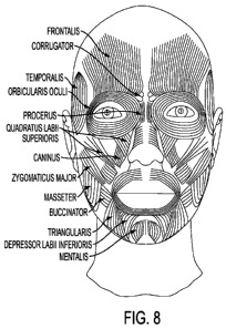
[00461 Figure 8: Map of the human facial anatomy to identify facial muscles
and
enveloping fascia subject to the micro incision screw or glue injection.
100471 Figure 9: Demonstrates the vast area of facial bone not containing an
origin of a
facial muscle or a critical nerve or vessels at risk from the screw
placements.
#4847-8914- 9445v4 15
CA 02744015 2011-05-17
WO 2010/059230 PCT/US2009/006219
[0048] Figure 10: Displays muscles that could be subject to fixation,
redirecting support
of the facial muscular mask
[0049] Figure 11: Displays endomysium of a muscle.
[0050] Figure 12: Displays areas where glue can influence intermuscular
adhesion and
intramuscular cohesiveness to effect contour and surface changes on the human
head and neck
region.
[0051] Figure 13: This patient underwent treatment for blepharsopasm using a
myo-
osseous fixation procedure. Unlike prior procedures the fixation was
accomplished with a
cyanoacrylate glue (2-Octyl cyanoacrylate). Post operatively blpeharsoapsm was
90% improved
in the first week. Lateral orbicularis muscles fixation accomplished with
tissue glue was
excellent and functioned in lieu of an mechanical fixation device to tether
muscle to bone and
effect improvement of involuntary eyelid closure. Serendipitously, the
crowsfeet wrinkle pattern
usually present in this location was significantly improved.
DETAILED DESCRIPTION
[0052] As used herein "osseous tissue" means bone tissue, the major structural
and
supportive connective tissue of the body. Osseous tissue forms the rigid part
of the bone organs
that make up the skeletal system.
[0053] As used herein the "superficial muscular aponeurotic system" or (SMAS)
refers to
an area of musculature of the face. This muscular system is manipulated during
facial cosmetic
surgery, especially rhytidectomy.
#4847-8914- 9445v4 16
CA 02744015 2011-05-17
WO 2010/059230 PCT/US2009/006219
[0054] As used herein, a "self-tapping or self drilling screw" is a screw that
has the
ability to advance when turned, while creating its own thread. Self-tapping or
self drilling
screws are commonly used with sheet metal and plastic components. This ability
is created
sometimes by having a gap in the continuity of the thread on the screw. These
edges can cut
their own threads as the screw is driven into the material, usually wood or
plastic. Self tapping
or self drilling or self drilling screws also exist for metal. They function
by having a cutting
edge which drills away the material, making a hole for the screw to go into.
Self tapping or self
drilling screws are most useful when doing metal and wood work.
[0055] As used herein, a "compression plate" is a surface that squeezes,
compresses or
pushes layers of soft tissue directly against bone to thereby cause fixation
of the tissue, inclusive
of muscular tissue, and alters the muscular tissue's relation to a low-
friction perimuscular
environment.
[0056] As used herein, the term "flush continuity with the osseous surface"
means the
screw or compression plate is placed tightly against bone such that the degree
of palpable ridge
from the skin surface is minimized.
[0057] As used herein, "bioadhesive" is any liquid or gel which when placed in
a plane
between muscles and bone causes adhesion. This allows a plastic surgeon to
remodel the soft
tissue to bone using free movement of the muscle overlying the bone
facilitated by the low
resistance fibro fatty plane underlying the muscle. A bioadhesive is able to
be injected under
muscle. This allows adherence of muscle to the bone structure which in turn
alters the origin of
the muscle, functional contractility of the muscle, and support of overlying
soft tissues generated
by muscle tone and support.
#4847-8914- 9445v4 17
CA 02744015 2011-05-17
WO 2010/059230 PCT/US2009/006219
[0058] As used herein, "low resistance plane" or "low friction plane" is the
fibro fatty
plane between bone and the undersurface of a muscle overlying the bone.
[0059] As used herein, "skin puncture" is a puncture through skin made with a
medical
needle between 4-32 gauge. In a preferred embodiment, a skin puncture is made
with a medical
needle between 25-30 gauge.
[0060] As used herein, "small incision" is an incision less than 10 mm. In a
preferred
embodiment of the present invention, a small incision is less than 3 mm.
[0061] As used herein, "large incision" is an incision greater than 1 cm.
[0062] As used herein, "external manipulation after bioadhesive injection"
refers to
movement of soft tissues (or muscle) over bone, facilitated by the low
resistance fibrofatty tissue
plane underlying the muscle, which is performed after injection of a
bioadhesive. This allows
binding of the undersurface of the muscle to bone thereby creating an
alteration of surface soft
tissues with respect to proportion, contour, function, skin wrinkle pattern,
elevation, or other
configuration desired by patient or surgeon.
[0063] As used herein, a "method of injection as to avoid nerves, arteries,
and veins"
refers to anatomic knowledge possessed by the surgeon, which contemplates
natural variations of
usual and unusual positions of critical nerves and vessels which can be
injured by
microincisions, implantable devices, or injections of bioadhesives.
[0064] As used herein, "body proportionality" refers to size or volume of a
body region
relative to another body region. For instance, upper cheek size relative to
lower cheek size, upper
neck size relative to lower neck size, and buttock size relative to thigh
size, and so on.
#4847-8914- 9445v4 18
CA 02744015 2011-05-17
WO 2010/059230 PCT/US2009/006219
[0065] As used herein "muscle compacting" or "muscle reshaping" by
bioadhesive,
refers to the process of injecting a muscle with a bioadhesive and shaping the
muscle to alter the
contour of the face or other body part by external manipulation, by which the
injected
bioadhesive functions as a connective synthetic matrix replacing the lax
collagen forming
endomysium, perimysium, and epimysium.
[0066] As used herein, "use of needle based thermal cautery to achieve
undersurface
muscle fixation" refers to another approach to cause fixation of muscle to
bone. The method
involves application of cautery via fine needle to the undersurface of the
muscle, which results in
fat deletion and contraction followed by inflammation and scarring in deep
facial layers, or other
body part layers. The result is an adhesion endogeneously created by the body,
which results in
adhesion and alteration in surface configuration. The cautery can be unipolar
microbipolar, or
battery powered device which transmitted energy through a puncture site to the
undersurface of
the muscle which created a adhesive scare. This method may be used singly or
in conjunction
with screws or bioadhesive methods as described elsewhere in this disclosure.
[0067] As used herein, the "use of lipolytic agents to achieve bioadhesion"
refers to using
lipolytic agents to affect the functional components and contribution of the
surrounding fat layers
to muscular contractile effect. These agents cause lipolysis, and can be used
to increase resistant
within surrounding muscle tissues and increase the resistance to muscular
contractility. Such
agents include bile acids or bile salts or other such agents which achieve
lipolysis Use of
physical energy systems such as radiofrequency or ultrasonic devices can also
be used to
dissolve fat cell membranes. This diminishes the fatty layers surrounding the
muscles and
#4847-8914- 9445v4 19
CA 02744015 2011-05-17
WO 2010/059230 PCT/US2009/006219
thereby increasing resistance against muscle action. Other physical agents
such as laser energy
applied via endoscope, or needle applied radiofrequency and the like can
achieve a similar effect.
[00681 As used herein, a "synthetic connective matrix" refers to bioadhesives
injected
into a muscle that insinuates and spreads when cured to form a "compaction"
and/or internal
fiber tethering, "internal restriction" of the muscle, decreased volume, and
decrease contractility
of the muscle.
[00691 As used herein, the "origin" of a muscle is the point at which it
attaches to a bone
or another muscle. The structure that the origin is attached to is not moved
by contraction of the
muscle. The opposite end of the muscle is the insertion.
100701 As used herein, the "insertion" of a muscle is the point at which it
attaches to the
skin, a bone, or another muscle. The insertion attaches the structure that
will be moved by the
contraction of the muscle. The opposite end of the muscle is the origin.
[00711 As used herein, "altered origin or insertion" of a muscle is creating
an adhesion of
muscle in area, for example in an area of bone, where normally no adhesion is
present.
[00721 The screw plate screw and the compression fixation plate can involve
the use of
tapping or self-tapping or self drilling or self drilling screws that are
placed in a strategic area in
such a fashion that a facial plane is compressed. The facial plane could
include the gala or the
SMAS or other deep facial or superficial facial plane.
[00731 The gala, if used, is fixated to the skull over the frontal bone
medially or laterally
to achieve brow elevation, or over the zygomatic bone to achieve mid face and
lower mid face
elevation, and in regions of the zygoma or the mandible and jowl elevation.
Such screws are
designed so that full penetration to the skull over the neurocranium does not
occur. In one
#4847-8914- 9445v4 20
CA 02744015 2011-05-17
WO 2010/059230 PCT/US2009/006219
embodiment, the length of such screws can vary from between about 0.25 mm to
about 10 mm,
and preferably from about 2 to about 8 mm for skull and facial bones. In
another embodiment,
the length of such screws can vary between from about 1 to about 50 mm,
depending on the body
region of interest. The selective advantage of this approach allows small
incisional surgery
without the use of extensive surface dissection which facilitates rapid wound
healing and reduces
the procedures to being minimally invasive, as opposed to more open requiring
larger wounds
and more extensive delays in wound healing and the potential for greater
scarring and
disfigurement post operatively.
[00741 The screw is customized to the portion of the skull being used.
Thicknesses of the
skull are determined anatomically and also with neuroradiography using
computerized axial
tomography for design. The screws are placed with a fixation wrench after a
small incision is
made, and adequate mobilization of the subcutaneous connective tissues are
accomplished. This
can be done with the patient awake, unlike former procedures for brow lifts in
which the patients
are often under general anesthesia. The fixation points are marked off and
local anesthesia is
used with diffusion enhancing agents such as hyaluronidase. Alternative forms
of anesthesia
involving regional nerve blocks are also possible to limit soft tissue
distortion so that the osseous
fixation can be more accurately accomplished. Once anesthesia is achieved of
the soft tissue
and bone, the facial plane is dissected and a small buttonhole is placed
through the facial plane.
The facial plane is then pulled to a desirous location and the bolt is placed
into the bone,
essentially lifting soft tissues below. In the case of brow lift, this is done
in a fashion to achieve
both accurate elevation and contouring of the brow. In the case of lower mid
face, the placement
is over the zygoma, fixating the suborbicularis fascia to points of fixation
on the lateral orbital
#4847-8914- 9445v4 21
CA 02744015 2011-05-17
WO 2010/059230 PCT/US2009/006219
rim over the zygoma. The compression plate used is a conventional washer over
a screw, to
achieve a large platform fixation over bone to further enhance the strength of
the elevation and
fixation point. Screws may be absorbing or non absorbing materials. Preferred
materials include
titanium, stainless steel, or absorbable material such as chromic, nylon,
polymethylmethacrylate,
or hardened forms of silicone. Glue arrangements may be arranged with various
forms of epoxy
to further enhance fixation of the screw's endplate. In other words, the screw
and/or
compression plate may be used in combination with a glue or adhesive. The glue
may not only
cause an increase in tensile strength of the screw's attachment to the bone,
but also may cause
adherence of the undersurface of the facial or targeted muscle to the bone. It
is important to note
that in order to facilitate natural contour and feel over a healed wound, the
screws are placed
flush with the skull so raised elevations are minimally perceived by the
patient, or any potential
observer.
[0075] Advantages of such approaches include a small incision or needle
puncture
incision with minimal suture closure and minimal surface scarring.
[0076] Diameter of screws can vary from between about 0.25 mm to about 4 mm,
depending on the fixation point. The head of the screw can vary from between
about 0.25 mm to
about 10 mm in diameter.
[0077] Said procedures are done with full knowledge of facial motor and
sensory neural
positions. Motor and sensory neural positions can be localized with nerve
stimulators, Doppler
devices for vascular bundle nerve identification, and general knowledge of
anatomic variation.
[0078] The screw washer plate can vary in size from about 2 mm to about 25 mm
in
diameter, preferably between about 2 mm to about 10 mm in diameter. It can
also have various
#4847-8914- 9445v4 22
CA 02744015 2011-05-17
WO 2010/059230 PCT/US2009/006219
geometric forms, for example circular or circular with flush projecting
platforms. Here again, it
is important that there is no direct extension of soft tissue over the
fixation plate, but rather a
flush bolting of soft tissue to skull or gluing the undersurface of muscle to
the skull, for example
by way of bioadhesive.
[0079] Screw head configurations can be philips, linear or any geometric
configuration
which enhances mechanical integration to the driver. The driver must be able
to fixation the
screw head in such a fashion to allow impalement of the facial plane to be
fixated so that the
deep or superficial facial planes can be elevated and fixated along a higher
position on the facial
bones.
[0080] As the natural aging process involves the descent of soft tissues, the
purpose of
the bolting screws or glue adhesives described herein is to create a
suspension of soft tissue back
on the position of the skull in which soft tissue has fallen. By virtue of
doing so, the contour of
the surface is changed, remodeled, reshaped and improved to a more juvenile
appearance.
Hyperplacement of soft tissue is anticipated with compensatory falling in a
postoperative period.
[0081] A diagram of such a screw arrangement and prototype is given in Figure
3c. This
diagram should not be limiting to other configurations in which there is a
screw and fixation
plate arrangement. Selective advantages of a fixation plate screw method
include:
1. Small incision.
2. Direct capability to do the procedure under local anesthesia.
3. Increased strength of fixation point over previous methods of fixation such
as purse string
sutures, tissue resection, or other forms of soft tissue suspensions with
ligatures.
4. Limited surgical dissection with increased rate of wound healing
postoperatively.
#4847-8914- 9445v4 23
CA 02744015 2011-05-17
WO 2010/059230 PCT/US2009/006219
5. Technical simplicity in performing with increased speed of procedure.
6. Capability to perform procedures on an outpatient basis without use of a
major operating
room.
7. Greater chance of permanent effect.
Concept of Alteration of the Origin and Insertion of Muscles Using External
Bolting to
Osseous Structures
[00821 The use of subcutaneous fixation self-tapping or self drilling or self
drilling
screws and muscles and soft tissue compression into osseous structures
effectively can be used to
change the origin and insertion of muscles. Muscles produce mammalian movement
via
attachment to various bone providing stability and direction to forces created
by muscle
contraction. By externally changing the fixation of muscles on bony platforms,
it is possible to
alter the vector forces generated by muscular contraction causing alteration
in the type, direction
and intensity of movement elicited by such contractions.
[00831 As an example, blepharospasm patients suffer from involuntary
contractions of
the orbicularis muscles. The orbicularis muscle has three basic segments: pre-
tarsal, pre-septal,
and pre-orbital. The pre-orbital section is the most well developed, eliciting
the most forceful
contraction on stimulation. The origin and insertion of this segment of this
muscle are the
medial canthal ligaments and adjacent bone. When the titantium bolt is placed
elevating the
brow, an alteration of the insertion and origin of the muscle is created such
that
(1) Resting tension on the muscle may be changed
#4847-8914- 9445v4 24
CA 02744015 2011-05-17
WO 2010/059230 PCT/US2009/006219
(2) Forces generated during contraction are re-directed in different vectors,
mitigating the
protraction (closure) of the eyelid
(3) Force generated by the muscle may be reduced
(4) Proprioceptive sensory feedback to the central nervous system may be
changed such
that involuntary movements are mitigated
[0084] Redistribution of muscular attachments can effectively change fiber
direction and
the extent to which fibers may contract.
[0085] In the case of the orbicularis muscle, fibers in the pre-orbital
segment are oriented
in a circular fashion except at medial points of attachment to tendon and
bone. The titanium peg,
or screw, or bolt or self-tapping or self drilling or self drilling screw, or
compression plate or
bioadhesive, re-orients the fibers into a direction such that the fibers are
no longer circular in
orientation but rather, obliquely configured as well as fixed to the
undersurface of the bone,
thereby producing immobilization of the fibers. This change in orientation and
configuration
depresses the contractility of the muscle, thereby altering function as well
as surface appearance
of the region. This arrangement also increases the frictional surface under
the muscle to thereby
restrict muscle contraction and increase inertia to movement.
[0086] Elevating the brow further functions to fixate the orbicularis to the
frontal bone
along the superior orbital rim both medially and laterally. The orbicularis
muscle is a sphincter
muscle that works by closing space in the middle. The effect of fixating the
orbicularis muscle is
to increase resting tension on the muscle fibers much like pulling on the
inside of an elastic band.
This fixation of the orbicularis muscle prevents sphincter muscle contraction.
The resting
contraction of the circular loop of muscle fibers, redirecting the angle of
muscle fibers so that the
#4847-8914- 9445v4 25
CA 02744015 2011-05-17
WO 2010/059230 PCT/US2009/006219
vector forces are re-angulated causing the forces caused by contraction of
muscle to have a
reduced effectiveness (reduced mechanical advantage) therefore reducing the
symptoms caused
by the involuntary muscle contraction. In other circumstances, providing a new
origin or
insertion may render weak contractions more effective by increasing the
tension and simulating
the direction of the muscle fibers causing a reduced force of contraction to
be more effective in
movement. Adherence of the undersurface of the facial muscle can serve to
reduce its tone once
a firm adherence occurs and the frictional relationship between the bone and
the attached muscle
is increased.
Screwplate, Pin, Screwhead Fixation Device
[0087] The method and technique require use of a sharply tapered screw or pin
capable
of penetrating both soft tissue and bone structure without undue trauma to
surrounding tissues.
In order for the surgeon to accomplish angulation of penetration or fascial-
muscle snaring
hooking and penetration, firm fixation of the compression pin, screw head, or
screw plate is
necessary. This allows adequate control of placement which is critical to
appropriate soft tissue
osseous integration and fixation which drives the change in surface structure
elevation and
contour changes.
[0088] The driver must firmly grasp the screw head via a circular holding
device which is
adequately tapered to fixate the screwhead and attach the head to the driver.
Such devices,
which may be referred to as "screw wrenches," "fixation devices," or "fixation
wrenches" herein
are conventionally engineered to the placement of screws to fixed fracture
plates, however such
#4847-8914- 9445v4 26
CA 02744015 2011-05-17
WO 2010/059230 _ PCT/US2009/006219
devices are not conventionally used for tissue snaring, looping or impaling.
Screw driver
wrenches used for bone plates for fracture repair can be adapted to achieve
this soft tissue
function via a micro-incision to snare, impale and ultimately fixate
connective tissue directly into
the bone structures under a compression plate. The screw wrench can resist
lateral torque
produced by the surgeon's hands and therefore can hold the screw in tight
potion allowing
directional control. The screw wrench can secure a screw at different
angulations. Analogy is
made between the screw wrench and screw to the surgeon's needle holder and
needle. The
needle must be held with precise control and fixation in order to avoid
impaling or otherwise
hurting, for example, arteries, veins, eyes and the like. In the case of
screws, the screw head
must be able to resist substantial torques and forces, for example substantial
lateral torques on
the screw head. The screw cannot be allowed to "tiddlywink" away from the
surgeon's desired
placement. It is contemplated that repeated adjustments of the screws may be
possible or
necessary. An example of a fixation system is given in Figure 3c.
Components of the Implantable Fixation Device
[0089] The implantable device consists of a compression plate (screw head with
and
without washer ring), and drilling-fixation shaft for soft tissue
stabilization. The drilling shaft
may vary from 1 mm to 50 mm, preferably 2 mm to 8 mm when used on the facial
or skull
bones. The diameter of the drill shaft may vary from 0.2 mm to 10 mm, but is
preferably 1 mm
to 3 mm. The compression plate may substantially vary in size and
configuration, however it is
#4847-8914- 9445v4 27
CA 02744015 2011-05-17
WO 2010/059230 PCT/US2009/006219
tailored to the anatomic region where the implantable device is positioned.
Compression plates
can vary between 3 cm to 1 mm.
Mitigation of Complications
[0090) Depth of penetration into the skeletal structure may result in
unnecessary pain and
risk of the insertion and soft tissue fixation procedure. Use of screw lengths
appropriate to the
anatomic thickness and position of the bone is critical in preventing
excessive penetration of the
fixation shaft of the compression screw and plate to the bone, excessive
penetration may:
(1) be associated with excessive pain
(2) cause damage to deep vascular structures and initiate hemorrhage
(3) represent a risk of disruption of intracranial vessels if transcranial
penetration occurs
during facial application
[00911 The use of computerized imaging devices using computerized axial
tomography
or other forms of imaging digital data devices to achieve intra-operative
anatomic registration is
helpful in confirming depth of penetration of the fixation shaft and optimum
placement of the
drill shaft into bone. Optimum fixation requires placement of the shaft into
bone of
appropriately matched thickness relative to the length of the fixation shaft,
avoidance of sinus
cavities which, if penetrated, can destabilize soft tissue fixation and
partial thickness penetration
to avoid damage to deeper structures (such as brain and meningeal coverings
for forehead and
facial fixations). Use of computerized imaging devices may be useful in
individual assessment
of bone thickness which can substantially vary among individuals.
#4847-8914- 9445v4 28
CA 02744015 2011-05-17
WO 2010/059230 PCT/US2009/006219
Attachments of Facial Muscles to the Facial Bones and the Aging Process
[0092] The facial muscles are contained within the superficial muscular
aponeurotic
system as has been described above. With aging, the connective tissue
(collagen) component of
this tissue becomes increasingly lax resulting in diminished support of soft
tissues associated
with sagging. Tissues may also intrinsically age with fat redistribution,
muscle atrophy, bone
remodeling and dermal laxity. An age related change often not cited on the
human face is
integrity and strength of deep facial muscle attachment to the facial and
skull bone. These tissue
origins essentially hold the highly motile SMAS to the bone lending support to
facial movement
and resting facial position.
[0093] The implantable devices described herein provide a method for increased
facial
muscular support which leads to alterations in facial contour and surface
configuration in a more
youthful and vital direction. Reinforcement of natural attachments (muscle
origins) results in
more youthful and functional performance of facial muscles, as well as other
muscle groups
targeted for attachment reinforcement. Natural critical attachments can be
studied for optimum
placement of implantable devices.
Bioadhesives
[0094] In another embodiment of the present invention, an alternate muscle to
bone
periosteum adhesion and muscle to muscle adhesion is accomplished with an
implantable or
injectable bioadhesive or tissue glue. This method may use concepts described
throughout the
instant specification with respect to screws and/or compression plates;
however, the use of
#4847-8914- 9445v4 29
CA 02744015 2011-05-17
WO 2010/059230 PCT/US2009/006219
bioadhesives improves the administration technique from microinsion to mere
skin puncture.
This leaves no appreciable scar. In one embodiment, an injectable material,
preferably
comprising a substantially inert material with quick drying dynamic is used.
This injectable
material is capable of sustaining a strong adhesion between the undersurface
of facial muscle and
underlying bony structures. This allows for fixation of the muscle and
support. This muscle
fixation is useful to control involuntary movements and also to allow for
support of soft tissue to
restore a youthful and aesthetically original contour surface structure to the
human face. In one
embodiment, the method reduces or eliminates wrinkle patterns during dynamic
facial
movements.
[00951 In one embodiment, injectable materials include bioadhesives consisting
of
cyanoacrylate derivatives, such as 2-Octyl Cyanoacrylate, fibrinogen-thrombin
combinations,
gluteraldyhyde based glues, dental crown cement fixation glues, and
inflammatory provoking
adhesives which may allow for a small inflammatory response causing a collagen
"scar" based
adhesion. In one embodiment, an injection device comprises a needle, a single
or multiple
chamber injections system, or a multiple needle system in order to inject the
glue in multiple
sites. On the human face, targeted areas would include placing the glue over
the surface of the
frontal bone, zygomatic and lateral orbital bone, face of the maxillae, and
rim anterior or
posterior surface of the mandible. As every human face pattern is different in
the aesthetic
application, emphasis on certain areas would be considered on a case by case
basis. The amount
of glue injected should be limited by inflammatory response, surface bumps or
bulging, and
anatomic consideration to avoid nerves, arteries and veins. Multiple injection
sessions over a
defined period of time can limit the risk of uncontrolled spread of the glue
and excessive
#4847-8914- 9445v4 30
CA 02744015 2011-05-17
WO 2010/059230 PCT/US2009/006219
inflammatory response, and to adjust adhesion strength. It is anticipated that
dispensing of the
glue shall be accomplished with unit injectable quantities associated with
single or multiple use
needles. Each injection quantity would have been predetermined to limit the
degree of
inflammatory reaction so not to promote excessive scar formation visible on
the skin surface.
[00961 In certain embodiments, adhesion is injected via a plastic or metallic
needle so
that the needle lumen is placed under the muscle within a mobile fat plane, so
that the adhesive
can be delivered through a puncture site (non incisional), and the adhesive
injected with
immediate surface elevation by the hand or direction of the physician or
clinician, so that the
underlying muscle within the mobile tissue plane containing the hardening glue
becomes
adherent to facial bone-periosteum causing an altered origin, that is fixation
point (support
point) so that a soft tissue face-lift can be achieved, without the need for
an incision. It is
anticipated that multiple injections of the tissue adhesive at the time of
applications and over a
period of time may be needed.
100971 The embodiments herein described altering muscle and soft tissue
attachment to
bone to achieve change a surface contour of the face and other body regions
without use of
conventional large incisions.
[0098] A map of the human facial anatomy is given in Fig. 8 to identify facial
muscles
and enveloping fascia subject to the micro incision screw or glue injection.
Fig 9 demonstrates
the vast area of facial bone not containing an origin of a facial muscle or a
critical nerve or
vessels at risk from the screw placements. In figure 9, any of the listed
muscles could be subject
to fixation, redirecting support of the facial muscular mask (see Fig. 10) .
These muscles with
attachments and enveloping fascia comprise the foundation for the individual
contour,
#4847-8914- 9445v4 31
CA 02744015 2011-05-17
WO 2010/059230 PCT/US2009/006219
movement and wrinkle characteristic and proportionality of the human face.
Arrows on Fig. 10
display the characteristic taper or V-shape of the human skull and face. The
overlying soft tissue
tends to sag as humans age, thereby distorting the natural shape and
proportionality of the human
face. Methods and devices described herein can correct or counteract this
distortion.
Screw Size
[00991 Screw sizes can vary as well as screw head designs. Dome screw head,
convex
screw head, flat screw heads, and screw heads with expandable compression
heads are all
possible types of the implant designs. The ideal screw head would be the most
flush to flat and
least palpable after surgical implantation. Screw head may vary considerably
from small
puncture incisions to large compression plates. Compression plats may be
washer shaped or
linear shaped and implanted via side punctures so that screw penetrates
compression plates at
one or more orifices. The compression plate may be absorbable or made of a not
absorbing
metal, acrylic, polymethyl methacrylate or silicone. The screw hand may
contain microhooks,
serrated surface to enhance anterior or superficial tissue hold. In one
embodiment of the
invention, the screw are small enough that no superficial suturing is needed.
Screw may be used
with various forms of glue. The current practice of soft tissue fixation to
bone involves and
predrilled cylindric hole with a plug in absorbing hooked device. This system
is flimsy and offers
less support than a drilled penetration into the outer table of skull flat
bone.
#4847-8914- 9445v4 32
CA 02744015 2011-05-17
WO 2010/059230 _ PCT/US2009/006219
Anatomic plane targeted for glue or device placement and method of delivery
1001001 As the intent of the minimally invasive procedure described herein is
to provide a
reinforcement of the origin and origin/insertion relationships between facial
muscles and bone,
the correct anatomic plane of the injection must include the fibro fatty plane
between the
undersurface of the facial muscle or other anatomically defined muscles and
the underlying bone
covered by periosteum. This plane should be fixated by either injectable glue,
placement of
compression screw or fixated by adhesion based implant for the purpose of
altering contractility
of muscle, and/or altering facial contour, and/or influencing wrinkle patterns
of skin during
dynamic muscular activity. The optimal method is least invasive, requiring the
smallest incision.
In one embodiment, the incision is merely a puncture site. Any anatomic plane
between bone
and muscle represents an important target for reducing muscular activity by
increasing friction,
inducing tethering, and functional resistance to the internal forces produced
by the muscle itself.
Internal muscular activity is conventionally defined by neuromuscular
relationships, muscle
mass, and effective chemical reaction between actin and myosin proteins.
Bioadhesives or Glues Applicable for Submuscular and Intramuscular injection
[001011 The following represent non-limiting exemplary bioadhesive or glue
types
which can be used in various embodiments as an injectable to achieve myo-
oseous fixation to
control contour changes, wrinkle pattern reduction, and/or elevation of the
human face via
injection These agents are qualified by adhesive properties, inflammatory
response and duration
of adhesion. Although variations are possible based on dose and position, the
overriding
#4847-8914- 9445v4 33
CA 02744015 2011-05-17
WO 2010/059230 PCT/US2009/006219
principle of myo-osseous fixation can be accomplished by one or more of these
agents, at
varying doses dependent on strength of adhesion, inflammatory complications
and duration of
effect. Other qualifying paramteres include coloration quality with clear or
lighter cured glue
color being preferable, flow characteristic via delivery device, curing rate
(hardening rate),
viscosity , and cohesiveness to prevent migration or possible embolic
complications.
Bioadhesives or glue may be administered in conjunction with metallic screws
or other types of
fixation devices which facilitates reconfiguration of attachments of the
undersurface of the facial
muscles to bone. External devices such as temporary bandages, masks, or tapes
can also be used
to assist in this minimally invasive procedure.
Fibrin-Thrombin-Fibrinogen
[001021 This glue type consists of a duo delivery system which involves the
injection of
fibrinogen followed by thrombin which catalyzes the conversion of fibrinogen
to monomers
which cross link to form the fibrin clot. The fibrinogen is often formulated
in higher
concentrations than the human plasma and can be derived from pooled plasma or
from
recombinant origins. Thrombin also can be derived from pooled human blood
products or from
recombinant manufacturing technology. Plasminogen may be removed from such
products as
this agent when activated may causes fibrinolysis which degrades the clot and
seal properties.
Alternatively, agents which ininhibit plasmin which can further limit clot
degradation. One such
inhibiot is aprotinin which is used in the commercial product Tisseel-TM.
Recombinant blood
#4847-8914- 9445v4 34
CA 02744015 2011-05-17
WO 2010/059230 PCT/US2009/006219
based products limit potential contamination with hepatitis B, C and HIV as
well as prion based
disease.
[00103] It is anticipated that glue delivery is needed in multiple locations
and given
serially to achieve a lasting seal and bioadhesion.
[00104] Fibrin based sealant have the potential to induce growth factor for
fibrocytes
which can enhance sealing capability over time. These materials can control
bleeding which can
be beneficial when injected into a vascularized muscle.
[00105] Commercially available products include Tisseel,Evicel, Vitage, and
Cryoseal
Thrombin
[00106] Thrombin may be used individually as a glue and binding agent and may
e used in
its recombinant form or native form from human or animal based donors. This
agent may be
used with gelatin.
Polyethylene Glycol Based Sealants and Glue
[00107] Currently Duraseal-TM and CoSeal-TM are available in the US. These
agents
can be used to control bleeding and have sealant and binding capability which
may be adapted to
muscle bone fixation. Durseal is commonly used in the head for outer brain
covering
procedures.
#4847-8914- 9445v4 35
CA 02744015 2011-05-17
WO 2010/059230 PCT/US2009/006219
Gluteraldehyde-Albumin Sealants
1001081 These sealant consist of purified albumin with gluteraldehyde which
when mixed
forms a covalent bond between the matrix and cell surfaces. BioGlue-TM is a
non-limiting
example.
Cyanoacrylate based Sealants
[001091 Non-limiting examples are 2-octyl cyanoacrylate, N-butyl 2
cyanoacrylate, and
absorbable cyanoacrylates, known under the trademark OMNEX-TM. These agent can
effectively stop bleeding. Binding strength is better than fibrin based
sealants. Low volume of
agent can be effective. Inflammatory potential can potentially enhance long
term nature of seal
when place under a thin facial muscle. Absorbable cyanoacrylates such as OMNEX
ay be
preferred based on reversibility and lack of long term potential for scarring
and foreign body
reactions. The use of cyanoacrylate is described in more detail below.
Other types of Bioadhesives
[001101 In other embodiments, plasma polymerized N-isopropyl acrylamide
(pNIPAM) or
ONYX-TM are suitable bioadhesives for the present invention.
#4847-8914- 9445v4 36
CA 02744015 2011-05-17
WO 2010/059230 PCT/US2009/006219
Use of Cyanoarcrylate derived Glues as an Injectable Agent for the purpose of
altering
myo-osseous fixation
[001111 Cyanoacrylate glues are well known adhesives which are known to
produce
strong bonding during polymerization and forming tight bonding with a degree
of heat release.
Medical applications have commonly included skin approximation as an
alternative to
conventional sutures for which the material is currently commercialized. Deep
tissue
implantation has also been advocated for neural trunk repair during soft
tissue surgery, vascular
repair, fixation of extraocular muscles to the ocular globe or implanted
prosthesis after globe
removal, embolization for vascular malformation using neuroradiologic
techniques.
[001121 Major concerns from regulators have included excessive inflammatory
reactions
from chemical breakdown of implanted glue which can form formaldehyde, a
chemical toxic to
tissues. Heat generation during polymerization and hardening has also been
noted but represents
a lesser problem. Efforts have been made to find cyanoacryolate derivative
which have a reduced
rate of formaldyhyde generation. Non-limiting examples of cyanoacrylastes
suitable for use with
the present invention included: alkyl 2-cyanoacrylate, alkenyl 2-
cyanoacrylate, alkoxyalkyl 2-
cyanoacrylate, and carbalkoxyalkyl 2-cyanoacrylate. The alkyl group may have 1
to 16 carbon
atoms and is preferably a C1 -C8 alkyl 2-cyanoacrylate. Suitable
cyanoacrylates include, for
example, methyl 2-cyanoacrylate, ethyl 2-cyanoacrylate, n-propyl 2-
cyanoacrylate, iso-propyl 2-
cyanoacrylate, n-butyl 2-cyanoacrylate, iso-butyl 2-cyanoacrylate, hexyl 2-
cyanoacrylate, n-octyl
2-cyanoacrylate, 2-octyl 2-cyanoacrylate, 2-methoxyethyl 2-cyanoacrylate, 2-
ethoxyethyl 2-
cyanoacrylate and 2-propoxyethyl 2-cyanoacrylate, methyl .alpha.-
cyanoacrylate, ethyl .alpha.-
cyanoacrylate, propyl .alpha.-cyanoacrylate, butyl .alpha.-cyanoacrylate, and
cyclohexyl .alpha.-
#4847-8914- 9445v4 37
CA 02744015 2011-05-17
WO 2010/059230 PCT/US2009/006219
cyanoacrylate, alkenyl and cycloalkenyl .alpha.-cyanoacrylates such as allyl
.alpha.-
cyanoacrylate, methallyl .alpha.-cyanoacrylate, and cyclohexenyl .alpha.-
cyanoacrylate, alkynyl
.alpha.-cyanoacrylates such as propargyl .alpha.-cyanoacrylate, aryl .alpha.-
cyanoacrylates such
as phenyl .alpha.-cyanoacrylate and toluyl .alpha.-cyanoacrylate, hetero atom-
containing methox
yethyl .alpha.-cyanoacrylate, ethoxyethyl .alpha.-cyanoacrylate, furfuryl
.alpha.-cyanoacrylate,
silicon atom-containing trimethylsilylmethyl .alpha.-cyanoacrylate,
trimethylsilylethyl .alpha.-
cyanoacrylate, trimethylsilylpropyl .alpha.-cyanoacrylate and
dimethylvinylsilylmethyl .alpha.-
cyanoacrylate.
[00113] A preferred cyanoacrylate is 2-octyly cyanoacrylate because of its
slow
degradation and reduced rate of formaldyhyde generation.
[00114] The surgical method of claim 1, wherein said bioadhesive has a slow
degradation
and reduced rate of formaldyhyde gereneration.
[00115] It is anticipated that multiple forms of delivery devices may be used
to inject
adhesives through a puncture site for the purpose of glue placement in the
defined fibro fatty
plane on the undersurface of the muscle tissue. These include prefilled
syringes with sterilized
liquid adhesive which remain air tight until the moment preceding use. Tube
enveloping
containers capable of deilivering a fixed volume of liquid adhesive also may
be used. Diaphragm
shaped containers containing a fixed amount of glue may alos be used. Any
container forms
mentioned are not limiting as multiple devices are possible so long as the
device maintains
sterility of the injectable and is able to deliver a fixed and consistent
volume of adhesive agent.
#4847-8914- 9445v4 38
CA 02744015 2011-05-17
WO 2010/059230 - PCT/US2009/006219
Absorbable Type Cyanoacrylates
[001161 A class of cyanoacrylate glues characterized as "absorbable tissue
glue" has been
advanced and advocated. A limiting factor of cycanoacrylate glues is granuloma
formation from
breakdown into toxic subcompoents such as formaldehyde. Glue designed to
absorb with lesser
chances of foreign body inflammatory reaction have lesser chance to induce
this form of
complication and have improved tolerability. Although the mechanical adhesion
may be
temporary, adjuctive inflammatory reaction from single or multiple injections
can induce fibrosis
which can function as an endogenous glue and adhesive achieving the sam e
effect as an foreign
substance. The net effect would be to create a more permanent or lasting
adhesion. The
tolerability of absorbable cyanocrylates will result in less secondary
inflammatory complications,
less scarring and an improved side effect profile than some of the
conventional preparations
listed above. Currently, Omnex-TM from Johnsosn and Johnson represents an
example of the
absorbable cyanoacrylate class appropriate for the practice described herein.
Absorble class glues
are not to be limited by one formulation or brand as other forms of absorbable
bioadhesives can
be used to achieve similar results.
Alternatives to Cyanacrylate derived glues
[001171 Because of the high degree of tissue reactivity induced by some
implanted
cyanoacrylates, alterative forms of liquid or gel bioadhesives can be applied
for the same
#4847-8914- 9445v4 39
CA 02744015 2011-05-17
WO 2010/059230 PCT/US2009/006219
purpose and deliveried by syringe and puncture site. Alterative materials may
include various
forms of silicone, acrylic or any other substance demonsrtaed to have less
than 5% induced
inflammatory rate. Devices may be injected through puncture sites to achieve
the same end.
These devices must be implable through a mcrincision or puncture, be capable
of fixation the
undersurface of muscle to bone by elevation and fixation of the undersurface
of the muscle to
bone and tether muscle to bone. The device is placed through the skin and
inserted between the
soft tissue and the bone. In one embodiment, a device is a linear implant with
extending side
barbs. The extending side barbs may be hard and stiff, and may be of various
lengths relative to
the length of the linear implant.
Method of Tissue Manipulation after Injection of the Liquid Adhesive
1001181 Once the clinician injects the liquid adhesive, it would be important
to manipulate
the soft tissue overlying the bony surface over which the injection has been
administered so that
the alteration in contour and suspension is placed in a desirable
configuration. For example, the
method of application using a liquid adhesive would be organized in the
following steps:
(1) Small amout of anesthestic (eg lidociane) injected over the desired bony
surface
within the low resistance fibro fatty tissue planel; (2)Injection of the
liquid adhesive agent (eg
Cyanoacrytlate glue) over the bone in the fibrofatty plane on the undersurface
of the muscle,
through a puncture site, not involving an incision in one embodiment, or
involving a microinsion
in another embodiment, so that harding and polymerization is initiated. Care
is taken to avoid
direct injection into nerve, arteries veins, or other anatomically sensitive
areas so as to avoid
#4847-8914- 9445v4 40
CA 02744015 2011-05-17
WO 2010/059230 PCT/US2009/006219
complications; (3) Tissue manipulation by the clinician externally so that the
soft tissue can be
respitioned or resuspened into a desirable configuration. For instance,
frontal bone injection can
be followed by manual elevation of the brow to achive a brow lift. Maxillary
and zygomatic
bone injections can be administered so as to achieve midface lift by manually
elevating the check
from the surface over the maxillae and zygomatic bonese. Mandibular inferior
border, anterior
or posterior surface can be injected followed by manipulation of the soft
tissue of the jowl region
so as to resuspend and tighten the jowl region so as to reshape the jowl to a
more contoured and
youthful configuration without the use of conventional incisional plastic
surgery of the face; and
(4) Other injection location in non facial areas of the bony and the concept
and technique should
not necessarily be limited by exact anatomic relationships.
Amount of Injectable Glues and Adhesives Used
[00119] The amount of glue or adhesives used is limited by the critical amount
which will
cause inflammatory, granulomatous reactions or excessively cuase tissue
contractions with
dimpling of the skin surface. In one embodiment, the amount may vary from
between about
0.001 mL to about 10 mL per injection site. In other embodiments, the amounts
may vary (all
given on a per injection site basis) from between about 0.001 ml to about 8
mL, from between
about 0.001 mL to about 6 mL, from between about 0.001 mL to about 4 mL, from
between
about 0.001 mL to about 2 mL, from between about 0.001 mL to about 1 mL, and
from between
about 0.001 mL to about 0.5 mL. In a preferred embodiment, the amount per
injection site is
between about 0.1 mL to about 0.2 mL. Multiple injections are needed depending
on the
#4847-8914- 9445v4 41
CA 02744015 2011-05-17
WO 2010/059230 PCT/US2009/006219
targeted region and strength of adhesion sought per given anatomic region.
Multiple injection
point per region are anticipated.
Device used for injections
[00120] In one embodiment, the device comprises a single use device comprising
a
needle or micropuncture device, with a bioadhesive, for example but not
limited to,
cyanoacrylate glue. Other forms of bioadhesives or tissue glues such as
thrombin fibrinogen may
involve compounded needle bores with individual ports corresponding to a given
component of
the glue substance needed for curing. A syringe with predetermined injection
neddle or
micropuncture would be the preferred embodiment of the delivery system with
the intention for
single puncture per syringe use. Alternatively a multiple use delivery syringe
may be used for
more rapid administration techniques.
Intramuscular Injection of Glue to Establish Muscle Fiber Support, Compaction
and
Connective Tissue reinforcement
[00121] In addition to muscle fixation to bone, some embodiments of the
present
invention may make use of intramuscular glue to establish a support to the
endomysium,
perimysium, and/or other intramuscular and intermuscular support of connective
tissue. The
function is to compact muscule groups, alter intermuscular adhesions, and/or
elevate muscular
dissent with repect to bone and intermuscular attacvhments for the purpose of
elevating,
tightening and altering the contour of surface structures for aesthetic
purpose. An example of this
#4847-8914- 9445v4 42
CA 02744015 2011-05-17
WO 2010/059230 PCT/US2009/006219
type of application is gluing the submental platysmal muscle to the digastrics
muscles for the
purpose of improving the angulation between the neck and undersurface of the
jaw. The glue in
this situation is placed between a flat flimsy muscles which is poorly
compacted by aging
collagen within the muscle proper to the well compacted and well supported
deeper muscles
effectively compacting the muscle groups and achieving a more youthful surface
appearance.
Similar enhancement of the jowl tissues to deeper muscle fascia of the
temoralis muscle can be
used to elevate the jowl region and gluing the lower portion of the jowl to
digastric,
sternomastoid, sclene muscles effectively achieve the same purpose. Glue may
be placed over
the masseter muscles, into adjacent parotid gland, or related fascial
surfaces.
[00122] Direct injection of glue into muscle functions to causes a compacting
effect on
the muscles volume. This is accomplished by providing a sunthetic matrix which
function like
the natural endomysium of the muscle as shown in Fig. 11. The muscle
endomycium is
structurally supported by collagen protein which loosens with age. The
synthetic matrix serves to
reinforce the lossening supporting structure within the muscle allowing the
injecting physician
and surgeon to control the muscle volume and supporting surface contour formed
by muscle
volume. The glue needs to be diffusely distributed throughout the target
muscle followed by
external surface manipulation to shape and contour the muscle to an
appropriate and desired
configuration. Care must be placed not to allow glue to cure at puncture or
micro incision sites as
healing can be impaired allowing a conduit for infection.
[00123] Bioadhesives injected via puncture with needle or micro incision
within muscle
can function as a synthetic perimysium, endomysium and epimysium for a
targeted muscle or
muscle group. Bioadhesive injection directly into a muscle can create a
restriction of
#4847-8914- 9445v4 43
CA 02744015 2011-05-17
WO 2010/059230 PCT/US2009/006219
contractility, hence this method may be useful in the therapy for disorders
involving involuntary
movement such as spasticity, dystonia, painful reflex muscle spasms,
myofascial pain, bruxism,
temporal madibular joint syndrome, twitches and other facial spasms, posture
deformity, cerebral
palsy, spasmodic disorders of growth and development such as scoliosis and
other spasm types.
[00124] With reference to Fig.12, arrows point to areas where glue can
influence
intermuscular adhesion and intramuscular cohesiveness to effect contour and
surface changes on
the human head and neck region. The glue may be for instance, placed under the
platysma
muscle to flatten neck bands, to decrease volume of the platysma muscle, and
to effect a
compression of the platysma muscle to the firmly adherent digastrics muscles
and muscles in
proximity to the hyoid bone, causing a more youthful acute neck angle and a
reduction in the
hanging jowl. As another example, deeper injection of glue into the masseter
muscle can be used
to compress this muscle reduce its volume and reduce a square lower face
deformity commonly
seen with aging. The masseter muscle accounts for a substantial portion of the
lower face
volume.
[00125] Other anatomic regions may be approached in similar manor and concept
and
application should not be limited by region.
EXAMPLES
Example 1: Mid Face Lift
[00126] The methods and materials described herein are ideal for accomplishing
a mid
face lift. The SMAS layer encompasses the zygomatic major and minor muscles as
well as
adjacent facial muscles. The malar eminence is formed by the zygomatic bone
which contains
#4847-8914- 9445v4 44
CA 02744015 2011-05-17
WO 2010/059230 - PCT/US2009/006219
no essential arteries or other neurovascular structures hence this bony
platform is ideal for
fixating the muscular structures. The zygomatic major and minor muscles are
impaled with
associated SMAS with the self-tapping or self drilling or self drilling screw
via a small incision
site over the lateral orbital region in the vicinity on or superior to the
zygomatic bone. The
muscles associated with its surrounding fascia are elevated and fixated
superior to the malar
eminence effectively lifting the attachments (insertions) of the zygomaticus
muscles (malar
eminence) and fixating the soft tissue lift to the facial bone. Once fixation
has been established
supporting fixation can be accomplished by use of one or more additional self
tapping or self
drilling compression fixation bolts to the bony platforms in this region.
Additional self tapping
or self drilling bolts may be placed in small incisions in the nasolabial
fold, or inferior orbital
rim, along natural cutaneous tension lines in order to increase fixation under
small cutaneous
puncture sites.
Example 2: Adaptability of Techniques to other Body Regions - Breast Ptosis
[00127] The concept of osseous soft tissue integration under a screw head or
compression
plate can be applied outside the facial regions. For instance, age-related
breast sagging (breast
ptosis in the female) is considered a culturally undesirable senescent change.
Use of
compression fixation to the clavicle via deep fascial can serve as a method to
treat breast ptosis.
Ptosis, defined as progressive dropping of the apex of the breast with age,
can be modified with
volume expanders such as breast implants (for example, silicone gel implants)
as well as superior
suspension with fascial fixation. Additionally, osseous fixation can be used
to modify breast
#4847-8914- 9445v4 45
CA 02744015 2011-05-17
WO 2010/059230 PCT/US2009/006219
contour, that is relative configuration of tissue curves by fixation to ribs,
sternum or clavicle.
Small incisions can be made under the clavicular region and the superficial
and/or deep fascial
can be impaled by the self-tapping or self drilling screw system and fixated
to the inferior border
of the clavicle creating effectively a breast lift. Self-tapping or self
drilling screws within this
anatomic region for such a procedure by necessity should be longer than those
used on the facial
region so that this larger anatomic region can be adequately fixated. Fixation
point should be the
inferior, inferior anterior or anterior surface of the clavicle, the rib or
the sternum. The clavicle
and sternum are relatively immobile fixation points (as are the facial bones
and pelvis) and serve
as excellent regions for soft tissue osseous fixation under a compression
plate or screw head.
Example 3: Adaptability of Techniques to other Body Regions - Buttock
Elevation
[00128] Buttock elevation and contour changing can be accomplished with
osseous
fixation using compression plates or self-tapping or self drilling screw
fixation to pelvic osseous
ridge both superiorly and posteriorly. Use of soft tissue compression over
regions not containing
critical nerves or vessels allows safe fixation tissue contouring and/or
contour curve alterations.
Adjunctive elements to the compression plates may include suspension cables
passed through
soft tissue to achieve a greater lift. The cable extensions may be fixated to
the compression
screws.
Example 4: Adaptability of Techniques to other Body Regions - Supra Pelvic
Lateral
Bulge (Love Handles)
[00129] Alteration of bulge around the waist line may be accomplished via
multiple soft
tissue compression plates to the upper portions of the pelvis effectively
reducing waist bulge.
#4847-8914- 9445v4 46
CA 02744015 2011-05-17
WO 2010/059230 PCT/US2009/006219
The same concept of fixating soft tissue via small incisions into the bone via
a self-tapping or
self drilling screw system is used to re-contour and re-suspend soft tissues
into the pelvic rim
altering the protrusion of the waist line, an age-related anatomic finding.
The compression
screws may be used in addition to existent methods of liposuction and other
forms of adipose
resection, thermal reductions, ultrasonic or radiofrequency fat reduction.
Example 5: Adaptability of Techniques to other Body Regions - Floppy Posterior
Arm Soft
Tissue Contour Fixation
[001301 Floppy posterior arm syndrome is a cosmetic deformity noted by many
middle
aged women. Efforts to treat this condition with radiofrequency have often
failed. The use of a
soft tissue fixation-compression plate or screw bolting arrangement may be
used to alter the
contour and increase support in this region to reduce the deformity. Care must
be taken not to
impale critical nerves or vessels into the extremity.
Example 6: Use of Suspension Cables fixated to a Soft Tissue Oseous Fixation
Plate
[001311 Soft tissue fixation may be enhanced using a tissue fixation cable
passed through
a soft tissue structure and stabilized under a fixation plate. An example
would be a non
absorbable material such as orolene fixated to a compression plate, or screw
head on the
underside of the mandibular bone to reduce jowl contour with the fixation
cable passed across
the submandibular region to the fixation screwhead on opposite side to sling
the submandibular
region causing enhancement on angulations of the neck, an aesthetically more
youthful
appearance. Here the fixation screw not only improves the contour of the jowl
region but also
serves to be an anchor for a suspension cable or suspension mesh.
#4847-8914- 9445v4 47
CA 02744015 2011-05-17
WO 2010/059230 PCT/US2009/006219
Example 7: Facial Palsy Applications
[00132] Chronic facial palsy can have major impact on quality of life
resulting from
disfigurement from asymmetric facial movements and laxity of facial structures
at rest. The
latter situation results in brow ptosis, dropping angle of the mouth and cheek
asymmetry.
Reasons for chronic facial palsy include Bell's palsy, traumatic skull injury,
surgery (for
example, acoustic neuroma resection), Ramsy Hunt syndrome, Mastoiditis, Lyme
disease, stroke
as well as other nosologic entities.
Example 8: Treatment of Brow Ptosis, Cheek Ptosis and Asymmetry of the Lower
Face
[001331 The compression plate and screws are placed in a similar manner as
described
above to achieve an enhanced symmetry at rest for such patients. Preferred
areas to accomplish
connective tissue fixation include frontal bone for brow ptosis, lateral
orbital rim for cheek
ptosis, maxillary face (anterior maxillary bone for angle of the mouth).
Positioning of the
fixation points needs to be individualized for each patient after an
assessment of the regional
severity of the deficits.
[001341 Treatment of paralyzed lid closure associated with facial paralysis
often involves
the use of an implantable gold weight into the upper lid which serves to aid
in closure of the eye
with the normal blink. The weight works on the principle that the levator
palebrae superioris
muscles have the natural brainstem impulse momentarily turned off during lid
closure occurring
during a natural blink. The gold loaded lid is much heavier than the normal
lid and the weight is
projected away from the fulcrum point on the superior surface of the eye,
hence torque is
#4847-8914- 9445v4 48
CA 02744015 2011-05-17
WO 2010/059230 PCT/US2009/006219
established causing lid closure when levator tone is interrupted. A closing
eyelid during blinking
is critical as the continuity of the tear film needs to be physically
maintained over the cornea for
transpiration of oxygen and to protect the epithial integrity of the optical
surface. Loss of an
ultrasmooth optical surface results is optical ray diffraction with irregular
astigmatism, surface
ulceration, scarring, neovascularization and potentially permanent visual
loss.
[00135] The compression screw procedure represents a method to tighten the
lateral
reticulum (and lateral canthal tendon) which serve to further enhance the
effect of implantable
gold weight on lid closure by tightening the horizontal support of the upper
and lower eyelids.
Furthermore, in this situation, the lower lids can be elevated, further
enhancing corneal
protection for the paralyzed face. In summary, the compression plate and screw
heads can be
used to tighten and re-suspend the lateral orbital retinaculum and lateral
canthal tendon as well as
the sub-orbicularis fascia effectively creating more protection of the ocular
surface from
exposures due to paralyzed eyelids. The procedure can be updated over time to
maintain the
benefit of the correction.
Example 9: Blepharospasm
[00136] Benign essential blepharospasm is a condition characterized by
involuntary
closure of the eyelids in the absence of primary eye disease. This condition,
although treated
with various medications, often requires surgical management. The surgical
management may
include brow lift. The condition is also characterized by a phenomenon known
as geste
antagonist. Geste antagonist is a French derivative word construction to
describe a rubbing
gesture on the brow or near the brow region which mitigates the involuntary
movements. The
#4847-8914- 9445v4 49
CA 02744015 2011-05-17
WO 2010/059230 PCT/US2009/006219
compression screw or compression plate can provide a nidus or point source for
sensory
stimulation often self-applied by the patient to relieve symptoms.
Additionally brow lifting
surgery is often a component of various surgical procedures which are used for
therapy of this
condition. The compression plate has been used in complicated management
problems in
patients afflicted with blepharospasm who have failed conventional therapy.
The procedure is
minimally invasive with quick recovery time.
Example 10: Use of Myo-osseous Fixation to Remove Facial Wrinkles
[001371 Myo osseous fixation accomplished by screw fixation devices, various
forms of
tissue glue, or scarring sclerosis agents or other methods to fixate facial
muscles to bone can be
used as a method to reduce static and dynamic frown lines crows-feet lines,
transverse forehead
wrinkles, cheek wrinkles, as well as alterations in facial cheek, jowl, chin,
and lip contours.
[001381 The principle creating this effect is weakening muscles that create
lines in the
surface associated with facial expression. Such lines and wrinkles are
associated with aging and
suppression of the appearance of such lines has been deemed pleasing to many
of the middle
aged and elderly population. Dynamic lines are associated with the contraction
of muscle, for
example during smiling, crows-feet lines may appear. Static lines are those
that are not
associated with contraction of muscle.
[001391 Fixation of muscles into underlying facial bones using implantable
screws, or
other agents which fixate facial muscles to bone or obliterate the freely
moveable pre-periosteal
fat pads of the facial bones, causes a relative reduction in facial tone and
possible resting facial
tone therefore, reducing the dynamic rhytide (facial wrinkle) associated with
facial muscle
#4847-8914- 9445v4 50
CA 02744015 2011-05-17
WO 2010/059230 _ PCT/US2009/006219
actions. The mechanisms of the facial muscle mini-bolt or other forms of
alteration of facial
muscle fixation causes a longer action, more permanent effect on the fibro
fatty tissues
underlying the facial muscles, thereby creating a more permanent mechanism for
reduction of
facial muscle contractility, and a more lasting reduction of facial rhytides.
[001401 Not to limit the application of this concept by example, crowsfeet can
be
eliminated or reduced by pinning lateral orbicularis muscle to lateral orbital
rim or surrounding
bones, glabellar lines can be limited by fixating glabellar muscles to the
glabellar bone, and
frontalis fixation into frontal bone can limit transverse forehead creases.
Example 11: Use of Tissue Glues to Fixate Facial Muscles
1001411 Other than tissue fixation screws or bolts, fixation glue - like
substances can be
used to fixate facial muscle to bone, as well as create scarring in the fibro
fatty lubricated sub-
muscular tissue plane which can result in decreased facial motility, reduce
unwanted facial
movement and treat static and dynamic facial lines (such as those in the
facial glabellar region,
lateral orbital crow's feet, and the transverse lines in the forehead).
[001421 The principle creating facial lines is weakening muscles that create
lines in the
surface associated with facial expression. Such lines and wrinkles are
associated with aging and
suppression of the appearance of such lines has been deemed pleasing to many
of the middle
aged and elderly population. Dynamic lines are associated with the contraction
of muscle, for
example during smiling, crows-feet lines may appear. Static lines are those
that are not
associated with contraction of muscle.
#4847-8914- 9445v4 51
CA 02744015 2011-05-17
WO 2010/059230 PCT/US2009/006219
[00143] Tissue glue can be made of various substances, such as polymethyl
methyacrylate
(PMMA) ("superglue"), cyanoacrylates as well as a fibrinogen fibrin conversion
with human
thrombin. Such glues may be used in addition to bolting devices to achieve a
reduction in facial
tone, movement, and surface wrinkles as well as to alter facial configuration
such as contour,
cheek browlift, chin alteration, lip augmentation. Currently tissue glue is
available as Tisseel-
TM, (fibrinogen-thrombin to fibrin) double chamber arrangement, 2 octly
cyanoacrylate, n-butyl
cyanoacrylate, as well as other formulations of cyanoacrylate mentioned
herein. .
[00144] The use of glues and adhesive devices within the pre-periosteal fat
layers of the
facial bone can be useful in altering facial contours such as configuration
and shape of the malar
eminences, jowl configuration, forehead configuration as well as submental
contours (under the
mandible and chin). To accomplish alteration in jowl configuration and hanging
fixation is
accomplished using between 1-8 compression boils on each side (preferably for
2 per side)
fixated to undersurface, outer surface or internal surface of the mandible,
with care taken to
avoid the mandibular branch of the facial nerve. The location best be close to
the symphesis of
the mandible or the angle of the mandible. Alternatively bioadhesives may be
injected followed
by mechanical support allowing fixation of the facial muscles to the mandible
and therefore
altering configuration of the jowl. Mechanical support may involve a temporary
compression
bandage or manual fixation and support by the surgeons hand. Other than jowl,
submental skin
hang "loose turkey neck syndrome", can also be treated with submental support
allowing facial
muscle adhesion to bone. Wrinkles within the neck region may also be addressed
with a similar
method. Lip rotation may also be accomplished allowing for eversion of the red
fleshy lip by
fixing the orbicularis on muscle to bone. It is anticipated that various
method of face
#4847-8914- 9445v4 52
CA 02744015 2011-05-17
WO 2010/059230 PCT/US2009/006219
immbolization with banages or temporary support devices or use with other
methods may be
necessary to sustain the desired effect while the bioadhesive cures, and an
appropriate level of
fibrosis around the injection site ensues.
[00145] It is apparent that glues may be used independently or with other
fixation devices
such a bolts, screws, screw washer arrangements to achieve, screw with
expendable heads, or
fixation plates. Glues or other fibrosis inducing agents can be place under
the facial muscles
producing the dynamaic lines so that the lines or wrinkles can be reduced.
Example 12: Myo-osseous integration with compression plate for the treatment
of
blepharospasm (specific procedure)
Pertinent Surgical Anatomy relative to the procedure
[00146] The orbiclaris oculii is conceptually divided into three components,
the pre-tarsal,
pre-septal and the pre-orbital components. The pre-tarsal and pre-septal
components generally
contribute to voluntary and involuntary blinking whereas the pre-orbital
contributes to voluntary
squeezing of the eyelids. Involuntary contraction of the pre-orbital
components accounts for the
severe squeezing associated with various forms of involuntary blepharospasm
and serves as the
target for the surgical modification.
[00147] The orbiclaris muscle is a sphincter muscle receiving innervations
from temporal,
zygomatic and buccal branches of the facial nerve and originates from the
maxillary process of
the frontal bone and the medial canthal tendon, fibrous lacrimal sac, and
posterior lacrimal crest.
The pre-tarsal and pre-septal areas intertwine at the lateral raphe whereas
the pre-orbital
component sweeps around the lateral orbital region forming a continuous
sphincter.
#4847-8914- 9445v4 53
CA 02744015 2011-05-17
WO 2010/059230 PCT/US2009/006219
[001481 Contraction of the pre-tarsal area creates force and increased
anterior posterior
torque at the tarsus relative to the globe causing uniform eyelid closure.
Contraction of the
orbicularis causes a concentric movement of the brow, temporal soft tissue and
cheek toward the
medial canthal region causing a tight palpebral closure. The concentric
closure of the pre-orbital
section is dependent on the loose attachments to underlying bone and loose
network integration
with the superficial muscular aponeurotic fascial system (SMAS).
[001491 The orbicularis muscle superiorly and super temporally is tightly
attached to the
dermis on its superficial surface and is separated from the bone by a fat pad
and the galea
aponeurotica. The galea aponeurotica and the eyebrow fat pad above the orbital
rim are loose
allowing for a "lubricated slide" during frontalis contraction to achieve brow
elevation during
facial expression. The galea aponeurotica divides into superficial and deep
layers which engulf
the orbicularis and frontalis muscles above the brow in the forehead region.
The posterior galea
is attached more firmly to the inner two thirds of the orbital rim with almost
no attachment to the
lateral orbital rim.
[001501 The functional consequence of this anatomic configuration is to create
a muscular
system with dermis and skin which roll over a loosely "lubricated" fibro-fatty
surface allowing
for the ease of brow excursion (Figure 7). The ease of excursion is greater
lateral than medically
as the aponeurotica is more firmly attached medially offering a check to
muscle driven elevation
(and decreased decent with aging). The configuration and friction reducing
effect of the sub
galeal aponeurotic fat is essential to the free movement of the forehead by
the frontalis muscle
and brow contractions by the orbicularis muscle.
#4847-8914- 9445v4 54
CA 02744015 2011-05-17
WO 2010/059230 PCT/US2009/006219
[001511 Pre periosteal fat in other regions of the face allows for variable
points of reduced
resistance allowing for active movement during various facial expressions. For
instance, cheek
elevation during active zygomaticus muscle group contraction is facilitated by
the loose pre-
periosteal fat pads of the maxillary face. For the orbicularis and frontalis
facial muscles, the fat
pads allowing for low friction movement along the upper orbital rim are
identified with arrows
on a Ti weighted surface coil MRI noted below (Figure 7). Fat interposed
between the anterior
surface of the frontalis and orbicularis muscle further can contribute to
reduction in muscle
resistance and also facilitate the effect of facial muscle contraction on
facial expression, brow
elevation, blink function, and other forms of facial movement. A graphic of
the extent of the
pre-periosteal, sub-aponeurotic and sub dermal fat can be seen on the magnetic
resonance image
of the forehead scalp and skull table. The Ti weighted image demonstrates the
high contrast fat
layer in the pre-periosteal, sub-aponeurotic region and in the anterior region
of the orbicularis
frontalis muscle region above the eyebrow.
1001521 Myo-osseous fixation for the treatment of hanging jowl is accomplished
by
fixation to the border of the mandible close the its symphesis, or angle with
efforts to avoid
damage to the mandibular branch of the facial nerve or other sensory nerves in
the region. The
injection will involve the mandibular border, anterior or posterior surface.
Multiple injections
points are usually necessary and multiple treatment cycle during various time
intervals would be
needed in many cases. The improvement in method over conventional facelifts
related to the
elimination of the surgical incision, use of extensive suturing, and extended
post operative heal
time as well as the ease of multiple touch up or maintenance injections
session. The effect of
reducing the procedure to a needle puncture and injection of a glue materials
or small
#4847-8914- 9445v4 55
CA 02744015 2011-05-17
WO 2010/059230 - PCT/US2009/006219
implantable screw allows for a very short post operative period with reduced
pain, edema,
erythema, and disfigurement associated with conventional surgical facelifts.
Even in the
situation of inconsistent muscle bone adherence, the opportunity to easily
repeat the procedure
without the morbidity of incisional surgery is ever present.
Basic Concept of the Procedure
[00153] The functional concept of the orbicularis fixation procedure is may be
described
as the following:
[00154] (1) To dampen and alter the force vector created by contraction of the
muscle by
modifying its attachments to the bone and creating adhesions and resistance to
the muscle's
undersurface via scar; and (2) achieve a brow lift.
[00155] The orbicularis muscle is fixated to skin on its superficial surface
and more
importantly overlies a fibro-fatty layer on its undersurface consisting of
posterior extensions of
the galea aponeurotica and brow fat pads. The layers of the brow can be
grouped into a tightly
connected skin, orbicularis-frontalis units overlaying a loose fat pad galea
unit. The motility of
the brow is dependent on both muscle contraction as well as a low resistance
undersurface of fat
and galea. Brow and lid movement contraction of the orbicularis is dependent
on the ease of
slide between the skin-orbicularis-frontalis unit and the underlying fat pad-
galea unit.
[00156] Compressing and fixating the orbicularis muscle directly to bone
alters this low
tension plane causing resistance to low tension slide and movement between
these functional
#4847-8914- 9445v4 56
CA 02744015 2011-05-17
WO 2010/059230 _ PCT/US2009/006219
tissue planes. Increasing frictional resistance via fixation and scaring
serves to decrease the
force generated by the muscle.
[00157] Direct immobilization of the orbicularis muscle shortening via
fixation of its
segments by pinning or gluing (for example with bioadhesive) the pre-orbital
portions to the
frontal bone represents another mechanism by which the procedure dampens the
contractility of
this muscle. Elevation of the superior portion of the muscle also may serve to
alter fiber
angulations contrary to optimal force generation. The horizontal fibers
directed perpendicular to
the eyelid fulcrum are now directed diagonally causing an alteration in force
component directed
toward the vertical plane.
[00158] Although it is not clear at this time which mechanism is most
important, the net
effect is to reduce forced closure of the upper eyelid during involuntary
contractions facial
contractions without impairing the natural blink movements associated with the
eyelid portions
of this muscle.
[00159] Beyond muscular effects, the sensory and proprioceptive effects of
orbicuclaris
fixation may play a significant mechanistic role. Fixation of a muscular
segment will modulate
proprioceptive output as well as create alteration in sensor y nerve
interplay, similar to the auto
stimulation self induced by many patients with essential blepharospasm (geste
anatgoniste).
Description of the Procedure
[00160] Three horizontal 2-3 mm marks are made equidistant along the superior
cilia of
the brow at the medial position, mid position and lateral position. Doppler
flow meter is used to
#4847-8914- 9445v4 57
CA 02744015 2011-05-17
WO 2010/059230 PCT/US2009/006219
identify the superior neurovascular bundle to avoid positioning the
compression screws directly
over the superior ophthalmic artery. The brow area is infiltrated with a 50:50
solution of 2%
lidocaine and marcaine containing hyaluronidase at a concentration of 5.0
U/cc. About 3-5 cc's
of anesthetic per brow are injected along the superior orbital rim on each
side and about 10
minutes with gentle pressure is allowed to elapse. Incisions are made through
epidermis and
superficial dermis, taking care not to penetrate the dermis and impale the
orbicularis muscle.
The orbicularis muscle is densely attached to dermis in this region and
connective tissue under
the head of the screw facilitates the muscle fixation.
[001611 Synthes-TM miniplate unit containing 1.0-1.3 mm by 3 to 4 mm length
self
tapping or self drilling titanium screws with 1.0-1.3 compression wrench are
the vital
instruments necessary for this procedure. The compression wrench engages the
screw head
firmly and functions much like a needle holder for this procedure. The brow is
maximally
elevated toward the vertex of the skull as the 1.0 or 1.3 mm screw loaded in
the compression
wrench impales the deep dermis, orbicularis, galea and sub galea fat driving
the self tapping or
self drilling screw directly into the outer table of the frontal bone.
[001621 The Philips head driver wrench is rotated clockwise boring a track
into the frontal
bone between 2-3 mm in depth, producing a tight fixation between the
orbicularis and bone. The
screw head is rotated firmly to insure a flush relationship with the bony
plane (failure to do so
results in a palpable even visible bump, a technical complication). Skin
distortion around the
entry point is eliminated by gentle undermining of the surrounding dermis.
Care is taken not to
produce excessive pressure on the wrench as the out table of the skull can
infracture producing
more post operative pain. Screws with a length greater than 5 mm may penetrate
through the
#4847-8914- 9445v4 58
CA 02744015 2011-05-17
WO 2010/059230 PCT/US2009/006219
diploe into the posterior table of the frontal bone causing increased post
operative pain and
increases the risk of full thickness skull table penetration.
[001631 The brow is generally more difficult to elevate medially because of
tighter
attachments of the galea aponeurotica to the medial orbital rim, however,
elevation is still
possible. Over-correction is desirable and descent of the brow tends to occur
over a two-week
period post-operatively. Generally three screws per brow are used for the male
brow and two
screws are used for smaller female brows. A fourth screw may be considered
along the lateral
orbital rim for severe cases. Skin closure is accomplished with 1, 6-0 silk
suture per puncture
site.
Example 13: Myo osseous Fixation for the Purpose of Altering Facial Contour
and
Wrinkles without the Use of a Fixation Device
1001641 As cases have been observed in which the muscle to bone fixation with
a titanium
screw and plate needed to be removed after a period of several months, the
opportunity presented
to observe whether the residual scar can hold the muscular tissues and SMAS to
the bone. In
these cases, it was noted that tissue elevation could still be achieved and
that a scar over the
fixation device could be imaged on computerized axial tomograms. This
observation indicated
that (1) a permanent fixation device may not be necessary and that (2)
fixation may be able to be
achieved with a fibrosis agent or plate without direct drilling into bone. The
fibrosis agent may
include injected glues, or fibrous provoking materials placed for the limited
purpose of causing
an adhesion of the undersurface of facial muscle to facial bone beyond the
usual extent. Such a
#4847-8914- 9445v4 59
CA 02744015 2011-05-17
WO 2010/059230 PCT/US2009/006219
closed system procedure can decrease contractility of the muscle by causing an
undersurface
adhesion, and therefore alter the dynamic lines seen with aging of the human
face.
#4847-8914- 9445v4 60