Note: Descriptions are shown in the official language in which they were submitted.
CA 02744055 2011-05-17
WO 2010/060768 PCT/EP2009/064654
-1-
POLYPEPTIDES, ANTIBODY VARIABLE DOMAINS & ANTAGONISTS
The present invention relates to immunoglobulin single variable domains (dAbs)
e.g. dAbs which are protease resistant, and also to formulations, and
compositions
comprising such dAbs for ocular delivery and to their uses to treat ocular
diseases and
conditions.
BACKGROUND OF THE INVENTION
A difficulty of treating ocular diseases and conditions has been the
inefficiency
of delivering therapeutic agents to the eye. When a drug is delivered to the
eye it very
often clears extremely rapidly from the ocular tissues. Additionally, when
therapeutics
are delivered topically to the eye a problem has been that they may not reach
the
posterior segments of the eye (the retina, vitreous and choroid). Hence, many
posterior
segment ocular conditions have been treated by administering drugs
intravenously or by
intravitreal administration. Many of these diseases, e.g. AMD, glaucoma,
diabetic
retinopathies cannot be treated optimally. Therefore a need exists to provide
further
agents which can be suitable for ocular delivery and which can treat or
prevent ocular
diseases and conditions.
Polypeptides and peptides have become increasingly important agents for use as
medical, therapeutic and diagnostic agents. However in certain in vivo
environments
e.g. the eye and in certain physiological states, such as cancer and
inflammatory states,
the amount of proteases present in a tissue, organ or animal can increase.
This increase
in proteases can result in accelerated degradation and inactivation of
endogenous
proteins and of therapeutic peptides, polypeptides and proteins that are
administered to
treat disease. Accordingly, some agents that have potential for in vivo use
(e.g., use in
treating, diagnosing or preventing disease) have only limited efficacy because
they are
rapidly degraded and inactivated by proteases.
CA 02744055 2011-05-17
WO 2010/060768 PCT/EP2009/064654
-2-
Protease resistant polypeptides provide several advantages. For example,
protease resistant polypeptides remaining active in vivo longer than protease
sensitive
agents and, accordingly, remaining functional for a period of time that is
sufficient to
produce biological effects.
VEGF is a secreted, heparin-binding, homodimeric glycoprotein existing in
several alternate forms due to alternative splicing of its primary transcript
(Leung et al.,
1989, Science 246: 1306). VEGF is also known as vascular permeability factor
(VPF)
due to its ability to induce vascular leakage, a process important in
inflammation.
In the eye VEGF and VEGF-receptors are known to stimulate both choroidal
and retinal vessel angiogenesis and regulate the vascular permeability of such
vessels.
Both these features contribute to retinal damage and consequential visual
acuity
deterioration which results from a number of retinal inflammatory conditions,
vasculopathies and maculopathies. Attempts to regulate VEGF activity or VEGF-
receptor activity has previously been shown to effectively manage the vascular
permeability in both animal models and human disease (Gragoudas et al., 2004:
N.
Engl. J. Med 351: 2805)
Targeting VEGF with currently available therapeutics is not effective in all
patients. Thus, a need exists for improved agents for treating pathological
conditions
mediated by VEGF e.g. vascular proliferative diseases (e.g. Age related
macular
degeneration (AMD)).
TNF-a (Tumour Necrosis Factor-a) is a pro-inflammatory cytokine which has
been implicated in a number of ophthalmic inflammatory conditions such as
uveitis and
AMD and in the generation of retinal vasculopathies in which there is an
inflammatory
component. The generation of choroidal neovascular lesions associated with age-
related
macular disease has been demonstrated to have an associated inflammatory
component.
Effective management of this associated inflammatory component has been
CA 02744055 2011-05-17
WO 2010/060768 PCT/EP2009/064654
-3-
demonstrated to directly effect the development of the choroidal neo-
angiogenic lesion
and the vascular permeability both of which can impact human disease. Recent
evidence in human AMD patients have suggested that the use of anti-TNFa
therapeutics
can impact disease in patients which are unresponsive to anti-VEGF therapies
(Theodossiadis et al., 2009: Am. J. Ophthalmol. 147: 825-830).
Interleukin 1 (IL-1) is an important mediator of the immune response that has
biological effects on several types of cells. Interleukin 1 binds to two
receptors
Interleukin 1 Receptor type 1 (IL-1R1, CD121a, p80), which transduces signal
into cells
upon binding IL-1, and Interleukin 1 Receptor type 2 (IL-1R1, CDw121b), which
does
not transduce signals upon binding IL-1 and acts as an endogenous regulator of
IL-1.
Another endogenous protein that regulates the interaction of IL-1 with IL-1 R1
is
Interleukin 1 receptor antagonist (IL-Ira). IL-Ira binds IL-1R1, but does not
activate
IL-1R1 to transduce signals.
Signals transduced through IL-1R1 upon binding IL-1 (e.g., IL-la or IL-1(3)
induce a wide spectrum of biological activities that can be pathogenic. For
example,
signals transduced through IL-1R1 upon binding of IL-1 can lead to local or
systemic
inflammation, and the elaboration of additional inflammatory mediators (e.g.,
IL-6, 11-8,
TNF). Accordingly, the interaction of IL-1 with IL-1R1 has been implicated in
the
pathogenesis of ocular diseases.
Certain agents that bind Interleukin 1 Receptor Type 1 (IL-1 R1) and
neutralize
its activity (e.g., IL- Ira) have proven to be effective therapeutic agents
for certain
inflammatory conditions.
SUMMARY OF THE INVENTION
In a first aspect the invention provides a composition which comprises or
consists of an immunoglobulin single variable domain (or dAb) which can bind
to a
CA 02744055 2011-05-17
WO 2010/060768 PCT/EP2009/064654
-4-
desired target molecule (e.g. VEGF, IL-1, or TNF-a), e.g. at the site of
delivery, for
administration to the eye.
The invention also provides compositions which comprise or consist of an
immunoglobulin single variable domain (or dAb) which can bind to a desired
target
molecule (e.g. VEGF, IL-1, or TNF-a, TNFR1, TNFR2, IL-lr), for use to treat,
prevent
or diagnose ocular diseases or conditions, such as Age related macular
degeneration
(AMD), Uveitis, glaucoma, dry eye, diabetic retinopathy, and diabetic macular
oedema.
In an embodiment the immunoglobulin single variable domain can be protease
resistant, e.g. resistant to one or more of the following: serine protease,
cysteine
protease, aspartate proteases, thiol proteases, matrix metalloprotease,
carboxypeptidase
(e.g., carboxypeptidase A, carboxypeptidase B), trypsin, chymotrypsin, pepsin,
papain,
elastase, leukozyme, pancreatin, thrombin, plasmin, cathepsins (e.g.,
cathepsin G),
proteinase (e.g., proteinase 1, proteinase 2, proteinase 3), thermolysin,
chymosin,
enteropeptidase, caspase (e.g., caspase 1, caspase 2, caspase 4, caspase 5,
caspase 9,
caspase 12, caspase 13), calpain, ficain, clostripain, actinidain, bromelain,
and separase.
In particular embodiments, the protease is trypsin, elastase or leucozyme.
Such
protease resistant polypeptides are especially suitable for delivery to
protease rich
environments in vivo such as the eye.The protease can also be provided by a
biological
extract, biological homogenate or biological preparation. In one embodiment,
the
protease is one found in the eye and/or tears. Examples of such proteases
found in the
eye include caspases, calpains, matric metalloproteases, disintegrin,
metalloproteinases
(ADAMs) and ADAM with thrombospondin motifs, the proteosomes, tissue
plasminogen activator, secretases, cathepsin B and D, cystatin C, serine
protease
PRSS1, ubiquitin proteosome pathway (UPP). In one embodiment, the protease is
a
non-bacterial protease. In an embodiment, the protease is an animal, e.g.,
mammalian,
e.g., human, protease.
The composition can be delivered to different regions of the eye, e.g. to the
surface of the eye, the cornea, or tear ducts or lachrymal glands or there can
be intra-
ocular delivery (e.g. to the anterior or posterior chambers of the eye such as
the vitreous
CA 02744055 2011-05-17
WO 2010/060768 PCT/EP2009/064654
-5-
humour) and to ocular structures such as the iris, ciliary body, lachrymal
gland,and the
composition can bind to target molecules (e.g. VEGF, IL-1, or TNF-a) in these
parts of
the eye. The composition can also be delivered to the peri-ocular region of
the eye.
The target molecule may for example be VEGF, IL-1, or TNF-a or it can be any
other desired target e.g. a target molecule present in the eye, for example on
the surface
of the eye, within the eye or in tear ducts or lachrymal glands, e.g. the
target can be IL-
l, IL-17 or TNF receptor such as TNFR1, TGFbeta, IL-6, IL-8, IL-21, IL-23,
CD20,
Nogo-a, Myelin associated glycoprotein (MAG) or Beta amyloid.
In one embodiment the invention provides a protease resistant immunoglobulin
single variable domain (or dAb) for administration to the eye, e.g. in the
form of eye
drops or as a gel or e.g. in an implant. The dAb can for example bind to a
target
molecule present in the eye e.g. VEGF, IL-1, or TNF-a.
Administration to the eye can be for example by topical administration, e.g.
in
the form of eye drops; or alternatively it can be by injection into the eye.
It can be useful to target the delivery of the immunoglobulin single variable
domain into particular regions of the eye such as the surface of the eye, or
the tear ducts
or lachrymal glands or there can be intra-ocular delivery (e.g. to the
anterior or posterior
chambers of the eye such as the vitreous humour). Hence the invention further
provides
a method of delivering a composition directly to the eye which compises
administering
said composition to the eye by a method selected from: intra-ocular injection,
topical
delivery, eye drops, peri-ocular administration and use of a slow release
formulations
(such as a polymeric nano or microparticle or gel) or by using delivery
devices making
use of iontophoresis.
It can also be useful if the immunoglobulin single variable domain is
delivered
to the eye e.g. by topical delivery e.g. as eye drops, along with an ocular
penetration
enhancer e.g. sodium caprate, or with a viscosity enhancer e.g.
Hydroxypropylmethylcellulose (HPMC). Accordingly the invention further
provides
compositions comprising (a) an immunoglobulin single variable domain that bind
to a
CA 02744055 2011-05-17
WO 2010/060768 PCT/EP2009/064654
-6-
target molecule e.g. in the eye (e.g. toVEGF, IL-1, or TNF-a), and also (b) an
ocular
penetration enhancer and /or (c) a viscosity enhancer e.g. for topical
delivery to the eye.
In one aspect, the immunoglobulin single variable domain to be delivered to
the
eye can be any one of the VEGF dAbs, disclosed in WO 2008/149146, WO
2008149147, or WO 2008149150 which bind to VEGF. For example it can be a
polypeptide encoded by an amino acid sequence that is at least 80% identical
to the
amino acid sequence of DOM15-26-593 (shown in figure la: SEQ ID NO 1). In one
embodiment, the percent identity is at least 70, 80, 85, 90, 91, 92, 93, 94,
95, 96, 97, 98
or 99% or 100%. In one embodiment the protease resistant polypeptide is
obtainable by
the method described herein for isolating protease resistant polypeptides. The
DOM15-
26-593 for delivery to the eye may also further comprises a domain of an
antibody
constant region. For example it may have an amino acid sequence identical to
the amino
acid sequence of DOM15-26-593-Fc fusion (shown in Figure lb: SEQ ID NO 2) or
the
percent identity maybe at least 70, 80, 85, 90, 91, 92, 93, 94, 95, 96, 97, 98
or 99% to
that shown in Figure lb: SEQ ID NO 2.
In one aspect the VEGF dAb which is encoded by an amino acid sequence that
is at least 80% identical to the amino acid sequence of DOMl5-26-593 (e.g. by
one
which is 97% identical or more) can comprise valine at position 6 and/or
leucine at
position 99, and/or lysine at position 30 (Kabat numbering) as described in WO
2008149150 and WO 2008149147 (the contents of which are incorporated herein by
reference).
In a further aspect, the immunoglobulin single variable domain to be delivered
to the eye can be any one of the anti TNFR1 dAbs disclosed in WO 2008/149144,
or
WO 2008/149148.
In one embodiment the immunoglobulin single variable domain which binds to
a-TNF-aRl can comprise an amino acid sequence that is at least 97% (e.g. 98%,
99%
or 100% identical) identical to the amino acid sequence of Dom lh-131-206
(shown in
CA 02744055 2011-05-17
WO 2010/060768 PCT/EP2009/064654
-7-
figure 4; SEQ ID NO 6). Preparation and selection of Dom lh-131-206 is
described in
W02008149148.
In yet a further aspect, the immunoglobulin single variable domain to be
delivered to the eye can be any one of the anti-IL-1R1 dAbs disclosed in WO
2008/149149.
In one embodiment the immunoglobulin single variable domain which binds to
IL-1 can comprise an amino acid sequence that is at least 97% (e.g. 98%, 99%
or 100%
identical) identical to: (a) the amino acid sequence of DOM 4-130-54 (shown in
figure
3; SEQ ID NO 5); or to (b) the amino acid sequence of DOM 0400 PEG (shown in
figure 2; SEQ ID NO 4).
Preparation and selection of DOM 4-130-54 is described in WO 2007063311
and also W02008149149. To prepare Dom 0400 the DOM 4-130-54 dAb sequence is
taken and is mutated such that a cysteine at position 80 replaces the proline
present in
DOM 4-130-54, this dAb is then attached to a 40KDa linear PEG molecule
(obtained
from NOF Corporation, Europe) by standard maleimide coupling to the free
cysteine at
position 80 of the dAb.
The invention also provides for use of any of the compositions comprising or
consisting of an immunoglobulin single variable domain in the manufacture of a
medicament for the treatment, prevention or diagnosis of an eye condition or
disease
e.g. wherein said eye disease is Age related Macular Degeneration (AMD),
Uveitis
glaucoma, dry eye, diabetic retinopathy, or diabetic macular oedema.
The invention also provides a composition comprising or consisting of an
immunoglobulin single variable domain e.g. a VEGF, IL-1, or TNF-a dAb, for use
in
CA 02744055 2011-05-17
WO 2010/060768 PCT/EP2009/064654
-8-
the treatment, prevention or diagnosis of an eye condition or disease e.g.
AMD, Uveitis
glaucoma, dry eye, diabetic retinopathy, or diabetic macular oedema.
In one alternative embodiment the immunoglobulin single variable domain for
delivery to the eye can be one which is not the amino acid sequence of DOMl5-
26-593
(shown in Figure 1 a; SEQ ID NO 1) or which is not the amino acid sequence of
DOM15-26-593-Fc fusion (shown in Figure lb; SEQ ID NO 2).
In another alternative embodiment the immunoglobulin single variable domain
for delivery to the eye can be one which is not a molecule which comprises or
consists
of any of the molecules disclosed in the following applications:
PCT/GB2008/050399,
PCT/GB2008/050400, PCT/GB2008/050406, PCT/GB2008/050405,
PCT/GB2008/050403, PCT/GB2008/050404, PCT/GB2008/050407.
In another alternative embodiment the immunoglobulin single variable domain
for delivery to the eye can be one which is not the amino acid sequence of
Domlh-131-
511, Domlh-131-201, Domlh-131-202, Domlh-131-203, Domlh-131-204, Domlh-
131-205 as disclosed in PCT/GB2008/050400.
In another alternative embodiment the immunoglobulin single variable domain
for delivery to the eye can be one which is not the amino acid sequence of
Dom4-130-
202 as disclosed in PCT/GB2008/050406.
In another alternative embodiment the immunoglobulin single variable domain
for delivery to the eye can be one which is not the amino acid sequence of
Domlh-131-
206 as disclosed in PCT/GB2008/050405.
It can also be useful to deliver other agents to the eye in combination or
association with the immunoglobulin single variable domains, for example it
can be
useful to deliver penetration enhancers such as sodium caprate or a viscosity
agent such
as Hydroxypropylmethylcellulose (HPMC).
CA 02744055 2011-05-17
WO 2010/060768 PCT/EP2009/064654
-9-
The single immunoglobulin variable domains (dAbs) for ocular delivery (e.g.
that bind to VEGF, IL-1, or TNF-a), can be formatted to have a larger
hydrodynamic
size, for example, by attachment of a PEG group, serum albumin, transferrin,
transferrin
receptor or at least the transferrin-binding portion thereof, an antibody Fc
region, or by
conjugation to an antibody domain. For example, the dAb monomer (e.g. VEGF
dAb),
can be formatted as a larger antigen-binding fragment of an antibody (e.g.,
formatted as
a Fab, Fab', F(ab)2, F(ab')2, IgG, scFv). The hydrodynamic size of the dAb and
its
serum half-life can also be increased by conjugating or linking it to a
binding domain
(e.g., an antibody or antibody fragment) that binds an antigen or epitope that
increases
half-live in vivo, as described herein (see, Annex 1 of W02006038027
incorporated
herein by reference in its entirety). For example, the VEGF dAb can be
conjugated or
linked to an anti-serum albumin or anti-neonatal Fc receptor antibody or
antibody
fragment, e.g. an anti-SA or anti-neonatal Fc receptor dAb, Fab, Fab' or scFv,
or to an
anti-SA affibody or anti-neonatal Fc receptor affibody.
Examples of suitable albumin, albumin fragments or albumin variants for use in
compositions described herein e.g. linked with VEGF-binding dAbs, are
described in
WO 2005/077042A2 and W02006038027, which are incorporated herein by reference
in their entirety.
Formatted dAbs (e.g. dAbs formatted by PEGylation) can have a molecular
weight which is e.g. between 30KDa and 100 KDa e.g. around 50-60 KDa and can
be
useful for delivery to the retina and/or the choroids and/or the lachrymal
fluid.
Naked (unformatted) dAbs which have a molecular weight around 15 KDa can
be useful for delivery to the vitreous and/or aqueous humour and/or retina
and/or
choroids.
In other embodiments of the invention described throughout this disclosure,
instead of the use of a single immunoglobulin variable domain or "dAb" in an
antagonist or ligand of the invention, it is contemplated that the skilled
addressee can
CA 02744055 2011-05-17
WO 2010/060768 PCT/EP2009/064654
-10-
use a domain that comprises the CDRs of a dAb that binds e.g. VEGF, IL-1, or
TNF-a
(e.g., CDRs grafted onto a suitable protein scaffold or skeleton, e.g. an
affibody, an SpA
scaffold, an LDL receptor class A domain or an EGF domain) or can be a protein
domain comprising a binding site for VEGF, IL-1, or TNF-a e.g., wherein the
domain is
selected from an affibody, an SpA domain, an LDL receptor class A domain or an
EGF
domain. The disclosure as a whole is to be construed accordingly to provide
disclosure
of antagonists, ligands and methods using such domains in place of a dAb.
Protease resistant dAbs described herein can be selected using the methods and
teachings described in WO 2008149143, the contents of which are incorporated
herein
by reference.
In one aspect, the invention provides a protease resistant immunoglobulin
single
variable domain comprising e.g. a VEGF, IL-1, or TNF-a binding site, wherein
the
variable domain is resistant to protease when incubated with
(i) a concentration (c) of at least 10 micrograms/ml protease at 37 C for time
(t) of at
least one hour; or
(ii) a concentration (c') of at least 40 micrograms/ml protease at 30 C for
time (t) of at
least one hour. In one embodiment, the ratio (on a mole/mole basis) of
protease, e.g.
trypsin, to variable domain is 8,000 to 80,000 protease:variable domain, e.g.
when C is
10 micrograms/ml, the ratio is 800 to 80,000 protease:variable domain; or when
C or
C' is 100 micrograms/ml, the ratio is 8,000 to 80,000 protease:variable
domain. In one
embodiment the ratio (on a weight/weight, e.g. microgram/microgram basis) of
protease
(e.g., trypsin) to variable domain is 16,000 to 160,000 protease:variable
domain e.g.
when C is 10 micrograms/ml, the ratio is 1,600 to 160,000 protease:variable
domain;
or when C or C' is 100 micrograms/ml, the ratio is 1,6000 to 160,000
protease:variable
domain. In one embodiment, the concentration (c or c') is at least 100 or 1000
micrograms/ml protease. In one embodiment, the concentration (c or c') is at
least 100
or 1000 micrograms/ml protease. Reference is made to the description herein of
the
conditions suitable for proteolytic activity of the protease for use when
working with
repertoires or libraries of peptides or polypeptides (e.g., w/w parameters).
These
CA 02744055 2011-05-17
WO 2010/060768 PCT/EP2009/064654
-11-
conditions can be used for conditions to determine the protease resistance of
a particular
immunoglobulin single variable domain. In one embodiment, time (t) is or is
about one,
three or 24 hours or overnight (e.g., about 12-16 hours). In one embodiment,
the
variable domain is resistant under conditions (i) and the concentration (c) is
or is about
10 or 100 micrograms/ml protease and time (t) is 1 hour. In one embodiment,
the
variable domain is resistant under conditions (ii) and the concentration (c')
is or is about
40 micrograms/ml protease and time (t) is or is about 3 hours. In one
embodiment, the
protease is selected from trypsin, elastase, leucozyme and pancreatin. In one
embodiment, the protease is trypsin. In one embodiment, the protease is a
protease
found in sputum, mucus (e.g., gastric mucus, nasal mucus, bronchial mucus),
bronchoalveolar lavage, lung homogenate, lung extract, pancreatic extract,
gastric fluid,
saliva or tears or the eye. In one embodiment, the protease is one found in
the eye
and/or tears. In one embodiment, the protease is a non-bacterial protease. In
an
embodiment, the protease is an animal, e.g., mammalian, e.g., human, protease.
In one embodiment, the variable domain is resistant to trypsin and/or at least
one
other protease selected from elastase, leucozyme and pancreatin. For example,
resistance is to trypsin and elastase; trypsin and leucozyme; trypsin and
pacreatin;
trypsin, elastase and leucozyme; trypsin, elastase and pancreatin; trypsin,
elastase,
pancreatin and leucozyme; or trypsin, pancreatin and leucozyme.
In one embodiment, the variable domain is displayed on bacteriophage when
incubated under condition (i) or (ii) for example at a phage library size of
106 to 1013
e.g. 108 to 1012 replicative units (infective virions).
In one embodiment, the variable domain specifically binds VEGF, IL-1, or
TNF-a following incubation under condition (i) or (ii), e.g. assessed using
BiaCore TM
or ELISA, e.g. phage ELISA or monoclonal phage ELISA.
In one embodiment, the variable domains specifically bind protein A or protein
L. In one embodiment, specific binding to protein A or L is present following
incubation under condition (i) or (ii).
CA 02744055 2011-05-17
WO 2010/060768 PCT/EP2009/064654
-12-
In one embodiment, the variable domains may have an OD450 reading in ELISA,
e.g.
phage ELISA or monoclonal phage ELISA) of at least 0.404, e.g., following
incubation
under condition (i) or (ii).
In one embodiment, the variable domains display (substantially) a single band
in
gel electrophoresis, e.g. following incubation under condition (i) or (ii).
In another embodiment, an agent (dAb) can be locally administered to the eye
via an implantable delivery device. Thus, in one embodiment, the invention
provides an
implantable delivery device containing e.g. the VEGF, IL-1, or TNF-a dAb, for
ocular
delivery
In a further aspect, the invention provides a pharmaceutical composition
comprising an immunoglobulin single variable domain (e.g.VEGF, IL-1, or TNF-a
dAb), and a pharmaceutically or physiologically acceptable carrier, excipient
or diluent
for ocular delivery.
BRIEF DESCRIPTION OF THE DRAWINGS
Fig. la: Depicts the amino acid sequence of DOMl5-26-593
Fig. lb: Depicts the amino acid sequence of DOMl5-26-593-Fc fusion
Fig. 1 c: Depicts the amino acid sequence of an antibody Fc
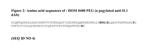
Fig. 2: Depicts the amino acid sequence of DOM 0400 PEG (a pegylated anti-
ILl dAb, molecular weight about 52 KDa)
Fig. 3: Depicts the amino acid sequence of DOM4-130-54 (An anti-ILl dAb)
Fig. 4: Depicts the amino acid sequence of Dom lh-131-206 (An anti TNF alpha
R1 dAb)
DETAILED DESCRIPTION OF THE INVENTION
CA 02744055 2011-05-17
WO 2010/060768 PCT/EP2009/064654
- 13-
Within this specification the invention has been described, with reference to
embodiments, in a way which enables a clear and concise specification to be
written. It
is intended and should be appreciated that embodiments may be variously
combined or
separated without parting from the invention.
Unless defined otherwise, all technical and scientific terms used herein have
the
same meaning as commonly understood by one of ordinary skill in the art (e.g.,
in cell
culture, molecular genetics, nucleic acid chemistry, hybridization techniques
and
biochemistry). Standard techniques are used for molecular, genetic and
biochemical
methods (see generally, Sambrook et at., Molecular Cloning: A Laboratory
Manual, 2d
ed. (1989) Cold Spring Harbor Laboratory Press, Cold Spring Harbor, N.Y. and
Ausubel et at., Short Protocols in Molecular Biology (1999) 4'h Ed, John Wiley
& Sons,
Inc. which are incorporated herein by reference) and chemical methods.
As used herein, the term "antagonist of vascular endothelial growth factor
(VEGF)" or "anti-VEGF antagonist" or the like refers to an agent (e.g., a
molecule, a
compound) which binds VEGF and can inhibit a (i.e., one or more) function of
VEGF.
As used herein, "peptide" refers to about two to about 50 amino acids that are
joined together via peptide bonds.
As used herein, "polypeptide" refers to at least about 50 amino acids that are
joined together by peptide bonds. Polypeptides generally comprise tertiary
structure
and fold into functional domains.
As used herein, a peptide or polypeptide (e.g. a domain antibody (dAb)) that
is
"resistant to protease degradation" is not substantially degraded by a
protease when
incubated with the protease under conditions suitable for protease activity. A
polypeptide (e.g., a dAb) is not substantially degraded when no more than
about 25%,
no more than about 20%, no more than about 15%, no more than about 14%, no
more
than about 13%, no more than about 12%, no more than about 11%, no more than
about
10%, no more than about 9%, no more than about 8%, no more than about 7%, no
more
than about 6%, no more than about 5%, no more than about 4%, no more than
about
3%, no more that about 2%, no more than about I%, or substantially none of the
protein
is degraded by protease after incubation with the protease for about one hour
at a
CA 02744055 2011-05-17
WO 2010/060768 PCT/EP2009/064654
-14-
temperature suitable for protease activity. For example at 37 or 50 degrees C.
Protein
degradation can be assessed using any suitable method, for example, by SDS-
PAGE or
by functional assay (e.g., ligand binding) as described herein.
As used herein, "target ligand" refers to a ligand which is specifically or
selectively bound by a polypeptide or peptide. For example, when a polypeptide
is an
antibody or antigen-binding fragment thereof, the target ligand can be any
desired
antigen or epitope. Binding to the target antigen is dependent upon the
polypeptide or
peptide being functional.
As used herein an antibody refers to IgG, IgM, IgA, IgD or IgE or a fragment
(such as a Fab , F(ab')2, Fv, disulphide linked Fv, scFv, closed conformation
multispecific antibody, disulphide-linked scFv, diabody) whether derived from
any
species naturally producing an antibody, or created by recombinant DNA
technology;
whether isolated from serum, B-cells, hybridomas, transfectomas, yeast or
bacteria.
As used herein, "antibody format" refers to any suitable polypeptide structure
in
which one or more antibody variable domains can be incorporated so as to
confer
binding specificity for antigen on the structure. A variety of suitable
antibody formats
are known in the art, such as, chimeric antibodies, humanized antibodies,
human
antibodies, single chain antibodies, bispecific antibodies, antibody heavy
chains,
antibody light chains, homodimers and heterodimers of antibody heavy chains
and/or
light chains, antigen-binding fragments of any of the foregoing (e.g., a Fv
fragment
(e.g., single chain Fv (scFv), a disulfide bonded Fv), a Fab fragment, a Fab'
fragment, a
F(ab')2 fragment), a single antibody variable domain (e.g., a dAb, VH5 VHH,
VL), and
modified versions of any of the foregoing (e.g., modified by the covalent
attachment of
polyethylene glycol or other suitable polymer or a humanized VHH).
The phrase "immunoglobulin single variable domain" refers to an antibody
variable domain (VH, VHH, VL) that specifically binds an antigen or epitope
independently of other V regions or domains. An immunoglobulin single variable
domain can be present in a format (e.g., homo- or hetero-multimer) with other
variable
regions or variable domains where the other regions or domains are not
required for
CA 02744055 2011-05-17
WO 2010/060768 PCT/EP2009/064654
- 15-
antigen binding by the single immunoglobulin variable domain (i.e., where the
immunoglobulin single variable domain binds antigen independently of the
additional
variable domains). A "domain antibody" or "dAb" is the same as an
"immunoglobulin
single variable domain" as the term is used herein. A "single immunoglobulin
variable
domain" is the same as an "immunoglobulin single variable domain" as the term
is used
herein. A "single antibody variable domain" is the same as an "immunoglobulin
single
variable domain" as the term is used herein. An immunoglobulin single variable
domain
is in one embodiment a human antibody variable domain, but also includes
single
antibody variable domains from other species such as rodent (for example, as
disclosed
in WO 00/29004, the contents of which are incorporated herein by reference in
their
entirety), nurse shark and Camelid VHH dAbs. Camelid VHH are immunoglobulin
single
variable domain polypeptides that are derived from species including camel,
llama,
alpaca, dromedary, and guanaco, which produce heavy chain antibodies naturally
devoid of light chains. The VHH may be humanized.
A "domain" is a folded protein structure which has tertiary structure
independent of the rest of the protein. Generally, domains are responsible for
discrete
functional properties of proteins, and in many cases may be added, removed or
transferred to other proteins without loss of function of the remainder of the
protein
and/or of the domain. A "single antibody variable domain" is a folded
polypeptide
domain comprising sequences characteristic of antibody variable domains. It
therefore
includes complete antibody variable domains and modified variable domains, for
example, in which one or more loops have been replaced by sequences which are
not
characteristic of antibody variable domains, or antibody variable domains
which have
been truncated or comprise N- or C-terminal extensions, as well as folded
fragments of
variable domains which retain at least the binding activity and specificity of
the full-
length domain.
As used herein, the term "dose" refers to the quantity of ligand administered
to a
subject all at one time (unit dose), or in two or more administrations over a
defined time
interval. For example, dose can refer to the quantity of ligand (e.g., ligand
comprising
an immunoglobulin single variable domain that binds target antigen)
administered to a
subject over the course of one day (24 hours) (daily dose), two days, one
week, two
CA 02744055 2011-05-17
WO 2010/060768 PCT/EP2009/064654
-16-
weeks, three weeks or one or more months (e.g., by a single administration, or
by two
or more administrations). The interval between doses can be any desired amount
of
time.
The phrase, "half-life," refers to the time taken for the serum concentration
of
the ligand (e.g., dAb, polypeptide or antagonist) to reduce by 50%, in vivo,
for example
due to degradation of the ligand and/or clearance or sequestration of the
ligand by
natural mechanisms. The ligands of the invention can be stabilized in vivo and
their
half-life increased by binding to molecules which resist degradation and/or
clearance or
sequestration. Typically, such molecules are naturally occurring proteins
which
themselves have a long half-life in vivo. The half-life of a ligand is
increased if its
functional activity persists, in vivo, for a longer period than a similar
ligand which is not
specific for the half-life increasing molecule. For example, a ligand specific
for human
serum albumin (HSA) and a target molecule is compared with the same ligand
wherein
the specificity to HSA is not present, that is does not bind HSA but binds
another
molecule. For example, it may bind a third target on the cell. Typically, the
half-life is
increased by 10%, 20%, 30%, 40%, 50% or more. Increases in the range of 2x,
3x, 4x,
5x, l Ox, 20x, 30x, 40x, 50x or more of the half-life are possible.
Alternatively, or in
addition, increases in the range of up to 30x, 40x, 50x, 60x, 70x, 80x, 90x,
100x, 150x
of the half-life are possible.
As used herein, "hydrodynamic size" refers to the apparent size of a molecule
(e.g., a protein molecule, ligand) based on the diffusion of the molecule
through an
aqueous solution. The diffusion, or motion of a protein through solution can
be
processed to derive an apparent size of the protein, where the size is given
by the
"Stokes radius" or "hydrodynamic radius" of the protein particle. The
"hydrodynamic
size" of a protein depends on both mass and shape (conformation), such that
two
proteins having the same molecular mass may have differing hydrodynamic sizes
based
on the overall conformation of the protein.
As referred to herein, the term "competes" means that the binding of a first
target to its cognate target binding domain is inhibited in the presence of a
second
binding domain that is specific for said cognate target. For example, binding
may be
inhibited sterically, for example by physical blocking of a binding domain or
by
CA 02744055 2011-05-17
WO 2010/060768 PCT/EP2009/064654
-17-
alteration of the structure or environment of a binding domain such that its
affinity or
avidity for a target is reduced. See W02006038027 for details of how to
perform
competition ELISA and competition BiaCore experiments to determine competition
between first and second binding domains.
Calculations of "homology" or "identity" or "similarity" between two sequences
(the terms are used interchangeably herein) are performed as follows. The
sequences
are aligned for optimal comparison purposes (e.g., gaps can be introduced in
one or
both of a first and a second amino acid or nucleic acid sequence for optimal
alignment
and non-homologous sequences can be disregarded for comparison purposes). In
an
embodiment, the length of a reference sequence aligned for comparison purposes
is at
least 30%, or at least 40%, or at least 50%, or at least 60%, or at least 70%,
80%, 90%,
100% of the length of the reference sequence. The amino acid residues or
nucleotides
at corresponding amino acid positions or nucleotide positions are then
compared. When
a position in the first sequence is occupied by the same amino acid residue or
nucleotide
as the corresponding position in the second sequence, then the molecules are
identical at
that position (as used herein amino acid or nucleic acid "homology" is
equivalent to
amino acid or nucleic acid "identity"). The percent identity between the two
sequences
is a function of the number of identical positions shared by the sequences,
taking into
account the number of gaps, and the length of each gap, which need to be
introduced for
optimal alignment of the two sequences. Amino acid and nucleotide sequence
alignments and homology, similarity or identity, as defined herein may be
prepared and
determined using the algorithm BLAST 2 Sequences, using default parameters
(Tatusova, T. A. et at., FEMS Microbiol Lett, 174:187-188 (1999).
Protease resistance:
The invention in one embodiment relates to dAbs, e.g. anti-VEGF dAbs, TNFR1
dAbs, IL-1 dAbs, for delivery to the eye, which have been selected by a method
of
selection for protease resistant dAbs that have a desired biological activity
e.g. binding
to VEGF, TNFR1 or IL-1. Two selective pressures are used in the method to
produce
an efficient process for selecting polypeptides that are highly stable and
resistant to
protease degradation, and that have desired biological activity. As described
herein,
CA 02744055 2011-05-17
WO 2010/060768 PCT/EP2009/064654
-18-
protease resistant peptides and polypeptides generally retain biological
activity. In
contrast, protease sensitive peptides and polypeptides are cleaved or digested
by
protease in the methods described herein, and therefore, lose their biological
activity.
Accordingly, protease resistant peptides or polypeptides are generally
selected based on
their biological activity, such as binding activity.
The ocular environment is one which is rich in proteases and hence use of
protease resistant dAbs for ocular delivery as described herein provides
several
advantages. For example, variable domains that are selected for resistance to
proteolytic degradation by one protease (e.g., trypsin), are also resistant to
degradation
by other proteases (e.g., elastase, leucozyme). Protease resistance can
correlate with a
higher melting temperature (Tm) of the peptide or polypeptide. Higher melting
temperatures are indicative of more stable variable domains, antagonists,
peptides and
polypeptides. Resistance to protease degradation can also correlate with high
affinity
binding to target ligands. Thus, the methods described and referenced herein
(in WO
2008149143) provide an efficient way to select, isolate and/or recover dAbs
that have a
desired biological activity and that are well suited for in vivo therapeutic
and/or
diagnostic ocular uses because they are protease resistant and stable. In one
embodiment protease resistance can correlate with an improved PK, for example
improved over a variable domain, antagonist, peptide or polypeptide that is
not protease
resistant. Improved PK may be an improved AUC (area under the curve) and/or an
improved half-life. Protease resistance can also correlate with an improved
stability of
the variable domain, antagonist, peptide or polypeptide to shear and/or
thermal stress
and/or a reduced propensity to aggregate during nebulisation, for example
improved
over a variable domain, antagonist, peptide or polypeptide that is not
protease resistant.
In one embodiment protease resistance correlates with an improved storage
stability, for
example improved over an variable domain, antagonist, peptide or polypeptide
that is
not protease resistant. In one aspect, one, two, three, four or all of the
advantages are
provided, the advantages being resistance to protease degradation, higher Tm
and high
affinity binding to target ligand.
CA 02744055 2011-05-17
WO 2010/060768 PCT/EP2009/064654
-19-
The methods described and referenced herein (in WO 2008/149143) can be used
as part of a program to isolate protease resistant peptides or polypeptides,
e.g. dAbs that
can comprise, if desired, other suitable selection methods. In these
situations, the
methods described herein can be employed at any desired point in the program,
such as
before or after other selection methods are used.
In certain embodiments, the dAb for ocular delivery is selected for resistance
to
degradation by trypsin, elastase or leucozyme and specifically binds VEGF. In
these
embodiments, a library or repertoire comprising dAbs is provided and combined
with
trypsin, elastase or leucozyme (or a biological preparation, extract or
homogenate
comprising trypsin) under conditions suitable for proteolytic digestion.
Trypsin,
elastase or leucozyme resistant dAbs are selected that bind VEGF. For example,
the
protease resistant dAb is not substantially degraded when incubated at 37 C in
a 0.04%
(w/w) solution of protease for a period of at least about 2 hours. In another
example,
the protease resistant dAb is not substantially degraded when incubated at 37
C in a
0.04% (w/w) solution of protease for a period of at least about 3 hours. In
another
example, the protease resistant dAb is not substantially degraded when
incubated at
37 C in a 0.04% (w/w) solution of protease for a period of at least about 4
hours, at least
about 5 hours, at least about 6 hours, at least about 7 hours, at least about
8 hours, at
least about 9 hours, at least about 10 hours, at least about 11 hours, or at
least about 12
hours.
In another aspect, there is provided a method of producing a repertoire of
protease resistant peptides or polypeptides (e.g., dAbs). The method comprises
providing a repertoire of peptides or polypeptides; combining the repertoire
of peptides
or polypeptides and a protease under suitable conditions for protease
activity; and
recovering a plurality of peptides or polypeptides that specifically bind
VEGF, whereby
a repertoire of protease resistant peptides or polypeptides is produced.
Proteases,
display systems, conditions for protease activity, and methods for selecting
peptides or
polypeptides that are suitable for use in the method are described herein with
respect to
the other methods.
CA 02744055 2011-05-17
WO 2010/060768 PCT/EP2009/064654
-20-
In some embodiments, a display system (e.g., a display system that links
coding
function of a nucleic acid and functional characteristics of the peptide or
polypeptide
encoded by the nucleic acid) that comprises a repertoire of peptides or
polypeptides is
used, and the method further comprises amplifying or increasing the copy
number of the
nucleic acids that encode the plurality of selected peptides or polypeptides.
Nucleic
acids can be amplified using any suitable method, such as by phage
amplification, cell
growth or polymerase chain reaction.
In particular embodiments, there is provided a method of producing a
repertoire
of protease resistant polypeptides that comprise anti-VEGF dAbs. The method
comprises providing a repertoire of polypeptides that comprise anti-VEGF dAbs;
combining the repertoire of peptides or polypeptides and a protease (e.g.,
trypsin,
elastase, leucozyme) under suitable conditions for protease activity; and
recovering a
plurality of polypeptides that comprise dAbs that have binding specificity for
VEGF.
The method can be used to produce a naive repertoire, or a repertoire that is
biased
toward a desired binding specificity, such as an affinity maturation
repertoire based on a
parental dAb that has binding specificity for VEGF.
Selection/Isolation/Recovery
A protease resistant peptide or polypeptide (e.g., a population of protease
resistant polypeptides) can be selected, isolated and/or recovered from a
repertoire or
library (e.g., in a display system) using any suitable method. In one
embodiment, a
protease resistant polypeptide is selected or isolated based on a selectable
characteristic
(e.g., physical characteristic, chemical characteristic, functional
characteristic). Suitable
selectable functional characteristics include biological activities of the
peptides or
polypeptides in the repertoire, for example, binding to a generic ligand
(e.g., a
superantigen), binding to a target ligand (e.g., an antigen, an epitope, a
substrate),
binding to an antibody (e.g., through an epitope expressed on a peptide or
polypeptide),
and catalytic activity. (See, e.g., Tomlinson et at., WO 99/20749; WO
01/57065; WO
99/58655). In one embodiment, the selection is based on specific binding to
VEGF. In
another embodiment, selection is on the basis of the selected functional
characteristic to
CA 02744055 2011-05-17
WO 2010/060768 PCT/EP2009/064654
-21-
produce a second repertoire in which members are protease resistant, followed
by
selection of a member from the second repertoire that specifically binds VEGF.
In some embodiments, the protease resistant peptide or polypeptide is selected
and/or isolated from a library or repertoire of peptides or polypeptides in
which
substantially all protease resistant peptides or polypeptides share a common
selectable
feature. For example, the protease resistant peptide or polypeptide can be
selected from
a library or repertoire in which substantially all protease resistant peptides
or
polypeptides bind a common generic ligand, bind a common target ligand, bind
(or are
bound by) a common antibody, or possess a common catalytic activity. This type
of
selection is particularly useful for preparing a repertoire of protease
resistant peptides or
polypeptides that are based on a parental peptide or polypeptide that has a
desired
biological activity, for example, when performing affinity maturation of an
immunoglobulin single variable domain.
Selection based on binding to a common generic ligand can yield a collection
or
population of peptides or polypeptides that contain all or substantially all
of the protease
resistant peptides or polypeptides that were components of the original
library or
repertoire. For example, peptides or polypeptides that bind a target ligand or
a generic
ligand, such as protein A, protein L or an antibody, can be selected, isolated
and/or
recovered by panning or using a suitable affinity matrix. Panning can be
accomplished
by adding a solution of ligand (e.g., generic ligand, target ligand) to a
suitable vessel
(e.g., tube, petri dish) and allowing the ligand to become deposited or coated
onto the
walls of the vessel. Excess ligand can be washed away and peptides or
polypeptides
(e.g., a repertoire that has been incubated with protease) can be added to the
vessel and
the vessel maintained under conditions suitable for peptides or polypeptides
to bind the
immobilized ligand. Unbound peptides or polypeptides can be washed away and
bound
peptides or polypeptides can be recovered using any suitable method, such as
scraping
or lowering the pH, for example.
Suitable ligand affinity matrices generally contain a solid support or bead
(e.g.,
agarose) to which a ligand is covalently or noncovalently attached. The
affinity matrix
can be combined with peptides or polypeptides (e.g., a repertoire that has
been
incubated with protease) using a batch process, a column process or any other
suitable
CA 02744055 2011-05-17
WO 2010/060768 PCT/EP2009/064654
-22-
process under conditions suitable for binding of peptides or polypeptides to
the ligand
on the matrix. Peptides or polypeptides that do not bind the affinity matrix
can be
washed away and bound peptides or polypeptides can be eluted and recovered
using any
suitable method, such as elution with a lower pH buffer, with a mild
denaturing agent
(e.g., urea), or with a peptide that competes for binding to the ligand. In
one example, a
biotinylated target ligand is combined with a repertoire under conditions
suitable for
peptides or polypeptides in the repertoire to bind the target ligand (VEGF).
Bound
peptides or polypeptides are recovered using immobilized avidin or
streptavidin (e.g.,
on a bead).
In some embodiments, the generic ligand is an antibody or antigen binding
fragment thereof. Antibodies or antigen binding fragments that bind structural
features
of peptides or polypeptides that are substantially conserved in the peptides
or
polypeptides of a library or repertoire are particularly useful as generic
ligands.
Antibodies and antigen binding fragments suitable for use as ligands for
isolating,
selecting and/or recovering protease resistant peptides or polypeptides can be
monoclonal or polyclonal and can be prepared using any suitable method.
Nucleic Acids, host cells and methods for producing protease resistant
polypeptides:
The protease resistant peptide or polypeptide selected by the method described
herein can also be produced in a suitable in vitro expression system e.g.
E.coli or Pichia
species e.g. P. pastoris, by chemical synthesis or by any other suitable
method.
Polypeptides, dAbs & Antagonists:
As described herein, protease resistant dAbs generally bind their target
ligand
with high affinity.
For example, the VEGF dAb can bind VEGF with an affinity (KD; KD=Koff
(kd)/Kon (ka) as determined by surface plasmon resonance) of 300 nM to 1 pM
(i.e., 3 x
10-7 to 5 x 10-12M), e.g. 50 nM to 1 pM, e.g. 5 nM to 1 pM and e.g. 1 nM to 1
pM; for
example KD of 1 x 10⁻⁷ M or less, e.g. 1 x 10⁻⁸ M or less, e.g. 1 x 10⁻⁹ M or
less, e.g. 1 x
10-10 M or less and e.g. 1 x 10-11 M or less; and/or a Koff rate constant of 5
x 10-1 s_1 to 1
x 10-7 s-1, e.g. 1 x 10.2 s_i to 1 x 10⁻⁶ s-1, e.g. 5 x 10⁻³ s_i to 1 x 10⁻⁵ s-
1, for example 5 x
CA 02744055 2011-05-17
WO 2010/060768 PCT/EP2009/064654
- 23 -
10-1 s_1 or less, e.g. 1 x 10.2 s_1 or less, e.g. 1 x 10⁻³ s_1 or less, e.g. 1
x 10-4 s_1 or less, e.g.
1 x 10⁻⁵ s-1 or less, and e.g. 1 x 10⁻⁶ s_i or less as determined by surface
plasmon
resonance.
Although we are not bound by any particular theory, peptides and polypeptides
that are
resistant to proteases are believed to have a lower entropy and/or a higher
stabilization
energy. Thus, the correlation between protease resistance and high affinity
binding may
be related to the compactness and stability of the surfaces of the peptides
and
polypeptides and dAbs selected by the method described herein.
In one embodiment, a VEGF dAb inhibits binding of VEGF at a
concentration 50 (IC50) of IC50 of about 1 M or less, about 500 nM or less,
about 100
nM or less, about 75 nM or less, about 50 nM or less, about 10 nM or less or
about 1
nM or less.
In certain embodiments, the VEGF dAb specifically binds VEGF, eg, human
VEGF, and dissociates from human VEGF with a dissociation constant (KD) of 300
nM
to 1pM or 300nM to 5pM or 50nM to 1pM or 50nM to 5pM or 50nM to 20 pM or about
10 pM or about l5pM or about 20pM as determined by surface plasmon resonance.
In
certain embodiments, the polypeptide, dAb or antagonist specifically binds
VEGF, eg,
human VEGF, and dissociates from human VEGF with a Koff rate constant of 5 x
10-1 s-
i to 1 x 10⁻⁷ s-1, e.g. 1 x 10.2 s-1 to 1 x 10⁻⁶ s-1, e.g. 5 x 10⁻³ s-1 to 1 x
10-5 s-1, for example
5 x 10⁻¹ s_1 or less, e.g. 1 x 10.2 s_i or less, e.g.l x 10-3 s_1 or less,
e.g. 1 x 10⁻⁴ s_i or less,
e.g. 1 x 10⁻⁵ s_1 or less, and e.g. 1 x 10⁻⁶ S-1 or less as determined by
surface plasmon
resonance.
. In certain embodiments, VEGF dAb specifically binds VEGF, eg, human
VEGF, with a Kon of 1x10⁻³ M-is 1 to 1x10⁻⁷ M-1s 1 or 1x10⁻³ M-is 1 to 1x10⁻⁶
M-1s_1 or
about 1x10⁻⁴ M-1s1 or about 1x10⁻⁵ M-IS1. In one embodiment, the polypeptide,
dAb or
antagonist specifically binds VEGF, eg, human VEGF, and dissociates from human
VEGF with a dissociation constant (KD) and a Koff as defined in this
paragraph. In one
embodiment, the polypeptide, dAb or antagonist specifically binds VEGF, eg,
human
CA 02744055 2011-05-17
WO 2010/060768 PCT/EP2009/064654
-24-
VEGF, and dissociates from human VEGF with a dissociation constant (KD) and a
Kon
as defined in this paragraph. In some embodiments, the polypeptide or dAb
specifically binds VEGF (eg, human VEGF) with a KD and/or Koff and/or Kon as
recited
in this paragraph and comprises an amino acid sequence that is at least or at
least about
80%, 85%, 90%, 91%, 92%, 93%, 94%, 95%, 96%, 97%, 98%, or 99% identical to the
amino acid sequence of a dAb with the amino acid sequence of DOMl5-26-593.
The dAb can be expressed in E. coli or in Pichia species (e.g., P. pastoris).
In
one embodiment, the ligand or dAb monomer is secreted in a quantity of at
least about
0.5 mg/L when expressed in E. coli or in Pichia species (e.g., P. pastoris).
Although,
the ligands and dAb monomers described herein can be secretable when expressed
in E.
coli or in Pichia species (e.g., P. pastoris), they can be produced using any
suitable
method, such as synthetic chemical methods or biological production methods
that do
not employ E. coli or Pichia species.
In some embodiments, the polypeptide, dAb or antagonist does not comprise a
Camelid immunoglobulin variable domain, or one or more framework amino acids
that
are unique to immunoglobulin variable domains encoded by Camelid germline
antibody
gene segments , eg at position 108, 37, 44, 45 and/or 47.
Antagonists of VEGF can be monovalent or multivalent. In some embodiments,
the antagonist is monovalent and contains one binding site that interacts with
VEGF, the
binding site provided by a polypeptide or dAb of the invention. Monovalent
antagonists
bind one VEGF and may not induce cross-linking or clustering of VEGF on the
surface
of cells which can lead to activation of the receptor and signal transduction.
Alternatively the antagonist of VEGF is multivalent. Multivalent antagonists
of
VEGF can contain two or more copies of a particular binding site for VEGF or
contain
two or more different binding sites that bind VEGF, at least one of the
binding sites
being provided by a dAb of the invention. For example, as described herein the
antagonist of VEGF can be a dimer, trimer or multimer comprising two or more
copies
of a dAb that binds VEGF, or two or more different dAbs that bind VEGF.
CA 02744055 2011-05-17
WO 2010/060768 PCT/EP2009/064654
-25-
In other embodiments, the, dAb specifically binds VEGF with a KD described
herein and inhibits tumour growth in a standard murine xenograft model (e.g.,
inhibits
tumour growth by at least about 10%, as compared with a suitable control). In
one
embodiment, the polypeptide, dAb or antagonist inhibits tumour growth by at
least
about 10% or by at least about 25%, or by at least about 50%, as compared to a
suitable
control in a standard murine xenograft model when administered at about 1
mg/kg or
more, for example about 5 or 10 mg/kg.
In other embodiments, the polypeptide, dAb or antagonist binds VEGF and
antagonizes the activity of the VEGF in a standard cell assay with an ND50 of
< 100
nM.
In certain embodiments, the dAbs are efficacious in animal models of ocular
disease when an effective amount is administered. Generally an effective
amount is
about 1 mg/kg to about 10 mg/kg (e.g., about 1 mg/kg, about 2 mg/kg, about 3
mg/kg,
about 4 mg/kg, about 5 mg/kg, about 6 mg/kg, about 7 mg/kg, about 8 mg/kg,
about 9
mg/kg, or about 10 mg/kg). The dAb can be administered at a dosing frequency
of e.g.
once or twice daily, once or twice weekly, once or twice monthly.
Generally, the dAbs will be utilized in purified form together with
pharmacologically appropriate carriers for ocular delivery. Typically, these
carriers can
include aqueous or alcoholic/aqueous solutions, emulsions or suspensions, any
including saline and/or buffered media. Suitable physiologically-acceptable
adjuvants,
if necessary to keep a polypeptide complex in suspension, may be chosen from
thickeners such as carboxymethylcellulose, polyvinylpyrrolidone, gelatin and
alginates.
Preservatives and other additives, such as antimicrobials, antioxidants,
chelating agents
and inert gases, may also be present (Mack (1982) Remington's Pharmaceutical
Sciences, 16th Edition). A variety of suitable formulations can be used,
including
extended release formulations. . These might comprise, implants, gels,
nanoparticles,
and microparticles. Drug loaded PLA nano- and microparticles have been used to
deliver drug to the posterior segment of the eye after sub-conjunctival
delivery of the
formulation (Kompella et al IOVS 2003 44(3) 1192-1201). In particular,
microspheres
CA 02744055 2011-05-17
WO 2010/060768 PCT/EP2009/064654
-26-
are retained at the site of delivery and appear to be more appropriate for
retinal drug
delivery compared to nanoparticles which may be cleared more readily (Amrite
et al
ARVO abstract #5067/B391 2003).
The ligands (e.g., antagonists) of the present invention may be used as
separately administered compositions or in conjunction with other agents.
These can
include various drugs for ocular delivery to the eye and/or ocular penetration
enhancers
and/or viscosity enhancers.
Pharmaceutical compositions can include "cocktails" of various other agents in
conjunction with the ligands of the present invention, or even combinations of
ligands
according to the present invention having different specificities, such as
ligands selected
using different target antigens or epitopes, whether or not they are pooled
prior to
administration.
The precise dosage and frequency of administration of the dAbs to the eye will
depend on the age, sex and condition of the patient, concurrent administration
of other
drugs, counterindications and other parameters to be taken into account by the
clinician.
The dAbs of this invention can be lyophilized for storage and reconstituted in
a
suitable carrier prior to use. This technique has been shown to be effective
with
conventional immunoglobulins and art-known lyophilisation and reconstitution
techniques can be employed. It will be appreciated by those skilled in the art
that
lyophilisation and reconstitution can lead to varying degrees of antibody
activity loss
(e.g. with conventional immunoglobulins, IgM antibodies tend to have greater
activity
loss than IgG antibodies) and that use levels may have to be adjusted upward
to
compensate.
The compositions containing the present dAbs or a cocktail thereof can be
administered for prophylactic and/or therapeutic treatments. In certain
therapeutic
CA 02744055 2011-05-17
WO 2010/060768 PCT/EP2009/064654
-27-
applications, an adequate amount to accomplish at least partial inhibition,
suppression,
modulation, killing, or some other measurable parameter, of a population of
selected
cells can be defined as a "therapeutically-effective dose". Amounts needed to
achieve
this dosage will depend upon the severity of the disease and the general state
of the
patient's own immune system. The skilled clinician will be able to determine
the
appropriate dosing interval to treat, suppress or prevent disease.
Treatment or therapy performed using the compositions described herein is
considered "effective" if one or more symptoms are reduced (e.g., by at least
10% or at
least one point on a clinical assessment scale), relative to such symptoms
present before
treatment, or relative to such symptoms in an individual (human or model
animal) not
treated with such composition or other suitable control. Symptoms will
obviously vary
depending upon the disease or disorder targeted, but can be measured by an
ordinarily
skilled clinician or technician. Such symptoms can be measured, for example,
by
monitoring the level of one or more biochemical indicators of the disease or
disorder
(e.g., levels of an enzyme or metabolite correlated with the disease, affected
cell
numbers, etc.), by monitoring physical manifestations or by an accepted
clinical
assessment scale.
Similarly, prophylaxis performed using a composition as described herein is
"effective" if the onset or severity of one or more symptoms is delayed,
reduced or
abolished relative to such symptoms in a similar individual (human or animal
model)
not treated with the composition.
In one embodiment, the invention is a method for treating, suppressing or
preventing ocular disease or condition, selected from for example cancer (e.g.
a solid
tumour), inflammatory disease, autoimmune disease, vascular proliferative
disease
(e.g.AMD (age related macular degeneration)) comprising administering to a
mammal
in need thereof a therapeutically-effective dose or amount of a polypeptide,
dAb which
binds to VEGF or antagonist of VEGF according to the invention or to IL-1, or
TNF-a
CA 02744055 2011-05-17
WO 2010/060768 PCT/EP2009/064654
-28-
or TNF-aR. Examples of such ocular diseases or conditions include AMD,
Uveitis, dry
eye, diabetic retinopathy and diabetic macular oedema.
Formats:
Increased half-life is useful in in vivo applications of immunoglobulins,
especially antibodies and most especially antibody fragments of small size.
Such
fragments (Fvs, disulphide bonded Fvs, Fabs, scFvs, dAbs) can suffer from
rapid
clearance from the body; thus, whilst they are able to reach most parts of the
body
rapidly, and are quick to produce and easier to handle, their in vivo
applications have
been limited by their only brief persistence in vivo. Hence the dAbs described
herein
can be modified to provide increased half-life in vivo and consequently longer
persistence times in the body.
Methods for pharmacokinetic analysis and determination of ligand half-life
will
be familiar to those skilled in the art. Details may be found in Kenneth, A et
al:
Chemical Stability of Pharmaceuticals: A Handbook for Pharmacists and in
Peters et at,
Pharmacokinetic analysis: A Practical Approach (1996). Reference is also made
to
"Pharmacokinetics", M Gibaldi & D Perron, published by Marcel Dekker, 2d Rev.
ex
edition (1982), which describes pharmacokinetic parameters such as t alpha and
t beta
half lives and area under the curve (AUC).
Half lives (t1/2 alpha and t1/2 beta) and AUC can be determined from a curve
of
serum concentration of ligand against time. The WinNonlin analysis package
(available
from Pharsight Corp., Mountain View, CA94040, USA) can be used, for example,
to
model the curve. In a first phase (the alpha phase) the ligand is undergoing
mainly
distribution in the patient, with some elimination. A second phase (beta
phase) is the
terminal phase when the ligand has been distributed and the serum
concentration is
decreasing as the ligand is cleared from the patient. The t alpha half life is
the half life
of the first phase and the t beta half life is the half life of the second
phase. Thus, in one
embodiment, the present invention provides a ligand or a composition
comprising a
ligand according to the invention having a to half-life in the range of 15
minutes or
more. In one embodiment, the lower end of the range is 30 minutes, 45 minutes,
1 hour,
CA 02744055 2011-05-17
WO 2010/060768 PCT/EP2009/064654
-29-
2 hours, 3 hours, 4 hours, 5 hours, 6 hours, 7 hours, 10 hours, 11 hours or 12
hours. In
addition, or alternatively, a ligand or composition according to the invention
will have a
to half life in the range of up to and including 12 hours. In one embodiment,
the upper
end of the range is 11, 10, 9, 8, 7, 6 or 5 hours. An example of a suitable
range is 1 to 6
hours, 2 to 5 hours or 3 to 4 hours.
In one embodiment, the dAb or a composition comprising a dAb according to
the invention has a t(3 half-life in the range of 30 minutes or more. In one
embodiment,
the lower end of the range is 45 minutes, 1 hour, 2 hours, 3 hours, 4 hours, 5
hours, 6
hours, 7 hours, 10 hours , 11 hours, or 12 hours. In addition, or
alternatively, a ligand
or composition according to the invention has a t(3 half-life in the range of
up to and
including 21 days. In one embodiment, the upper end of the range is 12 hours,
24
hours, 2 days, 3 days, 5 days, 10 days, 15 days or 20 days. In one embodiment
a ligand
or composition according to the invention will have a t(3 half life in the
range 12 to 60
hours. In a further embodiment, it will be in the range 12 to 48 hours. In a
further
embodiment still, it will be in the range 12 to 26 hours.
In addition, or alternatively to the above criteria, the present invention
provides
a dAb or a composition comprising a ligand according to the invention having
an AUC
value (area under the curve) in the range of 1 mg.min/ml or more. In one
embodiment,
the lower end of the range is 5, 10, 15, 20, 30, 100, 200 or 300 mg.min/ml. In
addition,
or alternatively, a ligand or composition according to the invention has an
AUC in the
range of up to 600 mg.min/ml. In one embodiment, the upper end of the range is
500,
400, 300, 200, 150, 100, 75 or 50 mg.min/ml. In one embodiment a ligand
according to
the invention will have a AUC in the range selected from the group consisting
of the
following: 15 to 150 mg.min/ml, 15 to 100 mg.min/ml, 15 to 75 mg.min/ml, and
15 to
50mg.min/ml.
dAbs of the invention can be formatted to have a larger hydrodynamic size, for
example, by attachment of a PEG group, serum albumin, transferrin, transferrin
receptor
or at least the transferrin-binding portion thereof, an antibody Fc region, or
by
conjugation to an antibody domain. For example, dAbs can be formatted as a
larger
antigen-binding fragment of an antibody, or as an antibody (e.g., formatted as
a Fab,
CA 02744055 2011-05-17
WO 2010/060768 PCT/EP2009/064654
-30-
Fab', F(ab)2, F(ab')2, IgG, scFv). In another embodiment dAbs according to the
invention can be formatted as a fusion or conjugate with another polypeptide
or peptide.
Hydrodynamic size of the ligands (e.g., dAb monomers and multimers) of the
invention may be determined using methods which are well known in the art. For
example, gel filtration chromatography may be used to determine the
hydrodynamic
size of a ligand. Suitable gel filtration matrices for determining the
hydrodynamic sizes
of ligands, such as cross-linked agarose matrices, are well known and readily
available.
The size of a ligand i.e. dAb format (e.g., the size of a PEG moiety attached
to a
dAb monomer), can be varied depending on the desired application e.g. if it is
desired to
have the dAb remain in the systemic circulation for a longer period of time
the size of
can be increased, for example by formatting as an Ig like protein.
Half-life extension by targeting _ an anti _ enamor epitope that increases
half-live in vivo
The hydrodynamic size of a ligand and its serum half-life can also be
increased
by conjugating or associating a dAb to a binding domain (e.g., antibody or
antibody
fragment) that binds an antigen or epitope that increases half-live in vivo,
as described
herein. For example, the VEGF dAb can be conjugated or linked to an anti-serum
albumin or anti-neonatal Fc receptor antibody or antibody fragment, eg an anti-
SA or
anti-neonatal Fc receptor dAb, Fab, Fab' or scFv, or to an anti-SA affibody or
anti-
neonatal Fc receptor Affibody or an anti-SA avimer, or an anti-SA binding
domain
which comprises a scaffold selected from, but preferably not limited to, the
group
consisting of CTLA-4, lipocallin, SpA, an affibody, an avimer, GroEl and
fibronectin
(see PCT/GB2008/000453 filed 8th February 2008 for disclosure of these binding
domain, which domains and their sequences are incorporated herein by reference
and
form part of the disclosure of the present text). Conjugating refers to a
composition
comprising polypeptide, dAb or antagonist of the invention that is bonded
(covalently
or noncovalently) to a binding domain that binds serum albumin.
Suitable polypeptides that enhance serum half-life in vivo include, for
example,
transferrin receptor specific ligand-neuropharmaceutical agent fusion proteins
(see U.S.
CA 02744055 2011-05-17
WO 2010/060768 PCT/EP2009/064654
-31 -
Patent No. 5,977,307, the teachings of which are incorporated herein by
reference),
brain capillary endothelial cell receptor, transferrin, transferrin receptor
(e.g., soluble
transferrin receptor), insulin, insulin-like growth factor 1 (IGF 1) receptor,
insulin-like
growth factor 2 (IGF 2) receptor, insulin receptor, blood coagulation factor
X, al-
antitrypsin and HNF I a. Suitable polypeptides that enhance serum half-life
also
include alpha-1 glycoprotein (orosomucoid; AAG), alpha-1 antichymotrypsin
(ACT),
alpha-1 microglobulin (protein HC; AIM), antithrombin III (AT III),
apolipoprotein A-1
(Apo A-1), apolipoprotein B (Apo B), ceruloplasmin (Cp), complement component
C3
(C3), complement component C4 (C4), C1 esterase inhibitor (C1 INH), C-reactive
protein (CRP), ferritin (FER), hemopexin (HPX), lipoprotein(a) (Lp(a)),
mannose-
binding protein (MBP), myoglobin (Myo), prealbumin (transthyretin; PAL),
retinol-
binding protein (RBP), and rheumatoid factor (RF).
Suitable proteins from the extracellular matrix include, for example,
collagens,
laminins, integrins and fibronectin. Collagens are the major proteins of the
extracellular
matrix. About 15 types of collagen molecules are currently known, found in
different
parts of the body, e.g. type I collagen (accounting for 90% of body collagen)
found in
bone, skin, tendon, ligaments, cornea, internal organs or type II collagen
found in
cartilage, vertebral disc, notochord, and vitreous humor of the eye.
Suitable proteins from the blood include, for example, plasma proteins (e.g.,
fibrin, a-2 macroglobulin, serum albumin, fibrinogen (e.g., fibrinogen A,
fibrinogen B),
serum amyloid protein A, haptoglobin, profilin, ubiquitin, uteroglobulin and
(3-2-
microglobulin), enzymes and enzyme inhibitors (e.g., plasminogen, lysozyme,
cystatin
C, alpha- l-antitrypsin and pancreatic trypsin inhibitor), proteins of the
immune system,
such as immunoglobulin proteins (e.g., IgA, IgD, IgE, IgG, IgM, immunoglobulin
light
chains (kappa/lambda)), transport proteins (e.g., retinol binding protein, a-1
microglobulin), defensins (e.g., beta-defensin 1, neutrophil defensin 1,
neutrophil
defensin 2 and neutrophil defensin 3) and the like.
Suitable proteins found at the blood brain barrier or in neural tissue
include, for
example, melanocortin receptor, myelin, ascorbate transporter and the like.
CA 02744055 2011-05-17
WO 2010/060768 PCT/EP2009/064654
-32-
Suitable polypeptides that enhance serum half-life in vivo also include
proteins
localized to the kidney (e.g., polycystin, type IV collagen, organic anion
transporter Kl,
Heymann's antigen), proteins localized to the liver (e.g., alcohol
dehydrogenase, G250),
proteins localized to the lung (e.g., secretary component, which binds IgA),
proteins
localized to the heart (e.g., HSP 27, which is associated with dilated
cardiomyopathy),
proteins localized to the skin (e.g., keratin), bone specific proteins such as
morphogenic
proteins (BMP5), which are a subset of the transforming growth factor (3
superfamily of
proteins that demonstrate osteogenic activity (e.g., BMP-2, BMP-4, BMP-5, BMP-
6,
BMP-7, BMP-8), tumor specific proteins (e.g., trophoblast antigen, herceptin
receptor,
oestrogen receptor, cathepsins (e.g., cathepsin B, which can be found in liver
and
spleen)).
Suitable disease-specific proteins include, for example, antigens expressed
only
on activated T-cells, including LAG-3 (lymphocyte activation gene),
osteoprotegerin
ligand (OPGL; see Nature 402, 304-309 (1999)), OX40 (a member of the TNF
receptor
family, expressed on activated T cells and specifically up-regulated in human
T cell
leukemia virus type-I (HTLV-I)-producing cells; see Immunol. 165 (1):263-70
(2000)).
Suitable disease-specific proteins also include, for example, metalloproteases
(associated with arthritis/cancers) including CG6512 Drosophila, human
paraplegin,
human FtsH, human AFG3L2, murine ftsH; and angiogenic growth factors,
including
acidic fibroblast growth factor (FGF-1), basic fibroblast growth factor (FGF-
2),
vascular endothelial growth factor/vascular permeability factor (VEGF/VPF),
transforming growth factor-a (TGF a), tumor necrosis factor-alpha (TNF-a),
angiogenin, interleukin-3 (IL-3), interleukin-8 (IL-8), platelet-derived
endothelial
growth factor (PD-ECGF), placental growth factor (P I GF), midkine platelet-
derived
growth factor-BB (PDGF), and fractalkine.
Suitable polypeptides that enhance serum half-life in vivo also include stress
proteins such as heat shock proteins (HSPs). HSPs are normally found
intracellularly.
When they are found extracellularly, it is an indicator that a cell has died
and spilled out
its contents. This unprogrammed cell death (necrosis) occurs when as a result
of trauma,
disease or injury, extracellular HSPs trigger a response from the immune
system.
CA 02744055 2011-05-17
WO 2010/060768 PCT/EP2009/064654
-33-
Binding to extracellular HSP can result in localizing the compositions of the
invention
to a disease site.
Suitable proteins involved in Fc transport include, for example, Brambell
receptor (also known as FcRB). This Fc receptor has two functions, both of
which are
potentially useful for delivery. The functions are (1) transport of IgG from
mother to
child across the placenta (2) protection of IgG from degradation thereby
prolonging its
serum half-life. It is thought that the receptor recycles IgG from endosomes.
(See,
Holliger et at, Nat Biotechnol 15(7):632-6 (1997).)
dAbs that Bind Serum Albumin
The invention in one embodiment a first dAb that binds to an ocular target
molecule, e.g. VEGF, IL-1, or TNF-a, and a second dAb that binds serum albumin
(SA), the second dAb binding SA with a KDas determined by surface plasmon
resonance of 1nM to 1, 2, 3, 4, 5, 10, 20, 30, 40, 50, 60, 70, 100, 200, 300,
400 or 500
M (i.e., x 10-9 to 5 x 10⁻⁴), or 100 nM to 10 M, or 1 to 5 M or 3 to 70 nM
or lOnM
to 1, 2, 3, 4 or 5 M. For example 30 to 70 nM as determined by surface
plasmon
resonance. In one embodiment, the first dAb (or a dAb monomer) binds SA (e.g.,
HSA)
with a KDas determined by surface plasmon resonance of approximately 1, 50,
70, 100,
150, 200, 300 nM or 1, 2 or 3 M. In one embodiment, for a dual specific
ligand
comprising a first anti-SA dAb and a second dAb to VEGF, the affinity (eg
KDand/or
Koff as measured by surface plasmon resonance, eg using BiaCore) of the second
dAb
for its target is from 1 to 100000 times (e.g. 100 to 100000, or 1000 to
100000, or
10000 to 100000 times) the affinity of the first dAb for SA. In one
embodiment, the
serum albumin is human serum albumin (HSA). For example, the first dAb binds
SA
with an affinity of approximately 10 M, while the second dAb binds its target
with an
affinity of 100 pM. In one embodiment, the serum albumin is human serum
albumin
(HSA). In one embodiment, the first dAb binds SA (eg, HSA) with a KD of
approximately 50, for example 70, 100, 150 or 200 nM. Details of dual specific
ligands
are found in W003002609, W004003019 and W004058821.
CA 02744055 2011-05-17
WO 2010/060768 PCT/EP2009/064654
-34-
The dAbs of the invention can in one embodiment comprise a dAb that binds
serum albumin (SA) with a KD as determined by surface plasmon resonance of 1nM
to
1, 2, 3, 4, 5, 10, 20, 30, 40, 50, 60, 70, 100, 200, 300, 400 or 500 M
(i.e., x 10-9 to 5 x
10-4), or 100 nM to 10 M, or 1 to 5 M or 3 to 70 nM or 10nM to 1, 2, 3, 4 or
5 M.
For example 30 to 70 nM as determined by surface plasmon resonance. In one
embodiment, the first dAb (or a dAb monomer) binds SA (e.g., HSA) with a KD as
determined by surface plasmon resonanceof approximately 1, 50, 70, 100, 150,
200, 300
nM or 1, 2 or 3 M. In one embodiment, the first and second dAbs are linked
by a
linker, for example a linker of from 1 to 4 amino acids or from 1 to 3 amino
acids, or
greater than 3 amino acids or greater than 4, 5, 6, 7, 8, 9, 10, 15 or 20
amino acids. In
one embodiment, a longer linker (greater than 3 amino acids) is used to
enhance
potency (KD of one or both dAbs in the antagonist).
In particular embodiments, the dAb binds human serum albumin and competes
for binding to albumin with a dAb selected from the group consisting of
MSA-16, MSA-26 (See W004003019 for disclosure of these sequences, which
sequences and their nucleic acid counterpart are incorporated herein by
reference and
form part of the disclosure of the present text),
DOM7m-16 (SEQ ID NO: 473), DOM7m-12 (SEQ ID NO: 474), DOM7m-26
(SEQ ID NO: 475), DOM7r-1 (SEQ ID NO: 476), DOM7r-3 (SEQ ID NO: 477),
DOM7r-4 (SEQ ID NO: 478), DOM7r-5 (SEQ ID NO: 479), DOM7r-7 (SEQ ID NO:
480), DOM7r-8 (SEQ ID NO: 481), DOM7h-2 (SEQ ID NO: 482), DOM7h-3 (SEQ ID
NO: 483), DOM7h-4 (SEQ ID NO: 484), DOM7h-6 (SEQ ID NO: 485), DOM7h-1
(SEQ ID NO: 486), DOM7h-7 (SEQ ID NO: 487), DOM7h-22 (SEQ ID NO: 489),
DOM7h-23 (SEQ ID NO: 490), DOM7h-24 (SEQ ID NO: 491), DOM7h-25 (SEQ ID
NO: 492), DOM7h-26 (SEQ ID NO: 493), DOM7h-21 (SEQ ID NO: 494), DOM7h-27
(SEQ ID NO: 495), DOM7h-8 (SEQ ID NO: 496), DOM7r-13 (SEQ ID NO: 497),
DOM7r-14 (SEQ ID NO: 498), DOM7r-15 (SEQ ID NO: 499), DOM7r-16 (SEQ ID
NO: 500), DOM7r-17 (SEQ ID NO: 501), DOM7r-18 (SEQ ID NO: 502), DOM7r-19
(SEQ ID NO: 503), DOM7r-20 (SEQ ID NO: 504), DOM7r-21 (SEQ ID NO: 505),
CA 02744055 2011-05-17
WO 2010/060768 PCT/EP2009/064654
-35-
DOM7r-22 (SEQ ID NO: 506), DOM7r-23 (SEQ ID NO: 507), DOM7r-24 (SEQ ID
NO: 508), DOM7r-25 (SEQ ID NO: 509), DOM7r-26 (SEQ ID NO: 510), DOM7r-27
(SEQ ID NO: 511), DOM7r-28 (SEQ ID NO: 512), DOM7r-29 (SEQ ID NO: 513),
DOM7r-30 (SEQ ID NO: 514), DOM7r-3l (SEQ ID NO: 515), DOM7r-32 (SEQ ID
NO: 516), DOM7r-33 (SEQ ID NO: 517) (See W02007080392 for disclosure of these
sequences, which sequences and their nucleic acid counterpart are incorporated
herein
by reference and form part of the disclosure of the present text; the SEQ ID
No's in this
paragraph are those that appear in W02007080392),
dAb8 (dAblO), dAb 10, dAb36, dAb7r2O (DOM7r2O), dAb7r2l (DOM7r2l),
dAb7r22 (DOM7r22), dAb7r23 (DOM7r23), dAb7r24 (DOM7r24), dAb7r25
(DOM7r25), dAb7r26 (DOM7r26), dAb7r27 (DOM7r27), dAb7r28 (DOM7r28),
dAb7r29 (DOM7r29), dAb7r29 (DOM7r29), dAb7r3l (DOM7r31), dAb7r32
(DOM7r32), dAb7r33 (DOM7r33), dAb7r33 (DOM7r33), dAb7h22 (DOM7h22),
dAb7h23 (DOM7h23), dAb7h24 (DOM7h24), dAb7h25 (DOM7h25), dAb7h26
(DOM7h26), dAb7h27 (DOM7h27), dAb7h3O (DOM7h3O), dAb7h3l (DOM7h3l),
dAb2 (dAbs 4,7,41), dAb4, dAb7, dAb11, dAbl2 (dAb7ml2), dAbl3 (dAb 15), dAbl5,
dAbl6 (dAb2l, dAb7ml6) , dAb17, dAb18, dAb19, dAb2l, dAb22, dAb23, dAb24,
dAb25 (dAb26, dAb7m26), dAb27, dAb30 (dAb35), dAb3l, dAb33, dAb34, dAb35,
dAb38 (dAb54), dAb4l, dAb46 (dAbs 47, 52 and 56), dAb47, dAb52, dAb53, dAb54,
dAb55, dAb56, dAb7ml2, dAb7ml6, dAb7m26, dAb7rl (DOM 7rl), dAb7r3
(DOM7r3), dAb7r4 (DOM7r4), dAb7r5 (DOM7r5), dAb7r7 (DOM7r7), dAb7r8
(DOM7r8), dAb7rl3 (DOM7rl3), dAb7rl4 (DOM7rl4), dAb7rl5 (DOM7rl5),
dAb7rl6 (DOM7rl6), dAb7rl7 (DOM7rl7), dAb7rl8 (DOM7rl8), dAb7rl9
(DOM7rl9), dAb7hl (DOM7hl), dAb7h2 (DOM7h2), dAb7h6 (DOM7h6), dAb7h7
(DOM7h7), dAb7h8 (DOM7h8), dAb7h9 (DOM7h9), dAb7hl0 (DOM7h10), dAb7hl1
(DOM7hl1), dAb7hl2 (DOM7hl2), dAb7hl3 (DOM7hl3), dAb7hl4 (DOM7hl4),
dAb7pl (DOM7pl), and dAb7p2 (DOM7p2) (see PCT/GB2008/000453 filed 8"
February 2008 and published as WO 2008/096158 for disclosure of these
sequences,
which sequences and their nucleic acid counterpart are incorporated herein by
reference
and form part of the disclosure of the present text). Alternative names are
shown in
CA 02744055 2011-05-17
WO 2010/060768 PCT/EP2009/064654
-36-
brackets after the dAb, e.g. dAb8 has an alternative name which is dAb 10 i.e.
dAb8
(dAb l0).
In certain embodiments, the dAb binds human serum albumin and comprises an
amino acid sequence that has at least about 80%, or at least about 85%, or at
least about
90%, or at least about 95%, or at least about 96%, or at least about 97%, or
at least
about 98%, or at least about 99% amino acid sequence identity with the amino
acid
sequence of a dAb selected from the group consisting of
MSA-16, MSA-26,
DOM7m-16 (SEQ ID NO: 473), DOM7m-12 (SEQ ID NO: 474), DOM7m-26
(SEQ ID NO: 475), DOM7r-1 (SEQ ID NO: 476), DOM7r-3 (SEQ ID NO: 477),
DOM7r-4 (SEQ ID NO: 478), DOM7r-5 (SEQ ID NO: 479), DOM7r-7 (SEQ ID NO:
480), DOM7r-8 (SEQ ID NO: 481), DOM7h-2 (SEQ ID NO: 482), DOM7h-3 (SEQ ID
NO: 483), DOM7h-4 (SEQ ID NO: 484), DOM7h-6 (SEQ ID NO: 485), DOM7h-1
(SEQ ID NO: 486), DOM7h-7 (SEQ ID NO: 487), DOM7h-22 (SEQ ID NO: 489),
DOM7h-23 (SEQ ID NO: 490), DOM7h-24 (SEQ ID NO: 491), DOM7h-25 (SEQ ID
NO: 492), DOM7h-26 (SEQ ID NO: 493), DOM7h-21 (SEQ ID NO: 494), DOM7h-27
(SEQ ID NO: 495), DOM7h-8 (SEQ ID NO: 496), DOM7r-13 (SEQ ID NO: 497),
DOM7r-14 (SEQ ID NO: 498), DOM7r-15 (SEQ ID NO: 499), DOM7r-16 (SEQ ID
NO: 500), DOM7r-17 (SEQ ID NO: 501), DOM7r-18 (SEQ ID NO: 502), DOM7r-19
(SEQ ID NO: 503), DOM7r-20 (SEQ ID NO: 504), DOM7r-21 (SEQ ID NO: 505),
DOM7r-22 (SEQ ID NO: 506), DOM7r-23 (SEQ ID NO: 507), DOM7r-24 (SEQ ID
NO: 508), DOM7r-25 (SEQ ID NO: 509), DOM7r-26 (SEQ ID NO: 510), DOM7r-27
(SEQ ID NO: 511), DOM7r-28 (SEQ ID NO: 512), DOM7r-29 (SEQ ID NO: 513),
DOM7r-30 (SEQ ID NO: 514), DOM7r-31 (SEQ ID NO: 515), DOM7r-32 (SEQ ID
NO: 516), DOM7r-33 (SEQ ID NO: 517) (the SEQ ID No's in this paragraph are
those
that appear in W02007080392),
dAb8, dAb 10, dAb36, dAb7r2O, dAb7r2l, dAb7r22, dAb7r23, dAb7r24,
dAb7r25, dAb7r26, dAb7r27, dAb7r28, dAb7r29, dAb7r3O, dAb7r3l, dAb7r32,
dAb7r33, dAb7h2l, dAb7h22, dAb7h23, Ab7h24, Ab7h25, Ab7h26, dAb7h27,
dAb7h30, dAb7h3l, dAb2, dAb4, dAb7, dAbll, dAbl2, dAbl3, dAbl5, dAbl6,
CA 02744055 2011-05-17
WO 2010/060768 PCT/EP2009/064654
-37-
dAbl7, dAbl8, dAbl9, dAb2l, dAb22, dAb23, dAb24, dAb25, dAb26, dAb27, dAb30,
dAb3l, dAb33, dAb34, dAb35, dAb38, dAb4l, dAb46, dAb47, dAb52, dAb53, dAb54,
dAb55, dAb56, dAb7ml2, dAb7ml6, dAb7m26, dAb7rl, dAb7r3, dAb7r4, dAb7r5,
dAb7r7, dAb7r8, dAb7rl3, dAb7rl4, dAb7rl5, dAb7rl6, dAb7rl7, dAb7rl8, dAb7rl9,
dAb7hl, dAb7h2, dAb7h6, dAb7h7, dAb7h8, dAb7h9, dAb7hlO, dAb7hl 1, dAb7hl2,
dAb7hl3, dAb7hl4, dAb7pl, and dAb7p2.
For example, the dAb that binds human serum albumin can comprise an amino
acid sequence that has at least about 90%, or at least about 95%, or at least
about 96%,
or at least about 97%, or at least about 98%, or at least about 99% amino acid
sequence
identity with DOM7h-2 (SEQ ID NO:482), DOM7h-3 (SEQ ID NO:483), DOM7h-4
(SEQ ID NO:484), DOM7h-6 (SEQ ID NO:485), DOM7h-1 (SEQ ID NO:486),
DOM7h-7 (SEQ ID NO:487), DOM7h-8 (SEQ ID NO:496), DOM7r-13 (SEQ ID
NO:497), DOM7r-14 (SEQ ID NO:498), DOM7h-22 (SEQ ID NO:489), DOM7h-23
(SEQ ID NO:490), DOM7h-24 (SEQ ID NO:491), DOM7h-25 (SEQ ID NO:492),
DOM7h-26 (SEQ ID NO:493), DOM7h-21 (SEQ ID NO:494), DOM7h-27 (SEQ ID
NO:495) (the SEQ ID No's in this paragraph are those that appear in
W02007080392),
dAb8, dAb 10, dAb36, dAb7h2l, dAb7h22, dAb7h23, Ab7h24, Ab7h25,
Ab7h26, dAb7h27, dAb7h30, dAb7h3l, dAb2, dAb4, dAb7, dAbll, dAbl2, dAbl3,
dAb 15, dAb 16, dAb 17, dAb 18, dAb 19, dAb2l , dAb22, dAb23, dAb24, dAb25,
dAb26,
dAb27, dAb30, dAb3l, dAb33, dAb34, dAb35, dAb38, dAb4l, dAb46, dAb47, dAb52,
dAb53, dAb54, dAb55, dAb56, dAb7hl, dAb7h2, dAb7h6, dAb7h7, dAb7h8, dAb7h9,
dAb7hlO, dAb7hl 1, dAb7hl2, dAb7hl3 and dAb7hl4.
In certain embodiments, the dAb binds human serum albumin and comprises an
amino acid sequence that has at least about 80%, or at least about 85%, or at
least about
90%, or at least about 95%, or at least about 96%, or at least about 97%, or
at least
about 98%, or at least about 99% amino acid sequence identity with the amino
acid
sequence of a dAb selected from the group consisting of
DOM7h-2 (SEQ ID NO:482), DOM7h-6 (SEQ ID NO:485), DOM7h-1 (SEQ
ID NO:486), DOM7h-7 (SEQ ID NO:487), DOM7h-8 (SEQ ID NO:496), DOM7h-22
(SEQ ID NO:489), DOM7h-23 (SEQ ID NO:490), DOM7h-24 (SEQ ID NO:491),
CA 02744055 2011-05-17
WO 2010/060768 PCT/EP2009/064654
-38-
DOM7h-25 (SEQ ID NO:492), DOM7h-26 (SEQ ID NO:493), DOM7h-21 (SEQ ID
NO:494), DOM7h-27 (SEQ ID NO:495) (the SEQ ID No's in this paragraph are those
that appear in W02007080392),
dAb7h2l, dAb7h22, dAb7h23, Ab7h24, Ab7h25, Ab7h26, dAb7h27, dAb7h3O,
dAb7h3l, dAb2, dAb4, dAb7, dAb38, dAb4l, dAb7hl, dAb7h2, dAb7h6, dAb7h7,
dAb7h8, dAb7h9, dAb7hl0, dAb7h11, dAb7hl2, dAb7hl3 and dAb7hl4.
In more particular embodiments, the dAb is a VK dAb that binds human serum
albumin and has an amino acid sequence selected from the group consisting of
DOM7h-2 (SEQ ID NO:482), DOM7h-6 (SEQ ID NO:485), DOM7h-1 (SEQ
ID NO:486), DOM7h-7 (SEQ ID NO:487), DOM7h-8 (SEQ ID NO:496) (the SEQ ID
No's in this paragraph are those that appear in W02007080392),
dAb2, dAb4, dAb7, dAb38, dAb4l, dAb54, dAb7hl, dAb7h2, dAb7h6,
dAb7h7, dAb7h8, dAb7h9, dAb7hl0, dAb7hl1, dAb7hl2, dAb7hl3 and dAb7hl4. ,
In more particular embodiments, the dAb is a VH dAb that binds human serum
albumin and has an amino acid sequence selected from dAb7h3O and dAb7h3 1.
In more particular embodiments, the dAb is dAb7hl 1 or dAb7hl4.
In other embodiments, the dAb, ligand or antagonist binds human serum
albumin and comprises one, two or three of the CDRs of any of the foregoing
amino
acid sequences, eg one, two or three of the CDRs of dAb7hl l or dAb7hl4.
Suitable Camelid VHH that bind serum albumin include those disclosed in WO
2004/041862 (Ablynx N.V.) and in W02007080392 (which VHH sequences and their
nucleic acid counterpart are incorporated herein by reference and form part of
the
disclosure of the present text), such as Sequence A (SEQ ID NO:518), Sequence
B
(SEQ ID NO:519), Sequence C (SEQ ID NO:520), Sequence D (SEQ ID NO:521),
Sequence E (SEQ ID NO:522), Sequence F (SEQ ID NO:523), Sequence G (SEQ ID
NO:524), Sequence H (SEQ ID NO:525), Sequence I (SEQ ID NO:526), Sequence J
(SEQ ID NO:527), Sequence K (SEQ ID NO:528), Sequence L (SEQ ID NO:529),
Sequence M (SEQ ID NO:530), Sequence N (SEQ ID NO:53 1), Sequence 0 (SEQ ID
CA 02744055 2011-05-17
WO 2010/060768 PCT/EP2009/064654
-39-
NO:532), Sequence P (SEQ ID NO:533), Sequence Q (SEQ ID NO:534), these
sequence numbers corresponding to those cited in W02007080392 or WO
2004/041862
(Ablynx N.V.). In certain embodiments, the Camelid VHH binds human serum
albumin and comprises an amino acid sequence that has at least about 80%, or
at least
about 85%, or at least about 90%, or at least about 95%, or at least about
96%, or at
least about 97%, or at least about 98%, or at least about 99% amino acid
sequence
identity with ALB1disclosed in W02007080392 or with any one of SEQ ID NOS:518-
534, these sequence numbers corresponding to those cited in W02007080392 or WO
2004/041862.
In some embodiments, the dAb composition comprises an anti-serum albumin
dAb that competes with any anti-serum albumin dAb disclosed herein for binding
to
serum albumin (e.g., human serum albumin).
Conjugation to a half-life extending moiety (eg, albumin)
In one embodiment, a (one or more) half-life extending moiety (eg, albumin,
transferrin and fragments and analogues thereof) is conjugated or associated
with the
VEGF-binding (or IL-1, or TNF-a binding or TNF-aR binding) dAb. Examples of
suitable albumin, albumin fragments or albumin variants for use in a VEGF (or
IL-1, or
TNF-a or TNF-aR) -binding format are described in WO 2005077042, which
disclosure is incorporated herein by reference and forms part of the
disclosure of the
present text. In particular, the following albumin, albumin fragments or
albumin
variants can be used in the present invention:
= SEQ ID NO:1 (as disclosed in WO 2005077042, this sequence being explicitly
incorporated into the present disclosure by reference);
= Albumin fragment or variant comprising or consisting of amino acids 1-387 of
SEQ ID NO:1 in WO 2005077042;
= Albumin, or fragment or variant thereof, comprising an amino acid sequence
selected from the group consisting of. (a) amino acids 54 to 61 of SEQ ID NO:1
in WO 2005077042; (b) amino acids 76 to 89 of SEQ ID NO:1 in WO
2005077042; (c) amino acids 92 to 100 of SEQ ID NO:1 in WO 2005077042; (d)
CA 02744055 2011-05-17
WO 2010/060768 PCT/EP2009/064654
-40-
amino acids 170 to 176 of SEQ ID NO:1 in WO 2005077042; (e) amino acids
247 to 252 of SEQ ID NO:1 in WO 2005077042; (f) amino acids 266 to 277 of
SEQ ID NO:1 in WO 2005077042; (g) amino acids 280 to 288 of SEQ ID NO:1
in WO 2005077042; (h) amino acids 362 to 368 of SEQ ID NO:1 in WO
2005077042; (i) amino acids 439 to 447 of SEQ ID NO:1 in WO 2005077042
(j) amino acids 462 to 475 of SEQ ID NO:1 in WO 2005077042; (k) amino
acids 478 to 486 of SEQ ID NO:1 in WO 2005077042; and (1) amino acids 560
to 566 of SEQ ID NO:1 in WO 2005077042.
Further examples of suitable albumin, fragments and analogs for use in a VEGF
binding format are described in WO 03076567, which disclosure is incorporated
herein
by reference and which forms part of the disclosure of the present text. In
particular,
the following albumin, fragments or variants can be used in the present
invention:
= Human serum albumin as described in WO 03076567, eg, in figure 3 (this
sequence information being explicitly incorporated into the present disclosure
by reference);
= Human serum albumin (HA) consisting of a single non-glycosylated polypeptide
chain of 585 amino acids with a formula molecular weight of 66,500 (See,
Meloun, et at., FEBS Letters 58:136 (1975); Behrens, et at., Fed. Proc. 34:591
(1975); Lawn, et at., Nucleic Acids Research 9:6102-6114 (1981); Minghetti, et
al., J. Biol. Chem. 261:6747 (1986));
= A polymorphic variant or analog or fragment of albumin as described in
Weitkamp, et at., Ann. Hum. Genet. 37:219 (1973);
= An albumin fragment or variant as described in EP 322094, eg, HA(1-373.,
HA(1-388), HA(1-389), HA(1-369), and HA(1-419) and fragments between 1-
369 and 1-419;
= An albumin fragment or variant as described in EP 399666, eg, HA(1-177) and
HA(1-200) and fragments between HA(1-X), where X is any number from 178
to 199.
CA 02744055 2011-05-17
WO 2010/060768 PCT/EP2009/064654
-41-
Where a (one or more) half-life extending moiety (eg, albumin, transferrin
and fragments and analogues thereof) is used to format the dAbs of the
invention, it can
be conjugated using any suitable method, such as, by direct fusion, for
example by
using a single nucleotide construct that encodes a fusion protein, wherein the
fusion
protein is encoded as a single polypeptide chain with the half-life extending
moiety
located N- or C-terminally to the dAb. Alternatively, conjugation can be
achieved by
using a peptide linker between moieties, eg, a peptide linker as described in
WO
03076567 or WO 2004003019 (these linker disclosures being incorporated by
reference
in the present disclosure to provide examples for use in the present
invention).
Typically, a polypeptide that enhances serum half-life in vivo is a
polypeptide which
occurs naturally in vivo and which resists degradation or removal by
endogenous
mechanisms which remove unwanted material from the organism (e.g., human). For
example, a polypeptide that enhances serum half-life in vivo can be selected
from
proteins from the extracellular matrix, proteins found in blood, proteins
found at the
blood brain barrier or in neural tissue, proteins localized to the kidney,
liver, lung, heart,
skin or bone, stress proteins, disease-specific proteins, or proteins involved
in Fc
transport.
The dAbs of the invention can be formatted as a fusion protein that contains a
first immunoglobulin single variable domain that is fused directly to a second
immunoglobulin single variable domain. If desired such a format can further
comprise
a half-life extending moiety. For example, the ligand can comprise a first
immunoglobulin single variable domain that is fused directly to a second
immunoglobulin single variable domain that is fused directly to an
immunoglobulin
single variable domain that binds serum albumin.
Generally the orientation of the polypeptide domains that have a binding site
with binding specificity for a target, and whether the ligand comprises a
linker, is a
matter of design choice. However, some orientations, with or without linkers,
may
provide better binding characteristics than other orientations. All
orientations (e.g.,
dAb 1-linker-dAb2; dAb2-linker-dAb l) are encompassed by the invention are
ligands
CA 02744055 2011-05-17
WO 2010/060768 PCT/EP2009/064654
-42-
that contain an orientation that provides desired binding characteristics can
be easily
identified by screening.
dAbs according to the invention, including dAb monomers, dimers and trimers,
can be linked to an antibody Fc region, comprising one or both of CH2 and CH3
domains, and optionally a hinge region. For example, vectors encoding ligands
linked
as a single nucleotide sequence to an Fc region may be used to prepare such
polypeptides.
In embodiments of the invention the dAbs can be encoded by codon optimized
nucleotide sequences e.g. optimized for expression by Pichia pastoris or E.
coli E.G. as
described in W02008149147.
EXEMPLIFICATION
Example 1:
Topical delivery of DOM15-26-593 (myc tagged anti-VEGF dAb) to eyes of
rabbits:
DOM 15-26-593 can be selected and prepared as described in W02008149147 and
has
the amino sequence shown in Figure la (SEQ ID NO 1).
Myc tagged DOM15-26-593 (- the Dom 15-26-593 dAb with amino acid sequence
shown in figure la (SEQ ID NO 1) was prepared and used as a c-myc-tagged anti-
VEGF dAb in this experiment) was prepared as an endotoxin free preparation at
a 2
mg/ml concentration formulated in a 50 mM sodium acetate buffer (pH 7.0)
supplemented with 104 mM sodium chloride, 0.02% (w/v) Tween 80, 0.5% (w/v)
Sodium caprate and either 0.3% or 1.5% (w/v) Hydroxypropyl methylcellulose
(HPMC). Adult Chinchilla Bastard rabbits were obtained from Charles River,
Germany.
The animals were allowed to acclimatize before use. The left eyes of six
female rabbits
were dosed every 20 minutes over a four hour period with 50 microlitres of 2
mg/ml
solution of anti-VEGF dAb. Each dose was placed in the subconjunctival sac.
Three
CA 02744055 2011-05-17
WO 2010/060768 PCT/EP2009/064654
-43-
rabbits received the anti-VEGF dAb formulated in 0.3% HPMC and three with the
drug
formulated in 1.5% HPMC. Two hours after the last dose the animals were
culled. As
close as possible to the time that euthanasia had been confirmed both eyes
from each
animal were enucleated. Each eye was washed in PBS to remove any excess drug
from
the surface. Samples of aqueous and vitreous humour were collected and stored
frozen
(-20 C) prior to analysis. The samples of aqueous and vitreous humour were
tested for
concentration of DOMl5-26-593 (anti-VEGF-dAb) present using a sandwich ELISA
assay where the dAb was captured on recombinant human VEGF protein coated
plates
and detected using an antibody with specificity for a c-myc tag.
The VEGF dAb ELISA assay described above was performed as follows:
The assay uses recombinant human VEGF ( rVEGF, obtained from R&D Systems)
coated onto the surface of Immunsorb plates (obtained from Nunc) to capture
the VEGF
dAb. The plates were washed to remove any unbound dAb. Bound dAb was
subsequently detected using an antibody to the Myc tag of the VEGF dab
(obtained
from Sigma). Excess antibody was removed by washing and the bound anti-myc
antibody detected using an anti-mouse IgG peroxidase conjugate (Sigma). The
assay
was developed using TMB solution and stopped using acid. The signal from the
assay is
proportional to the amount of dAb. The stages in the assay are summarized as
follows:
Coating the Plate:
1. Sufficient rVEGF was prepared at 0.25 g/mL to coat the plates (5 ml for
each ELISA
plate) was prepared. This was done by for each plate, by adding 25 L of stock
VEGF
to 5 mL of carbonate coating buffer (0.2M sodium carbonate-bicarbonate coating
buffer
solution pH 9.4 (Pierce, Cat No: 28382)) and mixing by inversion.
2. 50 L of rVEGF (0.25 g/mL) solution was added to each well of a Immunsorb
96-
well ELISA plate using a multichannel pipette.
3. The plate was covered with a plastic lid and stored at 4 C for
approximately 42
hours.
Washing and blocking plates:
CA 02744055 2011-05-17
WO 2010/060768 PCT/EP2009/064654
-44-
4. The plates were removed from 4 C storage
5. Each plate was washed 6 times with PBS+0.1 % Tween 20.
6. 100 L of assay blocking buffer (I% BSA/PBS) was added to all wells of each
plate.
7.The plates were incubated at room temperature with agitation for 1 hour.
Preparation of Samples and Standards:
8. Standards and samples were diluted in assay diluent (0.1% BSA/0.05%
Tween20/PBS) while the plates were blocking. The standard (reference material
i.e
Doml5-26-593) was serially (10-fold) diluted to produce a log dilution curve.
Addition of Samples:
9. Blocked plates were washed (as in 6 above).
10. S0 1 of diluted sample or standard was added to appropriate wells. 50
l/well assay
diluent was added to wells to act as negative controls.
11. Plates were incubated for 2 hours at room temperature with agitation.
12. Plates were washed 6 times and blotted dry (as in 6 above).
13. 50 L of anti-myc antibody was added (9E10 Sigma M5546) diluted 1:500 (in
assay
diluent: 0.1% BSA/0.05% Tween20/PBS) to all wells (i.e. add 10 L of anti-myc
antibody (9E10) to 5 ml assay diluent for each plate).
14. Plates were incubated on the rocker for at least 1 hour at room
temperature.
15. Plates were washed 6 times and blotted dry (as in 6 above).
16. 50 L of anti-mouse Ig HRP at 1:10000 was added (Sigma A9309) to all wells.
(i.e.
dilute stock antibody 1 : 10 by adding 5 L of anti-mouse Ig HRP antibody to 45
L of
assay diluent (0.1% BSA/0.05% Tween20/PBS). For each plate add 5 L of the 1 :
10
diluted stock to 5m1 assay diluent.
17. Plates were incubated on the rocker for at least 1 hour at room
temperature.
18. Plates were washed 6 times and blotted dry (as in 6 above).
19. 50 L of TMB substrate was added to all wells. As the development of this
assay is
quite fast, it is advisable to add TMB to no more than 3 plates at a time. TMB
can be
used directly from the fridge or at room temperature.
CA 02744055 2011-05-17
WO 2010/060768 PCT/EP2009/064654
- 45 -
20. The reaction was stopped (once sufficient colour has developed) by adding
50 L of
1 M HC1 to every well.
21. Plates were read on a 96 well plate reader at 450nm.
Results:
Results are shown in Table 1.
The dosing schedule was well tolerated with no signs of redness, irritancy or
abnormal
animal behaviour observed. The results of the ELISA assay carried out to
investigate
the level of anti-VEGF dAb (DOM15-26-593) present in vitreous and aqueous
humour
samples obtained from treated and contralateral (non-treated) eyes showed that
most of
the dAb detected was present in the vitreous humour of the treated eyes. The
rabbit
(animal 3) that had the highest concentration in the vitreous humour also had
detectable
levels of anti-VEGF dAb present in the aqueous humour of the treated eye.
It was observed that the dAb formulated in 1.5% HPMC (which was a more viscous
solution) appeared to be retained in the eye following each dose more
effectively than
the more fluid 0.3% HPMC containing formulation. The rabbits dosed with the
lower
HPMC concentration appeared to lose some of the later dosing material by
blinking it
out.
Table 1: Concentrations of anti-VEGF dAb (DOM15-26-593) present in vitreous
and
aqueous humour from topically treated and contralateral eyes. Treatment
consisted of 12
doses (each consisting of a 50 L volume of a 2 mg/ml solution) administered
in to the
subconjunctival sac (every 20 minutes over a 4 hour period)
Rabbit Vitreous Vitreous Aqueous Aqueous
no. humour (ng/ml) humour (ng/ml) humour (ng/ml) humour (ng/ml)
(% Treated eye Contralateral Treated eye Contralateral
HPMC) eye eye
1 8 ng/ml ND* ND* ND*
(1.5%)
2 5 ng/ml ND* ND* ND*
(1.5%)
CA 02744055 2011-05-17
WO 2010/060768 PCT/EP2009/064654
-46-
3 14 ng/ml ND* 6 ng/ml ND*
(1.5%)
4 5 ng/ml 4 ng/ml 5 ng/ml ND*
(0.3%)
4 ng/ml 4 ng/ml ND* ND*
(0.3%)
6 7 ng/ml ND* ND* ND*
(0.3%)
ND* = Not Detected (</= 2 ng/ml)
Conclusions:
5 The dose of anti-VEGF dAb was placed in the conjunctival sac. It was
expected that
some of the dAb may penetrate through the cornea and would subsequently be
detected
in the aqueous humour. Surprisingly the majority of the anti-VEGF dAb detected
was
present in the vitreous humour and this observation would be consistent with
the anti-
VEGF dAb entering the eye by diffusion from the eye socket across the sclera
and
choroidal membranes in order to enter the posterior chamber.
Hydroxypropylcellulose (HPMC) had been included in the formulation as a
viscosity
enhancer. The 1.5% formulation appeared to be retained in the treated eye more
effectively. The more fluid 0.3% formulation was less well retained and this
may
contribute to movement of anti-VEGF dAb to the contralateral eyes observed in
two out
of the three rabbits in this group.
Example 2:
Pharmacokinetics of DOM15-26-593 following intravitreal administration to eyes
of
rabbits:
An experiment was carried out to investigate the duration that the anti-VEGF
immunoglobulin single variable domain antibody (anti-VEGF dAb) DOM15-26-593
was retained in the eye following direct injection of the DOM15-26-593 into
vitreous
humour. Dom 15-26-593 dAb with amino acid sequence shown in figure la (SEQ ID
NO 1) was prepared (2 mg/ml concentration formulated in a 50 mM sodium acetate
CA 02744055 2011-05-17
WO 2010/060768 PCT/EP2009/064654
-47-
buffer (pH 7.0) supplemented with 104 mM sodium chloride, 0.02% (w/v) Tween
80),
and used as a c-myc-tagged anti-VEGF dAb in this experiment. Adult Chinchilla
Bastard rabbits were obtained from Charles River, Germany. The animals were
allowed
to acclimatize before use. Each rabbit was anaesthetized and 10 microlitres of
a 2
mg/ml solution (solution prepared as described in Example 1) (total 20 g) of
c-myc
tagged anti-VEGF dAb (DOM15-26-593) was injected directly into the vitreous
humour
of the left eye. The rabbits were euthanized at various times (2, 24 and 30
hours) after
the injection and both eyes were enucleated and samples of aqueous and
vitreous
humour were collected. These samples were stored frozen (-20 C) prior to
analysis. The
samples of aqueous and vitreous humour were tested for concentration of DOM15-
26-
593 present using a sandwich ELISA assay where the dAb was captured on
recombinant VEGF protein coated plates and detected using an antibody with
specificity for a c-myc tag.
Results:
The concentrations of DOMl5-26-593 (anti-VEGF dAb) are shown in the Table 2
below:
Table 2: Concentrations of anti-VEGF dAb (DOM15-26-593) present in vitreous
and
aqueous humour from intravitreally dosed and contralateral eyes. Treatment
consisted
of a single intravitreal injection (10 L of a 2 mg/ml solution) to the left
eye - rabbits
were allowed to recover from anesthesia and were culled at 2, 24 and 30 hours
after
dosing.
Rabbit Sample Vitreous Vitreous Aqueous Aqueous
no. time humour humour humour humour
(hours) (ng/ml) (ng/ml) (ng/ml) (ng/ml)
Treated eye Contralateral Treated eye Contralateral
eye eye
1 2 121.48 ND* 0.75 ND*
2 2 115.43 ND* ND* ND*
CA 02744055 2011-05-17
WO 2010/060768 PCT/EP2009/064654
-48-
3 24 88.61 0.10 1.58 ND*
4 24 157.79 ND* ND* ND*
30 88.90 ND* ND* ND*
6 30 17.31 ND* 0.14 ND*
ND* = not detected (< 0.1 ng/ml)
Concentrations rounded to 2 decimal places.
Conclusions:
5 The results of the experiment indicated that the concentration of DOM15-26-
593 was
maintained at levels approximating to the injected concentration at 24 hours
after
dosing. The domain antibody is present in vitreous humour at 24 and 30 hours
after
dosing.
Low concentrations of DOMl5-26-593 (anti-VEGF dAb) were detected in the
aqueous humour of some of the treated eye. However, there was minimal transfer
of
DOM15-26-593 to the contralateral untreated eye.
Example 3:
Rat laser-induced choroidal neovascularization (CNV) model:
Experimental choroidal neovascularization (CNV) was induced unilaterally in
groups of
five 2-4 month old female Dark Agouti (DA) rats. Laser light photocoagulation
(PC)
was used to rupture Bruch's membrane of anaesthetized rats. Dye laser PC was
performed using a diode-pumped, 532 nm argon laser (Novus Omni, Coherent Inc.,
Santa Clara, CA) attached to a slit lamp funduscope, and a handheld
planoconcave
contact lens (Moorfields Eye hospital, London, UK) applied to the cornea to
neutralize
ocular power. Five lesions (532 nm, 150 mW, 0.2 second, 200 gm diameter) were
made
in a single eye of each experimental animal. Lesions were made in a
peripapillary
distributed and standardized fashion centered on the optic nerve at 500 m
radius and
avoiding major vessels. The morphologic end point of the laser injury was
identified as
the temporary appearance of a cavitation bubble, a sign associated with the
disruption of
Bruch's membrane. Laser spots that did not result in the formation of a bubble
were
CA 02744055 2011-05-17
WO 2010/060768 PCT/EP2009/064654
-49-
excluded from the analysis. Immediately after laser CNV induction, each animal
was
dosed intravitreally with a 5 L volume (centered on the optic disc). (This
volume was
selected as it was calculated that there would be sufficient volume to cover
the retinal
area where the lesions had been made). The dAb was formulated as a 2 mg/ml
concentration in a 50 mM sodium acetate buffer (pH 5.5) supplemented with 104
mM
sodium chloride, 0.02% (w/v) Tween 80). The 5 L volume contained 50 g of
anti-
VEGF dAb (DOM15-26-593; with the amino acid sequence shown in figure la; SEQ
ID NO 1), 50 g of anti-VEGF DOM15-26-593-Fc fusion (with the amino acid
sequence shown in figure lb; SEQ ID NO 2) (or no compound (vehicle only,
negative
controls). In vivo image data of CNV and associated leakage was generated
using
confocal high-resolution SLO Fluorescein Angiography (0.2m1 10% intra-
abdominally
injected Fluorescein Sodium, FS) and OCT (Heidelberg Spectralis, Heidelberg,
Germany) at 7 and 14 days after lesion generation and injections. Baseline
reflectance
(at 488nm and 790nm) and autofluorescence (ex. 488nm, em. >498nm) images were
made prior to injection of FS to help locate lesions in fluorescein
angiographic images.
The artero-venous phase was recorded immediately after FS injection.
Fluorescein
angiograms were thereafter recorded one minute after injection and again four
minutes
after injection. The effect of drug treatment was evaluated by semi-
quantitative
assessment of late-phase fluorescein angiography. Leakage was defined as the
presence
of a hyperfluorescent lesion that increased in size with time in the late-
phase angiogram.
The intensity and area of staining in late-phase fluorescein angiography was
graded by
two examiners in a masked fashion. When the two scores given for a particular
lesion
did not coincide, the higher score was used for the analysis. Such discrepant
scoring
was observed in <10% of lesions analyzed, and the discrepancy was never by
more than
one grade. The study was carried out in a masked manner and the substances
were only
unmasked once all the data had been collected.
Results:
Results are shown below in Table 3.
At 7 and 14 days after induction of choroidal neovascularization (CNV) using
laser
bums to rat retina, fluoresecein angiography was used to observe each lesion.
The
CA 02744055 2011-05-17
WO 2010/060768 PCT/EP2009/064654
-50-
lesions were graded as follows: Grade 0 = no leakage, Grade 1 = Small leakage,
Grade
2 = Medium leakage and Grade 3 = Large leakage. The results for groups of rats
treated
intravitreally with anti-Vascular Endothelium Growth Factor domain antibody
(anti-
VEGF dAb, DOM15-26-593), DOM15-26-593-FC fusion and for negative control
vehicle dosed groups are tabulated below. The results indicate that treatment
with anti-
VEGF dAb (DOM15-26-593) or DOM15-26-593-FC fusion reduced the extent of
neovascularization and leakage compared with control (sham-treated) rats.
Table 3: CNV lesion scores in rat eyes at 7 and 14 after induction by
photocoagulation.
Substance Lesions at Day 7 Lesions at Day 14
Anti-VEGF dAb-Fc* * GO = 19 GO = 7
(DOM15-26-593 -FC G1 = 1 G1 = 7
FUSION) G2 = 6
Anti-VEGF dAb** GO = 14 GO = 6
(DOM15-26-593) G1 = 6 G1 = 7
G2 = 5
G3 = 2
Negative control GO = 1
(Vehicle) G1 = 5 G1 = 5
G2 = 7 G2= 11
G3= 7 G3= 4
* * = data at both time points statistically significant from controls (P <
0.05)
Conclusions:
The results indicated that DOM15-26-593 -Fc fusion, (anti-VEGF dAb-Fc) was
efficacious in a rat model where experimental choroidal neovascularization
(CNV)
induced by laser photo coagulation of the RPE-choroid was characterized by
fluorescence angiography. Results for DOM15-26-593 -Fc fusion were
significantly
better than the control vehicle dosed group at both 7 and 14 days. This group
appeared
to retain slightly more activity than the anti-VEGF dAb (DOM15-26-593) group.
However, anti-VEGF dAb (DOM15-26-593) was also efficacious (significantly
better
than the control at both 7 and 14 days post-laser induced injury).
CA 02744055 2011-05-17
WO 2010/060768 PCT/EP2009/064654
-51 -
These results indicate that anti-VEGF dAb (DOM15-26-593) and anti-VEGF dAb-Fc
were efficacious in an experimental rat CNV model. This demonstration of
efficacy in
an in vivo rodent model of ophthalmic disease indicates that the domain
antibodies may
be beneficial in the treatment of Choroidal Neovascularisation in Age-related
Macular
Degeneration (AMD).
Example 4:
Topical delivery of an anti-TNF-a antibody, an Fc-formatted anti-VEGF dAb, a
pegylated anti-IL-1 dAb and an anti-IL-1 dAb to rabbits:
Method
Female, adult Chinchilla Bastard rabbits were obtained from Charles River,
Germany.
The animals were allowed to acclimatize before use. A blood sample was
collected
from the marginal ear vein of every rabbit five days prior to commencement of
dosing.
The blood was allowed to clot at room temperature and was centrifuged (12000
rpm/2
minutes) to separate the serum. The serum was transferred to fresh tubes and
stored
frozen (-20 C).
Preparation and selection of DOM 4-130-54 is described in WO 2007063311
and also W02008149149. To prepare Dom 0400 the DOM 4-130-54 dAb sequence is
taken and is mutated such that a cysteine at position 80 replaces the proline
present in
DOM 4-130-54, this dAb is then attached to a 40KDa linear PEG molecule
(obtained
from NOF Corp., Europe) by standard maleimide coupling to the free cysteine at
position 80 of the dAb.
Domain antibodies (dAbs) with specificity for IL-1 in either a naked format
(DOM4-
130-54; IL-1 naked dAb, 12.026 kDa; with amino acid sequence shown in figure
3;
SEQ ID NO 5 ) or a pegylated format (DOM0400PEG; IL-1 pegylated dAb, 52.032
kDa; with amino acid sequence sequence shown in figure 2; SEQ ID NO 4) were
formulated at 8.5 and 10.4 mg/ml respectively in 20 mM succinate, 5% sorbitol,
pH 6Ø
CA 02744055 2011-05-17
WO 2010/060768 PCT/EP2009/064654
-52-
An Fc-formatted a-VEGF dAb (VEGF 15-26-593 with amino acid sequence shown in
figure lb; SEQ ID NO 2 ) was formulated at 9.1 mg/ml in 50 mM phosphate, 1% L-
arginine, 0.05 mM EDTAØ02% polysorbate and 0.3% NaCl pH 7Ø Monoclonal
antibody with specificity for TNF-a (commercially available) was reconstituted
from a
freeze-dried preparation at 10 mg/ml using sterile distilled water. The left
eyes of
groups of four rabbits were dosed five times daily (at 3 hour intervals) over
a period of
4.2 days. Animals were allowed a rest period of 12 hours (overnight) between
each
dosing day. Each dose consisted of 25 microlitres of a solution of the
relevant
compound placed under the top eye lid. The animals were held still for at
least 30
seconds after dosing. At various times prior to and during the dosing schedule
samples
of lachrymal (tear) fluid were collected by placing a small absorbent strip of
paper
under the eyelid to absorb some fluid. The area of paper impregnated with tear
fluid was
placed into a tube containing 200 L of phosphate buffered saline. The tube
was
centrifuged (12000 rpm/2 minutes), the paper removed and the recovered sample
stored
frozen (-20C) prior to analysis.
One hour after the last dose a blood sample was collected from the marginal
ear vein of
every rabbit. The blood was allowed to clot so that serum could be separated
and stored
by the methods described above. Immediately afterwards the animals were
euthanized.
As close as possible to the time that euthanasia had been confirmed both eyes
from each
animal were enucleated. Each eye was washed in PBS to remove any excess drug
from
the surface. Samples of aqueous and vitreous humour were collected and stored
frozen
(-20C) prior to analysis. Vitreous humour was subjected to a single
freeze/thaw cycle
before being tested in an assay. Eyes were dissected and the retina/choroid
collected.
Retina/choroid samples were weighed and 100 microlitres of lysis buffer (10 mM
Tris
pH 7.4; 0.1 % SDS; with proteinase inhibitor cocktail, (Roche)) was added to
each 15
mg of retina/choroid tissue. The samples were homogenized using ultrasonic
disruption
(Covaris S2 Sonolab Single) using a 2 minute cycle of repeated high and low
frequency
bursts. Samples of retina/choroid were centrifuged (12000 rpm/2minutes) in a
CA 02744055 2011-05-17
WO 2010/060768 PCT/EP2009/064654
-53-
microfuge (Heraeus). Supernatants were transferred to fresh tubes and stored
frozen (-
20 C).
The drug content of each sample was tested and measured using sandwich format
ELISA assays. The a-TNF-a antibody was captured using plates coated with
recombinant human TNF-a protein (Peprotech) and detected using an alkaline
phosphatase conjugated anti-human IgG (Fc specific) antibody (Sigma). The IL-1
and
pegylated IL-1 dAbs were captured using plates coated with recombinant human
IL-1
Receptor Type 1 Fc (Axxora) and detected using Protein L-peroxidase (Sigma).
VEGF-
Fc formatted dAb was captured using an in-house preparation of recombinant
VEGF
protein and detected with an anti-human IgG (Fc specific) Alkaline phosphatase
conjugated antibody (Sigma).
Results
In all cases drug dosing was well tolerated with no signs of redness,
irritancy or
abnormal animal behaviour.
The results of the various formats of domain antibodies and for the a-TNF-a
antibody
in aqueous and vitreous humour and in retina/choroid are shown in the
following tables.
Results are shown as for the mean concentrations (from three independent
assays where
each sample was tested in triplicate) +/- Standard Deviation (shown in
brackets)
Table 4: Concentration in Aqueous Humour after topical dosing (ng/ml)
1 1 2 2 3 3 4 4
Left Right Left Right Left Right Left Right
a-TNF-a ND ND 2.2 2.3 ND ND ND ND
antibody (2.0) (0.7)
VEGF-Fc 7.6 ND 2.0 1.9 1.1 ND ND ND
dAb (7.5) (1.3) (1.0) (0.4)
IL-1 pegylated ND ND 1.7 1.7 ND ND ND ND
DOMO40OPEG (2.9) (2.9)
CA 02744055 2011-05-17
WO 2010/060768 PCT/EP2009/064654
-54-
IL-1 131.2 3.0 2.3 2.3 1.7 1.9 1.6 1.3
DOM4-130-54 (53.0) (1.2) (0.3) (0.8) (0.3) (0.4) (0.2) (0.04)
ND = Not detected - below limit of quantitation (in at least 2 out of 3 repeat
assays)
Results in this table are rounded to one decimal place.
Table 5: Concentration in Vitreous Humour after topical dosing (ng/ml):
1 1 2 2 3 3 4 4
Left Right Left Right Left Right Left Right
a-TNF-a ND ND ND ND ND ND ND ND
antibody
VEGF-Fc ND ND ND ND ND ND ND ND
dAb
IL-1 pegylated ND 6.7 15.0 1.7 ND ND 5.0 1.7
DOM0400PEG (11.5) (21.8) (2.9) (5.0) (2.9)
IL-1 2.5 2.1 2.3 2.6 1.6 1.8 2.7 2.1
DOM4-130-54 (0.6) (0.5) (0.6) (0.9) (0.1) (0.2) (2.7) (0.9)
ND = Not detected - below limit of quantitation (in at least 2 out of 3 repeat
assays)
Results in this table are rounded to one decimal place.
Table 6: Concentration in Retina/Choroid after topical dosing
(ng/ml in samples where 100 L lysis buffer has been added to 15 mg of tissue):
1 1 2 2 3 3 4 4
Left Right Left Right Left Right Left Right
a-TNF-a ND ND 8.9 ND 7.3 ND ND ND
antibody (3.6) (2.6)
VEGF-Fc 7.5 29.5 1.1 2.0 7.6 2.2 0.6 ND
dAb (5.8) (14.2) (0.7) (0.4) (1.2) (0.001) (0.2)
IL-1 pegylated 3.3 3.3 11.7 5.0 5.0 3.3 10 5.0
DOM0400PEG (2.9) (2.9) (7.6) (5.0) (5.0) (2.9) (5.0) (5.0)
IL-1 91.9 4.6 3.4 2.7 5.3 3.4 3.9 3.3
DOM4-130-54 (90.3) (1.4) (0.5) (1.3) (0.5) (0.3) (1.2) (0.7)
ND = Not detected - below limit of quantitation (in at least 2 out of 3 repeat
assays)
Results in this table are rounded to one decimal place.
CA 02744055 2011-05-17
WO 2010/060768 PCT/EP2009/064654
-55-
Lachrymal fluid (tear) samples were collected from rabbits just prior to doses
20 and 21
results for concentrations of drug present are shown in Tables 4 and 5
respectively.
Material dosed was detected in all of the left (dosed) eyes (although there
was quite a
lot of variation in concentration detected between individual rabbits) and
some transfer
to most of the contralateral (right not dosed) eyes had also occurred. Dosing
material
was still present in the eye at 12 hours after dose 20. DOM040OPEG (pegylated
IL-1
dAb) and VEGF-Fc (15-26-593) appeared to be retained in tears at higher
concentrations over the 12 hour period between doses 20 and 21 than the naked
IL-1
dAb (DOM4-130-54).
The results for the concentrations of the various formats of domain antibodies
and for
the antibody in lachrymal fluid (tears) are shown in the following tables
(only the Left
eye was dosed):
Table 7: Concentration in Lachrymal fluid samples collected prior to dose 20
(3 hours after previous dose) (tg/ml). Standard deviation is in brackets:
1 1 2 2 3 3 4 4
Left Right Left Right Left Right Left Right
a-TNF-a 13.0 18.6 211.9 ND 249.3 0.5 6.7 ND
antibody (1.0) (2.2) (46.2) (40.2) (0.3) (0.7)
VEGF-Fc 28.6 ND 47.7 ND 19.4 0.3 32.5 0.2
dAb (4.7) (7.0) (1.2) (0.06) (4.6) (0.02)
IL-1 pegylated 1.0 0.3 3.4 0.5 25.0 0.3 21.1 ND
DOM0400PEG (0.2) (0) (0.6) (0.2) (11.1) (0) (6.4)
IL-1 7.5 0.8 3.9 0.2 3.8 ND 5.7 0.3
DOM4-130-54 (4.3) (0.1) (0.7) (0.02) (0.6) (1.8) (0.06)
Results in this table are rounded to one decimal place.
Table 8: Concentration in Lachrymal fluid samples collected prior to dose 21
(12 hours after previous dose): Concentration ( g/ml). Standard deviation is
in
brackets:
1 1 2 2 3 3 4 4
CA 02744055 2011-05-17
WO 2010/060768 PCT/EP2009/064654
-56-
Left Right Left Right Left Right Left Right
a-TNF-a 17.2 5.3 81.7 0.3 2.9 ND 24.2 ND
antibody (2.4) (3.0) (6.1) (0.2) (0.3) (1.2)
VEGF-Fc 21.2 1.3 12.5 ND 6.7 0.1 27.0 0.06
dAb (1.3) (1.9) (0.7) (0.5) (0.03) (5.0) (0.02)
IL-1 pegylated 0.4 ND 43.1 0.3 102.3 44.2 1.5 0.3
DOM0400PEG (0.2) (15.8) (0) (3.2) (39.1) (2.6) (0)
IL-1 5.6 0.9 3.6 ND 2.1 ND 3.0 0.1
DOM4-130-54 (3.4) (0.1) (0.9) (0.2) (0.7) (0.01)
Results in this table are rounded to one decimal place.
The results for the concentrations of the various formats of domain antibodies
and for
the antibody in prebleeds and in serum are shown in the following table:
Table 9: Concentration in prebleeds and serum collected just prior to
euthanasia
Concentration (ng/ml) with Standard Deviation in brackets:
1 1 2 2 3 3 4 4
Prebleed Serum Prebleed Serum Prebleed Serum Prebleed Serun
a-TNF-a ND ND 62.6 ND 220.1 * 102.7 ND 152.4
antibody (49.0) (115.0) (115.6) (130.4
VEGF-Fc ND 2.1 1.8 16.9 ND 16.9 ND 15.6
dAb (1.8) (1.3) (6.5) (15.6) (0.5)
IL-1 pegylated ND 3.33 ND 3.33 ND 3.33 ND 3.33
DOM0400PEG (5.8) (5.8) (5.8) (5.8)
IL-1 3.5 3.2 3.5 3.4 3.8 3.2 4.5 4.2
DOM4-130-54 (0.9) (0.6) (1.2) (1.2) (1.7) (0.9) (1.9) (1.9)
Results in this table are rounded to one decimal place.
* Rabbit 3 in the a-TNF-a antibody treated group had a prebleed that was red
in colour
owing to lysis and this may have contributed to the apparently high result.
Example 5:
Topical Delivery of a-TNF-aRl dAb
Methods
Adult male Chinchilla Bastard rabbits were obtained from Charles River,
Germany. The
animals were allowed to acclimatize before use. A blood sample was collected
from the
CA 02744055 2011-05-17
WO 2010/060768 PCT/EP2009/064654
-57-
marginal ear vein of every rabbit seven days prior to commencement of dosing.
The
blood was allowed to clot at room temperature before centrifugation (12000
rpm/2
minutes) to separate the serum. The serum was transferred to fresh tubes and
stored
frozen (-20 C).
A domain antibody (dAb) with specificity for TNF-aRl (i.e. an anti-TNFa
receptor
type 1dAb) which is Dom lh-131-206 with amino acid sequence shown in figure 4;
SEQ ID NO 6; Preparation and selection of Dom lh-131-206 is described in
W02008149148) was formulated in phosphate buffered saline at 10 mg/ml. The
left
eyes of a group of four rabbits were dosed 10 times on a single day (at hourly
intervals).
Each dose consisted of 50 microlitres of 10 mg/ml a-TNF-aRl dAb solution
placed
under the top eye lid. The animals were held still for at least 30 seconds
after dosing. At
various times prior to and during the dosing schedule samples of lachrymal
(tear) fluid
were collected by placing a small absorbent strip of paper under the eyelid to
absorb
some fluid. The area of paper impregnated with tear fluid was placed into a
tube
containing 200 L of phosphate buffered saline. The tube was centrifuged
(12000 rpm/2
o
minutes), the paper removed and the recovered sample stored frozen (-20C)
prior to
analysis.
One hour after the last dose a blood sample was collected from the marginal
ear vein of
every rabbit. The blood was allowed to clot so that serum could be separated
by the
methods described above. Immediately afterwards the animals were euthanized.
As
close as possible to the time that euthanasia had been confirmed both eyes
from each
animal were enucleated. Each eye was washed in PBS to remove any excess drug
from
the surface. Samples of aqueous and vitreous humour were collected and stored
frozen
(-20C) prior to analysis. Vitreous humour was subjected to a single
freeze/thaw cycle
o
before being tested in an assay. Eyes were dissected and the retina/choroid
was
collected. The retina/choroid samples were weighed and 900 microlitres of
lysis buffer
(10 mM Tris pH 7.4; 0.1% SDS; with proteinase inhibitor cocktail, (Roche)) was
added
to each sample. The samples were homogenized using ultrasonic disruption
(Covaris S2
CA 02744055 2011-05-17
WO 2010/060768 PCT/EP2009/064654
-58-
Sonolab Single) using a 2 minute cycle of repeated high and low frequency
bursts.
Samples of retina/choroid were centrifuged (12000 rpm/2 minutes) in a
microfuge
(Heraeus). Supernatants were transferred to fresh tubes and stored frozen (-20
C). The
samples were tested for concentration of a-TNF-aRl dAb by a sandwich ELISA
assay
where the dAb was captured using plates coated with recombinant human TNF
R1/TNFRSFIA/Fc chimera (R+D Systems) and detected with specificity for human
IgG (F(ab)2) fragments (Thermo). This antibody was not conjugated, so an anti-
goat/sheep-HRP reagent (Sigma) was used to detect bound antibody.
Results
Drug dosing was well tolerated with no signs of redness, irritancy or abnormal
animal
behaviour observed.
Concentrations of a-TNF-aRl dAb in ocular fluids and serum are shown for
samples
tested in triplicate. The a-TNF-aRl dAb was detected in all of the ocular
samples
tested.
Table 10: Concentrations of a-TNF-aRl dAb in ocular samples:
Aqueous Humour Vitreous Humour Retina/Choroid
(n /ml +/- s.e.) T(n /ml+/- s.e.) (n /100 m +/- s.e.)
Rabbit 1 2.6+/-0.4 0.7+/-0.2 209.1 +/- 7.5
Left (dosed) eye
Rabbit l 0.7+/-0.3 0.1 +/- 0.1 3.5+/-0.7
Right (un-treated)
Rabbit 2 10.8+/-5.4 0.4+/-0.2 229.4 +/- 42.0
Left (dosed) eye
Rabbit 2 0.3 +/- O.l 0.4 +/- O.l 63.9+/-1.8
Right (un-treated)
Rabbit 3 43.6+/-6.0 4.3+/-2.6 1086.6 +/- 20.7
Left (dosed) eye
Rabbit 3 12.3+/-1.6 1.3+/-0.4 25.1 +/-0.6
_Bight (un-treated)
Rabbit 4 12.3+/-1.4 0.7+/-0.2 88.1 +/-4.2
Left (dosed) eye
CA 02744055 2011-05-17
WO 2010/060768 PCT/EP2009/064654
-59-
Rabbit 4 0.5+1-0.1 0.2+/-0.03 74.3+/-4.3
Right (un-treated)
Results in Table 10 are rounded to one decimal place.
S.E. = Standard Error
Lachrymal fluid (tear) samples were collected from rabbits just prior to doses
2, 6, 10
together with 1 hour following the final dose and concentrations of a-TNF-aRl
dAb
detected in the samples are shown in Table 8. a-TNF-aRl dAb was detected in
all of
the left (dosed) eyes and some transfer to most of the contralateral (right
not dosed)
eyes had also occurred.
Table 11: Concentrations of a-TNF-aRl dAb in lachrymal fluid (tear) samples:
Left (dosed) eye Right (contralateral) eye
(Mean data for 4 rabbits) (Mean data for 4 rabbits)
( /ml +/- S.E.) ( /ml +/- S.E.)
Prior to Second dose 19.74 +/- 6.27 0.1+/-0.01
Prior to Sixth dose 20.75 +/- 5.15 1.77 +/- 0.47
Prior to Tenth dose 21.83 +/- 5.81 1.11 +/- 0.41
1h after Tenth dose 27.03 +/- 6.98 0.60 +/- 0.36
Results in Table 11 are rounded to two decimal places.
S.E. = Standard Error
Blood was collected for serum prior to the first dose and at the time of
euthanasia. The
resulting data is shown in Table 9. Low concentrations of a-TNF-aRl dAb were
detected in serum obtain from each of the four rabbits 1 hour following the
final dose.
Table 12: Concentrations of a-TNF-aR1 dAb in serum samples:
Prior to first dose 11 1h following final dose
CA 02744055 2011-05-17
WO 2010/060768 PCT/EP2009/064654
-60-
(n /ml +/- s.e)(n /ml +/- s.e.)
Rabbit 1 ND 0.36 +/- 0.21
Rabbit 2 ND 0.44 +/- 0.18
Rabbit 3 ND 0.51 +/- 0.08
Rabbit 4 ND 1.02 +/- 0.12
Results in Table 12 are rounded to two decimal places.
ND = Not detected
S.E. = Standard Error