Note: Descriptions are shown in the official language in which they were submitted.
CA 02744096 2011-06-23
-1-
DESCRIPTION
BIOMARICERS AND TARGETS FOR DIAGNOSIS, PROGNOSIS
AND MANAGEMENT OF PROSTATE DISEASE
BACKGROUND OF IHE INVENTION
A. Field of the Invention
The present invention relates generally to nucleic acid sequences usefid as
probes
for the diagnosis of cancer and methods relating thereto. More particularly,
the present
invention concerns probes and methods usefid in diagnosing, identifying and
monitoring the
progression of diseases of the prostate through measurements of gene products.
B. Description of the Related Art
Carcinoma of the prostate (PCA) is the second-most frequent cause of cancer
.
related death in men in the United States (Boring, 1993). The increased
incidence of
prostate cancer during the last decade has established prostate cancer as the
most prevalent
of all cancers (Carter and Coffey, 1990). Although prostate cancer is the most
common
cancer found in United States men, (approximately 200,000 newly diagnosed
cases/year),
the molecular changes underlying its genesis and progression remain poorly
understood
(Boring et al, 1993). According to American Cancer Society estimates, the
number of
deaths from PCA is increasing in excess of 8% annually.
An unusual challenge presented by prostate cancer is that most prostate tumors
do
not represent life threatening conditions. Evidence from autopsies indicate
that 11 million
American men have prostate cancer (Dbom, 1983). These figures are consistent
with
prostate carcinoma having a protracted natural history in which relatively few
tumors
CA 02744096 2011-06-23
-2-
progress to clinical significance during the lifetime of the patient. If the
cancer is well-
differentiated, organ-confined and focal when detected, treatment does not
extend the life
expectancy of older patients.
Unfortunately, the relatively few prostate carcinomas that are progressive in
nature
are likely to have already metastasized by the time of clinical detection.
Survival rates for
individuals with metastatic prostate cancer are quite low. Between these two
extremes are
patients with prostate tumors that will metastasize but have not yet done so.
For these
patients, surgical removal of their prostates is curative and extends their
life expectancy.
Therefore, determination of which group a newly diagnosed patient falls within
is critical in
determining optimal treatment and patient survival.
Although clinical and pathologic stage and histological grading systems (e.g.,
Gleason's) have been used to indicate prognosis for groups of patients based
on the degree
of tumor differentiation or the type of glandular pattern (Carter and Coffey,
1989; Diamond
et al, 1982), these systems do not predict the progression rate of the cancer.
While the use
of computer-system image analysis of histologic sections of primary lesions
for "nuclear
roundness" has been suggested as an aide in the management of individual
patients
(Diamond et aL, 1982), this method is of limited use in studying the
progression of the
disease.
Recent studies have identified several recurring genetic changes in prostate
cancer
including: 1) allelic loss (particularly loss of chromosome 8p and 16q) (Bova,
et al, 1993;
Macoska et al, 1994; Carter et aL, 1990); 2) generalized DNA hypennethylation
(Isaacs et
aL, 1994); 3) point mutations or deletions of the retinoblastoma (Rb) and p53
genes
(Bookstein et al., 1990a; Boolcstein et aL, 1990b; Isaacs et aL, 1991); 4)
alterations in the
level of certain cell-cell adhesion molecules (i.e., E-cadherin/alpha-catenin)
(Carter et a,
1990; Morton et aL, 1993; Umbas et at, 1992) and aneuploidy and aneusomy of
chromosomes detected by fluorescence in situ hybridization (FISH),
particularly
CA 02744096 2011-06-23
-3-
chromosomes 7 and 8 (Macoska et al, 1994; Visalcorpi et al, 1994; Takahashi et
al,
1994; Alcaraz et aL, 1994).
The analysis of DNA content/ploidy using flow cytometry and FISH has been
demonstrated to have utility predicting prostate cancer aggressiveness
(Pearsons et al,
1993; Macoska et al, 1994; Visakorpi et al, 1994; Takahashi et al, 1994;
Alcaraz et al,
1994; Pearsons et al, 1993), but these methods are expensive, time-consuming,
and the
latter methodology requires the construction of centromere-specific probes for
analysis.
Specific nuclear matrix proteins have been reported to be associated with
prostate
cancer. (Partin et al., 1993). However, these protein markers apparently do
not distinguish
between benign prostate hyperplasia and prostate cancer. (Partin et al, 1993).
Unfortunately, markers which cannot distinguish between benign and malignant
prostate
tumors are of little value.
It is known that the processes of transformation and tumor progression are
associated with changes in the levels of messenger RNA species (Simon et al.,
1984;
Sager et cii, 1993; Mok et al, 1994; Watson et al, 1994). Recently, a v-
ariation on PCR
analysis known as RNA fingerprinting has been used to identify messages
differentially
expressed in ovarian or breast carcinomas (Nang aL, 1992; Sager et al, 1993;
Mok et
al., 1994; Watson et al, 1994). By using arbitrary primers to generate
"fingerprints" from
total cell RNA, followed by separation of the amplified fragments by high
resolution gel
electrophoresis, it is possible to identify RNA species that are either up-
regulated or down-
regulated in =leer cells. Results of these studies indicated the presence of
several markers
of potential utility for diagnosis of breast or ovarian cancer, including a6-
integrin (Sager et
al., 1993), DEST001 and DEST002 (Watson et al., 1994), and LF4.0 (Mok et al.,
1994).
There remain, however, deficiencies in the prior art with respect to the
identification of the genes linked with the progression of prostate cancer and
the
CA 02744096 2011-06-23
-4-
development of diagnostic methods to monitor disease progression. Likewise,
the
identification of genes which are differentially expressed in prostate cancer
would be of
considerable importance in the development of a rapid, inexpensive method to
diagnose
prostate cancer.
SUMMARY OF THE INVENTION
The present invention addresses deficiencies in the prior art by identifying
and
characterizing RNA species that are differentially expressed in human prostate
diseases,
along with providing methods for identifying such RNA species. These RNA
species and
the corresponding encoded protein species have utility, for example, as
markers of prostate
disease and as targets for therapeutic intervention in prostate disease. The
disclosed
methods may also be applied to other tissues in order to identify
differentially expressed
genes that are markers of different physiological states of that tissue.
The identified markers of prostate disease can in turn be used to design
specific
oligonucleotide probes and primers. When used in combination with nucleic acid
hybridization and amplification procedures, these probes and primers permit
the rapid
analysis of prostate biopsy core specimens, serum samples, etc. This will
assist physicians
in diagnosing prostate disease and in determining optimal treatment courses
for indivichuils
with prostate tumors of varying malignancy. The same probes and primers may
also be
used for in situ hybridization or in situ PCR detection and diagnosis of
prostate cancer.
The identified markers of prostate disease may also be used to identify and
isolate
fuli length gene sequences, including regulatory elements for gene expression,
from
genomic human DNA libraries. The cDNA sequences identified in the present
invention
are first used as hybridization probes to screen genomic human DNA libraries
by standard
techniques. Once partial genomic clones have been identified, full-length
genes are isolated
by "chromosomal walking" (also called "overlap hybridization"). See, Chinault
& Carbon
CA 02744096 2011-06-23
-5-
"Overlap Hybridization Screening: Isolation and Characterization of
Overlapping DNA
Fragments Surrounding the LEU2 Gene on Yeast Chromosome III." Gene 5: 111-126,
1979. Nonrepetitive sequences at or near the ends of the partial genomic
clones are then
used as hybridization probes in further genomic library screening, ultimately
allowing the
isolation of entire gene sequences for the cancer markers of interest. Those
experienced in
the art will realize that full length genes may be obtained using the small
expressed
sequence tags (ESTs) described herein using technology currently available
(Sambrook et
al., 1989; Chinault & Carbon, 1979).
The identified markers may also be used to identify and isolate cDNA
sequences.
In the practice of this method, the EST sequences identified in the present
disclosure are
used as hybridization probes to screen human cDNA libraries by standard
techniques. In a
preferred practice, a high quality human cDNA library is obtained from
commercial or
other sources. The library is plated on, for example, agarose plates
containing nutrients,
antibiotics and other standard ingredients. Individual colonies are
transferred to nylon or
nitrocellulose membranes and the EST probes are hybridized to complementary
sequences
on the membranes. Hybridization is detected by radioactive or enzyme-linked
tags.
associated with the hybridized probes. Positive colonies are grown up and
sequenced by,
for example, dideoxy nucleotide sequencing or similar methods well known in
the art.
Comparison of cloned cDNA sequences with known human or animal cDNA or genomic
sequences is performed using computer programs and databases well known to the
skilled
practitioner.
In one embodiment of the present invention, the isolated nucleic acids of the
present invention are incorporated into expression vectors and expressed as
the encoded
proteins or peptides. Such proteins or peptides may in certain embodiments be
used as
antigens for induction of monoclonal or polyclonal antibody production.
CA 02744096 2011-06-23
-6-
One aspect of the present invention is thus, oligonucleotide hybridization
probes
and primers that hybridize selectively to specific markers of prostate
disease. These probes
and primers are selected from those sequences designated herein as SEQ ID
NO:1, SEQ ID
NO:2, SEQ ID NO:3, SEQ ID NO:4, SEQ ID NO:5, SEQ ID NO:6, SEQ ID NO:7, SEQ
ID NO:8, SEQ lD NO:9, SEQ NO:10, SEQ B1) NO:11, SEQ ID NO:12, SEQ ID
NO:13, SEQ B".) NO:14, SEQ NO:15, SEQ ID NO:16, SEQ ID NO:17, SEQ ID
NO:18, SEQ ID NO:19, SEQ ID NO:20, SEQ ID NO:21, SEQ ID NO:22, SEQID
NO:23, SEQ ID NO:45, SEQ D NO:46 and SEQ D NO:47. The availability of probes
and primers specific for such unique markers provides the basis for diagnostic
kits useful for
distinguishing between BPH, prostate organ confined cancer and prostate tumors
with
potential for metastatic progression.
In one broad aspect, the present invention encompasses kits for use in
detecting
prostate disease cells in a biological sample. Such a kit may comprise one or
more pairs of
primers for amplifying nucleic acids corresponding to prostate disease marker
genes. The
kit may further comprise samples of total mRNA derived from tissue of various
physiological states, such as normal, BPH, confined tumor and metastatically
progressive
tumor, for example, to be used as controls. The kit may also comprise buffers,
nucleotide
bases, and other compositions to be used in hybridization and/or amplification
reactions.
Each solution or composition may be contained in a vial or bottle and all
vials held in close
confmement in a box for commercial sale. Another embodiment of the present
invention
encompasses a kit for use in detecting prostate cancer cells in a biological
sample
comprising oligonucleotide probes effective to bind with high affinity to
markers oiprostate
disease in a Northern blot assay and containers for each of these probes. In a
further
embodiment, the invention encompasses a kit for use in detecting prostate
cancer cells in a
biological sample comprising antibodies specific for proteins encoded by the
nucleic acid
markers of prostate disease identified in the present invention.
CA 02744096 2011-06-23
-7-
In one broad aspect, the present invention encompasses methods for treating
prostate cancer patients by administration of effective amounts of antibodies
specific for the
peptide products of prostate cancer markers identified herein, or by
administration of
effective amounts of vectors producing anti-sense messenger RNAs that bind to
the nucleic
acid products of prostate cancer =ricers, thereby inhibiting expression of the
protein
products of prostate cancer marker genes. Antisense nucleic acid molecules may
also be
provided as RNAs, as some stable forms or RNA are now icnown in the art with a
long
half-life that may be administered directly, without the use of a vector. In
addition, DNA
constructs may be delivered to cells by Liposomes, receptor mediated
transfection and other
methods known in the art. The method of delivety does not, in and of itself
constitute the
present invention, but it is the delivery of an agent that will inhibit or
disrupt expression of
the targeted mRNAs as defined herein that constitute a critical step of this
embodiment of
the invention. Therefore, delivery of those agents, by any means known in the
art would be
encompassed by the present claims.
One aspect of the present invention is novel isolated nucleic acid segments
that are
useful as described herein as hybridization probes and primers that
specifically hybridize to
prostate disease markers. These disease markers, including both known genes
and
previously undescribed genes, are desaibed herein as those znRNA species shown
to be
differentially expressed (either up- or down-regulated) in a prostate disease
state as
compared to a normal prostate. The novel isolated segments are designated
herein as SEQ
ID NO:1, SEQ ID NO:2, SEQ ID NO:3, SEQ ID NO:4, SEQ ID NO:5, SDQ ID NO:10,
SEQ ID NO:11, SEQ ID NO:12, SEQ ID NO:13, SEQ ID NO:15, SEQ NO:16, SEQ
ED NO:17, SEQ ID NO:18, SEQ NO:19, SF,Q ID NO:20, SEQ ID NO:21, SEQ ID
NO:22, SEQ ID NO:23, SEQ ID NO:45 and SEQ ID NO:46.. The invention further
comprises an isolated nucleic acid of between about 14 and about 100 bases in
length,
either identical to or complementary to a portion of the same length earring
within the
disclosed sequences.
CA 02744096 2011-06-23
-8-
The present invention comprises proteins and peptides with amino acid
sequences
encoded by the aforementioned isolated nucleic acid segments. The invention
also
comprises methods for identifying biomarkers for prognostic or diagnostic
assays of human
prostate disease, using the techniques of RNA fingerprinting to identify RNAs
that are
differentially expressed between prostate cancers versus normal or benign
prostate. Such
fingerprinting techniques may utilim an oligodT primer and an arbitrary
primer, an oligodT
primer alone or random hexatners or any other method known in the art.
The invention further comprises methods for detecting prostate cancer cells in
biological samples, using hybridization primers and probes designed to
specifically hybridize
to prostate cancer markers. The hybridization probes are identified and
designated herein
as SEQ ID NO:1, SEQ ID NO:2, SEQ ID NO:3, SEQ ID NO:4, SEQ ID NO:5, SEQ ID
NO:6, SEQ ID NO:7, SEQ ID NO:8, SEQ ID NO:9, SEQ ID NO:10, SEQ ID NO:11,
SEQ ID NO:12, SEQ ID NO:13, SEQ ID NO:14, SEQ ID NO:15, SEQ ID NO:16, SEQ
ID NO:17, SEQ ID NO:18, SEQ ID NO:19, SEQ NO:20, SEQ ID NO:21, SEQ ID
NO:22, SEQ NO:23, SEQ ID NO:45, SEQ ID NO:46 and SEQ ID NO:47. This
method further comprises measuring the amounts of nucleic acid amplification
products
formed when primers selected from the designated sequences are used.
The invention further comprises the prognosis and/or diagnosis of prostate
cancer
by measuring the amounts of nucleic acid amplification products formed as
above. The
invention comprises methods of treating individuals with prostate cancer by
providing
effective amounts of antibodies and/or antisense DNA molecules which bind to
the
products of the above mentioned isolated nucleic acids. The invention further
comprises
kits for performing the above-mentioned procedures, containing amplification
primers
and/or hybridization probes.
The present invention firth& comprises production of antibodies specific for
proteins or peptides encoded by SEQ ID NO:1, SEQ D NO:2, SEQ ID NO:3, SEQ ID
CA 02744096 2011-06-23
-9-
NO:4, SEQ ID NO:5, SEQ ID NO:10, SEQ ID NO:11, SEQ ID NO:12, SEQ ID NO:13,
SEQ ID NO:15, SEQ ID NO:16, SEQ ID NO:17, SEQ ID NO:18, SEQ ID NO:19, SEQ
ID NO:20, SEQ ID NO:21, SEQ NO:22, SEQ ID NO:23, SEQ ID NO:45 and SEQ ID
NO:46., and the use of those antibodies for diagnostic applications in
detecting prostate
cancer. The invention further comprises therapeutic treatment of prostate
cancer by
administration of effective doses of inhibitors specific for the
aforementioned encoded
proteins.
BRIEF DESCRIPTION OF THE DRAWINGS
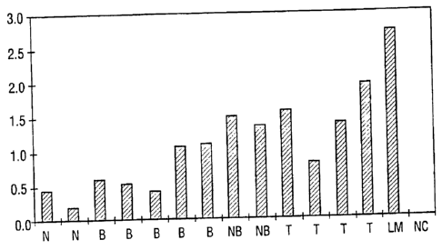
FIG. 1. Normalized quantitative RT-PCR of UC Band #25 (SEQ ID NO:1) shows
that it is overexpressed in prostate cancers and benign prostate compared with
normal
prostate tissues. The levels are particularly high in metastatic prostate
cancer. N = normal
prostate, B = benign prostatic hyperplasia (BPH), NB = needle core biopsy of
prostate
cancer, T = primary prostate cancer, LM = metastatic lymph node prostate
cancer, NC =
negative control..
FIG. 2. Normalized quantitative RT-PCR of UC Band #27 (SEQ ID NO:2) shows
that it is elevated in prostate cancers compared with normal or benign
prostates. N =
normal prostate, B = benign prostatic hyperplasia (BPH), NB = needle core
biopsy of
prostate cancer, T = primary prostate cancer, LM = metastatic lymph node
prostate
cancer, NC = negative control..
FIG. 3. Normalized quantitative RT-PCR of UC Band #28 (SEQ ID NO:3) shows
that it is elevated in prostate cancers, particularly in metastatic prostate
cancer, compared
with normal or benign prostates.. N = normal prostate, B = benign prostatic
hyperplasia
(BPH), NB = needle core biopsy of prostate cancer, T = primary prostate
cancer, LM =
metastatic lymph node prostate cancer, NC = negative control..
CA 02744096 2011-06-23
-10-
FIG. 4. Normalized quantitative RT-PCR of UC Band #31 (SEQ ID NO:4) shows
that it is overexpressed in benign and malignant prostate compared with normal
prostate..
N = normal prostate, B = benign prostatic hyperplasia (BPH), NB = needle core
biopsy of
prostate cancer, T = primary prostate cancer, LM = metastatic lymph node
prostate
cancer, NC = negative control..
FIG. 5. Normalized quantitative RT-PCR of a sequence from the human
fibronectin gene (SEQ 113 NO:7) shows that it is down regulated in BPH and
prostate
cancer compared with normal prostate.. N = normal prostate, B = benign
prostatic
hyperplasia (BPH), NB = needle core biopsy of prostate cancer, T = primary
prostate
cancer, LM = metastatic lymph node prostate cancer, NC = negative control..
FIG. 6. Normalized quantitative RT-PCR of UC Band #33 (SEQ ID NO:5) shows
that it is overexpressed in prostate cancers compared with normal or benign
prostate.. N =
normal prostate, B = benign prostatic hyperplasia (BPH), NB = needle core
biopsy of
prostate cancer, T = primary prostate cancer, LM = metastatic lymph node
prostate
cancer, NC = negative control..
FIG. 7. Quantitative RT-PCR of TGF-131 shows that it is overexpressed in
prostate
cancer compared to benign prostatic hypesplasia. . N = normal prostate, B =
benign
prostatic hyperplasia (BPH), NB = needle core biopsy of prostate cancer, T =
primary
prostate cancer, LM = metastatic lymph node prostate cancer, NC = negative
control.
FIG. 8. Quantitative RT-PCR of Cyclin A (SEQ ID NO:8) shows that it is
overexpressed in prostate cancer compared to normal prostate and benign
prostatic
hyperplasia N = normal prostate, B = benign prostatic hyperplasia (BPH), NB =
needle
core biopsy of prostate cancer, T = primary prostate cancer, LM = metastatic
lymph node
prostate cancer, NC = negative control..
CA 02744096 2011-06-23
-11-
FIG. 9. Oligonucleotide,s used in RT-PCR investigations of Her2ineu and a
truncated form of Her2Jneu. The binding sites for PCR primers are marked as P1
(Neug),
P2 (Neu3') and P5 (NeuT3'). The truncated form of Her2/neu also contains the
P1 binding
site. The regions within the Her2/neu coding sequence are: ECD (extracellular
domain),
MD (membrane domain), and 1CD (intracellular domain).
FIG. 10. Normalized quantitative RT-PCR for the full length Her2ineu transaipt
shows that it is overexpressed in prostate cancers compared to normal prostate
and benign
prostatic hyperplasia. . N = normal prostate, B = benign prostatic hyperplasia
(BPH), NB =
needle core biopsy of prostate cancer, T = primary prostate cancer, LM =
metastatic lymph
node prostate cancer, NC = negative control..
FIG. 11. Normalized quantitative RT-PCR for the truncated form of the Hez2ineu
transcript (SEQ ID NO:9) shows that it is overexpressed in prostate cancers
compared to
normal prostate and benign prostatic hyperplasia. . N = normal prostate, B =
benign
prostatic hyperplasia (BPH), NB = needle core biopsy of prostate cancer, T =
primary
prostate cancer, LM = metastatic lymph node prostate cancer, NC = negative
control.
FIG. 12. Amplification of 13-actin cDNA from 25 cDNAs synthesized from
various prostate tissues. The physiological states of these tissue, being
either normal
prostates, glands with BPH or prostate tumors are given in Table 4. Also
included on
this image molecular weight markers displayed as "ladders" and three isolated
bands
representing the PCR products from pools of (left to right) normal, BPH and
prostate
cancers.
FIG. 13 Amplification of a cDNA fragment derived from the UC42 mRNA in
the individual prostate cancers described in Table 4. Little are no detectable
expression
can be seen for this mRNA in either a pool of normal prostates or a pool of
prostate
glands with BPH. Strong signals from 7 of the 10 exarninal cancers indicates
very
CA 02744096 2011-06-23
-12-
significant induction of this gene in many prostate tumors. The normalized
data is
displayed graphically.
FIG. 14 Amplification of a cDNA fragment derived from the Hek (UC205)
rnRNA in the individual prostate cancers described in Table 4. Many, but not
all,
prostate glands with BPH are seen to have higher levels of expression of Hek
than seen
in a pool of normal glands. Examination of a gel also indicated that some of
the PCRs
are not in the linear phase of their amplification curves. Data was captured
on the
IS1000 and normalized as described in Table 4.
FIG. 15. 0-actin normalization of pooled cDNAs. Pools of cDNAs synthesized
from either normal prostates (N), prostate glands with BPH (B) or prostate
tumors (C)
were used as templates for [3-actin cDNA amplification. Four identical sets of
PCRs
were set up. These were stopped and examined after differing numbers of PCR
cycles.
The data for the 22 cycles were numerically captured on by the IS1000 and used
to
derive normalizing statistics. The normalizing statistics are obtained by
dividing the
average intensity of the three captured bands by the value of the three bands
separately. These normalizing statistics were then used to normalize the data
obtained
from the tnRNA of Hek (UC205). Hek mRNA is more abundant in the BPH and
prostate cancer pools than in the pool of normal prostates. At 34 and 37
cycles, the
PCRs for the BPH and cancer pools are observed in the Linear phase of their
arnplifcation curves. The data was normalized to the (3-actin data.
DETAILED DESCRIPTION OF ME INVENTION
The present invention concerns the early detection, diagnosis, prognosis and
treatment of prostate diseases, such as prostate cancer or benign prostatic
hyperplasia
(BPH). Markers of prostate disease, in the form of nucleic acid sequences
isolated from
human prostate tumors or prostate cancer cell lines, are disclosed. These
markers are
CA 02744096 2011-06-23
-13-
indicators of malignant transformation of prostate tissues and are diagnostic
of the potential
for metastatic spread of malignant prostate tumors.
Those skilled in the art will realize that the nucleic acid sequences
disclosed will find
utility in a variety of applications in prostate cancer detection, diagnosis,
prognosis and
treatment. Examples of such applications within the scope of the present
invention
comprise amplification of markers of prostate disease using specific primers,
detection of
markers of prostate disease by hybridization with oligonuclecnide probes,
incorporation of
isolated nucleic acids into vectors, expression of RNA ,peptides or
polypeptides from the
vectors, development of immunologic reagents corresponding to marker encoded
products,
and therapeutic treatments of prostate cancer using antibodies, anti-sense
nucleic acids, or
other inhibitors specific for the identified prostate cancer markers.
A. Nudeic Acids
As described herein, an aspect of the present disclosure is 26 markers of
prostate
disose, identified by RNA fingerprinting or quantitative RT-PCR. These include
20 ,
previously unknown gene products, as well as nucleic acid products of the a6-
integrin,
PAP, fibronectin and cyclin A genes and a truncated nucleic acid product of
the Her2/neu
gene. The latter three gene products have been identified in other forms of
cancer, but the
present invention is the first report of overexpression in prostate cancer.
In one embodiment, the nucleic acid sequences disclosed herein will find
utility as
hybridization probes or amplification primers. These nucleic acids may be
used, for
example, in diagnostic evaluation of tissue samples or employed to clone full
length cDNAs
or genomic clones corresponding thereto. In certain embodiments, these probes
and
primers consist of oligonucleotide fragments. Such fragments should be of
sufficient length
to provide specific hybridization to a RNA or DNA tissue sample. The sequences
typically
CA 02744096 2011-06-23
-14-
will be 10-20 nucleotides, but may be longer. Longer sequences, e.g., 40, 50,
100, 500 and
even up to full length, are preferred for certain embodiments.
Nucleic acid molecules having contiguous stretches of about 10, 15, 17, 20,
30, 40,
50, 60, 75 or 100 or 500 nucleotides from a sequence selected from SEQ 1D
NO:1, SEQ
ID NO:2, SEQ ID NO:3, SEQ ID NO:4, SEQ ID NO:5, SEQ ID NO:10, SEQ ID NO:11,
SEQ ID NO:12, SEQ ID NO:13, SEQ ID NO:15, SEQ 113 NO:16, SEQ ID NO:17, SEQ
ID NO:18, SEQ ID NO:19, SEQ ID NO:20, SEQ lD NO:21, SEQ lD NO:22, SEQ ID
NO:23, SEQ ID NO:45 and SEQ NO:46 are contemplated. Molecules that are
complementary to the above mentioned sequences and that bind to these
sequences under
high stringency conditions also are contemplated. These probes will be useful
in a variety
of hybridization embodiments, such as Southern and Northern blotting. In some
cases, it is
contemplated that probes may be used that hybridize to multiple target
sequences without
compromising their ability to effectively diagnose cancer.
Various probes and primers can be designed around the disclosed nucleotide
sequences. Primers may be of any length but, typically, are 10-20 bases in
length. By
assigning numeric values to a sequence, for example, the first residue is 1,
the second
residue is 2, etc., an algorithm dffning all primers can be proposed:
n to n + y
where n is an integer from 1 to the last number of the sequence and y is the
length of the
primer minus one (9 to 19), where n + y does not exceed the last number of the
sequence.
Thus, for a 10-mer, the probes correspond to bases 1 to 10, 2 to 11, 3 to 12
... and so on.
For a 15-mer, the probes correspond to bases 1 to 15, 2 to 16, 3 to 17 ... and
so on. For a
20-mer, the probes correspond to bases 1 to 20, 2 to 21, 3 to 22 ... and so
on.
CA 02744096 2011-06-23
-15-
The values of n in the algorithm above for each of the nucleic acid sequences
is:
SEQ ID NO:1, n=391; SEQ ID NO:2, n=514; SEQ ID NO:3, n=757; SEQ NO:4,
n=573; SEQ ID NO:5, n=358; SEQ ID NO:10, n=166; SEQ ID NO:11, n=107; SEQ ID
NO:12, n=183; SEQ ID NO:13, n=92; SEQ 1D NO:15, n=174; SEQ ID NO:16, n=132;
SEQ D NO:17, n=135; SEQ ID NO:18, n=415; SEQ ID NO:19, n=471; SEQ ID NO:20,
n=209, SEQ NO:21, n=407, SEQ ID NO:22, n=267, SEQ ID NO:23, n=333, SEQ ID
NO:45, n=369, and SEQ ID NO:46, n=301.
In certain embodiments, it is contemplated that multiple probes may be used
for
hybridization to a single sample. For example, a truncated form of Her2/neu
could be
detected by probing human tissue samples with oligonucleotides specific for
the 5' and 3'
ends of the full-length Her2/neu transcript. A fiill-length Her2/neu
transcript would bind
both probes, while a truncated form of the Her2Jneu transcript, indicative of
transformed
cells, would bind to the 5' probe but not to the 3' probe.
The use of a hybridization probe of between 14 and 100 nucleotides in length
- allows the formation of a duplex molecule that is both stable and
selective. Molecules
having complementary sequences over stretches greater than 20 bases in length
are
generally preferred, in order to increase stability and selectivity of the
hybrid, and thereby
improve the quality and degree of particular hybrid molecules obtained. One
will generally
prefer to design nucleic acid molecules having stretches of 20 to 30
nucleotides, or even
longer where desired. Such fragments may be readily prepared by, for example,
directly
synthesizing the fragment by chemical means or by introducing selected
sequences into
recombinant vectors for recombinant production.
Accordingly, the nucleotide sequences of the invention may be used for their
ability
to selectively form duplex molecules with complementary stretches of genes or
RNAs or to
provide primers for amplification of DNA or RNA from tissues. Depending on the
CA 02744096 2011-06-23
-I 6-
application envisioned, one will desire to employ varying conditions of
hybridi7ation to
achieve varying degrees of selectivity of probe towards target sequence.
For applications requiring high selectivity, one will typically desire to
employ
relatively stringent conditions to form the hybrids, e.g., one will select
relatively low salt
and/or high temperature conditions, such as provided by about 0.02 M to about
0.10 M
NaCI at temperatures of about 50 C to about 70 C. Such high stringency
conditions
tolerate little, if any, mismatch between the probe and the template or target
strand, and
would be particularly suitable for isolating specific genes or detecting
specific mRNA
transcripts. It is generally appreciated that conditions can be rendered more
stringent by the
addition of increasing amounts of fomuunide.
For certain applications, for example, substitution of amino acids by site-
directed
mutagenesis, it is appreciated that lower stringency conditions are required.
Under these
conditions, hybridization may occur even though the sequences of probe and
target strand
are not perfectly complementary, but are mismatched at one or more positions.
Conditions
may be rendered less stringent by increasing salt concentration and decreasing
temperature.
For example, a medium stringency condition could be provided by about 0.1 to
0.25 M
NaCI at temperatures of about 37 C to about 55 C, while a low stringency
condition could
be provided by about 0.15 M to about 0.9 M salt, at tempemtures ranging from
about 20 C
to about 55 C. Thus, hybridization conditions can be readily manipulated, and
thus will
generally be a method of choice depending on the desired results.
The following codon chart may be used, in a site-directed mutagenic scheme, to
produce nucleic acids encoding the same or slightly different amino acid
sequences of a
given nucleic acid:
yABLE 5
Amino Acids Codons
CA 02744096 2011-06-23
-17-
Amino Acids Codons
Alanine Ala A GCA GCC GCG GCU
Cysteine Cys C UGC UGU
Aspartic acid Asp D GAC GAU
Ghttatnic acid Giu E GAA GAG
Phenylalanine Phe F UUC UUU
Glycine Gly G GGA GGC GGG GGU
flistidine Ms H CAC CAU
Isoleucine Ile I AUA AUC AUU
Lysine Lys K AAA AAG
Leucine Leu L UUA UUG CUA CUC CUG CUU
Methionine Met M AUG
Asparagine Asn N AAC AAU
Proline Pro P CCA CCC CCG CCU
Glutamine Gin Q CAA CAG
Azgiuiite Arg R AGA AGG CGA CGC CGG CGU
CA 02744096 2011-06-23
-18-
TABLE 5 (continued)
Amino Acids Codons
Serine Ser S AGC AGU UCA UCC UCG UCU
Threonine Thr T ACA ACC ACG ACU
Valine Val V GUA GUC GUG GUU
Tryptophan Trp W UGG
Tyrosine Tyr Y UAC UAU
In other embodiments, hybridization may be achieved under conditions of for
example, 50 mM Tris-HC1 (pH 8.3), 75 mM Ka, 3 mM MgC12, 10 mM dithiothreitol,
at
temperatures between approximately 20 C to about 37 C. Other hybridization
conditions
utilized could include approximately 10 mM Tris-HC1 (pH 8.3), 50 mM KC1, 1.5
irM
M8C12, at temperatures ranging from approximately 40 C to about 72 C.
In certain embodiments, it will be advantageous to employ nucleic acid
sequences
of the present invention in combination with an appropriate means, such as a
label, for
determining hybridization. A wide variety of appropriate indicator means are
known in the
art, including fluorescent, radioactive, enzymatic or other ligands, such as
avidin/biotin,
which are capable of being detected. In preferred embodiments, one may desire
to employ
a fluorescent label or an enzyme tag such as urease, alkaline phosphatase or
peroxidase,
instead of radioactive or other environmentally undesirable reagents. In the
case of enzyme
tags, colorimetric indicator substrates are known which can be employed to
providea
detection means visible to the human eye or spectrophototnetrically, to
identify specific
hybridization with complementary nucleic acid-containing samples.
In general, it is envisioned that the hybridization probes described herein
will be
useful both as reagents in solution hybridization, as in PCR, for detection of
expression of
corresponding genes, as well as in embodiments employing a solid phase. In
embodiments
CA 02744096 2011-06-23
-19-
involving a solid phase, the test DNA (or RNA) is adsorbed or otherwise
affixed to a
selected matrix or surfitce. This fixed, single-stranded nucleic acid is then
subjected to
hybridization with selected probes under desired conditions. The selected
conditions will
depend on the particular circumstances based on the particular criteria
required (depending,
for example, on the CrFC content, type of target nucleic acid, source of
nucleic acid, size of
hybridization probe, etc.). Following washing of the hybridized surface to
remove non-
specifically bound probe molecules, hybridization is detected, or even
quantified, by means
of the label.
It will be understood that this invention is not limited to the particular
probes
disclosed herein and particularly is intended to encompass at least nucleic
acid sequences
that are hybridizable to the disclosed sequences or are functional sequence
analogs of these
sequences. For example, a partial sequence may be used to identify a
structurally-related
gene or the full length genomic or cDNA clone from which it is derived. Those
of skill in
the art are well aware of the methods for generating cDNA and genoraic
libraries which can
be used as a target for the above-described probes (Sambrook et al., 1989).
For applications in which the nucleic acid segments of the present invention
are
incorporated into vectors, such as plasmids, cosmids or viruses, these
segments may be
combined with other DNA sequences, such as promoters, polyadenylation signals,
restriction enzyme sites, multiple cloning sites, other coding segments, and
the like, such
that their overall length may vary considerably. It is contemplated that a
nucleic acid
fragment of almost any length may be employed, with the total length
preferably being
limited by the ease of preparation and use in theintended recombinant DNA
protocol.
DNA segments encoding a specific gene may be introduced into recombinant host
cells and employed for expressing a specific structural or regulatory protein.
Alternatively,
through the application of genetic engineering techniques, subportions or
derivatives of
selected genes may be employed. Upstream regions containing regulatory regions
such as
CA 02744096 2011-06-23
-20-
promoter regions may be isolated and subsequently employed for expression of
the selected
gene.
Where an expression product is to be generated, it is possible for the nucleic
acid
sequence to be varied while retaining the ability to encode the same product.
Reference to
the codon chart, provided above, will permit those of skill in the art to
design any nucleic
acid encoding for the product of a given nucleic acid.
B. Encoded Proteins
Once the entire coding sequence of a marker-associated gene has been
determined,
the gene can be inserted into an appropriate expression system. The gene can
be expressed
in any number of different recombinant DNA expression systems to generate
large amounts
of the polypeptide product which can then be purified and used to vaccinate
animals to
generate antisem with which further studies may be conducted.
Examples of expression systems known to the skilled practitioner in the art
include
bacteria such as E. coli, yeast such as Pichia pastoris, baculovirus, and
mammalian
expression systems such as in Cos or CHO cells. A complete gene can be
expressed or,
alternatively, fragments of the gene encoding portions of polypeptide can be
produced.
In certain broad applications of the invention, the gene sequence encoding the
polypeptide is analyzed to detect putative transmembrane sequences. Such
sequences are
typically very hydrophobic and are readily detected by the use of standard
sequence analysis
software, such as MacVector (ÞB1, New Haven, CT). The presence of
transmembrane
sequences is often deleterious when a recombinant protein is synthesized in
many
expression systems, especially E con, as it leads to the production of
insoluble aggreetes
which are difficult to renature into the native conformation of the protein.
Deletion of
CA 02744096 2011-06-23
-21-
tran.smembrane sequences typically does not significantly alter the
conformation of the
remaining protein structure.
Moreover, transrnembrane sequences, being by definition embedded within a
membrane, are inaccessible. Antibodies to these sequences may not, therefore,
prove useful
in in vivo or in situ studies. Deletion of transmembrane-encoding sequences
from the genes
used for expression can be achieved by standard techniques. For example,
fortuitously-
placed restriction enzyme sites can be used to excise the desired gene
fragment, or PCR-
type amplification can be used to amplify only the desired part of the gene.
Computer sequence analysis may be used to determine the location of the
predicted
major antigenic determinant epitopes of the polypeptide. Software capable of
carrying out
this analysis is readily available commercially, for example MacVector (MI,
New Haven,
CT). The software typically uses standard algorithms such as the
Kyte/Doolittle or
Hopp/Woods methods for locating hydrophilic sequences may be found on the
surface of
proteins and are, therefore, likely to act as antigenic determinants.
Once this analysis is made, polypeptides may be prepared which contain at
least the
essential features of the antigenic determinant and which may be employed in
the
generation of antisera against the polypeptide. Mmigenes or gene fusions
encoding these
determinants may be constructed and inserted into expression vectors by
standard methods,
for example, using PCR cloning methodology.
The gene or gene fragment encoding a polypeptide may be inserted into an
expression vector by standard subcloning techniques. An E. co/i expression
vector may be
used which produces the recombinant polypeptide as a fusion protein, allowing
rapid
affinity purification of the protein. Examples of such fusion protein
expression systems are
the gktrathione S-transferase system (Pharmacia, Piscataway, NJ), the maltose
binding
CA 02744096 2011-06-23
-22-
protein system (NEB, Beverley, MA), the FLAG system (IM, New Haven, CT), and
the
6xl-Es system (Qiagen, Chatsworth, CA).
Some of these systems produce recombinant polypeptides bearing only a small
number of additional amino acids, which are unlikely to affect the antigenic
ability of the
recombinant polypeptide. For example, both the FLAG system and the 6xlIs
system add
only short sequences, both of which are known to be poorly antigenic and which
do not
adversely affect folding of the polypeptide to its native conformation. Other
fusion systems
are designed to produce firsions wherein the fusion partner is easily excised
from the
desired polypeptide. In one embodiment, the fusion partner is linked to the
recombinant
polypeptide by a peptide sequence containing a specific recognition sequence
for a
protease. Examples of suitable sequences are those recognized by the Tobacco
Etch Virus
protease (Life Technologies, Gaithersburg, MD) or Factor 3Ca (New England
Biolabs,
Beverley, MA).
The expression system used may also be one driven by the baculovirus
polyhedron
promoter. The gene encoding the polypeptide may be manipulated by standard
techniques
in order to facilitate cloning into the baculovirus vector. One baculovirus
vector is the
pBlueBac vector (Invitrogen, Sorrento, CA). The vector carrying the gene for
the
polypeptide is transfected into Spodopterafrugiperda (Sf9) cells by standard
protocols, and
the cells are cultured and processed to produce the recombinant antigen. See
Summers et
al., A Manual of Methods for Baculovirus Vectors and Insect Cell Culture
Procedures,
Texas Agricultural Experimental Station U.S. Patent No. 4,215,051.
As an alternative to recombinant polypeptides, synthetic peptides
corresponding to
the antigenic determinants may be prepared. Such peptides are at least six
amino acid
residues long, and may contain up to approximately 35 residues, which is the
approximate
upper length limit of automated peptide synthesis machines, such as those
available from
CA 02744096 2011-06-23
-23-
Applied Biosystems (Foster City, CA). Use of such small peptides for
vaccination typically
requires conjugation of the peptide to an immunogenic carrier protein such as
hepatitis B
surface antigen, keyhole limpet hemocyanin or bovine serum albumin. Methods
for
performing this conjugation are well known in the art.
Amino acid sequence variants of the polypeptide may also be prepared. These
may,
for instance, be minor sequence variants of the polypeptide which arise due to
natural
variation within the population or they may be homologues found in other
species. They
also may be sequences which do not occur naturally but which are sufficiently
similar that
they function similarly and/or elicit an immune response that cross-reacts
with natural forms
of the polypeptide. Sequence variants may be prepared by standard methods of
site-
directed mutagenesis such as those described herein for removing the
transmembrane
sequence.
Amino acid sequence variants of the polypeptide may be substitutional,
insertional
or deletion variants. Deletion variants lack one or more residues of the
native protein
which are not essential for function or immunogenic activity, and are
exemplified by the
variants lacking a transmembrane sequence. Another common type of deletion
variant is
one lacking secretory signal sequences or signal sequences directing a protein
to bind to a
particular part of a cell. An example of the latter sequence is the SH2
domain, which
induces protein binding to phovhotyrosine residues.
Substitutional variants typically contain an alternative amino. acid at one or
more
sites within the protein, and may be designed to modulate one or more
properties of the
polypeptide such as stability against proteolytic cleavage. Substitutions
preferably are
conservative, that is, one amino acid is replaced with one of similar size and
charge.
Conservative substitutions are well known in the art and include, for example,
the changes
of alanine to serine; arginine to lysine; asparagine to glutamine or
histidine; aspartate to
glutamate; cysteine to serine; glutamine to asparagine; glutamate to
aspartate; glycine to
CA 02744096 2011-06-23
-24-
proline; histidine to asparagine or glutamine; isoleucine to leucine or
valine; leucine to
valine or isoleucine; lysine to arginine, glutamine, or glutamate; methionine
to leucine or
isoleucine; phenylalanine to tyrosine, leucine or methionine; serine to
threonine; threonine
to serine; tryptophan to tyrosine; tyrosine to tryptophan or phenylalanine;
and valine to
isoleucine or leucine.
Insertional variants include fusion proteins such as those used to allow rapid
purification of the polypeptide and also may include hybrid proteins
containing sequences
from other proteins and polypeptides which are homologues of the polypeptide.
For
example, an insertional variant may include portions of the amino acid
sequence of the
polypeptide from one species, together with portions of the homologous
polypeptide from
another species. Other insertional variants may include those in which
additional amino
acids are introduced within the coding sequence of the polypeptide. These
typically are
smoller insertions than the fusion proteins described above and are
introduced, for example,
to disrupt a protease cleavage site.
Major antigenic determinants of the polypeptide may be identified by an
empirical
approach in which portions of the gene encoding the polypeptide are expressed
in a
recombinant host, and the resulting proteins tested for their ability to
elicit an inumme
response. For example, PCR may be used to prepare a range of peptides lacking
successively longer fragments of the C-terminus of the protein. The
immunoprotective
activity of each of these peptides then identifies those fragments or domains
of the
polypeptide which are essential for this activity. Further studies in which
only a small
number of amino acids are removed at each iteration then allows the location
of the
antigenic determinants of the polypeptide.
Another method for the preparation of the polypeptides according to the
invention
is the use of peptide mimetics. hfunetics are peptide-containing molecules
which mimic
elements of protein secondary structure. See, for example, Johnson et
aL,"Peptide Turn
CA 02744096 2011-06-23
-25-
hfunetics" in BIOIECHNOLOGY AND PHARMACY ,Pezzuto et al, Eds., Chapman and
Hall, New York (1993). The underlying rationale behind the use of peptide
mimetics is that
the peptide backbone of proteins exists chiefly to orient amino acid side
chains in such a
way as to facilitate molecular interactions, such as those of antibody and
antigen. A peptide
mimetic is expected to permit molecular interactions similar to the natural
molecule.
Successful applications of the peptide mimetic concept have thus far focused
on
tnimetics of 0-turns within proteins, which are known to be highly antigenic.
Lilcely 0-turn
structure within a polypeptide may be predicted by computer-based algorithms
as discussed
herein. Once the component amino acids of the turn are determined, peptide
mimetics may
be constructed to achieve a similar spatial orientation of the essential
elements of the amino
acid side chains.
C. Preparation of Antibodies Specific for Encoded Proteins
1. Expression of Proteins from Cloned eDNAs
The cDNA species specified in SEQ ID NO:1, SEQ ID NO:2, SEQ ID NO:3, SEQ
ID NO:4, SEQ ID NO:5, SEQ NO:6, SEQ ID NO:7, SEQ ID NO:8, SEQ ID NO:9,
SEQ ID NO:10, SEQ ID NO:11, SEQ NO:12, SEQ ID NO:13, SEQ ID NO:14, SEQ
ID NO:15, SEQ ID NO:16, SEQ ID NO:17, SEQ ID NO:18, SF,Q ID NO:19, SEQ ID
NO:20, SEQ ID NO:21, SEQ ID NO:22, SEQ ID NO:23, SEQ ID NO:45, SEQ ID
NO:46 and SEQ ID NO:47 may be expressed as encoded peptides or proteins. The
engineering of DNA segment(s) for expression in a prokaryotic or eukaryotic
system raay
be performed by techniques generally known to those of sIdll in recombinant
expression. It
is believed that virtually any expression system may be employed in the
expression of the
Claimed nucleic acid sequences.
CA 02744096 2011-06-23
-26-
Both cDNA and genomic sequences are suitable for eukaryotic expression, as the
host cell will generally process the genomic transcripts to yield functional
mRNA for
translation into protein In addition, it is possible to use partial sequences
for generation of
antibodies against discrete portions of a gene product, even when the entire
sequence of
that gene product remains unknown. Computer programs are available to aid in
the
selection of regions which have potential immunologic significance. For
example, software
capable of carrying out this analysis is readily available commercially, for
example
MacVector (IBI, New Haven, CT). The software typically uses standard
algorithms such
as the Kyte/Doolittle or Hopp/Woods methods for locating hydrophilic sequences
which
are characteristically found on the surface of proteins and are, therefore,
likely to act as
antigenic determinants.
As used herein, the terms "engineered" and "recombinant" cells are intended to
refer to a cell into which an exogenous DNA segment or gene, such as a cDNA or
gene has
been introduced through the hand of man. Therefore, engineered cells are
distinguishable
from naturally occurring cells which do not contain a recombinantly introduced
exogenous
DNA segment or gene. Recombinant cells include those having an introduced cDNA
or
genomic gene, and also include genes positioned adjacent to a heterologous
promoter not
naturally associated with the particular introduced gene.
To express a recombinant encoded protein or peptide, whether mutant or wild-
type, in accordance with the present invention one would prepare an expression
vector that
comprises one of the claimed isolated nucleic acids under the control of or
operatively
linked to, one or more promoters. To bring a coding sequence "under the
control of' a
promoter, one positions the 5' end of the transcription initiation site of the
transcriptional
reading frame generally between about 1 and about 50 nucleotides "downstream"
(i.e., 3')
of the chosen promoter. The "upstream" promoter stimulates transcription of
the DNA and
promotes expression of the encoded recombinant protein. This is the meaning of
"recombinant expression" in this context.
CA 02744096 2011-06-23
-27-
Many standard techniques are available to construct expression vectors
containing
the appropriate nucleic acids and transcriptional/translational control
sequences in order to
achieve protein or peptide expression in a variety of host-expression systems.
Cell types
available for expression include, but are not limited to, bacteria, such as E.
coli and
B. subtilis transformed with recombinant bacteriophage DNA, plasmid DNA or
cosmid
DNA expression vectors.
Certain examples of prokaryotic hosts are .E coli strain RR1, E coli LE392,
E. coliB, E coil X1776 (ATCC No. 31537) as svell as E. coli W3110 (F-, lambda-
,
prototrophic, ATCC No. 273325); barilli such as Bacillus subtilis-, and other
enterobacteriaceae such as Salmonella ohimurium, Serratia marcescens, and
various
Pseudomonce species.
In general, plasmid vectors containing replicon and control sequences which
are
derived from species compatible with the host cell are used in connection with
these hosts.
The vector ordinarily carries a replication site, as well as marlcing
sequences which are
capable of providing phenotypic selection in transformed cells. For example, E
coli is
often transformed using pBR322, a plasmid derived from an E col/ species.
pBR322
contains genes for ampicillin and tetracycline resistance and thus provides
easy means for
identifying transformed cells. The pBR plasmid, or other microbial plasmid or
phage must
also contain, or be modified to contain, promoters which may be used by the
microbial
organism for expression of its own proteins.
In addition, phage vectors containing replicon and control sequences that are
compatible with the host microorganism may be used as transforming vectors in
cormection
with these hosts. For example, the phage lambda GEM174-11 may be utilized in
making a
recombinant phage vector which may be used to transform host cells, such as E
coli
LE392.
CA 02744096 2011-06-23
-28-
Further useful vectors include pIN vectors (Inouye et al, 1985); and pGEX
vectors, for use in generating glutathione S-tansferase (GST) soluble fusion
proteins for
later purification and separation or cleavage. Other suitable fusion proteins
are those with
13-galactosidase, ubiquitin, or the like.
Promoters that are most commonly used in recombinant DNA construction include
the13-lactamase (penicillinase), lactose and tryptophan (trp) promoter
systems. While these
are the most commonly used, other microbial promoters have been discovered and
utilized,
and details concerning their nucleotide sequences have been published,
enabling those of
skill in the art to ligate them functionally with plasmid vectors.
For expression in Saccharomyces, the plasmid YRp7, for example, is commonly
used (Stinchcomb et al., 1979; Kingsman et al, 1979; Tschemper et al, 1980).
This
plasmid already contains the trpl gene which provides a selection marker for a
mutant strain
of yeast lacking the ability to grow in tryptophan, for example ATCC No. 44076
or PEP4-
1 (Jones, 1977). The presence of the trpl lesion as a characteristic of the
yeast host cell
genome then provides an effective environment for detecting transformation by
growth in
the absence of tryptophan.
Suitable promoting sequences in yeast vectors include the promoters for 3-
phosphoglycerate kinase (Ifitzeinan et aL, 1980) or other glycolytic enzymes
(Hess et aL,
1968; Holland et al, 1978), such as enolase, glyceraldehyde-3-phosphate
dehydrogenase,
hexokinase, pyruvate decarboxylase, phosphofructokinase, glucose-6-phosphate
isomerase,
3-phosphoglycerate mutase, pyruvate kinase, triosephosphate isomerase,
phosphoglucose
isomerase, and glucokinase. In constructing suitable expression plasmids, the
termination
sequences associated with these genes are also ligated into the expression
vector 3' of the
sequence desired to be expressed to provide polyadenylation of the mRNA and
termination.
CA 02744096 2011-06-23
-29-
Other suitable promoters, which have the additional advantage of transcription
controlled by growth conditions, include the promoter region for alcohol
dehydrogenase 2,
isocytochrome C, acid phosphatase, degradative enzymes associated with
nitrogen
metabolism, and the aforementioned glyceraldehyde-3-phosphate dehydrogenase,
and
enzymes responsible for maltose and galactose utilization.
In addition to micro-organisms, cultures of cells derived from multicellular
organisms may also be used as hosts. In principle, any such cell culture is
workable,
whether from vertebrate or invertebrate culture. In addition to mammalian
cells, these
include insect cell systems infected with recombinant virus expression vectors
(e.g.,
baculovirus); and plant cell systems infected with recombinant virus
expression vectors
(e.g., cauliflower mosaic virus, CaMV; tobacco mosaic virus, TMV) or
transformed with
recombinant plasmid expression vectors (e.g., Ti plasmid) containing one or
more coding
sequences.
In a useful insect system, Autograph cahfornica nuclear polyhidrosi.s virus
(AcNPV) is used as a vector to =press foreign genes. The virus grows in
Spodoptera
frugiperda cells. The isolated nucleic acid coding sequences are cloned into
non-essential
regions (for example the polyhedrin gene) of the virus and placed under
control of an
AcNPV promoter (for example the polyhedrin promoter). Surce.ssfi 11 insertion
of the
coding sequences results in the inactivation of the polyhedrin gene and
production of non-
occluded recombinant virus (i.e., virus lacking the proteinaceous coat coded
for by the
polyhedrin gene). These recombinant viruses are then used to infect Spodoptera
frugiperda cells in which the inserted gene is expressed (e.g., U.S. Patent
No. 4,215,051
(Smith)).
Examples of useful mammalian host cell lines are VERO and HeLa cells, Chinese
hamster ovary (CHO) cell lines, W138, BHK, COS-7, 293, HepG2, 3T3, RIN and
MDCK
cell lines. In addition, a host cell strain may be chosen that modulates the
expression of the
CA 02744096 2011-06-23
-30-
inserted sequences, or modifies and processes the gene product in the specific
fashion
desired. Such modifications (e.g., glycosylation) and processing (e.g.,
cleavage) of protein
products may be important for the function of the encoded protein.
Different host cells have characteristic and specific mechanisms for the post-
translational processing and modification of proteins. Appropriate cells lines
or host
systems may be chosen to ensure the correct modification and processing of the
foreign
protein expressed. Expression vectors for use in mammalian cells ordinarily
include an
origin of replication (as necessary), a promoter located in front of the gene
to be expressed,
along with any necessary ribosome binding sites, RNA splice sites,
polyadenylation site, and
transcriptional terminator sequences. The origin of replication may be
provided either by
construction of the vector to include an exogenous origin, such as may be
derived from
SV40 or other viral (e.g., Polyoma, Adeno, VSV, BPV) source, or may be
provided by the
host cell chromosomal replication mechanism. If the vector is integrated into
the host cell
chromosome, the latter is often sufficient.
The promoters may be derived from the genome of mammalian cells (e.g.,
metallothionein promoter) or from mammalian viruses (e.g., the adenovirus late
promoter,
the vaccinia virus 7.5K promoter). Further, it is also possible, and may be
desirable, to
utilize promoter or control sequences normally associated with the desired
gene sequence,
provided such control sequences are compatible with the host cell systems.
A number of viral based expression systems may be utilized, for example,
commonly used promoters are derived from polyoma, Adenovirus 2, and most
frequently
Simian Virus 40 (SV40). The early and late promoters of SV40 virus are
particularly
useful because both are obtained estsily from the virus as a fragment which
also contains the
SV40 viral origin of replication. Smaller or larger SV40 fragments may also be
used,
provided there is included the approximately 250 bp sequence extending from
the limd 111
site toward the Bgl I site located in the viral origin of replication.
CA 02744096 2011-06-23
-31 -
In cases where an adenovirus is used as an expression vector, the coding
sequences
may be ligated to an adenovirus transcription/ translation control complex,
e.g., the late
promoter and tripartite leader sequence. This chimeric gene may then be
inserted in the
adenovirus genome by in vitro or in vivo recombination. Insertion in a non-
essential region
of the viral genome (e.g., region El or E3) will result in a recombinant virus
that is viable
and capable of expressing proteins in infected hosts.
Specific initiation signals may also be required for efficient translation of
the
claimed isolated nucleic acid coding sequences. These signals include the ATG
initiation
codon and adjacent sequences. Exogenous translational control signals,
including the ATG
initiation codon, may additionally need to be provided. One of ordinary skill
in the art
would readily be capable of determining this and providing the necessary
signals. It is well
known that the initiation codon must be in-frame (or in-phase) with the
reading frame of
the desired coding sequence to ensure translation of the entire insert. These
exogenous
translational control signals and initiation codons may be of a variety of
origins, both natural
and synthetic. The efficiency of expression may be enhanced by the inclusion
of
appropriate transcription enhancer elements or transcription terminators
(Bittner et al,
1987).
In eulcaryotic expression, one will also typically desire to incorporate into
the
transcriptional unit an appropriate polyadenylation site (e.g., 5'-AATAAA-3')
if one was
not contained within the original cloned segment. Typically, the poly A
addition site is
placed about 30 to 2000 nucleotides "downstream" of the termination site of
the protein at
a position prior to transcription termination.
For long-term, high-yield production of recombinant proteins, stable
expression is
preferred. For example, cell lines that stably express constructs encoding
proteins may be
engineered. Rather than using expression vectors that contain viral origins of
replication,
CA 02744096 2011-06-23
-32-
host cells may be transformed with vectors controlled by appropriate
expression control
elements (e.g., promoter, enhancer, sequences, transcription terminators,
polyadenylation
sites, etc.), and a selectable marker. Following the introduction of foreign
DNA,
engineered cells may be allowed to grow for 1-2 days in an enniched media, and
then are
switched to a selective media. The selectable marker in the recombinant
plasmid confers
resistance to the selection and allows cells to stably integrate the plasmid
into their
chromosomes and grow to form foci which in turn may be cloned and expanded
into cell
lines.
A number of selection systems may be used, including but not limited to, the
herpes
simplex virus thymidine lcinase (Wigler et al, 1977), hypoxanthine-guanine
phosphoribosyltranderase (Szybalslca et al, 1962) and adenine
phosphoribosyltransferase
genes (Lowy et al, 1980), in tk-, hgprt- or aprt- cells, respectively. Also,
antimetabolite
resistance may be used as the basis of selection for dhfr, that confers
resistance to
methotrexate (Wigler et al., 1980; O'Hare et al, 1981); gpt, that confers
resistance to
myc,ophenolic acid (Mulligan et al., 1981); neo, that confers resistance to
the
arninoglycoside 0-418 (Colberre-Garapin at, 1981); and hygro, that confers
resistance
to hygromycin (Santerre et al, Gene, 30(1-3): 147-56 1984).
It is contemplated that the isolated nucleic acids of the invention may be
"overexpressed", i.e., expressed in increased levels relative to its natural
expression in
human prostate cells, or even relative to the expression of other proteins in
the recombinant
host cell. Such overexpression may be assessed by a variety of methods,
including radio-
labelling and/or protein purification. However, simple and direct methods are
preferred, for
example, those involving SDS/PAGE and protein staining or Western blotting,
followed by
quantitative analyses, such as densitometric scanning of the resultant gel or
blot. A specffic
increase in the level of the recombinant protein or peptide in comparison to
the level in
natural human prostate cells is indicative of overexprmsion, as is a relative
abundance of the
CA 02744096 2011-06-23
-33-
specific protein in relation to the other proteins produced by the host cell
and, e.g., visible
on a gel.
2. Purification of &pressed Proteins
Further aspects of the present invention concern the purification, and in
particular
embodiments, the substantial purification, of an encoded protein or peptide.
The term
"purified protein or peptide" as used herein, is intended to refer to a
composition, isolatable
from other components, wherein the protein or peptide is purified to any
degree relative to
its naturally-obtainable state, i.e., in this case, relative to its purity
within a prostate cell
extract. A purified protein or peptide therefore also refers to a protein or
peptide, free
from the environment in which it may naturally occur.
Generally, "purified" will refer to a protein or peptide composition which has
been
subjected to fractionation to remove various other components, and which
composition
substantially retains its expressed biological activity. Where the term
"substantially
purified" is used, this will referto a composition in which the protein or
peptide forms the
major component of the composition, such as constituting about 50% or more of
the
proteins in the composition.
Various methods for quantifying the degree of purification of the protein or
peptide
will be known to those of skill in the art in light of the present disclosure.
These include,
for example, determining the specific activity of an active fraction, or
assessing the number
of polypeptides within a fraction by SDS/PAGE analysis. A preferred method for
assessing
the purity of a fraction is to calculate the specific activity of the
fraction, to compare it to
the specific activity of the initial extract, and to thus calculate the degree
of purity, herein
assessed by a "-fold purification number". The actual units used to represent
the amount of
activity will, of course, be dependent upon the particular assay teclutique
chosen to follow
CA 02744096 2011-06-23
-34-
the purification and whether or not the expressed protein or peptide exhibits
a detectable
activity.
Various techniques suitable for use in protein purification will be well known
to
those of skill in the art. These include, for example, precipitation with
ammonium sulphate,
PEG, antibodies and the like or by heat denaturation, followed by
centrifugation;
chromatography steps such as ion exchange, gel fihration, reverse phase,
hydroxylapatite
and affinity chromatography, isoelectric focusing; gel electrophoresis; and
combinations of
such and other techniques. As is generally known in the art, it is believed
that the order of
conducting the various purification steps may be changed, or that certain
steps may be
omitted, and still result in a suitable method for the preparation of a
substantially purified
protein or peptide.
There is no general requirement that the protein or peptide always be provided
in
the most purified state. Indeed, it is contemplated that less substantially
purified products
will have utility in certain embodiments. Partial purification may be
accomplished by using
fewer purification steps in combination, or by utilizing different forms of
the same general
purification scheme. For example, it is appreciated that a cation-exchange
column
chromatography performed utili7ing an HPLC apparatus will generally result in
a greater -
fold purification than the same technique utilizing a low pressure
chromatography system.
:Methods exhibiting a lower degree of relative purification may have
advantages in total
recovery of protein product, or in maintaining the activity of an expressed
protein.
It is known that the migration of a polypeptide may vary, sometimes
significantly,
with different conditions of SDS/PAGE (Capaldi eta, Biochem. Biophys. Res.
Comm.,
76:425, 1977). It will therefore be appreciated that under differing
electrophoresis
conditions, the apparent molecular weights of purified or partially purified
expression
products may vary.
CA 02744096 2011-06-23
-35-
3. Antibody Generation
For some embodiments, it will be desirable to produce antibodies that bind
with
high specificity to the polypeptide product(s) of an isolated nucleic acid
selected from SEQ
ID NO:1, SEQ ID NO:2, SEQ ID NO:3, SEQ 1D NO:4, SEQ ID NO:5, SEQ ID NO:10,
SEQ NO:11, SEQ ID NO:12, SEQ ID NO:13, SEQ NO:15, SEQ NO:16, SEQ
ID NO:17, SEQ ID NO:18, SEQ ID NO:19, SEQ ID NO:20, SEQ lD NO:21, SEQ
NO:22, SEQ ID NO:23, SEQ D NO:45 and SEQ ID NO:46. Means for preparing and
characterizing antibodies are well known in the art (See, e.g., Antibodies: A
Laboratory
Manual, Cold Spring Harbor Laboratory, 1988.
Methods for generating polyclonal antibodies are well known in the art.
Briefly, a
polyclonal antibody is prepared by immunizing an animal with an immunogenic
composition and collecting antis= from that immunized animal. A wide range of
animal
species may be used for the production of antisera. Typically the animal used
for
production of anti-antisesa is a rabbit, a mouse, a rat, a hamster, a guinea
pig or a goat.
Bectause of the relatively large blood volume of rabbits, a rabbit is a
preferred choice for
production of polyclonal antibodies.
As is well 'mown in the art, a given composition may vary in its
immunogenicity. It
is often necessary therefore to boost the host immune system, as may be
ac,hieved by
coupling a peptide or polypeptide inuntmogen to a carrier. Exemplary and
preferred
carriers are keyhole limpet hemocyanin (KLH) and bovine serum albumin (BSA).
Other
albumins such as ovalbumin, mouse serum albumin or rabbit serum albumin may
also be
used as carriers. Means for conjugating a polypeptide to a carrier protein are
well known in
the art and include glutaraldehyde, m-maleitnidobenzoyl-N-hydroxysuccinimide
ester,
carbodiimide and bis-biazotized benzidine.
CA 02744096 2011-06-23
-36-
As is also well known in the art, the inununogenicity of a particular
immunogen
composition may be enhanced by the use of non-specific stimulators of the
immune
response, known as adjuvants. Exemplary and preferred adjuvants include
complete
Freund's adjuvant (a non-specific stimulator of the immune response containing
killed
Mycobacterium tuberculosis), incomplete Freund's adjuvants and altunixnun
hydroxide
adjuvant.
The amount of immunogen composition used in the production of polyclonal
antibodies varies upon the nature of the immunogen as well as the animal used
for
immunization. A variety of routes may be used to administer the inununogen
(subcutaneous, intramuscular, intradermal, intravenous and intraperitoneal).
The
production of polyclonal antibodies may be monitored by sampling blood of the
immunized
animal at various points following inununization. A second, booster injection,
may also be
given. The process of boosting and titering is repeated until a suitable titer
is achieved.
When a desired level of irnmunogenicity is obtained, the immunized animal may
be bled and
the serum isolated and stored, and/or the animal may be used to generate MAbs.
For
production of rabbit polycdonal antibodies, the animal may be bled through an
ear vein or
alternatively by cardiac puncture. The removed blood is allowed to coagulate
and then
centrifuged to separate serum components from whole cells and blood clots. The
serum
may be used as is for various applications or else the desired antibody
fraction may be
purified by well-known methods, such as affinity chromatography using another
antibody
or a peptide bound to a solid matrix.
Monoclonal antibodies (MAbs) may be readily prepared through use of well-known
techniques, such as those exemplified in U.S. Patent 4,196,265.
Typically, this technique involves immunizing a suitable animal with a
selected
immunogen composition, e.g., a purified or partially purified expressed
protein, polypeptide
or peptide. The immunizing composition is administered in a manner effective
to stimulate
antibody producing cells.
CA 02744096 2011-06-23
-37-
The methods for generating monoclonal antibodies (MAbs) generally begin along
the same lines as those for preparing polyclonal antibodies. Rodents such as
mice and rats
are preferred animals, however, the use of rabbit, sheep or frog cells is also
possible. The
use of rats may provide certain advantages (Goding, 1986, pp. 60-61), but mice
are
preferred, with the BALB/c mouse being most preferred as this is most
routinely used and
generally gives a higher percentage of stable fusions.
The animals are injected with antigen as described above. The antigen may be
coupled to carrier molecules such as keyhole limpet hemocyanin if necessary.
The antigen
would typically be mixed with adjuvant, such as Freund's complete or
incomplete adjuvant.
Booster injections with the same antigen would occur at approximately two-week
intervals.
Following immunization, somatic cells with the potential for producing
antibodies,
specifically B lymphocytes (B cells), are Selected for use in the MAb
generating protocol.
These cells may be obtained from biopsied spleens, tonsils or lymph nodes, or
from a
peripheral blood sample. Spleen cells and peripheral blood cells are
preferred, the former
because they are a rich source of antibody-producing cells that are in the
dividing
plasrnablast stage, and the latter because peripheral blood is easily
accessible. Often, a
panel of animqls will have been immunized and the spleen of the animal with
the highest
antibody titer will be removed and the spleen lymphocytes obtained by
homogenizing the
spleen with a syringe. Typically, a spleen from an immunized mouse contains
approximately 5 X 10⁷ to 2 X 10s lymphocytes.
The antibody-producing B lymphocytes from the immunized animal are then fused
with cells of an immortal myeloma cell, generally one of the same species as
the animal that
was immunized. Myeloma cell lines suited for use in hybridoma-producing fusion
procedures preferably are non-antibody-producing, have high fusion efficiency,
and enzyme
CA 02744096 2011-06-23
-38-
deficiencies that render then incapable of growing in certain selective media
which support
the growth of only the desired fused cells (hybridomas).
Any one of a number of myeloma cells may be used, as are known to those of
skill
in the art (Goding, pp. 65-66, 1986; Campbell, pp. 75-83, 1984). For example,
where the
immunized animal is a mouse, one may use P3-X63/Ag8, X63-Ag8.653, NS1/1.Ag 4
1,
Sp210-Ag14, FO, NSO/U, MPC-11, MPC11-X45-GTG 1.7 and S194/53000 Bul; for rats,
one may use R210.RCY3, Y3-Ag 1.2.3, IR983F and 413210; and U-266, GM1500-GRG2,
LICR-LON-HMy2 and UC729-6 are all useful in connection with human cell
fusions.
One preferred murine myeloma cell is the NS-1 myeloma cell line (also termed
P3-
NS-1-Ag4-1), which is readily available from the NIGMS Human Genetic Mutant
Cell
Repository by requesting cell line repository number GM3573. .Another mouse
myeloma
cell line that may be used is the 8-azaguanine-resistant mouse murine myeloma
SP2/0
non-producer cell line.
Methods for generating hybrids of antibody-producing spleen or lymph node
cells
and myeloma cells usually comprise mixing somatic cells with myeloma cells in
a 2:1
proportion, though the proportion may vary from about 20:1 to about 1:1,
respectively, in
the presence of an agent or agents (chemical or electrical) that promote the
fusion of cell
membranes. Fusion methods using Sendai virus have been described by Kohler and
Milstein (1975; 1976), and those using polyethylene glycol (PEG), such as 3704
(v/v) PEG,
by Geer et al (1977). The use of electrically induced fusion methods is also
appropriate
(Goding pp. 71-74, 1986).
Fusion procedures usually produce viable hybrids at low frequencies, about 1 X
10"
6 tO 1 X 10⁴. However, this does not pose a problem, as the viable, fused
hybrids are
differentiated from the parental, unfused cells (particularly the unfused
myeloma cells that
would normally continue to divide indefinitely) by culturing in a selective
medium. The
CA 02744096 2011-06-23
-39-
selective medium is generally one that contains an agent that blocks the de
novo synthesis
of nucleotides in the tissue culture media. Exemplary and preferred agents are
arninopterin,
methotrexate, and azaserine. Aminopterin and methotrexate block de novo
synthesis of
both purines and pyrimidines, whereas azaserine blocks only purine synthesis.
Where
aminopterin or methotrexate is used, the media is supplemented with
hypoxanthine and
thymidine as a source of nucleotides (HAT medium). Where azaserine is used,
the media is
supplemented with hypoxanthine.
The preferred selection medium is HAT. Only cells capable of operating
nucleotide
salvage pathways are able to survive in HAT medium. The myeloma cells are
defective in
key enzymes of the salvage pathway, e.g., hypoxanthine phosphoribosyl
transferase
(HPRT), and they cannot survive. The B cells may operate this pathway, but
they have a
limited life span in culture and generally die within about two weeks.
Therefore, the only
cells that can survive in the selective media are those hybrids formed from
myeloma and
B cells.
This culturing provides a population of hybridomas from which specific
hybridomas
are selected. Typically, selection of hybridornas is performed by culturing
the cells by
single-clone dilution in microtiter plates, followed by testing the individual
clonal
supernatants (after about two to three weeks) for the desired reactivity. The
assay should
be sensitive, simple and rapid, such as radioinimunoassays, enzyme
immunoassays,
cytotoiticity assays, plaque assays, dot immunobinding assays, and the like.
The selected hybridomas would then be serially diluted and cloned into
individual
antibody-producing cell lines, which clones may then be propagated
indefinitely to provide
MAbs. The cell lines may be exploited for MAb production in two basic ways. A
sample
of the hybridoma may be injected (often into the peritoneal cavity) into a
histocompatible
animal of the type that was used to provide the somatic and myeloma cells for
the original
fusion. The injected animal develops tumors secreting the specific monoclonal
antibody
CA 02744096 2011-06-23
-40-
produced by the fused cell hybrid. The body fluids of the animal, such as
serum or ascites
fluid, may then be tapped to provide MAbs in high concentration. The
individual =Nines
may also be cultured in vitro, where the MAbs are naturally secreted into the
culture
medium from which they may be readily obtained in high concentrations. MAbs
produced
by either means may be further purified, if desired, using filtration,
centrifugation and
various chromatographic methods such as HPLC or affinity chromatography.
Large amounts of the monoclonal antibodies of the present invention may also
be
obtained by multiplying hybridoma cells in vivo. Cell clones are injected into
mammals
which are histocompatible with the parent cells, e.g., syngeneic mice, to
cause growth of
antibody-producing tumors. Optionally, the animals are primed with a
hydrocarbon,
especially oils such as pristane (tetrarnethylpentadecane) prior to injection.
In accordance with the present invention, fragments of the monoclonal antibody
of
the invention may be obtained from the monoclonal antibody produced as
described above,
by methods which include digestion with enzymes such as pepsin or papain
and/or cleavage
of disuffide bonds by chemical reduction. Alternatively, monoclonal antibody
fragments
encompassed by the present invention may be synthesized using an automated
peptide
synthesizer.
The monoclonal conjugates of the present invention are prepared by methods
known in the art, e.g., by reacting a monoclonal antibody prepared as
described above with,
for instance, an enzyme in the presence of a coupling agent such as
glutaraldehyde or
periodate. Conjugates with fluorescein markers are prepared in the presence of
these
coupling agents or by reaction with an isothiocyanate. Conjugates with metal
chelates are
similarly produced. Other moieties to which antibodies may be conjugated
include
radionuclides such as 3H, 1251, 131I 32P, "S, "C, "Cr, 36C1, "CO, 5sCo, 5sFe,
75Se, "2Eti, and
99"rC. Radioactively labeled monoclonal antibodies of the present invention
are produced
according to well-known methods in the art. For instance, monoclonal
antibodies may be
CA 02744096 2011-06-23
-41-
iodinated by contact with sodium or potassium iodide and a chemical oxidizing
agent such
as sodium hypochlotite, or an enzymatic oxidizing agent, such as
lactoperoxidase.
Monoclonal antibodies according to the invention may be labeled with
technetium-99 by
ligand exchange process, for example, by reducing pertechnate with stannous
solution,
chelating the reduced technetium onto a Sephadex column and applying the
antiody to this
column or by direct labelling techniques, e.g., by incubating pertechnate, a
reducing agent
such as SNC12, a buffer solution such as sodium-potassium phthalate solution,
and the
antibody.
It will be appreciated by those of skill in the art that monoclonal or
polyclonal
antibodies specific for proteins that are preferentially expressed in
metastatic or
nonmetastatic human prostate cancer will have utilities in several types of
applications.
These may include the production of diagnostic kits for use in detecting or
diagnosing
human prostate cancer. An alternative use would be to link such antibodies to
therapeutic
agents, such as chemotherapeutic agents, followed by administration to
individuals with
prostate cancer, thereby selectively targeting the prostate cancer cells for
destruction. The
skilled practitioner will realize that such uses are within the scope of the
present invention.
D. Immunodetection Assays
1. Immtmodetection Methods
In still further embodiments, the present invention concerns immunodetection
methods for binding, purifying, removing, quantifying or otherwise generally
detecting
biological components. The encoded proteins or peptides of the present
invention may be
employed to detect antibodies having reactivity therewith, or, alternatively,
antibodies
prepared in accordance with the present invention, may be employed to detect
the encoded
proteins or peptides. The steps of various inefill inmumodetection methods
have been
desaibed in the scientific literature, arch as, e.g., Nakamura et aL (1987).
CA 02744096 2011-06-23
42-
In general, the immunobinding methods include obtaining a sample suspected of
containing a protein, peptide or antibody, and contacting the sample with an
antibody or
protein or peptide in accordance with the present invention, as the case may
be, under
conditions effective to allow the formation of immunocomplexes.
The immunobincling methods include methods for detecting or quantifying the
amount of a reactive component in a sample, which methods require the
detection or
quantitation of any immune complexes formed during the binding process. Here,
one
would obtain a sample suspected of containing a prostate disease-marker
encoded protein,
peptide or a corresponding antibody, and contact the sample with an antibody
or encoded
protein or peptide, as the case may be, and then detect or quantify the amount
of immune
complexes formed under the specific conditions.
In terms of antigen detection, the biological sample analyzed may be any
sample
that is suspected of containing a prostate cancer-specffic antigen, such as a
prostate or
lymph node tissue section or specimen, a homogenized tissue extract, an
isolated cell, a cell
membrane preparation, separated or purified forms of airy of the above protein-
containing
compositions, or even any biological fluid that comes into contact with
prostate tissues,
including blood, lymphatic fluid, and even seminal fluid.
Contacting the chosen biological sample with the protein, peptide or antibody
under
conditions effective and for a period of time sufficient to allow the
formation of immune
complexes (primary immune complexes) is generally a matter of simply adding
the
composition to the sample and incubating the mixture for a period of time long
enough for
the antibodies to form immune complexes with, i.e., to bind to, any antigens
present. After
this time, the sample-antibody composition, such as a tissue section, FT ISA
plate, dot blot
or Western blot, will generally be washed to remove any non-specifically bound
antibody
CA 02744096 2011-06-23
-43-
species, allowing only those antibodies specifically bound within the primary
immune
complexes to be detected.
In general, the detection of immunocomplex formation is well known in the art
and
may be achieved through the application of munerous approaches. These methods
are
generally based upon the detection of a label or marker, such as any
radioactive,
fluorescent, biological or enzymatic tags or labels of standard use in the
art. U.S. Patents
concerning the use of such labels include 3,817,837; 3,850,752; 3,939,350;
3,996,345;
4,277,437; 4,275,149 and 4,366,241. Of course, one may find additional
advantages through the use of a secondary binding ligand such as a
second antibody or a biotin/avidin ligand binding arrangement, as is known in
the art.
The encoded protein, peptide or corresponding antibody employed in the
detection
may itself be linked to a detectable label, wherein one would then simply
detect this label,
thereby allowing the amount of the primary immune complexes in the composition
to be
determined.
Alternatively, the first added component that becomes bound within the primary
immune complexes may be detected by means of a second binding ligand that has
binding
affinity for the encoded protein, peptide or corresponding antibody. In these
cases, the
second binding ligand may be linked to a detectable label. The second binding
ligand is
itself often an antibody, which may thus be termed a "secondary" antibody. The
primary
immune complexes are contacted with the labeled, secondary binding ligand, or
antibody,
under conditions effective and for a period of time sufficient to allow the
formation of
secondary immune complexes. The secondary immune complexes are then generally
washed to remove any non-specifically bound labelled secondary antibodies or
ligands, and
the remaining label in the secondary immune complexes is then detected.
CA 02744096 2011-06-23
-44-
Further methods include the detection of primary immune complexes by a two
step
approach. A second binding ligand, such as an antibody, that has binding
affinity for the
encoded protein, peptide or corresponding antibody is used to form secondary
immune
complexes, as described above. After washing the secondary immune complexes
are
contacted with a third binding ligand or antibody that has binding affinity
for the second
antibody, again under conditions effective and for a period of time sufficient
to allow the
formation of immune complexes (tertiary immune completes). The third ligand or
antibody
is linked to a detectable label, allowing detection of the tertiary immune
complexes thus
formed. This system may provide for signal amplification if this is desired.
The irmnunodetection methods of the present invention have evident utility in
the
diagnosis of conditions such as prostate cancer and benign prostate
hyperplasia. Here, a
biological or clinical sample suspected of containing either the encoded
protein or peptide
or corresponding antibody is used. However, these embodiments also have
applications to
non-clinical samples, such as in the titering of antigen or antibody samples,
in the selection
of hybridomas, and the like.
In the clinical diagnosis or monitoring of patients with prostate cancer, the
detection of an antigen encoded by a prostate cancer marker nucleic add, or an
increase in
the levels of such an antigen, in comparison to the levels in a corresponding
biological
sample from a normal subject is indicative of a patient with prostate cancer.
The basis for
such diagnostic methods lies, in part, with the finding that the nucleic
acid.prostate cancer
markers identified in the present invention are overegressed in prostate
cancer tissue
samples (see Examples below). By extension, it may be inferred that at least
some of these
markers produce elevated levels of encoded proteins, that may also be used as
prostate
cancer markers.
Those of skill in the art are very familiar with differentiating between
significant
expression of a biotnarker, which represents a positive identification, and
low level or
CA 02744096 2011-06-23
-45-
background expression of a biornarker. Indeed, background expression levels
are often
used to form a "cut-off" above which increased staining will be scored as
significant or
positive. Significant expression may be represented by high levels of antigens
in tissues or
within body fluids, or alternatively, by a high proportion &cells from within
a tissue that
each give a positive signal.
2. Immunohistochemisby
The antibodies of the present invention may be used in conjunction with both
fresh-
frozen and fonnalin-fixed, paraffin-embedded tissue blocks prepared by
immunohistochemistry (IHC). Any IHC method well known in the art may be used
such as
those described in Diagnostic Immunopathology, 2nd edition. edited by, Robert
B. Colvin,
AtulK Bhan and Robert T. McCluslcey. Raven Press, New York., 1995, and in
particular, Chapter 31 of that reference entitled Gynecological
and Genitourinary Tumors (pages 579-597), by Debra A. Bell, Robert H. Young
and
Robert E. Scully and references therein.
3. ELISA
As noted, it is contemplated that the encoded proteins or peptides of the
invention
will find utility as immimogens, e.g., in connection with vaccine development,
in
immunohistochemistry and in E1 ISA assays. One evident utility of the encoded
antigens
and corresponding antibodies is in immunoassays for the detection of prostate
disease
marker proteins, as needed in diagnosis and prognostic monitoring.
Immunoassays, in their most simple and direct sense, are binding assays.
Certain
preferred immunoassays are the various types of enzyme linked immunosorbent
assays
(ELISAs) and radioinummoassays (IIIA) known in the art. Immunohistochemical
detection using tissue sections is also particularly useful. However, it will
be readily
CA 02744096 2011-06-23
-46-
appreciated that detection is not limited to such techniques, and Western
blotting, dot
blotting, FACS analyses, and the like may also be used.
In one exemplary ELISA, antibodies binding to the encoded proteins of the
invention are immobilized onto a selected surface exhibiting protein affinity,
such as a well
in a polystyrene microtiter plate. Then, a test composition suspected of
containing the
prostate disease marker antigen, such as a clinical sample, is added to the
wells. After
binding and washing to remove non-specifically bound immunec,omplexes, the
bound
antigen may be detected. Detection is generally achieved by the addition of a
second
antibody specific for the target protein, that is linked to a detectable
label. This type of
ELISA is a simple "sandwich ELISA". Detection may also be achieved by the
addition of a
second antibody, followed by the addition of a third antibody that has binding
affinity for
the second antibody, with the third antibody being linked to a detectable
label.
In another exempliuy ELISA, the samples suspected of containing the prostate
disease marker antigen are immobilized onto the well surface and then
contacted with the
antibodies of the invention. After binding and washing to remove non-
specifically bound
immunecomplexes, the bound antigen is detected. Where the initial antibodies
are linked to
a detectable label, the immunecomplexes may be detected directly. Again, the
imiminecomplexes may be detected using a second antibody that has binding
affinity for the
first antibody, with the second antibody being linked to a detectable label.
Another ELISA in which the proteins or peptides are immobilized, involves the
use
of antibody competition in the detection. In this ELISA, labelled antibodies
are added to
the wells, allowed to bind to the prostate disease marker protein, and
detected by means of
their label. The amount of marker antigen in an unknown sample is then
determined by
mixing the sample with the labelled antibodies before or during incubation
with coated
wells. The presence of marker antigen in the sample acts to reduce the amount
of antibody
available for binding to the well and thus reduces the ultimate signal. This
is appropriate for
CA 02744096 2011-06-23
-47-
detecting antibodies in an unknown sample, where the unlabeled antibodies bind
to the
antigen-coated wells and also reduces the amount of antigen available to bind
the labeled
antibodies.
Irrespective of the format employed, ELISAs have certain features in common,
such as coating, incubating or binding, washing to remove non-specifically
bound species,
and detecting the bound immunecomplexes. These are described as follows:
In coating a plate with either antigen or antibody, one will generally
incubate the
wells of the plate with a solution of the antigen or antibody, either
overnight or for a
specified period of hours. The wells of the plate will then be washed to
remove
incompletely adsorbed material. Any remaining available surfaces of the wells
are then
"coated" with a nonspecific protein that is antigenic* neutral with regard to
the test
antisera. These include bovine serum albumin (BSA), casein and solutions dm&
powder.
The coating allows for blocking of nonspecific adsorption sites on the
immobilizing surface
and thus reduces the background caused by nonspecific binding of antisera onto
the
surface.
In ELISAs, it is probably more customary to use a secondary or tertiary
detection
means rather than a direct procedure. Thus, after binding of a protein or
antibody to the
well, coating with a non-reactive material to reduce background, and washing
to remove
unbound material, the immobilizing surface is contacted with the control human
prostate
cancer and/or clinical or biological sample to be tested under conditions
effective to allow
inununecomplex (antigen/antibody) formation. Detection of the immunecomplex
then
requires a labeled secondary binding ligand or antibody, or a secondary
binding ligand or
antibody in conjunction with a labeled tertiary antibody or third binding
ligand.
"Under conditions effective to allow immunecomplex (antigen/antibody)
formation"
means that the conditions preferably include diluting the antigens and
antibodies with
CA 02744096 2011-06-23
-48-
solutions such as BSA, bovine gamma globulin (BGG) and phosphate buffered
saline
(PBS)/TweeX. These added agents also tend to assist in the reduction of
nonspecific
background.
The "suitable" conditions also mean that the incubation is at a temperature
and for a
period of time sufficient to allow effective binding. Incubation steps are
typically from
about 1 to 2 to 4 hours, at temperatures preferably on the order of 25 to 27
C, or may be
overnight at about 4 C or so.
Following all incubation steps in an ELISA, the contacted surface is washed so
as
to remove non-complexed material. A pt del led washing procedure includes
washing with
a solution such as PBS/Tween, or borate buffer. Following the formation of
specific
inununecomplexes between the test sample and the originally bound material,
and
subsequent washing, the occurrence of even minute amounts of immunecomplexes
may be
determined.
To provide a detecting means, the second or third antibody will have an
associated
label to allow detection. Preferably, this will be an enzyme that will
generate color
development upon incubating with an appropriate chromogenic substrate. Thus,
for
example, one will desire to contact and incubate the first or second
immtmecomplex with a
urease, glucose oxidase, alkaline phosphatase or hydrogen peroxidase-
conjugated antibody
for a period of time and under conditions that favor the development of
further
imnumecomplex formation (e.g., incubation fir 2 hours at room temperature in a
PBS-
containing solution such as PBS-Tween).
After incubation with the labeled antibody, and subsequent to washing to
remove
unbound material, the amount of label is quantified, e.g., by incubation with
a chromogenic
substrate such as urea and bromocresol purple or 2,2'-azido-di-(3-ethy1-
benzthiazoline-6-
sulfonic acid [ABTS] and H2O, in the case of peroxidase as the enzyme label.
Quantitation
CA 02744096 2011-06-23
-49-
is then achieved by measuring the degree of color generation, e.g., using a
visible spectra
spectrophotometer.
4. Use of Antibodies for Radioimaging
The antibodies of this invention will be used to quantify and localize the
expression
of the encoded marker proteins. The antibody, for example, will be labeled by
any one of a
variety of methods and used to visualize the localized concentration of the
cells producing
the encoded protein.
The invention also relates to an in vivo method of imaging a pathological
prostate
condition using the above described monoclonal antibodies. Specifically, this
method
involves administering to a subject an imaging-effective amount of a
detectably-labeled
prostate cancer-specific monoclonal antibody or fragment thereof and a
pharmaceutically
effective carrier and detecting the binding of the labeled monoclonal antibody
to the
diseased tissue. The term "in vivo imaging" refers to any method which permits
the
detection of a labeled monoclonal antibody of the present invention or
fragment thereof that
specifically binds to a diseased tissue located in the subject's body. A
"subject" is a
mammal, preferably a human. An "imaging effective amount" means that the
amount of the
detectably-labeled monoclonal antibody, or fragment thereof administered is
sufficient to
enable detection of binding of the monoclonal antibody or fragment thereof to
the diseased
tissue.
A factor to consider in selecting a radionuclide for in vtvo diagnosis is that
the half-
life of a nuclide be long enough so that it is still detectable at the time of
maximum uptake
by the target, but short enough so that deleterious radiation upon the host,
as well as
background, is minimized. Ideally, a radionuclide used for in vivo imaging
will lack a
particulate etnission, but produce a large number of photons in a 140-2000 keV
range,
which may be readily detected by conventional gamma cameras.
CA 02744096 2011-06-23
-50-
A radionuclide may be bound to an antibody either directly or indirectly by
using an
intermezliary functional group. Intermediary functional groups which are often
used to bind
radioisotopes which exist as metallic ions to antibody are
diethylenetriatninepentaacetic acid
(DTPA) and ethylene diaminetetracetic acid (EDTA). Examples of metallic ions
suitable
for use in this invention are 99117C, 1231, 131/ min, 131/, 97Ru, 67cu, 67,
1251, 68Ga, As, Zr,89
and 2 1T1.
In accordance with this invention, the monoclonal antibody or fragment thereof
1 0 may be labeled by any of several techniques known to the art. The
methods of the present
invention may also use paramagnetic isotopes for purposes of in vivo
detection. Elements
particularly useful in Magnetic Resonance Imaging ("MRI") include 152Cid,
55Mn, 162Dy,
52Cr, and 56Fe.
Administration of the labeled antibody may be local or systemic and
accomplished
intravenously, intraarterially, via the spinal fluid or the like.
Administration may also be
intradexmal or intracavitary, depending upon the body site under examination
After a
sufficient time has lapsed for the monoclonal antibody or fragment thereof to
bind with the
diseased tissue, for example 30 minutes to 48 hours, the area of the subject
under
investigation is examined by routine imaging techniques such as MRI, SPECT,
planar
scintillation imaging and emerging imaging techniques, as well. The exact
protocol will
necessarily vary depending upon factors specific to the patient, as noted
above, and
depending upon the body site under examination, method of administration and
type of
label used; the determination of specific procedures would be routine to the
skilled artisan
The distribution of the bound radioactive isotope and its increase or decrease
with time is
then monitored and recorded. By comparing the results with data obtained from
studies of
clinically normal individuals, the presence and extent of the diseased tissue
may be
determined.
CA 02744096 2011-06-23
-5 1 -
It will be apparent to those of skill in the art that a similar approach !nay
be used to
radio-image the production of the encoded prostate disease marker proteins in
human
patients. The present invention provides methods for the in vivo diagnosis of
prostate
cancer in a patient. Such methods generally comprise administering to a
patient an effective
amount of a prostate cancer specific antibody, which antibody is conjugated to
a marker,
such as a radioactive isotope or a spin-labeled molecule, that is detectable
by non-invasive
methods. The antibody-marker conjugate is allowed sufficient time to come into
contact
with reactive antigens that be present within the tissues of the patient, and
the patient is then
exposed to a detection device to identify the detectable marker.
5. Kits
In still further embodiments, the present invention concerns immunodetection
kits
for use with the immunodetection methods described above. As the encoded
proteins or
peptides may be employed to detect antibodies and the corresponding antibodies
may be
employed to detect encoded proteins or peptides, either or both of such
components may
be provided in the ldt. The immunodetection kits will thus comprise, in
suitable container =
means, an encoded protein or peptide, or a first antibody that binds to an
encoded protein
or peptide, and an immunodetection reagent.
In certain embodiments, the encoded protein or peptide, or the first antibody
that
binds to the encoded protein or peptide, may be bound to a solid support, such
as a column
matrix or well of a tnicrotiter plate.
The immunodetection reagents of the kit may take any one of a variety of
forms,
including those detectable labels that are associated with or linked to the
given antibody or
antigen, and detectable labels that are associated with or attached to a
secondary binding
ligand. Exemplary secondary ligands are those secondary antibodies that have
binding
CA 02744096 2011-06-23
-52-
affinity for the first antibody or antigen, and secondary antibodies that have
binding affinity
for a human antibody.
Further suitable immunodetection reagents for use in the present kits include
the
two-component reagent that comprises a secondary antibody that has binding
affmity for
the first antibody or antigen, along with a third antibody that has binding
affinity for the
second antibody, the third antibody being linked to a detectable label.
The kits may further comprise a suitably aliquoted composition of the encoded
protein or polypeptide antigen, whether labeled or unlabeled, as may be used
to prepare a
standard curve for a detection assay.
The ldts may contain antibody-label conjugates either in fully conjugated
form, in
the forrn of intermediates, or as separate moieties to be conjugated by the
user of the kit.
The components of the kits may be packaged either in aqueous media or in
lyophilized
form.
The container means of the kits will generally include at least one vial, test
tube,
flask, bottle, syringe or other container means, into which the antibody or
antigen may be
placed, and preferably, suitably aliquoted. Where a second or third binding
ligand or
additional component is provided, the kit will also generally contain a
second, third or other
additional container into which this ligand or component may be placed. The
kits of the
present invention will also typically include a means for containing the
antibody, antigen,
and any other reagent containers in close conthiement for commercial sale.
Such containers
may include injection or blow-molded plastic containers into which the desired
vials are
retained.
CA 02744096 2011-06-23
-53-
E. Detection and Quantitation of RNA Species
One embodiment of the instant invention comprises a method for identification
of
prostate cancer cells in a biological sample by amplifying and detecting
nucleic acids
corresponding to prostate cancer cell markers. The biological sample may be
any tissue or
fluid in which prostate cancer cells might be present. Various embodiments
include bone
marrow aspirate, bone marrow biopsy, lymph node aspirate, lymph node biopsy,
spleen
tissue, fine needle aspirate, skin biopsy or organ tissue biopsy. Other
embodiments include
samples where the body fluid is peripheral blood, lymph fluid, ascites, serous
fluid, pleural
effusion, sputum, cerebrospinal fluid, lacrimal fluid, stool or urine.
Nucleic acid used as a template for amplification is isolated from cells
contained in
the biological sample, according to standard methodologies. (Sambrook et aL,
1989) The
nucleic acid may be genomic DNA or fractionated or whole cell RNA. Where RNA
is
used, it may be desired to convert the RNA to a complementary cDNA. In one
embodiment, the RNA is whole cell RNA and is used directly as the template for
amplification.
Pairs of primers that selectively hybridize to nucleic acids corresponding to
prostate
cancer-specific markers are contacted with the isolated nucleic acid under
conditions that
permit selective hybridization. Once hybridized, the nucleic acid:primer
complex is
contacted with one or more enzymes that facilitate template-dependent nucleic
acid
synthesis. Multiple rounds of amplification, also referred to as "cycles," are
conducted until
a sufficient amount of amplification product is produced.
Next, the amplification product is detected. In certain applications, the
detection
may be performed by visual means. Alternatively, the detection may involve
indirect
identification of the product via chemihnninescence, radioactive scintigraphy
of
CA 02744096 2011-06-23
-54-
incorporated radiolabel or fluorescent label or even via a system using
electrical or thermal
impulse signals (Affymax technology; Bellus, 1994).
Following detection, one may compare the results seen in a given patient with
a
statistically significant reference group of normal patients and prostate
cancer patients. In
this way, it is possible to correlate the amount of marker detected with
various clinical
states.
1. Primers
The term primer, as defined herein, is meant to encompass any nucleic acid
that is
capable of priming the synthesis of a nascent nucleic acid in a template-
dependent process.
Typically, primers are oligonudeotides from ten to twenty base pairs in
length, but longer
sequences may be employed. Primers may be provided in double-stranded or
single-
stranded form, although the single-stranded form is preferred.
2. Template Dependent Amplificafian Methods
A number of template dependent processes are available to amplify the marker
sequences present in a given template sample. One of the best known
amplification
methods is the polymerase chain reaction (referred to as PCR) which is
described in detail
in U.S. Patent Nos. 4,683,195, 4,683,202 and 4,800,159, and in Innis et aL,
1990.
Briefly, in PC11., two primer sequences are prepared which are complementary
to
regions on opposite complementary strands of the marker sequence. An excess of
deoxynucleoside triphosphates are added to a reaction mixture along with a DNA
polymerase, e.g., Tag polymerase. If the marker sequence is present in a
sample, the
primers will bind to the marker and the polymerase will cause the primers to
be extended
CA 02744096 2011-06-23
-55-
along the marker sequence by adding on nucleotides. By raising and lowering
the
temperature of the reaction mixture, the extended primers will dissociate from
the marker
to form reaction products, excess primers will bind to the marker and to the
reaction
products and the process is repeated.
A reverse transciptase PCR amplification procedure may be performed in order
to
quantify the amount of mRNA amplified. Methods of reverse transcribing RNA
into
cDNA are well known and described in Sambrook et al., 1989. Alternative
methods for
reverse transcription utilize thennostable DNA polymerases. These methods are
described
in WO 90/07641 filed December 21, 1990. Polymerase chain reaction
methodologies are
well known in the art. The most preferred methods of RT-PCR are as described
herein in
Example 1.
Another method for amplification is the ligase chain reaction ("LCR"),
disclosed in
European Application No. 320 308. In LCR, two complementary probe pairs
are prepared, and in the presence of the target sequence, each pair will
bind to opposite complementary strands of the target such as they
abut. In the presence of a ligase, the two probe pairs will link to form a
single unit. By
temperature cycling, as in PCR, bound ligated units dissociate from the target
and then
serve as "target sequences" for ligation of excess probe pairs. U.S. Patent
4,883,750
describes a method similar to LCR for binding probe pairs to a target
sequence.
Qbeta Replicase, described in Publication no. WO 1987/006270 may also be
used as still another amplification method in the present invention. In this
method, a
replicative sequence of RNA which has a region complementary to that of a
target is added
to a sample in the presence of an RNA polymerase. The polymerase will copy the
replicative sequence which may then be detected.
CA 02744096 2011-06-23
-56-
An isothermal amplification method, in which restriction endonucleases and
ligases
are used to achieve the amplification of target molecules that contain
nucleotide 51-[a1pha-
thioktriphosphates in one strand of a restriction site may also be useful in
the amplification
of nucleic acids in the present invention. Walker et al., Proc. Nat'l Acad
Sci. USA 89:392-
396 (1992).
Strand Displacement Amplification (SDA) is another method of carrying out
isothermal amplification of nucleic acids which involves multiple rounds of
strand
displacement and synthesis, L e., nick translation. A similar method, called
Repair Chain
Reaction (RCR), involves annealing several probes throughout a region targeted
for
amplification, followed by a repair reaction in which only two of the four
bases are present.
The other two bases may be added as biotinylated derivatives for easy
detection. A similar
approach is used in SDA. Target specific sequences may also be detected using
a cyclic
probe reaction (CPR). In CPR, a probe having 3' and 5' sequences of non-
specific DNA
and a middle sequence of specific RNA is hybridized to DNA which is present in
a sample.
Upon hybridization, the reaction is treated with RNase H, and the products of
the probe
identified as distinctive products which are released after digestion. The
original template is
annealed to another cycling probe and the reaction is repeated.
Still other amplification methods described in GB Application No. 2 202 328,
and
Publication no. WO 1989/009284 may be used in accordance with the present.
invention. In the former application, "modified" primers are used in a PCR
like, template and enzyme dependent systhesis. The primers may be
modified by labelling with a capture moiety (e.g., biotin) and/or a detector
moiety (e,g., enzyme). In the latter application, an excess of labeled probes
are added to a sample. In the presence of the target sequence, the probe
binds and is cleaved catalytically. After cleavage, the target sequence is
released intact to
be bound by excess probe. Cleavage of the labelled probe signals the presence
of the target
sequence.
CA 02744096 2011-06-23
-57-
Other nucleic acid amplification procedures include transcription-based
amplification systems (TAS), including nucleic acid sequence based
amplification (NASBA)
and 3SR. Kwoh et cd., Proc. Nati Acad ScL USA 86:11173 (1989); Gingeras et cg,
PCT
Application WO 88/10315. In NASBA, the nucleic acids may be
prepared for amplification by standard phenol/chloroform
extraction, heat denaturation of a clinical sample, treatment with lysis
buffer and rninispin
columns for isolation of DNA and RNA or guanidinium chloride extraction of
RNA. These
amplification techniques involve annealing a primer which has target specific
sequences.
Following polymerization, DNA/RNA hybrids are digested with RNase H while
double
stranded DNA molecules are heat denatured again. In either case the single
stranded DNA
is made fully double stranded by addition of second target specific primer,
followed by
polymerization. The double-stranded DNA molecules are then multiply
transcribed by a
polymerase such as T7 or SP6. In an isothermal cyclic reaction, the RNA's are
reverse
transcribed into double stranded DNA, and transcribed once against with a
polymerase
such as T7 or SP6. The resulting products, whether truncated or complete,
indicate target
specific sequences.
Davey et al., European Application No. 329 822 disclose a nucleic
acid amplification process involving cyclicaly synthesizing
single-stranded RNA ("ssRNA"), ssDNA, and double-stranded DNA (dsDNA), which
may
be used in accordance with the present invention. The ssRNA is a first
template for a first
primer oligonucleotide, which is elongated by reverse transcriptase. (RNA-
dependent DNA
polymerase). The RNA is then removed from the resulting DNA:RNA duplex by the
action of ribonuclease H (RNase H, an RNase specific for RNA in duplex with
either DNA
or RNA). The resultant ssDNA is a second template for a second primer, which
also
includes the sequences of an RNA polymerase promoter (exemplified by 17 RNA
polymerase) 5' to its homology to the template. This primer is then extended
by DNA
polymerase (exemplified by the large "Klenow" fragment ofE. co/i DNA
polymerase I),
CA 02744096 2011-06-23
-58-
resulting in a double-stranded DNA ("dsDNA") molecule, having a sequence
identical to
that of the original RNA between the primers and having additionally, at one
end, a
promoter sequence. This promoter sequence may be used by the appropriate RNA
polymerase to make many RNA copies of the DNA. These copies may then re-enter
the
cycle leading to very swift amplification. With proper choice of enzymes, this
amplification
may be done isothermally without addition of enzymes at each cycle. Because of
the
cyclical nature of this process, the starting sequence may be chosen to be in
the form of
either DNA or RNA
Miller et aL, PCT Applicaticm WO 89/06700 disclose
a nucleic acid sequence amplification scheme based on the
hybridization of a promoter/printer sequence to a target single-stranded DNA
("ssDNA")
followed by transcription of many RNA copies of the sequence. This scheme is
not cyclic,
i.e., new templates are not produced from the resultant RNA transcripts. Other
amplification methods include "race" and "one-sided PCR." Frohman, MA, In: PCR
PROTOCOLS- A GUIDE TO METHODS AND APPLICATIONS, Academic Press, N.Y.
(1990) and Ohara et aL, Proc. Nat'l Acad Sci. USA, 86:5673-5677 (1989).
Methods based on ligation of two (or more) oligonucleotides in the presence of
nucleic acid having the sequence of the resulting "di-oligonucleotide",
thereby amplifying
the di-oligonucleotide, may also be used in the amplification step of the
present invention.
Wu et al., Genomics 4:560 (1989).
3. Separation Methods
Following amplification, it may be desirable to separate the amplification
product
from the template and the excess primer for the purpose of determining whether
specific
amplification has occurred. In one embodiment, amplification products are
separated by
CA 02744096 2011-06-23
-59-
agarose, agarose-actylamide or polyacrylamide gel electrophoresis using
standard methods.
See Sambrook et cd. , 1989.
Alternatively, chromatographic techniques may be employed to effect
separation.
There are many kinds of chromatography which may be used in the present
invention:
adsorption, partition, ion-exchange and molecular sieve, and many specialized
techniques
for using them including column, paper, thin-layer and gas chromatography
(Freifelder,
1982).
4. Identification Methods
Amplification products must be visualized in order to confirm amplification of
the
marker sequences. One typical visualization method involves staining of a gel
with
ethidium bromide and visualization under UV light. Alternatively, if the
amplification
products are integrally labeled with radio- or fluorometrically-labeled
nucleotides, the
amplification products may then be exposed to x-ray film or visualized under
the
appropriate stimulating spectra, following separation.
In one embodiment, vigkialiwition is achieved indirectly. Following separation
of
amplification products, a labeled, nucleic acid probe is brought into contact
with the
amplified marker sequence. The probe preferably is conjugated to a chromophore
but may
be radiolabeled. In another embodiment, the probe is conjugated to a binding
partner, such
as an antibody or biotin, where the other member of the binding pair carries a
detectable
moiety.
In one embodiment, detection is by Southern blotting and hybridization with a
labeled probe. The techniques involved in Southern blotting are well known to
those of
sldll in the art and may be found in many standard books on molecular
protocols. See
Sambrook et al, 1989. Briefly, amplification products are separated by gel
electrophoresis.
CA 02744096 2011-06-23
..60..
The gel is then contacted with a membrane, such as nitrocellulose, permitting
transfer of the
nucleic acid and non-covalent binding. Subsequently, the membrane is incubated
with a
chromophore-conjugated probe that is capable of hybridizing with a target
amplification
product. Detection is by exposure of the membrane to x-ray film or ion-
emitting detection
devices.
One example of the foregoing is described in U.S. Patent No. 5,279,721,
which discloses an apparatus and method for the automated
electrophoresis and transfer of nucelic acids. The apparatus permits
electrophoresis and blotting without external manipulation of the gel and is
ideally suited to
canying out methods according to the present invention.
5. 1W Components
All the essential materials and reagents required for detecting prostate
disease
markers in a biological sample may be assembled together in a Idt. The kit
generally will
comprise preselected primer pairs for one or more specific rnarkers. For
example a kit may
include primers to detect RNA markers of normal tissue, BPH tissue, confined
tumor tissue
or metastically progressive tumor tissue, or any combination of these. Also
included may
be enzymes suitable for tunplifying nucleic acids including various.
polymerases (RT, Taq,
etc.), deoxynucleotides and buffers to provide the necessary reaction mixture
for
amplification. Preferred kits may also comprise primers for the detection of a
control, non-
differentially expressed RNA such as 13-actin, for example.
The kits generally will comprise, in suitable means, distinct containers for
each
individual reagent and enzyme as well as for each marker primer pair.
Preferred pairs of
primers for amplifying nucleic acids are selected to amplify the sequenoes
designated herein
as SEQ ID NO:1, SEQ ID NO:2, SEQ NO:3, SEQ ro NO:4, SEQ ID NO:5, SEQ ID
NO:6, SEQ NO:7, SEQ ID NO:8, SEQ ID NO:9, SEQ ID NO:10, SEQ ID NO:11,
CA 02744096 2011-06-23
-61-
SEQ ID NO:12, SEQ ID NO:13, SEQ ID NO:14, SEQ lD NO:15, SEQ ID NO:16, SEQ
ID NO:17, SEQ NO:18, SEQ NO:19, SEQ ID NO:20, SEQ ID NO:21, SEQ ID
NO:22, SEQ ID NO:23, SEQ D NO:45, SEQ ID NO:46 or SEQ ID NO:47.
In certain embodiments, kits will comprise hybridization probes specific for
differentially expressed markers. The probes are designed to hybridize to a
sequence or a
complement of a sequence designated herein as SEQ ID NO:1, SEQ ID NO:2, SEQ ID
NO:3, SEQ ID NO:4, SEQ ID NO:5, SEQ ID NO:6, SEQ D NO:7, SEQ ID NO:8, SEQ
ID NO:9, SEQ ID NO:10, SEQ ID NO:11, SEQ ID NO:12, SEQ ID NO:13, SEQ ID
NO:14, SEQ ID NO:15, SEQ ID NO:16, SEQ ID NO:17, SEQ ID NO:18, SEQ ID
NO:19, SEQ 1:13 NO:20, SEQ ID NO:21, SEQ ID NO:22, SEQ 1D NO:23, SEQ
NO:45, SEQ ID NO:46 or SEQ ID NO:47. Such kits generally will comprise, in
suitable
means for close confmement, distinct containers for each individual reagent
and enzyme as
well as for each marker hybridization probe.
F. Use of RNA Fingerprinting to Identify Markers of Prostate Disease
RNA fingerprinting is a means by which RNAs isolated from many different
tissues,
cell types or treatment groups may be sampled simultaneously to identify RNAs
whose
relative abundances vary. Two forms of this technology were developed
simultaneously
and reported in 1992 as RNA fingerprinting by differential display (Liang and
Pardee, 1992;
Welsh et al, 1992). (See also Liang and Pardee, U.S. Patent 5,262,311
Both techniques were utilized in the studies described below.
Some of the studies described herein were performed similarly to Donahue et
aL,
Biol. Chem. 269: 8604-8609, 1994.
All forms of RNA fmgerprinting by PCR are theoretically similar but differ in
their
primer design and application. The most striking difference between
differential display and
other methods of RNA fingerprinting is that differential display utilizes
anchoring primers
CA 02744096 2011-06-23
-62-
that hybridize to the poly A tails of mRNAs. As a consequence, the PCR
products
amplified in differential display are biased towards the 3' untranslated
regions of mRNAs.
The basic technique of differential display has been described in detail
(Liang and
Pardee, 1992). Total cell MA is primed for first strand reverse transcription
with an
anchoring primer composed of oligo dT. The oligo dT primer is extended using a
reverse
transcriptaseõ for example, Moloney Murine Leukemia Virus (MMLV) reverse
transcriptase. The synthesis of the second strand is primed-With an
arbitrarily chosen
oligonucleotide, using reduced stringency conditions. Once the double-stranded
cDNA has
been synthesized, amplification proceeds by standard PCR techniques, utilizing
the same
primers. The resulting DNA fingerprint is analyzed by gel electrophoresis and
ethidium
bromide staining or autoradiography. A side by side comparison of fingerprints
obtained
from different cell derived RNAs using the Same oligonucleotide primers
identifies mRNAs
that are differentially expressed.
RNA fmgerprinting technology has been demonstrated as being effective in
identifying genes that are differentially expressed in cancer (Liang et aL,
1992; Wong et aL,
1993; Sager et aL, 1993; Mok et aL, 1994; Watson et al, 1994; Chen et aL,
1995; An et
aL, 1995). The present invention utilizes the RNA fingerprinting technique to
identify
genes that are differentially expressed in prostate cancer. These studies
utilized RNAs
isolated from tumor tissues and tumor-derived cell lines that behave as tumors
cells with
different metastatic potential.
The underlying concept of these studies was that genes that are differentially
expressed in cells with different metastatic potentials may be used as
indicators of
metastatic potential. Since metastasis is a prerequisite for prostate cancer
progression to
life threatening pathologies, indicators of metastatic potential are likely to
be indicators of
pathological potential.
CA 02744096 2011-06-23
-63-
G. Design and Theoretical Considerations for Relative Quantitative RT-
PCR
Reverse transcription (RT) of RNA to cDNA followed by relative quantitative
PCR
(RT-PCR) may be used to determine the relative concentrations of specific mRNA
species
in a series of total cell RNAs isolated from normal, benign and cancerous
prostate tissues.
By determining that the concentration of a specific mRNA species varies, it is
shown that
the gene encoding the specific mRNA species is differentially expressed. This
technique
may be used to confirm that mRNA transcripts shown to be differentially
regulated by RNA
fingerprinting are differentially expressed in prostate cancer progression.
In PCR, the number of molecules of the amplified target DNA increase by a
factor
approaching two with every cycle of the reaction until some reagent becomes
limiting.
Thereafter, the rate of amplification becomes increasingly diminished until
there is not an
increase in the amplified target between cycles. If one plots a graph on which
the cycle
number is on the X axis and the log of the concentration of the amplified
target DNA is on
the Y axis, one observes that a curved line of characteristic shape is formed
by connecting
the plotted points. Beginning with the first cycle, the slope of the line is
positive and
constant. This is said to be the linear portion of the curve. After some
reagent becomes
limiting, the slope of the line begins to decrease and eventually becomes
zero. At this point
the concentration of the amplified target DNA becomes asymptotic to some fixed
value.
This is said to be the plater portion of the curve.
The concentration of the target DNA in the linear portion of the PCR is
directly
proportional to the starting concentration of the target before the PCR was
begun. By
determining the concentration of the PCR products of the target DNA in PCR
reactions
that have completed the same number of cycles and are in their linear ranges,
it is possible
to determine the relative concentrations of the specific target sequence in
the original DNA
mixture. ff the DNA mixtures are cDNAs synthesized from RNAs isolated from
different
tissues or cells, the relative abundances of the specific mRNA from which the
target
CA 02744096 2011-06-23
-64-
sequence-was derived may be determined for the respective tissues or cells.
This direct
proportionality between the concentration of the PCR products and the relative
mRNA
abundances is only true in the linear range portion of the PCR reaction.
The final concentration of the target DNA in the plateau portion of the curve
is
determined by the availability of reagents in the reaction mix and is
independent of the
original concentration of target DNA Therefore, the one condition that must be
met
before the relative abundances of an mRNA species may be determined by RT-PCR
for a
collection of RNA populations is that the concentrations of the amplified PCR
products
must be sampled when the PCR reactions are in the linear portion of their
curves.
A second condition that must be met for an RT-PCR study to successfully
determine the relative abundances of a particular mRNA species is that
relative
concentrations of the amplifiable cDNAs must be normalized to some independent
1 5 standard. The goal of an RT-PCR study is to determine the abundance of
a particular
mRNA species relative to the average abundance of all mRNA species in the
sample. In the
studies described below, mRNAs for 8-actin, asparagine synthetase and
lipocortin II were
used as external and internal standards to which the relative abundance of
other mRNAs are
compared.
Most protocols for competitive PCR utilize internal PCR standards that are
approximately as abundant as the target These strategies are effective if the
products of
the PCR amplifications are sampled during their linear phases. If the products
are sampled
when the reactions are approaching the plateau phase, then the less abundant
product
becomes relatively over represented. Comparisons of relative abundances made
for many
different RNA samples, such as when examining RNA samples for differential
expression,
become distorted in such a way as to make differences in relative abundances
of RNAs
appear less than they actually are. This is not a significant problem if the
internal standard is
CA 02744096 2011-06-23
-65-
much more abundant than the target. If the internal standard is more abundant
than the
target, then direct linear comparisons may be made between RNA samples.
The discussion above describes the theoretical considerations for an RT-PCR
assay
for clinically derived materials. The problems inherent in clinical samples
are that they are
of variable quantity (making normalization problematic), and that they are of
variable
quality (necessitating the co-amplffication of a reliable internal control,
preferably of larger
size than the target). Both of these problems are overcome if the RT-PCR is
performed as
a relative quantitative RT-PCR with an internal standard in which the internal
standard is an
amplifiable cDNA fragment that is larger than the target cDNA fragment and in
which the
abundance of the mRNA encoding the internal standard is roughly 5-100 fold
higher than
the mRNA encoding the target. This assay measures relative abundance, not
absolute
abundance of the respective mRNA species.
Other studies described below were performed using a more conventional
relative
quantitative RT-PCR with an external standard protocol These assays sample the
PCR
products in the linear portion of their amplification curves. The number of
PCR cycles that
are optimal for sampling must be empirically determined for each target cDNA
fragment.
In addition, the reverse transcriptase products of each IOTA population
isolated from the
various tissue samples must be carefully normalized for equal concentrations
of amplifiable
cDNAs. This is very important since this assay measures absohrte mRNA
abundance.
Absolute mRNA abundance may be used as a measure of differential gene
expression only
in normalized samples. While empirical determination of the linear range of
the
amplification curve and normalization of cDNA preparations are tedious and
time
consuming processes, the resulting RT-PCR assays may be superior to those
derived from
the relative quantitative RT-PCR with an internal standard.
One reason for this is that without the internal standard/competitor, all of
the
reagents may be converted into a single PCR product in the linear range of the
amplification
CA 02744096 2011-06-23
-66-
curve, increasing the sensitivity of the assay. Another reason is that with
only one PCR
product, display of the product on an electrophoretic gel or some other
display method
becomes less complex, has less background and is easier to interpret.
H. Diagnosis and Prognosis of Human Cancer
In certain embodiments, the present invention allows the diagnosis and
prognosis of
human prostate cancer by screening for marker nucleic acids. The field of
cancer diagnosis
and prognosis is still uncertain. Various markers have been proposed to be
correlated with
metastasis and malignancy. They may be classified generally as cytologic,
protein or
nucleic acid markers.
Cytologic markers include such things as "nuclear roundedness" (Diamond et aL,
1982) and cell ploidy. Protein markers include prostate specific antigen (PSA)
and CA125.
Nucleic acid markers have included amplification of Her2/neu, point mutations
in the p53
or ras genes, and changes in the sizes of triplet repeat segments of
particular chromosomes.
All of these markers exhibit certain drawbacks, associated with fake positives
and
false negatives. A false positive result occurs when an individual without
malignant cancer
exhibits the presence of a "cancer marker". For example, elevated serum PSA
has been
associated with prostate carcinoma. However, it also occurs in some
individuals with non-
malignant, benign hyperplasia of the prostate. A false negative result occurs
when an
individual actually has cancer, but the test fails to show the presence of a
specific marker.
The incidence of false negatives varies for each marker, and frequently also
by tissue type.
For example, ras point mutations have been reported to range from a high 0f95
percent in
pancreatic cancer to a low of zero percent in some gynecologic cancers.
Additional problems arise when a marker is present only within the transformed
cell
itself Ras point mutations may only be detected within the mutant cell, and
are apparently
CA 02744096 2011-06-23
-67-
not present in, for example, the blood serum or urine of individuals with ras-
activated
carcinomas. This means that, in order to detect a malignant tumor, one must
take a sample
of the tumor itself or its metastatic cells. Since the object of cancer
detection is to identify
and treat tumors before they metastasize, essentially one must first identify
and sample a
tumor before the presence of the cancer marker can be detected.
Finally, specific problems occur with markers that are present in normal cells
but
absent in cancer cells. Most tumor samples will contain mixed populations of
both nonnal
and transformed cells. If one is searching for a marker that is present in
normal cells, but
occurs at reduced levels in transformed cells, the "background" signal from
the normal cells
in the sample may mask the presence of transformed cells.
The ideal cancer marker would be one that is present in malignant cancers, and
either missing or else expressed at significantly lower levels in benign
tumors and normal
cells. Further, since any single marker would typically be present only in
some proportion
of malignant cancers, it is better to have a number of such markers for each
cancer type.
The present invention addresses this need for prostate cancer by identifying
several new
nucleic acid markers that are expressed at much higher levels in malignant
prostate
carcinoma than in benign or normal prostate. In particular, the milts for
markers UC
Band #28 (SEQ ID NO:3) and UC Band #33 (SEQ ID NO:5), discussed in Examples 2
and 4 below, are quite promising in that these markers are apparently only
overexpressed in
malignant tumors and are present at very low levels in benign or normal
prostate. Further,
these markers are significantly elevated in a high percentage of human
prostate cancers
examined to date.
It is anticipated that in clinical applications, human tissue samples will be
screened
for the presence of the markers of prostate disease identified herein. Such
samples could
consist of needle biopsy cores, surgical resection samples, lymph node tissue,
or serum. In
certain embodiments, nucleic acids would be extracted from these samples and
amplified as
CA 02744096 2011-12-13
-68-
described above. Some embodiments would utilize kits containing pre-selected
primer
pairs or hybridinItion probes. The amplified nucleic acids would be tested for
the markers
by, for example, gel electrophoresis and ethidium bromide staining, or
Southern blotting, or
a solid-phase detection means as described above. These methods are well known
within
the art. The levels of selected markers detected would be compared with
statistically valid
groups of metastatic, non-metastatic malignant, benign or normal prostate
samples. The
diagnosis and prognosis of the individual patient would be determined by
comparison with
such groups.
Another embodiment of the present invention involves application of R.T-PCR
techniques to detect circulating prostate cancer cells (i.e., those that have
already
metastasized), using probes and primers selected from sequences or their
complements
designated herein as SEQ ID NO:1, SEQ ID NO:2, SEQ ID NO:3, SEQ ED NO:4, SEQ
ID NO:5, SEQ ID NO:6, SEQ ID NO:7, SEQ ID NO:8, SEQ J NO:9, SEQ ID NO:10,
SEQ ID NO:11, SEQ ID NO:12, SEQ ID NO:13, SEQ ID NO:14, SEQ ID NO:15, SEQ
ID NO:16, SEQ ID NO:17, SEQ ID NO:18, SEQ ID NO:19, SEQ ID NO:20, SEQ ID
NO:21, SEQ ID NO:22, SEQ ID NO:23, SEQ ID NO:45, SEQ ID NO:46 or SEQ ID
NO:47. Similar techniques have been described in PCT Patent Application No, WO
94/10343,
In this embodiment, metastatic prostate cancer cells are detected in
hernatopoietic
'samples by amplification of prostate cancer-specific nucleic acid sequences..
Samples taken
from blood or lymph nodes are treated as described below to purify total cell
RNA. The
isolated RNA is reverse transcribed using a reverse transcriptase and primers
selected to
bind under high stringency conditions to a nucleic acid sequence to the
sequence of SEQ
ID NO:1, SEQ ID NO:2, SEQ ID NO:3, SEQ 11) NO:4, SEQ ID NO:5, SEQ ID NO:6,
SEQ ID NO.7, SEQ LD NO:8, SEQ ID NO:9, SEQ ID NO:10, SEQ ID NO:11, SEQ ID
NO:12, SEQ ID NO:13, SEQ ID NO:14, SEQ ID NO:15, SEQ NO:16, SEQ ID
NO:17, SEQ ID NO:18, SEQ ID NO:19, SEQ ID NO:20, SEQ ID NO:21, SEQ ID
CA 02744096 2011-06-23
-69-
NO:22, SEQ D NO:23, SEQ ID NO:45, SEQ 11) NO:46 or SEQ ID NO:47. Following
reverse transcription, the resulting cDNAs are amplified using standard PCR
techniques
(described below) and a thermostable DNA polymerase.
The presence of amplification products corresponding to prostate cancer-marker
nucleic acids may be detected by several alternative means. In one embodiment,
the
amplification product may be detected by gel electrophoresis and ethidium
bromide
staining. Alternatively, following the gel electrophoresis step the
amplification product may
be detected by standard Southern blotting techniques, using an hybridization
probe selected
to bind specifically to a prostate cancer-marker nucleic acid sequence. Probe
hybridization
may in turn be detected by a standard labelling means, for example, by
incorporation of
[3211-nucleotides followed by autoradiography. The amplification products may
alternatively be detected using a solid phase detection system as described
above, utih7ing a
prostate cancer-marker specific hybridization probe and an appropriate
labelling means.
The presence of prostate cancer-marker nucleic acids in blood or lymph node
samples may
be taken as indicative of a patient with metastatic prostate cancer.
I. Targeted Inhibition of Prostate Cancer Markers
In principal, the prostate cancer markers identified in the present invention
may
serve as targets for therapeutic intervention in prostate cancer. One of the
identified genes,
cyclin A, has been described as a target for a number of agents that inhibit
tumor cell
growth by promoting differentiation or inhibiting cell division. For example,
L-tyrosine has
been reported to promote increased melanogenesis and replicative senescence in
the B16
melanoma cell line, correlated with a decrease in cyclin A activity. (Rieber &
Rieber, 1994)
Suramin is an antitumor agent that reduces the expression of cyclin A in the
DU-145
prostate carcinoma cell line. (Qiao et cd., 1994) Rapamycin inhibits cell
proliferation in the
YAC-1 T cell lymphoma and also inhibits cyclin A mRNA production. (Dumont et
cd.,
1994) It is not clear if these inhibitors are acting directly on cyclin A, or
somewhere
CA 02744096 2011-06-23
-70-
upstream in a signal transduction/phosphorylation cascade pathway. However,
inhibitors of
cyclin A should inhibit cell proliferation and decrease tumor growth. Such
inhibitors may
have utility as therapeutic agents for the treatment of prostate cancer.
Inhibitors could also potentially be designed for the previously unreported
prostate
cancer markers identified in the present invention. This is complicated by the
fact that no
specific function has been identified for most of these gene products, and no
data is
available on their three-dimensional structures.
Identification of protein function may be extrapolated, in some cases, from
the
primary sequence data, provided that sequence homology exists between the
unknown
protein and a protein of similar sequence and known finiction. Proteins tend
to occur in
large families of relatively similar sequence and function. For example, a
number of the
serine proteases, like trypsin and chymotrypsin, have extensive sequence
homologies and
relatively similar three-dimensional structures. Other general categories of
homologous
proteins include different classes of transcriptional factors, membrane
receptor proteins,
tyrosine lcinases, GTP-binding proteins, etc. The putative amino acid
sequences encoded
by the prostate cancer marker nucleic acids of the present invention may be
cross-checked
for sequence homologies versus the protein sequence database of the National
Biomedical
Research Fund. Homology searches are standard techniques for the skilled
practitioner.
Even three-dimensional structure may be inferred from the primary sequence
data
of the encoded proteins. Again, if homologies exist between the encoded amino
acid
sequences and other proteins of known structure, then a model for the
structure of the
encoded protein may be designed, based upon the structure of the known
protein. An
example of this type of approach was reported by Ribas de Pouplana and
Fothergill-
Gilmore (Biochemistry 33: 7047-7055, 1994). These authors developed a detailed
three-
dimensional model for the structure of Drosophila alcohol dehydrogenase, based
in part
upon sequence homology with the known structure of 3-cc, 20-13-hydroxysteroid
CA 02744096 2011-06-23
-71 -
dehycirogenase. Once a three-dimensional model is available, inhibitors may be
designed by
standard computer modeling techniques. This area has been recently reviewed by
Sun and
Cohen (Gene 137: 127-132, 1993).
Antisense constructs
The term "antisense" is intended to refer to polynucleotide molecules
complementary to a portion of a RNA marker of prostate disease as defined
herein.
"Complementary" polynucleotides are those which are capable of base-pairing
according to
the standard Watson-Crick complementarity rules. That is, the larger purines
will base pair
with the smaller pyrimidines to form combinations of guanine paired with
cytosine (G:C)
and adenine paired with either thymine (kT) in the case of DNA, or adenine
paired with
uracil (A:U) in the case of RNA. Inclusion of less common bases such as
inosine,
5-methylcytosine, 6-methyladenine, hypoxanthine and others in hybridizing
sequences does
not interfere with pairing.
Antisense polynucleotides, when introduced into a target cell, specifically
bind to
their target polynucleotide and interfere with transcription, RNA processing,
transport,
translation and/or stability. Antisense RNA constructs, or DNA encoding such
antisense
RNA's, may be employed to inhibit gene transcription or translation or both
within a host
cell, either in vitro or in vivo, such as within a host animal, including a
human subject.
The intracellular concentration of monovalent cation is approximately 160 mM
(10
mM Na; 150 mM IC). The intracellular concentration of divalent cation is
approximately
20 mM (18 mM Mg+., 2 mM CO. The intracellular protein concentration, which
would
serve to decrease the volume of hybridization and, therefore, increase the
effective
concentration of nucleic acid species, is 150 mg/ml. Constructs can be tested
in vitro under
conditions that mimic these in vivo conditions.
CA 02744096 2011-06-23
-72-
Antisense constructs may be designed to bind to the promoter and other control
regions, exons, introns or even exon-intron boundaries of a gene. It is
contemplated that
the most effective antisense constructs for the present invention will include
regions
complementary to the mRNA start site, or to those sequences identified herein
as prostate
disease markers. One can readily test such constructs simply by testing the
constructs in
vitro to determine whether levels of the target protein are affected.
Similarly, detrimental
non-specific inhibition of protein synthesis also can be measured by
determining target cell
viability in vitro.
As used herein, the terms "complementary" or "antisense" mean polynucleotides
that are substantially complementary over their entire length and have very
few base
mismatches. For example, sequences of fifteen bases in length may be termed
complementary when they have a complementary nucleotide at thirteen or
fourteen
nucleotides out of fifteen. Naturally, sequences which are "completely
complementary" will
be sequences which are entirely complementary throughout their entire length
and have no
base mismatches.
Other sequences with lower degrees of homology also are contemplated. For
example, an antisense construct which has limited regions of high homology,
but also
contains a non-homologous region (e.g., a ribozyme) could be designed. These
molecules,
though having less than 50% homology, would bind to target sequences under
appropriate
conditions.
As stated above, although the antisense sequences may be full length cDNA
copies,
or large fiagrnents thereof they also may be shorter fragments, or
"oligonudeotides,"
defined herein as polynucleotides of 50 or less bases. Although shorter
oligomers (8-20)
are easier to make and increase in vivo accessibility, numerous other factors
are involved in
determining the specificity of base-pairing. For example, both binding
affinity and sequence
specificity of an oligonucleotide to its complementary target increase with
increasing length.
CA 02744096 2011-06-23
-73-
It is contemplated that oligonucleotides of 8, 9, 10, 11, 12 13, 14, 15, 16,
17, 18, 19, 20,
25, 30, 35, 40, 45, 50 or 100 base pairs will be used. While all or part of
the gene sequence
may be employed in the context of antisense construction, statistically, any
sequence of 14
bases long should occur only once in the human genome and, therefore, suffice
to specify a
unique target sequence.
In certain embodiments, one may wish to employ antisense constructs which
include other elements, for example, those which include C-5 pmpyne
pyrimidines.
Oligonucleotides which contain C-5 propyne analogues of uriciine and cytidine
have been
shown to bind RNA with high affinity and to be potent antisense inhibitors of
gene
expression (Wagner et , 1993).
As an alternative to targeted antidense delivery, targeted ribozymes may be
used.
The term "ribozyme" is refers to an RNA-based enzyme capable of targeting and
cleaving
particular base sequences in both DNA and RNA. Ribozymes can either be
targeted
directly to cells, in the form of RNA oligonucleotides incorporating ribozyme
sequences, or
introduced into the cell as an expression vector encoding the desired
ribozymal RNA.
Ribozymes may be used and applied in much the same way as described for
antisense
polynucleoride. Ribozyme sequences also may be modified in much the same way
as
described for antisense polynucleotide. For example, one
could incorporate
non-Watson-Crick bases, or make mixed RNA/DNA oligonucleotides, or modify the
phosphodiester backbone, or modify the 2'-hydroxy in the ribose sugar group of
the RNA.
Alternatively, the antisense oligo- and polynucleotides according to the
present
invention may be provided as RNA via transcription from expression constructs
that carry
nucleic acids encoding the oligo- or polynucleotides. Throughout this
application, the term
"expression construct" is meant to include any type of genetic construct
containing a
nucleic acid encoding an antisense product in which part or all of the nudeic
acid sequence
is capable of being transcribed. Typical expression vectors include bacterial
plasmids or
CA 02744096 2011-06-23
-74-
phage, such as any of the pUC or Bluescriptim plasmid series or, as discussed
further
below, viral vectors adapted for use in eulcaryotic cells.
In preferred embodiments, the nucleic acid encodes an antisense oligo- or
polynucleotide under transcriptional control of a promoter. A "promoter"
refers to a DNA
sequence recognized by the synthetic machinery of the cell, or introduced
synthetic
machinery, required to initiate the specific transcription of a gene. The
phrase "under
transcriptional control" means that the promoter is in the correct location
and orientation in
relation to the nucleic acid to control RNA polymerase initiation.
The term promoter will be used here to refer to a group of transcriptional
control
modules that are clustered around the initiation site for RNA polymerase H.
Much of the
thinking about how promoters are organized derives from analyses of several
viral
promoters, including those for the HSV thymidine ldnase (tk) and SV40 early
transcription
units. These studies, augmented by more recent work, have shown that promoters
are
composed of discrete functional modules, each consisting of approximately 7-20
bp of
DNA, and containing one or = more recognition sites for transcriptional
activator or
repressor proteins.
At least one module in each promoter functions to position the start site for
RNA
synthesis. The best known example of this is the TATA box, but in some
promoters
lacking a TATA box, such as the promoter for the mammalian terminal
deoxynucleoticiy1
tntnsferase gene and the promoter for the SV40 late genes, a discrete element
overlying the
start site itself helps to fix the place of initiation.
Additional promoter elements regulate the frequency of transcriptional
initiation.
Typically, these are located in the region 30-110 bp upstream of the start
site, although a
number of promoters have recently been shown to contain functional elements
downstream
of the start site as well. The spacing between promoter elements frequently is
flexible, so
CA 02744096 2011-06-23
-75-
that promoter function is preserved when elements are inverted or moved
relative to one
another. In the tk promoter, the spacing between promoter elements can be
increased to 50
bp apart before activity begins to decline. Depending on the promoter, it
appears that
individual elements can fimction either co-operatively or independently to
activate
transcription.
The particular promoter that is employed to control the expression of a
nucleic acid
encoding the inhibitory peptide is not believed to be important, so long as it
is capable of
expressing the peptide in the targeted cell. Thus, where a human cell is
targeted, it is
preferable to position the nucleic acid coding the inhibitory peptide adjacent
to and under
the control of a promoter that is active in the human cell. Generally
speaking, such a
promoter might include either a human or viral promoter.
In various embodiments, the human cytomegalovirus (C1VIV) immediate early gene
promoter, the SV40 early promoter and the Rous sarcoma virus long terminal
repeat can be
used to obtain high-level expression of various proteins. The use of other
viral or
mammalian cellular or bacterial phage promoters which are well-known in the
art to
achieve expression of peptides according to the present invention is
contemplated as well,
provided that the levels of expression are sufficient for a given purpose.
By employing a promoter with well-known properties, the level and pattern of
expression of an antisense oligo- or polynucleotide can be optimized. Further,
selection of
a promoter that is regulated in response to specific physiologic signals can
permit inducible
expression of an inhibitory protein. For example, a nucleic acid under control
of the human
PAI-1 promoter results in expression inducible by tumor necrosis factor.
Additionally any
promoter/enhancer combination (as per the Eukaryotic Promoter Data Base EPDB)
also
could be used to drive expression of a nucleic acid according to the present
invention. Use
of a T3, T7 or SP6 cytoplasmic expression system is another possille
embodiment
Eulcaryotic cells can support cytoplasmic transcription from certain bacterial
promoters if
CA 02744096 2011-06-23
-76-
the appropriate bacterial polymerase is provided, either as part of the
delivery complex or
as an additional genetic expression construct.
In certain embodiments of the invention, the delivery of a nucleic acid in a
cell may
be identified in vitro or in vivo by including a marker in the expression
construct. The
marker would result in an identifiable change to the transfected cell
permitting easy
identification of expression. Enzymes such as herpes simplex virus thymidine
kinase (tk)
(eulcaryotic) or chloramphenicol acetyltransferase (CAT) (prokaryotic) may be
employed.
One also may include a polyadenylation signal to effect proper polyadenylation
of
the transcript. The nature of the polyadenylation signal is not believed to be
crucial to the
successful practice of the invention, and any such sequence may be employed.
For
example, the SV40, 13-g1obin or adenovirui polyadenylation signal may be
employed. Also
contemplated as an element of the expression cassette is a terminator. These
elements can
serve to enhance message levels and to minimize read through from the cassette
into other
sequences.
Liposomalformulations
In certain broad embodiments of the invention, the antisense oligo- or
-polynucleotides and/or expression vectors may be entrapped in a liposome.
Liposomes are
Vesicular structures characterized by a phospholipid bilayer membrane and an
inner aqueous
medium. Multilamellar liposomes have multiple lipid layers separated by
aqueous medium.
They form spontaneously when phospholipids are suspended in an excess of
aqueous
solution. The lipid components undergo self-rearrangement before the formation
of closed
structures and entrap water and dissolved solutes between the lipid bilayers
(Ghosh and
Bachhawat, 1991). Also contemplated are cationic lipid-nucleic acid complexes,
such as
lipofectamine-nucleic acid complexes.
CA 02744096 2011-06-23
-77-
In certain embodiments of the invention, the liposome may be complexed with a
hemagglutinating virus (HVJ). This has been shown to facilitate firsion with
the cell
membrane and promote cell entry of liposome-encapsulated DNA (Kaneda et al,
1989).
In other embodiments, the liposome may be complexed or employed in conjunction
with
nuclear non-histone chromosomal proteins (1314G-1) (Kato et al., 1991). In yet
further
embodiments, the liposome may be complexed or employed in conjunction with
both HVJ
and HMG-1. In that such expression vectors have been successfillly employed in
transfer
and expression of a polynucleotide in vitro and in vivo, then they are
applicable for the
present invention. Where a bacterial promoter is employed in the DNA
construct, it also
will be desirable to include within the liposome an appropriate bacterial
polymerase.
"Liposome" is a generic term encompassing a variety of single and
multilamellar
lipid vehicles formed by the generation of enclosed lipid bilayers.
Phospholipids are used
for preparing the liposomes according to the present invention and can carry a
net positive
charge, a net negative charge or are neutral. Dicetyl phosphate can be
employed to confer
a negative charge on the liposomes, and steazylamine can be used to confer a
positive
charge on the liposomes.
Lipids suitable for use according to the present invention can be obtained
from
commercial sources. For example, dirnyristyl phosphatidylcholine ("DMPC") can
be
obtained from Sigma Chemical Co., dicetyl phosphate ("DCP") is obtained from K
& K
Laboratories (Plainview, NY); cholesterol ("Chol") is obtained from adbiochern-
Behring;
dimyristyl phosphatidylglycerol ('DMPG") and other lipids may be obtained from
Avanti
Polar Lipids, Inc. (Birmingham, Ala.). Stock solutions of lipids in
chloroform,
chloroform/methanol or t-butanol can be stored at about -20 C. Preferably,
chloroform is
used as the only solvent since it is more readily evaporated than methanol.
Phospholipids from natural sources, such as egg or soybean
phosphatidylcholine,
brain phosphatidic acid, brain or plant phosphatidylinositol, heart
cardiolipin and plant or
CA 02744096 2011-06-23
-78-
bacterial phosphatidylethanolamine are preferably not used as the primary
phosphatide, Le.,
constituting 50% or more of the total phosphatide composition, because of the
instability
and leakiness of the resulting liposomes.
Liposomes used according to the present invention can be made by different
methods. The size of the liposomes varies depending on the method of
synthesis. A
liposome suspended in an aqueous solution is generally in the shape of a
spherical vesicle,
having one or more concentric layers of lipid bilayer molecules. Each layer
consists of a
parallel array of molecules represented by the formula XY, wherein X is a
hydrophilic
moiety and Y is a hydrophobic moiety. In aqueous suspension, the concentric
layers are
arranged such that the hydrophilic moieties tend to remain in contact with an
aqueous phase
and the hydrophobic regions tend to self-associate. For example, when aqueous
phases are
present both within and without the liposome, the lipid molecules will form a
bilayer,
known as a lamella, of the arrangement XY-YX.
Liposomes within the scope of the present invention can be prepared in
accordance
with known laboratory techniques. In one preferred embodiment, liposomes are
prepared
by mixing liposomal lipids, in a solvent in a container, e.g., a glass, pear-
shaped flask. The
container should have a volume ten-times greater than the volume of the
expected
suspension of liposomes. Using a rotary evaporator, the solvent is removed at
approximately 40 C under negative pressure. The solvent normally is removed
within
about 5 min to 2 hours, depending on the desired volume of the liposomes. The
composition can be dried further in a desiccator under vacuum. The dried
lipids generally
are aisrsirded after about I week because of a tendency to deteriorate with
time.
Dried lipids can be hydrated at approximately 25-50 mM phospholipid in
sterile,
pyrogen-free water by shaking until all the lipid film is resuspended. The
aqueous
liposomes can be then separated into aliquots, each placed in a vial,
lyophilized and sealed
under vacuum.
CA 02744096 2011-06-23
-79-
In the alternative, liposomes can be prepared in accordance with other known
laboratory procedures: the method of Bangham et aL (1965), the contents of
which are
incorporated herein by reference; the method of Gregoriadis, as desaibed in
DRUG
CARRIERS IN BIOLOGY AND MEDICINE, G. Gregoriadis ed. (1979) pp. 287-341, the
contents of which are incorporated herein by reference; the method of Deamer
and Uster
(1983), the contents of which are incorporated by reference; and the reverse-
phase
evaporation method as described by Szoka and Papahadjopoulos (1978). The
aforementioned methods differ in their respective abilities to entrap aqueous
material and
their respective aqueous space-to-lipid ratios.
The dried lipids or lyophilized liposomes prepared as described above may be
reconstituted in a solution of nucleic acid and diluted to an appropriate
concentration with
an suitable solvent, e.g., DPBS. The mixture is then vigorously shaken in a
vortex mixer.
Unencapsulated nucleic acid is removed by centrifugation at 29,000 x g and the
liposomal
pellets washed. The washed liposomes are resuspended at an appropriate total
phospholipid concentration, e.g., about 50-200 mM. The amount of nucleic acid
encapsulated can be determined in accordance with standard methods. After
determination
of the amount of nucleic acid encapsulated in the liposome preparation, the
liposomes may
be diluted to appropriate concentration and stored at 4 C until use.
In a preferred embodiment the lipid dioleoylphosphatidylchoine is employed.
Nuclease-resistant oligonucleotides were mixed with lipids in the presence of
excess
t-butanol. The mixture was vortexed before being frozen in an acetone/dry ice
bath. The
frozen mixture was lyophilized and hydrated with Hepes-buffered saline (1 mM
Hepes, 10
mM NaC1, pH 7.5) overnight, and then the liposomes were sonicated in a bath
type
sonicator for 10 to 15 min. The size of the liposomal-oligonucleotides
typically ranged
between 200-300 nm in diameter as deternrined by the submicron particle sizer
autodilute
model 370 (Nicomp, Santa Barbara, CA).
CA 02744096 2011-06-23
-80-
Alternative Delivery Syslems
Adenoviruses: Htunan adenoviruses are double-stranded DNA tumor viruses with
genome sizes of approximate 36 kB (Tome, 1981). As a model system for
eulcaryotic
gene expression, adenoviruses have been widely studied and well characterized,
which
makes them an attractive system for development of adenovirus as a gene
transfer system.
This group of viruses is easy to grow and manipulate, and they exhibit a broad
host range in
vitro and in vivo. In lyrically infected cells, adenoviruses are capable of
shutting off host
protein synthesis, directing cellular machineries to synthesize large
quantities of viral
proteins, and producing copious amounts of virus.
The El region of the genome includes El A and ElB which encode proteins
responsible for transcription regulation of the viral genome, as well as a few
cellular genes.
E2 expression, including E2A,and E213, allows synthesis of viral replicative
functions, e.g.
DNA-binding protein, DNA polymerase, and a terminal protein that primes
replication. E3
gene products prevent cytolysis by cytotoxic T cells and tumor necrosis factor
and appear
to be important for viral propagation. Functions associated with the E4
proteins include
DNA replication, late gene expression, and host cell shutoff The late gene
products
include most of the virion capsid proteins, and these are expressed only after
most of the
processing of a single primary transcript from the major late promoter has
occurred. The
major late promoter (MLP) exhibits high efficiency during the late phase of
the infection
(Stratford-Perricaudet and Perricaudet, 1991).
As only a small portion of the viral genome appears to be required in cis
(Toon,
1981), adenovirus-derived vectors offer excellent potential for the
substitution of large
DNA fragments when used in connection with cell lines such as 293 cells.
Ad5-transfonned human embryonic kidney cell lines (Graham, et aL, 1977) have
been
developed to provide the essential viral proteins in trans.
CA 02744096 2011-06-23
-81-
Particular advantages of an adenovirus system for delivering foreign proteins
to a
cell include (i) the ability to substitute relatively large pieces of viral
DNA by foreign DNA;
(ii) the structural stability of recombinant adenoviruses; Oh) the safety of
adenoviral
administration to humans; and (iv) lack of any known association of adenoviral
infection
with cancer or malignancies; (v) the ability to obtain high titers of the
recombinant virus;
and (vi) the high infectivity of adenovirus.
Further advantages of adenovirus vectors over retrovimses include the higher
levels
of gene expression. Additionally, adenovirus replication is independent of
host gene
replication, unlike retroviral sequences. Because adenovirus transforming
genes in the El
region can be readily deleted and still provide efficient expression vectors,
oncogenic risk
from adenovirus vectors is thought to be negligible (Grunhaus & Horwitz,
1992).
In general, adenovirus gene transfer systems are based upon recombinant,
engineered adenovims which is rendered replication-incompetent by deletion of
a portion of
its genome, such as El, and yet 011 retains its competency for infection.
Sequences
encoding relatively large foreign proteins can be expressed when additional
deletions are
made in the adenovirus genome. For example, adenoviruses deleted in both El
and E3
regions are capable of carrying up to 10 kB of foreign DNA and can be grown to
high titers
in 293 cells (Stratford-Perricaudet and Penicaudet, 1991). Surprisingly
persistent
expression of transgenes following adenoviral infection has also been
reported.
Other Viral Vectors as Expression Constructs. Other viral vectors may be
employed as expression constructs in the present invention. Vectors derived
from viruses
such as vaccinia virus (Ridgeway, 1988; Baichwal and Sugden, 1986; Coupar et
al, 1988)
adeno-associated virus (AAV) (Ridgeway, 1988; Baichwal and Sugden, 1986;
Hennonat
and Muzycska, 1984) and herpesviruses may be employed. They offer several
attractive
CA 02744096 2011-06-23
-82-
features for various mammalian cells (Friedmann, 1989; Ridgeway, 1988;
Baichwal and
Sugden, 1986; Coupar et al., 1988; Horwich et aL , 1990).
With the recent recognition of defective hepatitis B viruses, new insight was
gained
into the structure-function relationship of different viral sequences. In
vitro studies showed
that the virus could retain the ability for helper-dependent packaging and
reverse
transcription despite the deletion of up to 80% of its genome (Horvvich et
al., 1990). This
suggested that large portions of the genome could be replaced with foreign
genetic
material. The hepatotropism and persistence (mtegration) were particularly
attractive
properties for liver-directed gene transfer. Chang et al. recently introduced
the
chloramphenicol acetyltransferase (CAT) gene into duck hepatitis B virus
genome in the
place of the polyznerase, surface, and pre-surface coding sequences. It was
cotransfected
with wild-type virus into an avian hepatoma cell line. Culture media
containing high titers
of the recombinant virus were used to infect primary duckling hepatocytes.
Stable CAT
gene expression was detected for at least 24 days after transfection (Chang et
al., 1991).
Non-viral Methods. Several non-viral methods for the transfer of expression
vectors into cultured mammalian cells also are contemplated by the present
invention.
These include calcium phosphate precipitation (Graham and Van Der Eb, 1973;
Chen and
Okayama, 1987; Rippe et al, 1990) DEAE-dextran (Gopal, 1985), lipofectamine-
DNA
complexes, and receptor-mediated transfection (Wu and Wu, 1987; Wu and Wu,
1988).
Some of these techniques may be successfully adapted for in vivo or ex vivo
use.
In one embodiment of the invention, the expression construct may simply
consist of
naked recombinant vector. Transfer of the construct may be performed by any of
the
methods mentioned above which physically or chemically permeabilize the cell
membrane.
For example, Dubensky et al. (1984) successfully itliected polyomavirus DNA in
the form
of CaPO4 precipitates into liver and spleen of adult and newborn mice
demonstrating active
viral replication and acute infection. Benvenisty and Neshif (1986) also
demonstrated that
CA 02744096 2011-06-23
-83-
direct intraperitoneal injection of CaPO4 precipitated plasrnids results in
expression of the
transfected genes. It is envisioned that DNA encoding an antisense prostate
marker
construct may also be transferred in a similar manner in vivo.
Pharmaceutical Compositions and Routes of Administration
Where clinical application of liposomes containing antisense oligo- or
polynucleotides or expression vectors is undertaken, it will be necessary to
prepare the
liposome complex as a pharmaceutical composition appropriate for the intended
application. Generally, this will entail preparing a pharmaceutical
composition that is
essentially free of pyrogens, as well as any other impurities that could be
harmful to humans
or animals. One also will generally desire to employ appropriate buffers to
render the
complex stable and allow for uptake by target cells.
Aqueous compositions of the present invention comprise an effective amount of
the
antisense expression vector encapsulated in a liposome as discussed above,
further
dispersed in pharmaceutically acceptable carrier or aqueous medium. Such
compositions
also are referred to as inocula. The phrases "pharmaceutically or
pharmacologically
acceptable" refer to compositions that do not produce an adverse, allergic or
other
untoward reaction when administered to an animal, or a human, as appropriate.
As used herein, "pharmaceutically acdeptable carrier" includes any and all
solvents,
dispersion media, coatings, antibacterial and antifungal agents, isotonic and
absorption
delaying agents and the like. The use of such media and agents for
pharmaceutical active
substances is well known in the art. Except insofar as any conventional media
or agent is
incompatible with the active ingredient, its use in the therapeutic
compositions is
contemplated. Supplanentary active ingredients also can be incorporated into
the
compositions.
CA 02744096 2011-06-23
-84-
Solutions of therapeutic compositions can be prepared in water suitably mixed
with
a surfactant, such as hydroxypropylcelkilose. Dispersions also can be prepared
in glycerol,
liquid polyethylene glycols, mixtures thereof and in oils. Under ordinary
conditions of
storage and use, these preparations contain a preservative to prevent the
growth of
microorganisms.
The therapeutic compositions of the present invention are advantageously
administered in the form of injectable compositions either as liquid solutions
or suspensions;
solid forms suitable for solution in, or suspension in, liquid prior to
injection may also be
prepared. These preparations also may be emulsified. A typical composition for
such
purpose comprises a pharmaceutically acceptable carrier. For instance, the
composition
may contain 10 mg, 25 mg, 50 mg or up to about 100 mg of human serum albumin
per
milliliter of phosphate buffered saline. Other pharmaceutically acceptable
caniers include
aqueous solutions, non-toxic excipients, including salts, preservatives,
buffers and the like.
Examples of non-aqueous solvents are propylene glycol, polyethylene glycol,
vegetable oil and injectable organic esters such as ethyloleate. Aqueous
carriers include
water, alcoholic/aqueous solutions, saline solutions, parenteral vehicles such
as sodium
chloride, Ringer's dextrose, eta Intravenous vehicles include fluid and
nutrient replenishers.
Preservatives include antimicrobial agents, anti-oxidants, chelating agents
and inert gases.
The pH and exact concentration of the various components the pharmaceutical
composition are adjusted according to well known parameters.
An effective amount of the therapeutic composition is determined based on the
intended goal. The term "unit dose" or "dosage" refers to physically discrete
units suitable
for use in a subject, each unit containing a predetermined-quantity of the
therapeutic
composition calculated to produce the desired responses, discussed above, in
association
with its administration, i.e., the appropriate route and treatment regimen.
The quantity to
CA 02744096 2012-11-14
-85-
be administered, both according to number of treatments and unit dose, depends
on the
protection desired.
Precise amounts of the therapeutic composition also depend on the judgment of
the
practitioner and are peculiar to each individual. Factors affecting dose
include physical and
clinical state of the patient, the route of administration and the potency,
stability and toxicity
of the particular therapeutic substance. For the instant application, it is
envisioned that the
amount of therapeutic composition comprising a unit dose will range from about
5-30 mg
of polynucleotide.
The following examples are included to demonstrate preferred embodiments of
the
invention. It should be appreciated by those of skill in the art that the
techniques disclosed
in the examples which follow represent techniques discovered by the inventors
to function
well in the practice of the invention, and thus may be considered to
constitute preferred
modes for its practice. However, those of skill in the art should, in light of
the present
disclosure, appreciate that many changes may be made in the particular
embodiments which
are disclosed and still obtain a like or similar result.
J. Materials and Methods
I. Application of RNA fingerprinting to discover bionicrrkers for
praztato
cancers
RNA fingerprinting (according to Liang and Pardee, 1992; Welsh et at, 1992;
Liang and Pardee, 1993) was applied to nucleic acids isolated from primary
human prostate
tumors or from prostate tumor derived minims that behave as tumor rixlls with
different
metastatic potential. The human prostate cancer cell lines examined in these
studies were
LnCaP, PC-3(pf), PC-3(mt), and DU-145. These cell lines vary in their-
metastatic
CA 02744096 2011-06-23
-86-
potentials. LnCaP is only slightly metastatic while the other three cell lines
are very
aggressive and highly metastatic. The primary htunan prostate tumors used were
of varying
degrees of malignancy.
The minims were propagated in RPMI-1640 (GIBCO-BRL, Inc.) supplemented
with 10% fetal bovine serum, 5 units/nil penicillin G, 5 gal streptomycin,
and Fungizone
according to the supplier's directions. All antibiotics were purchased from
GIBCO-BRL,
Inc. Cells were harvested in late log phase of growth. RNA was isolated by the
guanidinium thiocyanate method (Chomczynsld and Sacchi, 1987). RNA was also
isolated
from solid prostate tumors by guanidinitun thiocyanate extraction (Chomczynsld
and
Sacchi, 1987), after the tumors were frozen and ground to a powder in liquid
nitrogen.
After RNA isolation, the nucleic acids were precipitated with ethanol. The
precipitates were pelleted by centrifugation and redissolved in water. The
redissolved
nucleic acids were then digested with RNase-free DNase I (Boehringer Mannheim,
Inc.)
following the manufacturer's instructions, followed by organic extraction with
phenol:chloroformisoamyl alcohol (25:24:1) and reprecipitation with ethanol.
The DNase I treated RNA was then pelleted by centrifugation and redissolved in
water. The purity and concentration of the RNA in solution was estimated by
determining
optical density at wave lengths of 260 nm and 280 nm (Sambrook et at, 1989). A
small
aliquot of the RNA was also separated by gel electrophoresis in a 3%
formaldehyde gel
with MOPS buffer (Sambrook et at, 1989) to confirm the estimation of
concentration and
to determine if the ribosomal RNAs were intact. This RNA, hereafter referred
to as total
cell RNA, was used in the studies described below.
CA 02744096 2011-06-23
-87-
2. Methods Utilized in the Differential Display Technique
There were two kinds of RNA fingerprinting studies performed with the total
cell
RNA. The first of these kinds of studies followed the differential display
protocol of Liang
and Pardee (1992) except that it was modified by using 5' biotinylated primers
for
nonisotopic PCR product detection.
In these studies, 0.2 lig of total cell RNA was primed for reverse
transcription with
an anchoring primer composed of oligo dT, then two arbitrarily chosen
nucleotides. The
anchoring primers used in these studies were further modified to be
biotinylated at the 5'
end.
Reverse transcription was performed with 200 units of MivILV (Moloney Murine
Leukemia Virus) reverse transcriptase (G1BCO/BRL) in the presence of 50 mM
Tris-HC1
(pH 8.3), 75 mM KC1, 3 mM MgC12, 10 mM MT, 500 pMdNTP, 1 pIvf biotinylated
anchored primer and 1 U/p1RNase inhibitor. The reaction mixture was incubated
at room
temperature for 10 minutes, then at 37 C for 50 minutes. After reverse
transcription the
enzyme was denatured by heating to 65 C for 10 minutes.
One tenth of the resulting reverse transcription reactions were then amplified
by
PCR using the same anchoring primer as was used in the reverse transcription
step and a
second oligonucleotide of arbitrarily chosen sequences. The PCR reaction
contained 10
rnM Tris-HC1 (pH 8.3), 50 rnM KC1, 20 M dN'IP, 1.5 pM MgC12, 200 nM arbitrary
decamer, 1 pM biotinylated anchored primer, and 1 unit of Taq DNA polymerase
(Boeluinger Mannheim) in a 40 pi volume. The amplification was performed in a
thermal
cycler (M:f Research) for 30 cycles with denaturing at 94 C for 30 sec,
annealing at 40 C
for 2 min, and extending at 72 C for 30 sec.
CA 02744096 2011-06-23
-88-
The PCR products were then separated on a 6% TBE-urea sequencing gel
(Sambrook et al, 1989) and detected by chemikuninescent reaction using the Seq-
Lightlm
detection system (Tropix, Inc). Differentially appearing PCR products were
excised from
the gels, reamplified using the same primers used in the original
amplification, and cloned
using the TA cloning strategy (Invitrogen, Inc. and Promega, Inc.).
3. Methods Utilized in the RNA Fingerprinting Technique
The second type of RNA fingerprinting studies performed more closely resembled
the protocol of Welsh et al. (1992). This approach used a variation of the
above as
modified by the use of agarose gels and non-isotopic detection of bands by
ethidium
bromide staining (An et al, 1995). Total RNAs were isolated from the frozen
prostate
tissues or cultured cells as described (Chomczynsld & Sacchi, 1987). Ten
micrograms of
total cellular RNAs were treated with 5 units of RNAse-free DNAse I
(G1BCO/BRL) in 20
mM Tris-HC1 (pH 8.4), 50 mM KC1, 2 raM MgC12, and 20 units of RNAse inhibitor
(Boehringer Mannheim). After extraction with phenol/chloroform and ethanol
precipitation, the RNAs were redissolved in DEPC-treated water.
Two pg of each total cell RNA sample was reverse transaibed into cDNA using
randomly selected hexamer primers and MMLV reverse transcriptase (GIBCO/BRL).
PCR was performed using one or two arbitrarily chosen oligonurleotide primers
(10-
12mers). PCR conditions were: 10 mM Tris-HC1 (pH 8.3), 50 mMKC1, 1.5 mM MgC12,
50 pM dNns, 0.2 ph4 of primer(s), 1 unit of Taq DNA polymerase (GB3CO/BRL) in
a
final volume of 20 pl. The amplification parameters included 35 cycles of
reaction with 30
sec denaturing at 94 C, 90 sec annealing at 40 C, and 60 sec extension at 72
C. A fmal
extension at 72 C was performed for 15 tnin. The resulting PCR products were
resolved
into a fmgerprint by size separation by electrophoresis through 2% agarose
gels in TBE
buffer (Sambrook et al, 1989). The fingerprints were visualized by staining
with ethidium
bromide. No reamplification was performed.
CA 02744096 2011-06-23
-89-
Differentially appearing PCR products, that might represent differentially
expressed
genes, were excised from the gel with a razor blade, purified fiem the agarose
using the
Geneclean kit (Bio 101, Inc.), eluted in water and cloned directly into
plasrnid vectors using
the TA cloning strategy (Invitrogen, Inc., and Promega, Inc.). These products
were not
reamplified after the initial PCR fingerprinting protocol.
4. Confirmation of Differential Erpression by Relative
Quantitative RT-
PCR: Protocols for RT-PCR
a Reverse transcription
Five lig of total cell RNA from each tissue sample was reverse transcribed
into
cDNA. Reverse transcription was performed with 400 units of MMLV reverse
transcriptase (GIBCO/BRL) in the presence of 50 niM Tris-HC1 (pH 8.3), 75 inM
KC1, 3
mM MgC12, 10 niM DTT, 500 MM dNIP, 50 ng random hexamers per microgram of
RNA, and 1 U/u1RNase inhibitor. The reaction volume was 60 pl. The reaction
mixture =
was incubated at room temperature for 10 minutes, then at 37 C for 50 minutes.
After
reverse transcription the enzyme was denatured by heating to 65 C for 10
minutes. After
heat denaturation the samples were diluted with water to a final volume of 300
1.
RT-PCR was utilized to examine mRNAs for differential expression. The
sequences of oligonucleotides used as primers to direct the amplification of
the various
cDNA fragments are presented in Table 2.
b. Relative Quantitative RT-PCR With an Internal Standard
The concentrations of the original total cell RNAs were determined by
measurement of OD260/280 (Sambrook et aì, 1989) and confirmed by examination
of
CA 02744096 2011-06-23
-90-
ribosomal RNAs on ethidium bromide stained agarose gels. It is required that
all
quantitative PCR reactions be normalized for equal amounts of amplifiable cDNA
after the
reverse transcription is completed. One solution to this is to terminate the
reactions by
driving the PCR reactions into plateau phase. This approach was utilized in
some studies
because it is quick and efficient. Lipocortin II was used as the internal
standard or
competitor. These PCRs were set up as:
Reagents: 200 AM each dNTP, 200 nM each oligonucleotide primer, 1X PCR buffer
(Boehringer Mannheim including 1.5 mM MgC12), 3 1 diluted cDNA, and 2.5 units
of Taq
DNA polymerase/100 I of reaction volume.
Cycling parameters: 30 cycles of 94 C for 1 min; 55 C for lmin; and 72 C for
two min.
Thermocyclers were either the MJ research thermocyeler or the Stratagene
Robocycler.
c. Relative Quantitative RT-PCR with an External Standard
There are three potential difficulties with the relative quantitative RT-PCR
strategy
described above. First, the internal standard must be roughly 4-10 times more
abundant
that the target for this strategy to normalize the samples. Second, because
most of the PCR
products are templated from the more abundant internal standard,sthe assay is
less than
optimally sensitive. Third, the internal standard must be truly unvarying. The
result is that
while the strategy descnbed above is fast, convenient and applicable to
samples of varying
quality, it bars sensitivity to modest changes in abundances.
To address these issues, a normalization was performed using both the 13-actin
and
asparagine synthetase mRNAs as external standards. These PCR reactions were
performed
with sufficient cycles to observe the products in the linear range of their
amplification
curves. Photographic negatives of gels of ethidium bromide stained PCR
products were
produced for each study. These negatives were scanned and quaniified using a
BioRad
CA 02744096 2011-06-23
-91-
densitometer. The quantified data was then normalized for variations in die
starting
concentrations of amplifiable cDNA by comparing the quantified data from each
study with
that derived from a similar study which amplified a cDNA fragment copied from
the &actin
niRNA. Quantified data that had been normalized to beta actin were converted
into bar
graph representations.
K. EXAMPLES
Example 1: Relative Quantitative Reverse Transcriptase -Po4snerase Chain
Reaction-A method to evaluate novel genes (ESTs) as diagnostic biomcrrkers.
The reverse transcription-polymerase chain reaction (RT-PCR) protocols
described in the following example were developed as a means to determine the
relative abundances of mRNA species that are expressed in various tissues,
organs and
cells. The protocols used to meet this need must be robust, reproducible,
relatively
quantitative, sensitive, conservative in its use of resources, rapid and have
a high
throughput rate. Relative quantitative RT-PCR has the technical features that,
in
theory, meet all of these criteria. In practice there are six important
barriers to
implementing an RT-PCR based assay that compares the relative abundances of
mRNA species. The protocol described herein addresses each of these six
barriers and
has permitted the realization of the potential of RT-PCR for this application.
Although
the present example is drawn to the identification and confirmation of
differential
expression in various physiological states in prostate tissue, the methods
described
herein may be applied to any type of tissue to provide a sensitive method of
identifying
differential expression.
The large majority of the candidate genes examined by this method are partial
cDNA fragments that have been identified by RNA fingerprinting methodologies.
This
necessitated development of a relatively quantitative approach to
independently
confirm the differential expression of the mRNA.s from which these partial
cDNA
CA 02744096 2011-06-23
-92-
fragments were derived. The key objective of the described screening protocol
is the
assessment of changes in the relative abundances of mRNA.
The gene discovery program described in the present disclosure is focused on
analysis of human tissue and confirmation must be performed on the same
biological
material. Access to human tissue for isolation of RNA is limited. This
limitation is
especially problematic in Northern blots, the traditional means to determine
differential
gene expression. Northern blots typically consume roughly 201..tg of RNA per
examined
tissue per gene identified. This means that for the average size of tissue
sample
available, only 1-5 Northern blots can be performed before all of the RNA from
a
tissue sample is completely consumed. Clearly Northern blots are seriously
limited for
primary confirmation of discovered genes and consume extremely valuable
biological
resources required for gene discovery and characterization.
Because of such limitations on the amount of available tissue, and because of
the need for high throughput and rapid turnaround of results, a two tiered
assay
protocol has been developed that is technologically grounded on reverse
transcription.
(RT) of RNA into cDNA followed by amplification of specific cDNA sequences by
polymerase chain reaction (PCR). 'This coupling of techniques is frequently
referred to
as RT-PCR.
One advantage of RT-PCR is that it consumes relatively small quantities of
RNA. With 20g.ig of RNA per examined sample, the amount of RNA required to
perform a single Northern blot experiment, 50-200 RT-PCR assays can be
performed
with up to four data points per assay. Another advantage is a high throughput,
eight
independent experiments which examine eight different mRNA species for
differential
expression can be performed simultaneously in a single PCR machine with 96
wells. A
single individual skilled in this technique can thereby examine and evaluate
eight genes
per day without significant time constraints. By comparison, even if RNA of
sufficient
CA 02744096 2011-06-23
-93-
quality and quantity were available to do this number of Northern blots, a
similarly
skilled individual performing Northern blots would be hard pressed to examine
and
evaluate eight genes per week. In addition to the lower throughput rate of
Northern
blots, eight Northern blots per week would require the consumption of about
400 Ci
of 32P per week. While not dangerous to use in the hands of a skilled
individual, 32P is
certainly inconvenient to use. RT-PCR avoids the use of radioactive materials.
An additional advantage of RT-PCR over Northern blots as a technological
platform for evaluating the relative expression of mRNA species is that RT-PCR
is
much less sensitive to differences in quality of the RNA being examined. The
human
tissues described herein were removed from patients for treatment purposes and
were
only incidentally saved for further studies. Hence the RNA, an extremely
labile
molecule, is expected to be at least partially degraded. Because the RNA is
separated
by size on a gel in the Northern blot assay, partially degraded RNA appears as
a smear,
rather than discrete bands. By contrast, RT-PCR amplifies only a section or
domain of
an RNA molecule, and as long as that portion is intact, the size or
degradation state of
the entire molecule is irrelevant. As a result, RNAs that are identical except
that they
vary by degree of partial degradation will give much more variable signals in
a
Northern blot than they will in an RT-PCR. When samples are of variable
quality, as is
often the case in hutnEtn studies, the relative sensitivities of the
techniques to variation
in sample quality is an important consideration.
In the practice of this method, total cell RNA is first converted into cDNA
using reverse transcriptase primed with random hexamers. This protocol results
in a
cDNA population in which each RNA has contributed according to its relative
proportion in original total cell RNA. If two RNA species differ by ten fold
in their
original relative abundances in the total cell RNA, then the cDNA derived from
these
two RNAs will also differ by ten fold in their relative abundances in the
resulting
CA 02744096 2011-06-23
-94-
population of cDNA. This is a conservation of relative proportionality in the
conversion of RNA to cDNA.
Another consideration is the relative rates of amplification of a targeted
cDNA
by PCR. In theory, the amount of an amplified product synthesized by PCR will
be
equal to M(E5. Where M is the mass of the targeted cDNA molecules before the
beginning of PCR and C is the number of PCR cycle performed. E is an
efficiency of
amplification factor. This factor is complex and varies between 1 and 2. The
important
consideration in this assay is that over most of a PCR amplification, E will
be nearly
constant and nearly equal to 2. In PCR reactions that are identical in every
way except
the cDNAs being used as templates are derived from different total cell RNAs,
then E
will have the same value in each reaction. If a cDNA target has an initial
mass of MI in
one PCR reaction and a mass of M2 in another PCR reaction and if E has the
same
value in each reaction, then after C cycles of PCR there will be a mass of
Mi(Ec) of the
amplified target in the first reaction and a mass of M2(E) of the amplified
target in the
second reaction. The ratios of these masses is unaltered by PCR amplification.
That is
M1/M2= [M1(Ec)]/M2(Ec). Hence, there is a conservation of relative
proportionality
of amplified products during PCR.
Since both reverse transcription and PCR can be performed in such a way as to
conserve proportionality, it is possible to compare the relative abundance of
an mRNA
species in two or more total cell RNA populations by first converting the RNA
to
cDNA and then amplifying a fragment of the cDNA derived from the specific mRNA
by PCR. The ratio of the amplified masses of the targeted cDNA is very close
to or
identical to the ratios of the mRNAs in the original total cell RNA
populations.
Six major challenges or barriers to be overcome in order to best use RT-PCR
to quantitate the relative abundances of RNA are as follows:
1.) Degradation of RNA must be minimized during RNA preparation.
CA 02744096 2011-06-23
-95-
2.) Genomic DNA must be eliminated.
3.) RNA must be free of contaminants that might interfere with reverse
transcription.
4.) The efficiency of RT is variable. cDNAs, not RNA, must be normalized for
equal
concentrations of amplifiable cDNA.
5.) Limited linear range requires multiple sampling points in any
amplification curve.
6.) Tube to tube variability in PCR
It is the development of techniques to overcome these barriers and to provide
a
sensitive and accurate method of quantitative RT-PCR that is applicable to any
tissue
type or physiological state that is a part of the present invention.
The first three barriers to successful RT-PCR are all related to the quality
of
the RNA used in this assay. The protocols described in this section address
the first
two barriers as described in the last section. These are the requirements that
degradation of RNA must be minimized during RNA preparation and that genomic
DNA must be eliminated from the RNA.
Two preferred methods for RNA isolation are the guanididium thiocyanate
method, which is well known in the art, and kits for RNA isolation
manufactured by
Qiagen, Inc. (Chatworth, CA), with the kits being the most preferred for
convenience.
Four protocols are performed on the RNA isolated by either method (or any
method)
before the RNA is be used in RT-PCR.
The first of these four protocols is digestion of the RNAs with DNaseI to
remove all genomic DNA that was co-isolated with the total cell RNA. Prior to
DNaseI digestion, the MA is in a particulate suspension in 70% ethanol.
Approximately 50 tig of RNA (as determined by OD260r2s0) is removed from the
suspension and precipitated. This RNA is resuspended in DEPC treated sterile
water.
To this is added 10X DNaseI buffer (200 inM Tris-HC1; pH 8.4, 20 mM MgC12, 500
CA 02744096 2011-06-23
-96-
mM KC1), 10 units of RNase Inhibitor (GIBCO-BRL Cat#15518-012) and 20 units of
DNaseI (GBICO-BRL Cat# 18068-015). The volume is adjusted to 50 111 with
additional DEPC treated water. The reaction is incubated at 37 C for 30
minutes.
After DNaseI digestion the RNAs are organic solvent-extracted with phenol and
chloroform followed by ethanol precipitation. This represents the second
ethanol
precipitation of the isolated RNA. Empirical observations suggest that this
repeated
precipitation improves RNA performance in the RT reaction to follow.
Following DNaseI digestion, an aliquot of the RNA suspension in ethanol is
removed and divided into thirds. A different procedure is performed on each
one of
the aliquot thirds. These three procedures are: (1). An OD26oaso is obtained
using a
standard protocol and is used to estimate the amount of RNA present and its
likely
quality. (2). An aliquot is run out on an agarose gel, and the RNA is stained
with
ethidium bromide. Observation that both the 28S and 18S RNAs are visible as
discreet
bands and that there is little staining above the point at which the 28S rRNA
migrates
indicate that the RNA is relatively intact. While it is not critical to assay
performance
that the examined RNAs be completely free of partial degradation, it is
important to
determine that the RNA is not so degraded as to significantly effect the
appearance of
the 28S rRNA. (3). The total cell RNAs are run using a PCR-based test that
confirms
that the DNaseI treatment actually digested the contaminating.genomic DNA to
completion. It is very important to confirm complete digestion of genomic DNA
because genomic DNA may act as a template in PCR reactions resulting in false
positive signals in the relative quantitative RT-PCR assay described below.
The assay
for contaminating genomic DNA utilizes gene specific oligonucleotides that
flank a
145 nucleotide long intron (intron #3) in the gene encoding Prostate Specific
Antigen
(PSA).This is a single copy gene with no psuedogenes. It is a member of the
lcallilaien
gene family of serine proteases, but the oligonucleotides used in this assay
are specific
to PSA. The sequences of these oligonucleotides are:
CA 02744096 2011-06-23
-97-
5'CGCCTCAGGCTGGGGCAGCATT 3' (SEQ ID NO:79)
and
5'ACAGTGGAAGAGTCTCATTCGAGAT 3' (SEQ ID NO:80).
In the assay for contaminating genomic DNA, 500ng to 1.0 g of each of the
DNaseI treated RNAs are used as templates in a standard PCR (35-40 cycles
under
conditions describe below) in which the oligonucleotides described above are
used as
primers. Human genomic DNA is used as the appropriate positive control. This
DNA
may be purchased from a commercial vender. A positive signal in this assay is
the
amplification of a 242 nucleotide genomic DNA specific PCR product from the
RNA
sample being tested as visualized on an ethidium bromide stained
electrophoretic gel.
There should be no evidence of genomic DNA as indicated by this assay in the
RNAs
used in the RT-PCR assay described below. Evidence of contaminating genomic
DNA
results in recligestion of the RNA with DNaseI and reevaluation of the DNase
treated
RNA by determining its OD2,s0,250 ratio, examination on electrophoretic gel
and
retesting for genomic DNA contamination using the described PCR assay.
The standard conditions used for PCR (as mentioned in the last paragraph) are:
IX GIBCO-BRL PCR reaction buffer [20mM Tris-Cl (pH 8.4), 50mM KCI]
1.5 mM MgC12
200 M each of the four dNTPs
200 nM each oligonucleotide primer
concentration of template as. appropriate
2.5 units of Taq polymerase per 100 1 of reaction volume.
Using these conditions, PCR is performed with 35-40 cycles of:
94 C for 45 sec
55 -60 C for 45 sec
CA 02744096 2011-06-23
-98-
72 C for 1:00 minute.
The protocols described in the above section permit isolation of total
cellular
RNA that overcomes two of the six barriers to successful RT-PCR, i.e. the RNA
is
acceptably intact and is free from contaminating genomic DNA.
Reverse transcriptases, also called RNA dependent DNA polymerases, as
applied in currently used molecular biology protocols, are known to be less
processive
than other commonly used nucleic acid polymerases. It has been observed that
not only
is the efficiency of conversion of RNA to cDNA relatively inefficient, there
is also
several fold variation in the efficiency of cDNA synthesis between reactions
that use
RNAs as templates that otherwise appear indistinguishable. The sources of this
variation are not well characterized, but empirically, it has been observed
that the
efficiencies of some reverse transcription (RT) reactions may be improved by
repeated
organic extractions and ethanol precipitations. This implies that some of the
variation
in RT is due to contaminates in the RNA templates. In this case, the DNaseI
treatment
described above may be aiding the efficiency of RT by subjecting the RNA to an
additional cycle of extraction with phenol and chloroform and ethanol
precipitation.
Contamination of the template RNA with inhibitors of RT is an important
barrier to
successful RT that is partially overcome by careful RNA preparation and
repeated
organic extractions and ethanol precipitations.
Reverse transcription reactions are performed using the Superscript'
Prearnplification System for First Strand cDNA Synthesis kit which is
manufactured by
GIBCO-BRL LifeTechnologies (Gaithersburg, MD). Superscript m is a cloned form
of
M-MLV reverse transcriptase that has been deleted for its endogenous RNaseH
activity in order to enhance its processivity. In the present example, the
published
protocols of the manufacturer are used for cDNA synthesis primed with random
hexamers. cDNA synthesis may also be primed with a mixture of random hexamers
CA 02744096 2011-06-23
-99-
(or other small oligonucleotides of random sequence) and oligo dT. The
addition of
oligo dT increases the efficiency of conversion of RNA to cDNA proximal to the
polyA tail. As template, either 5 or 10 micrograms of RNA is used (depending
on
availability). After the RT reaction has been completed according to the
protocol
provided by GD3CO-BRL, the RT reaction is diluted with water to a final volume
of
100121.
Even with the best prepared RNA and the most processive enzyme, there may
be significant variation in the efficiency of RT. This variation would be
sufficiently
great that cDNA made in different RTs could not be reliably compared. To
overcome
this possible variation, cDNA populations made from different RT reactions may
be
normalized to contain equal concentrations of amplifiable cDNA synthesized
from
mRNAs that are known not to vary between the physiological states being
examined.
In the present examples, cDNAs made from total cell RNAs are normalized to
contain
equal concentrations of amplifiable 13-actin cDNA.
One 1.11 of each diluted RT reaction is subjected to PCR using
oligonucleotides
specific to 13-actin as primers. These primers are designed to cross introns,
permitting
the differentiation of cDNA arid genomic DNA. These 13-actin specific
oligonucleotides
have the sequences:
5' CGAGCTGCCTGACGGCCAGGTCATC 3' (SEQ ID NO:81)
and
5' GAAGCATTTGCGGTGGACGATGGAG 3' (SEQ ID NO:82)
PCR is performed under standard conditions as described previously for either
19 or 20 cycles. The resulting PCR product is 415 nucleotides in length. The
product
is examined by PCR using agarose gel electrophoresis followed by staining with
ethidium bromide. The amplified cDNA fragment is then visualized by
irradiation with
CA 02744096 2011-06-23
-100-
ultra violet light using a transilluminator. A white light image of the
illuminated gel is
captured by an IS-1000 Digital Imaging System manufactured by Alpha Innotech
Corporation. The captured image is analyzed using either version 2.0 or 2.01
of the
software package supplied by the manufacturer to determine the relative
amounts of
amplified 13-actin cDNA in each RT reaction.
To normalize the various cDNAs, water is added to the most concentrated
cDNAs as determined by the assay described in the last paragraph. PCR using 1
I of
the newly rediluted and adjusted cDNA is repeated using the I3-actin
oligonucleotides
as primers. The number of cycles of PCR must be increased to 21 or 22 cycles
in order
to compensate for the decreased concentrations of the newly diluted cDNAs.
With this
empirical method the cDNAs can be adjusted by dilution to contain roughly
equal
concentrations of amplifiable cDNA. Sometimes this process must be repeated to
give
acceptable final normalization. By dividing the average optical density of all
observed
bands by that of a particular band, a normalization statistic can be created
that will
permit more accurate comparisons of the relative abundances of RNAs examined
in the
normalized panel of cDNAs. A representative gel is shown if FIG. 12. An
analysis of
the data is shown in Table 4.
Once the normalization statistics are derived, PCR may be performed using
different gene specific oligonucleotides as primers to determine the relative
abundances
of other mRNAs as represented as cDNAs in the normalized panel of diluted RT
reaction products. In FIG. 13, data derived from PCR products amplified from
the
same RT reactions as described in Figure 12 are shown. In this study, the
differential
abundance of a previously undescribed mRNA species is shown. From previous
studies
it was known that this inRNA was not significantly expressed in normal
prostates or
glands with BPH. It is clear from this data that this previously unknown gene,
named
UC42 (SEQ ID NO:18), is not significantly expressed in the average normal
prostate,
nor in glands with BPH. Among the examined tumors, most show very strong
CA 02744096 2011-06-23
-101-
expression of UC42. The intensities of the bands produced from the PCR
amplification
of the UC42 cDNA fragment were then measured using the IS 1000 image analysis
system. The data is shown in Table 4. The relative intensities of the UC42
bands is
then adjusted and normalized to (3-actin expression by multiplying the
intensity
quantities by the normalization statistics derived as shown in Table 4. These
normalized values represent the relative abundances UC42 mRNA in the surveyed
tissues. The derived normalized values are represented graphically in FIG. 13.
UC42 is an example of differential expression with a high level of induction
in
most prostate tumors relative to normal prostates and those with BPH. Most
mRNA
species are not as differentially expressed as is UC42. An example of this is
the mRNA
encoding the transmembrane tyrosine kinase receptor, Helc, that is
significantly up
regulated in BPH as compared to normal prostates.
In an examination of the relative abundance levels of Hek mRNA, the normal
and tumor specimens were examined as pools. Low level expression was observed
in
the pool &normal prostate tissues relative to that observed in BPH. By
normalizing
these values to the p3-actin standard using the normalization statistics, it
is possible to
quantify this difference in the relative abundances of Hek mRNA. These
normalized
data are displayed graphically in the bar graph shown in FIG. 14. Similar to
the
observations made for UC42, most but not all of the BPH specimens showed
elevated
abundances of Hek mRNA relative to a pool of nonnal prostates. On average, the
abundance of Hek mRNA was observed to be 2.9 fold higher in the BPH specimens
than in an average normal prostate gland as represented by the pool of normal
glands.
While these observations are consistent with many similar studies that
examined Hek expression using other tissue samples and cDNAs, they vary from
observations described in the next section in which an RT-PCR assay is
discussed that
uses pooled cDNAs and is more likely to capture data from PCRs while in the
linear
CA 02744096 2011-06-23
-102-
portions of their amplification curves. It was fairly obvious from the data
obtained in
the Hek study that at least some of the RT-PCR reactions were not in the
linear
portions of their amplification curves when the data was captured. This was
concluded
from observation that the intensity of the bands from BPH9 slightly decreased
from a
sample taken at 35 to a sample taken at 40 cycles. To a lesser extent this was
true for
other samples as well. This is a strong indication that the PCRs had left the
linear
portions of their amplification curves. While this observation limits the
qualitative
value of this experiment, it does not necessarily limit the ability of the
assay to
determine qualitative differences in mRNA abundances. The error caused by
observing
PCRs after the linear portion of PCR is in the direction of quantitatively
underestimating mRNA abundance differences. It is still valid to conclude that
Hek is
up regulated in many prostate glands with BPH even if the absolute fold
increase in
abundance can not be determined. By looking at individuals, it is possible to
examine
questions as to what portion of individuals of a particular physiologic class,
i.e.
individuals with BPH, similarly regulate the mRNA being examined. To determine
quantitative differences in mRNA expression, it is necessary that the data is
collected in
the linear portion of the respective PCR amplification curves. This
requirement is met
in the assay described in following paragraphs.
The last two barriers to RT-PCR are addressed in the sections that follow
involving the use of pooled cDNAs as templates in RT-PCR. In practice, the
protocols
using pooled templates are usually performed before the protocol described
above.
There are two additional barriers to relative mRNA quantitation with RT-PCR
that frequently compromise interpretations of results obtained by this method.
The first
of these involves the need to quantify the amplification products while the
PCR is still
in the linear portion of the process where "E" behaves as a constant and is
nearly equal
to two. In the "linear" portion of the amplification curve, the log of the
mass of the
amplified product is directly proportional to the cycle number. At the end of
the PCR
CA 02744096 2011-06-23
-103-
process, "E" is not constant. Late in PCR, "E" declines with each additional
cycle until
there is no increase in PCR product mass with additional cycles. The most
important
reason why the efficiency of amplification decreases at high PCR cycle number,
may be
that the concentration of the PCR products becomes high enough that the two
strands
of the product begin to anneal to each other with a greater efficiency than
that at which
the oligonucleotide primers anneal to the individual product strands. This
competition
between the PCR product strands and the ofigonucleotide primers creates a
decrease in
PCR efficiency. This part of the PCR where the efficiency of amplification is
decreased
is called the "plateau" phase of the amplification curve. When "E" ceases to
behave as
a constant and the PCR begins to move towards the plateau phase, the
conservation of
relative proportionality of amplified products during PCR is lost. This
creates an error
in estimating the differences in relative abundance of an mRNA species
occurring in
different total cell RNA populations. This error is always in the same
direction, in that
it causes differences in relative mRNA abundances to appear less than they
actually
are. In the extreme case, where all PCRs have entered the plateau phase, this
effect will
cause differentially expressed znRNAs to appear as if they are not
differentially
expressed at all.
To control for this type of error, it is important that the PCR products be
quantified in the linear portion of the amplification curve. This is
technically difficult
because currently used means of DNA quantitation are only sensitive enough to
quantify the PCR products when they are approaching concentrations at which
the
product strands begin to compete with the primers for annealing. This means
that the
PCR products can only be detected at the very end of the linear range of the
amplification curve. Predicting in advance at what cycle number the PCR
products
should be quantified is technically difficult.
Practically speaking, it is necessary to sample the PCR products at a variety
of
cycle numbers that are believed to span the optimum detection range in which
the
CA 02744096 2011-06-23
-104-
products are abundant enough to detect, but still in the linear range of the
amplification
curve. It is impractical to do this in a study that involves large numbers of
samples
because the number of different PCR reactions and/or number of different
electrophoretic gels that must be run becomes prohibitively large.
To overcome these limitations, a two tiered approach has been designed to
relatively quantitate mRNA abundance levels using RT-PCR. In the first tier,
pools of
cDNAs produced by combining equal amounts of normalized cDNA are examined to
determine how mRNA abundances vary in the average individual with a particular
physiological state. This reduces the number of compared samples to a very
small
number such as two to four. In the studies described herein, three pools are
examined.
These are pools of normal prostates, those with BPH and a variety of prostate
tumors.
Each pool may contain a large number of individuals. While this approach does
not
discriminate differences between individuals, it can easily discem broad
patterns of
differential expression. The great advantage of examining pooled cDNAs is that
it
permits many duplicate PCR reactions to be simultaneously set up.
The individual duplicates can be harvested and examined at different cycle
numbers of PCR. In studies described below, four duplicate PCR reactions were
set
up. One duplicate was collected at 31, 34, 37, and 40 PCR cycles.
Occasionally, PCR
reactions were also collected at 28 cycles. Examining the PCRs at different
cycle
numbers yielded the following benefits. It is very likely that at least one of
the RT-
PCRs will be in the optimum portion of the amplification curves to reliably
compare
relative mRNA abundances. In addition, the optimum cycle number will be known,
so
that studies with much larger sample sizes, such as the studies with UC42 and
Hek
described above, are much more likely to succeed. This is the second tier of a
two
tiered approach that has been taken to relatively quantitate mRNA abundance
levels
using RT-PCR. Doing the RT-PCR with the pooled samples permits much more
efficient application of RT-PCR to the samples derived from individuals. A
further
CA 02744096 2011-06-23
-105-
benefit, also as discussed below, tube to tube variability in PCR can be
discounted and
controlled because most studies yield multiple data points due to duplication.
Like the previously described protocol involving individuals, the first step
in
this protocol is to normalize the pooled samples to contain equal amounts of
amplifiable cDNA. This is done using oligonucleotides that direct the
amplification of
0.-actin. In this example, a PCR amplification of a cDNA fragment derived from
the i3-
actin inRNA from pools of normal prostates, glands with BPH and prostate
tumors
was performed. This study was set up as four identical PCR reactions. The
products of
these PCRs were collected and electrophoresed after 22, 25, 28 and 31 PCR
cycles.
Quantitation of these bands using the IS 1000 system shows that the PCRs are
still in
the linear ranges of their amplification curves at 22, 25 and 28 cycles but
that they have
left linearity at 31 cycles. This is known because the ratios of the band
intensities
remain constant and internally consistent for the data obtained from 22, 25
and 28
cycles, but these ratios become distorted at 31 cycles. This quantitation will
also permit
the derivation of normalizing statistics for the three pools relative to each
other in
exactly the same manner as was done previously for individuals (Table 4).
This study is then repeated using gene specific primers for a gene other than
0-
actin. For purposes of comparison, the inRNAs examined were the same as were
previously shown, UC42 and Hek. As was done previously for the samples derived
from individuals, the intensities of the relevant bands were quantitated using
the IS
1000 and normalized to the 0-actin signals. UC42 is abundantly expressed in
prostate
tumors as indicated by the PCR. This fragment entered stationary phase even at
31
cycles of PCR. UC42 is, therefore, close to being transcriptionally silent in
normal
prostates and those with BPH and is abundantly "turned on" in many prostate
tumors.
This is shown by there being no signal for UC42 mRNA in either normal or BPH
at
any cycle number up to 40. Clearly, UC42 is abundantly differentially
expressed in
CA 02744096 2011-06-23
-106-
prostate cancer and is a likely candidate for being an informative biomarker
for this
disease.
For Hek, the data deserves more interpretation. While the Hek derived PCR
product was observable at 34 cycles of PCR, the Hek PCR product was not nearly
as
abundant as that of UC42. At 40 cycles, the Hek derived PCR product was
present as
a bold band in the PCRs using either the pooled BPH samples or pooled prostate
tumor samples as templates. The Hek band obtained when a pool of nornial
prostates
is examined is barely visible. It is clear that Hek is more abundantly
expressed in BPH
and prostate tumors than it is in normal glands. Quantitation and
normalization of this
data as described previously was performed and shown in the bar graph in FIG.
15.
The central question to be answered in analyzing this data is whether the PCRs
have been examined in the linear portions of their amplification curves. A
test for this
can be devised by determining if the proportionality of the PCR products has
been
conserved as PCR cycle number has increased. At 34 cycles, the Hek product is
observed at 5.77 and 4.375 relative abundance units respectively for the
pooled BPH
and cancer samples as shown in FIG. 15. The ratio of these values is 1.32.
Similarly, at
37 cycles the values for BPH and cancer are 23.1 and 17.5. The ratio of these
values is
also 1.32. This is strong evidence that the PCRs were in the linear portions
of their
amplification curves when these observations were made. (This is better
conservation
of proportionality than is frequently observed. In some studies, data was
excepted
when the rations were similar but not identical.) This conservation of
proportionality is
lost at 40 cycles. The ratio of the BPH and cancer values has increased to
1.85. This
indicates that these PCRs are nearing the plateau phases of their
amplification curves.
Further evidence that the plateau phase is nearing can be directly observed in
the
relative increases in the numerical data observed in this study. From 34 to 37
cycles of
PCR the mass of the observed PCR products increased 4.0 fold in both the BPH
and
cancer reactions. Similar calculations of the increase in signals between 37
and 40
CA 02744096 2011-06-23
-107-
cycles indicate a 3.1 fold increase in the BPH reactions and only a 2.2 fold
increase for
the cancer reactions. In both cases, "E" is declining, and the reactions are
nearing their
plateau phases.
For the reactions that attempted to amplify Hek cDNA from a pool of normal
prostates, a band was only observed at 40 cycles. Since the BPH and cancer
reactions
had left their linear phases, direct numerical quantitation of the fold
increase in
abundance between normal, BPH and cancer is not possible. It is, however,
valid to
conclude that Hek mRNA is more abundant in samples derived from BPH or
prostate
tumors than it is in normal prostate glands. It may also be true that Hek is
more
abundant in the average BPH specimen than it is in the average prostate tumor.
This
has been observed in many studies including the one shown here, but the
difference in
relative expression of Hek between BPH and prostate cancers is always small,
as it is
here. It is possible that the higher levels of expression in the tumor pool
relative to
normal prostates may be due to BPH tissue contaminating the tumor specimens.
Alternatively, it may be due to higher Heir expression in the tumors
themselves.
Examination of tissue by in situ hybridization or by imrnunohistochemical
methods may
be required to distinguish between these possibilities.
The final major barrier to quantifying relative mRNA abundances with RT-PCR
is tube to tube variability in PCR. This can result from many factors,
including unequal
heating and cooling in the thermocycler, imperfections in the PCR tubes and
operator
error. To control for this source of variation, the Cole-Parmer digital
thermocouple
Model # 8402-00 was used to calibrate the thermocyclers used in these studies.
Only
slight variations in temperature were observed. To rigorously demonstrate that
PCR
tube to tube variability was not a factor in the studies described above, 24
duplicate
PCRs for il-actin using the same cDNA as template were performed. These PCR
tubes
were scattered over the stuface of a 96 well thermocycler, including the
corners of the
block where it might be suspected the temperature might deviate from other
areas.
CA 02744096 2011-06-23
-108-
Tubes were collected at various cycle numbers. Nine tubes were collected at 21
cycles.
Nine tubes were collected at 24 cycles, and six tubes were collected at 27
cycles.
Quantitation of the intensities of the resulting bands with the IS 1000 system
determined that the standard error of the mean of the PCR product abundances
was
13%. This is an acceptably small number to be discounted as a major source of
variability in an RT-PCR assay.
The RT-PCR protocol examining pooled cDNAs is internally controlled for
tube to tube variability that might arise from any source. By examining the
abundance
of the PCR products at several different cycle numbers, it can be determined
that the
mass of the expected PCR product is increasing appropriately with increasing
PCR
cycle number. Not only does this demonstrate that the PCRs are being examined
in the
linear phase of the PCR, where the data is most reliable, it demonstrates that
each
reaction with the same template is consistent with the data from the
surrounding cycle
numbers. If there was an unexplained source of variation, the expectation that
PCR
product mass would increase appropriately with increasing cycle number would
not be
met. This would indicate artifactual variation in results. Internal
duplication and
consistency of the data derived from different cycle numbers controls for
system
derived variation in tube to tube results.
As described in the preceding paragraphs, the RT-PCR protocol using pooled
cDNA templates overcomes the last two barriers to effective relative
quantitative RT-
PCR. These barriers are the need examine the PCR products while the reactions
are in
the linear portions of their amplification curves and the need to control tube
to tube
variation in PCR. The described protocol examines PCR products at three to
four
different cycle numbers. This insures that the PCRs are quantitated in their
linear
ranges and, as discussed in the last paragraph, controls for possible tube to
tube
variation.
CA 02744096 2011-06-23
-109-
One final question is whether fl-actin is an appropriate internal standard for
mRNA
quantitation. f3-actin has been used by many investigators to normalize mRNA
levels.
Others have argued that f3-actin is itself differentially regulated and
therefore unsuitable
as an internal normalization standard. In the protocols described herein
differential
regulation of I3-actin is not a concern. More than fifty genes have been
examined for
differential expression using these protocols. Fewer than half were actually
differentially expressed. The other half were regulated similarly to 13-actin
within the
standard error of 13%. Either all of these genes are coordinately
differentially
regulated with 13-actin, or none of them are differentially regulated. The
possibility that
all of these genes could be similarly and coordinately differentially
regulated with 13-
actin seems highly unlikely. This possibility has been discounted.
13-actin has also been criticized by some as an internal standard in PCRs
because of the large number of pseudogenes of f3-actin that occur in mammalian
genomes. This is not a consideration in the described assays because all of
the RNAs
used herein are demonstrated to be free of contaminating genomic DNA by a very
sensitive PCR based assay. In addition, the cycle number of PCR needed to
detect f3-
actin c.DNA from the diluted RT reactions, usually between 19 and 22 cycles,
is
sufficiently low to discount any cantribution that genomic DNA might make to
the
abundance of amplifiable I3-actin templates.
TABLE 4: Raw Numerical Data Captured on the IS1000 and Normalization by
Comparison to B-Actin
Type of Tissue Raw Data
Raw Data = Normalizing Raw Data Normalized Data Raw Data
for Normalized Data for
B-Actin corrected for Statistic for UC42 for
UC42 Hek (UC205) Hek (Uc205)
, backgrotmd .
Normal Pool 1 16 _ 11 1.42 .
Normal Pool 2 35 30 0.52
Total normal Pool 25.5 , 20.5 0.76 7
5.32 22 16.72
=BPH1 13 8
1.96_ 37 72.52 ,
BPH2 27 22 0.7110
17.1 cl
_
BPH3 36 31 0.544
22 0
.
N.,
BPH4 1
31 31
0.
,
õ BPH5 18 13 1.2
24 28.8
0
_
BPH6 15 10 , 1.56
41 63.96 . E 0,
BPH7 17 12 1.351
66.3
_
0
BPH8 21 16 0.975
39 38
1-,
1
BPH9 11 , 6 = 2.6 , ,
50 , 130 , 0
0,
BPH10 17 12 1.3
14 18.2 1
,
- _ _
_ N.,
BPH Pool , 19.4 14.4 1.08 1010.8
w
,
.
_
Cancerl õ 13 8 1.96 7 13.72
,
Cancer2 18 13 1.2 15 18
r Cancer3 22 17 = 0.92 17 15.64
. .
4
Cancer 25 20 0.78 96 74.88
,
,
, - , _
Cancer5 29 24 0.65 93 60.45
.
_
Cancer6 1 86 86
,
_ _ _
_
Cancer7 22 17 0.92 88 80.96
TABLE 4 (continued)
¨
Type of Tissue Raw Data
Raw Data Normalizing Raw Data Normalized Data Raw Data
for Normalized Data for
B-Actin corrected for Statistic for UC42
for tC42 Hek (UC205) Hek (1Jc205)
background
,
. _
Cancer8 22 17 0.92 86 79.12
-
Cancer9 15 10 1.56 12 18.72
_
Cancer10 16 11 1.42 73 103.66
.
_ _ .
Cancerl 1 11 6 2.6 70 182
_
0
Cancer(Met)12 34 = 29 0.54 77 _ 41.6 -
Cancer Pool 20.6 15.7 1 ,
41 41 0
N.,
No template , 5 0
0.
Background 5 0 ,
¨ , 0
ko
Total 4979 3779
Average 20.6 ,
. .--
,
'
15.6 .,
. 0
1-.
1-.
1
0
0,
1
N.,
w
CA 02744096 2011-06-23
-112-
Example 2: Identification of Markers of prostate disease by RNA
Fingerprinting
The technique of RNA fingerprinting was used to identify differentially
expressed
RNA species isolated from primary human prostate tumors or human prostate
cancer cell
lines grown in culture as described above. About 400 bands were observed in
these
studies. A number of these appeared to be differentially expressed, and were
cloned as
described above.
Slot blots of total cell RNA probed with nboprobes indicated that ten of the
clones
were differentially expressed. These ten cloned PCR products chosen for
further analysis
were named UC Band #4-2 (SEQ ID NO:6), UC Band #5-2 (A and B), UC Band #7-1,
UC Band #8-1, UC Band #25 (SEQ ID UC Band
#27 (SEQ ID NO:2), UC Band
#28 (SEQ ID NO:3), UC Band #31 (SEQ ID NO:4), UC Band #32 (SEQ ID NO:7) and
UC Band #33 (SEQ ID NO:5).
Early studies were performed utlli7ing total cell RNA from isolated human
prostate
cell lines grown in tissue culture. Prostate disease markers identified in
this series of studies
include UC Band #4-2, UC Band #5,2 (A and B), UC Band #7-1 and UC Band #8-1.
Riboprobes made from these clones were used as probes avast Northern blots of
the cell
line derived total cell RNAs. UC Band #8-1 was only marginally differentially
expressed
and therefore not examined in the RT-PCR assay described below.
Later studies were performed using total cell RNA isolated from human prostate
glands and primary human prostate tumor samples. The prostate disease markers
discovered in this series of studies include UC Band #25 (SEQ ID NO:1), UC
Band #28
(SEQ ID NO:3), UC Band #31 (SEQ II) NO:4), UC Band #32 (SEQ ID NO:7) and UC
Band #33 (SEQ ID NO:5). Differential expression of these gene products in
human
CA 02744096 2011-06-23
-113-
prostate tumors compared with benign and normal prostate tissues was confirmed
by
quantitative RT-PCR, as described below.
DNA sequence determination indicated that UC Band #25 (SEQ ID NO:1), UC
Band #27 (SEQ ID NO:2), UC Band #28 (SEQ ID NO:3), UC Band #31 (SEQ II) NO:4)
and UC Band #33 (SEQ NO:5) were previously unknown genes. UC Band #4-2 (SEQ
ID NO:6) was derived from the mRNA of a6-integrin. UC Band #32 (SEQ ID NO:7)
was
derived from the mRNA of fibronectin. The results with the latter two gene
products are
interesting because all of these genes have been previously identified as
being differentially
expressed in some cancers.
The =16-integrin is known in prostate cancer to be both up regulated and
inappropriately distributed on the cell surface (Knox et al, 1994). Urinary
fibronectin has
been proposed as a potential biomarker for prostatic cancer (Webb & Lin,
1980.)
The levels of expression for UC Band #5-2 (A and B), UC Band #25, UC Band
#27, UC Band #28, UC Band #31, UC Band #33, fibronectin and lipocortin II were
analyzed by the quantitative RT-PCR protocol in samples of normal, benign and
malignant
prostate glands. The results for UC Band #25 (SEQ NO:1), (FIG. 1), UC Band #27
(SEQ ID NO:2), (FIG. 2), UC Band #28 (SEQ ID NO:3), (FIG. 3), UC Band #31 (SEQ
ID NO:4), (FIG. 4), and UC Band #33 (SEQ NO:5), all show an increased level of
expression in prostate carcinomas (NB, T and LM) compared with benign (B) and
normal
(N) prostate samples.
The results for UC Band #28 (FIG. 3) and UC Band #33 (FIG. 6) are particularly
striking. These clones are expressed at very low levels in normal or benign
prostate, and at
significantly higher levels in metastatic and nomnetastatic prostate cancers.
As such, they
would provide excellent markers for the detection of malignant prostate tumors
in biopsy
samples containing a mixture of nomial, benign and malignant prostate. The
skilled
CA 02744096 2011-06-23
-114-
practitioner will realize that all of these clones, particularly UC Band #28
and UC Band
#33, have utility for the detection and diagnosis of prostate cancer, and such
uses are
included within the scope of the present invention.
The RT-PCR analysis for fibronectin (UC Band #32, FIG. 5) is also interesting.
This marker appears to only be expressed in normal prostate samples, and is
present at very
low levels in either benign or malignant prostate (FIG. 5). The down
regulation of
fibronectin expression in BPH is a novel result. This observation is
surprising in light of the
previous report that fibronectin is a potential marker for prostate cancer.
(Webb and Lin,
1980.) Those experienced in the art will realize that loss of fibronectin
expression in BPH
is futility in diagnosing and detecting this condition in patients. The mRNAs
for UC
Band #5-2 (A and B) and lipocortin II, while differentially expressed in the
cell lines were
not differentially expressed in tumors.
Further RNA fmgerprinting studies were done to identify genes that are
differentially regulated at the level of mIINA transcription in normal
prostate glands, glands
with BPH, prostate tumors and metastases of prostate tumors. Differential
expression was
confirmed by relative quantitative RT-PCR. The oligonucleotides used are
listed in Table
2. These studies resulted in the discovery of additional sequences that were
differentially
regulated. These sequences are designated herein as UC38, SEQ ID NO:10; UC40,
SEQ
ID NO:11; UC41, SEQ ID NO:12; UC42, SEQ NO:18; UC43, SEQ ID NO:19; UC45,
UC46, UC47, matches GenBank Accession #M34840, prostatic acid phosphatase Nt
901-
2095; UC201, SEQ ID NO:13; UC202, UC203, UC204 (matches GB#Z28521 and GB#
D42055), SEQ ID NO:20; UC205 (Humhek, GB# H8394, sense strand), SEQ ID NO:14;
UC206 (antisense strand), UC207 (sense strand), SEQ ID NO:15; UC208 (sense
strand),
UC209, SEQ ID NO:16; UC2I0 (sense strand), SEQ ID NO:17; UC211 (antisense
strand), SEQ ID NO:21; UC212 (sense strand), SEQ ID NO:22; and UC213 (sense
strand,
matches GB# T07736), SEQ ID NO:23. Of these UC38, UC41, UC42, UC47 and UC211
are more abundant in tumors and are potential tumor markers. UC40, UC205 and
UC207
CA 02744096 2011-06-23
-115-
are more abundant in BPH. UC43 is more abundant in normal and BPH glands and
is a
potential tumor suppressor. UC201 and UC210 are more abundant in some tumors
and are
potential progression markers. UC2I2 is more abundant in BPH and perhaps in
some
tumors. UC209 is down regulated in some tumors and is a possible suppressor of
progression, and UC213 is down regulated in tumors.
Those experienced in the art will recognize that the genes and gene products
(RNAs and proteins) for the above described markers of prostate disease and
normal
prostate marker are included within the scope of the invention herein
described. Those
experienced in the art will also recognize that the diagnosis and prognosis of
prostatic
cancer by detection of the nucleic acid products of these genes are included
within the
scope of the present invention.
3. Detection ofDifferentialkExpressed kVA Species Using Primers
Specific
for 7UF-13 cord Cyclin A
Relative quantitative RT-PCR with an external standard proved to be a powerful
means to examine mRNAs for differential expression in prostate cancer. Other
genes were
examined for differential expression by these means. These were selected
because they
were either known to be up regulated as a consequence of transformation or
could be
hypothesized to be up regulated as a consequence of transformation.
The results of two of these assays are included here. They show that TGF-B1
(FIG. 7) and cyclin A (FIG. 8) are both up regulated in prostate cancer
relative to normal
and benign glands. The cyclin A result is particularly interesting because
this protein is
known to be a positive regulator of cell cycle progression. It has
occasionally been shown
to be up regulated in some cancers, but this is the first observation of
cyclin A being up
regulated in most or all tumors derived from a single organ source (prostate).
The
sequence of cyclin A is identified as SEQ ID NO:8. Those skilled in the art
will recognize
CA 02744096 2011-06-23
-116-
that the genes and gene products (RNAs and proteins), including the diagnosis
and
prognosis of prostatic cancer by detection of the RNA products for these two
genes, are
included within the scope of the invention herein described.
Example 4: Identification ofMarkers of prostate disease Using Probes
Specific for a Truncated Form of Her2/neu
In the studies described below, a relative quantitative version of RT-PCR was
performed. The oligonucleotides used as primers to direct the amplification by
PCR of the
various cDNA fragments are given in Table 3. Briefly, three oligonucleotide
primers were
designed, which are identified in Table 3 as Neu5', SEQ NO:44; Neu3', SEQ ID
NO:71; and NeuT3', SEQ ID NO:72. Neu5' anneals to antisense sequence for both
the full
length and truncated form of the Her2/neu mRNAs at a position 5' of an
alternate RNA
processing site (see FIG. 9). Neu3' anneals to the sense stand of the full
length Her2/neu
mRNA at a position just 3' of the transmembrane domain (FIG. 9).
In an RT-PCR assay using Neu5' and Neu3' as primers, a 350 base pair long
amplification product was generated using the full length mRNA as a template.
Using these
primers, a cDNA fragment can not be generated from the truncated mRNA because
Neu3'
will not anneal to this mRNA or its cDNA. The third oligonucleotide primer,
NeuT3',
anneals to the sense strand of the 3' turtranslated region of the truncated
form of the
Her2/neu mRNA and cDNA (FIG. 9). In an RT-PCR assay using Neu5' and NeuT3' as
primers, a180 base pair long cDNA fragment was amplified using the truncated
mRNA as
a template. This primer pair can not direct the amplification of a fragment of
the full length
Her2/neu mRNA berauqe NeuT3' will not anneal to the full length transcript.
The results of relative quantitative RT-PCR clearly showed that the relative
abundance of the Her2/neu mRNA is increased in prostate cancers as compared to
either
normal prostate or benign prostatic hyperplasia (FIG. 10). These data were
generated from
CA 02744096 2011-06-23
-117 -
a densitometry scan of a photographic negative of a photograph of an ethidium
bromide
stained gel. The raw densitometry scan data were then normalized to a similar
scan of a
PCR amplification from the same template of13-actin, a gene whose expression
is not
expected to vary as a function of transfonnation or tumor progression. The
results are
completely consistent with the increased abundance of Her2./neu protein in
prostate tumors
that was previously described in the literature reviewed above.
A relative quantitative RT-PCR assay examining the relative abundance of the
truncated form of the Her2Jneu mRNA (SEQ ID NO:9) in various prostate tissues
was also
performed. This assay was similar to that shown above for the full length
Her2/neu
transcript. The data from this study was quantified and normalized to 13-actin
and is
displayed in FIG. 11.
As shown in FIG. 11, the relative abundance of this truncated transcript was
signifirAntly increased in prostate cancers as compared to normal and benign
prostate. As
discussed in a previous section, this truncated form of the Her2/neu mRNA has
been
previously described in breast, ovarian and gastric tumors. This is the first
report of.
differential expression of a truncated form of Her2/neu as a biomarker for
prostate cancer.
As indicated in Scott et at (1993), expression of this truncated Hea/neu mRNA
may alter the cellular behavior of cancer cells to the detriment of patients.
Those skilled in
the art will recognize that therapeutic treatment of prostate cancer targeted
towards the
gene products (mcluding mRNAs and proteins) of the truncated form of Her2/neu
is
included within the scope of this invention.
CA 02744096 2011-06-23
-118-
Table 1 Genes Whose mRNAs have Abundances that Vary in Prostate Cancer
Relative to
Normal and Benign Glands
Name of cDNA Sequence Confirmed by Previously SEQ ID NO:
Fragment Determined RT-PCR Known
UC Band #4-2 Yes Yes a6-integrin 6
UC Band #25 Yes Yes No 1
UC Band #27 Yes Yes No 2
UC Band #28 Yes Yes No 3
UC Band #31 Yes Yes No 4
UC Band #32 Yes Yes fibronectin 7
US Band #33 Yes Yes No 5
Cyclin A Yes Yes Cyclin A 8
Tnmc. Yes Yes Tru. HER2/neu 9
HER2/neu
UC Band #38 Yes Yes No 10
UC Band #40 Yes Yes No 11
UC Band #41 Yes Yes No 12
UC Band #42 Yes Yes No 18
UC Band #43 Yes Yes No 19
UC Band #47 Yes Yes Prostatic Acid 47
Phosphatase
IJC Band g201 Yes Yes No 13
UC Band #204 Yes Yes GB #Z28521 20
and
GB #D42055
UC Band #205 Yes Yes Humhek 14
UC Band #207 Yes Yes No 15
UC Band 1#209 Yes Yes No 16
CA 02744096 2011-06-23
-119-
Table 1 (continued)
Name of cDNA Sequence Confirmed by Previously SEQ 1D
NO:
Fragment Determined RT-PCR Known
UC Band nio Yes Yes No 17
UC Band #211 Yes Yes No 21
UC Band #212 Yes Yes No 22
UC Band #213 Yes Yes GB #T07736 23
UC Band #214 Yes Yes No 45
UC Band #215 Yes Yes No 46
TABLE 2. Oligonucleotides used in the relative quantitative RT-PCR portion of
these
studies.
Oligonucleotides used to examine the expression of genes:
UC Band #4-2 (ot6 integrin) (SEQ ID NO:6).
5' GGTCCGGATCCTTCAACTTGGACACTCGGGA 3', SEQ ID NO:24
5' ATCCTGAGATICTGACTCAGGACA 3', SEQ 1D NO:25
Cyan A (SEQ ID NO:8)
5' TGCGTTCACCATTCATGTGGATGAAGCAG 3', SEQ ID NO:26
5' CTCCTACTIVAACTAACCAGTCCACGAG 3', SEQ ID NO:27
UC Band #25 (SEQ ID NO:1)
5' GATGCMGAAGTTATCTCTCTTGG 3', SEQ ID NO:28
5' ATCAGTGTGGCAGATATAATGGACC 3', SEQ 113 NO:29
UC Band #27 (SEQ ID NO:2)
5' GCCCCAAATGCCAGGCTGCACTGAT 3', SEQ NO:30
5' GCCAGAAGACAAGAGTGTGAGCCTT 3', SEQ ID NO:31
UC Band #28 (SEQ ID NO:3)
5' GCTTCAGGGTGGTCCAATTAGAGTT 3', SEQ ID NO:32
5' TCCAACAACGACACATTCAGGAGTT 3', SEQ ID NO:33
CA 02744096 2011-06-23
-120-
UC Band #31 (SEQ ID NO:4)
5' GGACACAGAGTAAGATACCCACTGA 3', SEQ ID NO:34
5' CCTCGGTC1TTGGTCTTTGCATATC 3', SEQ NO:35
UC Band #32 (SEQ ID NO:7)
5' ACAAGGAAAGTGTCCCTATCTCTGA 3', SEQ ID NO:36
5' CTCGAGGTCTCCCACTGAAGTGCTC 3', SEQ ID NO:37
UC Band #33 (SEQ ID NO:5)
5VACTGCACATTAAGATGGAGCCCGA 3', SEQ ID NO:38
5VCTGTAGAAGTTCTGCTGrCGTGTGG 3', SEQ ID NO:39
UC Band #38 (SEQ D NO:10)
5' TCGCTCCACATTCATCCTTTCT 3', SEQ ID NO:49
5' TGATCCCTGGGTGATATAGAGCATA 3', SEQ D NO:50
UC Band #40 (SEQ ID NO:11)
5' GCCCCACATCTGAACAAGCTAATAA 3', SEQ ID NO:51
5' TGCGCCCTTCATACAGGCAGAGITG 3', SEQ ID NO:52
UC Band #41 (SEQ ID NO:12)
5' CACGATGCCATTCTGCCA1TTCTGT 3', SEQ ID NO:53
5' GGAAGAGATGGAATAGAAACTGTAA 3', SEQ ID NO:54
UC Band #42 (SEQ NO:18)
5' GGGACAGAAGGTGAGGGATGG 3', SEQ ID NO:55
5' AGACGGGATCTGGATTCAGTGAGAG 3', SEQ ID NO:56
UC Band #43 (SEQ ID NO:19)
5' CACTGGAACCAACAGGCCTGCCTCAAC 3', SEQ D NO:57
5' CCGAGCCAATTGGTACAGGTCTGTTCTCCC 3', SEQ ID NO:58
UC Band #47 (SEQ ID NO:47)
5' CCTCAAGACTGGTCCACGGAGTGTATGA 3', SEQ D NO:59
5' GGGTAATGGCCAAAGTATGTTCTCAAAGCA 3', SEQ ID NO:60
UC Band #201 (SEQ ID NO:13)
5' AAACAAACGTCTTTGGGTAAA 3', SEQ ID NO:61
CA 02744096 2011-06-23
-121-
5' CTGGACAAAGAGGAATATGA 3', SEQ ID NO:62
UC Band #204 (SEQ ID NO:20)
5' GCCCTTTATAAATACGATTAGTATGGAG 3', SEQ ID NO:63
5' TGTAGTTAGTGCAGCAAAAGGAAGA 3', SEQ IDNO:137
UC Band #205 (Hurnhek) (SEQ ID NO:14)
5' GATGTAATTAAAGCTGTAGATGAGGG 3', SEQ ID NO:65
5' GAATACTAACAATCTGCTCAAACTTGGG 3', SEQ ID NO:66
UC Band #207 (SEQ ID NO:15)
5' GCCAAATGGGTAGCATTGTTGCTCGG 3', SEQ ID NO:67
1 0 5' CAGAGTGGGGCAAGATACCCTTGAG 3', SEQ ID NO:68
UC Band #209 (SEQ ID NO:16)
5' AATGGAATTTCTTATGCCCT'C 3', SEQ ID NO:69
5' CAATGCCAAGCACCCACTGATTC 3', SEQ ID NO:70
UC Band #210 (SEQ ID NO:17)
5' ACACAGACACACACATGCACACCA 3', SEQ D NO:71
5' CCTACCTGTGCAGAAATCAA 3', SEQ D NO:72
UC Band #211 (SEQ ID NO:21)
5' AGCAGCATAGCCTCTCTGAAACTC 3', SEQ ID NO:73
5' CCTTCTCATGTAGCCTGCAACCTGCTC 3', SEQ ID NO:74
UC Band #212 (SEQ ID NO:22)
5' CATTGGTGCAGCAGGTITAGATGG 3', SEQ ID NO:75
5' GAGATATCAATTTATAAGCACCAAG 3', SEQ ID NO:76
UC Band #213 (SEQ ID NO:23)
5' ATCTCAATCATTGAGCCTGAAGG 3', SEQ ID NO:77
5' CAGCAGGTTGAGTGAGGGATTTGG 3', SEQ LD NO:78
Controls used to normalize relative quantitative RT-PCR
B-actin
5' CGAGCTGCCTGACGGCCAGGTCATC 3', SEQ ID NO:40
CA 02744096 2012-11-14
-122-
5' GAAGCATITGCGGTGGACGATGGAG 3', SEQ ID NO:41
Asparagine Synthetase (AS)
5' ACATTGAAGCACTCCGCGAC 3', SEQ ID NO:42
5' AGAGTGGCAGCAACCAAGCT 3', SEQ ED NO:43
Table 3: Oligonucleotide used for detection of the truncated Hed/neu mRN.A.
NEU T3'
5' CCCC-1-111 ATAGTAAGACrCCCCAGA 3', SEQ ID NO:44
All of the compositions and methods disclosed and claimed herein can be made
and
executed without undue experimentation in light of the present disclosure.
While the
compositions and methods of this invention have been described in terms of
preferred
embodiments, it will be apparent to those of skill in the art that vziations
may be applied to
the composition, methods and in the steps or in the sequence of steps of the
method
described herein.
More specifically, it will be apparent that certain agents which are both
chemically
and physiologically related may be substituted for the agents descnbed herein
while the
same or similar results would be achieved.
CA 02744096 2011-06-23
-123-
REFERENCES
The following literature citations as well as those cited above are
incorporated in
pertinent part by reference herein for the reasons cited in the above text.
Antibodies: A Laboratory Manual, Cold Spring Harbor Laboratory, Cold Spring
Harbor
Press, Cold Spring Harbor, New York, 1988.
Abbondanzo et aL, Breast Cancer Res. Treat, 16:182(#151), 1990.
Alcaraz et aL, Cancer Res., 55:3998-4002, 1994.
Allred et al, Breast Came r Res. Treat., 16182(#149), 1990.
An et aL, Proc. Amer. Assn. Canc. Re&, 36:82, 1995.
Bellus, J. Macromol. Sci. Pure AppL Chem., A31(1):1355-1376, 1994.
Bittner et al, Methods in Ertzymoi.,153:516-544, 1987.
Bookstein et al, Science, 247:712-715, 1990a.
Boolcstein et at, Proc. Nat'l Acad Sci. USA, 87:7762-7767, 1990b.
Boring et aL, CA-Cancer J. Pract, 43:7-26, 1993.
Bova et aL, Cancer Res., 53:3869-3873, 1993.
Brown et al, Breast Cancer Res. 7'reat, 16:192(#191), 1990.
CA 02744096 2011-06-23
-124-
Campbell, In: Monoclonal Antibody Technology, Laboratory Techniques in
Biochemistry
andMolecular Biology, Burden and Von Knippenberg, Eds., Vol. 13:75-83,
Elsevier,
Amsterdam, 1984.
Capaldi et al., Biochem. Biophys. Res. Comm., 76:425, 1977.
Carter et al., Proc. Nat' Acad Sci. USA, 87:8751-8755, 1990.
Carter and Coffey, In: Prostate Cancer: The Second Tokyo Symposium, J.P. Karr
and
H. Yamanak (eds.), pp. 19-27, New York: Elsevier, 1989.
Carter and Coffey, Prostate, 16:39-48, 1990.
Chen et al, Proc. Am. UroL Assn., 153:267A, 1995.
Chinault and Carbon, Gene, 5:111-126, 1979.
Chomczynski and Sacchi, And Biochem., 162:156-159, 1987.
Colberre-Garapin et aL, J. MoL Bid, 150:1, 1981.
Davey et at, HP0 No. 329 822.
Dbom, J. Cancer Res. Clin. OncoL, 106:210-218, 1983.
Diamond et al., .J Urol, 128:729-734, 1982.
Donahue et al., .1 Biol. Chem., 269:8604-8609, 1994.
CA 02744096 2011-06-23
-125-
Dumont et ai., Immuno/. , 152:992-1003, 1994.
Freifelder, Physical Biochemistry Applications to Biochemistry and Molecular
Biology,
2nd ed., Wm. Freeman and Co., New York, NY, 1982.
Frohlich et aL,Molec. Cell. BioL, 10:3216-3223, 1990.
Frohman, PCR PROTOCOLS: A GUIDE TO METHODS AND APPLICATIONS,
Academic Press, N.Y., 1990.
Gefter et al., Somatic Cell Genet., 3:231-236, 1977.
Gingeras et al, PCT Application WO 88/10315.
Goding, In: Monoclonal Antibodies: Principles and Practice, 2d ed., Orlando,
Fla.,
Academic Press, pp. 60-61, 65-66, 71-74, 1986.
Hess et aL, J. Adv. Enzyme Reg., 7:149, 1968.
HitzemanetaL,J BioL Chem., 255:2073, 1980.
Holland et aL, Biochemisfty, 17:4900, 1978.
Innis et aL, PCR Protocols, Academic Press, Inc., San Diego CA, 1990.
Inouye et al, Nuckic Acids Res., 13:3101-3109, 1985.
Rases el al, Seminars in Oncology, 21:1-18, 1994.
CA 02744096 2011-06-23
-126-
Isaacs et at, Cancer Res., 51:4716-4720, 1991.
Johnson et al, In.. BIOTECHNOLOGY AND PHARMACY, Pezzuto et al, Eds., Chapman
and Hall, New York, 1993.
Jones, Genetics, 85:12, 1977.
Kingsrnan et at, Gene, 7: 141, 1979.
Kohler and Milstein, Nature, 256:495-497, 1975.
Kohler and Milstein, Eur. J Immunol, 6:511-519, 1976.
Kwoh et at, Proc. Nat. Acad Sea. USA, 86:1173, 1989.
Liang and Pardee, Science, 257:967-971, 1992.
Liang and Pardee, U.S. Patent 5,262,311, 1993.
Liang et at, Cancer Re, 52:6966-6968, 1992.
Lowy et al, Cell, 22:817, 1980.
Macoska et at, Cancer Res., 54:3824-3830, 1994.
Miller et al, PCT Application WO 89/06700.
Mok et al., Gynecol Oncol, 52247-252, 1994.
CA 02744096 2011-06-23
-127-
Morton et al, Cancer, 71:3737-3743, 1993.
Morton et at, Cancer Res., 53:3585-3590, 1993.
Morton et at, In: CANCER MEDICINE (3rd Ed.), Holland, J.F., -
C.C. (eds), Lea and Febiger, Philadelphia, PA, pp. 1793-1824, 19
Mulligan et al., Proc. Nat'l Acad Sci. USA, 78:2072, 1981.
Nakamura et al., In: Handbook of Experimental Immunology (4ti _
Heszenberg, LA., Blackwell, C., Herzenberg, L. (eds), Vol. 1, Cxr_
Scientific Publ., Oxford, 1987.
O'Hare et aL, Proc. Nail Acad Sci. USA, 78:1527, 1981.
Ohara et al, Proc. Nati Accrd Sci. USA, 86:5673-5677, 1989.
Partin et aL, Cancer Res., 53:744-746, 1993.
Pearsons et al., J UroL, 150:120-125, 1993.
Qiao et aL, Biochem. Biophys. Re& Comm., 201:581-588, 1994.
Ribas de Pouplana and Fothergill-Gilmore, Biochemistry, 33:7047:
Rieber, M. and Rieber, M. S., Cell Growth IV, 5:1339-1346, 1S,
Sager et aL , FASEB J., 7:964-970, 1993.
CA 02744096 2011-06-23
-128-
Sambrook et al, (ed.), MOLECULAR CLONING, Cold Spring Harbor Laboratory Press,
Cold Spring Harbor, NY, 1989.
Santerre et al., Gene, 30(1-3):147-56, 1984
Scott et al., Molec. Cell BioL , 13:2247-2257, 1993.
Slamon et at, Science, 224:256-262, 1984.
Smith, U.S. Patent No. 4,215,051.
Stinchcomb et al, Nature, 282:39, 1979.
Sun and Cohen, Gene, 137:127-132, 1993.
Szybalska et aL, Proc. Nat' I Acad ScL USA, 48:2026, 1962.
Takahashi et aL, C,ancer Rea, 54:3574-3579, 1994.
Tschemper et aL, Gene, 10:157, 1980.
Umbas et al, Cancer Res., 52:5104-5109, 1992.
Visakorpi et aL, Am. I PathoL, 145:1-7, 1994.
Vishwanatha et aL, Carcinogenesis, 14:2575-2579, 1993.
Walker et aL, Proc. Nat` I Acaci Sci. USA, 89:392-396, 1992.
Watson et al., Cancer Res., 54:4598-4602, 1994.
CA 02744096 2011-06-23
-129-
Webb and Lin, Invest. Urol., 17:401-404, 1980.
Welsh et al., Nucleic Acids Res., 20:4965-4970, 1992.
Welsh and McClelland, Nucl. Acids. Res., 18:7213-7218, 1990.
Wigler et at, Cell, 11:223, 1977.
Wigler et al, Proc. Nat' 1 Acad S'ci. USA, 77:3567, 1980.
Wong et al, Int. J Oncol., 3:13-17, 1993.
Wu et al., Genomics, 4:560, 1989.