Note: Descriptions are shown in the official language in which they were submitted.
CA 02744203 2016-03-24
55661-4
METHOD AND DEVICE TO DELIVER PELVIC FLOOR IMPLANT
=
Cross-Reference to Related Applications
[1001] This application claims priority to U.S. Patent Application Serial
No.
12/623,867, filed November 23, 2009, and U.S. Provisional Application Serial
No. 61/120,196,
filed December 5, 2008.
[1002]
Background
[1003] The invention
relates generally to medical devices and more particularly to
implants and methods for delivering implants within a pelvic region of a
patient to treat
various pelvic dysfunctions.
[10041 A variety of
medical procedures are performed to treat various female pelvic
dysfunctions, including procedures to treat urinary incontinence, and to
correct various
prolapse conditions such as uterine prolapse, cystoceles, rectoceles, and
vaginal vault
prolapse.:
[1005] Women often
experience vaginal prolapse due to age or other factors. For
example, women may experience a cystocele, a rectocele and/or a hysterocele. A
cystocele
occurs when the bladder bulges into the vagina, and a rectocele occurs when
the rectum
bulges into the vagina. A hysterocele occurs when the uterus descends into the
vagina. An
enterocele (small bowel prolapse) can also occur, when the small bowel pushes
through the
upper wall of the vagina. It is relatively 'common for a hysterocele and
cystocele or
hysterocele and rectocele, or other combinations thereof to occur at the same
time. It is also
common for different types of prolapse to occur in relatively quick
succession.
[1006] Treatment has
included suturing procedures or the use of implants for support or
suspension. A hysterocele is often treated with a hysterectomy followed by a
vaginal vault
suspension. Various devices and procedures are used to deliver and secure
pelvic implants
1
CA 02744203 2016-03-24
55661-4
within a variety of different anatomical structures within a pelvic region.
Implants can be
delivered to a pelvic region through one or more vaginal incisions, and/or
through exterior
incisions in the patient.
[1007] Depending on the particular condition to be treated and the implant
used, pelvic
floor repair can require various fixation locations within a pelvic region.
For example, an
implant can be secured using a number of fixation points. Sutures are often
used to bridge,
anchor and/or suspend the implant in place. Sutures may not provide enough
surface area for
tissue in-growth, and may require knotting in order to be secured. Implants
formed with
mesh material can provide for tissue in-growth and the width of the mesh can
help prevent
tissue cutting. An implant can also have roughened or tanged edges to grip
surrounding
tissue and hold the mesh implant in place until tissue in-growth occurs.
Delivery of some
implants includes the use of a sleeve to cover some or all of an implant to
protect the implant
from damage during delivery and to prevent premature engagement of the implant
(including
the roughened or tanged edges) to surrounding tissue.
[1008] Various complications can occur during a procedure to deliver and
secure a pelvic
implant due to, for example, space constraints for performing the implantation
procedure.
Often, implants can become damaged during delivery due to the type of delivery
device
and/or the type of implant, or due to excessive handling of the implant during
the implant
procedure. Thus, it would be desirable to provide improved pelvic implants
that are easier to
manufacture and implant within a body of a patient and delivery processes
associated with
such implants to help prevent damage to the implant during implantation.
2
CA 02744203 2016-03-24
55661-4
Summary
11008a1 In one embodiment, there is provided an apparatus comprising:
a support
member configured to support a portion of a body of a patient, the support
member having a
width; a strap extending from the support member, the strap configured to be
inserted into a
tissue of the patient, the strap having a first end and a second end, the
strap having a width
less than the width of the support member; and a sleeve releasably coupled to
the strap, the
sleeve being releasably coupled to the strap by a releasable joint, the
releasable joint being
disposed between a first end of the sleeve and a second end of the sleeve, the
releasable joint
being disposed between the first end of the strap and the second end of the
strap, the sleeve
configured to be removed from the strap when at least a portion of the strap
is disposed within
the tissue of the patient.
[1009] In some embodiments, an apparatus includes a support member, a
strap
extending from the support member, and a sleeve releasably disposed over at
least a portion of
the strap. The support member is configured to support a portion of a body of
a patient. The
strap is configured to be inserted into a tissue of the patient. The sleeve is
releasably coupled
to the strap by a releasable joint. The sleeve is configured to be removed
from the strap when
at least a portion of the strap is disposed within the tissue of the patient.
2a
CA 02744203 2011-05-18
WO 2010/065592 PCT/US2009/066344
Brief Description of the Drawings
[1010] FIGS. 1 and 2 are schematic illustrations of an implant in a first
configuration and
a second configuration respectively, according to an embodiment.
[1011] FIGS. 3 and 4 are schematic illustrations of an implant in a first
configuration and
a second configuration respectively, according to an embodiment.
[1012] FIG. 5 is a top view of an implant, according to an embodiment.
[1013] FIG. 6 is a top view of a portion of the implant of FIG. 5.
[1014] FIG. 7 is an illustration of an implant being inserted into a body
of a patient,
according to an embodiment.
[1015] FIG. 8 is a top view of a portion of an implant, according to an
embodiment.
[1016] FIG. 9 is a top view of a portion of an implant, according to an
embodiment.
[1017] FIG. 10 is a top view of an implant, according to an embodiment.
[1018] FIG. 11 is a flow chart illustrating a method of inserting an
implant into a body of
a patient, according to an embodiment.
Detailed Description
[1019] In some embodiments, an apparatus includes a support member, a strap
extending
from the support member, and a sleeve releasably disposed over at least a
portion of the strap.
The support member is configured to support a portion of a body of a patient.
The strap is
configured to be inserted through at least a portion of a tissue of the
patient. The sleeve is
releasably coupled to the strap by a releasable joint. The sleeve is
configured to be removed
from the strap when at least a portion of the strap is disposed within the
tissue of the patient.
[1020] In some embodiments, an apparatus includes a sleeve releasably
coupled to a strap
of an implant by a releasable joint, a dilator coupled to the sleeve, and a
dart coupled to the
dilator. The dart is configured to pierce a tissue of the patient when the
dart is inserted
through the tissue. The dilator is configured to dilate the tissue of a
patient when the dilator
is inserted through the tissue. The releasable joint is configured to brake
and/or release and
3
CA 02744203 2011-05-18
WO 2010/065592 PCT/US2009/066344
the sleeve is configured to be removed from the strap of the implant when a
force is applied
to the strap at a position along the strap and the sleeve is pulled in a
direction away from the
strap.
[1021] In some embodiments, a method includes inserting a pelvic implant
into a body of
a patient through a vaginal incision. The pelvic implant includes a support
portion, a strap
extending from the support portion, and a sleeve disposed over at least a
portion of the strap.
After inserting the pelvic implant, the strap and the sleeve are pulled at
least partially through
a pelvic tissue such that the strap is disposed at least partially within the
pelvic tissue. A
releasable joint on the sleeve is then broken and the sleeve is removed from
the strap.
[1022] An implant, according to an embodiment, can include one or more
tanged
portions. The terms "tanged" or "tangs" as used herein mean roughened or
jagged edges or
areas, such as can result from cutting a woven or knit mesh material. The
tanged portion can
be used, for example, to anchor or secure the implant to tissue. An implant,
according to an
embodiment, can be implanted, for example, through a vaginal incision. A
procedure to
deploy the implant can include a single vaginal incision, such as an anterior
vaginal incision.
[1023] Implants can be delivered to a pelvic region of a patient using a
variety of
different delivery devices, only some examples of which are described herein.
Various
delivery aids are also described, some of which can be included as part of an
implant (e.g.,
provided to a physician assembled) and some of which can be coupled to or
associated with
an implant just prior to implantation. Such delivery aids are typically
removed after placing
one or more straps of an implant at a desired tissue securement location,
leaving the strap to
engage the tissue and support the support portion of the implant. For example,
a sleeve or
dilator assembly can be used to lead an implant or a strap of an implant
through a tissue in an
intracorporeal location (i.e., within the patient's body), such as the
sacrospinous ligament or
arcus tendineus fasciae pelvis. In other embodiments, a sleeve or dilator
assembly can be
used to lead an implant or a strap of an implant through a tissue and to an
extracorporeal
location (outside the patient's body), such as through an obturator membrane
or muscle and
out through an exterior incision in the patient.
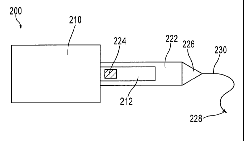
[1024] FIGS. 1 and 2 are schematic illustrations of an implant 100 in a
first configuration
and a second configuration, respectively, according to an embodiment. Implant
100 includes
4
CA 02744203 2016-03-24
55661-4
a support member 110, a strap 112, and a sleeve 122 configured to be
releasably coupled to
the strap 112.
[1025] The support member 110 is configured to be placed within a body of a
patient and
is configured to support a portion of the body. For example, the support
member 110 can be
similar to the grafts disclosed in U.S. Patent Application No. 61/017,257
entitled "Apparatus
and Method for Uterine Preservation," filed on December 28, 2007.
For example, the support member 110 can be a
variety of different shapes, sizes and configurations depending on the
intended use for the
particular implant. In some embodiments, the support member 110 can be
substantially
rectangular, square, oval, or elliptical. The support member 110 can be shaped
and sized to
support a bladder (e.g., to treat a cystocele) and/or a bladder neck and/or
support a uterus
(e.g., to treat a hysterocele) and/or to support a rectum (e.g. to treat a
rectocele).
[1026] The support member 110 can be formed with a mesh material to allow
tissue in-
growth to the implant 100 after implantation. For example, some or all of the
support
member 110 can be formed with a mesh material as described in U.S. Patent Pub.
2005/0038452 Al to Chu.
In some embodiments, some or all of the support member 110 can be formed with
the Advantage Mesh or the PolyformTM Synthetic Mesh material each provided by
Boston
Scientific Corporation ("BSC").
[1027] The strap 112 of the implant 100 is coupled to and extends from the
support
member 110 of the implant 100. The strap 112 is configured to support the
support member
110 of the implant 100 when the strap 112 is inserted into a tissue of the
patient.
[1028] In some embodiments, the strap 112 is formed with the same material
as the
support member. In other embodiments, the strap is formed with a different
material than the
support member. For example, the support member can be formed with a first
biocompatible
material and the strap can be formed with a second biocompatible material
different than the
first biocompatible material. In another example, the support member is formed
with a
biological material, and the strap can be formed with a synthetic material.
The strap and
support member can also have a different weave, pitch, texture, color, and/or
pattern from
each other. In some embodiments, the strap 112 is, for example, a polymer.
=
CA 02744203 2011-05-18
WO 2010/065592 PCT/US2009/066344
[1029] In some embodiments, the strap 112 is formed monolithically with the
support
member 110. In other embodiments, the strap is a separate component coupled to
the support
member. For example, the strap and the support member can be coupled in an
abutting
relationship, an overlapping relationship, or can be bridged. The strap can be
coupled to the
support member by, for example, heat bonding, gluing, using fasteners, and/or
sewing. In
some embodiments, the strap includes a heat seal along its length or a portion
of its length to
help prevent or reduce stretching of the strap.
[1030] In some embodiments the support member 110 and/or the strap 112
include one or
more tanged portions (as described above). The tangs allow the implant 100 to
be anchored
within tissue, such as pelvic tissue, without the use of additional anchoring
mechanisms or
sutures. In some embodiments, an implant 100 includes tangs on an edge along
an entire
length of the implant 100. In other embodiments, the implant 100 includes
tangs covering
substantially all of an exterior surface of the implant. In some embodiments,
tangs are only
on the strap 112 of the implant 100. For example, in some embodiments the
strap 112
includes a tanged portion to engage and/or help secure the implant to pelvic
tissue. Pelvic
tissue can include, for example, ligaments (such as a sacrospinous ligament),
muscle (such as
an obturator internus muscle or an obturator externus muscle), fascia, or any
other structure
or tissue within a pelvic region of a patient.
[1031] As with the support member 110, the strap 112 can have a variety of
different
configurations and/or different sizes (e.g. lengths, widths), depending on the
intended use for
the particular implant and the intended implantation site for the strap 112
within the pelvic
region. For example, the length of the strap 112 can depend on the particular
tissue (e.g.,
ligament, muscle) that the strap 112 is intended to be secured to, such that
trimming of the
strap 112 during or after placement can be reduced or eliminated. For example,
a strap can
have a length such that the strap can be placed through, and/or secured to,
tissue, such as a
sacrospinous ligament, but is not long enough to return back through a vaginal
insertion
point. In some embodiments, the strap 112 has a length such that it extends
from a pelvic
region through an exterior incision of the patient. In other embodiments, the
strap has a
length just sufficient to be secured to a target tissue site. This allows the
implant to be
formed with less material. The use of a strap having a length configured for
the particular use
can thus eliminate the need for trimming and also reduce the costs to
manufacture the
6
CA 02744203 2011-05-18
WO 2010/065592 PCT/US2009/066344
implant. Such embodiments of a strap can also help prevent strap stretch that
can occur
during insertion of the implant due to pulling on a longer length strap.
[1032] While the implant 100 is shown in FIG. 1 having a single strap 112,
in other
embodiments, the implant can have any number of straps depending on the
particular
intended use for the implant. For example, the implant can have between one
and twenty
straps. In some embodiments, one or more straps extend from the support member
at an
angle. Such an angle of a strap can vary in different embodiments, for
example, between 20
and 160 degrees from a centerline of the support member.
[1033] The sleeve 122 of the implant 100 can be made of any suitable
material, such as,
for example, polymer, and is releasably coupled to the strap 112 by a
releasable joint 124.
Because a releasable joint is used to couple the sleeve 122 to the strap 112,
the sleeve 122 is
uncoupled from the strap 112 without using a tool to sever a portion of the
sleeve 122 and/or
strap 112. For example, the releasable joint 124 can be configured to break
and/or release
when a predetermined force is exerted on the releasable joint 124. In some
embodiments, the
releasable joint 124 is frangible and configured to break and/or release when
a force is
exerted on the releasable joint. For example, in some embodiments the
releasable joint 124 is
configured to break and/or release when a force of about 4 lbf to 6 lbf is
exerted on the
releasable joint 124. In other embodiments, the releasable joint is configured
to break and/or
release when a force greater than 6 lbf is exerted on the releasable joint. In
still other
embodiments, the releasable joint is configured to break and/or release when a
force less than
6 lbf is exerted on the releasable joint.
[1034] The releasable joint 124 can include a heat weld, glue, an
interference fit, a
controllably tearable portion, and/or mechanical engagements such as
fasteners. For
example, in some embodiments, a polymer sleeve is heat welded to a polymer
strap. In other
embodiments, the sleeve is coupled to the strap by multiple releasable joints,
such as, for
example, multiple heat welds. This affords greater flexibility to the sleeve
and can minimize
damage to the strap when the releasable joints are broken. In some
embodiments, the sleeve
122 defines a lumen that is configured to receive at least a portion of the
strap 112.
[1035] The sleeve 122 can be used during the insertion of the implant into
a pelvic region
to prevent the strap 112 from prematurely engaging tissue during the delivery
procedure. For
example, if the strap 112 includes a tanged portion, the sleeve 122 can
prevent the tangs from
7
CA 02744203 2011-05-18
WO 2010/065592 PCT/US2009/066344
engaging tissue as the implant is being delivered into the pelvic region.
Conversely, when no
sleeve is coupled to the strap 112, the tangs can engage the surrounding
tissue making it
difficult to smoothly slide and/or adjust the strap 112. The sleeve 122 can
also help in
adjusting the tension of a strap 112, for example, to relieve strap tension.
The sleeve 122 can
also protect the strap 112 from damage during delivery.
[1036] The sleeve 122 can be transparent, semi-transparent, colored, non-
colored, or a
combination thereof The sleeve 122 can be, for example, tapered, flat, and/or
tubular. A
sleeve 122 can be formed for example, with a clear, thin, flexible
biocompatible polymer, and
be configured to allow the user to examine or view the implant 100 (e.g.,
straps) disposed
within the sleeve 122. After the strap 112 is positioned at a desired location
within the pelvic
region, the sleeve 122 can be removed from the strap 112, as described in more
detail below.
[1037] In one embodiment, the sleeve 122 extends beyond the strap 112. The
sleeve 122
can thus be used to provide an extension to the strap 112 to help in the
insertion process. The
sleeve 122 can also help maintain the cleanliness of the strap 112 during
insertion as a portion
of the strap 112 that will be secured within the pelvic region will be
protected within the
sleeve 122. This can also reduce friction between the strap 112 and an
interior surface of the
sleeve 122 (due to reduced surface area contact) allowing easier, removal of
the sleeve 122.
[1038] The implant 100 includes a first configuration (FIG. 1) and a second
configuration
(FIG. 2). The implant 100 is in the first configuration when the sleeve 122 is
coupled to the
strap 112 by the releasable joint 124. The implant 100 is moved from the first
configuration
to the second configuration, by pulling the sleeve 122 with respect to the
strap 112 in the
direction shown by the arrow AA in FIG. 2 while holding the strap 112 in
place. When the
sleeve 122 is pulled with respect to the strap 112, a force is exerted on the
releasable joint
124. When the force exerted is sufficient, the releasable joint 124 will break
and/or release
and the sleeve 122 can be removed from the strap 112. Once the sleeve 122 is
removed from
the strap 112, the implant 100 is in the second configuration.
[1039] In use, the implant 100 is inserted into a body of a patient while
in the first
configuration. In some embodiments, the implant 100 is disposed within the
pelvic region of
the patient. The strap 112 and the sleeve 122 are pulled through a pelvic
tissue, such as the
sacrospinous ligament or arcus tendineus fasciae pelvis. Once the strap 112 is
positioned
within the pelvic tissue, the sleeve is pulled with respect to the support
member 110 in the
8
CA 02744203 2011-05-18
WO 2010/065592 PCT/US2009/066344
direction shown by the arrow AA in FIG. 2 while holding the strap 112 in
place. The strap
112 can be held in place by, for example, a finger, an instrument, or the
pelvic tissue itself.
When the sleeve is pulled, a force sufficient to break and/or release the
releasable joint 124 is
exerted on the releasable joint 124 such that the releasable joint 124 breaks.
The sleeve 122
can then be removed from the strap 112 and the implant 100 moved into the
second
configuration. The strap 112 is left within the pelvic tissue to support the
support member
110 of the implant 100.
[1040] In some embodiments, once the sleeve 122 is removed from the strap
112 and the
strap 112 is disposed within the pelvic tissue, the strap 112 can be further
adjusted such that
the implant 100 adequately supports a portion of the body of the patient. In
other
embodiments, after the strap is disposed within the pelvic tissue, any excess
portions of the
strap can be removed from the strap.
[1041] In some embodiments, an implant can include a dilator, a leader,
and/or a needle
attached to the sleeve to aid in inserting the strap into a tissue. For
example, FIGS. 3 and 4
are schematic illustrations of an implant 200 having a dilator 226, a leader
230, and a needle
228, in a first configuration and a second configuration, respectively,
according to an
embodiment. The implant 200 also has a support member 210, a strap 212, and a
sleeve 222
coupled to the strap 212 by a releasable joint 224. The support member 210,
the strap 212,
the sleeve 222 and the releasable joint 224 are similar to the support member
110, the strap
112, the sleeve 122 and the releasable joint 124, described above.
[1042] As shown in FIG. 3, a dilator 226 is coupled to the sleeve 222 and
used to assist in
the delivery of the implant 200 to the pelvic region. A proximal end portion
(or trailing end)
of a dilator 226 is coupled to the sleeve 222 by, for example, crimping,
knotting, heat
bonding, heat sealing, stitching, stretching, tipping or a combination thereof
In some
embodiments, the sleeve 222 is formed monolithically with the dilator 226. The
dilator 226
is configured to produce a passage through tissue to facilitate strap
placement. Using a
dilator 226 to introduce the first strap 212 into a pelvic region can help
reduce handling or
pulling of the implant 200 itself, thereby reducing or eliminating potential
damage to the
implant 200.
[1043] The dilator 226 can have a variety of different configurations. For
example, the
dilator 226 can be a variety of different lengths, shapes, diameters, etc. The
dilator 226 can
9
CA 02744203 2011-05-18
WO 2010/065592 PCT/US2009/066344
expand a passage formed by a needle 228 (as described below) during insertion
through a
tissue to ease the transition of the opening of the tissue to a cross-section
of the sleeve 222.
The dilator 226 can be flexible, semi rigid, or rigid. The dilator 226 can be
curved or
substantially linear. In some embodiments, the dilator 226 is tubular shaped.
For example,
the dilator 226 can define a lumen therethrough. The dilator 226 can also be
tapered from a
larger diameter at a proximal or trailing end to a smaller diameter at a
distal or leading end of
the dilator 226. The dilator 226 can also be color-coded. For example, when an
implant
having multiple straps is to be delivered to a pelvic region, dilators each
having a unique
color to indicate where each strap is to be placed within a pelvic region can
be coupled to
each strap. Such color-coding can help with the organization of the delivery
process. In
some embodiments, the sleeves associated with the straps can be color-coded in
a similar
manner as described for the dilators. In some embodiments, both the sleeves
and the dilators
are color-coded.
[1044] As shown in FIG. 3, a leader 230 is coupled to a distal end portion
of the dilator
226 and a needle 228 is coupled to a distal end of the leader 230. In some
embodiments, the
leader 230 is a suture. The leader 230 can be formed, for example, with a
polymer. In other
embodiments, the leader is made from metal or other fiber and can be attached
at one or more
locations of a sleeve and/or dilator. The leader 230 is coupled to the dilator
226 and/or sleeve
222 by, for example, gluing, thermo-bonding, knotting or other methods of
attachment. In
some embodiments, the leader 230 is a portion of (or formed monolithically
with) a leader
used to couple the sleeve 222 to a strap 212.
[1045] The needle 228 can be formed with various biocompatible materials,
such as, for
example, stainless steel, or other surgical steel. In some embodiments, the
needle 228 is used
to associate the strap 212 of the implant 200 to a delivery device, such as
those described in
further detail herein.
[1046] A length of the leader 230 (measured from a distal end of the
dilator 226) can
vary. For example, in some embodiments, the length of the leader 230 is
sufficiently long to
be placed through a selected tissue anchoring site (after entering the pelvic
region via a
vaginal incision), and passed out through the vaginal incision, without
requiring the dilator
226 to enter the vagina (e.g., after passing through a tissue within the
pelvic region). In some
embodiments, the length of the leader 230 can allow the physician to remove
the needle 228
from a delivery device external to the body before a dilator 226 is pulled
into the tissue or
CA 02744203 2011-05-18
WO 2010/065592 PCT/US2009/066344
ligament. The insertion and delivery of an implant using a delivery device is
described in
further detail herein.
[1047] In other embodiments, rather than a leader and a needle, the dilator
or sleeve can
include a connector portion that can be used to associate the straps to a
delivery device. For
example, the dilator or sleeve can include a connector portion (not shown). In
some
embodiments, a loop connector is coupled to the sleeve or dilator. Such a
connector or
connector portion can be used to associate the dilator or sleeve to a delivery
device, as
described herein.
[1048] In use, the strap 212 can be pulled through a pelvic tissue using,
for example, the
sleeve 222 and/or the dilator 226 that is configured to dilate or expand the
tissue and provide
a lead-in (e.g., passageway) for the strap 212 to be pulled through the
tissue. The pelvic
tissue is dilated such that the strap 212 can be pulled through the tissue,
but then prolapses or
retracts to a smaller size to provide a frictional interaction between the
tissue and the strap
212. The strap 212 can also be flexible such that even if a width of the strap
212 is greater
than a width of a corresponding passage in the tissue formed by the lead-in
device (e.g.,
dilator or sleeve), the strap 212 can flex to be pulled through the tissue,
and the tissue can
dilate or expand to receive the strap 212. In some embodiments, one or more
straps are
tapered toward their distal end, and are larger in width near the support
portion, which further
provides a lead-in through the tissue.
[1049] Delivery devices can be used to deliver the strap 212 of the implant
200 to and/or
through a pelvic tissue, such as, for example, a levator muscle (e.g., levator
ani muscle), a
sacrospinous ligament, a tendineus arch of levator muscle (also referred to
herein as "arcus
tendineus fasciae pelvis" or "white line"), obturator muscles, an
iliococcygeus muscle, and/or
to other anatomical securement sites within the pelvic region of a patient.
The delivery
device can also be used to pass a suture end through a wall of a vagina or to
pass a suture
through the epithelium of a vaginal wall without passing the suture through
the vaginal wall.
For example, the strap 212 of the implant 200 can be deposited at selected
tissue sites within
the pelvic region and a portion of the implant 200 can also be coupled to a
vagina of the
patient, to a wall of the vagina, secured inside the vagina (e.g., within a
vaginal lumen) or
within the pelvic region.
11
CA 02744203 2016-03-24
55661-4
[10501 The implant 200 can be delivered using a transvaginal approach using
for
example, any device capable of placing and/or securing the implant 200 within
the pelvic
region of a patient. In one embodiment, for example, a Capio Suture Capture
Device
manufactured by BSC is used. An example or such a suturing device is described
in U.S.
patent No. 5,741,277.
Other types of delivery devices can alternatively be used, such as, for
example, the
suturing device described in U.S. Patent Pub. 2004/0181243 Al to Chu et al.,
entitled Re-
shapeable Medical Device.
In such a procedure, the implant 200 is inserted through, for example, a
single
vaginal incision. The incision can be, for example, through the anterior
vaginal mucosa.
[10511 The strap 212 of the implant 200 can alternatively be implanted
using, for
example, a delivery needle, such as an Obtryx Halo, Curve, Advantage or Lynx
device
each manufactured by BSC. An example of such devices is described in U.S.
Patent Pub. No.
2005/0075660 and U.S. Patent Pub. No. 2005/0177022.
[10521 The implant 200 can also be configured to be associated to other
delivery devices
not specifically described herein. In some embodiments, the strap 212 of the
implant 200
itself is configured to be associated to a delivery device. For example, a
connector can be
coupled directly to the strap 212 for association to a delivery device, or the
strap 212 can
include, for example, an opening or hole configured to associate the strap 212
to a delivery
device. In some embodiments, the leader and needle can be coupled directly to
a strap. In
other embodiments, the straps of the implant are delivered to or through a
pelvic tissue
without the use of a delivery device. In such an embodiment, the needles and
the straps are
inserted into the a tissue by hand. In this manner, the straps are secured to
the tissue.
[10531 Although the above-described embodiments describe securing a strap
222 to tissue
Without the use of a separate anchoring device (for example, securing with
tangs of a strap), it
should be understood that the implants described herein can also include
anchors or other
mechanical fasteners to secure one or more straps to the pelvic tissue. For
example, a suture
can be used to secure a strap or other portion of an implant to pelvic tissue.
[10541 In some embodiments, a portion of the support portion 210 is
separately attached
to a tissue within the pelvic region. Said another way, a portion of the
support portion 210
12
CA 02744203 2011-05-18
WO 2010/065592 PCT/US2009/066344
can be secured by means other than the straps. For example, a suture can be
threaded through
the mesh support portion 210 and attached to adjacent pelvic tissue. This can
provide
additional support for the support portion 210.
[1055] Once the strap 212 is positioned within the pelvic tissue, the
sleeve 222, the
dilator 226, the leader 230 and the needle 228 can be removed from the body of
the patient.
This is done by pulling the sleeve with respect to the support member 210 in
the direction
shown by the arrow BB in FIG. 4 while holding the strap 212 in place. The
strap 212 can be
held in place by, for example, a finger, an instrument, or the pelvic tissue
itself When the
sleeve is pulled, a force sufficient to break and/or release the releasable
joint 224 is exerted
on the releasable joint 224 such that the releasable joint 224 breaks. The
sleeve 222 can then
be removed from the strap 212 and the sleeve 222, the dilator 226, the leader
230 and the
needle 228 can be removed from the body of the patient. The strap 212 is left
within the
pelvic tissue to support the support member 210 of the implant 200.
[1056] FIG. 5 shows a top view of an implant 300, according to an
embodiment. Implant
300 includes a support portion 310, a first strap 312, a first sleeve assembly
320 coupled to
the first strap 312, a second strap 314, and a second sleeve assembly 350
coupled to the
second strap 314.
[1057] The support portion 310 of the implant 300 is functionally similar
to the support
portion 110 of the implant 100 described above. Specifically, the support
portion of the
implant is configured to support a portion of a pelvic floor of a patient.
[1058] The first strap 312 and the second strap 314 are also functionally
similar to the
strap 112 of implant 100 described above. The first strap 312 and the second
strap 314 are
configured to support the support portion 310 of the implant 300 when first
strap 312 and the
second strap 314 are disposed within a tissue of a patient.
[1059] FIG. 6 shows a detailed view of the first strap 312 and the first
sleeve assembly
320. The first sleeve assembly 320 includes a sleeve 322, a dilator 326, a
leader 330 and a
needle 328. The sleeve 322 can be made of a material such as a polymer and
defines a
lumen. The sleeve 322 is configured to be coupled to at least a portion of the
first strap 312,
such that the portion of the first strap 312 is disposed within the lumen
defined by the sleeve
322. Similar to the sleeves described above, the sleeve 322 can be used during
the insertion
13
CA 02744203 2011-05-18
WO 2010/065592 PCT/US2009/066344
of the implant 300 into a pelvic region to prevent the first strap 312 from
prematurely
engaging tissue during the delivery procedure.
[1060] The sleeve 322 of the first sleeve assembly 320 is releasably
coupled to the first
strap 312 by a releasable joint 324. Releasable joint 324 is functionally
similar to the
releasable joint 124, described above. The releasable joint 324 is configured
to break and/or
release when a sufficient force is exerted on the releasable joint, such as,
for example, about 4
lbf to 6 lbf. In this manner, the first sleeve assembly 320 can be removed
from the first strap
312 when the first strap 312 is disposed within a tissue of a patient.
Positioning the
releasable joint 324 close to the support portion 310 minimizes the chance
that the first strap
312 will stretch and/or inadvertently uncouple from the support portion 310
when the sleeve
322 of the first sleeve assembly 320 is pulled and a force is exerted on the
releasable joint, as
described above.
[1061] The dilator 326 is coupled to the sleeve, the leader 330 is coupled
to the dilator
326, and the needle 328 is coupled to the leader 330. Similar to the dilator
226, the leader
230 and the needle 228 of the implant 200 described above, the dilator 326,
the leader 330
and the needle 328 are used to help in the insertion of the implant 300 to the
pelvic region of
a patient. In some embodiments, the dilator 326 is thermo bonded to the sleeve
322 of the
first sleeve assembly 320.
[1062] In some embodiments, the first sleeve assembly 320 or a portion of
the first sleeve
assembly 320 is monolithically formed. For example, the dilator 326 and the
leader 330 can
be monolithically formed with the sleeve 322. In such an embodiment, the
needle 328 is
crimped to the leader 330. In other embodiments, the dilator, the leader, and
the needle are
monolithically formed with the sleeve.
[1063] The second sleeve assembly 350 is structurally and functionally
similar to the first
sleeve assembly 320. Additionally, the second sleeve assembly 350 is
associated with the
second strap 314 in a similar fashion as the first sleeve assembly 320 is
associated with the
first strap 312. In other embodiments, the second sleeve assembly is
structurally and/or
functionally different than the first sleeve assembly. For example, the length
of the second
sleeve assembly can be different than the length of the first sleeve assembly
and/or the force
needed to remove the second sleeve assembly from the second strap can be
different than the
force needed to remove the first sleeve assembly from the first strap.
14
CA 02744203 2011-05-18
WO 2010/065592 PCT/US2009/066344
[1064] FIG. 7 shows the implant 300 being inserted into the pelvic region
of a patient.
Specifically, the first strap 312 and the second strap 314 of the implant 300
are inserted into a
first portion of a sacrospinous ligament SSL and a second portion of a
sacrospinous ligament
SSL of the patient, respectively.
[1065] The first strap 312 of the implant 300 is inserted into the first
portion of the
sacrospinous ligament SSL by pulling the needle 328, the leader 330, the
dilator 326, and the
sleeve 322 of the first sleeve assembly 320 through the sacrospinous ligament
SSL. A
delivery device, such as those described above, can be used to aid in
inserting the first strap
312 and the first sleeve assembly 320 into the sacrospinous ligament SSL. Once
the first
strap 312 (still covered by the sleeve 322) is disposed within the
sacrospinous ligament, the
second strap 314 can be inserted into a second portion of the sacrospinous
ligament SSL
using the second sleeve assembly 350, as shown in FIG. 7.
[1066] Once both the first strap 312 and the second strap 314 are disposed
within the
sacrospinous ligament SSL in their respective positions, the first sleeve
assembly 320 and the
second sleeve assembly 350 can be removed from the first strap 312 and the
second strap 314
respectively.
[1067] The first sleeve assembly 320 is removed from the first strap 312 by
retaining the
first strap 312 while pulling the first sleeve assembly 320 in a direction
shown by the arrow
CC in FIG. 7. The first strap 312 can be retained by placing pressure on the
sacrospinous
ligament SSL at a location where the first strap 312 is disposed within the
sacrospinous
ligament SSL between an end of the sleeve 322 of the first sleeve assembly 320
and the
support member 310, such as point A in FIG. 7. This can be done by using a
finger and/or
other medical instrument, such as the shaft of a medical instrument and/or
forceps.
Alternatively, the tissue within which the first strap 312 is disposed can
sufficiently retain the
first strap 312. The pressure applied to point A holds the first strap 312 in
place while the
first sleeve assembly 320 is pulled in the direction shown by the arrow CC in
FIG. 7. This
causes the releasable joint 324 to break. Once the releasable joint 324 is
broken, the first
sleeve assembly 320 can be removed from the first strap 312. After the first
sleeve assembly
320 has been removed from the first strap 312, the second sleeve assembly 350
is removed
from the second strap 314 in a similar manner.
CA 02744203 2011-05-18
WO 2010/065592 PCT/US2009/066344
[1068] Once the first sleeve assembly 320 and the second sleeve assembly
350 are
removed from the first strap 312 and the second strap 314, respectively, the
first strap 312
and the second strap 314 engage the surrounding tissue and support the support
portion 310
in the pelvic region of the patient.
[1069] While the first sleeve assembly 320 is coupled to the first strap
312 by a single
releasable joint 324, FIG. 8 shows a sleeve assembly 420 coupled to a strap
412 by multiple
releasable joints 424. This can be, for example, multiple heat welds. The
multiple releasable
joints 424 can be configured to break and/or release when a force of about 4
lbf to 6 lbf is
applied to the releasable joints 424. Having multiple releasable joints 424
affords greater
flexibility to the sleeve and can minimize damage to the strap when the
releasable joints 424
are broken.
[1070] Similarly, FIG. 9 shows a sleeve assembly 520 coupled to a strap 512
by multiple
releasable joints 524. The releasable joints 524 are positioned with respect
to each other
along a longitudinal axis AL defined by the strap 512. Having multiple
releasable joints 524
positioned along the longitudinal axis AL helps prevent the strap 512 from
stretching and
dislodging from the sleeve assembly 520 during handling and delivery. While
shown in FIG.
9 as having three releasable joints 524, in other embodiments, any number of
releasable joints
can be used to couple the sleeve assembly to the strap.
[1071] While shown in FIG. 5 as having two straps, in other embodiments,
the implant
can have any number of straps. For example, FIG. 10 shows an implant 500
having a support
portion 510 and six straps 512. The implant 500 also includes six sleeve
assemblies 520
configured to be coupled to the six straps 512. The straps 512 and the sleeve
assemblies 520
are structurally and functionally similar to the straps and sleeve assemblies
described above.
Having multiple straps 512 provides additional support to the support portion
510. This
allows the support portion 510 to be larger and to support a larger portion of
the pelvic
region.
[1072] The multiple straps 512 can be inserted into a variety of tissues
within the pelvic
region of a patient. For example, two of the straps 512 can be placed in the
sacrospinous
ligament, two in the arcus tendineus fasciae pelvis and the other two in
another tissue area
within the pelvic region. In such an embodiment, the implant 500 can be
configured to help
support an anterior and/or a posterior portion of a pelvic region. In other
embodiments, the
16
CA 02744203 2011-05-18
WO 2010/065592 PCT/US2009/066344
number of straps and the size and shape of the support member vary depending
on the
application of the implant.
[1073] FIG. 11 is a flow chart of a method 600 of inserting a pelvic
implant into a body
of a patient, according to an embodiment. The method 600 includes inserting an
implant into
a body of a patient through a vaginal incision, at 602. The implant includes a
support portion,
a strap extending from the support portion, and a sleeve coupled to at least a
portion of the
strap. The strap and the sleeve of the implant are then pulled at least
partially through a
tissue such that the strap is disposed at least partially within the first
portion of the tissue, at
604. A releasable joint on the sleeve is then released and/or broken, at 606,
and the sleeve is
removed from the strap, at 608. In one embodiment, the strap is then adjusted
such that the
pelvic implant adequately supports a portion of the body of the patient.
[1074] While various embodiments have been described above, it should be
understood
that they have been presented by way of example only, and not limitation.
Where methods
described above indicate certain events occurring in certain order, the
ordering of certain
events may be modified. Additionally, certain of the events may be performed
concurrently
in a parallel process when possible, as well as performed sequentially as
described above.
[1075] For example, in some embodiments, the sleeve can have a reduced
profile at a
distal end portion, enabling it to more easily travel through the tissue
during delivery. For
example, the sleeve can be tapered.
[1076] In other embodiments, a support portion, a strap, and/or a sleeve
are provided as
separate components. For example, the support portion, the strap, and the
sleeve can be
provided to a user (e.g., a physician) unassembled. The user can then secure
the sleeve to the
strap and/or the strap to the support portion to form an implant.
[1077] Although various embodiments have been described as having
particular features
and/or combinations of components, other embodiments are possible having a
combination of
any features and/or components from any of embodiments where appropriate.
[1078] In some embodiments, an apparatus includes a support member, a strap
extending
from the support member and a sleeve. The support member is configured to
support a
portion of a body of a patient. The strap is configured to be inserted through
at least a portion
of a tissue of the patient. The sleeve is releasably coupled to at least a
portion of the strap by
17
CA 02744203 2011-05-18
WO 2010/065592 PCT/US2009/066344
a releasable joint. The sleeve is configured to be removed from the strap when
at least a
portion of the strap is disposed within the tissue of the patient.
[1079] In some embodiments, the releasable joint is configured to release
when a force of
about 4 lbf to 6 lbf is applied to the releasable joint. In some embodiments,
the releasable
joint includes a single weld. In other embodiments, the releasable joint
includes a plurality of
welds. In some embodiments, the releasable joint includes glue. In some
embodiments, the
releasable joint includes an interference fit. In some embodiments, the
releasable joint
includes a portion that can be controllably torn. In other embodiments, the
releasable joint
includes a fastener.
[1080] In some embodiments, the sleeve is configured to be removed from the
strap in
response to a force applied to the releasable joint. In some embodiments, the
strap is
configured to engage the tissue of the patient when the sleeve is removed from
the strap. In
some embodiments, the strap is configured to help retain the support member at
least partially
adjacent to the portion of the body of the patient when the strap is disposed
within the tissue
of the patient.
[1081] In some embodiments, the strap is a first strap, the sleeve is a
first sleeve and the
apparatus includes a second strap and a second sleeve. The second strap
extends from the
support member and is configured to be inserted through at least a portion of
the tissue of the
patient. The second sleeve is releasably coupled to at least a portion of the
second strap by a
releasable joint. The second sleeve is configured to be removed from the
second strap when
the second strap is secured to the tissue of the patient.
[1082] In some embodiments, the sleeve is directly coupled to the strap by
the releasable
joint. In some embodiments, the sleeve is configured to be removed from the
strap by
applying a force to the strap at a position along the strap between an end of
the sleeve and the
support member and pulling the sleeve in a direction away from the strap.
[1083] In some embodiments, an apparatus includes a sleeve, a dilator
coupled to the
sleeve, and a dart coupled to the dilator. The sleeve is releasably coupled to
a strap of an
implant by a releasable joint. The dilator is configured to dilate a tissue of
a patient when the
dilator is inserted through the tissue. The dart is configured to pierce the
tissue of the patient
when the dart is inserted through the tissue. The releasable joint is
configured to be broken
and the sleeve is configured to be removed from the strap of the implant when
a force is
18
CA 02744203 2011-05-18
WO 2010/065592 PCT/US2009/066344
applied to the strap at a position along the strap and the sleeve is pulled in
a direction away
from the strap.
[1084] In some embodiments, the sleeve is configured to be removed from the
strap in
response to a force applied to the releasable joint. In some embodiments, the
releasable joint
includes a single weld. In other embodiments, the releasable joint includes a
plurality of
welds. In some embodiments, the releasable joint includes glue. In some
embodiments, the
releasable joint includes an interference fit. In some embodiments, the
releasable joint
includes a portion that can be controllably torn. In other embodiments, the
releasable joint
includes a fastener.
[1085] In some embodiments, the sleeve, the dilator, and the dart are
integrally formed.
In some embodiments, the releasable joint is configured to release when a
force of about 4 lbf
to 6 lbf is applied to the releasable joint.
[1086] In some embodiments, a method includes inserting a pelvic implant
into a body of
a patient through a vaginal incision. The pelvic implant includes a support
portion, a strap
extending from the support portion, and a sleeve coupled to at least a portion
of the strap.
The strap and the sleeve are then pulled at least partially through a pelvic
tissue such that the
strap is disposed at least partially within the pelvic tissue. A releasable
joint on the sleeve is
then released and the sleeve removed from the strap.
[1087] In some embodiments the releasing includes applying a force to the
releasable
joint on the sleeve. In some embodiments, the removing includes pulling the
sleeve in a
direction away from the strap.
[1088] In some embodiments, the method further includes adjusting the strap
such that
the pelvic implant adequately supports a portion of the body of the patient.
In some
embodiments, the releasing includes applying a tension to the strap between an
end of the
sleeve and the support portion and pulling the sleeve in a direction away from
the strap.
19