Note: Descriptions are shown in the official language in which they were submitted.
1
ISOFORM SPECIFIC ANTI-HER4 ANTIBODIES
FIELD OF THE INVENTION
The present invention is directed to antibodies useful for detecting and
treating cancer and to
methods of using those antibodies.
BACKGROUND OF THE INVENTION
ErbB/HER receptors form a subfamily of receptor tyrosine kinases that includes
EGFR (also
known as ErbB1 or HER1), HER2 (c-Neu, ErbB2), HER3 (ErbB3), and HER4 (ErbB4).
ErbB
receptors are selectively activated by a number of EGF-like growth factors
leading to cellular
responses, such as cell proliferation, differentiation, migration, or
survival. ErbB receptors consist of a
glycosylated extracellular domain, a single transmernbrane domain, and an
intracellular domain
including a tyrosine kinase enzyme. Ligand binding to the receptor
extracellular domain triggers
receptor dimerization, subsequent activation of the kinase domain, receptor
autophosphorylation, and
multiple downstream signaling cascades (1).
EGFR and HER2 are well-establistied oncogenes and cancer drug targets. They
are
implicated in the pathogenesis of various epithelial and neural malignancies,
and their overactivity is
associated with poor patient outcome (2-4). Targeted therapeutics including
both monoclonal
antibodies and small molecular weight kinase inhibitors blocking the functions
of these receptors have
shown therapeutic effect on patient survival in clinical trials. Cetuximab
(ERBITUXThi, Imclone, Inc.)
is a chimeric monoclonal antibody that blocks ligand binding to EGFR, leading
to a decrease in
receptor dimerization, autophosphorylation and activation of signaling
pathways (5). Cetuximab is
currently approved for clinical use in late-stage chemorefractory colorectal
cancer and locally or
regionally advanced squamous cell carcinoma of the head and neck. Trastuzumab
(HERCEPTINTh4,
Genentech, Inc.) is a humanized monoclonal antibody against the extracellular
domain of HER2
currently used for the treatment of ErbB2-overexpressing breast cancers in
both adjuvant setting and
for advanced disease (6-9).
CA 2744512 2017-10-11
CA 02744512 2011-05-20
WO 2010/068437
PCT/US2009/065712
2
HER4 receptor antagonists have been shown to be useful in controlling
excessive migration
and/or proliferation or smooth muscle cells and, in particular, for the
treatment of stenosis. See United
States Patent No. 7,332,579.
The significance of HER4 in cancer is poorly understood. Some observations
indicate that
HER4 receptor is down-regulated in various cancers, or that its expression is
associated with
favorable prognostic markers, such as estrogen receptor expression (10, 11).
On the other hand, HER4
has been reported to have high expression levels in several cancers such as
thyroid (12), ovarian (13),
and breast cancer (14), as well as medulloblastoma (15), and ependymoma (16).
Furthermore, the
significance of HER4 expression levels for clinical outcome is conflicting
(17). One of the plausible
explanations for these contradictory data is that four structurally and
functionally different isoforms
are generated from a single HER4 gene by alternative splicing (18, 19). These
isoforms have
different tissue distribution profiles and differ in their ability to promote
tumorigcncsis in breast
cancer cell lines (26).
Accordingly, it is desirable to provide a therapeutic that is directed to
specific isoforms of the
HER4 receptor to more precisely treat HER4 mediated disorders.
SUMMARY OF THE INVENTION
One aspect of the invention provides for an isolated anti-HER4 antibody that
specifically
binds to the HER4 JM-a isoform. In one embodiment, the antibody competes for
binding to the JM-a
isoform with the anti-HER4 antibody mAb 1479 produced by the hybridoma cell
line deposited with
ATCC having accession No. PTA-9655. In another embodiment, the antibody binds
to the same
epitope as the epitope to which the monoclonal antibody mAb 1479 produced by
the hybridoma cell
line deposited with the ATCC having accession No. PTA-9655 binds. In some
embodiments, the
antibody is a chimeric, human, or humanized antibody.
In another embodiment, the isolated anti-HER4 antibody that specifically binds
to the HER4
JM-a isoform comprises a fragment from the monoclonal antibody mAb 1479
produced by the
hybridoma cell line deposited with the ATCC having accession No. PTA-9655 that
specifically binds
to the HER4 JM-a isoform. In one embodment, the fragment comprises the
variable region of the
monoclonal antibody mAb 1479. In some embodiments, the antibody is a humanized
antibody. In
some embodiments, the antibody is affinity matured. In one embodiment, the
isolated anti-HER4
antibody comprises the monoclonal antibody mAb 1479 produced by the hybridoma
cell line
deposited with the ATCC having accession No. PTA-9655 or humanized form
thereof
In some embodiments, the isolated anti-HER4 antibody is linked to a cytotoxic
agent.
CA 02744512 2011-05-20
WO 2010/068437
PCT/US2009/065712
3
In some embodiments, the isolated anti-HER4 antibody that specifically binds
to the HER4
JM-a isoform has less cardiotoxicity than an anti-HER4 antibody that is not
specific for the HER4
JM-a isoform.
Another aspect of the invention provides for a method of inhibiting the
proliferation of a
cancer cell that expresses the HER4 JM-a isoform, comprising contacting the
cancer cell with an
therapeutically effective amount of an anti-HER4 antibody that specifically
binds to the HER4 JM-a
isoform. In one embodiment, the anti-HER4 antibody reduces HER4 ectodomain
shedding. In some
embodiments, the cancer cell is a breast cancer, ovarian cancer, or
medulloblastoma cell.
Another aspect of the invention provides for a method of treating cancer in a
patient whose
cancer expresses the HER4 JM-a isoform comprising administering to the patient
a therapeutically
effective amount of an anti-HER4 antibody that specifically binds to the HER4
JM-a isoform. In one
embodiment, the anti-HER4 antibody reduces HER4 ectodomain shedding. In some
embodiments,
the cancer to be treated is breast cancer, ovarian cancer, or medulloblastoma.
Another aspect of the invention provides for a method of treating cancer in a
patient by
selecting a patient whose cancerous cells express the HER4 JM-a isoform and
administering to the
patient a therapeutically effective amount of an anti-HER4 antibody that
specifically binds to the
HER4 JM-a isoform. In one embodiment, the anti-HER4 antibody reduces HER4
ectodomain
shedding. In some embodiments, the cancer to be treated is breast cancer,
ovarian cancer, or
medulloblastoma.
Another aspect of the invention provides for a method of treating cancer in a
patient by
selecting a patient whose cancerous cells comprise increased levels of shed
Her4 ectodomain as
compared with non-cancerous cells of the same tissue type and administering to
the patient a
therapeutically effective amount of an anti-HER4 antibody that specifically
binds to the HER4 JM-a
isoform. In one embodiment, the anti-HER4 antibody reduces HER4 ectodomain
shedding. In some
embodiments, the cancer to be treated is breast cancer, ovarian cancer, or
medulloblastoma.
Another aspect of the invention provides for a method of reducing the risk of
cardiotoxicity
associated with cancer therapy in a patient with cancer comprising
administering to the patient a
therapeutically effective amount of an anti-HER4 antibody that specifically
binds to the HER4 JM-a
isoform. In one embodiment, the patient's cancer overexpresses the HER4 JM-a
isoform. In one
embodiment, the patient's cancer comprises increased levels of shed Her4
ectodomain as compared
with non-cancerous cells of the same tissue type. In some embodiments, the
patient's cancer is breast
cancer, ovarian cancer, or medulloblastoma.
Another aspect of the invention provides for a method of detecting the
presence of shed
HER4 ectodomain in a sample of cells comprising contacting the cells with an
antibody that
specifically binds to the JM-a HER4 isoform.
CA 02744512 2011-05-20
WO 2010/068437
PCT/US2009/065712
4
Yet another aspect of the invention provides for a method of diagnosing a
tumor in a patient
comprising detecting the presence of shed HER4 ectodomain in the tumor. In one
embodiment, the
method of detecting comprises contacting an anti-HER4 antibody that
specifically binds to the JM-a
isoform of HER4 with a sample obtained from the tumor. In one embodiment, the
method comprises
comparing the level of shed HER4 ectodomain in the tumor to the level of shed
HER4 ectodomain in
a non-cancerous control sample.
BRIEF DESCRIPTION OF THE DRAWINGS
Figure 1 is a diagram showing the alternative juxtamembrane domain HER4
isoforms JM-a
(SEQ ID NO:1) and JM-b (SEQ ID NO:2).
Figure 2 is a table describing characteristics of certain HER4 monoclonal
antibodies.
Figure 3 is a Western blot analysis depicting the binding specificity of mAb
1479 and a
control antibody, sc-283, for COS-7 cells expressing HER4 isoforms JM-a CYT-2
or JM-b CYT-1.
Figure 4 is a Western blot analysis depicting the binding specificity of mAb
1479 and control
antibodies anti-EGFR (sc-03), anti-HER2 (sc-284), anti- HER3 (sc-285) for N1H
3T3-7d and NR6
cells stably expressing different ErbB receptors.
Figure 5 is a graph showing the binding specificity of mAb 1479 for NIH 3T3-7d
and NR6
transfectants expressing HER4 JM-a CYT-2, HER4 JM-b CYT-1, EGFR, HER2, or HER3
as
analyzed by a cell enzyme-linked immunosorbent assay (ELISA) (gray columns).
mAb 1479 binding
was compared to binding of anti-EGFR (sc-03), anti-HER2 (sc-284), anti-HER3
(sc-285) and anti-
HER4 (sc-283) (binding of all control antibodies in white columns) in
dilutions 1:1000 (left) or 1:100
(right).
Figure 6 is a Western blot analysis showing binding of mAb 1479 to shed HER4
ectodomain
in human kidney tissue lysate and to recombinant HER4 ectodomain.
Figure 7 shows the results of an in vitro binding assay carried out with a
recombinant HER4
ectodomain and mAb 1479.
Figure 8 is a nonlinear affinity curve of an enzyme-linked immunosorbent assay
(ELISA)
measuring mAb 1479 binding to recombinant HER4 ectodomain.
Figure 9 is a Western blot analysis of 17 paired normal breast (N)/breast
tumor (T) tissue
samples using mAb 1479 to detect ectodomain shedding. Densitometric
quantitation of the expression
levels of 100 kDa HER4 ectodomain (gray) and 150 I(Da full-length HER4 (white)
are shown
underneath each western lane.
CA 02744512 2011-05-20
WO 2010/068437 PCT/US2009/065712
5 Figure 10 is a graph showing the mean expression of HER4 ectodomain and
full-length
receptor in the 17 matched normal and breast tumor tissue pairs quantitated by
densitometry. *, the
fraction of total HER4 present as ectodomain was greater within tumor tissues,
when compared to the
matched normal tissue controls (gray boxes; P<0.05; Wilcoxon signed-rank
test).
Figure 11A is a Western blot analysis showing the effect of mAb 1479 on HER4
tyrosine
phosphorylation of NRG-1 stimulated MCF-7 breast cancer cells. The membrane
was reblotted with
anti-HER4 (Abeam) and anti-actin as controls. Figure 11B is a Western blot
analysis showing the
effect of mAb 1479 on shedding of 100 kDa HER4 ectodomain into the culture
medium of COS-7
transfectants expressing HER4 JM-a CYT-2. Total cell lysates from the same
experiment were
analyzed by western blotting with anti-HER4 (Abeam) and anti-actin.
Figure 12 is a Western blot analysis showing the effect of mAb 1479 on
ubiquitination of
cleavable JM-a isoforms of HER4.
Figure 13 is a Western blot analysis showing effect of mAb 1479 on HER4
expression levels.
Figure 14 is a graph showing the effect of mAb 1479 on proliferation of the
human breast
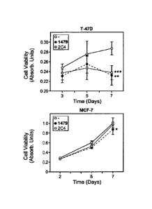
cancer cell lines T-47D and MCF-7 as compared to control antibody 2C4.
Figure 15 is a graph showing the effect of mAb 1479 on anchorage independent
growth of
human breast cancer cell lines T-47D and MCF-7 as compared to control antibody
3g6 as indicated by
a soft agar colony formation assay.
DETAILED DESCRIPTION
DEFINITIONS
Unless defined otherwise, technical and scientific terms used herein have the
same meaning
as commonly understood by one of ordinary skill in the art to which this
invention belongs, and are
consistent with: Singleton et al 1994 Dictionary of Microbiology and Molecular
Biology, 2nd Ed., J.
Wiley & Sons, New York, NY; and Janeway et al 2001 Immunobiology: the immune
system in health
and disease 5th Ed., Garland Publishing, New York.
The term ''HER4 polypeptide" or "HER4 receptor", as used herein, refers,
unless specifically
or contextually indicated otherwise, to any native or variant polypeptide that
is produced from a
HER4 gene disclosed, for example, in European Patent Application No. (EP)
599,274; Plowman at
al., Proc. Natl. Acad. Sci. USA, 90:1746-1750 (1993); and Plowman et al.,
Nature, 366:473-475
(1993). In one embodiment, the HER4 gene is a human HER4 gene. The terms
"HER4" and "ErbB4"
are used interchangeably in the art. The term encompasses naturally occurring
forms, naturally
occurring variant forms (e.g., alternatively spliced forms), naturally
occurring allelic variants, and
CA 02744512 2011-05-20
WO 2010/068437 PCT/US2009/065712
6
may include naturally occurring post-translational modifications such as
glycosylation and GPI
modifications.
The term "wild type" generally refers to a polypeptide comprising the amino
acid sequence of
a naturally occurring HER4 protein.
A "native sequence polypeptide" comprises a polypeptide having the same amino
acid
sequence as the corresponding polypeptide derived from nature. Such native
sequence polypeptides
can be isolated from nature or can be produced by recombinant or synthetic
means. The term "native
sequence polypeptide" specifically encompasses naturally occurring truncated
or secreted forms of the
specific polypeptide (e.g., an extracellular domain sequence), naturally
occurring variant forms (e.g.,
alternatively spliced fonns), naturally occurring allelic variants of the
polypeptide and may include
naturally occurring post-translational modifications such as glycosylation
etc.
The term "amino acid sequence variant" refers to naturally occurring
polypeptide having an
amino acid sequence that differs to some extent from the predominant native
sequence polypeptide.
The amino acid sequence variants possess substitutions, deletions, and/or
insertions at certain
positions within the amino acid sequence.
"Sequence identity" is defined as the percentage of residues in the amino acid
sequence
variant that are identical after aligning the sequences and introducing gaps,
if necessary, to achieve the
maximum percent sequence identity. Methods and computer programs for the
alignment are well
known in the art. One such computer program is "Align 2," authored by
Genentech, Inc., which was
filed with user documentation in the United States Copyright Office,
Washington, DC 20559, on
December 10, 1991, and which code is found in WO 2007/001851.
The "extracellular domain" or "ECD" refers to a form of the polypeptide that
is essentially
free of the transmembrane and cytoplasmic domains. It will be understood that
any transmembrane
domains identified for the polypeptides of the present invention are
identified pursuant to criteria
routinely employed in the art for identifying that type of hydrophobic domain.
The exact boundaries
of a transmembrane domain may vary but most likely by no more than about 5
amino acids at either
end of the domain as initially identified herein.
The term "antibody" herein is used in the broadest sense and specifically
covers monoclonal
antibodies, polyclonal antibodies, dimers, multimers, multispecific antibodies
(e.g., bispecific
antibodies), and antibody fragments, so long as they exhibit the desired
biological activity (Miller et al
2003 Jour. of Immunology 170:4854-4861). Antibodies may be murine, human,
humanized,
chimeric, or derived from other species. An antibody is a protein that is
capable of recognizing and
binding to a specific antigen (Janeway et al 2001 Immunobiology: the immune
system in health and
disease, 5th Ed., Garland Publishing, New York). A target antigen generally
has numerous binding
sites, also called epitopes, recognized by CDRs on multiple antibodies. Each
antibody that
CA 02744512 2011-05-20
WO 2010/068437
PCT/US2009/065712
7
specifically binds to a different epitope has a different structure. Thus, one
antigen may have more
than one corresponding antibody. An antibody includes a full-length
immunoglobulin molecule or an
immunologically active portion of a full-length immunoglobulin molecule, i.e.,
a molecule that
contains an antigen binding site that immunospecifically binds an antigen of a
target of interest or part
thereof, such targets including but not limited to, cancer cell or cells that
produce autoimmune
antibodies associated with an autoimmune disease. The immunoglobulin disclosed
herein can be of
any type (e.g., IgG, IgE, IgM, IgD, and IgA), class (e.g., IgGl, IgG2, IgG3,
IgG4, IgAl and IgA2) or
subclass of immunoglobulin molecule. The immunoglobulins can be derived from
any species such as
human, murine, or rabbit. For the structure and properties of the different
classes of antibodies, see
Basic & Clinical Immunology, 8th edition, Stites and Terr (eds.), Mcgraw-Hill,
Appleton & Lange,
Norwalk, CT, 1994 at Chapter 6.
"Native antibodies" arc usually hcterotctrameric glycoprotcins of about
150,000 Daltons,
composed of two identical light (L) chains (of which there are two types
called kappa and lambda),
and two identical heavy (H) chains. Each light chain is linked to a heavy
chain by one covalent
disulfide bond, while the number of disulfide linkages varies among the heavy
chains of different
immunoglobulin isotypes. Each heavy and light chain also has regularly spaced
intrachain disulfide
bridges. Each heavy chain has at one end a variable domain (VH) followed by a
number of constant
domains. Each light chain has a variable domain at one end (VL) and a constant
domain at its other
end. The constant domain of the light chain is aligned with the first constant
domain of the heavy
chain, and the light-chain variable domain is aligned with the variable domain
of the heavy chain.
Particular amino acid residues are believed to form an interface between the
light chain and heavy
chain variable domains.
A "parent antibody" may comprise a native or wild type sequence. A parent
antibody may be
directed against a target antigen of interest, e.g. a biologically important
polypeptide. Antibodies
directed against nonpolypeptide antigens (such as tumor-associated glycolipid
antigens; see US
5091178) are also contemplated.
An "isolated" antibody is one that has been identified and separated and/or
recovered from a
component of its natural environment. Contaminant components of its natural
environment are
materials that would interfere with diagnostic or therapeutic uses for the
antibody, and may include
enzymes, hormones, and other proteinaceous or nonproteinaceous solutes. In
certain embodiments,
the antibody will be purified (1) to greater than 95% by weight of antibody as
determined by the
Lowry method (2) to a degree sufficient to obtain at least 15 residues of N-
terminal or internal amino
acid sequence by use of a spinning cup scqucnator, or (3) to apparent
homogeneity by SDS-PAGE
under reducing or nonreducing conditions using Coomassie blue or, silver
stain. Isolated antibody
includes the antibody in situ within recombinant cells since at least one
component of the antibody's
CA 02744512 2011-05-20
WO 2010/068437 PCT/US2009/065712
8
natural environment will not be present. Ordinarily, however, isolated
antibody will be prepared by at
least one purification step.
"Antibody fragments" comprise a portion of a full-length antibody, generally
the antigen
binding or variable region thereof Examples of antibody fragments include Fab,
Fab', F(aby),, and Fv
fragments; diabodies; linear antibodies; minibodies (US 5641870, Example 2;
Zapata et al 1995
Protein Eng. 8(10): 1057-1062); Olafsen et al 2004 Protein Eng. Design & Sel.
17(4):315-323),
fragments produced by a Fab expression library, anti-idiotypic (anti-Id)
antibodies, CDR
(complementary determining region), and epitope-binding fragments of any of
the above which
immunospecifically bind to cancer cell antigens, viral antigens or microbial
antigens, single-chain
antibody molecules; and multispecific antibodies formed from antibody
fragments.
The term "monoclonal antibody" as used herein refers to an antibody obtained
from a
population of substantially homogeneous antibodies, i.e., the individual
antibodies comprising the
population are identical except for possible naturally occurring mutations
that may be present in
minor amounts and glycosylation differences. Monoclonal antibodies are highly
specific, being
directed against a single antigenic site. Furthermore, in contrast to
polyclonal antibody preparations
that include different antibodies directed against different determinants
(epitopes), each monoclonal
antibody is directed against a single determinant on the antigen. In addition
to their specificity, the
monoclonal antibodies are advantageous in that they may be synthesized
uncontaminated by other
antibodies. For example, the monoclonal antibodies to be used in accordance
with the present
invention may be made by the hybridoma method first described by Kohler et al
1975 Nature
256:495, or may be made by recombinant DNA methods (see for example: US
4816567; US
5807715). In the hybridoma method, a mouse or other appropriate host animal,
such as a hamster, is
immunized as described above to elicit lymphocytes that produce or are capable
of producing
antibodies that will specifically bind to the protein used for immunization.
Alternatively,
lymphocytes may be immunized in vitro. After immunization, lymphocytes are
isolated and then
fused with a myeloma cell line using a suitable fusing agent, such as
polyethylene glycol, to form a
hybridoma cell (Goding 1986 Monoclonal Antibodies: Principles and Practice,
pp.59-103 Academic
Press). The antibodies may also be isolated from phage antibody libraries
using the techniques
described in Clackson et al 1991 Nature 352:624-628; Marks et al 1991 J. Mol.
Biol. 222:581-597.
The DNA that encodes the antibody may be modified to produce "chimeric or
fusion antibody
polypeptides", for example, by substituting human heavy chain and light chain
constant domain (CH
and CO sequences for the homologous murine sequences (US 4816567; and Morrison
et al 1984 Proc.
Natl. Acad. Sci. USA, 81:6851), or by fusing the immunoglobulin coding
sequence with all or part of
the coding sequence for a non-immunoglobulin polypeptide (heterologous
polypeptide). The non-
immunoglobulin polypeptide sequences can substitute for the constant domains
of an antibody, or
they are substituted for the variable domains of one antigen-combining site of
an antibody to create a
CA 02744512 2011-05-20
WO 2010/068437 PCT/US2009/065712
9
chimeric bivalent antibody comprising one antigen-combining site having
specificity for an antigen
and another antigen-combining site having specificity for a different antigen.
The antibodies herein specifically include "chimeric" antibodies in which a
portion of the
heavy and/or light chain is identical with or homologous to corresponding
sequences in antibodies
derived from a particular species or belonging to a particular antibody class
or subclass, while the
remainder of the chain(s) is identical with or homologous to corresponding
sequences in antibodies
derived from another species or belonging to another antibody class or
subclass, as well as fragments
of such antibodies, so long as they exhibit the desired biological activity
(US 4816567; and Morrison
et al 1984 Proc. Natl. Acad. Sci. USA, 81:6851-6855). Chimeric antibodies of
interest herein include
"primatized" antibodies comprising variable domain antigen-binding sequences
derived from a non-
human primate (e.g., Old World Monkey, Ape etc) and human constant region
sequences.
"Humanized" forms of non-human (e.g., rodent) antibodies are chimeric
antibodies that
contain minimal sequence derived from the non-human antibody. For the most
part, humanized
antibodies are human immunoglobulins (recipient antibody) in which residues
from a hypervariable
region of the recipient are replaced by residues from a hypervariable region
of a non-human species
(donor antibody) such as mouse, rat, rabbit or non-human primate having the
desired antibody
specificity, affinity, and capability. In some instances, framework region
(FR) residues of the human
immunoglobulin are replaced by corresponding non-human residues. Furthermore,
humanized
antibodies may comprise residues that are not found in the recipient antibody
or in the donor antibody.
These modifications are made to further refine antibody performance. In
general, the humanized
antibody will comprise substantially all of at least one, and typically two,
variable domains, in which
all or substantially all of the hypervariable loops correspond to those of a
non-human immunoglobulin
and all, or substantially all, of the FRs are those of a human immunoglobulin
sequence. The
humanized antibody optionally also will comprise at least a portion of an
immunoglobulin constant
region (Fe), typically that of a human immunoglobulin. The Fe fragment
comprises the carboxy-
terminal portions of both H chains held together by disulfides. The effector
functions of antibodies
are determined by sequences in the Fe region, which region is also the part
recognized by Fe receptors
(FcR) found on certain types of cells (Jones et al 1986 Nature 321:522-525;
Riechmann et al 1988
Nature 332:323-329; Presta 1992 Curr. Op. Struct. Biol. 2:593-596; Verhoeyen
et al 1988 Science
239:1534-1536; Sims et al 1993 J. Immunol. 151:2296; Chothia et al 1987 J.
Mol. Biol. 196:901).
Other methods use a particular framework region derived from the consensus
sequence of all human
antibodies of a particular subgroup of light or heavy chains (Carter et al
1992 Proc. Natl. Acad. Sci.
USA, 89:4285; Presta et al 1993 J. Immunol. 151:2623).
A "human antibody" is one which possesses an amino acid sequence which
corresponds to
that of an antibody produced by a human and/or has been made using any of the
techniques for
making human antibodies as disclosed herein. Transgenic animals (e.g., mice)
are available that are
CA 02744512 2011-05-20
WO 2010/068437 PCT/US2009/065712
5 capable, upon immunization, of producing a full repertoire of human
antibodies in the absence of
endogenous immunoglobulin production. For example, it has been described that
the homozygous
deletion of the antibody heavy-chain joining region (JH) gene in chimeric and
germ-line mutant mice
results in complete inhibition of endogenous antibody production. Transfer of
the human germ-line
immunoglobulin gene array into such germ-line mutant mice will result in the
production of human
10 antibodies upon antigen challenge (Jakobovits et al 1993 Proc. Natl.
Acad. Sci. USA, 90:2551;
Jakobovits et al 1993 Nature, 362:255-258; Bruggemann et al 1993 Year in
Immuno. 7:33; US
5545806; US 5569825; US 5591669; US 5545807; and WO 1997/17852.
A "cysteine-engineered" antibody is where one or more amino acids of any form
of wild-type,
murine parent monoclonal antibody, human or humanized antibody are replaced
with a cysteine
amino acid. The engineered cysteine amino acid is a free cysteine acid and not
part of an intrachain or
interchain disulfide unit. The DNA encoding one or more amino acid residues of
the antibody of
interest is modified or ''engineered" such that one or more codons for a
cysteine amino acid is
introduced and thus free cysteine is available on the expressed antibody for
further modification such
a conjugation to a cytotoxic drug.
An -antigen" is a predetermined polypeptide, carbohydrate, nucleic acid,
lipid, hapten or
other naturally occurring or synthetic compound to which an antibody can
selectively bind. The cell
membrane of a cell can present a "cell surface exposed antigen".
An antibody "binds" a molecular target or an antigen of interest when the
binding to that
antigen is with sufficient affinity and specificity that an antibody-antigen
complex is formed that is
useful in targeting the epitopes of the antigen. The epitopes of the antigen
may be exposed on the
surface of cells or may be present on an isolated protein.
The term "specific binding" or "specifically binds to" or is "specific for" a
particular
molecular target or an antigen of interest or an epitope on a particular
molecular target or an antigen
of interest means binding that is measurably different from a non-specific
interaction. Specific
binding can be measured, for example, by determining binding of a molecule
compared to binding of
a control molecule, which generally is a molecule of similar structure that
does not have binding
activity. For example, specific binding can be determined by competition with
a control molecule that
is similar to the target, for example, an excess of non-labeled target. In
this case, specific binding is
indicated if the binding of the labeled target to a probe is competitively
inhibited by excess unlabeled
target. In one embodiment, such terms refer to binding where a molecule binds
to a particular
polypeptide or epitope on a particular polypeptide without substantially
binding to any other
polypeptide or polypeptide epitope. Alternatively, such terms can be described
by a molecule having a
Kd for the target of at least about 10-4 M, 10-5 M, 10-6 M, 10-7 M, 10-8 M, 10-
9 M, 10-10 M, 10-11
M, 10-12 M, or greater.
CA 02744512 2011-05-20
WO 2010/068437
PCT/US2009/065712
11
"Binding affinity" generally refers to the strength of the sum total of
noncovalent interactions
between a single binding site of a molecule (e.g., an antibody) and its
binding partner (e.g., an
antigen). Unless indicated otherwise, as used herein, "binding affinity"
refers to intrinsic binding
affinity which reflects a 1:1 interaction between members of a binding pair
(e.g., antibody and
antigen). The affinity of a molecule X for its partner Y can generally be
represented by the
dissociation constant (Kd). Affinity can be measured by common methods known
in the art,
including those described herein. Low-affinity antibodies generally bind
antigen slowly and tend to
dissociate readily, whereas high-affinity antibodies generally bind antigen
faster and tend to remain
bound longer. A variety of methods of measuring binding affinity are known in
the art, any of which
can be used for purposes of the present invention.
An "antibody-antigen complex" or an "antibody-drug conjugate-antigen complex"
is formed
as a result of specific binding. For example, when the antibody is one that
binds HER4 specifically,
or an isoform of HER4 specifically, it will usually preferentially bind one or
more epitopes found on
the native HER4, or isofonn thereof, and may be an antibody that does not have
significant binding
affinity (e.g. non-specific binding affinity or cross-reactivity), with other
antigens or proteins or other
isoforms of HER4. In such embodiments, the extent of non-specific binding
affinity or cross-reactive
binding to non-HER4, or other isoforms of HER4, will be less than 10%, 5%, 2%,
or 1% as
determined by fluorescence activated cell sorting (FACS) analysis or
radioimmunoprecipitation
(RIA).
An "intact antibody" herein is one comprising VL and VH domains, as well as a
light chain
constant domain (CL) and heavy chain constant domains, CH 1, CH2 and CH3. The
constant domains
may be native sequence constant domains (e.g., human native sequence constant
domains) or amino
acid sequence variant thereof. The intact antibody may have one or more
"effector functions" which
refer to those biological activities attributable to the Fe constant region (a
native sequence Fe region
or amino acid sequence variant Fe region) of an antibody. Examples of antibody
effector functions
include Clq binding; complement dependent cytotoxicity; Fe receptor binding;
antibody-dependent
cell-mediated cytotoxicity (ADCC); phagocytosis; and down regulation of cell
surface receptors such
as B cell receptor and BCR.
The term "variable" refers to the fact that certain portions of the variable
domains differ
extensively in sequence among antibodies and are used in the binding and
specificity of each
particular antibody for its particular antigen. However, the variability is
not evenly distributed
throughout the variable domains of antibodies. It is concentrated in three
segments called
hypervariable regions both in the light chain and the heavy chain variable
domains. The more highly
conserved portions of variable domains are called the framework regions (FRs).
The variable
domains of native heavy and light chains each comprise four FRs, largely
adopting a [3-sheet
configuration, connected by three hypervariable regions, which form loops
connecting, and in some
CA 02744512 2011-05-20
WO 2010/068437 PCT/US2009/065712
12
cases forming part of, the 3-sheet structure. The hypervariable regions in
each chain are held together
in close proximity by the FRs and, with the hypervariable regions from the
other chain, contribute to
the formation of the antigen-binding site of antibodies (see Kabat et al 1991
Sequences of Proteins of
Immunological Interest, 5th Ed. Public Health Service, National Institutes of
Health, Bethesda, MD).
The constant domains are not involved directly in binding an antibody to an
antigen, but exhibit
various effector functions, such as participation of the antibody in antibody
dependent cellular
cytotoxicity (ADCC).
The term "hypervariable region", "HVR", or ''HV", when used herein refers to
the regions of
an antibody variable domain that are hypervariable in sequence and/or form
structurally defined
loops. Generally, antibodies comprise six hypervariable regions; three in the
VH (H1, H2, H3), and
three in the VL (L1, L2, L3). A number of hypervariable region delineations
are in use and are
encompassed herein. The Kabat Complementarity Determining Regions (CDRs) are
based on
sequence variability and are the most commonly used (Kabat et al 1991).
Chothia refers instead to the
location of the structural loops (Chothia and Lesk 1987 J. Mol. Biol. 196:901-
917). The "contact"
hypervariable regions are based on an analysis of the available complex
crystal structures. The
residues from each of these hypervariable regions are noted below. Unless
otherwise denoted, Kabat
numbering according to the Kabat Database of aligned sequences of proteins
will be employed (Wu
and Kabat 1970 J. Exp. Med. 132:211-250; Johnson and Wu 2000 Nuc. Acids Res.
28(1):214-218).
Hypervariable region or "Complementarity Determining Regions" locations are
generally as follows:
amino acids 24-34 (VL CDR-L1), amino acids 49-56 (VL CDR-L2), amino acids 89-
97 (VL CDR-L3),
amino acids 26-35A (VH CDR-H1), amino acids 49-65 (VH CDR-H2), and amino acids
93-102 (VH
CDR-H3). Hypervariable regions may also comprise "extended hypervariable
regions", amino acids
24-36 for the VI CDR-L1 and amino acids 46-56 for the VI CDR-L2. The variable
domain residues
are numbered according to Kabat et al 1991, supra for each of these
definitions. An "altered
hypervariable region" for the purposes herein is a hypervariable region
comprising one or more (e.g.
one to about 16) amino acid substitution(s) therein. An "un-modified
hypervariable region" for the
purposes herein is a hypervariable region having the same amino acid sequence
as a non-human
antibody from which it was derived, i.e. one that lacks one or more amino acid
substitutions therein.
The terms "variable domain residue numbering as in Kabat", "amino acid
position numbering
as in Kabat", and variations thereof, refer to the numbering system used for
heavy chain variable
domains or light chain variable domains of the compilation of antibodies in
Kabat et al 1991 supra).
Using this numbering system, the actual linear amino acid sequence may contain
fewer or additional
amino acids corresponding to a shortening of, or insertion into, a FR or CDR
of the variable domain.
For example, a heavy chain variable domain may include a single amino acid
insert (residue 52a
according to Kabat) after residue 52 of H2 and inserted residues (e.g.
residues 82a, 82b, and 82c, etc
according to Kabat) after heavy chain FR residue 82. The Kabat numbering of
residues may be
CA 02744512 2011-05-20
WO 2010/068437
PCT/US2009/065712
13
determined for a given antibody by alignment at regions of homology of the
sequence of the antibody
with a "standard" Kabat numbered sequence.
"Framework" or "FR" residues are those variable domain residues other than the
hypervariable region residues as herein defined. A "human consensus framework"
is a framework
that represents the most commonly occurring amino acid residue in a selection
of human
immunoglobulin VL or VH framework sequences. Generally, the selection of human
immunoglobulin
VI or VII sequences is from a subgroup of variable domain sequences.
Generally, the subgroup of
sequences is a subgroup as in Kabat et al 1991 Sequences of Proteins of
Immunological Interest, 5th
Ed. Public Health Service, National Institutes of Health, Bethesda, MD). In
one embodiment, for the
VL, the subgroup is subgroup kappa I as in Kabat et al 1991. In one
embodiment, for the VH, the
subgroup is subgroup III as in Kabat et al A "VH subgroup III consensus
framework" comprises the
consensus sequence obtained from the amino acid sequences in variable heavy
subgroup III of Kabat
et al 1991. A "VL subgroup I consensus framework" comprises the consensus
sequence obtained
from the amino acid sequences in variable light kappa subgroup T of Kabat et
al 1991.
An "affinity matured" antibody is one with one or more alterations in one or
more CDRs
thereof which result in an improvement in the affmity of the antibody for
antigen, compared to an
antibody which does not possess those alteration(s). Affinity matured
antibodies will have nanomolar
or even picomolar affinities for the target antigen. Affinity matured
antibodies are produced affinity
maturation by VH and VL domain shuffling (Marks et al 1992 Bio/Technology
10:779-783), or
random mutagenesis of CDR and/or framework residues (Barbas et al 1994 Proc
Nat. Acad. Sci, USA
91:3809-3813; Schier et al 1995 Gene 169:147-155; Yelton et al 1995 J.
Immunol. 155:1994-2004;
Jackson et al 1995 J. Immunol. 154(7):3310-9; and Hawkins et al 1992 J. Mol.
Biol. 226:889-896).
"Fv" is the minimum antibody fragment that contains a complete antigen-
recognition and
antigen-binding site. This region consists of a dimer of one heavy chain and
one light chain variable
domain in tight, non-covalent association. It is in this configuration that
the three hypervariable
regions of each variable domain interact to define an antigen-binding site on
the surface of the VH-VL
dimer. Collectively, the six hypervariable regions confer antigen-binding
specificity to the antibody.
However, even a single variable domain (or half of an Fv comprising only three
hypervariable regions
specific for an antigen) has the ability to recognize and bind antigen,
although at a lower affinity than
the entire binding site.
The Fab fragment also contains the constant domain of the light chain and the
first constant
domain (CH1) of the heavy chain. Fab' fragments differ from Fab fragments by
the addition of a few
residues at the carboxy terminus of the heavy chain CH1 domain including one
or more cysteines
from the antibody hinge region. Fab'-SH is the designation herein for Fab' in
which the cysteine
residue(s) of the constant domains bear at least one free thiol group. F(ab')2
antibody fragments
CA 02744512 2011-05-20
WO 2010/068437
PCT/US2009/065712
14
originally were produced as pairs of Fab' fragments which have hinge cysteines
between them. Other
chemical couplings of antibody fragments are also known.
The "light chains" of antibodies from any vertebrate species can be assigned
to one of two
clearly distinct types, called kappa (x) and lambda (2.), based on the amino
acid sequences of their
constant domains.
"Single-chain Fv" or "scFv" antibody fragments comprise the VH and VL domains
of
antibody, wherein these domains are present in a single polypeptide chain. The
Fv polypeptide
further comprises a polypeptide linker between the VII and VL domains that
enables the scFv to form
the desired structure for antigen binding (Pliickthun 1994 The Pharmacology of
Monoclonal
Antibodies 113, Rosenburg and Moore eds., Springer-Verlag, New York, pp. 269-
315).
The term "diabodies" refers to small antibody fragments with two antigen-
binding sites,
which fragments comprise a VH connected to a VL in the same polypeptide chain
(VH - VL). By using
a linker that is too short to allow pairing between the two domains on the
same chain, the domains are
forced to pair with the complementary domains of another chain and create two
antigen-binding sites
(EP 404,097; WO 1993/11161; Hollinger et al 1993 Proc. Natl. Acad. Sci. USA
90:6444-6448).
The term "antibody-drug conjugate", or "immunoconjugates" comprise an antibody
conjugated to a cytotoxic agent such as a chemotherapeutic agent, a growth
inhibitory agent, a toxin
(e.g., an enzymatically active toxin of bacterial, fungal, plant, or animal
origin, or fragments thereof),
or a radioactive isotope (i.e., a radioconjugate).
A "free cysteine amino acid" refers to a cysteine amino acid residue that has
been engineered
into a parent antibody, has a thiol functional group (-SH), and is not paired
as, or otherwise part of, an
intramolecular or intermolecular disulfide bridge.
The term "thiol reactivity value" is a quantitative characterization of the
reactivity of free
cysteine amino acids. The thiol reactivity value is the percentage of a free
cysteine amino acid in a
cysteine-engineered antibody that reacts with a thiol-reactive reagent, and
converted to a maximum
value of 1. For example, a free cysteine amino acid on a cysteine-engineered
antibody that reacts in
100% yield with a thiol-reactive reagent, such as a biotin-maleimide reagent,
will form a biotin-
labelled antibody that has a thiol reactivity value of 1Ø Another cysteine
amino acid engineered into
the same or different parent antibody that reacts in 80% yield with a thiol-
reactive reagent will have a
thiol reactivity value of 0.8. Another cysteine amino acid engineered into the
same or different parent
antibody that fails totally to react with a thiol-reactive reagent have a
thiol reactivity value of 0.
Determination of the thiol reactivity value of a particular cysteine may be
conducted by ELISA assay,
mass spectroscopy, liquid chromatography, autoradiography, or other
quantitative analytical tests.
Thiol-reactive reagents which allow capture of the cysteine-engineered
antibody and comparison and
quantitation of the cysteine reactivity include biotin-PEO-maleimide ((+)-
biotiny1-3-
CA 02744512 2011-05-20
WO 2010/068437
PCT/US2009/065712
5 maleimidopropionamidy1-3,6-dioxaoctainediamine, Oda et al 2001 Nature
Biotechnology 19:379-382,
Pierce Biotechnology, Inc.) Biotin-BMCC, PEO-Todoacetyl Biotin, Todoacetyl-LC-
Biotin, and Biotin-
HPDP (Pierce Biotechnology, Inc.), and Na,-(3- maleimidylpropionyl)biocytin
(MPB, Molecular
Probes, Eugene, OR). Other commercial sources for biotinylation, bifunctional
and multifunctional
linker reagents include Molecular Probes, Eugene, OR, and Sigma, St. Louis, MO
10 An
antibody or an antibody-drug conjugate is "internalized" when, after forming a
complex
with a cell surface antigen, the antigen-antibody complex or the antigen-
antibody-drug conjugate
complex present on the cell surface membrane is removed from the surface of
the cell and
incorporated into the cell itself via a biochemical reaction. Several possible
post-endocytic trafficking
pathways may thereafter engage the complex. (See reviews Schroeder et al 2001
"Recent advances in
15 membrane microdomains: rafts, caveolae, and intracellular cholesterol
trafficking." Exp Biol. Med
(Maywood) Nov;226(10):873-90), Spooner et al 2006 "Retrograde transport
pathways utilized by
viruses and protein toxins" Virol J. 2006; 3: 26) Antibodies prepared against
denatured protein would
be useful in Western blots, but would not be expected to bind cell surface
epitopes nor form antigen-
antibody complexes and thus would not be internalized.
The terms "Fe receptor" or "FeR" mean a receptor that binds to the Fc constant
region of an
antibody. Moreover, an FcR is one that binds an IgG antibody (a gamma
receptor) and includes
receptors of the FeyRI, Fe7RIT, and Fey RITI subclasses, including allelic
variants and alternatively
spliced forms of these receptors. FcyRII receptors include FcyRITA (an
"activating receptor") and
FcyRTIB (an "inhibiting receptor"), which have similar amino acid sequences
that differ primarily in
the cytoplasmic domains thereof. Activating receptor FeyRIIA contains an
immunoreceptor tyrosine-
based activation motif (ITAM) in its cytoplasmic domain. Inhibiting receptor
FcyRIIB contains an
immunoreceptor tyrosine-based inhibition motif (ITIM) in its cytoplasmic
domain. (See review
Daeron 1997 Annu. Rev. Immunol. 15:203-234). FcRs are reviewed in Ravetch and
Kinet 1991
Annu. Rev. Immunol. 9:457-92; Capel et al 1994 Immunomethods 4:25-34; and de
Haas et al 1995 J.
Lab. Clin. Med. 126:330-41. Other FcRs, including those to be identified in
the future, are
encompassed by the term "FcR" herein. The term also includes the neonatal
receptor, FcRn, which is
responsible for the transfer of maternal IgGs to the fetus (Guyer et al 1976
J. Immunol. 117:587 and
Kim et al 1994 J. Immunol. 24:249).
"Complement dependent eytotoxicity- or "CDC- refers to the lysis of a target
cell in the
presence of complement. Activation of the classical complement pathway is
initiated by the binding
of the first component of the complement system (Clq) to antibodies (of the
appropriate subclass)
which are bound to their cognate antigen (Gazzano-Santoro et al 1996 J.
Immunol. Methods 202:163).
"Antibody-dependent cell-mediated cytotoxicity" and "ADCC" refer to a cell-
mediated
reaction in which nonspecific eytotoxic cells that express Fc receptors (FcRs)
(e.g., Natural Killer
CA 02744512 2011-05-20
WO 2010/068437 PCT/US2009/065712
16
(NK) cells, neutrophils, and macrophages) recognize bound antibody on a target
cell and subsequently
cause lysis of the target cell. The primary cells for mediating ADCC, NK
cells, express FcyRIII only,
whereas monocytes express FcyRI, FcyRII and FcyRIII. FcR expression on
hematopoietic cells in
summarized is Table 3 on page 464 of Ravetch and Kinet 1991 Annu. Rev.
Immunol. 9:457-92. To
assess ADCC activity of a molecule of interest, an in vitro ADCC assay, such
as that described in US
5500362 and US 5821337 may be performed. Useful effector cells for such assays
include peripheral
blood mononuclear cells (PBMC) and Natural Killer (NK) cells. Alternatively,
or additionally,
ADCC activity of the molecule of interest may be assessed in vivo, e.g., in an
animal model such as
that disclosed in Clynes et al 1998 Proc. Nat. Acad. Sci. USA 95:652-656.
"Human effector cells" are leukocytes that express one or more constant region
receptors
(FcRs) and perform effector functions. The cells express at least FcyRIII and
perform ADCC effector
function. Examples of human leukocytes that mediate ADCC include peripheral
blood mononuclear
cells (PBMC), natural killer (NK) cells, monocytes, cytotoxic T cells and
neutrophils. The effector
cells may be isolated from a native source thereof, e.g., from blood or PBMCs.
"Treating" or "treatment" or "alleviation" refers to both therapeutic
treatment and
prophylactic or preventative measures, wherein the object is to prevent or
slow down (lessen) the
targeted pathologic condition or disorder. Those in need of treatment include
those already with the
disorder as well as those prone to have the disorder or those in whom the
disorder is to be prevented.
A subject or mammal is successfully "treated" for a cancer if, after receiving
a therapeutic amount of
an antibody, or antibody-drug conjugate thereof, according to the methods of
the present invention,
the patient shows observable and/or measurable reduction in or absence of one
or more of the
following: reduction in the number of cancer cells or absence of the cancer
cells; reduction in the
tumor size; inhibition (i.e., slow to some extent or stop) of cancer cell
infiltration into peripheral
organs including the spread of cancer into soft tissue and bone; inhibition of
tumor metastasis;
inhibition of tumor growth; and/or relief to some extent, one or more of the
symptoms associated with
the specific cancer; reduced morbidity and mortality, and improvement in
quality of life issues. To
the extent the antibody, or antibody-drug conjugate thereof, may prevent
growth and/or kill existing
cancer cells, it may be cytostatic and/or cytotoxic. The parameters for
assessing successful treatment
and improvement in the disease are readily measurable by routine procedures
familiar to a physician.
For cancer therapy, efficacy can be measured by, for example, assessing the
time to disease
progression (TTP) and/or determining the response rate (RR). Metastasis can be
determined by
staging tests and by bone scan and tests for calcium level and other enzymes
to determine spread to
the bone. CT scans can also be used to look for spread to the pelvis and lymph
nodes in the area.
Chest X-rays and measurement of liver enzyme levels by known methods are used
to look for
metastasis to the lungs and liver, respectively. Other routine methods for
monitoring the disease
include transrectal ultrasonography (TRUS) and transrectal needle biopsy
(TRNB).
CA 02744512 2011-05-20
WO 2010/068437 PCT/US2009/065712
17
The terms "cancer" and "cancerous" refer to or describe the physiological
condition in
mammals that is typically characterized by unregulated cell growth. A "tumor"
comprises one or
more cancerous cells, and refers to all neoplastic cell growth and
proliferation, whether malignant or
benign, and all pre-cancerous and cancerous cells and tissues. Examples of
cancer include, but are not
limited to, carcinoma, lymphoma, blastoma, sarcoma, and leukemia or lymphoid
malignancies. More
particular examples of such cancers include colon cancer, squamous cell cancer
(e.g., epithelial
squamous cell cancer), lung cancer including small- cell lung cancer, non-
small cell lung cancer
("NSCLC"), adenocarcinoma of the lung and squamous carcinoma of the lung,
cancer of the
peritoneum, hepatocellular cancer, gastric or stomach cancer including
gastrointestinal cancer,
pancreatic cancer, glioblastoma, medulloblastoma, cervical cancer, ovarian
cancer, liver cancer,
bladder cancer, hepatoma, breast cancer, rectal cancer, colorectal cancer,
endometrial or uterine
carcinoma, salivary gland carcinoma, kidney or renal cancer, prostate cancer,
vulval cancer, thyroid
cancer, hepatic carcinoma, anal carcinoma, penile carcinoma, head and neck
cancer, and melanoma.
A cancer that "overexpresses" a polypeptide is one that has significantly
higher levels of the
polypeptide at the cell surface thereof, compared to a noncancerous cell of
the same tissue type. Such
overexpression may be caused by increased transcription or translation that in
turn may have been
caused by abnormalities or changes at the genetic level (e.g. DNA mutations or
alterations), mRNA
splice variations, or alterations in the activity of particular genetic
transcription factors, promoters or
enhancers. Overexpression may be determined in a diagnostic or prognostic
assay by evaluating
increased levels of the receptor protein present on the surface of a cell
(e.g., via an
immunohistochemistry assay; IHC). Alternatively, or additionally, one may
measure levels of
receptor-encoding nucleic acid in the cell, e.g., via fluorescent in situ
hybridization (FISH; see WO
1998/45479), southern blotting, or polymerase chain reaction (PCR) techniques,
such as real time
quantitative reverse-transcriptase PCR (qRT-PCR).
The terms "cell proliferative disorder" and "proliferative disorder" refer to
disorders that are
associated with some degree of abnormal cell proliferation. In one embodiment,
the cell proliferative
disorder is cancer.
The term "therapeutically effective amount" refers to an amount of a drug
effective to treat a
disease or disorder in a mammal. In the case of cancer, the therapeutically
effective amount of the
drug may reduce the number of cancer cells; reduce the tumor size; inhibit
(i.e., slow to some extent
or stop) cancer cell infiltration into peripheral organs; inhibit tumor
metastasis; inhibit, to some
extent, tumor growth; and/or relieve to some extent one or more of the
symptoms associated with the
cancer. To the extent the drug may prevent growth and/or kill existing cancer
cells, it may be
cytostatic and/or cytotoxic. The term "cytostatic" refers to the effect of
limiting the function of cells,
such as limiting cellular growth or proliferation of cells. For example in
cancer therapy, efficacy can
CA 02744512 2011-05-20
WO 2010/068437
PCT/US2009/065712
18
be measured by assessing the time to disease progression (TTP) and/or
determining the response rate
(RR).
The term "label" means any moiety which can be covalently attached to an
antibody and that
functions to: (i) provide a detectable signal; (ii) interact with a second
label to modify the detectable
signal provided by the first or second label, e.g. FRET (fluorescence
resonance energy transfer); (iii)
stabilize interactions or increase affinity of binding, with antigen or
ligand; (iv) affect mobility, e.g.
electrophoretic mobility, or cell-permeability, by charge, hydrophobicity,
shape, or other physical
parameters, or (v) provide a capture moiety, to modulate ligand affinity,
antibody/antigen binding, or
ionic complexation.
The phrase "pharmaceutically acceptable salt," as used herein, refers to
pharmaceutically
acceptable organic or inorganic salts of an ADC. Exemplary salts include, but
are not limited, to
sulfate, citrate, acetate, oxalate, chloride, bromide, iodide, nitrate,
bisulfate, phosphate, acid
phosphate, isonicotinate, lactate, salicylate, acid citrate or tartrate salts.
A pharmaceutically
acceptable salt may involve the inclusion of another molecule such as an
acetate ion, a succinate ion
or other counterion. The counterion may be any organic or inorganic moiety
that stabilizes the charge
on the compound. Furthermore, a pharmaceutically acceptable salt may have more
than one charged
atom in its structure. Instances where multiple charged atoms are part of the
pharmaceutically
acceptable salt can have multiple counter ions. Hence, a pharmaceutically
acceptable salt can have
one or more charged atoms and/or one or more counterion.
"Pharmaceutically acceptable solvate" refers to an association of one or more
solvent
molecules and an ADC. Examples of solvents that form pharmaceutically
acceptable solvates
include, but are not limited to, water, isopropanol, ethanol, methanol, DMSO,
ethyl acetate, acetic
acid, and ethanolamine.
"Carriers" as used herein include pharmaceutically acceptable carriers,
excipients, or
stabilizers that are nontoxic to the cell or mammal being exposed thereto at
the dosages and
concentrations employed. Often the physiologically acceptable carrier is an
aqueous pH buffered
solution. Examples of physiologically acceptable carriers include buffers such
as phosphate, citrate,
and other organic acids; antioxidants including ascorbic acid; low molecular
weight (less than about
10 residues) polypeptide; proteins, such as scrum albumin, gelatin, or
immunoglobulins; hydrophilic
polymers such as polyvinylpyrrolidone; amino acids such as glycine, glutamine,
asparagine, arginine
or lysine; monosaccharides, disaccharides, and other carbohydrates including
glucose, mannose, or
dextrins; chelating agents such as EDTA; sugar alcohols such as mannitol or
sorbitol; salt-forming
counterions such as sodium; and/or nonionic surfactants such as TWEEN ,
polyethylene glycol
(PEG), and PLURONICS .
CA 02744512 2011-05-20
WO 2010/068437
PCT/US2009/065712
19
COMPOSITIONS AND METHODS
HER4 Isoforms
Four structurally and functionally different isoforms are generated from a
single HER4 gene
by alternative splicing (18, 19). Two of the isoforms differ in the
intracellular cytoplasmic domain
(isoforms CYT-1 and CYT-2). CYT-1 has a 16 amino acid insert within the
cytoplasmic domain
while CYT-2 has no insert (18). The CYT-1 isoform can mediate coupling to
phosphoinositide 3-
kinase (P13-K), but the CYT-2 isoform cannot (20, 21).
The other two isoforms (JIM-a and JM-b) differ by an insertion of either 23 or
13 alternative
amino acids in the extracellular juxtamembrane region (Figure 1). JM-a is the
isoform with 23 amino
acids within the juxtamembrane region (NGPTSHDCIYYPWTGHSTLPQHA SEQ ID NO:1)
while
JM-lb is the isoform with 13 amino acids in this region (IGSSIEDCIGLMD SEQ ID
NO:2) (17, 23).
The extracellular isoform JM-a can be cleaved by tumor necrosis factor-a-
converting enzyme
(TACE) (22) whereas the JM-b isoform is proteinase-resistant (23). Cleavage by
TACE triggers a
second cleavage of HER4 involving y-secretase activity (24). As a result the
intracellular domain
(ICD) is released from the cell membrane and translocates to the nucleus where
it may function in
regulating gene transcription (25-28).
Consistent with the hypothesis that HER4 isoforms differ in their role in
tumorigenesis, the
cleavable HER4 JM-a CYT-2 isoform, but not its non-cleavable counterpart JM-b
CYT-2,
demonstrates ligand-independent activity and promotes cancer cell growth (26).
In addition,
localization of an intracellular HER4 epitope in the nuclei is associated with
shorter survival when
compared to localization of HER4 at the cell surface (29) suggesting that HER4
cleavage can regulate
tumor progression. These same cleavable isoforms have previously been shown to
be overexpressed
in a clinical series of breast cancer patient samples (18, 26).
Furthermore, the JM-a and JM-b isoforms exhibit different tissue distribution
patterns as well
with the JIM-a isoform being absent from cardiac tissue (23).
Isoform Specific Antibodies
One aspect of the invention provides for an antibody that specifically binds
to the JIM-a
isoform of HER4. In one embodiment, the antibody specifically binds to both
the intact full-length
HER4 receptor comprising the JM-a juxtamembrane region as well as the soluble
HER4 ectodomain.
In another embodiment, the antibody specifically binds to an amino acid
sequence comprising
NGPTSHDCIYYPWTGHSTLPQHA SEQ ID NO: 1. In another embodiment, the antibody
specifically binds to the amino acid sequence NGPTSHDCIYYPWTGHSTLPQHA (SEQ ID
NO:1).
CA 02744512 2011-05-20
WO 2010/068437 PCT/US2009/065712
5 Another aspect of the invention provides for an isolated anti-HER4
antibody that specifically
binds to the HER4 JM-a isoform where the antibody is the mAb 1479 monoclonal
antibody produced
by the hybridoma cell line deposited with the ATCC having accession No. PTA-
9655. In one
embodiment, the antibody is a humanized or affinity matured antibody derived
from the mAb 1479
monoclonal antibody produced by the hybridoma cell line deposited with the
ATCC having accession
10 No. PTA-9655.
Another aspect of the invention provides for an anti-HER4 antibody that
comprises a
fragment from the monoclonal antibody mAb 1479 produced by the hybridoma cell
line deposited
with the ATCC having accession No. PTA-9655. The antibody is specific for the
JM-a isoform of
HER4. In one embodiment, the fragment specifically binds to the HER4 JM-a
isoform. In one
15 embodiment, the fragment from mAb 1479 comprises at least a portion of
the hypervariable region. In
one embodiment, the fragment from mAb 1479 comprises the heavy chain
hypervariable region. In
one embodiment, the fragment from mAb 1479 comprises the light chain
hypervariable region. In
another embodiment, the fragment comprises the light and heavy chain
hypervariable regions of mAb
1479. In one embodiment, the fragment comprises the VH1, VH2, or VH3
hypervariable region. In
20 one embodiment, the fragment comprises at least two of the VH1, VH2, and
VH3 hypervariable
regions. In one embodiment, the fragment comprises all three of the VH1, VH2,
and VH3
hypervariable regions. In one embodiment, the fragment comprises the VL1, VL2,
or VL3
hypervariable region. In one embodiment, the fragment comprises at least two
of the VL1, VL2, and
VL3 hypervariable regions. In one embodiment, the fragment comprises all three
of the VL1, VL2,
and VL3 hypervariable regions. In one embodiment, the fragment comprises at
least one, two, or
three of the VH1, VH2, or VH3 hypervariable region and at least one, two, or
three VL1, VL2, and
VL3 hypervariable regions. In one embodiment, the fragment comprises all three
of the VH1, VH2,
or VH3 hypervariable region and all three of the VL1, VL2, and VL3
hypervariable regions.
In one embodiment, the fragment from mAb 1479 comprises at least a portion of
the variable
region. In one embodiment, the fragment from mAb 1479 comprises the heavy
chain variable region.
In one embodiment, the fragment from mAb 1479 comprises the light chain
variable region. In
another embodiment, the fragment comprises the light and heavy variable region
of mAb 1479.
In some embodiments, the fragment comprises mutations that do not
significantly decrease
the binding specificity of the antibody for the JM-a isoform.
Another aspect of the invention provides for an antibody that competes for
binding to the JM-
a isoform with the anti-HER4 antibody mAb 1479 produced by the hybridoma cell
line deposited with
ATCC having accession No. PTA-9655.
CA 02744512 2011-05-20
WO 2010/068437 PCT/US2009/065712
21
Yet another aspect of the invention provides for an antibody that binds to the
same epitope as
the epitope to which the monoclonal antibody mAb 1479 produced by the
hybridoma cell line
deposited with the ATCC having accession No. PTA-9655 binds.
In one embodiment, the epitope bound by monoclonal antibody mAb 1479 comprises
the
HER4 ectodomain. In another embodiment, the epitope bound by monoclonal
antibody mAb 1479
comprises at least a portion of the amino acid sequence
NGPTSHDCIYYPWTGHSTLPQHA (SEQ
ID NO:1). In another embodiment, the epitope bound by monoclonal antibody mAb
1479 comprises
the amino acid sequence NGPTSHDCIYYPWTGHSTLPQHA (SEQ ID NO:1).
In some embodiments, the JM-a isoform specific antibodies are chimeric, human,
or
humanized antibodies.
In some embodiments, the anti-HER4 antibody that is specific for the JM-a
isoform reduces
HER4 tyrosine phosphorylation. Suppression of receptor tyrosine
phosphorylation has been shown to
be associated with anti-tumor activity of therapeutic antibodies targeting
extracellular domains of
other ErbB receptors (39, 48, 49). The effect of an anti-HER4 antibody on HER4
phosphorylation
can be determined by methods well known in the art, one example of which is
described herein in
Examples 1 and 5. Briefly, cells expressing HER4 are treated with an anti-HER4
antibody then
stimulated with NRG-1. The cells arc lyscd and immunoprecipated with a general
anti-HER4
antibody, such as HFR-1 (R&D, Minneapolis, MN), separated in SDS-PAGE gels,
and analyzed by
Western blotting using an anti-phosphotyrosine antibody, such as 4G10 (Upstate
Biotechnology, Lake
Placid, NY). The blots can be scanned and analyzed by scanning densitometry to
provide a
quantitative analysis. In some embodiments a control is included. In one
embodiment, a control
comprises a sample of cells expressing HER4 stimulated with NRG-1 in the
absence of treatment with
the anti-HER4 antibody. The cells are analyzed by Western blot as with the
treated cells. In one
embodiment, the anti-HER4 antibody reduces HER4 tyrosine phosphorylation by at
least 30%, 40%,
50%, 60%, 70%, 80%, 85%, 90%, 95%, 96%, 97%, 98%, or 99%.
In some embodiments, the anti-HER4 antibody that is specific for the JM-a
isoform reduces
HER4 cleavage. Cleavage of HER4 results in the liberation of a 100 kDa
ectodomain fragment. This
event is also referred to as ectodomain shedding. The effect of an anti-HER4
antibody on HER4
cleavage can be determined by methods well known in the art, one example of
which is described
herein in Examples 1 and 5. Briefly, reduction in HER4 cleavage can be
detected by determining the
presence of the ectodomain in cell culture media of cells expressing HER4
treated with an anti-HER4
antibody. Cleavage can be enhanced by including phorbol 13-myristate 12-
acetate (PMA) in the
assay. In some embodiments a control is included. In one embodiment, a control
is included wherein
the presence of the ectodomain in cell culture media of cells expressing HER4
not treated with an
anti-HER4 antibody is determined. In one embodiment, the anti-HER4 antibody
reduces HER4
CA 02744512 2011-05-20
WO 2010/068437 PCT/US2009/065712
22
cleavage by at least 20%, 30%, 40%, 50%, 60%, 70%, 80%, 85%, 90%, 95%, 96%,
97%, 98%, or
99%.
In some embodiments, the anti-HER4 antibody that is specific for the JM-a
isoform is
internalized. Internalization of the antibody can be used to deliver antibody-
conjugated toxins to
cancerous cells that express HER4 JM-a isoform.
In some embodiments, the anti-HER4 antibody that is specific for the JM-a
isoform promotes
HER4 internalization. Internalization of tyrosine receptor kinases has been
associated with
downregulation of the receptors. In one embodiment, the anti-HER4 antibody
decreases the amount
of HER4 on the cell surface by at least 20%, 30%, 40%, 50%, 60%, 70%, 80%,
85%, 90%, 95%,
96%, 97%, 98%, or 99%. The effect of an anti-HER4 antibody on HER4
internalization can be
determined by methods well known in the art, one example of which is described
herein in Examples
1 and 6.
In some embodiments, the anti-HER4 antibody that specifically binds to the JM-
a isoform of
HER4 has less cardiotoxicity than an anti-HER4 antibody that is not specific
for the HER4 JM-a
isoform. Cardiotoxicity is a side-effect associated with many receptor
tyrosine kinase inhibitor
therapeutics (62, 63). Antibodies that are specific for the JM-a isoform of
HER4 are predicted to
produce less cardiotoxic effects in patients than anti-HER4 antibodies that
recognize the JM-b isoform
because the JM-a isoform is not present in cardiac tissue (23). Cardiotoxicity
in a patient is evidenced
by a number of symptoms including heart failure, Left Ventricular Dysfunction
(LVD), myocardial
ischemia, hypertension, venous thromboembolism, bradycardia, and QT interval
(measure of the time
between the start of the Q wave and the end of the T wave in the heart's
electrical cycle) prolongation.
Cardiotoxicity of a compound can be measured, for example, by using in vitro
or in vivo
diagnostic models.
In vitro determination of cardiotoxicity can be made by exposing cardiac cells
to a test
compound and observing any changes in the cell appearance or cell apopotic
rate. Relevant changes
in cell appearance include mitochondirial swelling and degeneration. Cell
apoptosis can be monitored
by, for example, terminal deoxynucleotidyl transferase-mediated deoxyuri dine
5-triphosphate nick
end labeling. Additionally, the increased secretion of apoptic related
chemicals or enzymes by the
cells is indicative of cardiotoxicity. Such chemicals or enzymes include
troponin, natriuretic peptides
such as N-terminal propeptide of B-type (pro-BNP), cytochrome-C and caspase-9.
Cardiac cells that
can be used as the cultured cell model include primary or cultured adult or
neonatal ventricular
myocytes (cardiomyocytes) obtained from a suitable animal model, such as mouse
or rat. (62-64).
In vivo determination of cardiotoxicity can be performed, for example, by
injecting a test
compound into a suitable animal model, such as a mouse or rat, and observing
the effect of the test
compound on the structure of the model's cardiac tissue, mitochondrial cardiac
appearance and
CA 02744512 2011-05-20
WO 2010/068437 PCT/US2009/065712
23
function, and/or on the model's cardiac tissue apoptosis rates (62). Isolated
heart models, or
Langendorf preps, are also useful for determining cardiotoxicity of compounds
(63).
Cardiotoxicity can also be measured by clinical observations. For example,
heart failure and
LVD arc measured by clinical diagnosis combining patient history and physical
examination with
diagnostic tests such as electrocardiogram (EKG), chest radiography, and
multigated acquisition scan
(MUGUA), Myocardial ischemia is determined by physical examination, detecting
myocardial
necrosis, detecting changes in EGK, and detecting increased elevations in
cardiac enzymes.
Hypertension is determined by measuring the blood pressure of a patient. Those
patients with a blood
pressure of greater or equal to 140/90 mm Hg are generally considered to have
hypertension. Venous
thromboembolism is detected by compression ultrasonography, tomography
angiography, magnetic
resonance pulmonary angiography, or nuclear medicine techniques. Bradycardia
is generally defined
as a heart rate of less than 60 beats per minute and is detected by
determining the heart rate combined
with an EKG or Holter monitor analysis. QT interval prolongation is an
abnormality of the electrical
activity of the heart and can be determined by EKG analysis. In general, a QT
interval of less than or
equal to 440 milliseconds is considered normal while a QT interval of greater
than 450 milliseconds
in men and 470 milliseconds in women is generally considered prolonged. (64).
Antibody Fragments
The present invention encompasses antibody fragments. Antibody fragments may
be
generated by traditional means, such as enzymatic digestion, or by recombinant
techniques. In certain
circumstances there are advantages of using antibody fragments, rather than
whole antibodies. The
smaller size of the fragments allows for rapid clearance, and may lead to
improved access to solid
tumors. For a review of certain antibody fragments, see Hudson et al. (2003)
Nat. Med. 9:129-134.
Various techniques have been developed for the production of antibody
fragments.
Traditionally, these fragments were derived via proteolytic digestion of
intact antibodies (see, e.g.,
Morimoto et al., Journal of Biochemical and Biophysical Methods 24:107-117
(1992); and Brennan et
al., Science, 229:81 (1985)). However, these fragments can now be produced
directly by recombinant
host cells. Fab, Fv and ScFv antibody fragments can all be expressed in and
secreted from E. coli,
thus allowing the facile production of large amounts of these fragments.
Antibody fragments can be
isolated from the antibody phage libraries discussed above. Alternatively,
Fab'-SH fragments can be
directly recovered from E. coli and chemically coupled to form F(ab')2
fragments (Carter et al.,
Bio/Technology 10:163-167 (1992)). According to another approach, F(ab52
fragments can be
isolated directly from recombinant host cell culture. Fab and F(ab')2 fragment
with increased in vivo
half-life comprising salvage receptor binding epitope residues are described
in U.S. Pat. No.
5,869,046. Other techniques for the production of antibody fragments will be
apparent to the skilled
practitioner. In certain embodiments, an antibody is a single chain Fv
fragment (scFv). See WO
CA 02744512 2011-05-20
WO 2010/068437 PCT/US2009/065712
24
93/16185; U.S. Pat. Nos. 5,571,894; and 5,587,458. Fv and scFv are the only
species with intact
combining sites that are devoid of constant regions; thus, they may be
suitable for reduced nonspecific
binding during in vivo use. scFv fusion proteins may be constructed to yield
fusion of an effector
protein at either the amino or the carboxy terminus of an scFv. See Antibody
Engineering, ed.
Borrebaeck, supra. The antibody fragment may also be a "linear antibody",
e.g., as described in U.S.
Pat. No. 5,641,870, for example. Such linear antibodies may be monospecific or
bispecific.
Humanized Antibodies
The invention encompasses humanized antibodies. Various methods for humanizing
non-
human antibodies are known in the art. For example, a humanized antibody can
have one or more
amino acid residues introduced into it from a source which is non-human. These
non-human amino
acid residues are often referred to as "import" residues, which are typically
taken from an "import"
variable domain. Humanization can be essentially performed following the
method of Winter and co-
workers (Jones et al. (1986) Nature 321:522-525; Riechmann et al. (1988)
Nature 332:323-327;
Verhoeyen et al. (1988) Science 239:1534-1536), by substituting hypervariable
region sequences for
the corresponding sequences of a human antibody. Accordingly, such "humanized"
antibodies are
chimeric antibodies (U.S. Patent No. 4,816,567) wherein substantially less
than an intact human
variable domain has been substituted by the corresponding sequence from a non-
human species. In
practice, humanized antibodies are typically human antibodies in which some
hypervariable region
residues and possibly some FR residues are substituted by residues from
analogous sites in rodent
antibodies.
The choice of human variable domains, both light and heavy, to be used in
making the
humanized antibodies can be important to reduce antigenicity. According to the
so-called "best-fit"
method, the sequence of the variable domain of a rodent antibody is screened
against the entire library
of known human variable-domain sequences. The human sequence which is closest
to that of the
rodent is then accepted as the human framework for the humanized antibody.
See, e.g., Sims et al.
(1993) J. Immunol. 151:2296; Chothia et al. (1987) J. Mol. Biol. 196:901.
Another method uses a
particular framework derived from the consensus sequence of all human
antibodies of a particular
subgroup of light or heavy chains. The same framework may be used for several
different humanized
antibodies. See, e.g., Carter et al. (1992) Proc. Natl. Acad. Sci. USA,
89:4285; Presta et al. (1993) J.
Immunol., 151:2623.
It is further generally desirable that antibodies be humanized with retention
of high affinity
for the antigen and other favorable biological properties. To achieve this
goal, according to one
method, humanized antibodies are prepared by a process of analysis of the
parental sequences and
various conceptual humanized products using three-dimensional models of the
parental and
humanized sequences. Three-dimensional immunoglobulin models are commonly
available and are
CA 02744512 2011-05-20
WO 2010/068437 PCT/US2009/065712
5 familiar to those skilled in the art. Computer programs are available
which illustrate and display
probable three-dimensional conformational structures of selected candidate
immunoglobulin
sequences. Inspection of these displays permits analysis of the likely role of
the residues in the
functioning of the candidate immunoglobulin sequence, i.e., the analysis of
residues that influence the
ability of the candidate immunoglobulin to bind its antigen. In this way, FR
residues can be selected
10 and combined from the recipient and import sequences so that the desired
antibody characteristic,
such as increased affinity for the target antigen(s), is achieved. In general,
the hypervariable region
residues are directly and most substantially involved in influencing antigen
binding.
Human Antibodies
Human antibodies of the invention can be constructed by combining Fv clone
variable
15 domain sequence(s) selected from human-derived phage display libraries
with known human constant
domain sequence(s) as described above. Alternatively, human monoclonal
antibodies of the invention
can be made by the hybridoma method. Human myeloma and mouse-human
heteromyeloma cell
lines for the production of human monoclonal antibodies have been described,
for example, by
Kozbor J. Immunol., 133: 3001 (1984); Brodeur et al., Monoclonal Antibody
Production Techniques
20 and Applications, pp. 51-63 (Marcel Dekker, Inc., New York, 1987); and
Boerner et al., J. Immunol.,
147: 86 (1991).
It is now possible to produce transgenic animals (e.g. mice) that are capable,
upon
immunization, of producing a full repertoire of human antibodies in the
absence of endogenous
immunoglobulin production. For example, it has been described that the
homozygous deletion of the
25 antibody heavy-chain joining region (JH) gene in chimeric and germ-line
mutant mice results in
complete inhibition of endogenous antibody production. Transfer of the human
germ-line
immunoglobulin gene array in such germ-line mutant mice will result in the
production of human
antibodies upon antigen challenge. See, e.g., Jakobovits et al., Proc. Natl.
Acad. Sci USA, 90: 2551
(1993); Jakobovits et al., Nature, 362: 255 (1993); Bruggermann et al., Year
in Immunol., 7: 33
(1993).
Gene shuffling can also be used to derive human antibodies from non-human,
e.g. rodent,
antibodies, where the human antibody has similar affinities and specificities
to the starting non-human
antibody. According to this method, which is also called "epitope imprinting",
either the heavy or
light chain variable region of a non-human antibody fragment obtained by phage
display techniques
as described herein is replaced with a repertoire of human V domain genes,
creating a population of
non-human chain/human chain scFv or Fab chimeras. Selection with antigen
results in isolation of a
non-human chain/human chain chimeric scFv or Fab wherein the human chain
restores the antigen
binding site destroyed upon removal of the corresponding non-human chain in
the primary phage
display clone, i.e. the epitope governs (imprints) the choice of the human
chain partner. When the
CA 02744512 2011-05-20
WO 2010/068437 PCT/US2009/065712
26
process is repeated in order to replace the remaining non-human chain, a human
antibody is obtained
(see PCT WO 93/06213 published April 1, 1993). Unlike traditional humanization
of non-human
antibodies by CDR grafting, this technique provides completely human
antibodies, which have no FR
or CDR residues of non-human origin.
Bispecific Antibodies
Bispecific antibodies are monoclonal antibodies that have binding
specificities for at least two
different antigens. In certain embodiments, bispecific antibodies are human or
humanized antibodies.
In certain embodiments, one of the binding specificities is for the HER4 7M-a
isoform and the other is
for any other antigen. In certain embodiments, bispecific antibodies may bind
to two different
epitopes of the HER4 7M-a isoform. Bispecific antibodies may also be used to
localize cytotoxic
agents to cells which express the HER4 JM-a isoform. These antibodies possess
a the HER4 JM-a
isoform-binding arm and an arm which binds a cytotoxic agent, such as, e.g.,
saporin, anti-interferon-
a, vinca alkaloid, ricin A chain, methotrexate or radioactive isotope hapten.
Bispecific antibodies can
be prepared as full length antibodies or antibody fragments (e.g. F(ab')2
bispecific antibodies).
Methods for making bispecific antibodies are known in the art. Traditionally,
the
recombinant production of bispecific antibodies is based on the co-expression
of two immunoglobulin
heavy chain-light chain pairs, where the two heavy chains have different
specificities (Milstein and
Cuello, Nature, 305: 537 (1983)). Because of the random assortment of
immunoglobulin heavy and
light chains, these hybridomas (quadromas) produce a potential mixture of 10
different antibody
molecules, of which only one has the correct bispecific structure. The
purification of the correct
molecule, which is usually done by affinity chromatography steps, is rather
cumbersome, and the
product yields are low. Similar procedures are disclosed in WO 93/08829
published May 13, 1993,
and in Traunecker et al., EMBO J., 10: 3655 (1991).
According to a different approach, antibody variable domains with the desired
binding
specificities (antibody-antigen combining sites) are fused to immunoglobulin
constant domain
sequences. The fusion, for example, is with an immunoglobulin heavy chain
constant domain,
comprising at least part of the hinge, CH2, and CH3 regions. In certain
embodiments, the first heavy-
chain constant region (CH1), containing the site necessary for light chain
binding, is present in at least
one of the fusions. DNAs encoding the immunoglobulin heavy chain fusions and,
if desired, the
immunoglobulin light chain, are inserted into separate expression vectors, and
are co-transfected into
a suitable host organism. This provides for great flexibility in adjusting the
mutual proportions of the
three polypeptide fragments in embodiments when unequal ratios of the three
polypeptide chains used
in the construction provide the optimum yields. It is, however, possible to
insert the coding sequences
for two or all three polypeptide chains in one expression vector when the
expression of at least two
CA 02744512 2011-05-20
WO 2010/068437 PCT/US2009/065712
27
polypeptide chains in equal ratios results in high yields or when the ratios
are of no particular
significance.
In one embodiment of this approach, the bispecific antibodies are composed of
a hybrid
immunoglobulin heavy chain with a first binding specificity in one arm, and a
hybrid immunoglobulin
heavy chain-light chain pair (providing a second binding specificity) in the
other arm. It was found
that this asymmetric structure facilitates the separation of the desired
bispecific compound from
unwanted immunoglobulin chain combinations, as the presence of an
immunoglobulin light chain in
only one half of the bispecific molecule provides for a facile way of
separation. This approach is
disclosed in WO 94/04690. For further details of generating bispecific
antibodies see, for example,
Suresh et al., Methods in Enzymology, 121:210 (1986).
According to another approach, the interface between a pair of antibody
molecules can be
engineered to maximize the percentage of heterodimers which are recovered from
recombinant cell
culture. The interface comprises at least a part of the CH3 domain of an
antibody constant domain. In
this method, one or more small amino acid side chains from the interface of
the first antibody
molecule are replaced with larger side chains (e.g. tyrosine or tryptophan).
Compensatory "cavities"
of identical or similar size to the large side chain(s) are created on the
interface of the second antibody
molecule by replacing large amino acid side chains with smaller ones (e.g.
alanine or threonine). This
provides a mechanism for increasing the yield of the heterodimer over other
unwanted end-products
such as homodimers.
Bispecific antibodies include cross-linked or ''heteroconjugate" antibodies.
For example, one
of the antibodies in the heteroconjugate can be coupled to avidin, the other
to biotin. Such antibodies
have, for example, been proposed to target immune system cells to unwanted
cells (US Patent No.
4,676,980), and for treatment of HIV infection (WO 91/00360, WO 92/00373, and
EP 03089).
Heteroconjugate antibodies may be made using any convenient cross-linking
method. Suitable cross-
linking agents are well known in the art, and are disclosed in US Patent No.
4,676,980, along with a
number of cross-linking techniques.
Techniques for generating bispecific antibodies from antibody fragments have
also been
described in the literature. For example, bispecific antibodies can be
prepared using chemical linkage.
Brennan et al., Science, 229: 81(1985) describe a procedure wherein intact
antibodies are
proteolytically cleaved to generate F(ab')2 fragments. These fragments are
reduced in the presence of
the dithiol complexing agent sodium arsenite to stabilize vicinal dithiols and
prevent intermolecular
disulfide formation. The Fab' fragments generated are then converted to
thionitrobenzoate (TNB)
derivatives. One of the Fab'-TNB derivatives is then reconverted to the Fab'-
thiol by reduction with
mercaptoethylamine and is mixed with an equimolar amount of the other Fab'-TNB
derivative to form
CA 02744512 2011-05-20
WO 2010/068437 PCT/US2009/065712
28
the bispecific antibody. The bispecific antibodies produced can be used as
agents for the selective
immobilization of enzymes.
Recent progress has facilitated the direct recovery of Fab'-SH fragments from
E. coli, which
can be chemically coupled to form bispecific antibodies. Shalaby et al., J.
Exp. Med., 175: 217-225
(1992) describe the production of a fully humanized bispecific antibody
F(ab')2 molecule. Each Fab'
fragment was separately secreted from E. coli and subjected to directed
chemical coupling in vitro to
form the bispecific antibody. The bispecific antibody thus formed was able to
bind to cells
overexpressing the HER2 receptor and normal human T cells, as well as trigger
the lytic activity of
human cytotoxic lymphocytes against human breast tumor targets.
Various techniques for making and isolating bispecific antibody fragments
directly from
recombinant cell culture have also been described. For example, bispecific
antibodies have been
produced using leucine zippers. Kostelny et al., J. Immunol., 148(5):1547-1553
(1992). The leucine
zipper peptides from the Fos and Jun proteins were linked to the Fab' portions
of two different
antibodies by gene fusion. The antibody homodimers were reduced at the hinge
region to form
monomers and then re-oxidized to form the antibody heterodimers. This method
can also be utilized
for the production of antibody homodimers. The "diabody" technology described
by Hollinger et al.,
Proc. Natl. Acad. Sci. USA, 90:6444-6448 (1993) has provided an alternative
mechanism for making
bispecific antibody fragments. The fragments comprise a heavy-chain variable
domain (VH)
connected to a light-chain variable domain (VL) by a linker which is too short
to allow pairing
between the two domains on the same chain. Accordingly, the VH and VL domains
of one fragment
are forced to pair with the complementary VL and VH domains of another
fragment, thereby forming
two antigen-binding sites. Another strategy for making bispecific antibody
fragments by the use of
single-chain Fv (sFy) dimers has also been reported. See Gruber et al., J.
Immunol., 152:5368 (1994).
Antibodies with more than two valencies are contemplated. For example,
trispecific
antibodies can be prepared. Tutt et al. J. Immunol. 147: 60 (1991).
Multivalent Antibodies
A multivalent antibody may be internalized (and/or catabolized) faster than a
bivalent
antibody by a cell expressing an antigen to which the antibodies bind. The
antibodies of the present
invention can be multivalent antibodies (which are other than of the IgM
class) with three or more
antigen binding sites (e.g. tetravalent antibodies), which can be readily
produced by recombinant
expression of nucleic acid encoding the polypeptide chains of the antibody.
The multivalent antibody
can comprise a dimerization domain and three or more antigen binding sites. In
certain embodiments,
the dimerization domain comprises (or consists of) an Fe region or a hinge
region. In this scenario, the
antibody will comprise an Fe region and three or more antigen binding sites
amino-terminal to the Fc
region. In certain embodiments, a multivalent antibody comprises (or consists
of) three to about eight
CA 02744512 2011-05-20
WO 2010/068437 PCT/US2009/065712
29
antigen binding sites. In one such embodiment, a multivalent antibody
comprises (or consists of) four
antigen binding sites. The multivalent antibody comprises at least one
polypeptide chain (for example,
two polypeptide chains), wherein the polypeptide chain(s) comprise two or more
variable domains.
For instance, the polypeptide chain(s) may comprise VD1-(X1)n -VD2-(X2)n -Fe,
wherein VD1 is a
first variable domain, VD2 is a second variable domain, Fe is one polypeptide
chain of an Fe region,
X1 and X2 represent an amino acid or polypeptide, and n is 0 or 1. For
instance, the polypeptide
chain(s) may comprise: VH-CH1-flexible linker-VH-CH1-Fc region chain; or VH-
CH1-VH-CH1-Fc
region chain. The multivalent antibody herein may further comprise at least
two (for example, four)
light chain variable domain polypeptides. The multivalent antibody herein may,
for instance, comprise
from about two to about eight light chain variable domain polypeptides. The
light chain variable
domain polypeptides contemplated here comprise a light chain variable domain
and, optionally,
further comprise a CL domain.
Single-Domain Antibodies
In some embodiments, an antibody of the invention is a single-domain antibody.
A single-
domain antibody is a single polypeptide chain comprising all or a portion of
the heavy chain variable
domain or all or a portion of the light chain variable domain of an antibody.
In certain embodiments,
a single-domain antibody is a human single-domain antibody (Domantis, Inc.,
Waltham, MA; see,
e.g., U.S. Patent No. 6,248,516 B1). In one embodiment, a single-domain
antibody consists of all or a
portion of the heavy chain variable domain of an antibody.
Antibody Variants
In some embodiments, amino acid sequence modification(s) of the antibodies
described
herein are contemplated. For example, it may be desirable to improve the
binding affinity and/or
other biological properties of the antibody. Amino acid sequence variants of
the antibody may be
prepared by introducing appropriate changes into the nucleotide sequence
encoding the antibody, or
by peptide synthesis. Such modifications include, for example, deletions from,
and/or insertions into
and/or substitutions of, residues within the amino acid sequences of the
antibody. Any combination
of deletion, insertion, and substitution can be made to arrive at the final
construct, provided that the
final construct possesses the desired characteristics. The amino acid
alterations may be introduced in
the subject antibody amino acid sequence at the time that sequence is made.
A useful method for identification of certain residues or regions of the
antibody that are
preferred locations for mutagenesis is called ''alanine scanning mutagenesis"
as described by
Cunningham and Wells (1989) Science, 244:1081-1085. Here, a residue or group
of target residues
are identified (e.g., charged residues such as arg, asp, his, lys, and glu)
and replaced by a neutral or
negatively charged amino acid (e.g., alanine or polyalanine) to affect the
interaction of the amino
acids with antigen. Those amino acid locations demonstrating functional
sensitivity to the
CA 02744512 2011-05-20
WO 2010/068437 PCT/US2009/065712
5 substitutions then are refined by introducing further or other variants
at, or for, the sites of
substitution. Thus, while the site for introducing an amino acid sequence
variation is predetermined,
the nature of the mutation per se need not be predetermined. For example, to
analyze the performance
of a mutation at a given site, ala scanning or random mutagenesis is conducted
at the target codon or
region and the expressed immunoglobulins are screened for the desired
activity.
10 Amino acid sequence insertions include amino- and/or carboxyl-terminal
fusions ranging in
length from one residue to polypeptides containing a hundred or more residues,
as well as
intrasequence insertions of single or multiple amino acid residues. Examples
of terminal insertions
include an antibody with an N-terminal methionyl residue. Other insertional
variants of the antibody
molecule include the fusion to the N- or C-terminus of the antibody to an
enzyme (e.g. for ADEPT) or
15 a polypeptide which increases the serum half-life of the antibody.
In certain embodiments, an antibody of the invention is altered to increase or
decrease the
extent to which the antibody is glycosylated. Glycosylation of polypeptides is
typically either N-
linked or 0-linked. N-linked refers to the attachment of a carbohydrate moiety
to the side chain of an
asparagine residue. The tripeptide sequences asparagine-X-serine and
asparagine-X-threonine, where
20 X is any amino acid except proline, are the recognition sequences for
enzymatic attachment of the
carbohydrate moiety to the asparagine side chain. Thus, the presence of either
of these tripeptide
sequences in a polypeptide creates a potential glycosylation site. 0-linked
glycosylation refers to the
attachment of one of the sugars N-aceylgalactosamine, galactose, or xylose to
a hydroxyamino acid,
most commonly serine or threonine, although 5-hydroxyproline or 5-
hydroxylysine may also be used.
25 Addition or deletion of glycosylation sites to the antibody is
conveniently accomplished by
altering the amino acid sequence such that one or more of the above-described
tripeptide sequences
(for N-linked glycosylation sites) is created or removed. The alteration may
also be made by the
addition, deletion, or substitution of one or more senile or threonine
residues to the sequence of the
original antibody (for 0-linked glycosylation sites).
30 Where the antibody comprises an Fc region, the carbohydrate attached
thereto may be altered.
Native antibodies produced by mammalian cells typically comprise a branched,
biantennary
oligosaccharide that is generally attached by an N-linkage to Asn297 of the
CH2 domain of the Fc
region. See, e.g., Wright et al. (1997) TIBTECH 15:26-32. The oligosaccharide
may include various
carbohydrates, e.g., mannose, N-acetyl glucosamine (G1cNAc), galactose, and
sialic acid, as well as a
fucose attached to a GlcNAc in the "stem" of the biantennary oligosaccharide
structure. in some
embodiments, modifications of the oligosaccharide in an antibody of the
invention may be made in
order to create antibody variants with certain improved properties.
For example, antibody variants are provided having a carbohydrate structure
that lacks fucose
attached (directly or indirectly) to an Fc region. Such variants may have
improved ADCC function.
CA 02744512 2011-05-20
WO 2010/068437
PCT/US2009/065712
31
See, e.g., US Patent Publication Nos. US 2003/0157108 (Presta, L.); US
2004/0093621 (Kyowa
Hatch) Kogyo Co., Ltd). Examples of publications related to "defucosylated" or
"fucose-deficient"
antibody variants include: US 2003/0157108; WO 2000/61739; WO 2001/29246; US
2003/0115614;
US 2002/0164328; US 2004/0093621; US 2004/0132140; US 2004/0110704; US
2004/0110282; US
2004/0109865; WO 2003/085119; WO 2003/084570; WO 2005/035586; WO 2005/035778;
W02005/053742; W02002/031140; Okazaki et al. J. Mol. Biol. 336:1239-1249
(2004); Yamane-
Ohnuki et al. Biotech. Bioeng. 87: 614 (2004). Examples of cell lines capable
of producing
defucosylated antibodies include Lec13 CHO cells deficient in protein
fucosylation (Ripka et al. Arch.
Biochem. Biophys. 249:533-545 (1986); US Pat Appl No US 2003/0157108 Al,
Presta, L; and WO
2004/056312 Al, Adams et al., especially at Example 11), and knockout cell
lines, such as alpha-1,6-
fucosyltransferase gene, FUT8, knockout CHO cells (see, e.g., Yamane-Ohnuki et
al. Biotech.
Bioeng. 87: 614 (2004); Kanda, Y. et al., Biotechnol. Bioeng., 94(4):680-688
(2006); and
W02003/085107).
Antibodies variants are further provided with bisected oligosaccharides, e.g.,
in which a
biantennary oligosaccharide attached to the Fc region of the antibody is
bisected by GlcNAc. Such
antibody variants may have reduced fucosylation and/or improved ADCC function.
Examples of such
antibody variants are described, e.g., in WO 2003/011878 (Jean-Mairet et al.);
US Patent No.
6,602,684 (Umana et al.); and US 2005/0123546 (Umana et al.). Antibody
variants with at least one
galactose residue in the oligosaccharide attached to the Fc region are also
provided. Such antibody
variants may have improved CDC function. Such antibody variants are described,
e.g., in WO
1997/30087 (Patel et al.); WO 1998/58964 (Raju, S.); and WO 1999/22764 (Raju,
S.).
In certain embodiments, an antibody variant comprises an Fc region with one or
more amino
acid substitutions which further improve ADCC, for example, substitutions at
positions 298, 333,
and/or 334 of the Fc region (Eu numbering of residues). Such substitutions may
occur in combination
with any of the variations described above.
In certain embodiments, the invention contemplates an antibody variant that
possesses some
but not all effector functions, which make it a desirable candidate for many
applications in which the
half life of the antibody in vivo is important yet certain effector functions
(such as complement and
ADCC) are unnecessary or deleterious. In certain embodiments, the Fc
activities of the antibody are
measured to ensure that only the desired properties are maintained. In vitro
and/or in vivo
cytotoxicity assays can be conducted to confirm the reduction/depletion of CDC
and/or ADCC
activities. For example, Fc receptor (FcR) binding assays can be conducted to
ensure that the
antibody lacks FcyR binding (hence likely lacking ADCC activity), but retains
FcRn binding ability.
The primary cells for mediating ADCC, NK cells, express FcR111 only, whereas
monocytes express
FcRI, FcRII and FcRIII. FcR expression on hematopoietic cells is summarized in
Table 3 on page
464 of Ravetch and Kinet, Annu. Rev. In-munol. 9:457-92 (1991). Non-limiting
examples of in vitro
CA 02744512 2011-05-20
WO 2010/068437 PCT/US2009/065712
32
assays to assess ADCC activity of a molecule of interest is described in U.S.
Patent No. 5,500,362
(see, e.g. Hellstrom, I., et al. Proc. Nat'l Acad. Sci. USA 83:7059-7063
(1986)) and Hellstrom, I et al.,
Proc. Nat'l Acad. Sci. USA 82:1499-1502 (1985); 5,821,337 (see Bruggemann, M.
et al., J. Exp. Med.
166:1351-1361 (1987)). Alternatively, non-radioactive assays methods may be
employed (see, for
example, ACTITm non-radioactive cytotoxicity assay for flow cytometry
(CellTechnology, Inc.
Mountain View, CA; and CytoTox 96 non-radioactive cytotoxicity assay
(Promega, Madison, WI).
Useful effector cells for such assays include peripheral blood mononuclear
cells (PBMC) and Natural
Killer (NK) cells. Alternatively, or additionally, ADCC activity of the
molecule of interest may be
assessed in vivo, e.g., in a animal model such as that disclosed in Clynes et
al. Proc. Nat'l Acad. Sci.
USA 95:652-656 (1998). Clq binding assays may also be carried out to confirm
that the antibody is
unable to bind Clq and hence lacks CDC activity. To assess complement
activation, a CDC assay
may be performed (see, for example, Gazzano-Santoro et al., J. Immunol.
Methods 202:163 (1996);
Cragg, M.S. et al., Blood 101:1045-1052 (2003); and Cragg, M.S. and M.J.
Glennie, Blood 103:2738-
2743 (2004)). FcRn binding and in vivo clearance/half life determinations can
also be performed
using methods known in the art (see, for example, Petkova, S.B. et al., Int'l.
Immunol. 18(12):1759-
1769 (2006)).
Other antibody variants having one or more amino acid substitutions are
provided. Sites of
interest for substitutional mutagenesis include the hypervariable regions, but
FR alterations are also
contemplated. Conservative substitutions are shown in Table 1 under the
heading of "preferred
substitutions." More substantial changes, denominated "exemplary
substitutions" are provided in
Table 1, or as further described below in reference to amino acid classes.
Amino acid substitutions
may be introduced into an antibody of interest and the products screened,
e.g., for a desired activity,
such as improved antigen binding, decreased immunogenicity, improved ADCC or
CDC, etc.
TABLE 1
Original Exemplary Preferred
Residue Substitutions Substitutions
Ala (A) Val; Leu; Ile Val
Arg (R) Lys; Gln; Asn Lys
Asn (N) Gln; His; Asp, Lys; Arg Gln
Asp (D) Glu; Asn Glu
Cys (C) Ser; Ala Ser
Gln (Q) Asn; Glu Asn
Glu (E) Asp; Gln Asp
Gly (G) Ala Ala
His (H) Asn; Gln; Lys; Arg Arg
CA 02744512 2011-05-20
WO 2010/068437
PCT/US2009/065712
33
Original Exemplary Preferred
Residue Substitutions Substitutions
Ile (I) Leu; Val; Met; Ala; Leu
Pile; Norleucine
Leu (L) Norleucine; Ile; Val; Ile
Met; Ala; Phe
Lys (K) Arg; Gln; Asn Arg
Met (M) Len; Phe; Ile Leu
Phe (F) Trp; Leu; Val; Ile; Ala; Tyr Tyr
Pro (P) Ala Ala
Ser (S) Thr Thr
Thr (T) Val; Ser Ser
Trp (W) Tyr; Phe Tyr
Tyr (Y) Trp; Phe; Thr; Ser Phe
Val (V) Ile; Leu; Met; Phe; Leu
Ala; Norleucine
Modifications in the biological properties of an antibody may be accomplished
by selecting
substitutions that affect (a) the structure of the polypeptide backbone in the
area of the substitution,
for example, as a sheet or helical conformation, (b) the charge or
hydrophobicity of the molecule at
the target site, or (c) the bulk of the side chain. Amino acids may be grouped
according to similarities
in the properties of their side chains (in A. L. Lehninger, in Biochemistry,
second ed., pp. 73-75,
Worth Publishers, New York (1975)):
(1) non-polar: Ala (A), Val (V), Leu (L), Tie (T), Pro (P), Phe (F), Trp (W),
Met (M)
(2) uncharged polar: Gly (G), Ser (S), Thr (T), Cys (C), Tyr (Y), Asn (N), Gln
(Q)
(3) acidic: Asp (D), Glu (E)
(4) basic: Lys (K), Arg (R), His(H)
Alternatively, naturally occurring residues may be divided into groups based
on common
side-chain properties:
(1) hydrophobic: Norleucine, Met, Ala, Val, Leu, Ile;
(2) neutral hydrophilic: Cys, Ser, Thr, Asn, Gln;
(3) acidic: Asp, Glu;
(4) basic: His, Lys, Arg;
(5) residues that influence chain orientation: Gly, Pro;
CA 02744512 2011-05-20
WO 2010/068437 PCT/US2009/065712
34
(6) aromatic: Trp, Tyr, Phe.
Non-conservative substitutions will entail exchanging a member of one of these
classes for
another class. Such substituted residues also may be introduced into the
conservative substitution
sites or, into the remaining (non-conserved) sites.
One type of substitutional variant involves substituting one or more
hypervariable region
residues of a parent antibody (e.g. a humanized or human antibody). Generally,
the resulting
variant(s) selected for further development will have modified (e.g.,
improved) biological properties
relative to the parent antibody from which they are generated. An exemplary
substitutional variant is
an affinity matured antibody, which may be conveniently generated using phage
display-based
affinity maturation techniques. Briefly, several hypervariable region sites
(e.g. 6-7 sites) are mutated
to generate all possible amino acid substitutions at each site. The antibodies
thus generated are
displayed from filamentous phage particles as fusions to at least part of a
phage coat protein (e.g., the
gene III product of M13) packaged within each particle. The phage-displayed
variants are then
screened for their biological activity (e.g. binding affinity). In order to
identify candidate
hypervariable region sites for modification, scanning mutagenesis (e.g.,
alanine scanning) can be
performed to identify hypervariable region residues contributing significantly
to antigen binding.
Alternatively, or additionally, it may be beneficial to analyze a crystal
structure of the antigen-
antibody complex to identify contact points between the antibody and antigen.
Such contact residues
and neighboring residues are candidates for substitution according to
techniques known in the art,
including those elaborated herein. Once such variants are generated, the panel
of variants is subjected
to screening using techniques known in the art, including those described
herein, and variants with
superior properties in one or more relevant assays may be selected for further
development.
Nucleic acid molecules encoding amino acid sequence variants of the antibody
are prepared
by a variety of methods known in the art. These methods include, but are not
limited to, isolation
from a natural source (in the case of naturally occurring amino acid sequence
variants) or preparation
by oligonucleotide-mediated (or site-directed) mutagenesis, PCR mutagenesis,
and cassette
mutagenesis of an earlier prepared variant or a non-variant version of the
antibody.
It may be desirable to introduce one or more amino acid modifications in an Fc
region of
antibodies of the invention, thereby generating an Fe region variant. The Fe
region variant may
comprise a human Fe region sequence (e.g., a human IgGl, IgG2, IgG3 or IgG4 Fe
region)
comprising an amino acid modification (e.g. a substitution) at one or more
amino acid positions
including that of a hinge cysteine.
In accordance with this description and the teachings of the art, it is
contemplated that in
some embodiments, an antibody of the invention may comprise one or more
alterations as compared
to the wild type counterpart antibody, e.g. in the Fe region. These antibodies
would nonetheless retain
35
substantially the same characteristics required for therapeutic utility as
compared to their wild type
counterpart. For example, it is thought that certain alterations can be made
in the Fe region that
would result in altered (i.e., either improved or diminished) Clq binding
and/or Complement
Dependent Cytotoxicity (CDC), e.g., as described in W099/51642. See also
Duncan & Winter,
Nature 322:738-40 (1988); U.S. Patent No. 5,648,260; U.S. Patent No.
5,624,821; and W094/29351
concerning other examples of Fe region variants. W000/42072 (Presta) and WO
2004/056312
(Lowman) describe antibody variants with improved or diminished binding to
FcRs. The content of
these patent publications are specifically incorporated herein by reference.
See, also, Shields et al. J.
Biol. Chem. 9(2): 6591-6604 (2001). Antibodies with increased half lives and
improved binding to
the neonatal Fe receptor (FcRn), which is responsible for the transfer of
maternal IgGs to the fetus
(Guyer etal., J. lmmunol. 117:587(1976) and Kim at al., J. Immunol. 24:249
(1994)), are described
in US2005/0014934A1 (Hinton et al.). These antibodies comprise an Fe region
with one or more
substitutions therein which improve binding of the Fe region to FcRn.
Polypeptide variants with
altered Fe region amino acid sequences and increased or decreased Cla bindine
capability are
described in US patent No. 6,194,551B1, W099/51642.
See, also, Idusogie etal. J. hnmunol. 164: 4178-4184
(2000).
In another aspect, the invention provides antibodies comprising modifications
in the interface
of Fc polyp eptides comprising the Fe region, wherein the modifications
facilitate and/or promote
heterodimerization. These modifications comprise introduction of a
protuberance into a first Fe
polypeptide and a cavity into a second Fe polypeptide, wherein the
protuberance is positionable in the
cavity so as to promote complexing of the first and second Fe polypeptides.
Methods of generating
antibodies with these modifications are known in the art, e.g., as described
in U.S. Pat. No. 5,731,168.
In yet another aspect, it may be desirable to create cysteine engineered
antibodies, e.g.,
"thioMAbs," in which one or more residues of an antibody are substituted with
cysteine residues. In
particular embodiments, the substituted residues occur at accessible sites of
the antibody. By
substituting those residues with cysteine, reactive thiol groups are thereby
positioned at accessible
sites of the antibody and may be used to conjugate the antibody to other
moieties, such as drug
moieties or linker-drug moieties, as described further herein. In certain
embodiments, any one or
more of the following residues may be substituted with cysteine: V205 (Kabat
numbering) of the
light chain; A118 (EU numbering) of the heavy chain; and S400 (EU numbering)
of the heavy chain
Fe region.
Antibody Derivatives
The antibodies of the present invention can be further modified to contain
additional
nonproteinaceous moieties that are known in the art and readily available.
Preferably, the moieties
CA 2744512 2017-10-11
CA 02744512 2011-05-20
WO 2010/068437 PCT/US2009/065712
36
suitable for derivatization of the antibody are water soluble polymers. Non-
limiting examples of
water soluble polymers include, but are not limited to, polyethylene glycol
(PEG), copolymers of
ethylene glycol/propylene glycol, carboxymethylcellulose, dextran, polyvinyl
alcohol, polyvinyl
pyrrolidone, poly-1, 3-dioxolane, poly-1,3,6-trioxane, ethylene/maleic
anhydride copolymer,
polyaminoacids (either homopolymers or random copolymers), and dextran or
poly(n-vinyl
pyrrolidone)polyethylene glycol, propropylene glycol homopolymers,
prolypropylene oxide/ethylene
oxide co-polymers, polyoxyethylated polyols (e.g., glycerol), polyvinyl
alcohol, and mixtures thereof.
Polyethylene glycol propionaldehyde may have advantages in manufacturing due
to its stability in
water. The polymer may be of any molecular weight, and may be branched or
unbranched. The
number of polymers attached to the antibody may vary, and if more than one
polymer are attached,
they can be the same or different molecules. In general, the number and/or
type of polymers used for
derivatization can be determined based on considerations including, but not
limited to, the particular
properties or functions of the antibody to be improved, whether the antibody
derivative will be used in
a therapy under defined conditions, etc.
In another embodiment, conjugates of an antibody and nonproteinaceous moiety
that may be
selectively heated by exposure to radiation are provided. In one embodiment,
the nonproteinaceous
moiety is a carbon nanotube (Kam et al., Proc. Natl. Acad. Sci. USA 102: 11600-
11605 (2005)). The
radiation may be of any wavelength, and includes, but is not limited to,
wavelengths that do not harm
ordinary cells, but which heat the nonproteinaceous moiety to a temperature at
which cells proximal
to the antibody-nonproteinaceous moiety are killed.
Certain Methods of Making Antibodies
Certain Hybridoma-Based Methods
Monoclonal antibodies of the invention can be made using the hybridoma method
first
described by Kohler et al., Nature, 256:495 (1975), and further described,
e.g., in Hongo et al.,
Hybridoma, 14 (3): 253-260 (1995), Harlow et al., Antibodies: A Laboratory
Manual, (Cold Spring
Harbor Laboratory Press, 2nd ed. 1988); Hammerling et al., in: Monoclonal
Antibodies and T-Cell
Hybridomas 563-681 (Elsevier, N.Y., 1981), and Ni, Xiandai Mianyixue,
26(4):265-268 (2006)
regarding human-human hybridomas. Additional methods include those described,
for example, in
U.S. Pat. No. 7,189,826 regarding production of monoclonal human natural IgM
antibodies from
hybridoma cell lines. Human hybridoma technology (Trioma technology) is
described in Vollmers
and Brandlein, Histology and Histopathology, 20(3):927-937 (2005) and Vollmers
and Brandlein,
Methods and Findings in Experimental and Clinical Pharmacology, 27(3):185-91
(2005).
For various other hybridoma techniques, see, e.g., US 2006/258841; US
2006/183887 (fully
human antibodies), US 2006/059575; US 2005/287149; US 2005/100546; US
2005/026229; and U.S.
Pat. Nos. 7,078,492 and 7,153,507. An exemplary protocol for producing
monoclonal antibodies
CA 02744512 2011-05-20
WO 2010/068437 PCT/US2009/065712
37
using the hybridoma method is described as follows. In one embodiment, a mouse
or other
appropriate host animal, such as a hamster, is immunized to elicit lymphocytes
that produce or are
capable of producing antibodies that will specifically bind to the protein
used for immunization.
Antibodies are raised in animals by multiple subcutaneous (sc) or
intraperitoneal (ip) injections of a
polypeptide comprising the HER4 JIM-a isoform, or a fragment thereof, and an
adjuvant, such as
monophosphoryl lipid A (MPL)/trehalose dicrynomycolate (TDM) (Ribi Immunochem.
Research,
Inc., Hamilton, MT). A polypeptide comprising the HER4 JIM-a isoform or a
fragment thereof may
be prepared using methods well known in the art, such as recombinant methods,
some of which are
further described herein. Serum from immunized animals is assayed for anti-
HER4 JIM-a isoform
antibodies, and booster immunizations are optionally administered. Lymphocytes
from animals
producing anti-HER4 JM-a isoform antibodies are isolated. Alternatively,
lymphocytes may be
immunized in vitro.
Lymphocytes are then fused with myeloma cells using a suitable fusing agent,
such as
polyethylene glycol, to form a hybridoma cell. See, e.g., Goding, Monoclonal
Antibodies: Principles
and Practice, pp.59-103 (Academic Press, 1986). Myeloma cells may be used that
fuse efficiently,
support stable high-level production of antibody by the selected antibody-
producing cells, and are
sensitive to a medium such as HAT medium. Exemplary myeloma cells include, but
are not limited
to, murine myeloma lines, such as those derived from MOPC-21 and MPC-11 mouse
tumors available
from the Salk Institute Cell Distribution Center, San Diego, California USA,
and SP-2 or X63-Ag8-
653 cells available from the American Type Culture Collection, Rockville,
Maryland USA. Human
myeloma and mouse-human heteromyeloma cell lines also have been described for
the production of
human monoclonal antibodies (Kozbor, J. Immunol., 133:3001 (1984); Brodeur et
al., Monoclonal
Antibody Production Techniques and Applications, pp. 51-63 (Marcel Dekker,
Inc., New York,
1987)).
The hybridoma cells thus prepared are seeded and grown in a suitable culture
medium, e.g., a
medium that contains one or more substances that inhibit the growth or
survival of the unfused,
parental myeloma cells. For example, if the parental myeloma cells lack the
enzyme hypoxanthine
guanine phosphoribosyl transferase (HGPRT or HPRT), the culture medium for the
hybridomas
typically will include hypoxanthine, aminopterin, and thymidine (HAT medium),
which substances
prevent the growth of HGPRT-deficient cells. Preferably, serum-free hybridoma
cell culture methods
are used to reduce use of animal-derived serum such as fetal bovine serum, as
described, for example,
in Even et al., Trends in Biotechnology, 24(3), 105-108 (2006).
Oligopcptides as tools for improving productivity of hybridoma cell cultures
arc described in
Franek, Trends in Monoclonal Antibody Research, 111-122 (2005). Specifically,
standard culture
media are enriched with certain amino acids (alanine, serine, asparagine,
proline), or with protein
hydrolyzate fractions, and apoptosis may be significantly suppressed by
synthetic oligopeptides,
CA 02744512 2011-05-20
WO 2010/068437 PCT/US2009/065712
38
constituted of three to six amino acid residues. The peptides are present at
millimolar or higher
concentrations.
Culture medium in which hybridoma cells are growing may be assayed for
production of
monoclonal antibodies that bind to the HER4 JM-a isoform. The binding
specificity of monoclonal
antibodies produced by hybridoma cells may be determined by
immunoprecipitation or by an in vitro
binding assay, such as radioimmunoassay (RIA) or enzyme-linked immunoadsorbent
assay (ELISA).
The binding affinity of the monoclonal antibody can be determined, for
example, by Scatchard
analysis. See, e.g., Munson et al., Anal. Biochem., 107:220 (1980).
After hybridoma cells are identified that produce antibodies of the desired
specificity, affinity,
and/or activity, the clones may be subcloned by limiting dilution procedures
and grown by standard
methods. See, e.g., Goding, supra. Suitable culture media for this purpose
include, for example, D-
MEM or RPMI-1640 medium. In addition, hybridoma cells may be grown in vivo as
ascites tumors
in an animal. Monoclonal antibodies secreted by the subclones are suitably
separated from the culture
medium, ascites fluid, or serum by conventional immunoglobulin purification
procedures such as, for
example, protein A-Sepharose, hydroxylapatite chromatography, gel
electrophoresis, dialysis, or
affinity chromatography. One procedure for isolation of proteins from
hybridoma cells is described in
US 2005/176122 and U.S. Pat. No. 6,919,436. The method includes using minimal
salts, such as
lyotropic salts, in the binding process and preferably also using small
amounts of organic solvents in
the elution process.
Certain Library Screening Methods
Antibodies of the invention can be made by using combinatorial libraries to
screen for
antibodies with the desired activity or activities. For example, a variety of
methods are known in the
art for generating phage display libraries and screening such libraries for
antibodies possessing the
desired binding characteristics. Such methods are described generally in
Hoogenboom et al. in
Methods in Molecular Biology 178:1-37 (O'Brien et al., ed., Human Press,
Totowa, NJ, 2001). For
example, one method of generating antibodies of interest is through the use of
a phage antibody
library as described in Lee et al., J. Mol. Biol. (2004), 340(5):1073-93.
In principle, synthetic antibody clones are selected by screening phage
libraries containing
phage that display various fragments of antibody variable region (Fv) fused to
phage coat protein.
Such phage libraries are panned by affinity chromatography against the desired
antigen. Clones
expressing FIT fragments capable of binding to the desired antigen are
adsorbed to the antigen and thus
separated from the non-binding clones in the library. The binding clones are
then eluted from the
antigen, and can be further enriched by additional cycles of antigen
adsorption/elution. Any of the
antibodies of the invention can be obtained by designing a suitable antigen
screening procedure to
select for the phage clone of interest followed by construction of a full
length antibody clone using the
CA 02744512 2011-05-20
WO 2010/068437
PCT/US2009/065712
39
Fv sequences from the phage clone of interest and suitable constant region
(Fe) sequences described
in Kabat et al., Sequences of Proteins of Immunological Interest, Fifth
Edition, NIH Publication 91-
3242, Bethesda MD (1991), vols. 1-3.
In certain embodiments, the antigen-binding domain of an antibody is formed
from two
variable (V) regions of about 110 amino acids, one each from the light (VL)
and heavy (VH) chains,
that both present three hypervariable loops (HVRs) or complementarity-
determining regions (CDRs).
Variable domains can be displayed functionally on phage, either as single-
chain Fv (scFv) fragments,
in which VH and VL are covalently linked through a short, flexible peptide, or
as Fab fragments, in
which they are each fused to a constant domain and interact non-covalently, as
described in Winter et
al., Ann. Rev. Immunol., 12: 433-455 (1994). As used herein, scFv encoding
phage clones and Fab
encoding phage clones are collectively referred to as "Fv phage clones" or
''Fv clones."
Repertoires of VH and VL genes can be separately cloned by polymerase chain
reaction
(PCR) and recombined randomly in phage libraries, which can then be searched
for antigen-binding
clones as described in Winter et al., Ann. Rev. Immunol., 12: 433-455 (1994).
Libraries from
immunized sources provide high-affinity antibodies to the immunogen without
the requirement of
constructing hybridomas. Alternatively, the naive repertoire can be cloned to
provide a single source
of human antibodies to a wide range of non-self and also self antigens without
any immunization as
described by Griffiths et al., EMBO J, 12: 725-734 (1993). Finally, naive
libraries can also be made
synthetically by cloning the unreaffanged V-gene segments from stem cells, and
using PCR primers
containing random sequence to encode the highly variable CDR3 regions and to
accomplish
rearrangement in vitro as described by Hoogenboom and Winter, J. Mol. Biol.,
227: 381-388 (1992).
In certain embodiments, filamentous phage is used to display antibody
fragments by fusion to
the minor coat protein pIII. The antibody fragments can be displayed as single
chain Fv fragments, in
which VH and VL domains are connected on the same polypeptide chain by a
flexible polypeptide
spacer, e.g. as described by Marks et al., J. Mol. Biol., 222: 581-597 (1991),
or as Fab fragments, in
which one chain is fused to pIII and the other is secreted into the bacterial
host cell periplasm where
assembly of a Fab-coat protein structure which becomes displayed on the phage
surface by displacing
some of the wild type coat proteins, e.g. as described in Hoogenboom et al.,
Nucl. Acids Res., 19:
4133-4137 (1991).
In general, nucleic acids encoding antibody gene fragments are obtained from
immune cells
harvested from humans or animals. If a library biased in favor of anti-HER4 JM-
a isoform clones is
desired, the subject is immunized with the HER4 JM-a isoform to generate an
antibody response, and
spleen cells and/or circulating B cells other peripheral blood lymphocytes
(PBLs) are recovered for
library construction. In a preferred embodiment, a human antibody gene
fragment library biased in
favor of anti-HER4 JM-a isoform clones is obtained by generating an anti-HER4
J114-a isoform
CA 02744512 2011-05-20
WO 2010/068437 PCT/US2009/065712
5 antibody response in transgenic mice carrying a functional human
immunoglobulin gene array (and
lacking a functional endogenous antibody production system) such that HER4 JM-
a isoform
immunization gives rise to B cells producing human antibodies against the HER4
JM-a isoform. The
generation of human antibody-producing transgenic mice is described below.
Additional enrichment for anti-HER4 JM-a isoform reactive cell populations can
be obtained
10 by using a suitable screening procedure to isolate B cells expressing
the HER4 JM-a isoform-specific
membrane bound antibody, e.g., by cell separation using affinity
chromatography or adsorption of
cells to fluorochrome-labeled HER4 JM-a isoform followed by flow-activated
cell sorting (FACS).
Alternatively, the use of spleen cells and/or B cells or other PBLs from an
unimmunized
donor provides a better representation of the possible antibody repertoire,
and also permits the
15 construction of an antibody library using any animal (human or non-
human) species in which the
HER4 JM-a isoform is not antigenic. For libraries incorporating in vitro
antibody gene construction,
stem cells are harvested from the subject to provide nucleic acids encoding
unrearranged antibody
gene segments. The immune cells of interest can be obtained from a variety of
animal species, such
as human, mouse, rat, lagomorpha, luprine, canine, feline, porcine, bovine,
equine, and avian species,
20 etc.
Nucleic acid encoding antibody variable gene segments (including VH and VL
segments) arc
recovered from the cells of interest and amplified. In the case of rearranged
VH and VL gene
libraries, the desired DNA can be obtained by isolating genomic DNA or mRNA
from lymphocytes
followed by polymerase chain reaction (PCR) with primers matching the 5' and
3' ends of rearranged
25 VH and VL genes as described in Orlandi et al., Proc. Natl. Acad. Sci.
(USA), 86: 3833-3837 (1989),
thereby making diverse V gene repertoires for expression. The V genes can be
amplified from cDNA
and genomic DNA, with back primers at the 5' end of the exon encoding the
mature V-domain and
forward primers based within the J-segment as described in Orlandi et al.
(1989) and in Ward et al.,
Nature, 341: 544-546 (1989). However, for amplifying from cDNA, back primers
can also be based
30 in the leader exon as described in Jones et al., Biotechnol., 9: 88-89
(1991), and forward primers
within the constant region as described in Sastry et al., Proc. Natl. Acad.
Sci. (USA), 86: 5728-5732
(1989). To maximize complementarity, degeneracy can be incorporated in the
primers as described in
Orlandi et al. (1989) or Sastry et al. (1989). In certain embodiments, library
diversity is maximized
by using PCR primers targeted to each V-gene family in order to amplify all
available VH and VL
35 arrangements present in the immune cell nucleic acid sample, e.g. as
described in the method of
Marks et al., J. Mol. Biol., 222: 581-597 (1991) or as described in the method
of Orum et al., Nucleic
Acids Res., 21: 4491-4498 (1993). For cloning of the amplified DNA into
expression vectors, rare
restriction sites can be introduced within the PCR primer as a tag at one end
as described in Orlandi et
al. (1989), or by further PCR amplification with a tagged primer as described
in Clackson et al.,
40 Nature, 352: 624-628 (1991).
CA 02744512 2011-05-20
WO 2010/068437 PCT/US2009/065712
41
Repertoires of synthetically rearranged V genes can be derived in vitro from V
gene
segments. Most of the human VH-gene segments have been cloned and sequenced
(reported in
Tomlinson et al., J. Mol. Biol., 227: 776-798 (1992)), and mapped (reported in
Matsuda et al., Nature
Genet., 3: 88-94 (1993); these cloned segments (including all the major
conformations of the H1 and
H2 loop) can be used to generate diverse VH gene repertoires with PCR primers
encoding H3 loops of
diverse sequence and length as described in Hoogenboom and Winter, J. Mol.
Biol., 227: 381-388
(1992). VH repertoires can also be made with all the sequence diversity
focused in a long H3 loop of
a single length as described in Barbas et al., Proc. Natl. Acad. Sci. USA, 89:
4457-4461 (1992).
Human Vx and Vk segments have been cloned and sequenced (reported in Williams
and Winter, Eur.
J. Immunol., 23: 1456-1461 (1993)) and can be used to make synthetic light
chain repertoires.
Synthetic V gene repertoires, based on a range of VH and VL folds, and L3 and
H3 lengths, will
encode antibodies of considerable structural diversity. Following
amplification of V-gene encoding
DNAs, germline V-gene segments can be rearranged in vitro according to the
methods of
Hoogenboom and Winter, J. Mol. Biol., 227: 381-388 (1992).
Repertoires of antibody fragments can be constructed by combining VH and VL
gene
repertoires together in several ways. Each repertoire can be created in
different vectors, and the
vectors recombined in vitro, e.g., as described in Hogrefe et al., Gene, 128:
119-126 (1993), or in vivo
by combinatorial infection, e.g., the loxP system described in Waterhouse et
al., Nucl. Acids Res., 21:
2265-2266 (1993). The in vivo recombination approach exploits the two-chain
nature of Fab
fragments to overcome the limit on library size imposed by E. coli
transformation efficiency. Naive
VH and VL repertoires are cloned separately, one into a phagemid and the other
into a phage vector.
The two libraries are then combined by phage infection of phagemid-containing
bacteria so that each
cell contains a different combination and the library size is limited only by
the number of cells present
(about 1012 clones). Both vectors contain in vivo recombination signals so
that the VH and VL genes
are recombined onto a single replicon and are co-packaged into phage virions.
These huge libraries
provide large numbers of diverse antibodies of good affinity (Kd-1 of about 10-
8 M).
Alternatively, the repertoires may be cloned sequentially into the same
vector, e.g. as
described in Barbas et al., Proc. Natl. Acad. Sci. USA, 88: 7978-7982 (1991),
or assembled together
by PCR and then cloned, e.g. as described in Clackson et al., Nature, 352: 624-
628 (1991). PCR
assembly can also be used to join VH and VL DNAs with DNA encoding a flexible
peptide spacer to
form single chain Fv (scFv) repertoires. In yet another technique, ''in cell
PCR assembly" is used to
combine VH and VL genes within lymphocytes by PCR and then clone repertoires
of linked genes as
described in Embleton et al., Nucl. Acids Res., 20: 3831-3837 (1992).
The antibodies produced by naive libraries (either natural or synthetic) can
be of moderate
affinity (Kd-1 of about 106 to 107 M-1), but affinity maturation can also be
mimicked in vitro by
constructing and reselecting from secondary libraries as described in Winter
et al. (1994), supra. For
CA 02744512 2011-05-20
WO 2010/068437 PCT/US2009/065712
42
example, mutation can be introduced at random in vitro by using error-prone
polymerase (reported in
Leung et al., Technique, 1: 11-15 (1989)) in the method of Hawkins et al., J.
Mol. Biol., 226: 889-896
(1992) or in the method of Gram et al., Proc. Natl. Acad. Sci USA, 89: 3576-
3580 (1992).
Additionally, affinity maturation can be performed by randomly mutating one or
more CDRs, e.g.
using PCR with primers carrying random sequence spanning the CDR of interest,
in selected
individual Fv clones and screening for higher affinity clones. WO 9607754
(published 14 March
1996) described a method for inducing mutagenesis in a complementarily
determining region of an
immunoglobulin light chain to create a library of light chain genes. Another
effective approach is to
recombine the VH or VL domains selected by phage display with repertoires of
naturally occurring V
domain variants obtained from unimmunized donors and screen for higher
affinity in several rounds
of chain reshuffling as described in Marks et al., Biotechnol., 10: 779-783
(1992). This technique
allows the production of antibodies and antibody fragments with affinities of
about 10-9 M or less.
Screening of the libraries can be accomplished by various techniques known in
the art. For
example, the HER4 JM-a isofonn can be used to coat the wells of adsorption
plates, expressed on host
cells affixed to adsorption plates or used in cell sorting, or conjugated to
biotin for capture with
streptavidin-coated beads, or used in any other method for panning phage
display libraries.
The phage library samples are contacted with immobilized the HER4 JM-a isoform
under
conditions suitable for binding at least a portion of the phage particles with
the adsorbent. Normally,
the conditions, including pH, ionic strength, temperature and the like are
selected to mimic
physiological conditions. The phages bound to the solid phase are washed and
then eluted by acid,
e.g. as described in Barbas et al., Proc. Natl. Acad. Sci USA, 88: 7978-7982
(1991), or by alkali, e.g.
as described in Marks et al., J. Mol. Biol., 222: 581-597 (1991), or by
antigen competition, e.g. in a
procedure similar to the antigen competition method of Clackson et al.,
Nature, 352: 624-628 (1991).
Phages can be enriched 20-1,000-fold in a single round of selection. Moreover,
the enriched phages
can be grown in bacterial culture and subjected to further rounds of
selection.
The efficiency of selection depends on many factors, including the kinetics of
dissociation
during washing, and whether multiple antibody fragments on a single phage can
simultaneously
engage with antigen. Antibodies with fast dissociation kinetics (and weak
binding affinities) can be
retained by use of short washes, multivalent phage display and high coating
density of antigen in solid
phase. The high density not only stabilizes the phage through multivalent
interactions, but favors
rebinding of phage that has dissociated. The selection of antibodies with slow
dissociation kinetics
(and good binding affinities) can be promoted by use of long washes and
monovalent phage display as
described in Bass et al., Proteins, 8: 309-314 (1990) and in WO 92/09690, and
a low coating density
of antigen as described in Marks et al., Biotechnol., 10: 779-783 (1992).
CA 02744512 2011-05-20
WO 2010/068437 PCT/US2009/065712
43
It is possible to select between phage antibodies of different affinities,
even with affinities that
differ slightly, for the HER4 JM-a isoform. However, random mutation of a
selected antibody (e.g. as
performed in some affinity maturation techniques) is likely to give rise to
many mutants, most binding
to antigen, and a few with higher affinity. With limiting HER4 JM-a isoform,
rare high affinity phage
could be competed out. To retain all higher affinity mutants, phages can be
incubated with excess
biotinylated HER4 JM-a isoform, but with the biotinylated HER4 JM-a isoform at
a concentration of
lower molarity than the target molar affinity constant for HER4 JM-a isoform.
The high affinity-
binding phages can then be captured by streptavidin-coated paramagnetic beads.
Such "equilibrium
capture" allows the antibodies to be selected according to their affinities of
binding, with sensitivity
that permits isolation of mutant clones with as little as two-fold higher
affinity from a great excess of
phages with lower affinity. Conditions used in washing phages bound to a solid
phase can also be
manipulated to discriminate on the basis of dissociation kinetics.
Anti-HER4 JM-a isoform clones may be selected based on activity. In certain
embodiments,
the invention provides anti-HER4 JM-a isoform antibodies that bind to living
cells that naturally
express the HER4 JM-a isoform. In one embodiment, the invention provides anti-
HER4 JM-a
isoform antibodies that block the binding between HER4 JM-a isoform and a HER4
ligand, such as
neuregulin, but do not block the binding between a neuregulin and a second
protein. Fv clones
corresponding to such anti-HER4 JM-a isoform antibodies can be selected by (1)
isolating anti-HER4
JM-a isoform clones from a phage library as described above, and optionally
amplifying the isolated
population of phage clones by growing up the population in a suitable
bacterial host; (2) selecting
HER4 JM-a isoform and a second protein against which blocking and non-blocking
activity,
respectively, is desired; (3) adsorbing the anti-HER4 JM-a isoform phage
clones to immobilized
HER4 JM-a isoform; (4) using an excess of the second protein to elute any
undesired clones that
recognize HER4 JM-a isoform-binding determinants which overlap or are shared
with the binding
determinants of the second protein; and (5) eluting the clones which remain
adsorbed following step
(4). Optionally, clones with the desired blocking/non-blocking properties can
be further enriched by
repeating the selection procedures described herein one or more times.
DNA encoding hybridoma-derived monoclonal antibodies or phage display Fv
clones of the
invention is readily isolated and sequenced using conventional procedures
(e.g. by using
oligonucleotide primers designed to specifically amplify the heavy and light
chain coding regions of
interest from hybridoma or phage DNA template). Once isolated, the DNA can be
placed into
expression vectors, which are then transfected into host cells such as E. coli
cells, simian COS cells,
Chinese hamster ovary (CHO) cells, or myeloma cells that do not otherwise
produce immunoglobulin
protein, to obtain the synthesis of the desired monoclonal antibodies in the
recombinant host cells.
Review articles on recombinant expression in bacteria of antibody-encoding DNA
include Skerra et
al., Curr. Opinion in Immunol., 5: 256 (1993) and Pluckthun, Immunol. Revs,
130: 151 (1992).
CA 02744512 2011-05-20
WO 2010/068437 PCT/US2009/065712
44
DNA encoding the Fv clones of the invention can be combined with known DNA
sequences
encoding heavy chain and/or light chain constant regions (e.g. the appropriate
DNA sequences can be
obtained from Kabat et al., supra) to form clones encoding full or partial
length heavy and/or light
chains. It will be appreciated that constant regions of any isotype can be
used for this purpose,
including IgG, IgM, IgA, IgD, and IgE constant regions, and that such constant
regions can be
obtained from any human or animal species. An Fv clone derived from the
variable domain DNA of
one animal (such as human) species and then fused to constant region DNA of
another animal species
to form coding sequence(s) for "hybrid," full length heavy chain and/or light
chain is included in the
definition of "chimeric" and "hybrid" antibody as used herein. In certain
embodiments, an Fv clone
derived from human variable DNA is fused to human constant region DNA to form
coding
sequence(s) for full- or partial-length human heavy and/or light chains.
DNA encoding anti-HER4 JM-a isoform antibody derived from a hybridoma of the
invention
can also be modified, for example, by substituting the coding sequence for
human heavy- and light-
chain constant domains in place of homologous murine sequences derived from
the hybridoma clone
(e.g. as in the method of Morrison et al., Proc. Natl. Acad. Sci. USA, 81:
6851-6855 (1984)). DNA
encoding a hybridoma- or Fv clone-derived antibody or fragment can be further
modified by
covalently joining to the immunoglobulin coding sequence all or part of the
coding sequence for a
non-immunoglobulin polypeptide. In this manner, "chimeric" or "hybrid"
antibodies are prepared that
have the binding specificity of the Fv clone or hybridoma clone-derived
antibodies of the invention.
Vectors, Host Cells, and Recombinant Methods
Antibodies may also be produced using recombinant methods. For recombinant
production of
an anti-HER4 JM-a isoform antibody, nucleic acid encoding the antibody is
isolated and inserted into
a replicable vector for further cloning (amplification of the DNA) or for
expression. DNA encoding
the antibody may be readily isolated and sequenced using conventional
procedures (e.g., by using
oligonucleotide probes that are capable of binding specifically to genes
encoding the heavy and light
chains of the antibody). Many vectors are available. The vector components
generally include, but
are not limited to, one or more of the following: a signal sequence, an origin
of replication, one or
more marker genes, an enhancer element, a promoter, and a transcription
termination sequence.
Signal sequence component
An antibody of the invention may be produced recombinantly not only directly,
but also as a
fusion polypeptide with a heterologous polypeptide, which is preferably a
signal sequence or other
polypeptide having a specific cleavage site at the N-terminus of the mature
protein or polypeptide.
The heterologous signal sequence selected preferably is one that is recognized
and processed (i.e.,
cleaved by a signal peptidase) by the host cell. For prokaryotic host cells
that do not recognize and
process a native antibody signal sequence, the signal sequence is substituted
by a prokaryotic signal
CA 02744512 2011-05-20
WO 2010/068437 PCT/US2009/065712
5 sequence selected, for example, from the group of the alkaline
phosphatase, penicillinase, lpp, or heat-
stable enterotoxin II leaders. For yeast secretion the native signal sequence
may be substituted by,
e.g., the yeast invertase leader, afactor leader (including Saccharomyces and
Kluyveromyces a-factor
leaders), or acid phosphatase leader, the C. albicans glucoamylase leader, or
the signal described in
WO 90/13646. In mammalian cell expression, mammalian signal sequences as well
as viral secretory
10 leaders, for example, the herpes simplex gD signal, are available.
Origin of replication
Both expression and cloning vectors contain a nucleic acid sequence that
enables the vector to
replicate in one or more selected host cells. Generally, in cloning vectors
this sequence is one that
enables the vector to replicate independently of the host chromosomal DNA, and
includes origins of
15 replication or autonomously replicating sequences. Such sequences are
well known for a variety of
bacteria, yeast, and viruses. The origin of replication from the plasmid
pBR322 is suitable for most
Gram-negative bacteria, the 21.t plasmid origin is suitable for yeast, and
various viral origins (SV40,
polyoma, adenovirus, VSV or BPV) arc useful for cloning vectors in mammalian
cells. Generally, the
origin of replication component is not needed for mammalian expression vectors
(the SV40 origin
20 may typically be used only because it contains the early promoter).
Selection gene component
Expression and cloning vectors may contain a selection gene, also termed a
selectable marker.
Typical selection genes encode proteins that (a) confer resistance to
antibiotics or other toxins, e.g.,
ampicillin, neomycin, methotrexate, or tetracycline, (b) complement
auxotrophic deficiencies, or (c)
25 supply critical nutrients not available from complex media, e.g., the
gene encoding D-alanine
racemase for Bacilli.
One example of a selection scheme utilizes a drug to arrest growth of a host
cell. Those cells
that are successfully transformed with a heterologous gene produce a protein
conferring drug
resistance and thus survive the selection regimen. Examples of such dominant
selection use the drugs
30 neomycin, mycophenolic acid and hygromycin.
Another example of suitable selectable markers for mammalian cells arc those
that enable the
identification of cells competent to take up antibody-encoding nucleic acid,
such as DHFR, glutamine
synthetase (GS), thymidine kinase, metallothionein-I and -II, preferably
primate metallothionein
genes, adenosine deaminase, omithine decarboxylase, etc.
35 For example, cells transformed with the DHFR gene are identified by
culturing the
transformants in a culture medium containing methotrexate (Mtx), a competitive
antagonist of DHFR.
Under these conditions, the DHFR gene is amplified along with any other co-
transformed nucleic
CA 02744512 2011-05-20
WO 2010/068437 PCT/US2009/065712
46
acid. A Chinese hamster ovary (CHO) cell line deficient in endogenous DHFR
activity (e.g., ATCC
CRL-9096) may be used.
Alternatively, cells transformed with the GS gene are identified by culturing
the transformants
in a culture medium containing L-methionine sulfoximinc (Msx), an inhibitor of
GS. Under these
conditions, the GS gene is amplified along with any other co-transformed
nucleic acid. The GS
selection/amplification system may be used in combination with the DHFR
selection/amplification
system described above.
Alternatively, host cells (particularly wild-type hosts that contain
endogenous DHFR)
transformed or co-transformed with DNA sequences encoding an antibody of
interest, wild-type
DHFR gene, and another selectable marker such as aminoglycoside 3'-
phosphotransferase (APH) can
be selected by cell growth in medium containing a selection agent for the
selectable marker such as an
aminoglycosidic antibiotic, e.g., kanamycin, neomycin, or G418. See U.S.
Patent No. 4,965,199.
A suitable selection gene for use in yeast is the trpl gene present in the
yeast plasmid YRp7
(Stinchcomb et al., Nature, 282:39 (1979)). The trpl gene provides a selection
marker for a mutant
strain of yeast lacking the ability to grow in tryptophan, for example, ATCC
No. 44076 or PEP4-1.
Jones, Genetics, 85:12 (1977). The presence of the trpl lesion in the yeast
host cell genome then
provides an effective environment for detecting transformation by growth in
the absence of
tryptophan. Similarly, Leu2-deficient yeast strains (ATCC 20,622 or 38,626)
are complemented by
known plasmids bearing the Leu2 gene.
In addition, vectors derived from the 1.6 i.trn circular plasmid pKD1 can be
used for
transformation of Kluyveromyces yeasts. Alternatively, an expression system
for large-scale
production of recombinant calf chymosin was reported for K. lactis. Van den
Berg, Bio/Technology,
8:135 (1990). Stable multi-copy expression vectors for secretion of mature
recombinant human
serum albumin by industrial strains of Kluyveromyces have also been disclosed.
Fleer et al.,
Bio/Technology, 9:968-975 (1991).
Promoter component
Expression and cloning vectors generally contain a promoter that is recognized
by the host
organism and is operably linked to nucleic acid encoding an antibody.
Promoters suitable for use with
prokaryotic hosts include the phoA promoter, P-lactamase and lactose promoter
systems, alkaline
phosphatase promoter, a tryptophan (trp) promoter system, and hybrid promoters
such as the tac
promoter. However, other known bacterial promoters are suitable. Promoters for
use in bacterial
systems also will contain a Shine-Dalgarno (S.D.) sequence operably linked to
the DNA encoding an
antibody.
CA 02744512 2011-05-20
WO 2010/068437
PCT/US2009/065712
47
Promoter sequences are known for eukaryotes. Virtually all eukaryotic genes
have an AT-
rich region located approximately 25 to 30 bases upstream from the site where
transcription is
initiated. Another sequence found 70 to 80 bases upstream from the start of
transcription of many
genes is a CNCAAT region where N may be any nucleotide. At the 3' end of most
eukaryotic genes is
an AATAAA sequence that may be the signal for addition of the poly A tail to
the 3' end of the coding
sequence. All of these sequences are suitably inserted into eukaryotic
expression vectors.
Examples of suitable promoter sequences for use with yeast hosts include the
promoters for 3-
phosphoglycerate kinase or other glycolytic enzymes, such as enolase,
glyceraldehyde-3-phos¨phate
dehydrogenase, hexokinase, pyruvate decarboxylase, phospho¨fructokinase,
glucose-6-phosphate
isomerase, 3-phosphoglycerate mutase, pyruvate kinase, triosephosphate
isomerase, phosphoglucose
isomerase, and glucokinase.
Other yeast promoters, which are inducible promoters having the additional
advantage of
transcription controlled by growth conditions, are the promoter regions for
alcohol dehydrogenase 2,
isocytochrome C, acid phosphatase, degradative enzymes associated with
nitrogen metabolism,
metallothionein, glyceraldehyde-3-phosphate dehydrogenase, and enzymes
responsible for maltose
and galactose utilization. Suitable vectors and promoters for use in yeast
expression are further
described in EP 73,657. Yeast enhancers also are advantageously used with
yeast promoters.
Antibody transcription from vectors in mammalian host cells can be controlled,
for example,
by promoters obtained from the genomes of viruses such as polyoma virus,
fowlpox virus, adenovirus
(such as Adenovirus 2), bovine papilloma virus, avian sarcoma virus,
cytomegalovirus, a retrovirus,
hepatitis-B virus, Simian Virus 40 (SV40), or from heterologous mammalian
promoters, e.g., the actin
promoter or an immunoglobulin promoter, from heat-shock promoters, provided
such promoters are
compatible with the host cell systems.
The early and late promoters of the SV40 virus are conveniently obtained as an
SV40
restriction fragment that also contains the SV40 viral origin of replication.
The immediate early
promoter of the human cytomegalovirus is conveniently obtained as a HindlII E
restriction fragment.
A system for expressing DNA in mammalian hosts using the bovine papilloma
virus as a vector is
disclosed in U.S. Patent No. 4,419,446. A modification of this system is
described in U.S. Patent No.
4,601,978. See also Reyes et al., Nature 297:598-601 (1982) on expression of
human 0-interferon
cDNA in mouse cells under the control of a thymidine kinase promoter from
herpes simplex virus.
Alternatively, the Rous Sarcoma Virus long terminal repeat can be used as the
promoter.
Enhancer element component
Transcription of a DNA encoding an antibody of this invention by higher
eukaryotes is often
increased by inserting an enhancer sequence into the vector. Many enhancer
sequences are now
known from mammalian genes (globin, elastase, albumin, a-fetoprotein, and
insulin). Typically,
CA 02744512 2011-05-20
WO 2010/068437
PCT/US2009/065712
48
however, one will use an enhancer from a eukaryotic cell virus. Examples
include the SV40 enhancer
on the late side of the replication origin (bp 100-270), the cytomegalovirus
early promoter enhancer,
the polyoma enhancer on the late side of the replication origin, and
adenovirus enhancers. See also
Yaniv, Nature 297:17-18 (1982) on enhancing elements for activation of
eukaryotic promoters. The
enhancer may be spliced into the vector at a position 5' or 3' to the antibody-
encoding sequence, but is
preferably located at a site 5' from the promoter.
Transcription termination component
Expression vectors used in eukaryotic host cells (yeast, fungi, insect, plant,
animal, human, or
nucleated cells from other multicellular organisms) will also contain
sequences necessary for the
termination of transcription and for stabilizing the mRNA. Such sequences are
commonly available
from the 5' and, occasionally 3', untranslated regions of eukaryotic or viral
DNAs or cDNAs. These
regions contain nucleotide segments transcribed as polyadenylated fragments in
the untranslated
portion of the mRNA encoding antibody. One useful transcription termination
component is the
bovine growth hormone polyadenylation region. See W094/11026 and the
expression vector
disclosed therein.
Selection and transformation of host cells
Suitable host cells for cloning or expressing the DNA in the vectors herein
are the prokaryote,
yeast, or higher eukaryote cells described above. Suitable prokaryotes for
this purpose include
eubacteria, such as Gram-negative or Gram-positive organisms, for example,
Enterobacteriaceae such
as Escherichia, e.g., E. coli, Enterobacter, Erwinia, Klebsiella, Proteus,
Salmonella, e.g., Salmonella
typhimurium, Smatia, e.g., Senatia marcescans, and Shigclla, as well as
Bacilli such as B. subtilis
and B. licheniformis (e.g., B. licheniformis 41P disclosed in DD 266,710
published 12 April 1989),
Pseudomonas such as P. aeruginosa, and Streptomyces. One preferred E. coli
cloning host is E. coli
294 (ATCC 31,446), although other strains such as E. coli B, E. coli X1776
(ATCC 31,537), and E.
coli W3110 (ATCC 27,325) are suitable. These examples are illustrative rather
than limiting.
Full length antibody, antibody fusion proteins, and antibody fragments can be
produced in
bacteria, in particular when glycosylation and Fe effector function are not
needed, such as when the
therapeutic antibody is conjugated to a cytotoxic agent (e.g., a toxin) that
by itself shows effectiveness
in tumor cell destruction. Full length antibodies have greater half life in
circulation. Production in E.
coli is faster and more cost efficient. For expression of antibody fragments
and polypeptides in
bacteria, see, e.g., U.S. 5,648,237 (Carter et. al.), U.S. 5,789,199 (Joly et
al.), U.S. 5,840,523
(Simmons et al.), which describes translation initiation region (TIR) and
signal sequences for
optimizing expression and secretion. See also Charlton, Methods in Molecular
Biology, Vol. 248
(B.K.C. Lo, ed., Humana Press, Totowa, NJ, 2003), pp. 245-254, describing
expression of antibody
fragments in E. coli. After expression, the antibody may be isolated from the
E. coli cell paste in a
CA 02744512 2011-05-20
WO 2010/068437
PCT/US2009/065712
49
soluble fraction and can be purified through, e.g., a protein A or G column
depending on the isotype.
Final purification can be carried out similar to the process for purifying
antibody expressed e.gõ in
CHO cells.
In addition to prokaryotes, cukaryotic microbes such as filamentous fungi or
yeast arc suitable
cloning or expression hosts for antibody-encoding vectors. Saccharomyces
cerevisiae, or common
baker's yeast, is the most commonly used among lower eukaryotic host
microorganisms. However, a
number of other genera, species, and strains are commonly available and useful
herein, such as
Schizosaccharomyces pombe; Kluyveromyces hosts such as, e.g., K. lactis, K.
fragilis (ATCC
12,424), K. bulgaricus (ATCC 16,045), K. wickeramii (ATCC 24,178), K. waltii
(ATCC 56,500), K.
drosophilarum (ATCC 36,906), K . thermotolerans, and K. marxianus; yanowia (EP
402,226); Pichia
pastoris (EP 183,070); Candida; Trichoderma reesia (EP 244,234); Neurospora
crassa;
Schwanniomyccs such as Schwanniomyccs occidentalis; and filamentous fungi such
as, e.g.,
Neurospora, Penicillium, Tolypocladium, and Aspergillus hosts such as A.
nidulans and A. niger. For
a review discussing the use of yeasts and filamentous fungi for the production
of therapeutic proteins,
see, e.g., Gerngross, Nat. Biotech. 22:1409-1414 (2004).
Certain fungi and yeast strains may be selected in which glycosylation
pathways have been
"humanized," resulting in the production of an antibody with a partially or
fully human glycosylation
pattern. See, e.g., Li et al., Nat. Biotech. 24:210-215 (2006) (describing
humanization of the
glycosylation pathway in Pichia pastoris); and Gerngross et al., supra.
Suitable host cells for the expression of glycosylated antibody are also
derived from
multicellular organisms (invertebrates and vertebrates). Examples of
invertebrate cells include plant
and insect cells. Numerous baculoviral strains and variants and corresponding
permissive insect host
cells from hosts such as Spodoptera frugiperda (caterpillar), Aedes aegypti
(mosquito), Aedes
albopictus (mosquito), Drosophila melanogaster (fruitfly), and Bombyx mori
have been identified. A
variety of viral strains for transfection are publicly available, e.g., the L-
1 variant of Autographa
californica NPV and the Bm-5 strain of Bombyx mori NPV, and such viruses may
be used as the virus
herein according to the present invention, particularly for transfection of
Spodoptera frugiperda cells.
Plant cell cultures of cotton, corn, potato, soybean, petunia, tomato,
duckweed (Lemnaceae),
alfalfa (M. truncatula), and tobacco can also be utilized as hosts. See, e.g.,
US Patent Nos. 5,959,177,
6,040,498, 6,420,548, 7,125,978, and 6,417,429 (describing PLANTIBODIESTM
technology for
producing antibodies in transgenic plants).
Vertebrate cells may be used as hosts, and propagation of vertebrate cells in
culture (tissue
culture) has become a routine procedure. Examples of useful mammalian host
cell lines are monkey
kidney CV1 line transformed by SV40 (COS-7, ATCC CRL 1651); human embryonic
kidney line
(293 or 293 cells subcloned for growth in suspension culture, Graham et al.,
J. Gen Virol. 36:59
CA 02744512 2011-05-20
WO 2010/068437 PCT/US2009/065712
5 (1977)) ; baby hamster kidney cells (BHK, ATCC CCL 10); mouse sertoli
cells (TM4, Mather, Biol.
Reprod. 23:243-251 (1980) ); monkey kidney cells (CV1 ATCC CCL 70); African
green monkey
kidney cells (VERO-76, ATCC CRL-1587); human cervical carcinoma cells (HELA,
ATCC CCL 2);
canine kidney cells (MDCK, ATCC CCL 34); buffalo rat liver cells (BRL 3A, ATCC
CRL 1442);
human lung cells (W138, ATCC CCL 75); human liver cells (Hep G2, HB 8065);
mouse mammary
10 tumor (MMT 060562, ATCC CCL51); TRI cells (Mather et al., Annals N.Y.
Acad. Sci. 383:44-68
(1982)); MRC 5 cells; FS4 cells; and a human hepatoma line (Hep G2). Other
useful mammalian host
cell lines include Chinese hamster ovary (CHO) cells, including DHFR- CHO
cells (Urlaub et al.,
Proc. Natl. Acad. Sci. USA 77:4216 (1980)); and myeloma cell lines such as NSO
and Sp2/0. For a
review of certain mammalian host cell lines suitable for antibody production,
see, e.g., Yazaki and
15 Wu, Methods in Molecular Biology, Vol. 248 (B.K.C. Lo, ed., Humana
Press, Totowa, NJ, 2003), pp.
255-268.
Host cells are transformed with the above-described expression or cloning
vectors for
antibody production and cultured in conventional nutrient media modified as
appropriate for inducing
promoters, selecting transformants, or amplifying the genes encoding the
desired sequences.
20 Culturing the host cells
The host cells used to produce an antibody of this invention may be cultured
in a variety of
media. Commercially available media such as Ham's F10 (Sigma), Minimal
Essential Medium
((MEM), (Sigma), RPMI-1640 (Sigma), and Dulbecco's Modified Eagle's Medium
((DMEM), Sigma)
are suitable for culturing the host cells. In addition, any of the media
described in Ham et al., Meth.
25 Enz. 58:44 (1979), Barnes et al., Anal. Biochem.102:255 (1980), U.S.
Pat. Nos. 4,767,704; 4,657,866;
4,927,762; 4,560,655; or 5,122,469; WO 90/03430; WO 87/00195; or U.S. Patent
Re. 30,985 may be
used as culture media for the host cells. Any of these media may be
supplemented as necessary with
hormones and/or other growth factors (such as insulin, transferrin, or
epidermal growth factor), salts
(such as sodium chloride, calcium, magnesium, and phosphate), buffers (such as
HEPES), nucleotides
30 (such as adenosine and thymidine), antibiotics (such as GENTAMYCINTm
drug), trace elements
(defined as inorganic compounds usually present at final concentrations in the
micromolar range), and
glucose or an equivalent energy source. Any other necessary supplements may
also be included at
appropriate concentrations that would be known to those skilled in the art.
The culture conditions,
such as temperature, pH, and the like, are those previously used with the host
cell selected for
35 expression, and will be apparent to the ordinarily skilled artisan.
Purification of antibody
When using recombinant techniques, the antibody can be produced
intracellularly, in the
periplasmic space, or directly secreted into the medium. If the antibody is
produced intracellularly, as
a first step, the particulate debris, either host cells or lysed fragments,
are removed, for example, by
CA 02744512 2011-05-20
WO 2010/068437 PCT/US2009/065712
51
centrifugation or ultrafiltration. Carter et al., Bio/Technology 10:163-167
(1992) describe a procedure
for isolating antibodies which are secreted to the periplasmic space of E.
coli. Briefly, cell paste is
thawed in the presence of sodium acetate (pH 3.5), EDTA, and
phenylmethylsulfonylfluoride (PMSF)
over about 30 min. Cell debris can be removed by centrifugation. Where the
antibody is secreted into
the medium, supernatants from such expression systems are generally first
concentrated using a
commercially available protein concentration filter, for example, an Amicon or
Millipore Pellicon
ultrafiltration unit. A protease inhibitor such as PMSF may be included in any
of the foregoing steps
to inhibit proteolysis and antibiotics may be included to prevent the growth
of adventitious
contaminants.
The antibody composition prepared from the cells can be purified using, for
example,
hydroxylapatite chromatography, hydrophobic interaction chromatography, gel
electrophoresis,
dialysis, and affinity chromatography, with affinity chromatography being
among one of the typically
preferred purification steps. The suitability of protein A as an affinity
ligand depends on the species
and isotype of any immunoglobulin Fc domain that is present in the antibody.
Protein A can be used
to purify antibodies that are based on human 71, y2, or 74 heavy chains
(Lindmark et al., J. Immunol.
Mcth. 62:1-13 (1983)). Protein G is recommended for all mouse isotypcs and for
human y3 (Guss et
al., EMBO J. 5:15671575 (1986)). The matrix to which the affinity ligand is
attached is most often
agarose, but other matrices are available. Mechanically stable matrices such
as controlled pore glass
or poly(styrenedivinyl)benzene allow for faster flow rates and shorter
processing times than can be
achieved with agarose. Where the antibody comprises a CH3 domain, the
Bakerbond ABXTmresin (J.
T. Baker, Phillipsburg, NJ) is useful for purification. Other techniques for
protein purification such as
fractionation on an ion-exchange column, ethanol precipitation, Reverse Phase
HPLC,
chromatography on silica, chromatography on heparin SEPHAROSETM chromatography
on an anion
or cation exchange resin (such as a polyaspartic acid column),
chromatofocusing, SDS-PAGE, and
ammonium sulfate precipitation are also available depending on the antibody to
be recovered.
Following any preliminary purification step(s), the mixture comprising the
antibody of
interest and contaminants may be subjected to low pH hydrophobic interaction
chromatography using
an elution buffer at a pH between about 2.5-4.5, preferably performed at low
salt concentrations (e.g.,
from about 0-0.25M salt).
In general, various methodologies for preparing antibodies for use in
research, testing, and
clinical are well-established in the art, consistent with the above-described
methodologies and/or as
deemed appropriate by one skilled in the art for a particular antibody of
interest.
Immunoconjugates
The invention also provides immunoconjugates (interchangeably referred to as
"antibody-
drug conjugates," or "ADCs") comprising an antibody conjugated to one or more
cytotoxic agents,
CA 02744512 2011-05-20
WO 2010/068437 PCT/US2009/065712
52
such as a chemotherapeutic agent, a drug, a growth inhibitory agent, a toxin
(e.g., a protein toxin, an
enzymatically active toxin of bacterial, fungal, plant, or animal origin, or
fragments thereof), or a
radioactive isotope (i.e., a radioconjugate).
Immunoconjugatcs have been used for the local delivery of cytotoxic agents,
i.e., drugs that
kill or inhibit the growth or proliferation of cells, in the treatment of
cancer (Lambert, J. (2005) Curr.
Opinion in Pharmacology 5:543-549; Wu et al (2005) Nature Biotechnology
23(9):1137-1146; Payne,
G. (2003) i 3:207-212; Syrigos and Epenetos (1999) Anticancer Research 19:605-
614; Niculescu-
Duvaz and Springer (1997) Adv. Drug Deliv. Rev. 26:151-172; U.S. Pat. No.
4,975,278).
Immunoconjugates allow for the targeted delivery of a drug moiety to a tumor,
and intracellular
accumulation therein, where systemic administration of unconjugated drugs may
result in
unacceptable levels of toxicity to normal cells as well as the tumor cells
sought to be eliminated
(Baldwin et al., Lancet (Mar. 15, 1986) pp. 603-05; Thorpe (1985) "Antibody
Carriers Of Cytotoxic
Agents In Cancer Therapy: A Review," in Monoclonal Antibodies '84: Biological
And Clinical
Applications (A. Pinchera et al., eds) pp. 475-506. Both polyclonal antibodies
and monoclonal
antibodies have been reported as useful in these strategies (Rowland et al.,
(1986) Cancer Immunol.
Immunother. 21:183-87). Drugs used in these methods include daunomycin,
doxorubicin,
methotrexate, and vindesine (Rowland et al., (1986) supra). Toxins used in
antibody-toxin conjugates
include bacterial toxins such as diphtheria toxin, plant toxins such as ricin,
small molecule toxins such
as geldanamycin (Mandler et al (2000) J. Nat. Cancer Inst. 92(19):1573-1581;
Mandler et al (2000)
Bioorganic & Med. Chem. Letters 10:1025-1028; Mandler et al (2002)
Bioconjugate Chem. 13:786-
791), maytansinoids (EP 1391213; Liu et al., (1996) Proc. Natl. Acad. Sci. USA
93:8618-8623), and
calicheamicin (Lode et al (1998) Cancer Res. 58:2928; Hinman et al (1993)
Cancer Res. 53:3336-
3342). The toxins may exert their cytotoxic effects by mechanisms including
tubulin binding, DNA
binding, or topoisomerase inhibition. Some cytotoxic drugs tend to be inactive
or less active when
conjugated to large antibodies or protein receptor ligands.
ZEVALINC) (ibritumomab tiuxetan, Biogen/Idec) is an antibody-radioisotope
conjugate
composed of a murine IgG1 kappa monoclonal antibody directed against the CD20
antigen found on
the surface of normal and malignant B lymphocytes and 111In or 90Y
radioisotope bound by a
thiourea linker-chelator (Wiseman et al (2000) Eur. Jour. Nucl. Med. 27(7):766-
77; Wiseman et al
(2002) Blood 99(12):4336-42; Witzig et al (2002) J. Clin. Oncol. 20(10):2453-
63; Witzig et al (2002)
J. Clin. Oncol. 20(15):3262-69). Although ZEVALIN has activity against B-cell
non-Hodgkin's
Lymphoma (NHL), administration results in severe and prolonged cytopenias in
most patients.
MYLOTARGTm (genituzumab ozogamicin, Wyeth Pharmaceuticals), an antibody-drug
conjugate
composed of a huCD33 antibody linked to calichcamicin, was approved in 2000
for the treatment of
acute myeloid leukemia by injection (Drugs of the Future (2000) 25(7):686; US
Patent Nos. 4970198;
5079233; 5585089; 5606040; 5693762; 5739116; 5767285; 5773001). Cantuzumab
mertansine
CA 02744512 2011-05-20
WO 2010/068437
PCT/US2009/065712
53
(Immunogen, Inc.), an antibody-drug conjugate composed of the huC242 antibody
linked via the
disulfide linker SPP to the maytansinoid drug moiety, DM1, is advancing into
Phase II trials for the
treatment of cancers that express CanAg, such as colon, pancreatic, gastric,
and other cancers. MLN-
2704 (Millennium Pharm., BZL Biologics, Immunogen Inc.), an antibody-drug
conjugate composed
of the anti-prostate specific membrane antigen (PSMA) monoclonal antibody
linked to the
maytansinoid drug moiety, DM1, is under development for the potential
treatment of prostate tumors.
The auristatin peptides, auristatin E (AE) and monomethylauristatin (MMAE),
synthetic analogs of
dolastatin, were conjugated to chimeric monoclonal antibodies cBR96 (specific
to Lewis Y on
carcinomas) and cAC10 (specific to CD30 on hematological malignancies)
(Doronina et al (2003)
Nature Biotechnol. 21(7):778-784) and are under therapeutic development.
In certain embodiments, an immunoconjugate comprises an antibody and a
chemotherapeutic
agent or other toxin. Chemotherapeutic agents useful in the generation of
immunoconjugates are
described herein (e.g., above). Enzymatically active toxins and fragments
thereof that can be used
include diphtheria A chain, nonbinding active fragments of diphtheria toxin,
exotoxin A chain (from
Pseudomonas aeruginosa), ricin A chain, abrin A chain, modeccin A chain, alpha-
sarcin, Aleurites
fordii proteins, dianthin proteins, Phytolaca americana proteins (PAPI, PAPII,
and PAP-S),
momordica charantia inhibitor, curcin, crotin, sapaonaria officinalis
inhibitor, gelonin, mitogellin,
restrictocin, phenomycin, enomycin, and the tricothecenes. See, e.g., WO
93/21232 published
October 28, 1993. A variety of radionuclides are available for the production
of radioconjugated
antibodies. Examples include 212Bi, 1311, 131In, 90Y, and 186Re. Conjugates of
the antibody and
cytotoxic agent are made using a variety of bifunctional protein-coupling
agents such as N-
succinimidy1-3-(2-pyridyldithiol) propionate (SPDP), iminothiolane (IT),
bifunctional derivatives of
imidoesters (such as dimethyl adipimidate HC1), active esters (such as
disuccinimidyl suberate),
aldehydes (such as glutaraldehyde), bis-azido compounds (such as bis (p-
azidobenzoyl)
hexanediamine), bis-diazonium derivatives (such as bis-(p-diazoniumbenzoy1)-
ethylenediamine),
diisocyanates (such as toluene 2,6-diisocyanate), and bis-active fluorine
compounds (such as 1,5-
difluoro-2,4-dinitrobenzene). For example, a ricin immunotoxin can be prepared
as described in
Vitctta et al., Science, 238: 1098 (1987). Carbon-14-labeled 1-
isothiocyanatobenzy1-3-
methyldiethylene triaminepentaacetic acid (MX-DTPA) is an exemplary chelating
agent for
conjugation of radionucleotide to the antibody. See W094/11026.
Conjugates of an antibody and one or more small molecule toxins, such as a
calicheamicin,
maytansinoids, dolastatins, aurostatins, a trichothecene, and CC1065, and the
derivatives of these
toxins that have toxin activity, are also contemplated herein.
CA 02744512 2011-05-20
WO 2010/068437
PCT/US2009/065712
54
Maytansine and maytansinoids
In some embodiments, the immunoconjugate comprises an antibody (full length or
fragments)
conjugated to one or more maytansinoid molecules.
Maytansinoids are mitototic inhibitors which act by inhibiting tubulin
polymerization.
Maytansine was first isolated from the east African shrub Maytenus serrata
(U.S. Patent No.
3,896,111). Subsequently, it was discovered that certain microbes also produce
maytansinoids, such
as maytansinol and C-3 maytansinol esters (U.S. Patent No. 4,151,042).
Synthetic maytansinol and
derivatives and analogues thereof are disclosed, for example, in U.S. Patent
Nos. 4,137,230:
4,248,870; 4,256,746; 4,260,608: 4,265,814; 4,294,757; 4,307,016; 4,308,268;
4,308,269; 4,309,428;
4,313,946; 4,315,929; 4,317,821: 4,322,348; 4,331,598; 4,361,650; 4,364,866;
4,424,219; 4,450,254;
4,362,663; and 4,371,533.
Maytansinoid drug moieties are attractive drug moieties in antibody drug
conjugates because
they are: (i) relatively accessible to prepare by fermentation or chemical
modification, derivatization
of fermentation products, (ii) amenable to derivatization with functional
groups suitable for
conjugation through the non-disulfide linkers to antibodies, (iii) stable in
plasma, and (iv) effective
against a variety of tumor cell lines.
Immunoconjugates containing maytansinoids, methods of making same, and their
therapeutic
use are disclosed, for example, in U.S. Patent Nos. 5,208,020, 5,416,064 and
European Patent EP 0
425 235 Bl, the disclosures of which are hereby expressly incorporated by
reference. Liu et al., Proc.
Natl. Acad. Sci. USA 93:8618-8623 (1996) described immunoconjugates comprising
a maytansinoid
designated DM1 linked to the monoclonal antibody C242 directed against human
colorectal cancer.
The conjugate was found to be highly cytotoxic towards cultured colon cancer
cells, and showed
antitumor activity in an in vivo tumor growth assay. Chari et al., Cancer
Research 52:127-131 (1992)
describe immunoconjugates in which a maytansinoid was conjugated via a
disulfide linker to the
murine antibody A7 binding to an antigen on human colon cancer cell lines, or
to another murine
monoclonal antibody TA.1 that binds the HER-2/neu oncogene. The cytotoxicity
of the TA.1-
maytansinoid conjugate was tested in vitro on the human breast cancer cell
line SK-BR-3, which
expresses 3 x 10⁵HER-2 surface antigens per cell. The drug conjugate achieved
a degree of
cytotoxicity similar to the free maytansinoid drug, which could be increased
by increasing the number
of maytansinoid molecules per antibody molecule. The A7-maytansinoid conjugate
showed low
systemic cytotoxicity in mice.
Antibody-maytansinoid conjugates are prepared by chemically linking an
antibody to a
maytansinoid molecule without significantly diminishing the biological
activity of either the antibody
or the maytansinoid molecule. See, e.g., U.S. Patent No. 5,208,020 (the
disclosure of which is hereby
expressly incorporated by reference). An average of 3-4 maytansinoid molecules
conjugated per
55
antibody molecule has shown efficacy in enhancing cytotoxicity of target cells
without negatively
affecting the function or solubility of the antibody, although even one
molecule of toxin/antibody
would be expected to enhance cytotoxicity over the use of naked antibody.
Maytansinoids are well
known in the art and can be synthesized by known techniques or isolated from
natural sources.
Suitable maytansinoids are disclosed, for example, in U.S. Patent No.
5,208,020 and in the other
patents and nonpatent publications referred to hereinabove. Preferred
maytansinoids are maytansinol
and maytansinol analogues modified in the aromatic ring or at other positions
of the maytansinol
molecule, such as various maytansinol esters.
There are many linking groups known in the art for making antibody-
maytansinoid
conjugates, including, for example, those disclosed in U.S. Patent No.
5,208,020 or EP Patent 0 425
235 BI, Chari et al., Cancer Research 52:127-131 (1992), U.S. Patent No.
8,088,3 87 B",, issued
on January 3, 2012.
Antibody-maytansinoid conjugates comprising the linker component SMCC may be
prepared as disclosed in U.S. Patent No. 8,0 88,3 87 B2, issued on Jan. 3,
2012.. The linking
groups include disulfide groups, thioether groups, acid labile groups,
photolabile groups, peptidase
labile groups, or esterase labile groups, as disclosed in the above-identified
patents, disulfide and
thioether groups being preferred. Additional linking groups are described and
exemplified herein.
Conjugates of the antibody and maytansinoid may be made using a variety of
bifunctional
protein coupling agents such as N-succinimidy1-3-(2-pyridyldithio) propionate
(SPDP), succinimidy1-
4-(N-maleimidornethyl) cyclohexane-l-carboxylate (SMCC), iminothiolane (IT),
bifunctional
derivatives of imidoesters (such as dimethyl adipimidate HC1), active esters
(such as disuccinimidyl
suberate), aldehydes (such as glutaraldehyde), bis-azido compounds (such as
bis (p-azidobenzoyl)
hexanediamine), bis-diazonium derivatives (such as bis-(p-diazoniumbenzoy1)-
ethylenediamine),
diisocyanates (such as toluene 2,6-diisocyanate), and bis-active fluorine
compounds (such as 1,5-
difluoro-2,4-dinitrobenzene). Particularly preferred coupling agents include N-
succininaidy1-3-(2-
pyridyldithio) propionate (SPDP) (Carlsson et al., Biochem. J. 173:723-737
(1978)) and N-
succinhnidy1-4-(2-pyridylthio)pentanoate (SPP) to provide for a disulfide
linkage.
The linker may be attached to the maytansinoid molecule at various positions,
depending on
the type of the link. For example, an ester linkage may be formed by reaction
with a hydroxyl group
using conventional coupling techniques. The reaction may occur at the C-3
position having a
hydroxyl group, the C-14 position modified with hydroxymethyl, the C-15
position modified with a
hydroxyl group, and the C-20 position having a hydroxyl group. In a preferred
embodiment, the
linkage is formed at the C-3 position of maytansinol or a maytansinol
analogue.
CA 2744512 2017-10-11
= 56
Auristatins and dolastatins
in some embodiments, the immunoconjugate comprises an antibody conjugated to
dolastatins
or dolostatin peptidie analogs and derivatives, the auristatins (US Patent
Nos. 5635483; 5780588).
Dolastatins and auristatins have been shown to interfere with microtubule
dynamics. GTP hydrolysis,
and nuclear and cellular division (Woykc et al (2001) Antimicrob. Agents and
Chemother.
1(1 45(12):3580-3584) and have an ic:anci,T (1. IS 5663149) and antifungal
activity (Pettit et al (1998)
Antimicrob. Agents Chernother. 42:2961-2965). The dolastatin or auristatin
drug moiety may be
attached to the antibody through the N (amino) terminus or the C (carboxyl)
terminus of the peptidic
drug moiety (WO 02/088172).
Exemplary auristatin embodiments include the N-terminus linked
monomethylauristatin drug
moieties DE and DF, disclosed in "Monomethylvaline Compounds Capable of
Conjugation to
Ligands", U.S. Patent No. 7,498,298 B2, issued on March 3, 2009.
Typically, peptide-based drug moieties can be prepared by forming a peptide
bond between
two or more amino acids and/or peptide fragments. Such peptide bonds can be
prepared, for example,
according to the liquid phase synthesis method (see E. Schroder and K. Liibke,
"The Peptides",
volume 1, pp 76-136, 1965, Academic Press) that is well known in the field of
peptide chemistry.
The auristatin/dolastatin drug moieties may be prepared according to the
methods of: US 5635483;
US 5780588; Pettit et al (1989) J. Am. Chem. Soc. 111:5463-5465; Pettit et al
(1998) Anti-Cancer
Drug Design 13:243-277; Pettit, G.R., et al. Synthesis, 1996, 719-725; and
Pettit et al (1996) J. Chem.
Soc. Perkin Trans. 1 5:859-863. See also Doronina (2003) Nat Biotechnol
21(7):778-784;
"Monomethylvaline Compounds Capable of Conjugation to Ligands", U.S. Patent
No. 7,498,298 B2,
issued on March 3, 2009. (disclosing, e.g., linkers
and methods of
preparing monomethylvaline compounds such as MMAE and MMAF conjugated to
linkers).
Calicheamicin
In other embodiments, the inununoconjugate comprises an antibody conjugated to
one or
more calicheamicin molecules. The calicheamicin family of antibiotics are
capable of producing
double-stranded DNA breaks at sub-picomolar concentrations. For the
preparation of conjugates of
the calicheamicin family, see U.S. patents 5,712,374, 5,714,586, 5,739,116,
5,767,285, 5,770,701,
5,770,710, 5,773,001, 5,877,296 (all to American Cyanamid Company). Structural
analogues of
calicheamicin which may be used include, but are not limited to, 711, a21,
a31, N-acetyl-71I, PSAG
and OH (Hinman et al., Cancer Research 53:3336-3342 (1993), Lode et al.,
Cancer Research 58:2925-
2928 (1998) and the aforementioned U.S. patents to American Cyanamid). Another
anti-tumor drug
that the antibody can be conjugated is QFA which is an antifolate. Both
calicheamicin and QFA have
CA 2744512 2017-10-11
CA 02744512 2011-05-20
WO 2010/068437
PCT/US2009/065712
57
intracellular sites of action and do not readily cross the plasma membrane.
Therefore, cellular uptake
of these agents through antibody mediated internalization greatly enhances
their cytotoxic effects.
Other cytotoxic agents
Other antitumor agents that can be conjugated to the antibodies include BCNU,
streptozoicin,
vincristine and 5-fluorouracil, the family of agents known collectively LL-
E33288 complex described
in U.S. patents 5,053,394, 5,770,710, as well as esperamicins (U.S. patent
5,877,296).
Enzymatically active toxins and fragments thereof which can be used include
diphtheria A
chain, nonbinding active fragments of diphtheria toxin, exotoxin A chain (from
Pseudomonas
aeruginosa), ricin A chain, abrin A chain, modeccin A chain, alpha-sarcin,
Aleurites fordii proteins,
dianthin proteins, Phytolaca americana proteins (PAPI, PAPII, and PAP-S),
momordica charantia
inhibitor, curcin, crotin, sapaonaria officinalis inhibitor, gelonin,
mitogellin, restrictocin, phenomycin,
enomycin and the tricothecenes. See, for example, WO 93/21232 published
October 28, 1993.
The present invention further contemplates an immunoconjugatc formed between
an antibody
and a compound with nucleolytic activity (e.g., a ribonuclease or a DNA
endonuclease such as a
deoxyribonuclease; DNase).
For selective destruction of the tumor, the antibody may comprise a highly
radioactive atom.
A variety of radioactive isotopes are available for the production of
radioconjugated antibodies.
Examples include At211, 1131, 1125, Y90, Re186, Rel 88, 5m153, Bi212, P32,
Pb212 and radioactive
isotopes of Lu. When the conjugate is used for detection, it may comprise a
radioactive atom for
scintigraphic studies, for example tc99m or 1123, or a spin label for nuclear
magnetic resonance
(NMR) imaging (also known as magnetic resonance imaging, mri), such as iodine-
123 again, iodine-
131, indium-111, fluorine-19, carbon-13, nitrogen-15, oxygen-17, gadolinium,
manganese or iron.
The radio- or other labels may be incorporated in the conjugate in known ways.
For example,
the peptide may be biosynthesized or may be synthesized by chemical amino acid
synthesis using
suitable amino acid precursors involving, for example, fluorine-19 in place of
hydrogen. Labels such
as tc99m or 1123, Re186, Rd 88 and 1n111 can be attached via a cysteine
residue in the peptide.
Yttrium-90 can be attached via a lysine residue. The IODOGEN method (Fraker et
al (1978)
Biochem. Biophys. Res. Commun. 80: 49-57) can be used to incorporate iodine-
123. "Monoclonal
Antibodies in lmmunoscintigraphy" (Chatal,CRC Press 1989) describes other
methods in detail.
Conjugates of the antibody and cytotoxic agent may be made using a variety of
bifunctional
protein coupling agents such as N-succinimidy1-3-(2-pyridyldithio) propionate
(SPDP), succinimidy1-
4-(N-maleimidomethyl) cyclohexane-1-carboxylate (SMCC), iminothiolane (TT),
bifunctional
derivatives of imidoesters (such as dimethyl adipimidate HC1), active esters
(such as disuccinimidyl
suberate), aldehydes (such as glutaraldehyde), bis-azido compounds (such as
bis (p-azidobenzoyl)
= 58
hexanediamine), bis-diazonium derivatives (such as bis-(p-diazoniumbenzoy1)-
ethylenediamine),
diisocyanates (such as toluene 2,6-diisocyanate), and bis-active fluorine
compounds (such as 1,5-
clilluoro-2,4-dirritrobenzenc). For example, a ricin imm0001oxin can be
prepared as described in
Vitctta et al., Science 238:1098 (1987). Carbon-14-labeled 1-
isothiocyanatobenzy1-3-
methyldiethylene triaminepentaacetic acid (MX-DTPA) is an exemplary chelating
agent for
conjugation of radionueleotide to thc antibody. See W094/11026. The linker may
be a "cleavable
linker" facilitating release of the cytotoxic drug in the cell. For example,
an acid-labile linker,
peptidase-sensitive linker, photolabile linker, dimethyl linker or disulfide-
containing linker (Chari et
al., Cancer Research 52:127-131(1992); U.S. Patent No. 5,208,020) may be used.
The compounds expressly contemplate, but are not limited to, ADC prepared with
cross-
linker reagents: BMPS, EMCS, GMBS, HBVS, LC-SMCC, MBS, MPBH, SBAP, SIA, SLAB,
SMCC, SMPB, SMPH, sulfo-EMCS, sulfo-GMBS, sulfo-K_MUS, sulfo-MBS, sulfo-SIAB,
sulfo-
SMCC, and sulfo-SMPB, and SVSB (succinimidy1-(4-vinylsulfone)benzoate) which
are
commercially available (e.g., from Pierce Biotechnology, Inc., Rockford, 1L.,
U.S.A). See pages 467-
498, 2003-2004 Applications Handbook and Catalog.
Preparation of antibody drug conjugates
In the antibody drug conjugates (ADC), an antibody (Ab) is conjugated to one
or more drug
moieties (D), e.g. about 1 to about 20 drug moieties per antibody, through a
linker (L). The ADC of
Formula I may be prepared by several routes, employing organic chemistry
reactions, conditions, and
reagents known to those skilled in the art, including: (1) reaction of a
nucleophilic group of an
antibody with a bivalent linker reagent, to form Ab-L, via a covalent bond,
followed by reaction with
a drug moiety D; and (2) reaction of a nucleophilic group of a drug moiety
with a bivalent linker
reagent, to form D-L, via a covalent bond, followed by reaction with the
nucleophilic group of an
antibody. Additional methods for preparing ADC are described herein.
Ab¨(L¨D)p
The linker may be composed of one or more linker components. Exemplary linker
components include 6-maleimidocaproyl ("MC"), maleimidopropanoyl ("MP"),
valine-citrulline
("val-cit"), alanine-phenylalanine ("ala-phe"), p-aminobenzyloxycarbonyl
('TAB"), N-Succinimidyl
4-(2-pyridylthio) pentanoate ("SPP"), N-Succinimidyl 4-(N-maleimidomethyl)
cyclohexane-1
carboxylate ("SMCC'), and N-Succinimidyl (4-iodo-acetyl) anainobenzoate
("SIAB"). Additional
linker components are blown in the art and some are described herein. See also
"Monomethylvaline
Compounds Capable of Conjugation to Ligands", U.S. Patent No. 7,498,298 B2,
issued on
March 3, 2009.
In some embodiments, the linker may comprise amino acid residues. Exemplary
amino acid
linker components include a dipeptide, a tripeptide, a tetrapeptide or a
pentapeptide. Exemplary
CA 2744512 2017-10-11
CA 02744512 2011-05-20
WO 2010/068437 PCT/US2009/065712
59
dipeptides include: valine-citrulline (ye or val-cit), alanine-phenylalanine
(af or ala-phe). Exemplary
tripeptides include: glycine-valine-citrulline (gly-val-cit) and glycine-
glycine-glycine (gly-gly-gly).
Amino acid residues which comprise an amino acid linker component include
those occurring
naturally, as well as minor amino acids and non-naturally occurring amino acid
analogs, such as
citrulline. Amino acid linker components can be designed and optimized in
their selectivity for
enzymatic cleavage by a particular enzymes, for example, a tumor-associated
protease, cathepsin B, C
and D, or a plasmin protease.
Nucleophilic groups on antibodies include, but are not limited to: (i) N-
terminal amine
groups, (ii) side chain amine groups, e.g. lysine, (iii) side chain thiol
groups, e.g. cysteine, and (iv)
sugar hydroxyl or amino groups where the antibody is glycosylated. Amine,
thiol, and hydroxyl
groups are nucleophilic and capable of reacting to form covalent bonds with
electrophilic groups on
linker moieties and linker reagents including: (i) active esters such as NHS
esters, HOBt esters,
haloformates, and acid halides; (ii) alkyl and benzyl halides such as
haloacetamides; (iii) aldehydes,
ketones, carboxyl, and maleimide groups. Certain antibodies have reducible
interchain disulfides, i.e.
cysteine bridges. Antibodies may be made reactive for conjugation with linker
reagents by treatment
with a reducing agent such as DTT (dithiothreitol). Each cysteine bridge will
thus form, theoretically,
two reactive thiol nucleophiles. Additional nucleophilic groups can be
introduced into antibodies
through the reaction of lysines with 2-iminothiolane (Traut's reagent)
resulting in conversion of an
amine into a thiol. Reactive thiol groups may be introduced into the antibody
(or fragment thereof) by
introducing one, two, three, four, or more cysteine residues (e.g., preparing
mutant antibodies
comprising one or more non-native cysteine amino acid residues).
Antibody drug conjugates may also be produced by modification of the antibody
to introduce
electrophilic moieties, which can react with nucleophilic substituents on the
linker reagent or drug.
The sugars of glycosylated antibodies may be oxidized, e.g. with periodate
oxidizing reagents, to form
aldehyde or ketone groups which may react with the amine group of linker
reagents or drug moieties.
The resulting imine Schiff base groups may form a stable linkage, or may be
reduced, e.g. by
borohydride reagents to form stable amine linkages. In one embodiment,
reaction of the carbohydrate
portion of a glycosylated antibody with either glactose oxidase or sodium meta-
periodate may yield
carbonyl (aldehyde and ketone) groups in the protein that can react with
appropriate groups on the
drug (Hermanson, Bioconjugate Techniques). In another embodiment, proteins
containing N-terminal
senile or threonine residues can react with sodium meta-periodate, resulting
in production of an
aldehyde in place of the first amino acid (Geoghegan & Stroh, (1992)
Bioconjugate Chem. 3:138-146;
US 5362852). Such aldehyde can be reacted with a drug moiety or linker
nucleophile.
Likewise, nucleophilic groups on a drug moiety include, but are not limited
to: amine, thiol,
hydroxyl, hydrazi de, oxime, hydrazine, thiosemicarbazone, hydrazine carboxyl
ate, and arylhydrazide
groups capable of reacting to form covalent bonds with electrophilic groups on
linker moieties and
CA 02744512 2011-05-20
WO 2010/068437
PCT/US2009/065712
5 linker reagents including: (i) active esters such as NHS esters, HOBt
esters, haloformates, and acid
halides; (ii) alkyl and benzyl halides such as haloacetamides; (iii)
aldehydes, ketones, carboxyl, and
maleimide groups.
Alternatively, a fusion protein comprising the antibody and cytotoxic agent
may be made,
e.g., by recombinant techniques or peptide synthesis. The length of DNA may
comprise respective
10 regions encoding the two portions of the conjugate either adjacent one
another or separated by a
region encoding a linker peptide which does not destroy the desired properties
of the conjugate.
In yet another embodiment, the antibody may be conjugated to a "receptor"
(such
streptavidin) for utilization in tumor pre-targeting wherein the antibody-
receptor conjugate is
administered to the patient, followed by removal of unbound conjugate from the
circulation using a
15 clearing agent and then administration of a "ligand" (e.g., avidin)
which is conjugated to a cytotoxic
agent (e.g., a radionucleotide).
Therapeutic Uses
The antibodies described herein can be used for the treatment of cancer,
including pre-
20 cancerous, non-metastatic, and cancerous tumors (e.g., early stage
cancer), or for the treatment of a
subject at risk for developing cancer, for example, breast cancer. The
antibodies and antibody
fragments can also be used to treat or prevent non-malignant diseases, such as
autoimmune and
neurological disorders.
The antibodies that are specific for the JM-a isoform find particular utility
in treating cancers
25 or other disorders characterized by expression of the JM-a isoform. In
one embodiment, the JM-a
isoform is expressed in the cancer cells at a higher level than in non-
cancerous cells of the same cell
type or in non-cancerous cells adjacent to the cancerous cells. In some
embodiments, the JM-a
isoform is expressed at levels that are least 5%, 10%, 15%, 20%, 25%, 30%,
35%, 40%, 45%, 50%,
55%, 60%, 65%, 70%, 75%, 80%, 85%, 90%, or 95% higher than expression levels
in the non-
30 cancerous cells.
The antibodies that are specific for the JM-a isoform find particular utility
in treating cancers
or other disorders characterized by an increased level of shed HER4
ectodomain. As presented in the
Examples, a series of histologically normal breast tissue and breast cancer
tissue samples collected
from the same patient demonstrated that the release of soluble HER4 ectodomain
was significantly
35 increased in the breast cancer when compared to the matched normal
control tissue. Furthermore,
nuclear localization of an intracellular HER4 epitope associates with
unfavorable clinical outcome
when compared to membranous HER4 expression, indicating that enhanced HER4
cleavage is
associated with poor survival (29). In one embodiment, the level of shed HER4
ectodomain present
in the cancer cells is at a higher level than in non-cancerous cells of the
same cell type or in non-
CA 02744512 2011-05-20
WO 2010/068437 PCT/US2009/065712
61
cancerous cells adjacent to the cancerous cells. In some embodiments, the
level of shed HER4
ectodomain is at least 5%, 10%, 15%, 20%, 25%, 30%, 35%, 40%, 45%, 50%, 55%,
60%, 65%, 70%,
75%, 80%, 85%, 90%, or 95% higher than the level of shed HER4 ectodomain in
non-cancerous cells.
Particular examples of cancers that overexpress the JM-a isoform and/or have
increased levels
of shed HER4 ectodomain include breast cancer, ovarian cancer, and
medulloblastoma (15) (60).
Cancers with increased levels of HER4 mutations are also expected to respond
favorably to treatment
with the antibodies that specifically bind to the HER4 JM-a isoform. Such
cancers include lung
cancer and melanoma (65) (66).
In some aspects of the invention, a patient is selected for treatment with an
antibody that is
specific for the JM-a isoforrn based on determining that the patient has a
cancer or other disorder
characterized by expression or overexpression of the JM-a isoform. As
discussed in detail in the
following Diagnostic Methods section, expression of the HER4 JM-a isoform can
be detected using a
number of methods including methods that detect the presence ofJM-a isoform
polypeptide, the
presence of JM-a isoform polynucleotide, or the presence of shed HER4
ectodomain.
Diagnostic Methods
Another aspect of the invention provides for methods of determining the
presence of the
HER4 JM-a isoform. In one embodiment, the presence of the HER4 JM-a isoform is
determined by
detecting expression of the JM-a isoform. In one embodiment, expression of the
HER4 JM-a isoform
is determined by detecting the presence of JM-a isoform polypeptide. In one
embodiment, expression
of the HER4 JM-a isoform is determined by detecting the presence of JM-a
isoform polynucleotide.
In another embodiment, expression of the HER4 JM-a isoform is determined by
detecting the
presence of shed HER4 ectodomain.
A variety of methods for detecting expression of the HER4 JM-a isoform
polypeptide and/or
the presence of shed HER4 ectodomain can be employed and include, for example,
immunohistochemical analysis, immunoprecipitation, Western blot analysis,
molecular binding
assays, ELISA, ELIFA, fluorescence activated cell sorting (FACS) and the like.
For example, an
optional method of detecting the expression of HER4 IM -a isoform polypeptide
and/or shed HER4
ectodomain in a tissue or sample comprises contacting the sample with an
antibody specific for the
HER4 JM-a isoform, a HER4 JM-a isoform binding fragment thereof, or a
recombinant protein
containing an antigen binding region of a HER4 JM-a isoform specific antibody;
and then detecting
the binding of HER4 JM-a isoform polypeptide or shed HER4 ectodomain in the
sample.
In particular embodiments of the invention, the expression of HER4 JM-a
isoform
polypeptide or presence of shed HER4 ectodomain in a sample is examined using
immunohistochemistry and staining protocols. Immunohistochemical staining of
tissue sections has
CA 02744512 2011-05-20
WO 2010/068437 PCT/US2009/065712
62
been shown to be a reliable method of assessing or detecting presence of
proteins in a sample.
Immunohistochemistry ("IHC") techniques utilize an antibody to probe and
visualize cellular antigens
in situ, generally by chromogenic or fluorescent methods.
For sample preparation, a tissue or cell sample from a mammal (typically a
human patient)
may be used. Examples of samples include, but are not limited to, tissue
biopsy, blood, lung aspirate,
sputum, lymph fluid, etc. The sample can be obtained by a variety of
procedures known in the art
including, but not limited to surgical excision, aspiration or biopsy. The
tissue may be fresh or frozen.
In one embodiment, the sample is fixed and embedded in paraffin or the like.
The tissue sample may be fixed (i.e. preserved) by conventional methodology
(See e.g.,
"Manual of Histological Staining Method of the Armed Forces Institute of
Pathology," 31i1 edition
(1960) Lee G. Luna, HT (ASCP) Editor, The Blakston Division McGraw-Hill Book
Company, New
York; The Armed Forces Institute of Pathology Advanced Laboratory Methods in
Histology and
Pathology (1994) Ulreka V. Mikel, Editor, Armed Forces Institute of Pathology,
American Registry
of Pathology, Washington, D.C.). One of skill in the art will appreciate that
the choice of a fixative is
determined by the purpose for which the sample is to be histologically stained
or otherwise analyzed.
One of skill in the art will also appreciate that the length of fixation
depends upon the size of the
tissue sample and the fixative used. By way of example, neutral buffered
formalin, Bouin's or
paraformaldehyde, may be used to fix a sample.
Generally, the sample is first fixed and is then dehydrated through an
ascending series of
alcohols, infiltrated and embedded with paraffin or other sectioning media so
that the tissue sample
may be sectioned. Alternatively, one may section the tissue and fix the
sections obtained. By way of
example, the tissue sample may be embedded and processed in paraffin by
conventional methodology
(See e.g., "Manual of Histological Staining Method of the Armed Forces
Institute of Pathology",
supra). Examples of paraffin that may be used include, but are not limited to,
Paraplast, Broloid, and
Tissuemay. Once the tissue sample is embedded, the sample may be sectioned by
a microtome or the
like (See e.g., "Manual of Histological Staining Method of the Armed Forces
Institute of Pathology",
supra). By way of example for this procedure, sections may range from about
three microns to about
five microns in thickness. Once sectioned, the sections may be attached to
slides by several standard
methods. Examples of slide adhesives include, but are not limited to, silane,
gelatin, poly-L-lysine
and the like. By way of example, the paraffin embedded sections may be
attached to positively
charged slides and/or slides coated with poly-L-lysine.
If paraffin has been used as the embedding material, the tissue sections are
generally
deparaffinized and rehydrated to water. The tissue sections may be
deparaffinized by several
conventional standard methodologies. For example, xylenes and a gradually
descending series of
alcohols may be used (See e.g., "Manual of Histological Staining Method of the
Armed Forces
Institute of Pathology", supra). Alternatively, commercially available
deparaffinizing non-organic
agents such as Hemo-De7 (CMS, Houston, Texas) may be used.
CA 02744512 2011-05-20
WO 2010/068437 PCT/US2009/065712
63
Optionally, subsequent to the sample preparation, a tissue section may be
analyzed using
IHC. 1HC may be performed in combination with additional techniques such as
morphological
staining and/or fluorescence in-situ hybridization. Two general methods of 1HC
are available; direct
and indirect assays. According to the first assay, binding of antibody to the
target antigen (e.g., HER4
JM-a isoform) is determined directly. This direct assay uses a labeled
reagent, such as a fluorescent
tag or an enzyme-labeled primary antibody, which can be visualized without
further antibody
interaction. In a typical indirect assay, unconjugated primary antibody binds
to the antigen and then a
labeled secondary antibody binds to the primary antibody. Where the secondary
antibody is
conjugated to an enzymatic label, a chromogenic or fluorogenic substrate is
added to provide
visualization of the antigen. Signal amplification occurs because several
secondary antibodies may
react with different epitopes on the primary antibody.
The primary and/or secondary antibody used for immunohistochemistry typically
will be
labeled with a detectable moiety. Numerous labels are available which can be
generally grouped into
the following categories:
(a) Radioisotopes, such as 35S, 14C, 12'1, 3H, and 1311. The antibody can
be labeled with
the radioisotope using the techniques described in Current Protocols in
Immunology, Volumes 1 and
2, Coligen et al., Ed. Wiley-Interscience, New York, New York, Pubs. (1991)
for example and
radioactivity can be measured using scintillation counting.
(b) Colloidal gold particles.
(c) Fluorescent labels including, but are not limited to, rare earth
chelates (europium
chelates), Texas Red, rhodamine, fluorescein, dansyl, Lissamine,
umbelliferone, phycocrytherin,
phycocyanin, or commercially available fluorophores such SPECTRUM ORANGE7 and
SPECTRUM GREEN7 and/or derivatives of any one or more of the above. The
fluorescent labels
can be conjugated to the antibody using the techniques disclosed in C'urrent
Protocols in Immunology,
supra, for example. Fluorescence can be quantified using a fluorimeter.
(d) Various
enzyme-substrate labels are available and U.S. Patent No. 4,275,149 provides
a review of some of these. The enzyme generally catalyzes a chemical
alteration of the chromogenic
substrate that can be measured using various techniques. For example, the
enzyme may catalyze a
color change in a substrate, which can be measured spectrophotometrically.
Alternatively, the enzyme
may alter the fluorescence or chemiluminescence of the substrate. Techniques
for quantifying a
change in fluorescence are described above. The chemiluminescent substrate
becomes electronically
excited by a chemical reaction and may then emit light which can be measured
(using a
chemiluminometer, for example) or donates energy to a fluorescent acceptor.
Examples of enzymatic
labels include luciferases (e.g., firefly luciferase and bacterial luciferase;
U.S. Patent No. 4,737,456),
luciferin, 2,3-dihydrophthalazinediones, malate dehydrogenase, urease,
peroxidase such as
horseradish peroxidase (HRPO), alkaline phosphatase, 13-galactosidase,
glucoamylase, lysozyme,
saccharide oxidases (e.g., glucose oxidase, galactose oxidase, and glucose-6-
phosphate
CA 02744512 2011-05-20
WO 2010/068437 PCT/US2009/065712
64
dehydrogenase), heterocyclic oxidases (such as uricase and xanthine oxidase),
lactoperoxidase,
microperoxidase, and the like. Techniques for conjugating enzymes to
antibodies are described in
O'Sullivan et al., Methods for the Preparation of Enzyme-Antibody Conjugates
for use in Enzyme
Immunoassay, in Methods in Enzym. (ed. J. Langone & H. Van Vunakis), Academic
press, New York,
73:147-166 (1981).
Examples of enzyme-substrate combinations include, for example:
(i) Horseradish peroxidase (HRPO) with hydrogen peroxidase as a substrate,
wherein the
hydrogen peroxidase oxidizes a dye precursor (e.g.. orthophenylenc diaminc
(OPD) or 3,3',5,5'-
tetramethyl benzidine hydrochloride (TMB));
(ii) alkaline phosphatase (AP) with para-Nitrophenyl phosphate as
chromogenic
substrate; and
(iii) I3-D-galactosidase (13-D-Gal) with a chromogenic substrate (e.g., p-
nitropheny1-13-D-
galactosidase) or fluorogenic substrate (e.g., 4-methylumbellifery1-13-D-
galactosidase).
Numerous other enzyme-substrate combinations are available to those skilled in
the art. For a
general review of these, sec U.S. Patent Nos. 4,275,149 and 4,318,980.
Sometimes, the label is
indirectly conjugated with the antibody. The skilled artisan will be aware of
various techniques for
achieving this. For example, the antibody can be conjugated with biotin and
any of the four broad
categories of labels mentioned above can be conjugated with avidin, or vice
versa. Biotin binds
selectively to avidin and thus, the label can be conjugated with the antibody
in this indirect manner.
Alternatively, to achieve indirect conjugation of the label with the antibody,
the antibody is
conjugated with a small hapten and one of the different types of labels
mentioned above is conjugated
with an anti-hapten antibody. Thus, indirect conjugation of the label with the
antibody can be
achieved.
Aside from the sample preparation procedures discussed above, further
treatment of the tissue
section prior to, during or following IHC may be desired. For example, epitope
retrieval methods,
such as heating the tissue sample in citrate buffer may be carried out (see,
e.g., Leong et al. Appl.
Immunohistochem. 4(3):201 (1996)).
Following an optional blocking step, the tissue section is exposed to primary
antibody for a
sufficient period of time and under suitable conditions such that the primary
antibody binds to the
target protein antigen in the tissue sample. Appropriate conditions for
achieving this can be
determined by routine experimentation. The extent of binding of antibody to
the sample is
determined by using any one of the detectable labels discussed above.
Preferably, the label is an
enzymatic label (e.g. HRPO) which catalyzes a chemical alteration of the
chromogenic substrate such
as 3,3'-diaminobenzidine chromogen. Preferably the enzymatic label is
conjugated to antibody which
binds specifically to the primary antibody (e.g. the primary antibody is
rabbit polyclonal antibody and
secondary antibody is goat anti-rabbit antibody).
CA 02744512 2011-05-20
WO 2010/068437 PCT/US2009/065712
5 Specimens thus prepared may be mounted and coverslipped. Slide
evaluation is then
determined, e.g. using a microscope, and staining intensity criteria,
routinely used in the art, may be
employed. As one example, staining intensity criteria may be evaluated as
follows:
10 TABLE 2
Staining Pattern Score
No staining is observed in cells. 0
Faint/barely perceptible staining is detected in more than 10% of 1+
the cells.
Weak to moderate staining is observed in more than 10% of the 2+
cells.
Moderate to strong staining is observed in more than 10% of the 3+
cells.
15 In alternative methods, the sample may be contacted with an HER4 JM-a
isofonn specific
antibody under conditions sufficient for an antibody-antigen complex to form,
and then detecting said
complex. The presence of the complex may be detected in a number of ways, such
as by Western
blotting and ELISA procedures for assaying a wide variety of tissues and
samples, including plasma
or serum. A wide range of immunoassay techniques using such an assay format
are available, see,
20 e.g., U.S. Pat. Nos. 4,016,043, 4,424,279 and 4,018,653. These include
both single-site and two-site
or "sandwich" assays of the non-competitive types, as well as in the
traditional competitive binding
assays. These assays also include direct binding of a labelled antibody to a
target antigen.
Sandwich assays are among the most useful and commonly used assays. A number
of
variations of the sandwich assay technique exist, and all are intended to be
encompassed by the
25 present invention. Briefly, in a typical forward assay, an unlabelled
antibody is immobilized on a
solid substrate, and the sample to be tested brought into contact with the
bound molecule. After a
suitable period of incubation, for a period of time sufficient to allow
formation of an antibody-antigen
complex, a second antibody specific to the antigen, labelled with a reporter
molecule capable of
producing a detectable signal is then added and incubated, allowing time
sufficient for the formation
30 of another complex of antibody-antigen-labelled antibody. Any unreacted
material is washed away,
and the presence of the antigen is determined by observation of a signal
produced by the reporter
molecule. The results may either be qualitative, by simple observation of the
visible signal, or may be
quantitated by comparing with a control sample containing known amounts of
antigen.
CA 02744512 2011-05-20
WO 2010/068437
PCT/US2009/065712
66
Variations on the forward assay include a simultaneous assay, in which both
sample and
labelled antibody are added simultaneously to the bound antibody. These
techniques are well known
to those skilled in the art, including any minor variations as will be readily
apparent. In a typical
forward sandwich assay, a first antibody having specificity for the biomarker
is either covalently or
passively bound to a solid surface. The solid surface is typically glass or a
polymer, the most
commonly used polymers being cellulose, polyacrylamide, nylon, polystyrene,
polyvinyl chloride or
polypropylene. The solid supports may be in the form of tubes, beads, discs of
microplates, or any
other surface suitable for conducting an immunoassay. The binding processes
are well-known in the
art and generally consist of cross-linking covalently binding or physically
adsorbing, the polymer-
antibody complex is washed in preparation for the test sample. An aliquot of
the sample to be tested
is then added to the solid phase complex and incubated for a period of time
sufficient (e.g. 2-40
minutes or overnight if more convenient) and under suitable conditions (e.g.
from room temperature
to 40 C such as between 25 C and 32 C inclusive) to allow binding of any
subunit present in the
antibody. Following the incubation period, the antibody subunit solid phase is
washed and dried and
incubated with a second antibody specific for a portion of the biomarker. The
second antibody is
linked to a reporter molecule which is used to indicate the binding of the
second antibody to the
molecular marker.
An alternative method involves immobilizing the target antigen in the sample
and then
exposing the immobilized target to specific antibody which may or may not be
labelled with a
reporter molecule. Depending on the amount of target and the strength of the
reporter molecule
signal, a bound target may be detectable by direct labelling with the
antibody. Alternatively, a second
labelled antibody, specific to the first antibody is exposed to the target-
first antibody complex to form
a target-first antibody-second antibody tertiary complex. The complex is
detected by the signal
emitted by the reporter molecule. By "reporter molecule", as used in the
present specification, is
meant a molecule which, by its chemical nature, provides an analytically
identifiable signal which
allows the detection of antigen-bound antibody. The most commonly used
reporter molecules in this
type of assay are either enzymes, fluorophores or radionuclide containing
molecules (i.e.
radioisotopes) and chemiluminescent molecules.
In the case of an enzyme immunoassay, an enzyme is conjugated to the second
antibody,
generally by means of glutaraldehyde or periodate. As will be readily
recognized, however, a wide
variety of different conjugation techniques exist, which are readily available
to the skilled artisan.
Commonly used enzymes include horseradish peroxidase, glucose oxidase, -
galactosidase and
alkaline phosphatase, amongst others. The substrates to be used with the
specific enzymes are
generally chosen for the production, upon hydrolysis by the corresponding
enzyme, of a detectable
color change. Examples of suitable enzymes include alkaline phosphatase and
peroxidase. It is also
possible to employ fluorogenic substrates, which yield a fluorescent product
rather than the
chromogcnic substrates noted above. In all cases, the enzyme-labelled antibody
is added to the first
CA 02744512 2011-05-20
WO 2010/068437 PCT/US2009/065712
67
antibody-molecular marker complex, allowed to bind, and then the excess
reagent is washed away. A
solution containing the appropriate substrate is then added to the complex of
antibody-antigen-
antibody. The substrate will react with the enzyme linked to the second
antibody, giving a qualitative
visual signal, which may be further quantitated, usually
spectrophotometrically, to give an indication
of the amount of biomarker which was present in the sample. Alternately,
fluorescent compounds,
such as fluorescein and rhodamine, may be chemically coupled to antibodies
without altering their
binding capacity. When activated by illumination with light of a particular
wavelength, the
fluorochrome-labelled antibody adsorbs the light energy, inducing a state to
excitability in the
molecule, followed by emission of the light at a characteristic color visually
detectable with a light
microscope. As in the ETA, the fluorescent labelled antibody is allowed to
bind to the first antibody-
molecular marker complex. After washing off the unbound reagent, the remaining
tertiary complex is
then exposed to the light of the appropriate wavelength. The fluorescence
observed indicates the
presence of the molecular marker of interest. Immunofluorescence and ETA
techniques are both very
well established in the art. However, other reporter molecules, such as
radioisotope,
chemiluminescent or bioluminescent molecules, may also be employed.
Methods of the invention further include protocols which examine the presence
and/or
expression of HER4 JM-a isoform mRNAs in a tissue or cell sample. Methods for
the evaluation of
mRNAs in cells are well known and include, for example, hybridization assays
using complementary
DNA probes (such as in situ hybridization using labeled riboprobes, Northern
blot and related techniques)
and various nucleic acid amplification assays (such as RT-PCR using
complementary primers specific for
HER4 JM-a isoform, and other amplification type detection methods, such as,
for example, branched
DNA, SISBA, TMA and the like).
Tissue or cell samples from mammals can be conveniently assayed for HER4 JM-a
isoform
mRNAs using Northern, dot blot or PCR analysis. For example, RT-PCR assays
such as quantitative
PCR assays are well known in the art. Such methods can include one or more
steps that allow one to
determine the levels of HER4 JM-a isoform mRNA in a biological sample (e.g. by
simultaneously
examining the levels a comparative control mRNA sequence of a "housekeeping"
gene such as an
actin family member). Specific protocols for determining the presence of HER4
JM-a isoform mRNA
are described in Junttila, T.T., et al, Clin. Cancer Res. 2003:9:5346-5357
(19).
Material embodiments of this aspect of the invention include HER4 JM-a isoform
primers and
primer pairs, which allow the specific amplification of the polynucleotides of
the invention or of any
specific parts thereof, and probes that selectively or specifically hybridize
to nucleic acid molecules of
the invention or to any part thereof Probes may be labeled with a detectable
marker, such as, for
example, a radioisotope, fluorescent compound, bioluminescent compound, a
chemiluminescent
compound, metal chelator or enzyme. Such probes and primers can be used to
detect the presence of
HER4 JM-a isoform polynucleotides in a sample and as a means for detecting a
cell expressing HER4
CA 02744512 2011-05-20
WO 2010/068437 PCT/US2009/065712
68
JM-a isoform polypeptides. As will be understood by the skilled artisan, a
great many different primers
and probes may be prepared based on the sequences provided in herein and used
effectively to amplify,
clone and/or determine the presence and/or levels of HER4 JM-a isoform mRNAs.
Optional methods of the invention include protocols which examine or detect
mRNAs, such
as HER4 JM-a isoform mRNAs, in a tissue or cell sample by microarray
technologies. Using nucleic
acid microarrays, test and control mRNA samples from test and control tissue
samples are reverse
transcribed and labeled to generate cDNA probes. The probes are then
hybridized to an array of
nucleic acids immobilized on a solid support. The array is configured such
that the sequence and
position of each member of the array is known. For example, a selection of
genes that have potential
to be expressed in certain disease states may be arrayed on a solid support.
Hybridization of a labeled
probe with a particular array member indicates that the sample from which the
probe was derived
expresses that gene. Differential gene expression analysis of disease tissue
can provide valuable
information. Microarray technology utilizes nucleic acid hybridization
techniques and computing
technology to evaluate the mRNA expression profile of thousands of genes
within a single
experiment. (see, e.g., WO 01/75166 published October 11, 2001; (See, for
example, U.S. 5,700,637,
U.S. Patent 5,445,934, and U.S. Patent 5,807,522, Lockart, Nature
Biotechnology, 14:1675-1680
(1996); Cheung, V.G. et al., Nature Genetics 21(Suppl):15-19 (1999) for a
discussion of array
fabrication). DNA microarrays are miniature arrays containing gene fragments
that are either
synthesized directly onto or spotted onto glass or other substrates. Thousands
of genes are usually
represented in a single array. A typical microarray experiment involves the
following steps: 1)
preparation of fluorescently labeled target from RNA isolated from the sample,
2) hybridization of the
labeled target to the microarray, 3) washing, staining, and scanning of the
array, 4) analysis of the
scanned image and 5) generation of gene expression profiles. Currently two
main types of DNA
microarrays are being used: oligonucleotide (usually 25 to 70 mers) arrays and
gene expression
arrays containing PCR products prepared from cDNAs. In forming an array,
oligonucleotides can be
either prefabricated and spotted to the surface or directly synthesized on to
the surface (in situ).
The Affymetrix GeneChip system is a commerically available microarray system
which
comprises arrays fabricated by direct synthesis of oligonucleotides on a glass
surface. Probe/Gene
Arrays: Oligonucleotides, usually 25 mers, are directly synthesized onto a
glass wafer by a
combination of semiconductor-based photolithography and solid phase chemical
synthesis
technologies. Each array contains up to 400,000 different oligos and each
oligo is present in millions
of copies. Since oligonucleotide probes are synthesized in known locations on
the array, the
hybridization patterns and signal intensities can be interpreted in terms of
gene identity and relative
expression levels by the Affymetrix Microarray Suite software. Each gene is
represented on the array
by a series of different oligonucleotide probes. Each probe pair consists of a
perfect match
oligonucleotide and a mismatch oligonucleotide. The perfect match probe has a
sequence exactly
CA 02744512 2011-05-20
WO 2010/068437 PCT/US2009/065712
69
complimentary to the particular gene and thus measures the expression of the
gene. The mismatch
probe differs from the perfect match probe by a single base substitution at
the center base position,
disturbing the binding of the target gene transcript. This helps to determine
the background and
nonspecific hybridization that contributes to the signal measured for the
perfect match oligo. The
Microarray Suite software subtracts the hybridization intensities of the
mismatch probes from those of
the perfect match probes to determine the absolute or specific intensity value
for each probe set.
Probes are chosen based on current information from Genbank and other
nucleotide repositories. The
sequences are believed to recognize unique regions of the 3' end of the gene.
A GeneChip
Hybridization Oven ("rotisserie" oven) is used to carry out the hybridization
of up to 64 arrays at one
time. The fluidics station performs washing and staining of the probe arrays.
It is completely
automated and contains four modules, with each module holding one probe array.
Each module is
controlled independently through Microarray Suite software using preprogrammed
fluidics
protocols. The scanner is a confocal laser fluorescence scanner which measures
fluorescence intensity
emitted by the labeled cRNA bound to the probe arrays. The computer
workstation with Microarray
Suite software controls the fluidics station and the scanner. Microarray Suite
software can control up
to eight fluidics stations using preprogrammed hybridization, wash, and stain
protocols for the probe
array. The software also acquires and converts hybridization intensity data
into a presence/absence
call for each gene using appropriate algorithms. Finally, the software detects
changes in gene
expression between experiments by comparison analysis and formats the output
into .txt files, which
can be used with other software programs for further data analysis.
The expression of a HER4 JM-a isoform may also be assessed by examining gene
deletion or
gene amplification. Gene deletion or amplification may be measured by any one
of a wide variety of
protocols known in the art, for example, by conventional Southern blotting,
Northern blotting to
quantitate the transcription of mRNA (Thomas, Proc. Natl. Acad. Sci. USA,
77:5201-5205 (1980)),
dot blotting (DNA analysis), or in situ hybridization (e.g., FISH), using an
appropriately labeled
probe, cytogenetic methods or comparative genomic hybridization (CGH) using an
appropriately
labeled probe. By way of example, these methods may be employed to detect
deletion or
amplification of HER4 JM-a isoform genes.
Expression of HER4 JM-a isoform in a tissue or cell sample may also be
examined by way of
functional or activity-based assays. For example, expression of HER4 JM-a
isoform can be detected
by determining the effect of treatment with tumor necrosis factor-alpha-
converting enzyme (TACE)
on tissue or cell samples suspected of expression HER4 JM-a isoform.
Pharmaceutical formulations
Pharmaceutical formulations of therapeutic antibody-drug conjugates (ADC) of
the invention
are typically prepared for parenteral administration, i.e. bolus, intravenous,
intratumor injection with a
CA 02744512 2011-05-20
WO 2010/068437
PCT/US2009/065712
5 pharmaceutically acceptable parenteral vehicle and in a unit dosage,
sterile injectable form. An
antibody-drug conjugate (ADC) having the desired degree of purity is
optionally mixed with
pharmaceutically acceptable diluents, carriers, excipients or stabilizers
(Remington: The Science and
Practice of Pharmacy 21st edition (2005) ed. Univ. of the Sciences
Philadelphia, Lippincott Williams
& Wilkins), in the form of a lyophilized formulation or an aqueous solution.
10 Acceptable diluents, carriers, excipients, and stabilizers are nontoxic
to recipients at the
dosages and concentrations employed, and include buffers such as phosphate,
citrate, and other
organic acids; antioxidants including ascorbic acid and methionine;
preservatives (such as
octadecyldimethylbenzyl ammonium chloride; hexamethonium chloride;
benzalkonium chloride,
benzethonium chloride; phenol, butyl or benzyl alcohol; alkyl parabens such as
methyl or propyl
15 paraben; catechol; resorcinol; cyclohexanol; 3-pentanol; and m-cresol);
low molecular weight (less
than about 10 residues) polypeptides; proteins, such as scrum albumin,
gelatin, or immunoglobulins;
hydrophilic polymers such as polyvinylpyrrolidone; amino acids such as
glycine, glutamine,
asparagine, histidine, arginine, or lysine; monosaccharides, disaccharides,
and other carbohydrates
including glucose, mannose, or dextrins; chelating agents such as EDTA; sugars
such as sucrose,
20 mannitol, trehalose or sorbitol; salt-forming counter-ions such as
sodium; metal complexes (e.g. Zn-
protein complexes); and/or non-ionic surfactants such as TWEENTm, PLURONICSTM
or polyethylene
glycol (PEG).
The active pharmaceutical ingredients may also be entrapped in microcapsules
prepared, for
example, by coacervation techniques or by interfacial polymerization, for
example,
25 hydroxymethylcellulose or gelatin-microcapsules and poly-
(methylmethacylate) microcapsules,
respectively, in colloidal drug delivery systems (for example, liposomes,
albumin microspheres,
microemulsions, nano-particles and nanocapsules) or in macroemulsions. Such
techniques are
disclosed in Remington: The Science and Practice of Pharmacy 21st edition
(2005) ed. Univ. of the
Sciences Philadelphia, Lippincott Williams & Wilkins.
30 Sustained-release preparations may be prepared. Suitable examples of
sustained-release
preparations include semi permeable matrices of solid hydrophobic polymers
containing the ADC,
which matrices are in the form of shaped articles, e.g. films, or
microcapsules. Examples of sustained-
release matrices include polyesters, hydrogels (for example, poly(2-
hydroxyethyl-methacrylate), or
poly(vinyl alcohol)), polylactides (US 3773919), copolymers of L-glutamic acid
and gamma-ethyl-L-
35 glutamate, non-degradable ethylene-vinyl acetate, degradable lactic acid-
glycolic acid copolymers
such as the LUPRON DEPOT'm (injectable microspheres composed of lactic acid-
glycolic acid
copolymer and leuprolide acetate), and poly-D-(-)-3-hydroxybutyric acid.
The formulations include those suitable for the foregoing administration
routes. The
formulations may conveniently be presented in unit dosage form and may be
prepared by any of the
CA 02744512 2011-05-20
WO 2010/068437 PCT/US2009/065712
71
methods well known in the art of pharmacy. Techniques and formulations
generally are found in
Pharmaceutical Preformulation and Formulation: A Practical Guide from
Candidate Drug Selection to
Commercial Dosage Form. 1st edition (August 1, 2001) Ed Mark Gibson, US
Informa Healthcare,
Marcel Dekker, CRC Press (ISBN-10 1574911201); Handbook of Pharmaceutical
Excipients 5th
edition (December 14, 2005) Raymond C. Rowe, Paul J. Sheskey, and Sian C. Owen
APhA
Publications (ISBN-10: 1582120587).
Such methods include the step of bringing into association the active
ingredient with the
carrier that constitutes one or more accessory ingredients. In general the
formulations are prepared by
uniformly and intimately bringing into association the active ingredient with
liquid carriers or finely
divided solid carriers or both, and then, if necessary, shaping the product.
Aqueous suspensions of the invention contain the active materials in admixture
with
excipients suitable for the manufacture of aqueous suspensions. Such
excipients include a suspending
agent, such as sodium carboxymethylcellulose, croscarmellose, povidone,
methylcellulose,
hydroxypropyl methylcelluose, sodium alginate, polyvinylpyrrolidone, gum
tragacanth and gum
acacia, and dispersing or wetting agents such as a naturally occurring
phosphatide (e.g., lecithin), a
condensation product of an alkylene oxide with a fatty acid (e.g.,
polyoxyethylene stearate), a
condensation product of ethylene oxide with a long chain aliphatic alcohol
(e.g.,
heptadecaethyleneoxycetanol), a condensation product of ethylene oxide with a
partial ester derived
from a fatty acid and a hexitol anhydride (e.g., polyoxyethylene sorbitan
monooleate). The aqueous
suspension may also contain one or more preservatives such as ethyl or n-
propyl p-hydroxy-benzoate,
one or more coloring agents, one or more flavoring agents and one or more
sweetening agents, such as
sucrose or saccharin.
The pharmaceutical compositions may be in the form of a sterile injectable
preparation, such
as a sterile injectable aqueous or oleaginous suspension. This suspension may
be formulated
according to the known art using those suitable dispersing or wetting agents
and suspending agents
which have been mentioned above. The sterile injectable preparation may also
be a sterile injectable
solution or suspension in a non-toxic parenterally acceptable diluent or
solvent, such as a solution in
1,3-butane-diol or prepared as a lyophilized powder. Among the acceptable
vehicles and solvents that
may be employed are water, Ringer's solution and isotonic sodium chloride
solution. In addition,
sterile fixed oils may conventionally be employed as a solvent or suspending
medium. For this
purpose any bland fixed oil may be employed including synthetic mono- or
diglycerides. In addition,
fatty acids such as oleic acid may likewise be used in the preparation of
injectables.
The amount of active ingredient that may be combined with the carrier material
to produce a
single dosage form will vary depending upon the host treated and the
particular mode of
administration. For example, an aqueous solution intended for intravenous
infusion may contain from
CA 02744512 2011-05-20
WO 2010/068437
PCT/US2009/065712
72
about 3 to 500 jig of the active ingredient per milliliter of solution in
order that infusion of a suitable
volume at a rate of about 30 mL/hr can occur.
Formulations suitable for parenteral administration include aqueous and non-
aqueous sterile
injection solutions which may contain anti-oxidants, buffers, bacteriostats
and solutes which render
the formulation isotonic with the blood of the intended recipient; and aqueous
and non-aqueous sterile
suspensions which may include suspending agents and thickening agents.
Although oral administration of protein therapeutics are disfavored due to
hydrolysis or
denaturation in the gut, formulations suitable for oral administration may be
prepared as discrete units
such as capsules, cachets or tablets each containing a predetermined amount of
the antibody.
The formulations may be packaged in unit-dose or multi-dose containers, for
example sealed
ampoules and vials, and may be stored in a freeze-dried (lyophilized)
condition requiring only the
addition of the sterile liquid carrier, for example water, for injection
immediately prior to use.
Extemporaneous injection solutions and suspensions are prepared from sterile
powders, granules and
tablets of the kind previously described. Unit dosage formulations are those
containing a daily dose
or unit daily sub-dose, as herein above recited, or an appropriate fraction
thereof, of the active
ingredient.
The compositions of the invention may also be formulated as immunoliposomes. A
"liposome" is a small vesicle composed of various types of lipids,
phospholipids and/or surfactant
which is useful for delivery of a drug to a mammal. The components of the
liposome are commonly
arranged in a bilayer formation, similar to the lipid arrangement of
biological membranes. Liposomes
containing the antibody are prepared by methods known in the art, such as
described in Epstein et al
1985 Proc. Natl. Acad. Sci. USA 82:3688; Hwang et al 1980 Proc. Natl Acad.
Sci. USA 77:4030;
US 4485045; US 4544545; US 5013556; WO 1997/38731. Liposomes can be generated
by the
reverse phase evaporation method with a lipid composition comprising
phosphatidylcholine,
cholesterol and PEG-derivatized phosphatidylethanolamine (PEG-PE). Liposomes
may be extruded
through filters of defined pore size to yield liposomes with the desired
diameter. Fab' fragments of
the compositions of the present invention can be conjugated to liposomes
(Martin et al 1982 J. Biol.
Chem. 257:286-288), via a disulfide interchange reaction. A chemotherapeutic
agent is optionally
contained within the liposome (Gabizon et al 1989 J. National Cancer Inst.
81(19):1484.)
Antibodies of the invention would be formulated, dosed, and administered in a
fashion
consistent with good medical practice. Factors for consideration in this
context include the particular
disorder being treated, the particular mammal being treated, the clinical
condition of the individual
patient, the cause of the disorder, the site of delivery of the agent, the
method of administration, the
scheduling of administration, and other factors known to medical
practitioners. The antibody need
not be, but is optionally formulated with one or more agents currently used to
prevent or treat the
CA 02744512 2011-05-20
WO 2010/068437 PCT/US2009/065712
73
disorder in question. The effective amount of such other agents depends on the
amount of antibody
present in the formulation, the type of disorder or treatment, and other
factors discussed above. These
are generally used in the same dosages and with administration routes as
described herein, or about
from 1 to 99% of the dosages described herein, or in any dosage and by any
route that is
empirically/clinically determined to be appropriate.
For the prevention or treatment of disease, the appropriate dosage of an
antibody of the
invention (when used alone or in combination with one or more other additional
therapeutic agents)
will depend on the type of disease to be treated, the type of antibody, the
severity and course of the
disease, whether the antibody is administered for preventive or therapeutic
purposes, previous
therapy, the patient's clinical history and response to the antibody, and the
discretion of the attending
physician. The antibody is suitably administered to the patient at one time or
over a series of
treatments. Depending on the type and severity of the disease, about 1 jig/kg
to 15 mg/kg (e.g.
0.1mg/kg-10mg/kg) of antibody can be an initial candidate dosage for
administration to the patient,
whether, for example, by one or more separate administrations, or by
continuous infusion. One
typical daily dosage might range from about 1 jig/kg to 100 mg/kg or more,
depending on the factors
mentioned above. For repeated administrations over several days or longer,
depending on the
condition, the treatment would generally be sustained until a desired
suppression of disease symptoms
occurs. One exemplary dosage of the antibody would be in the range from about
0.05 mg/kg to about
10 mg/kg. Thus, one or more doses of about 0.5 mg/kg, 2.0 mg/kg, 4.0 mg/kg or
10 mg/kg (or any
combination thereof) may be administered to the patient. Such doses may be
administered
intermittently, e.g. every week or every three weeks (e.g. such that the
patient receives from about two
to about twenty, or e.g. about six doses of the antibody). An initial higher
loading dose, followed by
one or more lower doses may be administered. An exemplary dosing regimen
comprises
administering an initial loading dose of about 4 mg/kg, followed by a weekly
maintenance dose of
about 2 mg/kg of the antibody. However, other dosage regimens may be useful.
The progress of this
therapy is easily monitored by conventional techniques and assays.
It is understood that any of the above therapeutic methods may be carried out
using an
immunoconjugate of the invention in place of or in addition to an antibody.
Combination therapy
An antibody of the invention may be combined in a pharmaceutical combination
formulation,
or dosing regimen as combination therapy, with a second compound having anti-
cancer properties.
The second compound of the pharmaceutical combination formulation or dosing
regimen may have
complementary activities to the antibody of the combination such that they do
not adversely affect
each other.
CA 02744512 2011-05-20
WO 2010/068437
PCT/US2009/065712
74
The second compound may be a chemotherapeutic agent, cytotoxic agent,
cytokine, growth
inhibitory agent, anti-hormonal agent, and/or cardioprotectant. Such molecules
are suitably present in
combination in amounts that are effective for the purpose intended. A
pharmaceutical composition
containing an antibody of the invention may also have a therapeutically
effective amount of a
chemotherapeutic agent such as a tubulin-forming inhibitor, a topoisomerase
inhibitor, a DNA
intercalator, or a DNA binder.
Other therapeutic regimens may be combined with the administration of an
anticancer agent
identified in accordance with this invention. The combination therapy may be
administered as a
simultaneous or sequential regimen. When administered sequentially, the
combination may be
administered in two or more administrations. The combined administration
includes
coadministration, using separate formulations or a single pharmaceutical
formulation, and consecutive
administration in either order, wherein there is a time period while both (or
all) active agents
simultaneously exert their biological activities.
Examples of such combination therapy include combinations with
chemotherapeutic agents
such as erlotinib (TARCEVA , Genentech/OSI Pharm.), bortezomib (VELCADE ,
Millenium
Pharm.), fulvestrant (FASLODEX , AstraZeneca), sutent (SU11248, Pfizer),
letrozole (FEMARA ,
Novartis), imatinib mesylate (GLEEVECO, Novartis), PTK787/ZK 222584
(Novartis), oxaliplatin
(EloxatinO, Sanofi), 5-FU (5-fluorouracil), leucovorin, Rapamycin (Sirolimus,
RAPAMUNE ,
Wyeth), lapatinib (TYKERB , GSK572016, GlaxoSmithKline), lonafarnib (SCH
66336), sorafenib
(BAY43-9006, Bayer Labs.), and gefitinib (IRESSA , AstraZeneca), AG1478,
AG1571 (SU 5271;
Sugen), alkylating agents such as thiotepa and CYTOXANk cyclosphosphamide;
alkyl sulfonates
such as busulfan, improsulfan and piposulfan; aziridines such as benzodopa,
carboquone, meturedopa,
and uredopa; ethylenimines and methylamelamines including altretamine,
triethylenemelamine,
triethylenephosphoramide, triethylenethiophosphoramide and trimethylomelamine;
acetogenins
(especially bullatacin and bullatacinone); a camptothecin (including the
synthetic analogue
topotecan); bryostatin; callystatin; CC-1065 (including its adozelesin,
carzelesin and bizelesin
synthetic analogues); cryptophycins (particularly cryptophycin 1 and
cryptophycin 8); dolastatin;
duocarmycin (including the synthetic analogues, KW-2189 and CB1-TM1);
eleutherobin;
pancratistatin; a sarcodictyin; spongistatin; nitrogen mustards such as
chlorambucil, chlornaphazine,
cholophosphamide, estramustine, ifosfamide, mechlorethamine, mechlorethamine
oxide
hydrochloride, melphalan, novembichin, phenesterine, prednimustine,
trofosfamide, uracil mustard;
nitrosoureas such as carmustine, chlorozotocin, fotemustine, lomustine,
nimustine, and ranimnustine;
antibiotics such as the enediyne antibiotics, calicheamicin, calicheamicin
gammalI and calicheamicin
omegaIl; dynemicin, including dynemicin A; bisphosphonates, such as
clodronate; an esperamicin; as
well as neocarzinostatin chromophore and related chromoprotein enediyne
antibiotic chromophores,
aclacinomysins, actinomycin, anthramycin, azaserine, bleomycins, cactinomycin,
carabicin,
CA 02744512 2011-05-20
WO 2010/068437 PCT/US2009/065712
5 carminomycin, carzinophilin, chromomycinis, dactinomycin, daunorubicin,
detorubicin, 6-diazo-5-
oxo-L-norleucine, ADRIAMYCIN doxorubicin (including morpholino-doxorubicin,
cyanomorpholino-doxorubicin, 2-pyrrolino-doxorubicin and deoxydoxorubicin),
epirubicin,
esorubicin, idarubicin, marcellomycin, mitomycins such as mitomycin C,
mycophenolic acid,
nogalamycin, olivomycins, peplomycin, potfiromycin, puromycin, quelamycin,
rodorubicin,
10 streptonigrin, streptozocin, tubercidin, ubenimex, zinostatin,
zorubicin; anti-metabolites such as
methotrexate and 5-fluorouracil (5-FU); folic acid analogues such as
denopterin, methotrexate,
pteropterin, trimetrexate; purine analogs such as fludarabine, 6-
mercaptopurine, thiamiprine,
thioguanine; pyrimidine analogs such as ancitabine, azacitidine, 6-azauridine,
carmofur, cytarabine,
dideoxyuridine, doxifluridine, enocitabine, floxuridine; androgens such as
calusterone,
15 dromostanolone propionate, epitiostanol, mepitiostane, testolactone;
anti-adrenals such as
aminoglutethimide, mitotane, trilostane; folic acid replenisher such as
frolinic acid; aceglatone;
aldophosphamide glycoside; aminolevulinic acid; eniluracil; amsacrine;
bestrabucil; bisantrene;
edatraxate; defofamine; demecolcine; diaziquone; elfornithine; elliptinium
acetate; an epothilone;
etoglucid; gallium nitrate; hydroxyurea; lentinan; lonidainine; maytansinoids
such as maytansine and
20 ansamitocins; mitoguazonc; mitoxantronc; mopidanmol; nitracrinc;
pcntostatin; phcnamct;
pirarubicin; losoxantrone; podophyllinic acid; 2- ethylhydrazide;
procarbazine; PSKO polysaccharide
complex (THS Natural Products, Eugene, OR); razoxane; rhizoxin; sizofiran;
spirogennanium;
tenuazonic acid; triaziquone; 2,2',2"-trichlorotriethylamine; trichothecenes
(especially 1-2 toxin,
verracurin A, roridin A and anguidine); urethan; vindesine; dacarbazine;
mannomustine; mitobronitol;
25 mitolactol; pipobroman; gacytosine; arabinoside ("Ara-C");
cyclophosphamide; thiotepa; taxoids,
e.g., paclitaxel (TAXOL , Bristol- Myers Squibb Oncology, Princeton, N.J.),
ABRAXANETM
Cremophor-free, albumin, nanoparticle formulation of paclitaxel (American
Pharmaceutical
Partners, Schaumberg, Illinois), and TAXOTEREO doxetaxel (Rhone- Poulenc
Rorer, Antony,
France); chloranbucil; GEMZAR gemcitabine; 6- thioguanine; mercaptopurine;
methotrexate;
30 platinum analogs such as cisplatin and carboplatin; vinblastine;
platinum; etoposide (VP-16);
ifosfamide; mitoxantrone; vincristine; NAVELBINE vinorelbine; novantrone;
teniposide;
edatrexate; daunomycin; aminopterin; xeloda; ibandronate; CPT-11;
topoisomerase inhibitor RFS
2000; difluorometlhylomithine (DMF0); retinoids such as retinoic acid;
capecitabine; and
pharmaceutically acceptable salts, acids or derivatives of any of the above.
35 Such combination therapy also includes: (i) anti-hormonal agents that
act to regulate or inhibit
hormone action on tumors such as anti-estrogens and selective estrogen
receptor modulators
(SERMs), including, for example, tamoxifen (including NOLVADEX tamoxifen),
raloxifene,
droloxifene, 4-hydroxytamoxifen, trioxifene, keoxifene, LY117018, onapristone,
and FARESTON.
toremifene; (ii) aromatase inhibitors that inhibit the enzyme aromatase, which
regulates estrogen
40 production in the adrenal glands, such as, for example, 4(5)-imidazoles,
aminoglutethimide,
CA 02744512 2011-05-20
WO 2010/068437
PCT/US2009/065712
76
MEGASE megestrol acetate, AROMASIN exemestane, formestanie, fadrozole,
RIVISOR
vorozole, FEMARAO letrozole, and ARIMIDEX anastrozole; (iii) anti-androgens
such as
flutamide, nilutamide, bicalutamide, leuprolide, and goserelin; as well as
troxacitabine (a 1,3-
dioxolane nucleoside cytosine analog); (iv) aromatase inhibitors; (v) protein
kinase inhibitors, such as
for example inhibitors of the EGFR pathway (EGFR, HER2, HER3, and HER4); (vi)
lipid kinase
inhibitors; (vii) antisense oligonucleotides, particularly those which inhibit
expression of genes in
signaling pathways implicated in abherant cell proliferation, such as, for
example, PKC-alpha, Ralf
and H-Ras; (viii) ribozymes such as a VEGF expression inhibitor (e.g.,
ANGIOZYMEO ribozyme)
and a HER2 expression inhibitor; (ix) vaccines such as gene therapy vaccines,
for example,
ALLOVECTINO vaccine, LEUVECTIN vaccine, and VAXID vaccine; PROLEUKIN rIL-2;
LURTOTECAN topoisomerase 1 inhibitor; ABAREL1X rmRH; (x) anti-angiogenic
agents such
as bevacizumab (AVASTIN , Genentech); and (xi) pharmaceutically acceptable
salts, acids or
derivatives of any of the above.
Preparation and dosing schedules for such chemotherapeutic agents may be used
according to
manufacturer's instructions or as determined empirically by the skilled
practitioner. Preparation and
dosing schedules for such chemotherapy are also described in Chemotherapy
Service, (1992) Ed.,
M.C. Perry, Williams & Wilkins, Baltimore, Md.
The combination therapy may provide "synergy" and prove "synergistic", i.e.
the effect
achieved when the active ingredients used together is greater than the sum of
the effects that results
from using the compounds separately. A synergistic effect may be attained when
the active
ingredients are: (1) co-formulated and administered or delivered
simultaneously in a combined, unit
dosage formulation; (2) delivered by alternation or in parallel as separate
formulations; or (3) by some
other regimen. When delivered in alternation therapy, a synergistic effect may
be attained when the
compounds are administered or delivered sequentially, e.g. by different
injections in separate
syringes. In general, during alternation therapy, an effective dosage of each
active ingredient is
administered sequentially, i.e. serially, whereas in combination therapy,
effective dosages of two or
more active ingredients are administered together.
Articles of Manufacture
In another embodiment of the invention, an article of manufacture, or "kit",
containing
materials useful for the treatment of the disorders described above is
provided. The article of
manufacture comprises a container and a label or package insert on or
associated with the container.
The package insert may refer to instructions customarily included in
commercial packages of
therapeutic products and that contain information about the indications,
usage, dosage, administration,
contraindications and/or warnings concerning the use of such therapeutic
products. Suitable
CA 02744512 2011-05-20
WO 2010/068437 PCT/US2009/065712
77
containers include, for example, bottles, vials, syringes, blister pack, etc.
The containers may be
formed from a variety of materials such as glass or plastic.
In one embodiment, the article of manufacture comprises a container and a
formulation of an
anti-HER4 antibody, or antibody-drug conjugate thereof, contained within the
container. The article
may further optionally comprise a label affixed to the container, or a package
insert included with the
container, that refers to the use of the composition of matter for the
therapeutic treatment or diagnostic
detection of a tumor. The container holding the formulation is effective for
storing and delivering the
therapeutic and may have a sterile access port (for example the container may
be an intravenous
solution bag or a vial having a stopper pierceable by a hypodermic injection
needle). The label or
package insert indicates that the formulation is used for treating the
condition of choice, such as
cancer. Alternatively, or additionally, the article of manufacture may further
comprise a second (or
third) container comprising a pharmaceutically-acceptable buffer, such as
bacteriostatic water for
injection (BWFI), phosphate-buffered saline, Ringer's solution and dextrose
solution. It may further
include other materials desirable from a commercial and user standpoint,
including other buffers,
diluents, filters, needles, and syringes.
Deposit of Material
The following materials have been deposited with the American Type Culture
Collection,
10801 University Blvd., Manassas, VA 20110-2209, USA (ATCC):
Material ATCC Dep. No. Deposit Date
Her-4 1H10.1E5 PTA-9655 December 11,2008
This deposit was made under the provisions of the Budapest Treaty on the
International
Recognition of the Deposit of Microorganisms for the Purpose of Patent
Procedure and the
Regulations thereunder (Budapest Treaty). This assures maintenance of a viable
culture of the deposit
for 30 years from the date of deposit. The deposit will be made available by
ATCC under the terms of
the Budapest Treaty, and subject to an agreement between Genentech, Inc. and
ATCC, which assures
permanent and unrestricted availability of the progeny of the culture of the
deposit to the public upon
issuance of the pertinent U.S. patent or upon laying open to the public of any
U.S. or foreign patent
application, whichever comes first, and assures availability of the progeny to
one determined by the
U.S. Commissioner of Patents and Trademarks to be entitled thereto according
to 35 USC '122 and
the Commissioner's rules pursuant thereto (including 37 CFR 1.14 with
particular reference to 886
OG 638).
The assignee of the present application has agreed that if a culture of the
materials on deposit
should die or be lost or destroyed when cultivated under suitable conditions,
the materials will be
78
promptly replaced on notification with another of the same. Availability of
the deposited material is
not to be construed as a license to practice the invention in contravention of
the rights granted under
the authority of any government in accordance with its patent laws.
The foregoing written specification is considered to be sufficient to enable
one skilled in the
art to practice the invention. The present invention is not to be limited in
scope by the constructs
deposited, since the deposited embodiments are intended to illustrate only
certain aspects of the
invention and any constructs that are functionally equivalent are within the
scope of this invention.
The deposit of material herein does not constitute an admission that the
written description herein
contained is inadequate to enable the practice of any aspect of the invention,
including the best mode
thereof, nor is it to be construed as limiting the scope of the claims to the
specific illustrations that
they represent. Indeed, various modifications of the invention in addition to
those shown and
described herein will become apparent to those skilled in the art from the
foregoing description and
fall within the scope of the appended claims.
It is understood that the application of the teachings of the present
invention to a specific
problem or situation will be within the capabilities of one having ordinary
skill in the art in light of the
teachings contained herein. Examples of the products of the present invention
and representative
processes for their isolation, use, and manufacture appear below, but should
not be construed to limit
the invention.
EXAMPLES
Example 1 - Materials and Methods
Anti-HER4 antibodies.
Specific oligonueleotides were synthesized on the basis of the HER4 DNA
sequence (30).
Total cellular RNA was extracted from MDA-MB-453 cells and used as a template
in RI PCR, to
generate the human HER4 extracellular domain (ECD) coding sequence.
A gDHER4 ECD fusion protein was constructed by ligating the coding sequences
for amino
acids 1-52 of herpes simplex virus type 1 glycoprotein D to the sequences
encoding amino acids 26-
640 of human HER4. (61) The gDHER4 ECD cDNA was inserted into the
cytomegalovirus-based
expression vector pRK5. This construct was transiently transfected into human
embryonic kidney 293
cells using a standard calcium phosphate precipitation protocol.
CA 2744512 2017-10-11
CA 02744512 2011-05-20
WO 2010/068437 PCT/US2009/065712
79
An affinity column was prepared by coupling the anti-gD monoclonal 5B6 to CNBR
sepharose (Pharmacia LKB Biotechnology, Uppsala Sweden). Supernatant from
gDHER4 ECD
transfected 293 cells was concentrated 20-40 fold on a ym30 membrane (Amicon,
Beverly MA) and
loaded onto the affinity resin. The column was washed with PBS and the
receptor was eluted with 100
mM acetic acid/500 mM NaC1 pH 2.4. The HER4 ECD was buffer exchanged into PBS
and
concentrated. Protein concentration was determined by 0D280.
Balb/c mice were immunized with approximately 5 ug of HER4 ECD in RIBI MPL +
TDM +
CWS Emulsion (RIBI ImmunoChem Research Inc., Hamilton, MT) in their rear
footpads on weeks 0,
1, 2 and 3. The immunized mice were tested for an antibody response by ELISA.
The mice with the
highest titers were given an additional 5ug of HER4 ECD in RIBI during week 4.
Three days later, the
lymphocytes from the popliteal arid inguinal nodes, were fused with mouse
myeloma line X63-
Ag8.653 using 50% polyethylene glycol 4000 (Boehringer Mannheim Corporation,
Indianapolis, IN)
by the procedure of Oi and Herzenberg, 1980. Fused cells were plated at a
density of 200,000 cells
per well in 96-well tissue culture plates and hybridoma selection using HAT
media supplement
(Sigma-Aldrich, St. Louis, MO) began one day post fusion. Beginning on day 10,
the hybridoma
supernatants were screened for the presence of HER4 specific antibodies using
a radioactive capture
assay. Stable antibody producing clones were obtained by limiting dilution and
large quantities of
specific mAbs were produced in ascites. The antibodies were purified on
protein A-Sepharose
columns (Fermentech, Inc., Edinburgh, Scotland) and stored sterile in PBS at 4
C.
Tissues.
Frozen sections of normal human heart and kidney were obtained from a four
year old male
died of an electric shock. Seventeen snap-frozen tissue sample pairs
representing human breast cancer
and histologically normal peripheral tissue from same patient were kindly
provided by Dr. Manolo M.
Morente, Spanish National Tumour Bank Network, Spanish National Cancer Centre
(CNIO), Madrid,
Spain. Use of all tissue samples was approved by an Institutional Review
Board, and an informed
consent was obtained from all study subjects.
Cell Culture.
COS-7, NIH 3T3-7d transfectants expressing EGFR, HER2 or HER3 (31) and HEK293
EBNA (Invitrogen, Carlsbad, CA) cells were maintained in DMEM, and T-47D and
MCF-7 cells in
RPMI, supplemented with 10% FCS (Autogen Bioclear UK Ltd., Wiltshire, UK) and
1% L-
glutamine-penicillin-streptomycin solution (Sigma-Aldrich).
CA 02744512 2011-05-20
WO 2010/068437 PCT/US2009/065712
5 Plasmid constructs.
The expression plasmids pcDNA3.1HER4JM-a 07-2, pcDNA3.1HER4JM-bCYT-2,
pcDNA3.1IHERVA1-aCYT-2-HA and pcDNA3.111ER4Jkl-bCYT-2-HA (26, 32) were used to
transiently express HER4 isoforms with or without carboxyterminal
hcmagglutinin (HA) epitope tags
in COS-7 cells. To generate kinase dead HER4 construct the putative ATP
binding site within the
10 kinase domain of HER4 was mutated (K751R) in pcDNA3.1HERVM-aCYT-2 using
site-directed
mutagenesis kit (Stratagene) to produce pcDNA3.1HER4JM-aCYT-2-K751R. To
generate
pcDNA3.1HER4ECD HER4 ectodomain encoding sequence was derived from full-length
pcDNA3.1HER4JM-aCYT-1 (26) by PCR using 5'-primer TTG GTA CCG CAC CAT GAA GCC
GGC GAC AGG AC (SEQ ID NO: 3) and 3'-primer T TAT CTC GAG TTA GTG ATG GTG ATG
15 GTG ATG TTG TGG TAA AGT GGA ATG (SEQ ID NO: 4). The 5'-primer introduced
a Kpn I
restriction endonucicase recognition sequence and a ribosome binding sequence
prior to start codon of
HER4 sequence. The 3'-primer introduced a hexahistidine encoding sequence, a
new stop codon and
a Xho T endonuclease recognition sequence after the last extracellular amino
acid His647 of HER4
JM-a (23).
Transfections.
Cells plated on 24-well plates (4 x 10⁴) or 6-well plates (1.5 x 10⁵) were
transfected with 0.5
- 1 ug of appropriate plasmid using FuGENE 6 Transfection Reagent (Roche,
Mannheim, Germany)
according to manufacturer's protocol.
Production of recombinant extracellular domain of HER4.
HEK293 EBNA cells transfected with pcDNA3.1HER4ECD were selected in medium
containing 150 th7,2," ml Hygromycin B (Roche) and after cloning maintained in
the presence 75 ug/ml
of Hygromycin B. Before recovering the soluble ectodomain from the medium, the
cells were
cultured in DMEM containing 0.5% FCS. The HIS-tagged ectodomain was purified
from collected
culture medium by immobilized metal chelate affinity chromatography (GE
Healthcare, Chalfont St.
Giles, UK) by stepwise pH gradient.
Immunoprecipitation and Western blot analyses.
To study isoform-spccificity of monoclonal anti-HER4 antibodies, COS-7 cells
were
transiently transfected with pcDNA3.1HER4JM-aCYT-2, pcDNA3.1HER4JM-bCYT-2, or
pcDNA3.1 vector. Twenty-four hours later, the cells were washed with ice cold
PBS, lysed in lysis
CA 02744512 2011-05-20
WO 2010/068437 PCT/US2009/065712
81
buffer (1% Triton X-100, 10 mM Tris-HCL (pH 7.4), 1 mM EDTA, 2 mM
phenylmethylsulfonyl
fluoride, 10 ughnl aprotinin, 10 ugiml leupeptin, 1 mM sodium orthovanadate,
10 mM sodium
fluoride, and 10 mM sodium phosphate) and centrifuged. The supernatants were
either boiled or not at
95 C for five minutes in sample buffer with or without dithiothreitol (DTT)
and analyzed by SDS-
PAGE and Western blotting, as previously described (21). ErbB expression in
NIH 3T3-7d
transfectants was analyzed by Western blotting under non-reducing conditions
using the following
primary antibodies: anti-EGFR (sc-03), anti-HER2 (sc-284), anti-HER3 (sc-285)
(all from Santa Cruz
Biotechnology, Santa Cruz, CA), and mAb 1479.
For Western analysis of HER4 protein in breast tissue, 80 um sections of
frozen tissue blocks
were cut, homogenized, and lysed in lysis buffer overnight at 4 C in a
shaker. Aliquots of lysates
equivalent to 50 ug of total protein were analyzed by Western blotting under
non-reducing conditions.
To study the effect of mAb 1479 on HER4 phosphorylation, MCF-7 and T-47D cells
were starved
overnight in RPMI without serum and treated with or without 1 ug/ml of mAb
1479 for one hour prior
to 30 minute stimulation with 50 ng/ml neuregulin-1 (NRG-1; R&D, Minneapolis,
MN). Cell lysates
equivalent to 1 mg of total protein were immunoprecipitated with anti-HER4
antibody (HFR-1),
separated in SDS-PAGE gels, and analyzed by Western blotting using anti-
phosphotyrosine antibody
(4G10; Upstate Biotechnology, Lake Placid, NY).
To study HER4 ubiquitylation, COS-7 cells were transiently transfected with
plasmids
encoding full length JM-a CYT-1 or JM-a CYT-2 together with Flag-tagged
ubiquitin (Katz et al.
2002), and analyzed by HER4 immunoprecipitation followed by anti-Flag Western
blotting, as
previously described (32). The cells were treated for one hour with or without
2 ug/ml mAb 1479
prior to analysis.
Immunofluorescence staining.
COS-7 cells were grown on coverslips and transfected with pcDNA3.1HER4JM-aCYT-
2-
HA, pcDNA3.1HER4JM-bCYT-2-HA, or pcDNA3.1 vector control. Twenty-four hours
after
transfection cells were fixed with methanol, stained with anti-HER4 (HFR-1;
Neomarkers, Fremont,
CA) or anti-HA (Roche) in 1:100 dilution, followed by incubation with Alexa
Fluor 488 goat anti-
mouse or Alexa Fluor 568 goat anti-rat (both from Molecular Probes, Leiden,
The Netherlands) in
1:250 dilution. Coverslips were mounted with Vectashield mounting medium
(Vector Laboratories,
Inc., Burlingame, CA). All images were obtained by Olympus BX60 (Olympus,
Hamburg, Germany)
fluorescence microscope.
CA 02744512 2011-05-20
WO 2010/068437 PCT/US2009/065712
82
Immunohistochemistry.
Frozen sections (5 uM) were stained using 20 ughnl of primary antibodies mAb
1479, anti-
HER4 (HFR-1), anti-CD44 (Hennes-3; kindly provided by Dr. Sirpa Jalkanen,
University of Turku,
Turku, Finland), or an antibody recognizing a chicken T-cell antigen (3g6;
kindly provided by Dr.
Sirpa Jalkanen), and secondary antibodies Alexa Fluor 488 goat anti-mouse
(1:200) or HRP-
conjugated goat anti-mouse (1:100; Santa Cruz Biotechnology). For peroxidase
staining sections were
treated with DAB peroxidase substrate (Vector Laboratories) according to
manufacturer's instructions
followed by staining with hematoxylin. For immunofluorescence staining
sections were treated with
25 mg/ml of 1,4-diazabicyclo[2.2.2loctan (Sigma-Aldrich) to prevent
photobleaching. All slides were
mounted with mowiol (Calbiochem) and visualized by Olympus BX60 fluorescence
microscope.
In vitro binding assay.
Recombinant HIS-tagged HER4 ectodomain (2 ug) was incubated with or without
0.5 ug
mAb 1479 or 3g6. Complex formation between the ectodomain and other proteins
was visualized by
Western blotting with anti-penta HIS antibody (Molecular Probes) under non-
reducing conditions.
Analysis of HER4 cleavage.
To study the effect of mAb 1479 on HER4 cleavage, T-47D cells were starved for
2 hours
without serum and treated for 1 hour with 1 [ig/m1mAb 1479 prior to
stimulating cleavage with 100
ng/ml phorbol 13-myristate 12-acetate (PMA; Sigma-Aldrich) (23, 33).
To study the effect of mAb 1479 on basal shedding of HER4 ectodomain into
culture
medium, COS-7 transiently expressing HER4 JM-a CYT-2 were treated with or
without 1 ug/ml mAb
1479 or 1475 for twenty-four hours. Ectodomain shedding was analyzed from 60
ul of medium
samples directly collected from cell culture dishes by Western blotting with
anti-HER4 antibody mAb
1464.
HER4 internalization.
COS-7 cells were grown on coverslips and transfected with pcDNA3.1HER4JM-aCYT-
2 or
kinase dead pcDNA3.1HER4JM-aCYT-2K751R (32) and Rab5a-GFP (34). Twenty-four
hours after
transfection the cells were treated for 5 min or 2 hours with 1 ug/ml of mAb
1479. The cells were
fixed with methanol and stained with Alexa Fluor 568 goat anti-mouse.
Coverslips were mounted
with Vectashield mounting medium. Images were obtained by LSM 510 Meta
confocal microscope
(Carl Zeiss, Inc., Thornwood, NY)
CA 02744512 2011-05-20
WO 2010/068437 PCT/US2009/065712
83
MTS proliferation assay.
T-47D and MCF-7 cells were starved overnight and plated (1.5 x 10⁴/well) on 96-
well plates
in RPMI containing 5% charcoal-stripped FCS and 1 ug/m1mAb 1479 or 10 ug/ml
2C4 (Genentech
Inc., South San Francisco, CA). The number of viable cells was estimated at
indicated time points
with CellTiter 96 Non-Radioactive Cell Proliferation Assay (Promega, Madison,
WI) following
manufacturer's instructions.
Anchorage-independent growth assay.
Bottom layers consisting of 2 ml RPMI, 0.5% Bacto agar, and 10% FCS were
applied on 6-
well plates. After solidification of the bottom layers, top layers consisting
of 30,000 cells/well in 1.2
ml RPMI, 0.33% Bacto Agar, and 10% FCS with or without 1 ugiml mAb 1479 were
applied on top.
All samples were prepared in triplicate. Cells were incubated for 14 days at
37 C. Colonies larger
than 8 cells were counted under microscope.
Statistical analysis.
Student's t-test was used for statistical analysis of different time points of
MTS and soft agar
assays. The analysis included five independent MTS and three independent soft
agar experiments. The
quantities of full-length HER4 and HER4 ectodomain in tumor vs. normal tissues
were compared
using matched tumor/normal tissue pairs with the Wilcoxon signed rank test.
Example 2 - mAb 1479 selectively recognizes the proteolytically cleavable JIM-
a isoforms of
HER4.
Twenty-nine hybridoma clones secreting antibodies against the ectodomain of
HER4 were
screened for selective recognition of HER4 isoforms (Figure 2). Isotyping of
the monoclonal
antibodies was done using a Mouse MonoAb ID/SP isotyping kit from Zymed
(Zymed, So. San
Francisco, CA) according to the manufacturer's instructions. The epitope
mapping was performed by
a cross-blocking ELISA. The HER4 mAbs were grouped into epitopes based on
their ability to block
binding of the others by 50% or greater in comparison to an irrelevant mAb
control. The specificity of
the HER4 mAbs was determined in an ELISA by testing the ability of the
biotinylated HER4 mAbs to
bind HER2 (aa 1-645), HER3 (aa 1- 617) and HER4 (aa 1-640) extracellular
domains (Genentech,
Inc.) coated on an ELBA plate at a concentration of 1 jig/ml.
CA 02744512 2011-05-20
WO 2010/068437 PCT/US2009/065712
84
COS-7 cells transiently expressing HER4 isoforms with alternative
extracellular
juxtamembrane domains (JM-a CYT-2 or JM-b CYT-2) were analyzed. Western
analysis was carried
out in three conditions representing different degrees of protein denaturation
(Figure 3). Prior to
SDS-PAGE, samples in lanes 1-3 of Figure 3 were dissolved in sample buffer
without DTT at room
temperature (non-red.); samples in lanes 4-6 were dissolved in sample buffer
without DTT but heated
for 5 min at 95 C (non-red., 95 C); and samples in lanes 7-9 were dissolved in
sample buffer
containing DTT as well as heated for 5 min at 95 C (reducing). Antibodies
against the carboxy-
terminus of HER4 (sc-283 and HFR-1) or against the HA-tag (anti-HA) were used
to control
transfection efficiency.
One of the antibodies, mAb 1479, recognized the JM-a isoform, but not the JM-b
isoform,
when used as the primary antibody in Western blotting (Figure 3). The HER4-
specific signal was
reduced in intensity when samples were boiled prior to analysis, and totally
abolished when samples
were boiled as well as subjected to reducing conditions with DTT. This
indicates that the mAb 1479
antibody only recognizes the native conformation. Under non-reducing
conditions HER4 appeared as
a band of 150 kD, close to the expected size of 144 kD deduced from cDNA (30),
and under reducing
conditions as a band of 180 kD (compare Fig. 3, bottom panel, lanes 1 vs. 7).
The specificity of mAb
1479 for JM-a isoform was confirmed by immunofluorescence staining of COS-7
cells transiently
expressing HA-tagged HER4 isoforms. mAb 1479 again recognized epitopes solely
on the surface of
JM-a-transfectants.
As family members of the ErbB receptor are homologous, the cross-reactivity of
mAb 1479
with other ErbB receptors was assessed by Western blotting using NIH 3T3-7d
and NR6 transfectants
stably expressing the different ErbB receptors (31). mAb 1479 gave a strong
signal for HER4 (JM-a
CYT-2), with faint band in western analysis from cells overexpressing EGFR
(Figure 4). In Figure 4,
parental (lane 2) or transfected (lanes 1 and 3) NIH 3T3-7d and NR6 (lane 4)
cells expressing
different ErbB receptors were analyzed by western blotting using anti-EGFR (sc-
03), anti-HER2 (se-
284), anti-HER3 (sc-285) or mAb 1479 as the primary antibody. The parental NIH
3T3-7d and N1R6
cells express endogenous HER2, which was detected in all analyzed
transfectants with anti-HER2.
A cell-based enzyme-linked immunosorbent assay (EL1SA) was used to further
assay
crossreactivity of mAB 1479. mAb 1479, as well as a positive control anti-HER4
antibody (sc-283),
clearly bound to NR6 transfectants expressing HER4 (JM-a CYT-2) even at the
lowest antibody
concentrations (1:1000 1.25 ug/ml of mAB 1479; 0.2 ug/ml of sc-283) tested
(Figure 5). However, no
binding of mAb 1479 was observed to NIH 3T3-7d cells expressing EGFR, HER2, or
HER3, or the
JM-b CYT-1 isoform of HER4 even at the highest antibody concentrated tested
(1:100, 12.5 ug/ml),
whereas the control antibodies anti-EGFR (sc-03), anti-HER2 (sc-284), anti-
HER3 (sc-285), and anti-
HER4 (sc-283) demonstrated binding (1:100, 2 ug/ml) (Figure 5). These data
indicate that mAb 1479
is specific for the JM-a isoform of HER4.
CA 02744512 2011-05-20
WO 2010/068437 PCT/US2009/065712
5
Example 3 - mAb 1479 recognizes the shed ectodomain of HER4.
To address whether mAb 1479 can be used to selectively analyze expression of
the HER4
JM-a isoforms in vivo, frozen sections of human kidney and heart were assessed
by
immunofluorescence staining. These two tissue types were selected as they have
been shown to
10 exclusively express either JM-a (kidney) or JM-b (heart) isoforms (23).
As expected based on the in
vitro specificity discussed in Example 2, mAb 1479 specifically stained kidney
but not heart tissue. A
positive control antibody against the carboxyterminal end of HER4 (HFR-1)
stained both tissues, and
no immunostaining was observed when a negative control antibody against a
chicken T-cell protein
(3g6) was used. An antibody against the membrane-anchored CD44 protein was
used to visualize the
15 cell membrane compartment. The staining pattern observed for mAb 1479
differed from that observed
for HFR-1. This can be explained by the different epitopes they recognize, but
more likely by the fact
that the HER4 molecule is cleaved and its ectodomain shed in kidney tissue, as
suggested by
localization of HER4 ICD in the nucleus of glomerular cells. Also in support
of HER4 cleavage in
kidney tissue in vivo, Western analysis of kidney tissue lysates demonstrated
that mAb 1479
20 recognized two major bands (Figure 6). One migrated at 150 kD
corresponding to the size of full-
length ErbB under non-reducing conditions (compare Fig. 6, lane 1 vs. Fig. 3,
lane 1) and the other
more prominent one at 100 kD corresponding to the size of recombinant HER4
ectodomain (Fig. 6,
lanes 1 vs. 2). A third band migrating at about 200 kDa was similar in size to
a weak band in the lane
with recombinant ectodomain and represent ectodomain dimers.
25 To more directly demonstrate that mAb 1479 can recognize the soluble
ectodomain of HER4,
an in vitro binding assay was carried out with a recombinant HER4 ectodomain
and mAb 1479. When
the ectodomain and the antibody were incubated together they formed a complex
of 250 kDa that was
detected in Western analysis (Figure 7). In this assay, HIS-tagged recombinant
HER4 ectodomain (2
g) was incubated with 0.5 ug of either mAb 1479 or the negative control
antibody 3g6 for 15 min and
30 the formation of a protein complex was visualized by western analysis
under nonreducing conditions
using an anti-HIS antibody. In Figure 7, free ectodomain is seen migrating at
the size of 100 kDa, free
antibodies at 150 kDa and the complex formed by the ectodomain bound to the
antibody at 250 kDa.
Neuregulin-1 alone also demonstrated binding to recombinant HER4 cctodomain,
confirming
that the recombinant ectodomain used for the experimentation was functional.
Moreover, an ELISA
35 with microwell-plate-immobilized HTS-tagged HER4 ectodomain (10Ong) gave
a Kd value of
0.85nM+/-0.077 for the interaction with mAb 1479 (at concentrations ranging
from 0.195 to 100 nM)
(Figure 8). These observations demonstrate that mAb 1479 binds HER4 ectodomain
with a relatively
high affinity.
CA 02744512 2011-05-20
WO 2010/068437
PCT/US2009/065712
86
Example 4 - HER4 ectodomain shedding is enhanced in breast cancer in vivo.
Cleavable JM-a isoforms, as well as the TACE enzyme capable of cleaving HER4,
are
overexpressed in breast tumor tissues in vivo (29), and carboxyterminal HER4
epitope is localized to
nuclei more frequently in breast cancer tissue than in histologically normal
mammary epithelium (35).
Moreover, nuclear localization of HER4 immunoreactivity is associated with
unfavorable survival
when compared to cell surface immunoreactivity (29). These findings imply that
HER4 cleavage and
ectodomain shedding are enhanced in breast cancer, as compared to normal
breast tissue, and that the
cleavage is of biological significance. To test whether transformation of
histologically normal breast
tissue to breast carcinoma is associated with enhanced shedding of HER4
ectodomain, 17 matched
normal breast/breast cancer tissue pairs were analyzed by Western blotting
with mAb 1479 (Figure 9).
The sample pairs consisted of frozen tissue material from breast cancer
patients from whom both
cancer tissue and histologically normal adjacent tissue were available. The
Western data, generated
under non-reducing conditions, were scored for the intensities of the 150 kD
signal representing full-
length HER4 and the 100 kD signal representing soluble ectodomain. Nine out of
17 (53%) of the
tumor samples demonstrated increased total HER4 expression when compared to
the matched normal
tissue pair. Only one tumor sample (1/17; 6%) had no detectable HER4
expression compared as
opposed to six samples of normal breast tissue (6/17; 35%) with no detectable
HER4. Similar
findings of enhanced HER4 protein levels in cancer versus normal tissues were
also obtained by
immunohistochemistry with mAb 1479 using the same sample pairs.
Interestingly, signal for HER4 ectodomain was observed in 12 (12/16; 75%) of
HER4
positive tumor samples but only in two HER4-positive normal breast tissue
samples (2/11; 18%).
When the Western signals for the 150 kD full-length receptor and the 100 kD
ectodomain were
quantitated by densitometry the tumor tissues expressed significantly more
HER4 ectodomain when
compared to normal tissues (P = 0.015) (Figure 10). However, the difference in
the expression of full-
length HER4 did not reach statistical significance (P = 0.33). The amount of
detected 100 kD
ectodomain also did not correlate with the total amount of HER4 (100 kD + 150
kD) in the same
sample (P = 0.15). These data suggest that the up-regulation of the quantities
of ectodomain in cancer
tissue were not a simple consequence of more protein being made. Moreover, of
the two patients who
demonstrated similar full-length HER4 levels in normal and cancer tissues
(patients fil and #3), both
demonstrated presence of soluble ectodomain only in the tumor sample. Taken
together, these data
indicate that shedding of HER4 ectodomain is enhanced during breast cancer
progression, and that
mAb 1479 can be used to detect HER4 ectodomain shed by human tumor tissue.
CA 02744512 2011-05-20
WO 2010/068437 PCT/US2009/065712
87
Example 5 - mAb 1479 suppresses HER4 phosphorylation and cleavage.
To analyze the consequences of mAb 1479 binding on HER4 function, HER4
phosphorylation was measured in MCF-7 breast cancer cells naturally expressing
JM-a isoforms (26).
MCF-7 cells were stimulated for 1, 2, or 3 hours with 0, 1, or 10 ug/ml mAb
1479 before a 15 minute
stimulation with 50 ng/ml neuregulin-1 (NRG-1), and analyzed for HER4 tyrosine
phosphorylation
using a phospho-specific antibody against pTyr1284 of HER4. The membrane was
reblotted with
anti-HER4 (Abeam) and anti-actin. mAb 1479 significantly suppressed NRG-1-
stimulated
phosphorylation of HER4 (Figure 11A).
Since activation of HER4 may also regulate HER4 cleavage (36) the effect of
mAb on both
basal and phorbol 13-myristate 12-acetate (PMA)-stimulated HER4 cleavage was
assessed. Shedding
of the 100 kDa HER4 ectodomain into the culture medium of COS-7 transfectants
expressing HER4
JM-a CYT-2 was analyzed by western blotting with an anti-HER4 antibody mAb
1464 after
stimulating the cells for 24 h with 1 g/ml mAb 1479 or a control antibody mAb
1475. Total cell
lysates from the same experiment were analyzed by western blotting with anti-
HER4 (Abeam) and
anti-actin (Figure 11B).
Treatment with mAb 1479 significantly decreased the amount of soluble 110 kDa
HER4
cctodomain shed into the medium of COS-7 transfectants, when compared to
nontreatcd cells or cells
treated with mAb1475 which recognizes a different HER4 epitope (Figure 11B).
These data indicate
that mAb 1479 blocks tyrosine phosphorylation and cleavage of HER4 JM-a
isoforms.
Example 6 - mAb 1479 is efficiently internalized by a mechanism dependent on
HER4 but
independent of HER4 kinase activity.
Inhibition of tumor growth by anti-HER2 antibodies has been shown to be
associated with an
intrinsic ability of the mAbs to induce endocytosis (37). mAb 1479 was
observed to become rapidly
depleted from the culture media of cells expressing HER4 but not from media of
vector control cells.
To address whether this was due to HER4-mediated internalization of the mAb,
COS-7 cells
transiently expressing HER4 JM-a were treated with mAb 1479 and analyzed by
confocal microscopy
to visualize the subcellular localization of the antibody. After five minutes
of incubation mAb 1479
was detected predominantly on the cell surfaces. However, in two hours the
antibody had been
internalized to the cytosol and partially co-localized with green fluorescent
protein (GFP)-tagged
Rab5, a marker of early endocytic vesicles. No cytoplasmic localization of
inAb 1479 was observed in
cells expressing the JM-b isoform. Although dependent on HER4 expression,
internalization of mAb
1479 did not require HER4 kinase activity as mAb 1479 was efficiently
internalized also when COS-7
cells transiently expressing a kinase-dead HER4 JM-a construct (K751R) were
analyzed.
CA 02744512 2011-05-20
WO 2010/068437 PCT/US2009/065712
88
mAb 1479 also enhanced ubiquitination of cleavable TM-a isoforms of HER4,
indicating that
mAB 1479 may actively stimulate ErB4 endocytosis into degradative vesicles as
shown in Figure 12.
The assay in Figure 12 was performed using COS-7 cells transiently expressing
TM-a CYT-1 or TM-a
CYT-2 together with Flag-tagged ubiquitin. The cells were treated for 0, 10,
30, or 120 minutes with
2 ug/ml mAb1479 and analyzed for HER4 ubiquitination by anti-HER4
immunoprecipitation with
HFR-1 followed by western blotting with an anti-Flag antibody. Loading of HER4
protein was
controlled by reblotting with anti-HER4 (Abcam) antibody.
Cetuximab has been suggested to suppress tumor growth by facilitating EGFR
down-
regulation (38). To address whether the efficient internalization of mAb 1479
into cells expressing
HER4 associated with stimulation of HER4 down-regulation, MCF-7 cells
expressing moderate levels
of endogenous HER4 were cultured for up to 72 hours in the presence of 1 g/ml
mAb 1479 and the
total levels of full-length HER4 were analyzed by Western blotting (Figure
13). Steady-state HER4
protein expression levels were analyzed by western blotting with an anti-HER4
(Abeam) antibody and
loading was controlled with anti-actin. Treatment with mAb 1479, but not with
the control antibody
3g6 (1 ug/ml), significantly decreased the HER4 steady-state levels. The
effect was already seen at 2
hour time point and lasted for the 72-hour duration of the experiment.
Example 7 - mAb 1479 suppresses proliferation and colony formation of breast
cancer cells.
To study the effect of mAb 1479 on breast cancer cell growth, MTS assays
measuring the
amount of viable cells were carried out with human breast cancer cell lines.
mAb 1479 significantly
suppressed the proliferation of T-47D (P = 0.0014) and MCF-7 (P = 0.047) cells
(Figure 14). In this
assay, the cell lines were treated with 1 ug/ml of mAb 1479 or the positive
control antibody 2C4
(Genentech) for the indicated periods of time. The number of viable cells were
estimated by MTS
assay. The antibodies significantly reduced the number of viable cells at the
7-day time point
(*P<0.05; **P<0.01; ***P<0.001; Student's t-test, five independent experiments
carried out in
triplicate). The effect seen with mAb 1479 was similar or more potent when
compared to the anti-
HER2 antibody 2C4 (Genentech), that blocks HER2 heterodimerization with other
HER receptors
(39).
mAb 1479 significantly suppressed the anchorage-independent growth of both T-
47D (P <
0.001) and MCF-7 (P = 0.043) cells in soft agar colony formation assays
(Figure 15). Colonies over
the size of 8 cells per x 20 field at the 14-day time point were calculated
from 10 microscopic fields
per experiment carried out three times. mAb 1479 treatment significantly
reduced the number of
colonies at the 14-day time point when compared to the control antibody 3g6
(**P<0.01; ***P<0.001;
Student's t-test, three independent experiments carried out in triplicate).
Mean and standard deviations
are shown in Figure 15.
CA 02744512 2011-05-20
WO 2010/068437 PCT/US2009/065712
89
References
1. Yarden Y, Sliwkowski MX. Untangling the ErbB signalling network. Nat
Rev Mol Cell Biol
2001;2(2):127-37.
2. Hynes NE, Lane HA. ERBB receptors and cancer: the complexity of targeted
inhibitors. Nat
Rev Cancer 2005;5(5):341-54.
3. Mendelsohn J, Baselga J. The EGF receptor family as targets for cancer
therapy. Oncogene
2000;19(56):6550-65.
4. Slamon DJ, Clark GM, Wong SG, Levin WJ, Ullrich A, McGuire WL. Human
breast cancer:
correlation of relapse and survival with amplification of the HER-2/neu
oncogene. Science
1987;235(4785):177-82.
5. Graham J, Muhsin M, Kirkpatrick P. Cetuximab. Nat Rev Drug Discov
2004;3(7):549-50.
6. Sarup JC, Johnson RM, King KL, et al. Characterization of an anti-
p185HER2 monoclonal
antibody that stimulates receptor function and inhibits tumor cell growth.
Growth Regul 1991;1(2):72-
82.
7. Austin CD, De Maziere AM, Pisacane PI, et al. Endocytosis and sorting of
ErbB2 and the site
of action of cancer therapeutics trastuzumab and geldanamycin. Mol Biol Cell
2004;15(12):5268-82.
8. Molina MA, Codony-Servat J, Albanell J, Rojo F, Arribas J, Baselga J.
Trastuzumab
(herceptin), a humanized anti-Her2 receptor monoclonal antibody, inhibits
basal and activated Her2
ectodomain cleavage in breast cancer cells. Cancer Res 2001;61(12):4744-9.
9. Clynes RA, Towers TL, Presta LG, Ravetch JV. Inhibitory Fe receptors
modulate in vivo
cytoxicity against tumor targets. Nat Med 2000;6(4):443-6.
10. Srinivasan R, Poulsom R, Hurst HC, Gullick WJ. Expression of the c-erbB-
4/HER4 protein
arid mRNA in normal human fetal and adult tissues arid in a survey of nine
solid tumour types. J
Pathol 1998;185(3):236-45.
11. Witton CJ, Reeves JR, Going JJ, Cooke TG, Bartlett JM. Expression of
the HER1-4 family of
receptor tyrosine kinases in breast cancer. J Pathol 2003;200(3):290-7.
12. Haugen DR, Akslen LA, Varhaug JE, Lillehaug JR. Expression of c-erbB-3
and c-erbB-4
proteins in papillary thyroid carcinomas. Cancer Res 1996;56(6):1184-8.
CA 02744512 2011-05-20
WO 2010/068437 PCT/US2009/065712
5 13. Furger C, Fiddes RJ, Quinn DI, Bova RJ, Daly RJ, Sutherland RL.
Granulosa cell tumors
express HER4 and are sensitive to the cytotoxic action of heregulin-
beta2/PE40. Cancer Res
1998;58(9):1773-8.
14. Srinivasan R, Benton E, McCormick F, Thomas H, Gullick WJ. Expression
of the c-erbB-
3/HER-3 and c-erbB-4/HER-4 growth factor receptors and their ligands,
neuregulin-1 alpha,
10 neuregulin-1 beta, and betacellulin, in normal endometrium and
endometrial cancer. Clin Cancer Res
1999;5(10):2877-83.
15. Gilbertson RJ, Perry RH, Kelly PJ, Pearson AD, Luncc J. Prognostic
significance of HER2
and HER4 coexpression in childhood medulloblastoma. Cancer Res
1997;57(15):3272-80.
16. Gilbertson RJ, Bentley L, Hernan R, et al. ERBB receptor signaling
promotes ependymoma
15 cell proliferation and represents a potential novel therapeutic target
for this disease. Clin Cancer Res
2002;8(10):3054-64.
17. Gullick WJ. c-erbB-4/HER4: friend or foe? J Pathol 2003;200(3):279-81.
18. Junttila TT, Sundvall M, Maatta JA, Elenius K. ErbB4 and its isoforms:
selective regulation
of growth factor responses by naturally occurring receptor variants. Trends
Cardiovasc Med
20 2000;10(7):304-10.
19. Junttila TT, Laato M, Vahlberg T, et al. Identification of patients
with transitional cell
carcinoma of the bladder overexpressing ErbB2, ErbB3, or specific ErbB4
isoforms: real-time reverse
transcription-PCR analysis in estimation of ErbB receptor status from cancer
patients. Clin Cancer
Res 2003:9(14):5346-57.
25 20. Elenius K, Choi CJ, Paul S, Santiestevan E, Nishi E, Klagsbrun M.
Characterization of a
naturally occurring ErbB4 isoform that does not bind or activate phosphatidyl
inositol 3-kinase.
Oncogene 1999;18(16):2607-15.
21. Kainulainen V, Sundvall M, Maatta JA, Santiestevan E, Klagsbrun M,
Elenius K. A natural
ErbB4 isoform that does not activate phosphoinositide 3-kinase mediates
proliferation but not survival
30 or chcmotaxis. J Biol Chem 2000;275(12):8641-9.
22. Rio C, Buxbaum JD, Peschon JJ, Corfas G. Tumor necrosis factor-alpha-
converting enzyme
is required for cleavage of erbB4/HER4. J Biol Chem 2000;275(14):10379-87.
23. Elenius K, Corfas G, Paul S, et al. A novel juxtamembrane domain
isoform of HER4/ErbB4.
Isoform-specific tissue distribution and differential processing in response
to phorbol ester. J Biol
35 Chem 1997;272(42):26761-8.
24. Lee HJ, Jung KM, Huang YZ, et al. Presenilin-dependent gamma-secretase-
like
intramembrane cleavage of ErbB4. J Biol Chem 2002;277(8):6318-23.
CA 02744512 2011-05-20
WO 2010/068437 PCT/US2009/065712
91
25. Komuro A, Nagai M, Navin NE, Sudol M. WW domain-containing protein YAP
associates
with ErbB-4 and acts as a co-transcriptional activator for the carboxyl-
terminal fragment of ErbB-4
that translocates to the nucleus. J Biol Chem 2003;278(35):33334-41.
26. Madtta JA, Sundvall M, Junttila TT, et al. Proteolytic cleavage and
phosphorylation of a
tumor-associated ErbB4 isoform promote ligand-independent survival and cancer
cell growth. Mol
Biol Cell 2006;17(1):67-79.
27. Ni CY, Murphy MP, Golde TE, Carpenter G. gamma -Secretase cleavage and
nuclear
localization of ErbB-4 receptor tyrosine kinase. Science 2001;294(5549):2179-
81.
28. Schlessinger J, Lemmon MA. Nuclear signaling by receptor tyrosine
kinases: the first robin of
spring. Cell 2006;127(1):45-8.
29. Junttila TT, Sundvall M, Lundin M, et al. Cleavable ErbB4 isoform in
estrogen receptor-
regulated growth of breast cancer cells. Cancer Res 2005;65(4):1384-93.
30. Plowman GD, Culouscou JM, Whitney GS, et al. Ligand-specific
activation of
HER4/p180HER4, a fourth member of the epidermal growth factor receptor family.
Proc Natl Acad
Sci U S A 1993;90(5):1746-50.
31. Zhang K, Sun J, Liu N, et al. Transformation of NIH 3T3 cells by HER3
or HER4 receptors
requires the presence of HER1 or HER2. J Biol Chem 1996;271(7):3884-90.
32. Sundvall M, Pen L, Maatta JA, et al. Differential nuclear localization
and kinase activity of
alternative ErbB4 intracellular domains. Oncogene 2007.
33. Vecchi M, Baulida J, Carpenter G. Selective cleavage of the heregulin
receptor ErbB-4 by
protein kinase C activation. J Biol Chem 1996;271(31):18989-95.
34. Gomes AQ, Ali BR, Ramalho JS, et al. Membrane targeting of Rab GTPases
is influenced by
the prenylation motif. Mol Biol Cell 2003;14(5):1882-99.
35. Srinivasan R, Gillett CE, Barnes DM, Gullick WJ. Nuclear expression of
the c-erbB-4/HER-4
growth factor receptor in invasive breast cancers. Cancer Res 2000;60(6):1483-
7.
36. Cheng QC, Tikhomirov 0, Zhou W, Carpenter G. Ectodomain cleavage of
ErbB-4:
characterization of the cleavage site and m80 fragment. J Biol Chem
2003;278(40):38421-7.
37. Hurwitz E, Stancovski I, Sela M, Yarden Y. Suppression and promotion
of tumor growth by
monoclonal antibodies to ErbB-2 differentially correlate with cellular uptake.
Proc Natl Acad Sci U S
A 1995;92(8):3353-7.
CA 02744512 2011-05-20
WO 2010/068437 PCT/US2009/065712
92
38. Sunada H, Magun BE, Mendelsohn J, MacLeod CL. Monoclonal antibody
against epidermal
growth factor receptor is internalized without stimulating receptor
phosphorylation. Proc Nat! Acad
Sci U S A 1986;83(11):3825-9.
39. Franklin MC, Carey KD, Vajdos FF, Leahy DJ, de Vos AM, Sliwkowski
MX. Insights into
ErbB signaling from the structure of the ErbB2-pertuzumab complex. Cancer Cell
2004;5(4):317-28.
40. Barnes NL, Khavari S, Boland GP, Cramer A, Knox WE, Bundred NJ. Absence
of HER4
expression predicts recurrence of ductal carcinoma in situ of the breast. Clin
Cancer Res
2005;11(6):2163-8.
41. Suo Z, Risberg B, Kalsson MG, et al. EGFR family expression in
breast carcinomas. c-erbB-2
and c-erbB-4 receptors have different effects on survival. J Pathol
2002;196(1):17-25.
42. Bieche I, Onody P, Tozlu S, Driouch K, Vidaud M, Lidereau R. Prognostic
value of ERBB
family mRNA expression in breast carcinomas. Int .1- Cancer 2003;106(5):758-
65.
43. Lodge AJ, Anderson JJ, Gullick WJ, Haugk B, Leonard RC, Angus B.
Type 1 growth factor
receptor expression in node positive breast cancer: adverse prognostic
significance of c-erbB-4. J Clin
Pathol 2003;56(4):300-4.
44. Muraoka-Cook RS, Sandahl M, Husted C, et al. The intracellular domain
of ErbB4 induces
differentiation of mammary epithelial cells. Mol Biol Cell 2006;17(9):4118-29.
45. Stern DF. ErbBs in mammary development. Exp Cell Res 2003;284(1):89-98.
46. Tang CK, Concepcion XZ, Milan M, Gong X, Montgomery E, Lippman ME.
Ribozyme-
mediated down-regulation of ErbB-4 in estrogen receptor-positive breast cancer
cells inhibits
proliferation both in vitro and in vivo. Cancer Res 1999;59(20):5315-22.
47. Zhu Y, Sullivan LL, Nair SS, et al. Coregulation of Estrogen Receptor
by ErbB4/HER4
Establishes a Growth-Promoting Autocrine Signal in Breast Tumor Cells. Cancer
Res
2006;66(16):7991-8.
48. Prewett M, Rothman M, Waksal H, Feldman M, Bander NH, Hicklin DJ. Mouse-
human
chimeric anti-epidermal growth factor receptor antibody C225 inhibits the
growth of human renal cell
carcinoma xenografts in nude mice. Clin Cancer Res 1998;4(12):2957-66.
49. Agus DB, Akita RW, Fox WD, et al. Targeting ligand-activated ErbB2
signaling inhibits
breast and prostate tumor growth. Cancer Cell 2002;2(2):127-37.
50. Cho HS, Mason K, Ramyar KX, et al. Structure of the extracellular
region of HER2 alone and
in complex with the Herceptin Fab. Nature 2003;421(6924):756-60.
CA 02744512 2011-05-20
WO 2010/068437
PCT/US2009/065712
93
51. Baulida J, Kraus MH, Alimandi M, Di Fiore PP, Carpenter G. All ErbB
receptors other than
the epidermal growth factor receptor are endocytosis impaired. J Biol Chem
1996;271(9):5251-7.
52. Liu Y, Tao YM, Woo RS, Xiong WC, Mei L. Stimulated ErbB4
internalization is necessary
for neurcgulin signaling in neurons. Biochcm Biophys Res Commun
2007;354(2):505-10.
53. Cuello M, Ettenberg SA, Clark AS, et al. Down-regulation of the erbB-2
receptor by
trastuzumab (herceptin) enhances tumor necrosis factor-related apoptosis-
inducing ligand-mediated
apoptosis in breast and ovarian cancer cell lines that overexpress erbB-2.
Cancer Res
2001;61(12):4892-900.
54. Klapper LN, Waterman H, Sela M, Yarden Y. Tumor-inhibitory antibodies
to HER-2/ErbB-2
may act by recruiting c-Cbl and enhancing ubiquitination of HER-2. Cancer Res
2000;60(13):3384-8.
55. Carter P. Improving the efficacy of antibody-based cancer therapies.
Nat Rev Cancer
2001;1(2):118-29.
56. Gilmore JL, Riese DJ, 2nd. secErbB4-26/549 antagonizes ligand-induced
HER4 tyrosine
phosphorylation. Oncol Res 2004:14(11-12):589-602.
57. Bao J, Wolpowitz D, Role LW, Talmage DA. Back signaling by the Nrg-1
intracellular
domain. J Cell Biol 2003;161(6):1133-41.
58. Iivanainen E, Paatero I, Heikkinen SM, et al. Infra- and extracellular
signaling by endothelial
neuregulin-1. Exp Cell Res 2007.
59. Boffell-Pages M, Rojo F, Albanell J, Baselga J, Arribas J. TACE is
required for the activation
of the EGFR by TGF-alpha in tumors. Embo J 2003;22(5):1114-24.
60. Gilmour LM, Macleod KG, McCaig A, Gullick WJ, Smyth JF, Langdon SP.
Expression of
erbB-4/HER-4 growth factor receptor isoforms in ovarian cancer. Cancer Res
2001;61(5):2169-76.
61. Paborsky LR, et al., Mammalian Cell Transient Expression of Tissue Factor
for Production of
Antigen. Protein Eng. 1990;3(6):547-53.
62. Chu, T. F., et al., Cardiotoxicity associated with tyrosine kinase
inhibitor sunitinib. Lancet 2007;
370 (9604):2011-2019.
63. Force, T. and Kerkela, R., Cardiotoxicity of the new cancer therapeutics -
mechanisms of, and
approaches to, the problem. Drug Discov Today 2008:13(17-18):778-784.
64. Yeh, E.T.H., and Bickford, CL., Cardiovascular Complications of Cancer
Therapy. J. Am. Coll.
Cardio. 2009:53(24): 2231-2247.
65. Ding, L., et al, Somatic Mutations affect key pathways in lung
adenocarcinoma. Nature
2008:455:1069-1075.
CA 02744512 2011-05-20
WO 2010/068437 PCT/US2009/065712
94
66. Prickett, T. D. , et al., Analysis of the tyrosine kinome in melanoma
reveals recuiTent mutations in
ERBB4. Nature Genetics 2009:41:1127 - 1132 (2009).
CA 02744512 2011-05-20
94a
SEQUENCE LISTING IN ELECTRONIC FORM
This description contains a sequence listing in electronic form in ASCII text
format. A copy of the sequence listing in electronic form is available from
the
Canadian Intellectual Property Office. The sequences in the sequence listing
in
electronic form are reproduced in the following Table.
SEQUENCE TABLE
<110> GENENTECH, INC.
UNIVERSITY OF TURKU
<120> ISOFORM SPECIFIC ANTI-HER4 ANTIBODIES
<130> 81014-395
<140> PCT/US2009/065712
<111> 2009-11-24
<150> 61/117903
<151> 2008-11-25
<160> 4
<170> PatentIn version 3.5
<210> 1
<211> 23
<212> PRT
<213> Homo Sapiens
<400> 1
Asn Gly Pro Thr Ser His Asp Cys Ile Tyr Tyr Pro Trp Thr Gly His
1 5 10 15
Ser Thr Leu Pro Gin His Ala
<210> 2
<211> 13
<212> PRT
<213> Homo Sapiens
<400> 2
Ile Gly Ser Ser Ile Glu Asp Cys Ile Gly Leu Met Asp
1 5 10
CA 02744512 2011-05-20
,
94h
<210> 3
<211> 32
<212> DNA
<213> Artificial Sequence
<220>
<223> PCR PRIMER
<400> 3
ttggtaccgc accatgaagc cggcgacagg ac 32
<210> 4
<211> 49
<212> DNA
<213> Artificial Sequence
<220>
<223> PCR PRIMER
<400> 4
ttatctcgag ttagtgatgg tgatggtgat gttgtggtaa agtggaatg 49