Note: Descriptions are shown in the official language in which they were submitted.
CA 02744710 2011-05-25
WO 2010/063008 PCT/US2009/066017
HEAD AND NECK RADIATION LOCALIZATION USING ORAL
APPLIANCE
Cross Reference to Related Applications
[0001] The present application claims priority to U.S. Provisional
Patent Application No. 61/118,139, filed November 26, 2008, entitled "Oral
Appliance for Head and Neck Radiation Localization and Methods Using
Same," the entire disclosure of which is hereby incorporated by reference in
its entirety.
Technical Field
[0002] Embodiments herein relate to the field of radiation therapy,
and, more specifically, to head and neck radiation localization using, in
part,
an oral appliance.
Background
[0003] Radiation therapy provides medical benefits for the treatment
of a variety of cancers. However, delivering radiation without damaging
healthy tissue remains challenging. The challenges are especially difficult
when trying to account for patient movement during treatment.
[0004] Head and neck malignancies make up about 4% of all cancers
with an estimated 34,360 new cases, and 7,550 estimated deaths, in 2007.
They have a wide range of presentation including locally confined tumors,
loco-regionally advanced disease, and distant metastatic disease. They
often require a multi-modality approach including surgery, chemotherapy,
and radiation. The 5-year overall survival rate can be reasonable even for
patients with locally advanced disease.
[0005] Techniques for delivery of radiation have changed dramatically
in the past 7-10 years with movement toward intensity modulated radiation
therapy (IMRT) as the standard of care for many head and neck sub-sites.
IMRT allows for conformal dose distributions around the primary tumor and
at-risk lymph node volumes in the neck while sparing critical structures
including the spinal cord and parotid glands. This translates into safer
1
CA 02744710 2011-05-25
WO 2010/063008 PCT/US2009/066017
treatments and reduced acute and permanent xerostomia for the patient, a
major determinant of quality-of-life in this population.
Brief Description of the Drawings
[0006] Embodiments will be readily understood by the following
detailed description in conjunction with the accompanying drawings.
Embodiments are illustrated by way of example and not by way of limitation
in the figures of the accompanying drawings.
[0007] Figure 1 illustrates an exemplary localization system in
accordance with various embodiments;
[0008] Figure 2 illustrates an exemplary localization system in
accordance with various embodiments;
[0009] Figures 3A, 3B, and 3C illustrate an exemplary oral appliance
including markers in accordance with various embodiments; and
[0010] Figure 4 shows tracings of movement caused by couch shifts
and the associated tracking of the localization system in accordance with
various embodiments.
Detailed Description of Disclosed Embodiments
[0011] In the following detailed description, reference is made to the
accompanying drawings which form a part hereof, and in which are shown
by way of illustration embodiments that may be practiced. It is to be
understood that other embodiments may be utilized and structural or logical
changes may be made without departing from the scope. Therefore, the
following detailed description is not to be taken in a limiting sense, and the
scope of embodiments is defined by the appended claims and their
equivalents.
[0012] Various operations may be described as multiple discrete
operations in turn, in a manner that may be helpful in understanding
embodiments; however, the order of description should not be construed to
imply that these operations are order dependent.
2
CA 02744710 2011-05-25
WO 2010/063008 PCT/US2009/066017
[0013] The description may use perspective-based descriptions such
as up/down, back/front, and top/bottom. Such descriptions are merely used
to facilitate the discussion and are not intended to restrict the application
of
disclosed embodiments.
[0014] The terms "coupled" and "connected," along with their
derivatives, may be used. It should be understood that these terms are not
intended as synonyms for each other. Rather, in particular embodiments,
"connected" may be used to indicate that two or more elements are in direct
physical or electrical contact with each other. "Coupled" may mean that two
or more elements are in direct physical or electrical contact. However,
"coupled" may also mean that two or more elements are not in direct contact
with each other, but yet still cooperate or interact with each other.
[0015] For the purposes of the description, a phrase in the form "A/B"
or in the form "A and/or B" means (A), (B), or (A and B). For the purposes of
the description, a phrase in the form "at least one of A, B, and C' means (A),
(B), (C), (A and B), (A and C), (B and C), or (A, B and C). For the purposes
of the description, a phrase in the form "(A)B" means (B) or (AB) that is, A
is
an optional element.
[0016] The description may use the terms "embodiment" or
"embodiments," which may each refer to one or more of the same or
different embodiments. Furthermore, the terms "comprising," "including,"
"having," and the like, as used with respect to embodiments, are
synonymous.
[0017] In various embodiments, methods, apparatuses, and systems
for accurate patient positioning and motion tracking before and/or during
head and neck radiation therapy, such as intensity modulated radiation
therapy (IMRT), are provided. In exemplary embodiments, a computing
system may be endowed with one or more components of the disclosed
apparatuses and/or systems and may be employed to perform one or more
methods as disclosed herein. Exemplary embodiments provide head and
neck radiation localization using, in part, an oral appliance.
3
CA 02744710 2011-05-25
WO 2010/063008 PCT/US2009/066017
[0018] In an embodiment, one or more markers/transponders, such as
sold by Calypso Medical, may be provided with/in an oral appliance, for
example a standard sports-style mouth guard. The markers may be used for
non-invasive tracking of tumors in general proximity of the mouth
(head/neck). In an embodiment, the markers may be used to track
head/neck movement to allow for pinpoint radiation delivery to cancer tissue.
[0019] The markers may each, when stimulated, emit a unique
magnetic field or other measurable wave/emission. The markers may be
stimulated by an array of a localization device placed in position over the
patient. A built-in detector within the device then senses/identifies the
positions of the markers. The patient may then be positioned prior to each
treatment and the array may be left in-place during treatment allowing for
continuous tracking of target volume motion throughout each treatment using
tracking of the movement of the markers as a proxy for movement of the
target volume/tissue. Should the target move out of tolerance, delivery of
radiation may be stopped automatically or manually and the patient or target
realigned prior to resuming treatment.
[0020] Delineation of gross tumor, suspected microscopic tumor, and
normal structures is routinely performed with customized treatment plans
being created for each patient based on their particular anatomy.
Challenges in conforming radiation dose delivery to tumor areas or areas at
risk for tumor involvement lie in target delineation and in providing adequate
margins to accommodate for setup irregularities, patient motion, anatomical
changes (e.g. weight loss), tumor motion, and normal tissue motion. Prior to
the advent of IMRT, tight tolerances were less concerning given the large
radiation fields, or ports, that were utilized. IMRT has brought an
unprecedented need for millimeter level accuracy to the forefront of radiation
oncology.
[0021] In IMRT, typically, a thermoplastic mask and head holder are
customized for each patient to help position the patient in a reproducible
manner on the treatment couch. The term "couch" refers broadly to the
patient platform. Despite this custom immobilization, a significant freedom of
4
CA 02744710 2011-05-25
WO 2010/063008 PCT/US2009/066017
movement within the system persists making exact reproduction of initial
positioning difficult and unreliable. For instance, when a patient is secured
in
their mask, there is a significant amount of flexion/extension of the neck
that
may occur within the mask. When orthogonal images or cone-beam
computed tomography (CBCT) scans are taken for image guidance, a bony
landmark in the neck may be aligned accurately while areas relatively distant
from this match point may not align correctly yet still be part of the overall
treatment field. This misalignment may be problematic.
[0022] Nodal volumes that require radiation typically may extend from
C1/C2 down to the level of the clavicular heads. These nodal volumes are
treated with margins to accommodate setup irregularities but must be small
(on the order of 3-5 mm) to protect surrounding normal tissues. Even small
variations in setup may result in poor dosimetric coverage of these volumes
while compromising more normal tissue than intended. Additionally, the
spinal cord is a critical structure that must be protected in all definitive
head
and neck radiation cases. Spinal cord myelopathy may occur above 50 Gy,
and gross tumor tissue is generally treated to doses approaching 70-75 Gy.
Variations in setup put the cord at risk for inadvertent movement into the
high-dose regions raising the possibility of long-term, devastating
complications, including paralysis. Similar considerations regarding the
parotid glands, larynx, esophagus, and pharyngeal constrictor muscles need
to be taken into account as well. The best possible patient positioning to
minimize relative anatomical deviations prior to each treatment is therefore a
critical component to clinically effective and safe IMRT of the head and neck.
Embodiments herein enable proper patient positioning.
[0023] Guidance images and/or couch shifts may be utilized to place a
patient into an initial position before treatment. Initial positioning prior
to
each treatment does not, however, address all concerns regarding patient
positioning for head and neck IMRT. A typical daily treatment utilizes 9-15
independent intensity-modulated fields arrayed around the patient.
Delivering these fields takes, on average, 20 minutes per patient and
sometimes longer if the setup needs to be adjusted and guidance images re-
CA 02744710 2011-05-25
WO 2010/063008 PCT/US2009/066017
taken. Initial positioning does not provide any information regarding intra-
fraction patient motion. This is a concern not only because of the long
treatment times but also because of the relative discomfort of the
thermoplastic mask and the severe acute morbidity induced over 6-7 weeks
of radiation treatments. Lying still is difficult for even the most
disciplined
patients making the continuous monitoring of patient motion highly desirable
but difficult to achieve. Thus, embodiments herein provide enhanced
positioning methods and motion tracking during treatment.
[0024] Embodiments herein provide continuous, real-time motion
tracking without the use of ionizing radiation, efficient patient setup with
sub-
millimeter accuracy, and decreased chance for human error from incorrect
couch shifts. Embodiments may be used in positioning and tracking of head
and neck cancer patients undergoing definitive radiation therapy.
[0025] Typical head and neck patient alignment relies on a
customized thermoplastic mask for daily immobilization. The position is
checked daily using some type of radiographic imaging modality. This daily
image may then be fused to the treatment planning CT scan or a simulation
radiograph. Correction then may be made to the patient's position on the
table by moving the table in four degrees of freedom: left/right,
superior/inferior, anterior/posterior, and rotation about the vertical axis
(couch kick). In an embodiment, radiographic volumetric imaging, such as
cone beam CT (CBCT), which is a CT scan obtained on the treatment couch
with the patient in treatment position, may be used in conjunction with other
imaging modalities discussed herein. This volumetric data provides the
qualitative information for translational (left/right, superior/inferior,
anterior/posterior) and rotational (around the vertical axis) couch shifts.
[0026] Rotations about the lateral and longitudinal axis cannot be
made with a traditional radiation oncology couch setup. Additionally, current
methods do not account for the fact that once the patient is immobilized, the
patient is essentially rigid and couch shifts are applied to the entire
patient
and not to a specific region of anatomy.
6
CA 02744710 2011-05-25
WO 2010/063008 PCT/US2009/066017
[0027] The use of the system described herein permits correction of
rotational errors around the lateral and longitudinal axes prior to masking.
Such correction(s) may be made by patient manipulation/repositioning
and/or by couch movements around the axes of rotation. In an embodiment,
these adjustments are made in near real-time, while the patient is on the
treatment couch.
[0028] In an embodiment, there is established a tight tolerance of less
than one degree for the lateral and longitudinal rotation axes, although other
tolerances may be established as desired such as 2, 3, 4, 5, 10, or more
degrees. Results indicate that correction around these rotational axes prior
to imaging improves the overall patient setup prior to masking, imaging, and
ultimate couch position correction.
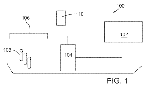
[0029] Figure 1 illustrates an exemplary electromagnetic localization
system 100 in accordance with an embodiment. System 100 includes a
computing system 102 that may house hardware and/or software for
treatment tracking, including providing various displays, inputs, etc.
Computing system 102 may be coupled, by wire or wirelessly, to a console
104 used to position the patient and/or the radiation treatment devices. In
an embodiment, computing system 102 and console 104 may be combined.
Console 104 may include, or may be coupled to, an electromagnetic array
106. An electromagnetic array 106 may include an energy source to excite
markers 108, and may include one or more receivers to detect
transmission(s)/emission(s) from markers 108. Markers 108 may be caused
to emit a magnetic field or another wave/emission that may be detected by
array 106. Electromagnetic array 106 may also be positioned, in part, based
on coordination with an optical system 110 including one or more cameras,
such as infrared cameras. Optical system 110 may coordinate with sensors
located in or on array 106.
[0030] In an alternative embodiment, markers may be configured with
radio frequency transmitters. The transmitters may emit/transmit radio
frequency signals that may be received by a receiver in the array or a similar
device. By utilizing different signals, such as different frequencies, for
each
7
CA 02744710 2011-05-25
WO 2010/063008 PCT/US2009/066017
marker/transponder, or by determining the relative strength of each signal,
the locations of the markers may be determined and tracked.
[0031] Figure 2 illustrates an exemplary electromagnetic localization
system 200 in accordance with an embodiment. As illustrated, system 200
includes an array 206. Array 206 may include an energy source to excite
one or more markers (not shown), and may include one or more receivers to
detect transmission(s)/emission(s) from the markers. An oral appliance
containing the one or more markers may be placed into the mouth of patient
212. A thermoplastic mask 214 may be placed into position onto/over the
face and head of patient 212. Before or after further positioning,
thermoplastic mask 214 may be attached to head holder platform 216. Array
206 may be positioned over patient 212 by manipulating positioning arm
218, and based on coordination with an optical system 210 including one or
more cameras, such as infrared cameras. Optical system 210 may
coordinate with sensors located in or on array 206. Patient 212 is on couch
220, which may also be moved to accurately position the patient for
treatment. In addition, as shown in Figure 2, data and/or test parameters
may be displayed on display 222.
[0032] In an exemplary embodiment, an oral appliance containing one
or more markers may be placed into the mouth of a patient. A thermoplastic
mask may be placed into position onto/over a patient's face and head but, in
an embodiment, not attached to the head holder platform initially. An
electromagnetic array may be moved into place in front of the patient's face
and aligned based, at least in part, on images collected by one or more
cameras located/mounted in the vicinity. The patient's head and neck may
be adjusted on the head holder and under the mask until x, y, and z
coordinates are all within a predefined threshold, such as within 0.05 cm, of
treatment plan positions, relative to a reference point that represents the
geographic center of the one or more markers. If not done previously, the
mask may then be attached to the head holder. Treatment may then be
initiated and the patient's position and motion may be continuously tracked
during treatment. In an embodiment, any motion outside of a predefined
8
CA 02744710 2011-05-25
WO 2010/063008 PCT/US2009/066017
tolerance may initiate a warning (such as an alarm, audible notification, or
indicator light) or may automatically halt the treatment.
[0033] In an embodiment, a method for delivering radiation to a
patient is provided comprising positioning the patient on a platform, the
patient defining at least a lateral rotational axis and a longitudinal
rotational
axis; adjusting the patient along the lateral rotational axis and/or the
longitudinal rotational axis; determining with a computing device a location
of
at least one marker, the at least one marker coupled to an oral appliance in
the patient's mouth; delivering radiation to the patient; and tracking with
the
computing device, during radiation delivery, the location of the at least one
marker.
[0034] In an embodiment, an oral appliance may be custom fit to each
patient. One or more markers, such as 1, 2, 3, 4, or more, may be coupled
(inserted, implanted, attached, etc.) to the oral appliance. In an
embodiment, the markers may be glass markers or transponders, each
containing a coiled wire such as a copper wire, which may emit or be
stimulated to emit a magnetic field or other detectable wave/emission. The
coordinates/locations of the markers may then be determined for example
using computed tomography (CT) and such coordinates may be used for
radiation treatment planning and/or targeted radiation delivery. During
radiation delivery, patient movement may be continuously tracked using the
markers as a tracking aid.
[0035] A suitable oral appliance may be constructed of one or more
parts. In a particular embodiment, a mouth guard may be provided for
engaging with the upper and/or lower teeth and maintaining the teeth/mouth
in a predetermined position defined by the orientation of the mouth guard.
[0036] In embodiments, an oral appliance may be constructed from
any suitable material, such as a polymeric material, for example a heat
moldable material, polycaprolactone, etc. In an embodiment, the material is
biocompatible.
[0037] An oral appliance may be fitted with one or more markers using
a variety of methods. In one method, channels or cavities may be formed in
9
CA 02744710 2011-05-25
WO 2010/063008 PCT/US2009/066017
the appliance into which the markers may be inserted. For example, cavities
may be formed by drilling or boring an appliance, or by forming the cavities
while molding the appliance. In another method, one or more markers may
be pressed into or otherwise mixed with a moldable material prior to
formation of the appliance. For example, a heat moldable material may be
heated and combined with one or more markers before/during molding and
formation of the appliance. In an embodiment, an appliance may be initially
formed in the general shape of an oral cavity and may then be heated and
placed into an individual's mouth to permit custom molding to that
individual's mouth. While the appliance is in a heated and moldable state,
the markers may be inserted into, such as pressed into, the moldable
material.
[0038] In an embodiment, markers may be placed in any suitable oral
appliance to provide for tracking of head movement for targeted radiation
delivery when the oral appliance is in-place in a patient's mouth. Figures 3A
and 3B illustrate an exemplary oral appliance 312 including markers 308.
Each marker/transponder 308 may be provided in any orientation with
respect to oral appliance 312 and with respect to the other markers 308.
[0039] As shown in Figure 3B, marker 308 is inserted into a channel
or opening in appliance 312. As an alternative, Figure 3C illustrates a
marker 308 embedded in appliance 312.
[0040] In an embodiment, there is provided an oral appliance,
comprising a biocompatible oral platform configured for insertion into a
patient's mouth; and one or more electromagnetic markers coupled to the
oral platform and configured to emit one or more waves in response to
stimulation.
[0041] In an example, volunteers were evaluated to determine the
effectiveness of embodiments described herein. Three markers
(transponders) were implanted into mouth guards. The mouth guards were
customized and fitted to each volunteer and thermal plastic masks were
fabricated. Isocenter was set at the anterior marker position. Each volunteer
was positioned on the treatment couch, the mask loosely was put in place,
CA 02744710 2011-05-25
WO 2010/063008 PCT/US2009/066017
the electromagnetic array was positioned, the neck was flexed/extended,
and the couch was moved until isocenter was within 0.5 mm in all three
dimensions. The head and neck positions may be adjusted based on real
time feedback from the localization system before masking. In
embodiments, some adjustments may be made after masking. The mask
was then secured to the head holder, and motion was tracked for five
minutes. The couch was then moved 0.5 cm left, right, in, out, up, and down
and these movements were compared to the tracings recorded by the
localization system. Marker positions were displayed as graphic readouts on
an associated monitor.
[0042] The sets of markers were localized and tracked successfully.
Adjustment of the neck position prior to masking was recorded by the
localization system. Motion-tracking data revealed a >2 mm deviation for
only 8% of total tracking time. Deviations of 1-2 mm were recorded for 30%
of total tracking time. The localization system accurately tracked all table
deviations to within 0.5 mm. In addition, masking did not force the
transponders out of tolerance (1 mm). See Figure 4 for results. In Figure 4,
time is reported in seconds on the x-axis and deviations in centimeters on
the y-axis. Lateral shifts are shown in the top tracing, longitudinal shifts
in
the middle tracing, and vertical shifts in the bottom tracing.
[0043] As shown by the above example, the localization system
provides an accurate method of continuous intra-fractional monitoring of
patient movement not possible with traditional imaging techniques that rely
on ionizing radiation. The described approach demonstrates that this system
is feasible for use in head and neck IMRT patients. Changes to neck position
prior to securing the mask were successfully tracked with this system as
well. This capability may also prove useful in providing accurate daily head
and neck IMRT patient setup.
[0044] In an alternative example, a method may be implemented for
participants with a squamous cell carcinoma of the head and neck who have
been indicated for definitive radiation. A customized mouth guard may be
constructed and three markers implanted into it. In this regard, a full dental
11
CA 02744710 2011-05-25
WO 2010/063008 PCT/US2009/066017
evaluation may be performed and any necessary extractions performed.
Stone impressions may be taken of the patient's upper and lower jaws.
Using the stone impression of the upper jaw, a customized mouth guard may
be fabricated using a vacuum-assisted thermoplastic polymer setup allowing
for a thin and rigid mouth guard to be created.
[0045] The markers may then be fixed to the inside surface of the
mouth guard paying attention to the orientation of the markers. The first
marker may be placed just posterior to the central incisors, the second
marker may be placed just medial to the patient's right pre-molar, 1st molar,
or 2nd molar, and the third marker may be placed just medial to the patient's
left pre-molar, 1st molar, or 2nd molar. The markers should generally be
placed to avoid as much metal dental filling material as possible. This
makes subsequent identification of the markers by computed tomography
(CT) more reliable and minimizes the risk of interference with the positioning
system by metal material in the teeth. Fixation may then be done with hot
wax to secure the markers' positions.
[0046] The mouth guard with attached markers may then be placed
back on the stone impression and another sheet of thermoplastic material
may be applied using the vacuum assisted setup once more. The edges of
the two thermoplastic layers may then be pinched together creating a seal
that effectively sandwiches the three markers between these layers
providing a very stable and reproducible setup.
[0047] The mouth guard may then be fitted to the patient's upper jaw
prior to CT simulation. CT simulation is carried out, including fabrication of
a
thermoplastic mask and head holder system as well as reconstruction of the
CT images into a 3 mm data set as well as a 1 mm data set. All images are
transferred to a commercially available treatment planning system. The 1
mm slice reconstruction may then be used to determine the 3D positional
coordinate locations of the markers. These coordinates are then input into
the system.
[0048] In an embodiment, patients may have their custom head holder
and mask molded with the mouth guard in place prior to a CT scan. Patients
12
CA 02744710 2011-05-25
WO 2010/063008 PCT/US2009/066017
may then receive fractionated IMRT. Patients may receive, for example, 35
total treatments at a dose of approximately 70 Gy, although adjustments
made be made or an alternative number of treatments and/or doses may be
provided. For each treatment fraction, the patient may insert the mouth
guard, be positioned on the head holder, loosely placed in the mask, and
have the electromagnetic array of a localization system brought into position.
Every other treatment may include positioning with the localization system
prior to fastening of the mask to the table.
[0049] In embodiments, after the customized mouth guard with the
markers is put in place, the mask may be put into place and the patient
aligned by laser to points on or affixed to the mask at the time of
simulation.
The mask may then be released from the head holder and the array may be
brought into place over isocenter. The array may be aligned by laser. The
patient may then be called up on the computing system and localization may
be performed. The tolerance for translations may be set at 1 cm, for
example, and when achieved, the tracking option may be selected.
[0050] If rotational alignment is out-of-tolerance, which is defined to be
one degree in this example, an error message appears that gives the
magnitude and axis of each rotation that is out of tolerance. The patient's
head may then be rotated under the loose mask to correct for these errors
and the mask may then be fixed in place.
[0051] The patient may then be re-loaded in the system and localized
again. Tracking may then be selected. If rotations are still greater than 1
degree off expected, another error message may appear. Re-alignment
steps may then be performed as necessary until the lateral and longitudinal
axes of rotation are correct to within one degree, or another threshold
tolerance as desired.
[0052] Treatments may be performed under daily image guidance
utilizing kV orthogonal pairs or cone-beam CT scans (CBCT). Images may
be evaluated pre-treatment by a radiation oncologist. Appropriate shifts of
the treatment couch may then be performed. Treatment may then occur with
continuous motion tracking throughout. All kV and CBCT images may be
13
CA 02744710 2011-05-25
WO 2010/063008 PCT/US2009/066017
sent to an online or offline system where shifts may be made by the
physician.
[0053] In an additional embodiment, an article of manufacture is
provided including a computer-readable medium having instructions stored
thereon that, in response to execution by a computing device, cause the
computing device to perform a method comprising determining whether a
current position of a patient is equal to or less than a predefined tolerance,
the patient defining at least a lateral rotational axis and a longitudinal
rotational axis, the predefined tolerance defining an acceptable out of
rotation alignment along the lateral rotational axis and/or the longitudinal
rotational axis; determining a location of at least one marker, the at least
one
marker coupled to an oral appliance in the patient's mouth; and tracking with
the computing device, during delivery of radiation, the location of the at
least
one marker.
[0054] Although certain embodiments have been illustrated and
described herein, it will be appreciated by those of ordinary skill in the art
that a wide variety of alternate and/or equivalent embodiments or
implementations calculated to achieve the same purposes may be
substituted for the embodiments shown and described without departing
from the scope. Those with skill in the art will readily appreciate that
embodiments may be implemented in a very wide variety of ways. This
application is intended to cover any adaptations or variations of the
embodiments discussed herein. Therefore, it is manifestly intended that
embodiments be limited only by the claims and the equivalents thereof.
14