Note: Descriptions are shown in the official language in which they were submitted.
CA 02744900 2011-05-27
WO 2010/060151 PCT/AU2009/001552
-1-
Modulation of an Ion Channel or Receptor
FIELD OF THE INVENTION
The present invention relates to methods of assaying compounds that modulate
one or
more ion channels or receptors that are involved in providing a waveform at a
biological
cell, and also to apparatuses and processes for performing such assays. The
present
invention especially relates to the use of a dynamic clamp in such assays.
BACKGROUND OF THE INVENTION
In many living organisms signals are transmitted between cells, such as
neurons and
muscle cells, by variations across cell membranes in electrophysiological
parameters such
as voltage, current or capacitance. Variations in such electrophysiological
parameters
often involve large numbers of multiple types of ion channels or receptors,
which together
produce a waveform at the biological cell. An action potential is an example
of one type
of waveform.
The waveform results from modulation of ion channels or receptors at the cell.
For
example, these ion channels or receptors may regulate the transmembrane and
intercellular
movement of physiological ions, such as Na+, K+, Cat+, and Cl-, which form
part of the
signal. Modulation of one, or a group of ion channels or receptors results in
electrophysiological changes at the membrane of the cell, causing further ion
channels to
be modulated. This process is closely coupled by feedback. Therefore the
waveform
produced at the biological cell varies depending on parameters such as the ion
channels or
receptors which are modulated and the length of time that those ion channels
or receptors
are activated or inhibited.
Compounds that affect waveforms produced at biological cells may be useful in
treating or
ameliorating a range of diseases and disorders. For example, action potentials
control the
function of nerve and muscle tissue, and accordingly influence many
physiological
functions including the capacity of a body to influence pathology. Similarly,
other
CA 02744900 2011-05-27
WO 2010/060151 PCT/AU2009/001552
-2-
waveforms such as synaptic events are involved in many nervous system
processes.
Compounds that affect the production of waveforms at biological cells may
therefore be
useful in the treatment or amelioration of, for example, a range of
neuromuscular, cardiac,
pain, affective and cognitive disorders.
However, the effect of any particular compound on a waveform is difficult to
assess. As
the production of a waveform in a cell involves individual contributions from
multiple ion
channel or receptor types, the duration of each waveform, the peak membrane
potential
and many other parameters may vary. Therefore, all necessary ion channel or
receptor
types to produce a waveform must be present and functional in order to
properly observe
the effects of the compound on the biological cell. This is usually performed
by observing
effects of compounds in intact samples of biological tissue, such as recording
action
potentials in nerve fibres in a living animal model or recording cardiac
action potentials by
isolation of a purkinje fibre from a dog heart. The requirement for biological
tissue limits
the number of compounds that can be assessed in a given period of time.
One method for determining the effects of a compound on an ion channel is the
patch
clamp technique. This employs an amplifier, which is connected to a biological
cell via an
electrode, to hold current (current clamp mode) or voltage (voltage clamp
mode) constant
at the membrane. For example, when current is held constant, voltage is
recorded.
However, such methods do not allow changes in a waveform to be monitored.
In particular, the cell attached or excised patch clamp technique allows the
determination
of the effect of a compound on a specific ion channel or receptor type of
interest. This
technique comprises an electrode which is attached to a patch of membrane of a
biological
cell around an ion channel or receptor of interest. A compound may then be
applied to the
inner or outer surface of the patch of membrane and the activity of that ion
channel or
receptor, as acted upon by the compound, measured. However, this process
requires the
harvesting of many cells to ascertain the effects of the compound on different
ion channels
or receptors and only determines the action of the compound on that specific
ion channel
or receptor without the reciprocal influence of the other ion channels or
receptors.
CA 02744900 2011-05-27
WO 2010/060151 PCT/AU2009/001552
-3-
Other patch clamp methods, such as the whole cell technique, allow analysis of
the
electrophysiology of an entire cell. Tests using these methods require many
parameters to
be simultaneously monitored, which greatly complicates the acquisition and
analysis of
results. These experimental difficulties mean that in many cases it takes a
substantial
amount of time to determine exactly how a compound is affecting the cell; it
is much more
difficult and time consuming to confidently determine on which ion channel or
receptor
type a compound acts.
Consequently, as waveforms are produced by a number of ion channel or receptor
types in
a biological cell, it has been difficult to determine the effect of a compound
at only one of
the ion channel or receptor types involved in producing the waveform. As all
of the ion
channel or receptor types involved in producing the waveform must be
functional, the
addition of a compound to this system may modulate any one or more of the ion
channel or
receptor types involved.
Conversely, it has been possible to determine if a compound binds to, for
example a
sodium channel, by directly measuring the binding at that channel. However, a
large
number of changes occur at, for example, sodium channels when they are
activated and it
is difficult to predict the effect that these channels have on other ion
channels when they
are assayed in isolation. Consequently, it has been difficult to determine the
effect that
modulation of an ion channel or receptor will have on the waveform that the
ion channel or
receptor produces.
SUMMARY OF THE INVENTION
The present invention is based on the surprising finding that a dynamic clamp
can be used
to determine the activity of compounds at one or more ion channel or receptor
types that
are involved in providing a waveform in a biological cell.
CA 02744900 2011-05-27
WO 2010/060151 PCT/AU2009/001552
-4-
Accordingly, in one aspect the present invention provides a method of assaying
a
compound for its ability to modulate an ion channel or receptor type, the
method
comprising:
a) providing a dynamic clamp in electrical contact with a biological cell (or
part
thereof) in which one or more ion channel or receptor types for providing a
waveform are functional and in which one or more ion channel or receptor
types for providing a waveform are either not present or not functional;
b) causing the dynamic clamp to apply a signal simulating the function of at
least one of the one or more ion channel or receptor types that are either not
present or not functional in the biological cell (or part thereof) based on
modulation of the ion channel or receptor types that are functional in the
biological cell (or part thereof) to thereby provide the waveform at the
biological cell (or part thereof);
c) exposing at least one of the one or more functional ion channel or receptor
types to a compound; and
d) detecting modulation of the waveform at the biological cell (or part
thereof),
wherein modulation of the waveform is indicative of a compound that
modulates the at least one functional ion channel or receptor types.
The dynamic clamp advantageously simulates the function of one or more ion
channel or
receptor types that are either not present or functional in the biological
cell (or part
thereof). This means that the assay may only involve a limited number of ion
channel or
receptor types in a biological cell, allowing assays to be conducted that
provide a greater
amount of information about the effect of the compound on the ion channel or
receptor
type that is modulated. Furthermore, the assay also illustrates the effect
that modulation of
the ion channel or receptor type may have on waveforms produced.
In another aspect, the present invention provides an apparatus for performing
the method
of the invention.
CA 02744900 2011-05-27
WO 2010/060151 PCT/AU2009/001552
-5-
In a further aspect, the present invention provides an apparatus for assaying
a compound's
ability to modulate an ion channel or receptor type in a biological cell (or
part thereof), the
apparatus including:
a) One or more electrodes adapted to be provided in electrical contact with
the
biological cell (or part thereof), wherein the one or more electrodes are
configured:
i. to detect modulation of one or more functional ion channels or
receptor types for providing a waveform at the biological cell (or
part thereof) and to provide a first signal based on the detected
modulation; and
ii. to apply a second signal to the biological cell (or part thereof);
b) A simulator to simulate the function of at least one or more ion channel or
receptor types for providing a waveform that are either not present or not
functional in the biological cell (or part thereof);
i. wherein the simulator is configured to receive the first signal from
the one or more electrodes and to provide the second signal to the
one or more electrodes;
ii. wherein the second signal simulates the function of at least one of
the one or more ion channel or receptor types that are either not
present or not functional based on the first signal, to thereby provide
the waveform at the biological cell (or part thereof).
In another aspect, the present invention provides an apparatus for assaying a
compound for
its ability to modulate an ion channel or receptor type, the apparatus
including:
(a) One or more electrodes to measure an electrophysiological parameter at a
biological cell (or part thereof) and to control a current or voltage applied
to
the biological cell (or part thereof), wherein the one or more electrodes are
adapted for electrical connection with the biological cell (or part thereof);
(b) One or more amplifiers to assist in measuring the electrophysiological
parameter at the biological cell (or part thereof) and to assist in
controlling
the current or voltage applied to the biological cell (or part thereof),
wherein
CA 02744900 2011-05-27
WO 2010/060151 PCT/AU2009/001552
-6-
the one or more amplifiers are electrically connected to the one or more
electrodes; and
(c) Software to simulate the function of one or more ion channel or receptor
types in a biological cell (or part thereof), which function is simulated by
receiving the measurement of the electrophysiological parameter at the
biological cell (or part thereof) from the one or more amplifiers,
determining the current or voltage to be applied to the biological cell (or
part thereof) based on said measurement, and transmitting an electrical
signal to the one or more amplifiers to control the current or voltage applied
to the biological cell (or part thereof).
In another aspect, the present invention provides a process, including:
receiving data detected from the modulation of at least one ion channel or
receptor type at a biological cell (or part thereof);
processing the data to determine a signal to be applied to the biological cell
(or part thereof), wherein the signal represents one or more ion channel or
receptor types that are either not functional or not present in the biological
cell
(or part thereof); and
applying the signal to the biological cell (or part thereof).
In further aspects, the present invention also provides a computer-readable
storage medium
having stored thereon programming instructions for performing the above
process, and a
system configured to perform the above process.
For a better understanding of the invention and to show how it may be
performed, an
embodiment of the invention is further described by way of non-limiting
example, by
reference to the accompanying drawings, in which:
Figure 1 shows a pipette patch clamp system for the measurement of
waveforms, in accordance with an embodiment of the present invention.
CA 02744900 2011-05-27
WO 2010/060151 PCT/AU2009/001552
-7-
Figure 2 shows a planar patch clamp system for the measurement of
waveforms, in accordance with an embodiment of the present invention.
Figure 3 is an example computing system that may be used in accordance
with an embodiment of the present invention.
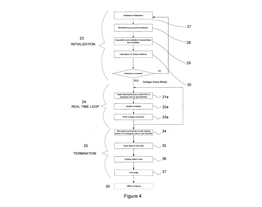
Figure 4 is a flow chart of a computer program operating in voltage clamp
mode in accordance with an embodiment of the present invention.
Figure 5 is a flow chart of a computer program operating in current clamp
mode in accordance with an embodiment of the present invention.
Figures 6a and 6b are exemplary electrocardiogram outputs, the output of
Figure 6b
showing an elongated QT interval.
Figure 7 is a diagram of a dynamic clamp system used in accordance with an
embodiment of the present invention.
Figure 8 illustrates a steady state action potential firing of 50-100 Hz at
HEK
cells controlled by a dynamic clamp system, in which the cells express Na,,1.4
sodium
channels.
Figure 9 illustrates the decrease in action potential firing rate achieved
when
carbamazepine is perfused onto HEK cells controlled by a dynamic clamp system,
in
which the cells express Na,,1.4 sodium channels.
Like features will hereinafter be referred to with like numbers.
DETAILED DESCRIPTION OF AN EMBODIMENT OF THE INVENTION
A dynamic clamp detects an electrophysiological parameter (which may, for
example,
include current, voltage or capacitance) of a biological cell (or part
thereof), and then
CA 02744900 2011-05-27
WO 2010/060151 PCT/AU2009/001552
-8-
applies a signal (for example, voltage or current) to the biological cell (or
part thereof) to
achieve a desired effect on the electrophysiological parameter. The step of
applying the
signal to the biological cell (or part thereof) requires the calculation of
the amount of, for
example, the voltage or current that must be applied to the cell (or part
thereof) to produce
the desired effect. Following the detection of an electrophysiological
parameter and the
subsequent application of the signal to the biological cell (or part thereof),
the dynamic
clamp continually repeats the process.
In an embodiment of the present invention, a dynamic clamp 1 is provided in
electrical
contact with a biological cell 2, as shown in Figures 1 and 2. In assaying a
compound for
its ability to modulate an ion channel or receptor type, the dynamic clamp
assists in
providing a waveform at a biological cell (or part thereof).
As used herein, the term "waveform" would be understood by a person skilled in
the art,
and includes any variation (for example variations in the amplitude or
frequency) in an
electrophysiological parameter (for example the trans-membrane voltage) over
time at a
cell. Such variations result from modulation of a number of ion channel or
receptor types
at the cell. In one embodiment, the waveform is an action potential or
synaptic event. In
another embodiment, the waveform is an action potential.
A waveform at a biological cell (or part thereof) is generally produced by
virtue of a
functional inter-relationship between a number of different types of ion
channels or
receptors. Modulation of one, or a group of ion channels or receptors results
in
electrophysiological changes at the membrane of the cell, causing further ion
channels to
be modulated, resulting in a waveform. Ion channels including, for example,
sodium
channels, potassium channels, calcium channels, chloride channels and
hyperpolarisation-
activated cation channels may involved.
Advantageously, in the present invention it is only necessary for one of the
ion channels or
receptor types to be present in the biological cell (or part thereof). The
function of the
remaining ion channels or receptor types which are required to provide a
waveform may be
CA 02744900 2011-05-27
WO 2010/060151 PCT/AU2009/001552
-9-
simulated using a dynamic clamp, which is configured to provide a real time
feedback loop
with the ion channels or receptor types that are present. To achieve this, the
dynamic
clamp can apply a signal to the cell or part thereof. The signal is used to
represent the
electrophysiological changes to the cell that would be induced by the
remaining ion
channels. This allows the effects of a compound at only one type of ion
channel or
receptor to be detected, while also observing the effect of the compound on
the waveform
of a more complex system.
This is particularly important as the effect of a compound on an ion channel
or receptor
involved in producing a waveform may affect parameters such as the frequency
of
waveform generation, and the morphology of the waveform generated. For
example, the
morphology of an action potential includes the half width, rise time, decay
time, time
between successive action potentials and rebound voltage. The assay according
to the
present invention may measure one, a number, or all of these changes.
The method of the present invention therefore provides a phenotypic screen
that provides
high content information on waveform properties and is rapid enough for the
drug
discovery cycle.
In one embodiment, the dynamic clamp applies a voltage signal to the
biological cell (or
part thereof), and modulation of the waveform at the biological cell (or part
thereof) is
detected by measuring a current signal at the biological cell (or part
thereof). In this
embodiment the voltage is clamped.
To simulate a particular voltage, the dynamic clamp may measure the membrane
current of
a biological cell (or part thereof), and use this parameter to determine the
amount of
voltage to be applied to the cell (or part thereof). If there is insufficient
current to produce
a waveform, then the dynamic clamp may modulate the amount of current applied
by
mathematical scaling in the feedback system.
In another embodiment, the dynamic clamp applies a current signal to the
biological cell
CA 02744900 2011-05-27
WO 2010/060151 PCT/AU2009/001552
-10-
(or part thereof), and modulation of the waveform at the biological cell (or
part thereof) is
detected by measuring a voltage signal at the biological cell (or part
thereof). In this
embodiment the current is clamped.
To simulate a particular conductance, the dynamic clamp may use the measured
membrane
potential of a biological cell (or part thereof) and the reversal potential
for that
conductance (the membrane potential at which there is no net flow of ions from
one side of
the membrane to the other) to determine the amount of current to be applied to
the cell (or
part thereof).
If there is insufficient current to produce a waveform, then a capacitive
current term may
be used to control the apparent capacitance of the cell (or part thereof) and
in this way
provide a precise control on the ratio of conductance to capacitance. The
capacitive
current term is calculated by measuring the rate of change of the voltage, and
its
application may decrease the apparent capacitance of the biological cell (or
part thereof) to
compensate for the lack of current.
The dynamic clamp may also be used to account for leak conductance at the cell
(or part
thereof). Leak conductance may occur because ion channels or receptors in the
cell (or
part thereof) are open, allowing the passage of ions. If the dynamic clamp
does not
account for leak conductance, then the assay results may be affected.
The dynamic clamp may also be used to account for and subtract the signal
arising from
one type of ion channels or receptors involved in the production of a waveform
at the
biological cell (or part thereof). For example, the signal arising from one
type of ion
channels or receptor can be removed using a dynamic clamp to provide further
information
on the effect of that ion channel or receptor on the waveform. Such techniques
are known
to a person skilled in the art and are discussed for example in Prinz et al.,
(2004) Trends in
Neurosciences, 27, 218-224.
Many types of dynamic clamp may be used in the method according to the present
CA 02744900 2011-05-27
WO 2010/060151 PCT/AU2009/001552
-11-
invention. As shown in Figures 1 and 2, the dynamic clamp 1 may include, but
is not
limited to, one or more electrodes 4, and a simulator. The simulator may
include an
amplifier 3, and computational software, which may be stored on and executed
by a
computing system 5.
In one embodiment, the one or more electrodes in contact with the biological
cell (or part
thereof) are sharp electrodes. A sharp electrode is a type of micropipette
that has a very
fine pore that allows slow movement (generally only capillary action) of
solution through
the electrode, thereby providing a minimal effect on the composition of the
intracellular
fluid. In use, a sharp electrode punctures the cell membrane so that the tip
of the electrode
is inside the cell.
In another embodiment, the one or more electrodes in contact with the
biological cell (or
part thereof) are patch electrodes. A patch electrode comprises a much larger
pore than a
sharp electrode. For a patch electrode, a high resistance (typically hundreds
of megaohms
to several gigaohms) electrical seal is formed between the electrode and the
membrane of a
biological cell. The membrane of the biological cell is then ruptured (such as
by suction)
so that a solution in a pipette (for pipette patch electrodes) or adjoining
the aperture (for a
planar patch electrode) is able to mix with the intracellular fluid. This is
also known as a
whole cell patch and allows an electrophysiological parameter across an entire
cell
membrane to be measured.
In one embodiment, a pipette patch electrode 4a (Figure 1) involves the
formation of a
high resistance electrical seal between a micropipette (the electrode) and a
membrane of
the biological cell 2. Once the seal is formed, a solution 8 in the
micropipette is able to
mix with the intracellular fluid.
In contrast, a planar patch electrode 4b (Figure 2) may involve the formation
of a high
resistance electrical seal between an aperture of a usually flat substrate
(the electrode) and
a membrane of the biological cell 2. In general, a well is provided at each
aperture of the
CA 02744900 2011-05-27
WO 2010/060151 PCT/AU2009/001552
-12-
substrate, and after a seal is formed and the membrane ruptured, a solution 8
in this well is
able to mix with the intracellular fluid.
As the planar electrode may comprise multiple apertures at which high
resistance electrical
seals may be formed with different cells, planar patch electrodes are
generally more
adaptable to high throughput, automated screening techniques. For example,
electrodes
which accommodate 16, 48, 96 or 384 cells for simultaneous recordings may be
employed.
Such electrodes could be, or would be similar to the QPlate (Sophion
Bioscience) or
PatchPlate PPC and PatchPlate substrates (MDS Analytical Technologies) or
those used
for the Patchliner and Synchropatch systems (Nanion Technologies GmbH) or the
lonFlux
system (Fluxion Biosciences).
Regardless of the type of patch electrode, it is important to achieve a high
resistance
electrical seal between the electrode and the membrane of the biological cell
(or part
thereof). If the seal is of poor quality, then assay results may be affected.
Many of the types of electrodes discussed above require the use of a solution
8 which is in
contact with the intracellular fluid of the cell. The composition of the
solution used with
the electrode depends on the assay to be conducted, and a person skilled in
the art would
be able to select a suitable solution without undue experiment. If the
solution is to be able
to mix with the intracellular fluid, the solution generally comprises a high
concentration of
electrolytes and is iso-osmotic to the intracellular fluid. When conducting
assays with
patch electrodes, this solution may be changed or altered. For example, in one
embodiment the concentration of compound to be tested in the solution may be
altered,
allowing a dose-response curve to be determined.
The dynamic clamp may comprise one or more electrodes 4. In one embodiment,
the
dynamic clamp comprises two electrodes which are in contact with a biological
cell (or
part thereof). In another embodiment, the dynamic clamp comprises one
electrode which
is in contact with a biological cell (or part thereof).
CA 02744900 2011-05-27
WO 2010/060151 PCT/AU2009/001552
- 13-
These electrodes may provide a continuous clamp, a discontinuous clamp or a
two
electrode clamp. A continuous clamp comprises one electrode, and that
electrode
simultaneously and continuously detects an electrophysiological parameter and
applies the
signal (such as the voltage or current) to a cell (or part thereof). In
contrast, a
discontinuous clamp also comprises one electrode, but that electrode switches
between
detecting an electrophysiological parameter and applying the signal to the
cell (or part
thereof). In a two electrode clamp there are two electrodes: one electrode
detects an
electrophysiological parameter and the other applies the signal to the cell
(or part thereof).
The dynamic clamp may also comprise a ground electrode. A ground electrode
sets the
ground reference point for electrophysiological measurements. The ground
electrode may
be in contact with a bath solution surrounding the biological cell (or part
thereof). In one
embodiment the ground electrode is a silver chloride coated silver wire. In
another
embodiment the ground electrode is a platinum electrode. The ground electrode
may also
be coated with agar.
The bath solution 6 selected may depend on a number of factors including, for
example,
the experiments to be conducted and the type of cell used. An appropriate bath
solution 6
may be selected by a person skilled in the art without undue experiment.
Other current and voltage clamp systems that may be adapted for use in the
method
according to the present invention are described in The Axon Guide: A Guide to
Electrophysiology and Biophysics Laboratory Techniques, MDS Analytical
Technologies,
2008.
In addition to the one or more electrodes, the dynamic clamp also comprises a
simulator to
simulate the function of at least one or more ion channel or receptor types
for providing a
waveform that are either not present or not functional in the biological cell
(or part
thereof). The simulator is configured to receive a first signal from the
electrode, which is
based on the detected modulation of the ion channel or receptor, and to
provide a second
signal to the electrode to be applied to the cell (or part thereof). The
signal provided to the
CA 02744900 2011-05-27
WO 2010/060151 PCT/AU2009/001552
-14-
cell simulates the function of at least one or more of the ion channel or
receptor types that
are either not present or not functional based on the first signal, to thereby
provide the
waveform at the biological cell (or part thereof).
The simulator may also include an output to display at least one of a waveform
or other
data to allow a compound's ability to modulate an ion channel or receptor type
to be
determined. In this embodiment, the other data displayed by the software may
include, for
example, the raw data obtained from the assay, or an icon or symbol that
indicates whether
or not there has been any change in the output following administration of the
compound
to the biological cell (or part thereof).
In another embodiment, the simulator comprises one or more amplifiers. The
simulator
may also comprise a suitably programmed computing system. In a further
embodiment,
the computing system operates to control the amplifier to provide the second
signal to the
one or more electrodes, and the computing system operates to receive the first
signal from
the one or more electrodes. The computing system may also operate to analyse
the first
signal and control the amplifier in accordance with analysis of the first
signal.
In one embodiment, the dynamic clamp comprises one or more amplifiers, as
shown for
example as 3 in Figures 1 and 2. Many amplifiers may be used to assist in the
measurement of an electrophysiological parameter at the biological cell (or
part thereof),
and to also assist in the control of the signal applied to that cell (or part
thereof). However,
in another embodiment, separate amplifiers may be used to perform these two
functions.
The type, or characteristics (for example input impedance or bandwidth), of
the amplifier
required will vary depending upon a number of factors including, but not
limited to, the
type of electrode used (for example sharp electrode or patch electrode) and if
the
electrodes provide a continuous clamp, a discontinuous clamp or a two
electrode clamp.
The amplifier may also provide features such as series resistance
compensation,
capacitance compensation, low-pass filters, Bridge Balance and features to
assist in record
keeping, cell penetration and patch rupture. The amplifier may also comprise a
feedback
CA 02744900 2011-05-27
WO 2010/060151 PCT/AU2009/001552
- 15-
amplification system to further control the current when using a patch clamp
in current
clamp mode (a patch clamp in voltage clamp mode does not require such a
feedback
amplification system).
For example, when performing patch electrode assays, suitable amplifiers may
include the
EPC 10 (HEKA Elektronik), the Axopatch 200B (Molecular Devices), the VE-2
(Alembic
Instruments Inc.) and the MultiClamp 700A (Molecular Devices). When performing
sharp
electrode experiments, the Axoclamp 2B (Molecular Devices) may be a suitable
amplifier.
A person skilled in the art would be able to select an appropriate amplifier
without undue
experiment.
The dynamic clamp may also comprise computational software, which may be
stored at a
computing system 5 or other similar processing device. The computing system 5
is
typically adapted to receive signals indicative of electrophysiological
parameters, perform
processing of the parameters and control the signal application to the cell.
Accordingly,
any suitable form of computing system can be used.
An example computing system is shown in Figure 3. In this example, the
computing
system 5 includes a processor 201, a memory 202, an input/output device 203,
such as a
keyboard and display or the like, and an external interface 204, coupled
together via a bus
205. In use, the external interface 204 may be coupled to a remote store, such
as a
database 211, as well as to the amplifier 3.
In use, the processor 201 executes software stored in the memory 202. The
software
defines instructions, typically in the form of commands, which cause the
processor 201 to
perform the steps outlined above, and described in more detail below, to
control the
dynamic clamp while performing the assay. The software may also display
results to allow
the outcome of the assay to be determined. Accordingly, the computing system
200 may
be any form of processing system, such as a computer server, a network server,
a web
server, a desktop computer, a lap-top or the like. Alternative specialised
hardware may be
used, such as FPGA (field programmable gate array), or the like.
CA 02744900 2011-05-27
WO 2010/060151 PCT/AU2009/001552
-16-
In one embodiment, the computing system is used to detect modulation of the
waveform at
the biological cell (or part thereof) (which is indicative of a compound that
modulates at
least one type of functional ion channel or receptor in the cell (or part
thereof)).
The computing system may also determine the signal that should be provided to
the
biological cell (or part thereof) to simulate the function of one or more ion
channel or
receptor types that are either not functional or not present in the biological
cell (or part
thereof). The amount of voltage or current to be provided to the cell (or part
thereof) is
determined based on modulation of the ion channels or receptors that are
functional in the
biological cell, as measured by electrophysiological measurements of that cell
(or part
thereof). This assists in understanding the effect that modulation of a type
of functional
ion channel or receptor in a biological cell (or part thereof) by a compound
will have on
the waveform.
The simulated signal is generated by modelling data representative of the
absent types of
ion channels or receptors, which modelling preferably occurs in software. The
data for the
model can be either collected by recording the action of those types of ion
channels or
receptors or by input of known data. As the data are representative of the
conductance of
ions across a cell membrane during a waveform, the data will normally be
stored in the
form of mathematical descriptions of virtual conductances (simulation
algorithms) in either
the memory 202 or database 211. In this manner, the software can model either
components of a biological cell or the entirety of a biological cell.
The simulation algorithms are designed to self-adjust to account for changes
in the cell.
The complexity of the simulation algorithms depends upon the number of factors
that the
dynamic clamp is designed to account for, including the number of ion channels
or
receptor types to be simulated. For example, for skeletal muscle cells the
action potential
produced largely arises from the interaction between sodium channels and
potassium
channels. However, for cardiac muscle cells the action potential produced
arises from the
interaction of a greater number of ion channels or receptor types, resulting
in more
CA 02744900 2011-05-27
WO 2010/060151 PCT/AU2009/001552
-17-
complex algorithms.
In addition, the data may contain parameters to account for losses in
hardware, losses in
the electrolyte in the pipette electrode (if used), at least one stimulation
protocol and
calculated variables as hereafter discussed. Accordingly, the simulation takes
the measured
waveform of the biological cell (or part thereof) and generates a signal
representative of
the absent types of ion channels or receptors, to encourage the waveform to
develop as it
would if the absent types of ion channels and receptors were functional.
The model of virtual conductances may include:
= the kinetics of the virtual conductance (the rates of change of conductance
to
particular stimuli);
= the voltage dependence of virtual conductances (the equilibrium open
probability
of a conductance);
= the maximum conductance of the biological channel expressed in the cell that
is
being recorded. This is particularly useful in determining a scaling factor
for
voltage clamp methods as this defines the maximum conductance that the
channels
expressed in the cell (or part thereof) will produce. Moreover, without such
scaling
there may be insufficient current to support waveform, and especially action
potential, generation. Scaling may also be useful for increasing
reproducibility of
the assay as variables such as membrane capacitance, leak conductance and
maximum conductance of the expressed channel can all be scaled to predefined
ratios;
= the electrochemical properties of the system, including the reversal
potentials of the
virtual conductances (the membrane potential at which there is no net
transmembrane flow of ions for a particular conductance); and
= other passive properties of the model system, including passive properties
of both
the biological cell (or part thereof) and the components or entirety of the
virtual
cell. This may include the desired capacitance and resting conditions (such as
resting conductance and resting voltage).
CA 02744900 2011-05-27
WO 2010/060151 PCT/AU2009/001552
- 18-
The stimulation protocol is a user defined signal applied to the biological
cell (or part
thereof) to generate desired physiological responses in the biological cell
(or part thereof).
In the present case, the desired physiological response is a waveform such as
an action
potential. These stimulation protocols allow the user to determine how the
cell (or part
thereof) will be stimulated and to what degree. For example, these protocols
allow the user
to determine whether the cell (or part thereof) is to be stimulated using
voltage or current
and the levels at which these stimuli will be set.
Stimulation protocols are useful where a biological cell (or part thereof) is
in a state
whereby a waveform will not be produced, or will not be produced repetitively.
When a
biological cell (or part thereof) is in such a state, assaying compounds may
not be possible
as the modulation of a waveform cannot be observed if no waveform is produced,
or if it is
produced too irregularly or too few times to allow accurate results to be
measured. In such
circumstances, the stimulation protocol can be used to produce a waveform, or
cause its
repetition. It achieves this by providing a stimulus that would not normally
be exhibited by
any of the types of ion channels or receptors the function of which the
simulated signal is
intended to replicate.
As biological cells differ in their electrophysiological properties,
calculated variables are
included in the simulation to allow the simulated signal to be tailored to the
biological cell
(or part thereof) to which the compounds to be assayed are exposed. The
calculated
variables include the capacitance of the biological cell (or part thereof)
(determined from
electrode measurements), modified virtual conductances (which are updated
according to
the cell (or part thereof) to which the apparatus is in contact and modelled
to form the
simulation algorithms), and an output command signal that is dependent on the
mode in
which the software is operating (i.e. voltage or current-clamp mode).
In the voltage-clamp mode, the transmembrane or ionic current is measured by
the
amplifier through the electrode. It is then scaled to match the electrical
parameters of the
model system. The simulated signal, or transmembrane voltage (membrane
potential), is
then calculated by collecting the contributions from each of the virtual
conductances, the
CA 02744900 2011-05-27
WO 2010/060151 PCT/AU2009/001552
-19-
capacitance of the virtual cell, the scaled ionic current recorded from the
biological cell (or
part thereof) and the selected stimulation protocol. The output command signal
is then set
to this transmembrane voltage and subsequently sent to an amplifier for
application to the
biological cell (or part thereof).
In the current-clamp mode, the transmembrane voltage of the biological cell is
measured
by the amplifier through the electrode. The measurement may be filtered and
sent to the
computing system. The filtration prevents amplification of noise that could
affect the
calculation of the capacitance compensation term as previously described. The
software
calculates the capacitance compensation term by determining the capacitance of
the cell
(or part thereof) and then applying a scaling factor to the rate of current
application from
each of the virtual conductances and the stimulation protocol. This can
mathematically
compensate for natural differences in the total capacitances of cells and
normalise to a
predefined capacitance level across all cells. The scaled output command
signal is then
sent to the amplifier for application to the biological cell (or part
thereof).
The software may be stored on any computer-readable medium such as a hard
disk,
removable memory device, external hard drive etc. In addition, the software
may only
contain those parameters, stimulation protocols etc that are relevant to
performing the task
to which the apparatus, interacting with the biological cell (or part
thereof), is put.
In order to take readings, the present system passes through a plurality of
operational
phases as illustrated in Figures 4 and 5. These phases optionally include, but
are not
limited to, initialization 23, real time looping for current or voltage-clamp
mode 24,
termination 25 and offline analysis 26.
The initialization phase 23, consists of hardware initialization 27,
stimulation protocol
selection 28 (for the reasons discussed earlier), acquisition and validation
of parameters
and variables 29, and calculation of initial conditions 30.
CA 02744900 2011-05-27
WO 2010/060151 PCT/AU2009/001552
-20-
In particular, the hardware is initialized and tested to ensure it is
functioning properly. This
part of the initialization phase may include the testing of the operational
limits of the
hardware; passing inputs, to which inputs there is a predetermined or expected
system
response, to the hardware and comparing the hardware response to the
predetermined
response; and so forth.
The acquisition and validation of parameters and variables is particularly
important so as to
ensure all data necessary for the accurate simulation of responses to
measurements taken
from the biological cell (or part thereof), can be produced. If some data is
missing, such as
a parameter representative of the response of a functional ion channel or
receptor type that
is not present or not functional in the biological cell, it may be collected
before testing
commences. This step may also ensure that the correct data for the operating
mode of the
apparatus, and the selected stimulation protocol, is acquired. It should be
noted that
although the system can operate in both current and voltage-clamp modes, the
parameters
and variables appropriate to one mode of operation may not be appropriate for
the other.
The last stage of initialization is the calculation of initial conditions.
This process sets the
equipment default and references values which are useful in the process of
recording data,
such as a reference voltage and current. In addition, this step allows the
calculated
variables to be determined in order to adapt the test to different biological
cells (or parts
thereof) and cells that have been intentionally experimentally modified (i.e.
by
administration of other compounds to simulate a condition the present compound
is being
developed to treat).
The next phase in the program is the real time looping phase 24. If the
apparatus is
operating in voltage-clamp mode, the transmembrane current from the biological
cell (or
part thereof) is measured 31a (Figure 4). The variables stored in software are
updated in
accordance with the measurement 32a and an output command is generated.
Simultaneously, this output command, that can be representative of the
restoration current
(the current required to return the membrane potential of the biological cell
(or part
thereof) to the resting potential), or is alternatively the ionic currents
that would be
CA 02744900 2011-05-27
WO 2010/060151 PCT/AU2009/001552
-21-
exhibited by functional ion channel and receptor types that are either not
present or not
functional in the biological cell (or part thereof), is written to memory 33a.
Similarly, when the apparatus is operating in current-clamp mode, the
transmembrane
voltage is measured by the amplifier through the electrode 31b (Figure 5). The
variables
stored in software are updated in accordance with the measurement 32b and an
output
command is generated. Simultaneously, this output command is written to memory
33b.
During the termination phase 25, the output commands are set to levels at
which it is safe
to hold the biological cell (or part thereof) 34 (Figures 4 and 5). This
ensures the cell
remains functional, without being damaged, that parameters against which
measurements
are taken and responses are generated remain fixed and that the cell is in a
predictable state
for the next experiment.
The data is then saved to hard disk or other appropriate medium 35, displayed
to the user if
desired 36, and the process is terminated 37.
Finally, during the offline analysis phase 26, calculations are performed to
identify the
initial conditions and parameters appropriate for the next iteration of
testing. This data may
also be displayed to the user. If a sufficient number of experiments have been
performed
at, for example, the various concentrations of compound, a model can be fitted
to the data
to describe the action of the compound on the system.
The program may be stored in a single place on a computer readable medium.
However, it
may be advantageous for individual devices to store data relevant to their own
operation.
For example, the amplifier may store its own initialization data and sequence
for
initializing, and the computing system may store data for applying tests to
determine the
responses generated by the software are appropriate.
The production of a waveform involves the activation of large numbers of
multiple types
of ion channels or receptors. Accordingly, it is possible to produce a
waveform in a whole
CA 02744900 2011-05-27
WO 2010/060151 PCT/AU2009/001552
-22-
biological cell or in a part of a biological cell. In one embodiment, a whole
biological cell
is used.
In another embodiment, part of a biological cell is used. For example, the
waveform may
be produced at a part of a biological cell using a macropatch. A macropatch
employs a
large diameter pipette (for a pipette patch electrode) or a large aperture
electrode (for a
planar patch electrode) to surround a number of ion channels or receptors on a
cell
membrane. After forming a seal on the cell membrane using the macropatch, the
electrode
may be quickly withdrawn to separate a portion of the cell membrane (an inside-
out patch).
Alternatively after forming a seal, the cell membrane inside the electrode may
be ruptured
and then the electrode slowly withdrawn to separate a portion of the cell
membrane (an
outside-out patch).
In the method according to the present invention, a waveform is provided at
the biological
cell (or part thereof), and the effect of the compound at a functional ion
channel or receptor
type is determined by detecting modulation of the waveform at the biological
cell (or part
thereof).
A waveform may be provided in the biological cell (or part thereof) in a
number of ways.
For example, in one embodiment the waveform may be initiated by the dynamic
clamp. In
another embodiment, the waveform may be initiated by the action of a compound
at the
one or more ion channel or receptor types that are functional in the
biological cell (or part
thereof).
At least one or more functional ion channel or receptor types may be exposed
to a
compound in a number of ways. For example, a compound may be applied to a bath
solution which surrounds the biological cell (or part thereof). In another
embodiment, the
compound may be administered to the inside of the cell (or part thereof)
through a
recording pipette or recording aperture (in the case of a planar electrode)
which is in
contact with the inside of the cell (or part thereof).
CA 02744900 2011-05-27
WO 2010/060151 PCT/AU2009/001552
-23-
The compound may modulate an ion channel or receptor by contacting that ion
channel or
receptor on the outside of the cell, or on the inside of the cell. Some
compounds will not
be able to pass through the cell membrane and their effect on the cell
therefore may be
more limited. On the other hand, some compounds will be able to pass through
the cell
membrane and act intracellularly or extracellularly. Compounds that are able
to pass
through a cell membrane may be advantageous as this is a desirable
characteristic of many
pharmaceuticals.
In the biological cell (or part thereof) according to the invention, one or
more ion channel
or receptor types for providing a waveform are functional, and one or more ion
channel or
receptor types for providing a waveform are either not present or not
functional.
As used herein, the term "functional", as applied to an ion channel or
receptor, means that
the ion channel or receptor may be involved in providing a waveform.
In one embodiment, an ion channel or receptor type is present in the
biological cell (or part
thereof), but that ion channel or receptor type is not functional due to
pharmacological
inhibition. This may allow a greater number of types of biological cells (or
parts thereof)
to be used in the assays according to the present invention. For example,
tetrodotoxin
(TTX), saxitoxin or lidocaine may be used to block most voltage gated sodium
channels.
In another example, tetraethylammonium (TEA) and 4-aminopyridine (4-AP) may be
used
to block most voltage gated potassium channels.
In another embodiment, an ion channel or receptor type is present in the
biological cell (or
part thereof), but the dynamic clamp is used to subtract the signal from that
ion channel or
receptor type. This may allow validation of the predicted effect of that ion
channel or
receptor type on the waveform produced at the biological cell (or part
thereof), or may
provide additional information regarding the behaviour of that ion channel or
receptor type
in the biological cell (or part thereof). Such techniques are known to a
person skilled in
the art and are discussed for example in Prinz et al., (2004) Trends in
Neurosciences, 27,
218-224. In some cases, the dynamic clamp may also be used to simulate ion
channels or
CA 02744900 2011-05-27
WO 2010/060151 PCT/AU2009/001552
-24-
receptors that are functional in the biological cell (or part thereof).
The biological cell may therefore be naturally occurring, already in
existence, genetically
modified or modified by interaction of, for example, an antagonist or virus.
In one embodiment, the one or more ion channel or receptor types for providing
a
waveform are functional as they are expressed in the biological cell (or part
thereof), and
the one or more ion channel or receptor types for providing a waveform are
either not
present or functional as they are not expressed in the biological cell (or
part thereof).
Therefore in one embodiment, the biological cell may be a cell in which the
genes for the
one or more functional ion channel or receptor types have been inserted, or
the biological
cell may be a cell in which the genes for one or more functional ion channel
or receptor
types have been removed. In one embodiment, the biological cell is a cell in
which the
genes for one or more functional ion channel types have been inserted.
To produce a cell expressing one or more ion channels or receptors, the DNA
sequence for
the ion channel or receptor type may be obtained and then incorporated into an
expression
vector with an appropriate promoter. Once the expression vector is
constructed, it may
then be introduced into the appropriate cell line using methods including
CaC12, CaPO4,
microinjection, electroporation, liposomal transfer, dendrimers, viral
transfer or particle
mediated gene transfer.
The biological cell line (or host cell) may comprise prokaryote, yeast or
higher eukaryote
cells. Suitable prokaryotes may include, but are not limited to, eubacteria,
such as Gram-
negative or Gram-positive organisms, including Enterobacteriaceae. Such
Enterobacteriaceae may include Bacilli (e.g. B. subtilis and B.
licheniformis), Escherichia
(e.g. E. coli), Enterobacter, Erwinia, Klebsiella, Proteus, Pseudomonas (e.g.
P.
aeruginosa), Salmonella (e.g. Salmonella typhimurium), Serratia (e.g. Serratia
marcescens), Shigella, and Streptomyces. Suitable eukaryotic microbes include,
but are
not limited to, Candida, Kluyveromyces (e.g. K. lactis, K. fragilis, K.
bulgaricus, K.
CA 02744900 2011-05-27
WO 2010/060151 PCT/AU2009/001552
-25-
wickeramii, K. waltii, K. drosophilarum, K thermotolerans and K. marxianus),
Neurospora crassa, Pichia pastoris, Trichoderna reesia, Saccharomyces
cerevisiae,
Schizosaccharomyces pombe, Schwanniomyces (e.g. Schwanniomyces occidentalis),
and
filamentous fungi (e.g. Neurospora, Penicillium, Tolypocladium, and
Aspergillus (e.g. A.
nidulans and A. niger)) and methylotrophic yeasts (e.g. Hansenula, Candida,
Kloeckera,
Pichia, Saccharomyces, Torulopsis, and Rhodotorula). Suitable multicellular
organisms
include, but are not limited to, invertebrate cells (e.g. insect cells
including Drosophila and
Spodoptera), plant cells, and mammalian cell lines (e.g. Chinese hamster ovary
(CHO
cells), monkey kidney line, human embryonic kidney line, mouse sertoli cells,
human lung
cells, human liver cells and mouse mammary tumor cells). An appropriate host
cell can be
selected without undue experimentation by a person skilled in the art.
In one embodiment, the biological cell (or part thereof) is selected from the
group
consisting of a human embryonic kidney (HEK) cell, a COS cell, an LTK cell, a
Chinese
hamster lung cell, or a Chinese hamster ovary (CHO) cell or a Xenopus oocyte.
In a
further embodiment, the biological cell (or part thereof) is a HEK cell or a
COS cell,
particularly a HEK 293 cell or a COS-7 cell. In another embodiment, the
biological cell
(or part thereof) is a HEK cell, particularly a HEK 293 cell.
The type of biological cell selected may affect the dynamic clamping technique
employed.
For example, the large size of Xenopus oocytes allows a two electrode clamp to
be used far
more readily than with mammalian cells, which are typically much smaller.
The cell line may then be cultured in conventional nutrient media modified for
inducing
promoters, selecting transformants, or amplifying the genes encoding the
desired
sequences. Culture conditions, such as media, temperature, pH, and the like,
can be
selected without undue experimentation by the person skilled in the art (for
general
principles, protocols and practical techniques, see Mammalian Cell
Biotechnology: A
Practical Approach, Butler, M. ed., IRL Press, 1991; Sambrook et al.,
Molecular Cloning:
A Laboratory Manual, Cold Spring Harbor Laboratory Press, 1989). The cells may
then be
selected and assayed for the expression of the desired ion channel or receptor
using
CA 02744900 2011-05-27
WO 2010/060151 PCT/AU2009/001552
-26-
standard procedures.
A number of functional ion channels or receptors are involved in providing a
waveform in
a biological cell. For example, this may include an ion channel selected from
the group
consisting of a sodium channel, a potassium channel, a calcium channel, a
chloride channel
or a hyperpolarisation-activated cation channel (H-channel). Accessory
subunits of these
channels may also be involved in providing a waveform.
As used herein, a receptor for providing a waveform is a receptor that is
modulated
following contact with a ligand. While modulation of an ion channel may also
involve
contact with a ligand (ligand-gated ion channels), ion channels may also open
and close in
response to changes in membrane potential (voltage-gated ion channels), or may
be
modulated by other means.
As used herein the term "modulating" is used in the broadest sense,
encompassing any
form or physical or chemical effect. For example, this may include activation
or inhibition
of the receptor, the effect of agonists or antagonists at the receptor, up-
regulation or down-
regulation of receptor, inhibition or activation of second messenger molecules
or receptor
internalisation. In one embodiment, modulation of the ion channel or receptor
type is
inhibition of the ion channel or receptor type. In another embodiment,
modulation of the
ion channel or receptor type is activation of the ion channel or receptor
type.
Modulation of an ion channel or receptor type also includes modulation of a
subunit of the
ion channel or receptor type. Selective modulation of specific subunits may be
advantageous in the development of compounds with appropriate pharmacological
characteristics.
In one embodiment of the invention, the one or more ion channel or receptor
types that are
functional in the biological cell (or part thereof) are one or more ion
channels. In a further
embodiment, the one or more ion channel or receptor types that are functional
in the
biological cell (or part thereof) are one or more voltage-gated ion channels.
CA 02744900 2011-05-27
WO 2010/060151 PCT/AU2009/001552
-27-
The ion channel may be selected from the group consisting of a sodium channel,
a
potassium channel, a calcium channel, a chloride channel or a
hyperpolarisation-activated
cation channel. In one embodiment, the ion channel is a sodium channel. In
another
embodiment, the ion channel is a potassium channel. In a further embodiment,
the ion
channel is a calcium channel. In another embodiment, the ion channel is a
hyperpolarisation-activated cation channel.
Calcium cations and chloride anions are involved in the production of a number
of types of
waveforms, such as the cardiac action potential and the action potential in
various single-
celled organisms. Calcium channels are known to play a role in controlling
muscle
movement as well as neuronal excitation, although intracellular calcium ions
can, in some
circumstances, activate particular potassium channels. In addition, chloride
channels are
known to aide in the regulation of pH, organic solute transport, cell
migration, cell
proliferation and differentiation.
In one embodiment, the ion channel or receptor type to be modulated is an N-
type calcium
channel or an L-type calcium channel. The N-type calcium channel may be an
alpha(2)delta calcium channel subunit. In another embodiment, the L-type
calcium
channel may be Ca,1.2. Compounds that modulate N-type calcium channels may be
useful
in the treatment or amelioration of pain indications. On the other hand,
compounds that
modulate L-type calcium channels may be useful in the treatment or
amelioration of a
variety of cardiac diseases.
Hyperpolarisation-activated cation channels activate due to hyperpolarisation
of the cell
membrane. These channels are often sensitive to cyclic nucleotides such as
cAMP and
cGMP and may be permeable to ions such as potassium ions and sodium ions.
These
channels assist in the propagation of an action potential. In one embodiment,
the
hyperpolarisation-activated cation channel is hyperpolarisation-activated
cyclic nucleotide-
gated potassium channel 1 (HCN1), hyperpolarisation-activated cyclic
nucleotide-gated
potassium channel 2 (HCN2), hyperpolarisation-activated cyclic nucleotide-
gated
CA 02744900 2011-05-27
WO 2010/060151 PCT/AU2009/001552
-28-
potassium channel 3 (HCN3), or hyperpolarisation-activated cyclic nucleotide-
gated
potassium channel 4 (HCN4).
Sodium channels are integral membrane proteins, and in cells such as neurons,
sodium
channels play a key role in the production of action potentials. Consequently,
compounds
affecting sodium channel function will generally have a more direct and
significantly
greater impact on the action potential of the biological cell than those
compounds affecting
calcium and chloride channel function. In one embodiment, the sodium channel
is a
Navl.l channel (voltage gated sodium channel, type I, alpha subunit; gene:
SCNIA), a
Nav1.2 channel (voltage gated sodium channel, type II, alpha subunit; gene:
SCN2A), a
Navl.3 channel (voltage gated sodium channel, type III, alpha subunit; gene:
SCN3A), a
Nav1.4 channel (voltage gated sodium channel, type IV, alpha subunit; gene:
SCN4A), a
Navl.5 channel (voltage gated sodium channel, type V, alpha subunit; gene:
SCN5A), a
Na,,l.6 channel (voltage gated sodium channel, type VIII, alpha subunit; gene:
SCN8A), a
Na,,1.7 channel (voltage gated sodium channel, type IX, alpha subunit; gene:
SCN9A); a
Na,,1.8 channel (voltage gated sodium channel, type X, alpha subunit; gene:
SCN l OA); or
a Nav1.9 channel (voltage gated sodium channel, type XI, alpha subunit; gene:
SCN11A).
In another embodiment, the sodium channel is a Na,,1.5 channel. In a further
embodiment,
the sodium channel is a Nav1.4 channel.
Potassium channels are known mainly for their role in repolarizing the cell
membrane
following action potentials. They effectively work to restore the cell
membrane to its
resting potential and to reprime sodium channels for subsequent action
potential firing.
For example, IKR and IKVLQT1 are known to be involved in repolarising the cell
after an
action potential. In one embodiment, the potassium channel is a neuronal
potassium
channel, a delayed rectifier potassium channel or an A-type potassium channel.
In a
further embodiment, the potassium channel is a K,4.2 channel (voltage gated
potassium
channel, Shal-related subfamily, member 2; gene: KCND2), a K,4.3 channel
(voltage
gated potassium channel, Shal-related subfamily, member 3; gene: KCND3), a
IKvLQT1
channel (also known as K,,7.1 channel; gene: KCNQ 1), a hERG channel (also
known as
Kv11. 1; gene: hERG (human Ether-a-go-go Related Gene or KCNH2)), a K;,2.1
channel
CA 02744900 2011-05-27
WO 2010/060151 PCT/AU2009/001552
-29-
(an inward rectifier potassium channel; gene: KCNJ2), a Kir2.2 channel (an
inward rectifier
potassium channel; gene: KCNJ12), a K;r2.3 channel (an inward rectifier
potassium
channel; gene: KCNJ4), a minK channel (voltage gated potassium channel, ISK-
related
family, member 1; gene: KCNE 1), a MiRP 1 channel (voltage gated potassium
channel,
ISK-related family, member 2; gene: KCNE2), a MiRP2 channel (voltage gated
potassium
channel, ISK-related family, member 3; gene: KCNE3) or a MiRP3 channel
(voltage gated
potassium channel, ISK-related family, member 4; gene: KCNE4). In another
embodiment, the potassium channel is a IK LQT 1 channel.
In one embodiment, the potassium channel is a leak channel. Leak channels are
also
known as tandem-pore-domain potassium channels, and are known to comprise
approximately 15 members. These channels are regulated by a number of factors
including oxygen tension, pH, mechanical stretch and G-proteins.
In the case of an action potential, as the membrane potential increases, both
the sodium and
potassium channels begin to open. This process increases the passage of sodium
ions into
the cell and the balancing passage of potassium ions out of the cell. For
small changes in
membrane potential, the flow of potassium ions will overcome the flow of
sodium ions and
the membrane potential will return to its resting potential. However, if the
voltage
increases past a critical threshold, the flow of sodium ions suddenly
increases and will
temporarily exceed the flow of potassium ions, resulting in a condition
whereby the
positive feedback from the flow of sodium ions activates even more sodium
channels.
Thus, the cell produces an action potential.
Therefore, in most cases the sodium and potassium channels are directly
responsible for
regulating the flow of ions across the cell membrane, which causes the firing
of an action
potential and the restoration of the cell membrane after the event.
In the development of pharmaceuticals, the testing of the interactions between
compounds
and, for instance, the firing of neurons, is a particularly important step in
obtaining
approval for new pharmaceuticals. Adverse effects are a barrier in the
development of new
CA 02744900 2011-05-27
WO 2010/060151 PCT/AU2009/001552
-30-
pharmaceuticals, particularly those that affect the functioning of the heart
and brain.
Ion channels or receptors that should not be affected by potential
pharmaceuticals may
include, for example, the hERG channel, the IKR channel, the IK,,LQT1 channel,
Navl.5
channel and the MiRP1 channel. In one embodiment, the ion channel or receptor
type that
is functional is a hERG channel, a IKR channel, a IK,,LQT1 channel or a MiRPI
channel.
In a further embodiment, the ion channel is the hERG channel, which is an ion
channel of
particular interest in testing pharmaceuticals for adverse effects. The hERG
channel
(which is encoded by human Ether-a-go-go Related Gene) is a pore-forming (a
pore is the
portion of the ion channel that opens to allow movement of ions) voltage-gated
potassium
channel, which is expressed in the heart and nervous tissue. In certain
circumstances, the
hERG channel can make up the entirety of the channel that conducts the delayed
rectifier
current for repolarization of cell membranes around the heart; the current
involved in the
firing of ventricular myocytes (muscle fibre cells) including the purkinje
fibres.
Very small changes in hERG channel function can reduce the ability of the
heart to operate
properly. Consequently, it is vital to the approval of compounds for
therapeutic use that
they be shown not to adversely affect the hERG channel. Some compounds, for
example,
have been found to have the effect of mirroring a condition representative of
illness such as
is seen in the genetic mutation of the hERG channel, leading to Long QT (where
Q and T
are regular points on an electrocardiogram (ECG) - see Figure 6a) syndrome -
where the
heart develops an arrhythmia which can lead to sudden death and cardiac
arrest, seen as an
elongation of the QT interval on an ECG (see Figure 6b). Accordingly, the
possibility of
undesirable interaction between hERG and a pharmaceutical compound of interest
is
necessary to avoid.
Present methods used for assaying compounds against their effect on the hERG
channel
can require the harvesting of one cell, containing the hERG channel, for each
test desired
to be performed. The cells are often taken from a dog such as a beagle.
Accordingly, to
perform such experiments the animals must be bred to ensure they are free from
diseases
CA 02744900 2011-05-27
WO 2010/060151 PCT/AU2009/001552
-31 -
that may alter results, the animal must be treated and killed, the cell
extracted and the
experiment set up. In addition, there can be considerable barriers to
obtaining approval for
such experiments and subsequently finding carriers of suitable cells. Methods
according to
preferred embodiments, as described herein, may remove the need for such
experiments
and also ameliorate some of the effects on results of variables that can be
difficult to
quantify, such as animal health and age.
It would be appreciated that when more functional ion channel or receptor
types for
providing a waveform are present in the cell (or part thereof), it is more
difficult to
determine which ion channel or receptor type is affected by the compound
assayed.
Accordingly, in one embodiment one ion channel or receptor type for providing
a
waveform is functional in the biological cell (or part thereof).
In another embodiment of the invention, the one or more ion channel or
receptor types that
are either not present or not functional in the biological cell (or part
thereof) are one or
more ion channels. In a further embodiment, the one or more ion channel or
receptor types
that are either not present or not functional in the biological cell (or part
thereof) are one or
more voltage-gated ion channels.
The ion channel that is either not present or not functional in the
biological. cell (or part
thereof) may be selected from the group consisting of a sodium channel, a
potassium
channel, a calcium channel, a chloride channel or a hyperpolarisation-
activated cation
channel. In one embodiment, the ion channel not present or not functional is a
sodium
channel. In another embodiment, the ion channel not present or not functional
is a
potassium channel. In a further embodiment, the ion channel not present or not
functional
is a calcium channel. Any, or combinations of, the channels to be modulated as
discussed
above, may also not be present or not functional in the biological cell (or
part thereof).
It is to be understood that assays performed in accordance with the invention
includes, for
example, an experiment at a single concentration to determine whether a
compound is
active, in addition to multiple experiments at a variety of concentrations so
as to obtain a
CA 02744900 2011-05-27
WO 2010/060151 PCT/AU2009/001552
-32-
dose response curve.
Using these assays, compounds that modulate ion channel or receptor types may
be
identified, and/or the activity of these compounds determined. The compounds
to be tested
could be produced synthetically, or through biological processes. Mixtures of
compounds
may also be tested, which may, for example, include testing of biological
samples or
extracts thereof.
While the compounds assayed may be new pharmaceuticals, they may also be used
in the
development of new pharmaceuticals or new lead compounds. For example, in one
embodiment a range of similar compounds could be assayed according to the
method of
the invention to develop a pharmacophore for the receptor or ion channel
assayed, assisting
in the development of new pharmaceuticals.
Using the method according to the present invention, new pharmaceuticals for a
wide
variety of diseases or conditions may be identified. For example, such
diseases or
conditions may include, but are not limited to, arrhythmia, short QT syndrome,
long QT
syndrome, pain, neuropathic pain, fibromyalgia, epilepsy, cognition and memory
disorders,
movement disorders, affective disorders, mood disorders, skeletal muscle
diseases, smooth
muscle diseases, blood pressure and tremors.
The above method allows rapid development of virtual conductance models and
the ability
to incorporate graphical tools in the control of experiments and the analysis
of data. As this
analysis includes the fitting of real conductance models that include the
effects of
compounds on waveforms, it may be used to select from candidate compounds
those
compounds suitable for further experimentation or use. This selectivity also
includes the
forecasting of the effects of the compounds on other parts of the anatomy
(i.e. a compound
treating arrhythmia may also be suitable for the treatment of problems in
other parts of the
body, and such advantageous use, or disadvantageous use in the case of adverse
effects,
can potentially be forecast) and the guiding of medicinal chemists in their
experimentations and compound selection.
CA 02744900 2011-05-27
WO 2010/060151 PCT/AU2009/001552
-33-
EXAMPLES
Human embryonic kidney (HEK) cells which stably express skeletal muscle Na,1.4
sodium channels were obtained as a gift from Professor Holger Lerche at the
University of
Ulm, Germany. The creation and characterization of these cells is described in
Mitrovic et
al., (1994) J Physiol., 478(Pt 3), 395-402. For maintenance, cells were
cultured in
Dulbecco's Modified Eagle Medium with 10% Fetal Bovine Serum in 144cm2 flask
and
incubated at 37 C in 5% C02.
Twenty-four hours prior to experimentation, cells were dissociated using
Versene (EDTA)
and plated at 10-12% confluency onto coverslips. The following day the
coverslips were
placed into the recording chamber and held at 22-25 C for the duration of the
experiments.
Borosilicate glass pipettes (WPI) were used for the whole cell assay. These
pipettes were
filled with an intracellular solution containing (mM): 10 NaF, 110 CsF, 20
TEA.CI, 2
ethylene glycol tetraacetic acid and 10 HEPES, with pH adjusted to 7.4 using
CsOH and
osmolarity adjusted to 310mosmoUL with sucrose. The pipettes, when filled with
this
solution, had a resistance of 2-6 MOhms.
The bath solution contained in (mM): 141 NaCl, 4 KC1, 1.0 MgC12, 1.8 CaCl2, 10
HEPES
buffer, and 4 tetraethylammonium (TEA).Cl, with pH adjusted to 7.4 with NaOH
and
osmolarity adjusted to 310mosmol/L with sucrose. Bath temperature was
controlled using
a Warner Instruments (Hamden, CT) controller (TC-344B) with inline solution
and bath
heating.
Electrophysiological recordings were made 10 minutes after establishing whole
cell
recording. Recordings were made on an EPC-9 patch clamp amplifier (Heka
Instruments,
Lambrecht, Germany) filtered at 14.4kHz with >80% series resistance
compensation and
sampled at 50kHz. The current monitor output from the EPC-9 was fed into the
analogue
input channel of a data acquisition card.
CA 02744900 2011-05-27
WO 2010/060151 PCT/AU2009/001552
-34-
The dynamic clamp system was implemented in Simulink with Realtime workshop
and the
xPC target toolkit (see Figure 7; All products from Mathworks). The model was
compiled
and downloaded to the target on a standard PC with a National Instruments PCI-
6052E
data acquisition board. The model runs in polling mode using the odes fixed
time step
solver with a step size of 50 S.
The dynamic clamp system was configured to account for leak conductance, and
to also
simulate the function of potassium channels, which were not present in the HEK
cell.
Leak current is given by:
I Leak = Leak X (V - VLeak )
VLeak = -85mV
The fast delayed rectifier potassium current is given by Cannon et al. (1993)
Biophys J.,
65(1), 270-88:
4
IK,=gKrxn x(V-Vk)
do
dt = a(V) x (1- n) -,3(V) x n
a(V) - an x (V - V)
1 - e -(V -Võ )/K_
N(V) xe -'v-v' 'K,+
Vk = -93.1320mV
an = 0.0131/ms/mV
Kaõ = 7mV
K,3õ = 40mV
,3n = 0.067/ms
Vn = -40mV
CA 02744900 2011-05-27
WO 2010/060151 PCT/AU2009/001552
-35-
Following attainment of a whole cell clamp in the HEK cell, a period of 10
minutes was
allowed for diffusion of the pipette solution into the intracellular volume of
the cell.
During this period cells were held at -85 mV.
Control was transferred to the Simulink system and a range of current
injections were
trialled to achieve a steady state action potential firing of 50-100 Hz
(Figure 8). This firing
was stable and continued as long as a stimulating current injection was
maintained.
Following a period of stable recording of action potential firing, 50 M
carbamazepine
(CBZ, Sigma-Aldrich C8981, a sodium channel blocker) was added to the bath
solution
and perfused onto the cells. This decreased the action potential firing rate.
Figure 9 is an
output of the simulator, showing the response of the system to a step of
stimulating current.
In the continued presence of 50 M CBZ a stimulating current step elicited
only 2-3 action
potentials and no further firing would occur.
This shows that a dynamic clamp in electrical contact with a cell expressing
sodium
channels may be used to assist in producing and monitoring consecutive
waveforms
(action potentials) at that cell. Furthermore, it is illustrated that by
modifying this system
by modulating these sodium channels with a compound, the resultant waveform
generated
is affected.
The described constructions have been advanced merely by way of example and
many
modifications and variations may be made without departing from the spirit and
scope of
the invention, which includes every novel feature and combination of features
herein
disclosed.
Throughout this specification and the claims which follow, unless the context
requires
otherwise, the word "comprise", and variations such as "comprises" and
"comprising", will
be understood to imply the inclusion of a stated integer or step or group of
integers or steps
but not the exclusion of any other integer or step or group of integers or
steps.
CA 02744900 2011-05-27
WO 2010/060151 PCT/AU2009/001552
-36-
The reference in this specification to any prior publication (or information
derived from it),
or to any matter which is known, is not, and should not be taken as an
acknowledgment or
admission or any form of suggestion that that prior publication (or
information derived
from it) or known matter forms part of the common general knowledge.