Note: Descriptions are shown in the official language in which they were submitted.
CA 02744906 2011-05-27
WO 2009/070794 PCT/US2008/085120
PROGESTERONE-CONTAINING COMPOSITIONS AND DEVICES
CROSS-REFERENCE TO RELATED APPLICATIONS
[0001] This application claims priority to U.S. Provisional Application Serial
No. 60/991,033, filed November 29, 2007, which is incorporated herein by
reference in
its entirety.
FIELD OF THE INVENTION
[0002] The present invention generally relates to anti-angiogenic, anti-
thrombotic, and/or anti-restenotic compositions, formulations, coated devices,
and
methods for their use.
BACKGROUND
[0003] Implantable medical devices, such as stents, are widely employed in
medical procedures. A stent is generally understood in the art to be an
expandable
prosthetic device for implantation in a body passageway (e.g., a lumen or
artery) to keep
a formerly blocked passageway open and/or to provide support to weakened
structures
(e.g. heart walls, heart valves, venous valves and arteries). A stent can be
used to obtain
and maintain the patency of the body passageway while maintaining the
integrity of the
passageway, and can be an alternative to surgery. Stent manufacture and usage
are
generally known in the medical arts.
[0004 ] One disadvantage of utilizing stents in a vessel is the potential
development of a thrombis formation and/or cellular response within the stent
causing a
re-occlusion of the artery, the so-called neointimal hyperplasia. This may
cause scar
tissue (cell proliferation) to rapidly grow over or within the stent, or some
other negative
reaction. A common theory of re-occlusion of arteries is that development of a
neointima is variable but can at times be so severe as to re-occlude the
vessel lumen (i.e.,
restenosis), especially in the case of smaller diameter vessels, which often
requires re-
intervention. Another disadvantage of utilizing stents in a vessel is that the
expansion of
the vessel upon insertion of the stent can weaken the vessel and/or cause
secretion of
undesirable biological factors due to the stress exerted on the artery. There
is an
occasional tendency for clots to form at the site where a stent is implanted
and it
potentially damages a vessel wall. This tendency may be higher for drug-
eluting stents.
Since platelets are involved in the clotting process, subjects must take
antiplatelet
1
CA 02744906 2011-05-27
WO 2009/070794 PCT/US2008/085120
therapy (e.g., clopidogrel, aspirin) afterwards, usually for at least six
months and
perhaps indefinitely. But antiplatelet therapy may be insufficient to fully
prevent clots;
these and cell proliferation within or near to the stent may cause the
standard ("bare-
metal") stents to become blocked.
[0005] A drug-eluting stent is generally understood in the art to be a stent
(i.e., a scaffold) placed into a vessel (e.g., a narrowed, diseased coronary
artery) that
slowly releases a drug, for example, to block cell proliferation. Blocking
cell
proliferation can prevent scar-tissue-like growth that, together with clots
(i.e., thrombus),
could otherwise block the stented vessel. For example, drug-eluting stents
releasing an
antiproliferative drug (drugs typically used against cancer or as
immunosuppressants)
can help avoid, at least in part, in-stent restenosis (re-narrowing or re-
occlusion, either in
part or in whole). Examples of current drug-eluting stents include CypherTM, a
sirolimus-eluting stent (Cordis Corp., Johnson & Johnson) and TaxusTM, a
paclitaxel-
eluting stent (Boston Scientific), both of stainless steel and using a polymer
as a drug
carrier. Other drugs reported to be used in conjunction with a stent include
zotarolimus
(ZoMaxx stent, Abbott Labs; Endeavor stent, Medtronic); everolimus (Champion
stent,
Xience stent, Abbott Labs). But recent studies have revealed that present drug
eluting
stents are associated with a 5 fold higher risk for thrombosis (with fatality
results in one-
third of patients who develop late thrombosis) compared to bare metal stents.
Bavry et al.
(2006) Am. J. Med. 119 (12), 1056-1061.
[0006] Current drug-eluting stents generally consist of three parts. The stent
itself is an expandable framework, usually metal. Added to this is a drug,
usually one to
prevent the artery from being re-occluded, or clogged. These typically have
been drugs
already in use as anti-cancer drugs or drugs that suppress the immune system.
Finally,
there is a carrier which slowly releases the drug over months. The carrier is
typically a
polymer, although phosphorylcholine or ceramics have also been reported.
Different
carriers can release the loaded drug at different rates.
[0007] The stent is often delivered to the target area of the body passageway
by a balloon and catheter system tracking over a guidewire. Once properly
located, the
balloon is expanded, plastically deforming the entire structure of the stent
against the
body passageway. Expansion can also crack and/or compress any plaque present
in the
vessel. The amount of force applied is usually at least that necessary to
expand the stent
(i.e., the force applied exceeds the minimum force above which the stent
material will
2
CA 02744906 2011-05-27
WO 2009/070794 PCT/US2008/085120
undergo plastic deformation) while maintaining the patency of the body
passageway. At
this point, the balloon is deflated and the balloon, catheter system, and
guidewire are
withdrawn from the lumen and subsequently removed from the body altogether.
Ideally,
the stent will remain in place and maintain the target area of the body
passageway
substantially free of blockage (or narrowing).
[0008] Furthermore, the administration of stents that carry therapeutic
coatings, such as one or more polymeric coatings including pharmacologically
active
agents, has been the subject of ongoing inquiry to reduce some of the problems
created
by the implantation of stents, such as restenosis and other biocompatibility
responses to
the foreign implant. Therefore, the search to find materials and coatings that
enhance
biocompatibility and prevent the re-occlusion of the passage through clotting
or cell or
tissue growth is a continuing pressing need.
BRIEF DESCRIPTION OF THE DRAWINGS
[0009] Those of skill in the art will understand that the drawings, described
below, are for illustrative purposes only. The drawings are not intended to
limit the
scope of the present teachings in any way.
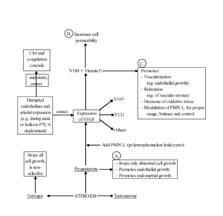
[ 0010 ] Figure 1 is a flow chart depicting, inter alia, suggested mechanisms
underlying the effect of progesterone in anti-angiogenic, anti-restenotic,
and/or anti-
thrombotic applications.
SUMMARY OF THE INVENTION
[0011] The present invention is directed to compositions containing
progesterone and their use in various formulations, medical device coatings,
and methods
of therapeutic treatment. The progesterone-containing formulations and medical
device
coatings described herein can maintain, or aid in maintaining, the opening of
a body
passageway.
[0012 ] In brief, the present invention provides progesterone-containing
compositions, formulations, and/or medical device coatings to give functional
properties
such as, for example, vessel relaxative, anti-oxidative, anti-restenotic, anti-
angiogenic,
and/or anti-thrombotic effects. The progesterone-containing compositions,
formulations,
and/or medical devices described herein can, inter alia, minimize or eliminate
inflammation, thrombosis, restenosis, neo-intimal hyperplasia, smooth muscle
cell
3
CA 02744906 2011-05-27
WO 2009/070794 PCT/US2008/085120
proliferation, rupturing of vulnerable plaque, and/or other effects related to
device
implantation.
[0013] One aspect of the invention provides a drug eluting medical device.
In some embodiments, the drug eluting device includes a medical device, a
progesterone-
containing composition, and at least one coating layer. In some embodiments,
the drug
eluting device includes a medical device comprising a drug-eluting mechanism
(e.g., a
well, pocket or crevice within the surface or body of a device) and a
progesterone-
containing composition. In some embodiments, the device includes both a
coating layer
and a drug-eluting mechanism. Usually, a coating layer is formed on at least a
portion of
a surface of the medical device and the coating layer will include the
progesterone-
containing composition. Such composition can be present in any of a number of
drug
eluting mechanisms, heretofore including a reservoir, pore, duct, channel,
chamber, side-
port, lumen, etc., within, proximal to, distal to, lateral to, underneath,
embedded within
or on the medical device. The progesterone-containing composition is usually
present in
a therapeutically effective amount. And the various components of the drug
eluting
medical device are configured such that the progesterone-containing
composition is
eluted from the medical device in vivo.
[0014] Another aspect of the invention provides for a method of treating a
target tissue of a subject. The method generally includes providing a drug-
eluting
medical device; and introducing the drug eluting medical device to a target
tissue of a
subject in need thereof. The drug eluting medical device generally includes a
medical
device, a progesterone-containing composition, and at least one coating layer
and/or drug
eluting mechanism formed on at least a portion of a surface of the medical
device. The
coating layer(s) generally contains the progesterone-containing composition,
and such
composition is eluted from the medical device in vivo. According to the
method,
progesterone is eluted from the delivered medical device in a therapeutically
effective
amount.
[0015] Another aspect of the invention provides for an anti-angiogenic, anti-
thrombotic, or anti-restenotic composition containing a therapeutically
effective amount
of progesterone and vitamin E. The therapeutically effective amount of
progesterone in
the composition is an amount that has an anti-angiogenic, anti-thrombotic,
and/or anti-
restenotic effect in a subject.
4
CA 02744906 2011-05-27
WO 2009/070794 PCT/US2008/085120
[0016] Another aspect of the invention provides for an anti-angiogenic, anti-
thrombotic, or anti-restenotic pharmaceutical formulation containing a
therapeutically
effective amount of progesterone and vitamin E along with a pharmaceutically
acceptable carrier. The therapeutically effective amount of progesterone in
the
formulation is an amount that has an anti-angiogenic, anti-thrombotic, and/or
anti-
restenotic effect in a subject.
[0017] Provided below are various embodiments of the different aspects of
the invention described below. It is understood that reference to, for
example, the
progesterone-containing composition can include reference to such composition
as
occurring in the drug eluting medical devices, methods, compositions, or
pharmaceutical
formulations described herein. Likewise, reference to various components of
the drug
eluting medical device can include reference to such components as occurring
in the drug
eluting medical device or methods described herein.
[0018] In various embodiments, the progesterone-containing composition
further comprises at least one additional therapeutic agent. For example, the
additional
therapeutic agent is an antiplatelet, anticoagulant, antifibrin,
antiinflammatory,
antithrombin, antiproliferative, antioxidants, and/or growth factors (e.g.,
VEGF). In
various embodiments, the progesterone-containing composition further comprises
vitamin E.
[0019] In various embodiments, the coating layer or drug eluting mechanism
of the drug-eluting medical device is made up of, at least in part, a
polymeric material.
The drug eluting medical device can comprise a second coating layer or drug
eluting
mechanism, wherein the second coating layer or drug eluting mechanism
comprises a
polymeric material and the second coating layer or drug eluting mechanism acts
as a
barrier layer to further control elution of the progesterone-containing
composition.
[0020] In various embodiments, the drug eluting medical device is a drug
eluting stent. In various embodiments, the drug eluting medical device is
configured to
treat vulnerable plaque lesions; bifurcated lesions or ostial lesions; or for
use in coronary,
cardiac, peripheral carotid, neurologic, vascular, organ, muscle, or body
cavity
applications.
[0021] In various embodiments, the therapeutically effective amount has one
or more effects such as an anti-angiogenic effect, anti-thrombotic effect,
anti-restenotic
effect, vessel-relaxative effect, anti-oxidative effect, or combinations
thereof.
CA 02744906 2011-05-27
WO 2009/070794 PCT/US2008/085120
DETAILED DESCRIPTION OF THE INVENTION
[0022] The present invention relates to compositions and devices that can
minimize or eliminate conditions and complications, such as inflammation,
thrombosis,
restenosis, neo-intimal hyperplasia, rupturing of vulnerable plaque, and/or
other effects.
More specifically, the present invention is directed to a progesterone-
containing
composition that can be administered directly and/or used in conjunction with
a medical
device to maintain opening of a body passageway. The progesterone-containing
composition can also include one or more additional pharmacologically active
therapeutic agents.
[0023] The progesterone-containing composition and devices can improve
the results of bare medical, polymeric, bioresorbable, combinations of these
or any other
non-progesterone containing devices, and allow constricted or blocked blood
vessels to
remodel in an open, relaxed position. Further, progesterone-containing
compositions,
applied directly (e.g., as in endoluminal paving) or as a device coating, can
reduce or
eliminate restenosis, thrombosis, and/or inflammation associated with
implantation of a
foreign device in a subject. Manufacturing various devices, such as stent
systems, with
the progesterone-containing composition described herein can impart many
advantageous qualities to the resulting device systems.
COMPOSITION
[0024] The composition of the invention generally includes progesterone.
Optionally, one or more additional active agents may be included in the
progesterone-
containing composition. One such preferred additional therapeutic agent is
vitamin E.
The progesterone-containing composition can be formulated for direct
administration,
device delivery, and/or as a device coating, as described below.
Progesterone
[0025] Progesterone is a natural plant derived product, and also occurs
naturally in the body. Progesterone belongs to a class of hormones called
progestogens,
and is the major naturally occurring human progestogen. Progesterone, like all
other
steroid hormones, is synthesized from pregnenolone, a derivative of
cholesterol.
Progesterone is involved in biosynthesis of, for example, the adrenal
corticosteroids and
sex hormones, including both estrogen and testosterone.
6
CA 02744906 2011-05-27
WO 2009/070794 PCT/US2008/085120
[0026] The progesterone-containing composition described herein can have
the effect of minimizing or eliminating adverse events such as thrombosis, neo-
intimal
hyperplasia, restenosis, smooth muscle cell proliferation, inflammation,
and/or other
deleterious effects. Such beneficial effects are provided in situ by coating a
device, or
delivery with a device, as described herein, so as to elute progesterone, and
optionally
additional agents, at a controlled rate over an extended period of time or as
a single or
multiple bolus. Progesterone can be used as the exclusive active ingredient in
the
composition or coated device, thereby avoiding deleterious side-effects
associated with
many currently employed drugs in coated stent applications. In contrast to
current drugs
employed in coated stent applications, progesterone is naturally occurring in
the body
and, as such, involves less deleterious side-effects. Alternatively, one or
more additional
active therapeutic agents can be included in the composition and/or coated
device.
[0027] The progesterone-containing composition described herein can relax
smooth muscle, including vascular smooth muscle cells; act as an anti-
inflammatory
agent; normalize, reduce, or prevent blood clotting; normalize vascular tone;
regulate
various types of collagen, which can aid in healing and strengthen blood
vessels; and/or
regulate deleterious effects of estrogen.
[0028] Anti-proliferation effects of the progesterone-eluting device can
reduce or eliminate proliferation-associated conditions such as restenosis.
Anti-
inflammatory effects of the progesterone-eluting device can reduce or
eliminate
inflammatory complications associated with various diseases and disorders,
such as
inflammation associated with coronary heart disease. The effect of
progesterone on
smooth muscle cells, which have been shown responsible for clotting and/or
subsequent
restenosis, can promote effective endothelial regeneration. The promotion of
effective
endothelial regeneration by the progesterone-containing composition can
decrease the
susceptibility of the treated vessel to late thrombosis. Progesterone eluted
from a coated
device or delivered from a device described herein can also protect the
integrity and
function of cell membranes, thereby protecting against thrombosis, restenosis,
and/or
rupturing of vulnerable plaque. The various effects of progesterone described
above can
occur in a dose-dependent manner.
[0029] The progesterone-containing composition described herein can oppose
various negative effects of estrogen. Estrogen is known to induce increased
coagulability of blood and increase the risk of ischemic stroke. Thus,
progesterone
7
CA 02744906 2011-05-27
WO 2009/070794 PCT/US2008/085120
eluted from a coated device or delivered by a device can oppose the negative
effects of
estrogen, reducing potentially elevated blood coagulability and/or reducing
the risk of
ischemic stroke. Both elevated blood coagulability and the risk of ischemic
stroke are
understood to be related to clotting reactions in the body.
[0030] The progesterone-containing composition may contain progesterone
or progesterone analogues that retain a substantial portion of the above
described
features. Other suitable progestogens may include, for example,
allyloestrenol,
dydrogesterone, lynestrenol, norgestrel, norethyndrel, norethisterone,
norethisterone
acetate, gestodene, levonorgestrel, medroxyprogesterone, and megestrol.
Various
synthetic progestins may not fulfill all or substantially all roles of
progesterone, as many
such synthetic progestins were designed solely to mimic progesterone's uterine
effects.
Preferably, the progesterone-containing composition and the coated device
described
herein contain natural progesterone, and not progestins (i.e., synthetically
produced
progestogens). Progesterone analogues, including synthetically produced
progestogens,
may be suitable provided they provide the desired reduction or elimination of
conditions
described above, such a restenosis, thrombosis, and/or inflammation.
[0031] The progesterone or progesterone analogue of the composition and
coated device described herein can be in United States Pharmacoepia (USP)
form, and
preferably is in USP form in various embodiments. It is noted that most USP
progesterone is extracted from plant sources, notably soy and yams. Soybeans
contain
the sterol stigmasterol, while yams contain the sterol diosgenin, both of
which have
progesterone-like effects. USP progesterone is generally produced by
hydrolyzing
extracts of soy or yam and converting saponins into sapogenins, from two of
which,
sarsasapogenin (soy) and diosgenin (yam), can be derived natural progesterone.
[0032] Progesterone for inclusion in the compositions described herein can be
derived from a species of flowering plant Dioscorea. Preferably, progesterone
for
inclusion in the compositions described herein is derived from Dioscorea
villosa,
Dioscoreafloribunda, Dioscorea macrostachya, and/or Dioscorea barbasco, and
more
preferably Dioscorea barbasco. The Mexican yam, Dioscorea barbasco, especially
is
known to have especially high levels of antioxidant effects, including
cardiovascular
protective and disease preventive effects. From a selected species, diosgenin
(a type of
saponin) from the yam can be derivatized to natural progesterone. In various
embodiments, the plant source is selected at a stage (e.g., season,
chronological age,
8
CA 02744906 2011-05-27
WO 2009/070794 PCT/US2008/085120
developmental age, etc.) during which the compound of interest (e.g.,
diosgenin) is at its
highest concentration within the tissues.
[0033] Progesterone, a steroid hormone, possesses a similar core structure as
compared to female estrogenic hormones and male androgenic hormones, as well
as
cholesterol and adrenal steroid hormones. Where an implant device, cell wall,
or
implanted issue has progesterone embedded in its surface or structure, passing
cholesterol in the blood may not be able to bind or embed itself into the
implant device,
cell wall, or tissue implant given the presence of progesterone occupying the
adherence
site. Furthermore, optional inclusion of vitamin E in the composition may
further repel
cholesterol. Optional inclusion of vitamin E may also be helpful in improving
effectiveness, transport, and longevity of the progesterone, as well as
providing anti-
oxidative benefits to the vessel.
[0034] The progesterone compositions described herein may help to attract
and increase concentration of High Density Lipoprotein (HDL). High
concentrations of
HDL (over 60 mg/dL) have been shown in epidemiological studies to have
protective
value against cardiovascular diseases such as myocardial infarction and
ischemic stroke.
Low concentrations of HDL (below 40 mg/dL for men, below 50 mg/dL for women)
are
a positive risk factor for these atherosclerotic diseases. In contrast, the
progesterone
compositions described herein may help to repel and/or decrease concentration
of Low
Density Lipoprotein (LDL).
[0035] While being under no obligation to provide a mechanism, nor limiting
the present invention in any way by providing such, potential mechanisms for
the
progesterone-containing composition include, but are not limited to inhibition
of nuclear
transcription factors, modulation of growth factor activity or receptor
binding, regulation
of extracellular matrix production, direct inhibition of smooth muscle cell
proliferation
and migration, and/or anti-inflammatory effect. For example, progesterone
selectively
increases V 189 (also known as VEGF 189, an isoform of vascular endothelial
growth
factor, VEGF) expression in perivascular decidual endometrial cells during the
mid-late
secretory phase of the menstrual cycle, and during early gestation, where V189
increases
capillary permeability, similarly to other VEGF isoforms (Ancelin et al.
(2002) Proc
Natl Acad Sci USA 99, 6023-6028). Capillary permeability may be helpful in
promoting
endothelialization, thus providing a positive foundation for a successful
stent
implantation, medical device implant, or medical device usage in the human
body. In
9
CA 02744906 2011-05-27
WO 2009/070794 PCT/US2008/085120
contrast to progesterone, estrogens are not selective and may increase
expression of all
VEGF isoforms (see Ancellin et al. (2002)). It is progesterone's ability to
selectively
induce V 189 that may, at least in part, contribute to the efficacy of the
progesterone-
containing compositions described herein.
[0036] Further potential mechanisms for the progesterone-containing
compositions described herein are provided, but provision of such is
understood to not
limit the scope of the invention in any way. The human endometrium is an
accepted
model for the study of physiological angiogenes, given that it is a tissue
that undergoes
rapid cyclic changes under the control of ovarian hormones, estradiol and
progesterone.
Polymorphonuclear leukocytes (PMN) in intimate contact with endometrial
endothelium
have been shown to be a source of intravascular VEGF for vessels undergoing
angiogenesis (Ancelin et al. (2002)). While PMN are found in only small
numbers in
intact tissue, elevated levels of PMN are found in areas of tissue breakdown
(e.g., in the
human endometrium during the premenstrual and menstrual periods). PMN and NK
cells (CD 56+) also infiltrate the endometrial stroma during the luteal phase
and
pregnancy, under the influence of progesterone.
[0037] It is thought that individual VEGF isoforms may have different
functions on different aspects of vascular growth (Herve et al. (2005)
Experimental Cell
Research 309, 24-31). For example, VEGF is up-regulated by the myocardial
ischemia
that develops as a result of epicardial coronary obstruction (Cheng et al.
(1997) Proc Natl
Acad Sci USA 94(22), 12081-12087). But some isoforms of VEGF have been shown
to
mediate various deleterious effects. It has been shown that the V 189 isoform
of VEGF
induces PMN chemotaxis, probably by binding to the Flt-1 receptor, and that
VEGF-
induced PMN migration is involved in angiogenesis and/or inflammation, via an
outcome regulatory loop (Ancelin et al. (2002)). V 189 has also been shown to
up-
regulate expression of Flk-1/KDR and stimulates endothelial cell migration
(Herve et al.
(2005) Experimental Cell Research 309, 24-31). The Flt-1 and Flk-1/KDR
receptors are
understood to mediate the angiogenic effects of VEGF (Herve et al. (2006)
Journal of
Endocrinology 188, 91-99). Progesterone or progesterone with vitamin E may
have a
chemotaxis effect on neutrophils (e.g., PMN) via relationship with VEGF189.
Ithas also
been shown that V189-induced PMN migration on fibronectin is dependent on B1-
integrin (Ancelin et al. (2002)). Further, V189 has been shown to induce cell
proliferation on corneal endothelial cells (Jonca et al. (1997) J Biol Chem.
272(39),
24203-9). Also, V189 over-expression enhanced angiogenicity in mice but with
reduced
CA 02744906 2011-05-27
WO 2009/070794 PCT/US2008/085120
tumorigenicity, hemorraging, and rupturing observed with over-expression of
other
VEGF isoforms (Cheng et al. (1997) Proc Natl Acad Sci USA 94(22), 12081-
12087).
Such reduction of hemorraging and rupturing may have beneficial implications
for the
reduction in thrombosis. It is known, for example, that smooth muscle cells
have
progesterone receptors mediating endometrial angiogenesis (Perrot-Applanat et
al.
(2000) Steroids 65(10-11), 599-603). So, V189 may seal off and prevent
continued
tumor cell proliferation, and also prevent or reduce vascular smooth muscle
cell
proliferation. Because individual VEGF isoforms may have different functions
on
different aspects of vascular growth as explained above, V 189 may play a role
in
balancing endothelial proliferation and the prevention or minimization of
restenosis,
especially in the presence of the progesterone compound as described herein.
[0038] Again, progesterone has been shown to selectively increase V189
(isoform of VEGF) expression. Thus, VEGF, V189 isoform, Flt-1 and Flk-1/KDR
receptors, PMN, and B1-integrin-fibronectin interactions may be involved in
the cascade
of lesion disease. And through selectively increasing expression of V189 and
mediating
the effects of Flt-1 and/or Flk-1/KDR receptors, the progesterone-containing
compositions described herein may promote endothelialization, prevent
restenotic lesions
from forming, and/or prevent clots and/or thrombosis from occurring at the
site of a
newly deployed drug-eluting stent or medical device.
[0039] The calculation of dosages, dosage rates and appropriate duration of
treatment with the progesterone-containing composition and/or coated device
are within
the ordinary skill of the art. Furthermore, additional therapeutic agents can
be loaded at
desired concentration levels per methods well known in the art to render the
device ready
for implantation.
Vitamin E
[0040] Vitamin E can increase effectiveness of the progesterone-containing
composition for direct delivery and/or when coated on or in a device.
Similarly, vitamin
E can be used in conjunction with the progesterone-containing composition for
prevention and/or treatment of other disorders related to uncontrolled cell
growth, such
as cancerous conditions.
[0041] Preferably, the progesterone-containing composition and/or device
coating contains tocopherol, or vitamin E. Vitamin E is a fat-soluble vitamin
that is an
important antioxidant. Vitamin E can be included in the composition, device-
coating, or
11
CA 02744906 2011-05-27
WO 2009/070794 PCT/US2008/085120
delivery device described herein in a variety of forms, including any or all
of the eight
different natural isomers (four tocopherols and four tocotrienols) and each of
their alpha,
beta, gamma, and delta forms. The alpha, beta, gamma, and delta forms are
variable on
the number of methyl groups on the chromanol ring of vitamin E. For example,
the
vitamin E in the progesterone-containing composition or coated device can be
E307 (a-
tocopherol), E308 (y-tocopherol), and E309 (8-tocopherol).
[0042] The progesterone-containing composition and/or device coating can
contain fully naturally occurring vitamin E, natural mixed tocopherols (e.g.,
mixed
tocopherols with an additional 25% - 200% w/w d-beta-, d-gamma-, and d-delta-
tocopherol), high gamma-tocopherol fractions, semi-synthetic vitamin E esters
(e.g., d-
alpha tocopheryl ester (acetate or succinate)), synthetic vitamin E (e.g., d,
1-tocopherol
or d, 1-tocopheryl acetate), or combinations thereof. Naturally occuring a-
tocopherol is
traditionally recognized as the most active form of vitamin E in humans.
Preferably, the
a-tocopherol form and/or the mixed tocopherol form of vitamin E is included in
the
progesterone-containing composition or coated device. Vitamin E contained in
the
progesterone-containing composition or device coating can be mycellized
vitamin E.
[0043] Vitamin E, as contained in the progesterone-containing composition
or device coating can, among other effects, act as an anticoagulant; prevent
the formation
of blood clots; facilitate penetration of biological membranes; prevent
oxidative stress;
act as a negatively charged component; and/or limit oxidation of LDL-
cholesterol. The
anticoagulant properties of vitamin E, along with its ability to prevent
formation of blood
clots, can serve to reduce or eliminate clot-related complications such as
thrombosis.
Prevention of oxidative stress can reduce the level of trauma to the target
tissue (e.g.,
vessel) during and after implantation of, or treatment with, a device.
Limiting oxidation
of LDL-cholesterol can reduce blockages and/or re-occlusions in coronary
arteries that
may lead to atherosclerosis, stroke, and/or heart attacks. The ability of
vitamin E to
increase penetration of biological membranes can act as a carrier for
progesterone and/or
other therapeutic agents of the composition or coated device.
[0044] Vitamin E, when included in the progesterone-containing composition
or device coating, can have a relaxative effect. Such effect can allow
constricted, closed,
or clogged blood vessels to open and/or become less restricted. Because an
interventional and/or intrusive device can be traumatic to the vessel, vitamin
E delivered
to the vessel before, during, and/or or after delivery, deployment, and/or
expansion can
12
CA 02744906 2011-05-27
WO 2009/070794 PCT/US2008/085120
result in reduction of thrombosis, restenosis, inflammation, and/or other
adverse events.
Vitamin E can aid in the reduction of fibrous tumors in, on, or near the areas
of
administration. Vitamin E can control blood lipoperoxidation and maintain
antioxidant
status.
[0045] Where used in conjunction with (e.g., before, during, after, or
formulated with) progesterone, vitamin E can reduce oxidative stress and aid
progesterone migration in the areas within the tissue and cellular environment
needing
its benefit. Vitamin E can aid dissolution/formulation of progesterone and
increase
absorption of the composition into the lymphatic system. The vitamin E, when
used in
conjunction with progesterone, can increase oxygenation in the tissues near
the area of
administration. Progesterone and vitamin E can improve the electrical
environment of
the coated stent or device, promote, endothelialization, and thus help to
prevent smooth
muscle cell proliferation.
[004 6] The calculation of dosages, dosage rates, and appropriate duration of
treatment as related to the vitamin E content of the composition and/or device
coating are
within the ordinary skill of the art. Exemplary ratios of progesterone to
vitamin E in the
compositions described herein can be from about 1:100 to about 100:1,
preferably about
1:10 to about 10:1 (e.g., about 1:9, 1:8, 1:7, 1:6, 1:5, 1:4, 1:3, 1:2, 1:1,
2:1, 3:1, 4:1,
5:1, 6:1, 7:1, 8:1, or 9:1), and more preferably about 3:1.
Additional therapeutic agents
[0047] Additional therapeutic agents can be included in the progesterone-
containing composition. For example, the composition can include one or more
additional therapeutic agent(s) that can inhibit the activity of vascular
smooth muscle
cells (e.g., inhibiting abnormal or inappropriate migration and/or
proliferation of smooth
muscle cells for the inhibition of restenosis). As another example, the
composition can
include one or more additional therapeutic agent(s) capable of exerting a
therapeutic or
prophylactic effect for a diseased condition (e.g., enhancing wound healing in
a vascular
site or improving the structural and elastic properties of the vascular site).
[0048] The additional therapeutic agent(s) can include antiproliferative,
antineoplastic, anti-inflammatory, antiplatelet, anticoagulant, antifibrin,
antithrombin,
antimitotic, antibiotic, antiallergic, antioxidant substances, and/or vascular
cell growth
factors. Examples of such antiproliferative substances include actinomycin D,
or
derivatives and analogs thereof (Sigma-Aldrich, Inc., WI; COSMEGEN, Merck &
Co.,
13
CA 02744906 2011-05-27
WO 2009/070794 PCT/US2008/085120
N.J.). Examples of such antineoplastics and/or antimitotics include paclitaxel
(e.g.,
TaxolTM, Bristol-Myers Squibb Co., CT), docetaxel (e.g., TaxotereTM, Aventis
S.A.,
Germany), methotrexate, azathioprine, vincristine, vinblastine, fluorouracil,
doxorubicin
hydrochloride (e.g., AdriamycinTM, Pharmacia & Upjohn, N.J.), and mitomycin
(e.g.,
MutamycinTM, Bristol-Myers Squibb Co.). Examples of such antiplatelets,
anticoagulants, antifibrin, and antithrombins include sodium heparin, low
molecular
weight heparins, heparinoids, hirudin, argatroban, forskolin, vapiprost,
prostacyclin and
prostacyclin analogues, dextran, D-phe-pro-arg-chloromethylketone (synthetic
antithrombin), dipyridamole, glycoprotein IIb/IIIa platelet membrane receptor
antagonist
antibody, recombinant hirudin, and thrombin inhibitors such as Angiomax a
(Biogen,
Inc., MA). Examples of such cytostatic or antiproliferative agents include
angiopeptin,
angiotensin converting enzyme inhibitors such as captopril (e.g. CapotenTM and
CapozideTM, Bristol-Myers Squibb Co.), cilazapril or lisinopril (e.g.,
PrinivilTM and
PrinzideTM from Merck & Co., Inc.); calcium channel blockers (such as
nifedipine),
colchicine, fibroblast growth factor (FGF) antagonists, fish oil (omega 3-
fatty acid),
various forms of omega 3, omega-6 and/or omega-9 fatty acids, histamine
antagonists,
lovastatin (an inhibitor of HMG-CoA reductase, a cholesterol lowering drug,
brand name
MevacorTM, Merck & Co., Inc.), monoclonal antibodies (such as those specific
for
Platelet-Derived Growth Factor (PDGF) receptors), nitroprusside,
phosphodiesterase
inhibitors, prostaglandin inhibitors, suramin, serotonin blockers, steroids,
thioprotease
inhibitors, triazolopyrimidine (a PDGF antagonist), and nitric oxide. An
example of an
antiallergic agent is permirolast potassium. Other therapeutic substances or
agents which
may be appropriate include alpha-interferon, genetically engineered epithelial
cells,
rapamycin and its derivatives and analogs, and dexamethasone.
[0049] While the foregoing additional therapeutic agents have been used to
prevent or treat restenosis, they are provided by way of example and are not
meant to be
limiting, since other therapeutic drugs may be known or developed which are
equally
applicable for use with the progesterone-containing composition described
herein. The
treatment of diseases using the above therapeutic agents is known in the art.
The
calculation of dosages, dosage rates and appropriate duration of treatment are
likewise
within the ordinary skill of the art. Furthermore, additional therapeutic
agents can be
loaded and/or coated at desired concentration levels per methods well known in
the art to
render a device ready for implantation.
14
CA 02744906 2011-05-27
WO 2009/070794 PCT/US2008/085120
[0050] As an example, heparin can be included in the progesterone-
containing composition delivered at or around the time of device implantation
(i.e.,
before, during, and/or after) and/or coated on or in the device. Heparin is a
potent
anticoagulant and is known to inhibit neointimal hyperplasia after balloon
injury or
implantation of a stent (see e.g., Frederick et al. (2001) Circulation 18(25),
3121-3124).
[0051 ] It is also contemplated that the progesterone-containing compositions
described herein can be co-administered, or co-formulated with other agents,
such as
micro-organisms (e.g., alive, dead, attenuated), enzymes, coenzymes, ferments,
fermentates, antigens, antibodies, harvested tissue, etc.
[0052] The various agents described herein, including progesterone and/or
vitamin E, can be further derivatized by, for example, attachment of a DNA,
nucleotide,
nucleoside, sugar, starch, tannin, saccharide, polysaccharide, cellulose,
glycoside,
vitamin, etc. For example an agent could be attached (bonded, chelated,
complexed) to a
carbohydrate compound which is a saccharide and whose monomeric units are
polyhydroxy mono-aldehydes or polyhydroxy mono-ketones, having the formula
CõH2O,,, wherein n is five or six, or the corresponding cyclic hemiacetals
thereof, or the
reaction derivatives thereof in which the carbon skeleton and the carbonyl
function or
hemiacetal function of the saccharide unit are not destroyed; and the
derivatives thereof
Composition Formulation
[0053] The progesterone-containing compositions described herein can be
formulated by any conventional manner using one or more pharmaceutically
acceptable
carriers and/or excipients as described in, for example, Gennaro (2005)
Remington: The
Science And Practice Of Pharmacy, 21st ed., Lippincott Williams and Wilkins,
ISBN-
10: 0781763789; Rowe et al. (2005) Handbook of Pharmaceutical Excipients, 5th
ed.,
APhA Publications, ISBN-10: 1582120587; Brunton et al. (2005) Goodman &
Gilman's
The Pharmacological Basis of Therapeutics, 11th ed., McGraw-Hill Professional,
ISBN-
10: 0071422803; and Gibson (2001) Pharmaceutical Preformulation and
Formulation: A
Practical Guide from Candidate Drug Selection to Commercial Dosage Form,
Informa
Healthcare, ISBN-10: 1574911201, incorporated herein by reference in its
entirety.
[0054] Such formulations can contain a therapeutically effective amount of
the active agent(s), preferably in purified form (e.g. USP grade of
progesterone), together
with a suitable amount of carrier so as to provide the form for proper
administration to a
subject. As recognized in the art, the pharmaceutical formulation (comprising
CA 02744906 2011-05-27
WO 2009/070794 PCT/US2008/085120
progesterone and, optionally, vitamin E) can include, for example, a carrier,
solvent,
adjuvant, emulsifier, wetting agent, solubilizer, surface active agent,
extending agent,
buffering agent, etc. The formulation should suit the mode of administration.
The
progesterone-containing compositions can be formulated by known methods for
administration to a subject using several routes which include, but are not
limited to,
parenteral, pulmonary, oral, topical, intradermal, intramuscular,
intraperitoneal,
intravenous, subcutaneous, intracutaneous, intrasternal, intraarticular,
intrathecal,
intranasal, epidural, ophthalmic, buccal, and rectal. Progesterone can also be
administered in combination with one or more additional agents and/or together
with
other biologically active or biologically inert agents. Such biologically
active or inert
agents may be in fluid or mechanical communication with progesterone and or
other
agent(s) or attached to progesterone and or other agent(s) by ionic, covalent,
Van der
Waals, hydrophobic, hydrophilic or other physical forces. The progesterone-
containing
compositions described herein can be lyophilized where appropriate for
formulation and
administration route.
[0055] A therapeutically effective amount of one of the agents described
herein can be employed in pure form or, where such forms exist, in
pharmaceutically
acceptable salt form and with or without a pharmaceutically acceptable
excipient. For
example, the agents of the invention can be administered, at a reasonable
benefit/risk
ratio applicable to any medical treatment, in an amount sufficient to minimize
or
eliminate inflammation, thrombosis, restenosis, neo-intimal hyperplasia,
rupturing of
vulnerable plaque, and/or other related effects.
[0056] Toxicity and therapeutic efficacy of such agents can be determined by
standard pharmaceutical procedures in cell cultures and/or experimental
animals for
determining the LD50 (the dose lethal to 50% of the population) and the ED50,
(the dose
therapeutically effective in 50% of the population). The dose ratio between
toxic and
therapeutic effects is the therapeutic index that can be expressed as the
ratio LD50/ED50,
where large therapeutic indices are preferred.
[0057] The amount of an agent that may be combined with a
pharmaceutically acceptable carrier to produce a single dosage form will vary
depending
upon the host treated and the particular mode of administration. It will be
appreciated by
those skilled in the art that the unit content of agent contained in an
individual dose of
each dosage form need not in itself constitute a therapeutically effective
amount, as the
16
CA 02744906 2011-05-27
WO 2009/070794 PCT/US2008/085120
necessary therapeutically effective amount could be reached by administration
of a
number of individual doses. Agent administration can occur as a single event
or over a
time course of treatment. For example, an agent can be administered daily,
weekly, bi-
weekly, or monthly. For some conditions, treatment could extend from several
hours to
several days to several weeks to several months or even a year or more.
[0058] The specific therapeutically effective dose level for any particular
subject will depend upon a variety of factors including the disorder being
treated and the
severity of the disorder; activity of the specific agent employed; the
specific composition
employed; the age, body weight, general health, sex and diet of the patient;
the time of
administration; the route of administration; the rate of excretion of the
specific agent
employed; the duration of the treatment; drugs used in combination or
coincidental with
the specific agent employed and like factors well known in the medical arts.
It will be
understood by a skilled practitioner that the total daily usage of the agents
for use in the
present invention will be decided by the attending physician within the scope
of sound
medical judgment. In addition, various dosage formulations can be provided in
a
packaged product that is made available to the treating physician. For
example, different
formulations and/or dosages can be provided in the same package. It is within
the skill
of the art for a treating physician to determine which formulation and/or
dosage is most
appropriate for the given condition and/or subject.
[0059] The progesterone-containing compositions described herein can be
micronized so as to enhance the rate of absorption and hence the effective
level in the
body. The progesterone-containing compositions described herein can be
compounded
in an oil base, extending effectiveness in the cardiovasculature, peripheral
anatomy,
neurovasculature, and elsewhere in the body. Because an oil base is absorbed
through
the lymphatic system first, the progesterone-containing composition can be
screened
from enzymes in the wall of the intestine or in the liver, and allow several
passes through
the body before being cleared via the liver. Preferably, the progesterone-
containing
composition is formulated, at least in part, in oils comprising long-chain
fatty acids
[0060] Controlled-release (or sustained-release) preparations can be
formulated to extend the activity of the agent and reduce dosage frequency.
Controlled-
release preparations can also be used to effect the time of onset of action or
other
characteristics, such as blood levels of the agent, and consequently affect
the occurrence
of side effects.
17
CA 02744906 2011-05-27
WO 2009/070794 PCT/US2008/085120
[0061 ] Controlled-release preparations can be designed to initially release
an
amount of an agent that produces the desired therapeutic effect, and gradually
and
continually release other amounts of the agent to maintain the level of
therapeutic effect
over an extended period of time. In order to maintain a near-constant level of
an agent in
the body, the agent can be released from the dosage form at a rate that will
replace the
amount of agent being metabolized and/or excreted from the body. The
controlled-
release of an agent may be stimulated by various inducers, e.g., change in pH,
change in
temperature (e.g., cryotherapy), enzymes, water, density, salt concentration,
a light
source (e.g., ultraviolet light), a radiofrequency, a radiation source (e.g.,
gamma,
infrared, or x-ray), magnetic resonance, magnetic signal, electrical impulse,
sound wave
(e.g., ultrasound), or other physiological conditions or molecules. For
example, the
controlled release system can be a gas filled liposphere, activated by time,
heat, cold,
energy, or ultrasound.
[0062] Controlled-release systems may include, for example, an infusion
pump (or infusion-like pump) that may be used to administer the agent in a
manner
similar to that used for delivering insulin or chemotherapy to specific organs
or tumors.
Typically, using such a system, the agent is administered in combination with
a
biodegradable, bioresorbable, bioerodable, and/or biocompatible polymeric
implant that
releases the agent over a controlled period of time at a selected site.
Examples of
polymeric materials include polyanhydrides, polyorthoesters, polyglycolic
acid,
polylactic acid, polyethylene vinyl acetate, and copolymers and combinations
thereof. In
addition, a controlled release system can be placed in proximity of a
therapeutic target,
thus requiring only a fraction of a systemic dosage.
[0063] The agents of the invention may be administered by other controlled-
release means or delivery devices that are well known to those of ordinary
skill in the art.
These include, for example, hydropropylmethyl cellulose, other polymer
matrices,
polymer delivery molecules, gels, permeable membranes, osmotic systems,
multilayer
coatings, microparticles, nanoscaffolds, nanofibers, nanogels, nanoparticles,
polymersome, polymer micelles, liposomes, microspheres, or the like, or a
combination
of any of the above to provide the desired release profile in varying
proportions. Other
methods of controlled-release delivery of agents will be known to the skilled
artisan and
are within the scope of the invention.
18
CA 02744906 2011-05-27
WO 2009/070794 PCT/US2008/085120
[0064] The progesterone-containing compositions described herein can be
administered through a variety of routes well known in the arts. Examples
include, but
are not limited to, direct injection (e.g., systemic or stereotactic), oral
delivery, inhalation
delivery, minimally invasive delivery (e.g., as in minimally invasive CABG
procedures
that go through the rib cage), pulmonary delivery, implantation of cells
engineered to
secrete the factor of interest, drug-releasing biomaterials, implantable
matrix devices,
implantable pumps, injectable gels and hydrogels, liposomes, micelles (e.g.,
up to 30
m), nanospheres (e.g., less than 1 m), microspheres (e.g., 1-100 m),
reservoir
devices, etc.
[0065] The progesterone-containing compositions described herein can be
encapsulated and administered in a variety of carrier delivery systems.
Examples of
carrier delivery systems include microspheres (see generally, Varde & Pack
(2004)
Expert Opin. Biol. 4(1) 35-51), nanospheres (see generally, Mu et al. (2002)
Journal of
Controlled Release 80, 129-144; Mozafari (2007) Nanomaterials and Nanosystems
for
Biomedical Applications, Springer, ISBN-10: 1402062885), nanogels (see
generally
Arayne et al. (2007) Pak J Pharm Sci 20(4), 340-348), hydrogels (see
generally,
Sakiyama et al. (2001) FASEB J. 15, 1300-1302), polymeric implants (see
generally,
Teng et al (2002) Proc. Natl. Acad. Sci. U.S.A. 99, 3024-3029), smart
polymeric carriers
(see generally, Stayton et al. (2005) Orthod Craniofacial Res 8, 219-225; Wu
et al.
(2005) Nature Biotech (2005) 23(9), 1137-1146), and liposomes (see generally
Galovic
et al. (2002) Eur. J. Pharm. Sci. 15, 441-448; Wagner et al. (2002) J.
Liposome Res. 12,
259-270). The carrier delivery system can incorporate a targeting ligand, such
as an
antibody (e.g., monoclonal antibody, antibody fragment, antibody-based fusion
molecule., etc) specific for target cells/tissue (see generally, Radbruch et
al. (2007)
Immunotherapy in 2020: Visions and Trends for Targeting Inflammatory Disease,
Springer, ISBN-10: 3540708502).
[0066] Carrier-based systems for use in various embodiments described
herein can: provide for intracellular delivery; tailor agent release rates;
increase the
proportion of agent that reaches its site of action; improve the transport of
the agent to its
site of action; allow co-localized deposition with other agents or excipients;
improve the
stability of the agent in vivo; prolong the residence time of the agent at its
site of action
by reducing clearance; decrease the nonspecific delivery of the agent to
nontarget tissues;
decrease irritation caused by the agent; decrease toxicity due to high initial
doses of the
19
CA 02744906 2011-05-27
WO 2009/070794 PCT/US2008/085120
agent; alter the immunogenicity of the agent; decrease dosage frequency,
improve taste
of the product; and/or improve shelf life of the product.
[0067] In various embodiments, the progesterone-containing compositions
described herein, optionally including vitamin E, can be delivered via
liposome. As an
example, the liposome delivery system can have a particle size of about 100 nm
to about
300 nm, preferably about 180 nm to about 235 nm, and most preferably about 200
nm.
Liposome of such sizes have been shown to increase the efficiency of
delivering
steroidal compositions to atherosclerotic lesions, through enhanced uptake by
macrophages and foam cells in the lesions, while minimizing complications
(Chono et al.
(2005) Journal of Drug Targeting 13(4) 267-276).
[0068] In various embodiments, vitamin E can be used as an emulsifier in the
preparation of progesterone-containing compositions in nanosphere delivery
systems.
As an emulsifier, vitamin E can, at least in part, stabilize the dispersed-
phase droplets
formed during emulsification, inhibit coalescence of droplets and determine
the particle
size, size distribution, the morphological properties and the release property
of the
nanospheres. Furthermore, natural surfactants such as vitamin E can have fewer
side
effects and better performance in preparation of polymeric nanospheres for
clinical
administration (e.g., anti-restenotic and/or anti-thrombotic) of the
compositions
described herein. Similarly, progesterone, via its structural similarity to
cholesterol, can
likewise act as a natural emulsifier in the preparation polymeric nanospheres.
Nanosphere and nanoparticulate delivery systems can improve bioavailability of
the
progesterone-containing compositions described herein by, for example,
improving drug
diffusion through biological barriers, permeation of cells for cellular
internalization,
permeation of connective tissue, and reducing capillary clogging. Nanosphere
and
nanoparticulate can include gelatin and albumin nanoparticles and magnetic
nanoparticles. Nanosphere and nanoparticulate can incorporate targeting
ligands for
directed delivery of the progesterone-containing compositions described herein
(see e.g.,
Arayne et al. (2007) Pak J Pharm Sci 20(4), 340-348). For example, the
progesterone-
containing compositions and coated devices described herein can be
encapsulated in, and
delivered by, fibrin targeted, lipid encapsulated, liquid perfluorocarbon
nanoparticles
(Arayne et al. (2007)). As another example, targeted delivery can utilize
adhesion
molecules such as vascular cell adhesion molecule-1 (VCAM) as a targeting
ligand
(Arayne et al. (2007)).
CA 02744906 2011-05-27
WO 2009/070794 PCT/US2008/085120
[0069] Various other delivery systems are known in the art and can be used to
administer the agents of the invention. Moreover, these and other delivery
systems may
be combined and/or modified to optimize the administration of the agents of
the present
invention.
COATED DEVICES
[0070] Progesterone-containing compositions described herein, and
formulations thereof, can be used to coat the surface of a variety of
implantable devices,
for example: drug-delivering vascular stents (e.g., self-expanding stents
typically made
from nitinol, balloon-expanded stents typically prepared from stainless steel,
cobalt
chrome, and others); other vascular devices (e.g., grafts, catheters, valves,
artificial
hearts, heart assist devices); implantable defibrillators; blood oxygenator
devices (e.g.,
tubing, membranes); surgical devices (e.g., sutures, staples, anastomosis
devices,
vertebral disks, bone pins, suture anchors, hemostatic barriers, clamps,
screws, plates,
clips, vascular implants, tissue adhesives and sealants, tissue scaffolds);
membranes; cell
culture devices; chromatographic support materials; biosensors; shunts for
hydrocephalus; wound management devices; endoscopic devices; infection control
devices; orthopedic devices (e.g., for joint implants, fracture repairs);
dental devices
(e.g., dental implants, fracture repair devices), urological devices (e.g.,
penile, sphincter,
urethral, bladder and renal devices, and catheters); colostomy bag attachment
devices;
ophthalmic devices (e.g. ocular coils); glaucoma drain shunts; synthetic
prostheses (e.g.,
breast); intraocular lenses; respiratory, peripheral, cardiovascular, spinal,
neurological,
dental, ear/nose/throat (e.g., ear drainage tubes); renal devices; iliac
devices; cardiac
devices; aortic devices (e.g. grafts or stents); and dialysis (e.g., tubing,
membranes,
grafts).
[0071 ] Examples of useful devices include urinary catheters (e.g., surface-
coated with antimicrobial agents such as vancomycin or norfloxacin),
intravenous
catheters (e.g., treated with additional antithrombotic agents such as
heparin, hirudin,
and/or coumadin), small diameter grafts, vascular grafts, artificial lung
catheters, atrial
septal defect closures, electro-stimulation leads for cardiac rhythm
management (e.g.,
pacer leads), glucose sensors (long-term and short-term), degradable, non-
degradable, or
partially degradable coronary stents, blood pressure and stent graft
catheters, birth
control devices, benign prostate and prostate cancer implants, bone
repair/augmentation
21
CA 02744906 2011-05-27
WO 2009/070794 PCT/US2008/085120
devices, breast implants, cartilage repair devices, dental implants, implanted
drug
infusion tubes, intravitreal drug delivery devices, nerve regeneration
conduits,
oncological implants, electrostimulation leads, pain management implants,
spinal/orthopedic repair devices, wound dressings, embolic protection filters,
abdominal
aortic aneurysm grafts, heart valves (e.g., mechanical, polymeric, tissue,
percutaneous,
carbon, sewing cuff), valve annuloplasty devices, mitral valve repair devices,
vascular
intervention devices, left ventricle assist devices, neuro aneurysm treatment
coils,
neurological catheters, left atrial appendage filters, hemodialysis devices,
catheter cuff,
anastomotic closures, vascular access catheters, cardiac sensors, uterine
bleeding
patches, urological catheters/stents/implants, in vitro diagnostics, aneurysm
exclusion
devices, and neuropatches.
[0072] Examples of other suitable devices include, but are not limited to,
vena cava filters, urinary dialators, endoscopic surgical tissue extractors,
atherectomy
catheters or devices, imaging catheters or devices (e.g., Intravascular
Ultrasound (IVUS),
Magnetic Resonance Imaging (MRI), or Optical Coherence Tomography (OCT)
catheters or devices), thrombis and/or clot extraction catheters or devices
(e.g.,
thrombectomy devices), percutaneous transluminal angioplasty catheters or
devices,
PTCA catheters, stylets (vascular and non-vascular), guiding catheters, drug
infusion
catheters, esophageal stents, pulmonary stents, bronchial stents, circulatory
support
systems, angiographic catheters, transition sheaths and dilators, coronary and
peripheral
guidewires, hemodialysis catheters, neurovascular balloon catheters or
devices,
tympanostomy vent tubes, cerebro-spinal fluid shunts, defibrillator leads,
percutaneous
closure devices, drainage tubes, thoracic cavity suction drainage catheters,
electrophysiology catheters or devices, stroke therapy catheters or devices,
abscess
drainage catheters, biliary drainage products, dialysis catheters, central
venous access
catheters, and parental feeding catheters or devices.
[0073] Examples of medical devices suitable for the present invention
include, but are not limited to catheters, implantable vascular access ports,
blood storage
bags, vascular stents, blood tubing, arterial catheters, vascular grafts,
intraaortic balloon
pumps, sutures (e.g., cardiovascular), total artificial hearts and ventricular
assist pumps,
extracorporeal devices such as blood oxygenators, blood filters, hemodialysis
units,
hemoperfusion units, plasmapheresis units, hybrid artificial organs such as
pancreas or
liver and artificial lungs, as well as filters adapted for deployment in a
blood vessel in
22
CA 02744906 2011-05-27
WO 2009/070794 PCT/US2008/085120
order to trap emboli (also known as "distal protection devices" or "distal
embolic
protection devices").
[0074] Numerous devices known to the art can be used to deliver the
progesterone-containing composition. Such devices include, but are not limited
to:
Wolinsky double-style balloon (e.g., USCI Division, CR Bard, Inc. Billerica,
MA);
microporous balloon (e.g., 15 cm holes, 0.4-0.8 m post sizes Cordis Corp,
Miami
Lakes, Fl); multichannel balloon (e.g., Boston Scientific, Watertown, MA);
Infusosleeve
(e.g., Local Med); dispatch catheter (e.g., SciMed); hydrogel balloon (e.g.,
Boston
Scientific); needle injection (e.g., BMI Inc, Oberpfaffenhofen, Germany); OROS
platform (ALZA Corp / Johnson and Johnson Corp.); Macroflux platform
(Macroflux
Corp.); and microcatheter (e.g., Terumo Medical Corp).
[0075] The coated device can be composed of any suitable biocompatible
material including, but not limited to, gold, tantalum, iridium, platinum,
nitinol, stainless
steel, platinum, titanium, tantalum, nickel-titanium, cobalt-chromium,
magnesium,
ferromagnetic, nonferromagnetic, alloys thereof, fiber, cellulose, various
biodegradable
or non-biodegradable polymers, or combinations thereof For example, the device
can be
composed of MP35N or MP20N (trade names for alloys of cobalt, nickel,
chromium, and
molybdenum, Standard Press Steel Co., PA). The coated device can be a metal
(e.g.,
transition, actinide, or lanthanide metal). The coated device can be non-
magnetic,
magnetic, ferromagnetic, paramagnetic, or superparamagnetic. The coated device
can
further include strength-reinforcement materials that include but are not
limited to,
thickened sections of base material, intermediate material, coating, fibers
(such as
composites, carbon, cellulose or glass), kevlar, and/or other material.
[0076] The coated device can be composed of a biodegradable, a bioerodable,
a non-biodegradable material, a non-bioerodable material, or a combination
thereof The
coated device can be permanent or temporary.
[0077] Suitable non-biodegradable polymers include: polyetheretherketone
(PEEK), polyethyleneteraphthalate, polyetherimide, polymide, polyethylene,
polyvinylfluoride, polyphenylene, polytetrafluroethylene-co-
hexafluoropropylene,
polymethylmethacrylate, polyetherketone, poly (ethylene-co-
hexafluoropropylene),
polyphenylenesulfide, polycarbonate, poly (vinylidene fluoride-co-
hexafluoropropylene),
poly (tetrafluoroethylene-co-ethylene), polypropylene, and polyvinylidene
fluoride.
23
CA 02744906 2011-05-27
WO 2009/070794 PCT/US2008/085120
[0078] Suitable biodegradable materials include: polycaprolactone, poly (D,-
lactide), polyhydroxyvalerate, polyanhydrides, polyhydroxybutyrate,
polyorthoesters,
polyglycolide, poly (L-lactide), copolymes of lactide and glycolide,
polyphosphazenes,
and polytrimethylenecarbonate. One example of a device of biodegradeable
material is
the Igaki-Tamai stent.
Stent
[007 9] Devices which are particularly suitable include vascular stents, such
as self-expanding stents and balloon expandable stents. All types of stents,
including
those known in the art, may be utilized in association with the present
invention.
Generally, a stent is a tube-like device made of metal or plastic that is
inserted into a
vessel or passage to keep the lumen open and prevent closure due to a
stricture or
external compression. The style and composition of the stent may comprise any
biocompatible material, or non-biocompatible material with a biocompatible
coating,
having the ability to support a vessel. The stent can have a mesh structure
and be
produced from, for example, metal, plastic, and/or fibers (e.g., PTFE,
polypropylene,
polyethylene, PEEK, silk, cotton and the like). The stent can have microscopic
or
macroscopic pores in the stent surface that serve as reservoirs for the
progesterone-
containing composition. The stent can be of a variety of designs, including
but not
limited to, slotted, hinged, braided, etc.
[0080] Examples of self-expanding stents and/or suitable balloon-expandable
stents useful in the present invention are illustrated in US Patent No.
7,186,789; US
Patent No. 7,163,555; US Patent No. 4,655,771; US Patent No. 4,954,126; US
Patent No.
5,061,275; US Patent No. 4,733,665; US Patent No. 4,800,882; US Patent No.
4,886,062; US Patent App. Pub. No. 2007/0032856; US Patent App. Pub. No.
2006/0287709; US Patent App. Pub. No. 2006/0271165; US Patent App. Pub. No.
2005/0070996; US Patent App. Pub. No. 2004/0215315; US Patent App. Pub. No.
2004/0215314; US Patent App. Pub. No. 2004/0133270; and US Patent App. Pub.
No.
2004/0093064, the contents of each of which is hereby incorporated by
reference.
[0081 ] The drug-eluting stents described herein are applicable to all
vascular
stent applications in the body including coronary, peripheral, carotid, and
neurological
arterial system. The drug-eluting stents described herein are also applicable
to all
vascular stent applications in the body including coronary, peripheral and
neurological
venous, endocrine, limbic, or hormonal system. Stents are commonly used, for
example,
24
CA 02744906 2011-05-27
WO 2009/070794 PCT/US2008/085120
to keep blood vessels open in the coronary arteries; in the esophagus for
strictures or
cancer; the ureter to maintain drainage from the kidneys; or the bile duct for
pancreatic
cancer or cholangiocarcinoma. Stents are also commonly utilized in other
vascular and
neural applications to keep blood vessels open and provide structural
stability to the
vessel. The coated stents described herein can be used to provide support to
weakened,
diseased, or problematic structures (e.g., heart valves, venous valves, heart
wall, nasal
sinuses, arteries, urinary tracts, reproductive tracts, airways, digestive
tracts, ear canal).
The coated stents described herein can be used as vessel grafts or vessel
extensions.
Stents are usually inserted under radiological guidance and can be inserted
percutaneously through, for example, the femoral, brachial, or radial
approach. The stent
or device can also be inserted intramuscularly (e.g., injected into a muscle
via an open
surgical procedure such as open heart surgery, or via a minimally invasive
procedure).
The coated stent described herein can also be utilized in the treatment of
vulnerable
plaque, such as thin fibrous-capped atheromatic vulnerable lesions. Treatment
of
vulnerable plaque with a coated stent described herein can provide desirable
drug and
release kinetics with site specificity.
[0082 ] Stents constructed with any suitable material may be utilized with the
progesterone-containing composition described herein. Stents can be made from,
for
example, gold, tantalum, iridium, platinum, nitinol, stainless steel,
platinum, titanium,
tantalum, nickel, cobalt, chromium, magnesium, ferromagnetic,
nonferromagnetic, alloys
thereof, fiber, cellulose, various biodegradable or non-biodegradable
polymers, various
bioerodadable or non-bioerodable polymers, other polymers or combinations
thereof.
For example, the device can be composed of MP35N or MP20N (trade names for
alloys
of cobalt, nickel, chromium, and molybdenum, Standard Press Steel Co., PA),
Elgiloy
(cobalt chromium alloy), 316L stainless steel, Biodur 108 (high nitrogen
stainless steel),
L-605 (cobalt chrome alloy), Elastinite (Nitinol), nickel-titanium alloy, or
platinum-
iridium alloy.
[0083] One example of a stent that maybe utilized with the present invention
includes weaved materials or braided materials such as metals (e.g. nitinol),
plastics (e.g.
polypropylene, polyethylene, PTFE, ePTFE, polyester, PEEK) and fibers (e.g.
cotton,
silk, PEEK), or combinations thereof. A mesh covering can be included over or
within
the stent, where the mesh is composed of the same or different materials as
the balance
of the stent. Examples of various polymers used in forming a mesh covering or
insert
include, for example, poly(methyl(meth)acrylate ("PMMA"), ethylenevinylalcohol
CA 02744906 2011-05-27
WO 2009/070794 PCT/US2008/085120
("EVAL"), poly(butyl(meth)acrylate) ("PBMA"), biodegradable polymers (i.e.,
Poly(glycolic acid) ("PGA") and poly(L-lactic acid) ("PLLA"), polyethylene
glycol
("PEG"), hyaluronic acid ("HA"), polyester amide ("PEA"), poly(glycerol-
sebacate)
("PGS") (developed by Yadong Wang, MIT), nanoscale structures of carbon,
acetal
copolymer, acetal homopolymer, acrylonitrile butadiene styrene, ABS and
polycarbonate, nylon, polyamide, polyacrylate, polyaryl sulfone,
polycarbonate,
polyetherketone, PEEK, polyetherimide, polyether sulfone, polyethylene
terephthalate,
polyimide, polyphenylene oxide, polyphenylene sulfide, polypropylene,
polysulfone,
polyurethane, polyvinyl chloride, styrene acrylonitrile and other suitable
polymers. It is
contemplated that the progesterone-containing composition can be coated on at
least a
portion of the stent, the mesh covering or insert, or both.
[0084] One example of a suitable stent is the Sorin Carbostent, which is 316
LVM stainless steel permanently coated with a thin film of turbostatic carbon.
Other
examples of suitable stents include Multi-Link PentaTM, Multi-Link TetraTM,
Multi-Link
VisionTM, Multi-Link FrontierTM (Advanced Cardiovascular Systems); BX
VelocityTM
(Cordis Corp., FL); and Express Stent (Boston Scientific Inc., MA).
[0085] One embodiment of the present invention includes single strand
stents. Single strand stents generally include a single strand of a suitable
material (e.g.,
gold, nitinol, stainless steel, biodegradable polymers, plastic and/or
combinations
thereof) that is shaped to provide a structural scaffolding, which supports
the walls of the
host tissue surrounding it. In various embodiments, at least a portion of the
single strand
stents are coated with a progesterone-containing composition described herein.
The
single strand stent can include metallic or polymeric spring, ring or any wire
shape
support that collapses for insertion into a catheter and then expands when
deployed from
the catheter to hold the stent against the blood vessel wall. The spring, ring
or wire can
be made out of any suitable material, such as gold, nitinol, stainless steel,
polymeric
material, rubber, etc. The material in these various embodiments for any or
all of the
components can also be biodegradable, bioresorbable, or bioerodable, either in
total or in
part. The spring, ring or wire is generally made so that it can collapse on
its side and
elongate to reduce its size so as to fit within a delivery catheter.
26
CA 02744906 2011-05-27
WO 2009/070794 PCT/US2008/085120
COATING
[0086] The device coating can be composed of one layer or multiple layers.
One layer will consist of a drug-eluting coating that contains progesterone
and,
optionally, additional therapeutic agents, such as vitamin E. In various
embodiments,
there are more than one drug-eluting layers, each containing progesterone
and/or
additional therapeutic agents. The device can also be coated with other
layers, such as a
primer layer, barrier layer, and/or topcoat layer. The primer layer, also
known as an
adhesion layer, generally prepares the exposed stent surface for the drug-
eluting coating.
The barrier layer and cap layer can provide an additional layer(s) of
protection for the
device and/or further control the elution profile of the drug(s). It is
contemplated that
one or more barrier layers can be formed between multiple drug-eluting layers.
For
example, a first barrier layer can be positioned between a first and a second
drug-eluting
layer; or a first and second barrier layer can be positioned between a first,
a second, and a
third drug-eluting layer, respectively.
[ 0087 ] Preferably the coating(s) is biodegradable and/or bioerodable. A
biodegradable and/or bioerodable coating can be combined with a slow release
agent that
allows the progesterone to act for an extended time period.
[0088] Preferably, the progesterone-containing composition is a component
of the drug-eluting layer(s) of the device. But it is also contemplated that
the
progesterone-containing composition can be a component of other layers, such
as an
adhesion layer, a barrier layer, and/or a cap layer.
[0089] The coated device described herein can contain more than one coating
layer. In one such embodiment, for example, the coating comprises at least two
different
layers. For example, a primer layer is applied; after which one or more drug-
polymer
layers are coated, each with or without progesterone, and each with or without
additional
therapeutic agents; after which a barrier topcoat layer is applied. These
different layers,
in turn, can cooperate in the resultant composite coating to provide an
overall release
profile having certain desired characteristics. In some embodiments, the
composition is
coated onto the device surface in one or more applications of a single
composition that
contains progesterone, together with optional additional therapeutic agent(s).
A
pretreatment layer or layers can be first applied to the surface of the
device, wherein
subsequent coating with the composition may be performed onto the pretreatment
layer(s).
27
CA 02744906 2011-05-27
WO 2009/070794 PCT/US2008/085120
[0090] A primer layer, or adhesion layer, can be disposed between other
layers, such as a barrier layer or drug-eluting layer, and the material of the
device. The
adhesion layer can enhance the adhesion between a surface of a device (e.g., a
metallic
surface of a stent) and a progesterone-containing composition. Examples of
adhesion
coatings/additives include a polyurethane, a phenoxy, poly(lactide-co-
glycolide),
polylactide, polysulfone, polycaprolactone, an adhesion promoter, silane
coupling
agents, photografted polymers, epoxy primers, polycarboxylate resins,
ParyleneTM
coatings, plasma treatments, physical roughening of the surface, or
combinations thereof.
It is further noted that the pretreatment compositions may be used in
combination with
each other or may be applied in separate layers to form a pretreatment coating
on the
surface of the medical device. The adhesion layer can be applied by any
suitable coating
method such as spraying, dipping, painting, ionizing, atomizing, brushing or
dispensing.
The adhesion layer can be dried at room temperature or at an elevated
temperature
suitable for driving off any solvents. A nitrogen, dehumidifying, and/or
vacuum
environment can be used to assist the drying process.
[0091] The progesterone-containing composition can be applied directly to
the surface of a device, or alternatively, to the surface of a surface-
modified device, by
dipping, spraying, brushing, ultrasonic deposition, or using any other
conventional
technique. The suitability of the progesterone-containing composition for use
on a
particular material, and in turn, the suitability of the coated composition
can be evaluated
by those skilled in the art, given the present description.
[0092] The progesterone-containing composition is usually applied in
conjunction with a polymer and suitable solvent (e.g., ethanol, chloroform, or
tetrahydrofuran (THF)). The drug-polymer solution can be dried by evaporating
the
solvent after application. The drying can be performed at room temperature or
an
elevated temperature. The drying can be performed at standard pressure or
under
vacuum. A nitrogen environment or other controlled environment can also be
used. For
example, the drug-polymer solution can be dried by driving off solvents in the
solution
via heating at an elevated temperature in an inert ambient nitrogen
environment under
vacuum. Alternatively, the drug-polymer solution can be dried by evaporating
the
majority of the solvent at room temperature, and further drying the solution
in a vacuum
environment between a temperature of about 25 C to about 45 C or higher to
extract
any pockets of solvent buried within the drug-polymer coating. Additional
coats can be
added to thicken the drug coating and/or to increase the drug dosage.
Additional layers
28
CA 02744906 2011-05-27
WO 2009/070794 PCT/US2008/085120
can be applied over the dried drug polymer; examples of such additional layers
including
a barrier layer, a cap layer, another drug-polymer layer, or combinations
thereof The
polymer layer, as well as other layers, can be applied to at least a portion
of the interior
surface and/or the exterior surface of the stent framework.
[0093] Preferably, progesterone is eluted from a polymer coating covering at
least a portion of the device. The polymer can provide controlled time and
dosage
delivery after deployment of the coated stent within a subject. Elution rates
of
progesterone and/or other therapeutic agents into the subject and the tissue
bed
surrounding the stent framework are based, at least in part, on the
constituency and
thickness of drug-polymer coating, the nature and concentration of the
therapeutic
agents, the thickness and composition of an optional capping coat,
physiological factors
of the anatomical location (e.g. low vs. high flow), and other factors.
[0094] The polymer coating can be made from any suitable biocompatible
polymer, examples of which include ethylene vinyl alcohol copolymer (commonly
known by the generic name EVOH or by the trade name EVAL);
poly(hydroxyvalerate);
poly(L-lactic acid); polycaprolactone; poly(lactide-co-glycolide);
poly(hydroxybutyrate);
poly(hydroxybutyrate-co-valerate); polydioxanone; polyorthoester;
polyanhydride;
poly(glycolic acid); poly(D,L-lactic acid); poly(glycolic acid-co-trimethylene
carbonate);
polyphosphoester; polyphosphoester urethane; poly(amino acids);
cyanoacrylates;
poly(trimethylene carbonate); poly(iminocarbonate); copoly(ether-esters)
(e.g.,
PEO/PLA); polyalkylene oxalates; polyphosphazenes; biomolecules, such as
fibrin,
fibrinogen, cellulose, starch, collagen and hyaluronic acid; polyurethanes;
silicones;
polyesters; polyolefins; polyisobutylene and ethylene-alphaolefin copolymers;
acrylic
polymers and copolymers; vinyl halide polymers and copolymers, such as
polyvinyl
chloride; polyvinyl ethers, such as polyvinyl methyl ether; polyvinylidene
halides, such
as polyvinylidene fluoride and polyvinylidene chloride; polyacrylonitrile;
polyvinyl
ketones; polyvinyl aromatics, such as polystyrene; polyvinyl esters, such as
polyvinyl
acetate; copolymers of vinyl monomers with each other and olefins, such as
ethylene-
methyl methacrylate copolymers, acrylonitrile-styrene copolymers, ABS resins,
and
ethylene-vinyl acetate copolymers; polyamides, such as Nylon 66 and
polycaprolactam;
alkyd resins; polycarbonates; polyoxymethylenes; polyinides; polyethers; epoxy
resins;
polyurethanes; rayon; rayon-triacetate; cellulose; cellulose acetate;
cellulose butyrate;
cellulose acetate butyrate; cellophane; cellulose nitrate; cellulose
propionate; cellulose
ethers; and carboxymethyl cellulose. The coating can also be, for example,
silicon foam,
29
CA 02744906 2011-05-27
WO 2009/070794 PCT/US2008/085120
neoprene, santoprene, or closed cell foam. The coating can also be, for
example, any
combination of the above materials and/or in combination with other
biodegradable, non-
biodegradable, bioerodable, non-bioerodable, biocompatible, or biocompatible
material(s). The above materials can also be used as a base filler, excipient,
or barrier
(temporary or permanent) material in addition to, or instead of, being used as
a coating.
[0095] To avoid too-rapid release of therapeutic agents from a drug-eluting
device and/or to provide protection for the device, the device can include a
barrier layer,
or a cap layer. Generally, a barrier layer and a cap layer are similar, both
providing
enhanced protection and increased control of elution, where the cap layer
usually refers
to the outermost coating layer of the device and the barrier layer refers to
intermediate
layers. Where a coated device contains both a barrier layer(s) and a cap
layer, the barrier
layer(s) and the cap layer can be of the same material or different materials.
The balance
of discussion will refer to the barrier layer, but one of skill in the art
will understand that
such a layer may be termed a cap layer when positioned as the outermost layer
of the
device.
[0096] The barrier layer can be disposed on top, within, peripheral to, or
below a drug-eluting layer. The barrier layer can provide, for example,
additional
protection from shear forces generated during device deployment. The barrier
layer can
aid in the control of the elution rate of progesterone and/or one or more
additional
therapeutic agents dispersed within or encased by the coatings. The barrier
coating can
be any suitable polymeric material discussed above, or known in the art, and
is
preferably a silicone-urethane copolymer, a polyurethane, a phenoxy, epoxy,
ethylene
vinyl acetate, polycaprolactone, polyimide, poly(lactide-co-glycolide),
parylene,
polylactide, pellathane, polysulfone, elastin, fibrin, collagen, chondroitin
sulfate, a
biocompatible polymer, a biostable polymer, a biodegradable polymer, a
bioerodable
polymer, or a combination of these or another appropriate material. For
example, the
barrier layer can be of parylene or its derivatives, PTFE, etc. Parylene is a
highly pure,
biocompatible, chemically inert coating material. The US FDA has approved the
use of
parylene in human implants. Parylene coatings can enhance biocompatibility and
surface smoothness of medical devices. The barrier layer can also contain
additional
bioactive therapeutic agents. For example, to improve haemo-compatibility,
anti-platelet
agents (e.g., Cilostazol, Plavix, Ticlid, etc.) can be added to the barrier
coating.
CA 02744906 2011-05-27
WO 2009/070794 PCT/US2008/085120
[0097] The method of applying the coating composition to the device is
typically governed by the geometry of the device and other process
considerations. The
coating(s) can be applied, for example, using any suitable application
technique such as
dipping, spraying, brushing, ultrasonic deposition, or painting. A coating
composition
can be provided in any suitable form, e.g., in the form of a true solution, or
fluid or paste-
like emulsion, mixture, dispersion or blend. The coated composition will
generally result
from the removal of solvents or other volatile components and/or other
physical-
chemical actions (e.g., heating or illuminating) affecting the coated
composition in situ
upon the surface. The coating material can be dissolved or suspended in a
suitable
solvent such as isopropyl alcohol, ethanol, or methanol, before application,
applied, and
then dried. The coating can be subsequently cured by, for example, evaporation
of the
carrier solvent. The coating material may be dried, for example, in air, at
room or
elevated temperature, and optionally with the assistance of vacuum and/or
controlled
humidity. In some cases, ultraviolet radiation (UV), gamma radiation or e-beam
irradiation may be used to aid in curing or cross-linking the coating
material.
[0100] The progesterone-containing composition can be coated on a device
through, for example, an evaporation process or some other known method. The
solvent
evaporation process entails combining polymeric materials, the therapeutic
agent(s) (i.e.,
progesterone and/or additional therapeutic agents), and a solvent (e.g.,
tetrahydrofuran)
forming a mixture. The mixture can then be applied to the device by, for
example,
spraying the solution onto the device, injecting into reservoirs in the
device, or dipping
the device into the solution. After the mixture has been applied, the device
can be
subjected to a drying process, during which, the solvent evaporates and the
polymeric
material and therapeutic agent form a thin film on the device. In various
embodiments,
therapeutic agent(s) in addition to progesterone can be added to the layer(s).
[0101] It is understood that one or more additional layers may be applied to
the coating layer(s) that include progesterone. Such layer(s) can be utilized
to provide a
number of benefits, such as biocompatibility enhancement, delamination
protection,
durability enhancement, and/or therapeutic agent(s) release control, to just
mention a
few. In another embodiment, one or more of the pretreatment materials may be
applied
as a topcoat or cap layer. Additionally, biocompatible topcoats (e.g. heparin,
collagen,
phosphorylcholine, extracellular matrices, cell receptors, hydroxyapatite,
etc.) can be
applied to the coating composition of the present invention. Such
biocompatible
topcoats may be adjoined to the coating composition of the present invention
by utilizing
31
CA 02744906 2011-05-27
WO 2009/070794 PCT/US2008/085120
photochemical or thermochemical techniques known in the art. Additionally,
release
layers may be applied to the coating composition of the present invention as a
friction
barrier layer or a layer to protect against delamination. Examples of
biocompatible
topcoats that may be used include those disclosed in U.S. Pat. Nos. 4,979,959
and
5,744,515.
[0102] Optionally, a hydrophilic topcoat can be provided. Such topcoats may
provide several advantages, including providing a relatively more lubricious
surface to
aid in medical device placement in situ, as well as to further increase
biocompatibility in
some applications. Examples of hydrophilic agents that may be suitable for a
topcoat in
accordance with the invention include polyacrylamide(36%)co-methacrylic
acid(MA)-
(10%)co-methoxy PEG1000MA-(4%)co-BBA-APMA compounds such as those
described in example 4 of US Patent App. Pub. No. 2002/0041899; photoheparin
such as
described in example 4 of US Patent No. 5,563,056; and a photoderivatized
coating as
described in Example 1 of US Patent No. 6,706,408, the contents of each of
which is
hereby incorporated by reference.
[0103] Optionally, the progesterone coating can be used in combination with
another coating, such as a radiopaque coating, fluoroscopic imaging coating,
and
liposomal delivery coating. If combined for radiopacity, the progesterone-
containing
composition can be compounded with material such as tantalum, barium sulfate,
bismuth
oxychloride, bismuth subcarbonate, tungsten, gold bismuth trioxide, or other
appropriately dense radiopaque material.
[0104] In some embodiments, the topcoat maybe used to control the elution
rate of progesterone and/or one or more other therapeutic agents from a
medical device
surface. For example, topcoats may be described as the weight of the topcoat
relative to
the weight of the underlying therapeutic agent(s) containing layer. For
example, the
topcoat may be about 1 percent to about 50 percent by weight relative to the
underlying
layer. In some embodiments, the topcoat may be about 2 percent to about 25
percent by
weight relative to the underlying layer. Optionally, in some embodiments, the
topcoat
may be about 5 percent to about 12 percent by weight relative to the
underlying layer. It
will be understood by one skilled in the art that such percentages are
exemplary and do
not serve to limit the invention.
[0105] Further, in some embodiments, progesterone and/or one or more other
therapeutic agents may be provided in a topcoat (sometimes referred to as a
topcoat
32
CA 02744906 2011-05-27
WO 2009/070794 PCT/US2008/085120
therapeutic agent(s)). The topcoat therapeutic agent(s) may be the same as or
distinguishable from the therapeutic agent(s) included in an underlying layer.
Providing
therapeutic agent(s) within the topcoat allows for the therapeutic agent(s) to
be in contact
with surrounding tissue in situ while providing a longer release profile
compared to
coating compositions provided without topcoats. Such topcoats may also be used
to
further control the elution rate of a therapeutic agent(s) from a medical
device surface,
such as by varying the amount of therapeutic agent(s) in the topcoat. The
degree to
which the therapeutic agent(s) containing topcoat affects elution will depend
on the
specific therapeutic agent(s) within the topcoat as well as the concentration
of the
therapeutic agent(s) within the topcoat. One example of a topcoat material is
parylene
and/or its derivatives (e.g., PTFE, ePTFE). Parylene is biocompatible,
chemically inert
coating material approved for use on human implants. Parylene coatings can
enhance
biocompatibility and surface smoothness of medical instruments.
[0106] Any suitable amount of a therapeutic agent may be included in the
topcoat. For example, the upper limit of the amount of agent in the topcoat
may be
limited only by the ability of the topcoat to hold additional agent. In some
embodiments,
the agent may comprise about 1 to about 75 percent of the topcoat. Optionally,
the agent
may comprise about 5 to about 50 percent of the topcoat. In yet other
embodiments, the
agent may comprise about 10 to about 40 percent of the topcoat.
[0107] A further example of a coating composition embodiment may include
a configuration of progesterone and/or one or more other therapeutic agents
within an
inner matrix structure, for example, within or delivered from a degradable
encapsulating
matrix or a microparticle structure formed of semipermeable cells and/or
degradable
polymers. One or more inner matrices may be placed in one or more locations
within the
coating composition and at one or more locations in relation to the substrate.
[0108] The overall weight of the coating upon the surface may vary
depending on the application. However, in some embodiments, the weight of the
coating
attributable to the therapeutic agent(s) is in the range of about 1 g to
about 10 mg of
therapeutic agent(s) per cm2 of the effective surface area of the device.
"Effective"
surface area is understood as the surface amenable to being coated with the
composition
itself. For a flat, nonporous, surface, for example, this will generally be
the macroscopic
surface area itself, while for considerably more porous or convoluted (e.g.,
corrugated,
pleated, or fibrous) surfaces, the effective surface area can be significantly
greater than
33
CA 02744906 2011-05-27
WO 2009/070794 PCT/US2008/085120
the corresponding macroscopic surface area. In various embodiments, the weight
of the
coating attributable to the therapeutic agent(s) is between about 0.005 mg and
about 10
mg, and in some embodiments between about 0.01 mg and about 1 mg of
therapeutic
agent(s) per cm2 of the gross surface area of the device. This quantity of
therapeutic
agent(s) is generally required to provide desired activity under physiological
conditions.
[0109] In turn, in various embodiments, the final coating thickness of a
coated composition will typically be in the range of about 0.1 m to about 100
m, and
in some embodiments, between about 0.5 m and about 25 m. This level of
coating
thickness is generally required to provide an adequate concentration of drug
to provide
adequate activity under physiological conditions.
[0110] Suitable additives to the polymer coating include cross-linking agents,
dispersants (wetting agents) and plasticizers. Cross linking agents (e.g.,
acylamine,
amidoformate) can provide structural integrity to the coating. Dispersants
(i.e., wetting
agents) can enhance dispersion of the polymer, to make the distribution of
components
of the solution more uniform, and ionic or non-ionic surfactants are suitable.
A
plasticizer can improve the mechanical characteristics of the coating.
Plasticizers
including linear polymers such as polyaether may be used.
[0111 ] The coating can substantially cover the entire device surface or only
a
portion of the device. For example, a stent coating can be on the outside
section, inner
lumen, struts only, sides of struts, mesh, links, rings, wires, crowns, hoops,
embedded
within pockets within the struts or structure, on the distal, middle, and/or
proximal edge
of the device, and in various patterns such as a helix, double helix, triple
helix, multi-
helix, striated pattern, spiral pattern, curved pattern, patches, polka dotted
pattern, or any
other geometric and/or random pattern and/or any combination of these or other
configurations
[0112] Where the progesterone-containing composition (and optional
additional therapeutic agent(s)) is coated on the outside, or within, of an
implanted
device, the composition can be delivered directly into the tissue contacting
the device
surface (e.g., vessel wall) or via osmosis within the fluid environment.
Inclusion of
vitamin E in the device coating can facilitate delivery of progesterone and/or
additional
therapeutic agents into the vessel wall and/or can improve its therapeutic
properties due
to its biochemical capabilities, as discussed herein. Where the progesterone-
containing
composition is coated on the inner surface of, or within, an implanted device,
such that
34
CA 02744906 2011-05-27
WO 2009/070794 PCT/US2008/085120
for example blood flows through it, the progesterone-containing composition
can be
delivered directly into the blood stream.
[0113] The progesterone-containing coating can dissolve quickly or slowly
over time. The coating can be designed to dissolve naturally in the body, or
be activated
by, for example, UV light, ultrasound, infrared, light, heat, ph change, radio
frequency
signal, magnetic signal, a chemical or agent, any combination of any of these,
or some
other form of activation.
[0114 ] Those skilled in the art will appreciate the manner in which the
combined effect of these various layers can be used and optimized to achieve
various
effects in vivo.
IN NEED THEREOF
[0115] The subject to which the progesterone-containing composition, coated
device, or delivery device is administered can be any subject in need of a
therapeutic
treatment. Therapeutic treatment is understood to also include prophylactic
treatment.
Preferably, the subject is a mammal, reptile, or avian. More preferably, the
patient is a
human. Furthermore, the composition delivery system or coated device can be
implanted in any location to which it is desired to effect a local therapeutic
response. A
subject in need thereof includes, but is not limited to, a subject diagnosed
with, at risk
for, or at risk for reoccurrence of conditions including coronary restenosis,
cardiovascular restenosis, angiographic restenosis, arteriosclerosis,
neointimal
hyperplasia, vulnerable plaque, and/or related diseases and conditions. A
determination
of the need for treatment will typically be assessed by a history and physical
exam
consistent with the disease or condition.
APPLICATIONS
[0116] The progesterone-containing composition and/or coated device can be
used for a variety of applications, including but not limited to, coronary,
cardiac,
peripheral, carotid, and/or neurovascular applications. For example, the
progesterone-
containing composition and/or coated device can be used for thromboresistance,
haemocompatibility, and biocompatibility in vascular grafts and heart valves.
CA 02744906 2011-05-27
WO 2009/070794 PCT/US2008/085120
[0117] The progesterone-containing composition and/or coated device are
effective to achieve a variety of effects in a variety of applications. The
progesterone-
containing composition, coated device, and/or delivery device can provide for
one or
more of the following: repel, slow, or eliminate neo-intimal hyperplasia, or
new cell
growth; prevent, slow, or eliminate the growth or regrowth of fatty tissue and
cholesterol
deposits; prevent, slow, or eliminate new lesion growth or lesion regrowth,
such as in
restenosis; prevent, slow, or eliminate tumor growth and/or tumor-like
growths, such as a
lesion in an blood vessel; minimize or prevent thrombus formation and
reduction of
inflammatory responses at or near the site of composition delivery or device
implantation; normalize blood clotting and vascular tone; mediate an anti-
proliferative
signal cascade; support a healthier type of neointimal formation (e.g.
endothelial cell
growth and/or lining of the arterial lumen); promote collagen development;
attract
increased levels of collagen in the proteoglycan matrix; decrease platelet
adherence on a
surface of an implanted device with less neutrophils and monocysts, resulting
in less
thrombus and/or leukocyte adherence; promote smooth endothelial lining;
promote
thinner neointimal layer; contribute to inhibition of smooth muscle cell
proliferation
and/or neointimal growth; act as an antiinflammatory agent and regulator of
the immune
response; reduce, eliminate, prevent, or minimize a harmful effect of
vulnerable plaque;
and repel cholesterol, fatty deposits, calcium, fatty esters and/or other
constituents of
potential lesion foundations.
[0118] Restenosis is a condition related to cell proliferation. Where a
device,
such as a guidewire, catheter, balloon, and/or stent, is used to open a blood
vessel
passageway, it can injure endothelial cells lining blood vessel and the smooth
muscle
cells surrounding the innermost membrane. An injured site is vulnerable until
the
endothelium is mature. Within the first 24 hours of injury, smooth muscle
cells,
leukocytes, and red blood cells are present, after which there is mostly
smooth muscle
cells. Endothelium begins to form within one week. After four to five weeks,
there
exists more mature endothelium, which can function to, for example, keep the
arteries
clear and lubricious. But wound repair mechanisms result in exposed smooth
muscle
cell proliferation and migration, again narrowing the opening in the vessel
in, for
example, three to six months after angioplasty. The anti-proliferation effects
of
progesterone in the compositions described herein can function to counter such
restenosis-related excess cell growth.
36
CA 02744906 2011-05-27
WO 2009/070794 PCT/US2008/085120
[0119] The progesterone-containing composition and/or coated device can
prevent proliferation and migration of certain repair entities, such as white
blood cells
and/or cytokines, to the site of injury, thereby preventing thrombus-like
reactions,
neointimal hyperplasia, and/or restenosis.
[0120] The progesterone-containing composition and/or coated device can
treat bifurcated lesions and/or ostial lesions (e.g., renal ostial, aortic
ostial and/or iliac
ostial locations).
[0121] The progesterone-containing composition and/or coated device can
repel cholesterol, fatty deposits, calcium, fatty esters and/or other
constituents of
potential lesion foundations. As known in the art, lesions may start as a
fatty streak,
building over time. By providing a surface of a material with a composition
that repels
cholesterol, fatty deposits, calcium, fatty esters, and/or other constituents
of potential
lesion foundations, then this surface can remain lesion free, or at least not
grow beyond a
reasonable size, such that it occludes the artery, vein or area of interest
being treated.
[0122] The progesterone-containing composition and/or coated device can
block potentially dangerous effects of estrogen. Estrogen in the uterus causes
proliferation of the cells. Under the influence of estrogen, uterine cells
multiply faster,
with progesterone produced with ovulation serving to inhibit the increased
cell
multiplication. Progesterone is understood to cause the cells to mature and
enter into a
secretory phase that causes the maturing of the uterine lining. Such anti-
proliferative
effects are useful for treatment of the conditions described herein.
[0123] The progesterone-containing composition and/or coated device can
prevent and/or remove cholesterol deposits or build up. One of the chief
causes of
coronary heart disease is not cholesterol per se, but oxidized cholesterol. As
such,
increases in cholesterol oxidation increases the risk of coronary heart
disease. The
progesterone-containing composition, along with optional agents such as
vitamin E, can
serve to decrease cholesterol oxidation.
[0124] The progesterone-containing composition can be used in conjunction
with biosynthetic blood vessels. Some such small diameter biosynthetic blood
vessels
are developed from collagen tubes and may become colonized with vascular cells
in situ.
The progesterone-containing composition can be infused within the collagen
tubes,
coated on the outside and/or inside, compounded in multiple layers, and/or
compounded
with other chemicals shown to be effective at preventing or reducing
colonization of
37
CA 02744906 2011-05-27
WO 2009/070794 PCT/US2008/085120
unwanted vascular and/or non-vascular cells in biosynthetic blood vessels in
situ. The
collagen framework of the biosynthetic blood vessels can be embedded with an
amount
of the progesterone-containing composition effective to allow some vascular
endothelium growth but prevent over-proliferation and/or uncontrolled growth.
Saphenous vein grafts are another example of vessels which can benefit from
such a
treatment, whether they are biosynthetic, synthetic, animal, human, or a
combination
thereof. The progesterone-containing compound can be employed within the
lumen,
outside of the lumen, into the lumen walls (i.e. between the lumen layers),
and/or in any
combination thereof.
[0125] It is understood that various progesterone-containing compositions
and coated devices described herein could be utilized in a variety of targeted
therapeutics, tissue and cellular imaging, tissue engineering, and biosensors
and
diagnostics applications.
Device Delivery
[012 6] In use, the coated device (e.g., a drug eluting stent) or delivery
device
can be deployed using conventional techniques. Once in position, the
therapeutic
progesterone-containing composition gradually diffuses into adjacent tissue at
a rate
dictated by the parameters associated with, for example, the polymer coat
layer. The total
dosage that is delivered is of course limited by the total amount of the
therapeutic active
agent(s) that had been loaded within the coating. The therapeutic active
agent(s) is
selected to treat the deployment site and/or locations downstream and/or
immediate
adjacent thereof. For example, deployment in one or more of the coronary
arteries can
serve to deliver the therapeutic composition to the arterial area of the
implant, but can
also be used to allow some or all of the composition to travel to and treat
the surrounding
area or the distal component of the vessel. If injected, pressed, embedded,
and/or
pressurized into the wall of the artery via a drug infusion device, balloon,
or other
technique, the composition can be used to treat via access of the advential
layer of the
arteries and/or the internal lumen of the artery and/or external to the artery
via the heart
muscle tissue (myocardium). As another example, deployment in the carotid
artery can
serve to deliver the therapeutic composition to the arterial area of the
implant, but can
also be used to allow some or all of the composition to travel to and treat
the surrounding
area of the implant, the area distal to the implant, the neurovasculature, or
brain.
38
CA 02744906 2011-05-27
WO 2009/070794 PCT/US2008/085120
[01271 Ina typical procedure to implant a stent, a guide wire is advanced
through the subject's vascular system by well known methods so that the distal
end of
the guide wire is advanced through and/or past the plaque or diseased area.
Prior to
implanting the stent, the cardiologist may wish to perform an angioplasty
procedure or
other procedure (e.g., atherectomy and/or IVUS) to open the lesioned vessel
region and
remodel the diseased area. Thereafter, the stent delivery catheter assembly is
advanced
over the guide wire so that the stent is positioned in the target area. The
stent position
may be monitored, for example, using radiopaque markers and/or radiopaque
fluid with
associated x-ray imaging systems. Once in place, the expandable member or
balloon is
inflated by well known means so that it expands radially outward and in turn
expands the
stent radially outward until the stent is apposed to the vessel wall. The
expandable
member is then deflated and the catheter withdrawn from the subject's vascular
system.
The guide wire typically is left in the lumen for post-dilatation procedures,
if any, and
subsequently is withdrawn from the subject's vascular system. The stent serves
to hold
open the artery after the catheter is withdrawn. Due to the formation of a
typical stent
from an elongated tubular member, the transverse cross-section is typically
relatively
flat, so that when the stent is expanded, it is pressed into the wall of the
artery and as a
result causes only minimal to no interference with the blood flow through the
artery.
The stent is pressed into the wall of the artery and eventually can be covered
with
endothelial cell growth which further minimizes blood flow interference.
[0128] Various other delivery systems are known in the art and can be used to
administer the agents of the invention. Moreover, these and other delivery
systems may
be combined and/or modified to optimize the administration of the agents of
the present
invention.
[0129] Having described the invention in detail, it will be apparent that
modifications, variations, and equivalent embodiments are possible without
departing the
scope of the invention defined in the appended claims. Furthermore, it should
be
appreciated that all examples in the present disclosure are provided as non-
limiting
examples.
EXAMPLES
[0130] The following non-limiting examples are provided to further illustrate
the present invention. It should be appreciated by those of skill in the art
that the
techniques disclosed in the examples that follow represent approaches the
inventors have
39
CA 02744906 2011-05-27
WO 2009/070794 PCT/US2008/085120
found function well in the practice of the invention, and thus can be
considered to
constitute examples of modes for its practice. However, those of skill in the
art should,
in light of the present disclosure, appreciate that many changes can be made
in the
specific embodiments that are disclosed and still obtain a like or similar
result without
departing from the spirit and scope of the invention.
EXAMPLE 1: CONTROLLED RELEASE LA YER COATING
[0131] For primer base coating of a stent, 0.5 g copolymer of ethylene and
vinyl alcohol is put into 10 ml N, N-dimethylacetamide. The mixture is
dispersed at
80 C and then sprayed onto stents. Thereafter the stents are dried in a vacuum
oven for
2 hours at 120 C.
[0132] For barrier layer coating, Parylene is prepared by vacuum vapor
deposition of 1,4-dimethylbenzene. First, 1-4-dimethylbenzene is heated to 950
C to
form dimethylbenzene dimer which cracks into monomer vapor at 680 C later.
Steel
stents are then put in a deposition chamber at room temperature. Monomer vapor
is
introduced in the deposition chamber to form compact polymer coatings on the
surface
of stents. The molecular weight of polymer is estimated at 500,000.
[0133] For addition of antiplatelet-aggregation components, while the
monomer steam is introduced into the substrate deposition chamber, the
platelet
antagonist grains (such as Cilostazol, Ticlid, Plavix and so on) are
introduced into the
deposition chamber. As a result, an even, compact, controllable release layer
with
antiplatelet aggregation function can be formed on the surface of the
substrate.
EXAMPLE 2: COATING COMPOSITION
[0134] One part composition and about 2 to about 1000 parts solvent are put
into a container and dispersed. Stems are coated uniformly with the dispersed
solution
and then cured in a vacuum oven for 0.5-72 hours at 20-200 C. This process can
be
repeated with the same drug, or a different drug, dispersed in solution.
Thereafter the
stents are coated with 1,4-dimethylbenzene through vacuum vapor deposition.
The
solvents utilized are able to disperse polymers, active components, and
additives
uniformly. The solvents should be stable, non-reactive with the polymers,
active
components, and additives. The solvents should not affect on the therapeutic
effect of
active components; and the solvents should be volatile and readily evaporate
from the
CA 02744906 2011-05-27
WO 2009/070794 PCT/US2008/085120
coating while the coating is curing. These solvents include water; alcohol and
ketone
such as glycerin, isopropanol acetone, cyclohexanone butanone, ester such as
ethyl
acetate, butyl acetate, alkane such as n-hexane chloroform dichloromethane
aromatic
hydrocarbon such as benzene, methylbenzene; heterocyclic aromatic hydrocarbon
such
as tetrahydrofuran; and amide such as N,N-dimethylformamide and N,N-
dimethylacetamide.
[0135] The polymers, active components, and additives are dispersed by
stirring or ultrasonic emulsification. Thereafter, the coating is applied to
the stent by
dipping, spray coating, or a combination of both. The coating is cured by heat
or
radiation.
EXAMPLE 3: PREPARATION OFA MULTI-LAYER PROGESTERONE-CONTAINING
COATING ONA STENTBYA DIP-COATING METHOD.
[0136] A coating solution is prepared by combining and agitating a
polyurethane polymer (3% wt.), progesterone (0 to 20% wt.), and THF, until
thoroughly
mixed. Prior to applying the layer, the stent surface is prepared and cleaned
by washing
it with methanol and drying it in a vacuum drier for approximately 30 minutes.
[01371 For dipping, the dry and clean stent is fully immersed into the coating
solution and dried at room temperature for approximately about 5 hours in a
beaker
saturated with THE This dipping/drying process is repeated about 5 times.
After the
fifth repetition, the stent is dried at room temperature for about 1 hour in a
vacuum drier.
[01381 For spraying, the coating solution is sprayed on the cleaned stent for
approximately 10 minutes and dried at room temperature. The spraying/drying
process
is repeated 10 times, after which the stent is dried in a vacuum drier for
approximately 1
hour.
[0139] An optional second layer coating solution is prepared by mixing an
optional additional therapeutic agent(s) (0 to 20%wt.) with or without
progesterone in a
suitable solvent (e.g., cyclohexane). The stent is then dipped into the second
solution and
dried at room temperature for about 1 hour, and is then dried in a vacuum
drier at room
temperature for about 6 hours.
41
CA 02744906 2011-05-27
WO 2009/070794 PCT/US2008/085120
EXAMPLE 4: WHOLE BLOOD TEST OF COATED STENT
[0140] Three stainless steel stents, A, B, and C, are provided for the whole
blood test. Stent A is left bare and had no coating applied. Stent B has a
single layer
coating of polyurethane, with progesterone loaded therein, applied to the
stent surface.
Finally, stent C has a single layer coating of polyurethane, with progesterone
and vitamin
E loaded therein, applied to the stent surface. All three stents are dipped in
fresh rabbit
blood for a period of approximately 3 minutes. After removal, the stents are
examined to
determine the level of thrombus formation on the stent surfaces. It is
expected that stent
A will be observed to have a relatively high level of thrombus formation and
blood
coagulation on its surface. It is also expected that stent B will be observed
to have a
decreased amount of thrombus formation and blood coagulation, when compared to
the
first stent. It is also expected that stent C will exhibit reduced amount of
thrombus
formation when compared to the second stent.
EXAMPLE 5: PLATELETADHESION TEST OF COATED STENT
[0141] Fresh rabbit blood is mixed with 3.8 wt % sodium citrate solution at a
9:1 ratio concentration. The blood is then placed in a centrifuge and spun at
2,000 rpm
for 10 minutes at 5 C to isolate the platelets in a plasma. The plasma
platelet
concentration is manipulated by adding platelet-poor plasma, spun at 4,000
RPM, until a
concentration level of 3x 10⁵ per l is obtained. Three stainless steel stents
are then
prepared as described above. The stents are incubated in the prepared plasma
at 37 C for
approximately 1 hour. After removal, the stents are washed three times with a
PBS
solution. The stents then undergo a platelet fixation process which consists
of incubating
the stents in 2.5% glutaraldehyde for 4 hours. Upon completion of platelet
fixation, the
stents are washed in 50%, 80%, and 100% ethanol aqueous solutions. After the
second
washing, the samples are freeze dried for 6 hours. The stents are then
examined under a
scanning electron microscope to determine the platelet concentration present
on each of
the stent's surface. The bare stent is expected to show a uniform distribution
of platelet
formation on its surface. The second stent, with a progesterone-containing
layer, and the
third stent, with a progesterone- and vitamin E-containing coating, is
expected to show a
further decrease in the level of platelet adhesions.
42
CA 02744906 2011-05-27
WO 2009/070794 PCT/US2008/085120
EXAMPLE 6: EVALUATION OF INFLAMMA TION OF COATED STENT IN RA T
[0142] A number of stainless steel strips of four varying types were prepared
with different compositions of surface coating. Strip types A, B, C, and D
have no
coating, a progesterone-containing coating, a vitamin E-containing coating, or
a
progesterone and vitamin E containing coating. Strip A contains no coating.
Strip B is
coated with a polyurethane layer loaded with progesterone (20 wt %). Strip C
is coated
with a polyurethane layer loaded with progesterone (20% wt.) and vitamin E
(20% wt.).
Strip D is coated with a polyurethane layer loaded with vitamin E (20% wt.).
The strips
are prepared for implantation into male Sprague-Dawley rats.
[0143] The rats, weighing between 200-300 g, are chosen at random. The rats
are first anesthetized with diethyl ether gas and secured to an operating
table. One of the
five types of steel strips is inserted into the back of each rat through an
incision made by
a scalpel. The strips are then recovered after either 14 or 30 days. The
strips are
recovered by anesthetizing the rats again with diethyl ether and then
surgically removing
a region right below where the inserted strip as well as the regions of tissue
where it
appears that restenosis has occurred. After removal, the strip and tissue are
washed with
a PBS buffer solution. The tissue is then fixed with a 4% formaldehyde
solution. Each
strip is then visually examined to determine the level of restenosis, if any,
that had
developed relative to the other strips.
[0144 ] It is expected that strip A, the bare strip, will show severe
restenosis
after 14 days. It is also expected that strip B will have reduced restenosis
as compared to
strip A, and that strip C will have further reduced restenosis.
EXAMPLE 7: ELUTION PROFILE
[0145] The amount of progesterone eluted from a single layer polyurethane
coating on a stainless steel sample is determined. Samples are incubated in a
buffer
(phosphate-buffered saline) solution at 37 C. The eluted progesterone is
measured for
up to about 700 hours. Intervals of measurement include 4, 8, 12, 24, 36, 48,
60, 144,
216 hours. An aliquot of the elution solution is removed at prescribed
intervals and used
for the analysis. For HPLC, the solution is extracted by using 6 ml DCM per
100 ml
buffer solution with strong agitation for about 15 seconds, the solution in
DCM part is
separated and dried under nitrogen gas, and the extracted progesterone is
dissolved in 1
ml acetonitrile and measured by HPLC. Alternatively, cumulative release of
43
CA 02744906 2011-05-27
WO 2009/070794 PCT/US2008/085120
progesterone is measured directly from the aliquot of buffer via UV-Vis
spectrophotometry .
[014 6] The cumulative release of progesterone (and/or additional therapeutic
agents) from the drug-eluting stent is assessed via a cumulative release plot
that shows
the release kinetics, with time plotted on a square-root scale.
EXAMPLE 8: EVALUATION OF RESTENOSIS FOR COATED STENT IN PIG
[0147 ] Stent Preparation and Animal Selection. Five groups of three stents
each are first prepared, the stents of each group having the same coating (or
no coating),
and each group having a distinct coating, varying in the drug composition.
Each group
has one of the following coatings: a polymer control stent, a bare stent, and
three coated
stents having a polymer layer and progesterone loaded at 0.1% wt, 1% wt, or 5%
wt.
Fifteen pigs are then selected and divided at random into groups containing
three pigs
each. The average pig weighs about 23 kg and prior to the experiment, the pigs
are all
kept in the same conditions and fed an experimental feed devoid of lipids. The
pigs are
also administered 300 mg/day of aspirin through their feed.
[0148] Each pig is systemically anesthetized with an injection of ketamine
(22 mg/kg) and prepared for surgery. Next, an incision is made in the front of
the neck at
the midline exposing the carotid artery. A dose of heparin (300 U/kg) is
injected into the
artery of the pig at this time. A guide-wire is then inserted into the carotid
artery through
a small incision in the arterial wall. A guide catheter is then inserted and
maneuvered to,
and inside of, the left and right coronary artery. An appropriate site on the
right coronary
artery is selected with the use of a coronary artery angiography.
[014 91 The appropriate stent is attached to a balloon catheter having a
balloon
capable of expanding to 10-20% larger than the diameter of the coronary
artery. The
balloon catheter is maneuvered to the site selected in the coronary artery and
the balloon
is inflated to its maximum size for 30 seconds at 4-12 atmospheric pressure to
intentionally damage the coronary artery. After the balloon is deflated, the
stent remains
at the site. It is noted that, to block the coronary artery spasm following
the blood vessel
damage, nitroglycerin (200 g) is continuously administered into the coronary
artery
through the guiding catheter. After the operation, a coronary artery
angiography is
conducted to observe the degree of damage to the coronary artery and the
patency of the
44
CA 02744906 2011-05-27
WO 2009/070794 PCT/US2008/085120
blood flow. The artery guide-wire is then removed and the slit in the carotid
artery is
ligated.
[0150] After 28 days, the pigs are again anesthetized and a guide-wire
inserted as before. A dose of heparin (300 U/kg) is again injected via guide-
wire into the
artery. After confirming the patency of the blood vessels in the coronary
artery, lethal
amounts of pentothal and potassium chloride are injected via the guide
catheter to induce
euthanasia. The pig's heart is then removed through the thorax. The heart is
then
subjected to a perfusion-fixation procedure. Before sacrificing the animals,
follow-up
coronary angiography using OEC (GE medical, USA) is employed to determine the
size
of blood vessels and pictures taken before and after blood vessel damage are
evaluated in
order to determine the location and degree of arterial narrowing of the
stented coronary
segment.
[0151] The damaged portion of the artery along with an additional 2 cm
region around the damaged site is removed from the heart. The specimen
containing the
stent is fixed using an embedding system (e.g., Technovit 7100, Kulzer,
Germany). The
specimen is then sliced into thin pieces with the use of a microtome equipped
with a
tungsten blade. Each slice is dyed with hematoxylin-eosin and elastic Van
Gieson.
[0152] Each slice is then studied under a microscope. The slices are
evaluated using the Schwartz scale. A quantitative and morphological analysis
of the
slices is conducted. In particular, the lumen area, internal elastic lamina
area and
external elastic area, intimal area, medial area, and the I/M ratio are
determined. It is
expected that the results will confirm that the coated stent loaded with
progesterone will
show a significantly reduced level of neointimal tissue volume at 28 days in a
dose
dependent manner when compared to the bare stent.
EXAMPLE 9: EVALUATION OF THE EFFECT OF VARIOUS COMPOUNDS ON SMOOTH
MUSCLE CELL PROLIFERATION
[0153] Progesterone and vitamin E are tested for their ability to prevent
unitary (visceral) smooth muscle cell proliferation. Unitary smooth muscle
cells are
grown in appropriate culture media supplemented with dosages and compositions
of
progesterone or vitamin E as described below for 0, 3, 7, 14, 21 and 28 days.
For cell
cultures grown longer than 3 days, culture medium is changed twice weekly,
according
to standard protocols in the art, and includes antibiotics and other standard
additives as
CA 02744906 2011-05-27
WO 2009/070794 PCT/US2008/085120
appropriate. The beginning cultures of unitary smooth muscle cells are
provided at a
sub-confluent density that will allow determination whether a given treatment
causes
either an increase or a decrease in the cell density.
[0154 ] Progesterone and vitamin E are tested at 7 different total doses, and
in
different compositions. The doses are (expressed as the g total of
progesterone and
vitamin E): 25, 50, 75, 100, 125, 150 and 200 g. The compositions are: (1)
progesterone, 0%; vitamin E, 100%; (2) progesterone, 25%; vitamin E, 75%; (3)
progesterone, 50%; vitamin E, 50%; (4) progesterone, 75%; vitamin E, 25%; and
(5)
progesterone, 100%; vitamin E, 0%. Both progesterone and vitamin E are
provided to
cell cultures in ethanol, so appropriate amounts of ethanol are added to the
culture
medium as a control. In the cell cultures grown longer than 3 days, the
indicated
amounts of progesterone and vitamin E are supplied with each change of culture
medium. Appropriate precautions are taken to prevent accelerated breakdown of
these
chemicals, including protection of the cell cultures from light.
[0155] At each timepoint, triplicate samples are analyzed via cell viablity,
growth and density measurements. The rate of cell proliferation is assayed by
counting
the number of cells. Absolute cell density is measured with a Coulter Counter
and can
be counted from photographs. Cell viability is determined by trypan blue
staining.
Rates of cell proliferation are also measured by determining the number of
days to cell
confluence in each treatment.
[0156] It is expected that one or more of the indicated dosages and
compositions of chemicals A and B will inhibit smooth muscle cell
proliferation without
causing cell mortality. This (these) dosage(s) and composition(s) will be
considered for
further testing in a larger cell-culture study.
INCORPORATION BY REFERENCE
[0157 ] All publications, patents, patent applications, and other references
cited in this application are incorporated herein by reference in their
entirety for all
purposes to the same extent as if each individual publication, patent, patent
application or
other reference was specifically and individually indicated to be incorporated
by
reference in its entirety for all purposes. Citation of a reference herein
shall not be
construed as an admission that such is prior art to the present invention.
46