Note: Descriptions are shown in the official language in which they were submitted.
CA 02745038 2011-05-27
WO 2010/059389 PCT/US2009/062621
BONE/POLYURETHANE COMPOSITES AND METHODS THEREOF
CROSS REFERENCES OF RELATED APPLICATIONS
[0001] The present application claims priority under 35 U.S.C. 119(e) to
U.S.
provisional patent applications, U.S.S.N. 61/109892, filed October 30, 2008;
U.S.S.N.
61/120836, filed December 8, 2008; and U.S.S.N. 61/242758, filed September 15,
2009, each
of which is incorporated herein by reference.
GOVERNMENT SUPPORT
[0002] This invention was made with support from the Rutgers-Cleveland Clinic
Consortium in the Armed Forces Institute of Regenerative Medicine, which is
funded by
Department of Defense (W81XWH-08-2-0034). This work was also supported by the
National Science Foundation through a CAREER award to SAG (DMR0847711), and by
the
Center for Military Biomaterials through the Department of Defense (W81XWH-04-
2-0031).
BACKGROUND
[0003] Bone is a composite material composed of impure hydroxyapatite,
collagen, and a
variety of non-collagenous proteins, as well as embedded and adherent cells.
Bone can be
processed into an implantable biomaterial, such as an allograft, for example,
by removing the
cells, leaving behind the extracellular matrix. The processed bone biomaterial
can have a
variety of properties, depending upon the specific processes and treatments
applied to it, and
may incorporate characteristics of other biomaterials with which it is
combined. For
example, bone-derived biomaterials may be processed into load-bearing
mineralized grafts
that support and integrate with the patient's own bone or may alternatively be
processed into
soft, moldable, or flowable demineralized bone biomaterials that have the
ability to induce a
cellular healing response.
[0004] The use of bone grafts and bone substitute materials in orthopedic
medicine is
well known. While bone wounds can regenerate without the formation of scar
tissue,
fractures and other orthopedic injuries take a long time to heal, during which
the injured bone
is unable to support physiologic loading. Metal pins, screws, and meshes are
frequently
needed to replace the mechanical functions of injured bone. However, metal is
significantly
stiffer than bone. Use of metal implants may result in decreased bone density
around the
Page 1 of 79
CA 02745038 2011-05-27
WO 2010/059389 PCT/US2009/062621
implant site due to stress shielding. Furthermore, most metal implants are
permanent and
unable to participate in physiological remodeling.
[0005] Bone's cellular healing processes, through bone tissue formation by
osteoblast
cells coordinated with bone and graft resorption by osteoclast cells, permit
bone grafts and
certain bone substitute materials to remodel into endogenous bone that is
almost
indistinguishable from the original. However, the use of bone grafts is
limited by the
available shape and size of grafts and the desire to optimize both mechanical
strength and
degradation rate. Variations in bone size and shape among patients (and
donors) also make
bone grafts a less optimal substitute material. Bone substitute materials and
bone chips are
quickly remodeled but cannot immediately provide mechanical support, while
cortical bone
grafts can support physiological stresses but remodel slowly.
[0006] Thus, it is desirable to have a biomaterial for structural grafts that
may be
produced in larger quantities than grafts derived solely from bone and that
may be fabricated
or molded into shapes without being limited by the shape of the originating
tissue. It is also
desirable to have injectable bone graft materials that may be implanted using
minimally
invasive techniques.
SUMMARY
[0007] The invention relates to injectable and/or moldable
composites/compositions
including at least bone particles and polyurethanes, methods of making such
composites,
methods of using such composites in orthopedic applications and various
related
compositions. The present invention provides porous composites which, when
implanted or
injected, promote cellular infiltration from adjacent osseous tissues, thus
accelerating the
remodeling process. Inventive composites comprise bone particles and polymers,
such as a
biocompatible polyurethane, and may further comprise additional components.
The present
invention also provides compositions, methods and processes that can be used
for the
preparation of such composites. The invention also provides methods and kits
for making
and/or using such inventive porous materials.
[0008] In some aspects, the present invention provides compositions and
composites
including a plurality of particles of an inorganic material, a bone substitute
material, a bone-
derived material, or any combination thereof, and a polymer with which the
particles are
combined. More specifically, in one aspect, the invention features a composite
including
Page 2 of 79
CA 02745038 2011-05-27
WO 2010/059389 PCT/US2009/062621
allograft bone and biodegradable polyurethane (PUR). In some embodiments, a
provided
composite has a porosity of at least 30%.
[0009] A composition of particles and polymer is naturally moldable and/or
injectable, or
the composite can be made moldable or injectable such as by heating or by the
addition of a
solvent. Compositions may range from a thick, flowable liquid to a moldable,
dough-like
substance. In some embodiments, a composition has a low enough viscosity to be
suitable
for injection. In come embodiments, a composition is workable so that it can
be molded into
an implantation site. Once cured, a composition may result in a porous
composite including
bone particles and polyurethane. In some embodiments, a composition may
include bone
particles and a reactive liquid. Such a reactive liquid can be a two-component
composition
for polyurethane include polyisocyanates, polyols, water and catalyst, and
optionally
additional components such as a stabilizer, a porogen, a plasticizer, a chain
extender, a
wetting agent, etc. In some embodiments, a composition may include bioactive
agents to
deliver such as antibiotics, growth factors, etc.
[0010] In some embodiments, provided porous composites have a porosity of at
least
about 30%, at least about 40%, at least about 50%, at least about 60%, at
least about 70%, at
least about 80%, at least about 90% or more than 90%. Porous composites of the
present
inventions may comprise pores or channels which, after implantation or
injection, can support
the in-growth of cell and/or the formation or remodeling of bone.
[0011] In some embodiments, provided porous composites have a bone weight
percentage of between about 30 wt% and about 90 wt%. For example, a weight
percentage
of bone particles may be about 30 wt%, about 40 wt%, about 45 wt%, about 50
wt%, about
55 wt%, about 60 wt%, about 70 wt%, about 80 wt%, 90 wt% or between any weight
percentages of above. In some embodiments, a volume percentage of bone
particles in
composite in accordance with the present invention may be about 30 vol%, 35
vol%, 40
vol%, 50 vol%, 60 vol%, 70 vol% or between any volume percentages of above.
[0012] Bone particles in a composite used in the present invention may have a
variety of
shapes including spheroidal, plate, fiber, cuboidal, sheet, rod, ellipsoidal,
string, elongated,
polyhedral, and mixtures thereof. Particles in the composite have a mean size
of about 10 to
about 1000 microns in diameter, for example, a mean size of about 20 to about
800 microns
in diameter. Smaller or larger irregularly shaped particles may also be found
in composites.
In certain embodiments, at least about 90% of the particles have a mean size
of about 100
microns to about 1000 microns in their greatest dimension.
Page 3 of 79
CA 02745038 2011-05-27
WO 2010/059389 PCT/US2009/062621
[0013] Polyurethane components used in preparing inventive composites may be
selected
from monomers, pre-polymers, oligomers, polymers, cross-linked polymers,
partially
polymerized polymers, partially cross-linked polymers, and any combinations
thereof. For
example, a composition may include polyurethane precursors. In some
embodiments,
polyurethane precursors include polyisocyanates prepolymers and polyols. In
certain
embodiments, polyisocyanates prepolymers may be prepared by reacting
isocyanates with
polyols. In certain embodiments, a polyol may include PEG.
[0014] Polyisocyanates or multi-isocyanate compounds for use in the present
invention
include aliphatic polyisocyanates. Exemplary aliphatic polyisocyanates
include, but are not
limited to, lysine diisocyanate, an alkyl ester of lysine diisocyanate (for
example, a methyl
ester or an ethyl ester), lysine triisocyanate, hexamethylene diisocyanate,
isophorone
diisocyanate (IPDI), 4,4'-dicyclohexylmethane diisocyanate (H12MDI),
cyclohexyl
diisocyanate, 2,2,4-(2,2,4)-trimethylhexamethylene diisocyanate (TMDI), dimers
prepared
form aliphatic polyisocyanates, trimers prepared from aliphatic
polyisocyanates and/or
mixtures thereof. In some embodiments, hexamethylene diisocyanate (HDI) trimer
sold as
Desmodur N3300A may be a polyisocyanate utilized in the present invention.
[0015] In some embodiments, polyols are polyester polyols. In some
embodiments,
polyester polyols may include poly(ethylene adipate), poly(ethylene
glutarate), poly(ethylene
azelate), poly(trimethylene glutarate), poly(pentamethylene glutarate),
poly(diethylene
glutarate), poly(diethylene adipate), poly(triethylene adipate), poly(1,2-
propylene adipate),
mixtures thereof, and/or copolymers thereof. In some embodiments, polyester
polyols can
include, polyesters prepared from caprolactone, glycolide, D, L-lactide,
mixtures thereof,
and/or copolymers thereof. In some embodiments, polyester polyols can, for
example,
include polyesters prepared from castor-oil.
[0016] In some aspects, the present invention features methods including
contacting bone
particles with precursors of polyurethane to form porous composites. Water
used in a
composition may act as a blowing agent to generate a porous composite.
[0017] In some aspects, the invention provides methods of administering an
inventive
composite and/or composition to a subject in need thereof. Among other things
the invention
provides composites, for example, comprising bone particles and polyurethanes,
for use in
medicine. Inventive composites are useful in orthopedic medicine. A composite
may be
used to repair a fracture or other bony defect in a subject's bone. A
composite may be used
as bone void fillers. A method includes providing a flowable or moldable
composition of a
polyurethane, a plurality of bone particles and any additional components;
administering the
Page 4 of 79
CA 02745038 2011-05-27
WO 2010/059389 PCT/US2009/062621
composition or composite to a subject in need thereof, and resulting in a
porous composite to
set in situ. Before administration, the composite may be made flowable or
moldable, for
example, by heating the composite or adding a solvent to the composite. A
composite may
be administered into an implantation site (e.g., a bony defect) followed by
setting the
composite. A composite may be allowed to remain at a target site providing the
strength
desired while at the same time promoting healing of the bone and/or bone
growth. Polymer
components of a composite may degraded or be resorbed as new bone is formed at
the
implantation site. In some embodiments, a composite may be resorbed over
approximately 1
month to approximately 6 years. In some embodiments, a porous composite may
start to be
remodeled in as little as a week as the composite is infiltrated with cells or
new bone in-
growth. The remodeling process may continue for weeks, months, or years.
[0018] In some embodiments, the present invention provides kits for the
treatment of
bone. A kit includes a composition including a plurality of bone particles and
polyurethane
with which the particles are combined. In some embodiments, a kit may include
a
composition being contained within a delivery system for delivering the
composite by
injection (e.g., a syringe). A kit may also include a high pressure injection
device for
implanting composition of higher viscosity. A kit may also include components
of the
composite packaged separately for mixing just prior to implantation or
injection. In some
embodiments, components of a composition used in accordance with the present
invention is
sterilely packaged separately. A kit may also include a heating apparatus for
warming the
composite to a temperature where it is moldable. A kit may also include a
solvent, a diluent,
or pharmaceutically acceptable excipient for combining with the composite. A
kit may
further include instructions for using the composite.
[0019] Embodiments may include one or more of the following features or
advantages.
Composites can allow and encourage direct boney in-growth and remodeling,
which can
improve patient outcome. Composites can be formed into a variety of shapes and
sizes.
Composite can be porous as-prepared and/or the porosity of the composite can
change (e.g.,
increase) over time to support in-growth of bone.
[0020] Other aspects, features and advantages will be apparent from the
description of the
following embodiments and from the claims.
Page 5 of 79
CA 02745038 2011-05-27
WO 2010/059389 PCT/US2009/062621
DEFINITIONS
[0021] The term "bioactive agent" is used herein to refer to compounds or
entities that
alter, promote, speed, prolong, inhibit, activate, or otherwise affect
biological or chemical
events in a subject (e.g., a human). For example, bioactive agents may
include, but are not
limited to osteogenic, osteoinductive, and osteoconductive agents, anti-HIV
substances, anti-
cancer substances, antibiotics, immunosuppressants, anti-viral agents, enzyme
inhibitors,
neurotoxins, opioids, hypnotics, anti-histamines, lubricants, tranquilizers,
anti-convulsants,
muscle relaxants, anti-Parkinson agents, anti-spasmodics and muscle
contractants including
channel blockers, miotics and anti-cholinergics, anti-glaucoma compounds, anti-
parasite
agents, anti-protozoal agents, and/or anti-fungal agents, modulators of cell-
extracellular
matrix interactions including cell growth inhibitors and anti-adhesion
molecules, vasodilating
agents, inhibitors of DNA, RNA, or protein synthesis, anti-hypertensives,
analgesics, anti-
pyretics, steroidal and non-steroidal anti-inflammatory agents, anti-
angiogenic factors,
angiogenic factors, anti-secretory factors, anticoagulants and/or
antithrombotic agents, local
anesthetics, ophthalmics, prostaglandins, anti-depressants, anti-psychotics,
targeting agents,
chemotactic factors, receptors, neurotransmitters, proteins, cell response
modifiers, cells,
peptides, polynucleotides, viruses, and vaccines. In certain embodiments, the
bioactive agent
is a drug. In certain embodiments, the bioactive agent is a small molecule.
[0022] A more complete listing of bioactive agents and specific drugs suitable
for use in
the present invention may be found in "Pharmaceutical Substances: Syntheses,
Patents,
Applications" by Axel Kleemann and Jurgen Engel, Thieme Medical Publishing,
1999; the
"Merck Index: An Encyclopedia of Chemicals, Drugs, and Biologicals", Edited by
Susan
Budavari et at., CRC Press, 1996, the United States Pharmacopeia-25/National
Formulary-
20, published by the United States Pharmcopeial Convention, Inc., Rockville
MD, 2001, and
the "Pharmazeutische Wirkstoffe", edited by Von Keemann et at., Stuttgart/New
York, 1987,
all of which are incorporated herein by reference. Drugs for human use listed
by the U.S.
Food and Drug Administration (FDA) under 21 C.F.R. 330.5, 331 through 361,
and 440
through 460, and drugs for veterinary use listed by the FDA under 21 C.F.R.
500 through
589, all of which are incorporated herein by reference, are also considered
acceptable for use
in accordance with the present invention.
[0023] The terms, "biodegradable", "bioerodable", or "resorbable" materials,
as used
herein, are intended to describe materials that degrade under physiological
conditions to form
a product that can be metabolized or excreted without damage to the subject.
In certain
Page 6 of 79
CA 02745038 2011-05-27
WO 2010/059389 PCT/US2009/062621
embodiments, the product is metabolized or excreted without permanent damage
to the
subject. Biodegradable materials may be hydrolytically degradable, may require
cellular
and/or enzymatic action to fully degrade, or both. Biodegradable materials
also include
materials that are broken down within cells. Degradation may occur by
hydrolysis, oxidation,
enzymatic processes, phagocytosis, or other processes.
[0024] The term "biocompatible" as used herein, is intended to describe
materials that,
upon administration in vivo, do not induce undesirable side effects. In some
embodiments,
the material does not induce irreversible, undesirable side effects. In
certain embodiments, a
material is biocompatible if it does not induce long term undesirable side
effects. In certain
embodiments, the risks and benefits of administering a material are weighed in
order to
determine whether a material is sufficiently biocompatible to be administered
to a subject.
[0025] The term "biomolecules" as used herein, refers to classes of molecules
(e.g.,
proteins, amino acids, peptides, polynucleotides, nucleotides, carbohydrates,
sugars, lipids,
nucleoproteins, glycoproteins, lipoproteins, steroids, natural products, etc.)
that are
commonly found or produced in cells, whether the molecules themselves are
naturally-
occurring or artificially created (e.g., by synthetic or recombinant methods).
For example,
biomolecules include, but are not limited to, enzymes, receptors,
glycosaminoglycans,
neurotransmitters, hormones, cytokines, cell response modifiers such as growth
factors and
chemotactic factors, antibodies, vaccines, haptens, toxins, interferons,
ribozymes, anti-sense
agents, plasmids, DNA, and RNA. Exemplary growth factors include but are not
limited to
bone morphogenic proteins (BMP's) and their active fragments or subunits. In
some
embodiments, the biomolecule is a growth factor, chemotactic factor, cytokine,
extracellular
matrix molecule, or a fragment or derivative thereof, for example, a cell
attachment sequence
such as a peptide containing the sequence, RGD.
[0026] The term "carbohydrate" as used herein, refers to a sugar or polymer of
sugars.
The terms "saccharide", "polysaccharide", "carbohydrate", and
"oligosaccharide", may be
used interchangeably. Most carbohydrates are aldehydes or ketones with many
hydroxyl
groups, usually one on each carbon atom of the molecule. Carbohydrates
generally have the
molecular formula CõH2õ0,,. A carbohydrate may be a monosaccharide, a
disaccharide,
trisaccharide, oligosaccharide, or polysaccharide. The most basic carbohydrate
is a
monosaccharide, such as glucose, sucrose, galactose, mannose, ribose,
arabinose, xylose, and
fructose. Disaccharides are two joined monosaccharides. Exemplary
disaccharides include
sucrose, maltose, cellobiose, and lactose. Typically, an oligosaccharide
includes between
three and six monosaccharide units (e.g., raffinose, stachyose), and
polysaccharides include
Page 7 of 79
CA 02745038 2011-05-27
WO 2010/059389 PCT/US2009/062621
six or more monosaccharide units. Exemplary polysaccharides include starch,
glycogen, and
cellulose. Carbohydrates may contain modified saccharide units such as 2'-
deoxyribose
wherein a hydroxyl group is removed, 2'-fluororibose wherein a hydroxyl group
is replaced
with a fluorine, or N-acetylglucosamine, a nitrogen-containing form of glucose
(e.g., 2'-
fluororibose, deoxyribose, and hexose). Carbohydrates may exist in many
different forms,
for example, conformers, cyclic forms, acyclic forms, stereoisomers,
tautomers, anomers, and
isomers.
[0027] The term "composite" as used herein, is used to refer to a unified
combination of
two or more distinct materials. The composite may be homogeneous or
heterogeneous. For
example, a composite may be a combination of bone particles and a polymer; or
a
combination of bone particles, polymers and antibiotics. In certain
embodiments, the
composite has a particular orientation.
[0028] The term "demineralized" is used herein to refer to bone (e.g.,
particles) that have
been subjected to a process that causes a decrease in the original mineral
content. As utilized
herein, the phrase "superficially demineralized" as applied to bone particles
refers to bone
particles possessing at least about 90% by weight of their original inorganic
mineral content.
The phrase "partially demineralized" as applied to the bone particles refers
to bone particles
possessing from about 8% to about 90% by weight of their original inorganic
mineral
content, and the phrase `fully demineralized" as applied to the bone particles
refers to bone
particles possessing less than about 8% by weight, for example, less than
about 1% by
weight, of their original inorganic mineral content. The unmodified term
"demineralized" as
applied to the bone particles is intended to cover any one or combination of
the foregoing
types of demineralized bone particles.
[0029] The term "deorganified" as herein applied to matrices, particles, etc.,
refers to
bone or cartilage matrices, particles, etc., that were subjected to a process
that removes at
least part of their original organic content. In some embodiments, at least
1%, 10%, 20%,
30%, 40%, 50%, 60%, 70%, 80%, 90%, or 99% of the organic content of the
starting material
is removed. Deorganified bone from which substantially all the organic
components have
been removed is termed "anorganic."
[0030] The term `Plowable polymer material" as used herein, refers to a
flowable
composition including one or more of monomers, pre-polymers, oligomers, low
molecular
weight polymers, uncross-linked polymers, partially cross-linked polymers,
partially
polymerized polymers, polymers, or combinations thereof that have been
rendered formable.
One skilled in the art will recognize that a flowable polymer material need
not be a polymer
Page 8 of 79
CA 02745038 2011-05-27
WO 2010/059389 PCT/US2009/062621
but may be polymerizable. In some embodiments, flowable polymer materials
include
polymers that have been heated past their glass transition or melting point.
Alternatively or
in addition, a flowable polymer material may include partially polymerized
polymer,
telechelic polymer, or prepolymer. A pre-polymer is a low molecular weight
oligomer
typically produced through step growth polymerization. The pre-polymer is
formed with an
excess of one of the components to produce molecules that are all terminated
with the same
group. For example, a diol and an excess of a diisocyanate may be polymerized
to produce
isocyanate terminated prepolymer that may be combined with a diol to form a
polyurethane.
Alternatively or in addition, a flowable polymer material may be a polymer
material/solvent
mixture that sets when the solvent is removed.
[0031] The term "mineralized" as used herein, refers to bone that has been
subjected to a
process that caused a decrease in their original organic content (e.g., de-
fatting, de-greasing).
Such a process can result in an increase in the relative inorganic mineral
content of the bone.
Mineralization may also refer to the mineralization of a matrix such as
extracellular matrix or
demineralized bone matrix. The mineralization process may take place either in
vivo or in
vitro.
[0032] The term "non-demineralized" as herein applied to bone or bone
particles, refers
to bone or bone-derived material (e.g., particles) that have not been
subjected to a
demineralization process (i.e., a procedure that totally or partially removes
the original
inorganic content of bone).
[0033] The term "nontoxic" is used herein to refer to substances which, upon
ingestion,
inhalation, or absorption through the skin by a human or animal, do not cause,
either acutely
or chronically, damage to living tissue, impairment of the central nervous
system, severe
illness or death.
[0034] The term "osteoconductive" as used herein, refers to the ability of a
substance or
material to provide surfaces which are receptive to the growth of new bone.
[0035] The term "osteogenic" as used herein, refers to the ability of a
substance or
material that can induce bone formation.
[0036] The term "osteoinductive" as used herein, refers to the quality of
being able to
recruit cells (e.g., osteoblasts) from the host that have the potential to
stimulate new bone
formation. In general, osteoinductive materials are capable of inducing
heterotopic
ossification, that is, bone formation in extraskeletal soft tissues (e.g.,
muscle).
[0037] The term "osteoimplant" is used herein in its broadest sense and is not
intended to
be limited to any particular shapes, sizes, configurations, compositions, or
applications.
Page 9 of 79
CA 02745038 2011-05-27
WO 2010/059389 PCT/US2009/062621
Osteoimplant refers to any device or material for implantation that aids or
augments bone
formation or healing. Osteoimplants are often applied at a bone defect site,
e.g., one resulting
from injury, defect brought about during the course of surgery, infection,
malignancy,
inflammation, or developmental malformation. Osteoimplants can be used in a
variety of
orthopedic, neurosurgical, dental, and oral and maxillofacial surgical
procedures such as the
repair of simple and compound fractures and non-unions, external, and internal
fixations,
joint reconstructions such as arthrodesis, general arthroplasty, deficit
filling, disectomy,
laminectomy, anterior cerival and thoracic operations, spinal fusions, etc.
[0038] The terms "polynucleotide", "nucleic acid", or "oligonucleotide" as
used herein,
refer to a polymer of nucleotides. The terms "polynucleotide", "nucleic acid",
and
"oligonucleotide", may be used interchangeably. Typically, a polynucleotide
comprises at
least three nucleotides. DNAs and RNAs are exemplary polynucleotides. The
polymer may
include natural nucleosides (i.e., adenosine, thymidine, guanosine, cytidine,
uridine,
deoxyadenosine, deoxythymidine, deoxyguanosine, and deoxycytidine), nucleoside
analogs
(e.g., 2-aminoadenosine, 2-thithymidine, inosine, pyrrolo-pyrimidine, 3-methyl
adenosine,
C5-propynylcytidine, C5-propynyluridine, C5-bromouridine, C5-fluorouridine, C5-
iodouridine, C5-methylcytidine, 7-deazaadenosine, 7-deazaguanosine, 8-
oxoadenosine, 8-
oxoguanosine, 0(6)-methylguanine, and 2-thiocytidine), chemically modified
bases,
biologically modified bases (e.g., methylated bases), intercalated bases,
modified sugars (e.g.,
2'-fluororibose, ribose, 2'-deoxyriboses, arabinose, and hexose), or modified
phosphate
groups (e.g., phosphorothioates and 5'-N-phosphoramidite linkages). The
polymer may also
be a short strand of nucleic acids such as RNAi, siRNA, or shRNA.
[0039] The terms "polypeptide", "peptide", or "protein" as used herein,
include a string
of at least three amino acids linked together by peptide bonds. The terms
"polypeptide",
"peptide", and "protein", may be used interchangeably. In some embodiments,
peptides may
contain only natural amino acids, although non-natural amino acids (i.e.,
compounds that do
not occur in nature but that can be incorporated into a polypeptide chain)
and/or amino acid
analogs as are known in the art may alternatively be employed. Also, one or
more of the
amino acids in a peptide may be modified, for example, by the addition of a
chemical entity
such as a carbohydrate group, a phosphate group, a farnesyl group, an
isofarnesyl group, a
fatty acid group, a linker for conjugation, functionalization, or other
modification, etc. In one
embodiment, the modifications of the peptide lead to a more stable peptide
(e.g., greater half-
life in vivo). These modifications may include cyclization of the peptide, the
incorporation of
Page 10 of 79
CA 02745038 2011-05-27
WO 2010/059389 PCT/US2009/062621
D-amino acids, etc. None of the modifications should substantially interfere
with the desired
biological activity of the peptide.
[0040] The terms "polysaccharide" or "oligosaccharide" as used herein, refer
to any
polymer or oligomer of carbohydrate residues. Polymers or oligomers may
consist of
anywhere from two to hundreds to thousands of sugar units or more.
"Oligosaccharide"
generally refers to a relatively low molecular weight polymer, while
"polysaccharide"
typically refers to a higher molecular weight polymer. Polysaccharides may be
purified from
natural sources such as plants or may be synthesized de novo in the
laboratory.
Polysaccharides isolated from natural sources may be modified chemically to
change their
chemical or physical properties (e.g., reduced, oxidized, phosphorylated,
cross-linked).
Carbohydrate polymers or oligomers may include natural sugars (e.g., glucose,
fructose,
galactose, mannose, arabinose, ribose, xylose, etc.) and/or modified sugars
(e.g., 2'-
fluororibose, 2'-deoxyribose, etc.). Polysaccharides may also be either
straight or branched.
They may contain both natural and/or unnatural carbohydrate residues. The
linkage between
the residues may be the typical ether linkage found in nature or may be a
linkage only
available to synthetic chemists. Examples of polysaccharides include
cellulose, maltin,
maltose, starch, modified starch, dextran, poly(dextrose), and fructose. In
some
embodiments, glycosaminoglycans are considered polysaccharides. Sugar alcohol,
as used
herein, refers to any polyol such as sorbitol, mannitol, xylitol, galactitol,
erythritol, inositol,
ribitol, dulcitol, adonitol, arabitol, dithioerythritol, dithiothreitol,
glycerol, isomalt, and
hydrogenated starch hydrolysates.
[0041] The term "porogen" as used herein, refers to a chemical compound that
may be
part of the inventive composite and upon implantation/injection or prior to
implantation/injection diffuses, dissolves, and/or degrades to leave a pore in
the osteoimplant
composite. A porogen may be introduced into the composite during manufacture,
during
preparation of the composite (e.g., in the operating room), or after
implantation/injection. A
porogen essentially reserves space in the composite while the composite is
being molded but
once the composite is implanted the porogen diffuses, dissolves, or degrades,
thereby
inducing porosity into the composite. In this way porogens provide latent
pores. In certain
embodiments, the porogen may be leached out of the composite before
implantation/injection. This resulting porosity of the implant generated
during manufacture
or after implantation/injection (i.e., "latent porosity") is thought to allow
infiltration by cells,
bone formation, bone remodeling, osteoinduction, osteoconduction, and/or
faster degradation
of the osteoimplant. A porogen may be a gas (e.g., carbon dioxide, nitrogen,
or other inert
Page 11 of 79
CA 02745038 2011-05-27
WO 2010/059389 PCT/US2009/062621
gas), liquid (e.g., water, biological fluid), or solid. Porogens are typically
water soluble such
as salts, sugars (e.g., sugar alcohols), polysaccharides (e.g., dextran
(poly(dextrose)), water
soluble small molecules, etc. Porogens can also be natural or synthetic
polymers, oligomers,
or monomers that are water soluble or degrade quickly under physiological
conditions.
Exemplary polymers include polyethylene glycol, poly(vinylpyrollidone),
pullulan,
poly(glycolide), poly(lactide), poly(lactide-co-glycolide), other polyesters,
and starches. In
certain embodiments, bone particles utilized in provided composites or
compositions may act
as porogens. For example, osteoclasts resorb allograft and make pores in
composites.
[0042] In some embodiments, porogens may refer to a blowing agent (i.e., an
agent that
participates in a chemical reaction to generate a gas). Water may act as such
a blowing agent
or porogen.
[0043] The term "porosity" as used herein, refers to the average amount of non-
solid
space contained in a material (e.g., a composite of the present invention).
Such space is
considered void of volume even if it contains a substance that is liquid at
ambient or
physiological temperature, e.g., 0.5 C to 50 C. Porosity or void volume of a
composite can
be defined as the ratio of the total volume of the pores (i.e., void volume)
in the material to
the overall volume of composites. In some embodiments, porosity (E), defined
as the volume
fraction pores, can be calculated from composite foam density, which can be
measured
gravimetrically. Porosity may in certain embodiments refer to "latent
porosity" wherein
pores are only formed upon diffusion, dissolution, or degradation of a
material occupying the
pores. In such an instance, pores may be formed after implantation/injection.
It will be
appreciated by these of ordinary skill in the art that the porosity of a
provided composite or
composition may change over time, in some embodiments, after
implantation/injection (e.g.,
after leaching of a porogen, when osteoclasts resorbing allograft bone, etc.).
For the purpose
of the present disclosure, implantation/injection may be considered to be
"time zero" (To). In
some embodiments, the present invention provides composites and/or
compositions having a
porosity of at least about 30%, at least about 40%, at least about 50%, at
least about 60%, at
least about 70%, at least about 80%, at least about 90% or more than 90%, at
time zero. In
certain embodiments, pre-molded composites and/or compositions may have a
porosity of at
least about 30%, at least about 40%, at least about 50%, at least about 60%,
at least about
70%, at least about 80%, at least about 90% or more than 90%, at time zero. In
certain
embodiments, injectable composites and/or compositions may have a porosity of
as low as
3% at time zero. In certain embodiments, injectable composites and/or
compositions may
Page 12 of 79
CA 02745038 2011-05-27
WO 2010/059389 PCT/US2009/062621
cure in situ and have a porosity of at least about 30%, at least about 40%, at
least about 50%,
at least about 60%, at least about 70%, at least about 80%, at least about 90%
or more than
90% after curing.
[0044] The term "remodeling" as used herein, describes the process by which
native
bone, processed bone allograft, whole bone sections employed as grafts, and/or
other bony
tissues are replaced with new cell-containing host bone tissue by the action
of osteoclasts and
osteoblasts. Remodeling also describes the process by which non-bony native
tissue and
tissue grafts are removed and replaced with new, cell-containing tissue in
vivo. Remodeling
also describes how inorganic materials (e.g., calcium-phosphate materials,
such as tricalcium phosphate) is replaced with living bone.
[0045] The term "setting time" as used herein, is approximated by the tack-
free time
(TFT), which is defined as the time at which the material could be touched
with a spatula
with no adhesion of the spatula to the foam. At the TFT, the wound could be
closed without
altering the properties of the material.
[0046] The term "shaped" as used herein, is intended to characterize a
material (e.g.,
composite) or an osteoimplant refers to a material or osteoimplant of a
determined or regular
form or configuration in contrast to an indeterminate or vague form or
configuration (as in
the case of a lump or other solid matrix of special form). Materials may be
shaped into any
shape, configuration, or size. For example, materials can be shaped as sheets,
blocks, plates,
disks, cones, pins, screws, tubes, teeth, bones, portions of bones, wedges,
cylinders, threaded
cylinders, and the like, as well as more complex geometric configurations.
[0047] The term "small molecule" as used herein, is used to refer to
molecules, whether
naturally-occurring or artificially created (e.g., via chemical synthesis),
that have a relatively
low molecular weight. In some embodiments, small molecules have a molecular
weight of
less than about 2,500 g/mol, for example, less than 1000 g/mol. In certain
embodiments,
small molecules are biologically active in that they produce a local or
systemic effect in
animals, such as mammals, e.g., humans. In certain embodiments, a small
molecule is a
drug. In certain embodiments, though not necessarily, a drug is one that has
already been
deemed safe and effective for use by an appropriate governmental agency or
body (e.g., the
U.S. Food and Drug Administration).
[0048] The term "transformation" as used herein, describes a process by which
a material
is removed from an implant site and replaced by host tissue after
implantation.
Transformation may be accomplished by a combination of processes, including
but not
limited to remodeling, degradation, resorption, and tissue growth and/or
formation. Removal
Page 13 of 79
CA 02745038 2011-05-27
WO 2010/059389 PCT/US2009/062621
of the material may be cell-mediated or accomplished through chemical
processes, such as
dissolution and hydrolysis.
[0049] The term "wet compressive strength " as used herein, refers to the
compressive
strength of an osteoimplant after being immersed in physiological saline
(e.g., phosphate-
buffered saline (PBS), water containing 0.9 g NaCI/100 ml water, etc.) for a
minimum of 12
hours (e.g., 24 hours). Compressive strength and modulus are well-known
measurements of
mechanical properties and is measured using the procedure described herein
[0050] The term "working time" as used herein, is defined in the IS09917
standard as
"the period of time, measured from the start of mixing, during which it is
possible to
manipulate a dental material without an adverse effect on its properties"
(Clarkin et at., J
Mater Sci: Mater Med 2009;20:1563 - 1570). In some embodiments, the working
time for a
two-component polyurethane is determined by the gel point, the time at which
the crosslink
density of the polymer network is sufficiently high that the material gels and
no longer flows.
According to the present invention, the working time is measured by loading
the syringe with
the reactive composite and injecting <0.25m1 every 30s. The working time is
noted as the
time at which the material was more difficult to inject, indicating a
significant change in
viscosity.
DESCRIPTION OF DRAWING
[0051] Figure 1 illustrates SEM images of allograft bone particles: (a) B-MBP,
(b) SDBP,
(c) DFMBP, (d) H-SDBP.
[0052] Figure 2 illustrates SDMBP/PUR scaffold porosity as a function of water
concentration at varying concentrations of DMAEE. The TEGOAMIN concentration
was 1.8
pphp (0.6 pphp TEDA) for all samples. Data are presented as mean standard
deviation of
triplicate samples.
[0053] Figure 3 illustrates compressive stress-strain curves for the 38%, 47%,
and 60%
porosity scaffolds fabricated from SDMBP.
[0054] Figure 4 illustrates compressive strengths of dry and wet 50 wt% (36
vol%)
SDMBP/PUR scaffolds at porosities ranging from 30-60%.
[0055] Figure 5 illustrates compressive moduli of dry and wet 50 wt% (36 vol%)
SDMBP/PUR foam scaffolds at varying porosities.
Page 14 of 79
CA 02745038 2011-05-27
WO 2010/059389 PCT/US2009/062621
[0056] Figure 6 illustrates the cure and working times of 50 wt% SDMBP/PUR
scaffolds
with varying TEDA concentrations. DMAEE and water concentrations were 0.6 and
4.0
pphp, respectively.
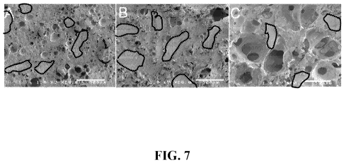
[0057] Figure 7 illustrates SEM micrographs of 50 wt% SDMBP/PUR foam scaffolds
at
(A) 35%, (B) 47%, and (C) 65% porosity. Example allograft bone particles are
traced in
black. Scale bar represents 500 m.
[0058] Figure 8 illustrates in vitro degradation of SDMBP/PUR scaffolds as a
function
of porosity. Samples were incubated in PBS at 37 C and mixed end over end, and
removed
and weighed at each time point.
[0059] Figure 9 illustrates CT images of H-SDMBP/PUR bone void filler
injected into
plug defects in the distal femurs of athymic rats. (A) - (B): Wound closed
immediately after
injection. (C) - (D): Wound closed 15 minutes after injection.
[0060] Figure 10 illustrates thin (e.g., 4 - 6 m) decalcified sections of the
composite
bone void filler injected in bilateral femoral plug defects in rats stained
with fuchsin red-
toluidene blue. (A) - (C): Low magnification images showing host bone (labeled
"HB", light
gray), residual polymer (labeled "P", dark gray), allograft particles embedded
in polymer that
have not been resorbed (labeled "A", light gray), regions of active remodeling
(labeled
"RM", medium gray) into the interior of the composite, osteoid (labeled "0",
medium gray),
and bone marrow (labeled "BM", medium gray) around the surface of the
material. Panel (A)
corresponds to the case where the wound was closed immediately after injection
of the
material, while Panels (B) and (C) correspond to the case where the wound was
closed 15
minutes after injection. (D) - (F): Higher magnification views of the implant
shown in Panel
(C). (G) - (H) Higher magnification of regions of active remodeling
characterized by
allograft (light gray) resorption, cells (dark gray), and collagen deposition
(medium gray).
Panel (G) shows the cellular pathway in an interior region of the composite,
while Panel (H)
shows the infiltration of cells into the composite from the bone marrow. In
the center of
Panel (H) there is an allograft particle undergoing active remodeling that
appears to be
embedded in polymer except for a small breach (labeled "B") where cells
infiltrated along the
allograft/polymer interface.
[0061] Figure 11 illustrates histological micrographs of Rabbit MBP/PUR
composite
plugs. In grayscale, old allograft is stained light gray, polymer is stained
black, and cells are
stained dark gray. As shown in Figure 1 IA, the boundary between the host bone
and the
implant is ambiguous. Extensive allograft bone resorption has occurred in this
region near
Page 15 of 79
CA 02745038 2011-05-27
WO 2010/059389 PCT/US2009/062621
the host bone. The combination of pores and pathways resulting from allograft
bone
resorption facilitated the infiltration of cells into the implant. Higher
magnification
micrographs (Figures 11B-11 E) further show cellular infiltration around
remnants of
polymer. Figure I 1D shows new bone formation around a piece of allograft as
evident by
osteoid lining the surface. Figure l lE shows extensive resorption of an
allograft particle
along with mineralization inside a pore.
DETAILED DESCRIPTION OF CERTAIN EMBODIMENTS
[0062] As used herein and in the appended claims, the singular forms "a," "an"
and "the"
include plural references unless the content clearly dictates otherwise. All
publications,
patent applications, patents, and other references mentioned herein are
incorporated by
reference in their entirety.
[0063] Bone/polyurethane composites described herein include bone (e.g., bone
particles), polyurethane, and in some embodiments, one or more additional
components (e.g.,
a porogen and/or a bioactive agent). As described below, bone and
biodegradable
polyurethanes are combined to form a porous composite (e.g., an osteoimplant).
In some
embodiments, porous composites retain strength and/or release bioactive agents
when present
in a body. In some embodiments, composites degrade and are replaced by new
tissue.
[0064] Inventive composites can be used in a large variety of clinical
applications, for
example, as bone void fillers, to repair or help healing of skeletal
deficiencies resulting from
trauma, tumors, surgery, iatrogenic, congenital, genetic, metabolic and
degenerative or
abnormal development, and inflammatory infection. In some embodiments,
inventive
composites promote cellular infiltration from adjacent osseous tissues, thus
accelerating the
remodeling process.
[0065] The invention also provides methods of preparing and using inventive
composites
as well as kits for preparing and/or administering inventive composites.
Inventive porous
composites may be prepared using any of a variety of methods. In some
embodiments,
inventive composites are prepared using a method that includes water as a
blowing agent. In
one embodiment, bone particles or other bone substitute materials are combined
with
polyurethanes and injected, extruded, molded, or similarly delivered to a
tissue site (e.g.,
bony defect) of a subject. Inventive composites are engineered to set in situ
to form a solid
composite that may have a desired or predetermined mechanical strength. In
certain
embodiments, polyurethane present in a composition or composite may include
monomers or
Page 16 of 79
CA 02745038 2011-05-27
WO 2010/059389 PCT/US2009/062621
pre-polymers. In some embodiments, polyurethane is a polymer that has been
rendered
formable through combination of two liquid components (i.e., a polyisocyanate
prepolymer
and a polyol).
Particulate Component
[0066] Particles used in accordance with the present invention may include a
bone-
derived material, an inorganic material, a bone substitute material, a
composite material, or
any combinations thereof.
[0067] Bone Particles. Any kind of bone and/or bone-derived particles may be
used in
the present invention. In some embodiments, bone particles employed in the
preparation of
bone particle-containing composites are obtained from cortical, cancellous,
and/or
corticocancellous bone. Bone particles may be obtained from any vertebrate.
Bone may be
of autogenous, allogenic, and/or xenogeneic origin. In certain embodiments,
bone particles
are autogenous, that is, bone particles are from the subject being treated. In
other
embodiments, bone particles are allogenic (e.g., from donors). In certain
embodiments, the
source of bone may be matched to the eventual recipient of inventive
composites (i.e., the
donor and recipient are of the same species). For example, human bone particle
is typically
used in a human subject. In certain embodiments, bone particles are obtained
from cortical
bone of allogenic origin. In certain embodiments, bone particles are obtained
from bone of
xenogeneic origin. Porcine and bovine bone are types of xenogeneic bone tissue
that can be
used individually or in combination as sources for bone particles and may
offer advantageous
properties. Xenogenic bone tissue may be combined with allogenic or autogenous
bone.
[0068] Bone particles can be formed by any process known to break down bone
into
small pieces. Exemplary processes for forming such particles include milling
whole bone to
produce fibers, chipping whole bone, cutting whole bone, grinding whole bone,
fracturing
whole bone in liquid nitrogen, or otherwise disintegrating the bone. Bone
particles can
optionally be sieved to produce particles of a specific size range. Bone
particles may be of
any shape or size. Exemplary shapes include spheroidal, plates, shards,
fibers, cuboidal,
sheets, rods, oval, strings, elongated particles, wedges, discs, rectangular,
polyhedral, etc.
[0069] In some embodiments, bone particles have a medium or mean diameter
about
1200 microns, 1100 microns, 1000 microns, 900 microns, 800 microns, 700
microns, 600
microns, 500 microns, 400 microns, 300 microns, 200 microns, 100 microns, etc.
In some
embodiments, diameters of bone particles are within a range between any of
such sizes. For
Page 17 of 79
CA 02745038 2011-05-27
WO 2010/059389 PCT/US2009/062621
example, medium or mean diameters of bone particles have a range from
approximately 100
microns to approximately 1000 microns.
[0070] As for irregularly shaped bone particles, recited dimension ranges may
represent
the length of the greatest or smallest dimension of the particle. As examples,
bone particles
can be pin shaped, with tapered ends having an average diameter of from about
100 microns
to about 500 microns. As will be appreciated by one of skill in the art, for
injectable
composites, the maximum particle size will depend in part on the size of the
cannula or
needle through which the material will be delivered.
[0071] In some embodiments, particle size distribution of bone particles
utilized in
accordance with the present inventions with respect to a mean value or a
median value may
be plus or minus, e.g., about 10% or less of the mean value, about 20% or less
of the mean
value, about 30% or less of the mean value, about 40% or less of the mean
value, about 50%
or less of the mean value, about 60% or less of the mean value, about 70% or
less of the mean
value, about 80% or less of the mean value, or about 90% or less of the mean
value.
[0072] In some embodiments, bone particles have a median or mean length of
about 1200
microns, 1100 microns, 1000 microns, 900 microns, 800 microns, 700 microns,
600 microns,
500 microns, 400 microns, 300 microns, 200 microns, 100 microns, etc. In some
embodiments, about 70, about 80 or about 90 percent of bone particles possess
a median or
mean length within a range of any of such sizes.
[0073] For bone particles that are fibers or other elongated particles, in
some
embodiments, at least about 90 percent of the particles possess a median or
mean length in
their greatest dimension in a range from approximately 100 microns to
approximately 1000
microns. Particles may possess a median or mean length to median or mean
thickness ratio
from at least about 5:1 up to about 500:1, for example, from at least about
50:1 up to about
500:1, or from about 50:1 up to about 100:1; and a median or mean length to
median or mean
width ratio of from about 10:1 to about 200:1 and, for example, from about
50:1 to about
100:1. In certain embodiments, bone particles are short fibers having a cross-
section of about
300 microns to about 100 microns and a length of about 0.1 mm to about 1 mm.
[0074] Processing of bone to provide particles may be adjusted to optimize for
the
desired size and/or distribution of bone particles. The properties of
resulting inventive
composites (e.g., mechanical properties) may also be engineered by adjusting
weight percent,
shapes, sizes, distribution, etc. of bone particles or other particles. For
example, an inventive
composite may be made more viscous and load bearing by including a higher
percentage of
particles.
Page 18 of 79
CA 02745038 2011-05-27
WO 2010/059389 PCT/US2009/062621
[0075] U.S. Patents 5,899,939; 5,507,813; 6,123,731; 6,294,041; 6,294,187;
6,332,779;
6,440,444; and 6,478,825; the contents of all of which are incorporated herein
by reference,
describe methods for preparing composites including allogenic bone for use in
orthopedic
applications.
[0076] Bone particles utilized in accordance with the present inventions may
be
demineralized, non-demineralized, mineralized, or anorganic. In some
embodiments, bone
particles are used "as is" in preparing inventive composites. In some
embodiments, bone
particles are defatted and disinfected. An exemplary defatting/disinfectant
solution is an
aqueous solution of ethanol. Other organic solvent may also be used in the
defatting and
disinfecting bone particles. For example, methanol, isopropanol, butanol, DMF,
DMSO,
diethyl ether, hexanes, glyme, tetrahydrofuran, chloroform, methylene
chloride, and carbon
tetrachloride may be used. In certain embodiments, a non-halogenated solvent
is used. A
defatting/disinfecant solution may also include a detergent (e.g., an aqueous
solution of a
detergent). Ordinarily, at least about 10 to about 40 percent by weight of
water (i.e., about 60
to about 90 weight percent of defatting agent such as alcohol) should be
present in the
defatting/disinfecting solution to produce optimal lipid removal and
disinfection within the
shortest period of time. An exemplary concentration range of a defatting
solution is from
about 60 to about 85 weight percent alcohol, for example, about 70 weight
percent alcohol.
[0077] In some embodiments, bone particles are demineralized. Bone particles
can be
optionally demineralized in accordance with known and/or conventional
procedures in order
to reduce their inorganic mineral content. Demineralization methods remove the
inorganic
mineral component of bone by employing acid solutions. Such methods are well
known in
the art, see for example, Reddi, et at., Proc. Nat. Acad. Sci., 1972, 69:1601-
1605, the contents
of which are incorporated herein by reference. The strength of the acid
solution, the shape
and dimensions of the bone particles and the duration of the demineralization
treatment will
determine the extent of demineralization. Reference in this regard is made to
Lewandrowski,
et at., J. Biomed. Mater. Res., 1996, 31:365-372 and U.S. Patent. 5,290,558,
the contents of
both of which are incorporated herein by reference.
[0078] In an exemplary defatting/disinfecting/demineralization procedure, bone
particles
are subjected to a defatting/disinfecting step, followed by an acid
demineralization step. An
exemplary defatting/disinfectant solution is an aqueous solution of ethanol.
In some
embodiments, at least about 10 to about 40 percent by weight of water (i.e.,
about 60 to about
90 weight percent of defatting agent such as alcohol) can be present in a
defatting/disinfecting solution to produce optimal lipid removal and
disinfection within a
Page 19 of 79
CA 02745038 2011-05-27
WO 2010/059389 PCT/US2009/062621
reasonable period of time. An exemplary concentration range of a defatting
solution is from
about 60 to about 85 weight percent alcohol, for example, about 70 weight
percent alcohol.
Ethanol is typically the alcohol used in this step; however, other alcohols
such as methanol,
propanol, isopropanol, denatured ethanol, etc. may also be used. Following
defatting, bone
particles can be immersed in acid over time to effect their demineralization.
The acid also
disinfects the bone by killing viruses, vegetative microorganisms, and spores.
Acids which
can be employed in this step include inorganic acids such as hydrochloric acid
and organic
acids such as peracetic acid. After acid treatment, demineralized bone
particles can be rinsed
with sterile water to remove residual amounts of acid and thereby raise the
pH. Bone
particles may be dried, for example, by lyophilization, before being
incorporated into a
composite. Bone particles may be stored under aseptic conditions, for example,
in a
lyophilized state, until they are used or sterilized using known methods
(e.g., gamma
irradiation) shortly before combining them with polyurethanes used in
inventive composites.
[0079] As utilized herein, the phrase "superficially demineralized" as applied
to the bone
particles refers to bone particles possessing at least about 90% by weight of
their original
inorganic mineral content. The phrase "partially demineralized" as applied to
the bone
particles refers to bone particles possessing from about 8% to about 90%
weight of their
original inorganic mineral content, and the phrase "fully demineralized" as
applied to the
bone particles refers to bone particles possessing less than about 8%,
preferably less than
about I%, by weight of their original inorganic mineral content. The
unmodified term
"demineralized" as applied to the bone particles is intended to cover any one
or combination
of the foregoing types of demineralized bone particles, that is, superficially
demineralized,
partially demineralized, or fully demineralized bone particles.
[0080] In alternative embodiments, surfaces of bone particles may be lightly
demineralized according to the procedures in our commonly owned U.S. Patent
Application,
U.S.S.N. 10/285,715, filed November 1, 2002, published as U.S. Patent
Publication No.
2003/0144743, on July 31, 2003, the contents of which are incorporated herein
by reference.
Even minimal demineralization, for example, of less than 5% removal of the
inorganic phase,
increases the hydroxylation of bone fibers and the surface concentration of
amine groups.
Demineralization may be so minimal, for example, less than 1%, that the
removal of the
calcium phosphate phase is almost undetectable. Rather, the enhanced surface
concentration
of reactive groups defines the extent of demineralization. This may be
measured, for
example, by titrating the reactive groups. Surface composition can also be
measured by x-ray
photoelectron spectroscopy (XPS), an experimental technique that measures the
atomic
Page 20 of 79
CA 02745038 2011-05-27
WO 2010/059389 PCT/US2009/062621
composition of the top 1 - 10 nm of the surface. In some embodiments, in a
polymerization
reaction that utilizes the exposed allograft surfaces to initiate a reaction,
the amount of
unreacted monomer remaining may be used to estimate reactivity of the
surfaces. Surface
reactivity may be assessed by a surrogate mechanical test, such as a peel test
of a treated
coupon of bone adhering to a polymer.
[0081] In certain embodiments, bone particles are subjected to a process that
partially or
totally removes their initial organic content to yield mineralized and
anorganic bone particles,
respectively. Different mineralization methods have been developed and are
known in the
are (Hurley, et at., Milit. Med. 1957, 101-104; Kershaw, Pharm. J. 6:537,
1963; and U.S.
Patent 4,882,149; each of which is incorporated herein by reference). For
example, a
mineralization procedure can include a de-greasing step followed by a basic
treatment (with
ammonia or another amine) to degrade residual proteins and a water washing
(U.S. Patent
5,417,975 and 5,573,771; both of which are incorporated herein by reference).
Another
example of a mineralization procedure includes a defatting step where bone
particles are
sonicated in 70% ethanol for 1-3 hours.
[0082] In some embodiments, bone particles can be modified in one or more
ways, e.g.,
their protein content can be augmented or modified as described, for example,
in U.S.
Patents. 4,743,259 and 4,902,296, the contents of both of which are
incorporated herein by
reference.
[0083] Mixtures or combinations of one or more of the foregoing types of bone
particles
can be employed. For example, one or more of the foregoing types of
demineralized bone
particles can be employed in combination with non-demineralized bone
particles, i.e., bone
particles that have not been subjected to a demineralization process, or
inorganic materials.
The amount of each individual type of bone particle employed can vary widely
depending on
the mechanical and biological properties desired. Thus, in some embodiments,
mixtures of
bone particles of various shapes, sizes, and/or degrees of demineralization
may be assembled
based on the desired mechanical, thermal, chemical, and biological properties
of a composite.
A desired balance between the various properties of composites (e.g., a
balance between
mechanical and biological properties) may be achieved by using different
combinations of
particles. Suitable amounts of various particle types can be readily
determined by those
skilled in the art on a case-by-case basis by routine experimentation.
[0084] The differential in strength, osteogenicity, and other properties
between partially
and fully demineralized bone particles on the one hand, and non-demineralized,
superficially
demineralized bone particles, inorganic ceramics, and other bone substitutes
on the other
Page 21 of 79
CA 02745038 2011-05-27
WO 2010/059389 PCT/US2009/062621
hand can be exploited. For example, in order to increase the compressive
strength of an
osteoimplant, the ratio of nondemineralized and/or superficially demineralized
bone particles
to partially or fully demineralized bone particles may favor the former, and
vice versa. Bone
particles in composites also play a biological role. Non-demineralized bone
particles bring
about new bone in-growth by osteoconduction. Demineralized bone particles
likewise play a
biological role in bringing about new bone in-growth by osteoinduction. Both
types of bone
particles are gradually remodeled and replaced by new host bone as degradation
of the
composite progresses over time. Thus, the use of various types of bone
particles can be used
to control the overall mechanical and biological properties, (e.g., strength,
osteoconductivity,
and/or osteoinductivity, etc.) of osteoimplants.
[0085] Surface Modification. Bone particles utilized in accordance with the
present
invention may be optionally treated to enhance their interaction with
polyurethanes and/or to
confer some properties to particle surface. While some bone particles will
interact readily
with monomers and be covalently linked to polyurethane matrices, it may be
desirable to
modify surface of bone particles to facilitate their incorporation into
polymers that do not
bond well to bone, such as poly(lactides). Surface modification may provide a
chemical
substance that is strongly bonded to the surface of bone, e.g., covalently
bonded to the
surface. Bone particles may, alternatively or additionally, be coated with a
material to
facilitate interaction with polymers of inventive composites.
[0086] In some embodiments, silane coupling agents are employed to link a
monomer or
initiator molecule to the surface of bone particles. Silane has at least two
sections, a set of
leaving groups and at least an active group. An active group may be connected
to the silicon
atom in the silane by an elongated tether group. An exemplary silane coupling
agent is 3-
trimethoxysilylpropylmethacrylate, available from Union Carbide. Three methoxy
groups
are leaving groups, and the methacrylate active group is connected to the
silicon atom by a
propyl tether group. In some embodiments, a leaving group is an alkoxy group
such as
methoxy or ethoxy. Depending on the solvent used to link the coupling agent to
bone
particles, hydrogen or alkyl groups such as methyl or ethyl may serve as
leaving groups. The
length of tethers determines the intimacy of connection between polymers and
bone particles.
By providing a spacer between bone particles and active groups, the tether
also reduces
competition between chemical groups at the particle surface and the active
group and makes
the active group more accessible to monomers during polymerization.
Page 22 of 79
CA 02745038 2011-05-27
WO 2010/059389 PCT/US2009/062621
[0087] In some embodiments, an active group is an analog of monomers of a
polymer
used in inventive composites. For example, amine active groups will be
incorporated into
polyurethane matrices, copolymers (e.g., polyesters, polycarbonates,
polycaprolactone), and
other polymer classes based on monomers that react with amines, even if the
polymer does
not contain an amine. Hydroxy-terminated silanes will be incorporated into
polyamino acids,
polyesters, polycaprolactone, polycarbonates, polyurethanes, and other polymer
classes that
include hydroxylated monomers. Aromatic active groups or active groups with
double bonds
will be incorporated into vinyl polymers and other polymers that grow by
radical
polymerization (e.g., polyacrylates, polymethacrylates). It is not necessary
that the active
group be monofunctional. Indeed, it may be preferable that active groups that
are to be
incorporated into polymers via step polymerization be difunctional. A silane
having two
amines, even if one is a secondary amine, will not terminate a polymer chain
but can react
with ends of two different polymer chains. Alternatively, the active group may
be branched
to provide two reactive groups in the primary position.
[0088] An exemplary list of silanes that may be used with the present
invention is
provided in U.S. Patent Publication No. 2004/0146543, the contents of which
are
incorporated herein by reference. Silanes are available from companies such as
Union
Carbide, AP Resources Co. (Seoul, South Korea), and BASF. Where a silane
contains a
potentially non-biocompatible moiety as the active group, it may be used to
tether a
biocompatible compound to bone particles using a reaction in which the non-
biocompatible
moiety is a leaving group. It may be desirable to attach the biocompatible
compound to the
silane before attaching the silane to the bone particle, regardless of whether
the silane is
biocompatible or not. The derivatized silanes may be mixed with silanes that
can be
incorporated directly into the polymer and reacted with bone particles,
coating the bone
particles with a mixture of "bioactive" silanes and "monomer" silanes. U.S.
Patent
6,399,693, the contents of which are incorporated herein by reference
discloses composites of
silane modified polyaromatic polymers and bone. In some embodiments, silane-
derivatized
polymers may be used in inventive composites instead of or in addition to
first silanizing
bone particles. In certain embodiments, polyurethanes and any copolymers used
in
accordance with the present inventions may not include silane modified
polyaromatic
polymers.
[0089] The active group of silanes may be incorporated directly into polymers
or may be
used to attach a second chemical group to bone particles. For example, if a
particular
Page 23 of 79
CA 02745038 2011-05-27
WO 2010/059389 PCT/US2009/062621
monomer polymerizes through a functional group that is not commercially
available as a
silane, the monomer may be attached to the active group.
[0090] Non-silane linkers may also be employed to produce composites according
to the
invention. For example, isocyanates will form covalent bonds with hydroxyl
groups on the
surface of hydroxyapatite ceramics (de Wijn, et at., Fifth World Biomaterials
Congress, May
29-June 2, 1996, Toronto, CA). Isocyanate anchors, with tethers and active
groups similar to
those described with respect to silanes, may be used to attach monomer-analogs
to bone
particles or to attach chemical groups that will link covalently or non-
covalently with a
polymer side group. Polyamines, organic compounds containing one or more
primary,
secondary, or tertiary amines, will also bind with both the bone particle
surface and many
monomer and polymer side groups. Polyamines and isocyanates may be obtained
from
Aldrich.
[0091] Alternatively or additionally, biologically active compounds such as a
biomolecule, a small molecule, or a bioactive agent may be attached to bone
particles through
a linker. For example, mercaptosilanes will react with sulfur atoms in
proteins to attach them
to bone particles. Aminated, hydroxylated, and carboxylated silanes will react
with a wide
variety functional groups. Of course, the linker may be optimized for the
compound being
attached to bone particles.
[0092] Biologically active molecules can modify non-mechanical properties of
inventive
composites as they degrade. For example, immobilization of a drug on bone
particles allows
it to be gradually released at an implant site as the composite degrades. Anti-
inflammatory
agents embedded within inventive composites will control inflammatory response
long after
an initial response to injection of the composites. For example, if a piece of
the composite
fractures several weeks after injection, immobilized compounds will reduce the
intensity of
any inflammatory response, and the composite will continue to degrade through
hydrolytic or
physiological processes. In some embodiments, compounds may also be
immobilized on the
bone particles that are designed to elicit a particular metabolic response or
to attract cells to
injection sites.
[0093] Some biomolecules, small molecules, and bioactive agents may also be
incorporated into polyurethane matrices used in inventive composites. For
example, many
amino acids have reactive side chains. The phenol group on tyrosine has been
exploited to
form polycarbonates, polyarylates, and polyiminocarbonates (see Pulapura, et
at.,
Biopolymers, 1992, 32: 411-417; and Hooper, et at., J. Bioactive and
Compatible Polymers,
1995, 10:327-340, the entire contents of both of which are incorporated herein
by reference).
Page 24 of 79
CA 02745038 2011-05-27
WO 2010/059389 PCT/US2009/062621
Amino acids such as lysine, arginine, hydroxylysine, proline, and
hydroxyproline also have
reactive groups and are essentially tri-functional. Amino acids such as
valine, which has an
isopropyl side chain, are still difunctional. Such amino acids may be attached
to the silane
and still leave one or two active groups available for incorporation into a
polymer.
[0094] Non-biologically active materials may also be attached to bone
particles. For
example, radiopaque (e.g., barium sulfate), luminescent (e.g., quantum dots),
or magnetically
active particles (e.g., iron oxide) may be attached to bone particles using
the techniques
described above. Mineralized bone particles are an inherently radiopaque
component of
some embodiments of present inventions, whereas demineralized bone particles,
another
optional component of inventive composites, are not radiopaque. To enhance
radiopacity of
inventive composites, mineralized bone particles can be used. Another way to
render
radiopaque the polymers utilized in accordance with the present inventions, is
to chemically
modify them such that a halogen (e.g., iodine) is chemically incorporated into
the
polyurethane matrices, as in U.S. patent application 10/952,202, now published
as U.S.
Patent Publication No. 2006-0034769, whose content is incorporated herein by
reference.
[0095] If a material, for example, a metal atom or cluster, cannot be produced
as a silane
or other group that reacts with bone particles, then a chelating agent may be
immobilized on
bone particle surface and allowed to form a chelate with the atom or cluster.
As bone
particles and polymers used in the present invention are resorbed, these non-
biodegradable
materials may be removed from tissue sites by natural metabolic processes,
allowing
degradation of the polymers and resorption of the bone particles to be tracked
using standard
medical diagnostic techniques.
[0096] In some embodiments, bone particle surface is chemically treated before
being
mixed with polyurethane. For example, non-demineralized bone particles may be
rinsed with
phosphoric acid, e.g., for 1 to 15 minutes in a 5-50% solution by volume.
Those skilled in
the art will recognize that the relative volume of bone particles and
phosphoric acid solution
(or any other solution used to treat bone particles), may be optimized
depending on the
desired level of surface treatment. Agitation will also increase the
uniformity of the
treatment both along individual particles and across an entire sample of
particles. A
phosphoric acid solution reacts with mineral components of bone particles to
coat the bone
particles with calcium phosphate, which may increase the affinity of the
surface for inorganic
coupling agents such as silanes and for polymer components of the composite.
As noted
above, bone particle surface may be partially demineralized to expose the
collagen fibers.
Page 25 of 79
CA 02745038 2011-05-27
WO 2010/059389 PCT/US2009/062621
[0097] Collagen fibers exposed by demineralization are typically relatively
inert but have
some exposed amino acid residues that can participate in reactions. Collagen
may be
rendered more reactive by fraying triple helical structures of the collagen to
increase exposed
surface area and number of exposed amino acid residues. This not only
increases surface
area of bone particles available for chemical reactions but also for their
mechanical
interactions with polymers as well. Rinsing partially demineralized bone
particles in an
alkaline solution will fray collagen fibrils. For example, bone particles may
be suspended in
water at a pH of about 10 for about 8 hours, after which the solution is
neutralized. One
skilled in the art will recognize that this time period may be increased or
decreased to adjust
the extent of fraying. Agitation, for example, in an ultrasonic bath, may
reduce the processing
time. Alternatively or additionally, bone particles may be sonicated with
water, surfactant,
alcohol, or some combination of these.
[0098] In some embodiments, collagen fibers at bone particle surface may be
cross-
linked. A variety of cross-linking techniques suitable for medical
applications are well
known in the art (see, for example, U.S. Patent 6,123,781, the contents of
which are
incorporated herein by reference). For example, compounds like 1-ethyl-3-(3-
dimethylaminopropyl) carbodiimide hydrochloride, either alone or in
combination with N-
hydroxysuccinimide (NHS) will crosslink collagen at physiologic or slightly
acidic pH (e.g.,
in pH 5.4 MES buffer). Acyl azides and genipin, a naturally occurring bicyclic
compound
including both carboxylate and hydroxyl groups, may also be used to cross-link
collagen
chains (see Simmons, et at, Biotechnol. Appl. Biochem., 1993, 17:23-29; PCT
Publication
W098/19718, the contents of both of which are incorporated herein by
reference).
Alternatively or additionally, hydroxymethyl phosphine groups on collagen may
be reacted
with the primary and secondary amines on neighboring chains (see U.S. Patent
No.
5,948,386, the entire contents of which are incorporated herein by reference).
Standard
cross-linking agents such as mono- and dialdehydes, polyepoxy compounds,
tanning agents
including polyvalent metallic oxides, organic tannins, and other plant derived
phenolic
oxides, chemicals for esterification or carboxyl groups followed by reaction
with hydrazide to
form activated acyl azide groups, dicyclohexyl carbodiimide and its
derivatives and other
heterobifunctional crosslinking agents, hexamethylene diisocyanate, and sugars
may also be
used to cross-link collagens. Bone particles are then washed to remove all
leachable traces of
materials. In other embodiments, enzymatic cross-linking agents may be used.
Additional
cross-linking methods include chemical reaction, irradiation, application of
heat,
dehydrothermal treatment, enzymatic treatment, etc. One skilled in the art
will easily be able
Page 26 of 79
CA 02745038 2011-05-27
WO 2010/059389 PCT/US2009/062621
to determine the optimal concentrations of cross-linking agents and incubation
times for the
desired degree of cross-linking.
[0099] Both frayed and unfrayed collagen fibers may be derivatized with
monomer, pre-
polymer, oligomer, polymer, initiator, and/or biologically active or inactive
compounds,
including but not limited to biomolecules, bioactive agents, small molecules,
inorganic
materials, minerals, through reactive amino acids on the collagen fiber such
as lysine,
arginine, hydroxylysine, proline, and hydroxyproline. Monomers that link via
step
polymerization may react with these amino acids via the same reactions through
which they
polymerize. Vinyl monomers and other monomers that polymerize by chain
polymerization
may react with these amino acids via their reactive pendant groups, leaving
the vinyl group
free to polymerize. Alternatively, or in addition, bone particles may be
treated to induce
calcium phosphate deposition and crystal formation on exposed collagen fibers.
Calcium
ions may be chelated by chemical moieties of the collagen fibers, and/or
calcium ions may
bind to the surface of the collagen fibers. James et al., Biomaterials 20:2203-
2313, 1999;
incorporated herein by reference. The calcium ions bound to the collagen
provides a
biocompatible surface, which allows for the attachment of cells as well as
crystal growth.
The polymer will interact with these fibers, increasing interfacial area and
improving the wet
strength of the composite.
[00100] In some embodiments, the surface treatments described above or
treatments such
as etching may be used to increase the surface area or surface roughness of
bone particles.
Such treatments increase the interfacial strength of the particle/polymer
interface by
increasing the surface area of the interface and/or the mechanical
interlocking of bone
particles and polyurethane. Such surface treatments may also be employed to
round the
shape or smooth the edges of bone particles to facilitate delivery of the
inventive composite.
Such treatment is particularly useful for injectable composites.
[00101] In some embodiments, surface treatments of bone particles are
optimized to
enhance covalent attractions between bone particles and polyurethanes. In some
embodiments, the surface treatment may be designed to enhance non-covalent
interactions
between bone particle and polyurethane matrix. Exemplary non-covalent
interactions include
electrostatic interactions, hydrogen bonding, pi-bond interactions,
hydrophobic interactions,
van der Waals interactions, and mechanical interlocking. For example, if a
protein or a
polysaccharide is immobilized on bone particle, the chains of polymer matrix
will become
physically entangled with long chains of the biological macromolecules when
they are
combined. Charged phosphate sites on the surface of bone particles, produced
by washing
Page 27 of 79
CA 02745038 2011-05-27
WO 2010/059389 PCT/US2009/062621
the bone particles in basic solution, will interact with the amino groups
present in many
biocompatible polymers, especially those based on amino acids. The pi-orbitals
on aromatic
groups immobilized on a bone particle will interact with double bonds and
aromatic groups of
the polymer.
[00102] Additional Particulate Materials. Any type of additional components
comprising
inorganic materials and/or other bone substitute materials (i.e., compositions
similar to
natural bone such as collagen, biocompatible polymers, osteoinductive agents,
other
commercial bone graft products, any composite graft, etc.), may be utilized in
the present
invention. Inorganic materials, including but not limited to, calcium
phosphate materials, and
other bone substitute materials, may also be exploited for use as particulate
inclusions in the
inventive composites. Exemplary materials utilized in accordance with the
present invention
include aragonite, dahlite, calcite, amorphous calcium carbonate, vaterite,
weddellite,
whewellite, struvite, urate, ferrihydrite, francolite, monohydrocalcite,
magnetite, goethite,
dentin, calcium carbonate, calcium sulfate, calcium phosphosilicate, sodium
phosphate,
calcium aluminate, calcium phosphate, hydroxyapatite, a-tricalcium phosphate,
dicalcium
phosphate, 0-tricalcium phosphate, tetracalcium phosphate, amorphous calcium
phosphate,
octacalcium phosphate, and BIOGLASSTM, a calcium phosphate silica glass
available from
U.S. Biomaterials Corporation. Substituted calcium phosphate phases are also
contemplated
for use with the invention, including but not limited to fluorapatite,
chlorapatite, magnesium-
substituted tricalcium phosphate, and carbonate hydroxyapatite. In certain
embodiments, the
inorganic material is a substituted form of hydroxyapatite. For example,
hydroxyapatite may
be substituted with other ions such as fluoride, chloride, magnesium, sodium,
potassium, and
groups such as silicates, silicon dioxides, carbonates, etc. Additional
calcium phosphate
phases suitable for use with the invention include those disclosed in U.S.
Patents RE 33,161
and RE 33,221 to Brown et al.; 4,880,610; 5,034,059; 5,047,031; 5,053,212;
5,129,905;
5,336,264; and 6,002,065 to Constantz et al.; 5,149,368; 5,262,166 and
5,462,722 to Liu et
al.; 5,525,148 and 5,542,973 to Chow et at., 5,717,006 and 6,001,394 to
Daculsi et at.,
5,605,713 to Boltong et at., 5,650,176 to Lee et at., and 6,206,957 to
Driessens et at, and
biologically-derived or biomimetic materials such as those identified in
Lowenstam HA,
Weiner S, On Biomineralization, Oxford University Press, 1989; each of which
is
incorporated herein by reference.
[00103] In some embodiments, a particulate composite material may be employed
to
combine with inventive composites in the present invention. For example,
inorganic
Page 28 of 79
CA 02745038 2011-05-27
WO 2010/059389 PCT/US2009/062621
materials such as those described above may be combined with proteins such as
bovine serum
albumin (BSA), collagen, or other extracellular matrix components to form a
composite. In
some embodiments, inorganic materials or bone-derived materials may be
combined with
synthetic or natural polymers to form a composite using the techniques
described in our co-
pending U.S. patent applications, U.S.S.N. 10/735,135, filed December 12,
2003; U.S.S.N.
10/681,65 1, filed October 8, 2003; and U.S.S.N. 10/639,912, filed August 12,
2003, the
contents of all of which are incorporated herein by reference.
Polymer Component
[00104] Synthetic polymers can be designed with properties targeted for a
given clinical
application. According to the present invention, polyurethanes (PUR) are a
useful class of
biomaterials due to the fact that they can be injectable or moldable as a
reactive liquid that
subsequently cures to form a porous composite. These materials also have
tunable
degradation rates, which are shown to be highly dependent on the choice of
polyol and
isocyanate components (Hafeman et at., Pharmaceutical Research
2008;25(10):2387-99;
Storey et at., JPoly Sci Pt A: Poly Chem 1994;32:2345-63; Skarja et at., JApp
Poly Sci
2000;75:1522-34). Polyurethanes have tunable mechanical properties, which can
also be
enhanced with the addition of bone particles and/or other components (Adhikari
et at.,
Biomaterials 2008;29:3762-70; Goma et at., JBiomed Mater Res Pt A
2003;67A(3):813-27)
and exhibit elastomeric rather than brittle mechanical properties.
[00105] Polyurethanes can be made by reacting together the components of a two-
component composition, one of which includes a polyisocyanate while the other
includes a
component having two or more hydroxyl groups (i.e., polyols) to react with the
polyisocyanate. For example, U.S. Pat. No. 6,306,177, discloses a method for
repairing a
tissue site using polyurethanes, the content of which is incorporated by
reference.
[00106] It is to be understood that by "a two-component composition" it means
a
composition comprising two essential types of polymer components. In some
embodiments,
such a composition may additionally comprise one or more other optional
components.
[00107] In some embodiments, polyurethane is a polymer that has been rendered
formable
through combination of two liquid components (i.e., a polyisocyanate
prepolymer and a
polyol). In some embodiments, a polyisocyanate prepolymer or a polyol may be a
molecule
with two or three isocyanate or hydroxyl groups respectively. In some
embodiments, a
polyisocyanate prepolymer or a polyol may have at least four isocyanate or
hydroxyl groups
respectively.
Page 29 of 79
CA 02745038 2011-05-27
WO 2010/059389 PCT/US2009/062621
[00108] Synthesis of porous polyurethane results from a balance of two
simultaneous
reactions. Reactions, in some embodiments, are illustrated below in Scheme 1.
One is a
gelling reaction, where an isocyanates and a polyester polyol react to form
urethane bonds.
The one is a blowing reaction. An isocyanate can react with water to form
carbon dioxide
gas, which acts as a lowing agent to form pores of polyurethane foam. The
relative rates of
these reactions determine the scaffold morphology, working time, and setting
time.
[00109] Exemplary gelling and blowing reactions in forming of polyurethane are
shown in
Scheme 1 below, where RI, R2 and R3, for example, can be oligomers of
caprolactone, lactide
and glycolide respectively.
Gelling reaction o O
0
JII ^ ^ ~ J~ JII~
0 '0, VO VO v~ 0 H' v v v O `N V V H~ `Oww
O=C=NO^/N=C=O H Ri O Y O Rp 'H O
+ o
N=C=O ~=O HN_~__O
R
O
lysine tri-isocyanate o
H
HN ',
polyester trio!
_O__1o o
Blowing reaction
t o t o t o
2 + H2O - " + CO
OCN NCO OCN H" H NCO
[00110] Biodegradable polyurethane scaffolds synthesized from aliphatic
polyisocyanates
been shown to degrade into non-toxic compounds and support cell attachment and
proliferation in vitro. A variety of polyurethane polymers suitable for use in
the present
invention are known in the art, many of which are listed in commonly owned
applications:
U.S. Ser. No. 10/759,904 filed on January 16, 2004, entitled `Biodegradable
polyurethanes
and use thereof' and published under No. 2005-0013793; U.S. Ser. No.
11/667,090 filed on
November 5, 2005, entitled "Degradable polyurethane foams" and published under
No. 2007-
029915 1; U.S. Ser. No. 12/298,158 filed on April 24, 2006, entitled
"Biodegradable
polyurethanes" and published under No. 2009-0221784; all of which are
incorporated herein
by reference. Polyurethanes described in U.S. Ser. No. 11/336,127 filed on
January 19, 2006
and published under No. 2006-0216323, which is entitled "Polyurethanes for
Osteoimplants"
and incorporated herein by reference, may be used in some embodiments of the
present
invention.
[00111] Polyurethanes foams may be prepared by contacting an isocyanate-
terminated
prepolymer (component 1, e.g, polyisocyanate prepolymer) with a hardener
(component 2)
Page 30 of 79
CA 02745038 2011-05-27
WO 2010/059389 PCT/US2009/062621
that includes at least a polyol (e.g., a polyester polyol) and water, a
catalyst and optionally, a
stabilizer, a porogen, PEG, etc. In some embodiments, multiple polyurethanes
(e.g., different
structures, difference molecular weights) may be used in a
composite/composition of the
present invention. In some embodiments, other biocompatible and/or
biodegradable
polymers may be used with polyurethanes in accordance with the present
invention. In some
embodiments, biocompatible co-polymers and/or polymer blends of any
combination thereof
may be exploited.
[00112] Polyurethanes used in accordance with the present invention can be
adjusted to
produce polymers having various physiochemical properties and morphologies
including, for
example, flexible foams, rigid foams, elastomers, coatings, adhesives, and
sealants. The
properties of polyurethanes are controlled by choice of the raw materials and
their relative
concentrations. For example, thermoplastic elastomers are characterized by a
low degree of
cross-linking and are typically segmented polymers, consisting of alternating
hard
(diisocyanates and chain extenders) and soft (polyols) segments. Thermoplastic
elastomers
are formed from the reaction of diisocyanates with long-chain diols and short-
chain diol or
diamine chain extenders. In some embodiments, pores in bone/polyurethanes
composites in
the present invention are interconnected and have a diameter ranging from
approximately 50
to approximately 1000 microns.
[00113] Prepolymer. Polyurethane prepolymers can be prepared by contacting a
polyol
with an excess (typically a large excess) of a polyisocyanate. The resulting
prepolymer
intermediate includes an adduct of polyisocyanates and polyols solubilized in
an excess of
polyisocyanates. Prepolymer can, in some embodiments, be formed by using an
approximately stoichiometric amount of polyisocyanates in forming a prepolymer
and
subsequently adding additional polyisocyanates. The prepolymer therefore
exhibits both low
viscosity, which facilitates processing, and improved miscibility as a result
of the
polyisocyanate-polyol adduct. Polyurethane networks can, for example, then be
prepared by
reactive liquid molding, wherein the prepolymer is contacted with a polyester
polyol to form
a reactive liquid mixture (i.e., a two-component composition) which is then
cast into a mold
and cured.
[00114] Polyisocyanates or multi-isocyanate compounds for use in the present
invention
include aliphatic polyisocyanates. Exemplary aliphatic polyisocyanates
include, but are not
limited to, lysine diisocyanate, an alkyl ester of lysine diisocyanate (for
example, the methyl
ester or the ethyl ester), lysine triisocyanate, hexamethylene diisocyanate,
isophorone
Page 31 of 79
CA 02745038 2011-05-27
WO 2010/059389 PCT/US2009/062621
diisocyanate (IPDI), 4,4'-dicyclohexylmethane diisocyanate (H12MDI),
cyclohexyl
diisocyanate, 2,2,4-(2,2,4)-trimethylhexamethylene diisocyanate (TMDI), dimers
prepared
form aliphatic polyisocyanates, trimers prepared from aliphatic
polyisocyanates and/or
mixtures thereof. In some embodiments, hexamethylene diisocyanate (HDI) trimer
sold as
Desmodur N3300A may be a polyisocyanate utilized in the present invention. In
some
embodiments, polyisocyanates used in the present invention includes
approximately 10 to
55% NCO by weight (wt % NCO=100*(42/Mw)). In some embodiments, polyisocyanates
include approximately 15 to 50% NCO.
[00115] Polyisocyanate prepolymers provide an additional degree of control
over the
structure of biodegradable polyurethanes. Prepared by reacting polyols with
isocyanates,
NCO-terminated prepolymers are oligomeric intermediates with isocyanate
functionality as
shown in Scheme 1. To increase reaction rates, urethane catalysts (e.g.,
tertiary amines)
and/or elevated temperatures (60-90 C) may be used (see, Guelcher, Tissue
Engineering:
Part B, 14 (1) 2008, pp 3-17).
[00116] Polyols used to react with polyisocyanates in preparation of NCO-
terminated
prepolymers refer to molecules having at least two functional groups to react
with isocyanate
groups. In some embodiments, polyols have a molecular weight of no more than
1000 g/mol.
In some embodiments, polyols have a rang of molecular weight between about 100
g/mol to
about 500 g/mol. In some embodiments, polyols have a rang of molecular weight
between
about 200 g/mol to about 400 g/mol. In certain embodiments, polyols (e.g.,
PEG) have a
molecular weight of about 200 g/mol. Exemplary polyols include, but are not
limited to,
PEG, glycerol, pentaerythritol, dipentaerythritol, tripentaerythritol, 1,2,4-
butanetriol,
trimethylolpropane, 1,2,3-trihydroxyhexane, myo-inositol, ascorbic acid, a
saccharide, or
sugar alcohols (e.g., mannitol, xylitol, sorbitol etc.). In some embodiments,
polyols may
comprise multiple chemical entities having reactive hydrogen functional groups
(e.g.,
hydroxy groups, primary amine groups and/or secondary amine groups) to react
with the
isocyanate functionality of polyisocyanates.
[00117] In some embodiments, polyisocyanate prepolymers are resorbable. Zhang
and
coworkers synthesized biodegradable lysine diisocyanate ethyl ester
(LDI)/glucose
polyurethane foams proposed for tissue engineering applications. In those
studies, NCO-
terminated prepolymers were prepared from LDI and glucose. The prepolymers
were chain-
extended with water to yield biocompatible foams which supported the growth of
rabbit bone
marrow stromal cells in vitro and were non-immunogenic in vivo. (see Zhang, et
at.,
Page 32 of 79
CA 02745038 2011-05-27
WO 2010/059389 PCT/US2009/062621
Biomaterials 21: 1247-1258 (2000), and Zhang, et at., Tiss. Eng., 8(5): 771-
785 (2002), both
of which are incorporated herein by reference).
[00118] In some embodiments, prepared polyisocyanate prepolymer can be a
flowable
liquid at processing conditions. In general, the processing temperature is no
greater than 60
C. In some embodiments, the processing temperature is ambient temperature (25
C).
[00119] Polyols. Polyols utilized in accordance with the present invention can
be amine-
and/or hydroxyl-terminated compounds and include, but are not limited to,
polyether polyols
(such as polyethylene glycol (PEG or PEO), polytetramethylene etherglycol
(PTMEG),
polypropylene oxide glycol (PPO)); amine-terminated polyethers; polyester
polyols (such as
polybutylene adipate, caprolactone polyesters, castor oil); and polycarbonates
(such as
poly(1,6-hexanediol) carbonate). In some embodiments, polyols may be (1)
molecules
having multiple hydroxyl or amine functionality, such as glucose,
polysaccharides, and castor
oil; and (2) molecules (such as fatty acids, triglycerides, and phospholipids)
that have been
hydroxylated by known chemical synthesis techniques to yield polyols.
[00120] Polyols used in the present invention may be polyester polyols. In
some
embodiments, polyester polyols may include polyalkylene glycol esters or
polyesters
prepared from cyclic esters. In some embodiments, polyester polyols may
include
poly(ethylene adipate), poly(ethylene glutarate), poly(ethylene azelate),
poly(trimethylene
glutarate), poly(pentamethylene glutarate), poly(diethylene glutarate),
poly(diethylene
adipate), poly(triethylene adipate), poly(1,2-propylene adipate), mixtures
thereof, and/or
copolymers thereof. In some embodiments, polyester polyols can include,
polyesters
prepared from caprolactone, glycolide, D, L-lactide, mixtures thereof, and/or
copolymers
thereof. In some embodiments, polyester polyols can, for example, include
polyesters
prepared from castor-oil. When polyurethanes degrade, their degradation
products can be the
polyols from which they were prepared from.
[00121] In some embodiments, polyester polyols can be miscible with prepared
prepolymers used in reactive liquid mixtures (i.e., two-component composition)
of the
present invention. In some embodiments, surfactants or other additives may be
included in
the reactive liquid mixtures to help homogenous mixing.
[00122] The glass transition temperature (Tg) of polyester polyols used in the
reactive
liquids to form polyurethanes can be less than 60 C, less than 37 C
(approximately human
body temperature) or even less than 25 C. In addition to affecting
flowability at processing
conditions, Tg can also affect degradation. In general, a Tg of greater than
approximately 37
Page 33 of 79
CA 02745038 2011-05-27
WO 2010/059389 PCT/US2009/062621
C will result in slower degradation within the body, while a Tg below
approximately 37 C
will result in faster degradation.
[00123] Molecular weight of polyester polyols used in the reactive liquids to
form
polyurethanes can, for example, be adjusted to control the mechanical
properties of
polyurethanes utilized in accordance with the present invention. In that
regard, using
polyester polyols of higher molecular weight results in greater compliance or
elasticity. In
some embodiments, polyester polyols used in the reactive liquids may have a
molecular
weight less than approximately 3000 Da. In certain embodiments, the molecular
weight may
be in the range of approximately 200 to 2500 Da or 300 to 2000 Da. In some
embodiments,
the molecular weight may be approximately in the range of approximately 450 to
1800 Da or
450 to 1200 Da.
[00124] In some embodiments, a polyester polyol comprise poly(caprolactone-co-
lactide-
co-glycolide), which has a molecular weight in a range of 200 Da to 2500 Da,
or 300 Da to
2000 Da.
[00125] In some embodiments, polyols may include multiply types of polyols
with
different structures, molecular weight, properties, etc.
[00126] Additional Components. In accordance with the present invention, two-
component compositions (i.e., polyprepolymers and polyols) to form porous
composites may
be used with other agents and/or catalysts. Zhang et at. have found that water
may be an
adequate blowing agent for a lysine diisocyanate/PEG/glycerol polyurethane
(see Zhang, et
at., Tissue Eng. 2003 (6):1143-57) and may also be used to form porous
structures in
polyurethanes. Other blowing agents include dry ice or other agents that
release carbon
dioxide or other gases into the composite. Alternatively, or in addition,
porogens (see detail
discussion below) such as salts may be mixed in with reagents and then
dissolved after
polymerization to leave behind small voids.
[00127] Two-component compositions and/or the prepared composites used in the
present
invention may include one or more additional components. In some embodiments,
inventive
compositions and/or composites may includes, water, a catalyst (e.g., gelling
catalyst,
blowing catalyst, etc.), a stabilizer, a plasticizer, a porogen, a chain
extender (for making of
polyurethanes), a pore opener (such as calcium stearate, to control pore
morphology), a
wetting or lubricating agent, etc. (See, U.S. Ser. No. 10/759,904 published
under No. 2005-
0013793, and U.S. Ser. No. 11/625,119 published under No. 2007-0191963; both
of which
are incorporated herein by reference).
Page 34 of 79
CA 02745038 2011-05-27
WO 2010/059389 PCT/US2009/062621
[00128] In some embodiments, inventive compositions and/or composites may
include
and/or be combined with a solid filler (e.g., carboxymethylcellulose (CMC) and
hyaluronic
acid (HA)). For example, when composites used in wound healing, solid fillers
can help
absorb excess moisture in the wounds from blood and serum and allow for proper
foaming.
[00129] In certain embodiments, additional biocompatible polymers (e.g., PEG)
or co-
polymers can be used with compositions and composites in the present
invention.
[00130] Water. Water may be a blowing agent to generate porous polyurethane-
based
composites. Porosity of bone/polymer composites increased with increasing
water content,
and biodegradation rate accelerated with decreasing polyester half-life,
thereby yielding a
family of materials with tunable properties that are usefull in the present
invention. See,
Guelcher et al., Tissue Engineering, 13(9), 2007, pp2321-2333, which is
incorporated by
reference.
[00131] In some embodiments, an amount of water is about 0.5, 1, 1.5, 2, 3, 4
5, 6, 7, 8, 9,
parts per hundred parts (pphp) polyol. In some embodiments, water has an
approximate
rang of any of such amounts.
[00132] Catalyst. In some embodiments, at least one catalyst is added to form
reactive
liquid mixture (i.e., two-component compositions). A catalyst, for example,
can be non-toxic
(in a concentration that may remain in the polymer).
[00133] A catalyst can, for example, be present in two-component compositions
in a
concentration in the range of approximately 0.5 to 5 parts per hundred parts
polyol (pphp)
and, for example, in the range of approximately 0.5 to 2, or 2 to 3 pphp. A
catalyst can, for
example, be an amine compound. In some embodiments, catalyst may be an
organometallic
compound or a tertiary amine compound. In some embodiments the catalyst may be
stannous
octoate (an organobismuth compound), triethylene diamine,
bis(dimethylaminoethyl)ether,
dimethylethanolamine, dibutyltin dilaurate, and Coscat organometallic
catalysts
manufactured by Vertullus (a bismuth based catalyst), or any combination
thereof.
[00134] Stabilizer. In some embodiments, a stabilizer is nontoxic (in a
concentration
remaining in the polyurethane foam) and can include a non-ionic surfactant, an
anionic
surfactant or combinations thereof. For example, a stabilizer can be a
polyethersiloxane, a
salt of a fatty sulfonic acid or a salt of a fatty acid. In certain
embodiments, a stabilizer is a
polyethersiloxane, and the concentration of polyethersiloxane in a reactive
liquid mixture
Page 35 of 79
CA 02745038 2011-05-27
WO 2010/059389 PCT/US2009/062621
can, for example, be in the range of approximately 0.25 to 4 parts per hundred
polyol. In
some embodiments, polyethersiloxane stabilizer are hydrolyzable.
[00135] In some embodiments, the stabilizer can be a salt of a fatty sulfonic
acid.
Concentration of a salt of the fatty sulfonic acid in a reactive liquid
mixture can be in the
range of approximately 0.5 to 5 parts per hundred polyol. Examples of suitable
stabilizers
include a sulfated castor oil or sodium ricinoleicsulfonate.
[00136] Stabilizers can be added to a reactive liquid mixture of the present
invention to,
for example, disperse prepolymers, polyols and other additional components,
stabilize the
rising carbon dioxide bubbles, and/or control pore sizes of inventive
composites. Although
there has been a great deal of study of stabilizers, the operation of
stabilizers during foaming
is not completely understood. Without limitation to any mechanism of
operation, it is
believed that stabilizers preserve the thermodynamically unstable state of a
polyurethane
foam during the time of rising by surface forces until the foam is hardened.
In that regard,
foam stabilizers lower the surface tension of the mixture of starting
materials and operate as
emulsifiers for the system. Stabilizers, catalysts and other polyurethane
reaction components
are discussed, for example, in Oertel, Gunter, ed., Polyurethane Handbook,
Hanser Gardner
Publications, Inc. Cincinnati, Ohio, 99-108 (1994). A specific effect of
stabilizers is believed
to be the formation of surfactant monolayers at the interface of higher
viscosity of bulk phase,
thereby increasing the elasticity of surface and stabilizing expanding foam
bubbles.
[00137] Chain extender. To prepare high-molecular-weight polymers, prepolymers
are
chain extended by adding a short-chain (e.g., <500 g/mol) polyamine or polyol.
In certain
embodiments, water may act as a chain extender. In some embodiments, addition
of chain
extenders with a functionality of two (e.g., diols and diamines) yields linear
alternating block
copolymers.
[00138] Plasticizer. In some embodiments, inventive compositions and/or
composites
include one or more plasticizers. Plasticizers are typically compounds added
to polymers or
plastics to soften them or make them more pliable. According to the present
invention,
plasticizers soften, make workable, or otherwise improve the handling
properties of polymers
or composites. Plasticizers also allow inventive composites to be moldable at
a lower
temperature, thereby avoiding heat induced tissue necrosis during
implantation. Plasticizer
may evaporate or otherwise diffuse out of the composite over time, thereby
allowing
composites to harden or set. Without being bound to any theory, plasticizer
are thought to
Page 36 of 79
CA 02745038 2011-05-27
WO 2010/059389 PCT/US2009/062621
work by embedding themselves between the chains of polymers. This forces
polymer chains
apart and thus lowers the glass transition temperature of polymers. In
general, the more
plasticizer added, the more flexible the resulting polymers or composites will
be.
[00139] In some embodiments, plasticizers are based on an ester of a
polycarboxylic acid
with linear or branched aliphatic alcohols of moderate chain length. For
example, some
plasticizers are adipate-based. Examples of adipate-based plasticizers include
bis(2-
ethylhexyl)adipate (DOA), dimethyl adipate (DMAD), monomethyl adipate (MMAD),
and
dioctyl adipate (DOA). Other plasticizers are based on maleates, sebacates, or
citrates such
as bibutyl maleate (DBM), diisobutylmaleate (DIBM), dibutyl sebacate (DBS),
triethyl citrate
(TEC), acetyl triethyl citrate (ATEC), tributyl citrate (TBC), acetyl tributyl
citrate (ATBC),
trioctyl citrate (TOC), acetyl trioctyl citrate (ATOC), trihexyl citrate
(THC), acetyl trihexyl
citrate (ATHC), butyryl trihexyl citrate (BTHC), and trimethylcitrate (TMC).
Other
plasticizers are phthalate based. Examples of phthalate-based plasticizers are
N-methyl
phthalate, bis(2-ethylhexyl) phthalate (DEHP), diisononyl phthalate (DINP),
bis(n-
butyl)phthalate (DBP), butyl benzyl phthalate (BBzP), diisodecyl phthalate
(DOP), diethyl
phthalate (DEP), diisobutyl phthalate (DIBP), and di-n-hexyl phthalate. Other
suitable
plasticizers include liquid polyhydroxy compounds such as glycerol,
polyethylene glycol
(PEG), triethylene glycol, sorbitol, monacetin, diacetin, and mixtures
thereof. Other
plasticizers include trimellitates (e.g., trimethyl trimellitate (TMTM), tri-
(2-ethylhexyl)
trimellitate (TEHTM-MG), tri-(n-octyl,n-decyl) trimellitate (ATM), tri-
(heptyl,nonyl)
trimellitate (LTM), n-octyl trimellitate (OTM)), benzoates, epoxidized
vegetable oils,
sulfonamides (e.g., N-ethyl toluene sulfonamide (ETSA), N-(2-hydroxypropyl)
benzene
sulfonamide (HP BSA), N-(n-butyl) butyl sulfonamide (BBSA-NBBS)),
organophosphates
(e.g., tricresyl phosphate (TCP), tributyl phosphate (TBP)),
glycols/polyethers (e.g.,
triethylene glycol dihexanoate, tetraethylene glycol diheptanoate), and
polymeric plasticizers.
Other plasticizers are described in Handbook of Plasticizers (G. Wypych, Ed.,
ChemTec
Publishing, 2004), which is incorporated herein by reference. In certain
embodiments, other
polymers are added to the composite as plasticizers. In certain particular
embodiments,
polymers with the same chemical structure as those used in the composite are
used but with
lower molecular weights to soften the overall composite. In other embodiments,
different
polymers with lower melting points and/or lower viscosities than those of the
polymer
component of the composite are used.
[00140] In some embodiments, polymers used as plasticizer are poly(ethylene
glycol)
(PEG). PEG used as a plasticizer is typically a low molecular weight PEG such
as those
Page 37 of 79
CA 02745038 2011-05-27
WO 2010/059389 PCT/US2009/062621
having an average molecular weight of 1000 to 10000 g/mol, for example, from
4000 to 8000
g/mol. In certain embodiments, PEG 4000, PEG 5000, PEG 6000, PEG 7000, PEG
8000 or
combinations thereof are used in inventive composites. For example,
plasticizer (PEG) is
useful in making more moldable composites that include poly(lactide), poly(D,L-
lactide),
poly(lactide-co-glycolide), poly(D,L-lactide-co-glycolide), or
poly(caprolactone). Plasticizer
may comprise 1-40% of inventive composites by weight. In some embodiments, the
plasticizer is 10-30% by weight. In some embodiments, the plasticizer is
approximately 10%,
15%, 20%, 25%, 30% or 40% by weight. In other embodiments, a plasticizer is
not used in
the composite. For example, in some polycaprolactone-containing composites, a
plasticizer
is not used.
[00141] In some embodiments, inert plasticizers may be used. In some
embodiments, a
plasticizer may not be used in the present invention.
[00142] Porogen. Porosity of inventive composites may be accomplished using
any
means known in the art. Exemplary methods of creating porosity in a composite
include, but
are not limited to, particular leaching processes, gas foaming processing,
supercritical carbon
dioxide processing, sintering, phase transformation, freeze-drying, cross-
linking, molding,
porogen melting, polymerization, melt-blowing, and salt fusion (Murphy et at.,
Tissue
Engineering 8(1):43-52, 2002; incorporated herein by reference). For a review,
see
Karageorgiou et at., Biomaterials 26:5474-5491, 2005; incorporated herein by
reference.
Porosity may be a feature of inventive composites during manufacture or before
implantation,
or porosity may only be available after implantation. For example, a implanted
composite
may include latent pores. These latent pores may arise from including porogens
in the
composite.
[00143] Porogens may be any chemical compound that will reserve a space within
the
composite while the composite is being molded and will diffuse, dissolve,
and/or degrade
prior to or after implantation or injection leaving a pore in the composite.
Porogens may
have the property of not being appreciably changed in shape and/or size during
the procedure
to make the composite moldable. For example, a porogen should retain its shape
during the
heating of the composite to make it moldable. Therefore, a porogen does not
melt upon
heating of the composite to make it moldable. In certain embodiments, a
porogen has a
melting point greater than about 60 C, greater than about 70 C, greater than
about 80 C,
greater than about 85 C, or greater than about 90 C.
Page 38 of 79
CA 02745038 2011-05-27
WO 2010/059389 PCT/US2009/062621
[00144] Porogens may be of any shape or size. A porogen may be spheroidal,
cuboidal,
rectangular, elonganted, tubular, fibrous, disc-shaped, platelet-shaped,
polygonal, etc. In
certain embodiments, the porogen is granular with a diameter ranging from
approximately
100 microns to approximately 800 microns. In certain embodiments, a porogen is
elongated,
tubular, or fibrous. Such porogens provide increased connectivity of pores of
inventive
composite and/or also allow for a lesser percentage of the porogen in the
composite.
[00145] Amount of porogens may vary in inventive composite from 1% to 80% by
weight.
In certain embodiments, the plasticizer makes up from about 5% to about 80% by
weight of
the composite. In certain embodiments, a plasticizer makes up from about 10%
to about 50%
by weight of the composite. Pores in inventive composites are thought to
improve the
osteoinductivity or osteoconductivity of the composite by providing holes for
cells such as
osteoblasts, osteoclasts, fibroblasts, cells of the osteoblast lineage, stem
cells, etc. Pores
provide inventive composites with biological in growth capacity. Pores may
also provide for
easier degradation of inventive composites as bone is formed and/or remodeled.
In some
embodiments, a porogen is biocompatible.
[00146] A porogen may be a gas, liquid, or solid. Exemplary gases that may act
as
porogens include carbon dioxide, nitrogen, argon, or air. Exemplary liquids
include water,
organic solvents, or biological fluids (e.g., blood, lymph, plasma). Gaseous
or liquid porogen
may diffuse out of the osteoimplant before or after implantation thereby
providing pores for
biological in-growth. Solid porogens may be crystalline or amorphous. Examples
of
possible solid porogens include water soluble compounds. Exemplary porogens
include
carbohydrates (e.g., sorbitol, dextran (poly(dextrose)), starch), salts, sugar
alcohols, natural
polymers, synthetic polymers, and small molecules.
[00147] In some embodiments, carbohydrates are used as porogens in inventive
composites. A carbohydrate may be a monosaccharide, disaccharide, or
polysaccharide. The
carbohydrate may be a natural or synthetic carbohydrate. In some embodiments,
the
carbohydrate is a biocompatible, biodegradable carbohydrate. In certain
embodiments, the
carbohydrate is a polysaccharide. Exemplary polysaccharides include cellulose,
starch,
amylose, dextran, poly(dextrose), glycogen, etc.
[00148] In certain embodiments, a polysaccharide is dextran. Very high
molecular weight
dextran has been found particularly useful as a porogen. For example, the
molecular weight
of the dextran may range from about 500,000 g/mol to about 10,000,000 g/mol,
preferably
from about 1,000,000 g/mol to about 3,000,000 g/mol. In certain embodiments,
the dextran
has a molecular weight of approximately 2,000,000 g/mol. Dextrans with a
molecular weight
Page 39 of 79
CA 02745038 2011-05-27
WO 2010/059389 PCT/US2009/062621
higher than 10,000,000 g/mol may also be used as porogens. Dextran may be used
in any
form (e.g., particles, granules, fibers, elongated fibers) as a porogen. In
certain embodiments,
fibers or elongated fibers of dextran are used as a porogen in inventive
composites. Fibers of
dextran may be formed using any known method including extrusion and
precipitation.
Fibers may be prepared by precipitation by adding an aqueous solution of
dextran (e.g., 5-
25% dextran) to a less polar solvent such as a 90-100% alcohol (e.g., ethanol)
solution. The
dextran precipitates out in fibers that are particularly useful as porogens in
the inventive
composite. Once the composite with dextran as a porogen is implanted into a
subject, the
dextran dissolves away very quickly. Within approximately 24 hours,
substantially all of
dextran is out of composites leaving behind pores in the osteoimplant
composite. An
advantage of using dextran in a composite is that dextran exhibits a
hemostatic property in
extravascular space. Therefore, dextran in a composite can decrease bleeding
at or near the
site of implantation.
[00149] Small molecules including pharmaceutical agents may also be used as
porogens in
the inventive composites. Examples of polymers that may be used as
plasticizers include
poly(vinyl pyrollidone), pullulan, poly(glycolide), poly(lactide), and
poly(lactide-co-
glycolide). Typically low molecular weight polymers are used as porogens. In
certain
embodiments, a porogen is poly(vinyl pyrrolidone) or a derivative thereof.
Plasticizers that
are removed faster than the surrounding composite can also be considered
porogens.
Components to Deliver
[00150] Alternatively or additionally, composites of the present invention may
have one or
more components to deliver when implanted, including biomolecules, small
molecules,
bioactive agents, etc., to promote bone growth and connective tissue
regeneration, and/or to
accelerate healing. Examples of materials that can be incorporated include
chemotactic
factors, angiogenic factors, bone cell inducers and stimulators, including the
general class of
cytokines such as the TGF-(3 superfamily of bone growth factors, the family of
bone
morphogenic proteins, osteoinductors, and/or bone marrow or bone forming
precursor cells,
isolated using standard techniques. Sources and amounts of such materials that
can be
included are known to those skilled in the art.
[00151] Biologically active materials, comprising biomolecules, small
molecules, and
bioactive agents may also be included in inventive composites to, for example,
stimulate
particular metabolic functions, recruit cells, or reduce inflammation. For
example, nucleic
Page 40 of 79
CA 02745038 2011-05-27
WO 2010/059389 PCT/US2009/062621
acid vectors, including plasmids and viral vectors, that will be introduced
into the patient's
cells and cause the production of growth factors such as bone morphogenetic
proteins may be
included in a composite. Biologically active agents include, but are not
limited to, antiviral
agent, antimicrobial agent, antibiotic agent, amino acid, peptide, protein,
glycoprotein,
lipoprotein, antibody, steroidal compound, antibiotic, antimycotic, cytokine,
vitamin,
carbohydrate, lipid, extracellular matrix, extracellular matrix component,
chemotherapeutic
agent, cytotoxic agent, growth factor, anti-rejection agent, analgesic, anti-
inflammatory
agent, viral vector, protein synthesis co-factor, hormone, endocrine tissue,
synthesizer,
enzyme, polymer-cell scaffolding agent with parenchymal cells, angiogenic
drug, collagen
lattice, antigenic agent, cytoskeletal agent, mesenchymal stem cells, bone
digester, antitumor
agent, cellular attractant, fibronectin, growth hormone cellular attachment
agent,
immunosuppressant, nucleic acid, surface active agent, hydroxyapatite, and
penetraction
enhancer. Additional exemplary substances include chemotactic factors,
angiogenic factors,
analgesics, antibiotics, anti-inflammatory agents, bone morphogenic proteins,
and other
growth factors that promote cell-directed degradation or remodeling of the
polymer phase of
the composite and/or development of new tissue (e.g., bone). RNAi or other
technologies
may also be used to reduce the production of various factors.
[00152] In some embodiments, inventive composites include antibiotics.
Antibiotics may
be bacteriocidial or bacteriostatic. An anti-microbial agent may be included
in composites.
For example, anti-viral agents, anti-protazoal agents, anti-parasitic agents,
etc. may be
include in composites. Other suitable biostatic/biocidal agents include
antibiotics, povidone,
sugars, and mixtures thereof. Exemplary antibiotics include, but not limit to,
Amikacin,
Gentamicin, Kanamycin, Neomycin, Netilmicin, Streptomycin, Tobramycin,
Paromomycin,
Geldanamycin, Herbimycin, Loravabef, etc. (See, The Merck Manual of Medical
Information -
Home Edition, 1999).
[00153] Inventive composites may also be seeded with cells. In some
embodiments, a
patient's own cells are obtained and used in inventive composites. Certain
types of cells
(e.g., osteoblasts, fibroblasts, stem cells, cells of the osteoblast lineage,
etc.) may be selected
for use in the composite. Cells may be harvested from marrow, blood, fat,
bone, muscle,
connective tissue, skin, or other tissues or organs. In some embodiments, a
patient's own
cells may be harvested, optionally selected, expanded, and used in the
inventive composite.
In other embodiments, a patient's cells may be harvested, selected without
expansion, and
used in the inventive composite. Alternatively, exogenous cells may be
employed.
Exemplary cells for use with the invention include mesenchymal stem cells and
connective
Page 41 of 79
CA 02745038 2011-05-27
WO 2010/059389 PCT/US2009/062621
tissue cells, including osteoblasts, osteoclasts, fibroblasts, preosteoblasts,
and partially
differentiated cells of the osteoblast lineage. Cells may be genetically
engineered. For
example, cells may be engineered to produce a bone morphogenic protein.
[00154] In some embodiments, inventive composites may include a composite
material
comprising a component to deliver. For example, a composite materials can be a
biomolecule (e.g., a protein) encapsulated in a polymeric microsphere or
nanoparticles. In
certain embodiments, BMP-2 encapsulated in PLGA microspheres may be embedded
in a
bone/polyurethane composite used in accordance with the present invention.
Sustained
release of BMP-2 can be achieved due to the diffusional barriers presented by
both the PLGA
and Polyurethane of the inventive composite. Thus, release kinetics of growth
factors (e.g.,
BMP-2) can be tuned by varying size of PLGA microspheres and porosity of
polyurethane
composite.
[00155] To enhance biodegradation in vivo, composites of the present invention
can also
include different enzymes. Examples of suitable enzymes or similar reagents
are proteases or
hydrolases with ester-hydrolyzing capabilities. Such enzymes include, but are
not limited to,
proteinase K, bromelaine, pronase E, cellulase, dextranase, elastase, plasmin
streptokinase,
trypsin, chymotrypsin, papain, chymopapain, collagenase, subtilisin,
chlostridopeptidase A,
ficin, carboxypeptidase A, pectinase, pectinesterase, an oxireductase, an
oxidase, or the like.
The inclusion of an appropriate amount of such a degradation enhancing agent
can be used to
regulate implant duration.
[00156] Components to deliver may not be covalently bonded to a component of
the
composite. In some embodiments, components may be selectively distributed on
or near the
surface of inventive composites using the layering techniques described above.
While
surface of inventive composite will be mixed somewhat as the composite is
manipulated in
implant site, thickness of the surface layer will ensure that at least a
portion of the surface
layer of the composite remains at surface of the implant. Alternatively or in
addition,
biologically active components may be covalently linked to the bone particles
before
combination with the polymer. As discussed above, for example, silane coupling
agents
having amine, carboxyl, hydroxyl, or mercapto groups may be attached to the
bone particles
through the silane and then to reactive groups on a biomolecule, small
molecule, or bioactive
agent.
Preparation of Composite
Page 42 of 79
CA 02745038 2011-05-27
WO 2010/059389 PCT/US2009/062621
[00157] In general, inventive composites are prepared by combining particles,
polymers
and optionally any additional components. To form inventive composites,
particles as
discussed herein may be combined with a reactive liquid (i.e., a two-component
composition)
thereby forming a naturally injectable or moldable composite or a composite
that can be
made injectable or moldable. Alternatively, particles may be combined with
polyisocyanate
prepolymers or polyols first and then combined with other components.
[00158] In some embodiments, particles may be combined first with a hardener
that
includes polyols, water, catalysts and optionally a solvent, a diluent, a
stabilizer, a porogen, a
plasticizer, etc., and then combined with a polyisocyanate prepolymer. In some
embodiments, a hardener (e.g., a polyol, water and a catalyst) may be mixed
with a
prepolymer, followed by addition of particles. In some embodiments, in order
to enhance
storage stability of two-component compositions, the two (liquid) component
process may be
modified to an alternative three (liquid)-component process wherein a catalyst
and water may
be dissolved in a solution separating from reactive polyols. For example,
polyester polyols
may be first mixed with a solution of a catalyst and water, followed by
addition of bone
particles, and finally addition of NCO-terminated prepolymers.
[00159] In some embodiments, additional components or components to be
delivered may
be combined with a reactive liquid prior to injection. In some embodiments,
they may be
combined with one of polymer precursors (i.e., prepolymers and polyols) prior
to mixing the
precursors in forming of a reactive liquid/paste.
[00160] Porous composites can be prepared by incorporating a small amount
(e.g., <5
wt%) of water which reacts with prepolymers to form carbon dioxide, a
biocompativle
blowing agent. Resulting reactive liquid/paste may be injectable through a 12-
ga syringe
needle into molds or targeted site to set in situ. In some embodiments, gel
time is great than
3 min, 4 min, 5 min, 6 min, 7 min, or 8 min. In some embodiments, cure time is
less than 20
min, 18 min, 16 min, 14 min, 12 min, or 10 min.
[00161] In some embodiments, catalysts can be used to assist forming porous
composites.
In general, the more blowing catalyst used, the high porosity of inventive
composites may be
achieved. In certain embodiments, surprisingly, surface demineralized bone
particles may
have a dramatic effect on the porosity. Without being bound to any theory, it
is believed that
the lower porosities achieved with surface demineralized bone particles in the
absence of
blowing catalysts result from adsorption of water to the hygroscopic
demineralized layer on
the surface of bones.
Page 43 of 79
CA 02745038 2011-05-27
WO 2010/059389 PCT/US2009/062621
[00162] Polymers and particles may be combined by any method known to those
skilled in
the art. For example, a homogenous mixture of polymers and/or polymer
precursors (e.g.,
prepolymers, polyols, etc.) and particles may be pressed together at ambient
or elevated
temperatures. At elevated temperatures, a process may also be accomplished
without
pressure. In some embodiments, polymers or precursors are not held at a
temperature of
greater than approximately 60 C for a significant time during mixing to
prevent thermal
damage to any biological component (e.g., growth factors or cells) of a
composite. In some
embodiments, temperature is not a concern because particles and polymer
precursors used in
the present invention have a low reaction exotherm.
[00163] Alternatively or in addition, particles may be mixed or folded into a
polymer
softened by heat or a solvent. Alternatively, a moldable polymer may be formed
into a sheet
that is then covered with a layer of particles. Particles may then be forced
into the polymer
sheet using pressure. In another embodiment, particles are individually coated
with polymers
or polymer precursors, for example, using a tumbler, spray coater, or a
fluidized bed, before
being mixed with a larger quantity of polymer. This facilitates even coating
of the particles
and improves integration of the particles and polymer component of the
composite.
[00164] After combination with particles, polymers may be further modified by
further
cross-linking or polymerization to form a composite in which the polymer is
covalently
linked to the particles. In some embodiments, composition hardens in a solvent-
free
condition. In some embodiments, compositions are a polymer/solvent mixture
that hardens
when a solvent is removed (e.g., when a solvent is allowed to evaporate or
diffuse away).
Exemplary solvents include but are not limited to alcohols (e.g., methanol,
ethanol, propanol,
butanol, hexanol, etc.), water, saline, DMF, DMSO, glycerol, and PEG. In
certain
embodiments, a solvent is a biological fluid such as blood, plasma, serum,
marrow, etc. In
certain embodiments, an inventive composite is heated above the melting or
glass transition
temperature of one or more of its components and becomes set after
implantation as it cools.
In certain embodiments, an inventive composite is set by exposing a composite
to a heat
source, or irradiating it with microwaves, IR rays, or UV light. Particles may
also be mixed
with a polymer that is sufficiently pliable to combine with the particles but
that may require
further treatment, for example, combination with a solvent or heating, to
become a injectable
or moldable composition. For example, a composition may be combined and
injection
molded, injected, extruded, laminated, sheet formed, foamed, or processed
using other
techniques known to those skilled in the art. In some embodiments, reaction
injection
molding methods, in which polymer precursors (e.g., polyisocyanate prepolymer,
a polyol)
Page 44 of 79
CA 02745038 2011-05-27
WO 2010/059389 PCT/US2009/062621
are separately charged into a mold under precisely defined conditions, may be
employed. For
example, bone particles may be added to a precursor, or it may be separately
charged into a
mold and precursor materials added afterwards. Careful control of relative
amounts of
various components and reaction conditions may be desired to limit the amount
of unreacted
material in a composite. Post-cure processes known to those skilled in the art
may also be
employed. A partially polymerized polyurethane precursor may be more
completely
polymerized or cross-linked after combination with hydroxylated or aminated
materials or
included materials (e.g., a particulate, any components to deliver, etc.).
[00165] In some embodiments, an inventive composite is produced with a
injectable
composition and then set in situ. For example, cross-link density of a low
molecular weight
polymer may be increased by exposing it to electromagnetic radiation (e.g., UV
light) or an
alternative energy source. Alternatively or additionally, a photoactive cross-
linking agent,
chemical cross-linking agent, additional monomer, or combinations thereof may
be mixed
into inventive composites. Exposure to UV light after a composition is
injected into an
implant site will increase one or both of molecular weight and cross-link
density, stiffening
polymers (i.e., polyurethanes) and thereby a composite. Polymer components of
inventive
composites used in the present invention may be softened by a solvent, e.g.,
ethanol. If a
biocompatible solvent is used, polyurethanes may be hardened in situ. In some
embodiments,
as a composite sets, solvent leaving the composite is released into
surrounding tissue without
causing undesirable side effects such as irritation or an inflammatory
response. In some
embodiments, compositions utilized in the present invention becomes moldable
at an elevated
temperature into a pre-determined shape. Composites may become set when
composites are
implanted and allowed to cool to body temperature (approximately 37 C).
[00166] The invention also provides methods of preparing inventive composites
by
combining bone particles and polyurethane precursors and resulting in
naturally flowable
compositions. Alternatively or additionally, the invention provides methods to
make a
porous composite include adding a solvent or pharmaceutically acceptable
excipient to render
a flowable or moldable composition. Such a composition may then be injected or
placed into
the site of implantation. As solvent or excipient diffuses out of the
composite, it may become
set in place.
[00167] Polymer processing techniques may also be used to combine particles
with a
polyurethane or precursors (e.g., polyisocyanates and polyols). In some
embodiments, a
composition of polyurethane may be rendered formable (e.g., by heating or with
a solvent)
and combined with particles by injection molding or extrusion forming.
Alternatively,
Page 45 of 79
CA 02745038 2011-05-27
WO 2010/059389 PCT/US2009/062621
polyurethanes and bone particles may be mixed in a solvent and cast with or
without
pressure. For example, a solvent may be dichloromethane. In some embodiments,
a
composition of particle and polymer utilized in the present invention is
naturally injectable or
moldable in a solvent-free condition.
[00168] In some embodiments, particles may be mixed with a polymer precursor
according to standard composite processing techniques. For example, regularly
shaped
particles may simply be suspended in a precursor. A polymer precursor may be
mechanically
stirred to distribute the particles or bubbled with a gas, preferably one that
is oxygen- and
moisture-free. Once components of a composition are mixed, it may be desirable
to store it
in a container that imparts a static pressure to prevent separation of the
particles and the
polymer precursor, which may have different densities. In some embodiments,
distribution
and particle/polymer ratio may be optimized to produce at least one continuous
path through
a composite along particles.
[00169] Interaction of polymer components with bone particles may also be
enhanced by
coating individual particles with a polymer precursor before combining them
with bulk
precursors. The coating enhances the association of the polymer component of
the composite
with the particles. For example, individual particles may be spray coated with
a monomer or
prepolymer. Alternatively, the individual particles may be coated using a
tumbler-particles
and a solid polymer material are tumbled together to coat the particles. A
fluidized bed
coater may also be used to coat the particles. In addition, the particles may
simply be dipped
into liquid or powdered polymer precursor. All of these techniques will be
familiar to those
skilled in the art.
[00170] In some embodiments, it may be desirable to infiltrate a polymer or
polymer
precursor into vascular and/or interstitial structure of bone particles or
into bone-derived
tissues. Vascular structure of bone includes such structures such as osteocyte
lacunae,
Haversian canals, Volksmann's canals, canaliculi and similar structures.
Interstitial structure
of bone particles includes spaces between trabeculae and similar features.
Many of
monomers and precursors (e.g., polyisocyanate prepolymers, polyols) suggested
for use with
the invention are sufficiently flowable to penetrate through the channels and
pores of
trabecular bone. Some may even penetrate into trabeculae or into mineralized
fibrils of
cortical bone. Thus, it may be necessary to incubate bone particles in
polyurethane
precursors for a period of time to accomplish infiltration. In certain
embodiments,
polyurethane itself is sufficiently flowable that it can penetrate channels
and pores of bone.
In certain embodiments, polyurethane may also be heated or combined with a
solvent to
Page 46 of 79
CA 02745038 2011-05-27
WO 2010/059389 PCT/US2009/062621
make it more flowable for this purpose. Other ceramic materials and/or other
bone-substitute
materials employed as a particulate phase may also include porosity that can
be infiltrated as
described herein.
[00171] Inventive composites utilized in the present invention may include
practically any
ratio of polyurethane and bone particles, for example, between about 5 wt% and
about 95
wt% bone particles. In some embodiments, composites may include about 40 wt%
to about
45 wt% bone particles, about 45 wt% to about 50 wt% bone particles or about 50
wt% to
about 55 wt% bone particles. In some embodiments, composites may include about
55 wt%
to about 70 wt% bone particles. In some embodiments, composites may include
about 70
wt% to about 90 wt% bone particles. In some embodiments, composites may
include at least
approximately 40 wt%, 45 wt%, 50 wt%, or 55 wt% of bone particles. In certain
embodiments, such weight percentages refer to weight of bone particles and
other particulates
such as calcium phosphate.
[00172] In some embodiments, composites may include at least approximately 30
vol%,
35 vol%, 40 vol%, or 50 vol% of bone particles. In some embodiments, a volume
percentage
of bone particles in composite in accordance with the present invention may be
about 30
vol%, 35 vol%, 40 vol%, 50 vol%, 60 vol%, 70 vol% or between any volume
percentages of
above. In some embodiments, injectable composites in accordance with the
present invention
may have a volume percentage (fraction) of at least approximately 36 vol% of
bone particles
and/or other particulate materials (e.g., calcium phosphate). In some
embodiments, volume
percentages (fractions) of bone particles and/or other particulate materials
in porous
composites in the present invention may be less than 64 vol%. In certain
embodiments, for a
certain volume percentage, corresponding weight percentage of bone particles
and/or other
particulate materials varies depending on density of particulate components.
[00173] Desired proportion may depend on factors such as injection sites,
shape and size
of the particles, how evenly polymer is distributed among particles, desired
flowability of
composites, desired handling of composites, desired moldability of composites,
and
mechanical and degradation properties of composites. The proportions of
polymers and
particles can influence various characteristics of the composite, for example,
its mechanical
properties, including fatigue strength, the degradation rate, and the rate of
biological
incorporation. In addition, the cellular response to the composite will vary
with the
proportion of polymer and particles. In some embodiments, the desired
proportion of
particles may be determined not only by the desired biological properties of
the injected
material but by the desired mechanical properties of the injected material.
That is, an
Page 47 of 79
CA 02745038 2011-05-27
WO 2010/059389 PCT/US2009/062621
increased proportion of particles will increase the viscosity of the
composite, making it more
difficult to inject or mold. A larger proportion of particles having a wide
size distribution
may give similar properties to a mixture having a smaller proportion of more
evenly sized
particles.
[00174] Inventive composites of the present invention can exhibit high degrees
of porosity
over a wide range of effective pore sizes. Thus, composites may have, at once,
macroporosity, mesoporosity and microporosity. Macroporosity is characterized
by pore
diameters greater than about 100 microns. Mesoporosity is characterized by
pore diameters
between about 100 microns about 10 microns; and microporosity occurs when
pores have
diameters below about 10 microns. In some embodiments, the composite has a
porosity of at
least about 30%. For example, in certain embodiments, the composite has a
porosity of more
than about 50%, more than about 60%, more than about 70%, more than bout 80%,
or more
than about 90%. In some embodiments, inventive composites have a porosity in a
range of
30% - 40%, 40% - 45%, or 45% - 50%. Advantages of a porous composite over non-
porous
composite include, but are not limited to, more extensive cellular and tissue
in-growth into
the composite, more continuous supply of nutrients, more thorough infiltration
of
therapeutics, and enhanced revascularization, allowing bone growth and repair
to take place
more efficiently. Furthermore, in certain embodiments, the porosity of the
composite may be
used to load the composite with biologically active agents such as drugs,
small molecules,
cells, peptides, polynucleotides, growth factors, osteogenic factors, etc, for
delivery at the
implant site. Porosity may also render certain composites of the present
invention
compressible.
[00175] In some embodiments, pores of inventive composite may be over 100
microns
wide for the invasion of cells and bony in-growth (Klaitwatter et at., J.
Biomed. Mater. Res.
Symp. 2:161, 1971; which is incorporated herein by reference). In certain
embodiments, the
pore size may be in a ranges of approximately 50 microns to approximately 750
microns, for
example, of approximately 100 microns to approximately 500 microns.
[00176] In some embodiments, compressive strength of dry inventive composites
may be
in an approximate range of 4 - 10 MPa, while compressive modulus may be in an
approximate range of 150 - 450 MPa. Compressive strength of the wet composites
may be in
an approximate range of 4 -13 MPa, while compressive modulus may be in an
approximate
50 - 350 MPa.
[00177] After implantation, inventive composites are allowed to remain at the
site
providing the strength desired while at the same time promoting healing of the
bone and/or
Page 48 of 79
CA 02745038 2011-05-27
WO 2010/059389 PCT/US2009/062621
bone growth. Polyurethane of composites may be degraded or be resorbed as new
bone is
formed at the implantation site. Polymer may be resorbed over approximately 1
month to
approximately 1 years. Composites may start to be remodeled in as little as a
week as the
composite is infiltrated with cells or new bone in-growth. A remodeling
process may
continue for weeks, months, or years. For example, polyurethanes used in
accordance with
the present invention may be resorbed within about 4-8 weeks, 2-6 months, or 6-
12 months.
A degradation rate is defined as the mass loss as a function of time, and it
can be measured by
immersing the sample in phosphate buffered saline or medium and measuring the
sample
mass as a function of time.
[00178] One skilled in the art will recognize that standard experimental
techniques may be
used to test these properties for a range of compositions to optimize a
composite for a desired
application. For example, standard mechanical testing instruments may be used
to test the
compressive strength and stiffness of composites. Cells may be cultured on
composites for
an appropriate period of time, and metabolic products and amount of
proliferation (e.g., the
number of cells in comparison to the number of cells seeded) may be analyzed.
Weight
change of composites may be measured after incubation in saline or other
fluids. Repeated
analysis will demonstrate whether degradation of a composite is linear or not,
and mechanical
testing of incubated materials will show changes in mechanical properties as a
composite
degrades. Such testing may also be used to compare enzymatic and non-enzymatic
degradation of a composite and to determine levels of enzymatic degradation. A
composite
that is degraded is transformed into living bone upon implantation.
Use and Application of Composite
[00179] As discussed above, polymers or polymer precursors, and particles may
be
supplied separately, e.g., in a kit, and mixed immediately prior to
implantation, injection or
molding. A kit may contain a preset supply of bone particles having, e.g.,
certain sizes,
shapes, and levels of demineralization. Surface of bone particles may have
been optionally
modified using one or more of techniques described herein. Alternatively, a
kit may provide
several different types of particles of varying sizes, shapes, and levels of
demineralization
and that may have been chemically modified in different ways. A surgeon or
other health
care professional may also combine components in a kit with autologous tissue
derived
during surgery or biopsy. For example, a surgeon may want to include
autogenous tissue or
cells, e.g., bone marrow or bone shavings generated while preparing a implant
site, into a
composite (see more details in co-owned U.S. Patent No. 7,291,345 and U.S.
Ser. No.
Page 49 of 79
CA 02745038 2011-05-27
WO 2010/059389 PCT/US2009/062621
11/625,119 published under No. 2007-0191963; both of which are incorporated
herein by
reference).
[00180] Composites of the present invention may be used in a wide variety of
clinical
applications. A method of preparing and using polyurethanes for orthopedic
applications
utilized in the present invention may include the steps of providing a curable
bone/polyurethane composition, mixing parts of a composition, and curing a
composition in a
tissue site wherein a composition is sufficiently flowable to permit injection
by minimally
invasive techniques. In some embodiments, a flowable composition to inject may
be pressed
by hand or machine. In some embodiments, a moldable composition may be pre-
molded and
implanted into a target site. Injectable or moldable compositions utilized in
the present
invention may be processed (e.g., mixed, pressed, molded, etc.) by hand or
machine.
[00181] Inventive composites and/or compositions may be used as injectable
materials
with or without exhibiting high mechanical strength (i.e., load-bearing or non-
load bearing,
respectively). In some embodiments, inventive composites and/or compositions
may be used
as moldable materials. For example, compositions (e.g., prepolymer, monomers,
reactive
liquids/pastes, polymers, bone particles, additional components, etc.) in the
present invention
can be pre-molded into pre-determined shapes. Upon implantation, the pre-
molded
composite may further cure in situ and provide mechanical strength (i.e., load-
bearing). A
few examples of potential applications are discussed in more detail below.
[00182] In some embodiments, compositions and/or composites of the present
invention
may be used as a bone void filler. Bone fractures and defects, which result
from trauma,
injury, infection, malignancy or developmental malformation can be difficult
to heal in
certain circumstances. If a defect or gap is larger than a certain critical
size, natural bone is
unable to bridge or fill the defect or gap. These are several deficiencies
that may be
associated with the presence of a void in a bone. Bone void may compromise
mechanical
integrity of bone, making bone potentially susceptible to fracture until void
becomes ingrown
with native bone. Accordingly, it is of interest to fill such voids with a
substance which helps
voids to eventually fill with naturally grown bone. Open fractures and defects
in practically
any bone may be filled with composites according to various embodiments
without the need
for periosteal flap or other material for retaining a composite in fracture or
defect. Even
where a composite is not required to bear weight, physiological forces will
tend to encourage
remodeling of a composite to a shape reminiscent of original tissues.
[00183] Many orthopedic, periodontal, neurosurgical, oral and maxillofacial
surgical
procedures require drilling or cutting into bone in order to harvest
autologous implants used
Page 50 of 79
CA 02745038 2011-05-27
WO 2010/059389 PCT/US2009/062621
in procedures or to create openings for the insertion of implants. In either
case voids are
created in bones. In addition to all the deficiencies associated with bone
void mentioned
above, surgically created bone voids may provide an opportunity for incubation
and
proliferation of any infective agents that are introduced during a surgical
procedure. Another
common side effect of any surgery is ecchymosis in surrounding tissues which
results from
bleeding of the traumatized tissues. Finally, surgical trauma to bone and
surrounding tissues
is known to be a significant source of post-operative pain and inflammation.
Surgical bone
voids are sometimes filled by the surgeon with autologous bone chips that are
generated
during trimming of bony ends of a graft to accommodate graft placement, thus
accelerating
healing. However, the volume of these chips is typically not sufficient to
completely fill the
void. Composites and/or compositions of the present invention, for example
composites
comprising anti-infective and/or anti-inflammatory agents, may be used to fill
surgically
created bone voids.
[00184] Inventive composites may be administered to a subject in need thereof
using any
technique known in the art. A subject is typically a patient with a disorder
or disease related
to bone. In certain embodiments, a subject has a bony defect such as a
fracture. In some
embodiment, a subject is typically a mammal although any animal with bones may
benefit
from treatment with the inventive composite. In certain embodiments, a subject
is a
vertebrate (e.g., mammals, reptiles, fish, birds, etc.). In certain
embodiments, a subject is a
human. In other embodiments, the subject is a domesticated animal such as a
dog, cat, horse,
etc. Any bone disease or disorder may be treated using inventive
composites/compositions
including genetic diseases, congenital abnormalities, fractures, iatrogenic
defects, bone
cancer, bone metastases, inflammatory diseases (e.g., rheumatoid arthritis),
autoimmune
diseases, metabolic diseases, and degenerative bone disease (e.g.,
osteoarthritis). In certain
embodiments, inventive osteoimplant composites are formulated for repair of a
simple
fracture, compound fracture, or non-union; as an external fixation device or
internal fixation
device; for joint reconstruction, arthrodesis, arthroplasty, or cup
arthroplasty of hips; for
femoral or humeral head replacement; for femoral head surface replacement or
total joint
replacement; for repair of vertebral column, spinal fusion or internal
vertebral fixation; for
tumor surgery; for deficit filling; for discectomy; for laminectomy; for
excision of spinal
tumors; for an anterior cervical or thoracic operation; for the repairs of a
spinal injury; for
scoliosis, for lordosis or kyphosis treatment; for intermaxillary fixation of
a fracture; for
mentoplasty; for temporomandibular joint replacement; for alveolar ridge
augmentation and
reconstruction; as an inlay osteoimplant; for implant placement and revision;
for sinus lift; for
Page 51 of 79
CA 02745038 2011-05-27
WO 2010/059389 PCT/US2009/062621
a cosmetic procedure; and, for the repair or replacement of the ethmoid,
frontal, nasal,
occipital, parietal, temporal, mandible, maxilla, zygomatic, cervical
vertebra, thoracic
vertebra, lumbar vertebra, sacrum, rib, sternum, clavicle, scapula, humerus,
radius, ulna,
carpal bones, metacarpal bones, phalanges, ilium, ischium, pubis, femur,
tibia, fibula, patella,
calcaneus, tarsal bones, or metatarsal bones, and for repair of bone
surrounding cysts and
tumors.
[00185] Composites and/or compositions of the present invention can be used as
bone void
fillers either alone or in combination with one or more other conventional
devices, for
example, to fill the space between a device and bone. Examples of such devices
include, but
are not limited to, bone fixation plates (e.g., cranofacial, maxillofacial,
orthopedic, skeletal,
and the like); screws, tacks, clips, staples, nails, pins or rods, anchors
(e.g., for suture, bone,
and the like), scaffolds, scents, meshes (e.g., rigid, expandable, woven,
knitted, weaved, etc),
sponges, implants for cell encapsulation or tissue engineering, drug delivery
(e.g., carriers,
bone ingrowth induction catalysts such as bone morphogenic proteins, growth
factors (e.g.,
PDGF, VEGF and BMP-2), peptides, antivirals, antibiotics, etc), monofilament
or
multifilament structures, sheets, coatings, membranes (e.g., porous,
microporous, resorbable,
etc), foams (e.g., open cell or close cell), screw augmentation, cranial,
reconstruction, and/or
combinations thereof.
[00186] These and other aspects of the present invention will be further
appreciated upon
consideration of the following Examples, which are intended to illustrate
certain particular
embodiments of the invention but are not intended to limit its scope, as
defined by the claims.
EXAMPLES
Example 1
[00187] Polyester macrotriol synthesis and characterization. E-Caprolactone,
the
blowing catalyst bis (2-dimethylaminoethyl) ether (DMAEE), the gelling
catalyst triethylene
diamine (TEDA), dipropylene glycol (DPG), and poly(ethylene glycol) (PEG, MW
200-Da)
were all obtained from Sigma-Aldrich (St. Louis, MO). Glycolide and D,L-
lactide were
purchased from Polysciences, Inc. (Warrington, PA), and a tertiary amine
gelling catalyst
(TEGOAMIN33) from Goldschimidt (Hopewell, VA). Lysine Triisocyanate (LTI) was
obtained from Kyowa Hakko USA. Bovine (B-MBP) and human (H-MBP) mineralized
bone
particles (MBP) with diameters in the range of 106-500 m were obtained from
Osteotech,
Page 52 of 79
CA 02745038 2011-05-27
WO 2010/059389 PCT/US2009/062621
Inc. (Eatontown, NJ). With the exception of E-caprolactone, PEG, DMAEE, and
glycerol, all
materials were used as received. Prior to use, PEG and glycerol were dried at
10 mm Hg for
at least 4 hours at 80 C, and e-caprolactone was dried over anhydrous
magnesium sulfate.
DMAEE was blended with DPG at a 70:30 mass ratio.
[00188] Polyester triols of 900-Da molecular weight, T6C3G1L900, were prepared
with a
trifunctional glycerol starter and 60 wt% e-caprolactone, 30% glycolide, 10%
D,L-lactide, and
stannous octoate catalyst (0.1 %), as previously described. These components
were mixed
with mechanical stirring in a three-neck flask for 36 hours under argon at 140
C. The
product was then dried under vacuum for at least 24 hours at 80 C, followed by
preparing a
concentrated solution in dichloromethane and washing 3x with hexane (Storey at
el., Journal
of Polymer Science, Part A: Polymer Chemistry 1994;32(12):2345-2363).
[00189] The OH number was measured by titration according to ASTM D 4274-99
Method C and the molecular weight was measured by GPC (Waters Breeze) using
two
MesoPore 300x7.5mm columns (Polymer Laboratories, Amherst, MA) in series and a
dichloromethane (DCM) mobile phase. The polyol hardener was produced by mixing
the
appropriate amounts of T6C3G1L900, deionized (DI) water, DMAEE, and TEGOAMIN33
in
a Hauschild SpeedMixerTM DAC 150 FVZ-K vortex mixer (FlackTek, Inc., Landrum,
SC).
In an alternative method, a high NCO quasi-prepolymer was synthesized by
adding the
polyester to hexamethylene diisocyanate (HDI). The %NCO of the prepolymer was
measured by titration using ASTM D 2572-97, and the hydroxyl number calculated
from the
mass balance and measured %NCO.
[00190] The molecular weight and OH number of the polyester macrotriol are
listed in
Table 1. The number-average molecular weight was measured to be 1405 g/mol,
compared
to the theoretical value of 900 g/mol. However, GPC is a relative measure of
molecular
weight, and is therefore not as useful for formulating two-component
polyurethanes, which
requires the absolute molecular weight. The OH number is a more reliable value
for
formulating the PUR composition (Storey et al., Journal of Polymer Science,
Part A:
Polymer Chemistry 1994;32(12):2345-2363). While the theoretical OH number was
187 mg
KOH/g, the measured value was 153 mg KOH/g, and the calculated value from the
prepolymer %NCO titration was 212 mg KOH/g. Considering that the theoretical
value of
the OH number was between the two measured values, the theoretical value was
used to
formulate the polyurethanes, as reported previously (Hafeman et al., Pharm Res
2008;25(10):2387-99; Guelcher et al., Tissue Engineering 2007;13(9):2321-
2333).
Page 53 of 79
CA 02745038 2011-05-27
WO 2010/059389 PCT/US2009/062621
[00191] Table 1. Characterization of polyester macrotriol.
Theoretical OH number (mg KOH/g) 187
Measured OH number (mg KOH/g) 153
OH number calculated from high NCO prepolymer (mg KOH/g) 212
Theoretical molecular weight (g mol-) 900
Mõ = 1405
M,,, = 2048
Measured molecular weight (g mol-)
Mp = 2036
PD = 1.46
Example 2
[00192] Prepolymer synthesis and characterization. The LTI-PEG prepolymer was
synthesized by adding poly(ethylene glycol) (200 g/mol, PEG200) dropwise over
the course
of 1 hour to LTI in a three-neck flask while stirring under argon. The mixture
was then
stirred for 24 hours at 45 C, and the subsequently dried under vacuum for at
least 24 hours at
80 C. The NCO:OH equivalent ratio of the prepolymer was 3.0:1Ø The %NCO was
measured by titration according to ASTM D 2572-97, the molecular weight
distribution was
measured by GPC as described previously, and the viscosity was determined
using a
Brookfield viscometer. The prepolymer was stored under argon at 4 C.
[00193] The %NCO of the prepolymer was measured to be 22.8%, which is in good
agreement with the theoretical value of 23%. The viscosity was measured to be
21,000 cP
using a Brookfield viscometer. As shown in Table 2, the molecular weight of
the prepolymer
is broadly distributed, ranging from monomeric LTI to the LTI-PEG-LTI-PEG-LTI-
PEG-
LTI-PEG-LTI adduct comprising 4 molecules of LTI and 3 molecules of PEG. This
observation is consistent with previously reported data for polyurethane
prepolymers, which
are typically characterized by a broad molecular weight distribution (Oertel
G., Polyurethane
Handbook. Berlin: Hanser Gardner Publications; 1994).
[00194] Table 2. Molecular weight distribution of LTI-PEG prepolymer. The
"theoretical"
value is calculated from the actual molecular weights of LTI and PEG200, and
the
"calculated" value is calculated from the measured Mõ of LTI and PEG and the
structure of
the component.
Page 54 of 79
CA 02745038 2011-05-27
WO 2010/059389 PCT/US2009/062621
Theoretical Measured Calculated Area
Component
M,,, g mol_i M,,, g mor, M,,, g mor, %
LTI 269 309 309 16.8
PEG 200 424 424 <0.5
LTI-PEG 469 708 733 <0.5
LTI-PEG-LTI 738 1071 1042 22.5
LTI-PEG-LTI-PEG-LTI 1207 1788 1775 17.0
LTI-PEG-LTI-PEG-LTI-PEG-LTI 1676 2470 2508 11.9
LTI-PEG-LTI-PEG-LTI-PEG-LTI-PEG-LTI 2145 3122 3241 31.7
Example 3
[00195] Preparation and characterization of surface-demineralized and defatted
allograft bone particles. Mineralized bovine bone particles (B-MBP) were
sonicated in O.1M
HC1 for 90 seconds. An equal volume of DI water was subsequently added. The
particles
were then filtered, rinsed with DI water, and vacuum-dried. This entire
process was repeated
for a total of two times, and the particles were subsequently rinsed with 70%
ethanol and
dried. The resulting surface-demineralized bone particles (SDMBP) were then
lyophilized at
-50 C for a minimum of 14 hours at 0.10 mbar. To prepare defatted mineralized
bovine bone
particles (DBMBP), mineralized bone particles were stirred with a 50/50%
volume solution
of acetone/chloroform in a volumetric ratio of 1:10 for at least 48 h.
[00196] Mineralized human bone particles (H-MBP) were used as received from
Osteotech. H-MBP was prepared by comminuting debrided and cleaned cortical
bone in a
mill. Ground particles were sieved between 106-500 gm diameter and defatted in
70%
denatured alcohol for at least an hour. Particles were washed with sterile
deionized water,
lyophilized for a minimum of 6 hrs at -35 C followed by a minimum of 12 hrs
at 35 C
below 500 mtorr. Lyophilized bone particles were treated with supercritical
carbon-dioxide at
105 C for at least 25 minutes. The bone was packaged under dry argon and
gamma
irradiated at 25-35 KGy.
[00197] B-MBP, SDMBP, DFMBP, and H-MBP were imaged by scanning electron
microscopy (Hitachi S-4200 SEM, Finchampstead, UK). The skeletal density,
which
accounts for both the volume of the solid as well as the blind (e.g.,
inaccessible) pores, was
Page 55 of 79
CA 02745038 2011-05-27
WO 2010/059389 PCT/US2009/062621
measured by gas pycnometry using nitrogen as the penetrating gas
(Micromeritics, Norcross,
GA). The skeletal density (PMBP) was used to calculate the porosity of the
composites
because it was assumed that the PUR binder would wet the external pores but
not the internal
(blind) pores. The particle size distribution was measured using a Saturn
DigiSizer 5200
V 1.12 (Micromeritics, Norcross, GA).
[00198] The surfaces of B-MBP, SDMBP, DFMBP, and H-MBP were characterized by
XPS using a PHI 5000 VersaProbe XPS with a 25W monochromatic Al K-a X-ray
source
and a 100- m spot size. Survey and high resolution spectra were collected
using 187.85 and
23.5 eV pass energies respectively. All the measurements were done using a 45
take-off
angle and charge neutralization under ultrahigh vacuum. Analysis of the data
was performed
using the software CasaXPS Version 2.3.14 ((0 1999-2008 Neal Fairley).
[00199] SEM images of B-MBP, SDMBP, DFMBP, and H-MBP are shown in Figure 1.
The mean particle sizes (measured by SEM) and skeletal densities (measured by
helium
pycnometry) are listed in Table 3. Considering that defatting and surface-
demineralization
only affected the external surfaces of the particles, these processes had
negligible effects on
the skeletal density or mean size of the particles. The compositions of the
surfaces of the
bone particles, as measured by XPS, are also presented in Table 3. B-MBP was
extensively
covered with a layer of fat, as evidenced by the high carbon content and low
oxygen,
calcium, and phosphorous concentration. Defatting the bone successfully
removed the layer
of fat on the surface, as shown by the reduction in carbon and increase in
oxygen, calcium,
and phosphorous concentrations. Similarly, surface-demineralization
effectively removed the
mineral content from the surface of the allograft particles. The surface of B-
SDMBP is
depleted in calcium and phosphorous but enriched in carbon and nitrogen,
indicating that the
surface of the allograft has been partially demineralized.
[00200] Characterization of bovine and human allograft bone particles.
Mean Density XPS XPS XPS XPS XPS
Material
size m g cm 3 %C %O %Ca %P %N
B-MBP 175 91 2.133 86.1 2.16 11.8 1.49 1.04 0.50 0.48 0.20 0.97 0.25
DFMBP N/A 2.199 51.6 0.35 31.1 0.57 6.75 0.49 4.5 0.42 6.05 0.07
SDMBP N/A 2.130 57.4 2.62 25.1 1.98 3.15 0.78 1.85 0.64 12.6 0.78
Page 56 of 79
CA 02745038 2011-05-27
WO 2010/059389 PCT/US2009/062621
H-MBP 98 48 2.18 45.9 4.2 33.4 3.3 7.03 1.15 4.57 0.35 9.07 0.50
Example 4
[00201] Synthesis and characterization of the injectable MBP/PUR composite
void filler.
To prepare the void filler, the hardener, LTI-PEG prepolymer, and SDMBP were
charged to a
mixing cup and hand-mixed for 1 minute. Composites incorporating bovine bone
were
prepared with 50 wt% (36 vol%) allograft particles, the maximum that could be
successfully
injected using the 5-ml syringe (for H-MBP it was 45 wt% (30 vol%)).
[00202] The relative amounts of the prepolymer and hardener components were
calculated
assuming an index of 115 (the index is defined as 100 x (no. of NCO
equivalents/no. of OH
equivalents)) (Guelcher et at., Tissue Eng 2006;12(5):1247-1259). The OH
titration, NCO
titration, and GPC measurement yielded different values of the OH number that
bracketed the
theoretical OH number; therefore, the theoretical OH number was used to
formulate the
composites. This approach has been reported to yield PUR scaffolds with
minimal sol
fraction when indexed at 115 (Guelcher et at., Tissue Eng 2006;12(5):1247-
1259).
[00203] The resulting reactive paste was subsequently transferred into a 5-ml
syringe and
injected into a mold. The composites were cured overnight at ambient
temperature prior to
the density measurements. The density of the scaffolds was determined from
mass and
volume measurements of triplicate cylindrical samples with 12 mm diameters and
lengths
varying from 15-25 mm. The porosity, defined as the volume fraction pores, was
calculated
from the composite foam density (Guelcher et at., Tissue Engineering
2006;12(5):1247-
1259), which was measured gravimetrically:
P (1)
PC
where P is the average measured composite foam density (cored) and p, is the
density of the
composite assuming there are no pores:
1
A = xB + I - xB (2)
PB PP
where s is the porosity, PF is scaffold density, PMBP = 2100 kg-m 3 is the
density of MBP
(measured by pycnometry), pPUR = 1200 kg-m 3 is the density of PUR (measured
gravimetrically), and xMBP is the weight fraction of MBP. Data are presented
as mean
Page 57 of 79
CA 02745038 2011-05-27
WO 2010/059389 PCT/US2009/062621
standard deviation of triplicate samples. Scanning electron microscope (SEM)
micrographs,
used to determine pore size, were obtained using a Hitachi S-4200
(Finchampstead, UK).
[00204] The density of the injectable composites was adjusted by varying the
concentrations of the catalysts and water, as well as the processing
technique. In preliminary
experiments with SDMBP, allograft composite foams were prepared using
published
techniques, wherein a hardener was first prepared by combining the polyester
triol, catalyst,
and water to form a hardener component (Guelcher et at., Tissue Eng
2006;12(5):1247-1259;
Guelcher et at., Tissue Engineering 2007;13(9):2321-2333). While previous
studies required
the use of a fatty acid-derived stabilizer and pore opener to generate small
(e.g., <1 mm)
pores, scaffolds synthesized from LTI-PEG prepolymer did not require these
components to
achieve the targeted porosity and pore size distribution. The SDMBP component
was added
to the hardener and mixed by hand for 30s, followed by addition of the
prepolymer and
mixing for 60s. The material was then charged to a 3m1 syringe and injected
into a mold. As
shown in Figure 2a, in the presence of the tertiary amine catalyst triethylene
diamine (TEDA,
added at a concentration of 0.8 parts per hundred parts polyol (pphp) as a 33%
solution in
triethylene glycol), the porosity of SDMBP/PUR composites varied over the
range of 2 -
48%. Even at higher water concentrations it was not possible to increase the
porosity beyond
50%. TEDA is a potent gelling catalyst that preferentially catalyzes the
isocyanate-polyol
reaction, but it also has some activity toward the isocyanate-water blowing
reaction (Oertel
G., Polyurethane Handbook. Berlin: Hanser Gardner Publications; 1994). In the
presence of
DMAEE, the maximum achievable porosity was increased to 70%, which is
consistent with
the fact that DMAEE is a tertiary amine catalyst that preferentially catalyzes
the isocyanate-
water blowing reaction relative to the isocyanate-polyol gelling reaction
(Oertel G.,
Polyurethane Handbook. Berlin: Hanser Gardner Publications; 1994). To
investigate the
effects of surface chemistry of the bovine bone particles on the density of
the materials,
composite foams were also prepared using bovine DFMBP in the hardener process
with no
DMAEE. As shown in Figure 2, the composition of the bone surface had a
dramatic effect
on the porosity. The lower porosities achieved with SDMBP in the absence of
DMAEE are
conjectured to result from adsorption of water in the hardener to the
hygroscopic
demineralized layer on the surface of the bone.
[00205] An important limitation of the two-component hardener process is the
storage
stability of the hardener component. When the hardener component comprising
polyol, water,
and catalyst was stored for >3 days at 37 C and subsequently used to prepare
composite
foams, the resulting materials exhibited dramatic (e.g., >10 - 2%) changes in
porosity. In
Page 58 of 79
CA 02745038 2011-05-27
WO 2010/059389 PCT/US2009/062621
order to prepare an injectable polyurethane with acceptable storage stability,
the two (liquid)
component process was modified to an alternative three (liquid)-component
process wherein
the TEDA catalyst (0.8 pphp) and water were dissolved in a dipropylene glycol
(DPG)
solution. Another advantage of the three-component process is that the volume
of DPG can
be increased to yield a sufficiently large solution volume that can be
reliably filled in a
syringe (e.g., -200 l for a clinically relevant batch size of 5g).
Allograft/PUR composite
foams were synthesized by first mixing the polyol and DPG+catalyst+water
solution for 60s,
followed by addition of allograft particles, and finally addition of the LTI-
PEG prepolymer.
The resulting reactive paste was mixed for 30s, charged to a 3-ml syringe, and
injected into a
3-ml polypropylene mold. There were no significant differences in the porosity
of the
composite foams between the two- and three-component processes.
[00206] The working and cure times were adjusted by varying the concentration
of TEDA
catalyst using the two-component process. At elapsed times shorter than the
working time,
the mixed components of the scaffold can be injected from the syringe and
manipulated
without disrupting the pore structure. The tack-free time is the amount of
time required for
the scaffold to sufficiently cure such that the surface can be touched with a
probe that is
subsequently removed without adhering to the surface (analogous to the setting
time of a
calcium phosphate bone cement). As shown in Figure 6, the tack-free time of
the
SDMBP/PUR scaffolds (porosity 40%) varied between 10 - 20 minutes by reducing
the
TEDA concentration from 0.8 to 0.4 parts per 100 parts polyol (pphp). The
working time
varied from 4 - 8 minutes over the same TEDA concentration range. Working and
tack-free
times were not strongly influenced by water concentration, allograft surface
chemistry, or the
type of allograft.
[00207] SEM images of the allograft/polymer composites are shown in Figure 7
for
composites with porosities of 35, 47, and 65%. Allograft bone particles
(outlined in black)
are dispersed throughout the scaffold, and are generally separated from one
another by a
polymer film. At 35% and 47% porosities, the pores are smaller (-25 - 250 m)
and are not
inter-connected. At 65% porosity, the pores are larger (100 - 500 m) and
appear to be inter-
connected, which is consistent with previous studies investigating non-filled
scaffolds
(Hafeman et at., Pharmaceutical Research 2008;25(10):2387-99.)
Example 5
Page 59 of 79
CA 02745038 2011-05-27
WO 2010/059389 PCT/US2009/062621
[00208] Mechanical Testing. Cylindrical samples with 12mm diameters and
lengths
ranging from 10-30mm were prepared. Samples that are designated "wet" were
submerged
in phosphate-buffered saline (PBS) for 24 hours prior to testing. Samples were
tested in
compression mode using the MTS Bionix system (Eden Prairie, MN USA) with 1 kN
load
cell. The displacement rate was adjusted on a lot-by-lot basis maintain a
relatively constant
strain rate for all test samples. The displacement rate varied between 2
mm/min and 6
mm/min; this corresponds to a strain rate of approximately 20-25%/min for each
test sample.
Data are presented as mean standard deviation of triplicate samples.
[00209] One objective of the present study was to synthesize MBP/PUR composite
scaffolds at the highest bone fraction that could be injected through a 12-ga
syringe needle.
While for formulation purposes it is easier to express the bone content in
terms of the weight
fraction (or wt%), the volume fraction IMBP controls the viscosity of the
suspension and is
calculated from the weight fraction xMBP as follows:
'xMBP
Y'MBP PMBP (3)
xMBP + XPUR I
PMBP PPUR
[00210] The highest weight fraction of bone particles that could be ejected
from a standard
laboratory 3-ml syringe was found to be 50 wt% (36.0 vol%) for B-MBP and 45
wt% (30
vol%) for H-MBP. Therefore, all subsequent experiments were performed at these
conditions.
[00211] As anticipated, the mechanical properties of the scaffolds are highly
dependent on
the porosity. Figure 3 shows the compressive stress-strain curves of the
SDBP/PUR scaffolds
with porosities ranging from 38 - 60%. Figure 4 shows that the compressive
strength of the
SDBP/PUR dry scaffolds varied from 4.38 - 9.47 MPa as the porosity was reduced
from 50
to 30%. The compressive modulus of the scaffolds ranged from 173.4 - 444.1 MPa
in the
same porosity range, as shown in Figure 5. For the wet samples, the
compressive strength of
the scaffolds varied from 4.06 - 12.88 MPa, while the compressive modulus
varied from 53.2
- 331.5 MPa as the porosity decreased from 47 to 30%. However, the wet 60%
porosity
scaffolds exhibited substantially lower mechanical properties, with
compressive strength
0.167 MPa and modulus 3.11 MPa. These compressive properties are in the range
previously
reported for unfilled PUR scaffolds (Hafeman et at., Pharm Res
2008;25(10):2387-99). For
composites with the same porosity, there were no significant differences in
modulus or
strength between materials prepared from SDMBP or DFMBP (data not shown).
Considering that the reinforcement of mechanical properties resulting from the
allograft
Page 60 of 79
CA 02745038 2011-05-27
WO 2010/059389 PCT/US2009/062621
component was retained at porosities <50%, the targeted porosity was selected
as 40% for
future experiments.
Example 6
[00212] In Vitro Degradation. Samples (6mm diameter x lmm long) were
individually
placed in small vials, immersed in PBS, and stored at 37 C under mechanical
agitation. At
each time point samples were immersed in DI water for at least 1 hour for a
total of 2 water
changes at room temperature. The samples were then lyophilized at -50 C and
0.1 mbar for
16 hours, and weighed to determine mass lost. Data are presented as mean
standard
deviation of quadruplicate samples.
[00213] In vitro degradation data are presented in Figure 8. At 18 weeks
degradation time,
the remaining mass of the scaffolds varied from 88 - 92 wt%, and there were no
significant
differences in degradation between the four different porosities (30, 40, 60,
and 70%).
Example 7
[00214] In Vivo Study. The polyol hardener, LTI-PEG prepolymer, and human MBP
(H-
MBP) were sterilized by gamma irradiation at a dosage of 25 - 35 kGy. The
components
were hand-mixed by charging the polyol, allograft bone particles, and
prepolymer to a 20-ml
cup and mixing for 1 minute. The catalyst solution comprising 5% TEDA and 1.2
pphp water
in DPG was subsequently added and the reactive paste mixed for another 30 s.
The mixture
was transferred to a syringe and injected into 4-mm unicortical femoral plug
defects in
athymic rats. Two approaches were pursued to investigate the effects of wound
closure time
on material properties. In one treatment group, the material was injected into
the defect and
the wound immediately closed. In the second treatment group, the material was
injected into
the defect and allowed to expand for 15 minutes before the wound was closed.
After 3 weeks,
the femurs were extracted, fixed in neutral buffered formalin, and imaged by
CT. The
bones were then decalcified with 10% formic acid solution followed by
dehydration in
increasing concentration of alcohol followed by a clearing agent. Finally,
samples were
soaked in in glycidyl methacrylate (GMA) and embedded in GMA. Post curing, 4-6
m thin
sections were cut, mounted on slides, and stained with toluidene blue/basic
fuchsin mixture.
Slides were washed in water followed by dehydration in increasing
concentration of alcohol
followed by a clearing agent. Dehydrated slides were cover-slipped and
prepared for
micrographs.
Page 61 of 79
CA 02745038 2011-05-27
WO 2010/059389 PCT/US2009/062621
[00215] A pilot study was performed in an athymic rat model to demonstrate
injectability
of the material and investigate its potential to support new bone formation.
The 40% porosity
formulation was selected due to its suitable mechanical properties for weight-
bearing
applications. Considering that the manufacture of surface-demineralized
allograft bone
particles is challenging, as well as the observation that the differences in
mechanical
properties between SDBMP and DFMBP composites were minimal, H-MBP composites
were selected for the animal study. The allograft concentration was 45 wt% (30
vol%),
which was the highest concentration which could be easily injected using a
standard-bore
syringe. WT images of the H-MBP/PUR void filler injected into the femoral
plug defects
are shown in Figure 9. For the images shown in Figures 9A-B, the wound was
immediately
closed after injection, while for the images in Figures 9 C-D, the wound was
closed 15
minutes after injection. Allograft within the composite, as well as evidence
of new bone
formation, can be seen in the materials. While the sample size is too small (n
= 2) to assess
the statistical significance, the two wound closure times do not appear to
resulting in
appreciable differences in bone content.
[00216] Thin (e.g., 4 - 6 m) decalcified sections stained with fuchsin
red/toluidene blue
mixture are shown in Figure 10. Panel A corresponds to the case where the
material was
injected and the wound immediately closed, while Panels B and C correspond to
the case
where the wound was closed 15 minutes after injection. Panels D, E, and F are
higher
magnification views of the material shown in Panel C. Polymer is stained red,
unresorbed
allograft and cortical bone are stained light pink, nuclei are stained purple,
and collagen and
connective tissue are stained blue. Direct apposition of the polymer (labeled
"P) against the
host bone (labeled "HB") surface is evident in the histological sections,
suggesting that the
injected composite established close contact with the host tissue. There is
evidence of new
bone growth adjacent to the material, as well as regions of active remodeling
(labeled "RM")
near the host bone/composite interface and also deep into the interior of the
composite.
These regions of active remodeling exhibit evidence of allograft resorption,
osteoid (0)
formation, collagen deposition, and new bone formation. While there is
extensive
remodeling of allograft particles throughout the composites, some of the
allograft particles
(labeled "A") were embedded in the polymer and thus did not remodel.
[00217] Cells appeared to infiltrate the material both by entering open pores
(labeled "V"),
as well as via resorption of allograft particles, as shown in Panels G and H.
Panel G shows
the cellular pathway in an interior region of the composite, while Panel (H)
shows the
Page 62 of 79
CA 02745038 2011-05-27
WO 2010/059389 PCT/US2009/062621
infiltration of cells near the composite/host bone interface, where cells from
the marrow
(labeled "BM") are observed to migrate into the composite. In the center of
Panel (H) there
is an allograft particle undergoing active remodeling that appears to be
embedded in polymer
except for a small breach (labeled "B") where cells infiltrated along the
allograft/polymer
interface. Similarly, Panels E and F show a large allograft particle that
appears to be
embedded in polymer except for two breaches where cells have begun to
infiltrate along the
allograft/polymer interface. These observations suggest that resorption of the
allograft
creates pores into which cells subsequently migrate, thereby presenting an
alternative
pathway (in addition to migration through open pores) by which cells can
infiltrate the
composite.
[00218] Injectable biomaterials enable the filling of irregularly-shaped
defects using
minimally-invasive procedures. Injectable calcium phosphate bone cements, such
as Norian
SRS (Synthes), have received FDA approval as a bone void filler for
orthopaedic
applications. In contrast to poly(methyl methacrylate) (PMMA), calcium
phosphate cements
are osteoconductive and biodegradable have been shown to support bone ingrowth
in vivo.
However, due to the small pore size (e.g., on the order of 1 m), the rate of
cellular
infiltration is slow (Chim et at., J Craniofac Surg 2009;20:29-33.; Hollier et
at., Clin Plastic
Surg 2004;31:423-428.), and the materials are prone to brittle fracture which
can lead to
infectious complications (Moreira-Gonzalez et at., J Craniofac Surg
2003;14:144-153;
United States Food and Drug Administration Center for Devices and Radiological
Health,
Manufacturer and User Facility Device Experience Database. Available at
http://www.fda.gov/cdrh/maude.html. Accessed November 5, 2008; Baker et at.,
Plast
Reconstr Surg 2002; 109:1789-1796). In this study, an injectable bone void
filler comprising
allograft bone particles and a reactive, biodegradable polyurethane binder,
has been
developed. By varying the amount of water added, the porosity of the
composites ranged
from <5 to 70%. The working and tack-free times were adjusted by varying the
concentrations of the tertiary amine catalysts, and varied from 4 - 8 min for
the working time
and from 10 - 20 min for the tack-free time (analogous to the setting time of
a calcium
phosphate cement).
[00219] As shown in Figure 2, the composition of the surface of the allograft
particles has
a dramatic effect on the porosity. For SDMBP, the porosity approaches 50% even
at very
high water contents (8 pphp) in the absence of DMAEE, while for DFMBP, 50%
porosity is
Page 63 of 79
CA 02745038 2011-05-27
WO 2010/059389 PCT/US2009/062621
attained at modest (4 pphp) water content. Furthermore, addition of the DMAEE
blowing
catalyst is required to increase the porosity of SDMBP composites above 50%.
Demineralized bone matrix (DBM) is well-known to be significantly more
hygroscopic than
allograft bone. Therefore, the process of surface demineralization, is
conjectured to present a
hygroscopic surface that serves as a water sink in vivo. Water sensitivity
should be
considered when using such materials in surgery, as blood present in the
defect before the
scaffold is fully cured could have a significant effect on the porosity, and
the on the
mechanical properties and rate of remodeling as well.
[00220] The compressive stress-strain curves show that the 50 wt% SDBP/PUR
scaffolds,
with the exception of the wet 60% porosity scaffold, exhibited elastomeric
properties up to
50% strain. The mechanical properties of the composites generally decreased
after
immersion in saline for 24 hours. In particular, the 60% porosity scaffolds
were substantially
weaker and failed under mechanical loading at strains less than 50%. This is
in agreement
with a previous study reporting that the organic/inorganic interfacial bonding
strength for
composites comprising biodegradable polymers and hydroxyapatite could be
reduced by 80-
90% after 30 hours in a humid environment (Neuendorf et at., Acta Biomater
2008;4:1288-
1296). Swelling of the allograft component is also conjectured to contribute
to the reduction
in mechanical properties at >50 vol% allograft.
[00221] The tack-free (e.g., setting) times of the injectable composites were
tunable in the
range of 10 - 20 minutes by reducing the TEDA concentration from 0.8 to 0.4
pphp (Figure
6). A short setting time is clinically desirable, since in many cases the
wound cannot be
closed until the material has sufficiently cured to preserve its shape and
morphology. The
TEDA catalyst concentration also controlled the working time of the
composites, which
ranged from 4 - 8 minutes. Clinically, it is desirable to maximize the working
time and
minimize the setting time to facilitate handling in the operating room. As
shown in Figure 6,
the working and setting times were related and decreased with increasing TEDA
concentration. The difference between the working and setting times also
decreased with
increasing TEDA concentration. The allograft composition had a negligible
effect on
working and setting times, which is not surprising due to the fact that the
onset of the gel
point in the polymer network depends primarily on the polymerization reaction
(Sperling LH.
Introduction to Physical Polymer Science. New York: Wiley-Interscience; 2001).
Thus the
cure properties of the allograft/PUR composites were comparable to the working
(6 - 10 min)
and setting (10 - 15 min) time requirements reported for injectable bone
cements and void
fillers (Clarkin et at., JMater Sci: Mater Med 2009;20:1563 - 1570; Lewis et
at., JBiomed
Page 64 of 79
CA 02745038 2011-05-27
WO 2010/059389 PCT/US2009/062621
Mater Res Part B : Appl Biomater 2007;81B:371-386). Furthermore, the effects
of wound
closure time did not appear to significantly affect new bone growth and
cellular infiltration,
which suggests that the waiting period after injecting the material may be
shortened by
closing the wound prior to the setting time.
[00222] After 14 weeks (98 days) incubation time in saline, the SDMBP/PUR
composites
(ranging from 30 - 70% porosity) retained 86 - 92% of their initial weight.
The degradation
time of the composites was slower than that measured for the pure polymer
scaffold (-50% of
initial weight remaining after 14 weeks in vitro) due to both lower porosity
as well as the
allograft component, which does not degrade in saline. Interestingly, the
allograft composites
degraded significantly faster than porous PUR/TCP composites reported
previously, where
>95% of the material remained after 14 weeks incubation time in saline despite
the lower
TCP content (<10 vol%) (Adhikari et at., Biomaterials 2008;29(28):3762-70).
The slower
degradation time of the TCP composites is conjectured to result from the
slower degradation
rate of the polymer component (Bonzani et at., Biomaterials 2007;28:423-33;
Hafeman et at.,
Pharm Res 2008;25(10):2387-99).
[00223] Previous studies have shown that non-porous allograft/polymer
composites exhibit
extensive cellular infiltration into the interior, as well as modest new bone
formation, when
implanted in femoral condyle plugs in rabbits (Boyce et al., Cellular
Penetration And Bone
Formation Depends Upon Allograft Bone Fraction In A Loadbearing Composite
Implant.
2005. p 133). Cellular infiltration was dramatically accelerated when the bone
volume
fraction approached the random close-packing (RCP) limit (64 vol%), resulting
in multiple
allograft particle-particle contacts which presented a continuous
osteoconductive surface
through the implant. In contrast, for PLLA/HA composites where the HA
component was
<40 wt% (-18 vol%), the rate of cellular infiltration and new bone formation
was very slow
(e.g., 5 - 7 years) and dependent on the rate of polymer degradation (Hasegawa
et at.,
Biomaterials 2006;27:1327-1332). Histological sections of allograft/polymer
composites
suggested that the allograft particles also functioned as a porogen, wherein
osteoclast-
mediated resorption of the allograft created pores in the implant into which
osteoblasts
migrated and deposited new bone. Thus, it is believed that a combination of
allograft
particles and pores would facilitate rapid cellular infiltration and
remodeling of the implant,
while providing sufficiently high initial mechanical properties comparable to
those of
calcium phosphate-based bone cements as well as trabecular bone.
Page 65 of 79
CA 02745038 2011-05-27
WO 2010/059389 PCT/US2009/062621
[00224] Two-component PUR/TCP porous and non-porous composites have been
reported
to exhibit polymer degradation and new bone formation when implanted or
injected into 6 x
12 m bilateral diaphyseal cortical defects in the femurs of skeletally mature
Merino wether
sheep (Adhikari et at., Biomaterials 2008;29(28):3762-70). The yield strength
varied from 6
- 13 MPa and the modulus from 270 - 580 MPa; these mechanical properties are
comparable
to the PUR/allograft composites of the present study. The materials implanted
or injected in
the sheep femoral plug defects exhibited either 42 or 55% porosity, and in one
case
incorporated 20 wt% (8.8 vol%) 5 m TCP. New bone formation and osteogenic
tissue were
observed within the initial pores, as well as in the voids resulting from
polymer degradation.
New bone formation progressively advanced towards the center of the materials
with
increasing implantation time (e.g., from 6 to 24 weeks), and cellular
infiltration and new bone
formation were faster in faster degrading materials relative to slower
degrading materials.
Additionally, while the 5 m TCP particles effectively reinforced the
mechanical properties
of the composites, their small size precluded remodeling by creeping
substitution (Malinin et
at., Open Orthop J 2007;1:19-24). Taken together, these observations suggest
that the rates
of cellular infiltration and new bone formation were controlled by the rate of
polymer
degradation. In contrast, the PUR/allograft composites of the present study
exhibited
allograft resorption, cellular infiltration, collagen deposition, and new bone
formation in the
interior of the implant as early as 3 weeks. Considering the large amount of
polymer
remaining throughout the composite, it is unlikely that the rapid remodeling
could be
attributed to polymer degradation. The histological sections (Figure 10)
suggest that allograft
remodeling by creeping substitution presented an alternative pathway for cells
to infiltrate the
composite by migrating along the allograft/polymer interface. These
observations suggest
that a continuous path for cellular migration into the interior of the implant
may be achieved
by a combination of open pores and allograft particles that are in the
desirable size range
(e.g., >100 m) for remodeling by creeping substitution.
[00225] Injectable, biodegradable allograft bone/polyurethane composite
scaffolds have
been synthesized with tunable porosities, mechanical properties, degradation
rates, and
setting and working times that are comparable to those of calcium phosphate
bone cements.
Increasing the allograft content while maintaining porosity would accelerate
cellular
infiltration into the composites through both migration of cells into open
pores, as well as
remodeling of allograft particles by creeping substitution. When injected in
femoral plug
defects in athymic rats, the composites supported extensive cellular
infiltration, allograft
Page 66 of 79
CA 02745038 2011-05-27
WO 2010/059389 PCT/US2009/062621
resorption, collagen deposition, and new bone formation at three weeks. The
combination of
both initial mechanical properties suitable for weight-bearing applications,
as well as the
ability of the materials to undergo rapid cellular infiltration and
remodeling, may present
potentially compelling opportunities for injectable allograft/polyurethane
composites as
biomedical devices for bone regeneration.
Example 8
[00226] Histological Evaluation. Components of a rabbit MBP/polyurethane
composite
were mixed, wherein the appropriate amounts of Tegoamin 33, polyester triol
(comprising
60% E-caprolactone, 30% glycolide, and 10% DL-lactide), rabbit MBP, and LTI-
PEG
prepolymer were added to a 10 mL cup and mixed using a Hauschild SpeedMixer
(FlackTek,
Inc., Landrum, SC). All composites incorporated 50 wt% (66.2 vol%) allograft
bone and
60% porosity. The reactive paste was injected into a cylindrical mold, de-
molded to yield a
green cylinder (6.1 mm diameter).
[00227] Two New Zealand White (NZW) rabbits weighing between 3.8 and 4.1 kg
were
used in this study. All surgical and care procedures were carried out under
aseptic conditions
per the approved IACUC protocol. Rabbit MBP/PUR composite plugs were
irradiated using
a dose of approximately 25 kGY. Glycopyrrolate was administered at 0.01 mg/kg
IM
followed by ketamine at 40 mg/kg IM. Bilateral defects of approximately 6.1 mm
diameter
by 11 mm in depth were drilled in the metaphysic of the distal femurs of each
rabbit.
MBP/PUR plugs (n = 2) and surface-demineralized bone/PUR plugs were
subsequently
inserted into each defect. Treatment groups for each composite were dispersed
randomly
among the rabbits. The rabbits were euthanized after six weeks using Fatal-
plus (2.2 mL/10
kg) intra-venously. After 6 weeks' implantation time, the femurs were
extracted and placed
in a 1 X phosphate buffer solution for 2 hours followed by dehydration in a
series of ethanol
and fixation in 10% formalin for 3 weeks.
[00228] A Faxitron LX-60 x-ray system was used to acquire micrographs of the
extracted
femurs after the PBS wash. Micrographs of each femur were taken at 40 kV with
an
exposure time 10 s. After fixation, the femurs were embedded in Technovit 7200
and 200- m
sections were cut from the resulting blocks using an Exakt band saw. The
sections were then
ground and polished using an Exakt grinding system to less than 100 m and
stained with
Sanderson's rapid bone stain counterstained with van Gieson. In grayscale, old
allograft is
stained light gray, polymer is stained black, and cells are stained dark gray.
Page 67 of 79
CA 02745038 2011-05-27
WO 2010/059389 PCT/US2009/062621
1002291 All of the histological micrographs suggest that the rabbit PUR/MBP
composite
plugs were biocompatible, as evidenced by the absence of a significant
inflammatory
response. Furthermore, the composites did not disrupt the normal wound healing
process, as
evidenced by the presence of osteoid lining the host bone surrounding the
implant. As shown
in Figure A, the boundary between the host bone and the implant is ambiguous.
Extensive
allograft bone resorption has occurred in this region near the host bone. The
combination of
pores and pathways resulting from allograft bone resorption facilitated the
infiltration of cells
into the implant. Higher magnification micrographs (Figures B-E) further show
cellular
infiltration around remnants of polymer. Figure D shows new bone formation
around a piece
of allograft as evident by osteoid lining the surface. Figure E shows
extensive resorption of
an allograft particle along with mineralization inside a pore.
[00230] All references, such as patents, patent applications, and
publications, referred to
above are incorporated by reference in their entirety.
[00231] Other embodiments are within the scope of the following claims.
Page 68 of 79