Note: Descriptions are shown in the official language in which they were submitted.
CA 02745265 2011-05-31
WO 2010/065444 PCT/US2009/066079
1
DRUG COMPOSITION CYTOTOXIC FOR PANCREATIC CANCER CELLS
Related Application
This application claims priority from co-pending US provisional applications
Serial No. 61/118,792, which was filed on 01 December 2008, and Serial No.
61/249,307, which was filed on 07 October 2009, both of which are incorporated
herein by reference in their entirety.
Statement of Government Rights
The invention was made with support from the US Government. Accordingly,
the government may have certain rights in the invention, as specified by law.
Field Of The Invention
The present invention relates to the field of drug development and, more
particularly, to a drug composition cytotoxic for pancreatic cancer cells.
Background Of The Invention
Pancreatic cancer is a lethal disease with a poor prognosis and a mortality
rate nearly the same as the rate of incidence. Moreover, the disease remains
poorly
understood. Multiple signal transduction proteins are activated during
pancreatic
ductal cell carcinogenesis, some may be secondary events, while many others
might
have critical roles and collectively contribute to the maintenance and the
progression
of the disease and its responsiveness to therapy. One of the major molecular
abnormalities is the overexpression and/or activation of the EGFR protein,
which has
an incidence of 30-50% of pancreatic cancer cases (1). Evidence indicates that
the
hyperactive EGF/EGFR duo is important in the disease maintenance and
progression (2). Similarly, the overexpression of the c-Src tyrosine kinase
occurs in
a large percentage of pancreatic adenocarcinoma and is observed to augment
EGFR activities during tumorigenesis (3, 4). The over-activity of Src family
kinases
leads to deregulation of tumor cell growth and survival, disruption of cell-to-
cell
contacts, and the promotion of migration and invasiveness, and the induction
of
tumor angiogenesis (4, 5).
CA 02745265 2011-05-31
WO 2010/065444 PCT/US2009/066079
2
Another molecular abnormality is the aberrant activation of Stat3, a member
of the Signal Transducer and Activator of Transcription (STAT) family of
cytoplasmic
transcription factors, which has also been detected in pancreatic tumors and
tumor
cell lines and been implicated in the disease (6-9). Stat3, as are the other
STATs,
requires extrinsic tyrosine phosphorylation to become activated and this is
induced
by growth factor receptors and cytoplasmic tyrosine kinases, such as Src and
Janus
kinase (Jaks) families (10). In contrast to normal STAT signaling that is
transient in
accordance with the requirements for normal biological processes, tumor cells
harbor
aberrant Stat3 activation. Studies show that aberrant Stat3 dysregulates cell
growth
and survival, promotes tumor angiogenesis, cell migration and invasion, and
induces
tumor immune tolerance (11-13).
De-regulated signal transduction provides the framework for functional
cooperativity and signaling cross-talk that would not only support the
malignant
phenotype and the disease progression, but also influence the drug
responsiveness.
Within the context of the concurrent activation of EGFR, Src and Stat3 in
pancreatic
cancer, the potential for cooperation between EGFR and Src kinases to induce
aberrant Stat3 activation and to cooperate in support of the cancer phenotype
is a
reasonable model to propose. Knowledge of this functional relationship and the
collective roles of the proteins in supporting pancreatic cancer can
facilitate the
design of effective, multiple-targeted therapy for disease. We provide
evidence that
EGFR and Src promote constitutive Stat3 activation, with a compensatory Stat3
activation mechanism from Jaks, and together support the pancreatic cancer
phenotype. Importantly, our study identifies that the concurrent inhibition of
aberrant
Stat3 and EGFR or Src is more effective in inducing antitumor cell response
and
pancreatic tumor regression in xenografts.
Summary Of The Invention
With the foregoing in mind, the present invention advantageously provides a
cytotoxic composition containing a drug combination targeting two or more
functional
elements in pancreatic cancer cells, the functional elements comprising EGFR
or Src
and Stat3 or Jaks. A preferred embodiment of the cytotoxic composition is one
wherein the drug combination contained therein is selected from ZD and S31-
201,
CA 02745265 2011-05-31
WO 2010/065444 PCT/US2009/066079
3
Das and 531-201, ZD and AG490, Das and AG490, and combinations thereof.
Furthermore, the preferred cytotoxic composition is that wherein the drug
combination inhibits said functional elements at substantially the same time.
The
preferred composition of the present invention may also comprise a nucleoside
analog inhibitory for DNA replication, for example, Gemcitabine.
The invention herein disclosed also includes a method of cytotoxically
affecting (which could result in killing) pancreatic cancer cells, the method
comprising
contacting the cells with a drug combination which inhibits two or more
cellular
functional elements, the functional elements including EGFR or Src and Stat3
or
Jaks. The method of the invention also includes an embodiment wherein the drug
combination is selected from ZD and 531-201, Das and 531-201, ZD and AG490,
Das
and AG490, and combinations thereof. A preferred method of the invention also
includes contacting the cells with a drug combination further comprising a
nucleoside
analog inhibitory for DNA replication, the nucleoside analog preferably being
Gemcitabine.
The invention additionally includes a method of making a therapeutic
medication cytotoxic for pancreatic cancer cells, the method comprising
preparing
a pharmaceutically acceptable composition containing a drug combination
selected
from ZD and 531-201, Das and 531-201, ZD and AG490, Das and AG490, and
combinations thereof. The method of making the medication preferably also
includes an embodiment wherein the drug combination further comprises a
nucleoside analog inhibitory for DNA replication, for example, Gemcitabine.
Brief Description Of The Drawings
Some of the features, advantages, and benefits of the present invention
having been stated, others will become apparent as the description proceeds
when
taken in conjunction with the accompanying drawings in which:
FIG. 1 shows EMSA and immunoblotting analyses of Stat3, Src and EGFR
activities for effects of inhibitors. (A) EMSA analysis of STAT DNA-binding
activity
using (i) high-affinity sis-inducible element (hSIE) probe that binds Stat3
and Stat1
or (ii) mammary gland factor element (MGFe) probe that binds Stat1 or Stats;
and
(B and C) Immunoblotting analysis of whole-cell lysates from cells (B) (i)
untreated
CA 02745265 2011-05-31
WO 2010/065444 PCT/US2009/066079
4
or (ii) treated with ZD 1839 (ZD), or Dasatinib (Das), or transfected with or
without
(iii) Src siRNA, (iv) EGFR siRNA, or scrambled siRNA control (con) and probing
for
pY416c-Src (pY416Src), Src, pY845EGFR, and EGFR; and (C) untreated or treated
with ZD or Das and probing for (i) pY1068EGFR, (ii) pY1086EGFR and (iii)
pY1173EGFR, and EGFR. Positions of STAT:DNA complexes in gel are shown;
*Supershifts were performed with antibodies specifically recognizing either
Stat1
(a-Statl ), Stat3 (a-Stat3), or Stat5 (a-Stat5a or a-Stat5b); asterisk
indicates position
of supershifted complexes. Data are consistent with those obtained from 4
independent experiments.
FIG. 2 depicts EMSA and immunoblotting analyses for effects of inhibitors on
Stat3. (A and B) EMSA analysis of Stat3 DNA-binding activity in (A) Panc-1 or
(B)
Colo-357 cells treated or untreated with the pan ErbB inhibitor, PD169540
(PD169),
ZD 1839 (ZD), Dasatinib (Das), the Jak inhibitor, AG490, the ErbB2-selective
inhibitor, AG879, or inhibitor combinations for the indicated times, or (C)
immunoblotting analysis of whole-cell lysates from Panc-1 cells transfected
with
EGFR siRNA, Src siRNA, or scrambled siRNA (control) and probing for pStat3 or
Stat3. *Supershift analysis. Data are consistent with those obtained from 3
independent experiments.
FIG. 3 presents data of cell viability studies for effects of inhibitors. (A
and B)
Trypan blue exclusion/phase-contrast microscopy for viable Panc-1 or Colo-357
cells
following treatment for 0-96-h inhibitor with 1 pM ZD, 100 nM Das, 50 pM 531-
201,
Jak inhibitor, AG490, or combinations; (C and D) CyQuant cell proliferation
assay for
viability of Panc-1 (C, left panel, and D(i)) or Colo-357 cells (C, right
panel and D(ii))
for effects of 48-h treatments with the designated concentrations of ZD, Das,
531-201, Gemcitabine (Gem) alone and in combinations. Values, mean and S.D.,
n=4 experiments each in triplicates. p values, * - <0.05, ** - <0.01, and *** -
<0.001.
FIG. 4 shows colony survival and apoptosis studies for effects of inhibitors.
(A) Number of colonies emerging from cells in culture (500 per 6 cm dish)
untreated
or treated once with ZD1839 (ZD), Dasatinib (Das), S31-201 (S31), or
combinations
and allowed to culture; or (B) Annexin V binding/Flow Cytometry analysis of
normal
HPDEC, Panc-1 or Colo-357 cells treated or untreated with inhibitors or
CA 02745265 2011-05-31
WO 2010/065444 PCT/US2009/066079
combinations. Values, mean and S.D., n=4 experiments each in triplicates. p
values,
-<0.05, ** - <0.01, and *** - <0.001.
FIG. 5 presents the concurrent inhibition of Stat3 and EGFR or Src inhibits
migration and invasion and suppresses c-Myc expression. (A) Effects of ZD1839
5 (ZD), Dasatinib (Das), and/or S31-201 (S31) on migration and invasion; (B)
Immunoblotting analysis of whole-cell lysates for c-Myc and b-Actin expression
in
Panc-1 cells. Values, mean and S.D., n= 3-4 experiments each in triplicates. p
values, * - <0.05, ** - <0.01, and *** - <0.001.
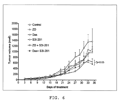
FIG. 6 is a line graph showing progression of tumorvolume under the different
therapies; concurrent inhibition of Stat3 and EGFR or Src induces human
pancreatic
tumor growth inhibition in xenografts.
Detailed Description of Preferred Embodiments
The present invention will now be described more fully hereinafter with
reference to the accompanying drawings, in which preferred embodiments of the
invention are shown. Unless otherwise defined, all technical and scientific
terms
used herein have the same meaning as commonly understood by one of ordinary
skill in the art to which this invention pertains. Although methods and
materials
similar or equivalent to those described herein can be used in the practice or
testing
of the present invention, suitable methods and materials are described below.
Any
publications, patent applications, patents, and other references mentioned
herein are
incorporated by reference in their entirety. In case of conflict, the present
specification, including any definitions, will control. In addition, the
materials,
methods and examples given are illustrative in nature only and not intended to
be
limiting. Accordingly, this invention may be embodied in many different forms
and
should not be construed as limited to the illustrated embodiments set forth
herein.
Rather, these illustrated embodiments are provided so that this disclosure
will be
thorough and complete, and will fully convey the scope of the invention to
those
skilled in the art. Other features and advantages of the invention will be
apparent
from the following detailed description, and from the claims.
Materials and Methods
Cells and Reagents.
CA 02745265 2011-05-31
WO 2010/065444 PCT/US2009/066079
6
v-Src-transformed mouse fibroblasts (NIH3T3/v-Src), human pancreatic
cancer (Panc-1) and leukemic (K562) lines have been described (14-16). The
human
pancreatic cancer lines, Colo-357 and Mia-PaCa-2 were kind gifts from Drs.
Lancaster and Mokenge (Moffitt Cancer Center). The immortalized human
pancreatic
duct epithelial cell (HPDEC) line was obtained from Dr. Tsao, OCI, UHN-PMH,
Toronto) (17). Except for HPDEC grown in Keratinocyte-SFM media supplemented
with 0.2 ng EGF, 30 pg/mL bovine pituitary extract and containing antimycol,
and
K562 line in RPMI 1640 containing 10% heat-inactivated FBS and 100 units/ml
penicillin-streptomycin, all other cell lines were grown in Dulbecco's
modified Eagle's
medium (DMEM) containing 5% iron-supplemented bovine calf serum and 100
units/ml penicillin-streptomycin. Recombinant human EGF (hEGF) is from
Creative
Biolabs, Port Jefferson Station, NY); Gemcitabine is from Ely Lilly
(Indianapolis, IN).
Nuclear Extract Preparation and Gel Shift Assays.
Nuclear extract preparation and DNA-binding with electrophoretic mobility
shift
assay (EMSA) were carried out, as previously reported (14, 15). The 32P-
labeled
oligonucleotide probes used were hSIE (high affinity sis-inducible element
from the
c-fos gene, m67 variant), 5'-AGCTTCATTTCCCGTAAATCCCTA; (SEQ ID NO:1)
that binds Stat1 and Stat3 (Wagner et al., 1990) and the MGFe (mammary gland
factor element from the bovine R-casein gene promoter, 5'-
AGATTTCTAGGAATTCAA; (SEQ ID NO:2) that binds Stat1 and StatS (Gouilleux et
al., 1995; Seidel et al., 1995).
SDS-PAGE/Western Blot Analysis.
Western blotting analysis was performed as previously described (15, 18).
Primary
antibodies used were anti-Stat3 (C20) (Santa Cruz, Santa Cruz, CA),
anti-pY845EGFR (Upstate Biotech, Millipore, Billerica, MA), and antibodies
against
pY705Stat3, Stat3, pY1068EGFR, pY1086EGFR, pY1173EGFR, EGFR, pY416Src,
Src, c-Myc, and 9-Actin from Cell Signaling (Danvers, MA).
Small-interfering RNA (siRNA) Transfection.
siRNA sequences for EGFR and Src were ordered from Dharmacon RNAi
Technologies, Thermo Scientific (Lafayette, CO). Sequences used are: EGFR
sense
strand, 5'-GAAGGAAACUGAAUUCAAAUU-3', SEQ ID NO:3; EGFR antisense
strand, 5'-UUUGAAUUCAGUUUCCUUCUU-3, SEQ ID NO:4'; control siRNA sense
CA 02745265 2011-05-31
WO 2010/065444 PCT/US2009/066079
7
strand, 5'-AGUAAUACAACGGUAAAGAUU-3', SEQ ID NO:5; and control siRNA
antisense strand, 5'-UCUUUACCGUUGUAUUACUUU-3', SEQ ID NO:6. The c-Src
SMARTpool siRNA reagent (NM-005417, Catalog # M-003175-01-05) was used for
Src. Transfection into cells was performed using 20 nM of EGFR siRNA or 25 nM
of
Src siRNA and 8 pl Lipofectamine RNAiMAX (Invitrogen Corporation, Carlsbad,
CA)
in OPTI-MEM culture medium (GIBCO, Invitrogen).
Cell Proliferation/Viability Assay and Annexin V Binding and Flow Cytometry.
Proliferating cells in 6-well or 96-well plates were treated once with 0.1-1
mM
ZD1839 (Iressa), 100 nM Dasatinib, 50-100 pM S31-201, 1 pM Gemcitabine, or
combinations of inhibitors for up to 96 h. Viable cells were counted by trypan
blue
exclusion/phase contrast microscopy or assessed by CyQuant cell viability
assay,
according to manufacturer's (Invitrogen) instructions, or cells were processed
for
Annexin V binding (BD Biosciences) with flow cytometry for apoptosis. S31-201
is
fully described in reference 30 (see below).
Colony Survival Assay.
Single-cell suspension of Panc-1 and Colo-357 cells were seeded in 6-cm
dishes (500 cells per well) and assayed as previously reported (19), treated
the next
day with inhibitors for 48 h, and allowed to grow until large colonies were
visible.
Colonies were stained with crystal violet for 4 h and counted under phase-
contrast
microscope.
Cell Migration and Matrigel Invasion Assays.
Cell migration and invasion experiments were carried out and quantified as
previously described (20), using Bio-Coat migration chambers (Becton
Dickinson,
Franklin, NJ) of 24-well companion plates with cell culture inserts containing
8 pm
pore size filters, according to the manufacturer's protocol.
Statistical analysis.
Statistical analysis was performed on mean values using Prism GraphPad
Software, Inc. (La Jolla, CA). The significance of differences between groups
was
determined by paired t-test at p <0.05k, <0.01**, and < 0.001***.
Results
Aberrant EGFR, Src and Stat3 in pancreatic cancer lines.
CA 02745265 2011-05-31
WO 2010/065444 PCT/US2009/066079
8
Consistent with published reports (6, 7), Stat3 activity, per DNA-binding with
EMSA analysis in nuclear extract preparations is constitutive in Panc-1 and
Colo-357, low in Mia-Paca-2, and undetectable in the normal human pancreatic
duct
epithelial cells (HPDEC), compared to aberrant levels in NIH3T3/v-Src (15)
(FIG.
1A(i)). Per supershift analysis, the DNA-protein complex contains Stat3 (FIG.
1A(i),
lane 3). By contrasts, Stat5 activity is undetectable in pancreatic cancer
cells (FIG.
1A(ii)), compared to aberrant levels in the K562 leukemic cells (16).
EGFR and c-Src are aberrant in many human cancers (2, 4). Immunoblotting
analysis showed a moderate pY416c-Src level in Mia-Paca-2, but enhanced levels
in Panc-1 and Colo-357 cells similar to levels in NIH3T3/v-Src, compared to
low
levels in HPDEC (FIG. 1 B(i), upper panel). The elevated pY416Src levels
parallel
enhanced levels of the Src-sensitive pY845EGFR motif (21) in Panc-1 and Colo-
357
cells, compared to low levels of same in HPDEC (FIG. 1 B(i), lower panel).
Total Src
or EGFR protein remained unchanged. Immunoblotting analysis further showed
elevated levels of the EGFR autophosphorylation motifs (22), pY1068EGFR (FIG.
1 C(i), lanes 2 and 7), pY1086EGFR (FIG. 1 C(ii), lanes 2 and 7) and
pY1173EGFR
(FIG. 1C(iii), lanes 2 and 7) in Panc-1 and Colo-357, compared to basal levels
of
same in HPDEC (FIG. 1C(i)-(iii), lane 1).
Functional integration of EGFR and Src in pancreatic cancer cells.
We next examined the functional relationship between the activated EGFR
and Src. Immunoblotting analysis showed treatment of cells with Dasatinib
(Das)
inhibited Src activity (pY416Src) (23) and induced an early (1 h) and a
sustained (24
h) decrease in pY845EGFR levels (FIG. 1 B(ii)). By contrast, no detectable
changes
in pY416Src and pY845EGFR levels were induced by treatment with the pan-ErbB
inhibitor, PD1 69540 (PD1 69) (24) (data not shown) or the selective EGFR
inhibitor,
ZD 1839 (ZD, Iressa) (25) (FIG. 1 B(ii)). In confirmation, siRNA knockdown of
c-Src
abrogated pY845EGFR levels (FIG. 1 B(iii), Src siRNA), while EGFR knockdown by
siRNA had minimal effect on pY416Src level (FIG. 1 B(iv), EGFR siRNA).
Scrambled
siRNA has no effect (FIG. 1 B(iii) and (iv), con siRNA). Thus, elevated
pY845EGFR
levels in pancreatic cancer cells are sensitive to Src activity.
Immunoblotting analysis further showed that treatment of Panc-1 and
Colo-357 cells with ZD diminished pY1173EGFR levels (FIG. 1C(iii), lanes 3, 4,
8
CA 02745265 2011-05-31
WO 2010/065444 PCT/US2009/066079
9
and 9) by as early as 1 hand up to 24 h, with no effect on pY1068EGFR (/FIG.
1C(i),
lanes 3, 4, 8 and 9) or pY1086EGFR level (FIG. 1C(ii), lanes 3, 4, 8 and 9),
suggesting that EGFR kinase is essential for the induction of pY1173EGFR
levels,
but not pY1068EGFR or pY1086EGFR. By contrast, Das treatment decreased
pY1068EGFR and pY1086EGFR levels (FIG. 1C(i) and (ii), lanes 5, 6, 10 and 11),
with minimal effect on pYEGFR1173 (FIG. 1C(iii), lanes 5, 6, 10 and 11).
Both EGFR and Src promote aberrant Stat3 activation.
Both the pY1068EGFR and pY1086EGFR levels are binding sites for Stat3
(27, 28). Given the concurrent EGFR and Src activation in Panc-1 and Colo-357
cells, we sought to define the regulation of aberrant Stat3 activation. By in
vitro
DNA-binding assay with EMSA analysis of nuclear extract preparations, we
observe
an early repression (in the first 30 min to 1 h of treatment) of
constitutively-active
Stat3 by the pan-ErbB inhibitor, PD169540 (PD169), the ErbB2-selective
inhibitor,
AG879 (7), ZD, or Das (FIG. 2A(i), lanes 4, 5, 7, and 8, and (ii), lanes 2, 4,
6, and
11, and FIG. 2B, 1 h), or by a combined PD169 and Das (FIG. 2A(i), lanes 10
and
11, and (ii), lane 8). However, the Stat3 activity in Panc-1 cells
consistently
rebounded following 24 h treatments with Das, ZD, or PD169 (FIG. 2A(i) and
(ii), 24
h), even though EGFR or Src activity remained inhibited (Fig 1 B and 1 C, 24
h).
Twenty-four hour treatment with the AG879 moderately inhibited Stat3 activity
(FIG.
2A(ii), lane 12), which we speculate may be due to its widespread activity as
a
pan-ErbB inhibitor. By contrast, treatment with the Jak inhibitor, AG490 for 1
h had
no effect on constitutive Stat3 activity, but surprisingly abolished Stat3
activity at 24
h treatment (FIG. 2A(ii), lanes 9 and 10). Moreover, combined treatment with
AG490
and ZD, Das or PD169 for 24 h similarly abolished constitutively-active Stat3
(FIG.
2A(ii), lanes 14, 15, and 16). In Colo-357, Stat3 activity was inhibited by
both ZD and
Das, with the effects more striking for Dasatinib (FIG.2B). These findings
together
reveal a pattern of constitutive Stat3 activation in pancreatic cancer cells
that is
mediated by both EGFR and Src, and a compensatory, Jak-dependent secondary
Stat3 activity. A similar pattern of Stat3 activation has been observed in
head and
neck squamous carcinoma, mesothelioma, squamous cell skin carcinoma, and
non-small cell lung cancer cell lines following the inhibition of Src (29). In
further
support, the siRNA knockdown of EGFR (EGFR siRNA) or Src (Src siRNA) led to
CA 02745265 2011-05-31
WO 2010/065444 PCT/US2009/066079
pStat3 suppression, as assayed by immunoblotting analysis (FIG. 2C). Scrambled
siRNA (con) has no effect. Immunoblotting analysis also shows that EGF
stimulation
induces pY705Stat3, pY1086EGFR, pY1173EGFR, pY845EGFR and pY416c-Src
(Supplemental FIG. S1(i)-(iii), lane 4) over and above constitutive levels in
Panc-1
5 cells, in a manner that is similar to the induction of same in response to
the
stimulation of normal HPDEC (Supplemental FIG. S1, lane 2), except for
pY1068EGFR levels in Panc-1 (FIG. S1(ii), upper right panel). In control
studies,
immunoblotting analysis showed elevated pErk1/pErk2MAPK and pAkt in Panc-1
and Colo-357 cells compared to normal HPDEC, neither of which was
significantly
10 affected by treatment with ZD or Das (data not shown).
Inhibition of Stat3 sensitizes pancreatic cancer cells in vitro to EGFR and
Src
inhibitors.
Given the preceding data on the inter-relation between EGFR, Src and Stat3
activation, we investigated the biological implications and the therapeutic
potential
of a combinatorial approach. Dasatinib and ZD were used at 100 nM and 0.1-1
pM,
respectively, as in literature reports (23, 24), while the Stat3 inhibitor,
S31-201 was
used at the sub-optimum, 50 pM, or at the 100 pM required to inhibit Stat3
activation
(30). Viable cell count by trypan blue exclusion/phase-contrast microscopy
showed
that treatment with 1 pM ZD, 100 nM Das, or 50 pM S31-201 alone minimally
affected
cell viability by 24 h (FIG. 3A Day 1). By contrast, treatment for 48 to 96 h
with or Das
or S31-201 alone progressively decreased cell viability, while treatment for
the same
period with ZD showed minimal effect (FIG. 3A), except at 96 h when the number
of
viable Panc-1 cells decreased (FIG. 3A(i), ZD, Day 4). Comparatively, the
combined
inhibition of Stat3 (by S31-201) and EGFR (by ZD) or Src (by Das) or the
combined
treatment with AG490 (Jaks inhibitor) and ZD or Das induced greater losses of
viability at 48-96 h (FIG. 3A and B). The effects on cell viability as
captured by trypan
blue exclusion were confirmed by the CyQuant cell proliferation/viability
assay. Unlike
24 h treatment duration that showed minimal effect on viability (FIG. 3A),
CyQuant
assay showed that 48-h treatment with each inhibitor alone decreased viable
cell
numbers (quantified as fluorescent unit, FU) in a dose-dependent manner (FIG.
3C,
ZD, Das and S31-201). We infer from the graphs that treatment with 1 pM ZD for
48
h has minimal effect on cell viability (FIG. 3C(i) and (iv)), as observed by
the trypan
CA 02745265 2011-05-31
WO 2010/065444 PCT/US2009/066079
11
blue exclusion assay (FIG. 3A). However, the observed effects of single agents
were
significantly weaker compared to the concurrent treatment with a Stat3
inhibitor and
an inhibitor of EGFR or Src. Results show that the treatment with S31-201
increased
the sensitivity of Panc-1 and Colo-357 cells to ZD and Das, shifting the
dose-response curves to the left (FIG. 3C, ZD + S31-201, and Das + S31-201).
Concurrent treatment with S31-201 significantly decreased the IC50 values as
follows: 17 to 0.4 pM, and 100 to 6 nM, respectively, for ZD and Das against
Panc-1
viability (FIG. 3C(i) and (ii)); and 6.5 to 2.4 pM, and 90 to 8 nM,
respectively for ZD
and Das against Colo-357 viability (FIG. 3C(iv) and (v)). For the impact of ZD
and
Das on the sensitivity to S31-201, CyQuant cell viability assay showed that
Das, but
not ZD increased the sensitivity of both cell lines to S31-201, decreasing its
IC50 from
40 to 15 pM, and from 45 to 20 pM, respectively, for effects on Panc-1 and
Colo-357
cells (FIG. 3C(iii) and (iv)). Thus, treatment with S31-201 sensitized cells
to ZD and
Das, while treatment with Das, but not ZD similarly sensitized cells to S31-
201.
Given the clinical implications of these findings, we extended these studies
to examine the effect of EGFR Src and Stat3 pathway on the response to
Gemcitabine, the anti-metabolite agent used in the treatment of pancreatic
cancer.
CyQuant cell proliferation/viability studies showed that inhibition of EGFR,
Src or
Stat3 sensitized Panc-1 and Colo-357 cells to Gemcitabine (FIG. 3D). More
importantly, the combined inhibition of Stat3 and EGFR or Src induced a higher
sensitization of cells to Gemcitabine than that induced by the inhibition of
any one
alone (FIG. 3D).
As known to the skilled, Gemcitabine is a nucleoside analog of cytidine which
interferes with DNA replication, arresting tumor growth and resulting in
apoptosis of
the cell. Gemcitabine is also known to bind to the active site of the enzyme
ribonucleotide reductase (RNR) to irreversibly inactive the enzyme, thus
interfering
with the cell's ability to produce deoxyribonucleotides necessary for DNA
replication
and repair. This also leads to apoptosis. As noted above, the combined
inhibition
of Stat3 and EGFR or Src induces a higher sensitization of cells to
Gemcitabine,
creating another possibility for combination therapy of tumors.
To further explore the sensitization potential of inhibition of aberrant
Stat3, we
performed colony survival assay (19). Results show that inhibition of Src (by
Das) or
CA 02745265 2011-05-31
WO 2010/065444 PCT/US2009/066079
12
Stat3 (by S31-201 (S31)), but not EGFR inhibition (by ZD) resulted in reduced
colony
numbers (FIG. 4A). More importantly, the concurrent inhibition of Stat3 and
EGFR
or Src resulted in much lower colony numbers (FIG. 4A), consistent with the
much
greater loss of viable cells due to the combined inhibition of Stat3 and EGFR
or Src
(FIG. 3). To extend these studies, we performed Annexin V binding/Flow
Cytometric
analysis for apoptosis. Higher percentages of Panc-1 and Colo-357 cells
undergoing
apoptosis were observed for concurrent inhibition of Stat3 and EGFR or Src
than for
the inhibition any one signaling molecule alone (FIG. 4B(ii) and (iii)).
Similar results
were obtained for the concurrent treatments with AG490 and ZD or Das (FIG.
4B(ii)
and (iii)). By contrast, similar treatments of normal HPDECs showed no
significant
apoptosis (FIG. 4B(i)) with the combination treatments. Thus, we establish
that
pancreatic cancer cells have higher sensitivity to concurrent inhibition of
Stat3 and
EGFR or Src than to the inhibition of a single entity.
EGFR, Src and Stat3 together promote pancreatic cancer cell migration and
invasion.
Aberrantly-active Src and Stat3 have both been implicated in tumor cell
motility, migration, invasion and metastasis (4, 23). in vitro matrigel assay
confirmed
that inhibition of Src or Stat3 alone suppresses migration and invasion (FIG.
5A).
However, concurrent inhibition of Stat3 and EGFR or Src for 24-h has a
stronger
effect on Colo-357 migration and Panc-1 invasion, except for Src inhibition,
which
showed a similar effect on Panc-1 migration (FIG. 5A). At the 24-h treatment
when
these studies were done, there is no significant effect on cell viability
(FIG. 3). These
findings are further evidence that pancreatic cancer lines are more sensitive
to
concurrent inhibition of Stat3 and Src or EGFR.
EGFR, Src and Stat3 module regulates c-Myc over-expression in pancreatic
cancer cells.
For insight into the underlying molecular mechanisms bywhich the EGFR, Src
and Stat3 pathway may support the cancer phenotype, we studied the regulation
of
key cancer-relevant genes. We show that c-Myc is over-expressed in pancreatic
cancer lines compared to normal HPDEC (FIG. 5B). Furthermore, the concurrent
inhibition of Stat3 and EGFR or Src consistently repressed c-Myc expression.
These
findings suggest a functional synergy between EGFR, Src and Stat3 in inducing
CA 02745265 2011-05-31
WO 2010/065444 PCT/US2009/066079
13
c-Myc expression in the context of pancreatic cancer phenotype and that the
stronger repression of c-Myc expression contributes to the antitumor cell
effects of
and the increased sensitivity of pancreatic cancer lines to concurrent Stat3
and
EGFR or Src inhibition.
Inhibition of Tumor Growth by Combination Treatment
Concurrent inhibition of Stat3 and EGFR or Src induces human pancreatic
tumor growth inhibition in xenografts. Subcutaneous xenografts of Colo-357, a
metastatic pancreatic adenocarcinoma line were used to study the therapeutic
implication of the Stat3, EGFR and Src inter-relationships and to evaluate the
in vivo
antitumor effects of concurrent inhibition of Stat3 and EGFR or Src. Data
showed
that in general, xenografts of Colo-357 cells showed low responsiveness to
treatment
with inhibitor of EGFR, Src or Stat3 alone, although, as the therapy
progressed,
those tumors treated with only one inhibitor alone appeared to show reduced
growth,
which was statistically not significant from the control, non-treated tumors
(FIG. 6).
By contrast, tumors from mice treated with combined S31-201 and Das or S31-201
and ZD consistently showed reduced growth and smaller tumor sizes throughout
the
entire study (FIG. 6). Thus, the residual tumor volumes (sizes) for tumors in
mice
treated with combination inhibitors were significantly different (p<0.05) from
tumor
volumes for tumors in control mice at days 20 and upwards post treatment.
These
in vivo antitumor effects of combination treatment with inhibitors of S31-201
and Das
or S31-201 and ZD are consistent with the in vitro antitumor cell data and
together
indicate that aberrant Stat3 cooperates with hyperactive EGFR or Src to
sustain
human pancreatic cancer.
Discussion
Within the context of aberrations in the EGFR, Src and Stat3 pathway in
pancreatic cancer, present study reveals a strong role for Src in supporting
aberrant
EGFR activation by not only inducing the phosphorylation of Y845EGFR motif
(31),
but also promoting the induction of pY1068EGFR and pY1086EGFR motifs. These
Src-promoted events will greatly influence the status of EGFR in pancreatic
cancer
cells. A role for EGFR in aberrant Stat3 activation in cancer cells has
previously been
reported in other tumor cells, including head and neck squamous cell carcinoma
and
breast cancer (26, 32). Present study extends those findings to pancreatic
cancer
CA 02745265 2011-05-31
WO 2010/065444 PCT/US2009/066079
14
and show that EGFR is key in facilitating aberrant Stat3 activation. Moreover,
the
pY1068EGFR and pY1086EGFR induction by Src is likely to have significant
impact
on the activation of Stat3, given that these two motifs are essential sites
for the
binding of Stat3 to EGFR in order to promote its phosphorylation and
activation (27,
28). Furthermore, Src may not only facilitate Stat3 activation via the
induction of
those two Tyr motifs of EGFR, but it can also directly phosphorylate Stat3, as
has
been previously reported in other systems (18). It is therefore consistent
that both
hyperactive EGFR and Src promote baseline constitutive Stat3 activation in
pancreatic cancer, as revealed by our study.
The present study is also in agreement with an earlier report of
ErbB-2-dependent constitutive Stat3 activation in Mia-Paca-2 and UK Pan-1
cells (7)
and another study that showed that the full induction of Stat3 activation by
ErbB2
required both Src and Jaks (33). Our findings indicate that Jaks contribute to
the
maintenance of constitutive activation in revealing a Jak-dependent
compensatory
mechanism of Stat3 activation upon inhibition of EGFR and Src. Given that Jaks
inhibition did not abolish aberrant Stat3 at the earliest time point, we
deduce that this
family of cytoplasmic tyrosine kinases may not be the predominant mediators of
the
baseline aberrant Stat3. Thus, in pancreatic cancer cells, a two-phase model
of
activation of Stat3 signaling emerges composed of an EGFR- and Src- dependent
baseline, constitutive Stat3 induction, and an induced Stat3 activation that
is
dependent on Jaks. The observed secondary induction of Stat3 activation via
Jaks
has similarly been reported in head and neck squamous cell carcinoma line (29)
and
could be due to growth-stimulatory factors released from tumor cells (34),
which in
turn would induce the activation of Jaks and thereby promote Stat3 activation.
EGFR, Src and Stat3 has each independently been established to have
critical roles in malignant transformation (6, 14, 23, 26, 35), while their
collective roles
in promoting tumorigenesis have not been explored. While the inhibition of the
activity of each of the three proteins induced antitumor cell response to some
degree, data presented here strongly indicate that the multiple targeting of
Stat3 and
EGFR or Src together has a higher potential to inhibit growth, viability,
survival,
malignant transformation, and migration and invasion in vitro.
CA 02745265 2011-05-31
WO 2010/065444 PCT/US2009/066079
Significantly, hyperactivation of the EGFR signaling has been deemed a
prognostic indicator of low survival among pancreatic cancer patients (36-38).
Also,
there is evidence to indicate that the concurrence with aberrant Src signaling
potentiates the effects of aberrant EGFR and induces biological synergy (3,
21, 39).
5 Given the potential collective roles of Stat3, EGFR and Src in promoting and
supporting pancreatic cancer, the inhibition of any single entity alone is
unlikely to
be insufficient to impact the disease. Present data that simultaneous
inhibition of
Stat3 and EGFR or Src induced greater antitumor cell effects and a higher
sensitization to Gemcitabine provides a strong support for the opinion that
Stat3 may
10 cooperate with EGFR and Src to support the malignant phenotype. Indeed, the
inhibition of Stat3 seemed to sensitize pancreatic cancer cells to the
antitumor cell
effects of ZD and Das. Multiple targeting of Stat3 and EGFR or Src therefore
has the
potential to induce a greater antitumor efficacy. This is supported by our
present data
that concurrent treatment with theStat3 inhibitor, S31-201 and ZD or Das
induced
15 greater regression of xenografts of Colo-357 than treatment with either
inhibitor
alone. Such a multiple-targeted therapy has received a strong interest in
recent
times, particularly given the dismal results in certain cases of molecular
targeted
monotherapy, such as anti-EGFR monotherapy (40, 41). Thus, a combined
Gemcitabine and Erlotinib (EGFR TK inhibitor) therapy has recently been
approved
for patients with locally advanced/metastatic pancreatic cancer (42, 43),
although we
note by our data that the inhibition of Stat3 and EGFR or Src together induces
a
higher Gemcitabine sensitivity than inhibition of EGFR alone. The enhanced
antitumor effects due combined Stat3 and EGFR or Src inhibitors may in part be
due
stronger repression of the expression of c-Myc oncogene. Altogether, present
study
provides support for a multiple-modality therapeutic approach and lays the
foundation for concurrent targeting of aberrant Stat3 and EGFR or Src as a
more
effective approach for achieving an enhanced antitumor therapeutic efficacy in
pancreatic cancer.
Accordingly, in the drawings and specification there have been disclosed
typical preferred embodiments of the invention and although specific terms may
have
been employed, the terms are used in a descriptive sense only and not for
purposes
of limitation. The invention has been described in considerable detail with
specific
CA 02745265 2011-05-31
WO 2010/065444 PCT/US2009/066079
16
reference to these illustrated embodiments. It will be apparent, however, that
various
modifications and changes can be made within the spirit and scope of the
invention
as described in the foregoing specification and as defined in the appended
claims.
References
1. Tzeng CW, Frolov A, Frolova N, et al. EGFR genomic gain and aberrant
pathway signaling in pancreatic cancer patients. J Surg Res 2007;143:20-6.
2. Korc M, Meltzer P, Trent J. Enhanced expression of epidermal growth factor
receptor correlates with alterations of chromosome 7 in human pancreatic
cancer.
Proc Natl Acad Sci U S A 1986;83:5141-4.
3. Lutz MP, Esser IB, Flossmann-Kast BBM, et al. Overexpression and
Activation of the Tyrosine Kinase Src in Human Pancreatic Carcinoma. Biochem
Biophys Res Commun 1998;243::503-8.
4. Trevion JG, Summy JM, Lesslie DP, et al. Inhibition of SRC expression and
activity inhibits tumor progression and metastasis of human pancreatic
adenocarcinoma cells in an orthotopic nude mouse model. Am J Pathol
2006; 168:962-72.
5. Parsons JT, Parsons SJ. Src family protein tyrosine kinases: cooperating
with
growth factor and adhesion signaling pathways. Curr Opin Cell Biol 1997;9:187-
92.
6. Scholz ASH, Detjen KM, Peters M, et al. Activated signal transducer and
activator of transcription 3 (STAT3) supports the malignant phenotype of human
pancreatic cancer. Gastroenterology 2003;125:891-905.
7. DeArmond D, Brattain MG, Jessup JM, et al. Autocrine-mediated ErbB-2
kinase activation of STAT3 is required for growth factor independence of
pancreatic
cancer cell lines. Oncogene 2003;22:7781-95.
8. Trevino JG, Gray MJ, Nawrocki ST, et al. Src activation of Stat3 is an
independent requirement from NF-kappaB activation for constitutive IL-8
expression
in human pancreatic adenocarcinoma cells. Angiogenesis 2006;9:101-10.
9. Toyonaga T, Nakano K, Nagano M, et al. Blockade of constitutively activated
Janus kinase/signal transducer and activator of transcription-3 pathway
inhibits
growth of human pancreatic cancer. Cancer Lett 2003;201:107-16.
10. Darnell JE. Validating Stat3 in cancer therapy. Nat Med 2005;11:595-6.
CA 02745265 2011-05-31
WO 2010/065444 PCT/US2009/066079
17
11. Yu H, Jove R. The STATS of Cancer-New molecular targets come of age. Nat
Rev Cancer 2004;4:97-105.
12. Turkson J. STAT proteins as novel targets for cancer drug discovery.
Expert
Opin Ther Targets 2004;8:409-22.
13. Yue P, Turkson J. Targeting STAT3 in cancer: how successful are we? Expert
Opin Investig Drugs 2009;18:45-56.
14. Garcia R, Bowman TL, Niu G, et al. Constitutive activation of Stat3 by the
Src
and JAK tyrosine kinases participates in growth regulation of human breast
carcinoma cells. Oncogene 2001;20:2499-513.
15. Turkson J, Bowman T, Garcia R, Caldenhoven E, De Groot RP, Jove R. Stat3
activation by Src induces specific gene regulation and is required for cell
transformation. Mol Cell Biol 1998;18:2545-52.
16. Huang M, Dorsey JF, Epling-Burnette PK, et al. Inhibition of Bcr-Abl
kinase
activity by PD180970 blocks constitutive activation of StatS and growth of CML
cells.
Oncogene 2002;21:8804-16.
17. Ouyang H, Mou LJ, Luk C, et al. Immortal human pancreatic duct epithelial
cell lines with near normal genotype and phenotype. Am J Pathol 2000;157:1623-
31.
18. Zhang Y, Turkson J, Carter-Su C, et al. Activation of Stat3 in v-Src
Transformed Fibroblasts Requires Cooperation of Jak1 Kinase Activity. J Biol
Chem
2000;275:24935-44.
19. Zhao S, Venkatasubbarao K, Lazor JW, et al. Inhibition of STAT3Tyr7O5
Phosphorylation by Smad4 Suppresses Transforming Growth Factor b-Mediated
Invasion and Metastasis in Pancreatic Cancer Cells. Cancer Res 2008;68:4221-8.
20. Siddiquee KAZ, Gunning PT, Glenn M, et al. An Oxazole-Based
Small-Molecule Stat3 Inhibitor Modulates Stat3 Stability and Processing and
Induces
Antitumor Cell Effects. ACS Chem Biol 2007;2:787-98.
21. Tice DA, Biscardi JS, Nickles AL, Parsons SJ. Mechanism of biological
synergy between cellular Src and epidermal growth factor receptor. Proc Natl
Acad
Sci U S A 1999;96:1415-20.
22. Downward J, Parker P, Waterfield MD. Autophosphorylation sites on the
epidermal growth factor receptor. Nature 1984;311 483-5.
CA 02745265 2011-05-31
WO 2010/065444 PCT/US2009/066079
18
23. Nam S, Kim D, Cheng JQ, et al. Action of the Src family kinase inhibitor,
dasatinib (BMS-354825), on human prostate cancer cells. Cancer Res
2005;65:9185-9.
24. Mahtouk K, Hose D, Reme T, et al. Expression of EGF-family receptors and
amphiregulin in multiple myeloma. Amphiregulin is a growth factorfor myeloma
cells.
Oncogene 2005;24:3512-24.
25. Wakeling AE, Guy SP, Woodburn JR, et al. ZD1 839 (Iressa): an orally
active
inhibitor of epidermal growth factor signaling with potential for cancer
therapy.
Cancer Res 2002;62:5749-54.
26. Song JI, Grandis JR. STAT signaling in head and neck cancer. Oncogene
2000; 19:2489-95.
27. Coffer PJ, Kruijer W. EGF receptor deletions define a region specifically
mediating STAT transcription factor activation. Biochem Biophys Res Commun
1995;210:74-81.
28. Shao H, Cheng HY, Cook RG, Tweardy DT. Identification and
Characterization of Signal Transducer and Activator of Transcription 3
Recruitment
Sites within the Epidermal Growth Factor Receptor. Cancer Res 2003;63:3923-30.
29. Johnson FM, Saigal B, Tran H, Donato NJ. Abrogation of signal transducer
and activator of transcription 3 reactivation after Src kinase inhibition
results in
synergistic antitumor effects. Clin Cancer Res 2007;13:4233-44.
30. Siddiquee K, Zhang S, Guida WC, et al. Selective chemical probe inhibitor
of
Stat3, identified through structure-based virtual screening, induces antitumor
activity.
Proc Natl Acad Sci U S A 2007;104:7391-6.
31. Biscardi JS, Maa MC, Tice DA, Cox ME, Leu TH, Parsons SJ. c-Src-mediated
phosphorylation of the epidermal growth factor receptor on Tyr845 and Tyrl 101
is
associated with modulation of receptor function. J Biol Chem 1999;274:8335-43.
32. Sartor Cl, Dziubinski ML, Yu CL, Jove R, Ethier SP. Role of epidermal
growth
factor receptor and STAT-3 activation in autonomous proliferation of SUM-102PT
human breast cancer cells. Cancer Res 1997;57:978-87.
33. Ren Z, Schaefer TS. ErbB-2 activates Stat3alpha in a Src- and
JAK2-dependent manner. J Biol Chem 2002;8:38486-93.
CA 02745265 2011-05-31
WO 2010/065444 PCT/US2009/066079
19
34. Salomon DS, Brandt R, Ciardiello F, Normanno N. Epidermal growth
factor-related peptides and their receptors in human malignancies. Crit Rev
Oncol
Hematol 1995;19:183-232.
35. Song L, Morris M, Bagui T, Lee FY, Jove R, Haura EB. Dasatinib
(BMS-354825) selectively induces apoptosis in lung cancer cells dependent on
epidermal growth factor receptor signaling for survival. Cancer Res 2006
66:5542-8.
36. Uegaki K, Nio Y, Inoue Y, et al. Clinicopathological significance of
epidermal
growth factor and its receptor in human pancreatic cancer. Anticancer Res
1997;17(5B):3841-7.
37. Dong M, Nio Y, Guo KJ, Tamura K, Tian YL, Dong YT. Epidermal growth
factor and its receptor as prognostic indicators in Chinese patients with
pancreatic
cancer. Anticancer Res 1998;18(6B):4613-9.
38. Ueda S, Ogata S, Tsuda H, et al. The correlation between cytoplasmic
overexpression of epidermal growth factor receptor and tumor aggressiveness:
poor
prognosis in patients with pancreatic ductal adenocarcinoma. Pancreas
2004;29:el -8.
39. Maa MC, Leu TH, McCarley DJ, Schatzman RC, Parsons SJ. Potentiation of
epidermal growth factor receptor-mediated oncogenesis by c-Src: implications
for the
etiology of multiple human cancers. Proc Natl Acad Sci U S A 1995;92:6981-5.
40. Saif MW. Erlotinib: the first biologic in the management of pancreatic
cancer.
Expert Opin Pharmacother 2008;9:1595-607.
41. Philip PA. Targeted therapies for pancreatic cancer. Gastrointest Cancer
Res
2008;2(Suppl 2):516-59.
42. Burris Hr, Rocha-Lima C. New therapeutic directions for advanced
pancreatic
cancer: targeting the epidermal growth factor and vascular endothelial growth
factor
pathways. Oncologist 2008;13:289-98.
43. Senderowicz AM, Johnson JR, Sridhara R, Zimmerman P, Justice R, Pazdur
R. Erlotinib/gemcitabine for first-line treatment of locally advanced or
metastatic
adenocarcinoma of the pancreas. Oncology (Williston Park) 2007;21:1696-706;
discussion 706-9, 712, 715.