Note: Descriptions are shown in the official language in which they were submitted.
CA 02745569 2011-07-07
DEMANDES OU BREVETS VOLUMINEUX
LA PRESENTE PARTIE DE CETTE DEMANDE OU CE BREVETS
COMPREND PLUS D'UN TOME.
CECI EST LE TOME 1 DE 2
NOTE: Pour les tomes additionels, veillez contacter le Bureau Canadien des
Brevets.
JUMBO APPLICATIONS / PATENTS
THIS SECTION OF THE APPLICATION / PATENT CONTAINS MORE
THAN ONE VOLUME.
THIS IS VOLUME _I OF 2
NOTE: For additional volumes please contact the Canadian Patent Office.
CA 02745569 2011-07-07
=
30725-351E
1
Biomarkers for diagnosing Alzheimer's disease
This is a divisional application of Canadian patent application Serial No.
2,496,321, filed
on August 11,2003.
Field of the invention
The invention is in the field of diagnostics. More specifically, the invention
is in the field
of assessing the state of Alzheimer's disease in subjects by detection of
Alzheimer's
disease-specific marker polypeptides. The subject matter of this divisional
application
relates to SEQ ID NO:4 for assessing the state of Alzheimer's disease. The
subject
matter of the parent application relates to SEQ ID NO:17. It should be
understood that
the expression "the invention" or the like encompasses the subject matter of
both the
parent and this divisional application.
Background of the invention
Alzheimer's disease
Alzheimer's disease is an increasingly prevalent form of neurodegeneration
that
accounts for approximately 50-60% of the overall cases of dementia among
people over
65 years of age. Pathologically, Alzheimer's disease neurodegeneration is
characterised
by prominent atrophy of corticolimbic structures with neuronal death and loss
of neuronal
synapses, neurofibrillary tangle (NET) formation, and the formation of senile
plaques
containing deposits of amyloid 01-42 (A042) aggregates in the brain [Francis
PT 19991.
The duration of the progressive cognitive decline is approximately 7 years
from the
occurrence of first signs until death. It is assumed that the clinical phase
is preceded by
a 15-30 years preclinical period of continuous deposition of amyloid plaques
and
neurofibrillary tangles. Age of onset and progression of the disease are
largely
determined by causative gene mutations and by genetic susceptibility factors.
Several
environmental risk factors may add to the individual genetic risk factors.
Genetic factors
known to be involved in the familial form of Alzheimer's disease with early
onset of the
disease are: mutations in presenilin 1 (PSI), presenilin 2 (PS2), and amyloid
precursor
protein (APP) genes, and the presence of the apolipoprotein E4 allele.
However, the
majority (95%) of Alzheimer's disease cases is sporadic and heterogeneous.
- CA 02745569 2011-07-07
WO 2004/019043
PCT/EP2003/008879
- 2 -
-
Currently, clinical diagnosis of Alzheimer's disease can only be established
at later
stages of the disease, when cognitive perfounance is Significantly decreased
and
paralleled by structural alterations of the brain. The clinical diagnostic
work up
requires a careful medical history; physical and neurological examination;
blood,
urine and cerebrospinal fluid (CSF) examinations to exclude metabolic and
medical
disease states that might masquerade Alzheimer's disease; detailed
psychometric
examinations to assess mental status and cognitive performance, and imaging
techniques such as computed tomographic scan or magnetic resonance imaging of
the
brain. Diagnostic evaluations at expert centres reach an accuracy of about 80-
85%.
Due to the fact that these tests are expensive and time consuming, and are
particularly inconvenient to patients, there is an increasing need for easy-
accessible
specific diagnostic biomolecule markers, which can be measured in body fluids,
such
as CSF, blood or urine, and which have a high positive predictive value for
diagnosis
of Alzheimer's .disease, or would help to distinguish Alzheimer's disease from
other
forms of dementia. Furthermore, reliable markers sensitive to disease
progression
may constitute surrogate parameters, a major prerequisite for the evaluation
and
development of new causal oriented and disease modifying therapeutic
strategies in
Alzheimer's disease.
Since CSF directly surrounds the brain, changes in its protein composition may
most
accurately reflect pathologic conditions that are associated with specific
alterations of
the protein expression patterns. Over the last decade, a number of biological
abnormalities have been reported in the cerebrospinal fluid (CSF) of
Alzheimer's
disease patients, in particular altered levels of the AB1-42 fragment of the
arnyloid
_ 25 precursor protein, and altered levels of the
hyperphosphorylated tau protein. The
sensitivity and specificity of these markers, however, is low or only modest
[The
Ronald and Nancy Reagan Research Tristitue of the Alzheimer's Association and
the
National Institute on Aging Working Group, 1998, Robles A 1998, Tennissen CE
et
al., 2002].
CA 02745569 2011-07-07
WO 2004/019043
PCT/EP2003/008879
- 3 -
Hence, there is a need for novel biomarkers with sufficient sensitivity and
specificity
for (i) detecting Alzheimer's disease as early as possible, and (ii) to allow
disease
differentiation from other types of dementia or neurodegenerative diseases,
and (iii)
monitoring therapeutic efficacy as surrogate parameter, e.g. in clinical drug
development, and to initiate pharmacotherapy as early as possible and postpone
loss
of memory and disease progression.
Protein Chip TechnolOgy
=
A Protein chip technology called Surface Enhanced Laser Desorption/Ionisation
time
of flight mass spectrometry (SELDI-TOF MS) has recently been developed to
facilitate protein profiling of complex biological mixtures [Davies HA 2000,
Fung
ET 2001, Merchant M 2000].
Protein chip mass spectrometry has already been used by several groups to
detect
potentially novel biomarkers of prostate and bladder [Adam BL 2001] or breast
cancer [Wulfkuhle JD 2001] in serum, seminal plasma, nipple fluid, urine or
cell
extracts. For a review on biomarker search using SELDI-TOF MS, see [Issaq HJ
2002].
Cystatin C
Initially described in 1961 in cerebrospinal fluid (CSF), cystatin C (y trace
or post-y
globulin, Acc. No. P01034) is a small cystein proteinase inhibitor present in
all
human body fluids at physiologically relevant concentrations. The
physiological role
of cystatin C is likely to regulate extracellular cysteine protease activity,
which
results from microbial invasion or release of lysosomal proteinq ses from
dying or
diseased cells. Cystatin C colocalises with B-amyloid (AB) within the
arteriolar walls
in Alzheimer's disease brains and cerebral arnyloid angiopathy [Levy E 2001].
There
are two common haplotypes of the CST3 gene coding for cystatin C (A and B)
that
differ from each other at three sites: two single base ppir changes in the
promoter
CA 02745569 2014-10-16
=
- 4 -
region and one in the signal peptide domain that causes, an amino acid
substinition
= (alanine to threonine). Recently, case control studies found associations
of CST3
with increased risk for late onset Alzheimer's disease [Crawford PC 2000,
Finckh U
2000, Beyer K 20011
Hereditary cerebral hemOrrhage with amyloidosis, Icelandic type (HCHWA-I),
also
= called hereditary cystatin C amyloid angiopathy (HCCAA), is an autosomal
dominant form of cerebral amyloid angiopathy (CAA). The amyloid deposited in
the
brain vessel's walls is composed mainly of a variant of cystatin C
characterised by
the presence of the Leu68-Gln Substitution [Cohen 1983, Ghiso 1986]. This
- pathology is also coupled to a decreased concentration of this
major cystein
proteinase inhibitor in cerebrospinal fluid and leads to its amyloid
deposition in the.
brain [Grubb AO 1984]. =
=
Leung-Tack et al have also purified two N-terminal truncated isoforms of
cystatin C
=
in urine from one patient who had received renal transplant. According to
their data,
(des1-4) cystatin C has an inhibiting effect on two functions of human
peripheral
=
rhononuclear cells (PMN): 02 release and phagocytosis, which may be due to.
the N-
terminal sequence 'KPPR'. Their data support a potentially important role for
cystatin
C as a possible ircununomodulator during inflammation. Accumulating evidence
indicates that increased free radical mediated damage to cellular function
contributes
.
=
to the ageing process and age-related neurodegenerative disorders. Oxidative
.stregs
may play a role in Alzheimer's disease, Parkinson's. disease, amyotrophic
lateral
= sclerosis (ALS). Although free-radical damage to neurons may not be the
primary
== 25 event initiating these diseases, it appears that free-radical
damage is involved in the =
pathogenetic cascade of these disorders.
=
Beta-2-microglobulin
=
=
Beta-2-microglobulin (Acc. No. P01884) constitutes the small .constant
component of
the class I major histocompatibility complex (CMH) and its presence in
biological
=
= =
CA 02745569 2011-07-07
=
WO 2004/019043 PCT/EP2003/008879
- 5 -
-
-
- '-
fluids represents the balance between membrane protein turnover and
elimination.
Since this peptide seems to be increased in sonic diseases characterised by an
elevation of the immune response, its quantification in body fluids has become
a =
useful index of immunological state in vivo [Hoekman et al 1985]. The function
of
this protein is unclear, but it seems to be implicated in diseases, which
involve glial
1
cell destruction [Ernerudh et al 1987].
!.
=
The technical problem whiCh is solved by the present invention is the
provision of
improved methods for diagnosing Alzheimer's disease and/or monitoring the
progression of Alzheimer's disease in a subject.
Neurosecretary Protein (VGF)
VGF (human VGF, Acc.-No.: 015240) is a secretory peptide precursor that is
expressed and processed by neuronal cells [Can et al. 1997]. In situ
hybridization
studies in the adult rat central nervous system have revealed that the VGF
mRNA is
widely distributed throughout the brain with prominent expression in the
hippocampus, entorhinal cortex, and neocortex. Furthermore, it has been shown
that
VGF transcription and secretion is selectively upregulated by neurotrophins
like
NGF and BDNF, and by depolariation in vitro. Increased BDNF expression can be
observed in dentate gyrus and CA3 regions of the hippocampus, which are
tissues
that appear to die early in Alzheimer Disease pathogenesis.
Description of the invention
The invention is based on the surprising finding that specific polypeptides
are
differentially expressed in subjects having Alzheimer's disease when compared
to a
healthy control group. These differentially expressed polypeptides can be,
e.g.,
detected in samples of cerebrospinal fluid of the subject in which Alzheimer's
disease 1
is to be diagnosed. The individual polypeptides of the invention can be
detected
and/or quantified alone or in combination with other polypeptides of the
invention.
CA 02745569 2013-09-23
30725-351E
- 6 -
=
The polypeptide maficers of the invention are defined bir their respective
molecular
weight. Five markers identified by the SAX2 method as described in the
Examples
show the following moldcular masses:
=
Marker 1 (M1): 4824 20 Da;
Marker 2 (M2): 7691 20 Da;
= Marker 3 (M3):11787 20 Da;
Marker 4 (Mi): 11988 20 Da;
=
Marker 5 (M5); 13416 201)a.
=
Table one shows the observed molecular weight of polypeptide markers M1 to M5
as
determined by SELDI-TOF MS, the amino acid sequences of observed fragments of
polypeptide markers M1 to M5, and the protein from which the polypeptide
markers
. 15 = M1 to M5 originate.
Table 1:
=
Marker SELDI observed MW Amino Acid Sequence Protein Name
SEQ ID NO
M1 4823.5 Da i 1.7 VGEEDEEAAF-ARAEAEEAER VGF4.8
17
M2 7691.4 Da i 4.9 xxADtL/I)AGHG(Q/K)EV(L/I)(L/I)m. human
myoglobin 1
HGTVV(L/I)TA(L/I)GG(L/I)(L/I)K . new variant ,
2
VNHVTLSQPK
3
M3 11786.9 Pa .j 7.6. VEHS'DLSFSK . human beta-2-
4
M4 11988.4 Da' 5.9 IEKVERSDLSFSI( microglobulin
5
SNFLNCYVSGFHPSDIEVDLLK
6
ASNDMYHSR =
7
- ALDFAVGEYNK
8
- RALDFAVGFIVIc
. =
9
LVGGFMDASVEEEGVR
10
11
M5 13416.4 Da 9.4 QIVAGVNYFLDVELGR
human Cystatin C
LVGGPMDASVEEEGVRR
12
KQIVAGVNYF.IADVELGR
13
TOPNLDNCP2HDQFPELK
14
=
TQAT4ONCPERDQPWILICR 15
=
SSPGF,PPRLVGGPMIASVEEEGVR
16
CA 02745569 2011-07-07
WO 2004/019043
PCT/EP2003/008879
- 7 -
The differentially expressed polypeptides of the invention can be, e.g.,
detected in
samples of cerebrospinal fluid of the subject in which Alzheimer's disease is
to be
diagnosed. In addition, depending on the specific embodiment, the source of
samples
to measure the abundance of the polypeptide markers of the invention, can also
be
blood, serum, or urine, but is not limited to these body compartments.
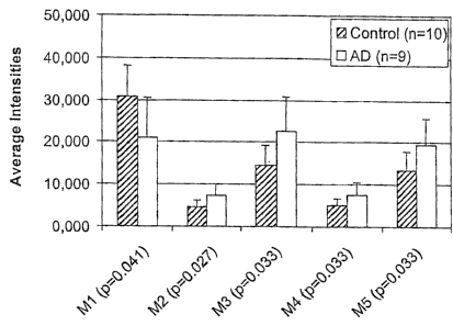
As shown in Figure 1, compared to a negative diagnosis (healthy controls), M1
is
under-expressed in the CSF of Alzheimer's disease patients (p < 0.05), while
the
markers M2 to M5 are over-expressed in CSF of Alzheimer's disease patients (p
<
0.05).
An altered level of one or several polypeptides of the invention, compared to
the
level of polypeptides of the invention in healthy control subjects, will allow
assessing
the state of and/or monitoring the progression of Alzheimer's disease in a
subject,
will allow monitoring the effectiveness of Alzheimer's disease treatment, and
will be
useful information for drug development. Furthermore, these biomolecule
markers
are useful for differentiating Alzheimer's disease from other forms of
dementia and
neurodegenerative disorders.
Preferred subjects in which Alzheimer's disease is to be diagnosed or
monitored are
human subjects. However, diagnosis of Alzheimer's disease according to the
invention is also possible with other mammals. If necessary, orthologues of
the
peptide markers of the invention can be used.
The invention also relates to the use of mass spectrometry (MS) for detecting
Alzheimer's disease in human subjects and for assessing the progression of
Alzheimer's disease in human subjects by detecting and/or quantifying the
amount of
specific polypeptides in samples drawn from the subject's body fluids. In a
preferred
embodiment of the invention, the sample is drawn from the subject's
cerebrospinal
fluid (CSF).
CA 02745569 2011-07-07
WO 2004/019043 PCT/EP2003/008879
- 8 -
Detection and/or quantification of the polypeptides of the invention is
preferably
achieved by quantifying the signal which is detected 8y MS at specific
molecular
mass to charge ratios (M/z) which correspond to the Mk ratios of the
polypeptides of
the invention. Preferably, M/z ratios close to the one of the polypeptides of
the
invention are also measured.
Detection and/or quantification of the polypeptides of the invention can also
be
achieved by using an immunoassay using specific antibodies raised against the
specified marker(s) or polypeptide fragments thereof. Antibodies can be
prepared by
using the purified marker(s) or fragments thereof, or using synthetic or re-
,
combinantly expressed polypeptide(s) consisting of the specific amino acid
sequence
of the marker(s) using any suitable method known in the art [Coligan 1991].
Such
techniques include, but are not limited to, antibody preparation by selection
of
antibodies from libraries of recombinant antibodies in phage or appropriate
vectors,
as well as preparation of polyclonal and monoclonal antibodies by immunising
rabbits or mice [Huse 1989, Ward 1989]. After the antibody is provided, a
marker
can be detected and/or quantified using any of a number of standard
immunological
binding assays [US Patents 4,366,241; 4,376,110; 4,517,288; and 4,837,168].
Useful
assays include, but are not limited to, for example, an enzyme immune assay
(ETA)
such as enzyme-linked immunosorbent assay (ELISA), a radioimmime assay (RTA),
a Western blot assay, or a slot or dot blot assay. For a review of the general
immunoassays see [Coligan 1991]. Generally, a sample obtained from a subject
can
be contacted with the antibody that specifically binds the marker. A powerful
technique to capture the specified marker(s) from a complex body fluid sample
is to
use the antibody fixed to solid supports, such as glass or plastic, e.g.
microtiter plate,
a stick, a bead, or microbead. Alternatively, marker(s) can also be captured
from the
body fluid sample by the specific antibody immobilised to a probe substrate or
a
ProteinChipTm array, as described for the SELDI-based immunoassay [Xiao 2001].
After incubating the sample with antibodies, the non-bound material is washed
under
specified conditions and the antibody-marker complex formed can be detected,
using
appropriate detection reagents. In an embodiment using the SELDI ProteinChipTm
CA 02745569 2011-07-07
WO 2004/019043 PCT/EP2003/008879
- 9 -
:5
array technique the marker(s) selectively enriched by the imnaobilised
antibody can
be detected and quantified by matrix-assisted laser desorption ionisation mass
spectrometry.
The invention specifically relates to
1. a method of assessing the state of Alzheimer's disease in a
subject comprising
detection of at leak one polypeptide comprised in a group of polypeptides
consisting of
i) a polypeptide having a molecular mass of 4824 20 Da,
a polypeptide having a molecular mass of 7691 20 Da,
a polypeptide having a molecular mass of 11787 20 Da,
iv) a polypeptide having a molecular mass of 11988 20 Da, and
v) a polypeptide having a molecular mass of 13416 20 Da.
The invention further relates to a method of assessing the state of
Alzheimer's
- disease in a subject comprising detection of at least one
polypeptide comprised
in a group of polypeptides having, respectively, molecular masses of 4824 20
.20 Da, of 7691 20 Da, of 11787 20 Da, of 11988 20 Da, of 13416 20
Da,
of 4769 20 Da, of 6958 20 Da, of 6991 20 Da, of 13412 20 Da, of
13787 20 Da, of 17276 20 Da, of 40437 20 Da, of 6895 20 Da, of 6928
Da, of 7691 20 Da, of 7769 20 Da, of 7934 20 Da, of 5082 20 Da,
of 6267 20 Da, of 6518 20 Da, of 7274 20 Da, and of 8209 20 Da.
=
Whereas detection of one such polypeptide is in most cases sufficient to
reliably diagnose Alzheimer's disease, detection of two or more polypeptides
of
the invention can increase the sensitivity and robustness of the method.
Preferably, 1, 2, 3, 4, 5, 10, and, most preferred, all of said polypeptides
will be
detected from the same sample. The detection can also be carried out
simultaneously with the detection of other polypeptides which are preferably
CA 02745569 2011-07-07
WO 2004/019043 PCT/EP2003/008879
- 10 -
-
also differentially expressed in subjects having Alzheim' er's disease as
compared to healthy subjects. "Assessing the state of Alzheimer's disease"
shall
be understood as diagnosing the presence of Alzheimer's disease in a subject
or
a patient, as assessing the progression of the disease in a subject or a
patient,
and/or as assessing the proneness of a subject to develop Alzheimer's disease.
2. The invention further relates to the method of point 1 in which 2, or 3,
or 4, or
5 polypeptides of said group of peptides are detected. The invention further
relates to a method of point 1 in which 2, or 3, or 4, or 5, or 10 or all
polypeptides of said group of peptides are detected.
3. The invention further relates to a method of assessing the state of
Alzheimer's
disease in a subject comprising detection of at least one polypeptide
comprising
the sequence of SEQ ID NO:1, SEQ ID NO:2, SEQ ID NO:3, SEQ ID NO:4,
SEQ JD NO:5, SEQ ID NO:6, SEQ ID NO:7, SEQ ID NO:8, SEQ ID NO:9,
SEQ ID NO:10, SEQ JD NO:11, SEQ ID NO:12, SEQ 11) NO:13, SEQ ID
NO:14, SEQ ID NO:15, and/or SEQ ID NO:16. The invention further relates to
a method of assessing the state of Alzheimer's disease in a subject comprising
detection of at least one polypeptide comprising the sequence of SEQ JD NO:1,
SEQ DD NO:2, SEQ ID NO:3, SEQ ID NO:4, SEQ ID NO:5, SEQ ID NO:6,
SEQ ID NO:7, SEQ JD NO:8, SEQ ID NO:9, SEQ ID NO:10, SEQ JD NO:11,
SEQ ID NO:12, SEQ JD NO:13, SEQ ID NO:14, SEQ ID NO:15, SEQ lb
NO:16, and/or SEQ ID NO:17. Whereas detection of one such polypeptide is in
most cases sufficient to reliably diagnose Alzheimer's disease, detection of
two
or more polypeptides of the invention can increase the sensitivity and
robustness of the method. Preferably, 1, 2, 3, 4, 5, 10, or all of said
polypeptides will be detected from the same sample. The detection can also be
carried out simultaneously with the detection of other polypeptides which are
preferably also differentially expressed in subjects having Alzheimer's
disease
as compared to healthy subjects.
CA 02745569 2011-07-07
WO 2004/019043
PCT/EP2003/008879
- 11 -
4. The invention further relates to a method of assessing the state
of Alzheimer's
disease in a subject comprising detection of at least one polypeptide
comprised
in a group of polypeptides consisting of
i) human cystatin C,
ii) human beta-2-microglobulin,
human myoglobin (new variant),
iv) a fragment a at least 5, 8, 10, or 20 amino acids of human cystatin C,
v) a fragment of at least 5, 8, 10 , or 20 amino acids of human beta-2-
microglobulin, and
vi) a fragment of at least 5, 8, 10 , or 20 amino acids of human myoglobin
(new variant).
The invention further relates to a method of assessing the state of
Alzheimer's
disease in a subject comprising detection of at least one polypeptide
comprised
in a group of polypeptides consisting of
i) human cystatin C,
ii) human beta-2-microglobulin,
iii) human myoglobjm. (new variant),
iv) human neurosecretory protein VGF,
v) a fragment of at least 5, 8, 10 , or 20 Smino acids of human cystatin C,
vi) a fragment of at least 5, 8, 10 , or 20 amino acids of human beta-2-
microglobulin,
vii) a fragment of at least 5, 8, 10 , or 20 Min() acids of human myoglobin
(new variant), and
viii) 'a fragment of at least 5, 8, 10 , or 20 amino acids of neurosecretory
protein VGF.
Whereas detection of one such polypeptide is in most cases sufficient to
reliably diagnose Alzheimer's disease, detection of two or more of said
CA 02745569 2011-07-07
WO 2004/019043 PCT/EP2003/008879
- 12 -
,
polypeptides can increase the sensitivity and robustness of the method.
Preferably, 1, 2, 3, 4, 5, or 6 of said polypeptides will be detected from the
same sample. More preferably, 1, 2, 3, 4, 5, 6 or all of said polypeptides
will be
detected from the same sample. The detection can also be carried out
simultaneously with the detection of other polypeptides which are preferably
also differentially expressed in subjects having Alzheimer's disease as
compared to healthy subjects.
5. The
invention further relates to a method of investigating the progression of
Alzheimer's disease in a subject characterised in that a method of any of
points
1. to 4 is performed with at least two distinct samples drawn from the same
subject. For this purpose, samples drawn from a subject at different points in
time will be analysed. Changes in the amount of the respective polypeptide(s)
will allow to draw conclusions on the progression of Alzheimer's disease in
the
subject.
. 6.
The invention further relates to a method of any of points 1 to 5, wherein
detection of said polypeptide(s) is by SELDI-TOF MS. Other suitable mass
spectrometric methods and other methods of detection can alternatively be
used. More specifically, the invention relates to a method of any of points 1
to
5, wherein detection of said polypeptide(s) is by SELDI-TOF MS in which the
hydrophobic H50, the WCX2, or the IMAC surface is used as a support upcin
-ionisation. Different supports for ionisation yield different sensitivity for
specific proteins of interest.
7. The
invention further relates to a method of any of points 1 to 5, wherein
specific antibodies or antibodies recognising said polypeptide(s) are used for
detection of said polypeptide(s).
8. The invention further relates to a method of any of points 1 to 7, wherein
detection is in a sample comprising CSF of said patient. A spmple drawn from
CA 02745569 2011-07-07
WO 2004/019043
PCT/EP2003/008879
- 13 -
a subject can be processed immediately after it has been taken, or it can
first be
frozen and be analysed later. Samples may also consist of or contain other
body
fluids such as blood, serum, plasma, urine, seminal plasma, nipple fluid, or
cell
extracts.
9. The invention further relates to a kit comprising a polypeptide
having a
molecular mass of 4824 20 Da, a polypeptide having a molecular mass of
7691 20 Da, a polypeptide having a molecular mass of 11787 20 Da, a =
polypeptide having a molecular mass of 11988 20 Da, and/or a polypeptide
having a molecular mass of 13416 20 Da. The invention further relates to a
kit comprising a polypeptide having a molecular mass of 4824. 20 Da, a
polypeptide having a molecular mass of 7691 20 Da, a polypeptide having a
molecular mass of 11787 20 Da, a polypeptide having a molecular mass of
11988 20 Da, and a polypeptide having a molecular mass of 13416 20 Da.
The invention further relates to a kit comprising polypeptides having a
molecular mass of 4824 d=E 20 Da, of 7691 20 Da, of 11787 20 Da, of =
11988 20 Da, of 13416 20 Da, of 4769 20 Da, of 6958 2.0 Da, of 6991
Da, of 13412 20 Da, of 13787 20 Da, of 17276 20 Da, of 40437
20 Da, of 6895 20 Da, of 6928 20 Da, of 7691 20 Da, of 7769 20 Da,
20 of 7934 20 Da, of 5082 20 Da, of 6267 20 Da, of 6518 20 Da, of
7274
20 Da, and/or of 8209 20 Da.Such a kit can be applied for various purposes,
e.g., for use as a standard in one of the above mentioned methods. Said kit
can
comprise 2, 5, 10, or all of the above polypeptides.
10. The invention further relates to a kit comprising a fragment of at least 5
amino
acids of human cystatin C, a fragment of at least 5 amino acids of human beta-
.
2-microglobulin, and a fragment of at least 5 amino acids of human myoglobin.
This kit can be applied for various purposes, e.g., for use as a standard in
one of
the above mentioned methods. The invention further relates to a kit comprising
a fragment of at least 5, 10 or 20 amino acids of human cystatin C, a fragment
of at least 5, 10 or 20 amino acids of human beta-2-microglobulin, a fragment
CA 02745569 2014-07-29
30725-351E
- 14 -
of at least 5, 10 or 20 amino acids of human myoglobin, and a fragment of at
least 5, 10
or 20 amino acids of neurosecretory protein VGF. These kits can be applied for
various
purposes, e.g., for use as a standard in one of the above mentioned methods.
The invention as claimed relates to:
- a peptide consisting of the sequence of SEQ ID NO: 4;
- a method of assessing the state of Alzheimer's disease in a subject,
comprising detection of a
polypeptide consisting of the sequence of SEQ ID NO: 4 in a sample from the
subject, and
comparing the amount of the polypeptide in the subject to the amount of the
polypeptide in
healthy controls;
- a method of determining the progression of Alzheimer's disease in a subject,
comprising
detection of a polypeptide consisting of the sequence of SEQ ID NO: 4 in at
least two distinct
samples taken at different times from the same subject, and comparing the
amount of the
polypeptide from the samples taken at the different times; and
- a kit comprising an antibody specifically recognising the polypeptide
consisting of the
sequence of SEQ ID NO: 4, and instructions for use in assessing the state of
Alzheimer's disease.
Brief description of the figures
Figure 1:
Average intensities of the five marker peptides of Table 1, which are
differentially expressed
in the diseased group when compared to the control groups.
CA 02745569 2013-09-23
=
30725-351E
- 15
ExaMples
The invention is further described by one or several of the following
examples. These
examples are not to be understood as restricting the scope of the invention to
the
examples by any means.
Example 1: Patients evaluation and CSF sampling
Diagnosis of Alzheimer's disease in human subjects was made according to
criteria
of the National Institute of Neurologic and Communicative Disorders and
Stroke¨
Alzheimer's disease and Related Disorders Association (NINCDS-ADRDA). The
Alzheimer's disease group consisted of 9 patients aged 75 7 years, six men and
three
women. The group of healthy control subjects consisted of 10 individuals aged
78
14 years, two men .and eight women with no history, symptoms or signs of
psychiatric or neurological disease.
Informed consent was given by each patient and the patients' caregivers before
the
investigation. The study was approved by the local ethics committee. After
lumbar
puncture, CSF samples were frozen on dry ice immediately upon withdrawal at
the
bedside in 0.5 ml aliquots and stored at -80 C until analyses. =
Example 2: ProteinChip SELDI analysis of CSF on SAX2 chip
SAX 2 Proteinehip array (Ciphergen Biosystems, Fremont, CA, USA) were
equilibrated for 5 min with 5111 of binding buffer (100mM Na. Acetate p11=4 0)
The
buffer was carefully removed with an handkerchief and 2.5 .1 of binding buffer
was
added to the wells. Crude CSF samples (2.51.11) were added to the wells and
incubated
for 20 mill at room temperature in a humidity chamber on a rocking platform.
CSF
was removed and the wells were individually washed with 10 1 of binding buffer
for
5 min. The arrays were then placed in a 15m1 conical EppendorfTM and washed
twice
with the binding buffer for 5 min. Finally, the chip was rinsed twice with
distilled
CA 02745569 2013-09-23
30725-351E
- 16 -
water. Excess of 1120 was removed and while the surface was still moist, two
additions per well of 0.5 I of sinapinic acid (SPA) (2mg/m1) in 50% (vol/vol)
acetonitrile and 0.5% (vol/vol) trifiuoroacetic acid was performed and dried.
The
arrays were then read in a ProteinChipTM reader system, PBS II series
(Ciphergen
Biosystems). The laser beam was focused on the sample in vacuo. This caused
the
proteins absorbed to the matrix to become ionised and, simultaneously to be
desorbed from the Proteinchip array surface. The ionised proteins were
detected and
their molecular masseTwere determined according to their time-of-flight (TOF).
TOF
- mass spectra, collected in the positive ion mode were generated using an
average of
65 laser shots throughout the spot at a laser power set slightly above
threshold (10-15
% higher than the threshold) High mass to acquire was set at 40kDa, optimised
from
1 to 15kDa. Spectra were collected and analysed using the Ciphergen
Proteinchip
(version 3.0) software. External calibration of the reader was performed using
the
"all-in-1" peptide molecular weight standards (Ciphergen biosystems, Inc.)
diluted in
the SPA matrix (11, volivol) and directly applied onto a well. Protein profile
comparison was performed after normalisation on total ion current of all the
spectra
mcluded in the same experiment The reproducibility was tested by analysing
different aliquots of the same CSF sample on 4 different wells of the same
proteinclaip array (intraassay intrachip reproducibility), on two different
chips
(intraassay interchip reproducibility) processed in parallel, and reproduced
in an
other experiment (interassay reproducibility).
Analysis of CSF samples from 9 patients diagnosed with Alzheimer's disease
relative
to 10 controls revealed that 5 peaks were significantly differentially
expressed
between the two groups (p<0.05). The approximate average SELDI mass associated
with the five differentially expressed proteins was 4.82kDa, 7.7IcDa, 11.8IcDa
and
12.0kDa and 13.4kDa (p<0.05) (see Table 1, Fig. 1).
CA 02745569 2011-07-07
307---351
- 17 -
Example 3: Strong Anionic exchange chromatography (SAX) purification
In order to identify the proteins corresponding to these peaks, a
fractionation of crude
CSF on a SAX spin column was performed. The eluted fractions were analysed by
SELDI-TOF MS.
SAX spin column, lot number SAX2-001116-01, (Ciphergen Biosystems, Fremont,
CA, USA) was rehYdrated overnight at 4 C in the equilibration buffer (20mM
tris(hydroxymethyl)sminomethane hydrochloride - (Tris-HC1), 5mM NaC1, pH 9.0).
The column was warmed up at room temperature and air bubbles were removed. The
equilibration buffer was let flow through column matrix by gravity.
Equilibration
buffer (0.5m1) was added to the column and passed through the resin twice. Two
ml
of control CSF was diluted in the equilibration buffer (1:1, vol/vol).
_Protein sample
was loaded to the column by fraction of 0.8m1 and allowed to run through the
column
by gravity until no drops came out of the column The column was then
centrifuged
at 150 x g for 1 min. The resin was then washed with an equivalent volume of
equilibration buffer. This step was repeated several times in order to load
the whole
sample onto the resin. Elution of the bound proteins was performed by
decreasing the
pH. Elution buffer A consisted of 20mM Tris-HC1, 5mM NaC1 pH 8.0; elution
buffer
B = 20mM sodium phosphale pH 7.0; elution buffer C = 20mM sodium phosphate
pH 6.0; elution buffer D = .20mM sodium phosphate and citrate pH 5.0; elution
buffer E = 20mM sodium phosphate and citrate pH 4.0; elution buffer F = 20mM
sodium phosphate and citrate pH 3.4; elution buffer G =- 30% acetonitrile in
elution
buffer F. Elution was performed by applying 2 x 75 ul of the elution buffer
and
- 25 centrifugation at 150 x g for lmin. Each collected fraction
(1500) was concentrated
on a speed-vac to a volume of 10111. Protein profiles were analysed on SELDI-
TOF
MS using SAX 2 Proteincbip arrays. The chip was equilibrated with a binding
buffer
consisting of 20mM Tris-11C1, 5mM NaC1, pH=9Ø An aliquot of 0.5 p.1 of each
concentrated fraction was applied directly onto 2.5 .1 of binding buffer per
spot and
processed as previously described. The rest of the fractions were loaded onto
a Tris
tricine gel as described below.
CA 02745569 2011-07-07
.WO 2004/019043 PCT/EP2003/008879
- 18 -
The differentially expressed peak of 13.4kDa was eluted with buffer A (20mM
Tris-
HC1 5mIVI NaC1 pH 8.0) and B (20mM sodium phosphate pH 7.0). The
differentially
expressed peaks of 11.8kDa and 12.0kDa were found in the fraction eluted with
buffer C (20mM sodium phosphate pH 6.0) and D (20mM sodium phosphate and
citrate pH 5.0). The cluster of 7.7kDa was eluted with buffer D (20mM sodium
phosphate and citrate pH 5.0) and E (20mM sodium phosphate and citrate pH
4.0).
Each eluted fraction was loaded on a 16.5% Tris Tricine sodium dodecyl sulfate
polyacrylamide gel and electrophoresed (SDS PAGE). After coloration with
coomassie blue, the bands seen on the gel confirmed the results obtained by
SELDI
analysis. The band corresponding to the cluster of 7.7kDa, 11.8kDa and 12.0kDa
were cut out. Proteins were extracted as described in Example 6 and identified
by Q-
TOF. The 7.7kDa peak MS analysis did not match with any known human protein,
however, may indicate to a new variant or homologue of myoglobin. Peptide
sequences were the following
MXAD(L/I)AGHG(Q/K)EV(L/l)(L/1)R and
HGTVV(L/I)TA(L/I)GG(L/I)(L/I)K.
The MS analysis of the 11.8kDa and 12.0kDa peaks identified beta-2-
microglobulin
for both of them.
Since the cluster of 13.4kDa could not be seen on the Tris Tricine gel, a Tris
glycine
SDS-PAGE electrophoresis was performed on crude CSF samples. The band
corresponding to the beta-2-microglobulin could be easily found on this
stained gel.
We concluded that the protein Migrating just above the beta-2-microglobulin
could
correspond to the next abundant protein seen on the SELDI profile, namely the
13.4kDa peak. The band was excised from the gel and digested by trypsin before
MALDI analysis. The peptide mass fingerprint analysis allowed to identify the
Cystatin C. The sequence coverage provided by the analysis was 60%.
= CA 02745569 2011-07-07
=
-
3072-051
- 19 -
Example 4: Monodimensional electrophoresis / Tris Glycine gels
Twenty p.1 of CSF were mixed with 10 p.1 of denaturing Laemmli buffer [Laemmli
5 1970]. The samples were heated to 95 C for 5 min, and loaded on a 15% T
(T = total
acrylamide concentration) SDS-polyacrylamide gel according to the method of
Laemmli. Gels were stained in a solution containing Coomassie Brilliant Blue R-
250
(0.1% w/v) and inetlianol (50% v/v) for 30 min. Destaining was done in a
solution
containing methanol (40% v/v) and acetic acid (10% v/v).
Example 5: Monodimensional electrophoresis : Tris Tricine Gels
Tris tricine SDS-PAGE electrophoresis was performed according to Schagger and
von Jagow [1987] using precast 16.5 % T gels (Biorad, Hercules, CA). The anode
15 buffer consisted of 0.2M Tris-HC1, pH 8.9 and the cathode buffer
consisted of 0.1M
Tris-HC1, 0.1M Tricine, 0.1% SDS, pH 8.25. Samples were diluted in 10p.1 of
50mM
Tris-HCI, 4% (w/v) SDS, 12% (w/v) sucrose, 5% (v/v)13-mercaptoethanol, and
trace
of bromophenol blue, pH 6.8. After denaturation at 95 C for 5min, samples were
loaded onto the gel. Gels were run at 80V for 3 hours. After electrophoresis,
gels
20 were fixed in 40% methanol? 10% acetic acid for 30min. Gels were then
stained with
Colloidal blue coomassie G250 overnight and destained in 30% methanol. Bands
to
be identified were immediately cut, placed in an eppendorf and kept at 4 C
until
further analysis. The apparent molecular masses were determined by running
polypeptide molecular weight (MW) standards: Triosephophate isomerase MW
25 26,625; Myoglobin MW 16,950; a-lactalbumin MW 14,437; Aprotinin MW
6,512;
Insulin b chain, oxidised MW 3,496 and Bacitracin MW 1,423 (Biorad).
=
Example 6: Protein digestion and peptide extraction [Bien-venu 1999]
30 Fragments of gels containing proieins of interest were cut out for
digestion of the
proteins with trypsin using previous published procedures [Shevchenko 1996,
CA 02745569 2013-09-23
30725-351E
- 20 -
Hellman 1994, Rosenfeld 1992] and modified as described below. The piece of
gel
was first destained with 100 p.1 of 50 mM ammonium bicarbonate, 30% (v/v)
acetonitrile during 15 min, at room temperature. Destaining solution was
removed
and replaced by 25 I of 10 mM DL-dithiothreitol (DTT) in 50 mM ammonium
bicarbonate and incubated 35 min at 56 C. DTT solution was then replaced by 25
p.1
of 55 mM iodoacetamide in 50 mM ammonium bicarbonate and incubated during 45
min at room temperature in. the dark. Gel pieces were washed for 10 min with
100 p.1
of 50 mM ammonium bicarbonate and for 10 min with 100 p.1 of 50 mM ammonium
bicarbonate and 30% (v/v) acetonitrile.
Gel pieces were then dried for 30 min in a HetovacTM vacu-um centrifuge
(11ETO,
Allerod, Denmark). Dried pieces of gel were rehydrated for 45 min at 4 C in 5-
20 p.1
of a solution of 50 mM ammonium bicarbonate containing trypsin at 6.25 ng/p.l.
After an over-night incubation at 37 C, gel pieces were dried under high
vacuum
centrifuge before being rehydrated by the addition of 20 p.1 of distilled
water and
finally dried again in a speed-vac for 30 min. Extraction of the peptides was
performed with 20 ill of 0.1% (v/v) trifluoroacetic acid (TFA) for 20 min at
room
temperature with occasional shaking. The TFA solution containing the peptides
was
transferred to a polypropylene tube. A second elution was performed with 20
p.1 of
0.1% (v/v) TFA in 50% (v/v) acetonitrile for 20 min at room temperature with
occasional shaking. The second TFA solution was pooled with the first one. The
volume of the pooled extracts was reduced to 1-2 p.1 by evaporation under
vacuuia.
Control extractions (blanks) were performed using pieces of gels devoid of
proteins.
Example 7: Protein identification by peptide mass fingerprinting analysis
1.5 p.1 of sample was placed on a MALDI 100-well target plate. Same volumes of
matrix (10 mg/ml a-Cyano-4-hydroxycinnamic acid in 50% (v/v) acetonitrile,
0.1%
(v/v) TFA) were added to the previously loaded digest. Samples were dried as
quickly as possible using a vacuum container. Mass measurement from liquid
solution were conducted with a MALDI-TOF mass spectrometer VoyagerTM Elite
CA 02745569 2013-09-23
30725-351E
- 21 -
and. Super STR (PerSeptive Biosystems, Framingham MA, USA) equipped with a
337 um nitrogen laser. The analyser was used in the reflectron mode at an
accelerating voltage of 20 kV, a delayed extraction parameter of 100-140 ns
and a
low mass gate of 850 Da. Laser power was set slightly above threshold (10-15 %
higher than the threshold) for molecular ion production. Spectra were obtained
by
summation of 10 to 256 consecutive laser shots. Masses of the 60 highest peaks
were
extracted from the spectra and used for protein identification using the Sma-
rtIdentTM
peptide mass fingerprint tool [Gras 1999]. The research was conducted against
SWISS-PROT and TrEMBL databases. The query was made for the human, .the
minimum number of matched masses was 4, the maximal tolerance for masses was
50 ppm after an internal calibration using autolysis product of trypsin, at
most one
missed cleavage for tryptic peptides was allowed, and the modifications
accepted
were carboxymethylation with iodoacetamide of cysteines and artefactual
oxidation
of niethionines.
Example 8: Protein identification by peptide fragmentation analysis
Prior to nanoLC (LC = liquid chromatography) separation, the volumes of
peptide
containing solutions were adjusted to 7111 by addition of a 0.1 % (v/v) formic
acid
solution. Samples were settle,d in a Triathlon autosampler (Spack, Emmen,
Holland).
For each experiment, 5 gi of peptide containing solution were injected on a
C18
reverse phase column of 75 um inner diameter (YMS-ODS-AQ200, Michrom
Bioresource; Auburn, CA). Peptides were eluted with an acetonitrile (ACN)
gradient
in the presence of 0.1 % (v/v) formic acid, using SunPlow TM pumps. (SunChrom,
Friderichsdorf, Germany). A flow splitter was used in order to decrease the
flow rate
after the pumps from 200 to 0.4 ill/min. Peptides were analysed with a
quadrupole
time-of-flight (Q-TOF) mass spectrometer (Mieromass, Wyth.enshawe, England). A
2700 V tension was applied on the nanoelectrospray capillary (New Objective,
Wobum, MA, USA). Argon was used as collision gas. The collision energy was
settled as a function of the precursor ion mass. MS/MS_ spectra were acquired
by
automatic switching between MS and MS/MS mode. Acquired MS/MS data were
CA 02745569 2013-09-23
30725-351E
- 22 -
TM
converted in a compatible format (DTA files) by ProteinT ynx software
(Micromass,
Wythenshawe, England) and analysed using conventional search engines against
SWISS-PROT, TrEL, NCBInr and EST databases. In cases of manual inter-
pretation of MS/MS data, identification was performed by sequence only search.
It was found that marker M5 was a fragnent of Cystatin C, markers M3 and M4
were
isoforms of beta-2-microglobulin and M2 was a new variant or homologue of
myoglobin. Marker MI was found to be a fragment of the neurosecretory protein
VGF.
- 10
Example 9: Statistical Analysis
P-values were calculated using standard statistical methods known to the
person
skilled in the art. P-values smaller than 0.05 were considered to be
statistically
significant.
Example 10: Isolation of the 4.8 kDa Fragment (Marker M1)
TM
Some CSF samples from control patients were fractionated by Centricon 30
filtration
device (Millipore Corp., Bedford, MA) in order to remove the protein with a
molecular weight higher than 30 kDa.. The salt and polypeptide with a
molecular
weight lower than 3 kDa were removed using a Centricon 3 (Millipore Corp.,
Bedford, MA). The Centric= 3 was then washed with ultrapure distilled water.
In
that wash fraction, the 4.82 kDa was found to be the major component. This
liquid
- 25 = fraction was first reduced with a 10 mM solution of 1,4-
Dithioerythritol for lh at
56 C, then alkylated with 54 DIM iodoacetamide for 45 min at room temperature.
Finally, the polypeptide was digested with 6 mg/1 trypsin overnight at 37 C.
This
liquid fraction was analysed by nanoLC and Q-TOF as previously described.
CA 02745569 2011-07-07
=
WO 2004/019043 PCT/EP2003/008879
- 23 -
Example 11: Different Surface Materials for SELDI-TOF Analysis
Using SELDI-TOF, an analysis of 10 CSF samples from AD patients and 10
controls
was performed on three different surfaces: the hydrophobic H50, the WCX2, and
the =
IMAC surface (Ciphergen Biosystems, Fremont, California, USA, resp.). Seven
differentially expressed peaks were found on the H50, five markers on the
WCX2,
and five markers on the "MAC surface. A diagnostic test using the markers on
the
1150 chip revealed a specificity and sensitivity of 100% and 70%,
respectively. The
combination of the markers found on H50 and WCX2 gave a specificity and
=
sensitivity of 100% and 80%. Finally, the combination of the markers found on
H50,
WCX2 and IMAC gave a specificity and sensitivity of 100% and 90%.
The average masses of the differentially expressed polypeptides as determined
by
SELDI-TOF using different surface materials were as follows:
Surface hydrophobic H50: 7 peaks
Marker 1: 4769 s.d. Da
Marker 2: 6958 s.d. Da
Marker 3: 6991 s.d. Da
Marker 4: 13412 s.d. Da
Marker 5: 13787 s.d. Da
Marker 6: 17276 s.d. Da
Marker 7: 40437 s.d. Da
Surface IMAC Cu: 5 peaks
Marker 1: 6895 s.d. Da
Marker 2: 6928 s.d.Da
Marker 3: 7691 s.d. Da
Marker 4: 7769 s.d. Da
Marker 5: 7934 s.d. Da =
CA 02745569 2011-07-07
WO 2004/019043 PCT/EP2003/008879
- 24 -
Surface WCX2: 5 peaks
Marker 1: 5082 s.d. Da
Marker 2: 6267 s.d. Da
Marker 3: 6518 s.d. Da
Marker 4: 7274 s.d. Da
Marker 5: 8209 s.d. Da
The standard deviation (s.d.) is 20 Da for each marker above. However, the
standard
deviation can also be 40 Da, or 10 Da, or 5 Da for each marker above.
Bibliography
Adam, B.L. et al. 2001, Proteomics 1(10): 1264-1270
Asgeirsson B, Haebel S, Thorsteinsson L, Helgason E, Gudmundsson KO,
Gudmundsson G, Roepstorff P. Hereditary cystatin C amyloid anopathy:
monitoring the presence of the Leu68Gln cystatin C variant in cerebrospinal
fluid
and monocyte cultures by MS. Biochem J. 1998, 329, 497-503.
Beyer K, Lao JI, Gomez M? Riutort N, Latorre P. Mate JL, Ariza A. Alzheimer's
disease and the cystatin C gene polymorphism: an association study.
Neuroscience
letters. 2001, 315, 17-20.
Canu N, Possenti R, Ricco AS, Rocchi M, Levi A. Cloning, structural
organization.
analysis, and chromosomal assignment of the human gene for the neurosecretory
protein VGF. Genomics 1997, 45 (2), 443-446.
Cohen DH, Feiner H, Jensson 0, Fran&ne B. Amyloid fibril in hereditary
cerebral
hemorrhage with amyloidosis (HCHWA-I) is related to the gastroentero-
pancreatic
neuroendocrine protein. I Exp. Med. 1983, 158, 623-628.
CA 02745569 2011-07-07
WO 2004/019043 PCT/EP2003/008879
-25-
1
Current Protocols in Immunology, Eds. Coligan IF, Kmisbeek AM, Margulies DH,
Shevach EM, and Strober W., John Wiley & Sons Inc. 691
Crawford U, Freeman MJ, Schinka IA, Abdullah LI, Gold M, Hartman R, Krivian K,
Morris MD, Richards D, Duara R, Anand R, 1VIullan MI. A polymorphism in the
cystatin C gene is a novel rsik factor for late-onset Alzheimer's disease.
Neurology.
2000, 55, 763-768.
Davies, H.A. 2000. The ProteinChip(R) System from Ciphergen: A new technique
for rapid, micro-scale protein biology. J. Molecular Medicine, 78 (7):B29 Deng
A,
Irizarry MC, Nitsch RM, Growdon JH, Rebeck GW. Elevation of Cystatin C in
susceptible neurons in Alzheimer's disease. Am. J. Pathol. 2001, 159, 1061-
1068.
Emerudh 3, Olsson T, Berlin G, von Schenck H. Cerebrospinal fluid
immunoglobulins and beta 2-microglobulin in lymphoproliferative and other
=neoplastic diseases of the central nervous system. Arch Neurol. 1987, 44(9),
915-20.
Finckh U, Von der Kammer H, Velden 3, Michel T, Andresen B, Deng A, 72aang
Muller-Thomsen T, Zuchowski K, Menzer G, Mann U, Papassotiropoulos A, Heun
R, Zurdel J, Hoist F, ben9si L, Stoppe G, Reiss J., Miserez AR, Staehelin HB,
Rebeck W, Hyman BT, Binetti G, Hock C, Growdon JH, Nitsch RM. Genetic
association of a cystatin C gene polymorphism with late-onset Alzheimer's
disease.
Arch Neurol. 2000, 57, 1579-1583.
Francis, P.T. et al. J. Neurol. Neurosurg. Psychiatry 1999, 66(2): 137-147
Fung, ET et al. Protein biochips for differential profiling. Current Opinion
in
Biotechnology, 2001, 12(1): 65-69
Ghiso I, Jensson 0, Frangione B. Amyloid fibrils in hereditary cerebral
hemorrhage
with amyloidosis of Icelandic type is a variant of gamma-trace basic protein
(cystatin
C). Proc Natl Acad Sci USA. 1986;83(9):2974-8.
CA 02745569 2011-07-07
WO 2004/019043 PCT/EP2003/008879
- 26 -
Ghiso 3, Pons-Estel B, Frangione B. Hereditary cerebal amyloid angiopathy: the
amyloid fibrils contain a protein which is a variant of cystatin C, an
inhibitor of
lysosomal cysteine proteases. Biochem Biophys Res Commun. 1986;136(2):548-54.
Gras R, Muller M, Gasteiger E, Gay S, Binz PA, Bienvenut WV, Hoogland C,
Sanchez JC, Bairoch A, Hochstrasser DF, Appel R. Improving protein
identification
from peptide mass frngerprinting through a parameterised multi-level scoring
algorithm and an optimised peak detection. Electrophoresis 1999, 20: 3535-3550
Grubb AO, Jensson 0, Gudmundsson G, Amason A, Lofberg H, Maim J. Abnormal
metabolism of g-trace alkaline microprotein. The basic defect in hereditary
cerebral
hemorrhage with amyloidosis. N. Engl. J. Med 1984, 311, 1547-1549.
Hellman U, Wernstedt .C, Gofiez, I, Heldin CH. Improvement of an "hi-Gel"
digestion procedure for the micropreparation of internal protein fragments for
amino
acid sequencing. Anal. Biochetn. 1995, 224, 451-5.
Hoekman K, Van Nieuwkoop JA, Willemze R. The significance of beta-2
microglobulin in clinical medicine. Neth J Med. 1985;28(12):551-7.
Huse WD, Sastry L, Iverson SA, Kang AS, Alting-Mees M, Burton DR, BenkoviC
SJ, Lerner RA. Generation of a large combinatorial library of the
immunoglobulin
repertoire in phage lambda. 1989, Science 246: 1275-1281.
Issaq HJ, Veenstra TD, Conrads TP, Felschow D. The SELDI-TOF MS approach to
proteomics: protein profiling and biomarker identification. Biochem. Biophys.
Res.
Commun. 2002, 292 (3): 587-592
=
CA 02745569 2011-07-07
WO 2004/019043 PCT/EP2003/008879
- 27 -
Kalman J., Marki-Zay I, Juhasz A, Santha A, Dux L, Janka Z. Serum and
Cerebrospinal fluid cystatin C levels in vascular and Alzheimer's dementia.
Acta
Neurol. Scold. 2000, 101, 279-282.
Lae=li UK. Cleavage of structural proteins during the assembly of the head of
bacteriophage T4. Nature. 1970 Aug 15;227(259):680-5.
Leung-Tack J, Tavefa C, Gensac MC, Martinez J, Colle A. Modulation of
phagocytosis-associated respiratory burst by human cystatin C: role of the N-
terminal
tetrapeptide Lys-Pro-Pro-Arg. Exp. Cell Research. 1990, 188, 16-22.
a
Levy E, Sastre M, Kumar A, Gallo G, Piccardo P, Ghetti B, Tagliavini F,
Codeposition of cystatin C with amyloid-beta protein in the brain of
Alzheimer's
disease patients. J. Neuropathol Exp Neurol 2001, 60, 94-104
Merchant M, Weinberger SR. . Recent advancements in surface-enhanced laser .
desorption/ionization-time of flight-mass spectrometry. Electrophoresis, 2000,
21:
1164-1167
90 Popovic T, Brzin J, Ritonja .,;k, Turk V. Different forms of human
Cystatin C. Biol.
Chem. Hoppe-Seyler. 1990, 371, 575-580.
Raymackers J, Daniels A, DeBrabandere V, Missiaen C, Dauwe M, Verhaert P,
Vanmechelen E, Meheus L. Identification of two dimensionally separated human
cerebrospinal fluid proteins by N-terminal sequencing, matrix-assisted laser-
desorption/ionization-mass spectrometry, nanoliquid chromatography-
electrospray
ionization-time of flight-mass spectrometry, and tandem mass spectrometry.
Electrophoresis, 2000, 21, 2266-2283.
The Ronald and. Nancy Reagan Research Institue of the Alzheimer's Association
and the National Institute on Aging Working Group. Consensus Report of the
CA 02745569 2011-07-07
WO 2004/019043 PCT/EP2003/008879
>
- 28 -
Working Group on: "Molecular and Biochemical Markers of Alit, eimer's
Disease".
Neurobiology of Aging, 1988, 19 (2): 109-116.
Robles A. . Some Remarks on biological markers of Alzheimer's Disease.
Neurobiology of Aging, 1998, 19 (2): 153-157
Rosenfeld J, Capdevielle J, Guillemot JC, Ferrara P. In-gel digestion of
proteins for
internal sequence analysis after one- or two-dimensional gel electrophoresis.
Anal.
Biochein. 1992, 203, 173-9.
Shevchenko A, Wilna M, Vorm 0, Mann M. Mass spectrometric sequencing of
proteins silver-stained polyacrylamide gels. Anal. Chem. 1996, 68, 850-8.
Tennissen CE, de Vente J, Steinbusch HWM, De Bruijn C.. Neurobiology of Aging,
2002, 23: 485-508
US Patent 4,366,241
US Patent 4,376,110
US Patent 4,517,288
US Patent 4,837,168
Ward ES et al., Binding activities of a repertoire of single immunoglobulin
variable
domains secreted from Escherichia coli. Nature 1989, 341, 544-546
Wei L, Berman Y, Castano EM, Cadene M, Beavis RC, Devi L, Levy E. Instability
of the amyloidogenic cystatin C variant of hereditary cerebral hemorrhage with
amyloidosis, Icelandic type. J Biol. Chem. 1998, 273, 11806-11814.
Wulauhle JD, McLean KC, Paweletz CP, Sgroi DC, Trock BJ, Steeg PS, Petricoin
EF . New approaches to proteomic analysis of breast cancer.
Proteomics 2001, 1 (10): 1205-1215
CA 02745569 2011-07-07
. WO 2004/019043
PCT/EP2003/008879
- 29 -
Xiao Z, Adam BL, Cazares LH, Clements MA, Davis IW, Schellhammer PF,
Dalmasso EA, Wright GL. Quantitation of serum prostate-specific membrane
antigen
by a novel protein biochip immunoassay discriminates benign from malignant
prostate disease. .2001, Cancer Research 61(16): 6029-6033
CA 02745569 2011-07-07
DEMANDES OU BREVETS VOLUMINEUX
LA PRESENTE PARTIE DE CETTE DEMANDE OU CE BREVETS
COMPREND PLUS D'UN TOME.
CEC1 EST LE TOME 1 DE 2
NOTE: Pour les tomes additionels, veillez contacter le Bureau Canadien des
Brevets.
JUMBO APPLICATIONS / PATENTS
THIS SECTION OF THE APPLICATION / PATENT CONTAINS MORE
THAN ONE VOLUME.
THIS IS VOLUME 1 _______________________ OF 2
NOTE: For additional volumes please contact the Canadian Patent Office.