Note: Descriptions are shown in the official language in which they were submitted.
= = CA 02745932 2013-09-30
- -
=
METHODS FOR PREDICTING PREGNANCY OUTCOME
IN A SUBJECT BY hCG ASSAY
=
This is a divisional application of Canadian application number 2,319,784
filed on February 3, 1999.
. 5
Background of the Invention
Throughout this application, various publications are
referenced by:author and date. Full citations. for these'
publications may be found listed alphabetically at the
end of the specification immediately preceding the
claims.
- Early pregnancy loss (EPL) is a widespread, but largely
undiagnosed problem. In order to adegUately diagnose
=
. . and develop treatments for EPL it is -essential to be .
able to detect and measure the rate of occurrence of
EFL. = This is critically important in epidemiological
studiea, same of which are related to exposUres to knowti
or suspected reproductive toxins in the workplace, in
the environment or by personal use.
These early.
=
=
.30 pregnancy lasses are often not recbgnized:by women or
physicians and are detected solely by the measurement of
hCG in the urine at the time between implantation and
expected menses. They are sometimes termed "chemical
pregnancieS" or "occult pregnancies."
A landmark
.35 epidemiological study established that the incidence.of
CA 02745932 2011-07-04
-2-
EPL was 22% in a population of healthy women attempting
to conceive (Wilcox, A. J., et al., 1988). This
investigation employed a very sensitive (0.01 ng/ml hCG)
assay which detected only the intact hCG molecule with
the unique beta subunit carboxyterminal peptide present.
There are multiple likely causes for EPL and clinical
spontaneous abortion including genetic abnormality,
immunological dysfunction, untreated infection or other
unknown physiological problems. In addition, losses may
be caused by failure of human chorionic gonadotropin
(hCG) to induce adequate response at its target, the
corpus luteum. This could result from inadequate
hormonal potency. "Nicking" of the beta subunit in the
loop 2 region of the molecule, specifically between
residues 44-49, can reduce biopotency of hCG. Cleaved
peptide bonds in this area of the molecule also exhibit
reduced biopotency and reduced immunochemical
recognition by monoclonal antibodies directed to the
heterodimeric hormone (Cole, L. A., et al., 1991a; Cole
, L. A., et al., 1991b; Puisieux, A., et al., 1990;
Nishimura, R., et al., 1988; Nishimura, R. T., et al.,
1989). Nicked forms of hCG were examined as possibly
more prevalent in EPL situations and, at least in part
responsible, for early pregnancy loss. Unfortunately
many of the reports claiming that substantial
concentrations of nicked hCG are produced during
pregnancy, losses or successful pregnancies, are not
accurate due to faulty assumptions regarding assay
specificity. Carbohydrate-modified hCG can also exhibit
either reduced or enhanced biopotency. It is known that
if the hCG has much reduced sialic acid content and its
carbohydrate chains terminate in galactose, much hCG
would be removed by the liver receptor for such altered
glycoproteins (Braun, J. R., et al., 1996; Kawasaki, T.
and G. Ashwell, 1996). The circulating life-time of
asialo hCG is reduced and its in vivo potency is thereby
CA 02745932 2011-07-04
-3-
low. Other carbohydrate changes also alter circulating
half life;
glycoproteins terminating in sulfate-N-
acetyl galactosamine are also extracted by a specific
liver receptor and have reduced circulating lifetime
(Baenziger, J. U., 1994; Fiete, D., et al., 1991).
At least two factors affect increased potency of hCG.
First, it is known that a larger Stoke's radius will
decrease clearance through the kidney glomerulus which
generally clears proteins above an effective size of
70,000 very slowly. The
effective size of urinary-
isolated hCG is just at this borderline reduced
clearance size. Generally, extra sugar content makes
the hydrated radius of glycoproteins larger. It has
been shown that by adding the hCG beta COOH-terminal
peptide to hFSH or hLH, their circulating life-times
greatly increased (Fares, F. A. et al., 1992; Matzuk, M.
M., 1990). This addition was thought mostly due to the
carbohydrate content of that peptide rather than simply
the extra polypeptide size. Second, increased negative
charge of a protein will prolong its circulating time
because of decreased renal clearance (Chmielewski, C.
1992, Quadri, K. H., et al., 1994; Maack, T., et al.,
1985). This increased negative charge can be due to
extra sialic acid or other negative groups, including
sulfate such as is present on hLH and on the pituitary
form of hCG (Birken, S., et al., 1996b). Changes which
affect signal transduction at the receptor may also
affect biopotency of hCG. It is known
that
deglycosylated hCG has much reduced receptor potency
(Ravindranath, N., et al., 1992; Sairam, M. R., and L.
G., Jiang, 1992; Browne, E. S., et al., 1990; Sairam, M.
R., 1989; Sairam, M. R., et al., 1988). Carbohydrate
reduced forms of hCG also have reduced signal
transduction (Amano, J., et al., 1990; Bahl, O. P., et
al., 1995; Moyle, W. R., 1975).
CA 02745932 2011-07-04
-4-
According to the present invention EPL or recurrent
spontaneous abortion is not due to an abnormal hCG form
that has reduced potency, such as nicked hCG. Instead,
the present invention provides evidence that in
successful outcome pregnancies women usually produce
forms of hCG which are very highly potent in very early
pregnancy; the standard urinary reference preparations
of hCG are less potent forms of the hormone produced
later in pregnancy. The increased potency could be
caused by a combination of factors from circulating
half-life to increased receptor affinity or signal
transduction or all of the preceding. Since hCG is low
very early in pregnancy, it is logical to find a more
potent form of hCG on a molar basis to carry out its
function until production levels rise as the
trophoblastic cellular mass increases. The
present
invention describes molecular and immunological tools
and methods including an antibody, B152, described
herein which recognizes the highly potent early
pregnancy associated molecular isoforms of hCG. The
determination of blood and urine profiles for the B152
hCG isoforms throughout healthy pregnancies can
delineate the pattern of isoforms in successful
pregnancies. These
isoforms can be measured by
immunoassay alone, obviating the need to perform complex
isoelectric focusing studies or other separation
techniques. Additionally, the methods described herein
are applicable to large numbers of samples.
CA 02745932 2011-07-04
-5-
Bummary of the Invention
The present invention provides a method of predicting
pregnancy outcome in a subject by determining the amount
of an early pregnancy associated molecular isoform of
hCG in a sample comprising: (a) contacting a sample with
an antibody which specifically binds to the early
pregnancy associated molecular isoform of hCG under
conditions permitting formation of a complex between the
antibody and the early pregnancy associated molecular
isoform of hCG; (b) measuring the amount of complexes
formed, thereby determining the amount of the early
pregnancy associated molecular isoform of hCG in the
sample; and (c) comparing the amount early pregnancy
associated molecular isoform of hCG in the sample
determined in step (b) with the amount determined for
temporally matched, normal pregnant subject(s) wherein
the relative absence of the early pregnancy associated
molecular isoform of hCG in the sample indicates a
negative outcome of pregnancy for the subject.
The present invention further provides a method of
predicting pregnancy outcome in a subject by determining
the amount of an early pregnancy associated molecular
isoform of hCG in a sample comprising: (a) contacting a
capturing antibody which specifically binds to the early
pregnancy associated molecular isoform of hCG with a
solid matrix under conditions permitting binding of the
antibody with the solid matrix; (b) contacting the bound
matrix with the sample under conditions permitting
binding of the antigen present in the sample with the
capturing antibody; (c) separating the bound matrix and
the sample; (d) contacting the separated bound matrix
with a detecting antibody which specifically binds to
hCG under conditions permitting binding of antibody and
antigen in the sample; (e) measuring the amount of bound
antibody on the bound matrix, thereby determining the
amount of early pregnancy associated molecular isoform
CA 02745932 2011-07-04
-6-
of hCG in the sample; and (f) comparing the amount early
pregnancy associated molecular isoform of hCG in the
sample determined in step (e) with the amount determined
for temporally matched, normal pregnant subject(s),
wherein amounts of the early pregnancy associated
molecular isoform of hCG in the sample similar to
amounts of early pregnancy associated molecular isoform
of hCG in temporally matched pregnant samples indicates
a positive outcome, amounts of early pregnancy
associated molecular isoform of hCG in the sample
similar to amounts of early pregnancy associated
molecular isoform of hCG in the non-pregnant samples
indicates a negative outcome of pregnancy for the
subject.
In addition, the present invention provides a method for
determining the amount of early pregnancy associated
molecular isoforms of in a sample comprising: (a)
contacting the sample with an antibody which
specifically binds to an early pregnancy associated
molecular isoform of hCG under conditions permitting
formation of a complex between the antibody and the
early pregnancy associated molecular isoform of hCG; and
(b) determining the amount of complexes formed thereby
determining the amount of early pregnancy associated
molecular isoform of hCG in the sample.
Further, the present invention provides a diagnostic kit
for determining the amount of early pregnancy associated
hCG is a sample comprising: (a) an antibody which
specifically binds to an early pregnancy associated
molecular isoform; (b) a solid matrix to which the
antibody is bound; and (c) reagents permitting the
formation of a complex between the antibody and a
sample.
The present invention additionally provides an antibody
CA 02745932 2011-07-04
-7-
which specifically binds to an early pregnancy
associated molecular isoform of human chorionic
gonadotropin.
Further, the present invention provides a method for
detecting non-trophoblast malignancy in a sample
comprising: (a) contacting a sample with an antibody
which specifically binds to the early pregnancy
associated molecular isoform of hCG under conditions
permitting formation of a complex between the antibody
and the early pregnancy associated molecular isoform of
hCG; (b) contacting the sample with a second antibody
which specifically binds to intact non-nicked hCG
without substantially cross-reacting with said antibody
under conditions permitting formation of a complex
between the antibody and the early pregnancy associated
molecular isoform of hCG; (c) measuring the amount of
complexes formed, thereby determining the amount of_the
early pregnancy associated molecular isoform of hCG in
the sample; and (d) comparing the amount of early
pregnancy associated molecular isoform of hCG in the
sample determined in step (b) with the amount of early
pregnancy associated molecular isoform of hCG in the
sample determined in step (c), wherein a positive
detection of early pregnancy associated molecular
isoform detected in step (b) and a relative absence of
the early pregnancy associated molecular isoform of hCG
detected in step (c) indicates the presence of non-
trophoblast malignancy in the sample.
Finally, the present invention provides a method for
detecting gestational trophoblast disease in a sample
from a subject comprising (a) contacting a sample with
an antibody which specifically binds to the early
pregnancy associated molecular isoform of hCG under
conditions permitting formation of a complex between the
antibody and the early pregnancy associated molecular
CA 02745932 2011-07-04
-8-
isoform of hCG; (b) contacting the sample with a second
antibody which specifically binds to intact non-nicked
hCG without substantially cross-reacting with
said antibody under conditions permitting formation of
a complex between the antibody and the early pregnancy
associated molecular isoform of hCG; (c) measuring the
amount of complexes formed, thereby determining the
amount of the early pregnancy associated molecular
isoform of hCG in the sample due to binding with the
first antibody, and late pregnancy associated molecular
isoform of hCG in the sample due to binding with the
second antibody; (d) determining the ratio of early
pregnancy associated molecular isoform of hCG to late
pregnancy associated molecular isoform of hCG in the
subject; and (e) comparing the ratio of early pregnancy
associated molecular isoform of hCG to late pregnancy
associated molecular isoform of hCG in the sample
determined in step (c) over time, wherein a continuing
high ratio of early pregnancy associated molecular
isoform of hCG to late pregnancy associated molecular
isoform of hCG in the sample determined in step (c)
indicates the presence of gestational trophoblast
disease in the subject.
CA 02745932 2011-07-04
-9-
Prief Description of the Figures
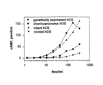
Figure 1.
Bioassay for forms of hCG. This is data from recombinant
CHO cells expressing the LH/CG receptor. The response
factor is CAMP production. The x-axis is dose of one of
four calibrated, pure hormones as described on graph
legends. Expressed hCG has no nicks; choriocarcinoma hCG
(C5) is 100% nicked; CR 127 was purified into a nick-
free (intact) and nick-enriched fraction as shown.
Figure 2.
Incidence (Panel A) and expression level (Panel B) of
hCG-related molecules in the positive samples for each
of the analyses measured (In early normal pregnancy,
n=214; EPL cycles, n=49; and negative cycles, n=297).
Figure 3.
Binding curves for three hCG types in the B152-B207*
assay (upper panel) and the B109-B108* assay (lower
panel).
Figure 4.
Ratio of hCG isoforms measured by the B152-B207* and
8109-B108* assays in normal pregnancy urine (n=103) at
different gestational ages. (Regression curve and 95%
confidence intervals are shown, r2=0.79) . An inflection
point in the curve occurs at approximately 29 weeks.
Figure 5.
Box plot of the B152/B109 ratio for pregnancy matched
serum/urine at 5-6 weeks of gestational age (n=12); or
at 36-39 weeks of gestational age (n=11) and in JAR cell
supernatant. Box extends to the 25th and 75th
percentile. The upper and lower symbols indicate the
90th and 10th percentile respectively. A solid line
inside the box marks the value of the 50th percentile.
CA 02745932 2011-07-04
-10-
Figure 6.
Ratio of hCG isoforms measured by the B152-13207* and
B109-B108* assays in the urine of IVF patients (n=65).
(Regression curve and 95% confidence =intervals are
shown, r2=0.59).
Figure 7. =
Immunoassay profiles of fractions from Superose* 12
column chromatography of a pooled urine concentrate from
=10= pregnant women during the first week of pregnancy
(Figure 7B) compared to the control (Figure 7A).
Trademark *
CA 02745932 2011-07-04
-11-
Detailed Description of the Invention
A method of predicting pregnancy outcome in a subject by
determining the amount of an early pregnancy associated
molecular isoform of hCG in a sample comprising: (a)
contacting a sample with an antibody which specifically
binds to the early pregnancy associated molecular
isoform of hCG under conditions permitting formation of
a complex between the antibody and the early pregnancy
associated molecular isoform of hCG; (b) measuring the
amount of complexes formed, thereby determining the
amount of the early pregnancy associated molecular
isoform of hCG in the sample; and (c) comparing the
amount early pregnancy associated molecular isoform of
hCG in the sample determined in step (b) with either (i)
the amount determined for temporally matched, normal
pregnant subject(s) or (ii) the amount determined for
non-pregnant subject (s), wherein the relative absence of
the early pregnancy associated molecular isoform of hCG
in the sample indicates a negative outcome of pregnancy
for the subject. In an embodiment of the present
invention, the antibody is B152. Another embodiment of
this invention is the early pregnancy associated
molecular isoform of hCG.
The hybridoma producing the B152 monoclonal antibody was
was deposited on February 3, 1998 with the American Type
Culture Collection (ATCC), 12301 Parklawn Drive,
Rockville, Maryland 20852, U.S.A. under the provisions
of the Budapest Treaty for the International Recognition
of the Deposit of Microorganism for the Purposes of
Patent Procedure. The hybridoma, was accorded ATCC
Accession Number HB-12467.
According to one embodiment of this invention, step (a)
further comprises a second antibody which specifically
binds to hCG without substantially cross-reacting with
CA 02745932 2011-07-04
-12-
said antibody under conditions permitting formation of
a complex between the antibody and the early pregnancy
associated molecular isoform of hCG. In an embodiment
of this invention, the second detection antibody is
8207. According to another embodiment of this invention,
step (a) further comprises a second assay antibody B109
which specifically binds to intact non-nicked hCG
without substantially cross-reacting with said antibody
under conditions permitting formation of a complex
between the antibody and the early pregnancy associated
molecular isoform of hCG. In an embodiment of this
invention, the detection antibody is B108. In an
embodiment of this invention, step (c) comprises
comparing the amount of the early pregnancy associated
molecular isoform of hCG determined in step (b) for
B152-B207 assay with the amount determined in step (b)
for the B109-B108 assay wherein a high ratio of amounts
determined for said antibody relative to the second
antibody indicates a positive outcome of pregnancy for
the subject, a low ratio indicates a negative outcome of
pregnancy for the subject.
In yet another embodiment of this invention, step (c)
comprises comparing the amount early pregnancy
associated molecular isoform of hCG in the sample
determined in step (b) with the amount determined for
temporally matched, normal pregnant subject(s), wherein
amounts of the early pregnancy associated molecular
isoform of hCG in the sample similar to amounts of early
pregnancy associated molecular isoform of hCG in
temporally matched pregnant samples indicates a positive
outcome, amounts of early pregnancy associated molecular
isoform of hCG in the sample similar to amounts of early
pregnancy associated molecular isoform of hCG in the
non-pregnant samples indicates a negative outcome of
pregnancy for the subject.
CA 02745932 2011-07-04
-13-
This invention also provides a method of predicting the
likelihood of a negative pregnancy outcome in a female
subject comprising: (a) contacting a sample from the
subject with a capture antibody which specifically binds
to an early pregnancy associated molecular isoform of
hCG under conditions permitting formation of a complex
between the antibody and the early pregnancy associated
molecular isoform of hCG; (b) contacting any complex
formed in step (a) with a labelled detection antibody
under conditions permitting binding to the complex the
capture antibody and the hCG isoform; (c) measuring the
amount of labeled detection antibody bound to the
complex so as to thereby determine the amount of the
early pregnancy associated molecular isoform of hCG in
the sample; and (d) comparing the amount early pregnancy
associated molecular isoform of hCG in the sample
determined in step (b) with the amount determined for a
normal pregnant subject, wherein the relative absence of
the early pregnancy associated molecular isoform of hCG
in the sample indicates a negative outcome of pregnancy
for the subject.
According to an embodiment of this invention, the sample
is a urinary sample or a blood sample. In one
embodiment of this invention, the sample is an aggregate
sample taken from at least one day. In
another
embodiment, sample may be taken from at least two
consecutive days and in a further embodiment, the sample
is taken in three days. In an
embodiment of this
invention, the sample is a spot urine sample, a first
morning void urine sample, or an aggregate sample of the
first morning void urine samples for at least two
consecutive days. In one embodiment of this invention,
the antibody is labeled with a detectable marker. In an
embodiment of this invention, the detectable marker is
a radioactive isotope, enzyme, dye, magnetic bead, or
biotin. In a
preferred embodiment, the radioactive
CA 02745932 2011-07-04
-14-
isotope is 1125.
The present invention further provides a method of
predicting pregnancy outcome in a subject by determining
the amount of an early pregnancy associated molecular
isoform of hCG in a sample comprising: (a) contacting a
capturing antibody which specifically binds to the early
pregnancy associated molecular isoform of hCG with a
solid matrix under conditions permitting binding of the
antibody with the solid matrix; (b) contacting the bound
matrix with the sample under conditions permitting
binding of the antigen present in the sample with the
capturing antibody; (c) separating the bound matrix and
the sample; (d) contacting the separated bound matrix
with a detecting antibody which specifically binds to
hCG under conditions permitting binding of antibody and
antigen in the sample; (e) measuring the amount of bound
antibody on the bound matrix, thereby determining the
amount of early pregnancy associated molecular isoform
of hCG in the sample; and (f) comparing the amount early
pregnancy associated molecular isoform of hCG in the
sample determined in step (e) with the amount determined
for temporally matched, normal pregnant subject(s),
wherein amounts of the early pregnancy associated
molecular isoform of hCG in the sample similar to
amounts of early pregnancy associated molecular isoform
of hCG in temporally matched pregnant samples indicates
a positive outcome, amounts of early pregnancy
associated molecular isoform of hCG in the sample
similar to amounts of early pregnancy associated
molecular isoform of hCG in the non-pregnant samples
indicates a negative outcome of pregnancy for the
subject.
An embodiment of this invention further comprises (a)
removing of the sample from the matrix; and (b) washing
the bound matrix with an appropriate buffer. In one
CA 02745932 2011-07-04
-15-
embodiment of this invention, the capturing antibody is
B152. In one
embodiment of this invention, the
detecting antibody is B207. In an embodiment of this
invention, step (a) further comprises a second capturing
antibody which specifically binds to intact non-nicked
hCG without substantially cross-reacting with
said antibody under conditions permitting formation of
a complex between the antibody and the early pregnancy
associated molecular isoform of hCG. According to an
embodiment of this invention, the second capturing
antibody is B109 and the second detection antibody is
B108. In an
embodiment of this invention, step (d)
further comprises a second detecting antibody which
specifically binds to hCG without substantially cross-
reacting with said antibody under conditions permitting
formation of a complex between the antibody and the
early pregnancy associated molecular isoform of hCG. In
an embodiment of this invention, step (f) comprises
comparing the amount of the early pregnancy associated
molecular isoform of hCG determined in step (e) for said
antibody with the amount determined in step (b) for the
second antibody, wherein a high ratio of amounts
determined for said antibody relative to the second
antibody indicates a positive outcome of pregnancy for
the subject, a low ratio indicates a negative outcome of
pregnancy for the subject.
According to an embodiment of this invention, the sample
is a urinary sample or a blood sample. In one
embodiment of this invention, the sample is an aggregate
sample taken from at least two consecutive days. In an
embodiment of this invention, the sample is a spot urine
sample, a first morning void urine sample, or an
aggregate sample of the first morning void urine samples
for at least two consecutive days. In one embodiment of
this invention, the antibody is labeled with a
detectable marker. In an embodiment of this invention,
CA 02745932 2011-07-04
-16-
the detectable marker is a radioactive isotope, enzyme,
dye, magnetic bead, or biotin. In a
preferred
embodiment, the radioactive isotope is 1125.
In addition, the present invention provides a method for
determining the amount of early pregnancy associated
molecular isoforms of in a sample comprising: (a)
contacting the sample with an antibody which
specifically binds to an early pregnancy associated
molecular isoform of hCG under conditions permitting
formation of a complex between the antibody and the
early pregnancy associated molecular isoform of hCG; and
(b) determining the amount of complexes formed thereby
determining the amount of early pregnancy associated
molecular isoform of hCG in the sample.
According to an embodiment of this invention, the
antibody specifically binds a region of the early
pregnancy associated molecular isoform of hCG comprising
a carbohydrate moiety. In one embodiment
of this
invention the antibody is produced by a hybridoma cell
line. In one embodiment of this invention the antibody
is B152.
Further, the present invention provides a diagnostic kit
for determining the amount of early pregnancy associated
hCG is a sample comprising: (a) an antibody which
specifically binds to an early pregnancy associated
molecular isoform; (b) a solid matrix to which the
antibody is bound; and (c) reagents permitting the
formation of a complex between the antibody and a
sample. In an
embodiment of this invention, the
antibody is B109 or B152. An
embodiment of this
invention further comprises control sample(s) normal
pregnant sample(s), nonpregnant sample(s), or male
sample(s). The kit
may also contain detection
antibodies such as B207 or B108.
CA 02745932 2011-07-04
-17-
According to an embodiment of this invention, the sample
is a urinary sample or a blood sample. In one
embodiment of this invention, the sample is an aggregate
sample taken from at least one or may be two or three
consecutive days. In an embodiment of this invention,
the sample is a spot urine sample, a first morning void
urine sample, or an aggregate sample of the first
morning void urine samples for at least one, or may be
two or three consecutive days. In one embodiment of this
invention, the antibody is labeled with a detectable
marker. In an
embodiment of this invention, the
detectable marker is a radioactive isotope, enzyme, dye,
magnetic bead, or biotin. In a preferred embodiment,
the radioactive isotope is 1125.
The present invention additionally provides an antibody
which specifically binds to an early pregnancy
associated molecular isoform of human chorionic
gonadotropin.
In an embodiment of this invention, the antibody
specifically binds to a region of the early pregnancy
associated molecular isoform of human chorionic
gonadotropin comprising a carbohydrate moiety.
According to one embodiment of this invention, the
monoclonal antibody is B152. In an embodiment of this
invention, a hybridoma cell (ATCC Accession No. HB-
12467) is provided capable of producing monoclonal
antibody B152. Another embodiment of this invention is
the early pregnancy associated molecular isoform of hCG
recognized by the B152 monoclonal antibody.
Further, the present invention provides a method for
detecting non-trophoblast malignancy in a sample
comprising: (a) contacting a sample with an antibody
which specifically binds to the early pregnancy
associated molecular isoform of hCG under conditions
CA 02745932 2011-07-04
-18-
permitting formation of a complex between the antibody
and the early pregnancy associated molecular isoform of
hCG; (b) contacting the sample with a second antibody
which specifically binds to intact non-nicked hCG
without substantially cross-reacting with said antibody
under conditions permitting formation of a complex
between the antibody and the early pregnancy associated
molecular isoform of hCG; (c) measuring the amount of
complexes formed, thereby determining the amount of the
early pregnancy associated molecular isoform of hCG in
the sample; and (d) comparing the amount of early
pregnancy associated molecular isoform of hCG in the
sample determined in step (b) with the amount of early
pregnancy associated molecular isoform of hCG in the
sample determined in step (c), wherein a positive
detection of early pregnancy associated molecular
isoform detected in step (b) and a relative absence of
the early pregnancy associated molecular isoform of hCG
detected in step (c) indicates the presence of non-
trophoblast malignancy in the sample.
According to an embodiment of this invention, the
antibody is B152 or B109. In an embodiment of this
invention, the detection antibody is B207 for B152
assay, B108 for B109 assay. In an embodiment of this
invention, the non-trophoblast malignancy is ovarian
malignancy or prostate malignancy.
According to an embodiment of this invention, the sample
is a urinary sample or a blood sample. In one
embodiment of this invention, the sample is an aggregate
sample taken from at least two consecutive days. In an
embodiment of this invention, the sample is a spot urine
sample, a first morning void urine sample, or an
aggregate sample of the first morning void urine samples
for at least two consecutive days. In one embodiment of
this invention, the antibody is labeled with a
CA 02745932 2011-07-04
-19-
detectable marker. In an embodiment of this invention,
the detectable marker is a radioactive isotope, enzyme,
dye, magnetic bead, or biotin. In a
preferred
embodiment, the radioactive isotope is I125.
Finally, the present invention provides a method for
detecting gestational trophoblast disease in a sample
from a subject comprising (a) contacting a sample with
an antibody which specifically binds to the early
pregnancy associated molecular isoform of hCG under
conditions permitting formation of a complex between the
antibody and the early pregnancy associated molecular
isoform of hCG; (b) contacting the sample with a second
antibody which specifically binds to intact non-nicked
hCG without substantially cross-reacting with
said antibody under conditions permitting formation of
a complex between the antibody and the early pregnancy
associated molecular isoform of hCG; (c) measuring_the
amount of complexes formed, thereby determining the
amount of the early pregnancy associated molecular
isoform of hCG in the sample due to binding with the
first antibody, and late pregnancy associated molecular
isoform of hCG in the sample due to binding with the
second antibody; (d) determining the ratio of early
pregnancy associated molecular isoform of hCG to late
pregnancy associated molecular isoform of hCG in the
subject; and (e) comparing the ratio of early pregnancy
associated molecular isoform of hCG to late pregnancy
associated molecular isoform of hCG in the sample
determined in step (c) over time, wherein a continuing
high ratio of early pregnancy associated molecular
isoform of hCG to late pregnancy associated molecular
isoform of hCG in the sample determined in step (c)
indicates the presence of gestational trophoblast
disease in the subject.
In an embodiment of this invention, the antibody is B152
CA 02745932 2011-07-04
-20-
or B109. In another embodiment of this invention, the
detection antibody is B108 for B109 assay, B207 for B152
assay. In an embodiment of the present invention, the
gestational trophoblast disease is choriocarcinoma or
hydatidiform mole.
According to an embodiment of this invention, the sample
is a urinary sample or a blood sample. In one
embodiment of this invention, the sample is an aggregate
sample taken from at least two consecutive days. In an
embodiment of this invention, the sample is a spot urine
sample, a first morning void urine sample, or an
aggregate sample of the first morning void urine samples
for at least two consecutive days. In one embodiment of
this invention, the detection antibody B207 or B108 is
labeled with a detectable marker. In an embodiment of
this invention, the detectable marker is a radioactive
isotope, enzyme, dye, magnetic bead, or biotin. In a
preferred embodiment, the radioactive isotope is In'.
As described herein below, unexpected isoforms of hCG
are produced during normal early pregnancy. Using an in
vitro bioassay, it appears that these isoforms have
enhanced potency for signal transduction. These
isoforms can be measured using the novel sensitive,
immunoassay described herein. This can help predict
pregnancy outcome where one cause of early pregnancy
loss is failure to produce the isoform of hCG of higher
potency produced by successful pregnancies. This
enables physicians to intervene to sustain a failing
pregnancy. Identification of the nature of the hCG
isoform required might provide the proper reagent needed
to sustain pregnancy.
New antibodies for measurement of nicked forms of hCG
described herein below were developed based on the
hypothesis that forms of hCG, which have greatly reduced
CA 02745932 2011-07-04
-21-
bioactivity, contribute to early pregnancy loss (EPL),
due at least in part to diminished biopotency. Evidence
was found that the hCG that appears in EPL patients
displays reduced biological activity. However, it was
determined that the cause of the reduced bioactivity is
not the presence of nicked hCG in EPL patients.
Instead, the hypothesis is that patients that carry
pregnancies forward produce an isoform of hCG with
enhanced bioactivity. The instant invention describes
a unique immunochemical assay to measure this unexpected
and previously un-characterized isoform of early
pregnancy hCG directly in clinical samples of blood and
urine. One of the antibodies developed reacted against
a nicked form of hCG isolated from a choriocarcinoma
patient, was not specific for a nicked form of hCG but
appeared to discriminate among carbohydrate variants of
hCG. This
antibody, designated B152, appears to
preferentially bind hCG forms from choriocarcinoma
patients. In
studying the content of hCG isoforms
during pregnancy, the unique and unexpected observation
was made that B152 in the first four weeks of pregnancy
measured much higher quantities of an isoform of hCG as
compared to the standard hCG isoforms measured by the
usual heterodimeric hCG assays exemplified by a
previously described B109 based assay. In fact, in
early pregnancy (days 9,10,11 postovulation) B152
measured as much as 20-fold more hCG, than did another
monoclonal antibody, B109. Later in pregnancy, the B152
isoform declines and is lower in third trimester
pregnancy urine than the standard isoforms measured by
B109. A further striking observation was that in very
early pregnancy, a high B152/B109 ratio correlates with
a successful pregnancy outcome while a low ratio
correlated with pregnancy loss. This
discovery is
important as the potentially overlooked isoforms of hCG
described herein during pregnancy may be predictors of
successful pregnancy outcome. Such an assay has wide
CA 02745932 2011-07-04
-22-
medical applications and provides a clinician with
opportunity to intervene very early in pregnancy if the
assay indicated that the pregnancy appeared troubled.
An antibody, designated BI52, produced by the hybridoma
cell accorded ATCC Accession number HB-12467 generated
against a nicked form of hCG isolated from a
choriocarcinoma patient, but not specific for nicked
isoform hCG is able to discriminate among carbohydrate
variants of hCG. B152 is specific for an early
pregnancy associated molecular isoform of hCG. which in
the first four weeks of pregnancy is measured at much
higher quantities than the hCG standard isoforms
measured by the usual heterodimeric hCG assays
exemplified by a previously described B109 based assay.
Later in pregnancy, the B152 isoform declines and is
lower in third trimester pregnancy urine than the
standard isoforms measured by B109.
This invention is illustrated in the Experimental
Details section which follows. These sections are set
forth to aid in an understanding of the invention but
are not intended to, and should not be construed to,
limit in any way the invention as set forth in the
claims which follow thereafter.
CA 02745932 2011-07-04
-23-
EXPERIMENTAL DETAILS
Example 1: Analysis
of Molecular Isoforms of hCG in
Early Pregnancy and Early Pregnancy Loss
Introduction
Almost all investigations of the incidence of early
pregnancy loss (EPL), either in normal populations or in
populations at risk as a consequence of exposure to
putative reproductive toxins (Hakim, R. B., et al.,
1995; Lasley, B. L., et al., 1995) use assays for
heterodimeric, non-nicked hCG or combination assays
which additionally include free beta subunit and beta
core fragment of hCG. One concern about the forms of
hCG to include in the measurement in EPL was heightened
with respect to the nicking phenomenon described above.
Because nicked hCG molecules are not measured by the
antibodies employed in most EPL studies, the incidence
of EPL is presumably underestimated by an aMOunt
proportional to the extent of nicking in the urinary
molecule. Another concern of significant importance was
a determination of the nature of the "hCG like"
immunoreactivity in the urine in the periovulatory surge
of the menstrual cycle (O'Connor J., et al., 1995).
Recent reports have confirmed the existence of and
documented the structure of a sulfated form of hCG
produced in the pituitary (Birken, S., et al., 1996b).
There is a pulsatile secretion of hCG in both men and
non-pregnant women. (Odell,
W. D.; Griffin, J., 1989
and Odell, W. D.; Griffin, J., 1987). The presence of a
non-pregnancy associated form of sulfated hCG of
pituitary origin, peaking at ovulation and perhaps
persisting into the luteal phase, could potentially
interfere with the accurate estimation of EPL.
Unappreciated isoforms of hCG in blood and urine very
early in pregnancy may be more potent in vivo than the
forms of hCG produced later in pregnancy. The absence
CA 02745932 2011-07-04
-24-
of such isoforms may be one cause of early pregnancy
loss. A sensitive and specific immunoassay system was
designed and made to measure unique early pregnancy
associated molecular isoforms (EPMI) of hCG. These
isoforms, likely to differ by carbohydrate composition,
are predictive of a successful pregnancy outcome. When
these early pregnancy associated molecular isoforms of
hCG are absent or present in low concentration, the
pregnancy may be lost very early and be observed as only
a "chemical" pregnancy. These hCG isoforms may resemble
the forms of hCG produced in some choriocarcinoma
patients from which the immunogen used to produce
monoclonal antibody B152 was derived as described herein
below. The isoforms resemble those from trophoblastic
disease not in terms of nicking or intact peptide chains
but likely in carbohydrate content. The
present
invention describes that the molar ratio of B152 to B109
epitopes are predictive of a successful pregnancy or a
loss. Three
categories of pregnant patients were
analyzed: (a) normal pregnant women, (b) women who
experience recurrent abortions, (c) women undergoing
embryo implantation.
It is possible to determine the hCG isoforms present in
the blood and urine of women who have a history of
recurrent spontaneous abortion and a similar analysis of
women undergoing embryo implantation. The combined EPL
and spontaneous abortion rate in healthy populations is
31%. Subjects who experience three consecutive recurrent
spontaneous abortions have a 32% risk of sustaining
another (Hill, J. A.; Anderson, D. J., 1990). In in
vitro fertilization IVF pregnancy, the loss rate is 70%
with non-donor sperm and 50% when donor sperm is used.
Delineation of pregnancies with a negative outcome from
pregnancies with a positive outcome can be based on
differences in the concentrations of EPMI hCG isoforms
(i.e. as differences in the B152/B109 ratio in
CA 02745932 2011-07-04
-25-
patients). In
addition, specimens from gestational
trophoblastic disease (GTD) can be used to discriminate
between GTD and normal pregnancy.
Results
In vitro bioassay for hLH/hCG
An hCG bioassay was constructed employing CHO cells
expressing functional human LH/CG receptor. Figure 1
illustrates the differences in vitro in biological
activity between nicked and non-nicked hCG as measured
by this assay. This system, has been used to evaluate
the activity of pituitary and placental hCG (Birken, S.,
et al., 1996b). Preparations of hCG were tested for
nicked and non-nicked molecular isoforms of hCG in a
second recombinant bioassay system (Ho, H-H., et al.,
1997). Similar results were obtained in both systems.
Normal pregnancy values compared with EPL values.
Figure 2 indicated that nicked hCG is not a significant
molar constituent of either early pregnancy or EPL.
Data indicated that biological activity is not
correlated with nicked hCG, but is instead ascribed to
a form of hCG recognized by the B152 monoclonal antibody
-- an early pregnancy associated molecular isoform of
hCG (EPMI hCG). It has been established that there is
diminished hCG bioactivity associated with EPL as
compared to early normal pregnancy (Ho, H-H., et al.,
1997). Thus, diminished hCG biological activity is a
factor in EPL as a consequence of a heretofore
unappreciated isoform of hCG - an early pregnancy
associated molecular isoform of hCG.
hCG Urinary Analytes. Metabolites of hCG and hLH were
studied in a variety of states (Birken, S., et al.,
1996a). One study indicated a 31% pregnancy loss
(Zinaman, MJ, et al., 1996) while another indicated a
17.4% rate of early pregnancy loss based on hCG assays
(Bllish, N. J., et al., 1996). It is known that hCG and
CA 02745932 2011-07-04
-26-
hCG beta core can be readily transferred from the uterus
to the circulation even in the absence of implantation
(Chang, P. L., 1997). The molecular spectrum of hCG
urinary analytes in EPL cycles, normal conceptive cycles
and non-conceptive cycles has been evaluated. The study
design and demographics of the investigation have been
described (Ellish, N. J., et al., 1996).
Briefly, three urine specimens per cycle, corresponding
to days 9,10, 11, post calculated day of ovulation were
collected and analyzed in a screening assay (the
"combo") which simultaneously detects intact, non-nicked
hCG, hCG free beta subunit, and hCG beta core fragment.
Individual determinations for each of these analytes, as
well as for nicked hCG, and the form of intact hCG
detected by monoclonal antibody B152 (EPMI hCG) were
performed on these specimens. In addition, since the
concentration of luteal phase hLH urinary analytes is a
concern because of cross-reaction in hCG assays, levels
of intact hLH, hLH free beta subunit and hLH beta core
fragment were determined in the normal pregnancy cycles
and the non-conceptive cycles. Table I summarizes the
characteristics of immunometric assays employed.
CA 02745932 2011-07-04
- 2 7 -
TABLE). Assay format and specificity
Assay format Primary analyte % cross-reactivity with related analytes
Infra- Inter-
assay assay
cv, % cv, %
B109-B108* intact non-nicked hCG <1%11 6 12
B201-C104* hCG free beta subunit 1% hCG; 10% hCG
nicked (pregnancy); 6 12
(non-nicked+nicked)
B210-B108* hCG beta core fragment 2% hLH beta core
fragment; 5 7
<1%
B151-B207* hCG nicked 10% hCG nicked free beta subunit; 5 15
12% hCG non-nicked;
2% hCG free beta subunit;
2% hLH; 5% hLH free beta subunit;
<1%`
B152-B207* choriocarcinoma hCG 100% hCG nicked
(C5); 6 13
(C5) and 190% hCG free beta nicked (from C5);
choriocarcinoma hCG 10% hCG nicked (pregnancy);
free beta subunit 5% hCG free beta nicked (pregnancy);
7.4 hCG (pregnancy);
6% hCG free beta subunit;
<1%`
8406-A201* hLH <1414' 4 10
B505-8503* hLH beta core fragment <1%' 9 9
B408-B409* hLH free beta subunit 29% hLH; 7
11
<1%'
'-(if not indicated) hLH, free beta hLH, hLH beta core fragment, hCG, free
beta
hCG, hCG beta core fragment;
b-(if not indicated ) the same as (1) plus nicked hCG and nicked free beta hCG
(pregnancy).
CA 02745932 2011-07-04
-28-
The results indicate that nicked hCG does not constitute
a significant mole fraction of urinary hCG
immunoreactivity in either EPL or early normal
pregnancy. In
addition, there is a substantial
excretion of hCG free beta subunit in some subjects in
both preqnancy and EPL. Further, both EPL and normal
pregnancy cycles variably express all of the measured
analytes. Although both the incidence and level of
expression are different between EPL's and normal
pregnancy, there is no hCG related analyte unique to
either state. There was, however, a clear difference
between the hLH associated analytes in the control
population (non-conceptive cycles) and the normal
pregnancy group. Virtually all of the non-pregnancy
cycles expressed hLH free beta subunit and hLH beta core
fragment while only a third of the conceptive cycles had
detectable levels of either analyte. Intact hLH proved
to be a minor constituent of the hLH profile in both
groups.
These findings demonstrate both the necessity of
measuring hCG beta core fragment in the detection of
EPL, and also of making sure that the hCG beta core
assay does not cross- react with beta core hLH, which is
demonstrated to be present in that part of the luteal
phase where EPL measurements are performed. The data is
summarized in Figure 2.
Statistical analysis was performed after transformation
of analyte values to mole fractions so as to produce a
more useful analysis due to the wide excursion of hCG
analyte values among groups. The mole fraction data
were evaluated by discriminant analysis and by a mixed
effects model incorporating LMP. The
discriminant
analysis was performed both with and without "outliers"
(defined as values greater than two standard deviation
from the mean) removed. Both
approaches produced
CA 02745932 2011-07-04
-29-
similar results.
A quadratic discriminant analysis based on a cross-
validation method in order to minimize bias correctly
classified 91t of the normal pregnancy subjects and 80t
of the EPL subjects.
The mixed effects analysis, testing for interactions
between mole fraction of analyte and time since LMP
found no significant time or group (EPL vs. normal)
effects in the intact hCG assay. In the free beta
subunit of hCG assay, there is a significant group
effect but no time trend. In both the hCG beta core
fragment measurement and the B152 measurement, both the
hormone levels and the time trend from LIMP were
significantly different between the EPL and pregnancy
groups. This study produced several important findings.
It defined the spectrum of analytes which in both early
pregnancy and EPL, thereby resolving the issue of which
hCG analytes to measure in epidemiological studies in
which EPL is the end point determination. More
importantly, it illustrated for the first time that
there are significant differences both in the pattern of
analytes and the time course of their appearance between
early normal pregnancy and EPL. This observation
facilitates very early prediction of a distressed
pregnancy by urinary hCG measurements at a time which
would permit therapeutic intervention.
Immunoreactivity of different forms of hCG in the two
IRMA's (B152-B207 and B209-3108)
The relative binding of three different forms of hCG
(urinary hCG, pituitary hCG and choriocarcinoma hCG C5)
has been characterized in the two hCG assays (Figure 3).
Urinary non-nicked hCG and pituitary hCG are recognized
nearly equally well by the two IRMASs, while C5
CA 02745932 2011-07-04
-30-
recognition is quite different. The B152-B207* assay is
more sensitive to C5, which is to be expected because
B152 antibody was developed and selected on the basis of
higher affinity to C5. Urinary non-nicked hCG is
purified from the CR127 preparation of pooled normal
pregnancy hCG. Conversely C5 is recognized with lower
affinity by the B109-B108* assay, which has primary
specificity for the hCG isoforms of later pregnancy.
We have developed a method to directly profile changes
of hCG isoforms in serum or urine throughout pregnancy.
Two IRMAs for hCG are employed, each based on monoclonal
antibodies to different hCG epitopes. The B109-B108*
assay is a commonly used intact hCG assay to the
heterodimeric-dependent epitope. A new assay, B152-
13207*, is most likely sensitive to the carbohydrate
portion of hCG carboxyterminal peptide. The same
standard non-nicked hCG was used in both assays. Non-
nicked hCG was employed since the B109 assay reacts
poorly with nicked forms of hCG while the B152 assay
does not discriminate between nicked and non-nicked
forms of the hormone. The B152 assay detected with
greatly enhanced sensitivity hCG isoforms which appear
earlier in pregnancy than isoforms measured by the B109
assay (O'Connor et al. 1998). Prior to development of
the new immunometric assay system described in this
report, it was not possible to readily discern the
changes in hCG isoforms from very early pregnancy to mid
pregnancy. The only available procedure for examining
these changes was isoelectric focusing of every patient
specimen followed by immunoassay of every focused
fraction (Berger et al. 1993; Ulloa-Aguirre et al.
1990). The IEF pattern reflects the heterogeneity of the
charged sugar, sialic acid which varies with the multi-
antennary structures of the carbohydrate moieties in
which sialic acid is the terminal sugar. Although we do
not yet know the precise nature of the isoform epitopes
CA 02745932 2011-07-04
-31-
being measured, the evidence for carbohydrate
discrimination is based upon the hyperglycosylated
structure of the immunogen, C5, used to develop the B152
monoclonal antibody and the antibody's reactivity with
the hCG isoforms found in the JAR choriocarcinoma cell
line. C5 hCG was isolated from a choriocarcinoma
patient and has been thoroughly characterized as to its
protein and carbohydrate content and structure (Elliott
et al. 1997). It has been shown that C5 (and hCG from
other choriocarcinoma subjects) differ in the protein
moiety mainly by the presence of an increased number of
nicked sites and by increased glycosylation relative to
the hCG of normal pregnancy. In comparison with the hCG
of normal pregnancy, choriocarcinoma derived hCG has
increased fucosylation of the N-linked biantennary
oligosaccharides in the beta subunit. In addition, the
0-linked oligosaccharides in preparation C5 (a form of
hCG produced from a single patient with choriocarcinoma)
has a 100% tetrasaccharide core on the COOH-terminal
region of the beta subunit. Normal mid pregnancy hCG
has only 10-20% of this structure (Elliott et al. 1997).
These observations, plus our own determination that the
hCG synthesized by the JAR choriocarcinoma cell line
provides a 8152/8109 isoform ratio similar to that
observed in early pregnancy, leads us to the conclusion
that in very early pregnancy, the developing trophoblast
secretes an isoform of hCG which resembles that produced
in choriocarcinoma.
We have also tested recognition of pituitary hCG since
its N-Asn carbohydrates differ somewhat from those of
placental hCG, bearing a closer resemblance to those of
Will which have both sialic acid and sulfate groups
(Eirken et al. 1996). The carbohydrate structure of the
b COOH-terminal portion of pituitary hCG is not yet
known. Since 8152 did not recognize any substantial
differences between pituitary and placental hCG (Figure
CA 02745932 2011-07-04
-32-
3), differences in N-Asn recognition are unlikely. In
terms of the ODOH-terminal carbohydrates, it appears
that pituitary and placental hCG (mid-pregnancy
isoforms) may be similar, assuming the 0-linked
carbohydrate on the C5 antigen is part of the epitope of
B152.
Example 2:
11152/B109 Ratio Predicts Pregnancy
Outcome
The B152/13109 ratio measured in urine samples throughout
the preanancy
The relative concentrations of hCG isoforms in 103
normal pregnancy urine samples (5-39 weeks post last
menstrual period - LMP) were determined by two
immunometric assays (B152-B207* and B109-8108*). Both
because of the wide range of hCG concentrations in
different samples, even at the same gestational age, and
because neither of the assays is totally specific for
the two (or more) families of hCG isoforms present, we
find that presenting the data as a ratio of the observed
two isoform groups more clearly delineates the change in
isoform content as pregnancy progresses. This
calculated ratio is shown in Figure 4. In weeks 5-8 of
pregnancy, the ratio of B152/B109 isoforms ranged
between 6.2 and 1.3, indicating a predominance of the
B152 isoform(s) in early pregnancy. During the 10 to 12
week period, the ratio ranged from 1 - 0.2, indicating
that an inversion in hCG isoform content is occurring as
pregnancy progresses. This
decline in the ratio
continues, ranging from 0.54 - 0.08 in the 15 - 18 week
period and reaching an inflection point at 29 weeks. At
that time, the ratio reached a value of around 0.06
after which the ratio displayed a rise to a range of 0.2
- 0.07 in the 37 - 39.5 weeks of gestation time period.
CA 02745932 2011-07-04
-33-
Statistical analysis involved fitting the log
transformed ratio data to second and third order
polynomial regression models. Since the third order term
was not significant (likelihood ratio c2(1)=1.32,
P=0.25), the second order model was used (r2=0.793) . The
log B152/B109 ratio reached an inflection point at
LMP.29 weeks, based on this model.
The B152-B207* values reflect a measurement of the B152
isoform in terms of later pregnancy hCG equivalents, not
in absolute quantities. It must be emphasized that the
"absolute" concentrations measure in the B152 assay
cannot be compared with the results of the B109 assay on
an equimolar basis since the potency of the
hyperglycosylated isoform is much higher in the B152
assay vis-a-vis the standard, i.e. normal later first
trimester pregnancy hCG. The actual molar values of this
isoform are on the order of tenfold less than those
recorded in the assay. For this reason we have chosen
not to analyze absolute molar quantities of the two
analytes but only the ratio of the two measurements.
Even in normal pregnancy, the hCG values obtained vary
widely according to the characteristics of the
immunological reagents employed (Cole and Kardana, 1992;
Cole et al. 1993). We hypothesize that the two assays
described in this report primarily detect hCG isoforms
at opposing ends of this spectrum, each primarily
recognizing a subset of closely related molecules in the
continuum of early to later pregnancy hCG molecular
forms.
We have retained the use of normal pregnancy hCG as the
standard in B152-B207* assay, despite its decreased
affinity in this antibody configuration. The reasons
=
for this include the limited and unrenewable supply of
C5 (which was isolated from the urine of a single
CA 02745932 2011-07-04
-34-
patient) and the variability in data which would result
from investigations using different standards. The
consequences of this choice are that the early pregnancy
hCG isoforms have markedly increased immunopotency over
that of normal pregnancy and hence their molar
quantities are overestimated in this assay. We use this
difference in affinity to our advantage by employing a
ratio of the molar results of two assays (B152 and
B109). Either assay taken alone obscures this change
due to the wide excursion of hCG values which occur in
normal pregnancy.
Others have documented progressive changes in hCG
isoforms throughout pregnancy. Skarulis et al. found
that= the fucose content of both intact hCG and also its
free beta subunit increased as pregnancy progressed
(Skarulis et al. 1992). Diaz-Cueto et al. investigating
the isoelectric focusing pattern of circulating hCG
throughout pregnancy, found that in early pregnancy,
more than 80% of the hCG isoforms were acidic. This
fraction decreased to less than half (47%) late in the
third trimester (Diaz-Cueto et al. 1996). In contrast,
Wide and Hobson found that the hCG of early pregnancy
was more "choriocarcinoma-like" by virtue of its greater
biological activity than the hCG of normal pregnancy
(Wide and Hobson, 1987). Fein et al., in a study which
employed gel filtration determined that first trimester
hCG was a larger size than that of the third trimester.
Treatment with exoglycosidases eliminated the size
differential, indicating that the first trimester hCG
was more highly glycosylated (Fein et al. 1980).
CA 02745932 2011-07-04
-35-
The B152/8109 ratio in matched serum/urine samples in
the first and third trimesters of pregnancy compared
with hCG from JAR cells.
The B152/B109 ratio in serum is analogous to that found
in matched urine samples and undergoes a similar change
as pregnancy progresses (Figure 5). The B152/B109 ratio
in the cell supernatant from JAR cells (a
choriocarcinoma derived cell line) was similar to that
of early pregnancy.
The B152/B109 ratios of both serum and urine hCG
concentrations are significantly higher in the first
trimester as compared to the third trimester of normal
pregnancies (Table 2). Significant differences between
serum and urine hCG concentration ratios as well as log
transformed ratios in early (5-6 weeks) and late (36-39
weeks) gestation were evaluated by paired t-tests (Table
3). In both the first and third trimesters, urinary
B152/B109 ratios were significantly higher than serum
ratios, indicating that there was a preferential
clearance of the B152-recognized isoform into urine,
regardless of the relative concentrations of the two
isoforms.
TABLE 2.
Analysis of the B152/B109 ratio in serum and in urine in
the first vs third trimesters of pregnancy.
_
Measure I T-test(df) r
Serum, ratio t(11)=6.65 0.0001
B152/8109
Serum, t(23)=21.61 0.0000
log(ratioB152/B109)
Urine, ratio t(11)=4.64 0.0007
8152/B109
Urine, t(15.7)=16.85 0.0001
log(ratioB152/B109)
CA 02745932 2011-07-04
-36-
TABLE 3.
Analysis of the B152/B109 ratio in serum vs urine in the
first and third trimesters of pregnancy.
Gestational Measure Paired-t
age (df)
5-6 weeks Ratio t(11)=3.25 0.0077
B152/B109 t(11)=6.25 0.0001
Log(ratioB152/
8109)
36-39 weeks Ratio t(10)=5.47 0.0003
B152/B109 t(10)=7.14 0.0001
Log(ratioB152/
B109)
The B152/8109 ratio in urine samples from 1VF patients
In urine samples from IVF patients (1-4 weeks post
embryo transfer - ET) the B152/B109 ratio was again
between 2-8 and decreased as pregnancy progressed
(Figure 6), similar to that observed in natural
conceptions. The effect of pregnancy duration with
respect to outcome variables could best be represented
by a linear or quadratic function. ANCOVA models
including the second order week were fitted to the
general equation: Outcome= (effect of time post
ET)+(effect of diagnosis). After an appropriate ANCOVA
model was determined, the least square means (adjusted
for week post ET effect) were compared among the normal
pregnancy, ectopic pregnancy and spontaneous abortion
populations (Table 4). The log transformed values of
both B109-8108* and 8152-B207* measured hCG forms
discriminated both ectopic pregnancy and spontaneous
abortions from normal pregnancy (P=0.0001). The ratio of
the log transformed values discriminated abortion from
normal pregnancy (P=0.016). However, neither the ratio
of B152/8109 nor the log of that ratio discriminated
either of the pregnancy disorders from normal pregnancy.
CA 02745932 2011-07-04
-37-
A significant number of spontaneous abortions and
ectopic pregnancies occur in IVF pregnancies. We did not
find a difference in the ratio of the isoforms between
either of these two categories as compared to normal
controls, possibly a consequence of low statistical
power. However a significant difference was found
between the B152 hCG isoforms levels in normal pregnancy
and spontaneous abortion. This supports our previous
finding in early pregnancy loss, where diminished or
absent levels of the B152 isoforms characterized an
early pregnancy loss (O'Connor et al. 1998).
CA 02745932 2011-07-04
-38-
TABLE 4.
IVF patients: analysis of covariance of hCG isoforms
among normal pregnancy (np), ectopic pregnancy and
spontaneous abortion as a function of gestational age.
Outcome `Adjusted R2 b-F P 4-Pairwise
Difference
= _
'Lag (ratio 0.51 0.09 0.41 none
B152/B109)
=
`Log (B109-B108*) 0.56 21.33 0.0001 np vs
abortion &
ectopic
bLog (B152)/log 0.45 4.34 0.016 np vs
(B109) abortion
bLog (B152-B207*) 0.50 26.94 0.0001 np vs
abortion &
ectopic
a _ ANCOVA
model with 2nd order polynomial
coefficient (or parameter).
b_ ANCOVA model with only 1st order (linear)
coefficient.
C_ Adjusted
R2 is a R2 adjusting number of
coefficients on the ANCOVA
model so that
comparisons of two R2 with different ANOVA
models with different number of coefficients are
meaningful.
d_= "Pairwise
difference" is based on t-test
comparing the least-square means of outcome
variables (after adjusting effect of week ET).
*- Degree of
freedom (dfl, df2) for F-test are
(2,82) for a model with only linear coefficient
and (2,81) for a model with both linear and 2nd
order coefficient.
BCG analysis of trophoblastic disease samples
Trophoblast disease serum (17 samples) and urine (28
samples) were obtained from patients post therapy and
hence contained low hCG levels. Due to limited amounts
of sample all of these specimens were run at a 1:10
initial dilutions. HCG levels in serum were low. The
highest hCG concentration in serum was 202 fmol/ml in
CA 02745932 2011-07-04
-39-
the B152-13207* assay, with a corresponding value of 148
fmol/ml in the B109-B108* determination. Six of
seventeen samples in serum had detectable levels, with
4/6 having a higher value in the 8152-B207* assay. Of
the 15/28 positive urine samples however, 14/15 had
higher levels in the B152-13207* assay than in the 3109-
B108* assay, with the highest hCG value being 20000
fmol/ml in the B152-B207* assay and 18715 fmol/ml in the
corresponding B109-B108* assay. Due to the small sample
size, no statistical treatment was performed on this
data, but even in these post-treatment patients the
B152/B109 ratio was >1, which corresponds to the early
pregnancy hCG isoform ratio.
The specimen limitations discussed above precludes our
reaching any definitive conclusion on the analysis of
trophoblastic disease samples. However it appears as
might be anticipated that the B152 assay is more
sensitive than B109 assay in detecting hCG
immunoreactivity in the blood and in the urine of
trophoblastic disease patients, even after treatment.
Chromatography of First Week of Gestation Pregnancy
Pool. In order
to determine whether the B152-B207*
assay recognized other forms of hCG associated
immunoreactivity in addition to the intact hCG molecule,
specimens were pooled. FPLC on
tandem Superose 12
columns followed by immunoassay of the fractions for all
of the characterized forms of hCG revealed that only the
intact hCG molecule (or hCG free beta subunit) gave a
signal in this assay (See Figure 7). There were no
lower molecular weight fragments identified by the B152-
B207* assay. The hCG free beta analyte was measured in
urine described in Figure 2 and was found to make a
negligible contribution to over all hCG immunoreactivity
in these specimens.
CA 02745932 2011-07-04
-40-
Molecules recoctnizell bv monoclonal antiloody B152 in
urine and pituitary extracts. In order to define the
nature of the hCG isoforms recognized by B152, high
resolution gel filtration columns of both pituitary
extracts and postmenopausal urine concentrates were
used. The rationale for use of pituitary extracts is to
determine cross-reactive molecules, specifically those
which are glycosylated, which are plentiful in pituitary
which contains the entire family of glycoprotein
hormones, hLH, hTSH, and hFSH as well as free subunits
and the pituitary form of hCG. Two peaks are detected
in both of these cases. Only one peak was detected in
similar studies of pregnancy urine concentrates as
described earlier. In the pituitary, it is likely that
the larger molecule is pituitary hCG (70K) while the
smaller sized molecule is hLH. Since hLH is present at
100x or so as compared to pituitary hCG, the apparent
similar concentration of immunoreactivity indicates that
B152 has reduced cross-reactivity to hLH as compared to
hCG. Likewise, both hCG and hLH occur in postmenopausal
urine, again with much more hLH than hCG and the B152
pattern is similar to that of the pituitary extract.
These results show that 8152 is generally hCG specific
except for cross reactivity to hLH (as shown by standard
cross-reaction studies in Table I) and that its
carbohydrate specificity is both to the protein portion
as well as to the carbohydrate moieties of hCG (and to
a lesser extent of hLH) since it does not react with the
multitude of other glycoyslated proteins present in the
pituitary nor with those in postmenopausal urine except
for hCG or hLH-related molecules.
Serum and urine specimens were analyzed using two
assays, B109-8108* and B152-8207*, which recognize the
difference in molecular isoforms of hCG. See Table I.
The in vitro bioassay for hLH/hCG is described above.
(See Figure 1). The immunometric assay employs 96-well
CA 02745932 2011-07-04
-41-
microliter plate technology. The coating antibody, at a
concentration determined to provide the most
satisfactory combination of sensitivity and range, is
applied to the microtiter wells (Immulon IV, Dynatech
Laboratories) in carbonate buffer (0.2M, pH 9.5). The
plates are incubated with the coating solution at 4 C,
overnight, then aspirated, washed with washing solution
(0.059; Tween, 0.15N NaC1), and blocked with a 1%
solution of BSA (three hours at room temperature). The
BSA solution is aspirated and the appropriate hCG
standards (200gL/we11), in buffer B (PBS/0.1% bovine
IgG/0.1% sodium azide), or in hCG free serum (Chemicon,
Inc.), or hCG free urine, as appropriate to the specimen
matrix, and specimens are added to the wells. The plates
are sealed with plate sealers, and incubated overnight
at 4 C. The controls, specimens, and standards are then
aspirated, the plates washed 5 times with washing
solution, and iodinated detection antibody in buffer B
(200 uL/well, 100,000 cpm/well) added and incubated
overnight at 4 C. The wells are again aspirated, washed
5 times with washing solution, separated and counted
(Packard Cobra gamma counted). Values are interpolated
from a smoothed spline transformation of the count data.
This assay .procedure, as well as assay validation has
been previously reported (O'Connor, J. F., et al.,
1988).
Creatinine analysis, when urine values are normalized to
creatinine, = is performed in a microtiter plate format
= following a modification of the Taussky procedure
(Taussky, H. H., 1954).
Descriptive statistical and graphical methods are
= applied to measures of serum and urine samples from
normal healthy pregnancies to identify the distributions
a) between patient first trimester average B152 levels,
B109 levels and B152/B109 ratio; b) between patient
Trademark *
CA 02745932 2011-07-04
-42-
variability in time to B152/B109 ratio reaching 1.00;
and c) between patient variability in time to 8152/B109
ratio declining by 1/3rd from first trimester maximum
levels. The variability in the timing of the crossover
in the ratio of these two analytes provides an empirical
basis from which to estimate the value of these markers
as biochemical signatures of a viable third trimester
fetus.
Comparison of the assay profile of healthy normal
pregnancies to those of unsuccessful pregnancies from
failed IVF implantations, two non-parametric hypotheses
are available: 1) the proportion of pregnancies in which
the B152/B109 ratio falls below 1.00 is no different in
healthy normal and unsuccessful IVF pregnancies; 2) the
proportion of pregnancies in which the B152/B109 ratio
declines by 1/3rd from first trimester maximum levels is
no different in healthy normal and unsuccessful IVF
pregnancies. These
hypotheses can be tested as a
difference between two proportions. For example, a
comparison of week 14 vs. week 9, week 13 vs. week 6,
week 12 vs. week 5 or week 11 vs. week 4 pregnancies to
show a reversal of the 8152/B109 ratio in healthy normal
pregnancies and unsuccessful IVF implantations,
respectively. The power analyses apply to an outcome
defined as the time at which the B152/B109 ratio
declines by 1/3rd from first trimester maximum levels,
although this outcome would necessarily provide earlier
detection of pregnancy failure than the reversal of the
B152/8109 ratio. Patterns of results less discriminantly
different from these indicate a rejection of the
dichotomous outcome of B152/B109 ratio reversal as a
clinically meaningful marker of pregnancy failure.
Alternatively, the same two non-parametric hypotheses
can be recast as parametric hypotheses by considering
the timing of the biochemical events within the assay
CA 02745932 2011-07-04
-43-
profile of healthy normal pregnancies and unsuccessful
pregnancies from failed IVF implantations: 1) the time
at which the B152/B109 ratio falls below 1.00 is no
different in healthy normal and unsuccessful IVF
pregnancies; 2) the time at which the B152/B109 ratio
declines by 1/3rd from first trimester maximum levels is
no different in healthy normal and unsuccessful IVF
pregnancies. Of course, the objective is to provide an
empirical basis from which clinicians may counsel their
patients. Thus, it is important to adopt a logistic
model for this component of the data analysis. With
pregnancy success as the outcome, logistic models allow
the estimation of the (symmetrical) hypothesis of
increase in risk of pregnancy failure for each
additional week where either the B152/B109 ratio has
failed to decline by one third from first trimester
baseline maximum values or the B152/8109 ratio has
failed to become less than 1.00 (measured in weeks).
The logistic model enables specification of the time at
which results indicate a particular pregnancy exceeds an
a priori defined likelihood of failure, given assay data
regularly available during pregnancy, and allows
incorporation of other risks for pregnancy failure in
the same data analytic framework to assess the relative
contribution of threats to pregnancy loss. The Cox
proportional hazard model may be used to examine
predictors of the crossover rates. Mixed effects models
can also analyze repeated measures of the B152/B109
ratios taken during entire cycles. These models are
particularly useful since they allow inclusion of
incomplete and imbalance data (i.e. data with missing
values and unequal timing of data collection), to
estimate effects of time-varying covariates, to model
dependency structure of repeated measures and to model
possible heterogeneity of the ratio measures within each
experimental group.
CA 02745932 2011-07-04
-44-
p152 hCG isoform iBoaate_ci from early pregnancy urine
and determination of their Protein and carbohydrate
structures. Using the
already developed scheme of
concentration and immunoaffinity extraction of urine,
hCG molecules are isolated from urine collected from
women in early pregnancy for both protein and
carbohydrate analyses. According
to one approach,
molecules are isolated from HPLC fractions, digested
with proteases before and after reduction of disulfide
bonds, examination of the resultant peptides by mass
spectrometry and/or sequence analysis, isolation of
carbohydrate moieties after glycosidase digestions and
determination of carbohydrate structures by a
combination of specific glycosidases and retention times
on specialized anion exchange columns as compared to
know branch-chain oligosaccharide standards. In a
similar approach, the final purification stage for the
isolated hCG isoforms is SDS gel electrophoresis. Both
protease digests and glycosidase digests are performed
on the blotted and cutout band. This method results in
greater purity of the protein and less artifactual
errors due to contamination by carbohydrates which are
not in the purified protein but are derived from outside
contaminants.
Carbohydrate compositionalAnalvses and oliciosaccharide
Dranched chain identifications. The MALDI
TOF mass
spectrometric method may be used to confirm
oligosaccharide structures by using specific
glycosidases on the glycopeptides and determining the
change in molecular weight as the sugars are digested
off the glycopeptide. Only the hCG beta COOH peptide
can be expected to contain 0-linked sugar moieties.
These are of special interest since it is thought that
B152 has significant reaction with this region. The
structures of this region can be determined in a similar
fashion using enzymes that specifically release 0-linked
CA 02745932 2011-07-04
-45-
glycans. The 0-linked structures has been previously
examined using standard reference pregnancy hCG (Cole,
L. A., et al., 1985). The 0-
linked branched chain
structure are determined by a similar strategy using the
Dionex chromatographic system as well as specific
glycosidases on the C-terminal glycopeptides and Mass
Spectrometry. In one
study (Elliott, M. M., et al.,
1997), these techniques were used to elucidate the
carbohydrate structures of CR series hCG preparations
(standard urinary pregnancy hCG) and compared them to
the structures of patient samples such as C5 which was
the immunogen employed to generate antibody B152. It
was found that C5 contained significantly more mono and
tri-antennary (2X mono and 3X tri-structures than the CR
preparations) on the N-Asn residues. It was also found
that more tetrasaccharide structures were on the hCG
COOH-terminal peptide O-Serine residues in the
choriocarcinoma hCG isoform than in the CR preparations.
Bioloaical activity and metabolic clearance of hCG
isoforms. Biological activity is a function both of
molecular structure and half-life in the circulation,
which can be influenced by structure. Alterations in
carbohydrate/sialic acid content of the glycoprotein
hormones are thought to be responsible for the changes
in hCG biological/immunological activity observed
throughout pregnancy. In addition, signal transduction
at the receptor is influenced by the pI of the hCG
isoform and the presence or absence of carbohydrate.
Thus, it is valuable to examine both receptor binding
and biological activity in vitro and, in order to
determine the mechanism of action, to distinguish
receptor binding and signal transduction as well as
relative potency of signal transduction along with in
vivo bioactivity determinants such as circulating half
life. Studies, including clearance rates, are performed
*Trade-mark
CA 02745932 2011-07-04
-46-
on B152 hCG isoforms of early successful pregnancy, hCG
from third trimester pregnancy, and the reference
urinary hCG preparation, CR 127.
Exa=cle 3: B152 and B151 immunoreactivity in non-
trophoblastic malignancy.
With the exception of trophoblastic disease and
testicular cancer, hCG is expressed in the blood of
about 20% of patients with all other types of
cancer(Hussa, R. O., 1987). HCG beta core fragment in
the urine has a significantly higher level of
expression, especially in gynecological malignancy.
Since the B152 antibody was developed to a form of hCG
produced in a malignancy, it was of interest to examine
the expression of B152 and nicked hCG immunoreactivity
(B151) in non-trophoblastic malignancy. Accordingly,
blood and urine derived from men undergoing chemotherapy
for prostate cancer or women for ovarian cancer were
evaluated for the expression of hCG isoforms in plasma
and urine. It is significant that in prostate cancer,
B152 hCG immunoreactivity is found in the blood and
urine of prostate cancer patients in instances when
there is n2.hCG detected by B109-B108*. In ovarian
cancer patients evaluated, there is evidence of nicked
hCG in the blood, even in the absence of both B109 and
2152 immunoreactivity. Neither of the above groups
demonstrated the presence of hCG immunoreactivity when
the standard pregnancy derived hcG assay was employed.
It is reassuring to find that nicked hCG, the existence
of which has been documented by several investigators,
can be found and reliably measured in a clinical
setting.
Exnerimental Discussion
In the course of these studies, a potentially important
new signal was observed in the urine of women early in
pregnancy, namely an epitope of a form of hCG which may
CA 02745932 2011-07-04
-47-
indicate the likely success of carrying a pregnancy.
Likewise, absence of this signal may indicate that EPL
will occur. Since EPL can be a very sensitive marker of
environmental toxins (Hakim, R. B., et al., 1995) and is
frequently used as an epidemiological marker of
exposure, the finding of this epitope provides a
powerful tool for monitoring the safety of the
environment. In
addition, this assay facilitates
increasing the success rate of IVF infertility programs
since the predictive value of the new measuring system
would rapidly indicate successful approaches. Described
herein is the novel and completely unexpected finding
that successful pregnancies display a high content of
unique isoforms of hCG that are maintained for the first
few weeks of pregnancy and then rapidly decline as
pregnancy progresses. Based on
properties of the
immunoassay system, it is hypothesized that these hCG
isoforms may be hyperglycosylated. This is a striking
observation never reported nor suspected earlier.
Carbohydrate analyses (Elliot, M., 1997) demonstrate
that C5 hCG employed as immunogen for antibody B152,
contains two times the monoantennary content and three
times the tri-antennary content of branch chain sugars
as compared to the CR series of natural pregnancy
urinary hCG. In addition, the 0-linked carbohydrates
are mostly tetrasaccharide instead of disaccharide in C5
as compared to CR 127 hCG. (CR 127 hCG is similar to the
WHO preparation, the third international hCG standard,
which was CR 119 hCG, prepared by Canfield and Birken
twenty years ago but still in use today)(Birken, S., et
al., 1991a). B152 recognizes C5 hCG much better than
nicked CR127 hCG or non-nicked CR 127 hCG (Birken, S.,
et al., 1993). In addition, JAR cell type hCG is known
to contain a similar array of carbohydrate moieties. It
was found to be recognized by B152 similar to the early
pregnancy isoforms in healthy pregnancies. The
observation that the hCG isoform produced by JAR cells
CA 02745932 2011-07-04
-48-
in culture (B152/B109 ratio) is similar to that found in
early pregnancy hCG isoforms supports the hypothesis
that the production of a type of hCG with a particular
glycosylation pattern is a prerequisite for a viable
pregnancy. This glycosylation pattern is not
characteristic of the hCG of later pregnancy.
A variety of pregnancy disorders are testable. One
category of patients consists of those women who
experience a high rate of recurrent abortions. Even in
populations with no known fertility problems, the total
rate of pregnancy loss is 32% (EPL plus clinically
recognized abortion) (Wilcox, A. J., et al., 1988). The
risk of recurrent abortion increases with the number of
spontaneous abortions experienced in the past, reaching
an incidence of 32% after three consecutive abortions.
(Hill, J. A., and Anderson, D. J., 1990). Probable
causes of recurrent spontaneous abortion, comprising
genetic, infectious, hormonal imbalance, or immunologic
factors can be established in less than 60% of all
spontaneous abortions, leaving 4044 of spontaneous
abortions with a completely unestablished etiology.
These facts, taken together with reports establishing
that the administration of exogenous hCG can be an
effective therapy in subjects with a history of
recurrent spontaneous abortion (Quenby, S., and
Farquharson, R. G., 1994; Harrison, R. F., 1985) lends
support to the hypothesis that a disproportionate
production of the ineffective isoforms of hCG in early
pregnancy is a causal factor in both early pre-clinical
loss as well as in spontaneous abortion.
A second category includes women undergoing embryo
transfer. These
patients provide several distinct
advantages: The patients undergoing this procedure are
not treated with crude hCG preparations, making
measurement of hCG isoforms easy and decisive since all
CA 02745932 2011-07-04
-49-
hCG forms derive from the embryo none from any injected
hCG preparations. Second, is the opportunity to monitor
the nature of the isoforms from day 9 of a successful
pregnancy. Third, is
the ability to obtain large
volumes of urine to purify the early pregnancy isoforms
to determine their structures. Fourth, since pregnancy
loss is from 50% to 70% in this population, the loss can
be defined as due to lack of the essential hCG isoform
recognized by B152 or due to other causes. Comparison
of early pregnancies in populations of women not
undergoing in vitro fertilization procedures with those
undergoing embryo implantation can, thus, assess whether
pregnancy loss situations present similar or different
patterns of hCG isoforms during the process. The
mechanism of pregnancy loss in the general population as
compared with the much higher rate of embryo loss in IVF
programs may be different. Additionally, it has been
established that the hCG produced in choriocarcinoma has
differences in carbohydrate structures, sialic acid
content and biological activity (Wide, L., and Hobson,
B., 1987; Elliot, M., et al., 1997; Hussa, R. A., 1987).
Since B152-B207* assay incorporate monoclonal antibodies
raised against an immunogen derived from
choriocarcinoma, specimens may be evaluated from
patients with gestational trophoblastic disease in order
to determine whether the above assays recognize the hCG
produced in these conditions with greater sensitivity
and specificity than do assays based on the hCG of
normal pregnancy, as is apparently the case for the hCG
produced in testicular and ovarian cancer.
There are few reports of changes of carbohydrate content
of hCG-related molecules during pregnancy. Blithe and
colleagues studied free alpha subunit of hCG whose
carbohydrate content differs from that of alpha within
hCG by additional carbohydrate antennae and fucose. The
carbohydrate of free alpha becomes increasingly complex
CA 02745932 2011-07-04
-50-
in terms of more branches and higher content as
pregnancy proceeds. It has also been reported that the
quantity of fucose increased in both hCG and in free
alpha as pregnancy proceeded (Skarulis, M. C., et al.,
1992). Thus, the literature indicates increasing
content and complexity of carbohydrate of hCG and free
alpha subunits. However, immunological data using the
B152 monoclonal antibody, implies a progression to
simpler carbohydrate content during pregnancy. Since
the beta COOH-region's 0-linked carbohydrates may be
involved in the epitope recognized by B152, it is
conceivable that the carbohydrate structures of this
region may be altered in a different pattern from the N-
linked glycans studied by Blithe and colleagues
(Skarulis, M. C., et al., 1992; Blithe, D. L., and Iles,
R. K., 1995). Data from Skarulis et al. indicate that
heterodimeric hCG may contain additional fucose but do
not provide data that this late pregnancy hCG becomes
hyperglycosylated as does free alpha.
Other studies indicated that the forms of hCG during EFL
likely differ in biological activity from those hCG
isoforms in successful pregnancies (Ho, H.-H., et al.,
1997). The in vitro bioassays employed in those studies
are unsuitable for large-scale studies and are not as
reliable as the immunoassays described herein.
Furthermore, it is likely that in vivo assays may give
different results since in vitro and in vivo assays
sometimes give completely disparate results. In this
case, in vivo and clearance assays are most important in
order to identify whether the hCG isoforms are truly
more potent in the whole animal and to identify the
reasons for the increased potency. Thus in vitro and in
vivo bioactivities of the early pregnancy isoforms of
hCG are highly significant.
Carbohydrate differences is a widely accepted
CA 02745932 2011-07-04
-51-
explanation for variations in biological to
immunological ratio such as the forms observed by
various studies of EPL (Ho, H.-H., et al., 1997).
Various studies (Grotjan, H. R. J., and Cole, L. A.,
1989; Hoermann, R., 1997; Stanton, P. G., et al., 1993;
Szkudlinski, M. W., et al., 1995, Thotakura, N. R., et
al., 1994; Szkudlinski, M. W., et al., 1993), have shown
that sialic acid differences are an explanation for such
heterogeneity in biological activities of glycoprotein
hormones. These studies have also confirmed the dogma
that in vitro biological activities can yield the
opposite results from in vivo studies because of altered
metabolic clearance rates in the latter studies. Thus,
more acidic (more highly sialylated) forms of
gonadotropins are more biopotent in the whole animal
'because of prolonged circulating half-lives. The same
molecules may appear less potent in in vitro assays due
to greater acidity, greater negative sialic acid
content. Hoermann et al. (Hoermann, R., et al., 1997)
demonstrated the exclusion of many of the acidic
circulating hormone forms from the urine, thus,
prolonging their half-lives. The pI pattern of normal
pregnancy as well as trophoblastic cancer hCG'in serum
is quite different from that of urine. Since the
studies described herein indicate that EPL hCG isoforms
have reduced in vitro biological activity, this finding
cannot be explained solely by what is known of
biological activity and sialic acid content. Early
pregnancy isoforms recognized by monoclonal antibody
8152 may be more potent in vivo by virtue of prolonged
half-life they may then display increased signal
transduction at the receptor as well. This may
be
explained by a hyperglycosylated form of hCG which is
not hypersiaylated. In this
case, the extra sugar
portion would help prolong circulating half-life of a
more basic pI form of hCG which also has increased in
vitro bioactivity.
CA 02745932 2011-07-04
-52-
Example 4: Diagnosis of Gestational Trophoblast
Disease.
An important application of the 8152 (early hCG
isoform)/B109 (late hCG isoform) ratio analysis
described herein above is in the very early (and facile)
diagnosis of gestational trophoblast disease. Examples
of gestational trophopblast disease include
choriocarcinoma or hydatidiform mole. In normal
pregnancy, the ratio of B152/B109 of the two isoforms of
hCG rapidly decreases, eventually inverting. In
gestational trophoblast disease including
choriocarcinoma or hydatidiform mole, the ratio is
initially higher than found in normal pregnancy, but
does not diminish during the course of the apparent
pregnancy. This approach provides a highly sensitive
and specific diagnostic marker for gestational
trophoblast disease.
Other pregnancy disorders in which hCG levels are
abnormally high or abnormally low include Down's
syndrome or other aneuploid pregnancies, ectopic
pregnancy, preeclampsia, and intra-uterine growth
retardation. Because
the hCG production in these
conditions is quantitatively abnormal compared with
normal pregnancy, an altered ratio of the hCG isoforms
identified by B152 (early hCG isoform) and 3109 (late
hCG isoform) can be detected.
Thus, the dual isoform analysis (B152/8109) further
provides a method for diagnosing pregnancy disorders and
gestational trophoblast disease.
Materials aq4 Methods
Hormones
The non-nicked hCG isolated from the CR127 preparation
CA 02745932 2011-07-04
-53-
of hCG was used as a standard in both assays (Birken et
al. 1993). The pituitary hCG was isolated as described
(Birken et al. 1996). C5, a 100% nicked hCG having extra
sugars on both N- and 0-linked carbohydrate moieties,
purified from the urine of a choriocarcinoma patient
(Elliott et al. 1997), was supplied by Dr. Laurence Cole
(Yale University School of Medicine). Although the C5
immunogen used in the development of B152 antibody was
100% nicked hCG isoform (i.e. had cleavages in the
peptide backbone of loop 2 of the b subunit) the
antibody did not discriminate nicked from non-nicked hCG
(O'Connor et al. 1998).
The same serial dilutions of non-nicked hCG, pituitary
hCG and C5 were used for binding characterization in hCG
assays. Hormone concentrations of initial stock
standards solutions were determined by amino acid
analysis.
Immunoradiometric assays (IRMA)
The methodology used in the construction and validation
of the 1B109-B108* assay has been fully described
elsewhere (O'Connor et al. 1988). The B152-B207* assay
has also been characterized (O'Connor et al. 1998). Both
assays were performed with a slight modification of the
published procedure: the capture antibody was adsorbed
onto the wells of microtiter plates (Immulon IV,
Dynatech, Chantilly, VA) by incubating a 5 pg/m1
solution (B109-B108* assay) or 25 mg/till solution (13152-
B207* assay) in coating buffer (0.2 M bicarbonate, pH
9.5) overnight at 4 C. The coating antibody solution was
aspirated, the plates washed (wash solution: 0.9% NaC1,
0.05% Tween 20) and blocked with a 1% solution of BSA in
The first antibody is a capture, the second antibody with
an asterisk is an iodinated detection antibody.
CA 02745932 2011-07-04
-54-
PBS with 0.1% sodium azide. Following incubation with
the BSA solution (minimum 3 hours at room temperature)
the blocking solution was removed, the wells again
washed with wash solution and 200 ml/well of the
appropriate hCG standards were added in phosphate buffer
B (PBS with 0.1% bovine gamma globulin and 0.1% sodium
azide). After overnight incubation at 4 C, the plates
were again aspirated and washed. The 200m1 (50,000 cpm-
100,000 cpm) of 1251- labeled antibody was added to the
wells which were again incubated for 24 h at 4 C. The
tracer was aspirated, the plates washed with wash
solution, the individual wells placed in glass tubes and
the radioactivity determined in a Packard Cobra gamma
counter. Doses were determined by interpolation from a
smoothed spline transformation of the data points.
All samples were stored frozen at -20 C prior to assay.
Because extreme values of sample pH may interfere with
antibody binding, the urine pH was adjusted with 1.0M
Tris (pH 9.0), 50 1/m1 urine prior to assay, so that the
final pH was in the range of 7-7.4 (O'Connor et al.
1988). Intra-assay variation was 6% for both assays,
inter-assay variation was 12% for B109-B108* and 13% for
B152-B207* assays. Sensitivity (least detectable dose)
defined as +2SD from the zero calibrator, was 1 fmol/ml
for the B109-B108* assay and 2.2 fmol/ml for B152-B207*
assay.
Patients samples
Urine samples from IVF patients were a gift from Dr. L.
Cole. They included spontaneous abortion (n.14, range of
gestational age 1.8-4.1 weeks from ET- embryo transfer),
ectopic pregnancies (n=7, gestational age 2.3-4 weeks)
and normal pregnancy controls (n=65, encompassing the
range 0.6 to 5.4 week from ET). Some of the normal
pregnancy urine samples throughout the pregnancies were
CA 02745932 2011-07-04
-55-
also obtained from Dr. Cole. Others were obtained from
the clinical practice of collaborating physicians at
Columbia Presbyterian Medical Center (CPMC) (Total
n=103). Matched
serum/urine samples from the first
(n=12) and the third (n=11) trimesters were provided by
Dr. Amalia Kelly at CPMC. Trophoblast disease serum
(n=17) and urine (n=28) samples were obtained from Dr.
Cole, but were collected by Dr.
Edward Newlands
(Charing Cross Hospital, London, UK). All specimen
collection protocols were approved by the appropriate
Institutional Review Board.
Statistical Analysis
Polynomial regression models of log transformed hormone
ratios were used to describe the relationship between
the change in ratio as a function of gestational age in
normal pregnancy. A paired t-test was used to evaluate
the relationship between matched serum and urine hormone
ratios. Analysis of covariance (ANCOVA) was used to
describe the time adjusted relationship of hormone
values in ectopic pregnancy and spontaneous abortion to
those of normal pregnancy.
Urine Zroceseina.
Twenty-four hour urine samples are collected from women
undergoing embryo transfer as well as women in early
natural pregnancy. The urine is refrigerated during the
collection procedure. After delivery of the urine to
the laboratory, sodium azide is added to lg/liter.
Women undergoing in vitro embryo transfer are not pre-
treated with hCG. Thus, all hCG which appears in their
blood or urine is derived from the embryo (except for
the small amounts of pituitary hCG present in all
people). Raw urine is freed
from particles by
centrifugation followed by Pellicon filtration through
a 0.45 micron membrane. Next, the procedure is to
= CA 02745932 2011-07-04
= -56- =
=
concentrate the urine with a Pellicon (Millipore) system
= which concentrates as much as 30 liters to 500m1
=
overnight (4 C) using a 3,000 MW cutoff Membrane.= =
= Smaller volumes can be concentrated in less that two
hours. Next, the urine is desalted and delipidated by
passage through a.large volume of Sephadex G25 in 0.1 M
ammonium bicarbonate. This- step greatly increases the
binding of CG to immunoaffinity columns. The desalted -
urinary concentrate is next size fractionated on the
PharMacia HiLoad Superdex 200 and the hCG and hCG
subunit peaks are identified by specific iMmunoassays
= ' (O'Connor, J. F., et al., 1994) and= the appropriate
fractions are pooled and dried.
The hCG and hCG
subunits are purified Irom the gel filtered urine
concentrate by immunoaffinity on insolubilized hCG
antibody columns as described but with the use of either
4M guanadine
tris acetate, pH 5) or ammonium
thiocyanate a eluant to decrease loss of Sialic acid
= = from the hormone. Alternatively, hCG is purified by
conventional chromatographic procedures, anion exchange
and hydrophobic chromatography.. The subunits are
= . separated on reverse phase HPLC using a 0.01M sodium
phosphate, pH 5 buffer. and acetonitrile, after
. . .
incubation in 4M guanadine, 0.IM trip acetate, pH 5. A
.
= 25 third method is final:purification and separation of the
= hCG subunits. on SDS PAGE% electrophoresis followed 'by
= = = electroblotting to PVDF. The PVDF band can be subjected
to protease digestion ¨to release = peptides and
= glycopeptides which can be separated on reverse phase
= = 30 HPLC in neutral pH 5 buffers.
= Separation of Glvcopeptidea fromjsolated hCG subunits.
To facilitate isolation of the glycopeptides from the
= hCG subunits, the subunits are both tryptic digested and
= 35 the products of digestion are separated on reverse .phase
.
='HPLC (using a pH 5 buffer). = This procedure results in
removal of the large beta COPH-terminal peptide which
=
Trademark * = =
CA 02745932 2011-07-04
-57-
contains 0-linked sugars. It also releases small, non
= glycopeptides from both subunits (Pollak, S., et al.,
1990, Birken, S., et al., 1987; Birken, S., et al.,
1986). Next, the main disulfide-linked core of each hCG
subunit, is reduced and carboxymethylated, and separated
on reverse phase HPLC at pH 5. At this stage, large
peptides are isolated, including the glycopeptides.
Each separated glycopeptide is redigested with trypsin
and re-separated on HPLC at pH 5. These glycopeptides
are next employed for two different methods of sugar
= chain analysis. One method is the approach of releasing
the oligosaccharides by enzymatic digestions uing PNGase
F for the N-linked glycans. The released glycans can be
separated from the peptides by ethanol precipitation,
desialyated with neuraminadase, and separated directly
on a Dionex Carbopadl' PA-100 column. Oligosaccharide
standards are available from Dionex, Oxford Glycosystems
= and other companies for calibrating column elution times
for various glycans (Hardy, M. R., and Townsend, R. R.,
1994, Rohrer, J. S., et al., 1993, Weitzhandler, M., et
al., 1993; Townsend, R. R., et al., 1989). Confirmation
of the released structures is obtained =by performing
carbohydrate =compositional analysis of eluted glycan
peaks as well ad performing digestions With specific
glycosidases and rechromatographing the modified glycan
on the Dionex system (Hardy, M. R., and Townsend, R. R.,
1994; Rohrer, J. S., et al 1993; Weitzhanlder, M., et
al., 1993; Townsend, R. R., et al., 1989; Townsend, R.
R., et al., 1991; Townsend, R. R., et al., 1989; Hardy,
'M. R., and Townsend, R. R., 1989; Townsend, R. R., et -
= al., 1988; Hardy, M. R., et al, 1997; Hardy, M. R., and
Townsend, R. R., 1988; Dionex, 1997; Spellman, M. W.,
1990; Kumarasamy, R., 1990). The newly modified glycan
can be observed to elute at the same time as the
appropriate standard oligosaccaharide and, in addition,
the released monosaccharide can frequently be identified
as well (Dionex, 1997).
Structure determination is
* Trademark
CA 02745932 2011-07-04
-58-
facilitated by the use of specific glycosidases for
branch chain cleavage as well as for digestion of
individual sugars from each of the branch chains. For
example, Endo H cleaves high mannose type and hybrid
oligosaccharide chains while glycosidase Endo F2 cleaves
biantennary complex types and PNAase F cleaves tri and
tetra-antennary chains down to the N-Asn bond.
Competitive recePtor binding and in vitro bioassay.
Bioassays are performed with recombinant-engineered CHO
cells transfected with the human receptor to LH/CG Cells
are maintained in Ham's F-12 medium, 4mM Glutamine,
400ug/m1 G418 (Gibco), 5% fetal calf serum, 100IU/m1
penicillin, 10Oug/m1 streptomycin. The cells are removed
from the flask surface by versene only.
A competitive receptor assay constructed as follows: The
receptor binding assay mixture contains 100 ul of the
appropriate dilution of serum/urine samples or hCG
dilutions for standard curve, 100u1 of "5-I-hCG (50,000-
100,000cpm) in buffer A(PBS/0.1%8SA) and 100u1 of CHO
cells (2x10⁵ cells in PBS). The mixture is incubated at
37 C with slight shaking followed by centrifugation for
10 minutes at 750xg. The supernatant is aspirated and
the cell pellet is counted in gamma-counter.
vitro bioassav. Transfected CHO cells are seeded
(200,000 cells/well) into a 24 well plate in culture
medium and incubated for 2-3 days until the cells reach
confluence. Non-transfected CHO cells are included to
monitor non-specific response. The medium is removed and
replaced with medium containing 1 mM
isobutylmethylxanthine with appropriate dilutions of
tested serum or urine. The plates are incubated at 37 C
for two hours. The supernatant is removed, and the wells
washed with Hank's balanced salt solution. The
intracellular cAMP is extracted with 95% ethanol, which
CA 02745932 2011-07-04
-59-
is diluted 1:5, (or up to 1:40, depending on cAMP
content) in assay buffer provided by the cAMP kit (New
England Nuclear). CAMP assay is performed according to
manufacturer's instructions. Response is normalized to
well protein content (BCA protein assay kit, Pierce,
Rockford, IL).
P2 vivo bioassay is determined by the uterine weight
assay in immature female mice, following the procedure
of Wide and Hobson (Wide, L., and Hobson, B., 1987).
The mice are injected subcutaneously with one third of
the total dose of gonadotropin on three consecutive days
and killed 72 hours after the first injection. Uteri are
dissected free from mesentery, fat and oviducts, blotted
to remove intrauterine fluid and weighed to the nearest
0.1mg. Five to ten mice are used at each of these dose
levels. The hCG standard preparation used is a nicked
hCG. This
material may be run concurrently with
specimens isolated from first and third trimester
pregnancy. Sham saline
injection may be used as a
control. The response signal is the log mouse uterine
weight.
glearance of hCG isoforms. The clearance of hCG is
determined in the rat. Blood (200u1/sample) is obtained
at 0, 120, 240, 360 and 480 minutes post injection, from
an indwelling catheter in an catheterized external
jugular vein, following the procedure described by
Newman et al. (Newman, C. B., et al., 1985) and Brown
and Hedge (Brown, M. R., and Hedge, G. A., 1972).
Briefly, adult male Sprague-Dawley rats (Charles River
Laboratories, Wilmington MA), wt 175-225g, are given
free access to food and water. Rats are handled for
acclimatization for one week after arrival, and several
days before the hCG infusion, the rats are cannulated
under pentobarbital anesthesia. A 21 gauge stainless
steel cannula is inserted into the one external jugular
CA 02745932 2011-07-04
-60-
vein. The placement of the catheter allows for the
collection of blood from the unrestrained, unstressed
rat. After the
animals have recuperated from the
cannula implacement , an hCG isoform is injected (10
pg/ml sterile saline) through the cannulated vein. Blood
samples are obtained at the four time intervals listed
above. The blood is allowed to clot and the serum
separated and stored at -80 C for immunometric assays
specific for different hCG isoforms.
Clearance rate of the isoforms of hCG from the
circulation of the rat are estimated by computer fitting
the concentration data to an equation of the general
form:
Concentration=Ae't +Be-olt at time t; A and a
are
parameters of the rapid component and B and ft are
parameters of the slow component. The metabolic
clearance rate (MCR) is calculated as MCR=
Dose/(A/a+B/P) and the initial volume of distribution is
calculated from Vd=Dose/(A+B) . The MCR is normalized to
body weight for statistical analysis, which is performed
using ANOVA with Duncan's range test for determination
of significance (Cassals, J. W., et al., 1989).
Mice. The mouse species
used in the experiments
described herein are Balb/c mice, aged 12-20 weeks old
and adult Sprague-Dawley rats of either sex. Mice used
for the production of monoclonal antibodies through
ascites and for the determination of in vivo biological
activity as described. Balbc/c mice are used because
hybridoma cell lines were developed using Balb/c
splenocytes.
CA 02745932 2011-07-04
-61-
References
Amano, J., R. Nishimura, S. Sato, and A. Kobata. 1990.
Glycobiology. 1:45-50.
Armstrong, E. G., P. H. Ehrlich, S. Birken, J. P.
Schlatterer, E. Siris, W. C. Hembree, and R. E.
Canfield. 1984. J. Clin. Endocrinol. Metab. 59:867-874.
Baenziger, J. U. 1994. FASEB J. 8:1019-1025.
Bahl, O. P., L. Marz, and W. R. Moyle. 1995. pp. 125-44.
In Anonymous In: Dufau ML, Means AR, ed. Hormone
binding and target cell activation in the testis. New
York, Plenum Press, 1974.
Berger, P., Schwarz, S., Spottl, G., Wick, G. and Mann,
K. (1993) Variants of human chorionic gonadotropin from
pregnant women and tumor patients recognized _by
monoclonal antibodies. J. Clin. Endocrinol. Metab. 77,
347-351.
Birken, S., M. A. Kolks, S. Amr, B. Nisula, and D.
Puett. 1987. Endocrinology 121:657-666.
Birken, S., Gawinowicz, MA, Kardana, A., Cole, LA. 1991.
Endocrinology 129: 1551-1558.
Birken, S. , Kovalevskaya, G. , O'Connor, J. Mol Cell
Endocrinol, 1996, 125:121-131.
Birken, S., M. A. Gawinowicz Kolks, S. Amr, B. Nisula,
and D. Puett. 1986. J. Biol. Chem. 261:10719-10727.
Birken, S., Y. Chen, M. A. Gawinowicz, J. W. Lustbader,
S. Pollak, G. Agosto, R. Buck, and J. O'Connor. 1993.
Endocrinology 133:1390-1397.
CA 02745932 2011-07-04
-62-
Birken, S., Y. Maydelman, M. A. Gawinowicz, A. Pound, Y.
Liu, and A. S. Hartree. 1996b. Endocrinology
137:1402-1411.
Blithe, D. L. and R. K. Iles. 1995. Endocrinology
136:903-910.
Braun, J. R., T. E. Willnow, S. Ishibashi, G. Ashwell,
and J. Herz. 1996. J. Biol. Chem. 271:21160-21166.
Brown, M. R. and G. A. Hedge. 1972. Neuroendocrinology.
9:158-174.
Browne, E. S., M. V. Flasch, M. R. Sairam, and V. K.
Bhalla. 1990. Biochim. Biophys. Acta 1033:226-234.
Cassals, J.W., Mann, K., Blithe, D.L., Nisula, B.C.,
Wehmann, R.E. 1989. Cancer 64:2313-2318.
Chang,P.L.,Canfield,R.E., Ditkoff,E.C., O'Connor,J.F.,
Sauer,M.V., 1998. Fertil.Sterii., 69:412-414.
Chmielewski, C. 1992. ANNA. J. 19:34-38.
Cole, L.A. and Kardana, A. (1992) Discordant results in
human chorionic gonadotropin assays. Clin. Chem. 38,
263-270.
Cole, L. A., A. Kardana, P. Andrade-Gordon, M. A.
Gawinowicz, J. C. Morris, E. R. Bergert, J. O'Connor,
and S. Birken. 1991a. Endocrinology 129:1559-1567.
Cole, L. A., A. Kardana, F. C. Ying, and S. Birken.
1991b. Yale J. Biol. Med. 64:627-637.
Cole, L. A., A. Kardana, S. Y. Park, and G. D.
Braunstein. 1993. J. Clin. Endocrinol. Metab.
CA 02745932 2011-07-04
-63-
76:704-710.
Cole, L. A., S. Birken, and F. Perini. 1985. Biochem.
Biophys. Res. Commun. 126:333-339.
Diaz-Cueto, L., Barrios-de-Tomasi, J., Timossi, C.,
Mendez, J.P. and Ulloa-Aguirre, A. (1996) More in-vitro
bioactive, shorter-lived human chorionic gonadotrophin
charge isoforms increase at the end of the first and
during the third trimesters of gestation. Mb/. Hum.
Reprod. 2, 643-650.
Dionex. 1997. Technical Note 42
Elliott, M. M., A. Kardana, J. W. Lustbader, and L. A.
Cole. 1997. Endocrine, 7:15-32.
Ellish, N.J., Saboda, K, O'Connor, J, Nasca, P. C.,
Stanek, EF, Boyle, C. Hum Reprod 1996 11:406-412.
Fares, F. A., N. Suganuma, K. Nishimori, P. S. LaPolt,
A. J. Hsueh, and I. Boime. 1992. Proc. Nat/. Acad. Sci.
U. S. A. 89:4304-4308.
Fein, H.G., Rosen, S.W. and Weintraub, B.D. (1980)
Increased glycosylation of serum human chorionic
gonadotropin and subunits from eutopic and ectopic
sources: comparison with placental and urinary forms. 4.7"
Clin Endocrinol Metab 50, 1111-1120.
Fiete, D., V. Srivastava, O. Hindsgaul, and J. U.
Baenziger. 1991. Cell 67:1103-1110.
Grotjan, H. R. J. and L. A. Cole. 1989. In
Microheterogeneity of Glycoprotein Hormones. H. R. J.
Grotjan and L. A. Cole, editors. CRC Press, Boca Raton.
219-237.
CA 02745932 2011-07-04 =
-64-
Hakim, R. B., R. H. Gray, and H. Zacur. 1995. Am. J.
Obstet. Gynecol. 172:1510-1517.
Hardy, M. R. and R. = R. Townsend. 1988. Proc. Natl. Acad.
Sci. U. S. A. 85:3289-3293.
== Hardy, M. R., R. R. Townsend, and Y. C. Lee. 1997. Ana/
=
Biochem 1988 Apr;170(1):54-62,'
= 10 Hardy, M. R. and R. R. r Townsend. 1989, Carbohydr. Res.
= 188:1-7.
Hardy, M. R. and R. R. Townsend. 1994. Methods Enzymol.
230:208-225.
Harrison, R. F. 1985. Europ. J. Obstet. Gynec. reprod.
= Biol. 20:159-168.
Hill, J. A. and D. J. Anderson. 1990. Archsm. Immun.
= Ther. Exp. '38:111-119.
Ho, H-H, O'Connor, JF, Overstreet, JW, Lasley, BL. 1997,
Characterization of hCG in normal and abnormal pregnancies.
Early Pregnancy: .Biology and Medicine 3:213-224. .
=
= 25 = Hoermann, R., G. Spoettl, M. Grossmann, B. Saller, and
K. Mann. 1997. J. Clin. Invest. 71:953-960.
Hoermann, R., P. Berger, G. Spoettl, F. Gillesberger, A.
Kardana, L. A. Cole, and K. =Mann. 1994. Clin. Chem.
0 40:2306-2312. 0, =
Hussa, R. O. 1987. The Clinical Marker hCG. Praeger, New
=
York. .
= Kagimoto, A., R. Sakakibara, N. Fukushima, 11). Ikeda, and
M. Ishiguro. 1995. Biol. Pharm. Bull. 18:810-817.
CA 02745932 2011-07-04
-65-
Kardana, A., G. D. Braunstein, and L. A. Cole. 1996.
Oncol. Res. 8:13-16.
Kardana, A. and L. A. Cole. 1992. Clin. Chem. 38:26-33.
Karlsson, F. A., P. Burman, O. Kampe, J. E. Westlin, and
L. Wide. 1993. Acta Endocrinol. (Copenh) 129:291-295.
Kawasaki, T. and G. Ashwell. 1976. J. Biol. Chem.
251:1296-1302.
Kovalevskaya, G, Birken, S, 0=Connor, J, Schlatterer, J,
Maydelman, Y, Canfield, R. Endocrine, 1995, 3:881-887.
Kumarasamy, R. 1990. J. Chromatogr. 512:149-155.
Lasley, B. L., P. Lohstroh, A. Kuo, E. B. Gold, B.
Eskenazi, S. J. Samuels, D. R. Stewart, and J._ W.
Overstreet. 1995. Am. J. Ind. Med. 28:771-781.
Maack, T., C. H. Park, and M. J. F. Camargo. 1985. In
The kidney: Physiology and pathophysiology. D. W. Seldin
and G. Giebisch, editors. Raven Press, New York.
1773-1803.
Matzuk, M. M., A. J. Hsueh, P. Lapolt, A. Tsafriri, J.
L. Keene, and I. Boime. 1990. [published erratum appears
in Endocrinology 1990 Apr;126(4):2204]. Endocrinology
126:376-383.
Moyle, W. R., O. P. Bahl, and L. Marz. 1975. journal of
Biological Chemistry 250:9163-9169.
Newman, C. B., S. L. Wardlaw, and A. G. Frantz. 1985.
Life Sci. 36:1661-1668.
Nishimura, R., T. Kitajima, K. Hasegawa, K. Takeuchi,
CA 02745932 2011-07-04
-66-
and M. Mochizuki. 1989. Jpn. J. Cancer Res. 80:968-974.
Nishimura, R., T. Utsunomiya, K. Ide, T. Kitajima, H. C.
Chen, J. L. Strobel, R. O. Hussa, and M. Mochizuki.
1987. ..7Pn. J. Cancer Res. 78:833-839.
Nishimura, R., K. Ide, T. Utsunomiya, T. Kitajima, Y.
Yuki, and M. Mochizuki. 1988. Endocrinology 123:420-425.
O'Connor, J., G. Kovalevskaya, S. Birken, J. P.
Schlatterer, D. Schechter, D. McMahon, and R. E.
Canfield. 1998. Hum. Reprod. 13:826-835.
O'Connor, J. F., S. Birken, J. W. Lustbader, A.
Krichevsky, Y. Chen, and R. E. Canfield. 1994. Endocr.
Rev. 15:650-683.
O'Connor, J. F., J. P. Schlatterer, S. Birken, A.
Krichevsky, E. G. Armstrong, D. McMahon, and R. E.
Canfield. 1988. Cancer Res. 48:1361-1366.
Odell, W. D. and J. Griffin. 1989. J. Clin. Endocrinol.
Metab. 69:528-532.
Odell, W. D. and J. Griffin. 1987. N. Engl. J. Med.
317:1688-1691.
Pollak, S., S. Halpine, B. T. Chait, and S. Birken.
1990. Endocrinology 126:199-208.
Puisieux, A., D. Benet, F. Troalen, A. Razafindratsita,
C. Lhomme, C. Bohuon, and J. M. Bidart. 1990.
Endocrinology 126:687-694.
Quadri, K. H., J. Bernardini, A. Greenberg, S. Laifer,
A. Syed, and J. L. Holley. 1994. Am. J. Kidney Die.
24:416-420.
CA 02745932 2011-07-04
-67-
Quenby, S. and R. G. Farquharson. 1994. Fertil. Steril.
62:708-710.
Ravindranath, N., N. S. Srilatha, M. R. Sairam, and N.
R. Moudgal. 1992. Indian J. Exp. Biol. 30:982-986.
Rohrer, J. S., G. A. Cooper, and R. R. Townsend. 1993.
Anal. Biochem. 212:7-16.
Sairam, M. R. and L. G. Jiang. 1992. Mol. Cell
Endocrinol. 85:227-235.
Sairam, M. R. 1989. FASEB J. 3:1915-1926.
Sairam, M. R., J. Linggen, and G. N. Bhargavi. 1988.
Biosci. Rep. 8:271-278.
Skarulis, M. C., R. E. Wehmann, B. C. Nisula, and D. L.
Blithe. 1992. J. Clin. Endocrinol. Metab. 75:91-96.
Spellman, M. W. 1990. Anal. Chem. 62:1714-1722.
Stanton, P. G., G. Poznek, P. G. Burgon, D. M.
Robertson, and M. T. W. Hearn. 1993. J Endocrinol.
138:529-543.
Szkudlinski, M. W., N. R. Thotakura, I. Bucci, L. R.
Joshi, A. Tsai, J. East-Palmer, J. Shiloach, and B. D.
Weintraub. 1993. Endocrinology 133:1490-1503.
Szkudlinski, M. W., N. R. Thotakura, J. E. Tropea, M.
Grossmann, and B. D. Weintraub. 1995. Endocrinology
136:3325-3330.
Taussky, H. H. 1954. J. Biol. Chem. 208:853-861.
Thotakura, N. R., M. W. Szkudlinski, and B. D.
CA 02745932 2011-07-04
-68-
Weintraub. 1994. Glycobiology. 4:525-533.
Townsend, R. R., M. Hardy, J. D. Olechno, and S. R.
Carter. 1988. Nature 335:379-380.
Townsend, R. R., P. H. Atkinson, and R. B. Trimble.
1991. Carbohydr. Res. 215:211-217.
Townsend, R. R., M. R. Hardy, and Y. C. Lee. 1989.
Methods Enzymol. 179:65-76.
Townsend, R. R., M. R. Hardy, D. A. Cumming, J. P.
Carver, and B. Bendiak. 1989. Ana/. Biochem. 182:1-8.
Ulloa-Aguirre, A., Mendez, J.P., Cravioto, A., Grotjan,
E., Damian-Matsumura, P. and Espinoza, R. (1990) Studies
on the microheterogeneity of chorionic gonadotrophin
secreted by the human cytotrophoblast in culture. Hum.
Reprod. 5, 661-669.
Weitzhandler, M., D. Kadlecek, N. Avdalovic, J. G.
Forte, D. Chow, and R. R. Townsend. 1993. J. Biol. Chem.
268:5121-5130.
Wide, L. and B. Hobson. 1987. Acta Endocrinol. (Copenh)
116:465-472.
Wilcox, A. J., C. R. Weinberg, J. F. O'Connor, D. D.
Baird, J. P. Schlatterer, R. E. Canfield, E. G.
Armstrong, and B. C. Nisula. 1988. N. Engl. J. Med.
319:189-194.
Zinaman, MJ, Clegg, ED, Brown, CC, O'Connor, J, Selevan,
SG. Pertil Steri1., 1996, 65:503-509.