Note: Descriptions are shown in the official language in which they were submitted.
CA 02746256 2016-06-27
WO 2010/068684
PCT/US2009/067363
METHODS AND COMPOSITIONS FOR SPECIFIC MODULATION OF MCL-1
Background of the Invention
The discovery of BCL-2 over twenty years ago revealed a new paradigm in cancer
biology, namely that the development and persistence of cancer can be driven
by molecular
roadblocks along the natural pathway to cell death (Bakhshi et al., 1985;
Cleary and Sklar,
1985; Tsujimoto et at., 1985). The subsequent identification of an expansive
family of BCL-2
proteins provoked an intensive investigation of the interplay among these
critical regulators of
cell death. What emerged was a network of guardians and executioners, each
participating in
a molecular choreography that dictates cell fate (Dania! and Korsmeyer, 2004).
Ten years into
the BCL-2 era, structural studies defined how an anti-apoptotic BCL-2 family
protein binds
and sequesters a pro-apoptotic protein by trapping its a-helical BH3 domain in
a hydrophobic
groove on the anti-apoptotic protein surface (Saltier et al., 1997). Because
reactivating
apoptosis in cancer is a desirable therapeutic goal, molecular targeting of
BCL-2 family
grooves has become a pharmacological quest. Small molecules and peptides that
effectively
target BCL-2 family members are beginning to demonstrate that clearing the
roadblock to cell
death may yield a medical breakthrough for cancer patients (Oltersdorf et al.,
2005; Perez-
Galan et al., 2007; Walensky et al., 2004).
MCL-1 functions at the mitochondrial outermembrane, where it neutralizes pro-
apoptotic proteins such as NOXA, PUMA, BIM, and BAK. MCL-1 overexpression has
been
linked to the pathogenesis of multiple myeloma (Derenne et al., 2002; Zhang et
al., 2002),
chemoresistance in acute myeloid leukemia cells (Konopleva et at., 2006), and
high tumor
grade and poor prognosis in breast cancer (Ding et al., 2007). Indeed,
sensitivity of cancer
cells to ABT-737 inversely correlates with cellular levels of MCL-1 (van Delft
et al., 2006);
and siRNA-induced decreases in MCL-1 levels have been shown to resensitize
cancer cells to
ABT-737 (Konopleva et al., 2006). The development of specific inhibitors for
the diversity
of anti-apoptotic proteins remains a formidable challenge due to the diversity
of their BH3-
binding pockets. However, identification of such compounds would provide
finely-tuned
therapies to treat specific diseases and avoid potential toxicities of broader
targeting. In
1
CA 02746256 2011-06-08
WO 2010/068684
PCMJS2009/067363
addition, such compounds would serve as invaluable research tools to probe the
biological
functions of individual BCL-2 family protein interactions. Although there is a
clear
therapeutic rationale for targeting MCL-1, to date, a selective small molecule
MCL-1
inhibitor has remained out of reach.
Brief Summary of the Invention
The present invention, at least in part, provides a series of stapled BCL-2
family
peptide helices that have been identified herein as targeting the survival
protein MCL-1 with
high affmity and unprecedented selectivity. Specifically, the MCL-1 inhibitor
SAHBs
described herein target the canonical BH3 groove of MCL-1, displacing the MCL-
1/BAK
interaction, and sensitizing MCL-1 dependent cancer cells to mitochondrial
apoptosis.
Compositions and kits comprising such compounds, and uses of such compounds,
including,
e.g., therapeutic, research and screening uses of such compounds, are
described.
The invention provides peptides that bind specifically to MCL-1 with at least
a 2-fold,
5-fold, 10-fold, 15-fold, or 20-fold greater affinity than to MCL-1 than any
other member of
the human BCL-2 family wherein the peptide is a stabilized a-helix with non-
natural amino
acids joined by one or more (e.g., 1, 2, 3, 4) staples. Such peptides can be
referred to as
MCL-1 specific binders. The ends of the one or more staples are located
between relative
positions i and i + 3, land 1+4, or i and j+7 derived from a polypeptide
sequence selected
from the group consisting of an MCL-1 stabilized alpha-helix of BCL-2 family
BH3 domain
(SAHB) peptide, a NOXA SAHB polypeptide, a BOK SAHB peptide, a tailored BIM
SAHB
peptide, a BAK SAHB peptide, and a MULE SAHB peptide.
In certain embodiments, the peptides include a sequence at least 80% identical
to the
sequence of LRXVGDXV, wherein X is any amino acid. The peptides can include a
sequence at least 70%, 80%, or 90% identical to at least 8, 9, 10, 11, 12, 13,
14, 15, 16, 17,
18, 19, or 20 contiguous amino acids of the sequence RKALETLRRVGDGVQRNHETAF.
In certain embodiments, the substitutions are conservative substitutions. In
certain
embodiments, the substitutions are non-conservative substitutions. In certain
embodiments,
the substitutions are a mixture of conservative and non-conservative
substitutions. The
substitutions can include natural and non-natural amino acids, including
staples. In certain
embodiments, the peptide sequences include RKALETLRRVGDGVXRNHXTAF,
RKXLETXRRVGDGVQRNHETAF, RKALETLRXVGDXVQRNHETAF,
RKALXTLRXVGDGVQRNHETAF, RKALETLRRVGDGVQRXHETXF,
KALETLRRVGDGVXRNHXTAF, KXLETXRRVGDGVQRNHETAF,
KALETLRXVGDXVQRNHETAF, KALXTLRXVGDGVQRNHETAF, and
2
CA 02746256 2011-06-08
WO 2010/068684
PCMJS2009/067363
KALETLRRVGDGVQRXHETXF wherein the X's are any amino acid, and in certain
embodiments, wherein at least one X is a staple position.
The invention provides peptides that include a sequence at least 60%
identical, 70%
identical, or 80% identical to the sequence LRRFGDKL. In certain embodiments,
the peptide
is at least 70%, 80%, 90% identical to at least The peptides can include a
sequence at least
70%, 80%, or 90% identical to at least 8,9, 10, 11, 12, 13, 14, 15, 16, 17,
18, 19, or 20
contiguous amino acids of the sequence LEVESATQLRRFGDKLNFRQKL. The
substitutions can be conservative or non-conservative substitutions or a
mixture thereof. In
certain embodiments, the a polypeptide include a sequence
LEVESATQLRXFGDXLNFRQKL; LEVXSATXLRRFGDKLNFRQKL;
LEVEXATQXRRFGDKLNFRQKL; LEVESXTQLXREGDKLNFRQKL;
LEVESXTQLXRFGDKLNF; LEVESATQLRRFGDKLXFRQXL, wherein the X's are any
amino acid, and in certain embodiments, wherein at least one X is a staple
position.
The invention provides peptides that include a sequence LLXLGDXL; LXRFGDKF;
LXRFGDK1; and LQXMGDXY, wherein the X's arc any amino acid, and in certain
embodiments, wherein at least one X is a staple position.
The invention provides peptides that include a sequence 70%, 80%, or 90%
identical
to a sequence RLAEVSAVLLXLGDXLE; IWIXQELXRFGDKFNAYYAR;
1W1XQELXRFGDKFNAYYAR; QVXRQLXRFGDK1NRRYD; and
VGQLLQXMGDXYQQYRSLTR, wherein the X's are any amino acid, and in certain
embodiments, wherein at least one X is a staple position.
The invention provides peptides of essentially any length, but preferably 8,
9, 10, 11,
12, 13, 14, 15, 16, 17, 18, 19, 20, 21, 22, 23, 24, 25, 26, 27, 28, 29, 30,
31, 32, 33, 34, 35, 36,
37, 38, 39, or 40 amino acids in length.
The peptides provided by the invention have an affinity for MCL-1 of at least
50 uM,
at least 40 M, at least 30 RM, at least 20 M, at least 10 M, at least 1 uM,
at least 100 nM,
at least 50 nM, at least 25 nM, at least 10 nM.
The invention provides a peptide that binds specifically to MCL-1,
particularly to a
BH3 domain of a human MCL-1. In certain embodiments, the peptide binds to MCL-
1 with
at least a 2-fold, 5-fold, 10-fold, 15-fold, or 20-fold greater affinity than
to any other member
of the human BCL-1 family. In certain embodiments, the human BCL-1 family is
understood
to include the human version of BIM, BID, BAD, NOXA, PUMA, BAK, BAX, BOK, BCL-
2, BCL-XL, BCL-W, and BFL-1/A1. In certain embodiments, the human BCL-1 family
is
understood to include the human version of BCL-2, BCL-XL, BCL-W, and BFL-1/Al.
In
certain embodiments, the peptide comprises the sequence of
3
CA 02746256 2011-06-08
WO 2010/068684
PCMJS2009/067363
XXLXTLRXVGDXVXRXHXTXX, wherein a pair two X's three amino acids apart are
joined by a staple, and wherein the remaining X's are any amino acid. In
certain
embodiments, the peptide comprises the sequence of XXLXTLRXVGDXVXRXHXTXX,
wherein a pair two X's three amino acids apart are joined by a staple, and
wherein the
.. remaining X's can include conservative amino acid substitution from the
sequence
KALETLRRVGDGVQRNHETAF. In certain embodiments, the peptide comprises the
sequence of KALETLRXVGDXVQRNHETAF wherein the X's are joined by a staple. In
certain embodiments, the peptide comprises the sequence of
KALXTLRXVGDGVQRNHETAF wherein the X's are joined by a staple. In certain
embodiments, the peptide comprises the sequence of KALETLRRVGDGVXRNHXTAF
wherein the X's are joined by a staple. In certain embodiments, the peptide
comprises the
sequence of KALETLRRVGDGVQRXHETXF wherein the X's are joined by a staple.
In an aspect, the instant invention provides a method for treating or
preventing a
disease or disorder in a subject via administration of an effective amount of
a selective MCL-
1 inhibitor and a pharmaceutically acceptable carrier to a subject. In one
embodiment, the
MCL-1 inhibitor includes a BH3 domain polypeptide, optionally a stapled BH3
domain
polypeptide. In another embodiment, the selective MCL-1 inihbitor includes one
or more of
the following polypeptides: a NOXA stabilized alpha-helix of BCL-2 family BH3
domain
(SAHB) polypeptide, a BOK SAHB peptide, an MCL-1 SAHB peptide, a wild type or
tailored BIM SAHB or BAK SAHB peptide, or a Mule SAHB peptide. In one
embodiment,
the NOXA SAHB peptide includes SEQ ID NO: 7 or 63-68, or a derivative thereof.
In
another embodiment, the BOK SAHB peptide includes SEQ ID NO: 11 or a
derivative
thereof. In an additional embodiment, the MCL-1 SAHB peptide includes SEQ ID
NO: 12 or
17-60, or a derivative thereof. In a further embodiment, the BIM SAHB peptide
includes
SEQ ID NO: 61 or 62, or a derivative thereof. In another embodiment, the BAK
SAHB
peptide includes SEQ ID NO: 69, or a derivative thereof. In an additional
embodiment, the
Mule SAHB peptide comprises SEQ ID NO: 70 or a derivative thereof In one
embodiment,
the sequence of said SAHB peptide is a chimeric sequence that includes
sequence(s) selected
from the group consisting of NOXA, BOK, BIM, BAK, and Mule SAHB polypeptide
sequences.
In an embodiment, the method further includes administering an effective
amount of
a BCL-2 inhibitor to the subject. Optionally, the BCL-2 inhibitor is a
selective BCL-2
inhibitor. In one embodiment, the BCL-2 inhibitor is N-(4-(44(2-(4-
chloropheny1)-5,5-
dimethyl-1-cyclohex-1- en-l-yl)methyl)pip erazin-l-yl)benzoy1)-4-(((lR)-3 -
(morpholin-4-y1)-
1-((phenyisulfanyl)methyl)propypamino)-3-
((trifluoromethyl)sulfonyObenzenesulfonamide
("ABT-263") or N-(4-(4-((4'-chloro(1,1'-bipheny1)-2-yemethyppiperazin-1-
yObenzoy1)-4-
4
CA 02746256 2011-06-08
WO 2010/068684
PCMJS2009/067363
(((1R)-3-(dimethylamino)-1-((phenylsulfanyl)methyl)propyl)amino)-3-
nitrobenzenesulfonamide ("ABT-737").
In an embodiment, the disease or disorder is a hyperproliferative disorder, an
inflammatory disease or disorder, an infectious disease or disorder, a cell
cycle regulation
disease or disorder, an autophagy regulation disease or disorder, or an
autoimmune disease or
disorder. Optionally, the hyperproliferative disease or disorder is a
lymphoma, leukemia,
carcinoma (e.g. hepatic, breast, lung), multiple myeloma, or a sarcoma. In one
embodiment,
the leukemia is AML or ALL. In a related embodiment, the hyperproliferative
disorder is a
resistant hyperproliferative disorder; optionally, one that is resistant to a
BCL-2 inhibitor. In
another embodiment, the hyperproliferative disorder is a relapsed or
refractory cancer.
In an embodiment, the method further comprises administering an effective
amount
of a chemotherapeutic to the subject. Optionally, the chemotherapeutic agent
is an alkylating
agent (e.g., carboplatin), an anti-metabolite (e.g., methotrexate), an
anthracycline (e.g.,
doxorubicin), a plant alkaloid (e.g., vincristine), an antibody (e.g.,
rituxan), a steroid (e.g.,
dexamethasone), a targeted therapy (e.g., TRAIL, bortezamib, ABT-263), or
another
cytotoxic or cytostatic agent.
In an embodiment, the cell cycle regulation disease or disorder is a cancer,
autoimmune disease or lymphoproliferative disease. Optionally, the cell cycle
regulation
disease or disorder is resistant to cytostatic or cytotoxic therapy.
In an aspect, the invention provides a method for regulating MCL-1 activity in
a cell
by contacting the cell with a polypeptide comprising a stapled BH3 domain. In
one
embodiment, MCL-1 activity is inhibited. In another embodiment, the
polypeptide is a
selective MCL-1 inhibitor. In an additional embodiment, apoptosis is enhanced
in the cell.
In an aspect, the invention provides a method for treating a refractory cancer
in a
subject via administration of an effective amount of a selective MCL-1
inhibitor and a
pharmaceutically acceptable carrier to a subject. in one embodiment, the
refractory cancer is
resistant to administration of a chemotherapeutic, or to administration of a
BCL-2 inhibitor.
Optionally, the refractory cancer is resistant to administration of N-(4-(4-
((2-(4-
chloropheny1)-5,5-dimethy1-1-cyclohex-1- en-l-yl)methyl)piperazin-1-
y1)benzoy1)-4-4(1R)-3-
(morpholin-4-y1)-1-((phenylsulfanypmethyl)propyl)amino)-3-
((frifluoromethyl)sulfonyObenzenesulfonamide, resistant to administration of N-
(4-(4-((4'-
chloro(1,1'-bipheny1)-2-yl)methyl)piperazin-l-yObenzoy1)-4-(((1R)-3-
(dimethylamino)-1-
((phenylsulfanyl)methyl)propyl)amino)-3-nitrobenzenesulfonamide, resistant to
administration of gossypol, or to administration of obatoclax. in an
additional embodiment,
the refractory cancer overexpresses MCL-1. In a further embodiment, the
selective MCL-1
5
CA 02746256 2011-06-08
WO 2010/068684
PCMJS2009/067363
inhibitor includes a polypeptide possessing a stapled BH3 domain. In another
embodiment,
the selective MCL-1 inhibitor includes a polypeptide that is a NOXA stabilized
alpha-helix of
BCL-2 BH3 domain (SAHB) polypeptide, a BOK SAHB polypeptide, an MCL-1 SAHB
domain polypeptide, a tailored BIM SAHB polypeptide, a tailored BAK SAHB
polypeptide,
.. or a Mule SAHB polypeptide.
In an aspect, the invention provides a method for treating a cancer in a
subject
involving administering an effective amount of a selective MCL-1 inhibitor and
a
pharmaceutically acceptable carrier to a subject, where the cancer is
resistant to
administration of N-(4-(4-((4'-chloro(1,1'-bipheny1)-2-yl)methyl)piperazin-1-
y1)benzoy1)-4-
(((1R)-3-(dimethylamino)-1-((phenylsulfanyl)methyppropyl)amino)-3-
nitrobenzenesulfonamide ("ABT-737") or to administration of N-(4-(44(2-(4-
chloropheny1)-
5,5-dimethyl-1-cyclohex-1- en-l-yl)methyl)piperazin-1-y1)benzoye-44(1R)-3 -
(morpholin-4-
y1)- l -((phenylsulfanyl)methyl)propyl)amino)-3-
((trifluoromethypsulfonyl)benzenesulfonamide (-ABT-263"); optionally, the
cancer also
overexpresses MCL-1.
In an aspect, the invention provides a method for enhancing the apoptotic
response of
a cell to a non-MCL-1 selective BCL-2 family polypeptide inhibitor by
contacting a selective
MCL-1 inhibitor with the cell. In one embodiment, the non-MCL- 1 selective BCL-
2 family
polypeptide inhibitor is a selective BCL-2 inhibitor. In another embodiment,
the non-MCL-1
selective BCL-2 family polypeptide inhibitor is N-(4-(4-42-(4-chloropheny1)-
5,5-dimethyl-1-
cyclohex-1-en-l-y1)methyl)piperazin-1-y1)benzoy1)-4-(((lR)-3-(morpholin-4-y1)-
1-
((phenylsulfanyl)methyl)propyl)amino)-3-
((trifluoromethyl)sulfonyObenzenesulfonamide
("ABT-263"); N-(4-(4-((4'-chloro(1,11-bipheny1)-2-yl)methyl)piperazin-1-
y1)benzoy1)-4-
(((1R)-3-(dimethylamino)-1-((phenylsulfanyl)methyl)propyl)amino)-3-
nitrobenzenesulfonamide ("ABT-737"); gossypol; or obatoclax.
In an aspect, the invention provides a method for enhancing autophagic cell
death in a
cell, or for inhibiting multimerization of MCL-1 in a cell, by contacting the
cell with a
selective MCL-1 inhibitor.
In an aspect, the invention provides a method for inducing multimerization of
MCL-1
in a cell by contacting the cell with a selective MCL-1 activator.
In an aspect, the invention provides a method for inhibiting the interaction
of MCL-1
with pro-apoptotic BAX or BAK polypeptide(s) in a cell by contacting the cell
with an
effective amount of a selective MCL-1 inhibitor, either to inhibit MCL-1
binding to BAX or
BAK, or to displace BAX or BAK from MCL-1.
6
CA 02746256 2011-06-08
WO 2010/068684
PCMJS2009/067363
In an aspect, the invention provides a method for identifying a compound that
modulates the activity of an MCL-1 polypeptide involving contacting an MCL-1
polypeptide
with a test compound under conditions suitable for interaction of the test
compound with the
MCL-1 polypeptide; and detecting modulation of an activity of the MCL-1
polypeptide,
where detection of such modulation identifies an MCL-1 modulatory compound. In
one
embodiment, the test compound is a small molecule or a polypeptide.
Optionally, the
polypeptide has a stapled BH3 domain. In one embodiment, the sequence of the
polypeptide
is at least 80% identical to an SAHB sequence listed in Figure 13. In another
embodiment,
the invention provides an MCL-1 modulatory compound or selective MCL-1 binding
agent
identified by a method of the invention.
In an aspect, the invention provides a method for identifying a selective MCL-
1
binding agent by contacting an MCL-1 polypeptide bound to an MCL-1 SAHB with a
test
compound under conditions suitable for interaction of the test compound with
the MCL-1
polypeptide; and detecting dissociation of the MCL-1 SAHB from the MCL-1
polypeptide,
where detection of such dissociation identifies a test compound as a selective
MCL-1 binding
agent. In one embodiment, the test compound is a small molecule or a
polypeptide. In
another embodiment, the MCL-1 binding agent is an MCL-1 inhibitor. In a
further
embodiment, the MCL-1 polypeptide, or, optionally, the MCL-1 SAHB, is labeled.
In one
embodiment, the label is FITC.
In an aspect, the invention provides a pharmaceutical composition that
includes a
selective MCL-1 inhibitor. In one embodiment, the selective MCL-1 inhibitor
includes a
polypeptide sequence that is at least 95% identical to a NOXA stabilized alpha-
helix of BCL-
2 family BH3 domain (SAHB) polypeptide sequence, a BOK SAHB peptide sequence,
an
MCL-1 SAHB peptide sequence, a wild type or tailored BIM SAHB or BAK SAHB
peptide
sequence or a Mule SAHB peptide sequence. In another embodiment, the selective
MCL-1
inhibitor includes a polypeptide that is a NOXA stabilized alpha-helix of BCL-
2 family BH3
domain (SAHB) polypeptide, a BOK SAHB peptide, an MCL-1 SAHB peptide, a wild
type
or tailored BIM SAHB or BAK SAHB peptide or a Mule SAHB peptide. In one
embodiment, the MCL-1 SAHB polypeptide includes SEQ ID NO: 16 or a derivative
thereof
In another embodiment, the BIM SAHB polypeptide includes SEQ ID NO: 31, 32 or
a
derivative thereof In an additional embodiment, the BAK SAHB domain
polypeptide
includes SEQ ID NO: 40.
Brief Description of the Drawings
7
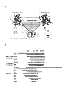
Figure 1 (A) illustrates how the three subgroups of BCL-2 family members
interact with one another
to form a signaling nework that regulates apoptosis. (B) depicts an alignment
of BCL-2 family members,
highlighting the conserved BCL-2 homology domains shared among the protein
subgroups.
Figure 2 shows how the BH3-binding pocket of an anti-apoptotic protein binds
and sequesters a
death helix.
Figure 3 shows how the bioactive BH3 helix can be reconstituted by hydrocarbon
stapling to yield a
helical, protease resistant, and cell permeable compound capable of targeting
BCL-2 family proteins.
Figure 4 shows the primary amino acid sequence of humans MCL-1 long (SEQ ID
NO: 1) and the
alternatively spliced MCL-1 short. The BH3 regions are underlined and the
alternative C-terminus of MCL-1
short is in italics. A small nuclear MCL-1, believed to be a cleavage product
of MCL-1 long has also been
reported (SEQ ID NO: 72). Finally, MCL-INC protein used for expression and
binding studies is shown
(SEQ ID NO: 73).
Figure 5 shows the primary amino acid sequence of human NOXA. The BH3 region
is underlined.
Figure 6 shows the primary amino acid sequence of human BOK. The BH3 region is
underlined.
Figure 7 shows a panel of stabilized alpha-helices of BCL-2 domains (SAHBs)
designed based on
the BH3 domains of pro- and anti-apoptotic BCL-2 family members. A pair of
crosslinking non-natural
amino acids (X) were substituted at i, 1+4 position of the non-interacting
helical surface and "stapled" by
ruthenium-catalyzed olefin metathesis. To optimize the activity of Grubbs'
ruthenium catalyst, sulfur-
containing methionines were replaced with norleucines, which are designated by
the letter B.
Figure 8A shows graph for determining dissociation constants for the binding
of fluorescently
labeled SAHBs to MCL-1ANAC by fluorescence polarization assay (FPA) and
nonlinear regression analysis.
The figure illustrates fluorescence polarization assays using FITC-derivatized
SAHBs and recombinant
MCL-1 protein, and distinguished MCL-1 targeting SAHBs from non-binders.
Figure 8B shows a graph for determining dissociation constants for the binding
of fluorescently
labeled SAHBs to MCL-LANAC by FPA and nonlinear regression analysis. The
figure illustrates
fluorescence polarization assays using FITC-derivatized SAHBs and recombinant
MCL-1 protein, and
distinguished MCL-1 targeting SAHBs from non-binders.
Figure 8C shows a graph for determining dissociation constants for the binding
of fluorescently
labeled SAHBs to MCL-LANAC by FPA and nonlinear regression analysis. The
figure illustrates
fluorescence polarization assays using FITC-derivatized SAHBs and recombinant
MCL-1 protein, and
distinguished MCL-1 targeting SAHBs from non-binders.
Figure 9 shows fluorescence polarization assays using FITC-derivatized SAHBs
and recombinant
anti-apoptotic proteins revealed high affinity MCL-1 targeting compounds (A),
with a subset exhibiting
selective MCL-1 interaction (A, B). (A) illustrates a table of KD values for
MCL-1-specific and pan-anti-
apoptotic binding SAHBs. (B) demonstrates the binding isotherms of a subset of
high affinity MCL-1
binders, including MCL-1 SAHB and
8
CA 2746256 2019-08-19
CA 02746256 2011-06-08
WO 2010/068684
PCMJS2009/067363
NOXA SAHB, which were selective for MCL-1 and did not engage BCL-XL, BCL-w, or
BFL1/Al. BOK SAHB displayed a significant preference for MCL-1 over the other
anti-
apoptotics.
Figure 10 illustrates the specificity determinants of the MCL-1 BH3 helix for
MCL-
1. A panel of sequential alanine mutants (alanine scan) of FITC-MCL-1 SAHB was
generated for FPA binding analysis, revealing key residues within the core BH3
sequence
required for high affinity MCL-1ANAC binding. Glutamate mutagenesis was also
performed
to evaluate the contribution of native alanine and glycine residues to MCL-
1ANAC binding.
Figure 11 Shows sampling of a variety of staple positions along the helical
surface
revealed disruption of MCL-1ANAC binding only by the G217-Q221 staple (MCL-1
SAHBC), which is located at the hydrophobic binding interface. MCL-1 SAHBD
exhibited
the strongest binding activity (KD, 10 nM), with 4-fold improvement over the
parental MCL-
1 SAHBA. (A) illustrates a "staple scan" of the MCL-1 SAHB, demonstrating
differential
placement of the hydrocarbon staple along the length of the MCL-1 BH3
sequence; (B)
Sampling a variety of staple positions along the helical surface revealed
disruption of MCL-
1ANAC binding only by the G217-Q221 staple (MCL-1 SAHBc), which is located at
the
hydrophobic binding interface. MCL-1 SAHBD exhibited the strongest binding
activity (KD,
10 nM), with 4-fold improvement over the parental MCL-1 SAHBA.
Figure 12 shows that circular dichroism revealed marked enhancement of a-
helical
structure for MCL-1 SAHBs compared to the corresponding unmodified peptide.
Hydrocarbon stapling converts the predominantly non-helical MCL-1 BH3 template
peptide
into a stabilized a-helical structure, with differentially stapled SAHBs
exhibiting percent
helical content ranging from 55-100%.
Figure 13 shows that like FITC-MCL-1 SAHBA, FITC-MCL-1 SAHBD display a
potent and exclusive interaction with MCL-1ANAC, as evidenced by FPA performed
against
a broad panel of anti-apoptotic targets.that like F1TC-MCL-1 SAHBA, F1TC-MCL-1
SAHBI)
displayed a potent and exclusive interaction with MCL-1ANAC, as evidenced by
FPA
performed against a broad panel of anti-apoptotic targets.
Figure 14 (A) Sequence alignment of select BH3 domains revealed key
differences in
the hydrophobic residues that engage the canonical BH3 pocket of anti-
apoptotic proteins.
MCL-1 BH3 contains a unique LXXVXXXV motif. Both the BCL-2/BCL-XL-selective
BAD BH3 domain and the pan-anti-apoptotic binding BIM BH3 domain contain a Phe
at the
position corresponding to Va1220 in MCL-1 BH3 (underlined). Interestingly, the
murine
NOXA BH3 and MULE BH3 (not shown) domains, which exhibit selectivity for MCL-
1,
both contain a Val in this position. (B) Site directed mutagenesis of MCL-1
SAHBA alters the
specificity for MCL-1. MCL-1 SAHBA V220F binds to both MCL-1 and BCL-XL,
9
CA 02746256 2011-06-08
WO 2010/068684
PCT/1JS2009/067363
demonstrates that V220 is a key specificity determinant for MCL-1 SAHBA
binding to both
MCL-1ANAC and BCL-XLAC as demonstrated by FPA.
Figure 15 shows that site-directed amino acid mutagenesis converted the pan-
anti-
apoptotic binder, BIM SAHB, into a selective MCL-1 binder. Specificity for MCL-
1 can also
be obtained by site directed mutagenesis of non-selective SAHBs. Mutagenesis
of 11e65 and
Glu68 to Phe and Lys, respectively, in BIM SAHBD, a staple variant of BIM
SAHBA,
resulted in selective inhibition of MCL-1 as determined by fluorescence
polarization assay.
Figure 16 shows (A) MCL-1 SAHBs effectively prevent sequestration of the BAK
BH3 helix by MCL-1ANAC, as demonstrated by competition FPA (FITC-BAK SAHB/MCL-
lANAC IC50, 0.27+0.06 jiM). (B) MCL-1 SAHBD dose-responsively sensitizes BID
BH3-
induced and BAK-dependent mitochondrial apoptosis, as measured by cytochrome c
release
assay performed on wild type and Bak mitochondria. (C) The native interaction
between
BAK and MCL-1 was dose-responsively disrupted by treatment of OPM2 multiple
myeloma
cells with MCL-1 SAHBD, as assessed by MCL-1 immunoprecipitation and BAK
western
analysis.
Figure 17 (A) shows that NOXA SAHB targets MCL-1 in situ and disrupts the
MCL-1/BAK interaction as demonstrated by MCL-1 co-immunoprecipitation; (B)
illustrates
that NOXA SAHB dose-responsively sensitizes chemoresistant U937 AML cells to
low-dose,
pro-apoptotic BIM SAHB.
Figure 18 shows that Jurkat and OPM2 cells were treated with increasing doses
of
TRAIL and Fas ligand (FasL), and cell viability was measured at 24 hours by
MTT assay.
Whereas TRAIL induced apoptosis of both Jurkat and OPM2 cells (A and B), only
Jurkat
cells were sensitive to FasL (A). These data represent baseline studies for
the experiments
performed in Figures 19-21.
Figure 19 shows that Jurkat T-cell leukemia and OPM2 cells were exposed to MCL-
1 SAHBD singly and in combination with low dose death receptor agonists TRAIL
and Fas
ligand in the presence or absence of the pan-caspase inhibitor, z-VAD. Cell
viability
measured by MTT assay at 24 hours revealed dose-responsive and caspase-
dependent
sensitization of Jurkat (TRAIL and FasL) and OPM2 (TRAIL) cells by MCL-1 SAHBD
(A).
The capacity of MCL-1 SAHBD to sensitize Jurkat and OPM2 cells to death
receptor stimuli
correlated with dose-responsive activation of caspase 3/7, as measured by
luminescence of
DEVD-cleaved substrate (B). (A) Jurkat T-cell leukemia and OPM2 cells were
exposed to
MCL-1 SAHBD singly and in combination with low dose death receptor agonists
TRAIL and
Fas ligand in the presence or absence of the pan-caspase inhibitor, z-VAD.
Cell viability
measured by MTT assay at 24 hours revealed dose-responsive and caspase-
dependent
sensitization of Jurkat (TRAIL and FasL) and OPM2 (TRAIL) cells by MCL-1
SAHBD. (B)
The capacity of MCL-1 SAHBD to sensitize Jurkat and OPM2 cells to death
receptor stimuli
CA 02746256 2011-06-08
WO 2010/068684
PCMJS2009/067363
correlated with dose-responsive activation of caspase 3/7, as measured by
luminescence of
DEVD-cleaved substrate.
Figure 20 shows that in contrast to contrast to NOXA SAHBA that binds both MCL-
1ANAC and BFL-1/A1AC, NOXA SAHBB, which contains an alternate staple position,
exhibits potent and exclusive MCL-1ANAC binding activity as measured by FPA.
Like
MCL-1 SAHBD, NOXA SAHBD sensitized the apoptotic response ofJurkat cells to
TRAIL
and FasL, as measured by MTT assay at 24 hours.
Figure 21 shows that BFL-1 SAHBA exhibited no binding activity toward anti-
apoptotic proteins by FPA and correspondingly showed no sensitization activity
in Jurkat
cells treated with low dose TRAIL or FasL.
Figure 22 shows that a shortened MCL-1 SAHB variant bind to MCL-1ANAC with
high affinity.
Figure 23 provides the compositions of stapled BH3 peptides (SAHBs) generated
to
assess MCL-1 binding specificity and selectively target MCL-1.
DETAILED DESCRIPTION AND PREFERRED EMBODIMENTS
A series of stapled BCL-2 family peptide helices have now been identified that
target
the survival protein MCL-1 with high affinity and unprecedented selectivity.
The MCL-1
inhibitor SAHBs described herein target the canonical BH3 groove of MCL-1,
displacing the
MCL-1/BAK interaction in vitro and in situ, and sensitizing MCL-1 dependent
cancer cells to
mitochondrial apoptosis.
Definitions
As used herein, the term "hydrocarbon stapling" or "stapling", refers to a
process for
stably cross-linking a polypeptide having at least two modified amino acids
that helps to
conformationally bestow the native secondary structure of that polypeptide.
Hydrocarbon
stapling allows a polypeptide, predisposed to have an alpha-helical secondary
structure, to
maintain its native alpha-helical conformation. This secondary structure
increases resistance
of the polypeptide to proteolytic cleavage and heat, and also may increase
target binding
affinity, hydrophobicity, and cell permeability. Accordingly, the hydrocarbon
"stapled"
(cross-linked) polypeptides described herein have improved biological activity
relative to a
corresponding non-hydrocarbon stapled (uncrosslinked) polypeptide. For
example, the cross-
linked polypeptide can include an alpha-helical domain of a BH3 BCL-2 homology
domain,
which, at least in the case of exemplary NOXA, BOK and MCL-1 BH3 domains, can
competitively interfere with the interaction of MCL-1 protein with native
ligands (including,
e.g., formation of MCL-1 dimers and/or multimers and/or the MCL-1/BAK
heterodimer),
thereby modulating MCL-1 activity in a cell. Modulation of MCL-1 activity can
produce a
11
CA 02746256 2016-06-27
WO 2010/068684
PCT/US2009/06730
number of effects, including, e.g., promotion of apoptosis in a cell,
modulation of cell cycle
regulation in a cell, modulation of autophagy in a cell, modulation of
cellular inflammatory
responses, modulation of cellular autoimmune responses, and modulation of RNA
splicing.
The cross-linked polypeptides described herein can be used prophylactically or
therapeutically, e.g., to treat or prevent hyperproliferative diseases, such
as cancer.
The hydrocarbon stapled polypeptides include one or more tethers (linkages)
between
two non-natural amino acids, which tether significantly enhances the alpha
helical secondary
structure of the polypeptide. Generally, the tether extends across the length
of one or two
helical turns (i.e., about 3.4 or about 7 amino acids). Accordingly, amino
acids positioned at i
and i+3; i and i+4; or i and i+7 are ideal candidates for chemical
modification and cross-
linking. Thus, for example, where a peptide has the sequence. . . Xl, X2, X3,
X4, XS, X6,
X7, X8, X9 . ,cross-links between X1 and X4, or between X1 and XS, or between
X1 and
X8 arc useful as are cross-links between X2 and XS, or between X2 and X6, or
between X2
and X9, etc. represent hydrocarbon stapled forms of that peptide. The use of
multiple cross-
links (e.g., 2, 3, 4 or more) is also contemplated. The use of multiple cross-
links is very
effective at stabilizing and optimizing the peptide, especially with
increasing peptide length.
Thus, the invention encompasses the incorporation of more than one crosslink
within the
polypeptide sequence to either further stabilize the sequence or facilitate
the structural
stabilization, proteolytic resistance, acid stability, thermal stability,
cellular permeability and
biological activity enhancement of longer polypeptide stretches. Additional
description
regarding making and use of hydrocarbon-stapled polypeptides can be found,
e.g., in U.S.
patent 8,921,323.
As used herein, the terms "stapled" and "hydrocarbon-stapled" are used
interchangeably.
The term "stable" or "stabilized", as used herein with reference to a
polypeptide,
refers to polypeptides which have been hydrocarbon-stapled to maintain their
natural alpha-
helical structure and/or improve protease resistance and/or improve acid
stability and/or
improve thermal stability and/or improve cellular permeability and/or improve
target binding
affinity and/or improve biological activity.
The term "active site" of MCL-1 refers to a region of an MCL-1 polypeptide or
MCL-
1-interacting polypeptide, as a result of its shape, amino acid content, and
charge potential,
that favorably interacts or associates with another agent (including, without
limitation, a
protein, polypeptide, peptide, molecule, compound, antibiotic, drug, and/or
nucleic acid) via
various covalent and/or non-covalent binding forces. BCL-2 family members may
have more
than one active site, as recently reported (Gavathiotis et al. Nature, 455:
1076, 2008). An
example of one defined "active site" on MCL-1 includes a hydrophobic groove
and
12
CA 02746256 2011-06-08
WO 2010/068684
PCMJS2009/067363
circumferential charged/hydrophilic residues which is capable of binding a
stabilized alpha
helix of a BCL-2 homology domain, such as human hydrocarbon-stapled MCL-1 BH3
(SEQ
ID NO:1, 12, 17-60), NOXA BH3 (SEQ ID NO:2, 7,63-68), BOK BH3 (SEQ ID NO:3,
11),
or wild-type or MCL-1 specificity-tailored BIM BH3 (SEQ ID NO:4, 61, 62) or
BAK BH3
(SEQ ID NO: 9, 69), or MULE BH3, a non-BCL-2 family member containing a BH3
homology domain (SEQ ID NO: 70), and which is formed by the juxtaposition of
alpha
helices 3, 4 and 5 of MCL-1 (PDB #2pqk and SEQ ID NO: 1), including residues
V216,
V220, H224, A227 and M231 of helix 3, residues V249, V253 and D255 of helix 4
and
residues G262, T266 and F270 of helix 5 or formed by the juxtaposition of
alpha helices 3,4
and 5 of MCL-1 (PDB# 2jm6), including residues V201, H205 and M212 of helix 3,
residues
S226, H233 and V234 of helix 4 and residues R244, 1247, L249 and F251 of helix
5. In one
embodiment, the active site includes two or more amino acids corresponding to
0262 and
F270 (PDB# 2pqk, SEQ ID NO: 1).
The term "MCL-1 polypeptide variant" refers to polypeptides that vary from a
reference MCL-1 family polypeptide by the addition, deletion or substitution
of at least one
amino acid to a natural amino acid or a non-natural amino acid or a mimetic
thereof It is well
understood in the art that some amino acids may be changed to others with
broadly similar
properties without changing the nature of the activity of the polypeptide
(e.g. conservative
substitutions such as glutamine for glutamate or hydrophobic for hydrophobic
or positively
charged for positively charged) as described hereinafter. Accordingly, MCL-1
polypeptide
variants as used herein encompass polypeptides that have pro- or anti-
apoptotic activity. The
term "variant" refers to a polypeptide having at least 30% amino acid sequence
identity with a
reference MCL-1 BCL-2 homology domain (e.g., MCL-1 BH3 domain) within a
protein or
any other functional domain thereof. More specifically, the term "variant"
includes, but is not
limited to, an MCL-1 polypeptide comprising an active site characterized by a
three
dimensional structure comprising the relative structural coordinates of alpha
helices 3, 4 and 5
of MCL-1 (PDB #1pqk, SEQ ID NO: 1), including residues V216, V220, H224, A227
and
M231 of helix 3, residues V249, V253 and D255 of helix 4 and residues 0262,
R263,1266
and F270 of helix 5 or of alpha helices 3, 4 and 5 of MCL-1 (PDB# 2jm6, SEQ ID
NO: 1),
including residues V201, H205 and M212 of helix 3, residues S226, H233 and
V234 of helix
4 and residues R244, 1247, L249 and F251 of helix 5 of SEQ ID NO: 1, in each
case, +/- a
root mean square deviation from the conserved backbone atoms of those residues
of not more
than 1.1 angstroms, in certain embodiments not more than 1.0 angstroms, and in
certain
additional embodiments not more than 0.5 angstroms.
An "MCL-1 polypeptide variant" further includes those polypeptides, or their
biologically active fragments, that comprise an amino acid sequence which is
at least 30%,
40%, 50%, 60%, 70%, 80%, 90%, 95%, 96%, 97%, 98%, 99% or more similar to an
amino
13
CA 02746256 2016-06-27
WO 2010/068684
PCT/US2009/067363
acid sequence of an MCL-1 BCL-2 homology domain (e.g., BH3 domain). In certain
embodiments, the BCL-2 homology domain comprises one or more conserved amino
acid
residues, such as amino acid residues corresponding to L213, G217, and/or D218
of MCL-1
(SEQ ID NO: 1) or conservative substitutions thereof.
The term "hydrophobic amino acid" means any natural or non-natural amino acid
or
mimetic thereof having an uncharged, non-polar side chain that is relatively
insoluble in
water. Examples of naturally occurring hydrophobic amino acids are alanine,
leucine,
isoleucine, valine, proline, phenylalanine, tryptophan and methionine.
The term ''hydrophilic amino acid" means any natural or non-natural amino acid
or
mimetic thereof having an uncharged, polar side chain that is relatively
soluble in water.
Examples of naturally occurring hydrophilic amino acids are scrim., threonine,
tyrosine,
asparagine, glutamine, and cysteine.
The term "negatively charged amino acid" includes any naturally occurring or
non-
natural amino acid or mimetic thereof having a negatively charged side chain
under normal
physiological conditions. Examples of negatively charged naturally occurring
amino acids are
aspartic acid and &ramie acid.
The term "positively charged amino acid" includes any naturally occurring or
non-
natural amino acid or mimetic thereof having a positively charged side chain
under normal
physiological conditions. Examples of positively charged naturally occurring
amino acids are
argininc, lysine and histidinc.
As used herein, the term, "BCL-2 family polypeptide" refers to an evolutionary
conserved family of proteins having as few as one to as many as four conserved
BCL-2
homology domains (BI-11, B1-12, B113 and/or BI14). The B11 domains are alpha-
helical
segments and are present in both the anti-apoptotic and pro-apoptotic
polypeptides of BCL-2
.. family proteins, which are conserved across many species, both at the
sequence level and
functionally (e.g., mouse BCL-2 family proteins bind human MCL-1). BCL-2
family
polypeptides include BCL-2, BCL-XL, BCL-w, MCL-1, BCL-B, Al/BFL-1, BOO/DIVA,
Nr-13, CED-9, BAX, B,AK, BOK/MTD, BID, BAD, B1K/NBK, BLK, HRK,I31M/BOD,
BNIP3, NIX, NOXA, PUMA, BMF, EGL-, and viral homologues. Functional BCL-2
family
homology domains can also be found in non-BCL-2 family proteins, such as
Beclin-1
(Oberstein et al. J Biol Chem, 282: 13123, 2007) and MULE (Zhong et al. Cell,
121:1085,
2005), which is a non-BCL-2 family protein that contains a BH3 domain. The
skilled artisan
will recognize that such non-BCL-2 family polypeptides can also be used in the
compositions,
methods and kits of the instant invention. Exemplary methods and compositions
for
modulating BCL-2 family polypeptides are described in U.S. patent 8,921,323.
14
CA 02746256 2011-06-08
WO 2010/068684
PCMJS2009/067363
The term "anti-apoptotic polypeptide" refers to BCL-2 family polypeptides
characterized by having one or more amino acid homology domains, BH1, BH2,
BH3, and/or
BH4, and that promote cell survival by attenuating or inhibiting apoptosis.
The "anti-
apoptotic polypeptides" further include those proteins, or their biologically
active fragments,
that are at least 30%, 40%, 50%, 60%, 70%, 80%, 90%, 95%, 96%, 97%, 98%, 99%
or more
similar in amino acid sequence to an anti-apoptotic BCL-2 homology domain
within a BCL-2
family polypeptide. In certain embodiments, the BCL-2 homology domain
comprises one or
more conserved amino acid residue, such as amino acid residues corresponding
to residues
L213, G217, and or D218 of MCL-1's BH3 domain (PDB# 1pqk, SEQ ID NO: 1). Anti-
apoptotic polypeptides include MCL-1, BCL-2, BCL-Xl, BCL-w, BCL-B, Al/BFL-1,
BOO/DIVA, Nr-13, CED-9, and viral homologues.
The term "pro-apoptotic polypeptide" refers to BCL-2 family polypeptides
characterized by having one or more amino acid homology domains, BH1, BH2,
and/or BH3,
and that promote cell death by activating apoptosis. The "pro-apoptotic
polypeptides" further
include those proteins, or their biologically active fragments, that are at
least 30%, 40%,
50%, 60%, 70%, 80%, 90%, 95%, 96%, 97%, 98%, 99% or more similar in amino acid
sequence to a pro-apoptotic BCL-2 homology domain within a BCL-2 family
polypeptide. In
certain embodiments, the BCL-2 homology domain comprises one or more conserved
amino
acid residues, such as amino acid residues corresponding to residues L29 and
G33 of
NOXA's BH3 domain (PubMed RefSeq: NP_066950.1, SEQ ID NO: 2, 5) or residues
L70
and G75 of BOK's BH3 domain (PubMed RefSeq: NP_115904.1, SEQ ID NO: 3, 9). Pro-
apoptotic polypeptides include BAX, BAK, BOK/MTD, BID, BAD, BIK/NBK, BLK, HRK,
BIM/BOD, BNIP3, NIX, NOXA, PUMA, BMF, EGL-1, and viral homologs. An example of
a non-BCL-2 family protein that regulates MCL-1 levels through targeted
degradation, and is
thus pro-apoptotic during physiologic stress, is the BH3 domain-containing
ubiquitin ligase
MULE.
As used herein, the term "apoptosis" refers to a regulated network of
biochemical
events which leads to a selective form of cell death that is characterized by
readily observable
morphological and biochemical changes, such as the fragmentation of the
deoxyribo-nucleic
acid (DNA), condensation of the chromatin, which may or may not be associated
with
endonuclease activity, chromosome migration, margination in cell nuclei, the
formation of
apoptotic bodies, mitochondrial swelling, widening of the mitochondrial
cristae, opening of
the mitochondrial permeability transition pores and/or dissipation of the
mitochondrial proton
gradient.
The term "compound" is used herein to denote a chemical agent, polypeptide,
nucleic
acid or combination thereof, or a mixture of chemical compounds and/or
polypeptides and/or
nucleic acids (e.g. DNA and/or RNA derivative), salts and solvates thereof,
and the like. In
CA 02746256 2011-06-08
WO 2010/068684
PCMJS2009/067363
certain embodiments, a compound of the invention binds to an active site of an
MCL-1
polypeptide. A "modulator" is a compound which changes (e.g.,
enhances/promotes or
inhibits/suppresses) the activity of an MCL-1 polypeptide.
The term "candidate compound" or "test compound" is used herein to denote a
chemical compound, peptide, nucleic acid or combination thereof, or a mixture
of chemical
compounds and/or polypeptides and/or nucleic acids, salts and solvates
thereof, and the like,
which is tested by a method of the invention and is found to bind to an active
site of an MCL-
1 polypeptide, and thus is believed to modulate the activity of the MCL-1
polypeptide.
As used herein, "small molecule" is understood to refer to a chemical compound
having a molecular weight below 2,000 daltons, more preferably between 300 and
1,000
daltons, and still more preferably between 400 and 700 daltons. It is
preferred that these
small molecules are organic molecules. In certain embodiments, "small
molecule" does not
include peptide or nucleic acid molecules.
The term "modulate" as used herein with reference to a compound refers to the
activation or inhibition of anti-apoptofic or pro-apoptotic activity of a BCL-
2 family
polypeptide or affects other protein-protein interactions involving a BCL-2
family member or
other protein target that binds a BCL-2 homology domain, and thereby regulates
a
biochemical pathway (e.g. unfolded protein response, glucose-stimulated
insulin secretion,
apoptosis). Methods for assaying both anti-apoptotic, pro-apoptotic, and other
biochemical
activities (e.g. unfolded protein response, glucose-stimulated insulin
secretion, apoptosis) are
well known in the art and described herein.
As used herein, the term "interacts" or "binds" refers to a condition of
proximity
between a compound, or portions thereof, and the active site of a BCL-2 family
polypeptide
or portions thereof. The interaction is between one or more moieties on the
compound and
one or more moieties of the amino acids of the active site. The association
may be non-
covalent--wherein the juxtaposition is energetically favored by hydrogen
bonding or van der
Waals or electrostatic interactions--or it may be covalent. For example,
hydrophobic and
hydrophilic amino acid residues of alpha helices 3, 4 and 5 of the MCL-1
polypeptide,
including residues V216, V220, H224, A227 and M231 of helix 3, residues V249,
V253 and
D255 of helix 4 and residues G262, R263 T266 and F270 of helix 5 are predicted
to interact
with residues T212, L213, R214, V216, G217, D218 and V220 of MCL-1 BH3 domain
(PubMed RefSeq: NP 068779.1, SEQ ID NO: 1, 16), residues A26, L29, G33 and L36
of
NOXA BH3 domain (PubMed RefSeq: NP 066950.1, SEQ ID NO: 2, 5) and residues
V66,
V69, L70, G75 and L79 of BOK BH3 domain (PubMed RefSeq:NP_115904.1, SEQ ID NO:
3, 9).
The term, "activates" refers to an increase in the anti-apoptotic or pro-
apoptotic
activity of a BCL-2 family polypeptide or other defined biochemical activity
based upon
16
CA 02746256 2011-06-08
WO 2010/068684
PCMJS2009/067363
protein-protein or protein-nucleic acid interaction. A compound that activates
a pro-apoptotic
activity will bind to an active site of a BCL-2 family polypeptide and cause,
for example, a
1.5x, 2x, 3x, 4x, 5x, 6x, 7x, 8x, 9x, 10x, 15x, 20x or more increase in the
pro-apoptotic
activity of the BCL-2 family polypeptide when compared with a control lacking
the
compound. In another embodiment, a compound that activates an anti-apoptotic
activity will
bind to an active site of a BCL-2 family polypeptide and cause, for example, a
1.5x, 2x, 3x,
4x, 5x, 6x, 7x, 8x, 9x, 10x, 15x, 20x or more increase in the anti-apoptotic
(survival) activity
of the BCL-2 family polypeptide when compared with a control lacking the
compound. In
another embodiment, a compound that modulates a biochemical activity (e.g.
cell cycle,
autophagy) will bind to an active site of a BCL-2 family polypeptide or other
BCL-2
homology domain binding target protein and cause, for example, a 1.5x, 2x, 3x,
4x, 5x, 6x,
7x, 8x, 9x, 10x, 15x, 20x or more increase in the biochemical activity of the
target protein
when compared with a control lacking the compound. Assays for assessing the
activation of
an anti-apoptotic or pro-apoptotic activity or the modulation of a biochemical
activity (e.g.
induction of autophagy, induction of cell cycle arrest) are known in the art
and described
herein.
The term "inhibits" refers to a decrease or blocking of the anti-apoptotic or
pro-
apoptotic activity of a BCL-2 family polypeptide, or other defined biochemical
activity based
upon protein-protein interaction. For example, a compound that inhibits a pro-
apoptotic
activity will bind to an active site of a BCL-2 family polypeptide and prevent
activation or
reduce the activity of the BCL-2 family polypeptide. Thus, the compound will
inhibit or
decrease the effects of a pro-apoptotic activity. Thus, pro-apoptotic
activity, e.g., cell death,
will be less than, for example, 75%, 70%, 60%, 50%, 40%, 30%, 20%, 10%, 5% or
less in a
population of cells in which an inhibitor is present than compared to a
control cell population
where the compound is not present. In another embodiment, a compound that
inhibits an anti-
apoptotic activity will bind to an active site of a BCL-2 family polypeptide
and prevent or
reduce the the anti-apoptotic activity of the BCL-2 family polypeptide. Thus,
anti-apoptotic
activity, e.g., survival, will be less than, for example, 75%, 70%, 60%, 50%,
40%, 30%,
20%, 10%, 5% or less in a population of cells in which an inhibitor is present
than compared
to a control cell population where the compound is not present. In yet another
embodiment, a
compound that modulates a biochemical activity (e.g. cell cycle, autophagy)
will bind to an
active site of a BCL-2 family polypeptide or other BCL-2 homology domain
binding target
protein and prevent or reduce the the biochemical activity of the protein
target. Thus, the
biochemical activity (e.g., autophagy, cell cycle arrest) will be less than,
for example, 75%,
70%, 60%, 50%, 40%, 30%, 20%, 10%, 5% or less in a population of cells in
which an
inhibitor is present than compared to a control cell population where the
compound is not
present.
17
CA 02746256 2016-06-27
WO 2019/068684
PCT/U82009/067363
As used herein, the term "BH3 SAHB" refers to the BCL-2 homology domain 3 of a
BCL-2 family polypeptidc and/or a BH3 domain-containing polypcptide (e.g.,
MULE) that
has been hydrocarbon stapled so as to form a stabilized alpha helix. The amino
acid
sequences of numerous BH3 domains are described heroin, (e.g., Fig. 7, 9, and
15 ). Methods
of making BH3 SAHB's are known in the art and described in U.S. Patent
Publication No.
US2005/0250680, filed November 5, 2004.
As used herein, the term "NOXA BH3 polypeptide" refers to a polypeptide having
a
BCL-2 homology domain 3 of NOXA. In one embodiment, the NOXA BH3 polypeptide
has
an amino acid sequence which is 30%, 40%, 50%, 60%, 70%, 80%, 90%, 95%, 96%,
97%,
98%, 99% or more identical to SEQ ID NO: 2 (Fig. 5) and includes one or more
of amino
acid residues corresponding to L29, G33, and/or D34 of SEQ ID NO: 2 or
conservative
substitutions thereof Optionally, the NOXA BH3 domain of the NOXA BH3
polypeptide
has an amino acid sequence which is 30%, 40%, 50%, 60%, 70%, 80%, 90%, 95%,
96%,
97%, 98%, 99% or more identical to the BH3 domain of SEQ ID NO: 2. In certain
embodiments, the NOXA BH3 polypeptide has the amino acid sequences of SEQ ID
NO: 7,
and SEQ ID NO: 63-68. In certain embodiments, the scope of the term "NOXA BH3
polypeptide" encompasses biologically active fragments of SEQ ID NO: 2, while
the scope of
"NOXA BH3 domain" similarly encompasses biologically active fragments of the
BH3
domain of SEQ ID NO: 2.
As used herein, the term "BOK BH3 polypeptide" refers to a polypeptide having
a
BCL-2 homology domain 3 of BOK. In one embodiment, the BOK BIB polypeptide has
an
amino acid sequence which is 30%, 40%, 50%, 60%, 70%, 80%, 90%, 95%, 96%, 97%,
98%,
99% or more identical to SEQ ID NO: 3 (Fig. 6) and includes one or more of
amino acid
residues corresponding to residues L70, 075, and/or D76 of SEQ ID NO: 3 or
conservative
substitutions thereof Optionally, the BOK BH3 domain of the BOK BH3
polypeptide has an
amino acid sequence which is 30%, 40%, 50%, 60%, 70%, 80%, 90%, 95%, 96%, 97%,
98%,
99% or more identical to the BH3 domain of SEQ ID NO: 3. In certain
embodiments, the
BOK BH3 polypcptide has the amino acid sequence of SEQ ID NO: 11. In certain
embodiments, the scope of the term -BOK BH3 polypeptide" encompasses
biologically active
fragments of SEQ ID NO: 3, while the scope of "BOK BH3 domain" similarly
encompasses
biologically active fragments of the BH3 domain of SEQ ID NO: 3.
As used herein, the term "MCL-1 BH3 polypeptide" refers to a polypeptide
having a
BCL-2 homology domain 3 of MCL-1. In one embodiment, the MCL-1 BH3 polypeptide
has
an amino acid sequence which is 30%, 40%, 50%, 60%, 70%, 80%, 90%, 95%, 96%,
97%,
98%, 99% or more identical to SEQ ID NO: 1 and includes one or more of amino
acid
residues corresponding to L213 and 0217 of SEQ ID NO: 1 (Fig. 4) or
conservative
18
CA 02746256 2011-06-08
WO 2010/068684
PCMJS2009/067363
substitutions thereof Optionally, the MCL-1 BH3 domain of the MCL-1 BH3
polypeptide
has an amino acid sequence which is 30%, 40%, 50%, 60%, 70%, 80%, 90%, 95%,
96%,
97%, 98%, 99% or more identical to the BH3 domain of SEQ ID NO: 1. In certain
embodiments, the MCL-1 BH3 polypeptide has the amino acid sequences of SEQ ID
NO: 12,
17-60. In certain embodiments, the scope of the term "MCL-1 BH3 polypeptide"
encompasses biologically active fragments of SEQ ID NO: 1, while the scope of
"MCL-1
BH3 domain" similarly encompasses biologically active fragments of the BH3
domain of
SEQ ID NO: 1.
As used herein, the term "MCL-1-specificity tailored BH3 polypeptide" refers
to a
polypeptide having a BCL-2 homology domain 3 of BCL-2 family members (Fig. 1B,
7) that
has been mutated to make its binding activity MCL-1 selective. In one
embodiment, the BH3
polypeptide has an amino acid sequence which is 30%, 40%, 50%, 60%, 70%, 80%,
90%,
95%, 96%, 97%, 98%, 99% or more identical to BIM BH3, SEQ ID NO: 4 and
includes one
or more of amino acid residues corresponding to L152 and G156 of SEQ ID NO: 4
(enumeration based on NCBI# NP_619527) or conservative substitutions thereof,
but also
includes one or more of amino acid residue mutations, for example converting
1155 to F
and/or E158 to K in SEQ ID NO: 3 or conservative substitutions thereof, to
achieve MCL-1
specificity. In certain embodiments, the MCL-1-specificity tailored BIM BH3
polypeptide
has the amino acid sequences of SEQ ID NO: 62. In another embodiment, the BH3
polypeptide has an amino acid sequence which is 30%, 40%, 50%, 60%, 70%, 80%,
90%,
95%, 96%, 97%, 98%, 99% or more identical to BAK BH3, SEQ ID NO: 9 and
includes one
or more of amino acid residues corresponding to L74 and G78 of SEQ ID NO: 6
(enumeration based on NCBI# NP_619527) or conservative substitutions thereof,
but also
includes one or more of amino acid residue mutations, for example converting
177 to F and/or
D84 to K (enumeration based on NCBI# NP 001179) or conservative substitutions
thereof, in
SEQ ID NO: 69 to achieve MCL-1 specificity.
As used herein, the term "non-BCL-2 family member BH3 polypeptide" refers to a
polypeptide having a BCL-2 homology domain 3 but is otherwise not
traditionally classified
as a BCL-2 family member or homologue. In one embodiment, the non-BCL-2 family
member BH3 polypeptide has an amino acid sequence which is 30%, 40%, 50%, 60%,
70%,
80%, 90%, 95%, 96%, 97%, 98%, 99% or more identical to SEQ ID NO: 70 and
includes one
or more of amino acid residues corresponding to L1980 and G1984 (enumeration
based on
NCBI# AAY98258) of SEQ ID NO: 70 (Fig. 13) or conservative substitutions
thereof.
The term "pharmacologically effective amount," "therapeutically effective
amount",
"pharmacologically effective dose" or simply "effective amount" refers to that
amount of an
agent effective to produce the intended pharmacological, therapeutic or
preventive result. The
pharmacologically effective amount results in the amelioration of one or more
symptoms of a
19
CA 02746256 2011-06-08
WO 2010/068684
PCMJS2009/067363
disorder, or prevents the advancement of a disorder, or causes the regression
of the disorder,
or prevents the disorder. For example, with respect to the treatment of a
disorder of excessive
cellular survival or proliferation, a therapeutically effective amount
preferably refers to the
amount of a therapeutic agent that decreases the rate of tumor growth,
decreases tumor mass,
decreases the number of metastases, increases time to tumor progression, or
increases survival
time by at least 5%, preferably at least 10%, at least 15%, at least 20%, at
least 25%, at least
30%, at least 35%, at least 40%, at least 45%, at least 50%, at least 55%, at
least 60%, at least
65%, at least 70%, at least 75%, at least 80%, at least 85%, at least 90%, at
least 95%, or at
least 100%.
For example, with respect to the treatment of a disorder associated with
increased
cellular death, e.g., ischemia, a therapeutically effective amount preferably
refers to the
amount of a therapeutic agent that prevents or limits tissue and/or cellular
damage that would
otherwise occur if treatment was not administered. The therapeutic agent
decreases tissue
and/or cellular damage by at least 5%, preferably at least 10%, at least 15%,
at least 20%, at
least 25%, at least 30%, at least 35%, at least 40%, at least 45%, at least
50%, at least 55%, at
least 60%, at least 65%, at least 70%, at least 75%, at least 80%, at least
85%, at least 90%, at
least 95%, or at least 100% compared to damage that occurs without the
administration of a
therapeutic agent of the invention.
The terms "treat," and "treating," as used herein with reference to a disorder
(e.g.,
hyperpoliferative disorder, excessive cellular survival or proliferation),
refers to a decrease in
the occurrence of pathological cells (e.g., hyperproliferative or neoplastic
cells) in an animal
or human. The prevention may be complete, e.g., the total absence of
pathological cells in a
subject. The prevention may also be partial, such that the occurrence of
pathological cells in a
subject is less than that which would have occurred without the present
invention. In some
embodiments, such terms refer to one, two, three or more results following the
administration
of one or more therapies: (1) a stabilization, reduction or elimination of the
cancer cell
population, (2) an increase in the length of remission, (3) a decrease in the
recurrence rate of
cancer, (4) an increase in the time to recurrence of cancer, and (6) an
increase in the survival
of the patient.
The terms "treat," and "treating," as used herein with reference to a disorder
associated with increased cellular death, e.g., ischemia, refer to a decrease
in the occurrence
of tissue and/or cellular damage in an animal or human. The prevention may be
complete,
e.g., the total absence of tissue damage in a subject. The prevention may also
be partial, such
that the occurrence of tissue damage in a subject is less than that which
would have occurred
without the therapeutic agent.
The temis "prevent," "preventing," and "prevention," as used herein, shall
refer to a
decrease in the occurrence of a disease or decrease in the risk of acquiring a
disease or its
CA 02746256 2011-06-08
WO 2010/068684
PCMJS2009/067363
associated symptoms in a subject. The prevention may be complete, e.g., the
total absence of
disease or pathological cells in a subject. The prevention may also be
partial, such that the
occurrence of the disease or pathological cells in a subject is less than that
which would have
occurred without the present invention.
The term "subject" refers to an animal or human, or to one or more cells
derived from
an animal or human. Preferably, the subject is a human. Subjects can also
include non-human
primates. Cells may be in any form, including but not limited to cells
retained in tissue, cell
clusters, immortalized, transfected or transformed cells, and cells derived
from an animal that
has been physically or phenotypically altered. A human subject can be known as
a patient.
The term "anti-tumor activity" refers to the ability of a substance or
composition to
block the proliferation of, or to induce the death of tumor cells which
interact with that
substance or composition.
As used herein, a "MCL-1 associated disorder", refers to a disorder associated
with a
deregulated MCL-1 polypeptide, particularly increased expression of MCL-1. An
MCL-1
associated disorder is characterized by having an MCL-1 at least a 10%, 20%,
30%, 40%,
50%, 60%, 70%, 80%, 90%, or more increase in the level of MCL-1 expression as
compared
to a normal control cell, preferably from the same subject. MCL-1 associated
disorders are
associated with excessive cellular survival and/or proliferation, e.g.,
cancer, or deregulation of
the cell cycle, or deregulation of the autophagic pathway, or deregulation of
cellular
autoinunune or inflammatory responses of a subject, or deregulation of RNA
splicing. An
MCL-1 associated disorder need not be diagnosed by identification of
deregulated MCL-1.
Instead, the disorder can initially be diagnosed by typical methods, e.g.,
imaging studies,
physical examination, biopsy, blood analysis, and confirmed to be an MCL-1
associated
disorder by histological analysis, PCR, or other methods known in the art. MCL-
1 associated
disorders include those described herein.
As used herein, a "hyperproliferative disorder" means cancer, neoplastic
growth,
hyperplastic or proliferative growth or a pathological state of abnormal
cellular development
or survival and includes solid tumors, non-solid tumors, and any abnormal
cellular
proliferation or accumulation, such as that seen in leukemia.
The terms "anticancer agent" and "anticancer drug," as used herein, refer to
any
therapeutic agents (e.g., chemotherapeutic compounds and/or molecular
therapeutic
compounds), antisense therapies, antibody therapies, peptide therapies,
nucleic acid therapies
(e.g. RNAi), radiation therapies, or combinations thereof, used in the
treatment of
hyperproliferative diseases such as cancer. In one embodiment, the invention
is directed to
methods of treating an MCL-1 associated disorder comprising administering an
effective dose
of an anticancer agent and a compound which binds to the active site, as
described herein, of
an MCL-1 polypeptide.
21
CA 02746256 2011-06-08
WO 2010/068684
PCMJS2009/067363
As used herein in relation to the position of an amino acid, e.g., L213 and
G217 of
SEQ ID NO: 1, the term -corresponding to" refers to an amino acid in a first
polypeptide
sequence, e.g., MCL-1, that aligns with a given amino acid in a reference
polypeptide
sequence, e.g., NOXA, when the first polypeptide and reference polypeptide
sequences are
aligned by homology or other algorithms (e.g., structural comparison).
Alignment is
performed by one of skill in the art using software designed for this purpose,
for example,
BLASTP version 2.2.2 with the default parameters for that version.
Corresponding amino
acids can also be identified upon structural comparisons of a first
polypeptide sequence and a
second polypeptide sequence. Such structural comparisons are known in the art
and described
herein. For example, Petros etal. Biochimica et Biophysica Acta 1644; 83-94
(2004) and
Suzuki etal., Cell. 103; 645-654 (2000) illustrated structural alignments
between BCL-2
homology domains of BCL-2 family members.
The term "amino acid" refers to a molecule containing both an amino group and
a
carboxyl group. Suitable amino acids include, without limitation, both the D-
and L-isomers
of the 20 common naturally occurring amino acids found in peptides (e.g., A,
R, N, C, D, Q,
E, G, H, I, L, K, M, F, P, S, T, W, Y, V (as known by the one letter
abbreviations)) as well as
the naturally occurring and non-naturally occurring amino acids (e.g.,
norleucine, modified
amino acids to allow for peptide stapling, amino acids linked by bonds other
than peptide
bonds) prepared by organic synthesis or other metabolic routes.
A "non-essential" amino acid residue is a residue that can be altered from the
wild-
type sequence of a polypeptide (e.g., MCL-1 BH3) without abolishing or
substantially
altering its ligand binding ability or otherwise significantly impacting,
particularly decreasing,
an activity of the polypeptide (e.g., reduces activity of the peptide less
than 40%, less than
30%, less than 20%, less than 10%). In certain embodiments, the activity of a
peptide can be
increased by modification of a non-essential amino acid. An "essential" amino
acid residue is
a residue that, when altered from the wild-type sequence of the polypeptide,
results in
abolishing or substantially abolishing the polypeptide's binding activity to
an MCL-1 active
site or otherwise dramatically alters the polypeptide's activity (e.g.,
decreases activity by at
least 60%, at least 70%, at least 80%, or at least 90%). In certain specific
examples, an
.. "essential" amino acid residue is limited to a residue that, when altered
from the wild-type
sequence of the polypeptide, results in abolishing or substantially abolishing
the polypeptide's
binding activity. For example, the essential and non-essential amino acid
residues of the BH3
domains of MCL-1, NOXA, BOK or other BCL-2 family polypeptide can readily be
determined by methods well known in the art and described herein. The term
"essential"
amino acid residue, as used herein, includes conservative substitutions of the
essential amino
acid. Generally, the "essential" amino acid residues are found at the
interacting face of the
BH3 polypeptide with the active site of the MCL-1 polypeptide.
22
CA 02746256 2011-06-08
WO 2010/068684
PCMJS2009/067363
As used herein, an "interacting face'' is understood as a surface of a protein
that
interacts with another protein or binding partner. The peptides of the
invention are
substantially alpha-helical. Alpha-helicies include 3.6 amino acids per turn,
i.e., the positions
of the amino acid in a helical peptide can be considered to be in positions
abcdefgabcdefg...
for the lenght of the helix. Therefore, the "face" of the alpha-helix could be
formed amino
acids at positions a and d, b and e, c and f, etc. which "stack" on top of
each other creating a
"face". The "face" can be wider than a single amino acid, wherein all
positions a, b, d, and c
form a face; ore, d, g, and a form a face; or the width of the face varies
along the face but is
composed of adjacent and/or "stacked" amino acids in the helix. In the
peptides of the
invention, it is preferred that the staple is not attached to amino acids that
interact directly
with the binding protein (e.g., MCL-1). As demonstrated by the alanine scan
and staple scan
herein, the peptides are typically more tolerant to mutations or alterations
on the non-
interacting face of the alpha-helix and less tolerant of mutations on the
interacting face of the
alpha-helix. Staples and mutations can be tolerated, and sometimes beneficial
when made on
the interacting face either immediately N- or C-terminal to the portion of the
helix that
interacts with the interacting protein. For example, it is noted that
placement of a staple
adjacent to the interacting face of the helix results in an increased affinity
of the peptide for
the target protein. Identification of amino acids on the interacting and non-
interacting faces
of the peptides of the invention is well within the ability of those of skill
in the art.
A "conservative amino acid substitution" is one in which the amino acid
residue is
replaced with a natural or non-natural amino acid residue having a similar
side chain. For
example, families of amino acid residues having similar side chains have been
defined in the
art. These families include amino acids with basic side chains (e.g., lysine,
argininc,
histidine), acidic side chains (e.g., aspartic acid, glutamic acid), uncharged
polar side chains
(e.g., glycine, asparagine, glutamine, serine, threonine, tyrosine, cysteine),
nonpolar side
chains (e.g., alanine, valine, leucine, isoleucine, norleucine, proline,
phenylalanine,
methionine, tryptophan), beta-branched side chains (e.g., threonine, valine,
isoleucine) and
aromatic side chains (e.g., tyrosine, phenylalanine, tryptophan, histidine).
Other conserved
amino acid substitutions can also occur across amino acid side chain families,
such as when
substituting an asparagine for aspartic acid in order to modify the charge of
a peptide. Thus,
a predicted nonessential amino acid residue in a BH3 domain polypeptide, for
example, is
preferably replaced with another amino acid residue from the same side chain
family or
homologues across families (e.g. asparagine for aspartic acid, glutamine for
glutamic acid).
In addition, individual substitutions, deletions or additions that alter, add
or delete a single
amino acid or a small percentage of amino acids in an encoded sequence are
also considered
"conservative substitutions." Appropriate conservative amino acid
substitutions can also be
identified by alignment with protein isoforms from other animals that express
the same
23
CA 02746256 2011-06-08
WO 2010/068684
PCMJS2009/067363
protein. In preferred embodiments, human sequences are compared with other
mammalian
sequences to identify possible conservative amino acid changes. Other mammals
for
sequence comparison include, but are not limited to, mouse, rat, dog, cat,
cow, goat, rabbits,
and non-human primates. Methods to perform sequence alignments are well known
as
discussed herein.
The terms "identical" or "percent identity," in the context of two or more
nucleic
acids or polypeptide sequences, refer to two or more sequences or subsequences
that are the
same or have a specified percentage of nucleotides or amino acids that are the
same, when
compared and aligned for maximum correspondence, as measured using one of the
following
sequence comparison algorithms, or by visual inspection.
"Similarity" or "percent similarity" in the context of two or more polypeptide
sequences, refer to two or more sequences or subsequences that are the same or
have a
specified percentage of amino acid residues, or conservative substitutions
thereof, that are the
same when compared and aligned for maximum correspondence, as measured using
one of
the following sequence comparison algorithms, or by visual inspection. By way
of example,
a first protein region can be considered similar to a region of an anti-
apoptotic MCL-1 protein
when the amino acid sequence of the first region is at least 30%, 40%, 50%,
60%, 70%, 75%,
80%, 90%, or even 95% identical, or conservatively substituted, to a region of
a second MCL-
1 protein or other protein (e.g., NOXA protein) when compared to any sequence
in the second
protein of an equal number of amino acids as the number contained in the first
region, or
when compared to an alignment of MCL-1 and homologs thereof (e.g., anti-
apoptotic BCL-2
family member proteins) that has been aligned by a computer similarity program
known in the
art, as discussed below. Preferably, the polypeptide region of the first
protein and the second
protein includes one or more conserved amino acid residues, e.g., such as
those illustrated in
Fig. 1, 7, and 15.
As used herein, the terms "identity" or "percent identity", refers to the
subunit
sequence similarity between two polymeric molecules, e.g., two polynueleotides
or two
polypeptides. When a subunit position in both of the two molecules is occupied
by the same
monomeric subunit, e.g., if a position in each of two peptides is occupied by
serine, then they
are identical at that position. The identity between two sequences is a direct
function of the
number of matching or identical positions, e.g., if half (e.g., 5 positions in
a polymer 10
subunits in length), of the positions in two peptide or compound sequences are
identical, then
the two sequences are 50% identical; if 90% of the positions, e.g., 9 of 10
are matched, the
two sequences share 90% sequence identity. The identity between two sequences
is a direct
function of the number of matching or identical positions. Thus, if a portion
of the reference
sequence is deleted in a particular peptide, that deleted section is not
counted for purposes of
calculating sequence identity. Identity is often measured using sequence
analysis software
24
CA 02746256 2011-06-08
WO 2010/068684
PCMJS2009/067363
e.g., BLASTN or BLASTP (www.ncbi.nih.gov/BLAST/). The default parameters for
comparing two sequences (e.g., -Blast"-ing two sequences against each other),
by BLASTN
(for nucleotide sequences) are reward for match = 1, penalty for mismatch = -
2, open gap = 5,
extension gap = 2. When using BLASTP for protein sequences, the default
parameters are
reward for match = 0, penalty for mismatch = 0, open gap = 11, and extension
gap = 1.
Additional, computer programs for determining identity are known in the art.
"Similarity" or "percent similarity" in the context of two or more polypeptide
sequences, refer to two or more sequences or subsequences that are the same or
have a
specified percentage of amino acid residues, or conservative substitutions
thereof, that are the
same when compared and aligned for maximum correspondence, as measured using
one of
the following sequence comparison algorithms, or by visual inspection. By way
of example,
a first polypeptide can be considered similar to BH3 domain of MCL-1 when the
amino acid
sequence of the first polypeptide is at least 20%, 30%, 40%, 50%, 60%, 70%,
75%, 80%,
90%, or even 95% or more identical, or conservatively substituted, to a region
of the BH3
domain of MCL-1 when compared to any sequence of an equal number of amino
acids as the
number contained in the first polypeptide as aligned by a computer similarity
program known
in the art and described herein. Preferably, the polypeptide region of the
first protein and the
second protein includes one or more conserved amino acid residues.
The term "amino acid side chain" refers to a moiety attached to the a-carbon
in an
amino acid. For example, the amino acid side chain for alanine is methyl, the
amino acid side
chain for phenylalanine is phenylmethyl, the amino acid side chain for
cysteine is thiomethyl,
the amino acid side chain for aspartate is carboxymethyl, the amino acid side
chain for
tyrosine is 4-hydroxyphenylmethyl, etc. Other non-naturally occurring amino
acid side
chains are also included, for example, those that occur in nature (e.g., an
amino acid
metabolite) or those that are made synthetically (e.g., an alpha di-
substituted amino acid).
The term "polypeptide" encompasses two or more naturally occurring or
synthetic
amino acids linked by a covalent bond (e.g., an amide bond). Polypeptides as
described
herein include full length proteins (e.g., fully processed proteins) as well
as shorter amino
acids sequences (e.g., splice variants of naturally occurring proteins,
fragments of naturally
occurring proteins or synthetic polypeptide fragments).
The term "selective MCL-1 binding agent," as used herein, refers to an agent
possessing greater ability to bind MCL-1 than to bind a non-MCL-1 anti-
apoptotic
multidomain protein (e.g., BCL-2, BCL-XL, BCL-B, BCL-w and BFL-1/A1). A
"selective
MCL-1 binding agent" is an agent capable of binding MCL-1 polypeptide with at
least 1.5-
fold greater affinity than the agent is capable of binding a non-MCL-1 BCL-2
family
polypeptide. in certain embodiments, a selective MCL-1 binding agent is
capable of binding
MCL-1 polypeptide at least 2-fold, 3-fold, 4-fold, 5-fold, 10-fold, 20-fold,
50-fold, 100-fold,
CA 02746256 2011-06-08
WO 2010/068684
PCMJS2009/067363
1000-fold or more than the selective MCL-1 binding agent is capable of binding
a non-MCL-
1 BCL-2 family polypeptide. Optionally, a selective MCL-1 inhibitor is capable
of binding
MCL-1 polypeptide at least 1.5-fold, 2-fold, 3-fold, 4-fold, 5-fold, 10-fold,
20-fold, 50-fold,
100-fold, 1000-fold or more than the selective MCL-1 binding agent is capable
of binding any
.. other BCL-2 family polypeptide. In certain embodiments, a "selective MCL-1
binding agent"
binds MCL-1 with a KD of approximately less than or equal to 1, 2, 5, 10, 20,
50, 100, 150 or
200 nM, whereas a non-MCL-1 BCL-2 family polypeptide (or all non-MCL-1 BCL-2
family
polypeptides) is bound with a KD of approximately 300, 400 or greater than
500nM.
Identification or assessment of an agent as a "selective MCL-1 binding agent"
can be
performed either directly via assessment of a physical interaction of an agent
with a domain
of an MCL-1 polypeptide relative to interaction of the agent with such a
domain in a non-
MCL-1 anti-apoptotic multidomain protein (e.g., monitoring of the ability of a
test agent to
bind to the active site of MCL-1 relative to the ability of the agent to bind
to the active site of
BCL-2), or indirectly via assessment of MCL-1 activity relative to the
activity of a non-MCL-
1 anti-apoptotic multidomain protein, e.g., via monitoring the modulation of
MCL-1 activity
by an agent (e.g., a test agent). Binding of such "MCL-1 binding agents" to an
MCL-1
polypeptide may inhibit MCL-1 polypeptide activity, activate MCL-1 polypeptide
activity,
otherwise modulate the activity of MCL-1 polypeptide, or effect no alteration
in MCL-1
polypeptide activity, depending upon the identity of the binding agent.
The term "selective MCL-1 inhibitor," as used herein, refers to an agent
possessing
greater ability to inhibit MCL-1 activity than to inhibit the activity of a
non-MCL-1 anti-
apoptotic multidomain protein (e.g., BCL-2, BCL-XL, BCL-B, BCL-w and BFL-
1/A1). A
"selective MCL-1 inhibitor" can be a therapeutic compound of any type,
including small
molecule-based, peptide-based, antibody-based, antisense-based, small
interfering RNA
("siRNA")-based, microRNA ("miRNA")-based compounds, or combination thereof.
The
inventive methods are useful with any known or hereafter developed selective
MCL-1
inhibitor. A "selective MCL-1 inhibitor" is an agent capable of inhibiting MCL-
1 activity
with at least 1.5-fold greater efficacy or potency than the agent is capable
of inhibiting the
activity of a non-MCL-1 BCL-2 family polypeptide. In certain embodiments, a
selective
MCL-1 inhibitor is capable of inhibiting MCL-1 activity at least 2-fold, 3-
fold, 4-fold, 5-fold,
10-fold, 20-fold, 50-fold, 100-fold, 1000-fold or more than the selective MCL-
1 inhibitor is
capable of inhibiting the activity of a non-MCL-1 BCL-2 family polypeptide.
Optionally, a
selective MCL-1 inhibitor is capable of inhibiting MCL-1 activity at least 1.5-
fold, 2-fold, 3-
fold, 4-fold, 5-fold, 10-fold, 20-fold, 50-fold, 100-fold, 1000-fold or more
than the selective
MCL-1 inhibitor is capable of inhibiting the activity of any other BCL-2
family polypeptide.
In certain embodiments, a "selective MCL-1 inhibitor" binds MCL-1 with a KD of
approximately less than or equal to 1, 2, 5, 10, 20, 50, 100, 150 or 200 nM,
whereas a non-
26
CA 02746256 2016-06-27
WO 2010/068684
PCI1US2009/067363
MCL-1 BCL-2 family polypeptide (or all non-MCL-1 13CL-2 family polypeptides)
is bound
with a KID of approximately 300, 400 or greater than 500nM. Identification or
assessment of
an agent as a "selective MCL-1 inhibitor" can be performed either directly or
indirectly by
assessing MCL-1 activity relative to the activity of a non-MCL-1 anti-
apoptotic multidomain
protein, e.g., via monitoring of modulation of MCL-1 activity by an agent
(e.g., a test agent),
or via assessment of a physical interaction of an agent with a domain of an
MCL-1
polypeptide relative to interaction of the agent with such a domain in a non-
MCL-1 anti-
apoptotic multidomain protein (e.g., monitoring of the ability of a test agent
to bind to the
active site of MCL-1 relative to the ability of the agent to bind to the
active site of BCL-2).
As used herein, the term "non-MCL-1 selective BCL-2 family polypeptide
inhibitor"
refers to an agent that does not possess any greater ability to inhibit MCL-1
activity than to
inhibit any other anti-apoptotic multidomain protein (e.g., BCL-2, BCL-XL, BCL-
B, BCL-w
and BFL-1/A1). Such compounds include "BCL-2 inhibitors," as the term "BCL-2
inhibitor"
refers to a therapeutic compound of any type, including small molecule-based,
antibody-
based, antisense-based, peptide-based, small interfering RNA ("siRNA")-based,
or
microRNA ("miRNA")-based compounds, that binds to a BCL-2 nucleic acid or
polypeptide,
and antagonizes the activity of the BCL-2 related nucleic acid or polypeptide.
Exemplary
BCL-2 inhibitors include N-(4-(4-((2-(4-chloropheny1)-5,5-dimethy1-1-cyclohex-
1-en-1 -
yl)methyppiperazin-l-yl)benzoy1)-4-(((lR)-3-(morpholin-4-y1)-1-
((phenylsulfany1)methyl)propyl)amino)-3-((uri
fluoromethyl)sulfonyl)benzenesulfonamide
("ABT-263"; see Tse et al., Shoemaker et al. and Lock et al.) and N-(4-(4-((4'-
chloro(1,1.-
bipheny1)-2-yl)methyl)piperazin-l-yl)benzoy1)-4-MIR)-3-(dimethylamino)-1-
((phenylsulfanypmethyl)propyl)amino)-3-nitrobenzenesulfonamide ("ABT-737"),
which
binds to each of BCL-2, BCL-XL, and BCL-w. The identity and use of exemplary
"BCL-2
inhibitor" compounds is disclosed, e.g., in US 2008/0146572, US 2008/0160545
and US
2008/0193943. Structures of other BCL-2
inhibitors are known in the art, and examples are summarized in Walensky,
L.D., Cell Death
and Differ, 13: 1339, 2006. Specific additional examples include gossypol (a
polyphenolic
aldehyde that permeates cells and acts as an inhibitor for several
dehydrogenase enzymes;
2,2'-bis-(Formy1-1,6,7-trihydroxy-5-isopropy1-3-methylnaphthalene); see, e.g.,
Tripathki et al.
Eur Biochetn. 2004 271(17):3488-502; Conners et al. Afol Biochetn Parasitol.
2005
142(2):137-48; and Choi et al. J. Med. Chem. 2007, 3841-3850) and obatoclax
(also referred
to as obatoclax mesylate or "GX15-070"; obatoclax is a small molecule indole
bipyrrole drug
compound; see, e.g., Trudel et at. Blood. 2007 109(12):5430-8; O'Brien et at.
Blood. 2008
Oct 17).
The term "inflammatory disease or disorder" refers to a fundamental pathogenic
process consisting of a dynamic complex of cytologic and histologic reactions
that occur in
27
CA 02746256 2011-06-08
WO 2010/068684
PCMJS2009/067363
the affected blood vessels and adjacent tissues in response to an injury or
abnormal
stimulation caused by physical, chemical, or biologic agent. Examples of
inflammatory
disease within the context of the present invention include rheumatoid
arthritis (RA), gout,
acute or chronic idiopathic inflammatory arthritis, psoriasis, chronic
dermatosis, myositis,
demyelinating diseases, chronic obstructive pulmonary disease (COPD),
interstitial lung
disease, glomerulonephritis, interstitial nephritis, chronic active hepatitis,
Crohn's disease,
ulcerative colitis, plaque formation in atherosclerosis, multiple sclerosis
(MS), degenerative
diseases of the joints or nervous system, osteoarthritis, etc.
Examples of apoptosis-mediated diseases or disorders include, but are not
limited to,
infectious diseases or disorders (Rajalingam etal.; Sly etal.; Cheng et al.;
Hasan etal.),
immune diseases or disorders, inflammatory diseases or disorders, diseases or
disorders which
cause liver injury or damage, including hepatocyte injury or damage, e.g.
acute and chronic
liver injury induced by viral and autoimmune hepatitis, fibrosis, a variety of
liver diseases,
such as immune related liver diseases, including acute and chronic liver
failure, hepatitis, e.g.
, HBV, HCV, fulminant hepatitis, alcohol induced hepatitis, cholestatic
hepatitis, Wilson's
disease, and autoimmune hepatitis, and transplant rejection, e.g. liver
transplant rejection.
Examples of inflammatory or immune system diseases or disorders, include, but
are
not limited to sepsis, disseminated intravascular coagulation, viral
infection, inflammatory
bowel disease, ulcerative colitis, leukocyte adhesion deficiency II syndrome,
peritonitis,
chronic obstructive pulmonary disease, lung inflammation, asthma, acute
appendicitis,
nephritis, amyloidosis, chronic bronchitis, sarcoidosis, scleroderma, lupus,
polymyositis,
Reiter's syndrome, psoriasis, pelvic inflammatory disease, inflammatory breast
disease,
orbital inflammatory disease, immune deficiency disorders (e.g., HIV, common
variable
immunodeficiency, congenital X-linked infantile hypogammaglobulinemia,
transient
hypogammaglobulinemia, selective TgA deficiency, chronic mucocutaneous
candidiasis,
severe combined immunodeficiency), and autoimmune diseases or disorders.
Examples of autoimmune diseases or disorders include multiple sclerosis,
insulin
dependent diabetes mellitus, arthritis (e.g., rheumatoid arthritis (RA),
juvenile rheumatoid
arthritis, osteoarthritis), myesthenia gravis, myocarditis, Guillan-Barre
Syndrome, systemic
lupus erythematosis, autoimmune thyroiditis, dermatitis, psoriasis, Sjogren's
Syndrome,
alopecia areata, Crohn's disease, aphthous ulcer, iritis, conjunctivitis,
keratoconjunctivitis,
ulcerative colitis, allergy, cutaneous lupus erythematosus, scleroderma,
vaginitis, proctitis,
drug eruptions, leprosy reversal reactions, erythema no do sum lepro sum,
autoimmune uveiti
s, allergic enc ephalomyeliti s, acute necrotizing hemorrhagic encephalopathy,
idiopathic
bilateral progressive sensorineural hearing loss, aplastic anemia, pure red
cell anemia,
idiopathic thrombocytopenia, polychondritis, Wegener's granulomatosis, chronic
active
hepatitis, Stevens-Johnson syndrome, idiopathic sprue, lichen planus, Graves
28
CA 02746256 2011-06-08
WO 2010/068684
PCMJS2009/067363
ophthalmopathy, sarcoidosis, cirrhosis, e.g. primary biliary cirrhosis,
uveitis posterior, and
interstitial lung fibrosis.
As used herein, the term "infectious disease or disorder" is defined as any
disease,
disorder, or infection which is caused by or related to infection by any
infectious agent. For
.. example, infectious diseases or disorders include diseases or disorders
caused by or related to
infection by a viral infectious agent, bacterial infectious agent, fungal
infectious agent, or
protozoal infectious agent. Examples of infectious diseases or disorders
include, but arc not
limited to diseases or disorders caused by or related to a viral infectious
agent, e.g. HIV,
AIDS-related dementia, AIDS-related cancers such as Kaposi's sarcoma, non-
Hodgkin's
.. lymphoma, primary central nervous system lymphoma, and invasive squamous
cell cancer,
AIDS-related diseases or disorders, viral infections including, but not
limited to CMV, RSV,
HSV, yellow fever virus, dengue fever virus, Japanese encephalitis virus,
Murray Valley
encephalitis, polioviruis, influenza, rhinovirus, west nile virus, Ebola
virus, foot and mouth
virus, cytomegalovirus (esp. Human), Rotavirus, Epstein-Barr virus, Varicella
Zoster Virus,
.. paramyxoviruses: Respiratory Syncytial virus, parainfluenza virus, measles
virus, mumps
virus, or influenza virus, human papilloma viruses (for example HPV6, 11, 16,
18 and the
like), other sexually transmitted diseases such as, but not limited to
hepatitis, e.g. HBV, HCV,
HGV, and herpes (HSV-2).
Other examples of apoptosis-mediated diseases and disorders are pulmonary
fibrosis,
.. toxic epidermal necrolysis, multiple sclerosis, ulcerative colitis,
Sjogren's syndrome,
Hashimoto's thyroiditis, and Helicobacter pylori-associated chronic gastritis.
In another embodiment, an apoptosis-mediated disease or disorder is mediated
by one
or more anti-apoptotic genes in which inhibition of expression of the anti-
apoptotic gene
resulting in increased or enhanced apoptosis would be beneficial, e.g. cancer.
Apoptosis-mediated diseases and disorders also include diseases or disorders
which
are related to anti-apoptotic genes, including, but not limited to, cellular
proliferation, growth,
differentiation, or migration disorders and diseases or disorders where there
is decreased
apoptosis or cell death. Such disorders include cancer, e.g. carcinoma,
sarcoma, lymphoma or
leukemia, examples of which include, but are not limited to, ovarian, lung,
breast,
endometrial, uterine, hepatic, gastrointestinal, prostate, colorectal, liver,
and brain cancer,
tumor angiogenesis and metastasis; skeletal dysplasia; and hematopoietic
andlor
myeloproliferative disorders. The terms "neoplasia," ''hyperplasia," and
"tumor" are often
commonly referred to as "cancer," which is a general name for more than 100
diseases that
are characterized by uncontrolled, abnormal growth of cells. As used herein, a
"tumor" also
includes a normal, benign, or malignant mass of tissue.
Subjects with "refractory cancer" or "refractory lymphoma" are those who have
failed
to achieve complete remission on their first course of chemotherapy, or to
patients who have
29
CA 02746256 2011-06-08
WO 2010/068684
PCMJS2009/067363
failed to achieve complete or partial remission on subsequent chemotherapy.
"Primary
refractory" patients are those who have never achieved complete remission even
at first
treatment.
A "relapsed cancer" or lymphoma refers to a cancer or lymphoma that has
recurred
following prior complete or partial remission in response to a prior
treatment. Recurrence can
be defined in any way, including a reappearance or re-growth of a tumor as
detected by
clinical, radiological, or biochemical assays, or by an increased level of a
cancer marker.
Prior treatments can include, but are not limited to, chemotherapy, radiation
therapy, and bone
marrow transplantation.
As used herein, the term "disease of cell cycle regulation" refers to a
disease or
disorder for which the underlying cause is attributable to abnormal regulation
of the cell
cycle. Exemplary diseases of cell cycle regulation include cancer, such as
Hodgkins disease
and B cell chronic lymphocytic leukaemia, as well as cancers caused by
mutation of cell cycle
inhibitors (e.g., tumor suppressor proteins) such as retinoblastoma protein
(RB), p53, etc.
The term "cell cycle arrest" includes cytostasis or other arrest of cell
growth (whether
cytotoxic or not) and cell senescence.
The term "autophagy" includes a catabolic process in which the cell degrade's
its
own components to as an adaptive mechanism for survival or as a manifestation
of a form of
programmed cell death.
The term "splicing" includes RNA splicing, in which the cell alternately
splices RNA
resulting in an altered RNA transcript.
As used herein, "changed as compared to a control reference sample" is
understood as
having a level or activity of an analytc, or in a whole organism change of
physical
characteristics or signs or symptoms of a disease, to be detected at a level
that is statistically
different than a sample from a normal, untreated, or control sample. Methods
to select and
test control samples are within the ability of those in the art. Control
samples typically
include a cell or an animal of the same type that has not been contacted with
an active agent
or been subjected to a particular treatment, and has optionally been contacted
with a carrier or
subjected to a sham treatment. Control samples also include a cell or an
animal not subjected
to an agent or treatment to induce a specific disease or condition.
The phrase "in combination with" is intended to refer to all forms of
administration
that provide a first agent together with a second agent, such as a second
inhibitory nucleic
acid molecule or a chemotherapeutic agent, where the two are administered
concurrently or
sequentially in any order. For two or more agents to be administered in
combination with
each other, the agents need not be administered simultaneously or in the same
formulation.
Agents administered in combination with each other simultaneously present or
have
biological activity in the subject to which the agents are delivered.
Determination of the
CA 02746256 2011-06-08
WO 2010/068684
PCMJS2009/067363
presence of a agent in a subject can be readily determined by empirical
monitoring or by
calculations using known pharmacokinetic properties of the agents.
In this disclosure, "comprises," "comprising," "containing" and "having" and
the like
can have the meaning ascribed to them in U.S. Patent law and can mean"
includes,"
"including," and the like; "consisting essentially of' or "consists
essentially" likewise has the
meaning ascribed in U.S. Patent law and the term is open-ended, allowing for
the presence of
more than that which is recited so long as basic or novel characteristics of
that which is
recited is not changed by the presence of more than that which is recited, but
excludes prior
art embodiments.
"Detect" refers to identifying the presence, absence or amount of the object
to be
detected. The amount detected can be none or below the level of detection.
By "an effective amount" is meant the amount of an agent required to
ameliorate the
symptoms of a disease relative to an untreated patient. The effective amount
of active
agent(s) used to practice the present invention for therapeutic treatment of a
neoplasia varies
depending upon the manner of administration, the age, body weight, and general
health of the
subject. Ultimately, the attending physician or veterinarian will decide the
appropriate
amount and dosage regimen. Such amount is referred to as an "effective"
amount.
As used herein, "isolated" or "purified" when used in reference to a
polypeptide
means that a naturally polypeptide has been removed from its normal
physiological
environment (e.g., protein isolated from plasma or tissue) or is synthesized
in a non-natural
environment (e.g., artificially synthesized in a heterologous system). Thus,
an "isolated" or
"purified" polypeptide can be in a cell-free solution or placed in a different
cellular
environment (e.g., expressed in a heterologous cell type). The term "purified"
does not imply
that the polypeptide or cell is the only polypeptide or cell present, but that
it is essentially free
(about 80-90%, or about 90-95%, up to 99-100% pure) of cellular or organismal
material
naturally associated with it, and thus is distinguished from naturally
occurring polypeptide.
"Isolated" when used in reference to a cell means the cell is in culture
(i.e., not in an animal),
either cell culture or organ culture, of a primary cell or cell line. Cells
can be isolated from a
normal animal, a transgenic animal, an animal having spontaneously occurring
genetic
changes, and/or an animal having a genetic and/or induced disease or
condition.
By "obtaining" is meant synthesizing, purchasing, or otherwise acquiring the
inhibitory nucleic acid molecule. "Providing," refers to obtaining, by for
example, buying or
making the, e.g., cells, polypeptide, drug, polynucleotide, probe, and the
like. The material
provided may be made by any known or later developed biochemical or other
technique.
The term "pharmaceutically-acceptable excipient" as used herein means one or
more
compatible solid or liquid filler, diluents or encapsulating substances that
are suitable for
administration into a human.
31
CA 02746256 2011-06-08
WO 2010/068684
PCMJS2009/067363
A "sample" as used herein refers to a biological material that is isolated
from its
environment (e.g., blood or tissue from an animal, cells, or conditioned media
from tissue
culture) and is suspected of containing, or known to contain an analyte or
other desired
material. A sample can also be a partially purified fraction of a tissue or
bodily fluid, e.g.,
from a subject having a specific disease or condition. A reference sample can
be a "normal"
sample, from a donor not having the disease or condition. A reference sample
can also be
from an untreated donor or cell culture not treated with an active agent
(e.g., no treatment or
administration of vehicle only) or not subjected to conditions to induce a
disease state. A
reference sample can also be taken at a "zero time point" prior to contacting
the cell with the
agent to be tested.
By "specifically binds" is meant a molecule that recognizes and binds another
molecule, e.g., protein or nucleic acid molecule of the invention, but which
does not
substantially recognize and bind other molecules in a sample, for example, a
biological
sample, which naturally includes a protein of the invention. Preferably, a
first molecule that
specifically binds a second molecule binds the second molecule with at least 5-
,10-, 15-, 20-,
25-, 50-75, 100-, 500-, 1000-, 5000-, or 10,000- fold preference over anon-
specific binding
partner (e.g., BSA for proteins, random nucleic acid sequence) or over a
structurally similar
protein.
Ranges provided herein are understood to be shorthand for all of the values
within the
range. For example, a range of 1 to 50 is understood to include any number,
combination of
numbers, or sub-range from the group icluding 1, 2, 3, 4, 5, 6, 7, 8, 9, 10,
11, 12, 13, 14, 15,
16, 17, 18, 19, 20, 21, 22, 23, 24, 25, 26, 27, 28, 29, 30, 31, 32, 33, 34,
35, 36, 37, 38, 39, 40,
41, 42, 43, 44, 45, 46, 47, 48, 49, or 50.
"At least" a particular value is understood to mean that value or more. For
example,
"at least 2" is understood to be the same as "2 or more" i.e., 2, 3, 4, 5, 6,
7, 8, 9, 10, 11, 12,
13, 14, 15, etc.
"Less than" or "up to" and the like is understood as the range from zero up to
and
including the value provided. For example, "less than 10" or "up to 10" is
understood as 0, 1,
2, 3, 4, 5, 6, 7, 8, 9, or 10.
Unless specifically stated or obvious from context, as used herein, the term
"or" is
understood to be inclusive.
Unless specifically stated or obvious from context, as used herein, the terms
"a", "an",
and "the" are understood to be singular or plural.
Unless specifically stated or obvious from context, as used herein, the term -
about" is
understood as within a range of normal tolerance in the art, for example
within 2 standard
deviations of the mean. About can be understood as within 10%, 9%, 8%, 7%, 6%,
5%, 4%,
32
CA 02746256 2016-06-27
WO 2010/068684
PCT/US2009/067363
3%, 2%, 1%, 0.5%, 0.1%, 0.05%, or 0.01% of the stated value. Unless obvious
from context,
all numerical values provided herein can be understood to be modified by the
term about.
The recitation of a listing of chemical groups in any definition of a variable
herein
includes definitions of that variable as any single group or combination of
listed groups. The
recitation of an embodiment for a variable or aspect herein includes that
embodiment as any
single embodiment or in combination with any other embodiments or portions
thereof.
Description
The development of selective inhibitors for discrete anti-apoptotic BCL-2
family
proteins implicated in pathologic cell survival remains a formidable but
pressing challenge.
Precisely tailored compounds would serve as both molecular probes and targeted
therapies to
respectively study and treat human diseases driven by specific anti-apoptotic
blockades. We
previously applied hydrocarbon stapling to transform unfolded BID, BAD, and
BIM BH3
peptides into protease-resistant and cell-permeable a-helices that engage and
modulate their
intracellular targets for both therapeutic benefit(Danial et al., Nat Med 14,
144. 2008;
Walensky et al., Science 305, 1466. 2004, and mechanistic
analysis(Gavathiotis et al., Nature 455, 1076.2008; Walensky et al., Mot Cell
24, 199. 2006.
MCL-1 has emerged as a major resistance factor in a broad
range of human cancers. By screening a library of stabilized alpha-helix of
BCL-2 domains
(SAHBs), we determined that the BH3 helix of MCL-1 itself, as well as other
SAHBs from
other BCL-2 family proteins, are potent and exclusive MCL-1 inhibitor. X-ray
crystallography and mutagenesis studies defined the critical determinants for
MCL-1 BH3
engagement of the MCL-1 binding groove. MCL-1 SAHB directly targets MCL-1,
neutralizes its inhibitory interaction with pro-apoptotic BAK, and sensitizes
MCL-1-
dependent cancer cells to caspase-dependent apoptosis. Thus, by leveraging
Nature's solution
to ligand selectivity, we generated a cell-permeable MCL-1-specific agent to
define the
structural and functional features of targeted MCL-1 inhibition.
A series of anti-apoptotic proteins including BCL-2, BCL-XL, BCL-w, MCL-1, and
BFL1/A1 promote cellular survival by trapping the critical apoptosis-inducing
BCL-2
homology domain 3 (BH3) a-helix of pro-apoptotic BCL-2 family members(Sattler
et al.,
Science 275, 983. 1997). Cancer cells exploit this physiologic survival
mechanism through
anti-apoptotic protein overexpression, establishing an apoptotic blockade that
secures their
immortality.
Anti-apoptotic proteins contain a hydrophobic binding pocket on their surface
that
engages BH3 a-helices (Sattler et al., 1997; Muchmore et al., Nature 381, 335.
1996).
33
CA 02746256 2016-06-27
WO 20101068684
PCT1US2009/067363
Because Nature's solution to anti-apoptotic targeting involves selective
interactions between
BH3 death domains and anti-apoptotic pockets (Chen et al., Ajol Cell 17, 393.
2005; Zhai et
al., J Biol Chem 283, 9580. 2008), molecular mimicry of the BH3 a-helix has
formed the
basis for developing small molecule inhibitors of anti-apoptotic proteins.
Promising
compounds undergoing clinical evaluation, such as ABT-263(Tse et al., Cancer
Res 68, 3421.
2008), obatoclax(Nguyen etal., Proc Nall Acad Set USA 104, 19512. 2007), and
AT-
1 01(Wang et al., .1 Aled Chem 49, 6139. 2006), each target three or more anti-
apoptotic
proteins The development of precise inhibitors that
target
individual anti-apoptotic proteins remains a significant challenge due to the
subtle differences
among BH3-binding pockets. Reminiscent of the long-term goals in kinasc
therapeutics, anti-
apoptotic inhibitors with tailored specificity would provide finely-tuned
therapies to treat
distinct diseases while potentially avoiding unwanted side-effects. Compounds
with such
specificity are provided herein. In addition, such compounds would serve as
invaluable
research tools to dissect the differential biological functions of anti-
apoptotic proteins.
The specificity of anti-apoptotic proteins for BH3 domains is conferred by the
topography of the canonical binding groove and the distinctive amino acid
composition of the
interacting BH3 helix. Whereas some BH3 domains, such as that of pro-apoptotic
BIM, can
tightly engage all anti-apoptotic pockets, others are more selective like the
BAD BH3 that
binds BCL-2, BCL-XL, and BCL-w and the NOXA BH3 that targets MCL-1 and BFL-
liAl(Chen et al., Adol Cell 17, 393. 2005). The differential binding capacity
of BH3 domains
and their mimeties is clinically relevant, as exemplified by the close
relationship between
inhibitor binding spectrum and biological activity. For example, ABT-737, the
prototype
small molecule 13143 mimetic modeled after the BH3 domain of BAD, was designed
to
specifically target BCL-2 and BCL-XL, and induces apoptosis in select cancers
that are
.. driven by these proteins(Oltersdorf et al., Nature 435, 677. 2005). This
demonstrates the
difficulty of preparing specific inhibitors for the desired target. Further,
ABT-737 fails to
show efficacy against cancer cells that overexpress MCL-1, as this anti-
apoptotic lies outside
the molecules' binding spectrum (Konopleva et al., Cancer Cell 10, 375. 2006;
Delft et al.,
Cancer Cell 10, 389. 2006). In an effort to overcome the challenge of
designing precision
.. small molecules to selectively target interaction surfaces that are
comparatively large and
more complex, we investigated whether Nature's BH3 domains could provide a
pharmacologic solution to anti-apoptotic specificity.
We chose MCL-1 as the template for this study because of its emerging role as
a
critical resistance factor in human cancer. MCL-1 overexpression has been
linked to the
pathogenesis of a variety of refractory cancers, including multiple myeloma,
acute myeloid
leukemia, melanoma, and poor prognosis breast cancer; therefore, it is
expected that the
MCL-1 inhibitors provided herein, particularly the MCL-1 specific inhibitors
will be useful
34
CA 02746256 2016-06-27
WO 2010/068684
PCT/LS2009/067363
for the treatment of such cancers. MCL-1 exerts its pro-survival activity at
the mitochondrial
outermembranc where it neutralizes pro-apoptotic proteins such as NOXA, PUMA,
BIM, and
BAK. The critical role of MCL-1 in selective apoptotic resistance has been
highlighted by the
sensitizing effects of small interfering RNAs that downregulatc MCL-1 protein
levels (Lin et
al., Oncogene 26, 3972. 2007; Taniai et al., Cancer Res 64, 3517. 2004.
Given the clear therapeutic rationale for targeting MCL-1, we sought to
develop a selective MCL-1 inhibitor for biological testing. As demonstrated
herein, the
specific MCL-1 inhibitors of the instant application show similar effects as
siRNAs that
downregulate MCL-1, demonstrating that the MCL-1 inhibitors of the instant
invention can
have a therapeutic effect in MCL-1 related diseases, particularly cancer.
BCL-2 proteins, like many protein families, are comprised of numerous members
sharing a high percentage of sequence identity and functional homology, making
the
development of specific inhibitors difficult. It is the subtle differences
among these
homologous proteins, however, that give rise to their unique interactions and
spectra of
activity. When implicated in pathologic protein interactions, it may be
desirable to neutralize
all anti-apoptotic family members or a discrete subset, with the drug profile
of choice dictated
by the nature and severity of the disease. In the case of targeting anti-
apoptotic BCL-2 family
proteins that cause uncontrolled cell survival, an ideal pharmacologic toolbox
would contain
agents that target individual, subsets, and all members. Achieving this goal
requires careful
structural dissection of both the unique and common elements of BH3
interactions with anti-
apoptotic targets. Guided by the natural BH3 binding selectivities, we have
identified a potent
and exclusive inhibitor of MCL-1 based on the peptide sequence of its own BH3
domain. We
find that targeting MCL-1 disrupts its capacity to bind and sequester pro-
apoptotic partners.
By identifying critical binding and specificity determinants for selective MCL-
1 inhibition,
the structure-function data provide a blueprint for the development of novel
therapeutics to
reactivate apoptosis in diseases driven by pathologic MCL-1-mediated cell
survival.
BCL-2 Family Proteins as Apoptotic Regulators
The BCL-2 family includes both pro- and anti-apoptotic proteins, which form a
complex network of checks and balances that dictate cell fate (Danial and
Korsmeyer, 2004)
(Fig. 1A). The family is structurally defined by the presence of up to four
conserved "BCL-2
homology" (BFI) domains, all of which include a-helical segments (Adams and
Cory, 1998;
Reed, 1998) (Fig. 1B). Anti-apoptotic proteins display sequence conservation
in all BH
domains, whereas pro-apoptotic proteins are divided into "multi-BH domain"
members and
"BH3-only" members that only display sequence similarity to the BH3 a-helical
domain.
The "BH3-only" subgroup is diverse and transmits pro-death signals arising
from disparate
stimuli to the core apoptotic machinery located at the mitochondrion.
Depending upon the
CA 02746256 2011-06-08
WO 2010/068684
PCMJS2009/067363
nature of apoptotic stimuli and cellular context, the BH3-only protein's death
signal will
either be neutralized by anti-apoptotic proteins or delivered, directly or
indirectly, to the
mitochondrial executioners BAX and BAK. When activated, these pro-apoptotic
multi-BH
domain members induce pemeabilization of the outer mitochondrial membrane,
enabling
released mitochondrial factors to activate caspases, which irreversibly
execute the death
program (Green, 2005).
As stated above, members of the evolutionarily conserved BCL-2 family arc
important regulators of apoptotic cell death and survival. The proteins BCL-2,
BCL-XL,
BCL-W, BFL-1/A1, BCL-B, and MCL-1 are death antagonists while BAX, BAK, BAD,
BCL-XS, BID, BIM, and BIK, among others, arc death agonists (Kroemer et al.,
Nature Med.
6:61420 (1997)).
The BCL-2 family is defined by the presence of up to four conserved "BCL-2
homology'' (BH) domains designated BH I, BH2, BH3, and BH4, all of which
include alpha-
helical segments (Chittenden et al. 1995 EMBO 14:5589; Wang et al. 1996 Genes
Dev.
10:2859) (Fig. 1). Anti-apoptotic proteins, such as BCL-2 and BCL-XL, display
sequence
conservation in all BH domains. Pro-apoptotic proteins are divided into
"multidomain"
members (e.g. BAK, BAX, BOK), which possess homology in the BH1, BH2, and BH3
domains, and the ''BH3-domain only" members (e.g. BID, BAD, BIM, BIK, NOXA,
PUMA),
that contain sequence homology exclusively in the BH3 amphipathic alpha-
helical segment.
BCL-2 family members have the capacity to form homo- and heterodimers,
suggesting that
competitive binding and the ratio between pro- and anti-apoptotic protein
levels dictates
susceptibility to death stimuli. Anti-apoptotic proteins function to protect
cells from pro-
apoptotic excess, i.e., excessive programmed cell death. In certain cell
types, death signals
received at the plasma membrane trigger apoptosis via a mitochondrial pathway.
The
mitochondria can serve as a gatekeeper of cell death by sequestering
cytochrome c, a critical
component of a cytosolic complex which activates caspase 9, leading to fatal
downstream
proteolytic events. Multidomain proteins such as BCL-2/BCL-XL and BAK/BAX play
dueling roles of guardian and executioner at the mitochondrial membrane, with
their activities
further regulated by upstream BH3-only members of the BCL-2 family. For
example, BID is
a member of the "BH3-domain only" subset of pro-apoptotic proteins, and
transmits death
signals received at the plasma membrane to effector pro-apoptotic proteins at
the
mitochondrial membrane. Select BH3-only members, such as BID and BIM, have
been
termed "activators" (Letai, A., et al. Cancer Cell 2, 183. 2002), and have the
unique
capability of interacting with both pro- and anti-apoptotic proteins (Walensky
Mol Cell 24,
199. 2006). Upon caspase 8 activation, BID is cleaved and the truncated
adduct, tBID,
triggers cytochrome c release and mitochondrial apoptosis through engagement
of BCL-2
family proteins.
36
CA 02746256 2011-06-08
WO 2010/068684
PCMJS2009/067363
Deletion and mutagenesis studies determined that the amphipathic alpha-helical
BH3
segment of pro-apoptotic family members functions as a death domain and thus
represents a
critical structural motif for interacting with multidomain apoptotic proteins.
Structural studies
have demonstrated that the BH3 helix interacts with anti-apoptotic proteins by
inserting into a
hydrophobic groove formed by the interface of BH1, 2 and 3 domains. tBID and
BIM can be
bound and sequestered by anti-apoptotic proteins (e.g., BCL-2 and BCL-XL) and
can trigger
activation of the pro-apoptotic proteins BAX and BAK, leading to cytochrome c
release and a
mitochondrial apoptosis program.
BCL-2-related ovarian killer (BOK) is the third member of the pro-apoptotic
multidomain subgroup and is also bound by activator SAHB ligands, such as BID
and BIM
SAHBs. BOK was cloned from an ovarian cDNA library and found to be highly
expressed in
ovary, uterus, and testis. BOK mRNA species have since been identified in a
broader
distribution of tissues, including heart, spleen, liver, colon, lung,
intestine, thyroid gland,
adrenal, pancreas, and bone marrow, and select cancer cell lines.
The first X-ray and NMR structure of a BCL-2 family protein (BCL-XL) was
reported
in 1996. BCL-XL consists of eight alpha-helices, two of which form a central
hydrophobic
core similar to the membrane insertion domains of pore-forming Diphtheria
toxin and
colicins. This structural analogy led to experimental confirmation that BCL-2
family
members can mediate pore-formation in liposomal and mitochondrial systems, an
activity that
is dependent upon core helices 5 and 6.
On the pro-apoptotic side, NMR structures of BH3-only BID and multidomain pro-
apoptotic BAX disclosed similarities between the proponents and opponents of
cell death.
BID and BAX likewise possess two central core helices that arc suffounded by 6
or 7
amphipathic helices, respectively. The amino terminal portions of BID and BAX
contain
unstructured loops, as do select anti-apoptotic proteins such as BCL-2 and BCL-
X1.
The structures of many of the BCL-2 family polypeptides, including, BCL-XF ,
BCL-
2, BID, BAX, BCL-w, MCL-1, BAX are known in the art and readily accessible.
For
example, BCL-2 family polypeptides can be obtained from the Protein Data Bank
("PDB")
(Research Collaboratory for Structural Bioinformatics; http://www. rcsb.org).
For example,
known BCL-2 family structural co-ordinates include BAX (PDB ID No. lfl 6), BAK
(PDB
ID No. 2ims), BCL-2 (PDB ID No. 1g5m), BCL-XL (PDB ID No. 11x1), in addition
to that
associated with this invention: BIM BH3-BAX (PDB ID No. 2k7w), as well as
others known
in the art.
Therapeutic Targeting of Anti-Apoptotic Proteins
37
CA 02746256 2011-06-08
WO 2010/068684
PCMJS2009/067363
Cancer cells overexpress anti-apoptotic proteins to repress pro-apoptotic
proteins, and
thereby mount an apoptotic blockade that ensures their survival (Fig. 2).
Pharmacologic
disruption of specific BCL-2 family protein interactions can induce apoptosis
in cancer cells.
For example, ABT-737, a small molecule BH3 mimetic modeled after the BH3-only
protein
BAD, was designed to specifically target BCL-2 and BCL-XL (Oltersdorf et al.,
2005) and
induces apoptosis in select cancers that are driven by these proteins (Kline
et al., 2007;
Konoplcva et al., 2006; van Delft et al., 2006). ABT-263 (Lock et al., 2008;
Shoemaker et al.,
2008; Tse et al., 2008), an oral form of ABT-737, is currently being evaluated
in phase I/IIa
cancer trials. The ABT compounds fail to show efficacy against cancer cells
that overexpress
MCL-1, as this anti-apoptotic lies outside their binding spectrum (Deng et
al., 2007;
Konopleva et al., 2006; van Delft et al., 2006). Further references describing
the connection
between MCL-1 and various forms of cancer (e.g., AML, breast and multiple
myeloma)
include Darenne et al., Zhang et al., Lin et al., Kim et al., Schulze-
Bergkamen et al., Hussain
et al. and Thallinger et al. (complete references below).
Stapled BH3 Peptides as Selective TICL-1 Inhibitors
Stabilized Alpha-Helices of BCL-2 domains, or SAHBs, were developed to
investigate and modulate BCL-2 family interactions in vitro and in vivo (Fig.
3). For
example, it was demonstrated that an all-hydrocarbon crosslink, inserted into
native pro-
apoptotic BID BH3 peptide sequence, successfully (1) restored and stabilized a-
helical
structure, (2) enhanced peptide half-life, (3) conferred cellular
permeability, (4) specifically
bound the target apoptotic proteins, and (5) reactivated cellular apoptosis in
a leukemia
xenograft model (Walensky et al., 2004). SAHBs have since been used to dissect
and
modulate discrete BCL-2 family protein interactions (Danial et al., 2008;
Gavathiotis et al.,
2008; Walensky et al., 2006). As described herein, a series of stapled BCL-2
family peptide
helices have been identified that target the survival protein MCL-1 with high
affinity and
unprecedented selectivity. The MCL-1 inhibitor SAHBs target the canonical BH3
groove of
MCL-1, displacing the MCL-1/BAK interaction in vitro and in situ, and
sensitizing MCL-1
dependent cancer cells to mitochondrial apoptosis.
MCL-1 Active Site
The present invention is based, at least in part, on the discovery that
hydrocarbon-
stapled and thus structurally-reinforced BH3 polypeptides, such as MCL-1 SAHB
and NOXA
SAHB, bind the active site on MCL-1 polypeptides, resulting in inhibition of
the anti-
apoptotic (survival) activity of MCL-1. The present studies also have provided
structural
information that has enabled identification of the region of the MCL-1
polypeptide involved
38
CA 02746256 2016-06-27
WO 2010/068684
PCT/US2009/067363
in the molecular interaction with such inhibitory SAHB agents (e.g., NOXA
stabilized alpha-
helix of BCL-2 family BH3 domain (SAHB) polypeptide, BOK SAHB peptide, MCL-1
SAHB peptide, wild type or tailored BIM SAIIB or BAK SAHB peptide, Mule SAHB
peptide), and thus inhibition of this polypeptide, thereby providing methods
for identifying
other specific modulators of MCL-1, with such methods of identifying specific
inhibitory
and/or binding agents also applicable to other BCL-2 family polypeptides (or
other class of
polypeptides) containing a corresponding active site.
The polypeptides of the present invention may have stabilized (e.g., cross-
linked)
alpha helical domains. In certain embodiments, the polypeptides are
hydrocarbon-stapled.
Hydrocarbon stapling is described in U.S. Publication No. 2005/0250680 .
The hydrocarbon stapled polypeptides include one or more tethers (linkages)
between
two non-natural amino acids, which tether significantly enhances the alpha
helical secondary
structure of the polypeptide. Generally, the tether extends across the length
of one or two
helical turns (i.e., about 3.4 or about 7 amino acids). Accordingly, amino
acids positioned at i
and i+3; i and i+4; or i and 1+7 are ideal candidates for chemical
modification and cross-
linking. Thus, for example, where a peptide has the sequence. . . XI, X2, X3,
X4, X5, X6,
X7, X8, X9 , cross-links between X1 and X4, or between X1 and X5, or
between X1 and
X8 are useful as are cross-links between Xa2 and X5, or between X2 and X6, or
between X2
and X9, etc. The use of multiple cross-links (e.g., 2, 3, 4 or more) is also
contemplated. The
use of multiple cross-links is very effective at stabilizing and optimizing
the peptide,
especially with increasing peptide length. Thus, the invention encompasses the
incorporation
of more than one crosslink within the polypeptide sequence to either further
stabilize the
sequence or facilitate the structural stabilization, proteolytic resistance,
acid stability, thermal
stability, and biological activity enhancement of longer polypeptide
stretches. The process of
hydrocarbon stapling is fully described, for example, in U.S. Patent
Publication No.
US2005/0250680
In one embodiment, a SAHB polypeptide has the formula (1),
0 0
[Xaa], NH .t
______________ [X aa] NH
x [Xaa]y
õ
. "2
-z
wherein;
39
CA 02746256 2011-06-08
WO 2010/068684
PCMJS2009/067363
each R1and R2 are independently H or a CI to Cio alkyl, alkenyl, alkynyl,
arylalkyl,
cycloalkylalkyl, heteroarylalkyl, or heterocyclylalkyl;
R3 is alkyl, alkenyl, alkynyl; [R4--K¨R4],; each of which is substituted with
0-6 R5;
R4 is alkyl, alkenyl, or alkynyl;
R5 is halo, alkyl, OR6, N(R6) 2, SR6, SOR6, S02R6, CO 2R6, R6, a fluorescent
moiety, or a
radioisotope;
K is 0, S, SO, SO2, CO, CO2, CONR6, or
-Lt/ \-`jjµ =
R6 is H, alkyl, or a therapeutic agent;
n is an integer from 1-4;
x is an integer from 2-10;
each y is independently an integer from 0-100;
z is an integer from 1-10 (e.g., 1, 2, 3, 4, 5, 6, 7, 8, 9, 10); and each Xaa
is independently an
amino acid. The SAHB polypeptides may include an amino acid sequence described
herein.
The tether can include an alkyl, alkenyl, or alkynyl moiety (e.g., G, C8 or
Clialkyl or
a C5, C8 or C11 alkenyl, or C5, C8 or C11 alkynyl). The tethered amino acid
can be alpha
disubstituted (e.g., C1-C3 or methyl).
In some instances, x is 2, 3, or 6.
In some instances, each y is independently an integer between 3 and 15.
In some instances each y is independently an integer between 1 and 15.
In some instances, R1and R2 are each independently H or C1-C6 alkyl.
In some instances, R1and R2 are each independently C1-C3 alkyl.
In some instances, at least one of R1 and R2 are methyl. For example R1and R2
are
both methyl.
in some instances R3 is alkyl (e.g., Cg alkyl) and xis 3.
In some instances, R3 is C11 alkyl and xis 6.
In some instances, R3 is alkenyl (e.g., Cg alkenyl) and x is 3.
In some instances x is 6 and R3 is C11 alkenyl.
In some instances, R3 is a straight chain alkyl, alkenyl, or alkynyl.
In some instances R3 is--CH9--CH2--CH2--CH=CH¨CH2--CH2--CH2--.
In certain embodiments the two alpha, alpha disubstituted stereocenters are
both in
the R configuration or S configuration (e.g., i, i+4 cross-link), or one
stereocenter is R and the
other is S (e.g., i, i+ 7 cross-link). Thus, where formula I is depicted as
CA 02746256 2011-06-08
WO 2010/068684
PCMJS2009/067363
0 0
[Xaa]õ-NH ______________ [Xaa]x-NH
\
\ [Xaa]
/C'- C"
R1
R2
R3
-z
the C' and C" disubstituted stereocenters can both be in the R configuration
or they can both
be in the S configuration, for example when X is 3. When x is 6, the C'
disubstituted
stereocenter is in the R configuration and the C" disubstituted stereocenter
is in the S
configuration. The R3 double bond may be in the E or Z stereochemical
configuration.
In some instances R3 is [R4--K--R 4],i; and R4 is a straight chain alkyl,
alkenyl, or
alkynyl.
In some embodiments the SAHB polypeptide comprises at least 3, 4, 5, 6, 7, 8,
9, 10,
11, 12, 13, 14, 15, 16, 17, 18, 19, 20, 21, 22, 23, 24, 25, 26, 27, 28, 29,
30, 31, 32, 33, 34, 35,
36, 37, 38, 39, 40, 45, 50, or more contiguous amino acids of a BH3 domain.
Each [Xaa]y is
a peptide that can independently comprise at least 5, 6, 7, 8, 9, 10, 11, 12,
13, 14, 15, 16, 17,
18, 19, 20, 25 or more contiguous amino acids of a BH3 domain. [Xaa]x is a
peptide that can
comprise 3 or 6 contiguous amino acids of acids of a BH3 domain.
The SAHB polypeptide can comprise 3, 4, 5, 6, 7, 8, 9, 10, 11, 12, 13, 14, 15,
16, 17,
18, 19, 20, 25, 30, 35, 40, 45, 50 contiguous amino acids of acids of a BH3
domain, wherein
two amino acids that are separated by two, three, or six amino acids are
replaced by amino
acid substitutes that are linked via R3. Thus, at least two amino acids can be
replaced by
tethered amino acids or tethered amino acid substitutes. Thus, where formula
(I) is depicted as
0 0
[Xaa] .-NH _____________ [Xaa]x-NH
Y
\ [Xaa]
z
Ri
R2
R3
-z
[Xaa], and [Xaa],- can each comprise contiguous polypeptide sequences from the
same or
different BH3 domains.
The invention features cross-linked polypeptides comprising 10 (11, 12, 13,
14, 15,
16, 17, 18, 19, 20, 21, 22, 23, 24, 25, 30, 35, 40, 45, 50 or more) contiguous
amino acids of a
BH3 domain, wherein the alpha carbons of two amino acids that are separated by
two, three,
or six amino acids are linked via R3, one of the two alpha carbons is
substituted by R1and the
other is substituted by R2 and each is linked via peptide bonds to additional
amino acids.
41
CA 02746256 2011-06-08
WO 2010/068684
PCMJS2009/067363
In another embodiment, the SAHB polypeptides of the invention have the formula
(11),
0 0
[Xaa]¨NH H [Xaa],¨NH
[Xaa] y
R 1 ( \,, \D
------------------------ n
- z
wherein
each R1and R2 are independently H, alkyl, alkenyl, alkynyl, arylalkyl,
cycloalkylalkyl;
heteroarylalkyl; or heterocyclylalkyl;
each n is independently an integer from 1-15;
x is 2, 3, or 6
each y is independently an integer from 0-100;
z is an integer from 1-10 (e.g., 1, 2, 3, 4, 5, 6, 7, 8, 9, 10);
each Xaa is independently an amino acid.
The modified polypeptide forms an alpha-helix and can have an amino acid
sequence which is 30% or more identical to an amino acid sequence disclosed
herein.
In still another embodiment, the SAHB polypeptides of the invention have the
formula (III),
o 0
[Xaa]¨NH _______________ [Xaa]x¨NH
[Xaa] y
\
R1/ \ / R2
R3 ______________________ R7
wherein;
each R1and R2 are independently H, alkyl, alkenyl, alkynyl, arylalkyl,
cycloalkylakl,
heteroarylalkyl, or heterocyclylalkyl;
R3 is alkyl, alkenyl, alkynyl; [R4--K--R4], or a naturally occurring amino
acid side chain; each
of which is substituted with 0-6 R5;
R4is alkyl, alkenyl, or alkynyl;
R5 is halo, alkyl, OR6, N(R6) 2, SR6, SORG, S02R6, CO 2R6, R6, a fluorescent
moiety, or a
radioisotope;
K is 0, S, SO, SO2, CO, CO2, CONR6, or
42
CA 02746256 2011-06-08
WO 2010/068684
PCMJS2009/067363
p,
R6 is H, alkyl, or a therapeutic agent;
R7 is alkyl, alkenyl, alkynyl; [R4--K--R4]õ or an naturally occurring amino
acid side chain;
each of which is substituted with 0-6 It5;
n is an integer from 1-4;
x is an integer from 2-10;
each y is independently an integer from 0-100;
z is an integer from 1-10 (e.g., 1, 2, 3, 4, 5, 6, 7, 8, 9, 10); and
each Xaa is independently an amino acid;
The polypeptide forms and alpha-helix and includes an amino acid sequence
which is
about 30% or more identical to an amino acid sequence described herein.
While hydrocarbon tethers have been described, other tethers are also
envisioned. For
example, the tether can include one or more of an ether, thioether, ester,
amine, or amide, or
triazole moiety. In some cases, a naturally occurring amino acid side chain
can be
incorporated into the tether. For example, a tether can be coupled with a
functional group
such as the hydroxyl in senile, the thiol in cysteine, the primary amine in
lysine, the acid in
aspartatc or glutamate, or the amide in asparaginc or glutamine. Accordingly,
it is possible to
create a tether using naturally occurring amino acids rather than using a
tether that is made by
coupling two non-naturally occurring amino acids. It is also possible to use a
single non-
naturally occurring amino acid together with a naturally occurring amino acid.
It is further envisioned that the length of the tether can be varied. For
instance, a
shorter length of tether can be used where it is desirable to provide a
relatively high degree of
constraint on the secondary alpha-helical structure, whereas, in some
instances, it is desirable
to provide less constraint on the secondary alpha-helical structure, and thus
a longer tether
may be desired.
Additionally, while examples of tethers spanning from amino acids i to i+3, i
to i+4;
and i to i+7 have been described in order to provide a tether that is
primarily on a single face
of the alpha helix, the tethers can be synthesized to span any combinations of
numbers of
amino acids and also used in combination to install multiple tethers.
As can be appreciated by the skilled artisan, methods of synthesizing the
compounds
of the described herein will be evident to those of ordinary skill in the art.
Additionally, the
various synthetic steps may be performed in an alternate sequence or order to
give the desired
compounds. Synthetic chemistry transformations and protecting group
methodologies
(protection and deprotection) useful in synthesizing the compounds described
herein are
known in the art and include, for example, those such as described in R.
Larock,
43
CA 02746256 2011-06-08
WO 2010/068684
PCMJS2009/067363
Comprehensive Organic Transformations, VCH Publishers (1989); T. W. Greene and
P. G.
M. Wuts, Protective Groups in Organic Synthesis, 2d. Ed., John Wiley and Sons
(1991); L.
Fieser and M. Fieser, Fieser and Fieser's Reagents for Organic Synthesis, John
Wiley and
Sons (1994); and L. Paquette, ed., Encyclopedia of Reagents for Organic
Synthesis, John
Wiley and Sons (1995), and subsequent editions thereof
As will be appreciated by the skilled artisan, insights derived from those
sequences
that bind MCL-1 (both pan-binders and selective binders) has been be used to
define the
essential binding residues for MCL-1 targeting. Such insights have, in turn,
been used to
develop optimized binders, via methods such as mutagenesis, incorporation of
other non-
natural amino acids, etc.
Drug Design
As will be appreciated by the skilled artisan, computer-based drug design
methods
can be used to develop small molecules that mimick the MCL-1 specific binding
elements of
SAHBs, such as those identified herein to specifically bind MCL-1, resulting
in modulation of
MCL-1 activity.
Specifically, identification of the active site of MCL-1, and of specific SAHB
agents
capable of selectively binding to the MCL-1 BH3 domain, aids the development
and
identification of compounds which are capable of modulating MCL-1 and other
BCL-2 family
polypeptides having a corresponding active site. For example, using this
information, a three-
dimensional computer generated interaction template of MCL-1 can be generated
by one of
ordinary skill in the art and used to design activators and inhibitors
specific for the MCL-1
active site. In another embodiment, one of ordinary skill in the art can apply
the MCL-1
active site to identify corresponding active sites in other BCL-2 family
members, or in other
non-BCL-2 family members possessing conserved domains, e.g., a BH3 domain of
the
MULE protein. This information may then be used to identify/develop compounds
capable of
modulating the other BCL-2 family polypeptides and/or polypeptides possessing
domains that
are conserved with BCL-2 family polypeptides.
Determination of the three dimensional structure of the MCL-1 polypeptide and
specifically the active site is critical to the rational identification and/or
design of agents that
may act as modulators of MCL-1 polypeptide activity. This is advantageous over
conventional drug assay techniques, in which the only way to identify such an
agent is to
screen thousands of test compounds until an agent having the desired
inhibitory effect on a
target compound is identified. Necessarily, such conventional screening
methods are
expensive, time consuming, and do not elucidate the method of action of the
identified agent
.. on the target compound. Using such a three dimensional structure,
researchers identify
putative binding sites and then identify or design agents to interact with
these binding sites.
44
CA 02746256 2011-06-08
WO 2010/068684
PCMJS2009/067363
These agents are then screened for a modulating effect upon the target
molecule. In this
manner, not only are the number of agents to be screened for the desired
activity greatly
reduced, but the mechanism of action on the target compound is better
understood.
It is contemplated that identification of the MCL-1 active site can be used to
computationally screen small molecule databases for compounds that can bind in
whole, or in
part, to one or more of the regions of the MCL-1 polypeptide's active site. In
one
embodiment of this method, the quality or fit of the compound identified to
the regions of the
active site can be judged either by shape complementarity or by estimated
interaction energy
(Meng et al., J. Comp. Chem. 13:505-524, 1992).
In a further embodiment, potential modulators that can be analyzed according
to the
methods of the invention can be obtained using any of the numerous approaches
in
combinatorial library methods known in the art. In one embodiment, potential
modulators are
first identified for pro-apoptotic or anti-apoptotic activity using the in
vitro assays (eg.
fluorescence polarization) described herein or known in the art. Once
potential modulators
are identified, and their structures determined, further optimization can be
carried out by
computational analyses using the structure information of the MCL-1 active
site described
herein. In another embodiment, a potential modulator is first identified in a
screen using an
interaction template developed from the structure coordinates of the MCL-1
active site and
further subjected to optimization by additional computational analyses.
Alternatively, further
optimization can be carried out by determining the NMR structural coordinates
of co-
complexes of the potential modulator and the MCL-1 active site using the
methods described
herein.
Various combinatorial libraries that can be used in the methods of the
invention
include, but are not limited to: biological libraries; spatially addressable
parallel solid phase or
solution phase libraries; synthetic library methods requiring deconvolution;
the 'one-bead
one-compound library method; and synthetic library methods using affinity
chromatography
selection. The biological library approach is limited to peptide libraries,
while the other four
approaches are applicable to peptide, non-peptide oligomer or small molecule
libraries of
compounds (Lam (1997) Anticancer Drug Des. 12:145).
In one embodiment, the library of compounds is a digital library. The binding
interaction is performed with a database searching program which is capable of
scanning a
database of small molecules of known three-dimensional structure for
candidates which fit
into the active site. Suitable software programs include CATALYST (Molecular
Simulations
Inc., San Diego, CA), UNITY (Tripos Inc., St Louis, MO), FLEXX (Rarey et al.,
J. Mot. Biol.
261: 470- 489 (1996)), CHEM-3-DBS (Oxford Molecular Group, Oxford, UK), DOCK
(Kuntz etal., ./. Mot. Biol 161: 269-288 (1982)), and MACCS-3-D (MDL
Information
Systems Inc., San Leandro, CA) and LUDI (Boehm, J. Comp. Aid. Mot. Des. 6:61-
78 (1992)),
CA 02746256 2011-06-08
WO 2010/068684
PCMJS2009/067363
CAVEAT (Bartlett et al. in "Molecular Recognition in Chemical and Biological
Problems",
special publication of The Royal Chem. Soc., 78:182-196 (1989)) and MCSS
(Miranker et al.
Proteins 11: 29-34 (1991)).
Further, examples of methods for the synthesis of molecular libraries can be
found in
the art, for example in: DeWitt etal. (1993) Proc. Natl. Acad. So. U.S.A.
90:6909; Erb et al.
(1994) Proc. Natl. Acad. Sci. USA 91:11422; Zuckermann et al. (1994). J. Med.
Chem.
37:2678; Cho et al. (1993) Science 261:1303; Carrell et al. (1994) Angew.
Chem. Int. Ed.
Engl. 33:2059; Carell et al. (1994) Angew. Chem. Int. Ed. Engl. 33:2061; and
in Gallop et al.
(1994) J. Med. Chem. 37:1233.
Libraries of compounds may be presented in solution (e.g., Houghten (1992)
Biotechniques 13:412-421), or on beads (Lam (1991) Nature 354:82-84), chips
(Fodor (1993)
Nature 364:555-556), bacteria (Ladner U.S. Pat. No. 5,223,409), spores (Ladner
U.S. Pat. No.
'409), plasmids (Cull et al. (1992) Proc Natl Acad Sci USA 89:1865-1869) or on
phage (Scott
and Smith (1990) Science 249:386-390); (Devlin (1990) Science 249:404-406);
(Cwirla et al.
(1990) Proc. Natl. Acad. Sci. 87:6378-6382); (Felici (1991) J. Mol. Biol.
222:301-310);
(Ladner supra.).
The potential modulator effect of a compound can be further analyzed prior to
its
actual synthesis and testing by use of computer modeling techniques using the
structural
coordinates of the MCL-1 active site. If the computer modeling indicates an
interaction, the
molecule can then be synthesized using standard methods known to those skilled
in the
chemical arts, and then tested for its ability to modulate the activity of a
MCL-1 or related
polypeptide using the assays set forth herein.
A modulator or other binding compound of a MCL-1 or related polypeptide may be
computationally evaluated and designed by means of a series of steps in which
chemical
entities or fragments are screened and selected for their ability to associate
with the individual
active site. As will be recognized by the skilled artisan, fragment-based drug
design can be
used to develop small molecules that mimick the MCL-1 specific binding
elements of MCL-
1-specific SAHBs, such as those agents described herein as selective for the
MCL-1 BH3
domain.
In other embodiments of the method of the invention, potential modulator
compounds
can be examined for their ability to associate with a MCL-1 or related
polypeptide's active
site. This process can involve visual inspection of, for example, the active
site on a computer
screen based on the structural coordinates of the MCL-1 active site. Selected
compounds or
chemical moieties can then be positioned in a variety of orientations, or
docked, within an
individual region of the active site as defined herein. Docking can be
accomplished using
software such as Quanta and SYBYL, followed by energy minimization and
molecular
dynamics with standard molecular mechanics forcefields, such as CHARMM and
AMBER.
46
CA 02746256 2011-06-08
WO 2010/068684
PCMJS2009/067363
In some embodiments, the invention involves the inputting of structural
coordinates
of MCL-1 or related polypeptides into an electronic storage medium to generate
a three-
dimensional computer model of the polypeptide. In one embodiment, the complete
structural
coordinates of a MCL-1 polypeptide are input. In an alternative embodiment, a
fragment, or
less than the complete structural coordinates, but including the active site
are inputted. The
structural coordinates may be known in the art or based on homology modeling.
For
example, known BCL-2 family structural coordinates include BAX (PDB ID No. lfl
6), BAK
(PDB ID No. 2ims), BCL-2 (PDB ID No. 1g5m), and BCL-XL (PDB ID No. 11x1), and
BIM
BH3-BAX (PDB ID No. 2k7w), as well as MCL-1 structural coordinates described
herein
(e.g., MCL-1's BH3 domain (PDB# 1pqk, SEQ ID NO: 1A), and others known in the
art.
Structural coordinates for many known BCL-2 family polypeptides can be
obtained from the
Protein Data Bank ("PDB") (Research Collaboratory for Structural
Bioinformatics;
http://www.rcsb.org).
The present invention further provides that the structural coordinates of the
present
invention may be used with standard homology modeling techniques in order to
determine the
unknown three-dimensional structure of a molecule or molecular complex.
Homology
modeling involves constructing a model of an unknown structure using
structural coordinates
of one or more related protein molecules, molecular complexes or parts thereof
(i.e., active
sites). Homology modeling may be conducted by fitting common or homologous
portions of
the protein whose three dimensional structure is to be solved to the three
dimensional
structure of homologous structural elements in the known molecule,
specifically using the
relevant (i.e., homologous) structural coordinates. Homology may be determined
using
amino acid sequence identity, homologous secondary structure elements, and/or
homologous
tertiary folds. Homology modeling can include rebuilding part or all of a
three dimensional
structure with replacement of amino acid residues (or other components) by
those of the
related structure to be solved.
Similar methods are known to those skilled in the art (Greer, 1985, Science
228,
1055; Bundell et al 1988, Eur. J. Biochem. 172, 513; Knighton et al., 1992,
Science 258:130-
135, http://biochem.vt.edu/courses/modeling/homology.htm). Computer programs
that can be
used in homology modeling include Quanta and the homology module in the
Insight II
modeling package (Accelrys, Inc., San Diego, CA) or MODELLER (Rockefeller
University,
www.iucr.ac:uk/sinris- topilogical/prg-modeller.html, Sali's Modeller also
from Accelrys,
Inc., San Diego, CA).
Once an interaction template is prepared compounds which bind the MCL-1 or
related polypeptide's active site can be identified. Specialized computer
programs that can
also be used in the process of selecting compounds or chemical entities
include:
1. SYBYL Available from Tripos Inc., 1699 South Hanley Rd., St. Louis, Mo.,
63144, USA
47
CA 02746256 2011-06-08
WO 2010/068684
PCMJS2009/067363
2. UNITY Available from Tripos Inc., 1699 South Hanley Rd., St. Louis, Mo.,
63144, USA
3. FlexX Available from Tripos Inc., 1699 South Hanley Rd., St. Louis, Mo.,
63144, USA
4. GRID (Goodford, P. J., "A Computational Procedure for Determining
Energetically
Favorable Binding Sites on Biologically Important Macromolecules", J. Med.
Chem., 28, pp.
849-857 (1985)). GRID is available from Oxford University, Oxford, UK.
5. MCSS (Miranker, A. and M. Karplus, "Functionality Maps of Binding Sites: A
Multiple
Copy Simultaneous Search Method." Proteins: Structure. Function and Genetics,
11, pp. 29-
34 (1991)). MCSS is available from Molecular Simulations, Burlington, Mass.
6. AUTODOCK (Goodsell, D. S. and A. J. Olsen, "Automated Docking of Substrates
to
Proteins by Simulated Annealing", Proteins: Structure. Function, and Genetics,
8, pp. 195-202
(1990)). AUTODOCK is available from Scripps Research Institute, La Jolla,
Calif.
7. DOCK (Kuntz, T. D. et al., "A Geometric Approach to Macromolecule-Ligand
Interactions", J. Mol. Biol., 161, pp. 269-288 (1982)). DOCK is available from
University of
California, San Francisco, Calif.
Once suitable compounds or chemical moieties have been selected, they can be
assembled into a single compound or inhibitor. Assembly may be proceed by
visual
inspection of the relationship of the compounds or moieties to each other on
the three-
dimensional image displayed on a computer screen in relation to the structure
coordinates of
the BAX/BIM-BH3 NMR binding studies. This could then be followed by manual
model
building using software such as Quanta or SYBYL.
Other useful programs to aid one of skill in the art in connecting the
individual
compounds or chemical entities include:
1. CAVEAT (Bartlett, P. A. et al, "CAVEAT: A Program to Facilitate the
Structure-
Derived Design of Biologically Active Molecules". In "Molecular Recognition in
Chemical
and Biological Problems", Special Pub., Royal Chem. Soc., 78, pp. 182-196
(1989)).
CAVEAT is available from the University of California, Berkeley, Calif
2. 3D Database systems such as MACCS-3D (MDL Information Systems, San
Leandro, Calif). This area is reviewed in Martin, Y. C., "3D Database
Searching in Drug
Design", J. Med. Chem., 35, pp. 2145-2154 (1992)).
3. HOOK (available from Molecular Simulations, Burlington, Mass.).
In other embodiments, BCL-2 family polypeptide modulators can be designed as a
whole or "de novo" using either an empty active site or optionally including
some portion(s)
of a known modulator(s). Programs which can aid in these methods include:
1. LUD1 (Bohm, H.-J., "The Computer Program LUD1: A New Method for the De
Novo Design of Enzyme Inhibitors", J. Comp. Aid. Molec. Design, 6, pp. 61-78
(1992)).
LUD1 is available from Biosym Technologies, San Diego, Calif.
2. LEGEND (Nishibata, Y. and A. Itai, Tetrahedron, 47, p. 8985 (1991)). LEGEND
is
48
CA 02746256 2011-06-08
WO 2010/068684
PCMJS2009/067363
available from Molecular Simulations, Burlington, Mass.
3. LeapFrog (available from Tripos Associates, St. Louis, Mo.).
Other molecular modeling techniques may also be employed in accordance with
this
invention. See, e.g., Cohen, N. C. et al., "Molecular Modeling Software and
Methods for
Medicinal Chemistry", J. Med. Chem., 33, pp. 883-894 (1990). See also, Navia,
M. A. and M.
A. Murcko, "The Use of Structural Information in Drug Design", Current
Opinions in
Structural Biology, 2, pp. 202-210 (1992).
Once a compound has been designed or selected by the above methods, the
efficiency
with which that compound modulates a MCL-1 or related polypeptide can be
tested and
optimized by computational evaluation. An effective MCL-1 polypeptide
modulator (or
modulator of an MCL-1-related polypeptide) must preferably demonstrate a
relatively small
difference in energy between its bound and free states (i.e., a small
deformation energy of
binding).
A compound designed or selected as a modulator of MCL-1 or related polypeptide
can be further computationally optimized so that in its bound state it would
preferably lack
repulsive electrostatic interaction with the target protein. Such non-
complementary (e.g.,
electrostatic) interactions include repulsive charge-charge, dipole-dipole and
charge-dipole
interactions. Specifically, the sum of all electrostatic interactions between
the modulator and
the enzyme when the modulator is bound to MCL-1 or related polypeptide
preferably make a
neutral or favorable contribution to the enthalpy of binding.
Specific computer software is available in the art to evaluate compound
deformation
energy and electrostatic interaction. Examples of programs designed for such
uses include:
Gaussian 92, revision C, M. J. Frisch, Gaussian, Inc., Pittsburgh, Pa.; AMBER,
version 4.0, P.
A. Kollman, University of California at San Francisco; QUANTA/CHARMM,
Molecular
Simulations, Inc., Burlington, Mass.; and Insight IT/Discover (Biosysm
Technologies Inc.,
San Diego, Calif.). These programs may be implemented, for instance, using a
Silicon
Graphics workstation, IRIS 4D/35 or IBM RISC/6000 workstation model 550. Other
hardware systems and software packages will be known to those skilled in the
art.
Furthermore, fragment-based drug discovery can be used to identify compounds
which interact with the active site of MCL-1 or related polypeptide. These
methods are
known and computational tools for their use are commercially available, for
example "SAR
by NMR" (Shukers, S. B., et al., Science, 1996, 274, 1531-1534), "Fragments of
Active
Structures" (www.stromix.com; Nienaber, V. L., et al., Nat. Biotechnol., 2000,
18, 1105-
1108), and "Dynamic Combinatorial X-ray Crystallography" (e.g., permitting
self-selection
by the protein molecule of self-assembling fragments; Congreve, M. S., et al.,
Angew. Chem.,
int. Ed., 2003, 42, 4479-4482). Bray et al. described one established fragment-
based
approach can be pursued (Bray, B.L.. Nature Reviews Drug Discovery, 2003.
2(7): p. 587-
49
CA 02746256 2011-06-08
WO 2010/068684
PCMJS2009/067363
593; MYUNG-CHOL KANG, B.B., et al., Methods and compositions for peptide
synthesis,
U.S.P.a.T. Office, Editor. January 18, 2000 USA). In this strategy, the
peptide is divided into
3 fragments, such that an N-terminal, central, and C-terminal portion are
synthesized
independently. These polypeptide fragments should be generated using solid
phase Fmoc
chemistry and ruthenium-catalyzed olefin metathesis on super-acid cleavable
resins, which
will yield fully protected peptides having an Fmoc at the N-terminus, and
either a C-terminal
amide (for the C-terminal fragment) or a free carboxylatc (for the central and
N-terminal
fragments). These fully protected fragments are purified by reverse-phase high
performance
liquid chromatography, followed by sequential deprotection, coupling, and
purification, to
.. yield the full length, fully protected polypeptides. Global &protection,
followed by reverse-
phase high performance liquid chromatography will yield the final products,
which can be
characterized using LC/MS mass spectrometry and amino acid analysis.
Once a MCL-1 polypeptide modulator (or modulator of MCL-1-related polypeptide)
has been optimally selected or designed, as described herein, substitutions
can then be made
in some of its atoms or side groups in order to improve or modify its binding
properties, again
using the information provided by the interaction and specificity templates to
identify regions
amiable to modification. Generally, initial substitutions are conservative,
i.e., the replacement
group will have approximately the same size, shape, hydrophobicity and charge
as the
original group. It should, of course, be understood that components known in
the art to alter
conformation should be avoided. Such substituted chemical compounds may then
be analyzed
for efficiency of fit to MCL-1 or related polypeptides by the same computer
methods
described in detail, above.
In certain embodiments the modulators have a Kd for MCL-1 polypeptides (or,
optionally, for MCL-1-related polypeptides) of less than 0.2 mM, less than 0.1
mM, less than
750 1\4, less than 500 [tM, less than 250 laM, less than 100 [tM, less than
501.iM, less than
500 nM, less than 250 nM, less than 50 nM, less than 30nm, less than 20nM,
less than 10 nM,
less than 5 nM, less than 3 nM, less than 1 nM, or less than 0.5 nM.
Designed modulators can be further evaluated using in vitro or in vivo assays
known
in the art and described herein.
In vitro assays for assessing MCL-I peptide modulation and compound binding
Determining the ability of a compound, found to bind the active site of a MCL-
1
polypeptide based on computer modeling, library screening, and/or fragment-
based drug
discovery, can be evaluated further for MCL-1 polypeptide interaction by
testing direct
binding. Determining the ability of a test compound to bind to a MCL-1
polypeptide can be
accomplished, for example, by coupling the MCL-1 polypeptide or compound with
a
radioisotope or enzymatic label such that binding of the MCL-1 polypeptide to
the potential
CA 02746256 2011-06-08
WO 2010/068684
PCMJS2009/067363
modulator can be determined by detecting the labeled MCL-1 polypeptide in a
complex. For
example, a compound can be labeled with 125j, 35s,
u or 3H, either directly or indirectly,
and the radioisotope detected by direct counting of radioemmission or by
scintillation
counting. Alternatively, the compound can be enzymatically labeled with, for
example,
horseradish peroxidase, alkaline phosphatase, or luciferase, and the enzymatic
label detected
by determination of conversion of an appropriate substrate to product. As a
further example,
the compound can be labelled with fluorescein and binding interactions between
ligand and
MCL-1 polypeptide quantitated using a fluorescence polarization assay. Binding
can also be
measured by HSQC NMR, as described herein.
In other embodiments, determining the ability of the modulator to bind to MCL-
1
polypeptides can be determined by detecting induction of a downstream event
(e.g., change in
conformation, oligomerization state, or subcellular localization of the
polypeptide, apoptosis,
release of mitochondrial cytochrome c, etc.) or detecting another MCL-1-
regulated cellular
response.
In another embodiment, the assay is a cell-free assay in which a MCL-1 protein
or
biologically active portion thereof containing an active site is contacted
with a test compound
and the ability of the test compound to modulate the activity of the MCL-1
protein or
biologically active portion thereof is determined. Determining the ability of
the test
compound to modulate the activity of a MCL-1 protein can be accomplished, for
example, by
determining the ability of the MCL-1 protein to bind to another MCL-1 protein
and/or another
BCL-2 family target molecule in the presence of the test compound.
Determining the ability of the MCL-1 protein to bind to a target molecule can
also be
accomplished using a technology such as real-time Biomolccular Interaction
Analysis (BIA).
Sjolander, S. and Urbaniczky, C. (1991) Anal. Chem. 63:2338-2345 and Szabo et
al. (1995)
Curr. Opin. Struct. Biol. 5:699-705. As used herein, ''BIA" is a technology
for studying
biospecific interactions in real time, without labeling any of the
interactants (e.g., BLAcore).
Changes in the optical phenomenon of surface plasmon resonance (SPR) can be
used as an
indication of real-time reactions between biological molecules.
In an alternative embodiment, determining the ability of the test compound to
modulate the activity of a MCL-1 protein can be accomplished by determining
the ability of
the MCL-1 protein to modulate the activity of a downstream MCL-1 target
molecule. For
example, the activity of the effector molecule on an appropriate target can be
determined, or
the binding of the effector to an appropriate target can be determined as
previously described.
In yet another embodiment, the cell-free assay involves contacting a MCL-1 or
biologically active portion thereof containing the active site, with a known
compound which
binds the MCL-1 protein (e.g., BAX or BAK, e.g., a hydrocarbon-stapled BAX or
BAK BH3
polypeptide) to form an assay, and determining the ability of the test
compound to interact
51
CA 02746256 2011-06-08
WO 2010/068684
PCMJS2009/067363
with the MCL-1 protein, wherein determining the ability of the test compound
to interact with
the MCL-1 protein comprises determining the ability of the test compound to
preferentially
bind to or modulate the activity of a MCL-1 protein and displace the known
compound.
In more than one embodiment of the above assay methods of the present
invention, it
may be desirable to immobilize either the MCL-1 polypeptide or its target
molecule to
facilitate separation of complexed from uncomplexed forms of one or both of
the proteins, as
well as to accommodate automation of the assay. Binding of a test compound to
a MCL-1
protein, or interaction of a MCL-1 protein with a target molecule in the
presence and absence
of a candidate compound, can be accomplished in any vessel suitable for
containing the
reactants. Examples of such vessels include microtiter plates, test tubes, and
microcentrifuge
tubes. In one embodiment, a fusion protein can be provided which adds a domain
that allows
one or both of the proteins to be bound to a matrix. For example, glutathione-
S-
transferase/MCL-1 fusion proteins or glutathione-S-transferaseitarget fusion
proteins can be
adsorbed onto glutathione sepharose beads (Sigma Chemical, St. Louis, Mo.) or
glutathione
derivatized microtiter plates, which are then combined with the test compound
or the test
compound and either the non-adsorbed target protein or MCL-1 protein, and the
mixture
incubated under conditions conducive to complex formation (e.g., at
physiological conditions
for salt and pH). Following incubation, the beads or microtiter plate wells
are washed to
remove any unbound components, the matrix immobilized in the case of beads,
complex
determined either directly or indirectly, for example, as described above.
Alternatively, the
complexes can be dissociated from the matrix, and the level of MCL-1 binding
or activity
determined using standard techniques.
Other techniques for immobilizing proteins on matrices can also be used in the
assays
of the invention. For example, either a MCL-1 protein or a MCL-1 target
molecule can be
immobilized utilizing conjugation of biotin and streptavidin. Biotinylated MCL-
1 protein or
target molecules can be prepared from biotin-NHS (N-hydroxy-succinimide) using
techniques
well known in the art (e.g., biotinylation kit, Pierce Chemicals, Rockford,
Ill.), and
immobilized in the wells of streptavidin-coated 96 well plates (Pierce
Chemical).
Alternatively, antibodies reactive with MCL-1 protein or target molecules but
which do not
interfere with binding of the MCL-1 protein to its target molecule can be
derivatized to the
wells of the plate, and unbound target or MCL-1 protein trapped in the wells
by antibody
conjugation. Methods for detecting such complexes, in addition to those
described above for
the GST-immobilized complexes, include immunodetection of complexes using
antibodies
reactive with the MCL-1 protein or target molecule, as well as enzyme-linked
assays which
rely on detecting an enzymatic activity associated with the MCL-1 protein or
target molecule.
The compounds that bind the active site of MCL-1 polypeptides may be
demonstrated
to inhibit tumor cell number in vitro or in vivo using a variety of assays
known in the art, or
52
CA 02746256 2011-06-08
WO 2010/068684
PCMJS2009/067363
described herein. Such assays can use cells of a cancer cell line or cells
from a patient in the
presence and absence of the compound of interest. Preferably the cell has a
deregulated
MCL-1 polypeptide pathway. The ability of a compound or a regimen of the
invention to
reduce the number of cancer cells or inhibit their proliferation can be
assessed by methods
known in the art and described herein.
The invention provides methods (also referred to herein as "screening assays")
for
identifying compounds which bind to an active site and modulate the activity
of MCL-1
proteins.
The binding affinity of polypeptides described herein can be determined using,
for
example, a titration binding assay. A MCL-1 polypeptide or polypeptide
comprising a BH
domain (e.g., MCL-1, etc.) can be exposed to varying concentrations of a
candidate
compound (i.e., polypeptide, small molecule) (e.g., 1 nM, 10 nM, 100 nM, 1 uM,
10 uM, 100
uM, 1 mM, and 10 mM) in the presence of a substrate such as a fluorescently
labeled BH3
containing polypeptide or a fragment thereof (e.g., MCL-1 etc.). The effect of
each
concentration of candidate compound is then analyzed to determine the effect
of the candidate
compound on MCL-1 polypeptide binding activity at varying concentrations,
which can be
used to calculate the Ki of the candidate compound. The candidate compound can
modulate
BCL-2 type activity in a competitive or non-competitive manner. Direct binding
assays can
also be performed between MCL-1 proteins and fluorescently labeled candidate
compounds
to determine the Kd for the binding interaction. Cell permeability screening
assays are also
envisioned, in which fluorescently or otherwise labeled candidate compounds
are applied to
intact cells, which are then assayed for cellular fluorescence by microscopy
or high-
throughput cellular fluorescence detection.
A compound, pharmaceutical composition, or regimen of the invention is
preferably
tested in vitro, using assays that correlate with in vivo activity, and then
in vivo for the desired
therapeutic or prophylactic activity prior to use in humans. For example,
assays which can be
used to determine whether administration of a specific compound is effective
include cell
culture assays in which a patient tissue sample (e.g., cancer cell) is grown
in culture and
exposed to, or otherwise contacted with, a compound of the invention, and the
effect of such
compound upon the tissue sample is observed. The tissue sample can be obtained
by biopsy
or blood/bone marrow draw from the patient. This test allows the
identification of the
therapeutically most effective therapy (e.g., prophylactic or therapeutic
agent) for each
individual patient.
The assays described herein can be performed with individual candidate
compounds
or can be performed with a plurality of candidate compounds. Where the assays
are
performed with a plurality of candidate compounds, the assays can be performed
using
mixtures of candidate compounds or can be run in parallel reactions with each
reaction having
53
CA 02746256 2011-06-08
WO 2010/068684
PCMJS2009/067363
a single candidate compound. The test compounds or agents can be obtained
using any of the
numerous approaches in combinatorial library methods known in the art.
In a preferred embodiment, cell-based assay is performed on a compound which
is
known to bind an active site (e.g., identified via computer modeling, direct
binding assay,
NMR, or other method) of a MCL-1 polypeptide in order to determine whether the
compound
also modulates the activity of the MCL-1 polypeptide.
In one embodiment, an assay is a cell-based assay in which a cell that
expresses a
MCL-1 protein or biologically active portion thereof is contacted with a
candidate compound,
and the ability of the candidate compound to bind to an active site and
modulate MCL-1 type
activity is determined (e.g., in some instances increase in apoptosis and in
other instances
decrease apoptosis, via intrinsic or extrinsic cell death pathways).
Determining the ability of
the test compound to modulate BCL-2 type activity within cells can be
accomplished by
monitoring, for example, release of cytochrome c from the mitochondria or
other relevant
physiologic readout (e.g., annexin V staining, MTT assay, caspase activity
assay, TUNEL
assay, change in mitochondrial membrane potential).
In vitro anti-tumor activity of the compounds found to bind to the active site
of a
MCL-1 polypeptide can be assayed by measuring the ability of the compound to
kill tumor
cells. Examples of cell lines include: human lung (A549); resistant human lung
with low topo
II activity (A549-VP); murine melanoma (B16); human colon tumor (HCT116);
human clone
tumor with elevated p170 levels (HCTVM); human colon tumor with low topo II
activity
(HCTVP); P388 murine lymph leukemia cells; and human colon carcinoma cell line
(Moser),
and many others known in the art.
Tumor inhibition assays are described, for example, in Kelly, et al., U.S.
Pat. No. No.
5,166,208, and in Pandley, et al., J. Antibiot. 3(11):1389-401 (1981). In one
assay, the cells
are allowed to grow for a 24 hour period under standard conditions. After the
cells are
allowed to attach to the plate for 24 hours (e.g., a 96-well flat bottom
plate), the cells are
incubated for 72 hours with serially diluted concentrations of the MCL-1
modulator
compound. From these data, the concentration of the compound at which 50% of
the cells are
killed or growth inhibited (IC50) is determined.
Screening for MCL-1-specific small molecules by competitive binding assay
The specificity of MCL-1-specific SAHB/MCL-1 complexes can be taken advantage
of for purpose of conducting a competitive fluorescence polarization binding
assay screen to
identify small molecule modulators of MCL-1. Such assays have the advantage of
being a
means by which to leverage the specificity of SAHB/MCL-1 complexes such as
those
described herein to identify targeted small molecule modulators of MCL-1 or
MCL-1-related
polypcptides.
54
CA 02746256 2011-06-08
WO 2010/068684
PCMJS2009/067363
For example, a high-throughput competitive fluorescence polarization binding
assay
can be employed to screen for small molecules that disrupt the interaction
between FITC-
MCL-1-specific SAHB and recombinant MCL-1. In such an assay, MCL-1 is
expressed and
purified by FPLC and delivered by automated liquid handler to 384 well plates
containing
small molecule libraries. After incubation at room temperature, FITC-ligand is
added to each
well by liquid handler and FP read at equilibrium (e.g. 30 min). Small
molecule hits are re-
examined in this assay using 4-place serial dilution of the compounds to
confirm dose-
responsive inhibition of FITC-MCL-1-specific SAHB binding (e.g. MCL-1 SEQ ID
NO: 12,
17-60, NOXA SEQ ID NO: 63-68, BOK SEQ ID: 11). Small molecule hits are
advanced to
rigorous quantitation of binding activity and specificity. Serial dilutions of
small molecules
in triplicate are mixed with FITC-MCL-1-specific SAHB, followed by addition to
384-well
plates containing recombinant MCL-1 diluted in binding buffer (e.g. 50 mM Tris
pH 7.4, 100
nM NaCl). FP (mP units) is measured at equilibrium by microplate reader (e.g.
Spectramax)
and Ki values calculated by nonlinear regression analysis of dose-response
curves using Prism
software (Graphpad). For specificity analysis, the identical experiment is
performed except
that serial dilutions of small molecule hits are mixed with a pan-anti-
apoptotic binder (e.g.
FITC-BIM SAHB, 25 nM), followed by addition to plates containing either
recombinant
MCL-1, BCL-2, BCL-X1, BCL-w, BCL-B, or BFL1/A1 protein. MCL-1- specific small
molecules can then be subjected to a battery of functional assays to assess
their capacity to
disrupt in vitro and in situ MCL-litarget protein interactions (e.g. MCL-
1/BAK) and
modulate MCL-1 functions in cells (e.g. sensitize or reactivate apoptosis in
cancer cells by
inhibiting MCL-1).
In vivo testing of compounds
The compounds of the invention can also be demonstrated to inhibit tumor
formation
.. in vivo. The compounds, pharmaceutical compositions, and regimens of the
invention can be
tested in suitable animal model systems prior to use in humans. Such animal
model systems
include, but are not limited to, rats, mice, chicken, cows, monkeys, pigs,
dogs, rabbits,
primates, etc, including transgenic animals and other animal models of
disease. Any animal
system known in the art may be used. Several aspects of the procedure may
vary; said aspects
include, but are not limited to, the temporal regime of administering the
therapeutic
modalities (e.g., prophylactic and/or therapeutic agents), whether such
therapeutic modalities
are administered separately or as an admixture, and the frequency of
administration of the
therapeutic modalities.
In vivo anti-tumor activity of MCL-1 modulator compounds of the invention can
be
assayed by a reduction of tumor cells in mammals (e.g., mice) and a resulting
increase in
survival time compared to untreated tumor bearing animals. For example, CDF1
mice are
CA 02746256 2011-06-08
WO 2010/068684
PCMJS2009/067363
injected interperitoneally with a suspension of P388 murine lymph leukemia
cells, Ehrlich
carcinoma cells, B16 melanoma cells, or Meth-A fibrosarcoma cells. Some of the
injected
mice are then treated interperitoneally with a MCL-1 modulator compound of the
invention,
and other mice are treated with saline. The in vivo activity of the compound
is then
determined in terms of the % T/C which is the ratio of the mean survival time
of the treated
group to the mean survival time of the saline treated group times 100. Yokoi,
et al., U.S. Pat.
No. 4,584,377; Kelly, et al., U.S. Pat. No. 5,155,208; Warnick-Pickle, et al.,
J. Antibiot.
34(11):1402-7 (1981); and Pandley et al., supra.
A vast number of animal models of hyperproliferative disorders, including
tumorigenesis and metastatic spread, are known in the art and are disclosed
herein (see
Chapter 317, "Principals of Neoplasia," in Harrison's: Principals of Internal
Medicine, 13th
Edition, Isselbacher et al., eds., McGraw-Hill, New York, p. 1814, and Lovejoy
et al., 1997, J.
Pathol. 181:130-135). Hyperpoliferative disorders include cellular
proliferation or apoptotic
blockage disorders such as cancer and autoimmune disease. Examples of BCL-2
related
cancers include, but are not limited to, solid tumors, leukemias, and
lymphomas. In one
embodiment, the disorder is a chemoresistant cancer. In another embodiment,
the
chemoresistant cancer is resistant to ABT-737 (available from Abbott; Abbott
Park, Illinois).
Specific examples include for lung cancer, transplantation of tumor nodules
into rats (Wang et
al., 1997, Ann. Thorac. Surg. 64:216-219) or establishment of lung cancer
metastases in SCID
mice depleted of NK cells (Yono and Sone, 1997, Gan To Kagaku Ryoho 24:489-
494); for
colon cancer, colon cancer transplantation of human colon cancer cells into
nude mice
(Gutman and Fidler, 1995, World J. Surg. 19:226-234), the cotton top tamarin
model of
human ulcerative colitis (Warren, 1996, Aliment. Pharmacol. Ther. Supp 12:45-
47) and
mouse models with mutations of the adenomatous polyposis tumor suppressor
(Polakis, 1997,
Biochim. Biophys. Acta 1332:F127-F147); for breast cancer, kansgenic models of
breast
cancer (Dankort and Muller, 1996, Cancer Treat. Res. 83:71-88; Amundadittir et
al., 1996,
Breast Cancer Res. Treat. 39:119-135) and chemical induction of tumors in rats
(Russo and
Russo, 5 1996, Breast Cancer Res. Treat. 39:7-20); for prostate cancer,
chemically-induced
and transgenic rodent models, and human xenograft models (Royal et al., 1996,
Semin.
Oncol. 23:35-40), for genitourinary cancers, induced bladder neoplasm in rats
and mice
(Oyasu, 1995, Food Chem. Toxicol 33:747-755) and xenografts of human
transitional cell
carcinomas into nude rats (Jarrett et al., 1995, J. Endourol. 9:1 -7); and for
hematopoietic
cancers, transplanted allogeneic marrow in animals (Appelbaum, 1997, Leukemia
11 (Suppl.
4):515- S17). Further, general animal models applicable to many types of
cancer have been
described, including, but not restricted to, the p53-deficient mouse model
(Donehower, 1996,
Semin. Cancer Biol. 7:269-278), the Min mouse (Shoemaker et al., 1997,
Biochem. Biophys.
56
CA 02746256 2011-06-08
WO 2010/068684
PCMJS2009/067363
Acta, 1332:F25-F48), and immune responses to tumors in rat 15 (Frey, 1997,
Methods,
12:173-188).
For example, a compound of the invention can be administered to a test animal,
in
one embodiment a test animal predisposed to develop a type of tumor, and the
test animal
subsequently examined for a decreased incidence of tumor formation in
comparison with an
animal not administered the compound. Alternatively, a compound can be
administered to
test animals having tumors (e.g., animals in which tumors have been induced by
introduction
of malignant, neoplastic, or transformed cells, or by administration of a
carcinogen) and
subsequently examining the tumors in the test animals for tumor regression in
comparison to
animals not administered the compound. A compound of the invention is
considered effective
in treating a hyperpoliferative disorder when administration of a
therapeutically effective
amount increases time to tumor progression or increases survival time by at
least 5%,
preferably at least 10%, at least 15%, at least 20%, at least 25%, at least
30%, at least 35%, at
least 40%, at least 45%, at least 50%, at least 55%, at least 60%, at least
65%, at least 70%, at
least 75%, at least 80%, at least 85%, at least 90%, at least 95%, or at least
100%. Similarly,
a compound of the invention is considered effective in treating a
hyperpoliferative disorder
when administration of a therapeutically effective amount decreases the rate
of tumor growth,
decreases tumor mass, decreases the number of metastases by at least 10%, at
least 15%, at
least 20%, at least 25%, at least 30%, at least 35%, at least 40%, at least
45%, at least 50%, at
least 55%, at least 60%, at least 65%, at least 70%, at least 75%, at least
80%, at least 85%, at
least 90%, at least 95%, or at least 100%. Such results can be determined by
one having
ordinary skill in the relevant art, e.g., oncologist.
In vitro and in vivo stability of stapled peptides
Structurally constrained peptides have demonstrated marked thermal stability
and
proteolytic stability in vitro as compared to native peptides
For example, stapled peptides 22 to 36 amino acids in length were subjected to
thermal unfolding, circular dichroism studies across a 1-91 'V temperature
range. It was
observed that select single and double stapling of the peptides conferred a-
helical stabilization
that was remarkably heat-resistant, sustaining an up to 2.3-fold enhancement
in a-helicity
even at 91 C as compared to the native peptide.
Further, a major limitation of peptides as therapeutics is their
susceptibility to rapid
proteolytic degradation. Biologically active peptides that are lengthy,
unfolded, and replete
with protease sites are particularly vulnerable. One of the potential benefits
of a covalent
crosslinking strategy to enforce peptide a-helicity is shielding of the
vulnerable amide bonds
from proteolysis. Because proteases require that peptides adopt an extended
conformation to
hydrolyze amide bonds, the structural constraint afforded by the hydrocarbon
staple can
57
CA 02746256 2011-06-08
WO 2010/068684
PCMJS2009/067363
render crosslinked peptides protease-resistant. To determine if hydrocarbon
stapling, and
especially double stapling, could protect the 36 to 37 amino acid peptides,
the native peptides
and stapled peptides were subject to direct protease exposure in vitro. To
especially challenge
the stapled peptides, a protease that could cleave the peptide at multiple
sites was selected.
In the presence of the protease, the native peptides exhibited rapid
degradation,
with half-lives of 12 and 14 minutes. In comparison, singly stapled peptides
displayed longer
half-lives that ranged from 21 to 200 minutes. The majority of doubly stapled
compounds
markedly surpassed their singly stapled counterparts, with select doubly
stapled peptides
achieving half-lives of up to 1275 minutes. In most cases, double stapling had
a stronger
influence on proteolytic stability than overall peptide a-helicity, as select
doubly stapled
peptides had lower a-helicity than select singly stapled peptides, but still
exhibited superior
protease resistance. Almost all stapled peptides had the identical number of
protease cleavage
sites as the corresponding unmodified peptides, emphasizing that the observed
protease
resistance derived from peptide stapling itself, rather than elimination of
cleavage sites.
Non-modified peptides typically have limited oral bioavailability in part due
to rapid
acid hydrolysis in the proximal digestive tract. The compelling protease
resistance of stapled
peptides at neutral pH prompted the exploration of their stability under
acidic conditions. In
each case, acidification of the peptide solutions significantly enhanced their
a-helical content
as measured by CD. Upon exposure to pepsin, the native peptides exhibited
rapid
degradation, with half-lives of 4 and 11 minutes, respectively. Select doubly
stapled peptides
displayed half-lives ranging from approximately 80-800-fold greater than the
unmodified
peptides, and consistently surpassed their singly stapled counterparts.
Remarkably, select
doubly-stapled peptides remained 80% intact after exposure to pepsin at pH 2
for more than
12 hours. As observed for protease resistance at neutral pH, double stapling
itself, rather than
overall peptide a-helicity or number of cleavage sites, correlated with the
superior resistance
to pepsin hydrolysis. These studies highlighted the capacity of stapled
peptides with
unprecedented resistance to proteolytic hydrolysis at both neutral and acidic
pH.
A stapled peptide was further analyzed for stability and bioavailabilty in
vivo as
compared to native peptide. Male C57/BL6 mice were administered intravenously
or by oral
gavage 10 mg/kg of either a doubly stapled peptide or the corresponding
unmodified peptide.
Blood samples withdrawn at 30 minutes by retro-orbital bleed were subjected to
quantitation
using LC/MS-based blood tests. The level of stapled peptide measured in the
blood was
more than 6-fold greater than the measured level of the corresponding
unmodified peptide.
Noncompartmental pharmacologic analysis based on serial blood draws, revealed
a 10-fold
enhancement in area under the curve of the stapled peptide as compared to the
native peptide.
Strikingly, 30 minutes after oral administration, intact stapled peptide was
detected in the
58
CA 02746256 2011-06-08
WO 2010/068684
PCMJS2009/067363
blood at measurable and dose-responsive levels, whereas the unmodified peptide
was
undetectable. Both AUC and clearance were improved by about 10-fold.
These data emphasize that hydrocarbon stapling confers unique pharmacologic
properties to stapled peptide sequences, enhancing their in vivo stability and
even conferring
measurable oral bioavailability. This experiment further demonstrated that an
equivalent oral
dose of stapled peptides could produce serum levels comparable that resulting
from
intravenous dosing of the unmodified peptide, suggesting that a
therapeutically effective dose
of a stapled peptide could be administered orally.
In vivo testing of MC'L-1-targeting SAHB compounds
MCL-1 modulators are optionally evaluated in cellular systems such as those
described in Example 5 below, for purpose of assaying impact of such
modulators upon
apoptosis induction, autophagy induction, cell cycle arrest, inhibiting
inflammatory responses,
neutralizing survival mechanisms of relevant pathogens, e.g. tuberculosis,
etc. Compounds
can be tested in animal models of relevant diseases (e.g., cancer xenografts,
inflammatory
models, infectious disease models), with such animal models described in
greater detail
below.
Implications of MCL-1 binding and the SAHB-mediated dissociation from native
complexes can lead to gain of function ¨ e.g., redirect MCL-1 to binding a
distinct target
(known or unknown). MCL-1 targeting SAHBs can be used in this context to
identify novel
targets and functions of MCL-1 (e.g. autophagy regulation, cell-cycle
regulation, RNA
splicing). For example, the following experimental approaches can be used:
Isolation of SAHB-bound proteins and protein complexes
Cells are treated with FITC-or biotin-conjugated MCL-1-selective SAHBs (5-20
jtM)
in scrum free medium followed by scrum replacement at 2-4 hours, and after
incubation at
various time points (e.g. 4, 8, 24 hours), cells are harvested and extracted
with lysis buffer.
SAHB-bound proteins/protein complexes are isolated by anti-FITC pull down,
performed as
described (Walensky et al. Mol Cell, 2006; Pitter et al, Methods in
Enzymology, 2008) or by
streptavidin agarose capture (see Example 5). The lysates are evaluated by
SDS/PAGE,
Silver Stain Plus (Biorad) and tandem mass spectrometry. Bands that appear in
SAHB-
exposed lysates, but not those treated with vehicle or SAHB point mutant, are
excised with a
razor and minced. The minced bands are washed once with water and twice with
25 mM
ammonium bicarbonate for 10 minutes at room temperature. The bands are
incubated with
1% hydroxide in 25 mM ammonium bicarbonate for 5 minutes to remove the silver
stain.
Once the gel slices are clear, the gel is washed in water, 1% formic acid,
50:50
water:acetonitrile with 1% formic acid, followed by acetonitrile for 5 minutes
each. The gel
59
CA 02746256 2011-06-08
WO 2010/068684
PCMJS2009/067363
slice is then subjected to proteolytic digestion, extraction, and tandem mass
spectrometry
(MSMS). Mascot Search engine software is used to match the identified
fragments with
known protein sequences.
Because some protein interactors are fleeting or may not survive the cellular
lysis
conditions, our alternative covalent crosslinking approach can be deployed to
identify MCL-
1-selective SAHB-bound proteins and protein complexes (reference Danial patent
and
Walensky BAX patent). A photoactivatable crosslinking moiety (e.g. BPA) is
installed in the
MCL-1 targeting SAHB sequence (e.g. SEQ ID NO: 34, 71, 72) and after cellular
treatment
or direct exposure to cellular lysates, UV irradiation induces covalent
intercalation of SAHB
into its bound target protein(s). SAHB-target retrieval is achieved using anti-
FITC or
streptavidin-biotin-based capture as described above.
Innyunoprecipitation of MCL-1 to identift SAHB-induced changes of MCL-1
binding
interactions
To evaluate the impact of targeting MCL-1 protein interactions by MCL-1-
selective
SAHBs (or identified small molecules, see above), MCL-1 over-expressing cancer
cells (10 x
106) are incubated with the MCL-1 targeting SAHB, vehicle, or mutant SAHB in
serum-free
media at 37 C for 4 hours, followed by serum replacement for 6 hours. After
cellular lysis
(e.g. 50 mM Tris (pH 7.4), 150 mM NaCl, 1 mM EDTA, 1% CHAPS and complete
protease
inhibitor pellet), cellular debris is pelleted at 14,000 g for 10 minutes at 4
C. The supernatant
is incubated with pre-equilibrated protein A/G sepharose beads. The pre-
cleared supernatant
is then treated with anti-MCL-1 antibody for 1.5 hours at 4 C with rotation,
followed by
exposure to the protein A/G sepharose beads for 1 hour. The beads are pelleted
and washed
with lysis buffer for 10 minutes at 4 C. The washed beads are pelleted, heated
to 90 C for 10
minutes in SDS loading buffer, and analyzed by SDS/PAGE. To search for novel
interactors
(comparing electropheresed proteins from untreated and SAHB-treated
immunoprecipitates),
the immunoprecipitates are evaluated by Silver Stain Plus (Biorad), and bands
that appear or
disappear in the SAHB-exposed immunoprecipitates, but not in those treated
with vehicle or
SAHB point mutant, arc excised and then analyzed by tandem mass spectroscopy
and
MASCOT fragment identification software.
The following is an example of an in vivo efficacy testing methodology (cancer
treatment model). Lead MCL-1-targeting SAHBs undergo pharmacokinetic (PK)
analysis in
mice. LC/MS-based analytical assays are developed in order to detect and
quantitate
compound levels in plasma. For PK analysis, SAHBs (e.g. 10, 50, 100 mg/kg) are
injected by
tail vein or intraperitoneally into male C57/BL6 mice. Blood samples are
withdrawn by retro-
orbital bleed at various time points and plasma isolated for compound
quantitation, followed
CA 02746256 2011-06-08
WO 2010/068684
PCMJS2009/067363
by calculation of plasma half-life, peak plasma levels, total plasma
clearance, and apparent
volume of distribution. Molecules that display a robust PK profile are
advanced to in vivo
efficacy studies.
Compound-sensitive hematologic cancer cell lines are retrovirally transduced
to
achieve stable luciferase expression (pMMP-LucNeo) and transplanted into SCID
beige mice
as previously desciibed(Armstrong et al., 2003; Walensky et al., 2004).
Initial xenograft
studies examine 5 mouse cohorts (n=10), treated with either vehicle alone, low
or high dose
SAHB, or low/high dose SAHB in combination with a pro-apoptotic stimulus (e.g.
subtherapeutic dosing of NOXA SAHB, doxorubicin, etoposide, dexamethasone).
For
example, starting on experimental day 1, mice receive a once daily tail
injection of SAHB
(e.g. 10 or 50 mg/kg, with or without co-treatment). For alternate day in vivo
tumor imaging,
mice are anesthetized with inhaled isoflurane and treated concomitantly with
intraperitoneal
injection of D-luciferin. Photonic emission is imaged (2 min exposure) using
the Xenogen In
Vivo Imaging System and total body bioluminescence quantified by the
integration of
photonic flux (photons/sec) using Xenogen's Living Image Software. The
survival
distributions of experimental mice are determined using the Kaplan-Meier
method and
compared using the log-rank test. The Fisher's exact test is used to compare
the proportion of
mice who fail treatment, where treatment failure is defined as progression or
death, and
success as stable disease or regression. If a treatment response is observed
with a particular
MCL-1 targeting SAHB, two additional cohorts, treated with either vehicle or
the effective
combination of SAHB and apoptotic co-stimulus, are used for pharmacodynamic
studies in
which pro-apoptotic activity is evaluated in tissues by TUNEL and activated
caspase-3
immunohistochemical staining (Danial et al., 2008).
Further, any assays known to those skilled in the art can be used to evaluate
the
prophylactic and/or therapeutic utility of a compound or pharmaceutical
composition
disclosed herein for disorder associated with excessive cellular proliferation
or cellular death
or one or more symptoms thereof
Methods of treatment
Agents of the present invention are useful for treating cells in which the
cell death
signal is down regulated and the affected cell has an inappropriately
diminished propensity
for cell death, which is referred to herein as being in a "decreased apoptotic
state." The
invention further provides methods for the administration to a subject of a
therapeutically
effective amount of an agent to treat an apoptosis-associated disease in which
it is desirable to
induce apoptosis in certain types of cells, such as virus-infected or
autoantibody-expressing
cells. Typically, the agent is substantially purified prior to administration.
The subject can be
an animal, including but not limited to, cows, pigs, horses, chickens, cats,
dogs, and the like,
61
CA 02746256 2011-06-08
WO 2010/068684
PCMJS2009/067363
and is typically a mammal, and in a particular embodiment human. In another
specific
embodiment, a non-human mammal is the subject.
The present invention provides for both prophylactic and therapeutic methods
of
treating a subject at risk of (or susceptible to) a disorder or having a
disorder associated with
aberrant (e.g., insufficient or excessive) BCL-2 family member expression or
activity (e.g.,
extrinsic or intrinsic apoptotic pathway abnormalities). As used herein, the
term "treatment" is
defined as the application or administration of a therapeutic agent to a
patient, or application
or administration of a therapeutic agent to an isolated tissue or cell line
from a patient, who
has a disease, a symptom of disease or a predisposition toward a disease, with
the purpose to
cure, heal, alleviate, relieve, alter, remedy, ameliorate, improve or affect
the disease, the
symptoms of disease or the predisposition toward disease. A therapeutic agent
includes, but is
not limited to, small molecules, peptides, antibodies, ribozymes, antisense
oligonucleotides,
other nucleic acid compositions, and combinations thereof.
BCL-2 type disorders can be caused, at least in part, by an abnormal level of
one or
more BCL-2 family members (e.g., over or under expression of MCL-1), or by the
presence
of one or more BCL-2 family members exhibiting abnormal activity. As such, the
invention
is directed to the reduction in the level and/or activity of the MCL-1 or MCL-
1-related
polypeptide or the enhancement of the level and/or activity of the MCL-1 or
MCL-1-related
polypeptide, which would bring about the amelioration of disorder symptoms.
For example, a
tumor maintained by excessive levels of an anti-apoptotic protein such as MCL-
1, can be
treated with a MCL-1 inhibiting compound in order to surmount or circumvent
apoptotic
blockade and induce apoptosis.
The compounds of the invention can be used to treat and prevent cancers and
neoplastic conditions. As used herein, the terms "cancer",
"hyperproliferative" and
"neoplastic" refer to cells having the capacity for autonomous growth and
defective cell death,
i.e., an abnormal state or condition characterized by rapidly proliferating
cell growth and/or
apoptotic blockade. Hyperproliferative and neoplastic disease states may be
categorized as
pathologic, i.e., characterizing or constituting a disease state, or may be
categorized as non-
pathologic, i.e., a deviation from normal but not associated with a disease
state. The term is
meant to include all types of cancerous growths or oncogenic processes,
metastatic tissues or
malignantly transformed cells, tissues, or organs, irrespective of
histopathologic type or stage
of invasiveness. "Pathologic hyperproliferative" cells occur in disease states
characterized by
malignant tumor growth. Examples of non-pathologic hyperproliferative cells
include
proliferation of cells associated with wound repair.
Examples of cellular proliferative and/or differentiative disorders include
cancer, e.g.,
carcinoma, sarcoma, or metastatic disorders. The compounds can act as novel
therapeutic
agents for controlling breast cancer, ovarian cancer, colon cancer, lung
cancer, metastasis of
62
CA 02746256 2011-06-08
WO 2010/068684
PCMJS2009/067363
such cancers and the like. A metastatic tumor can arise from a multitude of
primary tumor
types, including but not limited to those of breast, lung, liver, colon and
ovarian origin.
Examples of cancers or neoplastic conditions include, but are not limited to,
a
fibrosarcoma, myosarcoma, liposarcoma, chondrosarcoma, osteogenic sarcoma,
chordoma,
angiosarcoma, endotheliosarcoma, lymphangiosarcoma,
lymphangioendotheliosarcoma,
synovioma, mesothelioma, Ewing's tumor, leiomyosarcoma, rhabdomyosarcoma,
gastric
cancer, esophageal cancer, rectal cancer, pancreatic cancer, ovarian cancer,
prostate cancer,
uterine cancer, cancer of the head and neck, skin cancer, brain cancer,
squamous cell
carcinoma, sebaceous gland carcinoma, papillary carcinoma, papillary
adenocarcinoma,
cystadcnocarcinoma, medullary carcinoma, bronchogenic carcinoma, renal cell
carcinoma,
hepatoma, bile duct carcinoma, choriocarcinoma, seminoma, embryonal carcinoma,
Wilm's
tumor, cervical cancer, testicular cancer, small cell lung carcinoma, non-
small cell lung
carcinoma, bladder carcinoma, epithelial carcinoma, glioma, astrocytoma,
medulloblastoma,
craniopharyngioma, ependymoma, pinealoma, hemangioblastoma, acoustic neuroma,
oligodendroglioma, meningioma, melanoma, neuroblastoma, retinoblastoma,
leukemia,
lymphoma, or Kaposi sarcoma.
Examples of proliferative disorders include hematopoietic neoplastic
disorders. As
used herein, the term "hematopoietic neoplastic disorders" includes diseases
involving
hyperplastic/neoplastic cells of hematopoietic origin, e.g., arising from
myeloid, lymphoid or
erythroid lineages, or precursor cells thereof. Preferably, the diseases arise
from poorly
differentiated acute leukemias, e.g., erythroblastic leukemia and acute
megakaryoblastic
leukemia. Additional exemplary myeloid disorders include, but are not limited
to, acute
promycloid leukemia (APML), acute myclogenous leukemia (AML) and chronic
myelogenous leukemia (CML) (reviewed in Vaickus, L. (1991) Crit Rev. in
Oncol./Hemotol.
11:267-97); lymphoid malignancies include, but are not limited to acute
lymphoblastic
leukemia (ALL) which includes B-lineage ALL and T-lineage ALL, chronic
lymphocytic
leukemia (CLL), prolymphocytic leukemia (PLL), hairy cell leukemia (HLL) and
Waldenstrom's macroglobulinemia (WM). Additional forms of malignant lymphomas
include, but are not limited to non-Hodgkin lymphoma and variants thereof,
peripheral T cell
lymphomas, adult T cell leukemia/lymphoma (ATL), cutaneous T-cell lymphoma
(CTCL),
large granular lymphocyfic leukemia (LGF), Hodgkin's disease and Reed-Stemberg
disease.
Examples of cellular proliferative and/or differentiative disorders of the
breast
include, but are not limited to, proliferative breast disease including, e.g.,
epithelial
hyperplasia, sclerosing adenosis, and small duct papillomas; tumors, e.g.,
stromal tumors such
as fibroadenoma, phyllodes tumor, and sarcomas, and epithelial tumors such as
large duct
papilloma; carcinoma of the breast including in situ (noninvasive) carcinoma
that includes
ductal carcinoma in situ (including Paget's disease) and lobular carcinoma in
situ, and
63
CA 02746256 2011-06-08
WO 2010/068684
PCMJS2009/067363
invasive (infiltrating) carcinoma including, but not limited to, invasive
ductal carcinoma,
invasive lobular carcinoma, medullary carcinoma, colloid (mucinous) carcinoma,
tubular
carcinoma, and invasive papillary carcinoma, and miscellaneous malignant
neoplasms.
Disorders in the male breast include, but are not limited to, gynecomastia and
carcinoma.
Examples of cellular proliferative and/or differentiative disorders of the
lung include,
but are not limited to, bronchogenic carcinoma, including paraneoplastic
syndromes,
bronchioloalvcolar carcinoma, neuroendocrinc tumors, such as bronchial
carcinoid,
miscellaneous tumors, and metastatic tumors; pathologies of the pleura,
including
inflammatory pleural effusions, noninflammatory pleural effusions,
pneumothorax, and
.. pleural tumors, including solitary fibrous tumors (pleural fibroma) and
malignant
mesothelioma.
Examples of cellular proliferative and/or differentiative disorders of the
colon
include, but are not limited to, non-neoplastic polyps, adenomas, familial
syndromes,
colorectal carcinogenesis, colorectal carcinoma, and carcinoid tumors.
Examples of cellular proliferative and/or differentiative disorders of the
liver include,
but are not limited to, nodular hyperplasias, adenomas, and malignant tumors,
including
primary carcinoma of the liver and metastatic tumors.
Examples of cellular proliferative and/or differentiative disorders of the
ovary
include, but are not limited to, ovarian tumors such as, tumors of coelomic
epithelium, serous
tumors, mucinous tumors, endometeriod tumors, clear cell adenocarcinoma,
cystadenofibroma, brenner tumor, surface epithelial tumors; germ cell tumors
such as mature
(benign) teratomas, monodermal teratomas, immature malignant teratomas,
dysgerminoma,
endodermal sinus tumor, choriocarcinoma; sex cord-stomal tumors such as,
granulosa-thcca
cell tumors, thecomafibromas, androblastomas, hill cell tumors, and
gonadoblastoma; and
.. metastatic tumors such as Krukenberg tumors.
Some examples of immunologic disorders that can be treated with the compunds
described herein include but are not limited to organ transplant rejection,
arthritis, lupus, IBD,
crone's disease, asthma, multiple sclerosis, diabetes etc.
Some examples of neurologic disorders that can be treated with the
polypeptides
described herein include but are not limited to Alzheimer's Disease, Down's
Syndrome, Dutch
Type Hereditary Cerebral Hemorrhage Amyloidosis, Reactive Amyloidosis,
Familial
Amyloid Nephropathy with Urticaria and Deafness, Muckle-Wells Syndrome,
Idiopathic
Myeloma; Macroglobulinemia-Associated Myeloma, Familial Amyloid
Polyneuropathy,
Familial Amyloid Cardiomyopathy, Isolated Cardiac Amyloid, Systemic Senile
Amyloidosis,
Adult Onset Diabetes, Insulinoma, Isolated Atrial Amyloid, Medullary Carcinoma
of the
Thyroid, Familial Amyloidosis, Hereditary Cerebral Hemorrhage With
Amyloidosis, Familial
Amyloidotic Polyneuropathy, Scrapie, Creutzfeldt-Jacob Disease, Gerstmann
Straussler-
64
CA 02746256 2011-06-08
WO 2010/068684
PCMJS2009/067363
Scheinker Syndrome, Bovine Spongiform Encephalitis, a Prion-mediated disease,
and
Huntington's Disease.
Some examples of endocrinologic disorders that can be treated with the
polypeptides
described herein include but are not limited to diabetes, hypthyroidism,
hyopituitarism,
hypoparathyroidism, hypogonadism, etc.
Examples of cardiovascular disorders (e.g., inflammatory disorders) that can
be
treated or prevented with the compounds and methods of the invention include,
but are not
limited to, atherosclerosis, myocardial infarction, stroke, thrombosis,
aneurism, heart failure,
ischemic heart disease, angina pectoris, sudden cardiac death, hypertensive
heart disease; non-
coronary vessel disease, such as arteriolosclerosis, small vessel disease,
nephropathy,
hypertriglyceridemia, hypercholesterolernia, hyperlipidemia, xanthomatosis,
asthma,
hypertension, emphysema and chronic pulmonary disease; or a cardiovascular
condition
associated with interventional procedures ("procedural vascular trauma"), such
as restenosis
following angioplasty, placement of a shunt, stent, synthetic or natural
excision grafts,
indwelling catheter, valve or other implantable devices. Preferred
cardiovascular disorders
include atherosclerosis, myocardial infarction, aneurism, and stroke.
Depending upon the specific nature of cancer cell apoptotic blockade/survival
mechanisms, it may be beneficial to deploy an MCL-1 specific targeting agent
(e.g., if the cell
is exquisitely dependent on MCL-1, i.e. oncogene addiction) to lower the
threshold for
apoptosis induction and thereby decreasing the needed dosing levels of toxic
chemotherapy,
i.e., as described herein, MCL-1 inhibitors can be sensitizing agents for
other anti-cancer
agents (chemotherapy) and modalities (radiation), i.e. targeted therapy lowers
treatment
toxicity. In other contexts, the cells that are targeted for treatment may
have multiple anti-
apoptotic proteins overexpressed (i.e. relapsed and refractory cancer), so
that in addition to
MCL-1 targeting, the SAHB compound (or compound designed/identified to mimic
such
SAHB compound) deployed can be selected for its broad anti-apoptotic
inhibiting activity
that targets MCL-1 in addition to other anti-apoptotics.
Administration of Modulators
In one embodiment, the compounds of the invention are administered as
monotherapy
for the prevention, treatment, and/or management of cancer.
One aspect of the invention relates to a method of preventing, treating,
and/or
managing cancer in a patient (e.g., a human patient), the method comprising
administering to
the patient a prophylactically effective regimen or a therapeutically
effective regimen, the
regimen comprising administering to the patient a compound of the invention or
a
composition of the invention, wherein the patient has been diagnosed with
cancer. The
amount of a compound of the invention used in the prophylactic and/or
therapeutic regimens
CA 02746256 2011-06-08
WO 2010/068684
PCMJS2009/067363
which will be effective in the prevention, treatment, and/or management of
cancer can be
based on the currently prescribed dosage of the compound as well as assessed
by methods
disclosed herein.
In one embodiment of this aspect, the patient has received or is receiving
another
therapy. In another embodiment of this aspect, the patient has not previously
received a
therapy for the prevention, treatment, and/or management of the cancer.
The medical practitioner can diagnose the patient using any of the
conventional
cancer screening methods including, but not limited to physical examination
(e.g., prostate
examination, breast examination, lymph nodes examination, abdominal
examination, skin
surveillance), visual methods (e.g., colonoscopy, bronchoscopy, endoscopy),
PAP smear
analyses (cervical cancer), stool guaiac analyses, blood tests (e.g., complete
blood count
(CBC) test), blood chemistries including liver function tests, prostate
specific antigen (PSA)
test, carcinoembryonic antigen (CEA) test, cancer antigen (CA)-125 test, alpha-
fetoprotein
(AFP)), karyotyping analyses, bone marrow analyses (e.g., in cases of
hematological
.. malignancies), histology, cytology, a sputum analysis and imaging methods
(e.g., computed
tomography (CT), magnetic resonance imaging (MRI), ultrasound, X-ray imaging,
mammograph imaging, bone scans).
Another aspect of the invention relates to a method of preventing, treating,
and/or
managing a solid tumor in a patient (e.g., a human patient), the method
comprising
.. administering to a patient in need thereof a prophylactically effective
regimen or a
therapeutically effective regimen, the regimen comprising administering to the
patient a
compound or composition of the invention wherein the patient has been
diagnosed with a
solid tumor, and wherein the patient has undergone a primary therapy to reduce
the bulk of
the tumor.
Another aspect of the invention relates to a method of preventing, treating,
and/or
managing cancer, the method comprising administering to a patient in need
thereof a
prophylactically effective regimen or a therapeutically effective regimen, the
regimen
comprising administering to the patient a compound of the invention (as
described above), or
a pharmaceutically acceptable salt thereof wherein the patient received
another therapy. In
some embodiments, the prior therapy is, for example, chemotherapy,
radioimmunotherapy,
toxin therapy, prodrug-activating enzyme therapy, antibody therapy, surgical
therapy,
immunotherapy, radiation therapy, targeted therapy or any combination thereof.
In some embodiments, the prior therapy has failed in the patient. In some
embodiments, the therapeutically effective regimen comprising administration
of a compound
of the invention is administered to the patient immediately after patient has
undergone the
prior therapy. For instance, in certain embodiments, the outcome of the prior
therapy may be
unknown before the patient is administered a compound of the invention.
66
CA 02746256 2011-06-08
WO 2010/068684
PCMJS2009/067363
Another aspect of the invention relates to a method of preventing, treating,
and/or
managing cancer in a patient (e.g., a human patient), the method comprising
administering to
a patient in need thereof a prophylactically effective regimen or a
therapeutically effective
regimen, the regimen comprising administering to the patient a compound or
composition of
the invention, wherein the compound or composition of the invention is
administered at a
dose that is lower than the human equivalent dosage (HED) of the no observed
adverse effect
level (NOAEL) over a period of three months, four months, six months, nine
months, 1 year,
2 years, 3 years, 4 years or more. The NOAEL, as determined in animal studies,
is useful in
determining the maximum recommended starting dose for human clinical trials.
For instance,
the NOAELs can be extrapolated to determine human equivalent dosages.
Typically, such
extrapolations between species are conducted based on the doses that are
normalized to body
surface area (i.e., mg/m2). In specific embodiments, the NOAELs are determined
in mice,
hamsters, rats, ferrets, guinea pigs, rabbits, dogs, primates, primates
(monkeys, marmosets,
squirrel monkeys, baboons), micropigs or minipigs. For a discussion on the use
of NOAELs
and their extrapolation to determine human equivalent doses, see Guidance fbr
Industry
Estimating the Maximum Safe Starting Dose in Initial Clinical Trials for
Therapeutics in
Adult Healthy Volunteers, U.S. Department of Health and Human Services Food
and Drug
Administration Center for Drug Evaluation and Research (CDER), Pharmacology
and
Toxicology, July 2005.
In certain embodiments, the regimens comprise administering a prophylactically
effective regimen and/or a therapeutically effective regimen, wherein the
regimen results in a
reduction in the cancer cell population in the patient. In one embodiment, the
patient
undergoing the regimen is monitored to determine whether the regimen has
resulted in a
reduction in the cancer cell population in the patient.
Typically, the monitoring of the cancer cell population is conducted by
detecting the
number or amount of cancer cells in a specimen extracted from the patient.
Methods of
detecting the number or amount of cancer cells in a specimen are known in the
art. This
monitoring step is typically performed at least 1, 2,4, 6, 8, 10, 12, 14, 15,
16, 18, 20, or 30
days after the patient begins receiving the regimen.
In some embodiments, the specimen may be a blood specimen, wherein the number
or amount of cancer cells per unit of volume (e.g., 1 mL) or other measured
unit (e.g., per unit
field in the case of a histological analysis) is quantitated. The cancer cell
population, in
certain embodiments, can be determined as a percentage of the total blood
cells.
In other embodiments, the specimen extracted from the patient is a tissue
specimen
(e.g., a biopsy extracted from suspected cancerous tissue), where the number
or amount of
cancer cells can be measured, for example, on the basis of the number or
amount of cancer
cells per unit weight of the tissue.
67
CA 02746256 2011-06-08
WO 2010/068684
PCMJS2009/067363
The number or amount of cancer cells in the extracted specimen can be compared
with the numbers or amounts of cancer cells measured in reference samples to
assess the
efficacy of the regimen and amelioration of the cancer under therapy. In one
embodiment, the
reference sample is a specimen extracted from the patient undergoing therapy,
wherein the
specimen from the patient is extracted at an earlier time point (e.g., prior
to receiving the
regimen, as a baseline reference sample, or at an earlier time point while
receiving the
therapy). In another embodiment, the reference sample is extracted from a
healthy,
noncancer-afflicted patient.
In other embodiments the cancer cell population in the extracted specimen can
be
compared with a predetermined reference range. In a specific embodiment, the
predetermined
reference range is based on the number or amount of cancer cells obtained from
a
population(s) of patients suffering from the same type of cancer as the
patient undergoing the
therapy.
If the reduction in the cancer cell population is judged too small upon
comparing the
number, amount, or percentage of cancer cells in the specimen extracted from
the patients
undergoing therapy with the reference specimen, then the medical practitioner
has a number
of options to adjust the therapeutic regimen. For instance, the medical
practitioner can then
either increase the dosage of the compound or composition of the invention
administered, the
frequency of the administration, the duration of administration, or any
combination thereof.
In a specific embodiment, after the determination is made, a second effective
amount of a
compound or composition of the invention can be administered to the patient.
In other embodiments, the regimens comprise administering a compound or
composition of the invention, wherein the regimen results in a reduction in
the number,
amount, or percentage of cancer cells and a reduction in the number, amount,
or percentage of
cancer cells in the patient.
The amount of a compound of the invention used in the prophylactic and/or
therapeutic regimens which will be effective in the prevention, treatment,
and/or management
of cancer can be based on the currently prescribed dosage of the compound as
well as
assessed by methods disclosed herein and known in the art. The frequency and
dosage will
vary also according to factors specific for each patient depending on the
specific compounds
administered, the severity of the cancerous condition, the route of
administration, as well as
age, body, weight, response, and the past medical history of the patient. For
example, the
dosage of a compound of the invention which will be effective in the
treatment, prevention,
and/or management of cancer can be determined by administering the compound to
an animal
model such as, e.g., the animal models disclosed herein or known to those
skilled in the art.
In addition, in vitro assays may optionally be employed to help identify
optimal dosage
ranges.
68
CA 02746256 2011-06-08
WO 2010/068684
PCMJS2009/067363
In some embodiments, the prophylactic and/or therapeutic regimens comprise
titrating the dosages administered to the patient so as to achieve a specified
measure of
therapeutic efficacy. Such measures include a reduction in the cancer cell
population in the
patient.
In certain embodiments, the dosage of the compound of the invention in the
prophylactic and/or therapeutic regimen is adjusted so as to achieve a
reduction in the number
or amount of cancer cells found in a test specimen extracted from a patient
after undergoing
the prophylactic and/or therapeutic regimen, as compared with a reference
sample. Here, the
reference sample is a specimen extracted from the patient undergoing therapy,
wherein the
specimen is extracted from the patient at an earlier time point. In one
embodiment, the
reference sample is a specimen extracted from the same patient, prior to
receiving the
prophylactic and/or therapeutic regimen. In specific embodiments, the number
or amount of
cancer cells in the test specimen is at least 2%, 5%, 10%, 15%, 20%, 30%, 40%,
50%, 60%,
70%, 80%, 90%, 95% or 99% lower than in the reference sample.
In some embodiments, the dosage of the compound of the invention in the
prophylactic and/or therapeutic regimen is adjusted so as to achieve a number
or amount of
cancer cells that falls within a predetermined reference range. In these
embodiments, the
number or amount of cancer cells in a test specimen is compared with a
predetermined
reference range.
In other embodiments, the dosage of the compound of the invention in
prophylactic
and/or therapeutic regimen is adjusted so as to achieve a reduction in the
number or amount of
cancer cells found in a test specimen extracted from a patient after
undergoing the
prophylactic and/or therapeutic regimen, as compared with a reference sample,
wherein the
reference sample is a specimen is extracted from a healthy, noncancer-
afflicted patient. In
specific embodiments, the number or amount of cancer cells in the test
specimen is at least
within 60%, 50%, 40%, 30%, 20%, 15%, 10%, 5%, or 2% of the number or amount of
cancer
cells in the reference sample.
in treating certain human patients having solid tumors, extracting multiple
tissue
specimens from a suspected tumor site may prove impracticable. In these
embodiments, the
dosage of the compounds of the invention in the prophylactic and/or
therapeutic regimen for a
human patient is extrapolated from doses in animal models that are effective
to reduce the
cancer population in those animal models. In the animal models, the
prophylactic and/or
therapeutic regimens are adjusted so as to achieve a reduction in the number
or amount of
cancer cells found in a test specimen extracted from an animal after
undergoing the
prophylactic and/or therapeutic regimen, as compared with a reference sample.
The reference
sample can be a specimen extracted from the same animal, prior to receiving
the prophylactic
and/or therapeutic regimen. In specific embodiments, the number or amount of
cancer cells in
69
CA 02746256 2011-06-08
WO 2010/068684
PCMJS2009/067363
the test specimen is at least 2%, 5%, 10%, 15%, 20%, 30%, 40%, 50% or 60%
lower than in
the reference sample. The doses effective in reducing the number or amount of
cancer cells in
the animals can be normalized to body surface area (e.g., mg/m2) to provide an
equivalent
human dose.
The prophylactic and/or therapeutic regimens disclosed herein comprise
administration of compounds of the invention or pharmaceutical compositions
thereof to the
patient in a single dose or in multiple doses (e.g., 1, 2, 3, 4, 5, 6, 7, 8,
10, 15, 20, or more
doses).
In one embodiment, the prophylactic and/or therapeutic regimens comprise
administration of the compounds of the invention or pharmaceutical
compositions thereof in
multiple doses. When administered in multiple doses, the compounds or
pharmaceutical
compositions are administered with a frequency and in an amount sufficient to
prevent, treat,
and/or manage the condition. In one embodiment, the frequency of
administration ranges
from once a day up to about once every eight weeks. In another embodiment, the
frequency
of administration ranges from about once a week up to about once every six
weeks. In
another embodiment, the frequency of administration ranges from about once
every three
weeks up to about once every four weeks.
Generally, the dosage of a compound of the invention administered to a subject
to
prevent, treat, and/or manage cancer is in the range of 0.01 to 500 mg/kg, and
more typically,
in the range of 0.1 mg/kg to 100 mg/kg, of the subject's body weight. In one
embodiment,
the dosage administered to a subject is in the range of 0.1 mg/kg to 50 mg/kg,
or 1 mg/kg to
50 mg/kg, of the subject's body weight, more preferably in the range of 0.1
mg/kg to 25
mg/kg, or 1 mg/kg to 25 mg/kg, of the patient's body weight.
In a specific embodiment, the dosage of a compound of the invention
administered to
a subject to prevent, treat, and/or manage cancer in a patient is 500 mg/kg or
less, preferably
250 mg/kg or less, 100 mg/kg or less, 95 mg/kg or less, 90 mg/kg or less, 85
mg/kg or less, 80
mg/kg or less, 75 mg/kg or less, 70 mg/kg or less, 65 mg/kg or less, 60 mg/kg
or less, 55
mg/kg or less, 50 mg/kg or less, 45 mg/kg or less, 40 mg/kg or less, 35 mg/kg
or less, 30
mg/kg or less, 25 mg/kg or less, 20 mg/kg or less, 15 mg/kg or less, 10 mg/kg
or less, 5 mg/kg
or less, 2.5 mg/kg or less, 2 mg/kg or less, 1.5 mg/kg or less, or 1 mg/kg or
less of a patient's
body weight.
In another specific embodiment, the dosage of a compound of the invention
administered to a subject to prevent, treat, and/or manage cancer in a patient
is a unit dose of
0.1 to 50 mg, 0.1 mg to 20 mg, 0.1 mg to 15 mg, 0.1 mg to 12 mg, 0.1 mg to 10
mg, 0.1 mg to
8 mg, 0.1 mg to 7 mg, 0.1 mg to 5 mg, 0.1 to 2.5 mg, 0.25 mg to 20 mg, 0.25 to
15 mg, 0.25
to 12 mg, 0.25 to 10 mg, 0.25 to 8 mg, 0.25 mg to 7 m g, 0.25 mg to 5 mg, 0.5
mg to 2.5 mg,
CA 02746256 2011-06-08
WO 2010/068684
PCMJS2009/067363
1 mg to 20 mg, 1 mg to 15 mg, 1 mg to 12 mg, 1 mg to 10 mg, 1 mg to 8 mg, 1 mg
to 7 mg, 1
mg to 5 mg, or 1 mg to 2.5 mg.
In a specific embodiment, the dosage of a compound of the invention
administered to
a subject to prevent, treat, and/or manage cancer in a patient is in the range
of 0.01 to 10 g/m2,
and more typically, in the range of 0.1 g/m2 to 7.5 g/m2, of the subject's
body weight. In one
embodiment, the dosage administered to a subject is in the range of 0.5 g/m2
to 5 g/m2, or 1
g/m2 to 5 g/m2 of the subject's body's surface area.
In other embodiments, the prophylactic and/or therapeutic regimen comprises
administering to a patient one or more doses of an effective amount of a
compound of the
invention, wherein the dose of an effective amount achieves a plasma level of
at least 0.1
pg/mL, at least 0.5 pg/mL, at least 1 pg/mL, at least 2 pg/mL, at least 5
pg/mL, at least 6
pg/mL, at least 10 pg/mL, at least 15 pg/mL, at least 20 pg/mL, at least 25
pg/mL, at least 50
pg/mL, at least 100 pg/mL, at least 125 pg/mL, at least 150 pg/mL, at least
175 pg/mL, at
least 200 pg/mL, at least 225 pg/rnL, at least 250 pg/mL, at least 275
,tig/mL, at least 300
pg/mL, at least 325 pg/mL, at least 350 pg/mL, at least 375 pg/mL, or at least
400 pg/mL of
the compound of the invention.
In other embodiments, the prophylactic and/or therapeutic regimen comprises
administering to a patient a plurality of doses of an effective amount of a
compound of the
invention, wherein the plurality of doses maintains a plasma level of at least
0.1 pg/mL, at
least 0.5 pg/mL, at least 1 pg/mL, at least 2 pg/mL, at least 5 pg/mL, at
least 6 pg/mL, at
least 10 pg/mL, at least 15 pg/mL, at least 20 pg/mL, at least 25 pg/mL, at
least 50 pg/mL, at
least 100 pg/mL, at least 125 pg/mL, at least 150 pg/mL, at least 175 pg/mL,
at least 200
pg/mL, at least 225 pg/mL, at least 250 pg/mL, at least 275 pg/mL, at least
300 pg/mL, at
least 325 pg/mL, at least 350 pg/mL, at least 375 pg/mL, or at least 400 pg/mL
of the
compound of the invention for at least 1 day, 1 month, 2 months, 3 months, 4
months, 5
months, 6 months, 7 months, 8 months, 9 months, 10 months, 11 months, 12
months, 15
months, 18 months, 24 months or 36 months.
In some embodiments, the prophylactic and/or therapeutic regimen comprises
administration of a compound of the invention in combination with one or more
additional
anticancer therapeutics. Preferably, the dosages of the one or more additional
anticancer
therapeutics used in the combination therapy is lower than those which have
been or are
currently being used to prevent, treat, and/or manage cancer. The recommended
dosages of
the one or more additional anticancer therapeutics currently used for the
prevention,
treatment, and/or management of cancer can be obtained from any reference in
the art
including, but not limited to, Hardman et al., eds., Goodman & Gilman's The
Pharmacological Basis Of Basis Of Therapeutics, 10th ed., Mc-Graw-Hill, New
York, 2001;
71
CA 02746256 2016-06-27
WO 2010/068684
PCMJS2009/067363
Physician 's Desk Reference (60th ed., 2006) .
The compound of the invention and the one or more additional anticancer
therapeutics can be administered separately, simultaneously, or sequentially.
In various
.. embodiments, the compound of the invention and the additional anticancer
therapeutic are
administered less than 5 minutes apart, less than 30 minutes apart, less than
1 hour apart, at
about 1 hour apart, at about 1 to about 2 hours apart, at about 2 hours to
about 3 hours apart,
at about 3 hours to about 4 hours apart, at about 4 hours to about 5 hours
apart, at about 5
hours to about 6 hours apart, at about 6 hours to about 7 hours apart, at
about 7 hours to about
.. 8 hours apart, at about 8 hours to about 9 hours apart, at about 9 hours to
about 10 hours apart,
at about 10 hours to about 11 hours apart, at about 11 hours to about 12 hours
apart, at about
12 hours to 18 hours apart, 18 hours to 24 hours apart, 24 hours to 36 hours
apart, 36 hours to
48 hours apart, 48 hours to 52 hours apart, 52 hours to 60 hours apart, 60
hours to 72 hours
apart, 72 hours to 84 hours apart, 84 hours to 96 hours apart, or 96 hours to
120 hours part. In
preferred embodiments, two or more anticancer therapeutics are administered
within the same
patient visit.
In certain embodiments, the compound of the invention and the additional
anticancer
therapeutic arc cyclically administered. Cycling therapy involves the
administration of one
anticancer therapeutic for a period of time, followed by the administration of
a second
anticancer therapeutic for a period of time and repeating this sequential
administration, i.e.,
the cycle, in order to reduce the development of resistance to one or both of
the anticancer
therapeutics, to avoid or reduce the side effects of one or both of the
anticancer therapeutics,
and/or to improve the efficacy of the therapies.
In a preferred embodiment, the anticancer therapeutics are administered
concurrently
to a subject in separate compositions. The combination anticancer therapeutics
of the
invention may be administered to a subject by the same or different routes of
administration.
In a specific embodiment, cycling therapy involves the administration of a
first
anticancer therapeutic for a period of time, followed by the administration of
a second
anticancer therapeutic for a period of time, optionally, followed by the
administration of a
third anticancer therapeutic for a period of time and so forth, and repeating
this sequential
administration, i.e., the cycle in order to reduce the development of
resistance to one of the
anticancer therapeutics, to avoid or reduce the side effects of one of the
anticancer
therapeutics, and/or to improve the efficacy of the anticancer therapeutics.
When a compound of the invention and the additional anticancer therapeutic are
administered to a subject concurrently, the term "concurrently" is not limited
to the
administration of the anticancer therapeutics at exactly the same time, but
rather, it is meant
that they are administered to a subject in a sequence and within a time
interval such that they
72
CA 02746256 2011-06-08
WO 2010/068684
PCMJS2009/067363
can act together (e.g., synergistically to provide an increased benefit than
if they were
administered otherwise). For example, the anticancer therapeutics may be
administered at the
same time or sequentially in any order at different points in time; however,
if not administered
at the same time, they should be administered sufficiently close in time so as
to provide the
.. desired therapeutic effect, preferably in a synergistic fashion. The
combination anticancer
therapeutics of the invention can be administered separately, in any
appropriate form and by
any suitable route. When the components of the combination anticancer
therapeutics are not
administered in the same pharmaceutical composition, it is understood that
they can be
administered in any order to a subject in need thereof. For example, a
compound of the
invention can be administered prior to (e.g., 5 minutes, 15 minutes, 30
minutes, 45 minutes, 1
hour, 2 hours, 4 hours, 6 hours, 12 hours, 24 hours, 48 hours, 72 hours, 96
hours, 1 week, 2
weeks, 3 weeks, 4 weeks, 5 weeks, 6 weeks, 8 weeks, or 12 weeks before),
concomitantly
with, or subsequent to (e.g., 5 minutes, 15 minutes, 30 minutes, 45 minutes, 1
hour, 2 hours, 4
hours, 6 hours, 12 hours, 24 hours, 48 hours, 72 hours, 96 hours, 1 week, 2
weeks, 3 weeks, 4
.. weeks, 5 weeks, 6 weeks, 8 weeks, or 12 weeks after) the administration of
the additional
anticancer therapeutic, to a subject in need thereof In various embodiments,
the anticancer
therapeutics are administered 1 minute apart, 10 minutes apart, 30 minutes
apart, less than 1
hour apart, 1 hour apart, 1 hour to 2 hours apart, 2 hours to 3 hours apart, 3
hours to 4 hours
apart, 4 hours to 5 hours apart, 5 hours to 6 hours apart, 6 hours to 7 hours
apart, 7 hours to 8
hours apart, 8 hours to 9 hours apart, 9 hours to 10 hours apart, 10 hours to
11 hours apart, 11
hours to 12 hours apart, no more than 24 hours apart or no more than 48 hours
apart. In one
embodiment, the anticancer therapeutics are administered within the same
office visit. In
another embodiment, the combination anticancer therapeutics of the invention
are
administered at 1 minute to 24 hours apart.
.. Formulations
The present invention provides compositions that are suitable for veterinary
and/or
human administration (e.g., pharmaceutical compositions). The pharmaceutical
compositions
of the present invention can be in any form that allows for the composition to
be administered
to a subject, said subject preferably being an animal, including, but not
limited to a human,
.. mammal, or non-human animal, such as a cow, horse, sheep, pig, fowl, cat,
dog, mouse, rat,
rabbit, guinea pig, etc., and is more preferably a mammal, and most preferably
a human.
The formulation of a compound of the invention used in the prophylactic and/or
therapeutic regimens which will be effective in the prevention, treatment,
and/or management
of cancer can be based on the currently available formulation. Alternatively
the compounds
.. can be reformulated based on knowledge within the art and the teachings
herein. For
example, the compound may be in the form of a solid, liquid or gas (aerosol).
Typical routes
73
CA 02746256 2011-06-08
WO 2010/068684
PCMJS2009/067363
of administration may include, without limitation, oral, topical, parenteral,
sublingual, rectal,
vaginal, ocular, intradermal, intratumoral, intracerebral, intrathecal, and
intranasal. Parenteral
administration includes subcutaneous injections, intravenous, intramuscular,
intraperitoneal,
intrapleural, intrasternal injection or infusion techniques. In a specific
embodiment, the
compositions are administered parenterally. In a more specific embodiment, the
compositions
are administered intravenously. Pharmaceutical compositions of the invention
can be
formulated so as to allow a compound of the invention to be bioavailablc upon
administration
of the composition to a subject. Compositions can take the form of one or more
dosage units,
where, for example, a tablet can be a single dosage unit, and a container of a
compound of the
.. invention in aerosol form can hold a plurality of dosage units.
Materials used in preparing the pharmaceutical compositions can be non-toxic
in the
amounts used. It will be evident to those of ordinary skill in the art that
the optimal dosage of
the active ingredient(s) in the pharmaceutical composition will depend on a
variety of factors.
Relevant factors include, without limitation, the type of subject (e.g.,
human), the overall
health of the subject, the type of cancer the subject is in need of treatment
of, the use of the
composition as part of a multi-drug regimen, the particular form of the
compound of the
invention, the manner of administration, and the composition employed.
The pharmaceutically acceptable carrier or vehicle may be particulate, so that
the
compositions are, for example, in tablet or powder form. The carrier(s) can be
liquid, with the
compositions being, for example, an oral syrup or injectable liquid or topical
cream. In
addition, the carrier(s) can be gaseous, so as to provide an aerosol
composition useful in, e.g.,
inhalatory administration.
The term "carrier" refers to a diluent, adjuvant or excipient, with which a
compound
of the invention is administered. Such pharmaceutical carriers can be liquids,
such as water
and oils, including those of petroleum, animal, vegetable or synthetic origin,
such as peanut
oil, soybean oil, mineral oil, sesame oil and the like. The carriers can be
saline, gum acacia,
gelatin, starch paste, talc, keratin, colloidal silica, urea, and the like. In
addition, auxiliary,
stabilizing, thickening, lubricating and coloring agents can be used. In one
embodiment,
when administered to a subject, the compounds of the invention and
pharmaceutically
acceptable carriers are sterile. Water is a preferred carrier when the
compound of the
invention is administered intravenously. Saline solutions and aqueous dextrose
and glycerol
solutions can also be employed as liquid carriers, particularly for injectable
solutions.
Suitable pharmaceutical carriers also include excipients such as starch,
glucose, lactose,
sucrose, gelatin, malt, rice, flour, chalk, silica gel, sodium stearate,
glycerol monostearate,
talc, sodium chloride, dried skim milk, glycerol, propylene, glycol, water,
ethanol and the
like. The present compositions, if desired, can also contain minor amounts of
wetting or
emulsifying agents, or pH buffering agents.
74
CA 02746256 2011-06-08
WO 2010/068684
PCMJS2009/067363
The composition may be intended for oral administration, and if so, the
composition
is preferably in solid or liquid form, where semi-solid, semi-liquid,
suspension and gel forms
are included within the forms considered herein as either solid or liquid.
As a solid composition for oral administration, the composition can be
formulated
into a powder, granule, compressed tablet, pill, capsule, chewing gum, wafer
or the like form.
Such a solid composition typically contains one or more inert diluents. In
addition, one or
more of the following can be present: binders such as ethyl cellulose,
carboxymethylcellulose, microcrystalline cellulose, or gelatin; excipients
such as starch,
lactose or dextrins, disintegrating agents such as alginic acid, sodium
alginate, Primogel, corn
starch and the like; lubricants such as magnesium stearate or Sterotex;
glidants such as
colloidal silicon dioxide; sweetening agents such as sucrose or saccharin, a
flavoring agent
such as peppermint, methyl salicylate or orange flavoring, and a coloring
agent.
When the pharmaceutical composition is in the form of a capsule, e.g., a
gelatin
capsule, it can contain, in addition to materials of the above type, a liquid
carrier such as
polyethylene glycol, cyclodextrin or a fatty oil.
The pharmaceutical composition can be in the form of a liquid, e.g., an
elixir, syrup,
solution, emulsion or suspension. The liquid can be useful for oral
administration or for
topical administration or for delivery by injection. When intended for oral
administration, a
composition can comprise one or more of a sweetening agent, preservatives,
dye/colorant and
flavour enhancer. In a composition for administration by injection, one or
more of a
surfactant, preservative, wetting agent, dispersing agent, suspending agent,
buffer, stabilizer
and isotonic agent can also be included.
The liquid compositions of the invention, whether they are solutions,
suspensions or
other like foim, can also include one or more of the following: sterile
diluents such as water
for injection, saline solution, preferably physiological saline, Ringer's
solution, isotonic
sodium chloride, fixed oils such as synthetic mono or digylcerides which can
serve as the
solvent or suspending medium, polyethylene glycols, glycerin, cyclodextrin,
propylene glycol
or other solvents; antibacterial agents such as benzyl alcohol or methyl
paraben; antioxidants
such as ascorbic acid or sodium bisulfite; chelating agents such as
ethylenediaminetetraacetic
acid; buffers such as acetates, citrates or phosphates and agents for the
adjustment of tonicity
such as sodium chloride or dextrose. A parenteral composition can be enclosed
in an
ampoule, a disposable syringe or a multiple-dose vial made of glass, plastic
or other material.
Physiological saline is a preferred adjuvant. An injectable composition is
preferably sterile.
The pharmaceutical compositions comprise an effective amount of a compound of
the
invention such that a suitable dosage will be obtained. The pharmaceutical
compositions may
comprise the known effective amount of the compounds as currently prescribed
for their
respective disorders.
CA 02746256 2011-06-08
WO 2010/068684
PCMJS2009/067363
Typically, the effective amount is at least 0.01% of a compound of the
invention by
weight of the composition. When intended for oral administration, this amount
can be varied
to be between 0.1% and 80% by weight of the composition. Preferred oral
compositions can
comprise from between 4% and 50% of the compound of the invention by weight of
the
composition. Preferred compositions of the present invention are prepared so
that a parenteral
dosage unit contains from between 0.01% and 2% by weight of the compound of
the
invention.
The compounds of the invention can be administered by any convenient route,
for
example, by infusion or bolus injection, by absorption through epithelial or
mucocutaneous
linings (e.g., oral mucosa, rectal and intestinal mucosa, etc.).
Administration can be systemic
or local. Various delivery systems are known, e.g., microparticles,
microcapsules, capsules,
etc., and may be useful for administering a compound of the invention. In
certain
embodiments, more than one compound of the invention is administered to a
subject.
Methods of administration may include, but are not limited to, oral
administration and
parenteral administration; parenteral administration including, but not
limited to, intradermal,
intramuscular, intraperitoneal, intravenous, subcutaneous; intranasal,
epidural, sublingual,
intranasal, intracerebral, intraventricular, intrathecal, intravaginal,
transdermal, rectally, by
inhalation, or topically to the ears, nose, eyes, or skin. The preferred mode
of administration
is left to the discretion of the practitioner, and will depend in-part upon
the site of the medical
condition (such as the site of cancer, a cancerous tumor or a pre-cancerous
condition).
In one embodiment, the compounds of the invention are administered
parenterally. In
a specific embodiment, the compounds of the invention are administered
intravenously.
In specific embodiments, it can be desirable to administer one or more
compounds of
the invention locally to the area in need of treatment (e.g., location of the
tumor or ischemic
condition). This can be achieved, for example, and not by way of limitation,
by local infusion
during surgery; topical application, e.g., in conjunction with a wound
dressing after surgery;
by injection; by means of a catheter; by means of a suppository; or by means
of an implant,
the implant being of a porous, non-porous, or gelatinous material, including
membranes, such
as sialastic membranes, or fibers. In one embodiment, administration can be by
direct
injection at the site (or former site) of a cancer, tumor, or precancerous
tissue.
Pulmonary administration can also be employed, e.g., by use of an inhaler or
nebulizer, and formulation with an aerosolizing agent, or via perfusion in a
fluorocarbon or
synthetic pulmonary surfactant. In certain embodiments, the compounds of the
invention can
be formulated as a suppository, with traditional binders and carriers such as
triglycerides.
In yet another embodiment, the compounds of the invention can be delivered in
a
controlled release system. In one embodiment, a pump can be used (see Sefton,
CRC Crit.
Ref Blamed. Eng. 1987, 14, 201; Buchwald etal., Surgery 1980, 88: 507; Saudek
et al., N.
76
CA 02746256 2011-06-08
WO 2010/068684
PCMJS2009/067363
Engl. J. Med. 1989, 321: 574). In another embodiment, polymeric materials can
be used (see
Medical Applications of Controlled Release, Langer and Wise (eds.), CRC Pres.,
Boca Raton,
FL, 1974; Controlled Drug Bioavailability, Drug Product Design and
Performance, Smolen
and Ball (eds.), Wiley, New York, 1984; Ranger and Peppasõ./. Macromol. Sci.
Rev.
Macromol. Chem. 1983, 23, 61; see also Levy et al., Science 1985, 228, 190;
During et al.,
Ann. Neurol., 1989, 25, 351; Howard et al., J. Neurosurg., 1989, 71, 105). In
yet another
embodiment, a controlled-release system can be placed in proximity of the
target of the
compounds of the invention, e.g., the brain, thus requiring only a fraction of
the systemic dose
(see, e.g., Goodson, in Medical Applications of Controlled Release, supra,
vol. 2, 1984, pp.
115-138). Other controlled-release systems discussed in the review by Langer
(Science 1990,
249, 1527-1533) can be used.
In another embodiment, polymeric materials can be used to achieve controlled
or
sustained release of the compounds of the invention (see, e.g., U.S. Pat. No.
5,679,377; U.S.
Pat. No. 5,916,597; U.S. Pat. No. 5,912,015; U.S. Pat. No. 5,989,463; U.S.
Pat. No.
5,128,326; PCT Publication No. WO 99/15154; and PCT Publication No. WO
99/20253.
Examples of polymers used in sustained release formulations include, but are
not limited to,
poly(2-hydroxy ethyl methacrylate), poly(methyl methacrylate), poly(acrylic
acid),
poly(ethylene-co-vinyl acetate), poly(methacrylic acid), polyglycolides (PLG),
polyanhydrides, poly(N-vinyl pyrrolidone), poly(vinyl alcohol),
polyacrylamide,
poly(ethylene glycol), polylactides (PLA), poly(lactide-co-glycolides) (PLGA),
and
polyorthoesters. In a preferred embodiment, the polymer used in a sustained
release
formulation is inert, free of leachable impurities, stable on storage,
sterile, and biodegradable.
Whether in solid, liquid or gaseous form, the compositions of the present
invention
can comprise an additional active agent selected from among those including,
but not limited
to, an additional prophylactic agent, an additional therapeutic agent, an
antiemetic agent, a
hematopoietic colony stimulating factor, an adjuvant therapy, a vaccine or
other immune
stimulating agent, an antibody/antibody fragment-based agent, an anti-
depressant and an
analgesic agent. For instance in a particular embodiment, the pharmaceutical
composition
comprises a compound of the invention, an additional anticancer agent, and a
pharmaceutically acceptable carrier or vehicle.
The invention also provides a pharmaceutical pack or kit comprising one or
more
containers filled with one or more of the ingredients of the pharmaceutical
compositions of
the invention. Optionally associated with such container(s) can be a notice in
the form
prescribed by a governmental agency regulating the manufacture, use or sale of
pharmaceuticals or biological products, which notice reflects approval by the
agency of
manufacture, use or sale for human administration. In addition, optionally
associated with
such kit or pharmaceutical pack will be instructions for use of such kit or
pack.
77
CA 02746256 2016-06-27
WO 2010/068684
PCT/US2009/067363
The practice of the present invention employs, unless otherwise indicated,
conventional techniques of chemistry, molecular biology, microbiology,
recombinant DNA,
genetics, immunology, cell biology, cell culture and transgenic biology, which
are within the
skill of the art. See, e.g., Maniatis et al., 1982, Molecular Cloning (Cold
Spring Harbor
Laboratory Press, Cold Spring Harbor, N.Y.); Sambrook et al., 1989, Molecular
Cloning, 2nd
Ed. (Cold Spring Harbor Laboratory Press, Cold Spring Harbor, N.Y.); Sambrook
and
Russell, 2001, Molecular Cloning, 3rd Ed. (Cold Spring Harbor Laboratory
Press, Cold
Spring Harbor, N.Y.); Ausubel et al., 1992), Current Protocols in Molecular
Biology (John
Wiley & Sons, including periodic updates); Glover, 1985, DNA Cloning (IRL
Press, Oxford);
Anand, 1992; Guthrie and Fink, 1991; Harlow and Lane, 1988, Antibodies, (Cold
Spring
Harbor Laboratory Press, Cold Spring Harbor, N.Y.); Jakoby and Pastan, 1979;
Nucleic Acid
Hybridization (B. D. Homes & S. J. Higgins eds. 1984); Transcription And
Translation (B. D.
Homes & S. J. Higgins eds. 1984); Culture Of Animal Cells (R. I. Freshney,
Alan R. Liss,
Inc., 1987); Immobilized Cells And Enzymes (IRL Press, 1986); B. Perbal, A
Practical Guide
To Molecular Cloning (1984); the treatise, Methods In Enzymology (Academic
Press, Inc.,
N.Y.); Gene Transfer Vectors For Mammalian Cells (J. H. Miller and M. P. Cabs
eds., 1987,
Cold Spring Harbor Laboratory); Methods In Enzymology, Vols. 154 and 155 (Wu
et al.
eds.), Immunochemical Methods In Cell And Molecular Biology (Mayer and Walker,
eds.,
Academic Press, London, 1987); Handbook Of Experimental Immunology, Volumes I-
IV (D.
M. Wcir and C. C. Blackwell, eds., 1986); Rion, Essential Immunology, 6th
Edition,
Blackwell Scientific Publications, Oxford, 1988; Hogan et al., Manipulating
the Mouse
Embryo, (Cold Spring Harbor Laboratory Press, Cold Spring Harbor, N.Y., 1986);
Westerfield, M., The zebrafish book. A guide for the laboratory use of
zebrafish (Danio
rerio), (4th Ed., Univ. of Oregon Press, Eugene, 2000).
Unless otherwise defined, all technical and scientific terms used herein have
the same
meaning as commonly understood by one of ordinary skill in the art to which
this invention
belongs. Although methods and materials similar or equivalent to those
described herein can
be used in the practice or testing of the present invention, suitable methods
and materials are
described below.
In case of a conflict in terminology between publications, patent
applications, patents,
product descriptions, protocols and other documents cited herein and the
present,
specification, including definitions, the present specification will control.
In addition, the
materials, methods, and examples are illustrative only and not intended to be
limiting in
any way.
78
CA 02746256 2016-06-27
WO 2010/068684
PCT/US2009/067363
EXAMPLES
Example 1: Materials and Methods
Peptide Synthesis
Hydrocarbon-stapled peptides corresponding to the BH3 domains of BCL-2 family
proteins and their F1TC-derivatives were synthesized, purified, and
characterized by circular
dichroism as previously described (see, e.g., Bird et al., Methods Enzymol
446, 369 (2008)
and WO 2009/11)8261 . All peptides were purified by
liquid chromatography-mass spectroscopy to >95% purity and quantitated by
amino acid
analysis.
Anti-Apoptotic Protein Production
Recombinant and tagless MCL-IANAC, BCL-2AC, BCL-XLAC, BCL-wAC, and
BFL1/A1AC were expressed and purified as previously described (Pitter et al.,
Methods
Enzymol 446, 387 (2008) . Briefly, transformed Escherichia coli
BL21 (DE3) were cultured in ampicillin-containing Luria Broth and protein
expression was
induced with 0.5 mM isopropyl 13-D-1-thiogalactopyranoside (IPTG). The
bacterial pellet
was resuspended in buffer (250 mM NaCl, 20 mM Tris, complete protease
inhibitor tablet, pH
7.2), sonicated, and after centifugation at 45,000xg for 45 minutes, the
supernatant was
applied to a glutathione-agarose (Sigma) column and washed with PBS. On-bead
digestion of
GST-tagged proteins was accomplished by overnight incubation at room
temperature in the
presence of thrombin (75 units) in PBS (3 inL), and the cleaved proteins were
puffed by fast
protein liquid chromatography (FPLC) using 150 mM NaCl, 50 mM Tris, pH 7.4
buffer
conditions.
Fluorescence Polarization Binding Assays
Binding assays were performed as previously described (Pitter et al., Methods
Enzymol 446, 387 (2008). Briefly, F1TC-SAHB (50 nM) was added to serial
dilutions of
FPLC-purified recombinant protein in binding buffer (50 mM Tris, 100 mM NaCl,
pH 8.0).
For competition assays, serial dilutions of acetylated MCL-1 SAHBs were mixed
with FITC-
BAK SAHB (25 nM), followed by addition of MCL-1ANAC (100 nM) diluted in
binding
buffer (50 mM Tris, 100 mM NaCl, pH 8.0). Multiwell plates were incubated in
the dark at
room temperature until equilibrium was reached and fluorescence polarization
(mP units) was
measured by microplate reader (SpectraMax, Molecular Devices). For direct
binding
experiments, dissociation constants (KD) were calculated by nonlinear
regression analysis of
dose-response curves using Prism software (Graphpad). For competition
experiments, Ki
values were determined by nonlinear regression analysis of dose-response
curves using a one-
site competition model.
Cytochrome c Release Assays
79
CA 02746256 2011-06-08
WO 2010/068684
PCMJS2009/067363
Mouse liver mitochondria (0.5 mg/mL) were isolated and cytochrome c release
assays
performed according to established methods (Pitter et al., Methods Enzymol
446, 387 (2008).
Briefly, isolated mitochondria were incubated at 37 C for 40 minutes in the
presence of a
serial dilution of MCL-1 SAHBs, singly or in combination with BID BH3 peptide.
The pellet
and supernatant fractions were isolated by centifugation, and cytochrome c was
quantitated
using a colorimetric ELISA assay (R&D Systems). Percent cytochrome c released
into the
supernatant (/ocytocsup) from releasable mitochondrial pools was calculated
according to the
following equation: %cytoc=[(cytocsup-cytocbackgr)/(cytoctotal-
cytocbackgr)]*100, where
background release represents cytochrome c detected in the supernatant of
vehicle-treated
(1% DMSO) samples and total release represents cytochrome c measured in 1%
Triton-X 100
treated samples.
MCL-1 immunoprecipitation Assay
OPM2 cells (1 x 10') were incubated with vehicle or MCL-1 SAHB at the
indicated
concentrations in Opti-MEM medium (Invitrogen) at 37 C for 4 hours. Cells were
washed
once with cold PBS and lysed on ice with 500 L of cold NP-40 lysis buffer (50
mM Tris pH
7.4, 150 mM NaCl, 1 mM EDTA, 1 mM DTT, 0.5% NP40, complete protease inhibitor
pellet). Cellular debris was pelleted at 14,000 g for 10 minutes at 4 C and
the supernatant
was collected and exposed to pre-equilibrated protein A/G sepharose beads. The
pre-cleared
supernatant was incubated with anti-MCL-1 antibody (S-19, Santa Cruz
Biotechnology)
overnight at 4 C, followed by the addition of protein A/G sepharose beads for
1 hour. The
beads were then pelleted, washed with NP-40 lysis buffer (3x) for 10 minutes
at 4 C, and
protein sample eluted from the beads by heating at 90 C for 10 minutes in SDS
loading
buffer. The immunoprecipitates were subjected to electrophoresis and western
analysis using
NT anti-BAK antibody (CalBioChem).
Cell Viability Assay
OPM2 multiple myeloma cells and Jurkat T-cell leukemia were passaged and
maintained in RPMI 1640 medium (Invitrogen) supplemented with 10% fetal bovine
serum,
100 11/mL penicillin, 100 itg/mL streptomycin, 2 niM L-glutamine, 50 mM HEPES
and 50
jiM fi-mercaptoethanol. For viability testing, OPM2 and Jurkat cells (4 x 10⁴)
were treated
with the indicated agents in Opti-MEM media at 37 C in a final volume of 100
L. Cell
viability was measured at 24 hours by MTT assay, for which cells were
incubated with 20 L
thiazolyl blue tetrazolium bromide (5 mg/mL in DPBS) at 37 C for 4 hours, the
precipitate
solubilized with 0.1 N HC1 isopropanol (100 L), and absorbance measured at
570 nm and
650 nm. For synergy studies with TRAIL or FasL, cells were treated
simultaneously with
MCL-1 SAHB and the death receptor ligands in the presence or absence of. the
pan-caspase
inhibitor z-VAD (50 M), which was administered to the cells 30 minutes prior
to treatment
with the pro-apoptotic agents.
CA 02746256 2011-06-08
WO 2010/068684
PCMJS2009/067363
Capsase 3/7 Activation Assay
OPM2 and Jurkat cells (2 x 10⁴ cells) were treated with the indicated agents
in Opti-
MEM media at 37 C in a final volume of 50 1iL. Caspase 3/7 activation was
measured at 4
hours using the ApoONE Caspase 3/7 kit according to the manufacturer's
instructions
(Promega). For synergy studies with TRAIL and FASL, cells were treated
simultaneously
with MCL-1 SAHB and the death receptor ligands.
Example 2: Design. Synthesis, and Optimization of MCL-1 Targeting SAHBs
A library of Stabilized Alpha-Helices of BCL-2 domains (SAHBs) was generated
based upon the primary amino acid sequence of BCL-2 homology BH3 domains
across all
BCL-2 family subgroups, including multidomain anti-apoptotic, multidomain pro-
apoptotic,
and BH3-only (Fig. 7). Non-natural amino acids containing olefinic side chains
were
synthesized and then inserted into the target peptide sequence at i(i+4)
positions as previously
described (Walensky et al Science 2004, Bird et al Methods in Enzymology
2008). SAHBs
were synthesized using solid phase Fmoc chemistry followed by ruthenium
catalyzed olefin
methathesis using the Grubbs first-generation catalyst. Peptides were
derivatized at the
amino-terminus with fluorescein (for binding and imaging studies) or
acetylated at the amino-
terminus, deprotected, cleaved and purified by LC/MS. LC/MS and amino acid
analysis were
used to determine peptide composition and purity.
To measure the binding activity of SAHBs for MCL-1, fluorescence polarization
binding assays were performed (Fig. 8). To generate a stable MCL-1 protein
suitable for such
binding studies (i.e., the published full-length and carboxy-terminus deleted
forms of
recombinant MCL-1 degrade rapidly, rendering it difficult to maintain a
homogenous solution
of pure MCL-1 protein), a recombinant form of MCL-1 (rMCL-1) was produced that
lacked
its amino- and carboxy-terminus, "MCL-1ANAC", but retained the critical BCL-2
homology
domains and adjoining protein sequences that give rise to the BH3-binding
cleft (SEQ ID NO:
72). The pGEX-4T vector was used to produce the GST-fusion protein of MCL-
1ANAC and
purification was achieved by glutathione sepharose chromatography, affinity
tag cleavage
with thrombin at room temperature overnight, followed by size exclusion
chromatography.
Serial dilutions of MCL-1ANAC in 50 mM Tris pH 8, 150 mM NaC1 were incubated
with a
fixed concentration of FITC-derivatized SAHB (10-50 nM) until equilibrium was
reached.
Fluorescence polarization was measured on a BMG POLARstar Optima or Spectramax
and
dissociation constants determined by nonlinear regression analysis using Prism
software 4.0
(Graphpad) (Fig. 8). This assay identified MCL-1 binders and distinguished
them from non-
binders (Fig. 9A).
81
CA 02746256 2011-06-08
WO 2010/068684
PCMJS2009/067363
Compounds that bound MCL-1 with high affinity were analyzed for MCL-1
selectivity by measuring SAHB affinities for recombinant BCL-Xi AC, BCL-WAC
and BFL-
1/A1AC (Fig. 8B). Recombinant carboxy-terminal deleted anti-apoptotic proteins
were
expressed using the pGEX-41 vector, and purified as described for rMCL-1. The
purified
.. proteins were incubated with FITC-SAHBs, and binding affinities were
analyzed by
fluorescence polarization assay as described above. A discrete subset of SAHBs
was
determined to exhibit striking selectivity for MCL-1ANAC (e.g. MCL-1 SAHB,
NOXA
SAHB) as demonstrated in Fig. 9B. BOK SAHB exhibited a significant preference
for MCL-
1, as compared to the other anti-apoptotics. It was also demonstrated that
site-directed amino
acid mutagenesis could readily be applied to convert a pan-anti-apoptotic
binder, BIM SAHB,
into a selective MCL-1 binder (Fig. 15). These results established a
foundation for design of
tailored SAHBs based upon the desired anti-apoptotic selectivity, such as high
affinity pan-
anti-apoptotic binders (e.g., BAK SAHB, BIM SAHB) and their MCL-1 specific
analogs
(e.g., SEQ ID NO: 69 and SEQ ID NOs: 61-62, respectively). Optionally, design
and
optimization of tailored SAHB compounds for anti-apoptotic targeting also
employed
computational modeling. Potent and specific compounds (e.g., MCL-1 SAHB) were
also
optimized by sampling staple positions along the length of the peptide
sequence (Fig. 11A),
yielding, for example, MCL-1 SAHBs with up to 6-fold enhancement in MCL-1
binding
activity (Fig. 11C).
Example 2: MCL-1-Targeting SAHBs exhibited enhanced alpha-helicity, protease
resistance,
and cellular penetrance
Alpha-helicity
To evaluate secondary structure improvements of hydrocarbon-stapled peptides,
circular dichroism (CD) spectra were recorded and analyzed on an Aviv
Biomedical
spectrometer (model 410), as has been previously reported (Walensky eta!
Science, 2004;
Bird et al. Methods in Enzymology, 2008). Generally, short peptides do not
exhibit
significant a-helical structure in solution because the entropic cost of
maintaining a
conformationally restricted structure is not overcome by the enthalpic gain
from hydrogen
bonding of the peptide backbone. Indeed, unmodified BH3 peptides were found to
display a-
helical propensities of less than 20% (18% for MCL-1 BH3, Fig. 12), whereas
installation of
a chemical staple typically enhanced a-helicity of MCL-1 targeting SAHBs by at
least 3-5
fold, with MCL-1 SAHBs displaying percent helical content that ranged from 55-
100% (Fig.
12). The a-helicity of MCL-1 targeting SAHBs were compared to their unmodified
counterparts by CD. A total of five scans from 190-260 nm in 0.5 nm increments
with 0.5 sec
averaging time were collectively averaged to obtain each spectrum using a 1 mm
path length
82
CA 02746256 2011-06-08
WO 2010/068684
PCMJS2009/067363
cell. The target peptide concentration for CD studies was 25-50 M in 50 mM
potassium
phosphate (pH 7.5) or Milli-Q deionized water, and exact concentrations were
confirmed by
quantitative amino acid analysis of two CD sample dilutions. The CD spectra
were initially
plotted as wavelength versus millidegree. Once the precise peptide
concentration was
confirmed, the mean residue ellipticity [A], in units of
degree=cm2=dmorl=residue-1, was
derived from the equation, [A] = millidegree / molar concentration / number of
amino acid
residues. After conversion to mean residue ellipticity, percent a-helicity was
calculated using
the equation, % helicity = 100 x [01)29 / max[0]722, wheremax [ 22, = -40,000
x [1 - (2.5/number
of amino acid residues). Curve-fitting CDDN software was also used to
calculate the relative
fractions of secondary structure including a-helix, parallel and antiparallel
13-sheet, f3-turn and
random coil.
Protease resistance
In vitro proteolytic degradation was measured by LC/MS (Agilent 1200) using
the
following parameters: 20 [IL injection, 0.6 mL flow rate, 15 min run time
consisting of a
gradient of water (0.1% formic acid) to 20-80% acetonitrile (0.075% formic
acid) over 10
min, 4 min wash to revert to starting gradient conditions, and 0.5 min post-
time. The DAD
signal was set to 280 nm with an 8 nm bandwidth and MSD set to scan mode with
one
channel at (M+2H)/2, + 1 mass units and the other at (M+3H)/3, + 1 mass units.
Integration
of each MSD signal yielded areas under the curve of >108 counts. Reaction
samples were
composed of 5 [IL peptide in DMSO (1 mM stock) and 195 [IL of buffer
consisting of 50 mM
phosphate buffer pH 7.4 containing 2 mM CaCl2. Upon injection of the 0 hr time
point
sample, 2 1i1_, of 50 ng/ L chymotrypsin (Sigma) was added and the amount of
intact peptide
quantitated by serial injection over time. An internal control of acetylated
tryptophan
carboxamide at a concentration of 100 [tM was used to normalize each MSD data
point. A
plot of MSD area versus time yielded an exponential decay curve and half-lives
were
determined by nonlinear regression analysis using Prism software (GraphPad).
Cell penetrance
Flow cytometry based studies were used as an initial high throughput screen to
determine cellular permeability of MCL-1 targeting SAHBs. FITC-SAHBs
reconstituted with
DMSO were diluted in serum-free media. Jurkat cells (50,000) were incubated
with SAHBs
in serum-free media at a concentration of 1-10 [tM for 1-4 hours at 37 C in
duplicate. After
the indicated time point, the cells were pelleted, washed with PBS, treated
with trypsin for 5
minutes to cleave surface proteins (thereby removing any surface-bound FITC-
SAHBs) and
finally quenched with 10% FBS media. The cells were washed with PBS and
resuspended in
FACS buffer. Cellular fluorescence was measured using a FACSCalibur flow
cytometer
(Becton Dickinson) and analyzed with FlowJo software (Tree Star). The
fluorescence
83
CA 02746256 2011-06-08
WO 2010/068684
PCMJS2009/067363
intensity was measured for 10,000 events in triplicate and documented robust
fluorescence of
cells treated with F1TC-MCL-1 targeting SAHBs, but not those exposed to the
corresponding
FITC-unmodified peptides.
In addition to FACS-based analysis, live confocal microscopy was used to
visualize
cellular uptake and intracellular localization of MCL-1 targeting SAHBs. For
live confocal
microscopy, Jurkat cells were incubated with FITC-SAHB and live cell organelle
markers
(e.g., dextran to label pinosomes, Mitotracker to label mitochondria). At
defined time points
(e.g., 4, 8, 12, and 24 hours), cells were washed twice with PBS, resuspended
in PBS, wet
mounted and imaged by confocal microscopy. Jurkat cells treated with FTTC-MCL-
1 SAHB
and FITC-NOXA SAHB, for example, exhibited striking fluorescence of pinosomes
at early
time points and subsequent colocalization with Mitotracker, the site of outer
mitochondrial
membrane-embedded BCL-2 family proteins, such as anti-apoptotic MCL-1. Cell
permeability and intracellular targeting can also be optimized through site-
directed
mutagenesis; for example, converting negatively charged residues to neutral or
positively
charged residues (e.g. SEQ ID NO: 39) can enhance cellular uptake as was
previously
reported (Bernal et al JACS, 2007).
Example 3: X-ray crystallographic analysis of an MCL-1 SAHB/ MCL- LAIVAC
complex
detailed the molecular interactions of the MCL-1 binding interface
To determine the structure of the MCL-1 SAHB/MCL-1 binding interface, MCL-
lANAC (6.3 mg/mL) was incubated with MCL-1 SAHB at a 1:1 ratio, and
crystallization
conditions were screened using 96-well sitting drop plates (Crystal Quick,
Hampton
Research) set up using the Screenmaker by Innovadyne Technologies. Initial
screening
conditions employed HT Index Screen (Hampton Research), JSCG+ Suite (Qiagen)
and Pro-
Complex Suite (Qiagen). Screening around the best hit was performed to
identify the optimal
condition for crystal growth. Formed crystals were removed, washed in the
crystallization
buffer and analyzed by SDS/PAGE and mass spectroscopy to verify the presence
of the
protein and peptide within the crystal. Co-complex crystals were soaked in
cyroprotectant,
flash frozen, and stored in liquid nitrogen. Initial diffraction patterns were
measured at the
MIT Department of Biology X-ray source and subsequently at the Argonne
National
Laboratory synchrotron facility. Phases were obtained by molecular replacement
followed by
data analysis and refinement (Phaser, Phenix, and Coots software). The MCL-1
SAHB/
MCL-1ANAC structure was compared and contrasted to that of MCL-1 with other
BH3
domains (e.g., NOXA, BIM) to isolate unique features of the selective MCL-1
SAHB
interaction. Unique MCL-1 SAHB contacts that dictate MCL-1 specificity are
exploited to
optimize SAHB selectivities and form the basis for the design of small
molecule MCL-1
modulators (see above). To confirm the specificity of the binding interface,
point mutations
84
CA 02746256 2011-06-08
WO 2010/068684
PCMJS2009/067363
in the SAHB and/or MCL-1 protein were generated to evaluate the impact of
discrete residue
changes on the binding interaction, as measured by fluorescence polarization.
Inactivating
SAHB point mutations were particularly informative as negative controls for in
vitro, cell-
based, and in vivo studies (see below).
Example 4: MCL-1 Targeting SAHBs displaced BAK SAHB from recombinaint MCL-1
and
sensitized mitochondrial apoptosis in vitro
Three in vitro assays were used to verify the capacity of selective MCL-1-
targeting
SAHBs to sensitize mitochondrial apoptosis. First, SAHBs were tested for their
ability to
dissociate FITC-BAK SAHB from MCL-1ANAC in a competitive FP assay, in which a
serial
dilution of acetylated test SAHB was added to a solution of FITC-BAK SAHB
(e.g. 25 nM)
in 50 mM Tris pH 7.4, 100 nM N aCI, to which MCL-IANAC (e.g., 250 nM) was then
added.
FP (mP units) was measured at equilibrium by microplate reader (e.g.,
Spectramax) and Ki
values calculated by nonlinear regression analysis of dose-response curves
using Prism
software (Graphpad). Effective dose-dependent competition for MCL-1ANAC was
exhibited
by a panel of MCL-1 SAHBs (Fig. 16). Compounds that successfully displaced BAK
SAHB
were advanced to a mitochondrial assay in which the ability to disrupt the
native MCL-
1/BAK complex was monitored by MCL-1 immunoprecipitation. Wild-type mouse
liver
mitochondria (MLM) were isolated as described (Pitter et al. Methods in
Enzymology, 2008)
and treated with vehicle, MCL-1 SAHB, or MCL-1 SAHB mutant, followed by
protein
extraction, MCL-1 immunoprecipitation, and BAK western analysis. Compounds
that
disrupted the native MCL-1/BAK interaction, as demonstrated by the absence of
co-
immunoprecipitated BAK, were advanced to mitochondrial cytochrome c release
assays,
which were performed according to a previously published method (Pitter et al.
Methods in
Enzymology, 2008). Serial dilutions of test SAHB were exposed to wild-type MLM
alone or
in the presence of a BAK activator, such as BID BH3. Mitochondria exposed to
vehicle or
1% Triton X-100 alone served as negative and positive controls, respectively.
The
experimental mixtures were incubated at room temperature for 40 min, and then
the plates
centrifuged at 3000 rpm for 10 min at 4 C to pellet the mitochondria. The
relative amount of
cytochrome c released into the supernatant was quantified by ELISA assay per
the
manufacturer's protocol (Roche ). All experimental conditions were also tested
on (1) Bak
mitochondria to ensure that the observed cytochrome c release from wild-type
mitochondria
derived from BAK activation and on (2) Mc/-r- mitochondria to confirm that the
molecule's
sensitization activity derived from MCL-1 targeting. The capacity of MCL-1
SAHBE to dose-
responsively sensitize wild-type MLM, but not Bak-/- MLM, to BID BH3-triggered
mitochdonrial apoptosis was demonstrated, as shown in the histograms of Fig.
16B.
CA 02746256 2011-06-08
WO 2010/068684
PCMJS2009/067363
Example 5: MCL-1 Targeting SAHBs dissociated MCL-1/BAK in situ and sensitized
cancer
cells to apoptosis induction
To link MCL-1-targeting SAHB activity with its capacity to specifically engage
MCL-1 in situ, U937 AML cells were treated with FITC-conjugated SAHB followed
by
cellular lysis and SAHB retrieval by (1) anti-FTTC pull-down, performed as
reported
(Walensky et al. Mot Cell, 2006; Pitter et al. Methods in Enzymology, 2008),
or by (2)
streptavidin-based biotin-SAHB pull down. For the latter approach, cancer
cells were treated
with SAHBs (5-20 [TM) in serum free medium followed by serum replacement at 2-
4 hours,
and after incubation at various time points (e.g., 4, 8, 24 hours), cells were
harvested and
treated with lysis buffer. Lysates were then exposed to streptavidin agarose
and incubated at
4 C for 1 hour. The beads were washed with lysis buffer, heated to 90 C for 10
minutes in
SDS loading buffer and analyzed by Western analysis for the variety of anti-
apoptotic
proteins. The lysates were also evaluated by SDS/PAGE, Silver Stain Plus
(Biorad) and
tandem mass spectrometry. Bands that appeared in SAHB-exposed lysates, but not
those
treated with vehicle or SAHB point mutant, were excised with a razor and
minced. The
minced bands were washed once with water and twice with 25 mM ammonium
bicarbonate
for 10 minutes at room temperature. The bands were incubated with 1% hydroxide
in 25 mM
ammonium bicarbonate for 5 minutes to remove the silver stain. Once the gel
slices were
clear, the gel was washed in water, 1% formic acid, 50:50 water:acetonitrile
with 1% formic
acid, followed by acetonitrile for 5 minutes each. The gel slice was then
subjected to
proteolytic digestion, extraction, and tandem mass spectrometry (MSMS).
To evaluate the impact of targeting MCL-1 protein interactions in situ, MCL-1
over-
expressing cancer cells, such as OPM2 multiple myeloma and U937 AML cells, (10
x 106)
were incubated with vehicle or the MCL-1 targeting SAHB (e.g. MCL-1 SAHBE,
NOXA
SAHBD) in serum-free media at 37 C for 4 hours, followed by serum replacement
for 6 hours.
After cellular lysis in 50 mM Tris (pH 7.4), 150 mM NaCl, 1 mM EDTA, 1% CHAPS
and
complete protease inhibitor pellet, cellular debris was pelleted at 14,000 g
for 10 minutes at
4 C. The supernatant was incubated with pre-equilibrated protein A/G sepharose
beads. The
pre-cleared supernatant was treated with anti-MCL-1 antibody for 1.5 hours at
4 C with
rotation, followed by exposure to the protein A/G sepharose beads for 1 hour.
The beads
were pelleted and washed with lysis buffer for 10 minutes at 4 C. The washed
beads were
pelleted, heated to 90 C for 10 minutes in SDS loading buffer, analyzed by
SDS/PAGE, and
immunoblotted for the known MCL-1 interactor, BAK. In each case, incubation of
the cells
with the MCL-1 targeting SAHBs, MCL-1 SAHBE and NOXA SAHBD, resulted in the
dissociation of BAK from MCL-1 (Fig. 16C, 17A). The SAHB-induced dissociation
of
86
CA 02746256 2011-06-08
WO 2010/068684
PCMJS2009/067363
MCL-1/BAK correlated with sensitization of U937 cells to apoptosis induction
by a pro-
apoptotic stimulus (see below).
A cell-based screen was used to evaluate the pro-apoptotic activity of MCL-1-
targeting SAHBs in MCL-1-dependent hematologic cancer cell lines, including
U937
(histiocytic lymphoma), Pfeiffer (diffuse large B-cell lymphoma), OCI-AML3
(acute myeloid
leukemia), K562 (chronic myelogenous leukemia) and OPM-2 (multiple myeloma)
cells.
These cell lines, which represent a cross section of hematologic malignancies,
overexpress
MCL-1 and were previously found only to be sensitive to ABT-737 upon siRNA
reduction of
MCL-1 levels (Chen et al., 2007). Briefly, the cells were treated with MCL-1-
selective
SAHBs alone or in combination with BIM SAHB (or other pro-apoptotic stimulus,
such as
subtherapeutic doxorubicin, etoposide, dexamethasone), and then cell viability
was evaluated
by MTT assay as described (Pitter et al., 2008; Walensky et al., 2004) and
exemplified in Fig.
17B. Whereas MCL-1 SAHBE and NOXA SAHBD had no toxic effect when administered
alone, both compounds dose-responsively sensitized U937 AML cells to apoptosis
induction
by BIM SAHB, a broad-acting BCL-2 family modulator. MCL-1-selective SAHBs that
decreased cell viability in combination with pro-apoptotic stimuli were then
screened for
cellular apoptosis sensitization by annexin V binding and FACS analysis, and
by cell
fractionation-based mitochondrial cytochrome c release, as described
(Gavathiotis et al.,
2008).
Example 6: Design, Synthesis, and Optimization of Further MCL-1 Targeting
SAHBs
After the experiments set forth above, further members of the library of
stabilized
alpha-helices of BCL-2 domains (SAHBs) modeled after the BH3 domains of BCL-2
family
proteins were generated in order to identify potent and selective inhibitors
of MCL-1. The
native alpha-helical structure of BH3 domains was reinforced by incorporating
non-natural
amino acids containing olefin tethers at the (i, i+4) positions of the non-
interacting face,
followed by ruthenium catalyzed olefin metathesis to yield a panel of stapled
BH3 domains
(Fig. 7). Fluorescence polarization assays (FPA) were performed to measure the
binding
affinity of fluorescently labeled SAHBs for recombinant human MCL-1ANAC (amino
acids
172-320), a deletion construct that contains the BH3-binding pocket and
affords enhanced
expression, purity, and stability. SAHBs corresponding to the BH3 domains of
(1) BH3-only
proteins NOXA, PUMA, BID, and BIM, (2) multi-domain pro-apoptotics BOK, BAX
and
BAK, and (3) anti-apoptotic MCL-1 itself exhibited high affinity binding for
MCL-1 (Kd<50
nM) (Fig. 14B). To identify MCL-1-selective SAHBs, we first screened for
recombinant
BCL-XLAC binding, which eliminated PUMA, BID, BIM, BOK, BAX, and BAK SAHBs
and then for recombinant BFL1/A1AC binding, which eliminated NOXA SAHB.
Indeed,
87
CA 02746256 2011-06-08
WO 2010/068684
PCMJS2009/067363
binding analysis of MCL-1 SAHB using an expanded panel of anti-apoptotic
proteins,
including MCL-1ANAC, BCL-2AC, BCL-XLAC, BCL-wAC and BFL-1/A1AC, confirmed
that MCL-1 SAHB displayed potent and selective binding affinity for MCL-1
alone (KD, 43
nM) (Fig. 9A).
Example 7¨Characterization of AICL-1 Specific Peptides
To define the binding and specificity determinants for the interaction between
the
MCL-1 BH3 helix and MCL-1ANAC, we performed alanine scanning, site-directed
mutagenesis, and staple scanning. Amino acid residues within MCL-1 SAHB were
sequentially replaced with alanine and the corresponding fluorescently labeled
SAHBs were
tested for MCL-1ANAC binding by FPA. The alanine scan was supplemented with
glutamate
mutagenesis of alanine and glycine residues. Whereas mutagenesis of N- and C-
terminal
residues had little to no impact on MCL-1ANAC binding affinity, alanine
mutagenesis of
Leu213, Arg214, Va1216, Gly217, Asp218 and Va1220 decreased the binding
affinity of
MCL-1 SAHB for MCL-1ANAC by 10- to 100-fold, revealing the key MCL-1 BH3
residues
for MCL-1ANAC engagement (Fig. 10). Comparative analysis of BH3 domain
sequences
indicated that the combination of core hydrophobic residues Leu213, Va1216,
and Va1220 is
unique to MCL-1 BH3 (Fig. 10) and alanine mutagenesis of any one of these
hydrophobic
residues is especially detrimental to MCL-1ANAC binding. Interestingly, BAD
BH3, which
exhibits a restricted binding profile to BCL-2, BCL-XL, and BCL-w, and BIM
BH3, which
broadly engages anti-apoptotic proteins, possess a phenylalanine at the
position corresponding
to Va1220 in MCL-1 BH3 (Fig. 14A). Scanning mutagenesis of the BIM BH3
sequence
previously documented that replacement of this phenylalanine with alanine,
glutamate, or
lysine abrogated BCL-XL binding but had minimal impact on MCL-1 binding. We
find that
a single V220F point mutation in MCL-1 SAHB abolished selectivity for MCL-
1ANAC,
conferring binding activity to both MCL-1ANAC (1(1), 191 nM) and BCL-XLAC
(K1), 89 nM)
(Fig. 14B). Whereas select binding determinants such as the conserved amino
acids Leu213,
Arg214, Gly217, and Asp218 are shared among many BH3 domains, other discrete
residues
in the appropriate context, such as Va1220 in MCL-1 BH3, can dictate
selectivity.
We next performed a "staple scan" that effectively replaced pairs of amino
acid
residues within the BH3 sequence with crosslinked norleucine-like side chains
to (1) address
which surface along the MCL-1 BH3 helix is essential to MCL-1ANAC engagement
and (2)
sample alternate staple positions to identify constructs with optimal a-
helicity and binding
activity for biological studies. In agreement with the alanine scan,
mutagenesis of residues
E211, R215, G219, Q221, N223, and A227, and insertion of staples at i, i+4
pairings of these
sites, did not disrupt the MCL-1ANAC interaction (Fig. 11C). However,
placement of the
crosslink at positions G217 to Q221 abrogated binding activity, consistent
with disruption of
88
CA 02746256 2011-06-08
WO 2010/068684
PCMJS2009/067363
a critical hydrophobic interface between MCL-1 SAHB and MCL-1ANAC by the
hydrocarbon staple. Among the MCL-1 SAHBs generated, MCL-1 SAHBD exhibited the
second highest 0-helical content (91%) and the strongest binding activity (KD,
10 nM),
achieving 4-fold enhancement in MCL-1ANAC affinity compared to the parental
MCL-1
SAHBA while retaining MCL-1ANAC selectivity (Fig. 11B, 13).
Example 8-- Analysis of Mutagenesis Data in view of Crystal Structure
Analysis of the three-dimensional structure revealed that MCL-1 SAHBD is an
alpha-
helix that engages MCL-1ANAC at the canonical BH3-binding groove comprised of
helices
a2 (BH3) and portions of a3, a4, a5 (BH1), and a8 (BH2). Hydrophobic residues
Leu213,
Va1216, Gly217, and Va1220 of MCL-1 SAHBD make direct contact with the
hydrophobic
groove at the surface of MCL-1ANAC, consistent with the negative ramifications
of alanine
mutagenesis of these amino acids (Fig. 15A). The hydrophobic interactions are
reinforced by
complementary electrostatic pairings of MCL-1 SAHBD Arg214 and Asp218 with MCL-
lANAC Asp256 and Arg263, respectively. These charged residues of MCL-1 SAHBD
reside
in hydrogen bond networks consisting of MCL-1ANAC Asp256, Va1253, Arg263 and
His252
for Arg214 and MCL-1ANAC Arg263 and Asn260 for Asp218.
The differential binding activities of MCL-1 SAHBs A-E are consistent with the
structure of the MCL-1 SAHBD/MCL-1ANAC complex. MCL-1 SAHBE is the only
construct
that exhibits poor binding activity and, based on the structure, it bears the
only staple location
(G217, Q221) that would sterically clash with the binding surface.
Interestingly, the
hydrocarbon staple of MCL-1 SAHBD, whose alkene functionality is in cis
conformation,
makes discrete hydrophobic contacts with the perimeter of the MCL-1ANAC
binding site. A
methyl group of the a,a-dimethyl functionality occupies a groove consisting of
MCL-1ANAC
G1y262, Phe318, and Phe319, and additional contacts are also evident for the
aliphatic side
chain. Thus, the superior binding affinity of MCL-1 SAHBD may derive both from
its
enhanced a-helicity (Fig. 15C, 18) and the recruitment of additional
hydrophobic contacts by
the staple itself. Indeed, these structural data highlight the potential to
harness the staple
functionality to optimize the potency of SAHB ligands while retaining their
natural biological
specificities.
Example 9¨Analysis of sensitization of mitochondrial apoptosis in vitro
We next conducted a series of functional studies to determine if MCL-1 SAHBD
could effectively target MCL-1 and sensitize mitochondrial apoptosis in vitro
and in cells
using methods such as those to test MCL-1 SAHBE described above. We first
performed a
competitive FPA to measure the capacity of MCL-1 SAHBD to dissociate a BAK BH3
helix
from MCL-1ANAC, simulating the displacement activity required for in situ
function.
89
CA 02746256 2011-06-08
WO 2010/068684
PCMJS2009/067363
Consistent with the direct binding data (Fig. 13), MCL-1 SAHBD was most
effective at
antagonizing the interaction between FITC-BAK SAHB and MCL-1ANAC (Fig. 16A).
To
determine if the ability of MCL-1 SAHBD to disrupt the FITC-BAK SAHB/MCL-1ANAC
complex translated into SAHB-mediated sensitization of BAK-induced cytochrome
c release,
mitochondrial assays were performed as described.
Wild-type mouse liver mitochondria that contain BAK were exposed to BID BH3, a
direct activator of BAK, in the presence and absence of a serial dilution of
MCL-1 SAHBD.
Whereas MCL-1 SAHBD had no effect on the mitochondria in the absence of BID
BH3,
addition of MCL-1 SAHBD or SAHBE to BID BH3-exposed mitochondria triggered
dose-
.. responsive enhancement of BAK-mediated cytochrome c release (Fig. 16B and
data not
shown). To confirm that cytochrome c release specifically derived from BAK
activation, the
identical experiment was performed with Bale- mitochondria, and no release was
observed
(Fig. 16B). To extend these findings to a cellular context, we tested the
ability of MCL-1
SAHBD to dissociate native MCL-1/BAK complexes. OPM2 multiple myeloma cells
were
treated with vehicle or increasing concentrations of MCL-1 SAHBD, followed by
cellular
extraction and anti-MCL-1 immunoprecipitation. BAK western analysis revealed
co-
immunoprecipitation of MCL-1/BAK from vehicle-treated cells but dose-
responsive
dissociation of the MCL-1/BAK interaction by MCL-1 SAHBI) (Fig. 16C). Taken
together,
these mechanistic data demonstrate that MCL-1 SAHBD can disrupt the inhibitory
MCL-
.. 1/BAK interaction in vitro and in cells, and sensitize BAK-mediated
mitochondrial
cytochrome c release.
Example 10¨MCL-1-Specific SAHB peptides Sensitize Caspase-Dependent Cellular
Apoptosis
Importantly, selective liberation of pro-apoptotic proteins from MCL-1 may not
activate cellular apoptosis if alternative anti-apoptotics are present to bind
and neutralize
them. From a functional standpoint, a selective MCL-1 inhibitor would instead
be expected
to phenocopy the pro-apoptotic activity of MCL-1 knockdown by siRNA, for
example in the
context when elimination of MCL-1, as opposed to modulation of MCL-1 activity
(which
may also be achieved by SAHB but not by siRNA), is desired. Thus, to examine
the
functional consequences of selective pharmacologic blockade of MCL-1 in cells,
we tested
the capacity of MCL-1 SAHBD to sensitize cancer cells to death receptor
agonists that are
specifically neutralized by MCL-1, as documented by siRNA-mediated MCL-1
knockdown.
Jurkat T-cell leukemia and OPM2 cells were first exposed to serial dilutions
of MCL-1
SAHBD and the extrinsic pathway activators TRAIL and Fas ligand (FasL) as
single agents
to obtain baseline viability measurements by MTT assay at 24 hours (Fig. 18A,
18B). MCL-1
SAHBD had no effect on cell viability even at 40 M dosing. Jurkat cells
exhibited dose-
CA 02746256 2011-06-08
WO 2010/068684
PCMJS2009/067363
responsive cytotoxicity in response to both TRAIL and FasL, whereas OPM2 cells
were
sensitive to TRAIL but not FasL (Fig. 19A). To determine if direct and
selective MCL-1
blockade could sensitize the cells to TRAIL- and FasL-induced apoptosis, a
serial dilution of
MCL-1 SAHBD was combined with low-dose death receptor ligands. MCL-1 SAHBD
dose-
responsively sensitized Jurkat cells to both TRAIL and FasL, and selectively
sensitized
OPM2 cells to TRAIL (Fig. 19). MCL-1 SAHBD had no effect on OPM2 cells exposed
to
FasL, consistent with the observed resistance of OPM2 cells to FasL treatment
(Fig. 19A).
To confirm that MCL-1 SAHBD-induced sensitization was caspase-dependent, cell
viability testing was also conducted in the presence of the pan-caspase
inhibitor, z-VAD,
which completely abrogated the negative effects on cell viability (Fig. 19A).
Consistent with
these data, MCL-1 SAHBD triggered dose-responsive caspase 3/7 activation when
used in
combination with low dose TRAIL and FasL in Jurkat cells and with TRAIL but
not FasL in
OPM2 cells (Fig. 19B). Importantly, BFL-1 SAHBA, which displayed no binding
activity
toward anti-apoptotic proteins, did not sensitize Jurkat cells to TRAIL or
FasL (Fig. 20). In
addition, NOXA SAHBB, a stapled NOXA BH3 helix with a sequence distinct from
MCL-1
SAHBD but that also binds MCL-1ANAC exclusively, behaved identically to MCL-1
SAHBD
in this sensitization study (Fig. 21B). These cellular data demonstrate that
MCL-1 SAHBD is
a selective, cell-permeable MCL-1 antagonist, which sensitizes cancer cells to
apoptotic
stimuli that are suppressed by MCL-1. Thus, MCL-1 selective SAHBs are
demonstrated to be
effective when used in combination with a diversity of pro-apoptotic
stimulants such as
TRAIL and FasL (Figures 17-21) or with BCL-2 family targeted agents such as
SAHBs that
exhibit non-MCL-1 selective activity (Fig. 23, e.g. combination of BAD and MCL-
1 SAHBs)
or more broad apoptotic protein targeting (Fig. 17; e.g. combination of NOXA
and BIM
SAHBs).
Example 11: Analysis of Binding of a Truncated MCL-1-SAHB4 to MCL-14NJC
A truncated MCL1-SAHBG (LRXVGDXV, SEQ ID NO: 31) was generated and
tested for binding ot MCL-1ANAC using fluorescence polarization assay as set
forth above.
The stapled 9 amino acid stabilized peptide was found to bind to MCL-1 SAHB
(Figure 22).
This demonstrates that a core consensus sequence is sufficient to promote
binding to MCL-1.
References
Adams, J.M., and S. Cory. 1998. The Bc1-2 protein family: arbiters of cell
survival. Science.
281:1322-6.
91
CA 02746256 2011-06-08
WO 2010/068684
PCT/1JS2009/067363
Armstrong, S.A., A.L. Kung, M.E. Mabon, L.B. Silverman, R.W. Slam, M.L. Den
Boer, R.
Pieters, J.H. Kersey, S.E. Sallan, J.A. Fletcher, T.R. Golub, J.D. Griffin,
and S.J.
Korsmeyer. 2003. Inhibition of FLT3 in MLL. Validation of a therapeutic target
identified by gene expression based classification. Cancer Cell. 3:173-83.
Bakhshi, A., J.P. Jensen, P. Goldman, J.J. Wright, O.W. McBride, A.L. Epstein,
and S.J.
Korsmeyer. 1985. Cloning the chromosomal breakpoint of t(14;18) human
lymphomas: clustering around JH on chromosome 14 and near a transcriptional
unit
on 18. Cell. 41:899-906.
Chen, S., Y. Dai, H. Harada, P. Dent, and S. Grant. 2007. Mc1-1 down-
regulation potentiates
ABT-737 lethality by cooperatively inducing Bak activation and Bax
translocation.
Cancer Res. 67:782-91.
Cheng YW, Lee H, Shiau MY, Wu TC, Huang TT, Chang YH. Human papillomavirus
type
16/18 up-regulates the expression of interleukin-6 and antiapoptotic Mel-1 in
non-
small cell lung cancer. Clin Cancer Res. 2008 Aug 1;14(15):4705-12.
Cleary, M.L., and J. Sklar. 1985. Nucleotide sequence of a t(14;18)
chromosomal breakpoint
in follicular lymphoma and demonstration of a breakpoint-cluster region near a
transcriptionally active locus on chromosome 18. Proc. Natl Acad Sci USA.
82:7439-
43.
Danial, N.N., and S.J. Korsmeyer. 2004. Cell death: critical control points.
Cell. 116:205-19.
Danial, N.N., L.D. Walensky, C.Y. Zhang, C.S. Choi, J.K. Fisher, A.J. Molina,
S.R. Datta,
K.L. Pitter, G.H. Bird, J.D. Wikstrom, J.T. Deeney, K. Robertson, J. Morash,
A.
Kulkarni, S. Neschen, S. Kim, M.E. Greenberg, B.E. Corkey, U.S. Shirihai, G.I.
Shulman, B.B. Lowell, and S.J. Korsmcycr. 2008. Dual role of proapoptotic BAD
in
insulin secretion and beta cell survival. Nat Med. 14:144-53.
Deng, J., N. Carlson, K. Takeyama, P. Dal Cin, M. Shipp, and A. Letai. 2007.
BH3 profiling
identifies three distinct classes of apoptotic blocks to predict response to
ABT-737
and conventional chemotherapeutic agents. Cancer Cell. 12:171-85.
Derenne, S., B. Monia, N.M. Dean, J.K. Taylor, M.J. Rapp, J.L. Harousseau, R.
Bataille, and
M. Amiot. 2002. Antisense strategy shows that Mel-1 rather than Bc1-2 or Bc1-
x(L) is
an essential survival protein of human myeloma cells. Blood. 100:194-9.
Ding, Q., X. He, W. Xia, J.M. Hsu, C.T. Chen, L.Y. Li, D.F. Lee, J.Y. Yang, X.
Xie, J.C. Liu,
and M.C. Hung. 2007. Myeloid Cell Leukemia-1 Inversely Correlates with
Glycogen
Synthase Kinase-3 {beta} Activity and Associates with Poor Prognosis in Human
Breast Cancer. Cancer Res. 67:4564-".
Gavathiotis, E., M. Suzuki, M.L. Davis, K. Pitter, G.H. Bird, S.G. Katz, H.C.
Tu, H. Kim,
E.H. Cheng, N. Tjandra, and L.D. Walensky. 2008. BAX activation is initiated
at a
novel interaction site. Nature. 455:1076-81.
92
CA 02746256 2011-06-08
WO 2010/068684
PCMJS2009/067363
Green, D.R. 2005. Apoptotic pathways: ten minutes to dead. Cell. 121:671-4.
Hasan, Z, Ashraf M, Tayyebi A, Hussain R. M. leprae inhibits apoptosis in THP-
1 cells by
downregulation of Bad and Bak and upregulation of Mc1-1 gene expression. BMC
Microbial 2006, 6:78.
Hussain SR, Cheney CM, Johnson AJ, Lin TS, Greyer MR, Caligiuri MA, Lucas DM,
Byrd
JC. Mel-1 is a relevant therapeutic target in acute and chronic lymphoid
malignancies: down-regulation enhances rituximab-mediated apoptosis and
complement-dependent cytotoxicity. Clin Cancer Res. 2007 Apr 1;13(7):2144-50.
Kim SH, Ricci MS, El-Deiry WS. Mc1-1: a gateway to TRAIL sensitization. Cancer
Res.
2008 Apr 1;68(7):2062-4.
Kline, M.P., S.V. Rajkumar, M.M. Timm, T.K. Kimlinger, J.L. Haug, J.A. Lust,
P.R. Greipp,
and S. Kumar. 2007. ABT-737, an inhibitor of Bc1-2 family proteins, is a
potent
inducer of apoptosis in multiple myeloma cells. Leukemia. 21:1549-60.
Konopleva, M., R. Contractor, T. Tsao, I. Samudio, P.P. Ruvolo, S. Kitada, X.
Deng, D. Zhai,
Y.X. Shi, T. Sneed, M. Verhaegen, M. Soengas, V.R. Ruvolo, T. McQueen, W.D.
Schober, J.C. Watt, T. Jiffar, X. Ling, F.C. Marini, D. Harris, M. Dietrich,
Z. Estrov,
J. McCubrey, W.S. May, J.C. Reed, and M. Andreeff. 2006. Mechanisms of
apoptosis
sensitivity and resistance to the BH3 mimetic ABT-737 in acute myeloid
leukemia.
Cancer Cell. 10:375-88.
Lin X, Morgan-Lappe S, Huang X, Li L, Zakula DM, Vernetti LA, Fesik SW, Shen
Y. 'Seed'
analysis of off-target siRNAs reveals an essential role of Mel-1 in resistance
to the
small-molecule Bc1-2/Bc1-XL inhibitor ABT-737. Oncogene. 2007 Jun
7;26(27):3972-9.
Lock, R., H. Carol, P.J. Houghton, C.L. Morton, E.A. Kolb, R. Gorlick, C.P.
Reynolds, J.M.
Mans, S.T. Keir, J. Wu, and M.A. Smith. 2008. Initial testing (stage 1) of the
BH3
mimetic ABT-263 by the pediatric preclinical testing program. Pediatr Blood
Cancer.
50:1181-9.
Oltersdorf, T., S.W. Elmore, A.R. Shoemaker, R.C. Armstrong, D.J. Augeri, B.A.
Belli, M.
Bruncko, T.L. Deckwerth, J. Dinges, P.J. Hajduk, M.K. Joseph, S. Kitada, S.J.
Korsmeyer, A.R. Kunzer, A. Letai, C. Li, M.J. Mitten, D.G. Nettesheim, S. Ng,
P.M.
Nimmer, J.M. O'Connor, A. Oleksijew, A.M. Petros, J.C. Reed, W. Shen, S.K.
Tahir,
C.B. Thompson, K.J. Tomaselli, B. Wang, M.D. Wendt, H. Zhang, S.W. Fesik, and
S.H. Rosenberg. 2005. An inhibitor of Bc1-2 family proteins induces regression
of
solid tumours. Nature. 435:677-81.
Perez-Galan, P., G. Roue, N. Villamor, E. Campo, and D. Colomer. 2007. The BH3-
mimetic
GX15-070 synergizes with bortezomib in mantle cell lymphoma by enhancing Noxa-
mediated activation of Bak. Blood. 109:4441-9.
93
CA 02746256 2011-06-08
WO 2010/068684
PCT/US2009/067363
Pitter, K., F. Bernal, J.L. LaBelle, and L.D. Walensky. 2008. Chapter 23
Dissection of the
BCL-2 Family Signaling Network with Stabilized alpha-Helices of BCL-2 Domains.
Methods Enzymol. 446:387-408.
Rajalingam K, Sharma M, Lohmann C, Oswald M, Thieck 0, Froelich CJ, Rudel T.
Mel-1 is
a key regulator of apoptosis resistance in Chlamydia trachomatis-infected
cells. PLoS
ONE. 2008 Sep 1;3(9):e3102.
Reed, J.C. 1998. Bc1-2 family proteins. Oncogene. 17:3225-36.
Sattler, M., H. Liang, D. Nettesheim, R.P. Meadows, J.E. Harlan, M. Eberstadt,
H.S. Yoon,
S.B. Shuker, B.S. Chang, A.J. Minn, C.B. Thompson, and S.W. Fesik. 1997.
Structure of Bc1-xL-Bak peptide complex: recognition between regulators of
apoptosis. Science. 275:983-6.
Schulze-Bergkamen H, Fleischer B, Schuchmann M, Weber A, Weinmann A, Krammer
PH,
Galle PR. Suppression of Mel-1 via RNA interference sensitizes human
hepatocellular carcinoma cells towards apoptosis induction. BMC Cancer. 2006
Oct
2;6:232.
Shoemaker, A.R., M.J. Mitten, J. Adickes, S. Ackler, M. Refici, D. Ferguson,
A. Oleksijew,
J.M. O'Connor, B. Wang, D.J. Frost, J. Bauch, K. Marsh, S.K. Tahir, X. Yang,
C.
Tse, S.W. Fesik, S.H. Rosenberg, and S.W. Elmore. 2008. Activity of the Bc1-2
family inhibitor ABT-263 in a panel of small cell lung cancer xenograft
models. Clin
Cancer Res. 14:3268-77.
Sly LM, Hingley-Wilson SM, Reiner NE, McMaster WR. Survival of Mycobacterium
tuberculosis in host macrophages involves resistance to apoptosis dependent
upon
induction of antiapoptotic Bc1-2 family member Mel-i. Immunol. 2003 Jan
1;170(1):430-7.
Thallinger C, Wolschek MF, Maierhofer H, Skvara H, Pehamberger H, Monia BP,
Jansen B,
Wacheck V. Selzer E. Mel-1 is a novel therapeutic target for human sarcoma:
synergistic inhibition of human sarcoma xenotransplants by a combination of
mcl-1
antisense oligonucleotides with low-dose cyclophosphamide. Clin Cancer Res.
2004
Jun 15;10(12 Pt 1):4185-91.
Tse, C., A.R. Shoemaker, J. Adickes, M.G. Anderson, J. Chen, S. Jin, E.F.
Johnson, K.C.
Marsh, M.J. Mitten, P. Nimmer, L. Roberts, S.K. Tahir, Y. Xiao, X. Yang, H.
Zhang,
S. Fesik, S.H. Rosenberg, and S.W. Elmore. 2008. ABT-263: a potent and orally
bioavailable Bc1-2 family inhibitor. Cancer Res. 68:3421-8.
Tsujimoto, Y., J. Gorham, J. Cossman, E. Jaffe, and C.M. Croce. 1985. The
t(14;18)
chromosome translocations involved in B-cell neoplasms result from mistakes in
VDJ
joining. Science. 229:1390-3.
94
CA 02746256 2016-06-27
WO 2010/068684 PCT/US2009/067363
van Delft, ME., A.H. Wei, K.D. Mason, C.J. Vandenberg, L. Chen, P.E. Czabotar,
S.N.
Willis, C.L. Scott, C.L. Day, S. Cory, J.M. Adams, A.W. Roberts, and D.C.
Huang.
2006. The BH3 mimetic ABT-737 targets selective Bc1-2 proteins and efficiently
induces apoptosis via Bak/Bax if Mc1-1 is neutralized. Cancer Cell. 10:389-99.
Walensky, L.D., A.L. Kung, 1. Escher, T.J. Malia, S. Barbuto, R.D. Wright, G.
Wagner, G.L.
Verdine, and S.J. Korsmeyer. 2004. Activation of apoptosis in vivo by a
hydrocarbon-stapled BH3 helix. Science. 305:1466-70.
Walensky, L.D., K. Pitter, J. Morash, K.J. Oh, S. Barbuto, J. Fisher, E.
Smith, G.L. Verdine,
and S.J. Korsmeyer. 2006. A stapled BID BH3 helix directly binds and activates
BAX. Mol Cell. 24:199-210.
Zhang, B., T. Gojo, and R.G. Fenton. 2002. Myeloid cell factor-1 is a critical
survival factor
for multiple myeloma. Blood. 99:1885-93.
The scope of the claims should not be limited by specific embodiments and
examples
provided in the disclosure, but should be given the broadest interpretation
consistent with
the disclosure as a whole.
95