Note: Descriptions are shown in the official language in which they were submitted.
CA 02746660 2011-06-08
WO 2010/077494
PCT/US2009/065625
1
NERVE ELECTRODE
Background
The present disclosure relates to nerve stimulation and recording systems. In
particular, it
relates to electrodes adapted to stimulate nerves or record neurogenic
responses.
In many invasive medical procedures, steps are taken to preserve healthy
surrounding
tissues while performing the procedure on a target tissue. In one example, in
surgeries involving
the head and neck, a surgeon must guard against unintentional damage to
surrounding nerves
while excising other tissue, such as a tumor. This damage may result from
direct trauma (e.g. an
incision) or "blind" trauma, such as stretching, torsion, compression,
ischemia, thermal damage,
electrical damage, or other surgical manipulations. Blind damage is of
particular concern because
the damage may be cumulative over the course of the surgery but may not be
recognizable by the
surgeon during the surgery.
One conventional technique of preserving the nerve includes the surgeon
periodically
applying a stimulation probe at the nerve and simultaneously measuring the
neurogenic response
from an associated innervated muscle via electromyography or other techniques.
Accordingly,
each time the surgeon desires to check the health or integrity of the nerve,
the surgeon will
maneuver the probe to contact the nerve, and apply the stimulation signal.
After measuring and
observing the response to the stimulus, the surgeon removes the probe from
contact with the
nerve.
Unfortunately, this conventional technique can lead to many inconsistencies.
For
example, it is difficult to establish accurate information about the response
of an unimpaired nerve
because the stimulation probe is placed in a slightly different location each
time it is applied,
resulting in a slightly different stimulus to the nerve. This contact
variability in applying the
stimulus leads to a slightly different response pattern. Accordingly, the
slightly different locations
of stimulation tend to cloud ascertainment of a normal or typical response of
the innervated
muscle (when the nerve is not impaired) and also cloud identification of a
response signal that
corresponds to an impairment or disturbance of the nerve. Moreover, because
the stimulation
probe is applied intermittently, there is no assurance whether the response
signal is being
CA 02746660 2014-03-25
2
measured at the time that the nerve is being impaired or being measur6d at the
time the
nerve is not being impaired.
Accordingly, the conventional techniques used during a medical procedure to
monitor the health of a nerve fall short of the consistency and accuracy that
would be
desirable to reliably ascertain the integrity of the nerve during surgery..
Accordingly, in one aspect the present invention resides in Use of a nerve
electrode for monitoring a nerve, the nerve electrode comprising: a recess-
shaped contact
portion having an electrode contact; and a trunk having an electrical lead
that extends
through the trunk and that is in electrical communication with the electrode
contact of the
recess-shaped contact portion; wherein the nerve electrode is insertable
through a wall of
a sheath and advanceable through an interior of the sheath; and is
maneuverable within
the sheath to cause the recess-shaped contact portion of the nerve electrode
to releasably
engage a target nerve located within the sheath and to releasably anchor the
nerve
electrode relative to the target nerve; when the trunk of the nerve electrode
is interposed
between a first structure and a second structure within the sheath; a first
portion of the
recess-shaped contact portion is interposed between the target nerve and the
first structure
within the sheath; and a second portion of the recess-shaped contact portion
is interposed
between the target nerve and the second structure within the sheath; said
nerve electrode
is operable to apply a stimulation signal to the target nerve via the
electrical lead that
=
extends through the trunk of the nerve electrode.
In another aspect the present invention resides in an electrode assembly
comprising: a nerve-engaging portion including: a pair of generally arcuate
fingers, with
each finger including a proximal end, a distal end, and a first side surface
extending
between the proximal end and the distal end; and a base hinge portion from
which the
respective fingers extend and configured to cause movement of the fingers
between a
closed position in which the first side surface of the respective fingers are
releasably,
slidably engaged against each other in a side-by-side relationship to define a
lumen and
CA 02746660 2014-03-25
2a
an open position in which distal ends of the fingers are spaced apart to
provide access to
the lumen, wherein the base hinge portion supports an electrode contact
exposed at a
surface of the lumen with the exposed electrode contact being separate from,
and
independent of, the fingers, wherein each finger is configured with a radial
length
sufficient to cause the distal end of each finger to releasably contact a
portion of the base
hinge portion when the fingers are in the closed position; an actuator
mechanism
including a pair of grip members, both extending outwardly from the base hinge
portion
and spaced apart from each other such that a releasable pressing action of the
grip
members relative to each other causes movement of the fingers from the closed
position
to the open position; and an electrical lead extending through one of the
respective grip
members to be in electrical communication with the electrode contact.
In a further aspect the present invention resides in an electrode assembly
comprising: a nerve-engaging portion including: a pair of generally arcuate
fingers, with
each finger including a proximal end, a distal end, and a first side surface
extending
between the proximal end and the distal end; and a base hinge portion from
which the
respective fingers extend and configured to cause movement of the fingers
between a
closed position in which the first side surface of the respective fingers
releasably, slidably
engage each other in a side-by-side relationship to define a lumen and an open
position in
which distal ends of the fingers are spaced apart to define a gap providing
access to the
lumen, wherein the base hinge portion supports an electrode contact exposed at
a surface
of the lumen; an actuator mechanism including a pair of gip members, both
extending
outwardly from the base hinge portion and spaced apart from each other such
that a
releasable pressing action of the grip members relative to each other causes
movement of
the fingers from the closed position to the open position, wherein the pair of
grip
members includes a first grip member and a second grip member, the first grip
member
defining an elongate beam that is substantially longer than the second grip
member,
wherein the base hinge portion includes a central bending region that is
offset relative to
a longitudinal axis of the elongate beam and wherein the electrode contact is
sized and
shaped to be in alignment with the longitudinal axis of the elongate beam; and
an
CA 02746660 2015-06-16
2b
electrical lead extending through one of the respective grip members to be in
electrical
communication with the electrode contact, wherein the electrical lead extends
through the
elongate beam and wherein the elongate beam and the electrical lead extend in
a
substantially single direction throughout a length of the elongate beam.
Accordingly, in one aspect, the present invention resides in an electrode
assembly
comprising: a nerve-engaging portion including: a pair of generally arcuate
fingers, with
each finger including a proximal end, a distal end, and a first side surface
extending
between the proximal end and the distal end; and a base hinge portion from
which the
respective fingers extend and configured to cause movement of the fingers
between a
closed position in which the first side surface of the respective fingers are
releasably,
slidably engaged against each other in a side-by-side relationship to define a
lumen and
an open position in which distal ends of the fingers are spaced apart to
provide access to
the lumen, wherein the base hinge portion supports an electrode contact
exposed at a
surface of the lumen and wherein the exposed electrode contact is separate
from, and
independent of, the fingers; an actuator mechanism including a pair of grip
members,
both extending outwardly from the base hinge portion and spaced apart from
each other
such that a releasable pressing action of the grip members relative to each
other causes
movement of the fingers from the closed position to the open position; and an
electrical
lead extending through one of the respective grip members to be in electrical
communication with the electrode contact.
Brief Description of the Drawings
Figure 1 A is a schematic illustration of a nerve condition monitoring system
and
including a block diagram of a nerve monitor, in accordance with principles of
the
present disclosure;
Figure 1B is a block diagram of a response module of a nerve monitor, in
accordance with principles of the present disclosure;
CA 02746660 2015-06-16
2c
Figure 1C is a block diagram of a baseline module of a nerve monitor, in
accordance with principles of the present disclosure;
Figure 1D is a block diagram of an impairment sorter of a nerve monitor, in
accordance with principles of the present disclosure;
Figures lE and 1F are a series of graphs schematically illustrating a method
of
evaluating a neurogenic response of innervated muscle, in accordance with
principles of
the present disclosure;
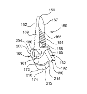
Figure 2A is perspective view of a nerve electrode, in accordance with
principles
of the present disclosure;
Figure 2B is a partial sectional view of an electrode contact, in accordance
with
principles of the present disclosure;
Figure 2C is a partial plan view of a contact portion of a nerve electrode, in
accordance with principles of the present disclosure;
Figure 3 is another perspective view of the nerve electrode of Figure 2A, in
accordance with principles of the present disclosure;
CA 02746660 2011-06-08
WO 2010/077494
PCT/US2009/065625
3
Figure 4 is front plan view of the nerve electrode of Figure 2A, in accordance
with
principles of the present disclosure;
Figure 5 is sectional view of the nerve electrode as taken along lines 5-5 of
Figure 3, in
accordance with principles of the present disclosure;
Figure 6 is a side plan view of the nerve electrode, in accordance with the
principles of the
present disclosure;
Figure 7 is a schematic illustration of a method of deploying the nerve
electrode, in
accordance with principles of the present disclosure;
Figure 8 is a flow diagram of a method of monitoring a nerve, in accordance
with
principles of the present disclosure;
Figure 9 is a perspective view of a nerve electrode, in accordance with
principles of the
present disclosure;
Figure 10 is a side plan view of a nerve electrode, in accordance with
principles of the
present disclosure;
Figure 11 is partial sectional view of the nerve electrode of Figures 9-10, in
accordance
with principles of the present disclosure;
Figure 12 is a perspective view of a nerve electrode, in accordance with
principles of the
present disclosure;
Figure 13 is a sectional view as taken along lines 13-13 of Figure 12, in
accordance with
principles of the present disclosure; and
Figure 14 is a schematic illustration of a nerve electrode releasably engaging
nerve within
a sheath, in accordance with principles of the present disclosure.
CA 02746660 2011-06-08
WO 2010/077494
PCT/US2009/065625
4
Detailed Description
Embodiments of the present disclosure are directed to electrically monitoring
a nerve
during a surgical procedure on a target tissue that is in the vicinity of the
nerve. In general terms,
the method includes removably securing a cuff electrode about the nerve
adjacent to the target
tissue and then establishing a baseline neurogenic response by applying a
series of stimulation
signals to the nerve via the cuff electrode. In some embodiments, the
neurogenic response is
recorded (e.g., measured) at the innervated muscle via electromyography, while
in other
embodiments, the neurogenic response is recorded at the nerve as a direct
nerve potential. In yet
other embodiments, other known neuro-monitoring techniques are employed to
measure and
record the result of neurogenic stimulation or to measure and record a
response on a body tissue.
For example, in one non-limiting example, the neurogenic response is measured
and recorded via
chemical-based biometrics, such as tracking levels of gastric acid,
perspiration, or chlorides that
are indicate of whether or not a nerve is impaired. In another non-limiting
example, the potential
impairment of a nerve is monitored by measuring and recording the neurogenic
response via other
biometrics, such as monitoring rhythmic contraction of smooth muscles to move
contents through
the digestive tract (commonly referred to as peristalsis).
In one aspect, this baseline response generally corresponds to the state of
the nerve prior to
any potential impairment related to the surgical procedure. Accordingly, after
establishing this
baseline response pattern, the surgical procedure is performed on the target
tissue while
automatically stimulating (via the cuff electrode) the nerve with a
stimulation signal at periodic
intervals. Upon comparing a measured neurogenic response to a periodic
stimulation signal
relative to the baseline neurogenic response pattern, one can determine
whether the health of the
nerve is being impaired.
With this in mind, in some embodiments, the term neurogenic refers to a neural-
related
response or activity initiated by natural neural processes while in other
embodiments, the term
neurogenic refers to a neural-related response or activity initiated by an
external stimulus, such as,
but not limited to an evoked potential stimulus. In yet other embodiments, the
term neurogenic
refers to a neural-related response or activity caused by both a naturally
neural process and an
external stimulus. In some embodiments, the term nerve refers to neuro
structures in general or
some specific neuro structures, including (but not limited to) one or more of
an entire nerve, a
CA 02746660 2011-06-08
WO 2010/077494
PCT/US2009/065625
nerve fiber, multiple nerve fibers, an axon, spatial grouping of axons, or a
functional grouping of
axons within the nerve.
One non-limiting example of automatically monitoring a nerve during a surgical
procedure
includes monitoring a vagus nerve during a surgery of the head and/or neck.
For example, in
5 surgeries affecting the thyroid gland, one embodiment of the present
disclosure includes
removably securing a cuff electrode about a nerve, such as the vagus nerve or
its branches, like
the recurrent laryngeal nerve and superior laryngeal nerve. In particular, the
cuff electrode is
placed around the vagus nerve within the carotid sheath with the cuff
electrode located proximal
or adjacent (relative to the brain stem) to the distal site of the surgical
procedure (e.g., tumor
removal). In addition, in some embodiments, an EMG-based endotracheal tube
electrode or
another type of insertable measurement electrode(s) is removably inserted
adjacent the vocal
cords and/or other muscles innervated by the nerve to be monitored. In other
embodiments, a
monitoring electrode, such as a cuff electrode, is placed about the nerve at a
point spaced apart
from the location at which the stimulation is applied.
With the cuff electrode securely positioned about the nerve, the monitor
automatically
stimulates the nerve at periodic intervals (e.g., from less than one second to
greater than 60
seconds, and in-between) and the monitor tracks the neurogenic response. In
one aspect, the
surgeon can select the frequency of intervals at which the nerve is stimulated
and adjustment of
periodic intervals can be based on the urgency of the monitoring. A control of
the periodic
interval selection includes a slow stimulation rate (e.g., every 60 seconds or
less often), fast
stimulation rates (e.g., every second or more often), and intermediate rates
between the slow rate
and the fast rate. In one non-limiting example, faster stimulation rates are
used during surgical
periods in which there is a greater risk to neurologic structures and slower
stimulation rates are
used during surgical periods posing with less risk. With this arrangement,
stimulation to the nerve
is applied no more than necessary in order to avoid potential fatigue of the
nerve or muscle
In one aspect, possible nerve impairment (e.g., due to stretching or
manipulation) is
identified by one measured neurogenic response or a series of measured
neurogenic responses that
differ from a baseline neurogenic response pattern. These differences are
tracked and the surgeon
is automatically notified via graphical alarm information (e.g., trending
patterns, threshold, etc.)
and/or audible alarm information when a limit has been exceeded. In one
aspect, the audible
CA 02746660 2011-06-08
WO 2010/077494
PCT/US2009/065625
6
alarm comprises a graduated alarm in which a volume of the alarm is in
proportion to a level of
deviation of the measured neurogenic response from the baseline neurogenic
response pattern. Of
course, at any time the surgeon can choose to visually monitor the graphical
information even
when no alarm has been triggered. In one example, a decrease (or trend of
decreases) in
amplitude and/or increase in latency from the baseline response beyond a
predetermined or user-
defined limit (or criteria) may indicate deteriorating vagus nerve quality,
and upon providing
automatic notification to the surgeon enable the surgeon to take actions to
alleviate the nerve
impairment.
In some embodiments, a potential nerve impairment is automatically identified
by
observing a neurogenic response waveform without reference to a baseline
response pattern. In
these embodiments, an overall morphology pattern, synchrony pattern,
amplitude, or latency of
the neurogenic response includes recognizable irregularities indicative of
nerve impairment. For
example, in the context of a synchrony pattern, such irregularities are
observable as a response
waveform having many peaks or humps where one or few peaks or humps are
expected. In
another non-limiting example, other irregularities include several peaks or
humps having
substantially different peak values instead of substantially similar peak
values or instead of
substantially harmonious peak values. These disrupted synchrony waveform
patterns would be
indicative of a disorganized response by the various axons or motor units of
the nerve and
therefore indicative of nerve impairment. Accordingly, by recognizing certain
signatory patterns
indicative of nerve dysfunction, these embodiments can automatically identify
nerve impairment
without reference to a measured baseline response pattern on the monitored
nerve.
It is understood that embodiments of the present disclosure are not limited to
monitoring
the vagus nerve but apply to other cranial nerves, spinal nerves, or
peripheral nerves. This
monitoring can be applied to motor (Efferent) nerves, sensory (Afferent)
nerves, and/or mixed
nerve fiber situations for the somatic and autonomic nervous systems.
Moreover, while the
electrode is described above in the context of evoked potential monitoring of
nerves during a
surgical procedure, it is understood that in some embodiments, the electrodes
of the present
disclosure also are employable with implantable stimulators, to provide
therapies associated with
stimulating other target nerves, including but not limited to, the vagus
nerve.
CA 02746660 2011-06-08
WO 2010/077494
PCT/US2009/065625
7
In some embodiments, the cuff electrode employed in monitoring the nerve
comprises an
elongate body and a cuff portion. The cuff electrode is configured to be
removably secured to the
nerve to enable stable positioning of the cuff electrode during the surgical
procedure. In one
embodiment, the cuff portion includes a pair of generally curved fingers that
are slidably
engageable in a side-by-side relationship. In particular, the fingers are
configured to releasably
engage each other in a closed position to define a lumen that automatically
self adjusts to the
proper size to encircle the nerve. The cuff portion is also configured with a
hinge mechanism at a
base of the fingers such that application of a pressing or squeezing force on
a tab (relative to the
elongate body) adjacent the hinge portion causes the fingers to separate away
from each other
with their distal tips spaced apart, resulting in an open position of the cuff
portion. When in this
open position, cuff portion is readily mounted onto, or readily removed from,
the nerve.
In another embodiment, the nerve electrode comprises an elongate body and a
cuff
portion. In one aspect, the cuff portion includes a generally arcuate nerve
contact portion of the
elongate body and a single flexible, resilient arm that extends from the
elongate body. In an open
position, the arm is free to be slidably maneuvered underneath a nerve and
around the nerve so
that the nerve contact portion (of the elongate body) and a proximal portion
of the arm define a
lumen encircling the nerve. In a further aspect, a distal portion of the arm
is slidably advanced
into a recess of the electrode body to removably secure the proximal portion
of the arm in the
closed position relative to the nerve contact portion of the elongate body.
By removably securing a nerve electrode (of one of the embodiments of the
present
disclosure) relative to a target nerve and monitoring the ensuing neurogenic
response, a surgeon
can achieve and maintain a hands-free, automatic continuous (or substantially
continuous)
monitoring of the health and integrity of a nerve in a reliably consistent
manner during a surgical
procedure.
These embodiments, and other embodiments, are described more fully in
association with
Figures 1A-14.
A nerve monitoring system 10 is shown in Figure 1A, in accordance with
principles of the
present disclosure, and comprises a stimulation electrode 20, a response
electrode 30 and a
monitor 12 that includes at least a stimulation module 40 and a response
module 60. In general
terms, the stimulation module 40 of monitor 12 applies a stimulation signal to
nerve 22 via
CA 02746660 2015-06-16
8
stimulation electrode 20 while response module 60 of monitor 12 measures a
neurogenic response
signal at muscle 32 via measurement electrode 30 (or at nerve 22 via measuring
a direct action
potential with a second cuff electrode similar to and spaced apart from
electrode 20). The
response is communicated to the surgeon via a user interface 90 of the monitor
12. Accordingly,
by using monitor 12, a surgeon can inferentially determine the relative health
and function of a
nerve by stimulating that nerve and measuring a corresponding neurogenic
response at muscle 32
or at nerve 22.
With the above general construction of system 10 in mind, nerve stimulation
monitor 12 is
further described. In doing so, it is understood that the features and
components of the monitor 12
can be arranged in many different forms and groupings, and therefore monitor
12 is not strictly
limited to the particular arrangement or groupings of functions illustrated in
Figure 1A.
Nevertheless, in the illustrated embodiment, monitor 12 additionally comprises
a controller 50,
memory 52, and the previously mentioned user interface 90.
In one aspect, user interface 90 of monitor 12 comprises a graphical user
interface or other
display that provides electronic control touchpad features, and as such,
monitor 12 provides for
the simultaneous display and/or activation of the modules (e.g., stimulation
module 40, response
module 60, etc.), functions, and features of monitor 12 described in
association with Figure 1A.
In other embodiments, user interface 90 includes one or more thumbwheels,
buttons, or other
electromechanical control mechanisms for implementing one or more the
functions of the nerve
monitoring system 10. In some embodiments, system 10 includes a remote control
54 that is in
wired or wireless communication with monitor 12 and that enables a user to
control at least some
of the modules, functions, and/or features controllable normally via user
interface 90 but at a
distance spaced apart from monitor 12.
In some embodiments, user interface 90 includes a notify function 92 which
enables the
user to select a preferred format (e.g., graphical, audible, mixed) by which
they will receive
information about potential nerve impairment. In one aspect, the notify
function 92
communicates information according to one or more specific parameters tracked
via an
identification function 66 that will be described later in more detail in
association with response
module 60 of Figure 1A. In some embodiments, via visual function 96, the
notify function 92
provides graphical reports of trends in the parameters of a neurogenic
response signal to enable
CA 02746660 2011-06-08
WO 2010/077494
PCT/US2009/065625
9
the user (e.g., a surgeon) to identify whether potential nerve impairment is
increasing or
decreasing depending upon the particular action taken during the surgical
procedure. In some
embodiments, either apart from or in combination with visual function 96, user
interface 90
comprises an audio function 94 configured to provide audible alerts to one or
more different
reports provided by the monitor 12. Among other reporting functions, the audio
function 94
provides an audible alert when response module 60 has identified potential
impairment of the
nerve being monitored. In one embodiment, based on the measured neurogenic
response, the
audio function 94 provides a faster rate or higher volume of audible sounds to
indicate increased
potential for impairment of the nerve being monitored and a lower rate or
lower volume of
audible sounds to indicate decreased potential for impairment of the nerve
being monitored. In
this way, notify function 92 of monitor 12 provides direct, ongoing feedback
to the surgeon on
whether their current course of actions are improving or impairing the health
of the nerve.
In one aspect, the audio function 94 provides information distinct, and
independent from, a
conventional acoustic feedback signal reported via electromyography. In other
embodiments, this
acoustic feedback signal is made selectively available via audio function 94
in addition to the
types of automatic audio or graphical notification previously described above.
In one embodiment, controller 50 comprises one or more processing units and
associated
memories configured to generate control signals directing the operation of
monitor 12 of system
10. In particular, in response to or based upon commands received via user
interface 90 and/or
instructions contained in the memory 52 associated with controller 50,
controller 50 generates
control signals directing operation of stimulation module 40 and/or response
module 60.
For purposes of this application, in reference to the controller 50 the term
"processing
unit" shall mean a presently developed or future developed processing unit
that executes
sequences of instructions contained in a memory. Execution of the sequences of
instructions
causes the processing unit to perform steps such as generating control
signals. The instructions
may be loaded in a random access memory (RAM) for execution by the processing
unit from a
read only memory (ROM), a mass storage device, or some other persistent
storage, as represented
by memory 52. In other embodiments, hard wired circuitry may be used in place
of or in
combination with software instructions to implement the functions described.
For example,
controller 50 may be embodied as part of one or more application-specific
integrated circuits
CA 02746660 2014-03-25
(ASICs). Unless otherwise specifically noted, the controller is not limited to
any specific
combination of hardware circuitry and software, nor limited to any particular
source for the
instructions executed by the processing unit.
= In one embodiment, monitor 12 includes at least substantially the same
features and
attributes as the nerve integrity monitor (NIM) described and illustrated in
assignee's U.S. Patent
6,334,068, titled 1NTRAOPERATIVE NEUROELECTROPHYSIOLOGICAL MONITOR.
= Referring again to Figure 1A, stimulation module 40 of monitor 12
includes a frequency
function 42, an amplitude function 44, a pulse width function 45, and an
electrode function 46. In
= one aspect, the frequency function 42, amplitude function 44, and pulse
width function 45 enable
user selection and tracking of the frequency, the amplitude, and the pulse
width, respectively, of a
stimulation signal. In another aspect, the electrode function 46 enables user
selection and tracking
of stimulation of nerve 22 via nerve electrode 20. In one embodiment, nerve
electrode 20
comprises a cuff-type electrode, as schematically illustrated in Figure 1A.
More specific
embodiments ofncrve electrode 20 are described and illustrated in more detail
in association with
Figures 2-7 and 9-14.
As illustrated in Figure IA, response module 60 of monitor 12 includes one or
more of an
amplitude function 62, a latency function 64, an other response parameter
function 65, an
identification function 66, a baseline function 68, an RF input function 69,
an EMG function 70, a
direct nerve measurement function 71, an electrode function 72, a chemical-
based biometric
function 73, a tissue-based biometrics function 74, and an impairment sorter
75.
In one aspect, the EMG function 70 enables user control over measuring the
response of
the muscle via electromyography. In another aspect, via direct function 71,
responses are
= measured at the stimulated nerve as a direct action potential. In
cooperation with the EMG
function 70, the electrode function 72 controls measuring response of muscle
32 via measurement
electrode 30. In one embodiment, measurement electrode 30 comprises a typical
EMG electrode
(e.g., an endotracheal tube electrode), which is schematically illustrated in
Figure lA via dashed
lines 30. In one aspect, in cooperation with the EMG function 70, the
amplitude function 62 and
latency function 64 enable tracking of the amplitude and the latency,
respectively, of the response
signal measured at muscle 32 via EMG function 70.
CA 02746660 2011-06-08
WO 2010/077494
PCT/US2009/065625
11
In some embodiments, monitor 12 includes RF input function 69, which in
general terms,
is configured to receive radiofrequency input associated with a monopolar or
bipolar
electrocautery device used in the surgical procedure adjacent the monitored
nerve. During the
surgical procedure, the electrocautery device can indirectly damage adjacent
nerves via local
heating effects. In addition, direct electrocautery will sever and destroy
tissue. Accordingly,
variations in the degree of heating of the adjacent nerves can cause various
levels of nerve injury
as the electrocautery device contacts its target tissue. Therefore, tracking
when an electrocautery
device is being used is helpful in determining whether impairment of the nerve
is caused by
electrocautery of tissue adjacent the monitored nerve. If the electrocautery
device is determined
to be the likely cause of the impairment, then the surgeon can modify their
procedure to avoid
further impairment to the nerve.
With this in mind, as the electrocautery device is operated it emits
radiofrequency signals
which can be tracked and are indicative of when and how the electrocautery
device is being used.
Accordingly, in this one embodiment, RF input function 69 receives RF signals
associated with
activity of the electrocautery device. In some embodiments, the RF signals are
obtained via a
muting detector feature of monitor 12 when monitor 12 includes one or more
features and
attributes of a monitor having substantially the same features and attributes
as the previously
identified U.S. Patent 6, 334,068. In this example, the muting detector
mechanism is inductively
clamped to an electrocautery probe and therefore the muting detector mechanism
captures an RF
signal representing the activity of the electrocautery device. In this way,
the RF signal associated
with the activity of the electrocautery device is provided to RF input
function 69 to monitor 12.
With the availability of the RF signal via RF input function 69, monitor 12
substantially
continuously checks to see if a detected impairment to the monitored nerve is
occurring
synchronously with (i.e., at the same time as) heightened activity of the
electrocautery device
when the electrocautery device is near the nerve. Accordingly, at the same
time that RF input
function 69 is tracking the electrocautery activity, other mechanisms
described herein for
measuring a neurogenic response (to an evoked potential or stimulation signal)
are used to detect
whether an impairment is occurring. For example, in some embodiments, the
impairment is
detected by measuring a neurogenic response at an innervated muscle via
electromyography, at
the nerve as a direct nerve potential, via chemical-based biometrics, or via
smooth muscle
monitoring, as further described herein in association with Figure 1A.
CA 02746660 2011-06-08
WO 2010/077494
PCT/US2009/065625
12
Consequently, using both the RF input function 69 and detected impairments,
monitor 12
determines whether or not a given impairment is likely being caused by an
electrocautery device.
In some embodiments, instead of capturing RF signals via the muting detector,
the RF
signals are obtained via other patient leads connected to monitor 12 that are
suitable for picking
up RF signals and generally tracking activity of the electrocautery device.
A further description of identifying nerve impairment caused by electrocautery
activity is
described later in more detail in association with verbal function 98 of
Figure lA and in
association with assessment module 110 of Figure 1D.
However, prior to measuring a neurogenic response of the target nerve during a
surgical
procedure, a user employs the baseline function 68 of the response module 60
to determine a
baseline neurogenic response pattern via measurements taken at the innervated
muscle 32 or at the
nerve 22 upon stimulating nerve 22. In other words, before attempting to
determine whether the
integrity of the target nerve is being impaired, the baseline function 68 is
employed to determine
the response signal or pattern (via amplitude function 62, latency function
64, or other parameters
further described later in association with other response function 65) that
normally occurs in the
absence of a potential nerve impingement during a surgical procedure.
In some embodiments, the response module 60 employs identification function 66
of the
response module 60 and notify function 92 of user interface 90 to enable the
monitor 12 to
automatically notify the user when a parameter (e.g., amplitude) of the
measured response signal
differs from a predetermined limit, such as preset percentage of the baseline
response signal (e.g.,
25%, 50%, 75%) or some other user defined setting, criteria, or value.
For example, in some embodiments, the identification function 66 tracks and
identifies
changes in parameters of the measured response signal relative to the baseline
response pattern.
These changes in parameters tracked via the identification function 66
include, but are not limited
to one or more of: (1) one or more decreases in amplitude; (2) one or more
increases in latency; or
(3) a decrease in an amplitude-based energy (i.e., the area of) of the
measured response curve.
In further reference to Figure 1, in some embodiments, response module 60 also
includes
the chemical-based biometrics function 73 configured to measure a neurogenic
response (in
response to stimulation of a target nerve) via chemical-based biometrics, such
as tracking levels of
CA 02746660 2011-06-08
WO 2010/077494
PCT/US2009/065625
13
gastric acid, perspiration, or chlorides that are indicate of whether or not a
particular nerve is
impaired. In some embodiments, response module 60 also includes the tissue-
based or smooth
muscle-based biometrics function 74 configured to measure a neurogenic
response (in response to
stimulation of a target nerve) via tissue-based biometrics (or smooth muscle
based biometrics),
such as monitoring rhythmic contraction of smooth muscles to move contents
through the
digestive tract (commonly referred to as peristalsis).
In further reference to Figure 1, in some embodiments, the identification
function 66
tracks and identifies changes in parameters of the measured response signal
(relative to the
baseline response pattern) according to the other response parameter function
65, separately from
or in combination with amplitude function 62, latency function 64, and/or an
energy parameter (as
part of the amplitude function 62). For example, as schematically illustrated
in Figure 1B, these
changes in parameters tracked via the identification function 66 include, but
are not limited to one
or more of: (1) a nerve refractory recovery parameter 77 configured to
identify one or more
changes in a nerve recovery refractory waveform (as explained in more detail
below); (2) a nerve
conduction velocity parameter 76 configured to identify one or more changes in
a nerve
conduction velocity function; (3) a nerve stimulation threshold parameter 78
configured to
identify one or more changes in a nerve stimulation threshold (e.g., the
amount of stimulation at
which the nerve begins to produce an observable neurogenic response); or (4) a
nerve stimulation
saturation parameter 79 configured to identify one or more changes in a nerve
stimulation
saturation threshold (e.g., the point at which the nerve response signal does
not further increase
with further increased levels of stimulation).
In some embodiments, the nerve refractory recovery parameter 77 identifies a
potential
nerve impairment by monitoring a response of the nerve to a paired stimuli
(also know as a paired
difference stimulus or a t-test stimulus) which applies a pair of identical
stimulus signals to an
axon (or a group of axons defining a nerve) separated by a fixed time delay.
In one aspect, this
monitoring method is used to provide increased sensitivity in measuring
neurogenic response
properties because of neuronal injury.
In some embodiments, monitoring a neurogenic response to such paired stimuli
protocols
includes observing or measuring changes in at least one of an overall response
waveform
morphology 77A, a synchrony waveform pattern 77B, an double response time 77C
(e.g., the time
CA 02746660 2011-06-08
WO 2010/077494
PCT/US2009/065625
14
between consecutive responses), an amplitude 77D, or a latency 77E of the
response to the
second stimulus by itself and/or relative to the response to the first
stimulus. In some
embodiments, this method includes applying a series of paired stimuli in which
the initial time
delay (between the first stimulus pulse and the second stimulus pulse) is
equal to or greater than
the natural refractory recovery period (the time taken for the nerve to fully
recovery before a
second stimulus is applied). Thereafter, the monitoring of the nerve is
performed continually as
the time delay between the consecutive first and second stimuli is gradually
decreased (in each
successive application of the pair stimuli) to be less than the natural
refractory recovery period.
By driving the time delay to lower and lower values, the monitor 12 can
determine the health of
the nerve based on how the nerve responds to the decreasing time delay between
consecutive
pulses.
In one aspect, in the context of applying a paired stimuli, the overall
response waveform
morphology 77A illustrates and identifies the extent to which some form of
nerve impairment has
occurred or is occurring based on one or more portions (e.g., response pulse
width, response pulse
peak, rate of increase to pulse peak, multiple peaks, absence of significant
peak, etc.) of the
waveform morphology substantially differing from a known response waveform
pattern for that
type of nerve. Upon recognizing this altered or abnormal morphology, the
refractory recovery
parameter 77 indicates the likelihood of nerve impairment.
In another aspect, in the context of applying a paired stimuli, the synchrony
waveform
pattern 77B illustrates and identifies the extent to which the axons or motor
units of a nerve
respond together in an organized manner or synergistic fashion. In other
words, in the absence of
nerve impairment, the waveform of the neurogenic response will have a
recognizable pattern that
corresponds to normal nerve function, as would be recognized by those skilled
in the art.
However, when the nerve is impaired, the axons of the nerve will respond in a
disorganized
manner (e.g., a dissynchronous manner), producing substantial irregularities
indicative of the
various axons responding separately from each other, with some axons not
responding at all, some
axons responding with a weaker response signal, some axons responding at the
wrong time, etc.
Accordingly, the synchrony waveform pattern 77B is configured to indicate
nerve impairment via
automatically recognizing at least a portion of a neurogenic response pattern
that includes
multiple perturbations or erratic characteristics (e.g., many smaller humps
instead of a single
CA 02746660 2011-06-08
WO 2010/077494
PCT/US2009/065625
integrated hump) where a generally smooth or predictable waveform would
otherwise be
expected.
In some embodiments, operation of the nerve refractory recovery function 77
includes
monitoring changes in the refractory recovery period on a segmented basis. In
other words,
5 consecutive segments within a single neurogenic response waveform are
compared with each
other to observe changes in waveform morphology, synchrony waveform patterns,
amplitude, or
latency from segment-to-segment that would be indicative of nerve impairment.
In some embodiments, the nerve refractory recovery parameter 77 is configured
to
perform a comparison of the neurogenic response to the first stimulus relative
to the neurogenic
10 response to the second stimulus of the paired stimuli (having a fixed
time delay between the
consecutive stimulation pulses), as represented by paired difference parameter
77F. In this
comparison, an algebraic subtraction is performed in which the second response
waveform (i.e.,
the response to the second stimulus) is inverted relative to the first
response waveform ( i.e., the
response to the first stimulus) and then a subtraction is performed of
corresponding data points of
15 the second response waveform from the first response waveform. When
little or no difference is
observed based on this algebraic subtraction, then there is little or no
likelihood of potential nerve
impairment. However, if the comparison via the algebraic subtraction results
in a one or more
large observed differences or in many smaller observable substantial
differences, then there is a
likelihood of potential nerve impairment. Accordingly, this comparison
provides a derived
response pattern and may be referred to as a paired-difference-response (PDR).
In one aspect, changes in neuronal response observed according to operation of
the nerve
refractory recovery parameter 77 as described above provide feedback
information to the surgeon
to indicate that one or more types of nerve impairment is occurring. These
types of impairment
include, but are not limited to, compression, traction (i.e., tension), heat
injury, or a composite
impairment. In one aspect, the type or degree of impairment is recognized via
the observed
changes in the morphology waveform, synchrony waveform pattern, amplitude,
latency, or
elapsed time (as described above), wherein the observed changes are associated
the various sub-
populations of axons arranged concentrically within a diameter of the nerve
and/or the degree of
myelinization of the axonal elements.
CA 02746660 2011-06-08
WO 2010/077494
PCT/US2009/065625
16
In one aspect, these other response parameters 76-79 associated with function
65 provide
the capability to detect more subtle changes in a neurogenic response (that
might not otherwise be
recognized via tracking more conventional response parameters), which in turn,
may detect the
development for potential nerve impairment long before it becomes readily
apparent via
conventional monitoring of nerve integrity during a surgical procedure. For
example, in another
aspect, these other response parameters (according to other response parameter
function 65)
provide more discriminating information that would otherwise be available via
conventional
acoustic feedback from an innervated muscle, and thereby enable quicker and
more effective
detection of potential nerve impairment. In some embodiments, the baseline
response pattern
tracked via baseline function 68 is based on (or derived from) one or more of
the following
screening parameters of neurogenic responses (measured in the absence of
potential impingement)
according to an exclusion function 80 as schematically illustrated in Figure
1C. These parameters
include, but are not limited to: (1) a variability parameter 81 configured to
apply a selective
exclusion of some responses of multiple evoked neurogenic responses based on a
degree of
variability of the multiple responses; (2) a maximum/minimum parameter 82
configured to apply
a selective exclusion of a maximum value and/or a minimum value of multiple
evoked neurogenic
responses; or (3) a non-evoked parameter 83 configured to apply a selective
exclusion of artifacts,
such as any non-evoked neurogenic responses or other artifacts not indicative
of an evoked
neurogenic response.
In some embodiments, the baseline response pattern tracked via baseline
function 68 is
based on (or derived from) one or more of the following screening parameters
of neurogenic
responses (measured in the absence of potential impingement) according to an
inclusion function
84 as schematically illustrated in Figure 1C. These parameters include, but
are not limited to: (1)
a single response parameter 85 configured to enable selective use of a single
evoked response or
of multiple evoked responses; (2) a statistical mean parameter 86 configured
to use a statistical
mean of multiple evoked neurogenic responses; (3) a variance measuring
parameter 87 configured
to use variance measuring (e.g., standard deviation) of multiple evoked
neurogenic responses; (4)
a rate change parameter 88 configured to use a rate of change of a series of
evoked neurogenic
responses; or (5) a rolling window parameter 89 configured to use a continuous
sequence (or
rolling window) of evoked neurogenic responses. In one aspect, the rolling
window parameter 89
monitors a generally constant number of evoked neurogenic responses (e.g., 5,
10, or 15) and
continually adds one or more new responses to the set or window while removing
the oldest one
CA 02746660 2011-06-08
WO 2010/077494
PCT/US2009/065625
17
or more responses from the set or window. In this manner, the most recent set
(e.g., 5, 10, or 15)
of responses are always in the monitoring window. In some embodiments, the
monitoring
window includes responses in series to help observe trends, while in other
embodiments, the
monitoring window includes an average of the responses in the window, which is
more akin to a
rolling average.
In some embodiments, one or more parameters of the baseline function 68 are
identified
via a Poisson distribution, as further described later in association with
tools module in Figure 1D.
In one aspect, these screening parameters of baseline response pattern
function 68 are used
to establish a baseline response pattern that is more indicative of a typical
baseline neurogenic
response than would otherwise be ascertained without the sorting process
enabled via one or more
of the identified screening parameters. In other words, these screening
parameters help to ensure
that a legitimate difference of the measured response signal (relative to a
baseline response
pattern) is identified because the screening parameters enable removing
components from the
baseline response pattern that are atypical within a sample of multiple evoked
responses.
Referring again to Figure 1A and keeping in mind the parameters tracked via
the baseline
function 68 and via the identification function 66, in one example, the
identification function 66 is
used to set an alarm limit relative to the baseline response pattern. In this
arrangement, an
amplitude of the measured response signal (during the surgical procedure) that
is less than the
alarm limit would trigger a notification of potential nerve impairment via
notify function 92.
Likewise, in another example, the identification function 66 is used to set a
latency limit relative
to the baseline response signal or pattern such that a latency of the measured
response signal
(during the surgical procedure) that exceeds the latency limit would trigger a
notification of
potential nerve impairment via notify function 92. In still other examples,
similar limits are
arranged to trigger the notify function 92 based on a limit (e.g., criteria,
threshold, value) set
according to any one or more of the previously identified parameters of the
identification function
66.
In one aspect, this notification is communicated to the user via user
interface 90
graphically via visual function 96 and/or audibly via audio function 94 of
user interface 90, as
previously described. In some embodiments, the audio function 94 comprises a
tone function 97
and/or a verbal function 98. As just one example, audio function 94 of monitor
12 enables a
CA 02746660 2015-06-16
18
surgeon to be notified of potential impingement of a target nerve without
requiring the surgeon to
look away from their procedure. This audible notification signal provides an
immediate "no-
look" feedback to the surgeon, thereby enhancing their concentration on the
surgical procedure
instead of being distracted with conventional techniques of monitoring a
nerve. Moreover,
because the electrode 20 is secured about nerve 22, the visual function 96 or
the audio function 94
of the notify function 96 enable the surgeon to monitor the target nerve in a
hands-free manner,
thereby further enhancing their freedom to carry out the main procedure on the
target tissue. In
some embodiments, the alarm provided via the tone function 97 (of the audio
function 94) is
configured to emit several different types of tones such that each different
type of tone
corresponds to a relative degree of deviation of the measured neurogenic
response from the
baseline neurogenic response pattern. In other words, different tones
represent different amounts
of deviation from the baseline neurogenic pattern.
In some embodiments, audio function 94 includes verbal function 98 which is
configured
to provide a notification in the form of a verbal expression, such as the
known words, to the
surgeon to inform them of the condition of the nerve, such as "normal",
"impairment", etc. In
some embodiments, this verbal function 98 is configured to audibly identify
the type of
impairment that is occurring through the use of words such as "tension",
"compression", etc. In
some embodiments, the verbal function 98 is configured to identify the
intensity of impairment
through the use of words such as "low", "moderate", and "severe". Operation of
the verbal
function 98 is later described in more detail in cooperation with an
impairment sorter 75 that is
illustrated and described in association with Figure 1D.
In further reference to Figure 1A, in some embodiments, the alarms provided
via the audio
function 94 or the visual function 96 comprise a graduated alarm function 99
in which a volume
of the alarm (audible or graphical) is in proportion to a degree of deviation
of the measured
neurogenic response from the baseline neurogenic response pattern.
In some embodiments, the response module 60 includes an impairment sorter 75,
which is
further illustrated in Figure 1D. As shown in Figure 1D, impairment sorter 75
includes an
assessment module 102 and a report module 104.
In general terms, the report module 104 operates in cooperation with the
notify function 92
of user interface 90 and is configured to report the condition of the
monitored nerve to the
CA 02746660 2015-06-16
19
surgeon. In some embodiments, the report module 104 includes a type function
124 and an
intensity function 125. In general terms, the type function 124 indicates the
type of damage
identified via the differentiator function 112, such as whether the nerve is
experiencing minor
irritation, tension, compression, a composite impairment of both tension and
compression, or
impairment directly or indirectly caused by electrocautery (as previously
described in association
with RF input function 69 in Figure 1A).
Accordingly, when there is some impairment of the monitored nerve, then the
type of
impairment is communicated via verbal function 98 as one or more verbal
expressions (e.g.,
words like tension, compression, etc.) in real-time to the surgeon during
surgery.
In general terms, the intensity module 125 of the report module 104 is
configured to
provide an indication (via verbal function 98 of notify function 92 in Figure
1) to the surgeon of
the relative intensity of the impairment of the nerve. In one embodiment, the
intensity module
125 includes a low function 126, a moderate function 127, and a severe
function 128.
Accordingly, when there is some impairment of the monitored nerve, then any
such impairment is
communicated via verbal function 98 as a verbal expression in real-time to the
surgeon during
surgery. In one aspect, such verbal expressions include, but are not limited
to, the words low,
moderate, or severe or other similar meaning words that indicate a relative
degree of intensity.
Further, in some embodiments, intensity function 125 provides and communicates
at least two
different levels of intensity.
In some embodiments, the assessment module 102 of impairment sorter 75
includes a
tools module 110 and a differentiator module 112. In general terms, the tools
module 110 is
configured to apply different forms of statistical analysis ancUor other
filters to sort data of one or
more measured neurogenic responses. In cooperation with differentiator 112,
the tools module
110 removes noise while transforming the data to more accurately identify
changes in nerve
function with such changes including changes in amplitude, latency, or other
enumerated aspects
of nerve function previously described in association with identification
function 66 in Figure I.
Recognition of these changes from response-to-response or over time through
multiple responses
provides an indication of the type or extent of impainnent to a nerve.
In one embodiment, the tools module 110 includes a distribution function 114,
a
correlation function 116, a wavelet function 117, and a Fourier function 118.
CA 02746660 2011-06-08
WO 2010/077494
PCT/US2009/065625
In one embodiment, the distribution function 114 is configured to recognize
which type of
statistical distribution that best characterizes neurogenic responses (for
example, EMG responses)
resulting from stimulation pulses. In one example, the neurogenic responses
fit best within a
Poisson distribution and therefore observations regarding the neurogenic
response information is
5 calculated from the Poisson distribution. However, other distributions
are not excluded. In a few
non-limiting examples, by using the Poisson distribution the mean of the
received data provides a
measure of the average delay while a standard deviation provides a measure the
degree to which
the responses are erratic. As another example, changes in the delay and signal
spread recognized
in the distribution are indicative of possible nerve impairment. In another
aspect, the Poisson
10 distribution is used to disregard some data as spontaneous activity. For
example, this distribution
can be used to disregard EMG responses appearing at a far end a lower tail of
the Poisson
distribution or appearing at a far end of an upper tail of the distribution
because there is a very low
probability that such responses are truly indicative of the condition of the
nerve.
In some embodiments, this distribution tool 114 is used in cooperation with or
as part of
15 baseline function 68 of response module 60, as previously described in
association with Figure
1A.
The other functions of tools module 110 generally relates to classifying
different features
of the measured neurogenic response signals with such functions including but
not limited to a
correlation function 116, a wavelet function 117, and a Fourier function 118.
In general terms,
20 the spreading or narrowing of the EMG response as well as the response
amplitude and overall
shape of the EMG response is used to identify a damaged or stressed nerve.
Further, these
methods of classification provided via tools module 110 are used to classify
the response
waveform into different categories that identify the type and/or extent of the
impairment. In one
aspect, these methods are used to augment the current methods or employed as a
separate method
of classifying aspects of the responses to indicate the extent or type of
nerve impairment.
In some embodiments, the correlation function 116 is configured to provide
auto-
correlation and or cross-correlation techniques are used to identify the EMG
response waveform
as a recognizable stimulated response so that other aspects of a response
signal not following such
patterns can be ignored. In one aspect, the received data of neurogenic
responses is correlated
relative to stored response waveforms of different types to classify the
response. In one non-
CA 02746660 2011-06-08
WO 2010/077494
PCT/US2009/065625
21
limiting example, a first stored response waveform is indicative of
compression on a nerve while
a second stored response waveform is indicative of excess tension on the
nerve. When a
waveform in the received data matches one of these respective first or second
stored response
waveforms, then the correlation function 116 provides an indication of whether
the impairment on
the nerve is compression or tension. In some embodiments, the neurogenic
response waveform is
also correlated relative to a baseline response pattern of the target nerve to
evaluate changes in the
response of the nerve compared to the responses occurring prior to surgery.
In some embodiments, the wavelet function 117 provides another mechanism to
classify
the response data to recognize patterns indicative of a type or extent of
nerve impairment.
Likewise in some embodiments, a Fourier analysis is applied via Fourier
function 118 to the
response data to identify the frequency content of the signals to enhance the
identification of
changes to the nerve function and/or recognize changes over time. One example
of the
application of the Fourier function 118 is later described in more detail in
association with Figure
lE and Figure 1F.
In general terms, the differentiator 112 further sorts the results obtained
from tools
module 110 to place the measured neurogenic responses into different
categories that
communicate to the surgeon the type of ongoing trauma to the monitored nerve.
In one
embodiment, the differentiator 112 includes an irritation parameter 120, a
tension parameter 121,
a compression parameter 122, a composite parameter 123, or an electrocautery
parameter 129.
In some embodiments, differentiator 112 also assists in identifying or
differentiating the
size of nerve fibers affected by the nerve impairment. For example, the
response latency is used
to differentiate surgical damage according to the size of the nerve fibers.
In particular, the nerve conduction velocity of the stimulated response
propagation is
related to the diameter of the axons of the nerve and to the presence or
absence, or condition of
the myelin sheath. For example, increased nerve conduction velocities are
associated with the
presence of a myelin sheath and associated with larger nerve axons, resulting
in a relatively
shorter response latency. In one aspect, damage to the myelin sheath will
decrease the conduction
velocity, and increase the response latency. In another aspect, by tracking
the response latency,
one can differentiate the surgical damage relative to the size of the nerve
fibers. For example,
larger axons will move the signal faster, and thereby produce the shortest
latency. Another
CA 02746660 2011-06-08
WO 2010/077494
PCT/US2009/065625
22
observable feature includes a larger electromyography response for larger
axons which innervates
neuromuscular junctions and therefore activates a greater number of motor
nerve units.
With this in mind, the irritation parameter 120 identifies a general
irritation to the
monitored nerve caused by minor tension and is detected by an increase in the
response latency
and an increase in the evoked response amplitude. The tension parameter 121
identifies
impairment by excessive tension and is detected by an increase in response
latency and a decrease
in the evoked response amplitude. In particular, this excessive tension
typically damages the
myelin sheath thereby increasing the response latency while the decrease in
amplitude is caused
by damage to the large axons of the nerve.
The compression parameter 122 identifies impairment by excess compression on
the
nerve and is detected by a decrease in evoked response amplitude without a
substantial change in
latency. In particular, this compression is associated with damage to the
nerve which results in
activation of a decreased number of motor units resulting in the decrease in
measured amplitude.
Because this compression generally does not significantly affect the myelin
sheath over a
significant distance, there is no major change in latency.
The composite parameter 123 identifies impairment by more than one type of
impairment,
such as both compression and tension.
In some embodiments, the electrocautery parameter 129 identifies impairment at
least
partially caused by an electrocautery event impacting the nerve and is
detected via an occurrence
of one of the previously described types of nerve impairment simultaneous with
or synchronously
an electrocautery event or activity during the surgical procedure. For
example, electrocautery
parameter 129 of differentiator function 112 substantially continuous monitors
an RF signal for
electrocautery event waveforms via RF input function 69 of monitor 12, as
previously described
in association with Figure 1A. When one of the types of impairment
(irritation, tension,
compression, composite) is separately identified via the measured neurogenic
response signals,
the electrocautery parameter 129 of differentiator 112 checks to see if the
identified impairment
occurred synchronously with (at the same time as) an electrocautery event or
recognizable
electrocautery activity. If so, electrocautery parameter 129 indicates that an
electrocautery
impairment likely has occurred. This information can guide the surgeon to
modify their surgical
procedure to avoid any further impact to the nerve during use of the
electrocautery device.
CA 02746660 2011-06-08
WO 2010/077494
PCT/US2009/065625
23
Accordingly, in cooperation with the verbal function 98 of notify function 92
(Figure 1A),
differentiator 112 provides a real-time audible indication as a verbal
expression to the surgeon of
the type of impairment occurring on a monitored nerve, such as an irritation,
tension,
compression, composite, or electrocautery impairment. Upon hearing such
notification, the
surgeon can immediately modify or adjust their technique to reduce and/or
avoid further
impairment to be monitored nerve situated adjacent to their primary surgical
target. However, it
is understood that other verbal expressions (i.e. words other than irritation,
tension, compression,
composite, or electrocautery) are selectable or programmable to be audibly
communicated to
represent the underlying respective general irritation, tension impairment,
compression
impairment, composite impairment, or electrocautery impairment.
In this way, assessment module 102 and report module 104 of impairment sorter
75 further
enable the hands-free and watch-free monitoring of a nerve during surgery.
Figure lE provides a series of graphs 130, 132, 134, 136 that schematically
illustrate, in
both the time domain and the frequency domain, an electromyography (EMG)
response as a
baseline response pattern and as a response signal after injury of the
monitored nerve. In general
terms, by applying a fast Fourier transform to this signal information, one
can accurately identify
the change in the function of the nerve due to impairment while excluding data
that is not
indicative of this change. With this in mind, graph 130 illustrates a baseline
EMG response via
signal 131A that has a peak amplitude 131B while graph 132 illustrates a
baseline EMG response
after application of a Fourier transform. As illustrated in graph 132, the
transformed signal 133A
includes a first peak 133B, a second peak 133C, and a third peak 133D. The
first peak 133B
indicates a response amplitude of about 13 while the other peaks 133C, 133D
illustrate
significantly lower amplitudes in the frequency domain.
As illustrated and described above in association with Figure 1E, the Fourier
transform is
applied in a method of identifying one or more signal features of the baseline
response pattern
(such as, but not limited to, an amplitude) that are indicative of a condition
of a nerve.
Accordingly, this method includes, at least, comparing the baseline response
pattern as expressed
in the frequency domain relative to the same baseline response pattern as
expressed in the time
domain.
CA 02746660 2011-06-08
WO 2010/077494
PCT/US2009/065625
24
In comparison to graph 130, graph 134 illustrates an EMG response after or
during
impairment to the nerve. As shown in graph 134, response signal 135A includes
a peak 135B
having an amplitude significantly lower than that shown in graph 134 (i.e.,
the baseline response
of the monitored nerve). However to ensure that an accurate observation is
made regarding any
changes to condition of the nerve, a Fourier transform is applied to the
signal 135A (in graph 134)
which results in the signal 137A illustrated in graph 136. By observing the
response after or
during impairment in the frequency domain provided via graph 136, a single
peak 137B
corresponding to the response amplitude is clearly recognizable and
distinguished from other
aspects of the response signal. By comparing the transformed signal 137A in
graph 136 and the
transformed signal 133A in graph 132, the Fourier function 118 of tools module
110 identifies a
significant change in the response amplitude after injury. In particular,
graph 132 illustrates a
response amplitude of about 13 prior to injury while graph 136 illustrates
response amplitude of
about 5 after injury. Accordingly, by using the Fourier function 118, a clear
indication is
provided of the altered condition of the nerve as detected by a change in the
response amplitude to
a stimulation pulse.
As illustrated and described above in association with Figure 1E, the Fourier
transform is
applied to the measured neurogenic response signal in a method of identifying
one or more signal
features (such as, but not limited to, an amplitude) indicative of a condition
of a nerve.
Accordingly, this method includes, at least, comparing a measured neurogenic
response signal as
expressed in the frequency domain relative to the same measured neurogenic
response signal as
expressed in the time domain.
Figure 1F provides a further schematic illustration of application of Fourier
function 118
to an EMG response signal. In particular, Figure 1F provides graph 140 which
illustrates a
measured EMG response signal 141A after or during impairment to a nerve where
noise is present
in the signal. As shown in graph 140, signal 141A illustrates a first response
peak 141B and a
series of peaks 141C expected to be caused from noise. Meanwhile, graph 142
illustrates a signal
143A that represents the measured EMG response signal 141A of graph 140 after
application of a
fast Fourier transform via Fourier function 118. As shown in graph 142, the
neurogenic response
signal is clearly recognizable as peak 143B (based on its similarity to
amplitude waveforms of
prior neurogenic responses) whereas the noise when expressed in the frequency
domain does not
match the waveform of a response signal and is excludable from peak 143B. In
one aspect,
CA 02746660 2011-06-08
WO 2010/077494
PCT/US2009/065625
dashed lines N¨N represent a demarcation of the response signal waveform
(including peak
143B) from the noise appearing to the right of the dashed line and represented
by indicator 144.
As illustrated and described in association with Figure 1F, the Fourier
transform is applied
to the measured neurogenic response signal in a method of identifying one or
more signal features
5 (including, but not limited to, an amplitude) indicative of a condition
of a nerve. Accordingly,
this method includes analyzing the measured neurogenic response signal in the
frequency domain
to differentiate noise from the signal features of the measured neurogenic
response signal. In one
aspect, this differentiation is performed by recognizing the noise as having a
pattern in the
measured neurogenic response signal in the frequency domain that is
substantially different than a
10 pattern of the baseline response pattern in the frequency domain or
substantially different than a
pattern of one or more prior measured neurogenic response signals (in the
frequency domain)
without noise.
Accordingly, different classification tools and reporting tools as provided
via impairment
sorter 75 provide a useful mechanism to sort and evaluate one or more
neurogenic responses,
15 which in turn, enhances the ability to detect and classify different
types of impairment to a nerve.
Figures 2-7 are different views that illustrate a nerve electrode 150, in
accordance with
principles of the present disclosure that is usable to stimulate a nerve or
record a response at a
nerve. As illustrated in the perspective views of Figures 2 and 3, electrode
150 comprises an
elongate body 152 and a cuff portion 160. The nerve electrode 150 includes a
proximal end 156
20 and a distal end 157. In one aspect, the elongate body 152 includes a
distal portion 158 adjacent
the cuff portion 204 and extends from the distal portion 158 to the proximal
end 156 of the
electrode 150. In some embodiments, elongate body 152 comprises a ribbed
surface 154 and a
smooth surface 165 on each of two opposite faces 167 of elongate body 152. In
another aspect,
elongate body 152 includes opposite side edges 169, which are generally smooth
in some
25 embodiments.
In further reference to Figures 2-4, each respective face 167 has a width (W1
in Figure 4)
substantially greater than an average width (W2 in Figure 6) of each
respective side edges 169. In
another aspect, elongate body 152 has a length (L1) substantially greater than
an inner diameter
(D1) of cuff portion 160, as illustrated in Figure 5. In one non-limiting
example, the length Ll is
at least twice as large as, and up to ten times larger than, the inner
diameter D 1. In another aspect,
CA 02746660 2015-06-16
26
the width (W1) of face 167 of elongate body 152 (Figure 5) is substantially
greater than the width
(W2 in Figure 6) of side edge 169. In one non-limiting example, the width W1
is at least twice as
large as, and up ten times larger than, the width W2.
Accordingly, because the elongate body 152 is substantially longer than a
diameter of the
cuff portion 160 (and of the lumen 185) and has a substantial width (W1), the
elongate body 152
provides a strong support or anchor against which forceps can be used to move
tab 162 toward
and against the elongate body 152, as further described and illustrated later
in association with
Figure 7. In one aspect, the substantial width of the elongate body 152
provides an ample target
that the distal tips of the forceps can grasp while the substantial length
(L1) of the elongate body
provides better reach to facilitate advancing the cuff portion 160 about the
nerve 22. With these
features in mind, elongate body 152 is sometimes herein referred to as a beam
or trunk.
Referring again to Figures 2-4, the cuff portion 160 extends distally directly
from the
distal portion 158 of elongate body 152. In one embodiment, the cuff portion
160 includes a first
finger 172 and second finger 174 arranged in a side-by-side relationship
(Figures 2-5) and
extending from a base portion 177. As best seen in Figures 5-6, each finger
172, 174 defines a
generally arcuate cross-sectional shape. In one aspect, the base portion 177
defines a junction
between, and supports both of, the elongate body 152 and tab 162. As further
described later, the
elongate body 152 and tab 162 also act as a pair of group members of an
actuator mechanism while
the base portion 177 also functions as a hinge controllable by the actuator
mechanism to enable
rotational movement of first finger 172 and second finger 174 away from each
other, as illustrated
in Figure 7. In particular, the base portion 177 includes a central bending
region or central hinge
region (represented via dashed lines 178) approximately midway between the
trunk 152 and tab
162 such that trunk 152 is off-axis relative to this central hinge region 178.
Stated differently, the
central hinge region 178 is laterally offset relative to (i.e., not aligned
with) a longitudinal axis
(represented by line Z as shown in Figure 6) of trunk 152.
In some embodiments, each finger 172,174 comprises a generally circular cross-
sectional
shape (best seen in Figures 5-6) while in other embodiments, each finger
172,174 comprises a
generally elliptical cross-sectional shape. When in their at-rest, closed
configuration, the side-by-
side combination of the generally circular fingers 172, 174 define lumen 185
which is sized and
shaped to receive a nerve. Moreover, because the respective fingers 172, 174
are in this side-by-
CA 02746660 2011-06-08
WO 2010/077494
PCT/US2009/065625
27
side relationship along a length of the lumen (i.e., in a direction generally
parallel to a longitudinal
axis of the nerve that will extend through lumen 185), each finger 172, 174
independently defines
at least a portion of a length of the lumen 185.
Moreover, in one aspect, in combination with base hinge portion 177, each
respective
finger 172, 174 independently provides a substantially 360 degree coverage
that encompasses a
circumference of the target nerve. In another aspect, in combination with base
hinge portion 177,
a pair of fingers 172, 174 act together to effectively provide more than 360
degrees (and up to 570
degrees) of coverage that encompass the circumference of the target nerve to
the extent that the
distal portion of the respective fingers 172, 174 overlap in a side-by-side
fashion.
In yet another aspect, the independent 360 degree coverage encompassing the
nerve by the
separate fingers 172, 174 also provides an automatic mechanism for the nerve
electrode 150 to
self-adjust its size to different sized nerves while maintaining the electrode
contact 180 in direct
pressing contact against an outer surface of the nerve. This arrangement
contributes to the sealing
action of the lumen 185 against an outer surface of the nerve, thereby
preventing intrusion of
fluids or other matter into the nerve-electrode contact interface, which in
turn improves the
reliability and quality of the stimulation or recording signal.
In some embodiments, as best seen in Figure 6, with base portion 177 extending
at least
from trunk 152 to tab 162, first finger 172 has a radial length generally
equal to a radial length of
second finger 174 wherein a tip 214 of second finger 174 is represented by
dashed line 219.
However, it is understood that in other embodiments, the first fingers 172 has
a substantially
different length than second finger 174.
As best seen in Figures 2-4, first finger 172 includes a generally straight
outer edge 200
and a generally angled inner edge 202, which converge to form a curved
junction at a distal tip
204. Similarly, second finger 174 includes a generally straight outer edge 210
and a generally
angled inner edge 212, which converge to form a curved junction at a distal
tip 214. In one
aspect, as best seen in Figures 3-4, the generally angled inner edge 202 of
the first finger 172
forms a generally helical relationship relative to the generally angled first
edge 212 of the second
finger 174. Stated in other terms, the generally angled inner edge 202 of
first finger 172 and the
generally angled inner edge 212 of second finger 174 form complementary angles
relative to each
other (as represented by the complementary angles 0 and illustrated in Figure
4).
CA 02746660 2011-06-08
WO 2010/077494
PCT/US2009/065625
28
In one aspect, this complementary relationship between the inner edge 202 of
the first
finger 172 and the inner edge 212 of the second finger 174 enables the first
finger 172 and the
second finger 174 to be in releasable, slidable contact against each other in
a nested arrangement.
In one aspect, this nested arrangement enables the angled, side-by-side
fingers 172, 174 to
provide a more robust enclosure about a nerve than if the inner edges of the
fingers were simply
generally parallel to each other (along a line generally perpendicular to the
lumen or along a line
generally parallel to the lumen) or than if the distal ends of the fingers
simply contacted each
other in a conventional end-to-end closed relationship. In other words, the
angled, nested
relationship of the fingers 172, 174 results in releasable interlocking of the
fingers 172, 174
relative to each other, thereby helping to prevent possible dislodgement of
the nerve electrode 150
from becoming dislodged from the nerve about which it is removably secured.
Moreover, this
releasable interlocking feature of the fingers 172, 174 insures that the
electrode contact 180
remains in stable and close fitting contact against the nerve, thereby
contributing to accuracy and
consistency in applying a stimulation signal to the nerve via the nerve
electrode 150.
In another aspect, each finger 172,174 comprises a semi-flexible, generally
resilient
member. With this construction, the respective fingers 172, 174 generally
retain their generally
circular or generally arcuate shape in their closed position (shown in Figures
2-6) and in their
open position shown in Figure 7. On the other hand, as the fingers 172, 174
move from their
closed position to their open position shown in Figure 7, the base hinge
portion 177 (defining a
junction between elongate body 152 and tab 162) flexes considerably to permit
rotation of the tab
162 toward and against distal portion 158 of elongate body 152. Accordingly,
as shown in Figure
7, the hinge portion 177 generally straightens out to force the distal tips of
the respective fingers
172, 174 away from each other. However, as soon as the pressing action of the
forceps 194 is
removed, tab 162 automatically rotates back to its at-rest position (best seen
in Figures 2-3 and 5)
due to the resiliency of the base hinge portion 177 of cuff portion 160 and
thereby allows the
return of the fingers 172, 174 to their closed position. In one embodiment,
base hinge portion 177
comprises a generally resilient or elastic living hinge, as would be
understood by one skilled in
the art. With this in mind, beam 152 and tab 162 act as pair of oppositely
disposed grip members
of an actuator mechanism such that pressing action of the respective grip
members activates
bending of base hinge portion 177 to cause displacement of fingers 172, 174
away from each
other into the open position and release of these grip members reverses
bending of base hinge
portion 177 to cause fingers 172, 174 to once again releasably engage each
other in a side-by-side
CA 02746660 2015-06-16
29
manner. In some embodiments, in further reference to Figures 2-6, first finger
172 includes a
substantially larger base portion 215 (adjacent tab 162) than a base portion
205 (Figure 6) of
second finger 174. In one aspect, the large base portion 215 of first finger
172 extending from tab
162 provides a robust support structure to withstand the stress induced when
tab 162 is rotated
toward elongate body 152 (to move the fingers 172, 174 to their open position)
as shown in
Figure 7.
In some embodiments, the body of nerve electrode 150 is formed of a molded
elastomeric
material suitable to provide the elastic performance of the base hinge portion
177 of electrode
150. In one embodiment, electrode 150 is molded from a rubber material, from a
silicone
elastomeric material, or other elastomeric material.
In one aspect, as best seen in the sectional view of Figure 5, electrode lead
182 extends
through elongate body 152 with a distal portion ,183 of the electrode lead 182
including an electrode
contact 180 exposed at surface 161 of cuff portion 160. It is also understood
the nerve electrode
150 generally includes lead 182 and that the lead 182 is omitted from Figures
2-4 and 6-7 merely
for illustrative clarity. Referring again to Figure 5, a proximal portion of
lead 182 extends
outwardly from the proximal end 156 of the body 152 for electrical connection
to, and electrical
communication with, the monitor 12. In one aspect, the electrode contact 180
is a generally
circular shaped member of electrically conductive material and is in general
alignment with a
longitudinal axis of beam 152 and lead 182 extending through beam 152. In one
embodiment, the
beam 152 and a lead 182 extend and a substantially single direction throughout
an entire length of
the beam 152.
In some embodiments, the electrode contact 180 includes a generally circular
shape
defining a first area and the contact portion 161 of the electrode 150
surrounding that the
electrode contact 180 defines a second area that is substantially larger than
the first area. In
combination with the gripping action of the fingers 172,174 (which maintains
the electrode
contact 180 in pressing contact against an outer surface of the nerve), the
substantially larger area
of the surrounding contact portion 161 of cuff portion 160 further seals the
nerve-to-electrode
interface apart from unwanted fluids or other material that could otherwise
interfere with the
measurement or stimulation on the nerve through the nerve-to-electrode
interface.
CA 02746660 2015-06-16
However, it is understood that in some other embodiments, electrode contact
180 is
replaced with an array of spaced apart electrode contact arranged on the
contact portion 161 of
cuff portion 160 and/or of the fingers 172, 174.
In general terms, the electrode contact 180 of the nerve electrode 150 is
configured to
5 enhance a bioelectric contact interface and thereby enhance stimulation
and/or recording of
neurogenic responses of the target nerve. Accordingly, in some embodiments,
the electrode
contact 180 comprises a contact material or a contact plating material made
from a biocompatible
metal (or noble metal) that includes (but is not limited to) one or more of
the following materials:
316 Stainless Steel, silver, gold, platinum, palladium, or rubidium. In some
embodiments, the
10 contact material or contact plating material of electrode contact 180 is
made from a conductive
filled flexible circuit, elastomeric material, a conductive ink, or vapor
deposited conductor.
Moreover, in addition to incorporating a particular type of material, in some
embodiments the
electrode contact 180 is configured to increase a contact surface area via an
irregular surface 189
(such as an undulating surface, a knurled surface, a brushed surface, etc.) as
illustrated in Figure
15 2B.
In other embodiments, electrode contact 180 is configured to decrease a
contact resistance
via sintering of the electrode contact or via etching of the contact. As
further illustrated in Figure
2C, drugs are embedded in electrode contact 180 and/or contact portion 161 of
nerve-engaging
cuff 160 (as represented by markings 187) via mixing or molding the drugs with
the respective
20 conductive or elastomeric materials during construction of the electrode
contact 180 or contact
portion 161. The embedding of drugs enables them to be defused from nerve
electrode 150
during surgery or during long term implantation. The embedded drugs include,
but are not limited
to, anti-inflammatory agents or drugs to promote implant integration and
biocompatibility.
In some embodiments, barium sulfate is added to and mixed with the elastomeric
material
25 so that the molded nerve electrode 150 forms a visibly radio opaque
element viewable under radio
fluoroscopy.
In some embodiments, nerve electrode 150 includes the tab 162 forming a
protrusion that
extends outward from outer portion 168 of cuff portion 160. In one aspect, tab
162 includes a
wall portion 190 and a lip 192. In one aspect, the lip 192 extends in
direction generally opposite
30 to the elongate body 152 and is configured to reciprocally engage (i.e.,
releasably catch) a distal
CA 02746660 2015-06-16
31
tip of a forceps, as further described later in association with Figure 7. In
another aspect, as best
seen in side plan view of Figure 6, tab 162 forms an angle (as represented by
a) that is a sub-
straight angle (i.e., less than 150 degrees) relative to beam 152. In some
embodiments, the angle
(a) is between about 30 to about 110 degrees relative to the beam 152.
However, in some
embodiments, the acute angle is at least about 40 and may extend up to about
90 degrees, while in
other embodiments the acute angle is between about 60 to about 70 degrees. In
one embodiment,
the acute angle between tab 162 and elongate body 152 is about 67 degrees.
Figure 7 is a schematic illustration of a method of installing electrode 150,
in accordance
with principles of the present disclosure. In this method, a surgeon uses a
tool such as a forceps
194 with a squeezing action so that one distal tip 196A of the forceps 194
presses against the tab
162 and engages the lip 192. At the same time, the other distal tip 196B of
the forceps 194
presses against the ribbed surface 154 of the elongate body 152. In one
aspect, distal tip 196B
engages between a pair of ribs of the ribbed surface 154 to prevent slipping
of distal tip 196B
during the pressing action of the forceps 194. With this pressing action, the
user further
manipulates the arms 198 of the forceps 194 to squeeze the tab 162 toward the
elongate body 152,
thereby moving the distal tip 204 of the first finger 172 away from the distal
tip 214 of the second
finger 174. In other words, pressing action of the tab 162 toward the elongate
body 152, results in
the bending of the base hinge portion 177 and the formation of an opening 220
between the distal
tip 204 of first finger 172 and distal tip 214 of second finger 174.
With the cuff portion 160 in this opened position, the cuff portion 160 is
maneuvered
about the target nerve 22 until both the contact surface 161 and the electrode
contact 180 of the
cuff portion 160 engage the target nerve 22, at which time the surgeon
releases tab 162 (via
opening of distal tips 196 of forceps 1 94). This action allows the first and
second fingers 172,
174 to be released from their open position to their closed position in which
first and second
fingers 172, 174 resume their side-by-side, releasable interlocking
relationship (Figures 2-6) that
defines lumen 185 encircling the nerve 22.
Keeping in mind the construction of the nerve electrode 150, Figure 8
illustrates a method
275 of monitoring a nerve during a surgical procedure on a target tissue, in
accordance with
principles of the present disclosure. In one embodiment, method 275 is
performed using a system
and/or cuff electrode having at least substantially the same features and
attributes as the system 10
CA 02746660 2011-06-08
WO 2010/077494
PCT/US2009/065625
32
and cuff electrodes 20, 150, as previously described in association with
Figures 1-7. However, in
another embodiment, method 275 is performed using systems and/or electrodes
other than those
described and illustrated in association with Figures 1-7.
Referring again to Figure 8, at 280 method 275 comprises removably securing a
cuff
electrode about a nerve adjacent to the target tissue and then establishing a
baseline neurogenic
response pattern of the nerve by stimulating the nerve via the cuff electrode,
as shown at 282, in
which this neurogenic response is measured at an innervated muscle or directly
at the nerve. As
shown at 284, the surgical procedure is performed on the target tissue while
automatically
stimulating (via the cuff electrode) the nerve with a stimulation signal at
periodic intervals. The
method 275 also includes measuring neurogenic responses to each periodic
stimulation signal
relative to the baseline neurogenic response pattern (as shown at 286) and
then monitoring
differences between the measured neurogenic responses and the baseline
neurogenic response
pattern relative to a limit, as shown at 288. The limit can be a user-defined
value, criteria or other
threshold. As shown at 290, when the limit is exceeded, the surgeon is then
automatically
notified (via graphical means or audibly) of any monitored differences or
trends of monitored
differences that may be indicative of potential impairment to the nerve.
Accordingly, because the cuff electrode is secured about the nerve and the
method
automatically applies the stimulation signal at periodic intervals, the
surgeon can monitor the
nerve in a hands-free manner which allows the surgeon to devote more attention
to the surgical
procedure on the target tissue.
Figures 9-11 are views illustrating a nerve electrode 300, in accordance with
the
principles of the present disclosure. As illustrated in the perspective view
of Figure 9, nerve
electrode 300 comprises an elongate body 302 and a cuff portion 304 extending
from the elongate
body 302. The elongate body 302 includes a recess 310 and a nerve contact
portion 320. The
recess 310 is defined by finger 314 and midportion 312 of elongate body 302
while the nerve
contact portion 320 includes a first edge 322 and a second edge 324. In one
embodiment, as
illustrated in Figure 11, recess 310 includes a mouth 311 oriented in
generally the same direction
as the generally curved surface of the nerve contact portion 320. In another
aspect, as further
illustrated in Figure 11, the recess 310 defines a slot 311 that extends
generally parallel to a
CA 02746660 2011-06-08
WO 2010/077494
PCT/US2009/065625
33
longitudinal axis of the elongate body 302 and in a direction proximally
relative to the nerve
contact portion 320.
As further shown in the partial sectional view of Figure 11, lead 360 extends
through the
elongate body 302 and includes a contact electrode 362 exposed at a surface of
the nerve contact
portion 320 of the elongate body 302. In one embodiment, the lead 360 and
electrode contact 362
comprises at least substantially the same features and attributes as the lead
182 and electrode
contact 180, respectively, previously described in association with Figures 2-
7.
In general terms, the nerve contact portion 320 forms a generally arcuate
shape adapted to
wrap around a portion of an outer circumference of the target nerve. In some
embodiments, the
nerve contact portion 320 forms a generally semi-circular shape. In other
embodiments, the nerve
contact portion 320 forms a generally elliptical shape.
In another aspect, the cuff portion 304 includes a proximal portion 340 and a
distal
portion 342. The proximal portion 340 extends directly from a first edge 322
(i.e., closed edge) of
the nerve contact portion 320 and is bendable relative to the first edge 322
at a point represented
by dashed line A in Figure 11. In one aspect, the cuff portion 304 is formed
of a generally
flexible and resilient material, so that cuff portion 304 tends to maintain
its shape while being
adapted to flexibly move between: (1) a generally neutral configuration
(Figures 9 and 11); (2) an
open, insertion configuration (indicated by dashed lines 350 in Figure 11);
(3) and a releasably
closed configuration (Figure 10).
In the neutral configuration shown in Figures 9 and 11, distal portion 342 of
cuff portion
304 extends freely, independent of the elongate body 302. In this neutral
configuration, a surgeon
can maneuver the nerve electrode 300 adjacent to a nerve and manipulate the
nerve electrode 300
into a removably secured position about the nerve. In particular, the surgeon
further manipulates
the cuff portion 304 into the open insertion configuration (represented by
dashed lines 350 in
Figure 11) and then slidably advances the distal portion 342 of cuff portion
304 underneath the
nerve. Upon pulling the distal portion 342 around the nerve, this action
brings the nerve contact
portion 320 of elongate body 302 into pressing contact against the nerve.
Next, using a forceps or
other tool, the surgeon removably inserts the distal portion 342 of the cuff
portion 304 into recess
310 of elongate body 302 to form the releasably closed configuration of Figure
10. In the
releasably closed configuration, the distal portion 342 of the cuff portion
304 is removably
CA 02746660 2011-06-08
WO 2010/077494
PCT/US2009/065625
34
inserted into recess 310, thereby closing proximal portion 340 relative to
nerve contact portion
320. This arrangement, in turn, encloses the cuff portion 304 about a nerve to
force electrode
contact 362 into contact against an outer surface of the nerve. The
substantially larger surface
area contact portion 320 surrounding electrode contact 362 acts a seal to
prevent intrusions of
fluids or other matter from interference with the nerve to electrode
interface, resulting in their
stimulation signals and or recording signals.
With cuff electrode 300 removably secured about the nerve, a surgeon can
maintain the
integrity of that nerve in accordance with performing method 275 (Figure 8),
in accordance with
use of the system 10 and nerve electrode 150 (Figures 1-7), or in accordance
with other methods
or systems adapted to monitor the integrity of a nerve.
Figures 12-14 are views schematically illustrating a nerve electrode 400, in
accordance
with the principles of the present disclosure. As illustrated in the
perspective view of Figure 12
and the sectional view of Figure 13, nerve electrode 400 includes a proximal
end 402, a distal end
403, a proximal elongate body 415 that forms a trunk, and a distal nerve-
engaging portion 417.
For illustrative clarity, the transition between the lead body 415 and the
distal nerve-engage
portion 417 is represented by dashed lines A-- A, as shown in Figure 13.
In general terms, at least a portion of the distal nerve-engaging portion 417
is configured
to releasably engage a target nerve to establish electrical communication
between an electrode
440 of lead 430 and an outer surface of the respective target nerve. In one
embodiment, the target
nerve comprises a vagus nerve within a carotid sheath, as will be further
described later in
association with Figure 14. In other embodiments, the target nerve comprises a
different nerve
not located within the carotid sheath.
In one embodiment, the distal nerve-engaging portion 417 forms a generally Y-
shaped
member as best seen in Figures 12-13. In one aspect, the Y-shaped member
defines a pair of
wedge-shaped fingers or branches 416, 418 that are spaced apart from each
other and that form an
angle (r) relative to each other. In one aspect, this arrangement provides a
recess portion 420
between the respective fingers 416, 418 wherein the recess portion 420 is
configured to slidably
engage an outer surface of the target nerve, which in turn, brings an
electrode contact 440 into
secure engagement in electrical conduction with the outer surface of the
target nerve. In one
embodiment, recess portion 420 comprises an at least partially concave shape.
In one
CA 02746660 2011-06-08
WO 2010/077494
PCT/US2009/065625
embodiment, this angle (r) between the fingers 416, 418 is between about 60
and about 120
degrees, while in other embodiments, the angle (t) is between about 80 and 100
degrees, and in
still other embodiments, the angle (r) is between about 85 and 105 degrees,
such as 90 degrees.
In one aspect, electrode 400 further includes electrical lead 430 that extends
proximally
5 from the proximal end 402 of elongate body 415. As best seen in Figure
13, electrical lead 430
also includes a distal portion 460 that extends through a lumen 462 of, and
along the length of, the
elongate body 415 and the nerve-engaging portion 417. At its distal end,
distal portion 460 of
electrical lead 430 terminates as the electrode contact 440 exposed at a
surface of recess portion
420. In one embodiment, the lead 430 and electrode contact 440 comprises at
least substantially
10 the same features and attributes as the lead 182, 360 and electrode
contact 180, 362, respectively,
as previously described in association with Figures 2-7 and 9-11.
In another aspect, as best seen in Figure 13, each wedge-shaped finger 416,
418 includes
an outer side 421 and an inner side 427. In one embodiment, the outer side 421
includes a first
portion 426 and a second portion 428 are slightly angled relative to each
other and that merge
15 together at peak 423. In some other embodiments, outer side 421 defines
a substantially straight
portion and that omits peak 423 between first portion 426 and second portion
428.
In some embodiments, the inner side 427 and the second portion 428 of the
outer side
421 (of each finger 416, 418) form an angle (f3) of about 30 degrees, and at
least falls within a
range of about 15 to about 45 degrees. This angle (p) is selected to achieve
the desired amount of
20 anchoring and/or amount of separation between a target nerve and
adjacent structures (e.g. nerve,
vein, artery, etc.) surrounding the target nerve.
In other embodiments, each finger 416, 418 is configured with a relatively
larger angle (0)
that is used to increase the amount of separation between the target nerve and
adjacent structures,
to increase the degree of anchoring between target nerve and adjacent
structure, or to occupy
25 more space created by a relatively smaller sized target nerve or
adjacent structure. In one aspect,
these larger angles fall within a range between about 25 to 45 degrees. On the
other hand, in
some embodiments, each finger 416, 418 configured with a relatively smaller
angle that is used to
decrease the amount of separation between target nerve and adjacent
structures, to decrease the
degree of anchoring between target nerve and adjacent structures, or to occupy
less space created
CA 02746660 2011-06-08
WO 2010/077494
PCT/US2009/065625
36
by a relatively larger sized target nerve or adjacent structure. In one
aspect, these smaller angles
fall within a range between about five and 15 degrees.
In another aspect, the generally Y-shaped member generally corresponds to the
general
shape of a concave quadrilateral (or concave polygon) in which recess portion
420 of electrode
400 is generally analogous to a concave portion of the concave quadrilateral.
In addition, a
proximal region 433 of nerve-engaging portion 417 is generally analogous to a
convex portion of
a concave quadrilateral that is directly opposite the concave portion of the
concave quadrilateral.
In this arrangement, the two sides of the concave quadrilateral that generally
correspond to the
inner side 427 of each finger 416,418 together form the recess portion 420 of
electrode 400.
Meanwhile, each of the other two sides of the concave quadrilateral generally
corresponds to the
respective outer side 421 of the respective fingers 416, 418 and are arranged
to contact a
surrounding tissue on opposite sides of the distal-engaging portion 417. With
this arrangement,
the general concave quadrilateral shape of the nerve-engaging portion 417
effectively trisects the
target nerve and two other adjacent structures. In one aspect, this trisection
of the target nerve and
surrounding tissues ensures stable and robust anchoring of the nerve-engaging
portion 417
relative to the target nerve without encircling the target nerve, thus easing
selective release the
electrode 400 relative to the target nerve when it is desired to remove the
electrode 400 from the
target nerve. In one embodiment, both fingers 416, 418 have substantially the
same shape and
size, while in other embodiments, one of the respective fingers 416, 418 has a
size and/or shape
that is substantially different (e.g., longer, shorter, wider, narrower, etc.)
than the size and/or
shape of the other respective finger 416, 418. However, it is understood that
in either case, the
combination of fingers 416, 418 provide the recess portion 420 configured to
engage target nerve
510. In one aspect, the embodiment of differently shaped or sized fingers
416,418 is configured
to accentuate separation of target nerve 510 from the other structures within
the carotid sheath
depending upon the relative size of those other structures and/or the relative
spacing between
those respective structures and the target nerve 510.
As further illustrated in Figure 12, elongate body 415 also includes a pair of
apertures 442
adjacent proximal end 402 and a second pair of apertures 444 located distal to
the first pair of
apertures 442. The respective apertures 442 and 444 or sized and positioned on
the elongate body
415 and spaced apart from the nerve-engaging portion 417 to facilitate
suturing or otherwise
fixing elongate body 415 relative to structures surrounding or adjacent to the
target nerve.
CA 02746660 2011-06-08
WO 2010/077494
PCT/US2009/065625
37
With this arrangement in mind, Figure 14 schematically illustrates a method
500 of
releasably engaging electrode 400 against a target nerve 510 (e.g., vagus
nerve) within a sheath
502 (such as the carotid sheath) and relative to surrounding tissues 512, 514
(such as the internal
jugular vein and the common carotid artery). After making an incision in
sheath 502, nerve-
engaging portion 417 is introduced and advanced within an interior space
contained via sheath
502 until the recess portion 420 releasably engages target nerve 510 and until
the fingers 416, 418
separate target nerve 510 from each of a first surrounding tissue 512 (e.g., a
common carotid
artery) and a second surrounding tissue 514 (e.g., an internal jugular vein).
Once the nerve electrode 400 is maneuvered into the position shown in Figure
14, sutures
or other biologically compatible fasteners are used to anchor elongate body
415 of electrode 400.
In particular, the first pair of apertures 442 is generally located external
to sheath 502 and provide
sites for securing sutures or other fasteners onto elongate body 415. These
respective sutures or
fasteners are then secured to the sheath 502 or other structures. In another
aspect, the second pair
of apertures 444 is used in a similar fashion to secure the elongate body 415
relative to the sheath
502 and/or other surrounding structures. Accordingly, electrode 400 is
robustly, releasably
secured for stimulating or monitoring nerve 510 via: (1) the general pressure
of tissues within
sheath 502 that acts to maintain the nerve-engaging portion 417 in its
trisecting position between
the target nerve 510 and other tissues 512, 514; and (2) the suturing of
elongate body 415 relative
to sheath 502 (or other structures) that acts to maintain an orientation of
elongate body 415 that
further maintains the trisecting position of the nerve-engaging portion 417.
Moreover, as seen
from Figure 14, in one embodiment, elongate body 415 has a length configured
to ensure that a
proximal end 402 (and at least the first pair of apertures 442) extend
externally outside of sheath
502 when the nerve-engaging portion 417 is releasably engaging the target
nerve 510.
Embodiments of the present disclosure enable consistent and accurate
monitoring of the
integrity or health of a nerve adjacent to a target tissue during a surgical
procedure on that target
tissue.
CA 02746660 2014-03-25
38
Although the present disclosure has been described with reference to preferred
embodiments, workers skilled in the art will recognize that changes can be
made in form and
- detail without departing from the scope of the present disclosure.