Note: Descriptions are shown in the official language in which they were submitted.
CA 02747318 2011-06-16
WO 2010/074675 PCT/US2008/014001
INFLAMMATION TARGETING PARTICLES
STATEMENT FOR FEDERALLY FUNDED RESEARCH
Some research underlying the invention has been supported by federal funds
from under
grants nos. W81XWH-07-1-0596 and DoD TATRC W81XWH-07-2-0101. The U.S.
government may have certain rights in this invention.
FIELD
The present disclosure generally relates to vehicles for delivery active
agents, such as a
therapeutic agent or an imaging agent and, in particular, to micro or
nanoparticles capable to
target inflammation.
SUMMARY
According to one embodiment, a method for treating or monitoring a condition
associated
with an inflammation, comprises administering to a subject in need thereof a
composition
comprising opsonizable micro- or nanoparticles, that contain at least one
active agent,
wherein a surface of the micro or nanoparticles a) has a positive electrical
charge and b) does
not contain targeting ligands.
According to another embodiment, a composition comprises opsonizable micro- or
nanoparticles, that contain at least one active agent, wherein a surface of
the micro or
nanoparticles a) has a positive electrical charge and b) does not contain
targeting ligands.
Yet according to another embodiment, a method for targeting inflamed cells in
a subject,
comprises administering to the subject a composition comprising opsonizable
micro- or
nanoparticles, that contain at least one active agent, wherein a surface of
the micro or
nanoparticles a) has a positive electrical charge and b) does not contain
targeting ligands.
-1-
CA 02747318 2011-06-16
WO 2010/074675 PCT/US2008/014001
DRAWINGS
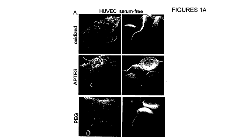
Figures I A-D relate to uptake of oxidized, APTES, or PEGylated silicon
particles by Human
Umbilical Vein Endothelial Cells (HUVECs) and J774 macrophage cells. Figure IA
presents
Scanning electron micrographs of serum-free internalization of 3.2 m silicon
particles by
HUVECs. Left images have a resolution bar of 5 m; right images have a
resolution bar of 2
m. Figure 1B is a diagram that compares internalization by HUVECs between
serum free
and opsonized particles after 1 hour incubation at 37 C. Figures 1 C is a
Table presenting
electrostatic (zeta) potential of 3.2 m microparticles before and after serum
opsonization
(100% serum for 60 min, 4 C). Figure 1D demonstrates an impact of serum on
uptake of 1.6
m particles by J774 macrophage (* p< 0.03) after 1 hour incubation at 37 C. Y-
axis in
Figures 1 B and 1 D is the percentage of cells with particles (high side
scatter cells).
Figures 2A-C relate to uptake of IgG opsonized silicon particles by HUVEC
cells and J774
macrophage cells. Figures 2A and 2B present results of flow cytometry analysis
of uptake by
HUVEC (A) and J774 (B) cells serum-free vs IgG-opsonized 3.2 m oxidized
microparticles
after 1 hr incubation at 37 C. Figure 2C presents quantitative surface
expression of FCyRs
determined by flow cytometric analysis.
Figures 3A-D relate to uptake of silicon particles by cytokine stimulated
HUVEC cells and
J774 macrophage cells. Figure 3A is a diagram that compares an uptake of
oxidized, APTES,
and PEGylated 3.2 p.m silicon particles between control HUVEC cells and
cytokine-
stimulated HUVEC cells. Figure 3B is a diagram that compares an uptake of
oxidized,
APTES, and PEGylated 3.2 m silicon particles between control J774 cells and
cytokine-
stimulated J774 cells. Figure 3C and 3D are scanning electron micrographs of
3.2 m silicon
particle uptake by HUVEC (C) and J774 (D) cells (30 min incubation at 37 C).
Figures 4A-C relate to internalization of oxidized silicon particles by HUVECs
(serum-free).
Figure 4A shows scanning electron micrographs of HUVECs grown on silicon chips
after
incubation with either 1.6 gm, 3.2 gm, or both sizes of oxidized silicon
particles at 37 C for
15 min, 30, or 60 min. Figure 4B shows confocal micrographs of HUVECs
incubated with
3.2 m oxidized silicon microparticles for 15 and 120 min at 37 C using Alexa
Fluor 555
Phalloidin for actin staining. Figure 4C shows confocal projection images
cropped through
the center to illustrate particle location at either 60 or 120 min.
-2-
CA 02747318 2011-06-16
WO 2010/074675 PCT/US2008/014001
Figures 5A-C relate to an early uptake of oxidized silicon particles and FITC
dextran by
HUVECs (serum-free). Figures 5A and 5B are transmission electron micrographs
showing
HUVEC uptake of either 1.6 m (A) or 3.2 m (B) silicon particles after
incubation at 37 C
for 15 min. Figure 5C shows results of flow cytometric analysis of FITC
Dextran
internalization by HUVECs incubated for 1 hr with no particles (solid green
peak, second
from the left), 1.6 pm (red open peak, the right peak), or 3.2 m (purple open
peak, second
from the right) silicon particles. The solid blue peak (the left peak)
represents HUVECs
incubated in media without FITC Dextran. In Figure 5C, the x-axis is
fluorescence due to
internalized FITC dextran and the y-axis is counts (the height is dependent on
the number of
cells).
Figures 6A-B demonstrate cellular location of internalized particles at 2 hrs.
Figure 6A shows
that smaller 1.6 pm particles are located in the perinuclear region of the
cell. Membranes can
be seen surrounding some of the particles. Figure 6B shows that larger 3.2 m
particles are
more scattered and lack apparent membranes, which may be indicative of
endosomal escape.
The resolution scale bar is 10 microns for major images in Figures 6A and 6B
and 500 nm for
insets.
DETAILED DESCRIPTION
Related Applications
The following research articles and patent documents, which are all
incorporated herein by
reference in their entirety, may be useful for understanding the present
inventions:
1) PCT publication no. WO 2007/120248 published October 25, 2007;
2) PCT publication no. WO 2008/041970 published April 10, 2008;
3) PCT publication no. WO 2008/021908 published February 21, 2008;
4) US Patent Application Publication no. 2008/0102030 published May 1, 2008;
5) US Patent Application Publication no. 2003/0114366 published June 19, 2003;
6) US Patent Application Publication no. 2008/0206344 published August 28,
2008;
7) US Patent Application Publication no. 2008/0280140 published November 13,
2008;
8) Tasciotti E. et al, 2008 Nature Nanotechnology 3, 151 - 157.
-3-
CA 02747318 2011-06-16
WO 2010/074675 PCT/US2008/014001
Definitions
Unless otherwise specified "a" or "an" means one or more.
"Microparticle" means a particle having a maximum characteristic size from 1
micron to
1000 microns, or from 1 micron to 100 microns. "Nanoparticle" means a particle
having a
maximum characteristic size of less than 1 micron.
"Opsonin" is a protein that, when bound to a particle, increases the
particle's phagocytosis.
"Dysopsonin" is a protein that, when bound to a particle, decreases the
particle's
phagocytosis.
"Opsonizable" refers to a particle, that can undergo opsonization when exposed
to the blood
or a blood component, such as serum, i.e. the particle that can bind one or
more proteins from
the blood or its component. Preferably, when exposed to the blood or a blood
component, the
opsonizable particle binds one or more opsonins and does not bind dysopsonins.
"Nanoporous" or "nanopores" refers to pores with an average size of less than
1 micron.
"Biodegradable" refers to a material that can dissolve or degrade in a
physiological medium
or a biocompatible polymeric material that can be degraded under physiological
conditions
by physiological enzymes and/or chemical conditions.
"Biocompatible" refers to a material that, when exposed to living cells, will
support an
appropriate cellular activity of the cells without causing an undesirable
effect in the cells such
as a change in a living cycle of the cells; a change in a proliferation rate
of the cells and a
cytotoxic effect.
Disclosure
The present inventors discovered that opsonizable micro- or nanoparticles,
that have a
positive surface charge, can undergo opsonization in blood or a blood
component, such as
serum, in such a manner that the particles can preferentially bind proteins,
that can allow the
particles, after undergoing opsonization, to specifically target inflamed
cells in a body of a
subject. Preferably, prior to the opsonization, the positively charged
opsonizable micro- or
nanoparticles do not contain targeting ligands, such as antibodies, peptides
and/or aptamers,
disposed on their surface.
-4-
CA 02747318 2011-06-16
WO 2010/074675 PCT/US2008/014001
After undergoing opsonization, the positively charged opsonizable micro- or
nanoparticles
can also have a lower uptake by immune cells, such as macrophages, compared to
otherwise
identical micro or nanoparticles, which have, prior to opsonization, a
negative surface charge
or no surface charge. In the present context, the lower uptake can mean that
it can take a
longer time for the opsonized positively charged particles to be internalized
by the immune
cells than for the opsonized negatively charged particles or the neutral ones.
As the result,
the opsonized positively charged particles can avoid an uptake by immune cells
in the body
of the subject, when targeting the inflamed cells.
Preferably, prior to opsonization, a surface of the opsonizable particle does
not contain an
anti-opsonization coating, such as a coating formed by polyethylene glycol
(PEG) or other
hydrophilic chains. Although coating particles with hydrophilic chains, known
as
PEGylation, may reduce or prevent a rapid internatization of the particles by
macrophages, at
the same time PEGylation can often prevent particles from binding to target
cell(s).
In many embodiments, the surface of the opsonizable particles, prior to the
opsonization,
does not contain albumin. In many embodiments, the surface of the opsonizable
particles
particles, prior to the opsonization, does not contain any opsonins. In many
embodiments,
the surface of the opsonizable particle, prior to the opsonization, does not
contain any
proteins.
The positively charged opsonizable particles can be used for treating,
preventing and/or
monitoring a condition associated with an inflammation, such as cytokine
stimulated
inflammation, in a subject, such as an animal with a blood system, by
specifically targeting
inflamed cells in the body of the subject. In many embodiments, the subject
can be a
mammal, such as a human.
The positively charged opsonizable particles can be used for specifically
targeting inflamed
vasculature and thereby for treating, preventing and/or monitoring a condition
or disease
associated with an inflammation.
Examples of such conditions include, but not limited to, allergies, asthma,
Alzheimer's
disease, diabetes, hormonal imbalances, autoimmune diseases, such as
rheumatoid arthritis
and psoriasis, osteoarthritis, osteoporosis, atherosclerosis, including
coronary artery disease,
vasculitis, chronic inflammatory conditions, such as obesity, ulcers, such as
Marjolin's ulcer,
respiratory inflammations caused by asbestos or cigarette smoke, foreskin
inflammations,
-5-
CA 02747318 2011-06-16
WO 2010/074675 PCT/US2008/014001
inflammations caused by viruses, such as Human papilloma virus, Hepatitic B or
C or
Ebstein-Barr virus, Schistosomiasis, pelvic inflammatory disease, ovarian
epitheal
inflammation, Barrett's metaplasia, H. pylori gastritis, chronic pancreatitis,
Chinese liver
fluke infestation, chronic cholecystitis and inflammatory bowel disease; and
inflammation-
associated cancers, which include prostate cancer, colon cancer, breast
cancer;
gastrointestinal tract cancers, such as gastric cancer, hepatocellular
carcinoma, colorectal
cancer, pancreatic cancer, gastric cancer, nasopharyngeal cancer, esophageal
cancer,
cholangiocarcinoma, gallbladder cancer and anogenital cancer; intergumentary
cancer, such
as skin carcinoma; respiratory tract cancers, such as bronchial cancer and
mesothelioma;
genitourinary tract cancer, such as phimosis, penile carcinoma and bladder
cancer;
reproductive system cancer, such as ovarian cancer.
In particular, the positively charged opsonizable particles can be used for
preventing certain
types by specifically targeting inflamed cells associated with an inflammatory
condition,
which can lead to the cancer. For example, by targeting inflammation caused by
Majolin's
ulcer, the positively charged opsonizable particles can prevent skin
carcinoma; by targeting
inflammation caused by asbestos, silica or smoking, the particles can prevent
bronchial
cancer; by targeting foreskin inflammation the particles can prevent phimosis;
by targeting
inflammation caused by Human papilloma virus the particles can prevent penile
carcinoma
and/or anogenital cancer; by targeting inflammation caused by Schistosomiasis
the particles
can prevent bladder cancer; by targeting inflammation caused by pelvic
inflammatory disease
or ovarian epithelial inflammation the particles can prevent ovarian cancer;
by targeting
inflammation caused by Ebstein-Barr virus the particles can prevent
nasopharyngeal cancer;
by targeting inflammation caused by Barrett's metaplasia the particles can
prevent
esophageal cancer; by targeting inflammation caused by H. pylori gastritis the
particles can
prevent gastric cancer; by targeting inflammation caused by chronic
pancreatitis the particles
can prevent pancreatic cancer; by targeting inflammation caused by Chinese
liver fluke
infestation the particles can prevent cholangiocarcinoma; by targeting
inflammation caused
by chronic cholecyctitis the particles can prevent gallbladder cancer; by
targeting
inflammation caused by Hepatitis B or C the particles can prevent hepacellucar
carcinoma;
by targeting inflammation caused by inflammatory bowel disease the particles
can prevent
colorectal cancer.
-6-
CA 02747318 2011-06-16
WO 2010/074675 PCT/US2008/014001
Conditions and diseases associated with an inflammation are disclosed in the
following
references: 1) M. Macarthur et al. Am. J. Physiol Gastrointest Livel Physiol.
286" G515-520,
2004; 2) Calogero et al. Breast Cancer Research, v. 9(4), 2007; Wienberg et
al. J. Clin.
Invest, 112: 1796-1808, 2003; Xu et. al. J. Clin Invest, 112:1821-1830, 2003.
The positively charged opsonizable particles can be used as a part of a
multistage drug
delivery system disclosed in US patent application no. US2008280140 and in PCT
publication no. W02008021908. For example, in some embodiments, the positive
charged
opsonizable particles can contain at least one second stage particle which can
comprise an
active agent.
PARTICLE
The opsonizable particle can have a variety of shapes and sizes.
The dimensions of the opsonizable particle are not particularly limited and
depend on an
application. For example, for intravascular administration, a maximum
characteristic size of
the particle can be smaller than a radius of the smallest capillary in a
subject, which is about 4
to 5 microns for humans.
In some embodiments, the maximum characteristic size of the particle may be
less than about
100 microns or less than about 50 microns or less than about 20 microns or
less than about 10
microns or less than about 5 microns or less than about 4 microns or less than
about 3
microns or less than about 2 microns or less than about 1 micron. Yet in some
embodiments,
the maximum characteristic size of the particle may be from 100 rim to 3
microns or from
200 nm to 3 microns or from 500 nm to 3 microns or from 700 nm to 2 microns.
Yet in some embodiments, the maximum characteristic size of the particle may
be greater
than about 2 microns or greater than about 5 microns or greater than about 10
microns.
The shape of the particle is not particularly limited. In some embodiments,
the particle can
be a spherical particle. Yet in some embodiments, the particle can be a non-
spherical
particle. In some embodiments, the particle can have a symmetrical shape. Yet
in some
embodiments, the particle can have an asymmetrical shape.
In some embodiments, the particle can have a selected non-spherical shape
configured to
facilitate a contact between the particle and a surface of the target site,
such as endothelium
surface of the inflamed vasculature. Examples of appropriate shapes include,
but not limited
-7-
CA 02747318 2011-06-16
WO 2010/074675 PCT/US2008/014001
to, an oblate spheroid, a disc or a cylinder. In some embodiments, the
particle can be such
that only a portion of its outer surface defines a shape configured to
facilitate a contact
between the particle and a surface of the target site, such as endothelium
surface, while the
rest of the outer surface does not. For example, the particle can be a
truncated oblate
spheroidal particle.
The dimensions and shape of particle that can facilitate a contact between the
particle and a
surface of the target site can be evaluated using methods disclosed in US
Patent Application
Publication no. 2008/0206344 and U.S. Application no. 12/181,759 filed July
29, 2008.
In many embodiments, the opsonizable particle can be a porous particle, i.e. a
particle that
comprises a porous material. The porous material can be a porous oxide
material or a porous
etched material. Examples of porous oxide materials include, but no limited
to, porous
silicon oxide, porous aluminum oxide, porous titanium oxide and porous iron
oxide. The
term "porous etched materials" refers to a material, in which pores are
introduced via a wet
etching technique, such as electrochemical etching. Examples of porous etched
materials
include porous semiconductors materials, such as porous silicon, porous
germanium, porous
GaAs, porous InP, porous SiC, porous Si,,Gei_,,, porous GaP, porous GaN.
Methods of
making porous etched particles are disclosed, for example, US Patent
Application Publication
no. 2008/0280140.
In many embodiments, the porous particle can be a nanoporous particle.
In some embodiments, a average pore size of the porous particle may be from
about 1 nm to
about 1 micron or from about I nm to about 800 nm or from about 1 nm to about
500 nm or
from about I nm to about 300 rim or from about I nm to about 200 nm or from
about 2 nm to
about 100 nm.
In some embodiments, the average pore size of the porous particle can be no
more than I
micron or no more than 800 nm or more than 500 rim or more than 300 nm or no
more than
200 nm or no more than 100 nm or no more than 80 nm or no more than 50 rim.
In some embodiments, the average pore size of the porous particle can be size
from about 5 to
about 100 nm or about 10 to about 60 nm or from about 20 to about 40 nm or
from about 30
nm to about 30 nm.
In some embodiments, the average pore size of the porous particle can be from
about I nm to
about 10 nm or from about 3 nm to about 10 rim or from about 3 rim to about 7
rim.
-8-
CA 02747318 2011-06-16
WO 2010/074675 PCT/US2008/014001
In general, pores sizes may be determined using a number of techniques
including N2
adsorption/desorption and microscopy, such as scanning electron microscopy.
In some embodiments, pores of the porous particle may be linear pores. Yet in
some
embodiments, pores of the porous particle may be sponge like pores.
In some embodiments, at least one of the porous particle may comprise a
biodegradable
region. In many embodiments, the whole particle may be biodegradable.
In general, porous silicon may be bioinert, bioactive or biodegradable
depending on its
porosity and pore size. Also, a rate or speed of biodegradation of porous
silicon may depend
on its porosity and pore size, see e.g. Canham, Biomedical Applications of
Silicon, in
Canham LT, editor. Properties of porous silicon. EMIS datareview series No.
18. London:
INSPEC. p. 371-376. The biodegradation rate may also depend on surface
modification.
Porous silicon particles and methods of their fabrication are disclosed, for
example, in Cohen
M.H. et al Biomedical Microdevices 5:3, 253-259, 2003; US patent application
publication
no. 2003/0114366; US patents nos. 6,107,102 and 6,355,270; US Patent
Application
Publication no. 2008/0280140; PCT publication no. WO 2008/021908; Foraker,
A.B. et al.
Pharma. Res. 20 (1), 110-116 (2003); Salonen; J. et al. Jour. Contr. Rel. 108,
362-374
(2005). Porous silicon oxide particles and methods of their fabrication are
disclosed, for
example, in Paik J.A. et al. J. Mater. Res., Vol 17, Aug 2002, p. 2121.
The opsonizable particles may be prepared using a number of techniques.
In some embodiments, the opsonizable particle may be a top-down fabricated
particle, i.e. a
particle produced utilizing a top-down microfabrication or nanofabrication
technique, such as
photolithography, electron beam lithography, X-ray lithography, deep UV
lithography,
nanoimprint lithography or dip pen nanolithography. Such fabrication methods
may allow
for a scaled up production of particles, that are uniform or substantially
identical in
dimensions.
Active agent
The active agent can be a therapeutic agent, an imaging agent or a combination
thereof. The
active agent can be an agent that can be released from a particle containing
it. The selection
of the active agent depends on the application.
-9-
CA 02747318 2011-06-16
WO 2010/074675 PCT/US2008/014001
Therapeutic Agent
The therapeutic agent may be any physiologically or pharmacologically active
substance that
can produce a desired biological effect in a targeted site in an animal, such
as a mammal or a
human. The therapeutic agent may be any inorganic or organic compound, without
limitation, including peptides, proteins, nucleic acids, and small molecules,
any of which may
be characterized or uncharacterized. The therapeutic agent may be in various
forms, such as
an unchanged molecules, molecular complexe, pharmacologically acceptable salt,
such as
hydrochloride, hydrobromide, sulfate, laurate, palmitate, phosphate, nitrite,
nitrate, borate,
acetate, maleate, tartrate, oleate, salicylate, and the like. For acidic
therapeutic agent, salts of
metals, amines or organic cations, for example, quaternary ammonium, can be
used.
Derivatives of drugs, such as bases, esters and amides also can be used as a
therapeutic agent.
A therapeutic agent that is water insoluble can be used in a form that is a
water soluble
derivative thereof, or as a base derivative thereof, which in either instance,
or by its delivery,
is converted by enzymes, hydrolyzed by the body pH, or by other metabolic
processes to the
original therapeutically active form.
The therapeutic agent can be a chemotherapeutic agent, an immunosuppressive
agent, a
cytokine, a cytotoxic agent, a nucleolytic compound, a radioactive isotope, a
receptor, and a
pro-drug activating enzyme, which may be naturally occurring or produced by
synthetic or
recombinant methods, or any combination thereof.
Drugs that are affected by classical multidrug resistance, such as vinca
alkaloids (e.g.,
vinblastine and vincristine), the anthracyclines (e.g., doxorubicin and
daunorubicin), RNA
transcription inhibitors (e.g., actinomycin-D) and microtubule stabilizing
drugs (e.g.,
paclitaxel) can have particular utility as the therapeutic agent.
A cancer chemotherapy agent may be a preferred therapeutic agent. Useful
cancer
chemotherapy drugs include nitrogen mustards, nitrosorueas, ethyleneimine,
alkane
sulfonates, tetrazine, platinum compounds, pyrimidine analogs, purine analogs,
antimetabolites, folate analogs, anthracyclines, taxanes, vinca alkaloids,
topoisomerase
inhibitors and hormonal agents. Exemplary chemotherapy drugs are Actinomycin-
D,
Alkeran, Ara-C, Anastrozole, Asparaginase, BiCNU, Bicalutamide, Bleomycin,
Busulfan,
-10-
CA 02747318 2011-06-16
WO 2010/074675 PCT/US2008/014001
Capecitabine, Carboplatin, Carboplatinum, Carmustine, CCNU, Chlorambucil,
Cisplatin,
Cladribine, CPT-11, Cyclophosphamide, Cytarabine, Cytosine arabinoside,
Cytoxan,
Dacarbazine, Dactinomycin, Daunorubicin, Dexrazoxane, Docetaxel, Doxorubicin,
DTIC,
Epirubicin, Ethyleneimine, Etoposide, Floxuridine, Fludarabine, Fluorouracil,
Flutamide,
Fotemustine, Gemcitabine, Herceptin, Hexamethylamine, Hydroxyurea, Idarubicin,
Ifosfamide, Irinotecan, Lomustine, Mechlorethamine, Melphalan, Mercaptopurine,
Methotrexate, Mitomycin, Mitotane, Mitoxantrone, Oxaliplatin, Paclitaxel,
Pamidronate,
Pentostatin, Plicamycin, Procarbazine, Rituximab, Steroids, Streptozocin, STI-
571,
Streptozocin, Tamoxifen, Temozolomide, Teniposide, Tetrazine, Thioguanine,
Thiotepa,
Tomudex, Topotecan, Treosulphan, Trimetrexate, Vinblastine, Vincristine,
Vindesine,
Vinorelbine, VP-16, and Xeloda.
Useful cancer chemotherapy drugs also include alkylating agents, such as
Thiotepa and
cyclosphosphamide; alkyl sulfonates such as Busulfan, Improsulfan and
Piposulfan;
aziridines such as Benzodopa, Carboquone, Meturedopa, and Uredopa;
ethylenimines and
methylamelamines including altretamine, triethylenemelamine,
trietylenephosphoramide,
triethylenethiophosphaoramide and trimethylolomelamine; nitrogen mustards such
as
Chlorambucil, Chlornaphazine, Cholophosphamide, Estramustine, Ifosfamide,
mechlorethamine, mechlorethamine oxide hydrochloride, Melphalan, Novembiehin,
Phenesterine, Prednimustine, Trofosfamide, uracil mustard; nitroureas such as
Cannustine,
Chlorozotocin, Fotemustine, Lomustine, Nimustine, and Ranimustine; antibiotics
such as
Aclacinomysins, Actinomycin, Authramycin, Azaserine, Bleomycins, Cactinomycin,
Calicheamicin, Carabicin, Carminomycin, Carzinophilin, Chromoinycins,
Dactinomycin,
Daunorubicin, Detorubicin, 6-diazo-.5-oxo-L-norleucine, Doxorubicin,
Epirubicin,
Esorubicin, Idambicin, Marcellomycin, Mitomycins, mycophenolic acid,
Nogalamycin,
Olivomycins, Peplomycin, Potfiromycin, Puromycin, Quelamycin, Rodorubicin,
Streptonigrin, Streptozocin, Tubercidin, Ubenimex, Zinostatin, and Zorubicin;
anti-
metabolites such as Methotrexate and 5-fluorouracil (5-FU); folic acid
analogues such as
Denopterin, Methotrexate, Pteropterin, and Trimetrexate; purine analogs such
as Fludarabine,
6-mercaptopurine, Thiamiprine, and Thioguanine; pyrimidine analogs such as
Ancitabine,
Azacitidine, 6-azauridine, Carmofur, Cytarabine, Dideoxyuridine,
Doxifluridine,
Enocitabine, Floxuridine, and 5-FU; androgens such as Calusterone,
Dromostanolone
-11-
CA 02747318 2011-06-16
WO 2010/074675 PCT/US2008/014001
Propionate, Epitiostanol, Rnepitiostane, and Testolactone; anti-adrenals such
as
aminoglutethimide, Mitotane, and Trilostane; folic acid replenisher such as
frolinic acid;
aceglatone; aldophosphamide glycoside; aminolevulinic acid; Amsacrine;
Bestrabucil;
Bisantrene; Edatraxate; Defofamine; Demecolcine; Diaziquone; Elfornithine;
elliptinium
acetate; Etoglucid; gallium nitrate; hydroxyurea; Lentinan; Lonidamine;
Mitoguazone;
Mitoxantrone; Mopidamol; Nitracrine; Pentostatin; Phenamet; Pirarubicin;
podophyllinic
acid; 2-ethylhydrazide; Procarbazine; PSK ; Razoxane; Sizofrran;
Spirogermanium;
tenuazonic acid; triaziquone; 2, 2',2"-trichlorotriethylamine; Urethan;
Vindesine;
Dacarbazine; Mannomustine; Mitobronitol; Mitolactol; Pipobroman; Gacytosine;
Arabinoside ("Ara-C"); cyclophosphamide; thiotEPa; taxoids, e.g., Paclitaxel
(TAXOL ,
Bristol-Myers Squibb Oncology, Princeton, NJ) and Doxetaxel (TAXOTERE , Rhone-
Poulenc Rorer, Antony, France); Chlorambucil; Gemcitabine; 6-thioguanine;
Mercaptopurine; Methotrexate; platinum analogs such as Cisplatin and
Carboplatin;
Vinblastine; platinum; etoposide (VP-16); Ifosfamide; Mitomycin C;
Mitoxantrone;
Vincristine; Vinorelbine; Navelbine; Novantrone; Teniposide; Daunomycin;
Aminopterin;
Xeloda; Ibandronate; CPT- 11; topoisomerase inhibitor RFS 2000;
difluoromethylornithine
(DMFO); retinoic acid; Esperamicins; Capecitabine; and pharmaceutically
acceptable salts,
acids or derivatives of any of the above. Also included are anti-hormonal
agents that act to
regulate or inhibit hormone action on tumors such as anti-estrogens including
for example
Tamoxifen, Raloxifene, aromatase inhibiting 4(5)-imidazoles, 4
Hydroxytamoxifen,
Trioxifene, Keoxifene, Onapristone, And Toremifene (Fareston); and anti-
androgens such as
Flutamide, Nilutamide, Bicalutamide, Leuprolide, and Goserelin; and
pharmaceutically
acceptable salts, acids or derivatives of any of the above.
Cytokines can be also used as the therapeutic agent. Examples of such
cytokines are
lymphokines, monokines, and traditional polypeptide hormones. Included among
the
cytokines are growth hormones such as human growth hormone, N-methionyl human
growth
hormone, and bovine growth hormone; parathyroid hormone; thyroxine; insulin;
proinsulin;
relaxin; prorelaxin; glycoprotein hormones such as follicle stimulating
hormone (FSH),
thyroid stimulating hormone (TSH), and luteinizing hormone (LH); hepatic
growth factor;
fibroblast growth factor; prolactin; placental lactogen; tumor necrosis factor-
a and -/3;
mullerian-inhibiting substance; mouse gonadotropin-associated peptide;
inhibin; activin;
-12-
CA 02747318 2011-06-16
WO 2010/074675 PCT/US2008/014001
vascular endothelial growth factor; integrin; thrombopoietin (TPO); nerve
growth factors
such as NGF-0; platelet growth factor; transforming growth factors (TGFs) such
as TGF-a
and TGF-/3; insulin-like growth factor-I and -II; erythropoietin (EPO);
osteoinductive factors;
interferons such as interferon-o -0 and -'y, colony stimulating factors (CSFs)
such as
macrophage-CSF (M-CSF); granulocyte-macrophage-CSF (GM-CSF); and granulocyte-
CSF
(GCSF); interleukins (ILs) such as IL-1, IL-la, IL-2, IL-3, IL-4, IL-5, IL-6,
IL-7, IL-8, IL-9,
IL-11, IL-12, IL-15; a tumor necrosis factor such as TNF-a or TNF-0; and other
polypeptide
factors including LIF and kit ligand (KL). As used herein, the tern cytokine
includes proteins
from natural sources or from recombinant cell culture and biologically active
equivalents of
the native sequence cytokines.
In some embodiments, the therapeutic agent can be an antibody-based
therapeutic agent, such
as herceptin.
In some embodiments, the therapeutic agent can be a nanoparticle. For example,
in some
embodiments, the nanoparticle can be a nanoparticle that can be used for a
thermal oblation
or a thermal therapy. Examples of such nanoparticles include iron and gold
nanoparticles.
Imaging agent
The imaging agent can be any substance that can provide imaging information
about a
targeted site in a body of an animal, such as a mammal or a human being. The
imaging agent
can comprise a magnetic material, such as iron oxide or a gadolinium
containing compound,
for magnetic resonance imaging (MRI). For optical imaging, the active agent
can be, for
example, semiconductor nanocrystal or quantum dot. For optical coherence
tomography
imaging, the imaging agent can be metal, e.g. gold or silver, nanocage
particles. The imaging
agent can be also an ultrasound contrast agent, such as a micro or nanobubble
or iron oxide
micro or nanoparticle.
Administration
The opsonizable micro or nanoparticle(s) can be administered as a part of a
composition, that
includes a plurality of the particles, to a subject, such as human, via a
suitable administration
-13-
CA 02747318 2011-06-16
WO 2010/074675 PCT/US2008/014001
method in order to treat, prevent and/or monitor a physiological condition,
such as a disease.
The opsonizable micro or nanoparticle(s) are administered in such a manner so
that, upon the
administration, the particles can undergo opsonization in the blood of the
subject.
The particular method employed for a specific application can be determined by
the attending
physician. Typically, the composition can be administered by one of the
following routes:
topical, parenteral, inhalation/pulmonary, oral, vaginal and anal.
The particles can be particularly useful for oncological applications, i.e.
for treatment and/or
monitoring cancer or a condition, such as tumor associated with cancer.
The majority of therapeutic applications can involve some type of parenteral
administration,
which includes intravenous (i.v.), intramuscular (i.m.) and subcutaneous
(s.c.) injection.
Administration of the particles can be systemic or local. The non-parenteral
examples of
administration recited above are examples of local administration.
Intravascular
administration can be either local or systemic. Local intravascular delivery
can be used to
bring a therapeutic substance to the vicinity of a known lesion by use of
guided catheter
system, such as a CAT-scan guided catheter. General injection, such as a bolus
i.v. injection
or continuous/trickle-feed i.v. infusion are typically systemic.
Preferably, the composition containing opsonizable particles is administered
via i.v. infusion,
via intraductal administration or via intratumoral route.
The opsonizable particles can be formulated as a suspension that contains a
plurality of the
particles. Preferably, the particles are uniform in their dimensions and their
content. To form
the suspension, the particles as described above can be suspended in any
suitable aqueous
carrier vehicle. A suitable pharmaceutical carrier is one that is non-toxic to
the recipient at the
dosages and concentrations employed and is compatible with other ingredients
in the
formulation. Preparation of suspension of microfabricated particles is
disclosed, for example,
in US patent application publication No. 20030114366.
Embodiments described herein are further illustrated by, though in no way
limited to, the
following working examples.
EXAMPLE
Nanoporous hemispherical silicon microparticles were designed, engineered, and
fabricated
in the Microelectronics Research Center at The University of Texas at Austin.
Two sizes of
-14-
CA 02747318 2011-06-16
WO 2010/074675 PCT/US2008/014001
microparticles were generated, with mean diameters of 1.6 0.2 and 3.2 0.2
m, and pore
sizes ranging from either 5-10 or 30-40 nm (porosity can be altered for
different
applications). Processing details are disclosed in Tasciotti E. et al, 2008
Nature
Nanotechnology 3, 151 - 157.
Briefly, heavily doped p++ type (100) silicon wafers with resistivity of 0.005
ohm-cm
(Silicon Quest, Inc, Santa Clara, CA) were used as the silicon source. A 100
nm layer of
low-stress silicon nitride was deposited using a Low Pressure Chemical Vapor
Deposition
(LPCVD) system. Standard photolithography was used to pattern the
microparticles over the
wafer using a contact aligner (EVG 620 aligner) and AZ5209 photoresist.
Nitride on particle
patterns was selectively removed by CF4 based reactive ion etching (RIE).
After the
photoresist was stripped in piranha solution, the wafer was placed in a home-
made Teflon
cell for two-step electrochemical etching. Firstly, the wafers were etched in
a mixture of
hydrofluoric acid (HF) and Ethanol (1:1 v/v) by applying a current density of
6mA/cm2 for
105 s for 3.2 m particles or 40 s for 1.6 m particles, respectively. Then a
high porosity
release layer was formed by changing the current density to 320 mA/cm2 for 6 s
in a 2:5 v/v
mixture of HF and Ethanol. Finally, the nitride layer was removed in HF after
etching, and
microparticles were released by ultrasound in isopropyl alcohol (IPA) for 1
min. The IPA
solution containing porous silicon microparticles was collected and stored at
4 C. The
morphology of the microparticles was examined by SEM.
Oxidation of Silicon Microparticles.
Silicon microparticles in isopropyl alcohol (IPA) were dried in a glass beaker
kept on a hot
plate (110 C). The dried microparticles were then treated with piranha
solution (1 volume
H2O2 and 2 volumes of H2SO4). The suspension was heated to 110-120 C for 2 hr
with
intermittent sonication to disperse the microparticles. The suspension was
then washed in
deionized (DI) water until the pH of the suspension was - 5.5 - 6.
Surface Modification of Silicon Microparticles with APTES.
The oxidized microparticles were washed in IPA 3-4 times. They were then
suspended in IPA
containing 0.5% (v/v) APTES (Sigma) for 2 hr at room temperature. The APTES
modified
microparticles were washed and stored in IPA. APTES modification was evaluated
by
-15-
CA 02747318 2011-06-16
WO 2010/074675 PCT/US2008/014001
measuring the zeta potential and by colorimetric analysis of amine density.
The later was
found to correlate with zeta potential measurements.
PEG Conjugation.
APTES modified microparticles were reacted with 10 mM mPEG-SCM-5000 (methoxy
poly-
ethylene glycol succinimidyl carboxymethyl; purchased from Laysan Bio Inc) in
acetonitrile
for 1.5 hr. The microparticles were then washed in distilled water 4-6 times
to remove any
unreacted mPEG. Zeta potential measurements were used to indicate adequate
surface
coating.
1.6 m and 3.2 p.m silicon microparticles were oxidized with a piranha
solution [30:70 (v/v);
H202:H2SO4] to create negatively charged, hydroxylated microparticles. Next,
the oxidized
microparticles were surface modified with 3-aminopropyltriethoxysilane
(APTES), which
yielded positively charged, amine modified microparticles. APTES modified
microparticles
were further conjugated with PEG for comparison.
Overall, the following three types of silicon microparticles have been
compared: 1)
negatively charged hydroxylated microparticles; 2) positively charged, amino
modified
microparticles; 3) PEGylated microparticles.
Using human umbilical vein endothelial cells (HUVECs), which are known as a
model for
vascular endothelium, see Klein et al., Pathobiology, 1994, 62, 199-208,
scanning electron
microscope (SEM) images were taken of cells after incubation with
microparticles. HUVEC
were purchased from Lonza Walkersville, Inc (Walkersville, Maryland) and were
cultured in
EBM -2 medium (Clonetics , CC-3156). Cells were maintained at 37 C in a
humidified
5% CO2 atmosphere. HUVEC samples were sputter coated with a 10 nm layer of
gold using
a Plasma Sciences CrC-150 Sputtering System (Torr International, Inc.). SEM
images were
acquired under high vacuum, at 20.00 kV, spot size 3.0-5.0, using a FEI Quanta
400 FEG
ESEM equipped with an ETD (SE) detector.
After one hour at 37 C, both positive and negative microparticles were
internalized by
HUVECs in serum-free media (Fig. IA). While both positive and negative
microparticles are
internalized by HUVECs in serum-free media, it was surprisingly found that
serum
opsonization inhibits uptake of negative (oxidized) microparticles, without
significantly
affecting positively charged aminomodified particles.
-16-
CA 02747318 2011-06-16
WO 2010/074675 PCT/US2008/014001
For opsonization, particles were suspended in 100% serum for 1 hour on ice.
Serum in the
experiments was Fetal bovine serum from Clonetics . Surface modification of
silicon
microparticles with PEG suppressed internalization of microparticles by HUVECs
(Fig. I B).
In Figure 1 B, the y-axis is a percentage with internalized particles.
Internalization
experiments in Figure 1 B were performed for 1 hour at 37 C. Ratio of cell to
particles was 1
cell per 20 particles in each of the experiments.
Activation of endothelial cells by pro-inflammatory cytokines can alter
expression of cell
surface receptors and thus can alter binding to particles, see Klein et al.,
Pathobiology, 1994,
62, 199-208. Endothelial cells (HUVECs) were stimulated with cytokines [TNF-a
(10
ng/ml) and IFN-y (100 U/ml), both obtained from Invitrogen] for 48 hrs.
Subsequently, the
stimulated HUVECs incubated with silicon particles, either negative (oxidized)
or positive
[amine (APTES)-modified] particles, following serum opsonization of the
particles.
Internalization of serum opsonized silicon microparticles by HUVECs was
enhanced for all
groups of microparticles following exposure to TNF-a and IFN-y; however a
clear preference
for opsonized positive microparticles continued to exist, see Figure 3A. In
contrast to
endothelial cells, macrophages (J774 cells) preferentially interacted with
serum-opsonized
negative microparticles. This preference for opsonized negative, oxidized
microparticles by
macrophages was significantly enhanced (I I%) in the presence of cytokines (p
= 0.045), see
Figure 3B. On the other hand, uptake of APTES and PEG modified microparticles
by
macrophages was not affected by exposure to TNF-a and IFN-y.
The experiments on HUVECS and J774 cells exposed to TNF-a and IFN-y were
performed
as follows:
HUVECs (1.5 x 10⁵ cells/well) were seeded into 6 well plates and 24 hr later
the cells were
incubated with serum opsonized silicon microparticles (20 microparticles/cell)
for 1 hr. at
37C. Cells were then washed with PBS, harvested by trypsinization (HUVEC) or
scrapping
(J774), and resuspended in PBS containing 1.0% BSA and 0.1% sodium azide (FACS
wash
buffer). Microparticle association with cells was determined by measuring side
scatter using
a Becton Dickinson FACSCalibur Flow equipped with a 488-nm argon laser and
CellQuest
software (Becton Dickinson; San Jose, CA). Data is presented as the percentage
of cells with
microparticles (percent of cells with high side scatter). Side scatter due to
cells in the
absence of particles has been subtracted from the presented data.
-17-
CA 02747318 2011-06-16
WO 2010/074675 PCT/US2008/014001
J774A.1 macrophage cells were purchased from American Type Culture Collection
(Manassas, VA). Growth medium was Dulbecco's Modified Eagle's Medium
containing
10% FBS, 100 g/ml streptomycin and 100 U/ml Penicillin (Invitrogen; Carlsbad,
CA).
Cells were collected by scrapping.
Diagrams 3C-D are SEM images of silicon microparticle uptake by HUVEC (C) and
J774
(D) cells (30 min, 37 C) in the presence of serum. Cells were plated in 24
well plates
containing 5 x 7 mm Silicon Chip Specimen Supports (Ted Pella, Inc., Redding,
CA) at 5 x
104 cells per well. When cells were confluent, media containing microparticles
(1:10,
cell:microparticles, 0.5 ml/well) was introduced and cells were incubated at
37 C for the 30
min. Samples were washed with PBS and fixed in 2.5% glutaraldehyde for 30 min
(Sigma-
Aldrich; St. Louis, MO). After washing in PBS, cells were dehydrated in
ascending
concentrations of ethanol (30%, 50%, 70%, 90%, 95%, and 100%) for 10 min each.
HUVECs were then incubated in 50% alcohol-hexamethyldisilazane (Sigma)
solution for 10
min followed by incubation in pure HMDS for 5 min to prepare for overnight
incubation in a
desiccator. Specimens were mounted on SEM stubs (Ted Pella, Inc.) using
conductive
adhesive tape (12mm OD PELCO Tabs, Ted Pella, Inc.). Samples were sputter
coated with a
run layer of gold using a Plasma Sciences CrC-150 Sputtering System (Ton
International,
Inc.). SEM images were acquired under high vacuum, at 20.00 kV, spot size 3.0-
5.0, using a
FEI Quanta 400 FEG ESEM equipped with an ETD (SE) detector.
This research can suggest that vascular targeting of endothelial cells can be
enhanced by
serum opsonins that preferentially bind to positively charged microparticles.
In contrast,
serum opsonins binding to negatively charged microparticles strongly inhibit
uptake by
endothelial cells. Fortunately, professional phagocytes, such as macrophages,
showed a
preference for negatively charged opsonized microparticles. Although the
present inventions
are in no way limited by a theory, it can be suggested that opsonins binding
to negatively
charged microparticles can be reflective of serum components, which can
decorate bacteria
and apopotic cells, both of which have a net negative surface charge and can
be targets for
uptake by neutrophils and macrophages, see e.g. Fadok,V.A. et al. J. Immunol.
148, 2207-
2216 (1992) and Dickson, J.S. & Koohmaraie, M. Appl. Environ. Microbiol. 55,
832-836
(1989). Directing microparticle uptake through directed serum opsonization can
resist the
need for PEGylation and concurrent compromised targeting and altered
degradation rates.
-18-
CA 02747318 2011-06-16
WO 2010/074675 PCT/US2008/014001
Microparticle internalization by endothelial cells can be enhanced by pro-
inflammatory
cytokine stimulation, supporting superior uptake of positively charged
microparticles at sites
of chronic inflammation. Thus, opsonized microengineered particles with a
positive surface
charge can preferentially targeting of endothelium associated with inflamed
pathologies, such
as coronary artery disease, vasculitis, and cancer.
Additional References
1. Campos S. The oncologist 2003;8 Suppl 2:10-6.
2. Lyass 0, Uziely B, Ben-Yosef R, et al. Cancer 2000;89:1037-47.
3. Valero V. Oncology (Williston Park) 2002;16:35-43.
4. Blum JL, Savin MA, Edelman G, et al. Clinical breast cancer 2007;7:850-6.
5. Gradishar WJ. Expert Opin Pharmacother 2006;7: 1041-53.
6. Iyer AK, Khaled G, Fang J, Maeda H. Drug Discov Today 2006;11:812-8.
7. US patent application publication no. 20070237827.
Although the foregoing refers to particular preferred embodiments, it will be
understood that
the present invention is not so limited. It will occur to those of ordinary
skill in the art that
various modifications may be made to the disclosed embodiments and that such
modifications are intended to be within the scope of the present invention.
All of the publications, patent applications and patents cited in this
specification are
incorporated herein by reference in their entirety.
-19-