Note: Descriptions are shown in the official language in which they were submitted.
CA 02747537 2016-08-04
Treatment Through Electrical Stimulation of a Nerve
FIELD OF THE INVENTION
The present invention relates to a method and device for medical and/or
cosmetic treatment. In particular, one aspect of the invention relates to a
method for reducing peripheral vascular resistance in the blood circulation of
a
patient; other aspects of the invention relate to methods of treatment of
disorders characterised by increased peripheral vascular resistance. The
invention also relates to a device for implementing the method. Further
aspects of the invention relate to other uses of the device.
BACKGROUND OF THE INVENTION
A method and device for reduction or treatment of deep vein thrombosis
(DVT) are described in international patent application W02006/054118. This
device includes electrodes which are secured to the leg of a patient and used
to provide electrical stimulation to the muscles. Preferably the electrodes
are
arranged to stimulate the lateral and/or medial popliteal nerves, which causes
the calf muscles to contract. This in turn activates the calf musculovenous
pump, in which blood circulation is promoted by muscle contraction, so
serving to reduce the risk of thrombosis in the limb. Other musculovenous
pumps include the foot pump, and the device may be used to stimulate this as
well as, or instead of, the calf pump. The device is preferably used to induce
isometric contraction of the muscles, such that the musculovenous pumps
may be activated but limb movement from the stimulation is reduced or
avoided.
As described in W02006/054118, use of the device in the manner described
has been demonstrated to increase venous emptying in the leg, as well as to
increase cortical blood flow in the long bones of the leg. Due to these
effects,
the device is suggested for use to treat conditions other than DVT
characterised by impaired venous blood flow, including ulcers, varicose veins,
ischaemia, oedema, phlebitis, osteoporosis, peripheral vascular disease,
coronary heart disease, and hypertension. These disorders are considered to
1
CA 02747537 2011-06-17
WO 2010/070332
PCT/GB2009/051713
be treatable on the basis that the device and method can increase venous
blood flow.
We have now surprisingly determined that the device, and similar devices,
may be used to alter the patterns of blood flow in a patient.
SUMMARY OF THE INVENTION
We present here evidence that electrical stimulation of muscles may be used
not only to activate the calf musculovenous pump to increase venous
emptying, but also to alter the patterns of blood flow in a patient. In
particular,
diastolic flow reversal in the artery may be reduced or even prevented. This
is
thought to be a consequence of a reduction in peripheral vascular resistance.
While enhancing blood flow was previously known, the finding that blood flow
may be significantly altered is unexpected, and offers several novel ways in
which the device may be used.
According to a first aspect of the present invention, there is provided a
method
of reducing peripheral vascular resistance in a leg of a patient, the method
comprising administering one or more electrical stimuli to a plurality of leg
muscles sufficient to cause isometric contraction of the muscles. Also
provided is a method of reducing or preventing diastolic flow reversal in an
artery in a leg of a patient, the method comprising administering one or more
electrical stimuli to a plurality of leg muscles sufficient to cause isometric
contraction of the muscles.
The reduction of peripheral vascular resistance, and the reduction of
diastolic
flow reversal, allow for the treatment of additional conditions which were not
previously known to be treatable by electrical stimulation of muscles. In
particular, the invention also provides a method for treatment of disorders
characterised by increased peripheral vascular resistance. Such disorders
include lower limb arterial disease (peripheral arterial disease); impaired
lower
limb lymphatic drainage; cardiac diseases; restless leg syndrome (Wittmaack-
Ekbom's syndrome); soft tissue injury of the lower limb (including skin and
muscle bruising and micro tears; and sports injuries); and inflammation. The
invention provides methods for treatment of each of these disorders. The
2
CA 02747537 2011-06-17
WO 2010/070332
PCT/GB2009/051713
reduction of peripheral vascular resistance is also thought to be of benefit
in
sports training and rehabilitation, whether or not the subject has an injury.
For example, the methods of the invention may be used to reduce recovery
time after a sports event. After such an event, for example, a football match,
or an athletics race, even if a participant is not injured, their performance
may
take several days to recover to the pre-event level. We believe that the
method of the invention may be of benefit in reducing this recovery time; for
example, if applied from 2-24 hours after an event, or between training
sessions.
The method may also be of benefit in other conditions in which blood pooling
may be a problem. In particular, for prevention or avoidance of G-LOC (g-
force induced loss of consciousness). In such an embodiment, the method
may also comprise the steps of monitoring g-force experienced by a subject,
and adjusting the stimuli in response to variations in the monitored g-force
(for
example, an increased g-force may result in an increased frequency of
stimulation). Other applications include maintaining blood flow in reduced
gravity, for example, during space travel; reducing the likelihood of blood
pooling during prolonged periods of standing (for example, among soldiers on
parade); or reducing or avoiding blackouts experienced on sudden standing.
The leg muscles are preferably the calf muscle, although in certain
embodiments of the invention, stimulation of the ankle and/or foot musculature
may instead or in addition be used. The leg muscles are preferably involved in
a musculovenous pump; for example, the calf, foot, and/or thigh pumps.
The stimuli may be applied directly to the muscles, or indirectly via
stimulation
of a suitable nerve. For example, a favoured approach is to indirectly
stimulate
the lower limb musculature by accessing nerve groups in the area of the
popliteal fossa, where the nerve groups are in general easily accessible in
individuals, regardless of body mass, and with minimum energy requirement.
Unless otherwise specified, it will be appreciated that all reference herein
to
stimulation of a muscle is intended to encompass both direct stimulation and
indirect stimulation.
3
CA 02747537 2011-06-17
WO 2010/070332
PCT/GB2009/051713
A possibly undesirable effect of isolated contraction of the calf muscles is
the
plantar-flexion of the foot. In a seated individual this may cause the knee to
rise, so making the process more obtrusive. Isometric contraction ensures that
opposing muscles or groups of muscles are stimulated such that there is no or
little movement of the limb as a result. The stimulus may be applied directly
to
posterior calf muscles; conveniently the soleus and/or gastrocnemius
muscles. Indirect stimulation of the lower limb muscles may be achieved by
electrical stimulation of the lateral popliteal nerve in the region of the
popliteal
fossa. Specifically at the inner margin of the biceps femoris muscle, behind
the fibula at the inner side of the tendon of the biceps femoris.
Additionally,
indirect stimulation of the lower limb muscles may be further achieved by
electrical stimulation of the medial popliteal nerve, which is located
medially
from the lateral popliteal nerve in the region of the popliteal fossa.
A second stimulus may be applied to shin muscles; conveniently the tibialis
anterior. Preferably the second stimulus is applied simultaneously to the
stimulus applied to the calf muscles. Stimulation of the tibial muscle alone
will
promote blood flow to some extent, although the primary purpose of this
second stimulation is to prevent unwanted limb movement. Application of a
stimulus only to a posterior calf muscle may have the unwanted side effect of
causing movement of the ankle joint. Application of a stimulus to the shin
muscle will counteract any movement of the ankle joint caused by contraction
of the calf muscle, so keeping the ankle and knee joints relatively still.
Alternatively, stimulation of the lateral popliteal nerve, in the region of
the
popliteal fossa, has the advantage of initiating the contraction of both
posterior
and anterior lower limb muscle groups from a single stimulation point. Such
simultaneous stimulation results in isometric contraction; hence the ankle and
knee joints would not be typically mobilised. Stimulation of the lateral
popliteal
also elicits contraction of the foot muscles and hence the so-called "foot-
pump". Additionally, the surprising advantage of selective stimulation of the
lateral popliteal nerve is that the resultant muscular contractions are
entirely
compatible with standing and walking. An additional benefit of this mode of
indirect stimulation is the involvement of the muscles in the sole of the
foot,
4
CA 02747537 2011-06-17
WO 2010/070332
PCT/GB2009/051713
which have been shown to contribute substantially to clearance of blood from
the lower leg. It has further been identified that stimulation of the nerve in
this
way, rather than the muscles directly, allows the method to be operated so as
to engender little or no noticeable skin sensation or discomfort when used to
stimulate muscle contraction.
In a clinical environment, where standing and walking are not a pre-requisite,
the medial popliteal nerve may be stimulated, either in isolation or in
combination with stimulation of the lateral popliteal nerve. A preferred
version
of dual medial and lateral popliteal nerve stimulation may result in near
maximal contraction of the entire lower limb musculature, leading to enhanced
efficiency and activity of both the calf and foot venous pumps, and by
extension, movement of venous blood out of the lower limb, centrally towards
the abdomen.
The method preferably comprises repeatedly administering an electrical
stimulus to the muscles.
A typical electrical stimulus may be at a current of between 0 to 100 mA,
preferably 0 to 50 mA, more preferably 1 to 40 mA, and most preferably
between 1 to 20 mA. Other examples of stimulus currents include between 15
and 30 mA.
The stimulus may be an AC waveform, although it is preferably a DC
waveform, more preferably a pulsed DC waveform. The stimulus may have a
frequency of 0.01 to 100 Hz, preferably 0.1 to 80 Hz, more preferably 0.1 to
50 Hz; and more preferably still 0.1 to 5 Hz. The most preferred frequencies
are 0.5-5 Hz, 1-5 Hz, preferably 1-3 Hz; for example, 1, 2 or 3 Hz. In other
embodiments, the frequency may be from 30 to 60 Hz, and more preferably
40 to 50 Hz. Alternatively, a stimulus with a frequency from 0.1 to 1 Hz, or
from 0.33 to 1 Hz may be used. The precise desired frequency may depend
on the purpose of the method, the desired physiological mode of action it is
intended to cause, and the general physical condition, age, sex, and weight of
the patient, among other factors.
5
CA 02747537 2011-06-17
WO 2010/070332
PCT/GB2009/051713
Specific examples of preferred stimuli include 20 mA, at a frequency of 5 Hz,
30 mA at 3 Hz, and 28 mA at 1 Hz. Other stimuli may of course be used.
The stimulus may be applied for a duration between 0 and 1000 ms, between
100 and 900 ms, between 250 and 750 ms, between 350 and 650 ms, or
between 450 and 550 ms. In certain embodiments, the stimulus may be
applied for up to 5000 ms, up to 4000 ms, up to 3000 ms, or up to 2000 ms.
Other durations may be used; again this may depend on the details of the
patient or the mode of action intended. Other preferred durations include from
70 to 600 ms. In certain embodiments, yet shorter durations may be used, for
example from 25 jts to 800 s.
Characteristics of the stimulus may vary over time. For example, a single
stimulus may increase in current over the duration of the stimulus. Preferably
the increase is gradual up to a peak; the stimulus may then either be
maintained at the peak; terminate at the peak; or decrease in a gradual
manner. Alternatively, where repeated stimuli are applied, characteristics of
the stimuli may vary between different stimuli. For example, successive
stimuli
may be applied at increasing levels of current. Again, these successive
stimuli
may increase up to a peak gradually, followed by maintenance at that peak, or
decrease from the peak. A cycle of increasing stimuli may be repeated a
number of times. In preferred embodiments, each stimulus is a single pulse,
rather than multiple brief pulses.
Stimuli may be applied at a plurality of locations on the muscles. For
example,
stimuli may be applied along the main (long) axis of the leg. Such stimuli may
be applied simultaneously, or preferably sequentially such that a 'wave' of
stimuli proceeds along the leg. Preferably, such a wave proceeds upward
toward the body of the patient. This wave effect serves to generate a
corresponding wave of muscle contraction which wave may help to promote
blood flow away from the leg. However, in preferred embodiments of the
invention a stimulus is applied at a single point on the leg, to stimulate the
lateral popliteal fossa nerve, as described above. "A single point" may
include
stimulation by more than one electrode, for example, a pair of positive and
negative electrodes, with a sufficiently small separation (for example, 1-3
cm,
6
CA 02747537 2011-06-17
WO 2010/070332
PCT/GB2009/051713
or up to 2 cm) such that the stimulation is experienced at a point by the user
rather than over a larger area.
Also provided is a method for diagnosing conditions characterised by
increased peripheral vascular resistance, the method comprising
administering one or more electrical stimuli at a first frequency and/or
current
to a plurality of leg muscles sufficient to cause isometric contraction of the
muscles; and monitoring blood flow in the leg to determine whether diastolic
reversal of arterial flow is reduced or prevented and/or peripheral vascular
resistance is reduced.
The method may further comprise repeating the stimulation and monitoring
steps at a second frequency and/or current, and determining the level of
frequency and/or current required to effect reduction or prevention of
diastolic
reversal of arterial flow and/or reduction of peripheral vascular resistance.
The
level at which this occurs may give some information as to the severity of the
condition.
The method may further comprise comparing the level of frequency and/or
current required to effect reduction or prevention of diastolic reversal of
arterial flow and/or reduction of peripheral vascular resistance with the
levels
required in a healthy control patient. Again, this may help diagnose a
condition
or give some indication as to the severity of the condition. The healthy
control
patient may be selected so as to be otherwise comparable to the patient.
Also provided according to the present invention is a method for promoting
circulation in a patient having a heart condition, the method comprising
administering one or more electrical stimuli to a plurality of leg muscles
sufficient to cause isometric contraction of the muscles. As noted above,
electrical stimulation of the musculovenous pump promotes altered blood flow
patterns, which may be beneficial in patients having heart conditions. The
heart condition may include cardiac arrest, suspected cardiac arrest,
arrhythmia, brachycardia, or angina. The method may also be used as an
adjunct to defibrillation in the case of cardiac arrest. Also provided is a
device
for use in promoting circulation in a patient having a heart condition, the
7
CA 02747537 2011-06-17
WO 2010/070332
PCT/GB2009/051713
device comprising at least one electrode for administering an electrical
stimulus to opposed leg muscles of a patient; a power supply connectable to
the electrode; and a control means for activating the electrode to administer
an electrical stimulus to the muscles sufficient to cause the muscles to
contract isometrically. The invention also provides a kit comprising such a
device in combination with a defibrillator. Alternatively, the device may
include
a defibrillator.
Further aspects of the present invention relate to the modification of
cortical
blood flow in bone. As noted in W02006/054118, the method of isometric
muscle stimulation has been shown to promote cortical blood flow. We have
since discovered, and demonstrate herein, that bone oxygenation and bone
perfusion are increased by use of the method. This allows more effective
delivery of pharmaceutical agents to the bone, particularly those intended for
treatment of bone disorders including osteoporosis. Thus, according to a
further aspect of the present invention there is provided a method for
improving administration of medicaments for treatment of bone disorders, the
method comprising administering said medicament to a patient, and
administering one or more electrical stimuli to a plurality of leg muscles
sufficient to cause isometric contraction of the muscles and enhance bone
perfusion. The bone disorder may be osteoporosis. Also provided is a kit for
treatment of bone disorders, the kit comprising a medicament for treatment of
a bone disorder and a device comprising at least one electrode for
administering an electrical stimulus to opposed leg muscles of a patient; a
power supply connectable to the electrode; and a control means for activating
the electrode to administer an electrical stimulus to the muscles sufficient
to
cause the muscles to contract isometrically.
Improved perfusion may also be useful for improving delivery of contrast
agents (for example for medical imaging purposes) to tissues such as the
bones, tendons, ligaments, etc. An aspect of the invention therefore provides
a method for improving delivery of contrast agents, the method comprising
administering said contrast agent to a patient, and administering one or more
8
CA 02747537 2011-06-17
WO 2010/070332
PCT/GB2009/051713
electrical stimuli to a plurality of leg muscles sufficient to cause isometric
contraction of the muscles and enhance perfusion of said agent.
A still further aspect of the invention relates to cosmetic therapy. As
demonstrated herein, use of the method increases peripheral blood
circulation, in particular circulation in the skin. The method also increases
skin
temperature where circulation is increased. These effects may be of benefit in
the cosmetic treatment of individuals. For example, the effects may include
reduction of cellulite or collagen deposits, improvement of skin tone, or
improvement of skin condition. Thus, the invention provides a method for
cosmetic treatment of a patient, the method comprising administering an
electrical stimulus to at least one leg muscle of a patient sufficient to
cause
the muscles to contract isometrically. The cosmetic treatment may be selected
from reduction of cellulite or collagen deposits, improvement of skin tone, or
improvement of skin condition. Also provided is a kit for beauty therapy, the
kit
comprising a device comprising at least one electrode for administering an
electrical stimulus to opposed leg muscles of a patient; a power supply
connectable to the electrode; and a control means for activating the electrode
to administer an electrical stimulus to the muscles sufficient to cause the
muscles to contract isometrically.
The device described in W02006/054118 includes certain electrode
configurations which may be used. We demonstrate herein data showing
specific novel electrode configurations which are perceived as more
comfortable by users. The present invention therefore provides a device
comprising positive and negative electrodes for administering an electrical
stimulus to opposed leg muscles of a patient; a power supply connectable to
the electrode; and a control means for activating the electrodes to administer
an electrical stimulus to the muscles sufficient to cause the muscles to
contract isometrically, wherein one electrode substantially encloses the
other.
By "substantially encloses" is meant that one electrode surrounds at least
66%, preferably at least 75%, more preferably at least 85%, more preferably
at least 90%, and most preferably 100% of the perimeter of the other. It is
not
9
CA 02747537 2016-08-04
essential that the one electrode be entirely enclosed by the other, although
it
is preferred.
It has been determined that this arrangement of electrodes leads to improved
perception of user comfort.
Preferably the positive electrode substantially encloses the negative
electrode.
In some embodiments the electrodes are in the form of concentric, or
substantially concentric, circles. In others, the electrodes are generally
elongate, preferably generally quadrilateral, such as rectangular, or C-shaped
or U-shaped.
Preferably one electrode has a larger area than the other electrode;
preferably
the larger electrode is the positive electrode.
Preferably the control means is configured to administer an AC electrical
stimulus. Preferably the waveform of the current is asymmetric; conveniently
the waveform provides an initial (positive) pulse of large magnitude and short
duration, followed by a (negative) pulse of small magnitude and long duration.
The area under the curve of the two pulses will be equal. In one embodiment,
the initial pulse is of a generally square waveform.
A further aspect of the present invention provides a device comprising
positive
and negative electrodes for administering an electrical stimulus to a nerve
innervating opposed leg muscles of a patient, so as to cause isometric
contraction of the muscles; a power supply connectable to the electrodes; and
control means for activating the electrodes.
According to one embodiment, there is provided a device comprising positive
and negative electrodes for administering an electrical stimulus to a nerve
innervating opposed leg muscles of a patient a power supply connectable to
the electrodes; and control means for activating the electrodes; wherein:
the device comprises a flexible substrate;
the elongate flexible substrate comprises an elongate tongue at one
end and a moulded recess at the other;
CA 02747537 2016-08-04
the electrodes are mounted or directly printed on to the elongate
tongue, with a positive electrode spaced longitudinally along the substrate
from
the negative electrode;
the positive electrode is larger than the negative electrode;
the power supply, and the control means are placed within the moulded
recess;
the power supply is connected to the electrodes by a conductive track;
the conductive track of the positive electrode is separated from the
negative electrode by one or more insulative strips or regions;
insulative strips or regions are arranged at the edge of the tongue to
prevent unwanted leakage of current; and
the device further comprises a press button to activate or deactivate
the device.
Preferably the positive and negative electrodes are separated by 20-30mm;
we have found that this provides a preferred degree of stimulation.
The electrodes may be of different sizes; preferably the positive electrode is
larger than the negative. This provides a higher charge density at the motor
point, and greater capacitance overall. The electrodes may be silver
electrodes. The electrodes may be continuous, or may include holes ¨ for
10a
CA 02747537 2011-06-17
WO 2010/070332
PCT/GB2009/051713
example, the electrodes may be solid electrodes, or may be in the form of a
mesh.
In preferred embodiments, the device comprises a flexible substrate on which
are mounted the electrodes, the power supply, and the control means. The
control means may be, for example, a PCB configured to activate the
electrodes as appropriate. The power supply may be an electrical cell. The
substrate is preferably flexible, but not stretchable ¨ this reduces the risk
of
the electrodes cracking or breaking. For example, the substrate may be a
thermoplastic elastomer.
The electrodes may be directly printed onto the substrate, by conventional
printing means (for example pad or tampo printing). Similarly, conductive
tracks may also be printed onto the substrate if desired.
The substrate may be in the form of an elongate strip or tongue, with the
electrodes spaced along the strip. Such an arrangement may require a
conductive track to be placed from the power supply to the further electrode,
passing close to the nearer electrode. In such arrangements, the device may
further comprise one or more insulative strips or regions arranged to separate
the conductive track from the nearer electrode; insulative strips may also or
instead be arranged along the edges of the strip to prevent current leaking
outside the area of the strip. Alternatively, or in addition, the substrate
may
comprise a recessed groove within which a conductive track may be located;
thereby serving to separate the track from the electrode.
In certain embodiments the device may be configured to be implantable in a
patient, for example, implantable subcutaneously. This would be of benefit in
chronic indications where long term use of the device is required.
The device further comprises a conductive gel overlying the electrodes. The
gel is preferably in a single piece overlying both electrodes, for ease of
manufacture as well as structural integrity. We have determined that a single
piece of gel may be used, based on the bulk resistivity of the material and
geometry, so that leakage resistance is much greater than delivery resistance.
Examples of gels which may be used include hydrogel or silicone.
11
CA 02747537 2011-06-17
WO 2010/070332
PCT/GB2009/051713
The device may be assembled as follows. The flexible substrate may be
produced as a generally flat elongate strip and a recess forming a
compartment. The electrodes and conductive tracks are then printed onto the
substrate, and the power supply and control means placed into the recess.
This serves to connect all the electrical connections. The recess may then be
closed, for example, by sonic welding a cover to seal the power supply and
control means into the recess. Finally a gel is placed over the electrodes.
The device may further comprise a locating mark to aid correct placement in
use.
The device may include a press button for activating or deactivating the
device. The control means may be configured to provide a plurality of
activation modes (for example, with different stimulation characteristics);
the
press button may be used to cycle through these modes. The device may
include a display means, such as a light or an LED, to indicate the selected
activation mode.
Preferably the device is for reducing diastolic flow reversal.
In certain embodiments the device may be disposable; for example, after a
single use.
The device is intended to be sufficiently small and light ¨ for example, less
than 10 cm in length, and weighing less than 100 g, preferably less than 20 g
¨ so as to be highly portable.
In use the device may be operated so as to engender little or no noticeable
skin sensation or discomfort when activated to stimulate muscle contraction.
BRIEF DESCRIPTION OF THE DRAWINGS
Figures 1 to 3 show measurement of arterial blood flow in three separate test
subjects at different levels of stimulation.
Figure 4 compares the speed of skin blood flow in stimulated and
unstimulated limbs at different levels of stimulation.
12
CA 02747537 2011-06-17
WO 2010/070332
PCT/GB2009/051713
Figure 5 compares skin temperature in stimulated and unstimulated limbs at
different levels of stimulation.
Figure 6 shows oxyhemoglobin levels measured by infrared spectroscopy in
the tibia during stimulation cycles.
Figure 7 shows the change in deoxyhemoglobin levels in all patients during
stimulation.
Figure 8 shows a first desired electrode arrangement.
Figure 9 shows a second desired electrode arrangement.
Figure 10 shows several electrode arrangements tested.
Figure 11 shows asymmetric and symmetric waveforms tested.
Figures 12 and 13 show results from electrode and waveform comfort testing.
Figures 14 to 17 show views of an embodiment of a device according to the
present invention.
DETAILED DESCRIPTION OF THE DRAWINGS
A device for electrically stimulating leg muscles is described in detail in
W02006/054118, and the reader is referred to that publication for a full
description of the device. The present invention is primarily based on a
number of unexpected effects observed from use of that and similar devices,
although we also describe a particularly preferred embodiment of the device.
In brief, though, one embodiment of the device as described in
W02006/054118 includes a loop of elasticated material which, in use, may be
worn around a user's lower limb. On the interior surface of the elasticated
material are disposed first and second electrodes connected by conductive
wires to a cradle which is integral with the elasticated material.
Mounted within the cradle is a control module, which includes a power cell, a
control processor, and an external LED.
13
CA 02747537 2011-06-17
WO 2010/070332
PCT/GB2009/051713
The control module is removable from the cradle, with a pair of detents and
corresponding recesses allowing the cradle and control module to interlock.
The control module and cradle carry corresponding electrical contact surfaces
which provide for electrical communication between the control module and
the first and second electrodes via the conductive wires.
The control processor includes a timer module, a data store, a program store,
and a logic unit.
In use, the device is operated as follows. The elasticated loop is worn on a
user's lower limb, such that the first electrode is in contact with the calf
muscle
at the rear of the limb, and the second electrode is in contact with the
anterior
muscle of the limb. When the control module is engaged with the cradle, the
device is automatically activated.
The program store is preloaded with an operating program arranged to
activate the electrodes each minute using a 40 Hz pulsed DC of 20 mA for 0.1
second. Both electrodes are activated simultaneously. The timer module
serves to generate appropriate timing signals, while the logic unit executes
the
program of the program store.
As the electrodes are activated, the user's muscles are stimulated to
contract.
Contraction of the rear calf muscle, caused by the first electrode, serves to
pump blood out of the leg using the calf pump thereby reducing pooling of the
blood. Contraction of the anterior muscle, caused by the second electrode, is
intended to reduce unwanted movement of the ankle by counterbalancing the
contraction of the rear calf muscle. Simultaneously with each activation of
the
electrodes, the LED on the outer surface of the control module is also
activated; this provides a visual confirmation that the device is operating.
The foregoing is a description of one embodiment of the device. However, a
suitable device for stimulating muscles may be assembled from conventional
skin electrodes and a suitable electrical power supply. It is this form of
test rig
which was used in the following experiments.
Experimental Design
14
CA 02747537 2011-06-17
WO 2010/070332
PCT/GB2009/051713
Study Title: A study to determine the effects of a novel method for enhancing
lower limb blood flow in Healthy Adult Volunteers.
Objectives: The primary objective of this study was to evaluate the
effectiveness of topical electrical stimulation in enhancing lower limb
perfusion. The secondary objective was to evaluate with duplex ultrasound
and plethysmography techniques the blood flow velocity and volume changes
associated with varying the intensity and level of electrical stimulation.
Study Design: One-
centre, physiological response study in healthy
Volunteers.
Stimulus Application: The effects of electrical stimulation on lower limb
blood flow were investigated in healthy volunteers during a 4-hour period of
prolonged sitting. Each subject completed his or her study sat in an Industry
Standard airline seat. The stimulator used custom stimulation protocols.
Superficial electrical stimulation was applied to the lateral popliteal nerve
located in the area of the popliteal fossa.
Sample Size: 30 Volunteers
Environmental Conditions
The examinations were carried out in a quiet, stable, draught free
environment, both temperature and humidity controlled (24 1 C, relative
humidity 30-40%). Volunteers were instructed to have a light breakfast,
avoiding fatty foods, tobacco and caffeine and to abstain from vigorous
exercise from the previous evening onwards. The volunteers were lightly clad
(in shorts), sat in a comfortable position with legs bent at the knees.
The effects of electrical stimulation on lower limb blood flow were
investigated
in healthy Volunteers during a 4-hour period of prolonged sitting. Each
subject
completed his or her study sat in an Industry Standard airline seat, which has
been specifically obtained for this investigation.
The leg clearance distance was be set at 34 inches, by positioning of a toe-
bar. Each subject was positioned in the seat by a safety belt to maintain a
CA 02747537 2011-06-17
WO 2010/070332
PCT/GB2009/051713
close uniformity of posture and actively encouraged to remain as passive as
can be tolerated by the individual.
Physiological Assessments
During this phase, the amplitude and frequency of the electrical stimulation
was varied and associated changes in blood flow recorded.
Changes in lower limb blood flow were evaluated using routine non-invasive
plethysmographic techniques (photoplethysmography, strain gauge
plethysmography and air plethysmography), transcutaneous oxygen and
where possible, colour flow duplex ultrasound.
Changes in blood flow and volume in response to the protocols were
compared to blood flow and velocity changes determined by voluntary muscle
action i.e. Volunteers were be asked to perform 10 plantar flexions (10 toe
lifting movements - with the heel on the ground). This is the maximum
physiological response that can be obtained in the sitting position.
Volunteers were asked to evaluate acceptance and tolerability of electrical
stimulation sequences by use of a questionnaire (Verbal Rating Scores) and a
scoring index (Visual Analogue Scores). Discomfort was related to normal
measurement of blood pressure, measured on the upper arm using a
standard sphygmomanometer cuff.
Following the period of sitting for 4-hours Volunteers will be re-examined
with
duplex ultrasound to recheck the status of the deep veins to exclude the
development of significant thrombi. The study was performed on each subject
at two separate occasions which were then averaged to reduce experimental
bias.
Stimulator
The device produced a range of pre-set programmes corresponding to
different stimulation currents, and pulse frequencies. The waveform was
specifically designed for motor nerve stimulation, as opposed to direct muscle
stimulation. Pulse amplitudes ranged from 1mA to 40mA, with frequencies
16
CA 02747537 2011-06-17
WO 2010/070332
PCT/GB2009/051713
ranged from 1 Hz to 5 Hz, which is a significant departure from the
Physiotherapy and TENS protocols (which generally apply substantively
higher currents and frequencies).
We applied a succession of 15 different stimulation programmes to each
subject during the course of each study, according to a 2-dimensional matrix
of amplitude and frequency, as shown in Table 1. The duration of each
stimulation programme was 5 minutes and will be followed by a 10-minute
recovery phase to allow vascular re-equilibration prior to the next sequence.
Table 1: Stimulation sequence
Programme # Amplitude/mA Frequency/Hz
1 1 1
2 1 3
3 1 5
4 5 1
5 5 3
6 5 5
7 10 1
8 10 3
9 10 5
20 1
11 20 3
12 20 5
13 40 1
14 40 3
40 5
During each of the 15 programmes, non-invasive blood flow and volume
parameters were measured as specified above, with reference to the levels
observed during voluntary muscle contraction, and with reference to levels
observed in the contralateral limb.
Example 1 : Blood flow patterns
The patterns of venous blood flow in volunteers were monitored using
vascular ultrasound of the stimulated leg. Representative examples are shown
17
CA 02747537 2011-06-17
WO 2010/070332
PCT/GB2009/051713
in Figures 1-3. Figure la shows stimulation in a first subject at 20 mA, 5 Hz;
Figure lb at 5 mA, 5 Hz; and Figure lc with no stimulation. Figure 2a shows a
second subject stimulated at 20 mA, 3 Hz; Figure 2b the same subject
immediately after stimulation; and Figure 2c the subject at rest. Figure 3a
shows a third subject undergoing stimulation at 10 mA, 3 Hz; Figure 3b at 1
mA, 3 Hz; Figure 3c at 20 mA, 5 Hz; Figure 3d at 5 mA, 1 Hz; Figure 3e at 5
mA, 3 Hz; and Figure 3f the subject at rest.
In these examples there was a four-fold increase in venous blood flow velocity
from baseline. There was also a significant increase in frequency of cephalad
(toward the head) venous blood flow with application of the stimulus.
Flow velocity in the superficial femoral artery doubles and the reverse flow
components of the pulse wave arterial flow waveform are completely
abolished with application of the stimulus.
Reverse flow in the superficial femoral artery is due to high resistance of
the
peripheral vessels; therefore forward flow throughout the cardiac cycle
suggests a significant reduction in peripheral vascular resistance.
A fall in total peripheral resistance (consequent of the increase in vascular
pump activation by the device) may be illustrated by the laser Doppler and
vascular venous vessel ultrasound blood flow increases. The consequence of
this is that cardiac output tends to increase. We have also shown that there
is
no significant increase in the heart rate (beats per minute). This may be
demonstrated by the increase in the arterial blood flow and the change in the
waveform.
Importantly the increases in blood flow in the various tissues in the leg are
proportionate, and therefore there is an increase in blood flow in all of the
tissues; hence no 'steal' of blood from any adjacent tissue. All tissues,
skin,
muscle, bone etc have increased perfusion of blood.
Resistance of blood flow can influence arterial pressure, cardiac output,
distribution of cardiac output to systemic organs, distribution of organ blood
flow to the various organ tissues, partitioning of tissue blood flow between
18
CA 02747537 2011-06-17
WO 2010/070332
PCT/GB2009/051713
capillaries and arteriovenous anastomoses, capillary hydrostatic pressure,
and the distribution of blood flow within the cardiovascular system. All of
which
are upregulated by the device at certain, defined settings.
A parallel is in exercise, where the total peripheral resistance also
decreases
as work load, measure by oxygen consumption increases. The fall in vascular
resistance is accompanies by a progressive increase in cardiac output. The
device mimics this event without a substantive increase in workload and
hence minimal oxygen consumption compared to exercise.
Increases in microcirculatory blood flow may additionally be explained by an
increased utilisation of previously closed or 'resting' capillary networks,
which
become available for local exchange. The effect of this is a greatly increased
tissue perfusion and a further effect on peripheral vascular resistance.
This is a novel and unique observation, which has significant impact on the
cardio-vascular system and vascular therapeutics.
Thus, application of the electrical stimulus can increase venous blood flow,
and can reduce or prevent diastolic flow reversal in the artery. Note that
this
does not occur at all settings; Figure 3d shows no flow reversal when
stimulated at 5 mA, 1 Hz.
This effect has the potential for a wide range of therapeutic and diagnostic
applications. For example, as the effect only occurs at certain settings, it
is
likely that the current and frequency at which it appears in individual
patients
may be characteristic of their normal arterial flow and/or peripheral vascular
resistance. This may be used to diagnose the presence and/or severity of
circulatory disorders in a patient. Therapeutically, the modified arterial
flow
and reduced peripheral vascular resistance may be of benefit in treatment of a
range of conditions, including ischaemia, cardiac vessel disease, ulceration,
and so on.
Example 2
19
CA 02747537 2011-06-17
WO 2010/070332
PCT/GB2009/051713
Laser Doppler Fluxmetry (LDF) was used to measure the speed of skin blood
flow; the results are shown in Figure 4. LDF flux (speed of blood) is
increased
up to ¨ 1000% in stimulated leg compared to baseline and the unstimulated
leg, which showed values only around baseline level.
Example 3
Skin temperature was measured in stimulated and unstimulated legs; the
results are shown in Figure 5. There is a slight increase in temperature at
all
stimulations in the stimulated leg compared to unstimulated leg. Temperature
in the body is generated by metabolism and blood flow. As the metabolism is
not altered during the stimulations the slight increase in skin temperature is
an
indicator for increased blood flow in superficial layers of the skin.
Example 4
Therapy for Osteoporosis
Every year there are approximately 2 million osteoporotic fractures worldwide.
(in 1990 there were 1.66 million, and 6 million per year forecast by 2050
according to World Health Organisation). High-risk groups include the elderly
population, and people with spinal cord injuries.
In the healthy individual, bone is constantly being remodelled according to
physical requirements. Osteoclast cells remove minerals from bone, allowing
collagen matrix to resorb, while osteoblasts lay down new collagen matrix and
mineral deposits.
Various theoretical models have been proposed over the last century for the
mechanism by which the body controls bone density. Wolff, in 1892, proposed
that bone deposits followed the patterns of stress in the bone. Frost's 1987
"mechanostat" theory suggested that bone was maintained to maintain
uniform strain under habitual loads.
Models for explaining why some individuals developed problems with
maintaining bone density initially focused on disuse. In the ageing
individual,
decreasing use of the bone leads to lower doses of the stresses and strains
CA 02747537 2011-06-17
WO 2010/070332
PCT/GB2009/051713
required to signal bone maintenance. More recently, however, it has been
suggested that there is a vascular component to the aetiology. Osteoporosis
appears to occur in individuals with impaired bone perfusion, either by
reduced angiogenesis (itself aggravated by disuse), atherosclerosis
restricting
flow in existing vessels, or simply lower activity levels causing less blood
circulation. (Trueta J. The role of the vessels in osteogenesis. J Bone Joint
Surg Br. 1993).
The present invention has the potential to mitigate vascular risk factors for
osteoporosis, by increasing perfusion of bone. This can help in two ways.
Firstly, augmenting blood supply overcomes limitation of bone modelling
caused by reduced perfusion. Secondly, pharmaceutical interventions for
osteoporosis can be delivered more effectively to the bone by improving bone
perfusion.
A study carried out under the supervision of the inventors has demonstrated
that 1) Blood flow in the tibia and femur are enhanced when the device is
active; and 2) Perfusion indices indicate that the bone is less hypoxic when
the device is active.
Figure 6 shows Oxyhaemoglobin level measured by infrared spectroscopy in
the tibia, during stimulation cycles (100 seconds on, 100 seconds off). Total
blood content (top line) drops during stimulation, indicating that the calf
pump
aids evacuation, and that oxyhaemoglobin levels rise during stimulation,
indicating better oxygenation (reduced hypoxia).
Figure 7 shows the results for 12 subjects summated, showing the mean and
standard deviation reduction of deoxyhaemoglobin relative to baseline. The
device (labelled NMS) on the chart shows a significant reduction when active.
As an idea of scale, this is compared with the reduction achieved by
augmenting blood supply using the tilt-table method. This is a known
hydrostatic step-change, which consists of the subject lying supine on a tilt
table, and while strapped to the table they are tilted into a standing upright
position, providing a very large hydrostatic vascular stimulus. This chart may
21
CA 02747537 2011-06-17
WO 2010/070332
PCT/GB2009/051713
be considered analogous to comparing DVT parameters with the device to
foot flexion.
The foregoing examples indicate that the device and method may be used to
address new clinical targets. These include:
= Lower limb arterial disease ¨ Peripheral Arterial Disease
. Enhanced lower limb lymphatic drainage.
. Cardiac diseases
= Fractures
= Enhancement of bone marrow perfusion ¨ for example the management
of sickle cell crises, ischaemic bone marrow, stem cell and bone marrow
harvest procedures ¨ as well as improving treatment of cancers by
delivering drugs to the bone marrow.
= Soft tissue injury of the lower limb ¨ skin and muscle bruising and micro
tears.
= Sports training and rehabilitation.
= Restless Leg Syndrome (Wittmaack-Ekbom's syndrome)
= Enhancement of endothelial-derived nitric oxide and prostacyclin
release.
Example 5
Discomfort
Neuromuscular stimulation is commonly used to elicit muscle activity for
several different applications. These include exercise, rehabilitation and
22
CA 02747537 2011-06-17
WO 2010/070332
PCT/GB2009/051713
restoration of function (eg drop foot stimulator) and more recently
augmentation of blood supply using the soleus pump for various purposes.
NMS has commonly been used previously for restoration of function in
insensate individuals, eg with spinal cord injury. In these users, discomfort
or
pain associated with the stimulation is not an issue.
In the sensate user, however, discomfort or pain during stimulation is an
issue, and sometimes a limiting factor in the level of stimulation applied.
In NMS, an electrical stimulus is used to cause contraction of a system of
skeletal muscles. Unfortunately, efferent (motor) and afferent (sensory)
nerves
are typically bundled together in the same nerve conduit, and additional
sensory nerves are present in the skin. This means that, as well as
stimulating
motor nerves, NMS causes some stimulation of sensory nerves. If sensation
signals arrive at the brain in large numbers and rapid succession, they may be
perceived as pain in some individuals.
Relationships have been found between electrode size and stimulatory
response. It has also been found that stimulation quality and tolerance are
sensitive to electrode position. These relationships have now been
investigated further by the inventors, in a series of experiments.
One hypothesis tested was that smaller electrodes would be better tolerated,
since they allow us to target accurately the region of the peroneal lateral
popliteal, without unnecessary stimulation of surrounding areas of skin
receptors. This was not found to be reliably the case in our experiments. This
finding may be rationalised as follows.
Current density is usually maximal at the skin/electrode interface, whereas
the
quality of muscle contraction is determined by the current density at the
point
of excitation.
For a given current, a smaller electrode provides increased current density at
the skin. However, this does not necessarily translate to maximal current
density at the point of excitation. The electrodes are necessarily spaced from
23
CA 02747537 2011-06-17
WO 2010/070332
PCT/GB2009/051713
each other to avoid short circuit Charge flows through the tissues from one
electrode to the other electrode in a plurality of indirect routes. Therefore
the
charge takes a wider path in the tissue than at the interface between
electrode
and skin, with the effect that the charge density is at its highest in the
skin,
and lower within the tissue, and at the excitation point of the nerve.
Experiments were conducted with various arrangements of electrodes to allow
smaller differentials between current density at the skin interface and at the
desired stimulation point.
It has been found advantageous to have two electrodes of different size.
Since excitation of the nerve is achieved by depolarising the nerve (which
normally has a positive extracellular charge and a negative intracellular
charge) it is the negative electrode (cathode) that causes the nerve to
achieve
action potential. It is found to be advantageous to position a small cathode
in
the precise region to be stimulated, and a larger anode at a site somewhat
removed, allowing high current density at the stimulation site only, and low
current density (below action potential) generally.
A refinement to this technique is to provide anodes either side of the
cathode,
giving a much wider spread of (accordingly lower) charge density at the
anodes. Two possible embodiments of electrodes include three parallel strips
(centre negative) ¨ see Figure 8 ¨ and target (bull's-eye negative) ¨ see
Figure 9. The target variant may have a closed or open outer circle, and may
be oval.
The electrode structures were tested experimentally.
Ten normal healthy subjects were used, ranging in age between 24 and 50. A
Visual Analog Score was measured by asking each subject to draw a mark on
a standard 10cm line segment, representing where their sensation was on a
scale from no discomfort (far left) to extreme pain (far right). A system was
adopted for normalising these scores relative to a standard sensation, which
was taken to be the existing electrode configuration and waveform used in the
previous studies.
24
CA 02747537 2011-06-17
WO 2010/070332
PCT/GB2009/051713
A normalised discomfort score was then derived for each configuration based
on the horizontal distance between the VAS for this configuration and the VAS
for the standard configuration. Thus, a positive score will indicate less
comfortable, and a negative score will indicate more comfortable.
Figure 10 (A-F) describes the electrode configurations used.
Two waveforms were used, symmetric and asymmetric (see Figure 11). In
both cases, the overall charge is balanced (area A is equal), so no galvanic
irritation is possible.
Table 2 gives the key to the electrode/waveform combinations used.
Table 2
Config Electrode configuration waveform
1 A Asym
2 A Sym
3 B Asym
4 B Sym
5 C Asym
6 C Sym
7 D Asym
8 D Sym
9 E Asym
10 E Sym
11 F Asym
12 F Sym
Figure 12 shows each stimulation configuration as a number on the x axis. For
each, the median normalised VAS is shown as a blue bar, with the range
between first and third quartiles shown as whiskers.
It can be seen that the most preferred combinations are C, D, and to a lesser
extent B, all with the asymmetrical waveform.
Note that configuration 1 shows a score of 0 in every case by definition.
CA 02747537 2011-06-17
WO 2010/070332
PCT/GB2009/051713
Figure 13 shows the normalised VAS ratings for each subject as a separate
coloured line. This representation makes still more obvious the preference for
the asymmetrical waveform.
Optimal configuration is the symmetrical/target arrangement, negative
electrode in the middle, and positive larger than negative. Waveform findings
indicate that asymmetrical but charge-balanced (large positive spike followed
by smaller but longer duration negative current) is optimal for comfort.
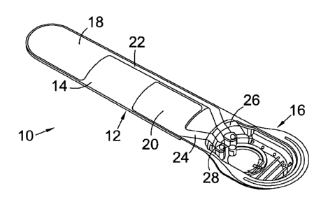
A preferred embodiment of a device according to the invention is shown in
Figures 14 to 17. The device 10 comprises a flexible, non-stretchable
thermoplastic elastomer substrate 12 which includes an elongate tongue 14 at
one end, and a moulded recess 16 at the other.
On the tongue 14 are printed positive 18 and negative 20 electrodes. The
positive is slightly larger than the negative. Each electrode includes a
conductive track 22, 24 leading from the electrode to a respective contact
point 26, 28 located in the recess 16.
Not shown in the figures are an insulative strip arranged between the positive
track 22 and the negative electrode 20, and similar strips at the edges of the
tongue, to prevent unwanted leakage of current.
Within the recess 16 are placed an electrical cell (not shown), and a PCB (not
shown) including suitable circuitry to control the electrodes. Together with
the
conductive tracks 22, 24 and contact points 26, 28, this forms a complete
circuit. A plastic cover is then sonically welded over the recess 16 to seal
the
components. A layer of gel is then placed over the whole device 10; this
provides an electrical contact with a user's limb and helps keep the device
adhered to a user. The gel may be protected in transit by a peelable backing
layer.
The outer surface of the recess 16 is formed with an integral diaphragm
button 30 and an aperture 32 for displaying an LED. The button 30 is
arranged to contact a corresponding button on the battery housing or PCB to
26
CA 02747537 2011-06-17
WO 2010/070332
PCT/GB2009/051713
activate the device. The aperture 32 displays an LED which indicates whether
the device is operating.
27