Note: Descriptions are shown in the official language in which they were submitted.
CA 02747982 2011-06-21
WO 2010/078145 PCT/US2009/069143
Docket No. 83529.0042.PCT
FRONTAL SINUS SPACER
RELATED APPLICATIONS
[0001] This patent application is a continuation-in-part of copending United
States Patent
Application 12/100,361 entitled "Ethmoidotomy System And Implantable Spacer
Devices
Having Therapeutic Substance Delivery Capability For Treatment Of Paranasal
Sinusitis" filed
on April 9, 2008, which is a continuation-in-part of copending United States
Patent Application
11/544,009 entitled "Implantable Devices and Methods for Treating Sinusitis
and Other
Disorders" filed on October 4, 2006, which is a continuation-in-part of Serial
No. 11/234,395
entitled "Devices and Methods for Delivering Therapeutic Substances for the
Treatment of
Sinusitis and Other Disorders" filed on September 23, 2005, which is a
continuation-in-part of
Serial No. 11/037,548 entitled "Devices, Systems and Methods for Treating
Disorders of the Ear,
Nose and Throat" filed on January 17, 2005, which is a continuation-in-part of
Serial No.
10/912,578 entitled "Implantable Device and Methods for Delivering Drugs and
Other
Substances to Treat Sinusitis and Other Disorders" filed on August 4, 2004,
which is a
continuation-in-part of Serial No. 10/829,917 entitled "Devices, Systems and
Methods for
Diagnosing and Treating Sinusitis and Other Disorders of the Ears, Nose and/or
Throat" filed on
April 21, 2004, the entire disclosure of each such application being expressly
incorporated herein
by reference.
FIELD OF THE INVENTION
[0002] The present invention relates generally to medical devices and methods
and more
particularly to substance delivering implants and methods for treating a broad
range of disorders
including but not limited to sinusitis and other ear, nose and throat
disorders.
BACKGROUND
[0003] The paranasal sinuses require adequate ventilation to prevent microbial
chronic
infection within the sinus cavities. Normally, ventilation is provided through
the small natural
openings, known as ostia, through which the sinus cavities open into the nose.
In addition to
ventilation, the natural ostia serve as drainage channels as ciliated cells
lining the interior of the
sinus cavity continually direct a flow of mucus toward the ostia. Thus, when
the natural ostia
1
Doc. # CC-218417 v.1
CA 02747982 2011-06-21
WO 2010/078145 PCT/US2009/069143
Docket No. 83529.0042.PCT
become narrowed or blocked, ventilation and drainage from the sinus cavity is
impaired. The
resultant hypoxia, pH changes and mucus stasis within the sinus cavity gives
rise to an
environment in which some types of microbial growth can flourish. Such
microbial infection
can, in itself, result in further mucosal inflammation and even further
constriction or blockage of
the natural sinus ostium.
Techniques For Improving Ventilation and Drainage of Paranasal Sinuses
[0004] Functional endoscopic sinus surgery (FESS) is a common type of surgery
wherein an
endoscope is inserted into the nose and, under visualization through the
endoscope, the surgeon
may remove diseased or hypertrophic tissue or bone and may surgically enlarge
the ostia of the
sinuses to restore normal ventilation and drainage of the sinuses.
[0005] As an alternative to incisional surgery, in some patients, a balloon
catheter may be
advanced into the constricted sinus ostium and used to dilate the ostium,
thereby eliminating the
need for cutting or removing tissue surrounding the ostium (Balloon
SinuplastyTM technology,
Acclarent, Inc., Menlo Park, California). Examples of such balloon dilation
procedures are
described in United States Patent Application Publications No. 2006/0004286,
2006/0063973,
2006/0210605, 2007/0129751, 2007/0135789, 2007/0167682, 2007/0208252,
2007/0208301 and
2007/0293727, the entire disclosure of each such patent application being
expressly incorporated
herein by reference.
Implantation of Stents and Space Occupying Materials to Deter Re-Occlusion
Following Surgery
[0006] In cases where tissue adjacent to the ostium has been surgically
removed or incised,
post-operative scar tissue, fibrosis, polyposis or tissue ingrowth can result
in re-occlusion of the
sinus ostium. To deter such re-occlusion of frontal and sphenoid sinuses
following surgery,
small tubular stents have been placed in the surgically altered sinus ostium
or outflow tract for a
limited time period following surgery.
[0007] One example of a commercially available frontal sinus stent is the
FreemanTM
Frontal Sinus Stent (InHealth Technologies, Inc., Carpinteria, California. The
FreemanTM stent
comprises a silicon tube that has flanges on either end to retain the stent
within the frontal
outflow tract for a desired period of time following surgery. Other
commercially available
2
Doc. # CC-218417 v.1
CA 02747982 2011-06-21
WO 2010/078145 PCT/US2009/069143
Docket No. 83529.0042.PCT
frontal sinus stents include the Jasin Frontal Sinus Stent (Medtronic Xomed,
Inc., Jacksonville,
Fla.), and the Salman FES Stent (Boston Medical Products, Westborough, Mass.).
[0008] A sphenoid sinus stent is described in United States Patent No.
7,235,099
(Duncavage, et al.). This stent comprises a soft compressible plastic tube
having a generally
hemispherical hollow dome on one end. The diameter of the dome is greater than
the
predetermined diameter of the plastic tube. The stent further includes an
annular flange located a
predetermined distance from the hemispherical dome. The device is designed to
be fitted through
a surgically enlarged ostium of the sphenoid sinus such that the dome resides
within the sinus
cavity and the flange abuts the bony wall surrounding the ostium. This stent
serves maintain
patency of the surgically altered ostium during the postoperative period and
allows
irrigation/suctioning through the lumen of the stent. This sphenoid sinus
stent is also
commercially available as the SP-82020 Sphenoid Sinus Stent (Micromedics,
Inc., St. Paul,
Minnesota).
[0009] The above-described frontal and sphenoid sinus stents do not deliver
therapeutic
substances. Thus, they are frequently used concurrently with orally
administered drugs (e.g.,
corticosteroids) and/or topical nasal sprays.
[0010] In some cases, in lieu of a stent, surgeons may place gel-like
materials within the
surgically altered ostium or outflow tract to prevent ingrowth of scar tissue
during the post-
surgical period. One example of such material is the MeroPackTM Bioresorbable
Nasal Dressing
and Sinus Stent available from Medtronic ENT, Inc., Jacksonville, Florida. The
MeroPackTM
material consists of 80 percent esterified hyaluronic acid and 20 percent
collagen. This material
is inserted while in its dry state and, upon hydration, swells to 1.0cm
diameter in about six
seconds. When in its hydrated state, this material is a biocompatible, muco-
adhesive gel.
Local Drug Delivery In the Treatment of Sinus Disease
[0011] Various drug delivery implants have been proposed for use in or around
the paranasal
sinuses to treat sinusitis and/or to deter re-occlusion of surgically altered
outflow tracts or ostia
following surgery.
[0012] For example, United States Patent Application Publication No.
20050043706 (Eaton
et al.) describes biodegradable implants for treating sinusitis, such implants
having a size, shape,
3
Doc. # CC-218417 v.1
CA 02747982 2011-06-21
WO 2010/078145 PCT/US2009/069143
Docket No. 83529.0042.PCT
density, viscosity, and/or mucoadhesiveness that prevents them from being
substantially cleared
by the mucociliary lining of the sinuses during the intended treatment period.
These
biodegradable implants deliver therapeutic agents such as antibiotics,
steroids or both. These
biodegradable implants may be in various forms such as rods, pellets, beads,
strips, or
microparticles, and may be delivered into a sinus in various pharmaceutically
acceptable carriers.
[0013] Also, United States Patent Application Publication No. 20070005094
(Eaton et al.)
describes implantable devices useable for the treatment of paranasal sinus
conditions. The
devices include cavity members that have a first collapsed configuration that
permits the device
to pass through a sinus ostium and a second expanded configuration after
placement into the
sinus cavity. In addition to a cavity member, the devices may include a nasal
portion and an
ostial member that is configured to reside within the sinus ostium. The cavity
member is attached
to the distal end of the ostial member. The nasal portion is attached to the
proximal end of the
ostial member and lies within the nasal passage. The active agent may be
incorporated into all
portions of the device or only included in the expandable cavity member, the
ostial member, or
nasal portion.
[0014] Some investigators have proposed adding drug delivery capability to
frontal sinus
stents to deliver controlled amounts of drug to the surgically altered outflow
tract following
frontal sinus surgery. For example, United States Patent Application
Publication
2004/0116958A1 (Gopferich et al.) describes a tubular sheath or "spacer"
formed of
biodegradable or non-biodegradable polymer that, prior to insertion in the
frontal outflow tract,
is loaded with a controlled amount of an active substance, such as a
corticosteroid or anti-
proliferative agent. After surgery to create a fenestration in a frontal sinus
as been performed,
the sheath (which has been preloaded with the active substance) is inserted
into the surgically
created fenestration where it a) deters closure of the surgically created
fenestration, b) serves as a
conduit to facilitate drainage from the sinus and c) delivers the active
substance. In some
embodiments, the sheath is formed of multiple layers of polymeric material,
one or more of
which is/are loaded with the active substance and one or more of which is/are
free of the active
substance. In other embodiments, the sheath has a "hollow body" which forms a
reservoir
system wherein the active substance is contained and a membrane which controls
the release of
the active substance from the reservoir. In some embodiments, the sheath may
be anchored by
4
Doc. # CC-218417 v.1
CA 02747982 2011-06-21
WO 2010/078145 PCT/US2009/069143
Docket No. 83529.0042.PCT
causing the end of the sheath that extends into the sinus to swell or
otherwise enlarge. Also,
United States Patent Application Publication No. 2005/0245906 (Makower et al.)
describes a
biodegradable polymeric device that comprises a spacer positionable within a
sinus ostium. The
spacer has a plurality of substance-eluting struts. The device may be
implanted such that the
struts are substantially parallel to the cilial flow of mucus along the sinus
cavity walls so that
normal mucociliary transport is not interrupted.
[0015] Additionally, various other types of implantable drug delivery devices
have been
proposed for use in the nose and/or paranasal sinuses. For example, United
States Patent No.
3,948,254 (Zaffaroni) describes implantable drug delivery reservoirs having
microporous walls.
The reservoir may be formed of a solid drug carrier that is permeable to
passage of the drug and
the rate of passage of the drug through the microporous wall may be slower
than the rate at
which the drug passes through the solid drug carrier that forms the reservoir.
Zaffaroni also
describes a number of applications for the implantable drug delivery devices
including placement
in a nasal passage. Specifically, Zaffaroni claims a nasal delivery device for
dispensing a drug
within a nasal passage at a controlled rate wherein the nasal device is
comprised of (a) a wall
defining the device dimensioned for insertion and placement within a nasal
passage, with the
wall formed of a nasal acceptable microporous material, (b) a reservoir
surrounded by the wall
and comprised of a solid carrier permeable to drug and containing drug in an
amount sufficient
for the device to meter it at a continuous and controlled rate for a prolonged
period of time from
the device, (c) a liquid medium permeable to the passage of drug by diffusion
charged in the
micropores, and (d) wherein the device releases drug when in a nasal
environment by passage of
drug from the carrier and through the liquid to the exterior of the device to
produce a useful
result. The entire disclosure of United States Patent No. 3,948,254
(Zaffaroni) is expressly
incorporated herein by reference.
[0016] Other publications have also reported that introduction of drugs
directly into the
paranasal sinuses is effective in the treatment of sinusitis. See,.Tarasov,
D.I., et al., Application
of Drugs Based on Polymers in the Treatment of Acute and Chronic Maxillary
Sinusitis, Vestn
Otorinolaringol. Vol. 6, Pages 45-7 (1978). Also, R. Deutschmann, et al., A
Contribution to the
Topical Treatment of [Maxillary] Sinusitis Preliminary Communication, Stomat.
DDR 26
(1976), 585-592 describes the placement of a resorbable drug delivery depot
within the maxillary
Doc. # CC-218417 v.1
CA 02747982 2011-06-21
WO 2010/078145 PCT/US2009/069143
Docket No. 83529.0042.PCT
sinus for the purposes of eluting drugs, specifically Chloramphenicol. In this
clinical series a
water soluble gelatin was used as carrier and was mixed with the drug prior to
application and
introduced as a mass into the sinus. Since the substance had little mechanical
integrity and
dissolved in a relatively short timeframe, to achieve a therapeutic effect,
the author suggested
that it must be instilled every 2 to 3 days. An alternative to gelatin could
be a sponge loaded
with the therapeutic substance as suggested in United States Patent No.
6,398,758 (Jacobsen, et
al.). In this patent directed at delivering a sustained release device against
the wall of a blood
vessel, a hollow cylindrical sponge is loaded with drug and pressed against
the wall. This allows
the drug to contact the wall while sustaining blood flow within the center of
the lumen. Further,
a skin is provided to direct the drug into the walls of the blood vessel and
prevent drug from
flowing into the lumen. While sponges loaded with drug at the time of their
application do
permit some degree of sustained release, the time required to load them also
correlates closely
the time over which they will elute substance. Thus, if delivery is required
for a longer period of
time additional mechanisms must be employed to regulate their release.
[0017] There are also several examples in the patent literature where various
sustained
release mechanisms have generally been proposed using systems with drugs pre-
incorporated
into matrices or polymers. These include 3,948,254 (Zaffaroni), US
2003/0185872A2
(Kochinke), WO 92/15286 (Shikani), and 5,512,055 (Domb, et al.). In general,
these references
discuss various materials and structures that may be used to construct
sustained drug delivery
vehicles and provide a good overview of the state of sustained drug delivery
art. While helpful
in laying out certain materials and schemes for creating sustained release
systems for drugs,
these references do not, however, describe specific methods, means or
structures which would
permit them to be easily adapted for intended uses that are targeted in the
present application.
[0018] Other examples of implantable drug delivery devices include those
described in
United States Patent Nos. 3,993,073; 4,217,898; 5,304,123; 6,042,561;
6,183,461; 6,780,168 and
6,783,522, the entire disclosure of each such patent being expressly
incorporated herein by
reference.
Techniques for Treatment of Ethmoid Disease
[0019] To date, the use of stents and spacers in relation to nose and sinus
surgery has been
largely limited to placement in the frontal outflow tract or sphenoid sinus
ostium following
6
Doc. # CC-218417 v.1
CA 02747982 2011-06-21
WO 2010/078145 PCT/US2009/069143
Docket No. 83529.0042.PCT
surgery wherein tissue and bone have been cut away or removed. However, as new
devices and
methods become available for the treatment of other types of nasal and sinus
disorders, there will
likely be a need for intranasal or sinus spacers and stents (with or without
drug eluting
capabilities) suitable for placement at various locations lot limited to the
frontal outflow tract.
[0020] In the prior art, diseased ethmoid air cells have sometimes been
treated by a
procedure known as an ethmoidectomy wherein a man made passageway is formed
between the
interiors of the ethmoid air cells and the nasal cavity. Stenting and/or
delivery of drugs or other
therapeutic substances into these manmade ethmoidectomy passageways has been,
in at least
some cases, desirable. To accomplish this, strips of gauze soaked with
medication may be
pushed into the manmade opening and later extracted. Also, in this regard,
United States Patent
No. 6,543,452 (Lavigne) describes a nasal intubation device that comprises a
flexible tube
having a flanged distal tip whereon the flanges generally from an arrow shape.
The distal tip of
this device is capable of penetrating through tissue (e.g., through the
ethmoid bulla) to a desired
position (e.g., within the ethmoid air cells). Openings are formed in a distal
portion of the
intubation device so that medication (e.g., a typical steroid) injected
through the flexible tube
will flow out of the tube into contact with the adjacent area (e.g., the
diseased ethmoid air cells).
In some cases, a cannula-trocar may be initially inserted and the nasal
intubation device may
then be advanced through that cannula-trocar. Also, European Patent
Publication EP0624349
(Milewski) describes a balloon-tipped catheter having an anatomically shaped
balloon which
may be inserted through a surgically created opening into a body cavity (e.g.,
frontal sinus or
ethmoid cell) and inflated to create a tamponade by being shaped to suit the
anatomical shape of
the cavity.
Techniques for Treating the Frontal Sinus
[0021] Various of the above challenges are also specifically relevant to the
treatment of the
frontal sinuses. Additionally, due to the unique anatomy of the frontal
sinuses, there are
additional challenges. That is, accessing the frontal sinuses can require
specialized
instrumentality. Moreover, it has been found that conventional FESS procedures
on the frontal
sinuses have a higher tendency of scarring. Such scarring can lead to a
relapse of insufficient
drainage and ventilation.
7
Doc. # CC-218417 v.1
CA 02747982 2011-06-21
WO 2010/078145 PCT/US2009/069143
Docket No. 83529.0042.PCT
[0022] Although corticosteroids have been found to be effective in reducing
reactive scarring
in the frontal sinuses, there remains a number of key limitations. Nasal
sprays and ointments
generally do not reach critical areas around the frontal sinus outflow tract.
Also, it can be
difficult to deliver interventional devices deep within the frontal sinus
cavity and there are
challenges associated with the retention of interventional instruments in the
frontal outflow tract.
[0023] Accordingly, there remains a need for the development of new devices
and methods
for delivering drugs and other therapeutic or diagnostic substances over a
sustained period of
time into paranasal sinuses, Eustachian tubes, middle ear and/or other
locations within the body
for the treatment of sinusitis, otitis or other diseases and disorders. In
particular, there is a need
for an approach to conveniently and effectively access and treat the sinuses
such as the frontal
sinus.
[0024] The present disclosure address these and other needs.
8
Doc. # CC-218417 v.1
CA 02747982 2011-06-21
WO 2010/078145 PCT/US2009/069143
Docket No. 83529.0042.PCT
SUMMARY
[0025] The present invention provides substance delivering spacer devices and
methods
including expandable reservoirs that are implantable in paranasal sinuses and
other cavities,
openings and passageways of the body to maintain patency and/or to provide
sustained local
delivery of a therapeutic or diagnostic substance. Also provided are sinus
penetrator devices,
systems and methods for creating ethmoidotomy openings or other openings in
the walls of
paranasal sinuses or other anatomical structures.
[0026] In one particular approach, a system and method have been developed to
specifically
treat the frontal sinuses. The system can include an elongate shapeable tube
or sheath adapted to
navigate patient anatomy and to present structure for accessing the frontal
sinuses. Various
approaches to substance delivery spacers with retention structure have also
been developed. In
this way, compensations can be made for variations in patient anatomy. Also,
in one aspect, the
spacer device can additionally include an atraumatic tip such as that formed
by a soft polymer.
[0027] One embodiment of a substance delivery spacer adapted to treat a
frontal sinus
includes a shaft and an expandable reservoir attached to a distal portion of
the shaft. The
reservoir can be introduced within a patient in a collapsed configuration,
mounted to the frontal
sinuses and then expanded. To expand the reservoir, a substance such as a drug
or other
therapeutic substance can be loaded within the reservoir. Additionally, the
reservoir can embody
openings through which the drug or therapeutic substance can elute to thereby
treat the frontal
sinuses. Moreover, the shaft can be cut to length as desired when leaving the
spacer at the
interventional site. The spacer can further include retention structure
configured to facilitate
securing the spacer at or within the frontal sinuses. In this regard, one or
more retention wings
extending along various portions of the reservoir are contemplated. Such wings
can assume a
compressed configuration for delivery to the interventional site and expanded
configurations for
securing the spacer within anatomy.
[0028] One embodiment of a device and method for treating ethmoid sinusitis
involves a
penetrator device that has a distal tip and a stopping mark or member located
a spaced distance
proximal to its distal tip. The distance between the stopping mark or member
and the distal tip is
less than the distance between the ethmoid bulla and the ipsalateral sphenoid
sinus. An
9
Doc. # CC-218417 v.1
CA 02747982 2011-06-21
WO 2010/078145 PCT/US2009/069143
Docket No. 83529.0042.PCT
ethmoidotomy channel is formed by advancing the penetrator through the ethmoid
bulla in a
direction that is non-perpendicular to the skull base and generally directed
toward the ipsalateral
sphenoid sinus. Advancement of the penetrator is stopped when the stopping
mark or member is
approximately flush with the ethmoid bulla. Thereafter, the penetrator is
removed. Optionally, a
stent, spacer or substance delivering spacer device may then be placed in the
ethmoidotomy
channel for a period of time to maintain patency of the channel and/or to
effect local delivery of
a therapeutic substance.
[0029] According to one embodiment, a sinus penetrator device and method may
be used to
form an ethmoidotomy channel or other opening in a paranasal sinus wall or
other body
structure. Such device comprises an elongate penetrator member and a handle
coupled with the
penetrator member at or near its proximal end. A sighting member is disposed
along the handle
or the elongate member at a location to make it visible from an extracorporeal
vantage point
when the distal end of the elongate member is inserted into the patient. Such
sighting member is
useable by a user of the device to generally aim the distal end of the
penetrator in a desired
direction within the patient's body. In some embodiments, the sighting member
may comprise a
fin. The sighting member may extend in a plane that is substantially parallel
to a plane in which
the elongate the penetrator extends from the handle and, optionally may
include another member
(e.g., a cross member) that is substantially normal to the plane in which the
elongate penetrator
extends from the handle. In some embodiments, the elongate penetrator may have
a curve
formed therein and at least a portion of the sighting member may be parallel
to the portion of the
elongate penetrator that is distal to the curve, thereby providing an
indication of the direction or
trajectory on which the distal portion of the elongate penetrator is being
advanced.
[0030] Still further in accordance with the invention, there is provided a
substance delivering
spacer device and method. In one embodiment, the substance delivering spacer
device
comprises a shaft and an expandable reservoir located on the shaft. The
reservoir may be
introduced into a body cavity or opening (e.g., a paranasal sinus,
ethmoidotomy channel, frontal
sinus outflow tract, or other body cavity, opening, passageway) while in a
collapsed
configuration. Thereafter, a therapeutic substance may be loaded into the
reservoir, causing the
reservoir to expand in situ. The shaft may be severed or cut at a desired
location and the
proximal portion of the shaft may be removed after the reservoir has been
loaded. The reservoir
Doc. # CC-218417 v.1
CA 02747982 2011-06-21
WO 2010/078145 PCT/US2009/069143
Docket No. 83529.0042.PCT
is designed such that the substance will elute from the reservoir over a
period of time. The
reservoir may have a side wall and tapered ends, with openings being formed in
the sidewall and
tapered ends such that a therapeutic substance loaded into the reservoir will
elute through the
openings and out of the reservoir. In some embodiments, the device may be
equipped with
apparatus for holding the reservoir in a desired position within the body
(e.g., retention wings,
projections, suture loops, etc.) for holding the reservoir in a desired
position within the body.
[0031] Still further in accordance with the invention, there is provided a
method and system
wherein a substance delivering spacer device of the above-described character
is used in
combination with a sinus penetrator (e.g., the ethmoidotomy device described
above or any other
penetrator) and a sheath. The sheath is initially disposed over the sinus
penetrator and the
penetrator/sheath combination is advanced through a wall of a paranasal sinus
or air cell. The
penetrator is then removed, leaving the sheath in place. The substance
delivering spacer device
is advanced into the sheath. The sheath is then removed, leaving the substance
delivering spacer
device in place within the sinus or air cell. A diagnostic or therapeutic
substance is then loaded
into the reservoir such that the substance will thereafter elute from the
reservoir into the
paranasal sinus or air cell.
[0032] Still further in accordance with the invention, there is provided an
embodiment of a
method for treating sinusitis where an implantable device having a substance
eluting reservoir is
positioned within a paranasal sinus or within the ostium or outflow tract of a
paranasal sinus.
Thereafter, a steroid is introduced into the substance eluting reservoir so
that the steroid elutes
from the reservoir in an amount that is effective to treat the sinusitis.
[0033] Still further aspects and details of the present invention will be
understood upon
reading of the detailed description and examples set forth below.
11
Doc. # CC-218417 v.1
CA 02747982 2011-06-21
WO 2010/078145 PCT/US2009/069143
Docket No. 83529.0042.PCT
BRIEF DESCRIPTION OF THE DRAWINGS
[0034] Figure 1 shows a spacer device of the present invention being used in
conjunction
with an endoscope to treat a human subject.
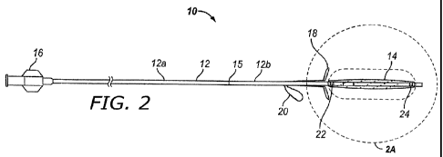
[0035] Figure 2 is a side view of one embodiment of a spacer device of the
present invention.
[0036] Figure 2A is an enlarged longitudinal sectional view of a distal
portion of the device
of Figure A.
[0037] Figure 2B is an enlarged longitudinal sectional view of a distal
portion of the device
of Figure A during infusion of a substance into the reservoir portion of the
device.
[0038] Figure 2C is a longitudinal sectional view through the proximal hub of
the device of
Figure 2.
[0039] Figure 2D is a side view of the device of Figure 2 with a constraining
sheath in a
retracted position.
[0040] Figure 2E is a side view of the device of Figure 2 with a constraining
sheath in an
advanced position.
[0041] Figure 2F is a diagram of the expandable reservoir of the device of
Figure 2.
[0042] Figure 2G is an enlarged view of region 2G of Figure 2F.
[0043] Figure 2 H is a proximal end view of the expandable reservoir of Figure
2F.
[0044] Figure 3 is a side view of a distal portion of another embodiment of a
spacer device of
the present invention incorporating an alternative retention system.
[0045] Figure 4 is a side view of one embodiment of a sheath that is useable
in conjunction
with an ethmoidotomy needle of the present invention.
[0046] Figure 5 is a side view of one embodiment of an ethmoidotomy needle
device of the
present invention.
[0047] Figure 5A is a longitudinal sectional view through a distal portion of
the handpiece of
the ethmoidotomy needle device of Figure 5.
[0048] Figure 5B is a distal end view of the ethmoidotomy needle device of
Figure 5.
12
Doc. # CC-218417 v.1
CA 02747982 2011-06-21
WO 2010/078145 PCT/US2009/069143
Docket No. 83529.0042.PCT
[0049] Figure 5C is a side view of the distal tip of the ethmoidotomy needle
device of Figure
5.
[0050] Figure 5D is a side view of another embodiment of an ethmoidotomy
device of the
present invention incorporating a rotating burr tip.
[0051] Figure 5E is an enlarged side view of the rotating burr tip of the
ethmoidotomy device
of Figure 5D.
[0052] Figure 6 is a side view of the ethmoidotomy needle device of Figure 5
with the sheath
of Figure 4 positioned thereon.
[0053] Figures 7A through 7K show steps in a method for performing an
ethmoidotomy and
implanting a substance delivering spacer device in accordance with the
ethmoidotomy channel in
accordance with the present invention.
[0054] Figures 8A-8G show steps in a method for using a guide catheter for
implantation of
the substance delivering spacer device of Figure 2 within the outflow tract of
the frontal sinus of
a human subject in accordance with the present invention.
[0055] Figures 9A-9D show steps in a method fir using the sheath of Figure 4
and an
optional dilator for implantation of the substance delivering spacer device of
Figure 2 within the
outflow tract of the frontal sinus of a human subject in accordance with the
present invention.
[0056] Figure 1 OA shows a frontal paranasal sinus substance delivery system
according to
one embodiment of the present invention.
[0057] Figures IOB-1OE depict various views and details of a frontal paranasal
sinus
substance delivery device and constraining sheath for the device according to
one embodiment of
the present invention.
[0058] Figures 1 IA-D depict various alternative embodiments of frontal sinus
spacer
devices.
[0059] Figure 12A shows a guide device for guiding a substance delivery device
into a
frontal paranasal sinus according to one embodiment of the present invention.
[0060] Figures 12B-12E depict various views and embodiments of a distal end of
a guide
devices similar to the device of Figure 12A.
13
Doc. # CC-218417 v.1
CA 02747982 2011-06-21
WO 2010/078145 PCT/US2009/069143
Docket No. 83529.0042.PCT
[0061] Figures 13A-13H show steps in a method for treating a frontal sinus.
[0062] Figures 14A-D depict the implantation of various different spacer
devices within the
frontal sinus.
[0063] Figure 15 is a graph showing Lund McKay Scores for 14 human subjects
referred to
below.
[0064] Figure 16 is a bar graph showing the average SNOT 20 scores at various
time points
for human subjects referred to below.
[0065] Figure 17 is a perspective view of a substance delivering/bone
penetrating screw
device of the present invention.
[0066] Figure 17A is a diagram showing the substance delivering/bone
penetrating screw
device of Figure 17 implanted in a bony intranasal structure covered with
mucosal tissue.
[0067] Figure 18 is a partial left/right sagittal section of a human head
showing an
ethmoidotomy needle having a depth controlling stop member inserted through
the subject's
nostril and advanced into the ethmoid sinuses until the stop member has
abutted against the
subject's nose, thereby preventing further advancement of the needle.
[0068] Figures 19A- D are various views of another embodiment of a substance
delivering
spacer device of the present invention incorporating a multi-layer expandable
reservoir.
14
Doc. # CC-218417 v.1
CA 02747982 2011-06-21
WO 2010/078145 PCT/US2009/069143
Docket No. 83529.0042.PCT
DETAILED DESCRIPTION
[0069] The following detailed description and the accompanying drawings are
intended to
describe some, but not necessarily all, examples or embodiments of the
invention. The contents
of this detailed description do not limit the scope of the invention in any
way.
[0070] Figures 1 through 2G show one embodiment of an implantable substance
delivery
device and/or spacer 10 of the present invention. This device 10 comprises an
elongate flexible
catheter shaft 12 having a proximal portion 12a and a distal portion 12b which
may be severed
from one another at separation marker 15. The proximal shaft portion 12a and
distal shaft
portion 12b may be formed of the same or different materials and may have the
same or different
dimensions (e.g., diameter, wall thickness, etc.). For example, in some
embodiments intended
for implantation in paranasal sinuses or other ear, nose or throat locations,
the proximal shaft
portion 12a may be made of a suitable biocompatible material of sufficient
column strength (e.g.,
pushability) to enable a user to push the substance delivery device 10 into
the paranasal anatomy.
One such material is polyamide. In some embodiments, the distal shaft portion
12b may be
made of a more flexible biocompatible material such as nylon or polyethylene
teraphthalate
(PET). A lumen 13 extends continuously through the shaft 12. The distal shaft
portion 12a may
be tapered or necked down to a smaller diameter than the proximal shaft
portion to facilitate
insertion of the device, as described below. A plug 23 is mounted in the
distal end of lumen 13.
The plug 23 may comprise any suitable closure member such as a wall of closed
end on the tube,
an end cap, a mass within the end of the lumen 13 or any other suitable flow
blocking member.
In the particular example shown in the drawings, the plug 23 comprises a
biocompatible
polymeric adhesive disposed within the distal end of lumen 13. In some
embodiments the plug
23 may include a soft, atraumatic (e.g., bulbous or blunt) tip member that
protrudes beyond the
distal end of the distal shaft portion 12b.
[0071] An expandable reservoir 14 is mounted in a collapsed configuration on
the distal shaft
portion 12b near its distal end and expands to an expanded configuration as it
is filled. Details
of one embodiment of such reservoir 14 are seen in Figures 2A and 2B as well
as 2F, 2G and 2H.
In this embodiment, the reservoir 14 comprises a balloon that has a
cylindrical side wall wherein
openings 31 are formed. The reservoir 14 may be formed of any suitable
biocompatible material
and, in some embodiments, may comprise a balloon formed of non-compliant or
semi-compliant
Doc. # CC-218417 v.1
CA 02747982 2011-06-21
WO 2010/078145 PCT/US2009/069143
Docket No. 83529.0042.PCT
material such as Nylon 12. In at least some embodiments, it is preferable that
the material and
wall thickness of the reservoir be such that the reservoir is flexible enough
to a) allow the device
to be extracted and removed from the body without causing significant trauma,
b) not force all of
the contents of the reservoir to come out at once and c) maintain
substantially consistent size of
the openings 31 as the reservoir expands. The number of reservoir(s) 14 (such
as two or more),
the size of the reservoir(s) and the number and size of the openings 31 may
vary on the basis of
the intended implantation location and/or the potency, viscosity, particle
size (for suspensions)
and/or other properties of the substance being delivered. For example, in an
embodiment of the
device 10 intended to be passed through an ethmoidotomy channel and positioned
within an
ethmoid air cell to treat ethmoid sinusitis, the reservoir 14 may have a
length of from about 0.5
cm to about 3.5 cm and typically approximately 2cm, a diameter when fully
expanded of about
0.1 cm to about 0.5 cm and typically approximately 0.3 cm. Also, depending on
the substance
and the intended elution rate, there may be any suitable number of openings
31. Typically there
will be between about 50 and about 5000 openings 31 sized in the range of from
about 5 microns
in diameter to about 80 microns in diameter.
[0072] As described in further below, this embodiment of the reservoir 14 may
be inserted,
in a collapsed configuration, into a body opening, passageway or cavity (such
as, for example, a
frontal sinus outflow tract, paranasal sinus ostium, antrostomy, ethmoidotomy
opening, or other
location within the ear, nose or throat of a subject) and, thereafter, the
reservoir may be loaded
with the desired substance, causing the reservoir to transition to an expanded
state. For example,
for applications intended to treat inflammation of a paranasal sinus using the
particular reservoir
14 described above with the opening size/pattern seen in Figures 2F-2H, the
reservoir 14 may be
loaded with approximately 0.10 ml of an aqueous suspension containing 40 mg/ml
of
Triamcinolone Acetonide Injectable Suspension, USP (Kenalog -40, Bristol-Myers
Squibb,
Somerville, New Jersey). This will cause approximately 100 gg of Triamcinolone
Acetonide to
elute from the reservoir per day over a period of 14 days. When used for the
treatment of fungal
sinusitis or other fungal infections, this reservoir 14 may also be used to
deliver an antifungal
agent such as liposomal or non-liposomal Amphotericin B of 0.3 to 1.5 mg/kg
available from
Pfizer as Amphocin anti-fungal. Systemically administered Amphotericin
typically has limited
distribution from the bloodstream across the mucus membranes and vice versa.
With this
substance delivery device 10, Amphotericin may be released locally into the
mucus membrane
16
Doc. # CC-218417 v.1
CA 02747982 2011-06-21
WO 2010/078145 PCT/US2009/069143
Docket No. 83529.0042.PCT
where the offending fungal organisms are present and therapeutic
concentrations of the drug may
remain in the mucus as it is distributed through the sinuses by ciliary
action. However,
substantial amounts of the Amphotericin will not be substantially absorbed
through the sinus
mucosa, thereby avoiding the potential for untoward systemic effects of the
Amphotericin such
as renal toxicity. Also, this reservoir 14 maybe capable of delivering
solutions as well as
suspensions to the surrounding anatomy. This is especially useful for delivery
of steroids since
most steroids are available as suspensions.
[0073] Also, the reservoir 14 need not be used to deliver a therapeutic
substance in all
applications. It may, in fact, be used as a space occupying device (e.g.,
instead of a sinus stent).
In such applications, the reservoir 14 may be loaded in situ with saline
solution of other inert
liquid causing the reservoir 14 to expand and frictionally engage or contact
adjacent anatomical
structure(s), thereby providing a degree of retention at the desired
implantation location. This
aspect of the reservoir 14 may be further facilitated by the provision of
surface projections on the
reservoir. In cases where it is intended for the reservoir 14 to function
[0074] The reservoir 14 may be relatively small in diameter when in its
collapsed
configuration, thus allowing it to be introduced or removed easily. In
embodiments where the
reservoir 14 is formed of non-compliant or semi-compliant material, the
reservoir 14 will not
undergo substantial elastic deformation in the filling process and thus will
not exert pressure on
its contents in order to expel the desired substance through openings 31.
Rather, the substance in
the reservoir 14 will be carried out through the openings 31 by gravity or by
being in contact
with the mucus or blood that is continually moved along by the ciliary action
in the sinuses. This
non-pressurized delivery allows for the slow release of the desired substance
over several days.
In some other embodiments, the reservoir 14 may be formed of compliant or
elastic material with
small openings 31 such that the material of which the balloon 14 is formed
will contract as
substance passes out of the openings 31, thereby maintaining pressure within
the balloon. In
cases where the reservoir 14 is intended to be inserted into a sinus ostium,
outflow tract,
antrostomy opening or ethmoidectomy/ethmoidotomy opening and used to deliver
an aqueous
suspension containing 40 mg/ml of Triamcinolone Acetonide Injectable
Suspension, USP
(Kenalog -40, Bristol-Myers Squibb, Somerville, New Jersey) or another
substance of similar
consistency, the reservoir 14 may have approximately 2200 laser cut openings
31 approximately
17
Doc. # CC-218417 v.1
CA 02747982 2011-06-21
WO 2010/078145 PCT/US2009/069143
Docket No. 83529.0042.PCT
20 to 40 microns in diameter formed in the sidewall of the reservoir 14. As
seen in Figures 2F-
2H, the openings 31 may be aligned in longitudinal rows and the positioning of
the individual
openings 31 may be staggered one row to the next. In this particular example,
the longitudinal
distance Dl between individual openings is 0.30 +/- 0.03 mm and the distance
D2 between rows
is 0.68 +/- 0.1 mm. Also, in this example, the reservoir has a cylindrical
side wall 14a which
defines the working length of the reservoir, a distal taper 14b which
transitions from the
cylindrical side wall 14a to the distal shaft 12b (distal to the reservoir)
and a proximal taper 14c
that transitions from the cylindrical side wall 14a to the distal shaft 12b
(proximal to the
reservoir) and the openings 31 extend onto the proximal and distal tapers 14b,
14c, as shown.
Also in this example, the reservoir 14 has an overall length of about 16 mm
and a working
length (i.e., the length of the cylindrical side wall 14c) of about 13 mm and
is expandable to a
fully expanded diameter of 3.0 to 3.5 mm. Approximately 768 laser cut openings
31 are formed
in the side wall 14a of the reservoir 14. The diameter of each laser cut
opening 31 is 40 microns.
This particular reservoir design, when loaded with 0.31 to 0.35 ml of 40 mg/ml
Triamcinolone
Acetonide Injectable Suspension, USP (Kenalog -40, Bristol-Myers Squibb,
Somerville, New
Jersey) will deliver a dose of approximately 100 g Triamcinolone Acetonide per
day for a
period of 28 days.
[0075] In the particular example shown, the distal shaft portion 12b may be
made of Nylon
12 and may have an outer diameter of 0.028 inches, an inner diameter of 0.020
inches and length
of 17mm. An aperture 28 as seen in Figures lB-1C is formed in the catheter
shaft 12 to
facilitate filling of the reservoir 14. A valve 26 allows the substance (or
component(s) of the
substance) to flow from the lumen 13 of the catheter shaft 12 into the
reservoir 14 (see Figure
1 C) but does not allow substantial backflow from the reservoir 14 into the
lumen 13 (see Figure
1B). The valve 26 may comprise any suitable type of one way valve. In the
particular
embodiment shown, the valve 28 comprises an elastomeric sleeve valve made of a
segment of C-
flex thermoplastic elastomer tubing (Consolidated Polymer Technologies, Inc.,
Clearwater,
Florida).
[0076] Optionally, a distal radiopaque marker 24 and proximal radiopaque
marker 22 may be
provided to facilitate the desired positioning of the reservoir 14 within a
subject's body. Each of
these markers 22, 24 may be made of a ring of radiopaque material and may be
mounted on the
18
Doc. # CC-218417 v.1
CA 02747982 2011-06-21
WO 2010/078145 PCT/US2009/069143
Docket No. 83529.0042.PCT
shaft 12 in alignment with each end of the reservoir's cylindrical sidewall
14a. In this particular
example each marker 22, 24 comprises a band of Platinum-Iridium alloy having
outer diameter
0.034 inches and inner diameter 0.030 inches. These markers are visible under
various imaging
techniques including fluoroscopy and CT scanning.
[0077] In the example shown, the proximal shaft portion 12a may be made of
polyimide
tubing of outer diameter 0.0618 inches and inner diameter 0.052 inches and
length 20cm. A hub
16 comprising a female Luer connector made of clear polycarbonate (Part No.
41519, Qosina,
Edgewood, NY) is attached to the proximal end of shaft 12. As seen in Figure
2C, this hub 16
has a proximal bore 100 that gradually narrows to a distal bore 102, thereby
facilitating infusion
of suspensions and viscous liquids. The distal bore 102 is approximately the
same diameter as,
and is continuous with, the shaft lumen 13.
[0078] Additionally, in the example shown, the device incorporates two types
of position
retaining apparatus, namely a suture loop 20 as well as a pair of projections
in the nature of
retention wings 18. The retention wings 18 are located at diametrically
opposed locations on the
shaft 12, proximal to the reservoir 14 to help retain the reservoir 14 at a
desired position within
the body, as will be explained in substantial detail below. In this example,
each retention wing
18 comprises a preformed loop of nickel-titanium (nitinol) wire of diameter
0.0086 inches. Each
retention wing 18 may be flexed or compressed to a collapsed position where it
lies substantially
flat against the outer surface of the shaft 12. However, these retention wings
18 are biased to a
preformed configuration such that, when unconstrained, each retention wing 18
will resiliently
spring outwardly to an extended position wherein it extends at an angle of
from about 65 to 90
degrees relative to the longitudinal axis of the shaft 12. Such pre-forming of
these wings 18 may
be accomplished by heat treating the nitinol wire loop at 520 c for 20 minutes
to produce an
austenite finish temperature (Af) of 20 C. Various alternatives to these
retention wings 18 may
be used. For example, Figure 3 shows an alternative retaining member 88
comprising proximal
and distal resilient elastomeric flanges 90, 92 which are at spaced apart
locations so as to rest
against and engage opposite sides of an anatomical wall or structure. In
Figure 3, the anatomical
wall or structure comprises a bulla or sinus wall formed of bone B covered by
mucosal tissue M.
The distal flange 88 is sufficiently resilient and flexible to collapse while
passing through the
19
Doc. # CC-218417 v.1
CA 02747982 2011-06-21
WO 2010/078145 PCT/US2009/069143
Docket No. 83529.0042.PCT
small opening in the anatomical wall and to thereafter resume its expanded
shape as seen in
Figure 3.
[0079] The suture loop 20 (e.g., an eyelet or ring) may be formed of supple,
flexible,
resilient, elastic or superelastic material such as suture thread or nickel-
titanium alloy (Nitinol)
wire. In the particular embodiment shown, the suture loop is formed of black
monofilament
Nylon non-absorbable surgical suture material having a diameter of 0.0075
inches. The suture
loop 20 may be collapsed against the outer surface of shaft 12. The suture
loop 20 may be
affixed to the outer surface of shaft 12 by winding the wire or other material
around the shaft and
securing the wire to the shaft using a suitable adhesive such as
cyanoacrylate, epoxy or UV
curable adhesive and/or by mounting a polymeric sleeve or heat shrinkable
member about the
portions of wire that are wound around the shaft 12. In some embodiments, the
suture loop may
be colored so as to be visually distinguishable from blood and the red-pink
color of the intra-
nasal mucosa. For example, the suture loop 20 may be black, bright blue or
green in color so as
to be easily locatable by the surgeon. This suture loop 20 may be sutured to
the adjacent tissue
to anchor the distal portion of the device 10 in place.
[0080] As seen in Figures 2D and 2E, a tubular constraining sheath 30 may be
positioned
over the shaft 12. In the particular example shown, this constraining sheath
30 comprises a 10cm
length of plastic tubing having an outer diameter of .084 inches and an inner
diameter of .075
inches. This constraining sheath 30 is moveable back and forth between a
retracted position
(seen in Figure 2D) and an extended position (seen in Figure 2E). When in the
extended
position, the constraining sheath extends over the retention wings 18, suture
loop 20 and the
collapsed reservoir 14, thereby holding the retention wings 18 in their
collapsed positions and
forming a smooth protective covering over the retention wings 18, suture loop
20 and collapsed
reservoir 14. Also, when in the extended position, the constraining sheath 30
will add column
strength to the over all device and will deter kinking of the shaft 12 as it
is pushed through
relatively narrow and/or tortuous anatomical passages. After the device 10 has
been inserted to
the desired position, the constraining sheath 12 may be withdrawn to its
retracted position,
thereby allowing the suture loop 20 to be accessible, the retention wings 18
to spring outwardly
to their extended positions and the reservoir 14 to undergo expansion when it
is subsequently
loaded with the desired substance.
Doc. # CC-218417 v.1
CA 02747982 2011-06-21
WO 2010/078145 PCT/US2009/069143
Docket No. 83529.0042.PCT
[0081] Although the particular examples of the spacer device 10 described
above include a
reservoir 14 formed of a single layer balloon, in some embodiments, the
reservoir may comprise
a balloon having multiple layers with different sized openings in each layer.
The substance may
then be selectively introduced between the particular layers that will
facilitate the desired
delivery of that particular substance at the desired rate. In this regard, by
way of example,
Figures 19 through 19D show another embodiment of a substance delivering
spacer device 610
having a shaft 612 and a multi-layered reservoir balloon 614. The shaft 612
may be constructed
and equipped in the same manner as the shaft 12 of the device 10 described
above. However, in
the embodiment three lumens 616, 618 and 620 extend through the shaft 612 and
the reservoir
614 comprises a balloon having three layers 614a, 614b and 614c. The outermost
layer 614a has
openings 63l a of a first size. The middle layer 614b has openings 631b of a
second size that is
smaller than the size of the openings 631a formed in the outer layer 614a. The
inner-most layer
614c has openings 631c of a third size that is smaller than the size of the
openings 631b formed
in the middle layer 614b. First lumen 616 opens into the space within the
innermost layer 614c.
Second lumen 618 opens into the space between the inner-most layer 614c and
the middle layer
614b. Third lumen 620 opens into the space between the middle layer 614b and
the outer-most
layer 614a. In this manner, the operator may select the particular space into
which a particular
substance is to be infused so that the substance will be required to pass
through either: a) only the
openings 631 a in the outer-most layer 614a; b) the openings 631b in the
middle layer 614b as
well as the openings 631 a in the outer layer 614a; or c) all of the openings
631 a, 63 lb, 631 c in
all three layers 614a, 614b and 614c. In this manner, the rate of elution of
the substance may be
optimized.
[0082] As will be described in more detail below, the substance delivering
spacer device 10,
610 may be implanted in any suitable part or location of the body of a human
or animal subject
to perform a spacing function (e.g., to prevent tissue ingrowth, scarring,
fibrosis, adhesion
formation, etc.) and/or to deliver any desired therapeutic substance. For
example, in ear, nose
and throat applications the device 10, 610 may be implanted in a natural
ostium or man-made
opening formed in any paranasal sinus or air cell or in any other natural,
surgically modified or
surgically created opening or passageway, such as the outflow tract of a
frontal sinus, the
inferior, superior or medial meatus, etc.
21
Doc. # CC-218417 v.1
CA 02747982 2011-06-21
WO 2010/078145 PCT/US2009/069143
Docket No. 83529.0042.PCT
[0083] Figures 4-5E show an example of an ethmoidotomy system that may be used
separately or in conjunction with a substance delivering spacer device 10, 610
of the type
described above. This ethmoidotomy system comprises a sheath 40 seen in Figure
5 and a sinus
needle 60 seen in Figure 6. The sheath 40 and sinus needle 60 may be used
separately or in
combination. The combination of the sheath 40 and sinus needle 60 is shown in
Figure 6.
[0084] The sheath 40 may be formed of a biocompatible polymer such as PEBAX
and
comprises a proximal sheath body 42 of a first diameter, a distal sheath body
44 of a second
diameter (smaller than the first diameter) and a tapered step-down segment 54
between the
proximal sheath body 42 and the distal sheath body 44. A flared region 46 is
located at the
proximal end PE of the sheath 40. A visual marker band 50 is optionally
provided on the
proximal sheath body 42 near its proximal end PE. A second visual marker band
48 is optionally
located on the distal shaft portion 44 approximately 17 mm from the distal end
DE. Also
optionally, radiopaque markers 52, 56 may be provided at spaced apart
locations on the distal
sheath body 44. In the particular example shown, the distal radiopaque marker
56 is located
approximately 1.5 mm from the distal end and the proximal radiopaque marker 52
is located
approximately 17 mm from the distal end DE and beneath the distal edge of
visual marker 48.
Additionally, in some embodiments, optional wing members 53 may extend
laterally from the
distal sheath body 44 in the region of visual marker 48. These optional wing
members 53 may
be constructed in substantially the same manner as the retention wings 18 of
the substance
delivering spacer device 10 described above and, when extended, each wing
member 53 may
have a length of about 2 cm. These optional wing members 53 will abut against
adjacent an
adjacent anatomical structure to limit the distance through which the sheath
40 may be advanced
through an opening or channel within the body. This sheath 40 may be used to
facilitate
insertion of the above-described substance delivering spacer device 10 or this
sheath 40 may be
used alone to facilitate suctioning of matter or for delivery of therapeutic
or diagnostic
substances.
[0085] In the embodiment shown in Figure 5, the sinus needle 60 comprises an
elongate,
curved needle body 62 having a sharp trocar tip 63. The proximal end of the
needle body 62 is
firmly, non-rotateably anchored to handpiece 64. As seen in Figure 5A, this
may be
accomplished by forming a 90 degree bend in the proximal end of the needle
body 62 and
22
Doc. # CC-218417 v.1
CA 02747982 2011-06-21
WO 2010/078145 PCT/US2009/069143
Docket No. 83529.0042.PCT
molding it in place within the handpiece 64 thereby providing a strong
connection and
preventing the needle body 62 from rotating relative to the handpiece 64. In
the embodiment
shown in the drawings, the needle body 48 is formed of solid stainless steel
wire having an outer
diameter of approximately 0.07 inches. A curve 65 is formed in the needle body
62. The needle
body 62 is about 102 mm in length and the center of the curve 65 is located
about 31 mm from
the distal tip 63 of the needle body 62. The curve 65 forms an angle A2 of
approximately 33
degrees. This particular embodiment of the sinus needle 60 is particularly
suited for a needle
ethmoidotomy as described below and the curve 52 allows the distal portion of
the needle body
62 to be advanced through the ethmoid bulla and into the ethmoid air cells
with decreased
potential for inadvertent penetration through the adjacent skull base which
protects the subject's
brain. Also, as indicated in the enlarged views of Figures 5B and 5C, in this
example the trocar
tip 63 has three bevelled edges arranged symmetrically around the central axis
of needle shaft
with each bevelled edge being disposed at an angle B of about 20 degrees
relative to the
longitudinal axis of the needle body 62. This design enables sinus needle
device 60 to be used
for penetration through soft tissue (e.g., mucosa) as well as thin bone (e.g.,
the ethmoid bulla and
other bones separating individual ethmoid air cells.
[0086] The handpiece 64 comprises a sighting member such as a fin 66, a top
elongate
member 70 and a bottom elongate member 68 that is attached to and
substantially parallel to the
top elongate member. The handpiece may also comprise a distal grip portion
72,. All or part of
the handpiece 64 may be coated with an elastomeric material and/or may be
provided with
grooves, ridges or surface configurations that facilitate firm grasping of the
handpiece 64 by the
operator.
[0087] The sighting fin 66 extends from the handpiece in a plane that is
parallel to the plane
of the needle curve 65, thereby providing to the operator a visual indication
of the lateral
direction in which the distal portion of the needle body 62 is advancing even
when the distal end
of the needle body 62 is within the subject's body and out of direct sight of
the operator.
Additionally, the top edge 67 of the vertical sighting fin 66 is parallel to
and in substantial
alignment with the distal portion of the needle body 62, thereby providing to
the operator a
visual indication of the vertical tilt or trajectory on which the needle tip
63 is advancing even
23
Doc. # CC-218417 v.1
CA 02747982 2011-06-21
WO 2010/078145 PCT/US2009/069143
Docket No. 83529.0042.PCT
when the distal end of the needle body 62 is within the subject's body and out
of direct sight of
the operator.
[0088] Figure 6 shows the needle sheath 40 positioned on the sinus needle body
62. As
shown, the length of the needle sheath 40 is such that when the sheath 40 is
fully advanced onto
the needle body 62, the flared region 46 located at the proximal end PE of the
sheath 40 will abut
against the distal surface of the handpiece 64 and the distal tip 63 of the
needle body 62 will
protrude out of and beyond the distal end DE of the sheath 40. The sheath 40
is flexible enough
to conform to the curve 65 of sinus needle body 62, as shown. Optionally, for
some applications,
an optical or electrical image guidance component 74 (e.g., sensors,
reflectors, light sources, etc.)
may be attached to the upper elongate member 70 of the handpiece 64, as seen
in Figure 6,
thereby allowing an optical or electromagnetic image guidance system to be
used, in accordance
with techniques well known in the art of ear, nose and throat surgery, to
determine and/or guide
the positioning of the needle tip 63 within the body of human or animal
subject.
[0089] United States Patents Nos. 5,314,417 entitled "Safety Trocar" and
5,267,965 entitled
"Safety Trocar", the entire disclosures of which are incorporated herein by
reference, disclose
safety mechanisms that may optionally be used in combination with the sinus
needle device 60
and sheath 40.
[0090] As an alternative to a needle body 63 having a sharp tip such as a
trocar tip 63, the
sinus needle may comprise any other suitable tissue penetrating apparatus
capable of forming the
desired penetration through the intended tissue (e.g., for ethmoid
applications, through mucosal
tissue and bone). These other suitable tissue penetrating apparatus include
but are not limited to
rotating drills, burs, bipolar or monopolar radiofrequency or electrocautery
probes, laser probes,
etc. Figures 5D and 5E show one example of an alternative sinus penetrator 60a
which is similar
in construction to the sinus needle 60 described above except that the bottom
elongate member
68 of the handpiece is replaced by a housing 68a having an electric motor (not
shown) positioned
therewithin and an on-off button. Also, in this device, the needle body 62 is
replaced by a
rotating bur assembly which comprises an elongate curved tube 62a having a
flexible rotating
drive shaft 84 extending therethrough and a rotating burr tip 82 attached to
the distal end of the
drive shaft 84, as shown in Figure 5E. Because the drive shaft 84 is flexible,
it is capable of
rotating even though it extends through the curve 65a of the tubular body 62a.
The rotating burr
24
Doc. # CC-218417 v.1
CA 02747982 2011-06-21
WO 2010/078145 PCT/US2009/069143
Docket No. 83529.0042.PCT
tip may be a 0.6 mm, 0.7mm or 0.8mm diamond bur tip and the motor, drive shaft
84 and bur tip
82 may be substantially the same as used in the UltraburTM Fixed Tip Drill
(Invotec
International, Inc., Jacksonville, Florida).
[0091] In other alternative embodiments where the needle 62 is replaced by a
laser probe, a
fiber optic laser waveguide may extend through the probe and a suitable type
of laser light may
be delivered through the wave guide and out of the distal end of the probe to
penetrate through
the desired anatomical structure. For penetration through the ethmoid bulla or
other soft tissue or
bony paranasal structures one suitable type of laser is a holmium:YAG laser.
See, Metson,
Ralph; Holmium:YAG Laser Endoscopic Sinus Surgery: A Randomized, Controlled
Study;
Laryngoscope; 106(1) Supplement 77:1-18 (January 1996).
Treatment of Ethmoid Sinusitis by Needle Ethmoidotomy and Implantation of
Substance
Delivering Spacer Device With Sustained Corticosteroid Delivery
[0092] Figures 7A-7K show one example of a method by which the above-described
sinus
needle device 60, sheath 40 and substance delivery device 10 may be used to
perform a needle
ethmoidotomy, to effectively "stent" the ethmoidotomy channel and to deliver a
therapeutic
substance (e.g., a corticosteroid) into the diseased ethmoid sinuses for a
period of time
postoperatively.
[0093] Initially, as seen in Figure 7A, the needle sheath 40 is placed on the
needle body 62 as
shown in Figure 6. In this embodiment, the inner diameter of the proximal
sheath portion 42 is
large enough to allow the constraining sheath 30 of the substance delivery
device (shown in
Figures 1D and 1E) to pass therethrough, whereas the internal diameter of the
distal sheath
portion 44 is the same or smaller than the outer diameter of the moveable
sheath 30 but still
sufficiently large in diameter to allow the collapsed reservoir 14 and non-
deployed retention
wings 18 to pass thereinto.
[0094] The subject is anesthetized or appropriate analgesia/sedation is
administered. As
shown in Figure 7A, the needle body 62 having the sheath 40 mounted thereon is
inserted
through the subject's nostril along with an endoscope 400 such as a Storz
HopkinsTM II, 0
degree, autoclavable 4mm X l8mm telescope with a Storz Xenon 300TM or Xenon
NovaTM
light source (Karl Storz GmbH & Co., Tuttlingen, Germany). Also, in this
example, a C-arm
fluoroscope system may optionally be used to provide fluoroscopic images
during portions of the
Doc. # CC-218417 v.1
CA 02747982 2011-06-21
WO 2010/078145 PCT/US2009/069143
Docket No. 83529.0042.PCT
procedure. One example of a commercially available C arm fluoroscope system
that is suitable
for this purpose is the OEC 9800 P1usTM Digital Mobile Imaging System (G.E.
OEC Medical
Systems, Inc., Salt Lake City, Utah). The operator may verify that the distal
portion of the
needle body 62 is in the proper vertical tilt and lateral direction by viewing
the sighting fin 66
and its leading edge 67. Under endoscopic guidance, the needle tip 50 is
pushed through the
ethmoid bulla EB and into one or more ethmoid air cells EAC. The approximately
thirty-three
degree angle 65 formed in this embodiment of the sinus needle body 62 allows
the distal tip 63 to
be advanced on a trajectory that is substantially parallel to (or in some
cases even divergent
from) the adjacent skull base SB. In this regard, when the procedure is
performed on an adult
human, the curve 65 of the ethmoidotomy needle body 62 may have a radius of
about 0.75 inch
and may form an angle A of about 33 degrees. The distal portion of the needle
body 62 (i.e., the
portion extending from the curve 62 to its distal tip 63) has a length of
about 24 mm, thereby
allowing for ease of maneuvering the needle/sheath assembly and allowing it to
be inserted along
side an endoscope 400 with the endoscope 400 being above or below the
needle/sheath
assembly. The ethmoidotomy needle body 62 is formed of a .073" diameter 304
stainless steel
wire having a measured tensile strength (ASTM A313-03) in the range of about
253852 to
258665 psi. In cases where an image guidance component 74 is attached to the
handpiece 64 of
the sinus needle device 60, the operator may additionally use known techniques
and apparatus
for optical or electromagnetic image guidance of the advancement of the sinus
needle body 62
relative to the skull base SB and other critical anatomical structures. Also,
the depth of
penetration must be carefully controlled so as not to penetrate all the way
though the sphenoid
wall and into the sphenoid sinus SS. To ensure that the sphenoid wall is not
breached, the
surgeon may choose a sheath 40 wherein the distance from the distal end DE of
the sheath 40 to
the proximal edge of visual marker 48 is less than the distance from the
anterior surface of the
ethmoid bulla EB to the wall of the sphenoid sinus SS. The distal visual
marker 48 on the sheath
40 may then be visualized via the endoscope to gage the depth of penetration
into the ethmoid air
cells. The advancement may be stopped when the proximal end of visual marker
48 is seen to be
flush with the ethmoid bulla EB, thereby ensuring that the sphenoid wall has
not been breached.
Also, if the sheath 40 incorporates the optional wing members 53, the device
may be advanced
until those wing members 53 abut against the anterior surface of the ethmoid
bulla EB.
Additionally, as seen in Figure 18, an optional external stop member 600 may
be attached by any
26
Doc. # CC-218417 v.1
CA 02747982 2011-06-21
WO 2010/078145 PCT/US2009/069143
Docket No. 83529.0042.PCT
suitable means, such as a clip 602, grasper, adhesive, frictional engagement
or any other means,
to the sheath 40 at a location which will cause the stop member 600 to abut
against the subjects
nose, thereby preventing the needle 62 and sheath 40 from being advanced
beyond a safe
distance into the ethmoids. The distance between the proximal and distal
radiographic markers
52, 56 is substantially the same as the length of the reservoir 14 and such
markers 44 may be
viewed by fluoroscopy. The surgeon can use such fluoroscopic image to position
the markers
52, 56 such that they demarcate the locations where the proximal and distal
ends of the reservoir
14 are intended to reside.
[0095] As shown in Figure 7B, after the sheath 40 has been placed in the
desired position,
the needle 49 is withdrawn leaving the sheath 40 in place, with the proximal
end of the sheath 40
extending out of the subject's nostril.
[0096] Prior to insertion of the substance delivering spacer device 10, the
physician may
optionally retract the constraining sheath 30 to expose suture loop 20, and a
length of 2-0 or 3-0
suture material 17 having a straight or curved needle 19 may be passed through
suture loop and
doubled over. The constraining sheath 30 may then be moved to its advanced
position, and the
opposite ends of the doubled over suture 17 will be caused to protrude out of
the proximal end of
the constraining sheath 30 as shown in Figure 7C. The substance delivery
device 10 with its
reservoir in a collapsed state and the constraining sheath 30 in its advanced
position (as shown in
Figure 2E) is then inserted into the proximal end of the needle sheath 40 as
seen in Figure 7C.
[0097] Thereafter, as seen in Figure 7D, the substance delivery device 10 with
the
constraining sheath 30 in its advanced position is advanced through the sheath
40 to a position
where slight resistance to further advancement is felt due to abutment of the
distal end of the
constraining sheath 30 with the narrowed wall of the internal surface of the
tapered segment 54.
[0098] Thereafter, as shown in Figure 7E, the surgeon will apply sufficient
force to
overcome the resistance to advancement, causing the constraining sheath 30 to
move proximally
to its retracted position (shown in Figure 2D) as the distal portion of the
substance delivering
spacer device 10, including the collapsed reservoir 14, advances into the
distal sheath portion 42.
The positioning of the reservoir 14 within the distal sheath portion 42 may
then be verified
fluoroscopically by viewing the positions of the radiographic marker 24 on the
device 10 relative
to the positions of the radiographic markers 44 on the distal sheath portion
42. Also, using these
27
Doc. # CC-218417 v.1
CA 02747982 2011-06-21
WO 2010/078145 PCT/US2009/069143
Docket No. 83529.0042.PCT
markers, the actual positioning of the reservoir 14 relative to the
surrounding anatomy may be
checked.
[0099] Thereafter, as shown in Figure 7F, the sheath 40 with the constraining
sheath 30
contained therein may be withdrawn proximally. This allows the retention wings
18 to spring
outwardly and engage the adjacent septal walls between ethmoid air cells EAC
or alternatively
the internal wall surface of the ethmoid bulla EB. The deployment and
engagement of the
retention wings 18 may be verified fluoroscopically. This also allows the
suture loop 20 to be
exposed within the nasal cavity adjacent to the ethmoid bulla EB. Because the
suture loop is
colored differently from blood and the surrounding mucosa, the exposure of the
suture loop may
also be verified endoscopically.
[00100] Thereafter, as seen in Figure 7G, a syringe containing 0.31cc to
0.35cc of
Triamcinolone Acetonide injectable suspension (Kenalog 40, Brystol-Myers
Squibb Company,
Princeton, New Jersey) is attached to the proximal Luer connector 16 of the
substance delivering
spacer device 10 and the Triamcinolone Acetonide injectable suspension is
injected, thereby
causing the reservoir 14 to expand. In some embodiments, the shaft 12 of the
substance
delivering spacer device 10 may be transparent so that the delivery of the
substance through
lumen 13 may be viewed through the endoscope 400.
[00101] Thereafter, as shown in Figure 7H, the shaft 12 adjacent to proximal
Luer connector
16 is cut thereby removing the Luer hub 16. This allows the sinus needle
sheath 42 with the
constraining sheath 30 contained therein to be removed, thereby freeing the
suture 17 and needle
19 for suturing to an anatomical structure adjacent to the suture loop 20.
Alternatively, in some
embodiments, the sheath 40 can be provided with a longitudinal perforation or
weakened region
which will allow the sheath to be peeled away and removed.
[00102] As seen in Figure 71, the sinus needle sheath 40 with the constraining
sheath
contained therein is removed and the suture 17 is used to attach suture loop
20 to adjacent tissue,
such as the mucosa M of the intranasal septum or that covering the nasal
surface of the ethmoid
bulla EB.
[00103] Thereafter, as seen in Figure 7J, the shaft 12 is cut at or distal to
separation mark 15,
and the proximal shaft 12a is removed.
28
Doc. # CC-218417 v.1
CA 02747982 2011-06-21
WO 2010/078145 PCT/US2009/069143
Docket No. 83529.0042.PCT
[00104] As seen in Figure 7K, this procedure results in an ethmoidotomy
channel or opening
extending into one or more ethmoid air cell(s) EAC with the substance eluting
reservoir 14 and
distal shaft 12b remaining in place for a period of time (e.g., between 1 hour
to 90 days,
preferably between 7 to 29 days, most preferably about 14 days and in some
cases about 7 days)
following the performance of the needle ethmoidotomy procedure. Additionally,
a small amount
of the substance will remain in the distal shaft 12b distal to the location at
which it is cut. This
remaining substance may slowly leak out of the cut end of the distal shaft 12b
thereby providing
medication to adjacent turbinate or other nearby anatomical structures within
the nasal antrum.
[00105] In this ethmoid example, the sinus needle sheath 40 has a distal shaft
portion 44 made
of Nylon having an outer diameter of .087 inches and inner diameter of .075
inches and length of
25mm. Intermediate tapered region 54 is about 5 mm in length and is tapered
from an outer
diameter of .104 inches and an inner diameter of .088 inches at its proximal
end, to an outer
diameter of .092 inches and an inner diameter of .075 inches at its distal
end. Proximal shaft
portion 42 is made of Nylon 12 and has an outer diameter of .102 inches and
inner diameter of
.088 inches and length of 3.5 inches. Distal and proximal sheath markers 44
are made of rings of
a Pt-Ir alloy with an outer diameter of 0.087 inches and an inner diameter of
0.085 inches. The
distal shaft marker 44 is located lmm from the distal end DE of needle sheath
134. Proximal
shaft marker 148 is located 18mm from the distal end of needle sheath 40. The
total length of
needle sheath 40 is 115mm.
[00106] Although the example of Figures 7A-7K is specific to treatment of
ethmoid disease,
the system of devices shown in the example of Figures 3A-3L may also be used
to form
penetration tracts or openings (e.g., antrostomy openings, etc.) in various
paranasal sinuses and
other anatomical structures and to position the substance delivering spacer
device 10 within such
penetration tracts or openings. Additionally, the substance delivering spacer
device 10 may be
used separately from the sinus needle device 60 in various ostia, openings,
incisions and
passageways of the body to act simply as a spacer and/or to deliver a desired
diagnostic or
therapeutic substance. In the treatment of sinus disease, steroids such as
Kenalog -40
(Triamcinolone Acetonide Injectable Suspension, USP) are delivered to a
paranasal region such
as the ethmoid sinuses with device 10.
29
Doc. # CC-218417 v.1
CA 02747982 2011-06-21
WO 2010/078145 PCT/US2009/069143
Docket No. 83529.0042.PCT
[00107] The implantable device 10 can be used to preferably deliver fluids or
suspensions
with a low surface tension. Fluids with low surface tension easily spread
across a surface. This
is especially useful to deliver substances over a large surface area,
especially in anatomical
regions such as ethmoid sinuses that have complicated 3-D geometries. In one
embodiment, the
low surface tension fluid comprises a surfactant. In one method embodiment, a
low surface
tension irrigating fluid containing one or more substances is delivered to the
ethmoid sinuses. In
some embodiments, a substantially inert fluid such as saline solution may be
delivered to
moisten the surrounding tissues and the device may perform a spacing and/or
drainage/ventilation function. In other embodiments, an active substance such
as a therapeutic or
diagnostic substance may be delivered in addition to the spacing and/or
drainage/ventilation
function of the implanted device 10.
[00108] In some applications, the substance delivering spacer device 10 maybe
implanted
within openings (e.g., natural ostia, surgically altered ostia, other man-made
openings) of
paranasal sinuses to facilitate the treatment of a disease or disorder
affecting the paranasal sinus.
In such applications, the opening of the paranasal sinus may be enlarged
(e.g., dilated) before or
after placement of a device 10, 104 of the present invention within such
opening. One such
procedure is balloon dilation of sinus cavity ostia. In such procedure, a
guide catheter having a
substantially fixed shape is inserted through the nose and advanced to a
position where the distal
end of the guide catheter is adjacent to the ostium of a paranasal sinus. A
guidewire is then
advanced through the guide catheter (e.g., RelievaTM Guide Catheter,
Acclarent, Inc., Menlo
Park, California) and into the paranasal sinus. Thereafter, a balloon catheter
(e.g., RelievaTM
Balloon Catheter, Acclarent, Inc., Menlo Park, California) is advanced over
the guidewire and is
used to dilate the ostium of the paranasal sinus, thereby improving drainage
from and/or
ventilation of that paranasal sinus. Examples of such devices and procedures
for balloon dilation
of a paranasal sinus ostium are described in United States Patent Application
Nos.: 10/829,917
entitled "Devices, Systems and Methods for Diagnosing and Treating Sinusitis
and Other
Disorders of the Ears, Nose and/or Throat;" 10/944,270 entitled "Apparatus and
Methods for
Dilating and Modifying Ostia of Paranasal Sinuses and Other Intranasal or
Paranasal Structures;"
11/116,118 entitled "Methods and Devices for Performing Procedures Within the
Ear, Nose,
Throat and Paranasal Sinuses;" 11/150,847 entitled "Devices, Systems And
Methods Useable
For Treating Sinusitus" and 11/234,395 entitled "Devices and Methods for
Delivering
Doc. # CC-218417 v.1
CA 02747982 2011-06-21
WO 2010/078145 PCT/US2009/069143
Docket No. 83529.0042.PCT
Therapeutic Substances for the Treatment of Sinusitis and Other Disorders,"
the entire disclosure
of each such patent application being expressly incorporated herein by
reference.
Treatment of Frontal Sinusitis by Balloon Dilation of Frontal Outflow Tract
and Implantation of
Spacer Device With Sustained Corticosteroid Delivery
[00109] Figures 8A through 8G show an example of one method by which the
substance
delivering spacer device 10 may be placed in the frontal sinus outflow tract
FSO to perform a
stenting and substance delivery function following balloon dilation of the
frontal sinus outflow
tract FSO.
[00110] In this procedure, the endoscope 400 is inserted and, optionally, a C-
arm fluoroscope
(not shown) may also be positioned to provide fluoroscopic images of the
procedure when
desired. Although, for clarity and visual simplicity, the endoscope 400 is
shown only in Figures
8A and 8B, such endoscope 400 may remain in place throughout all or any part
of this procedure
and may be used for real time visualization of the movement and operation of
the devices, as
described in this example.
[00111] Under endoscopic visualization, a frontal sinus guide catheter 500
(e.g., the Relieva
70 Degree Sinus Guide Catheter; Acclarent, Inc., Menlo Park, California) is
inserted through the
nostril and advanced to a position where its distal end is within or aligned
with the frontal sinus
ostium FSO. Such positioning of the guide catheter 500 may be verified by
endoscopic
visualization and/or fluoroscopy.
[00112] Thereafter, a guidewire GW (Relieva Sinus Guidewire; Acclarent, Inc.,
Menlo park,
California) is advanced through the guide catheter 500 and into the frontal
sinus FS, as shown in
Figure 8A. The fluoroscope 404 may be used to verify that the guidewire GW has
become
coiled within the frontal sinus FS.
[00113] Thereafter, as shown in Figure 8B, a dilation catheter 502 (e.g., the
Relieva or
Relieva XL Sinus Balloon Catheter, Acclarent, Inc., Menlo Park, California) is
advanced over
the guidewire GW and through the guide catheter 500 to a position where its
dilator balloon 504
is positioned within the frontal sinus ostium FSO. The fluoroscope 400 may be
used to verify
that the guidewire GW has become coiled within the frontal sinus FS. With the
dilator 504 so
positioned, the dilator balloon 504 is expanded to cause dilation of the
frontal sinus ostium FSO
or other frontal sinus outflow tract. This procedure is described in detail in
copending United
31
Doc. # CC-218417 v.1
CA 02747982 2011-06-21
WO 2010/078145 PCT/US2009/069143
Docket No. 83529.0042.PCT
States Patent Application Serial No. 11/355,512, the entire disclosure of
which is expressly
incorporated herein by reference. After the dilation has been completed, the
dilator balloon 504
is again collapsed and the dilation catheter 502 is removed, leaving the
guidewire 504 in place.
Although this example includes this step of dilating the frontal sinus ostium
FSO, this dilation
step is optional. In some patients, the sinus ostium may have already been
dilated or altered in a
previous surgery or the physician may determine that dilation of the ostium is
not needed prior to
introduction of the spacer device 10.
[00114] Thereafter, the substance delivering spacer device 10 is prepared and
advanced
through the guide catheter 500 and into the frontal sinus FS. Prior to
insertion of the device 10
into the guide catheter 500, the constraining tube 42 may be removed and the
retention wings 18
may be manually folded forward (i.e., in the distal direction) using finger
pressure as the spacer
device 10 is inserted into the proximal end of the guide catheter 500. As the
distal end of the
spacer device emerges out of the distal end of the guide catheter 500, the
retention wings 18 will
spring outwardly and will engage the frontal sinus ostium FSO as shown in
Figure 8C.
Positioning of the reservoir 14 within the frontal sinus and successful
deployment of the
retention wings 18 may be verified fluoroscopically.
[00115] Thereafter, as shown in Figure 8D, a syringe containing 0.31cc to
0.35cc of
Triamcinolone Acetonide injectable suspension (Kenalog 40, Brystol-Myers
Squibb Company,
Princeton, New Jersey) is attached to the proximal Luer connector of the sinus
spacer device 10
and the Triamcinolone Acetonide injectable suspension is injected, thereby
causing the reservoir
14 to expand. Successful expansion of the reservoir 14a may be verified by CT.
[00116] Thereafter, as shown in Figures 8E and 8F, the proximal end of the
sinus spacer
device 10 is cut off and the guide catheter is retracted proximally and
removed. The operator
may grasp the shaft 12 of the spacer device distal to the distal end of the
guide catheter 500 as
the guide catheter is removed to stabilize the spacer device 10 and to prevent
it from being
inadvertently dislodged from the frontal sinus FS during removal of the guide
catheter 500.
[00117] Thereafter, a proximal portion of the shaft 12 of spacer device 10 may
be cut away,
leaving a short length of the shaft 12 hanging within the nose. The suture 17
with straight needle
19 is used to suture the suture tab 20 of the spacer device 10 to tissue
within the nose, thereby
helping to retain the implanted portion of the spacer device 10 in its desired
position for a desired
32
Doc. # CC-218417 v.1
CA 02747982 2011-06-21
WO 2010/078145 PCT/US2009/069143
Docket No. 83529.0042.PCT
time period following the procedure. Some of the substance will remain in the
remaining
segment of shaft 12 distal to the cut and may leak into the nasal cavity
subsequent to the
procedure, thereby providing medication to other structures within the nasal
cavity as well.
Treatment of Frontal Sinusitis by Implantation of Spacer Device With Sustained
Corticosteroid
Delivery
[00118] In this example, Figures 9A-9D show steps in another method in which
frontal
sinusitis is treated in an adult human subject. The frontal sinus ostium FSO
may or may not have
been previously surgically altered or dilated as described above. Under
endoscopic visualization,
a frontal sinus guide catheter 500 (e.g., the Relieva 70 Degree Sinus Guide
Catheter; Acclarent,
Inc., Menlo Park, California) is inserted through the nostril and advanced to
a position where its
distal end is within or aligned with the frontal sinus ostium FSO. Such
positioning of the guide
catheter 500 may be verified by endoscopic visualization and/or fluoroscopy.
[00119] Thereafter, a guidewire GW (Relieva Sinus Guidewire; Acclarent, Inc.,
Menlo park,
California) is advanced through the guide catheter 500 and into the frontal
sinus FS, as shown in
Figure 9A. The fluoroscope 404 may be used to verify that the guidewire GW has
become
coiled within the frontal sinus FS.
[00120] With reference to Figure 9B, after the guidewire GW has been inserted
into the
frontal sinus, the frontal sinus guide catheter 500 is removed, leaving the
guidewire GW in place.
A 5 French vascular dilator 420 (e.g., 5 F vessel dilator (inner diameter of
0.038 in.), Merritt
Medical Systems, Inc., South Jordan, UT). The sheath/dilator combination is
then advanced over
the guidewire GW. The C-arm fluoroscope and/or the endoscope 400 may be used
to observe
the advancement of the sheath/dilator combination to a position where the
proximal radiographic
marker 52 of the sheath 40 is distal to the frontal recess (i.e., within the
cavity of the frontal
sinus). Once the sinus sheath 40 has been advanced to such location while
within the lumen of
the dilator, the 5F dilator 420 and the guidewire GW are removed leaving the
sheath 40 in place,
as shown in Figure 9C.
[00121] Thereafter, the substance spacer device 10 is prepared as described
above and the
constraining tube 30 is placed in its advanced position so as to constrain and
cover the suture
loop 20, retention wings 18 and reservoir 14 in their collapsed positions. The
device 10 is
33
Doc. # CC-218417 v.1
CA 02747982 2011-06-21
WO 2010/078145 PCT/US2009/069143
Docket No. 83529.0042.PCT
advanced into the previously inserted sheath 40 in substantially the same
manner as described
above and shown in Figures 8C-8E.
[00122] Thereafter, the sheath 40 and constraining tube 30 are retracted and a
syringe
containing 0.31cc to 0.35cc of Triamcinolone Acetonide injectable suspension
(Kenalog 40,
Brystol-Myers Squibb Company, Princeton, New Jersey) is attached to the
proximal Luer
connector of the sinus spacer device 10 and the Triamcinolone Acetonide
injectable suspension
is injected, thereby causing the reservoir 14 to expand within the frontal
sinus FS, as previously
described above. Successful expansion of the reservoir 14a may be verified by
CT scan if
desired.
[00123] Thereafter, the proximal end of the sinus spacer device 10 is cut off,
and the sheath 40
and constraining tube 30 are retracted proximally and removed in the same
manner as described
above and shown in Figures 8H-8I.
[00124] Thereafter, a proximal portion of the shaft 12 of spacer device 10 may
be cut away,
leaving a short length of the shaft 12 hanging within the nose. The suture 17
with straight needle
19 is used to suture the suture tab 20 of the spacer device 10 to tissue
within the nose, thereby
helping to retain the implanted portion of the spacer device 10 in its desired
position for a desired
time period following the procedure as seen in Figure 9D. As described above,
Triamcinolone
Acetonide suspension that remains in the short segment of shaft 12 distal to
the cut may
subsequently leak into the nasal cavity NC, providing some additional
therapeutic benefit to
tissues in that area.
Alternative Approach to Frontal Sinusitis Treatment
[00125] Figure l0A shows an embodiment of a substance delivery system 500
configured for
delivering substances to frontal paranasal sinuses. In one embodiment, the
system 500 may
include a substance delivery device 510 including a sinus spacer 511, a sheath
530 for covering
the sinus spacer 530 before and during part of the delivery process, and a
guide device 540
through which the spacer 511 is guided into a frontal paranasal sinus. Each of
these devices will
be described in greater detail below. In some embodiments, the system 500 may
also include a
syringe (not shown) or other substance/fluid injecting device for coupling
with the proximal end
of the substance delivery device 510 and injecting substance/fluid through the
device 510 and
34
Doc. # CC-218417 v.1
CA 02747982 2011-06-21
WO 2010/078145 PCT/US2009/069143
Docket No. 83529.0042.PCT
into the spacer 511. Some embodiments may further include an amount of
preloaded substance
in the syringe, including but not limited to any of the substances listed
herein. Optionally, some
embodiments may also include a handle (not shown) for coupling with the guide
device 540 to
facilitate handling and advancement of the guide device 540 into a nostril of
a patient. Any
suitable handle may be used, in alternative embodiments, for example the
Relieva SidekickTM
Sinus Guide Catheter Handle (Acclarent, Inc., Menlo Park, California) may be
used in one
embodiment.
[00126] Figures I OB through 1 OE depict in further detail the substance
delivery device 510
and implantable sinus spacer 511 shown in FIG. 10A. The device 510 includes an
elongate
flexible catheter shaft 512 having a proximal portion 512a and a distal
portion 512b, where the
distal portion 512b is considered part of the sinus spacer 511. As with
previously described
embodiments, the proximal and distal portions 512a, 512b may be severed from
one another,
such as by cutting, at or near a junction 515. In the embodiment shown,
proximal shaft portion
512a is opaque and distal shaft portion 512b is relatively translucent or
clear, so that during a
procedure, junction 515 may be viewed with an endoscope. Further, the proximal
shaft portion
512a and distal shaft portion 512b may be formed of the same or different
materials and may
have the same or different dimensions (e.g., diameter, wall thickness, etc.).
For example, one
such material is polyamide. In some embodiments, the distal shaft portion 512b
may be made of
a more flexible biocompatible material such as nylon or polyethylene
teraphthalate (PET). A
lumen extends continuously through the shaft 512. Moreover, the distal shaft
portion 512a may
be tapered or necked down to a smaller diameter than the proximal shaft
portion to facilitate
insertion of the device. A plug can be mounted in the distal end of the lumen.
The plug may
comprise any suitable closure member such as a wall of closed end on the tube,
an end cap, a
mass within the end of the lumen or any other suitable flow blocking member.
[00127] The sinus spacer 511 generally includes the distal shaft portion 5
12b, an expandable
reservoir 514 mounted on the distal shaft portion 512b near its distal end,
collapsible retention
members 518 for retaining the spacer 511 in the sinus, and a suture loop 517
allowing a
physician the option of attaching the spacer 511 to mucosal tissue to further
ensure that the
spacer 511 stays in a desired, implanted location. In general, the sinus
spacer 511 may have any
suitable dimensions, features, number of reservoir holes/apertures, sizes and
shapes and numbers
Doc. # CC-218417 v.1
CA 02747982 2011-06-21
WO 2010/078145 PCT/US2009/069143
Docket No. 83529.0042.PCT
of retention members 518 and the like. Many of these features and details have
been described
above and thus will not be repeated here. Several differences between the
frontal sinus spacer
511 and the spacers described above for ethmoid sinuses are as follows. The
wing span of the
retention members 518, measured from tip to tip in the expanded configuration,
is approximately
13-16 mm for the frontal spacer device 511, versus approximately 9-12 mm for
the ethmoid
spacers. Each retention member 518 has an angle, in its expanded configuration
and relative to
the shaft 512, of about 70 degrees, versus about 80 degrees in the ethmoid
spacers. Additionally,
the overall length of the frontal sinus spacer 511 (i.e., the clear distal
shaft portion 5 12b) is
approximately 65 mm +/- 3 mm, versus approximately 50 mm +/- 3 mm in the
ethmoid spacers.
Of course, these features describe but one embodiment of the frontal sinus
spacer 511, and
various alternative embodiments may have different dimensions.
[00128] The reservoir 514 assumes an expanded configuration as it is filled.
Here, the
reservoir 514 may be formed of any suitable biocompatible material and, in
some embodiments,
may comprise a balloon formed of non-compliant or semi-compliant material such
as Nylon 12.
The reservoir can include a plurality of openings and can be configured as
shown in FIGS. 2A
and 2B. Moreover, as before, it is preferable that the material and wall
thickness of the reservoir
be such that the reservoir is flexible enough to a) allow the device to be
extracted and removed
from the body without causing significant trauma, b) not force all of the
contents of the reservoir
to come out at once and c) maintain substantially consistent size of openings
531 [John-I don't
see the 531 label in the drawings.] formed therein as the reservoir expands.
[00129] As described below, the reservoir 514 may be inserted in a collapsed
configuration
into a frontal sinus ostia or outflow tract and, thereafter, the reservoir may
be loaded with the
desired substance, causing the reservoir to transition to an expanded state.
[00130] In some embodiments, the reservoir 514 need not be used to deliver a
therapeutic
substance. It may, in fact, be used as a space occupying device. In such
applications, the
reservoir 514 may be loaded in situ with saline solution or other inert
liquid, causing the
reservoir 514 to expand and frictionally engage or contact adjacent anatomical
structure(s),
thereby providing a degree of retention at the desired implantation location.
This aspect of the
reservoir 514 may be further facilitated by the provision of surface
projections on the reservoir.
36
Doc. # CC-218417 v.1
CA 02747982 2011-06-21
WO 2010/078145 PCT/US2009/069143
Docket No. 83529.0042.PCT
[00131] The reservoir 514 may be relatively small in diameter when in its
collapsed
configuration, thus allowing it to be introduced or removed easily. In
embodiments where the
reservoir 514 is formed of non-compliant or semi-compliant material, the
reservoir 514 will not
undergo substantial elastic deformation in the filling process and thus will
not exert pressure on
its contents in order to expel the desired substance through openings 531.
Rather, the substance
in the reservoir 514 will elute through the openings 531 by gravity and/or by
the passage of
mucus through the sinus via ciliary action. This non-pressurized delivery
allows for the slow
release of the desired substance over several days. In some other embodiments,
the reservoir 514
may be formed of compliant or elastic material with small openings 531 such
that the material of
which the balloon 514 is formed will contract as substance passes out of the
openings 531,
thereby maintaining pressure within the balloon. Also, in this example, the
reservoir has a
cylindrical side wall 514a which defines the working length of the reservoir,
a distal taper 514b
which transitions from the cylindrical side wall 514a to the distal shaft 512b
(distal to the
reservoir) and a proximal taper 514c that transitions from the cylindrical
side wall 514a to the
distal shaft 512b (proximal to the reservoir), and the openings 531 extend
onto the proximal and
distal tapers 514b, 514c, as shown. The reservoir 514 may have dimensions,
openings, and
overall configuration as described previously with reference to other
embodiments.
[00132] The distal shaft portion 512b may be made of Nylon 12 in one
embodiment. As
mentioned above, in one embodiment the full length of sinus spacer 511, from
the distal tip to
the proximal end of the distal shaft portion 512b, may be about 65 mm +/- 3
mm. An aperture is
formed in the catheter shaft 512 to facilitate filling of the reservoir 514. A
valve can also be
provided to allow the substance (or component(s) of the substance) to flow
from the lumen of the
catheter shaft 12 into the reservoir 514 and prevent substantial backflow from
the reservoir 514
into the lumen 513. The valve may comprise any suitable type of one way valve.
[00133] Also, a distal radiopaque marker 524 and proximal radiopaque marker
522 may be
provided to facilitate the desired positioning of the reservoir 514 within a
subject's body. Each
of these markers 522, 524 may be made of a ring of radiopaque material and may
be mounted on
the shaft 512 in alignment with each end of the reservoir's cylindrical
sidewall 514a. For
example, each marker 522, 524 can embody a band of Platinum-Iridium alloy.
These markers
are visible under various imaging techniques including fluoroscopy and CT
scanning.
37
Doc. # CC-218417 v.1
CA 02747982 2011-06-21
WO 2010/078145 PCT/US2009/069143
Docket No. 83529.0042.PCT
[00134] The proximal shaft portion 512a may be made of polyimide tubing in one
embodiment. A hub 516 comprising a female Luer connector made of clear
polycarbonate can
be attached to the proximal end of shaft 512. Proximal shaft portion 512a and
distal shaft portion
512b may, in general, have any of the dimensions, features, materials and the
like of similar
catheter shafts described in reference to other embodiments above. In one
embodiment, the
proximal shaft portion 512a may include a shaft marker 513. The shaft marker
513 may be
positioned along the shaft 512 such that when a distal end of the shaft marker
513 reaches a
proximal end of the stop member 531 on the sheath 530 during advancement
(described in
greater detail below), then the distal end of the sinus spacer 511 is adjacent
the distal end of the
guide device 540. When the proximal end of the shaft marker 513 reaches the
proximal end of
the stop member 531, then the sinus spacer 511 has been advanced out of the
distal end of the
guide 540. Use of the shaft marker during a sinus spacer placement procedure
is described more
fully below in reference to FIGS. 13A-13H.
[00135] Additionally, the implantable substance delivery device or spacer 510
can include a
pair of retention wings 518. It is to be recognized that the spacer 510 can
alternatively include
three or four or more such wings. The retention wings 518 are located at
diametrically opposed
locations on the shaft 512, and extend distally about the reservoir 514. Each
retention wing 518
can embody a preformed loop of nickel-titanium (nitinol) wire. Each retention
wing 518 may be
flexed or compressed to a collapsed position where it lies substantially flat
against the outer
surface of the reservoir 514. However, the retention wings 518 are biased to a
preformed
configuration such that, when unconstrained, each retention wing 518 will
resiliently spring
outwardly to an extended position wherein it extends at an angle of from about
20 degrees to
about 90 degrees relative to the longitudinal axis of the shaft 512, and more
ideally from about
50 degrees to about 80 degrees, and in one embodiment about 70 degrees. In
various
embodiments, the wings 518 may define a wing span of about 9 mm to about 20 mm
or more,
and more ideally from about 13 mm to about 16 mm, and in one embodiment about
15 mm. In
one embodiment, the distal end of the spacer device 511 may be provided with
an atraumatic tip
formed from a soft polymer. Further, in some embodiments, the geometry and
positioning of the
wings 518 may be selected so that drug housed in the reservoir 514 can elute
out of openings 531
both proximal and distal to the wings 518. In an alternative embodiment, wings
518 may be
38
Doc. # CC-218417 v.1
CA 02747982 2011-06-21
WO 2010/078145 PCT/US2009/069143
Docket No. 83529.0042.PCT
positioned along the shaft 512 such that all of the openings 531 are disposed
distal to the wings
518.
[00136] As seen in Figures I OD and 10E, a tubular constraining sheath 530
[This is labelled
30, rather than 530, in the drawings.] may be positioned over the shaft 512 of
the implantable
substance delivery device 510. In one embodiment, for example, this
constraining sheath 530
may be configured as a length of plastic tubing having a length of about 75 mm
+/- 2mm
(although other lengths may be used in alternative embodiments). The
constraining sheath 530 is
moveable back and forth between a retracted position (seen in Figure I OD) and
an extended
position (seen in Figure 1 OE). When in the extended position, the
constraining sheath extends
over the retention wings 518, and the collapsed reservoir 514, thereby holding
the retention
wings 518 in their collapsed positions and forming a smooth protective
covering. When in the
extended position, the constraining sheath 530 will add column strength to the
overall device 510
and will minimize kinking of the shaft 512 as it is pushed through relatively
narrow and/or
tortuous anatomical passages. After the device 510 has been inserted to the
desired position, the
constraining sheath 512 may be withdrawn to its retracted position, thereby
allowing the
retention wings 518 to spring outwardly to their extended positions and the
reservoir 514 to
undergo expansion when it is subsequently loaded with the desired substance.
[00137] In one embodiment, the sheath 530 includes a proximal stop member 531.
The stop
member 531 is configured to abut a proximal luer on the guide device 540 as
the sheath 530 and
the substance delivery device 510 are advanced into the guide 540. Thus, as
the sheath 530 and
substance delivery device 510 are advanced, the stop member 531 stops
advancement of the
sheath 530, and the substance delivery device 510 continues to be advanced, so
that the sinus
spacer exits out of the distal end of the guide 540. In other words, the
sheath 530 with stop
member 531 allows the wings 518 of the sinus spacer 511 to be constrained
before and during
advancement into the patient, until the spacer 511 is advanced out of the
distal end of the guide
540.
[00138] The sheath 530 may also include a slanted or bevelled distal tip 532.
This slanted
distal tip 532 shape may facilitate pulling the sinus spacer 511 back into the
sheath 530 if that is
necessary during a procedure. The tip 532 may also facilitate advancement of
the sheath 530
through the guide 540.
39
Doc. # CC-218417 v.1
CA 02747982 2011-06-21
WO 2010/078145 PCT/US2009/069143
Docket No. 83529.0042.PCT
[00139] Further alternative embodiments of an implantable substance delivery
device are
shown i n FIGS. 1 lA-l 1D. In one approach, as shown in FIG. 1 IA, the
implantable device 510
can include a pair of retention wings 521, 523 forming loops and being located
at staggered
locations along the device. For example, one wing 521 can be attached to a
first side of the
device adjacent the junction between the reservoir 514 and the shaft 512 and a
second wing 523
can be attached to the shaft 512 proximal to the first wing 518. With this
wing placement, the
bottom wing 523 may push against a wall of a frontal recess (the passageway
leading to and just
proximal to the frontal sinus), thereby helping maintain a desired position of
the top wing 521
within the frontal sinus. In another approach (See FIG. 11 B), the wings 521,
523 can be
similarly staggered but configured to project from a common side of the
implantable substance
delivery device 510. In yet another approach (See FIGS. 11C and l1D), in
addition to
embodying diametrically opposed wings 518 extendable along a periphery of the
reservoir 514,
the implantable device 510 can include a bottom wing 525 having various
configurations, such
as that forming an overall L-shape (FIG. I 1 D) or an S-shape (FIG. 11 Q.
Further, the bottom
wing 525 can have a strength and geometry suited for resting on a frontal
recess and for pushing
the shaft 512 of the device laterally. The multiple offset wings also
facilitate enhanced
anchoring of the substance delivery device 510 in the frontal recess/frontal
sinus anatomy.
[00140] Turning now to FIGS. 12A-D, various embodiments of a sheath or guide
540 are
shown. Guides 540 are configured for advancing an implantable device such as
those described
above into a frontal recess and/or frontal paranasal sinus, as shown, for
example, in FIGS. 13A-
13E. Generally, the guide 540 may include a proximal straight portion 542, a
distal curved
portion 544 having a radiopaque distal tip 545, and a proximal luer connector
546. In one
embodiment the guide 540 may be made of a polymer shaped as tube and curved to
form the
distal portion 544, with a straight hypotube disposed over the proximal
portion 542. In one
embodiment, the curved distal portion 544 may be relatively flexible, so that
it has sufficient
pushability to be advanced through the nasal cavity but sufficient flexibility
to prevent damage to
the nasal cavity walls and structures. The overall length, curvature angle and
configuration of
the guide 540 may be designed so that the proximal luer connector 546 remains
outside a
patient's nose while the guide 540 extends through a nostril and nasal cavity
such that the distal
end 545 is positioned in or near a frontal paranasal sinus ostium.
Doc. # CC-218417 v.1
CA 02747982 2011-06-21
WO 2010/078145 PCT/US2009/069143
Docket No. 83529.0042.PCT
[00141] In some embodiments, the sheath or guide 540 may have a shapeable
(bendable,
malleable, etc.) distal portion or tip that can be adjusted to fit a patient's
anatomy. The tip may
be preshaped with a given curve but may be adjusted by the user as necessary.
The material of
such a tip may be a bendable or malleable tubing that may retain its shape and
may also be
repeatedly shaped as needed. For example, in one embodiment, the material may
be a type of
plastic with braided wires 527 (See FIG. 12B) or a spring 529 (See FIG. 12C).
Moreover, such
structure can be encapsulated by the plastic (See FIGS. 12D-E) or the
structure can surround the
plastic (FIGS. 12B-C). The material may also be made of a malleable metal.
Accordingly, if the
preshaped tip of the sheath or guide does not work, the tip may be shaped in
order to navigate
through the frontal recess and into the frontal sinus.
[00142] With reference now to FIGS. 13A-H, another method for treating a
frontal sinus is
depicted. For clarity, this method embodiment is shown without use of a handle
coupled with
the guide 540, however, in alternative embodiments a handle (as described
above) may be
coupled with the guide 540 before the initial advancement step.
[00143] In some embodiments, the substance delivery device 510 may be prepared
as
described above, and the constraining sheath 530 may be placed in its advanced
position to
constrain and cover the retention wings 518 and reservoir 514 in their
collapsed positions. In
alternative embodiments, the sheath 530 and substance delivery device 510 may
be provided in a
configuration ready to be used.
[00144] As shown in Figure 13A, under endoscopic visualization, a frontal
sinus guide
catheter 540 is inserted through the nostril and advanced to a position where
its distal end is
proximal to, within or aligned with the frontal sinus ostium FSO. Such
positioning of the guide
540 may be verified by visualization with an endoscope 400 and/or via
fluoroscopy (for
example, visualizing the radiopaque distal tip of the guide relative to the
frontal sinus ostium).
As mentioned previously, because the frontal sinus has a relatively long
outflow tract (the frontal
recess), it may be desirable to position the spacer device 510 at least
partially within the outflow
tract, and the guide or sheath 540 may be positioned to accomplish this.
[00145] As shown in Figure 13B, as a next step, the substance delivery device
510, with the
constraining sheath 530 disposed over the sinus spacer 511, may be advanced
into and through
the guide catheter 540. Next, as shown in Figure 13C, the substance delivery
device 510 and
41
Doc. # CC-218417 v.1
CA 02747982 2011-06-21
WO 2010/078145 PCT/US2009/069143
Docket No. 83529.0042.PCT
sheath 530 are further advanced through the guide 540 until the stop member
531 on the sheath
abuts the luer connector 546 on the guide 540. The substance delivery device
510 is further
advanced through the guide 540 and the stopped sheath 530, until the distal
end of the shaft
marker 513 reaches the proximal end of the stop member 531, thus indicating
that the distal end
of the sinus spacer 511 is adjacent the distal end of the guide device 540. As
shown in Figure
13D, as the substance delivery device 510 is further advanced through the
guide 540 and the
stopped sheath 530, when the proximal end of the shaft marker 513 reaches the
proximal end of
the stop member 531, then the sinus spacer 511 has been advanced out of the
distal end of the
guide 540, thus deploying the retention wings 518 within the frontal sinus.
[00146] Thereafter, as shown in Figure 13E, a substance is injected through
the catheter shaft
512 into the expandable reservoir 514 of the sinus spacer 511 to expand the
reservoir 514 and to
allow the substance to elute out of holes in the reservoir 514. In one
embodiment, a syringe
containing a substance, for example a corticosteroid suspension such as but
not limited to
triamcinolone acetonide, an antibiotic, an anti-fungal, a nonsteroidal anti-
inflammatory and/or
the like, is attached to the proximal luer connector 516 of the substance
delivery device 510. The
injectable composition is then injected, thereby causing the reservoir 514 to
expand. In some
embodiments, the injected substance and/or the reservoir 514 may be
radiopaque, so that
successful expansion of the reservoir 514a may be verified by fluoroscopy or
other suitable
radiographic technique.
[00147] Next, as shown in Figure 13F, the luer connector 516 on the proximal
end of the
substance delivery device 510 is cut off or otherwise removed. As in Figure
13G, the guide 540
may then be slid proximally over the substance delivery device 510 to remove
the guide 540
from the nostril. As shown in Figure 13H, the sinus spacer device 511 is then
separated from the
proximal catheter shaft portion 512a, such as by inserting scissors into the
nostril and cutting at
or near the junction of the proximal portion 512a and the distal portion 512b.
The proximal
shaft portion 5 12a may then be removed from the patient, leaving the sinus
spacer 511 in place
within the frontal sinus and extending into the frontal outflow tract/frontal
recess. In some
embodiments, the physician may wish to further secure the sinus spacer 511 by
attaching the
eyelet of the suture loop 517 to the mucosal within the nasal cavity. However,
this is not a
42
Doc. # CC-218417 v.1
CA 02747982 2011-06-21
WO 2010/078145 PCT/US2009/069143
Docket No. 83529.0042.PCT
required step, and the retention wings 518 will typically secure the sinus
spacer 511 within the
frontal sinus.
[00148] The sinus spacer 511 may contain any suitable substance or combination
of
substances, such as but not limited to any of the substances listed in the
present application. The
sinus spacer 511 may be left in the frontal sinus for any length of time, such
as from one day to
one year, and more ideally from about 7 days to about 90 days, and even more
ideally from about
14 days to about one month. In some embodiments, substance may only be
delivered to a frontal
paranasal sinus using the frontal sinus spacer 511. Alternatively, substance
may be delivered to
the frontal sinus and the frontal recess or outflow tract and in some cases
even farther proximally
within the nasal cavity. Oftentimes, the delivered substance will be chosen to
have a beneficial
effect not only within a sinus but in any other location in the nasal cavity
to which it might be
delivered.
[00149] As with the embodiments described above, the method just described may
be
performed after the frontal sinus ostium and/or frontal outflow tract is
expanded using a Balloon
SinuplastyTM sinus dilation procedure. Alternatively, the method may be
performed on a
"native," non-operated frontal paranasal sinus. Whether a prior Balloon
SinuplastyTM sinus
dilation procedure is performed may sometimes depend on how wide the frontal
sinus ostium
and/or the frontal outflow tract are.
[00150] Referring now to FIGS. 14A-D, alternative embodiments of the spacer
device 510 are
shown implanted in the frontal sinus. In one alternate approach, the membrane
or reservoir 514
of a spacer 510 has a relatively longer profile such that a portion of its
length extends proximally
within the frontal sinus recess. Additionally, embodiments of a spacer device
510 can include
differently positioned, staggered retention wings 521, 523 are shown in FIGS.
14B and 14C,
where the proximally located wing 523 is placed in apposition with a frontal
sinus recess such
that desired orientation of spacer device 510 is achieved. Also, a spacer
device 510 including a
bottom wing 525 can be implanted such that the bottom wing 525 aids in both
securing and
orienting the spacer at the interventional site (See FIG. 14D).
[00151] Although the accompanying drawings and above-described examples have
specifically shown techniques for implanting the substance delivering spacer
device 510 in the
frontal sinus, similar techniques may be employed to implant the device in
other sinuses. Of
43
Doc. # CC-218417 v.1
CA 02747982 2011-06-21
WO 2010/078145 PCT/US2009/069143
Docket No. 83529.0042.PCT
course, various other approaches and delivery equipment may be required to
accomplish the
same.
Stability of Triamcinolone Acetonide Within Spacer Device Following _
I_mplantation
[00152] A study was performed to confirm that the Triamcinolone Acetonide
injectable
suspension (Kenalog 40, Brystol-Myers Squibb Company, Princeton, New Jersey),
when
loaded into the reservoir 14 of the substance delivering spacer device 10,
remains intact and
capable of pharmacologic activity for at least 30 days following implantation
of the device. In
this study, the reservoir 14 of one device 10 was loaded by injecting 0.31 cc
of the Triamcinolone
Acetonide injectable suspension as described above (hereinafter referred to as
"Triamcinolone
Acetonide loaded reservoir"). The reservoir 14 of a second device 10 was
loaded with saline
(hereinafter referred to as "placebo loaded reservoir") and the reservoir of a
third device
remained empty (hereinafter referred to as "blank reservoir") All three
devices were maintained
under ICH stability conditions (40 C 2 C/ 75% RH 5% RH) in Caron Model 6030
Environmental Stability Chamber. High Performance Liquid Chromatography (HPLC)
was
performed on aliquots of the contents of each reservoir at day 0 and day 30.
The results of this
study are summarized in Table 1 below:
Attributes Results at Each Time Point, %
0-Da 30-Day
Assa : 93.5% 85.4%
Individual Impurity:
RRT=0.31 0.05 0.05
RRT=0.47 0.10 0.11
RRT=0.77 0.03 0.03
RRT=0.87 0.03 0.03
RRT=1.19 0.05 0.04
RRT=1.28 0.02 0.00
RRT=1.37 0.02 0.03
RRT=1.62 0.00 0.06
Total Imurities: 0.3 0.34
RRT = RT of peak/RT of TA (RRT = Relative Retention Time, RT = Retention Time)
% Individual impurity = (Peak area of imp/Total peak area of K-40 sinus
spacer) x 100
Total average peak area of K-40 sinus spacer at 0-day = 5869075
Total average peak area of K-40 sinus spacer at 30-day = 5233043
44
Doc. # CC-218417 v.1
CA 02747982 2011-06-21
WO 2010/078145 PCT/US2009/069143
Docket No. 83529.0042.PCT
% Total impurities = (Total peak area of imp/Total peak area of K-40 sinus
spacer) x 100
Only those impurity peaks > 0.02% are considered significant impurities.
[00153] The Triamcinolone Acetonide potency of samples obtained from the
Triamcinolone
Acetonide loaded reservoir at days 0 and day 30, respectively, was confirmed
by HPLC. In this
study, the levels of impurities rose within acceptable ranges and the potency
of Triamcinolone
Acetonide present in the Triamcinolone Acetonide reservoir remained sufficient
to cause the
intended local anti-inflammatory effect for at least 30 days.
Efficacy of Triamcinolone Acetonide Delivered Using Substance Delivering
Spacer Device 10
[00154] Use of topical corticosteroid therapy to treat chronic sinus
inflammatory conditions is
based on the rationale that more effective drug concentrations can be achieved
at local receptor
sites in the nasal mucosa, with minimal risk of systemic adverse effects.
Triamcinolone
Acetonide (TA) is a second generation synthetic corticosteroid of which there
are currently six
compounds approved for intranasal use. All six corticosteroids appear to be
relatively equal with
regard to potency and effectiveness. TA was chosen for use in the Ethmoid
Sinus Spacer as the
compound with the longest safety record and for its availability in a
concentrated solution
suitable for use in this device. Specifically, Kenalog-40 was used as it is
one of the approved
and marketed formulations of Triamcinolone Acetonide.
[00155] It has been established that intranasal and/or inhaled doses of
Triamcinolone
Acetonide do not cause hypothalamo-pituitary-adrenal (HPA) suppression even
when drug is
delivered for up to three years. See, Klossek JM et al., Local Safety Of
Intranasal Triamcinolone
Acetonide: Clinical And Histological Aspects Of Nasal Mucosa In The Long-Term
Treatment Of
Perennial Allergic Rhinitis, Rhinology, 39(1):17-22 (2001); Lund, VJ., Maximal
Medical
Therapy for Chronic Rhinosinusitis, Otolaryngol Clin N Am 38, 1301-1310 (2005)
and Laliberte
F et al., Clinical And Pathologic Methods To Assess The Long-Term Safety Or
Nasal
Corticosteroids, Allergy 55(8): 718-722 (2000).
[00156] Locally administered TA has been demonstrated to provide a reduction
in the severity
of both early and late phase reaction to allergens, reduced sensitivity of
local nasal irritant
receptors and reduced local inflammation and a decreased likelihood for
secondary rhinovirus
Doc. # CC-218417 v.1
CA 02747982 2011-06-21
WO 2010/078145 PCT/US2009/069143
Docket No. 83529.0042.PCT
infections. Even long term local delivery of TA to the nasal sinus does not
appear to damage
nasal mucosa.
[00157] The volume of vehicle in the substance delivering spacer device 10
used in this study
has a reservoir 14 that will hold 0.1 ml. when loaded to maximum capacity. If
loaded to
maximum capacity with the Kenalog-40, the reservoir will contain 4mg of TA.
This amount of
TA is roughly equivalent in potency to the 35-40 mg of cortisol produced daily
by normal human
adrenal glands. Thus, a total 4 mg TA, even if released all at once, would not
be expected to
adversely affect adrenal corticoid activity.
[00158] As explained above, the openings 31 in the reservoir 14 of the
substance delivering
spacer device 10 may be designed to limit diffusion of TA from the reservoir
so that only a small
daily dose of drug is delivered over the course of two weeks. In this manner,
the dose delivered
locally into the ethmoids or other paranasal sinuses may be less than the
recommended dose of
TA delivered with commercially available nasal inhalers (e.g., Nasacort
Inhaler, Sanofi-
Aventis, Bridgewater, New Jersey).
[00159] Thirteen human subjects suffering from ethmoid sinusitis were treated
by
performance of a needle ethmoidotomy with post operative delivery of TA by
implantation of a
substance delivering spacer device 10 substantially as described above and
shown in Figures 7A
through 8J. Nine of these subjects were treated bilaterally and the remaining
four unilaterally.
Thus, twenty-two ethmoid sinuses were treated in total. A fourteenth patient
suffering from
frontal sinusitis was treated by a balloon dilation of the frontal outflow
tract and with post
operative delivery of TA by implantation of a substance delivering spacer
device 10 substantially
as described above and shown in Figures 8A through 8G.
Post-Operative Follow-up and Data Collection
Subject Nos. 1-10:
[00160] Blood levels of TA were determined in subjects 1-10 prior to the
administration of
anesthesia and at 1, 2 and 4 hours following implantation and loading of the
substance delivering
spacer device 10. If the particular subject stayed overnight at the treatment
facility, a blood
sample was taken at 23 hours or just prior to discharge. Additional blood
samples were collected
46
Doc. # CC-218417 v.1
CA 02747982 2011-06-21
WO 2010/078145 PCT/US2009/069143
Docket No. 83529.0042.PCT
in subjects 1-10 at 3, 7, 10 and 14 days after the procedure and immediately
prior to explant and
removal of the substance delivering spacer device 10.
[00161] In addition to collection of blood samples as noted above, the patient
was asked to fill
out a Sino-Nasal Outcomes Test (SNOT-20) questionnaire. (SNOT-20 - Piccirillo,
JF et al.,
Psychometric and clinimetric validity of the 20-Item Sino-Nasal Outcome Test
(SNOT-20),
Copyright (0 1996 by Jay F. Piccirillo M.D., Washington University, St. Louis,
Missouri) at
baseline and at 1, 2 and 6 weeks following explant and removal of the
substance delivering
spacer device 10. Additionally, the patient was asked to fill out a
questionnaire specific to the
tolerability of the substance delivering spacer device 10. The device was
removed during an
office visit at day 14 following implantation. Quantification of residual drug
in the device
provided additional information relevant to the elution of triamcinolone
acetonide from the
Ethmoid Sinus Spacer during the implantation of the device.
[00162] Investigators provided post-operative care as required except no
steroid nasal sprays
and nasal rinses were administered. Post-operative antibiotic treatment was
administered at the
discretion of the Investigator, as needed. This was to minimize the effects of
concomitant
medications on the study outcome.
[00163] A final CT scan of the ethmoid sinuses was taken at eight weeks to
assess the
condition of the ethmoid sinuses and when compared to baseline, the degree of
improvement.
Subject Nos. 11-13:
[00164] Subjects Nos. 11 through 13 received the same post-operative care and
blood sample
collection as Subject Nos. 1-10, with the following exceptions:
[00165] Following the baseline blood sample taken prior to the administration
of anesthesia,
blood was drawn at 4 hours following device implantation on the day of the
procedure and days
1, (optional) 3, 7, 14, 21 and 28, prior to explant and removal of the
substance delivering spacer
device 10.
[00166] Six follow-up visits were scheduled throughout the study, at days 3,
7, 14, 21 and 28
and a final visit 10 weeks following the procedure. In addition to collection
of blood samples as
noted above, the patient was asked to fill out a SNOT-20 Quality of Life (QOL)
questionnaire at
baseline, and at 1, 2, and 6 weeks following explant. Additionally, the
patient was asked to fill
47
Doc. # CC-218417 v.1
CA 02747982 2011-06-21
WO 2010/078145 PCT/US2009/069143
Docket No. 83529.0042.PCT
out a questionnaire specific to the tolerability of the Ethmoid Sinus Spacer.
The device was
removed during an office visit at day 28 following implantation.
[00167] Investigators provided post-operative care as required except no
steroid nasal sprays
were administered. Post-operative antibiotic treatment was administered at the
discretion of the
Investigator, as needed. This was to minimize the effects of concomitant
medications on the
study outcome.
[00168] A final CT scan of the ethmoid sinuses was taken at ten weeks (six
weeks post
explant) to assess the condition of the ethmoid sinuses and when compared to
baseline, the
degree of improvement.
Subject No. 14:
[00169] Subject No. 14 was the one who was treated for frontal sinusitis
rather than ethmoid
disease. Subject No. 14 received post-operative follow-up and data collection
(e.g., blood
samples and SNOT-20 questionaires) in substantially the same manner as Subject
Nos. 1-10.
CT Scan Results
[00170] The CT scans were read and improvement in the affected sinuses was
scored by the
Lund McKay scoring method. These Lund McKay Scores are shown graphically in
Figure 15.
At baseline (pre-operative) the average Lund McKay Score was 10.4. The average
Lund McKay
score at follow-up 3.9. Thus, the 14 subjects studied exhibited an average
reduction of 65.1 % in
Lund McKay score.
SNOT-20 And Questionnaire Results
[00171] Figure 16 shows the average improvement from baseline in the SNOT-20
scores as
measured at 1, 2, 4, and 6 weeks (Note: One patient deviated from protocol and
completed the 6
week follow-up visit at four weeks.) A score of 1.07 or below was deemed to be
indicative of a
clinically significant reduction of sinusitis symptoms (i.e., a reduction of
at least 0.8 from the
baseline SNOT 20 score). These data indicate that clinically significant
reductions in the
average SNOT 20 scores were observed at 2, 4 and 6 weeks after the procedure.
[00172] Symptom improvement was also documented in the Patient Questionnaire
administered at one, two and six weeks post procedure. All patients reported
they were
48
Doc. # CC-218417 v.1
CA 02747982 2011-06-21
WO 2010/078145 PCT/US2009/069143
Docket No. 83529.0042.PCT
significantly improved or improved six weeks following the procedure, no
patients reported
feeling worse. At six weeks a majority of patients were satisfied with the
results and stated that if
given this treatment choice again, they would agree to have the surgery.
Statistical Analysis of Results
[00173] The SNOT-20 results were recorded during 5 post-procedural evaluation
visits. The
null and alternative hypotheses evaluated based on this endpoint are as
follows:
Ho: (Day 42 minus baseline) = 0
Ha: (Day 42 minus baseline) 0
[00174] Univariate analysis results from comparing the change in the SNOT-20
scores from
baseline were analyzed using a paired-difference t-test. The results from this
analysis revealed a
significant reduction in the SNOT-20 scores within 7 days of the procedure. A
consistent
reduction was observed during all successive post-procedure evaluation visits.
At the time
enrollment was stopped, 13 of the 14 patients (92.9%) had completed the 42-day
post-procedure
evaluation visit. The reduction from baseline 42 days post-procedure was -
22.08 points on the
SNOT-20 scale, with a standard deviation of 14.69 and a probability value
<0.001. The effect
size of this result is 1.50, which is reflective of a very strong treatment
effect.
Lund-McKay Scores (CT Scan)
[00175] CT scans were performed at baseline and 42 days following the
procedure to derive
the LMK score. The null and alternative hypotheses evaluated based on this
endpoint are as
follows:
Ho: (Day 42 minus baseline) = 0
Ha: (Day 42 minus baseline) 0
[00176] Univariate analysis results from comparing the change in the LMK
scores from
baseline were analyzed using a paired-difference t-test. The results from this
analysis revealed a
significant reduction in the LMK scores 42 days after the procedure. The
reduction for baseline
was 6.50 points on the LMK scale, with a standard deviation of 3.96, and a
probability value
<0.001. The effect size from this result is 1.64, which is reflective of a
very strong treatment
effect.
49
Doc. # CC-218417 v.1
CA 02747982 2011-06-21
WO 2010/078145 PCT/US2009/069143
Docket No. 83529.0042.PCT
[00177] A subset analysis was performed comparing the change in the LMK scores
from the
ethmoid sinuses. The results from this analysis also revealed a significant
reduction in the LMK
scores 42 days after the procedure. The reduction for baseline was 2.21 points
on the LMK
scale, with a standard deviation 1.53, and a probability value <0.001. The
effect size of this
result is 1.44, which is reflective of a very strong treatment effect.
[00178] The primary efficacy endpoint was examined using inferential
statistics. Based on
the performance success of the 14 patients enrolled (100%), the lower 95%
exact binomial
confidence interval was 76.84%.
Blood Plasma Analysis
[00179] To assess the secondary endpoint of the ability of TA to elute from
the substance
delivering spacer device 10 to over time, TA concentrations were determined in
blood plasma
from each of the collected blood samples. These data are summarized in Table 2
below.
Table 2
Summary of Plasma /levels of TA at Different Time Points
(measured in picograms per mL)
Time of Maximum
Plasma # # Concentration Average
Sample Patients Patients Detected Amount
Post Dose Tested Detected ( /mL) ( /mL)
1 hr 10 10 168 65.66
2 hr 10 10 237 77.18
4 hour 13 13 273 86.32
24 hour 9 9 142 51.64
3 days 14 13 82.1 26.65
7 days 13 8 149 32.56
days 10 5 86.5 33.30
14 days 14 8 85 22.08
21 days 3 3 15.8 10.88
28 days 3 2 7.94 7.36
[00180] Low (i.e., not systemically active) but detectable TA levels were
determined in the
subjects' blood plasma as far out as 28 days following implantation and
loading of the substance
delivering spacer device 10.
Doc. # CC-218417 v.1
CA 02747982 2011-06-21
WO 2010/078145 PCT/US2009/069143
Docket No. 83529.0042.PCT
Conclusion
[00181] Locally effective doses of TA were delivered from the substance
delivering spacer
device 10 for up to 28 days post-procedure. The CT scans indicated significant
reduction in
sinus inflammation. The subjects also realized substantial improvement in
sinusitis symptoms
on the basis of the SNOT 20 and patient questionnaire results.
[00182] The term substance as used herein is to be broadly construed to
include any feasible
drugs, prodrugs, proteins, gene therapy preparations, cells, diagnostic
agents, contrast or imaging
agents, biologicals, etc. Such substances may be in bound or free form, liquid
or solid, colloid or
other suspension, solution or may be in the form of a gas or other fluid or
non-fluid. For
example, in some applications where it is desired to treat or prevent a
microbial infection, the
substance delivered may comprise a pharmaceutically acceptable salt or dosage
form of an
antimicrobial agent (e.g., antibiotic, antiviral, antiparasitic, antifungal,
etc.), a corticosteroid or
other anti-inflammatory (e.g., an NSAID), a decongestant (e.g.,
vasoconstrictor), a mucous
thinning agent (e.g., an expectorant or mucolytic), an agent that prevents of
modifies an allergic
response (e.g., an antihistamine, cytokine inhibitor, leucotriene inhibitor,
IgE inhibitor,
immunomodulator), an anesthetic agent with or without a vasoconstriction
agents (e.g.
Xylocaine with or without Epinephrine), an analgesic agent, an allergen or
another substance that
causes secretion of mucous by tissues, hemostatic agents to stop bleeding,
anti-proliferative
agents, cytotoxic agents e.g. alcohol, biological agents such as protein
molecules, stem cells,
genes or gene therapy preparations, viral vectors carrying proteins or nucleic
acids such as DNA
or mRNA coding for important therapeutic functions or substances, cauterizing
agents e.g. silver
nitrate, etc.
[00183] Some non-limiting examples of antimicrobial agents that may be used in
this
invention include acyclovir, amantadine, rimantadine, oseltamivir, zanamivir,
aminoglycosides
(e.g., amikacin, gentamicin and tobramycin), amoxicillin,
amoxicillin/clavulanate, amphotericin
B, ampicillin, ampicillin/sulbactam, atovaquone, azithromycin, cefazolin,
cefepime, cefotaxime,
cefotetan, cefpodoxime, ceftazidime, ceftizoxime, ceftriaxone, cefuroxime,
cefuroxime axetil,
cephalexin, chloramphenicol, clotrimazole, ciprofloxacin, clarithromycin,
clindamycin, dapsone,
dicloxacillin, doxycycline, erythromycin, fluconazole, foscamet, ganciclovir,
atifloxacin,
imipenem/cilastatin, isoniazid, itraconazole, ketoconazole, metronidazole,
nafcillin, nafcillin,
51
Doc. # CC-218417 v.1
CA 02747982 2011-06-21
WO 2010/078145 PCT/US2009/069143
Docket No. 83529.0042.PCT
nystatin, penicillins including penicillin G, pentamidine,
piperacillin/tazobactam, rifampin,
quinupristin-dalfopristin, ticarcillin/clavulanate,
trimethoprim/sulfamethoxazole, valacyclovir,
vancomycin, mafenide, silver sulfadiazine, mupirocin, nystatin,
triamcinolone/nystatin,
clotrimazole/betamethasone, clotrimazole, ketoconazole, butoconazole,
miconazole, tioconazole,
detergent-like chemicals that disrupt or disable microbes (e.g., nonoxynol-9,
octoxynol-9,
benzalkonium chloride, menfegol, and N-docasanol); chemicals that block
microbial attachment
to target cells and/or inhibits entry of infectious pathogens (e.g., sulphated
and sulponated
polymers such as PC-515 (carrageenan), Pro-2000, and Dextrin 2 Sulphate);
antiretroviral agents
(e.g., PMPA gel) that prevent retroviruses from replicating in the cells;
genetically engineered or
naturally occurring antibodies that combat pathogens such as anti-viral
antibodies genetically
engineered from plants known as "plantibodies;" agents which change the
condition of the tissue
to make it hostile to the pathogen (such as substances which alter mucosal pH
(e.g., Buffer Gel
and Acidform); non-pathogenic or "friendly" microbes that cause the production
of hydrogen
peroxide or other substances that kill or inhibit the growth of pathogenic
microbes (e.g.,
lactobacillus); antimicrobial proteins or peptides such as those described in
United States Patent
No. 6,716,813 (Lin et al.) which is expressly incorporated herein by reference
or antimicrobial
metals (e.g., colloidal silver).
[00184] Additionally or alternatively, in some applications where it is
desired to treat or
prevent inflammation the substances delivered in this invention may include
various steroids or
other anti-inflammatory agents (e.g., nonsteroidal anti-inflammatory agents or
NSAIDs),
analgesic agents or antipyretic agents. For example, corticosteroids that have
previously
administered by intranasal administration may be used, such as beclomethasone
(Vancenase or
Beconase ), flunisolide (Nasalide ), fluticasone proprionate (Flonase ),
triamcinolone
acetonide (Nasacort ), budesonide (Rhinocort Aqua ), loterednol etabonate
(Locort) and
mometasone (Nasonex ). Other salt forms of the aforementioned corticosteroids
may also be
used. Also, other non-limiting examples of steroids that may be useable in the
present invention
include but are not limited to aclometasone, desonide, hydrocortisone,
betamethasone,
clocortolone, desoximetasone, fluocinolone, flurandrenolide, mometasone,
prednicarbate;
amcinonide, desoximetasone, diflorasone, fluocinolone, fluocinonide,
halcinonide, clobetasol,
augmented betamethasone, diflorasone, halobetasol, prednisone, dexamethasone
and
methylprednisolone. Other anti-inflammatory, analgesic or antipyretic agents
that may be used
52
Doc. # CC-218417 v.1
CA 02747982 2011-06-21
WO 2010/078145 PCT/US2009/069143
Docket No. 83529.0042.PCT
include the nonselective COX inhibitors (e.g., salicylic acid derivatives,
aspirin, sodium
salicylate, choline magnesium trisalicylate, salsalate, diflunisal,
sulfasalazine and olsalazine;
para-aminophenol derivatives such as acetaminophen; indole and indene acetic
acids such as
indomethacin and sulindac; heteroaryl acetic acids such as tolmetin, dicofenac
and ketorolac;
arylpropionic acids such as ibuprofen, naproxen, flurbiprofen, ketoprofen,
fenoprofen and
oxaprozin; anthranilic acids (fenamates) such as mefenamic acid and meloxicam;
enolic acids
such as the oxicams (piroxicam, meloxicam) and alkanones such as nabumetone)
and Selective
COX-2 Inhibitors (e.g., diaryl-substituted furanones such as rofecoxib; diaryl-
substituted
pyrazoles such as celecoxib; indole acetic acids such as etodolac and
sulfonanilides such as
nimesulide).
[00185] Additionally or alternatively, in some applications, such as those
where it is desired to
treat or prevent an allergic or immune response and/or cellular proliferation,
the substances
delivered in this invention may include a) various cytokine inhibitors such as
humanized anti-
cytokine antibodies, anti-cytokine receptor antibodies, recombinant (new cell
resulting from
genetic recombination) antagonists, or soluble receptors; b) various
leucotriene modifiers such as
zafirlukast, montelukast and zileuton; c) immunoglobulin E (IgE) inhibitors
such as Omalizumab
(an anti-IgE monoclonal antibody formerly called rhu Mab-E25) and secretory
leukocyte
protease inhibitor).
[00186] Additionally or alternatively, in some applications, such as those
where it is desired to
shrink mucosal tissue, cause decongestion or effect hemostasis, the substances
delivered in this
invention may include various vasoconstrictors for decongestant and or
hemostatic purposes
including but not limited to pseudoephedrine, xylometazoline, oxymetazoline,
phenylephrine,
epinephrine, etc.
[00187] Additionally or alternatively, in some applications, such as those
where it is desired to
facilitate the flow of mucous, the substances delivered in this invention may
include various
mucolytics or other agents that modify the viscosity or consistency of mucous
or mucoid
secretions, including but not limited to acetylcysteine (MucomystTM,
MucosilTM) and
guaifenesin.
[00188] Additionally or alternatively, in some applications such as those
where it is desired to
prevent or deter histamine release, the substances delivered in this invention
may include various
53
Doc. # CC-218417 v.1
CA 02747982 2011-06-21
WO 2010/078145 PCT/US2009/069143
Docket No. 83529.0042.PCT
mast cell stabilizers or drugs which prevent the release of histamine such as
cromolyn (e.g.,
Nasal Chrom ) and nedocromil.
[00189] Additionally or alternatively, in some applications such as those
where it is desired to
prevent or inhibit the effect of histamine, the substances delivered in this
invention may include
various antihistamines such as azelastine (e.g., Astylin ), diphenhydramine,
loratidine, etc.
[00190] Additionally or alternatively, in some embodiments such as those where
it is desired
to dissolve, degrade, cut, break or remodel bone or cartilage, the substances
delivered in this
invention may include substances that weaken or modify bone and/or cartilage
to facilitate other
procedures of this invention wherein bone or cartilage is remodeled, reshaped,
broken or
removed. One example of such an agent would be a calcium chelator such as EDTA
that could
be injected or delivered in a substance delivery implant next to a region of
bone that is to be
remodeled or modified. Another example would be a preparation consisting of or
containing
bone degrading cells such as osteoclasts. Other examples would include various
enzymes of
material that may soften or break down components of bone or cartilage such as
collagenase
(CGN), trypsin, trypsin/EDTA, hyaluronidase, and tosyllysylchloromethane
(TLCM).
[00191] Additionally or alternatively, in some applications, the substances
delivered in this
invention may include other classes of substances that are used to treat
rhinitis, nasal polyps,
nasal inflammation, and other disorders of the ear, nose and throat including
but not limited to
anti-cholinergic agents that tend to dry up nasal secretions such as
ipratropium (Atrovent
Nasal ), as well as other agents not listed here.
[00192] Additionally or alternatively, in some applications such as those
where it is desired to
draw fluid from polyps or edematous tissue, the substances delivered in this
invention may
include locally or topically acting diuretics such as furosemide and/or
hyperosmolar agents such
as sodium chloride gel or other salt preparations that draw water from tissue
or substances that
directly or indirectly change the osmolar content of the mucous to cause more
water to exit the
tissue to shrink the polyps directly at their site.
[00193] Additionally or alternatively, in some applications such as those
wherein it is desired
to treat a tumor or cancerous lesion, the substances delivered in this
invention may include
antitumor agents (e.g., cancer chemotherapeutic agents, biological response
modifiers,
54
Doc. # CC-218417 v.1
CA 02747982 2011-06-21
WO 2010/078145 PCT/US2009/069143
Docket No. 83529.0042.PCT
vascularization inhibitors, hormone receptor blockers, cryotherapeutic agents
or other agents that
destroy or inhibit neoplasia or tumorigenesis) such as; alkylating agents or
other agents which
directly kill cancer cells by attacking their DNA (e.g., cyclophosphamide,
isophosphamide),
nitrosoureas or other agents which kill cancer cells by inhibiting changes
necessary for cellular
DNA repair (e.g., carmustine (BCNU) and lomustine (CCNU)), antimetabolites and
other agents
that block cancer cell growth by interfering with certain cell functions,
usually DNA synthesis
(e.g., 6 mercaptopurine and 5-fluorouracil (5FU), antitumor antibiotics and
other compounds that
act by binding or intercalating DNA and preventing RNA synthesis (e.g.,
doxorubicin,
daunorubicin, epirubicin, idarubicin, mitomycin-C and bleomycin) plant (vinca)
alkaloids and
other anti-tumor agents derived from plants (e.g., vincristine and
vinblastine), steroid hormones,
hormone inhibitors, hormone receptor antagonists and other agents which affect
the growth of
hormone-responsive cancers (e.g., tamoxifen, herceptin, aromatase ingibitors
such as
aminoglutethamide and formestane, trriazole inhibitors such as letrozole and
anastrazole,
steroidal inhibitors such as exemestane), anti-angiogenic proteins, small
molecules, gene
therapies and/or other agents that inhibit angiogenesis or vascularization of
tumors (e.g., meth-1,
meth-2, thalidomide), bevacizumab (Avastin), squalamine, endostatin,
angiostatin, Angiozyme,
AE-941 (Neovastat), CC-5013 (Revimid), medi-522 (Vitaxin), 2-methoxyestradiol
(2ME2,
Panzem), carboxyamidotriazole (CAI), combretastatin A4 prodrug (CA4P), SU6668,
SU11248,
BMS-275291, COL-3, EMD 121974, IMC-1C11, IM862, TNP-470, celecoxib (Celebrex),
rofecoxib (Vioxx), interferon alpha, interleukin-12 (IL-12) or any of the
compounds identified in
Science Vol. 289, Pages 1197-1201 (August 17, 2000) which is expressly
incorporated herein by
reference, biological response modifiers (e.g., interferon, bacillus calmette-
guerin (BCG),
monoclonal antibodies, interluken 2, granulocyte colony stimulating factor
(GCSF), etc.), PGDF
receptor antagonists, herceptin, asparaginase, busulphan, carboplatin,
cisplatin, carmustine,
cchlorambucil, cytarabine, dacarbazine, etoposide, flucarbazine, flurouracil,
gemcitabine,
hydroxyurea, ifosphamide, irinotecan, lomustine, melphalan, mercaptopurine,
methotrexate,
thioguanine, thiotepa, tomudex, topotecan, treosulfan, vinblastine,
vincristine, mitoazitrone,
oxaliplatin, procarbazine, streptocin, taxol, taxotere, analogs/congeners and
derivatives of such
compounds as well as other antitumor agents not listed here.
[00194] Additionally or alternatively, in some applications such as those
where it is desired to
grow new cells or to modify existing cells, the substances delivered in this
invention may include
Doc. # CC-218417 v.1
CA 02747982 2011-06-21
WO 2010/078145 PCT/US2009/069143
Docket No. 83529.0042.PCT
cells (mucosal cells, fibroblasts, stem cells or genetically engineered cells)
as well as genes and
gene delivery vehicles like plasmids, adenoviral vectors or naked DNA, mRNA,
etc. injected
with genes that code for anti-inflammatory substances, etc., and, as mentioned
above, osteoclasts
that modify or soften bone when so desired.
[00195] Any of the devices and methods described herein may also be used to
deliver
substances to the brain or alter the functioning of the olfactory system. Such
examples include,
the delivery of energy or the deposition of devices and/or substances and/or
substance delivering
implant(s) to occlude or alter olfactory perception, to suppress appetite or
otherwise treat obesity,
epilepsy (e.g., barbiturates such as phenobarbital or mephoobarbital;
iminostilbenes such as
carbamazepine and oxcarbazepine; succinimides such as ethylsuximide; valproic
acid;
benzodiazepines such as clonazepam, clorazepate, diazepam and lorazepam,
gabapentin,
lamotrigine, acetazolamide, felbamate, levetiraceam, tiagabine, topiramate,
zonisamide, etc.),
personality or mental disorders (e.g., antidepressants, antianxiety agents,
antipsychotics, etc.) ,
chronic pain, Parkinson's disease (e.g., dopamine receptor agonists such as
bromocriptine,
pergolide, ropinitrol and pramipexole; dopamine precursors such as levodopa;
COMT inhibitors
such as tolcapone and entacapone; selegiline; muscarinic receptor antagonists
such as
trihexyphenidyl, benztropine and diphenhydramine) and Alzheimer's disease,
Huntington's
disease or other dementias, disorders of cognition or chronic degenerative
diseases (e.g. tacrine,
donepezil, rivastigmine, galantamine, fluoxetine, carbamazepine, clozapine,
clonazepam and
proteins or genetic therapies that inhibit the formation of beta-amyloid
plaques), etc.
[00196] The devices and methods disclosed herein may be used to deliver
several
combinations of two or more substances disclosed herein to a suitable target
anatomical region.
In one particular embodiment, the devices and methods disclosed herein are
used to deliver a
combination of an anti-inflammatory agent (e.g. a steroid or an NSAID) and a
mucolytic agent.
[00197] The devices and methods disclosed herein may be used to deliver gels
or viscous
liquids comprising one or more substances to anatomical regions such as
paranasal sinuses.
Such gels or viscous liquids may coat and adhere to a mucous membrane and thus
provide
sustained delivery of one or more substances to the mucous membrane. In one
embodiment, a
plasticized hydrocarbon gel comprising gelatin, pectin and sodium
carboxymethylcellulose and a
suitable substance may be delivered to a mucous membrane such as the mucous
membrane of a
56
Doc. # CC-218417 v.1
CA 02747982 2011-06-21
WO 2010/078145 PCT/US2009/069143
Docket No. 83529.0042.PCT
paranasal sinus. Such gels can be used for sustained delivery of the suitable
substance to the
mucous membrane.
[00198] One or more of the substance reservoirs disclosed herein may comprise
multiple
compartments such that each compartment stores a particular substance
formulation. The
multiple compartments prevent mixing of multiple substance formulations before
substance
formulations are delivered to the anatomy.
[00199] One or more of the substance reservoirs comprising holes or pores may
be filled with
a suitable substance at a sufficiently high pressure to cause a portion of the
substance to squirt
out of the holes or pores. This process may be used to deliver an initial
bolus of the substance to
the surrounding anatomy.
[00200] One or more of the substance reservoirs disclosed herein may be filled
with a suitable
substance after the substance reservoir is introduced in an anatomical region.
Alternatively, one
or more of the substance reservoirs disclosed herein may be filled with a
suitable substance
before the substance reservoir is introduced in an anatomical region.
Alternatively, one or more
of the substance reservoirs disclosed herein may be pre-filled with a solid,
lyophilized or
concentrated substance. The solid, lyophilized or concentrated substance is
converted to an
active form by introducing a solvent into the substance reservoir. This may be
done just before
or after the substance reservoir is introduced in an anatomical region.
Alternatively, one or more
of the substance reservoirs disclosed herein may be pre-filled with an
inactive form of a
substance. The inactive form of the substance is converted to an active form
by introducing an
activating agent into the substance reservoir. This may be done just before or
after the substance
reservoir is introduced in an anatomical region.
[00201] It is to be further appreciated that, as described herein, the
implantable portion of a
substance delivering spacer device 10 may include a through lumen that may
function as a vent
and/or drain when such implantable portion device is in a paranasal sinus, air
cell, Eustachian
tube, opening formed in the tympanum or any other location within the body.
[00202] The devices and methods disclosed herein may be used to mark an
anatomical region
with a suitable imageable marker. For example, the devices and methods
disclosed herein may
be used to deliver a radio opaque marker such as a radio opaque contrast agent
to an ostium of a
57
Doc. # CC-218417 v.1
CA 02747982 2011-06-21
WO 2010/078145 PCT/US2009/069143
Docket No. 83529.0042.PCT
paranasal sinus. This enables a user to image the ostium of the paranasal
sinus using X-rays or
fluoroscopy.
[00203] One or more of the substance delivery devices disclosed herein may
comprise a
curved, bent or angled region to enable the drug delivery devices to navigate
through the
anatomy.
[00204] The distal-most regions of one or more substance delivery devices
disclosed herein
may comprise an atraumatic tip. The atraumatic tip is used to prevent or
reduce damage to the
anatomy by the distal-most regions of the one or more substance delivery
devices.
[00205] The outer surface of one of more substance delivery devices disclosed
herein may
comprise a coating that reduces or eliminates the risk of encrusting of the
outer surface by a
biological material. In one embodiment, the coating comprises a material that
absorbs water to
form a gel. Examples of such materials include, but are not limited to
hyaluronic acid, etc.
[00206] One or more of the substance delivery devices disclosed herein may be
designed to be
easily removable from the anatomy after completion of a treatment.
[00207] One or more of the substance delivery devices disclosed herein may be
refilled after a
significant volume of substance filled in a substance reservoir has been
delivered to the anatomy.
[00208] One or more of the substance delivery devices disclosed herein may
comprise one or
more markers to enable a user to locate and/or navigate the substance delivery
devices through
the anatomy. For example, the substance delivery devices may comprise visual
markers to
enable the user to determine the depth of insertion of the substance delivery
devices into the
anatomy. In another example, the substance delivery devices may comprise
imaging markers to
enable the user to locate and/or navigate the substance delivery devices using
imaging modalities
such as X-rays, MRI, etc.
[00209] As used herein, the term "opening of a paranasal sinus" shall include
any opening in a
paranasal sinus or air cell such as natural ostia, surgically altered natural
ostia, surgically created
openings, antrostomy openings, ostiotomy openings, burr holes, drilled holes,
ethmoidotomy
openings, ethmoidectomy openings, natural or man made passageways, etc.
[00210] As used herein, the term "implantable" shall include any device that
is maintained in
the body of a human or animal for a period ranging from 30 minutes to 60 days.
58
Doc. # CC-218417 v.1
CA 02747982 2011-06-21
WO 2010/078145 PCT/US2009/069143
Docket No. 83529.0042.PCT
[00211] In each of the above-described examples wherein an endoscope 400 is
employed, the
endoscope 400 is shown as being inserted separately form the other devices.
However, in any
applications or embodiments of the invention where feasible, an endoscope may
be attached to or
integrated with one or more of the other devices used during the procedure as
described in parent
application Serial No. .
[00212] It is to be appreciated that Examples 2 and 3 above describe
techniques which may be
used for introducing the spacer device 10 into frontal sinuses which may or
may not have been
previously altered by surgery or prior balloon dilations. In some cases, such
as where the frontal
outflow tract has been previously dilated or modified by surgery so that the
frontal sinus FS is
relatively easy to access, the operator may simply deliver the spacer device
10 through the
constraining tube 42 (or sinus sheath 40, 40a) and into the frontal sinus,
with or without the use
of forceps or other operative instruments, thereby eliminating the need for
the use of a guide
catheter, guidewire, dilator or other devices for guiding or facilitating
advancement of the spacer
device 10 into the frontal sinus as described in Examples 2 and 3.
[00213] It is to be further appreciated that, although Examples 1, 2 and 3
above describe
Triamcinolone Acetonide injectable suspension (Kenalog 40, Brystol-Myers
Squibb Company,
Princeton, New Jersey) as the therapeutic agent that is loaded into and elutes
from the reservoir,
various other therapeutic agents may be used in addition to, or as an
alternative to, this
Triamcinolone Acetonide injectable suspension. In some cases where it is
desired to use the
implanted spacer device 1 Oa to deliver a steroid, the steroid may be prepared
as a solution rather
than a suspension. In such cases, the steroid will be dissolved in a suitable,
biologically
compatible solvent. For example, Cyclodextrins have been described as suitable
solvents for
dissolution of at least some steroids. Khomutov, S.M., Dovbnya, D.V. and
Donova, M.V.,
Dissolution of a Mixture of Steroids in Cyclodextrin Solutions: a Model
Description;
Pharmaceutical Chemistry Journal; Vol. 35, No. Ii, pp.627-629 (November,
2001).
[00214] In some instances, the devices of the present invention may be used to
deliver steroids
or other substances in formulations that are commercially available as, or
otherwise suitable for,
intra-nasal delivery to the nasal mucosa as nasal drops or sprays (i.e., nasal
solutions). In at least
some cases, such nasal solutions are prepared so that they are similar to
nasal secretions and,
thus, do not interfere with normal ciliary action. Such nasal solutions
usually are isotonic and
59
Doc. # CC-218417 v.1
CA 02747982 2011-06-21
WO 2010/078145 PCT/US2009/069143
Docket No. 83529.0042.PCT
slightly buffered to a pH of 5.5 to 6.5. In addition, antimicrobial
preservatives, similar to those
used in ophthalmic preparations, and appropriate drug stabilizers, if
required, may be included in
the formulation. Various commercial nasal preparations are known and include,
for example,
antibiotics, steroids, antihistamines, decongestants and ipitropium bromide.
[00215] Where possible and appropriate, any of the substances delivered by
devices of the
present invention may be in the form of liposomes or nanoparticles (e.g.,
nanocapsules). The
formation and use of liposomes is generally known to those of skill in the
art. Liposomes are
formed from phospholipids dispersed in an aqueous medium such that they
spontaneously form
multilamellar concentric bilayer vesicles sometimes referred to as
multilamellar vesicles
(MLVs). MLVs are typically from 25 nm to 4 gm in diameter. When sonicated,
MLVs form
small unilamellar vesicles (SUVs) of about 200 to 500 angstroms in diameters
having cores
which contain the aqueous solution. In general, when dispersed in an aqueous
medium,
phospholipids can form various structures other than liposomes, depending on
the molar ratio of
lipid to water. At low molar lipd to water ratios, liposomes will form. The
physical
characteristics of liposomes depend on pH, tonicity and the presence or non-
presence of divalent
cations. Liposomes can interact with cells by different mechanisms, including
1) endocytosis
(e.g., phagocytosis of the liposome by cells such as macrophages and
neutrophils), adsorption to
the cell surface, 2) interaction with cell-surface components, 3) fusion with
the plasma cell
membrane by insertion of the lipid bilayer of the liposome into the plasma
membrane or 4)
transfer of liposomal lipids to cellular or subcellular membranes, or vice
versa. Varying the
liposome formulation can alter which mechanism(s) by which the lyposomes will
interact with
cells in the paranasal sinus, nasal mucosa, etc.
[00216] A nanocapsule is any nanoparticle that consists of a shell and a
space, in which
desired substances may be placed. Techniques for forming nanocapsules are
known in the art.
Polymeric nanocapsules can be made in specific sizes and shapes. They can be
produced as
monodisperse particles which have precisely defined physical and chemical
properties and, thus,
can be tailored to facilitate release of the therapeutic or diagnostic
substance in response to
particular bimolecular triggering mechanisms, such as pH, mucous flow or other
conditions
present within the paranasal sinus or other area in the ear, nose or throat
where the device is
implanted. Nanocapsules can be used in the present invention as "smart drugs"
which have
Doc. # CC-218417 v.1
CA 02747982 2011-06-21
WO 2010/078145 PCT/US2009/069143
Docket No. 83529.0042.PCT
specific chemical receptors or binding sites that will bind to specific target
cells (e.g., cancer
cells associated with sinus or nasal tumors or cells associated with
inflammatory conditions.
[00217] It is to be appreciated that the invention has been described
hereabove with reference
to certain examples or embodiments of the invention but that various
additions, deletions,
alterations and modifications may be made to those examples and embodiments
without
departing from the intended spirit and scope of the invention. For example,
any element or
attribute of one embodiment or example may be incorporated into or used with
another
embodiment or example, unless otherwise specified of if to do so would render
the embodiment
or example unsuitable for its intended use. Also, where the steps of a method
or process have
been described or listed in a particular order, the order of such steps may be
changed unless
otherwise specified or unless doing so would render the method or process
unworkable for its
intended purpose. All reasonable additions, deletions, modifications and
alterations are to be
considered equivalents of the described examples and embodiments and are to be
included
within the scope of the following claims.
61
Doc. # CC-218417 v.1