Note: Descriptions are shown in the official language in which they were submitted.
CA 02748117 2011-06-22
WO 2010/075414 PCT/US2009/069244
-1-
METHOD FOR DETECTION OF XMRV
RELATED APPLICATION(S)
This application claims the benefit of U.S. Provisional Application No.
61/203,556, filed on December 23, 2008. The entire teachings of the above
application(s) are incorporated herein by reference.
GOVERNMENT SUPPORT
The invention was supported, in whole or in part, by a grant W81XWH-07-
1-0338 from the Department of Defense Prostate Cancer research program (PCRP).
The Government has certain rights in the invention.
BACKGROUND OF THE INVENTION
Prostate cancer is the leading cause of non-cutaneous malignancies and the
second leading cause of cancer-related deaths among American men. A need
exists
for improved methods of detection, particularly early detection, of prostate
cancer.
SUMMARY OF THE INVENTION
The present invention relates to the identification of Xenotropic murine
leukemia virus (MLV) related virus (XMRV) nucleic acid by polymerase chain
reaction (PCR) analysis (e.g., real time PCR (RT/PCR); nested RT/PCR using Tth
DNA polymerase and Hot start polymerase) and the uses thereof. In particular,
the
invention provides methods for the detection, and in particular early
detection, of
XMRV nucleic acic (e.g., RNA, DNA) in samples (e.g., urine samples; expressed
prostate secretion (EPS), blood, semen, seminal vesicle fluids or the like) of
prostate
cancer patients and normal individuals.
In one aspect, the invention is directed to a method of detecting the presence
of xenotropic MLV related virus (XMRV) in an individual. The method comprises
contacting a sample of the individual with at least one set of primers wherein
the set
of primers comprises at least one forward primer and at least one reverse
primer
CA 02748117 2011-06-22
WO 2010/075414 PCT/US2009/069244
-2-
which are complementary to all or a portion of an XMRV G1 gag nucleotide
sequence, at least one forward primer and at least one reverse primer which
are
complementary to all or a portion of an XMRV G2 gag nucleotide sequence, at
least
one forward primer and at least one reverse primer which are complementary to
all
or a portion of an XMRV G3 gag nucleotide sequence, at least one forward
primer
and at least one reverse primer which are complementary to all or a portion of
an
XMRV El envelope nucleotide sequence, at least one forward primer and at least
one reverse primer which are complementary to all or a portion of an XMRV E2
envelope nucleotide sequence, at least one forward primer and at least one
reverse
primer which are complementary to all or a portion of an XMRV E3 envelope
nucleotide sequence, at least one forward primer and at least one reverse
primer
which are complementary to all or a portion of an XMRV P 1 Pol nucleotide
sequence, or a combination thereof. The sample is maintained under conditions
which amplify the primers if XMRV is present in the sample to produce
amplified
XMRV sequences. Whether amplified XMRV sequences are present in the sample
are detected, wherein if amplified XMRV sequences are detected in the sample,
then
XMRV is present in the individual.
In another aspect, the invention is directed to method of detecting prostate
cancer (e.g., at an early stage) in an individual. The method comprises
contacting a
sample of the individual with at least one set of primers wherein the set of
primers
comprises at least one forward primer and at least one reverse primer which
are
complementary to all or a portion of an XMRV G1 gag nucleotide sequence, at
least
one forward primer and at least one reverse primer which are complementary to
all
or a portion of an XMRV G2 gag nucleotide sequence, at least one forward
primer
and at least one reverse primer which are complementary to all or a portion of
an
XMRV G3 gag nucleotide sequence, at least one forward primer and at least one
reverse primer which are complementary to all or a portion of an XMRV E 1
envelope nucleotide sequence, at least one forward primer and at least one
reverse
primer which are complementary to all or a portion of an XMRV E2 envelope
nucleotide sequence, at least one forward primer and at least one reverse
primer
which are complementary to all or a portion of an XMRV E3 envelope nucleotide
sequence, at least one forward primer and at least one reverse primer which
are
CA 02748117 2011-06-22
WO 2010/075414 PCT/US2009/069244
-3-
complementary to all or a portion of an XMRV P1 Pol nucleotide sequence, or a
combination thereof. The sample is maintained under conditions which amplify
the
primers if XMRV is present in the sample to produce amplified XMRV sequences,
and whether amplified XMRV sequences are present in the sample are detected.
The
detection of amplified XMRV sequences in the sample indicates that the
individual
has prostate cancer at an early stage.
The invention also provides a method of detecting an individual at risk for
developing prostate cancer. The method comprises contacting a sample of the
individual with at least one set of primers wherein the set of primers
comprises at
least one forward primer and at least one reverse primer which are
complementary to
all or a portion of an XMRV G1 gag nucleotide sequence, at least one forward
primer and at least one reverse primer which are complementary to all or a
portion
of an XMRV G2 gag nucleotide sequence, at least one forward primer and at
least
one reverse primer which are complementary to all or a portion of an XMRV G3
gag
nucleotide sequence, at least one forward primer and at least one reverse
primer
which are complementary to all or a portion of an XMRV El envelope nucleotide
sequence, at least one forward primer and at least one reverse primer which
are
complementary to all or a portion of an XMRV E2 envelope nucleotide sequence,
at
least one forward primer and at least one reverse primer which are
complementary to
all or a portion of an XMRV E3 envelope nucleotide sequence, at least one
forward
primer and at least one reverse primer which are complementary to all or a
portion
of an XMRV P1 Pol nucleotide sequence, or a combination thereof. The sample is
maintained under conditions which amplify the primers if XMRV is present in
the
sample to produce amplified XMRV sequences, and whether amplified XMRV
sequences are present in the sample are detected. The detection of amplified
XMRV
sequences in the sample indicates that the individual is at risk for
developing
prostate cancer.
The invention also provides a method of detecting recurrence of prostate
cancer in an individual. The method comprises contacting a sample of the
individual
with at least one set of primers wherein the set of primers comprises at least
one
forward primer and at least one reverse primer which are complementary to all
or a
portion of an XMRV Gl gag nucleotide sequence, at least one forward primer and
at
CA 02748117 2011-06-22
WO 2010/075414 PCT/US2009/069244
-4-
least one reverse primer which are complementary to all or a portion of an
XMRV
G2 gag nucleotide sequence, at least one forward primer and at least one
reverse
primer which are complementary to all or a portion of an XMRV G3 gag
nucleotide
sequence, at least one forward primer and at least one reverse primer which
are
complementary to all or a portion of an XMRV El envelope nucleotide sequence,
at
least one forward primer and at least one reverse primer which are
complementary to
all or a portion of an XMRV E2 envelope nucleotide sequence, at least one
forward
primer and at least one reverse primer which are complementary to all or a
portion
of an XMRV E3 envelope nucleotide sequence, at least one forward primer and at
least one reverse primer which are complementary to all or a portion of an
XMRV
P 1 Pol nucleotide sequence, or a combination thereof. The sample is
maintained
under conditions which amplify the primers if XMRV is present in the sample to
produce amplified XMRV sequences, and whether amplified XMRV sequences are
present in the sample are detected. The detection of amplified XMRV sequences
in
the sample indicates the recurrence of prostate cancer in the individual.
The invention also provides a method of monitoring a treatment of an
individual that has prostate cancer. The method comprises contacting a sample
of the
individual with at least one set of primers wherein the set of primers
comprises at
least one forward primer and at least one reverse primer which are
complementary to
all or a portion of an XMRV Gl gag nucleotide sequence, at least one forward
primer and at least one reverse primer which are complementary to all or a
portion
of an XMRV G2 gag nucleotide sequence, at least one forward primer and at
least
one reverse primer which are complementary to all or a portion of an XMRV G3
gag
nucleotide sequence, at least one forward primer and at least one reverse
primer
which are complementary to all or a portion of an XMRV El envelope nucleotide
sequence, at least one forward primer and at least one reverse primer which
are
complementary to all or a portion of an XMRV E2 envelope nucleotide sequence,
at
least one forward primer and at least one reverse primer which are
complementary to
all or a portion of an XMRV E3 envelope nucleotide sequence, at least one
forward
primer and at least one reverse primer which are complementary to all or a
portion
of an XMRV P1 Pol nucleotide sequence; or a combination thereof. The sample is
maintained under conditions which amplify the primers if XMRV is present in
the
CA 02748117 2011-06-22
WO 2010/075414 PCT/US2009/069244
-5-
sample to produce amplified XMRV sequences, and whether amplified XMRV
sequences are present in the sample are detected. The detection of amplified
XMRV
sequences in the sample indicates that the treatment is likely not effective
or is likely
not yet effective.
BRIEF DESCRIPTION OF THE DRAWINGS
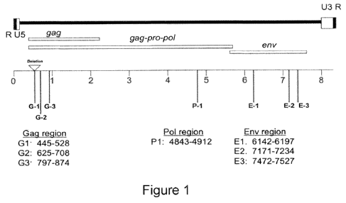
Figure 1 is a schematic diagram of the gag (G1, G2, G3), pol (P1) and env
(El, E2, E3) regions of XMRV.
Figures 2A and 2B are standard curves of envelope RNA using E3 (Figure
2A) and G3 (Figure 2B) which was diluted to different dilutions and analyzed
by
qRT/PCR using Ag-Path kit.
Figures 3A and 3B show the results of XMRV RNA copy number in urine of
prostate cancer patient VP663 using El site in env and E2 site in env,
respectively.
Results are shown of qRT-PCR assays performed six times (x) and in comparison
to
a standard curve generated with a 1.85 kb XMRV env RNA produced by in vitro
transcription. Y axis shows the Ct values, x axis shows the log of the copy
number.
Figure 4 shows the results of detection of XMRV RNA in EPS of prostate
cancer patient VP 657 and VP 635 using El site in env. Results of qRT-PCR
assays
were preformed in duplicate and are shown in comparison to a standard curve
generated with a 1.85 kb XMRV env RNA produced by in vitro transcription. Y
axis
shows the Ct values, x axis shows the log of the copy number.
Figure 5 shows the amplification plot of qRT/PCR identification of XMRV
RNA in prostatitis patient using the El primer-probe combination. Duplicate
samples were assayed which shows very high Ct value corresponding to very low
copy number.
Figure 6A shows an amplification plot of qRT/PCR analysis of XMRV RNA
in prostate cancer patient's EPS (pj 339) using the G2 primer-probe
combination.
Similarly the assay shown in Figure 6B shows an amplification plot of prostate
cancer patient EPS (pj 301, 302 and 304) using the El primer probe
combination.
Figure 7, upper panel, provides a schematic diagram showing the regions
used in the nested RT/PCR analysis. The lower panel of Figure 7 is an agarose
gel
showing the detection of XMRV RNA isolated from an XMRV infected prostate
CA 02748117 2011-06-22
WO 2010/075414 PCT/US2009/069244
-6-
cancer cell line and RNA from prostate cancer patient EPS (pj 339) generated
bands
of 218 and 112 nucleotides in length.
Figure 8 shows a 2% agarose gel of the nested RT-PCR product of RNA
samples isolated from 6 prostate cancer patients' urine samples using Tth
polymease
for RT and first round PCR, followed by Taq DNA Polymerase for second round
PCR amplification.
Figure 9 shows a 2% agarose gel of the nested RT-PCR product of RNA
samples isolated from 17 prostate cancer patients' expressed prostate
secretion
(EPS) during prostatectomy using Tth polymerase for RT and first round PCR,
followed by Taq DNA Polymerase for second round PCR amplification.
Figure 10 shows the sequences of the bands of 112 (SEQ ID NOs: 34, 35 and
36) and 218 (SEQ ID NOs: 37, 38 and 39) nucleotides in length referred to in
Figure 7.
Figure 11 is a gel of singleplex nested RT-PCR of RNA isolated from 3
prostate cancer patient urine samples, reaction time were done in triplicates.
Oligos
6200R and 5922F were used for the first round followed by 6159R and 5942F for
the second round of amplification.
Figure 12 is a graph showing the detection and determination of XMRV
DNA copy numbers in DNA isolated from tumor-bearing prostate tissues of men
with the RNASEL QQ genotype following prostatectomy.
DETAILED DESCRIPTION OF THE INVENTION
Hereditary prostate cancer (HPC), which accounts for 43% of early onset
cases and about 9% of all cases, is due to germline mutations in HPC genes
(Carter,
B.S., et at., Proc. Natl. Acad. Sci. USA, 89(8):3367 (1992)). In 2002, the
first HPC
gene was reported (Carpten, J., et at., Nat. Genet., 30(2):181 (2002)). HPCJ
encodes RNase L, an essential protein in antiviral innate immunity (reviewed
in
Silverman, R., Cytkine Growth Factor Rev., 18(5-6):381 (2007)). Genetic
evidence
that an antiviral gene suppresses prostate cancer led to examination of the
possibility
that chronic viral infections might predispose men to prostate cancer. In 2006
discovery of a new human retrovirus, xenotropic MLV related virus (XMRV), in
tumor-bearing prostate tissues, was reported (Urisman, A., et at., PLoS
Pathog.,
CA 02748117 2011-06-22
WO 2010/075414 PCT/US2009/069244
-7-
2(3):e25 (2006)). Remarkably, XMRV is present in prostate tissues of men that
are
homozygous for a reduced activity variant of RNase L, but rarely in men with
wild
type RNase L. In 2007, construction of an infectious viral molecular clone of
XMRV was reported (Dong, B., et at., Proc. natl. Acad. Sci., USA, 104(5):1655
(2007)). Methods of monitoring XMRV infections as an indicator or predictor of
prostate cancer progression or aggressiveness are provided herein.
Specifically, described herein is the development of polymerase chain
reaction (PCR) assays (e.,g., real-time quantitative RT-PCR (qRT-PCR) assays)
for
the detection of XMRV nucleic acid in a sample (e.g., urine and other bodily
fluids,
such as prostate secretions, and semen) obtained from an individual (e.g.,
patient). In
particular aspects, highly sensitive, specific and quantitative real-time (RT)
PCR
assays for XMRV nucleic acid (e.g., DNA; RNA) and nested RT-PCR assays for
detection of XMRV nucleic acid are described. These assays are useful for
determination of viral loads in tissues and fluids from individuals with and
without
cancer. Also described herein is the correlation of the prevalence and load of
XMRV
in prostate cancer cases with disease parameters. XMRV is a newly discovered
infection of tumor-bearing prostate that correlates with mutations in a
prostate
cancer susceptibility gene (RNASEL). The occurrence of XMRV infections in
prostate cancer cases provides for pathogenesis of the disease, assessing
risk, and
novel therapeutic options.
In one aspect, total RNA was extracted from prostate tissue samples using
Trizol reagent (Invitrogen, Carlsbad, CA) and from urine and prostatic
secretion
using the MagmaxTM Viral RNA Isolation Kit (Ambion Inc., Austin TX) and then
stored at -80 C until further processing. Initial screening of samples to
detect XMRV
gene sequences was performed using qRT-PCR (performed on an Applied
Biosystems 7500 Real Time PCR system). In one embodiment, these reactions are
performed using a one-step RT-PCR reaction (AgPath-IDTM kit, Applied
Biosystems). PCR assays for seven different regions in XMRV RNA have been
developed (Figure 1) which involved the design of sets of Taqman-based
primers/probe used to detect three regions in XMRV RNA, including one region
of
gag (G1) and two regions of env (E1 & E2).
CA 02748117 2011-06-22
WO 2010/075414 PCT/US2009/069244
-8-
The presence of XMRV in prostatic secretions and urine as shown herein is
significant because such data provides a prostatic secretion- and urine-based
XMRV
detection assay that is non-invasive, rapid, and easy to perform, avoiding the
morbidity and difficulty of obtaining blood or tissue specimens for sampling.
Current screening for prostate cancer by prostate-specific antigen (PSA)
levels and
digital rectal exam often does not begin until age 50 and has significant
limitations
and inaccuracies. In contrast to these tests, the assays for XMRV described
herein
can be performed on much younger men, especially those with a family history
of
prostate cancer. Because men with XMRV infections, especially those that fail
to
clear the virus, are likely at increased risk of prostate cancer, these
studies provide a
new diagnostic for evaluating risk of prostate cancer initiation or
progression.
Accordingly, in one aspect, the invention is directed to a method of detecting
the presence of xenotropic MLV related virus (XMRV) in an individual.
As used herein, "XMRV" refers to an infectious gammaretrovirus found in
prostate tumors, particularly in prostate tumors of patients homozygous for
RNASEL
variant, R462Q (e.g., Urisman, A., et at., PLoS Pathog., 2(3):e25 (2006);
Dong, B.,
et at., Proc. Natl. Acad. Sci., USA, 104(5):1655 (2007); and WO 2006/110589;
all of
which are incorporated herein by reference in their entirety). The term "XMRV"
includes any strain of the virus including XMRV VP35 (GenBank Accession No.
DQ241301), XMRV VP42 (GenBank Accession No. DQ241302) and XMRV VP62
(GenBank Accession No. DQ399707).
As used herein an "individual" refers to any subject in need of screening. In
particular embodiments, the individual is a mammal, such as a primate (e.g.,
human), cow, sheep, goat, horse, dog, cat, rabbit, guinea pig, rat, mouse or
other
bovine, ovine, equine, canine feline, rodent or murine species). In one
embodiment,
the individual is a human. In another embodiment, the individual is a human
under
the age of 50 years, 40 years, 30 years or 20 years. In another embodiment,
the
individual is a cancer patient (e.g., a prostate cancer patient; and HPC
patient). In
another embodiment, the individual is in remission from prostate cancer. In
another
embodiment, the individual has or has had a (one or more) XMRV infection. In
another embodiment, the individual's genome comprises a wild type, a
heterozygous
or a homozygous mutation of the RNase L gene. In yet another embodiment, the
CA 02748117 2011-06-22
WO 2010/075414 PCT/US2009/069244
-9-
individual expresses a mutated or variant form of RNase L (e.g., R462Q; QQ
RNASEL).
Although the invention is performed herein using urine and/or prostatic
secretion samples so as to demonstrate that the method is non-invasive, rapid
and
easy to perform, one of skill in the art will appreciate that any suitable
biological
sample obtained from an individual can be used in the methods of the
invention. The
sample can be a biological fluid, a tissue sample (e.g., prostate, bladder,
seminal
glands, testes, kidney, bone marrow, colon, ileum, jejunum, pancreas, adrenal
glands, liver, heart, lung, spleen, brain cortex, brain stem, cerebellum,
inguinal
lymph node, axillar lymph node and mesenteric lymph node), a tumor sample
(e.g.,
a prostate tumor, a bladder tumor, other tumors of the male and female
genitourinary
tracts) and combinations thereof. A suitable sample can be obtained for
example by
cell or tissue biopsy. A sample can also be obtained from other tissues,
bodily fluids
and products, e.g., from a tissue smear, tissue scrape, and the like. Thus,
the sample
can be a biopsy specimen (e.g, tumor, polyp, mass (solid, cellular)),
aspirate, and/or
smear sample). The sample can be from a tissue that has a tumor (e.g.,
cancerous
growth) and/or tumor cells, or is suspected of having a tumor and/or tumor
cells.
For example, a tumor biopsy can be obtained in an open biopsy, a procedure in
which an entire (excisional biopsy) or partial (incisional biopsy) mass is
removed
from a target area. Alternatively, a tumor sample can be obtained through a
percutaneous biopsy, a procedure performed with a needle-like instrument
through a
small incision or puncture (with or without the aid of a imaging device) to
obtain
individual cells or clusters of cells (e.g., a fine needle aspiration (FNA))
or a core or
fragment of tissues (core biopsy).
In a particular embodiment, the sample is a biological fluid. Examples of a
biological fluid that can be used in the methods include urine, prostatic
fluids, blood
and semen. As used herein, "prostatic fluids" include expressed prostate
secretions
(EPS) such as semen.
As described herein, the sample is contacted with at least one set of primers
and maintained under conditions which amplify the primers if XMRV is present
in
the sample to produce amplified XMRV nucleic acid sequences, also referred to
CA 02748117 2011-06-22
WO 2010/075414 PCT/US2009/069244
- 10-
herein as "amplicons" or "XMRV amplicons". The amplified XMRV nucleic acid
sequences can be, for example, XMRV DNA or XMRV RNA.
A "set of primers" comprises at least one forward primer and at least one
reverse primer, wherein the forward primer and the reverse primer in the set
are
complementary to all or a portion of an XMRV nucleotide sequence (e.g., XMRV
G1, XMRV G2, XMRV G3, XMRV P1, XMRV El, XMRV E2, XMRV E3).
Typically, the forward primer and the reverse primer within a set of primers
are
complementary to all or a portion of the same region or a similar region of
the
XMRV nucleotide sequence (e.g., the gag region, the env region, the pol
region). As
used herein, the term "primer" refers to an oligonucleotide, which is capable
of
acting as a point for the initiation of synthesis of a primer extension
product that is
complementary to a target nucleotide sequence that is to be amplified,
referred to as
the target or template nucleic acid sequence. In this instance, the target or
template
nucleic acid sequence is all or a portion (e.g., the gag region, the env
region, the pol
region) of an XMRV nucleic acid sequence. The primer may occur naturally, as
in a
purified restriction digest, or be produced synthetically. The appropriate
length of a
primer depends on the intended use of the primer, but typically ranges from
about 5
to about 100; from about 5 to about 75; from about 5 to about 50; from about 5
to
about 10; from about 10 to about 35; from about 18 to about 22 nucleotides. A
primer need not reflect the exact sequence of the target sequence but must be
sufficiently complementary to hybridize with the target sequence for primer
elongation to occur, i.e., the primer is sufficiently complementary to the
target
nucleotide sequence such that the primer will anneal to the template under
conditions that permit primer extension. Reverse transcription can be
performed
with M-MLV RT (such as SuperscriptTM 1, II or III (Invitrogen)) or Tth DNA
polymerase in RT buffer using Oligo dT, random hexamer or XMRV gene specific
primers. As used herein, the phrase "conditions that permit primer extension"
refers
to those conditions, e.g., salt concentration (metallic and non-metallic
salts), pH,
temperature, and necessary cofactor concentration, among others, under which a
given polymerase enzyme catalyzes the extension of an annealed primer.
Conditions
for the primer extension activity of a wide range of polymerase enzymes are
known
in the art. As one example, conditions permitting the extension of a nucleic
acid
CA 02748117 2011-06-22
WO 2010/075414 PCT/US2009/069244
-11-
primer by Taq polymerase include the following (for any given enzyme, there
can
and often will be more than one set of such conditions): reactions are
conducted in a
buffer containing 50 mM KC1, 10 mM Tris (pH 8.3 - 8.6), 1.5 - 4 mM MgClz, 200
O O
gM of dNTPs; reactions can be performed at about 68 - 72 C.
It will be clear to persons skilled in the art that the size of the primer and
the
stability of hybridization will be dependent to some degree on the ratio of A-
T to
C-G base pairings, since more hydrogen bonding is available in a C-G pairing.
Also,
the skilled person will consider the degree of homology between the extension
primer to other parts of the amplified sequence and choose the degree of
stringency
accordingly. Guidance for such routine experimentation can be found in the
literature, for example, Molecular Cloning: a laboratory manual by Sambrook,
J.,
Fritsch E. F. and Maniatis, T. (1989) which is incorporated herein by
reference.
In addition to the primer pairs, probes can be included with reporter dye at
the 5' end (e.g., fluorescein, 6-carboxy fluorescein (FAM), 6-FAM, 5-FAM,
TAMRA) and quencher dye at the 3' end (e.g., BHQ- 1, BHQ-2, TAMRA, MGB)
which will bind to the XMRV DNA during PCR (e.g., U.S. Patent No. 7,374,833
which is incorporated herein by reference).
For detection purposes, the primer can comprises at least one tag or label.
As used herein, "tag" or "label" are used interchangeably to refer to any
moiety that
is capable of being specifically detected (e.g., by a partner moiety), either
directly or
indirectly, and therefore, can be used to identify and/or isolate a
polynucleotide
sequence that comprises the tag. Suitable tags for the present invention
include,
among others, affinity tags (e.g., biotin, avidin, streptavidin), haptens,
ligands,
peptides, nucleic acids, fluorophores, chromophores, and epitope tags that are
recognized by an antibody (e.g., digoxigenin (DIG), hemagglutinin (HA), myc,
Flag) (Andrus, A. "Chemical methods for 5' non-isotopic labelling of PCR
probes
and primers" (1995) in PCR 2: A Practical Approach, Oxford University Press,
Oxford, pp. 39-54). Other suitable tags include, but are not limited to,
chromophores, fluorophores, haptens, radionuclides (e.g., 32P, 33P, 35S),
fluorescence
quenchers, enzymes, enzyme substrates, affinity tags (e.g., biotin, avidin,
streptavidin, etc.), mass tags, electrophoretic tags and epitope tags that are
recognized by an antibody. In certain embodiments, the label is present on the
5
CA 02748117 2011-06-22
WO 2010/075414 PCT/US2009/069244
-12-
carbon position of a pyrimidine base or on the 3 carbon deaza position of a
purine
base.
The primers have a nucleotide sequence that is complementary to all or a
portion of an XMRV sequence. In particular embodiments, the primers have a
nucleotide sequence that is complementary to all or a portion of an XMRV gag
sequence, an XMRV pol, an XMRV env sequence or a combination thereof.
In one embodiment, at least one forward primer and at least one reverse
primer are complementary to all or a portion of an XMRV G1 gag nucleotide
sequence. As used herein, an "XMRV G1 gag nucleotide sequence" refers to a
sequence that is from about nucleotide 445 to about nucleotide 528 of an XMRV
genomic sequence. The length of the probe which binds between two primers in
the
G1 gag nucleotide sequence can vary between about 12 to about 40 nucleotides
complementary to all or a portion of XMRV G1 gag nucleotide sequence. Minor
groove binding principle can also be applied when the probe size is as short
as about
12 nucleotides. In a particular embodiment, the forward primer complementary
to all
or a portion of an XMRV G1 gag nucleotide sequence has a nucleotide sequence
comprising GGACTTTTTGGAGTGGCTTTGTT (SEQ ID NO: 1), the reverse
primer complementary to all or a portion of an XMRV G1 gag nucleotide sequence
has a nucleotide sequence comprising GCGTAAAACCGAAAGCAAAAT (SEQ ID
NO: 2) and the probe has a nucleotide sequence comprising
ACAGAGACACTTCCCGCCCCCG (SEQ ID NO: 3).
In another embodiment, at least one forward primer and at least one reverse
primer are complementary to all or a portion of an XMRV G2 gag nucleotide
sequence. As used herein, an "XMRV G2 gag nucleotide sequence" refers to a
sequence that is from about nucleotide 625 to about nucleotide 708 of an XMRV
genomic sequence. The length of probe which binds between two primers in the
G2
gag nucleotide sequence can vary between about 12 to about 40 nucleotides
complementary to all or a portion of XMRV G1 gag nucleotide sequence. Minor
groove binding can also be applied when the probe size is as short as about 12
nucleotides. In a particular embodiment, the forward primer complementary to
all or
a portion of an XMRV G2 gag nucleotide sequence has a nucleotide sequence
comprising GTAACTACCCCTCTGAGTCTAACCT (SEQ ID NO: 4), the reverse
CA 02748117 2011-06-22
WO 2010/075414 PCT/US2009/069244
- 13-
primer complementary to all or a portion of an XMRV G3 gag nucleotide sequence
has a nucleotide sequence comprising CTTCTTGACATCCACAGACTGGTT (SEQ
ID NO: 5) and the probe has a nucleotide sequence comprising
TCCAGCGCATTGCATC (SEQ ID NO: 6).
In another embodiment, at least one forward primer and at least one reverse
primer are complementary to all or a portion of an XMRV G3 gag nucleotide
sequence. As used herein, an "XMRV G3 gag nucleotide sequence" refers to a
sequence that is from about nucleotide 797 to about nucleotide 874 of an XMRV
genomic sequence. The length of the probe which binds between two primers in
the
G3 gag nucleotide sequence can vary between about 12 to about 40 nucleotides
complementary to all or a portion of XMRV G1 gag nucleotide sequence. Minor
groove binding principle can also be applied when the probe size is as short
as about
12 nucleotides. In a particular embodiment, the forward primer complementary
to all
or a portion of an XMRV G3 gag nucleotide sequence has a nucleotide sequence
comprising CTCAGGTCAAGTCTAGAGTGTTTTGT (SEQ ID NO: 7), the reverse
primer complementary to all or a portion of an XMRV G2 gag nucleotide sequence
has a nucleotide sequence comprising CCTCCCAGGTGACGATATATGG (SEQ
ID NO: 8) and the probe has a nucleotide sequence comprising
CCCCACGGACACCC (SEQ ID NO: 9).
In another embodiment, at least one forward primer and at least one reverse
primer are complementary to all or a portion of an XMRV P 1 Pol nucleotide
sequence. As used herein, an "XMRV P1 Pol nucleotide sequence" refers to a
sequence that is from about nucleotide 4843 to about nucleotide 4912 of an
XMRV
genomic sequence. The length of the probe which binds between two primers in
the
P 1 pol nucleotide sequence can vary between about 12 to about 40 nucleotides
complementary to all or a portion of XMRV G1 gag nucleotide sequence. Minor
groove binding principle can also be applied when the probe size is as short
as about
12 nucleotides. In a particular embodiment, the forward primer complementary
to all
or a portion of an XMRV P1 pol nucleotide sequence has a nucleotide sequence
comprising CGGGACAGAACTATCCAGTATGTGA (SEQ ID NO: 10), the
reverse primer complementary to all or a portion of an XMRV P1 pol nucleotide
sequence has a nucleotide sequence comprising TGGCTTTGCTGGCATTTACTTG
CA 02748117 2011-06-22
WO 2010/075414 PCT/US2009/069244
-14-
(SEQ ID NO: 11) and the probe has a nucleotide sequence comprising
ACCTGCACCGCCTGTG (SEQ ID NO: 12).
In another embodiment, at least one forward primer and at least one reverse
primer are complementary to all or a portion of an XMRV E 1 env nucleotide
sequence. As used herein, an "XMRV El env nucleotide sequence" refers to a
sequence that is from about nucleotide 6142 to about nucleotide 6197 of an
XMRV
genomic sequence. The length of the probe which binds between two primers in
the
El env nucleotide sequence can vary between about 12 to about 40 nucleotides
complementary to all or a portion of XMRV Gl gag nucleotide sequence. Minor
groove binding principle can also be applied when the probe size is as short
as about
12 nucleotides. In a particualr embodiment, the forward primer complementary
to all
or a portion of an XMRV El env nucleotide sequence has a nucleotide sequence
comprising GGCCGAGAGAGGGCTACT (SEQ ID NO: 13), the reverse primer
complementary to all or a portion of an XMRV El env nucleotide sequence has a
nucleotide sequence comprising TGATGATGATGGCTTCCAGTATGC (SEQ ID
NO: 14) and the probe has a nucleotide sequence comprising
CACATCCCCATTTGCC (SEQ ID NO: 15).
In another embodiment, at least one forward primer and at least one reverse
primer are complementary to all or a portion of an XMRV E2 env nucleotide
sequence. As used herein, an "XMRV E2 env nucleotide sequence" refers to a
sequence that is from about nucleotide 7171 to about nucleotide 7234 of an
XMRV
genomic sequence. The length of the probe which binds between two primers in
the
E2 env nucleotide sequence can vary between about 12 to about 40 nucleotides
complementary to all or a portion of XMRV Gl gag nucleotide sequence. Minor
groove binding principle can also be applied when the probe size is as short
as about
12 nucleotides. In a particular embodiment, the forward primer complementary
to all
or a portion of an XMRV E2 env nucleotide sequence has a nucleotide sequence
comprising CCCTAGTGGCCACCAAACAA (SEQ ID NO: 16), the reverse primer
complementary to all or a portion of an XMRV E2 env nucleotide sequence has a
nucleotide sequence comprising AAGGCCCCAAGGTCTGTATGT (SEQ ID NO:
17) and the probe has a nucleotide sequence comprising
TCGAGCAGCTCCAGGCAGCCA (SEQ ID NO: 18).
CA 02748117 2011-06-22
WO 2010/075414 PCT/US2009/069244
- 15-
In another embodiment, at least one forward primer and at least one reverse
primer are complementary to all or a portion of an XMRV E3 env nucleotide
sequence. As used herein, an "XMRV E3 env nucleotide sequence" refers to a
sequence that is from about nucleotide 7472 to about nucleotide 7527 of an
XMRV
genomic sequence. The length of the probe which binds between two primers in
the
E3 env nucleotide sequence can vary between about 12 to about 40 nucleotides
complementary to all or a portion of XMRV G1 gag nucleotide sequence. Minor
groove binding principle can also be applied when the probe size is as short
as about
12 nucleotides. In a particular embodiment, the forward primer complementary
to all
or a portion of an XMRV E3 env nucleotide sequence has a nucleotide sequence
comprising TCAGGACAAGGGTGGTTTGAG (SEQ ID NO: 19), the reverse
primer complementary to all or a portion of an XMRV E3 env nucleotide sequence
has a nucleotide sequence comprising GGCCCATAATGGTGGATATCA (SEQ ID
NO: 20) and the probe has a nucleotide sequence comprising
TTAACAGGTCCCCATGGTTCACGACCA (SEQ ID NO: 21).
The primers are amplified using any suitable method known in the art. As
used herein, "amplification" or an "amplification reaction" refers to any
suitable
method for amplification of a nucleic acid sequence including polymerase chain
reaction (PCR), ligase chain reaction (LCR), rolling circle amplification
(RCA),
strand displacement amplification (SDA) and multiple displacement
amplification
(MDA), as will be understood by a person of skill in the art. Such methods for
amplification typically comprise, e.g., primers that anneal to the nucleic
acid
sequence to be amplified, a DNA polymerase, and nucleotides. Furthermore,
amplification methods, such as PCR, can be solid-phase amplification, polony
amplification, colony amplification, emulsion PCR, bead RCA, surface RCA,
surface SDA, etc., as will be recognized by one of skill in the art. It will
also be
recognized that it is advantageous to use an amplification method that results
in
exponential amplification of free DNA molecules in solution or tethered to a
suitable
matrix by only one end of the DNA molecule. In addition, it will be recognized
that
it is often advantageous to use amplification protocols that maximize the
fidelity of
the amplified products to be used as templates in DNA sequencing procedures.
Such protocols use, for example, DNA polymerases with strong discrimination
CA 02748117 2011-06-22
WO 2010/075414 PCT/US2009/069244
- 16-
against misincorporation of incorrect nucleotides and/or strong 3' exonuclease
activities (also referred to as proofreading or editing activities) to remove
misincorporated nucleotides during polymerization.
In one embodiment, a PCR method is used to amplify the primers. As known
to those of skill in the art, PCR is a technique in which a DNA polymerase is
used
to amplify a piece of DNA (e.g., a gene or portion thereof, a non-coding
region) by
in vitro enzymatic replication. As PCR progresses, the DNA generated is used
as a
template for replication which sets in motion a reaction in which the DNA
template
is exponentially amplified. With PCR a single or few copies of a piece of DNA
are
amplified across several orders of magnitude, generating millions or more
copies of
the DNA piece. As is also known in the art, PCR can be extensively modified to
perform a wide array of genetic manipulations.
PCR applications typically employ a heat-stable (thermostabile) polymerase
(e.g., DNA polymerase). A variety of polymerases for use in PCR are known to
this
of skill in the art and include Taq polymerase, an enzyme originally isolated
from
the bacterium Thermus aquaticus, and Vent and Tth polymerases derived from
microorganisms that normally reside at high temperature. Consequently, these
polymerase enzymes are quite stable to heat denaturation, making them ideal
enzymes for use in the polymerase chain reaction. These polymerases, such as a
DNA polymerase enzymatically assembles a new DNA strand from DNA building
blocks, the nucleotides, by using single-stranded DNA as a template and DNA
oligonucleotides (also called DNA primers), which are required for initiation
of
DNA synthesis.
PCR methods typically use thermal cycling, i.e., alternately heating and
cooling the PCR sample to a defined series of temperature steps. These thermal
cycling steps physically separate the strands (at high temperatures) in a
e.g., DNA
double helix (DNA melting) used as the template during DNA synthesis (at lower
temperatures) by the DNA polymerase to selectively amplify the target DNA.
Selectivity of PCR arises from the use of primers that are complementary to
the
DNA region targeted for amplification under specific thermal cycling
conditions.
PCR typically involves the use of several components and reagents such as a
nucleic acid (e.g., DNA) template that contains the region (target) to be
amplified;
CA 02748117 2011-06-22
WO 2010/075414 PCT/US2009/069244
- 17-
one or more, typically two or more, primers which are complementary to the
nucleic
acid regions at the 5' (five prime) or 3' (three prime) ends of the nucleic
acid region;
one or more polymerases e.g., with a temperature optimum at around 70 C; one
or
more deoxynucleoside triphosphates (dNTPs; also very commonly and erroneously
called deoxynucleotide triphosphates), the building blocks from which the DNA
polymerases synthesizes a new DNA strand; one or more buffer solutions,
providing
a suitable chemical environment for optimum activity and stability of the
polymerase; one or more divalent cations, e.g., magnesium or manganese ions;
generally Mg2+ is used, but Mn2+ can be utilized for PCR-mediated DNA
mutagenesis, as higher Mn2+ concentration increases the error rate during DNA
synthesis; and one or more monovalent cation potassium ions.
PCR is commonly carried out in a reaction volume of 10-200 l in small
reaction tubes (0.2-0.5 ml volumes) in a thermal cycler which heats and cools
the
reaction tubes to achieve the temperatures required at each step of the
reaction.
Although one of skill in the art will appreciate that PCR can occur in a
variety of
ways depending upon the desired result(s), an example of a PCR can occur as
follows. The PCR can begin with an initialization step, which involves heating
the
reaction to a temperature of about 94-96 C (or about 98 C if extremely
thermostable
polymerases are used), which is held for about 1-9 minutes. This is typically
used
with DNA polymerases that require heat activation by hot-start PCR. A
denaturation
step, which is the first regular cycling event, involves heating the reaction
to about
94-98 C for about 20-30 seconds. This results in melting of DNA template and
primers by disrupting the hydrogen bonds between complementary bases of the
DNA strands, yielding single strands of DNA. An annealing step, which involves
lowering the temperature to about 50-65 C for about 20-40 seconds allowing
annealing of the primers to the single-stranded DNA template, can then be
carried
out. Typically the annealing temperature is about 3-5 degrees Celsius below
the
melting temperature (Tm) of the primers used. Stable DNA-DNA hydrogen bonds
are generally formed when the primer sequence very closely matches the
template
sequence. The polymerase binds to the primer-template hybrid and begins DNA
synthesis.
CA 02748117 2011-06-22
WO 2010/075414 PCT/US2009/069244
-18-
In the extension/elongation step, the temperature depends on the DNA
polymerase used; Taq polymerase has its optimum activity temperature at about
75-
80 C, and commonly a temperature of about 72 C is used with this enzyme. At
this
step the DNA polymerase synthesizes a new DNA strand complementary to the
DNA template strand by adding dNTPs that are complementary to the template in
5'
to 3' direction, condensing the 5'-phosphate group of the dNTPs with the 3'-
hydroxyl
group at the end of the nascent (extending) DNA strand. The extension time
depends
both on the DNA polymerase used and on the length of the DNA fragment to be
amplified. A final elongation step is occasionally performed at a temperature
of
about 70-74 C for about 5-15 minutes after the last PCR cycle to ensure that
any
remaining single-stranded DNA is fully extended. A final hold step at about 4-
15 C
for an indefinite time can be employed for short-term storage of the reaction.
In a particular embodiment, a real time (RT/PCR) or quantitative, real time
PCR (qRT/PCR) reaction is used to amplify the primers if XMRV is present in
the
sample. As understood by one of skill in the art, RT/PCR DNA simultaneously
quantifies and amplifies the nucleic acid. In this method, the nucleic acid is
specifically amplified by polymerase chain reaction. After each round of
amplification, the DNA is quantified. Common methods of quantification include
the use of fluorescent dyes that intercalate with double-strand nucleic acid
and
modified oligonucleotides (called probes) that fluoresce when hybridized with
a
complementary DNA.
Specifically, quantitative PCR (Q-PCR) is used to measure the quantity of a
PCR product (preferably real-time). The method quantitatively measures
starting
amounts of DNA, cDNA or RNA. Q-PCR is commonly used to determine whether a
DNA sequence is present in a sample and the number of its copies in the
sample, and
is also known as RT-PCR (Real Time PCR), RQ-PCR, QRT-PCR or RTQ-PCR.
RT-PCR commonly refers to reverse transcription PCR, which can also be used in
the methods described herein, and is often used in conjunction with Q-PCR. QRT-
PCR methods use fluorescent dyes, such as Sybr Green, or fluorophore-
containing
DNA probes, such as TaqMan, to measure the amount of amplified product in real
time.
CA 02748117 2011-06-22
WO 2010/075414 PCT/US2009/069244
- 19-
Real-time polymerase chain reaction, also called quantitative real time
polymerase chain reaction (Q-PCR/qPCR) or kinetic polymerase chain reaction,
is
based on the polymerase chain reaction, which is used to amplify and
simultaneously quantify a targeted DNA molecule. It enables both detection and
quantification (as absolute number of copies or relative amount when
normalized to
DNA input or additional normalizing genes) of a specific sequence in a DNA
sample.
The procedure follows the general principle of polymerase chain reaction; its
key feature is that the amplified DNA is quantified as it accumulates in the
reaction
in real time after each amplification cycle. Two common methods of
quantification
are the use of fluorescent dyes that intercalate with double-stranded DNA, and
modified DNA oligonucleotide probes that fluoresce when hybridized with a
complementary DNA.
Frequently, real-time polymerase chain reaction is combined with reverse
transcription polymerase chain reaction to quantify low abundance messenger
RNA
(mRNA), enabling one of skill in the art to quantify relative gene expression
at a
particular time, or in a particular cell or tissue type. Although real-time
quantitative
polymerase chain reaction is sometimes incorrectly abbreviated as RT-PCR, it
should not be confused with reverse transcription polymerase chain reaction,
also
known as RT-PCR.
The reaction is typically run in a thermocycler as described herein, and after
each cycle, the levels of fluorescence are measured with a detector; the dye
only
fluoresces when bound to the dsDNA (i.e., the PCR product). With reference to
a
standard dilution, the dsDNA concentration in the PCR can be determined.
A comparison of a measured DNA/RNA sample to a standard dilution
provides a fraction or ratio of the sample relative to the standard, allowing
relative
comparisons between different tissues or experimental conditions. The method
can
further comprise normalizing expression of a target gene to a stably expressed
gene.
In another embodiment, fluorescent reporter probes are used. A sequence-
specific RNA and/or DNA-based probe is used to quantify the nucleic acid
containing the probe sequence; therefore, use of the reporter probe can
increase
specificity, and allow quantification even in the presence of some non-
specific DNA
CA 02748117 2011-06-22
WO 2010/075414 PCT/US2009/069244
-20-
amplification. This allows for multiplexing - assaying for several genes in
the same
reaction by using specific probes with different-coloured labels, provided
that all
genes are amplified with similar efficiency.
The reaction is typically carried out with an RNA-based probe with a
fluorescent reporter at one end and a quencher of fluorescence at the opposite
end of
the probe. The close proximity of the reporter to the quencher prevents
detection of
its fluorescence; breakdown of the probe by the 5' to 3' exonuclease activity
of the
polymerase (e.g.,taq polymerase) breaks the reporter-quencher proximity and
thus
allows unquenched emission of fluorescence, which can be detected. An increase
in
the product targeted by the reporter probe at each PCR cycle therefore causes
a
proportional increase in fluorescence due to the breakdown of the probe and
release
of the reporter.
The PCR is prepared as usual, and the reporter probe is added. As the
reaction commences, during the annealing stage of the PCR both probe and
primers
anneal to the DNA target. Polymerisation of a new DNA strand is initiated from
the
primers, and once the polymerase reaches the probe, its 5'-3-exonuclease
degrades
the probe, physically separating the fluorescent reporter from the quencher,
resulting
in an increase in fluorescence. Fluorescence is detected and measured in the
real-
time PCR thermocycler, and its geometric increase corresponding to exponential
increase of the product is used to determine the threshold cycle (CT; Ct) in
each
reaction.
In intact probes, reporter fluorescence is quenched. Probes and the
complementary DNA strand are hybridized and reporter fluorescence is still
quenched. During PCR, the probe is degraded by the polymerase and the
fluorescent
reporter released.
Relative concentrations of DNA present during the exponential phase of the
reaction can be determined by plotting fluorescence against cycle number on a
logarithmic scale (so an exponentially increasing quantity will give a
straight line).
A threshold for detection of fluorescence above background is determined. The
cycle at which the fluorescence from a sample crosses the threshold is called
the
cycle threshold, Ct. Since the quantity of DNA doubles every cycle during the
CA 02748117 2011-06-22
WO 2010/075414 PCT/US2009/069244
-21-
exponential phase, relative amounts of DNA can be calculated, e.g. a sample
whose
Ct is 3 cycles earlier than another's has 23 = 8 times more template.
Amounts of RNA or DNA are then determined by comparing the results to a
standard curve produced by real-time PCR of serial dilutions (e.g. undiluted,
1:4,
1:16, 1:64) of a known amount of RNA or DNA. As mentioned above, to quantify
gene expression, the measured amount of RNA from the gene of interest is
divided
by the amount of RNA from a control sequence (also referred to herein as a
reference or housekeeping sequence) (e.g., gene) measured in the same sample
to
normalize for possible variation in the amount and quality of RNA between
different
samples. This normalization permits accurate comparison of expression of the
sequence of interest between different samples, provided that the expression
of the
reference (housekeeping) sequence used in the normalization is very similar
across
all the samples.
In another embodiment, nested, reverse transcription PCR is used to amplify
the primers. Nested PCR is a PCR with a second round of amplification using a
different set of primers. This second set of primers is specific to a sequence
found
within the nucleotide sequence of the initial conventional PCR amplicon. The
use of
a second amplification step with the "nested" primer set results in a reduced
background from products amplified during the initial PCR due to the nested
primers' additional specificity to the region. The amount of amplicon produced
is
increased as a result of the second round of amplification and due to a
reduction in
any inhibitor concentrations. Reverse transcription, nested PCR indicates that
the
reaction is initiated with DNA that has been reverse transcribed from RNA.
As described herein, whether amplified XMRV sequences are present in the
sample are detected, wherein if amplified XMRV sequences are detected in the
sample, then XMRV is present in the individual. Detection of amplified XMRV
sequences can be achieved by resolving sequences by means of, for example, gel
electrophoresis (e.g., agarose gel), high-resolution denaturing
polyacrylamide/urea
gel electrophoresis, capillary separation, or other resolving means; followed
by
detecting the sequence using, for example, a scanning spectrophotometer or
fluorometer. In a particular embodiment, fluorescently-labeled amplified XMRV
sequences are resolved by gel electrophoresis, according to procedures that
are well
CA 02748117 2011-06-22
WO 2010/075414 PCT/US2009/069244
-22-
known in the art, and are subsequently detected in the gel using a standard
fluorometer. In one embodiment, a positive XMRV generates a band of 218
nucleotides in length, 112 nucleotides in length or a combination thereof.
The method can further comprise determining the sequences of the amplified
XMRV sequence using procedures well known in the art.
As discussed above and as apparent to one of skill in the art, the method of
detecting the presence of XMRV in a sample can further comprise the use of a
control. That is, the amount or level of amplified XMRV nucleic acid sequences
in
the sample can be compared to the amount or level of amplified XMRV nucleic
acid
sequences in a control sample. Suitable controls are well recognized in the
art and
include, for example, a sample from an individual that is known to not be
infected
with XMRV, a sample from an individual that is known to be infected with XMRV,
a sample from an individual that is a prostate cancer patient (e.g., HPC
patient),
and/or a reference standard of authentic (positive) XMRV RNA. The control
sample
can be the same type of sample as the sample obtained from the individual
(e.g., the
sample obtained from the individual and the control sample are urine samples)
or the
control sample can be a different sample (e.g., the sample obtained from the
individual is a urine sample and the control sample is a tissue sample such as
a
prostate tissue sample).
The methods for detecting XMRV is an individual can be used for a variety
of purposes such as for diagnostic and/or prognostic purposes for predicting
(or
indicating) a clinical outcome (e.g., relapse, metastasis, survival) of a
newly
diagnosed prostate cancer patient or a prostate cancer patient that is
undergoing or
has undergone therapy.
Accordingly, the invention is directed to method of detecting prostate cancer
at an early stage in an individual. The method comprises contacting a sample
of the
individual with at least one set of primers wherein the set of primers
comprises at
least one forward primer and at least one reverse primer which are
complementary to
all or a portion of an XMRV G1 gag nucleotide sequence, at least one forward
primer and at least one reverse primer which are complementary to all or a
portion
of an XMRV G2 gag nucleotide sequence, at least one forward primer and at
least
one reverse primer which are complementary to all or a portion of an XMRV G3
gag
CA 02748117 2011-06-22
WO 2010/075414 PCT/US2009/069244
-23-
nucleotide sequence, at least one forward primer and at least one reverse
primer
which are complementary to all or a portion of an XMRV El envelope nucleotide
sequence, at least one forward primer and at least one reverse primer which
are
complementary to all or a portion of an XMRV E2 envelope nucleotide sequence,
at
least one forward primer and at least one reverse primer which are
complementary to
all or a portion of an XMRV E3 envelope nucleotide sequence, at least one
forward
primer and at least one reverse primer which are complementary to all or a
portion
of an XMRV P1 Pol nucleotide sequence, or a combination thereof. The sample is
maintained under conditions which amplify the primers if XMRV is present in
the
sample to produce amplified XMRV sequences, and whether amplified XMRV
sequences are present in the sample are detected. The detection of amplified
XMRV
sequences in the sample indicates that the individual has prostate cancer at
an early
stage.
The invention also provides a method of detecting an individual at risk for
developing prostate cancer. The method comprises contacting a sample of the
individual with at least one set of primers wherein the set of primers
comprises at
least one forward primer and at least one reverse primer which are
complementary to
all or a portion of an XMRV Gl gag nucleotide sequence, at least one forward
primer and at least one reverse primer which are complementary to all or a
portion
of an XMRV G2 gag nucleotide sequence, at least one forward primer and at
least
one reverse primer which are complementary to all or a portion of an XMRV G3
gag
nucleotide sequence, at least one forward primer and at least one reverse
primer
which are complementary to all or a portion of an XMRV El envelope nucleotide
sequence, at least one forward primer and at least one reverse primer which
are
complementary to all or a portion of an XMRV E2 envelope nucleotide sequence,
at
least one forward primer and at least one reverse primer which are
complementary to
all or a portion of an XMRV E3 envelope nucleotide sequence, at least one
forward
primer and at least one reverse primer which are complementary to all or a
portion
of an XMRV P1 Pol nucleotide sequence, or a combination thereof. The sample is
maintained under conditions which amplify the primers if XMRV is present in
the
sample to produce amplified XMRV sequences, and whether amplified XMRV
sequences are present in the sample are detected. The detection of amplified
XMRV
CA 02748117 2011-06-22
WO 2010/075414 PCT/US2009/069244
-24-
sequences in the sample indicates that the individual is at risk for
developing
prostate cancer.
The invention also provides a method of detecting recurrence of prostate
cancer in an individual. The method comprises contacting a sample of the
individual
with at least one set of primers wherein the set of primers comprises at least
one
forward primer and at least one reverse primer which are complementary to all
or a
portion of an XMRV G1 gag nucleotide sequence, at least one forward primer and
at
least one reverse primer which are complementary to all or a portion of an
XMRV
G2 gag nucleotide sequence, at least one forward primer and at least one
reverse
primer which are complementary to all or a portion of an XMRV G3 gag
nucleotide
sequence, at least one forward primer and at least one reverse primer which
are
complementary to all or a portion of an XMRV El envelope nucleotide sequence,
at
least one forward primer and at least one reverse primer which are
complementary to
all or a portion of an XMRV E2 envelope nucleotide sequence, at least one
forward
primer and at least one reverse primer which are complementary to all or a
portion
of an XMRV E3 envelope nucleotide sequence, at least one forward primer and at
least one reverse primer which are complementary to all or a portion of an
XMRV
P 1 Pol nucleotide sequence, or a combination thereof. The sample is
maintained
under conditions which amplify the primers if XMRV is present in the sample to
produce amplified XMRV sequences, and whether amplified XMRV sequences are
present in the sample are detected. The detection of amplified XMRV sequences
in
the sample indicates the recurrence of prostate cancer in the individual.
The invention also provides a method of monitoring a treatment of an
individual that has prostate cancer. The method comprises contacting a sample
of the
individual with at least one set of primers wherein the set of primers
comprises at
least one forward primer and at least one reverse primer which are
complementary to
all or a portion of an XMRV Gl gag nucleotide sequence, at least one forward
primer and at least one reverse primer which are complementary to all or a
portion
of an XMRV G2 gag nucleotide sequence, at least one forward primer and at
least
one reverse primer which are complementary to all or a portion of an XMRV G3
gag
nucleotide sequence, at least one forward primer and at least one reverse
primer
which are complementary to all or a portion of an XMRV El envelope nucleotide
CA 02748117 2011-06-22
WO 2010/075414 PCT/US2009/069244
-25-
sequence, at least one forward primer and at least one reverse primer which
are
complementary to all or a portion of an XMRV E2 envelope nucleotide sequence,
at
least one forward primer and at least one reverse primer which are
complementary to
all or a portion of an XMRV E3 envelope nucleotide sequence, at least one
forward
primer and at least one reverse primer which are complementary to all or a
portion
of an XMRV P1 Pol nucleotide sequence; or a combination thereof. The sample is
maintained under conditions which amplify the primers if XMRV is present in
the
sample to produce amplified XMRV sequences, and whether amplified XMRV
sequences are present in the sample are detected. The detection of amplified
XMRV
sequences in the sample indicates that the treatment is likely not effective
or is likely
not yet effective.
Example 1 Detection of XMRV RNA by qRT-PCR
METHODS
Primers and Probes used in qRT/PCR study of XMRV
Gag (G1):
Q445T: GGACTTTTTGGAGTGGCTTTGTT (SEQ ID NO: 1)
Q528R: GCGTAAAACCGAAAGCAAAAT (SEQ ID NO: 2)
Probe (480F): FAM/ACAGAGACACTTCCCGCCCCCG/BHQ1 (SEQ ID NO: 3)
Product size: 84 nts
Gag TagMari MGB (G2):
625F: GTAACTACCCCTCTGAGTCTAACCT (SEQ ID NO: 4)
708R: CTTCTTGACATCCACAGACTGGTT (SEQ ID NO: 5)
Probe (668F): FAM /TCCAGCGCATTGCATC/MGB (SEQ ID NO: 6)
Product size: 82 nts
Gag TagMari MGB (G3):
797F: CTCAGGTCAAGTCTAGAGTGTTTTGT (SEQ ID NO: 7)
874R: CCTCCCAGGTGACGATATATGG (SEQ ID NO: 8)
Probe (834F): FAM /CCCCACGGACACCC/ MGB (SEQ ID NO: 9)
Product size: 78 nts
CA 02748117 2011-06-22
WO 2010/075414 PCT/US2009/069244
-26-
Pol TagMari MGB (P 1):
4843F: CGGGACAGAACTATCCAGTATGTGA (SEQ ID NO: 10)
4912R: TGGCTTTGCTGGCATTTACTTG (SEQ ID NO: 11)
Probe (4873F): FAM /ACCTGCACCGCCTGTG/MGB (SEQ ID NO: 12)
Product size: 70 nts
GP70 TagMari MGB (El):
6124F: GGCCGAGAGAGGGCTACT (SEQ ID NO: 13)
6197R: TGATGATGATGGCTTCCAGTATGC (SEQ ID NO: 14)
Probe (6159R): FAM /CACATCCCCATTTGCC/ MGB (SEQ ID NO: 15)
Product size: 72 nts
P15E ENV (E2):
ENV- F: CCCTAGTGGCCACCAAACAA (7171F) (SEQ ID NO: 16)
ENV- R: AAGGCCCCAAGGTCTGTATGT (7234R) (SEQ ID NO: 17)
Probe (7192F): FAM/TCGAGCAGCTCCAGGCAGCCA/BHQ1 (SEQ ID NO: 18)
Product size: 64 nts
P15E Env (E3):
ENV- F: TCAGGACAAGGGTGGTTTGAG (7472F) (SEQ ID NO: 19)
ENV- R: GGCCCATAATGGTGGATATCA (7527R) (SEQ ID NO: 20)
Probe (7480): TAM/TTAACAGGTCCCCATGGTTCACGACCABHQI (SEQ ID
NO: 21)
Product size: 56 nts
TagMari MGB (minor groove binder) primer probe combination is obtained
as a premix format (25X concentration) from Applied Biosystems. For non-MGB
primers and probes, the oligonucleotides were resuspended to a stock
concentration
of 100 uM (100 picomoles/ul) in 1X Tris-EDTA (TE) buffer. Aliquots of 50 uM of
primers and 10 uM of probe working solution were made. The working probe was
protected from light by covering with aluminum foil.
CA 02748117 2011-06-22
WO 2010/075414 PCT/US2009/069244
-27-
REAGENTS
1. Trizol Reagent (Invitrogen)
2. MagMaxTM Viral RNA isolation kit (Ambion Cat: AM 1939)
3. Non-stick RNase-Free 1.5 ml microfuge tubes (Ambion Cat: AM
12450)
4. RNA Storage solution (Ambion Cat: AM 7000)
5. AgPath-IDTM one-step RT/PCR Kit (Ambion Cat: AM 1005)
6. 1X TE buffer (USB Cat: 75893)
7. DEPC treated water (USB Cat: 70783)
8. PCR-Qualified water (USB Cat: 71875)
9. 96 well plate and optical adhesive cover (Applied Biosystems P/N
4311971).
Standard RNA and PCR precautions were used (e.g., used powder free
gloves, filter tips and clean area for RNA and PCR work).
Recommendations:
1. Freshly frozen prostate tissue was the best source for XMRV
identification. Paraffin embedded tissue was not a good source.
2. During the prostatectomy in the operating room, the prostate juice was
collected in RNase free labeled tubes and immediately frozen in dry ice.
3. Glass homogenizer were not used to mince the frozen prostate tissues to
avoid cross contamination.
4. When possible, a separate space for dealing with prostate RNA ws
designated.
5. Preferably separate pipettes were used to add reagents, which were not
used to pipette high copy standard plasmid or RNA.
6. Preferably, at least two replicates of qRT/ PCR were performed on various
patient RNA samples.
RNA:
1. RNA was isolated from prostate tissue or from prostate secretion.
CA 02748117 2011-06-22
WO 2010/075414 PCT/US2009/069244
-28-
2. In vitro transcribed XMRV RNA (XMRV VP62 RNA sequence
between nucleotides 5761 and 7691) was used for detection of Env RNA or in
vitro
transcribed XMRV RNA (XMRV VP62 RNA sequence between nucleotides 1 and
991 for detection of Gag RNA.
Protocol:
Isolation of RNA from prostate tissue
The standard RNA isolation method from prostate tissues using Trizol
reagent following manufacturer's instruction was used. The required amount of
Trizol was added to a clean Petri-dish and the frozen tissue was minced
directly in
the reagent using disposable forceps. The yield was about 15-20 ugs RNA from
<1
cm3 prostate tissue.
Isolation of RNA in expressed prostate secretion (EPS) and urine
The prostatic fluids were collected in RNAse-free microfuge tubes by
manually milking secretions from the prostate after the prostate was removed
during
surgery, flash frozen and stored at -80 C until RNA isolation. The RNA
isolation
from 100-200 1 samples was performed using MagMAXTM Viral RNA Isolation kit
(Ambion, Texas, USA) with some modifications as stated. For each isolation,
the
EPS sample was added to 602 gl of Lysis/Binding solution containing 300 gl of
Lysis/Binding solution concentrate, 2 gl of carrier RNA and 300 gl of
isopropanol.
This was followed by addition of 40 gl of Bead Mix containing 20 gl of RNA
binding beads and 20 gl of Lysis/Binding enhancer. The washing step was
performed following manufacturer's protocol and eluted in 40-60 gl of
preheated
elution buffer. Generally, the amount of RNA obtained was in the range of 50-
100
ng/ l as assessed using NanoDropTM ND 1000 Spectrophotometer (NanoDrop
Technologies).
qRT/PCR using AgPath-IDTM kit
In one aspect, a two primer probe combination in separate reactions was used
for the detection of XMRV in RNA isolated from a patient sample.
CA 02748117 2011-06-22
WO 2010/075414 PCT/US2009/069244
-29-
1. The standard RNA and prostate RNA were thawed in two different
ice buckets to avoid cross contamination during addition.
2. At least six different dilution of RNA (10 fold each dilution) in RNA
storage solution was made.
3. All the reagents of AgPath-IDTM kit were thawed on ice (except the
enzyme).
4. A master mix without RNA was made.
5. In 96 well plate the following were added on ice:
2X RT/PCR buffer: 12.5 ul
50 uM Forward Oligo: 0.45 ul (900 nM)
50 uM Reverse Oligo: 0.45 ul (900 nM)
10 uM probe: 0.625 ul (250 nM)
Water from kit: 4.975 ul
25X RT/PCR enzyme: 1 ul
**RNA: 3-5 ul (add last)
Total volume: 25 ul
* * Limit prostate RNA to 100 - 200 ngs. Volume should not be less than 3u1.
While using MGB probe, 1.25 ul of the probes was used instead of
individual primers and probe.
Instrument set up:
Set up the instrument as recommended by the manufacturer.
Condition:
Stage 1, Rep=l
Step 1: 45 C for 10 mins
Stage 2, Rep=1
Step 1: 95 C for 10 mins
CA 02748117 2011-06-22
WO 2010/075414 PCT/US2009/069244
-30-
Stage 3, Rep=55
Step 1: 95 C for 0.15 min
Step 2: 58 C for 1.0 min (Data Collection)
* * * Generally, the prostate RNA has very low copy of XMRV RNA with Ct
value of > 35.
Data analysis:
Save the data and transfer to lab computer. Transfer the data to Excel
spreadsheet for analysis. The graph with the standard RNA should generate R2
value
of > 0.98 and slope - -3.3 to -3.5.
Results
Standard gag and Envelope RNA was diluted to different dilution and
performed qRT/PCR using Ag-Path kit. The standard curve results are shown in
Figures 2A and 2B.
Figures 3A and 3B shows the results of XMRV RNA copy number in urine
of prostate cancer patient VP663 using El and E2 sites in env, respectively.
Results
of qRT-PCR assays were performed six times (x) and is shown in comparison to a
standard curve generated with a 1.85 kb XMRV env RNA produced by in vitro
transcription. Y axis shows the Ct values, x axis shows the log of the copy
number.
Figure 4 shows qRT/PCR identification of XMRV RNA in prostate cancer
patients (VP 635 and VP 657) expressed prostate secretions (EPS) by manual
milking of the prostate during radical prostatectomy. Assays were done in
duplicate
as shown in comparison to a standard curve generated with a 1.85 kb XMRV env
RNA produced by in vitro transcription. Y axis shows the Ct values, x axis
shows
that log of the copy number.
Figure 5 shows the amplification plot of qRT-PCR analysis of XMRV RNA
isolated from prostatitis patient's EPS (Patient P1). Very high Ct corresponds
to low
copy of XMRV RNA in the sample. In no template control sample, water was added
in the reaction instead of RNA.
CA 02748117 2011-06-22
WO 2010/075414 PCT/US2009/069244
-31 -
Figures 6A and 6B show the amplification of plot qRT-PCR analysis of
XMRV RNA isolated from prostate cancer patients' EPS (pj 339, pj 301, pj 302,
pj
304). Only one dilution of three positive standard RNA was used in the
reaction.
Example 2 Detection of XMRV DNA by qPCR
Reagents:
QlAamp DNA mini kit (Qiagen Cat: 51306)
TagMari Universal PCR Master Mix (Applied Biosystems Cat:4304437)
Protocol:
Using sterile forceps and scalpel, a slice of frozen prostate tissue was cut.
The tissue slice was minced on a petri dish. DNA was isolated using a standard
protocol from the QlAamp DNA mini kit. The DNA was alcohol precipitated,
washed with 70% ethanol and resuspended in 20 gl of TE. About 250-500 ngs of
DNA was used in each reaction.
Instrument Set Up:
The instrument was set up as recommended by the manufacturer
Condition:
Stage 1, Rep=l
Step 1: 95 C for 10 min
Stage 2, Rep=55 * * *
Step 1: 95 C for 0.15 min
Step 2: 58 C for 1.0 min (Data Collection)
Figure 13 shows the qPCR generated amplification plot using DNA from
prostate cancer patients VP 222, VP432 and VP 229. G1 primer probe combination
was used in the reaction.
Example 3 Tth-NESTED RT/PCR FOR DETECTION OF XMRV RNA
CA 02748117 2011-06-22
WO 2010/075414 PCT/US2009/069244
-32-
Recommendations:
1. A clean area of the lab dedicated for patient samples only was
assigned. High copy XMRV nucleic acids should not be present in this area.
2. During nested PCR, extreme precaution was taken to avoid cross-
contamination from positive samples. It is advisable to standardize the PCR of
positive control before the experimental sample. Use the condition for patient
sample without any positive control.
3. Preferably separate pipettes were used to add reagents, which were
not used to pipette high copy standard plasmid or RNA.
Standard RNA and PCR precautions were taken (e.g., powder free gloves,
filter tips and clean area for RNA and PCR work).
Materials:
Oligonucleotides:
Set 1:
Round 1:
6200R: CCCATGATGATGATGGCTTCCAGTATGC (20 M) (SEQ ID
NO: 22)
5922F: GCTAATGCTACCTCCCTCCTGG (20 M) (SEQ ID NO: 23)
350F: GAGTTCGTATTCCCGGCCGCAGC (20 M) (SEQ ID NO: 24)
718R: GGTAACCCAGCGCCTCTTCTTGACATCC (20 M) (SEQ ID
NO: 25)
Round 2:
5942F: GGGGACGATGACAGACACTTTCC (10 M) (SEQ ID NO: 26)
6159R: CACATCCCCATTTGCCACAGTAG (10 M) (SEQ ID NO: 27)
424F: ATCAGTTAACCTACCCGAGTCGGAC (10 M) (SEQ ID NO: 28)
535R: GGTTTCGGCGTAAAACCGAAAGC (10 M) (SEQ ID NO: 29)
Set 2:
1st round:
CA 02748117 2011-06-22
WO 2010/075414 PCT/US2009/069244
-33-
6273R: GGAGCCCACTGAGGAATCAAAACAGG (20 M) (SEQ ID NO:
30)
5922F: GCTAATGCTACCTCCCTCCTGG (20 M) (SEQ ID NO: 31)
350F: GAGTTCGTATTCCCGGCCGCAGC (20 M) (SEQ ID NO: 32)
718R: GGTAACCCAGCGCCTCTTCTTGACATCC (20 M) (SEQ ID
NO: 33)
2nd round:
6200R: CCCATGATGATGATGGCTTCCAGTATGC (10 M) (SEQ ID
NO: 22)
5942F: GGGGACGATGACAGACACTTTCC (10 M) (SEQ ID NO: 26)
424F: ATCAGTTAACCTACCCGAGTCGGAC (10 M) (SEQ ID NO: 28)
535R: GGTTTCGGCGTAAAACCGAAAGC (10 M) (SEQ ID NO: 29)
The oligonucleotides were resuspended in 1X TE buffer.
Reagents:
1X TE buffer (Cat: 75893. USB Corporation, Cleveland, Ohio, USA)
DEPC treated RNase free water (Cat: 70783. USB Corporation, Cleveland,
Ohio, USA)
PCR-Qualified Water (Cat: 71785. USB Corporation, Cleveland, Ohio,
USA)
Tth DNA Polymerase kit (Cat: 70052. USB Corporation, Cleveland, Ohio,
USA)
HotStart-IT FideliTagTM Master Mix 2X (Cat: 71156. USB Corporation,
Cleveland, Ohio, USA)
PCR Nucleotide mix (Cat: 77212. USB Corporation, Cleveland, Ohio, USA)
RNA Storage solution (Ambion Cat: AM 7000)
RNase Inhibitor 40 u/ul (Cat: 71571. USB Corporation, Cleveland, Ohio,
USA)
RNA:
CA 02748117 2011-06-22
WO 2010/075414 PCT/US2009/069244
-34-
RNA was isolated from prostate cancer patient urine samples.
RNA was isolated from expressed prostate secretion (EPS) of prostate cancer
patient during prostatectomy.
Positive control full length XMRV RNA was isolated from XMRV infected
prostate cancer cell line.
METHOD:
1. All the reagents were thawed on ice. The RNA was saved on dry ice until the
reaction mixture was ready.
2. In sterile 200 gl PCR tubes on ice, the following were added:
RNA: -200-300 ngs
gM 6200R: 1.5 gl
20 gM 718R: 1.5 gl
15 10mMdNTP: 0.4gl
Water: Up to 14 gl
3. The tubes were placed at 70 C for 5 minutes, and immediately chilled on ice
for
at least 1 minute. Then to individual tubes, the following were added:
20 5X buffer: 2 1(0.5X final)
MnC12: 2 gl
Tth: 1 gl
RNase Inhibitor: 1 gl
The tubes were placed on benchtop (-25 C) for 5 minutes followed by
incubation at 57 C for 30 minutes.
4. The Chelate buffer was made by adding the following reagents on ice:
5X Chelate buffer: 20 gl (From Tth Polymerase kit)
20 gM 5922F: 1.5 gl
20 gM 350F: 1.5 gl
10 mM dNTP: 3 l
CA 02748117 2011-06-22
WO 2010/075414 PCT/US2009/069244
-35-
PCR Qualified Water: 50 gl
Total: 80 gl
5. After the reverse transcription step, 80 gl of chelate buffer was added to
each tube
and mixed properly.
6. The following program in the PCR machine was performed:
Step 1: 94 C for 2 minutes
Step 2: 94 C for 30 seconds
Step 3: 57 C for 30 seconds
Step 4: 72 C for 45 seconds
Step 5: Go to step 2 for 45X
Step 6: 72 C for 2 minutes
Step7: 4 C hold
7. 2nd Round PCR
Set up second round PCR mix by adding the following:
2X Hot Start master mix: 25 gl
10 gM 5942F: 1 gl
10 gM 6159R: 1 gl
10 gM 424F: 1 l
10 gM 535R: 1 gl
mM MgC12: 2 gl
1st round PCR product: 3 gl
25 PCR qualified water: Up to 50 gl
The following progam in the PCR machine was performed:
Step 1: 94 C for 2 minutes
Step 2: 94 C for 30 seconds
Step 3: 57 C for 30 seconds
Step 4: 72 C for 45 seconds
Step 5: Go to step 2 for 35X
CA 02748117 2011-06-22
WO 2010/075414 PCT/US2009/069244
-36-
Step 6: 72 C for 2 minutes
Step 7: 4 C hold
After the PCR, 8 ul of the product was loaded on to 2% Agarose gel and the
band was visualized under UV transilluminator. A positive RNA generated bands
of
218 and 112 nucleotides in length. In the upper panel of Figure 7, the
location of the
primers used for multiplex RT-PCR of XMRV is shown. In the lower panel of
Figure 7, a gel of multiplex RT-PCR, which was performed using 3000 to 30
copies
of XMRV RNA along with RNA isolated from a prostate cancer patient EPS
(pj339), is shown. The respective sequences are shown in Figure 10.
In one aspect, the PCR products are gel purified and the sequence is verified.
Figure 8 is a gel of singleplex nested RT-PCR of RNA isolated from 6
prostate cancer patient urine samples using Tth and HotStart Polymerase
following
the protocol described above. Oligos 6200R and 5922F were used for the first
round
followed by 6159R and 5942F for the second round of amplification.
Figure 9 is a gel of singleplex nested RT-PCR of RNA isolated from 17
prostate cancer patient expressed secretions during prostatectomy. Oligos
6200R and
5922F were used for the first round followed by 6159R and 5942F for the second
round of amplification.
Figure 11 is a gel of singleplex nested RT-PCR of RNA isolated from 3
prostate cancer patient urine samples, reaction time were done in triplicates.
Oligos
6200R and 5922F were used for the first round followed by 6159R and 5942F for
the second round of amplification.
Summary
Prostate tissue, urine and prostatic secretions were collected from patients.
For patients with prostate cancer, urine samples were obtained immediately
prior to
surgery. Prostatic fluid was also collected from the same patients at the time
of
prostatectomy by manually milking secretions from the prostate and seminal
vesicles once the specimen had been removed from the patient. Approximately 50
prostate secretions and a similar number of urine samples from men with
prostate
cancer were assayed. About 20 bladder cancer tissue samples were also assayed
and
CA 02748117 2011-06-22
WO 2010/075414 PCT/US2009/069244
-37-
none were positive for XMRV. Regions of two XMRV genes (gag and env) were
assayed in duplicate or triplicate. Evidence of XMRV in the prostatic tissue,
prostatic secretions and urine of several men with prostate cancer has been
demonstrated by simultaneously detecting XMRV gag and env sequences through
qRT-PCR. A subset of these samples were confirmed by sequencing of the
amplified regions of qRT/PCR respectively. In 5 cases, XMRV gag sequences
isolated from the patient's urine and prostatic fluid were sequenced following
PCR
and found to be 100% identical to each other. The gag fragment also shared
100%
homology with that of two XMRV strains,VP62 (GenBank Accession No.
DQ399707) and VP35 (GenBank Accession No. DQ241301), and shared 98%
homology with XMRV VP42 (GenBank Accession No. DQ241302). All three were
from men with the QQ RNASEL genotype. In five cases a 218 nt region of the
XMRV env gene was also identified by nested RT-PCR with a sequence that was
identical with that of previously published strains for XMRV. An example of
the
detection of XMRV RNA by qRT-PCR in urine from a QQ RNASEL prostate
cancer patient is presented (Figure 3A). An example of a nested XMRV env RT-
PCR product from a prostatic secretion isolated from a prostate cancer case
with the
RNASEL QQ genotype is shown (Figure 3A). Detection and determination of
XMRV DNA copy numbers were also determined in DNA isolated from tumor-
bearing prostate tissues of men with the RNASEL QQ genotype following
prostatectomy (Example 2, Figure 12).
The teachings of all patents, published applications and references cited
herein are incorporated by reference in their entirety.
While this invention has been particularly shown and described with
references to example embodiments thereof, it will be understood by those
skilled in
the art that various changes in form and details may be made therein without
departing from the scope of the invention encompassed by the appended claims.