Note: Descriptions are shown in the official language in which they were submitted.
CA 02748433 2011-08-04
- 1 -
SYSTEMS AND METHODS OF DISCRIMINATING CONTROL SOLUTION
FROM A PHYSIOLOGICAL SAMPLE
BACKGROUND OF THE INVENTION
Analyte concentration determination in physiological fluids (e.g., a test
fluid
such as blood or blood derived products such as plasma) is of ever increasing
importance to today's society. Such assays find use in a variety of
applications and
settings, including clinical laboratory testing, home testing, etc., where the
results of
to such testing play a prominent role in the diagnosis and management of a
variety of
disease conditions. Analytes of interest include glucose for diabetes
management,
cholesterol for monitoring cardiovascular conditions, and the like.
A common method for analyte concentration determination assays is based on
electrochemistry. In such methods, an aqueous liquid sample is placed into a
sample
reaction chamber in an electrochemical cell made up of at least two
electrodes, i.e., a
reference and working electrode, where the electrodes have an impedance which
renders
them suitable for amperometric or coulometric measurement. The component to be
analyzed is allowed to react directly with an electrode, or directly or
indirectly with a
reagent to form an oxidizable (or reducible) substance in an amount
corresponding to the
concentration of the component to be analyzed, i.e., analyte. The quantity of
the
oxidizable (or reducible) substance present is then estimated
electrochemically and
related to the amount of analyte present in the initial sample.
An automated device, e.g., an electrochemical test meter is typically employed
for determining the concentration of the analyte in the sample. Many test
meters
advantageously allow for an analyte concentration, and usually a plurality of
analyte
concentrations, to be stored in the memory of the meter. This feature provides
the user
with the ability to review analyte concentration levels over a period of time,
often times
as an average of previously collected analyte levels, where such averaging is
performed
according to an algorithm associated with the meter. However, to ensure that
the system
is functioning properly, the user will occasionally perform test using a
control fluid
instead of blood sample. Such control fluids (also referred to as control
solutions) are
generally aqueous solutions having a known concentration of glucose. The user
can
perform a test with the control solution and compare the displayed results
with the
CA 02748433 2011-08-04
- 2 -
known concentration to determine if the system is functioning properly.
However, once
the control solution test is performed, the glucose concentration level of the
control fluid
is stored in the memory of the meter. Thus, when a user seeks to review
previous tests
and/or the average concentration of previous test results, the results may be
skewed to
the concentration of the control fluid analyte level.
Thus, it is desirable to be able to distinguish control solutions and sample
fluids
during a test. One option is to manually flag the fluids as either control or
test fluids.
However automatic flagging would be preferable since it minimizes user
interaction and
increases ease-of-use.
As such, there is continued interest in the development of new methods and
devices for use in the determination of analyte concentrations in a sample. Of
particular
interest would be the development of such methods and devices that include the
ability
to automatically flag a sample as control fluid or test fluid and to store or
exclude
measurements accordingly. Of particular interest would be the development of
such
methods that are suitable for use with electrochemical based analyte
concentration
determination assays.
SUMMARY
The present invention generally provides systems and methods for
distinguishing
between a control solution and a blood sample. In one aspect, described
herein, are
methods of using a test strip in which a potential is applied and a current is
measured.
Current values are used to determine if a sample is a blood sample or a
control solution
based on at least one characteristic. Further described herein are methods for
calculating
a discrimination criteria based upon at least two characteristics. Still
further described
herein are systems for distinguishing between blood samples and control
solutions.
In one embodiment described herein a method for distinguishing between a
blood sample and a control solution sample is disclosed. The method includes
introducing a sample into an electrochemical cell having first and second
electrodes and
applying a first test potential between the first electrode and the second
electrode. A
resulting first current transient is then measured. A second test potential is
applied
between the first electrode and the second electrode and a second current
transient is
_
CA 02748433 2011-08-04
- 3 -
then measured. The method can also include applying a third test potential
between the
first electrode and the second electrode, and measuring a third current
transient.
Based on the first current transient, a first reference value related to the
quantity
of redox species in the sample is calculated. In addition, based on the second
and third
current transients, a second reference value related to reaction kinetics is
calculated.
The first and second reference values are then used to determine whether the
sample is a
control sample or a blood sample.
In one aspect, the first reference value is proportional to a concentration of
an
interferent in the sample. For example, the first reference value can be an
interferent
index calculated based upon at least one current value from the first current
transient.
The second reference values can be a function of a percent completion of a
chemical
reaction. For example, the second reference value can be a residual reaction
index
calculated based upon at least one current value from the second current
transient and at
least one current value from the third current transient. In one aspect, the
residual
reaction index is calculated based upon a ratio of a second current value and
a third
current value.
In another aspect, the method can perform the step of measuring a
concentration
of an analyte in the sample. If the sample is found to be a blood sample, the
measured
concentration can be stored. Conversely, if the sample is found to be a
control sample,
the measured concentration can be flagged, stored separately, and/or
discarded.
In one embodiment, statistical classification can be used to determine if the
sample is a control solution or a blood sample. For example, an equation
representing
an empirically derived discrimination line can be used to evaluate the first
and second
reference values.
In another aspect, an open-circuit potential is applied to the electrochemical
cell
before the step of applying the first test potential. In addition, an open-
circuit potential
can be applied after the step of applying the first test potential.
Further described herein is a system for distinguishing between a blood sample
and a control solution sample, the system including a test strip and a test
meter. The test
strip comprises electrical contacts for mating with the test meter and an
CA 02748433 2015-04-24
- 4 -
electrochemical cell. The test meter includes a processor adapted to receive
current data
from the test strip, and data storage containing discrimination criteria for
distinguishing
a blood sample from a control sample based on antioxidant concentration and
reaction
kinetics. The discrimination criteria can be derived from an interferent index
that is
representative of antioxidant concentration and a residual reaction index that
is
representative of reaction kinetics. For example, the discrimination criteria
can include
an empirically derived discrimination line. The system can further include a
control
solution that is substantially devoid of redox species.
Still further described herein is a method for calculating a discrimination
criterion. The discrimination criterion can be programmed into a test meter
for
distinguishing between a blood sample and a control solution sample. In one
embodiment, the method includes calculating an interferent index and a
residual
reaction index for a plurality of control solution samples and calculating a
discrimination criterion based on a regression of the interferent index and
the residual
reaction index for the plurality of control solution samples.
In one aspect, the discrimination criterion is a discrimination line. For
example,
the method can include plotting an interferent index and a residual reaction
index for a
plurality of blood samples and shifting the discrimination line towards the
plurality of
blood samples.
In one embodiment, there is provided a system for distinguishing between a
blood sample and a control solution sample, the system comprising:
A. a test strip including electrical contacts for mating with a
test
meter and an electrochemical cell comprising,
(i) a first electrode and a second electrode in a spaced apart
relationship,
and
(ii) a reagent, and;
B. a test meter including a processor programmed to receive
respective
first, second and third current transients resulting from respective first,
second and third
electrical potentials applied to the test strip, calculate a residual reaction
index based on
DOCSTOR 2924352\2
CA 02748433 2015-04-24
- 4a -
a ratio of the third current to the second current transients and an
interferent index, and
compare both the interferent index and the residual reaction index to data
storage
containing discrimination criteria relating to both the interferent index and
residual
reaction index.
In one embodiment, there is provided a system that includes a control solution
sample that is substantially devoid of redox species.
In one embodiment, the interferent index is representative of antioxidant
concentration of the sample and the residual reaction index is representative
of the
percentage of the analyte that is unconsumed.
In one embodiment, the discrimination criteria includes an empirically derived
discrimination line.
BRIEF DESCRIPTION OF THE DRAWINGS
The novel features of the invention are set forth with particularity in the
appended claims. A better understanding of the features and advantages of the
present
invention will be obtained by reference to the following detailed description
that sets
forth illustrative embodiments, in which the principles of the invention are
utilized,
and the accompanying drawings of which:
Figure 1 A is a perspective view of an exemplary assembled test strip for use
in
method described herein;
Figure 1B is an exploded perspective view of the test strip of Figure 1A;
DOCSTOR 2924352\2
CA 02748433 2011-08-04
- 5 -
Figure 1C is an expanded perspective view of a proximal portion of the test
strip
of Figure 1A;
Figure 2 is a bottom plan view of the test strip of Figure 1A;
Figure 3 is a side plan view of the test strip of Figure 1A;
Figure 4A is a top plan view of the test strip of Figure 1A;
Figure 4B is an expanded partial side view of the proximal portion of the test
strip consistent with arrows 4A-4A of Figure 4A;
Figure 5 is a simplified schematic showing a test meter electrically
interfacing
with portions of the test strip;
Figure 6 shows an example of a potential waveform in which the test meter
applies a series of open-circuit potentials and test potentials for prescribed
time
intervals;
Figure 7 shows a current transient generated by the test meter that is testing
the
test strip with the potential waveform of Figure 6 with a control solution
sample (CS,
dotted line) and a blood sample (BL, solid line);
Figure 8 shows the summation of current values at 02. and 0.5 seconds for a
control solution, plasma, a blood sample with 48% hematocrit, and a blood
sample is
77% when a potential of 20 mV was applied;
Figure 9 is an expanded view of Figure 7 showing a first test current
transient
and second test current transient for control solution (CS) and blood (BL);
_
CA 02748433 2011-08-04
- 6 -
Figure 10 is a chart showing a non-linear relationship between the % of
substrate
consumed and the residual reaction index for blood samples having various
hematocrit
levels and for control solution (diamonds = 25% hematocrit blood, squares =
42% blood,
triangles = 60% hematocrit blood, x = control solution; and
Figure 11 is a chart showing a relationship between an interferent index and a
residual reaction index for a plurality of blood samples (diamonds) and
control solution
samples (squares).
DETAILED DESCRIPTION OF ILLUSTRATIVE EMBODIMENTS OF THE
INVENTION
The subject systems and methods are suitable for use in the determination of a
wide variety of analytes in a wide variety of samples, and are particularly
suited for use
in the determination of analytes in whole blood or derivatives thereof, where
an analyte
of particular interest is glucose. In one embodiment, the subject invention
provides
methods for a test meter to determine whether control solution or blood has
been applied
to a test strip. In one aspect, at least two characteristics are used to
distinguish between
a blood sample and a control solution. Described herein are structures of an
exemplary
test strip embodiment which can be used with the methods and systems disclosed
herein.
Yet further described herein are methods for calculating a discrimination
criterion based
upon at least two characteristics. Further, described herein are systems for
distinguishing between a blood sample and a control solution.
The subject methods may be used, in principle, with any type of
electrochemical
cell having spaced apart first and second electrodes and a reagent layer. For
example, an
electrochemical cell can be in the form of a test strip. In one aspect, the
test strip
includes two opposing electrodes separated by a thin spacer layer, where these
components define a sample reaction chamber or zone in which is located a
reagent
layer. One skilled in the art will appreciate that other types of test strips,
including, for
example, test strips with co-planar electrodes could also be used with the
methods
described herein.
CA 02748433 2011-08-04
- 7 -
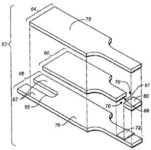
Figures 1A to 4B show various views of an exemplary test strip 62 suitable for
use with the methods described herein. Test strip 62 can include an elongate
body
extending from a proximal end 80 to a distal end 82, and having lateral edges
56, 58.
The proximal portion of body 59 can include a reaction chamber 61 having
electrodes
and a reagent, while the distal portion of test strip body 59 can include
features adapted
for electrically communicating with a test meter. Physiological fluid or
control solution
can be delivered to reaction chamber 61 and electrochemically analyzed.
In the illustrative embodiment, test strip 62 comprises a first electrode
layer 66
and a second electrode layer 64, with a spacer layer 60 positioned
therebetween. The
first electrode layer 66 can provide a first electrode 166 and a first
connection track 76
for electrically connecting the first electrode 166 to a first electrical
contact 67.
Similarly, second electrode layer 64 can provide a second electrode 164 and a
second
connection track for electrically connecting the second electrode 164 with a
second
electrical contact 63.
In one embodiment, sample reaction chamber 61 is defined by first electrode
166, second electrode 164, and spacer 60 as shown in Figures IA to 4B.
Specifically,
first electrode 166 and second electrode 164 define, respectively, the bottom
and top of
sample reaction chamber 61. A cutout area 68 of spacer 60 can define the side
walls of
sample reaction chamber 61. In one aspect, reaction chamber 61 can further
include
ports 70 that provide a sample inlet and/or a vent. For example, one of the
ports can
provide a fluid sample ingress and the other port can act as a vent.
Reaction chamber 61 can have a small volume. In one embodiment, the volume
ranges from about 0.1 microliters to 5 microliters, preferably about 0.2
microliters to
about 3 microliters, and more preferably about 0.3 microliters to about 1
microliter. To
provide the small sample volume cutout 68 can have an area ranging from about
0.01
cm2 toabout 0.2 cm2, preferably about 0.02 cm2 toabout 0.15 cm2, and more
preferably
about 0.03 cm2 toabout 0.08 cm2. In addition, first and second electrode 166,
164 can
be spaced in the range of about 1 micron to 500 microns, preferably between
about 10
microns and 400 microns, and more preferably between about 40 microns and 200
microns. The close spacing of the electrodes can also allow redox cycling to
occur,
where oxidized mediator generated at first electrode 166, can diffuse to
second electrode
CA 02748433 2011-08-04
-8-
164 to become reduced, and subsequently diffuse back to first electrode 166 to
become
oxidized again.
At the distal end of test strip body 59, first electrical contact 67 can be
used to
establish an electrical connection to a test meter. Second electrical contact
63 can be
accessed by the test meter through U-shaped notch 65 as illustrated in Figure
2. One
skilled in the art will appreciate that test strip 62 can include a variety of
alternative
electrical contact configured for electrically connecting to a test meter. For
example,
U.S. Patent No. 6,379,513 discloses an electrochemical cell connection means s
In one embodiment, first electrode layer 66 and/or second electrode layer 64
can
be a conductive material formed from materials such as gold, palladium,
carbon, silver,
platinum, tin oxide, iridium, indium, and combinations thereof (e.g., indium
doped tin
oxide). In addition, the electrodes can be formed by disposing a conductive
material
onto an insulating sheet (not shown) by a sputtering, electroless plating, or
a screen
printing process. In one exemplary embodiment, second electrode layer 64 can
be a
sputtered gold electrode and first electrode layer 66 can be a sputtered
palladium
electrode. Suitable materials that can be employed as spacing layer 60 include
the
variety of insulating materials, such as, for example, plastics (e.g., PET,
PETG,
polyimide, polycarbonate, polystyrene), silicon, ceramic, glass, adhesives,
and
combinations thereof
Reagent layer 72 can be disposed within reaction chamber 61 using a process
such as slot coating, dispensing from the end of a tube, ink jetting, and
screen printing.
Such processes are described, for example, in the following U.S. Patent Nos.
6,749,887;
6,869,411; 6,676,995; and 6,830,934.,
In one embodiment, reagent layer 72 includes at least a mediator and an
enzyme and is deposited onto first electrode 166. Examples of suitable
mediators
include ferricyanide, ferrocene, ferrocene derivatives, osmium bipyridyl
complexes, and
quinone derivatives. Examples of suitable enzymes include glucose oxidase,
glucose
dehydrogenase (GDH) based on pyrroloquinoline quinone (PQQ) co-factor, GDH
based
on nicotinamide adenine dinudeotide co-factor, and FAD-based GDH
[E.C.1.1.99.10].
One exemplary reagent formulation, which would be suitable for making reagent
layer
72, is described in pending U.S. Application entitled, Method of
CA 02748433 2011-08-04
- 9 -
Manufacturing a Sterilized and Calibrated Biosensor-Based Medical Device,
published
as U.S. Published Patent Application No. 2004/0120848.
Either first electrode 166 or second electrode 164 can perform the function of
a
working electrode which oxidizes or reduces a limiting amount of mediator
depending
on the polarity of the applied test potential of the test meter. For example,
if the current
limiting species is a reduced mediator, then it can be oxidized at first
electrode 166 as
long as a sufficiently positive potential was applied with respect to second
electrode 164.
In such a situation, first electrode 166 performs the function of the working
electrode
and second electrode 164 performs the function of a counter/reference
electrode. It
should be noted that unless otherwise stated for test strip 62, all potentials
applied by test
meter 100 will hereinafter be stated with respect to second electrode 164.
Similarly, if a sufficiently negative potential is applied with respect to
second
electrode 164; then the reduced mediator can be oxidized at second electrode
164. In
such a situation, second electrode 164 performs the function of the working
electrode
and first electrode 166 performs the function of the counter/reference
electrode.
A first step in the subject methods can include introducing a quantity of the
fluid
sample of interest into test strip 62 which includes first electrode 166,
second electrode
164 and a reagent layer 72. The fluid sample can be whole blood or a
derivative or
fraction thereof, or control solution. The fluid sample, e.g., blood, is dosed
into sample
reaction chamber 61 via port 70. In one aspect, port 70 and /or reaction
chamber 61 are
adapted such that capillary action causes the fluid sample to fill sample
reaction
chamber 61.
Figure 5 provides a simplified schematic showing a test meter 100 interfacing
with first electrical contact 67 and second electrical contact 63, which are
in electrical
communication with first electrode 166 and second electrode 164, respectively,
of test
strip 62. Test meter 100 is adapted to electrically connect to first electrode
166 and
second electrode 164, via first electrical contact 67 and second electrical
contact 63,
respectively (as shown in Figures 2 and 5). The variety of known test meters
can be
used with the method described herein. However, in one embodiment the test
meter
includes at least a processor for performing calculations related to
discriminating
between blood and a control sample and data storage.
CA 02748433 2011-08-04
- 10 -
As illustrated in Figure 5, electrical contact 67 can include two prongs
denoted as
67a and 67b. In one exemplary embodiment, test meter 100 separately connects
to
prongs 67a and 67b, such that when test meter 100 interfaces with test strip
62 a circuit
is completed. Test meter 100 can measure the resistance or electrical
continuity between
prongs 67a and 67b to determine whether test strip 62 is electrically
connected to test
meter 100. One skilled in the art will appreciate that test meter 100 can use
a variety of
sensors and circuits to determine when test strip 62 is properly positioned
with respect to
test meter 100.
In one embodiment, test meter 100 can apply a test potential and/or a current
between first electrical contact 67 and second electrical contact 63. Once
test meter 100
recognizes that strip 62 has been inserted, test meter 100 turns on and
initiates a fluid
detection mode. In one embodiment, the fluid detection mode causes test meter
100 to
apply a constant current of 1 microampere between first electrode 166 and
second
electrode 164. Because test strip 62 is initially dry, test meter 100 measures
a maximum
voltage, which is limited by the hardware within test meter 100. However, once
a user
doses a fluid sample onto inlet 70, this causes sample reaction chamber 61 to
become
filled. When the fluid sample bridges the gap between first electrode 166 and
second
electrode 164, test meter 100 will measure a decrease in measured voltage
(e.g., as
described in U.S. Patent No. 6,193, 873) which is below a predetermined
threshold
causing test meter 100 to automatically initiate the glucose test.
It should be noted that the measured voltage may decrease below a pre-
determined threshold when only a fraction of sample reaction chamber 61 has
been
filled. A method of automatically recognizing that a fluid was applied does
not
necessarily indicate that sample reaction chamber 61 has been completely
filled, but can
only confirm a presence of some fluid in sample reaction chamber 61. Once test
meter
100 determines that a fluid has been applied to test strip 62, a short, but
finite amount of
time may still be required to allow the fluid to completely fill sample
reaction chamber
61.
In one embodiment, once test meter 100 has determined that a fluid has been
dosed onto test strip 62, test meter 100 can perform a glucose test by
applying a plurality
of open-circuit potentials and a plurality of test potentials to the test
strip 62 for
prescribed intervals as shown in Figure 6. A glucose test time interval TG
represents an
CA 02748433 2011-08-04
- 11 -
amount of time to perform the glucose test (but not necessarily all the
calculations
associated with the glucose test) where glucose test time interval TG can
include a first
open-circuit time interval Toci, a first test potential time interval T1, a
second open-
circuit time interval T0c2, a second test potential time interval T2, and a
third test
potential time interval T3, Glucose test time interval TG can range from about
1 second
to about 5 seconds, While two open-circuit time intervals and three test
potential time
intervals are described, one skilled in the art will appreciate that the
glucose test time
interval can comprise different numbers of open-circuit and test potential
time intervals.
to For example, the glucose test time interval could include a single open-
circuit time
interval and/or only two test potential time intervals.
Once the glucose assay has been initiated, test meter 100 switches to a first
open-
circuit for a first open-circuit potential time interval T0c1, which in the
illustrated
embodiment is about 0.2 seconds. In another embodiment, first open-circuit
time
interval Tom can be in the range of about 0.05 seconds to about 2 seconds and
preferably between about 0.1 seconds to about 1.0 seconds, and most preferably
between
about 0.15 seconds to about 0.6 seconds.
One of the reasons for implementing the first open-circuit is to allow
sufficient
time for the sample reaction chamber 61 to fill or partially fill with sample.
Typically, at
ambient temperature (i.e, 22 C), sample reaction chamber 61 takes about 0.1
seconds to
about 0.5 seconds to completely fill with blood. Conversely, at ambient
temperature
(i.e. 22 C), sample reaction chamber 61 takes about 0.2 seconds or less to
completely
fill with control solution, where the control solution is formulated to have a
viscosity of
about 1 to about 3 centipoise.
While control solutions are composed of known components and are generally
uniform, blood samples can vary in their make-up and/or composition. For
example,
high hematocrit blood samples are more viscous than low hematocrit blood
samples,
therefore higher hematocrit blood samples require additional time to fill
compared with
lower hematocrit blood samples. Thus, depending on a variety of factors, blood
sample
filling time can vary.
After applying the first open-circuit potential, test meter 100 applies a
first test
potential E1 between first electrode 166 and second electrode 164 (e.g., ¨0.3
Volts in
Figure 6), for a first test potential time interval T1 (e.g., 0.15 seconds in
Figure 6). Test
CA 02748433 2011-08-04
- 12 -
meter 100 measures the resulting first current transient, which can be
referred to as la(t)
as shown in Figure 7. In one embodiment, first test potential time interval T1
can be in
the range of about 0.05 seconds to about 1.0 second and preferably between
about 0.1
seconds to about 0.5 seconds, and most preferably between about 0.1 seconds to
about
0.2 seconds.
As discussed below, a portion or all of the first current transient can be
used in
the methods described herein to determine whether control solution or blood
was applied
to test strip 62. The magnitude of the first transient current is effected by
the presence of
easily oxidizable substances in the sample. Blood usually contains endogenous
and
exogenous compounds that are easily oxidized at second electrode 164.
Conversely,
control solution can be formulated such that it does not contain oxidizable
compounds.
However, blood sample composition can vary and the magnitude of the first
current
transient for high viscosity blood samples will be smaller than low viscosity
samples (in
- 15 some cases even less than control solution samples) because sample
reaction chamber 61
may be not be completely filled after 0.2 seconds. An incomplete fill will
cause the
effective area of first electrode 166 and second electrode 164 to decrease
which in turn
causes the first current transient to decrease. Thus the presence of
oxidizable substances
in a sample, by itself, is not always a sufficient discriminatory factor
because of
variations in blood samples.
After test meter 100 stops applying first test potential El, it switches to a
second
open-circuit for a second open-circuit time interval TOC2, which in this case
is about
0.65 seconds, as shown in Figure 6. In another embodiment, second open-circuit
time
interval TOC2 can be in the range of about 0.1 seconds to about 2.0 seconds
and
preferably between about 0.3 seconds to about 1.5 seconds, and most preferably
between
about 0.5 seconds to about 1.0 seconds.
One of the reasons for implementing the second open-circuit is to provide
sufficient time for sample reaction chamber 61 to completely fill, to allow
reagent layer
72 to dissolve, and to allow reduced mediator and oxidized mediator to re-
equilibrate at
the respective first electrode 166 and second electrode 164 from the
perturbation caused
by first test potential El. Although sample reaction chamber 61 fills rapidly,
second
open-circuit time interval Tom can be sufficiently long to account for
conditions which
CA 02748433 2011-08-04
-13 -
can cause fill times to increase such as low ambient temperature (e.g., about
5 C) and
high hematocrit (e.g., >60% hematocrit).
During first test potential El, reduced mediator was depleted at second
electrode
164 and generated at first electrode 166 to form a concentration gradient.
Second open-
circuit potential provides time for the reduced mediator concentration profile
to become
closer to the state immediately before first test potential E1 was applied. As
will be
described below, a sufficiently long second open-circuit potential is useful
because it
can allow for glucose concentration to be calculated in the presence of
interferents.
An alternative embodiment test potential El' can be applied between the
electrodes for a duration between when the meter detects that the strip is
filling with
sample and before a second test potential E2 is applied. In one aspect, test
potential El'
is small. For example, the potential can be between about 1 to 100 mV,
preferably
between about 5 mV and 50 mV and most preferably between about 10 mV and 30
mV.
The smaller potential perturbs the reduced mediator concentration gradient to
a lesser
extent compared to applying a larger potential difference, but is still
sufficient to obtain
a measure of the oxidizable substances in the sample. The potential El' can be
applied
for a portion of the time between detection of fill and when E2 is applied or
can be
applied for the whole of that time period. If E1' is to be used for a portion
of the time
then an open-circuit could be applied for the remaining portion of the time.
The
combination of number of open-circuit and small voltage potential
applications, their
order and times applied is not critical in this embodiment, as long as the
total period for
which the small potential El' is applied is sufficient to obtain a current
measurement
indicative of the presence and/or quantity of oxidizable substances present in
the sample.
In a preferred embodiment the small potential E1' is applied for the entire
period
between when fill is detected and when E2 is applied.
Once second open-circuit time interval T0c2 or an equivalent time in the small
potential E1' embodiment has elapsed, test meter 100 applies a second test
potential E2
between first electrode 166 and second electrode 164 for a second test
potential time
interval T2. During second test potential time interval T2, test meter 100 can
measure a
second current transient which may be referred to as ib(t). After second
potential time
interval T2 has elapsed, test meter 100 can apply a third test potential E3
between first
electrode 166 and second electrode 164 for a third test potential time
interval T3, which
CA 02748433 2011-08-04
- 14 -
may be referred to as i(t). Second test potential time interval Ty and third
test potential
time interval T3 can each range from about 0.1 seconds to 4 seconds. For the
embodiment shown in Figure 6, second test potential time interval T2 was 3
seconds and
third test potential time interval T3 was 1 second. As mentioned above, in one
aspect, an
open circuit potential time period can be allowed to elapse between the second
test
potential Ey and the third test potential E3. Alternatively, the third test
potential E3 can
be applied immediately following the application of the second test potential
Ey. Note
that a portion of the first, second, or third current transient may be
generally referred to
as a cell current or a current value.
In one embodiment, first test potential E1 and second test potential Ey both
have
a first polarity, and that third test potential E3 has a second polarity which
is opposite to
the first polarity. However, one skilled in the art will appreciate the
polarity of the first,
second, and third test potentials can be chosen depending on the manner in
which
analyte concentration is determined and/or depending on the manner in which
test
samples and control solutions are distinguished.
First test potential El and second test potential Ey can be sufficiently
negative in
magnitude with respect to second electrode 164 such that second electrode 164
functions
as a working electrode in which a limiting oxidation current is measured.
Conversely,
third test potential E3 can be sufficiently positive in magnitude with respect
to second
electrode 164 such that first electrode 166 functions as a working electrode
in which a
limiting oxidation current is measured. A limiting oxidation occurs when all
oxidizable
species have been locally depleted at the working electrode surface such that
the
measured oxidation current is proportional to the flux of oxidizable species
diffusing
from the bulk solution towards the working electrode surface. The term bulk
solution
refers to a portion of the solution sufficiently far away from the working
electrode where
the oxidizable species was not located within the depletion zone. First test
potential El,
second test potential Ey, and third test potential E3 can range from about
¨0.6 Volts to
about +0.6 Volts (with respect to second electrode 164) when using either a
sputtered
gold or palladium working electrode and a ferricyanide mediator.
Figure 7 shows a first, second, and third current transients generated by test
meter 100 and test strip 62 using either a control solution sample (dotted
line) or a blood
sample (solid line). The control solution sample contained a 525 mg/dL glucose
CA 02748433 2011-08-04
- 15 -
concentration and the blood sample contained a 530 mg/dL glucose concentration
with a
25% hematocrit. Figure 8 shows an expanded view of first and second current
transients in
Figure 7. Figures 7 and 8 show the resulting current transients when applying
the potential
waveform shown in Figure 6. The description below details how the current
transients can
be converted into an accurate glucose measurement for the test solution or
control solution.
Assuming that a test strip has an opposing face or facing arrangement as shown
in
Figures IA to 4B, and that a potential waveform is applied to the test strip
as shown in
Figure 6, a glucose concentration can be calculated using a glucose algorithm
as shown in
Equation (Eq.) 1.
I. Y
Eq. 1 [G]=-- X (axii¨ Z)
i3)
In Eq. 1, [G] is the glucose concentration, i1 is a first current value, i2 is
a second
current value, and i3 is a third current value, and the terms p, Z, and a are
empirically
derived calibration constants. A derivation of Eq. 1 can be found in U.S.
Application
Publication No. 2007/0074977 which was filed on September 30, 2005 and
entitled
"METHOD AND APPARATUS FOR RAPID ELECTROCHEMICAL ANALYSIS". First
current value i1 and second current value i2 are calculated from the third
current transient
and i3 is calculated from the second current transient. One skilled in the art
will appreciate
that names "first," "second," and "third" are chosen for convenience and do
not necessarily
reflect the order in which the current values are calculated. In addition, all
current values
(e.g., i1, i2, and i3) stated in Eq. 1 use the absolute value of the current.
In another embodiment of this invention, the term i1 can be defined to include
peak
current values from the second and third current transients to allow for more
accurate
glucose concentrations in the presence of interferents as shown in Eq. 2.
Eq. 2 21pb c2
+
pc sr
CA 02748433 2011-08-04
- 16 -
The term ipb represents a peak current value for second test potential time
interval T2
and the term ip, represents a peak current value for third test potential time
interval T3. The
term iõ is the steady-state current which occurs after the application of
third test potential
E3. Where Eq. 2 is used, second open-circuit potential time interval TOC2 is
preferably
sufficiently long so as to allow Eq. 2 to compensate for the presence of
interferents. When
second open-circuit potential time interval T0C2 is too short, second peak
current value ipb
can become distorted and can reduce the effectiveness of the interferent
correction
calculations. The use of peak current values to account for interferents in a
physiological
sample are described in U.S. Application Publication No. 2007/0227912 entitled
"Methods
and Apparatus for Analyzing a Sample in the Presence of Interferents" which
was filed on
March 21, 2006.
In one embodiment of this invention, Eq.'s 1 and 2 can be used together to
calculate
a glucose concentration for either blood or control solution. In another
embodiment of this
invention, the algorithm of Eq.'s 1 and 2 can be used for blood with a first
set of calibration
factors (i.e. a, p, and Z) and a second set of calibration factors can be used
for the control
solution. When using two different sets of calibration factors, the methods
described herein
for discriminating between a test fluid and a control solution can improve the
effectiveness
of the analyte concentration calculations.
In addition, if the test meter determines that the sample type is control
solution, the
test meter can store the resulting glucose concentration of the control sample
such that a user
can review test sample concentration data separately from control solution
data. For
example, the glucose concentrations for control solutions can be stored in a
separate
database, can be flagged, and/or discarded (i. e., not stored or stored for a
short period of
time).
Another advantage of being able to recognize control solutions is that a test
meter
can be programmed to automatically compare the results (e.g., glucose
concentration) of the
test of the control solution with the expected glucose concentration of the
control solution.
For example, the test meter can be pre-programmed with the expected glucose
level(s) for
the control solution(s). Alternatively, a user could input the expected
glucose concentration
for the control solution. When the test meter recognizes a control solution,
the test meter
can compare the measured control solution
CA 02748433 2011-08-04
- 17 -
glucose concentration with the expected glucose concentration to determine if
the meter
is functioning properly. If the measured glucose concentration is out of the
expected
range, the test meter can output a warning message to alert the user.
In one embodiment, the method described herein uses the presence of redox
species to distinguish a control solution from a blood sample. The method can
include
the step of applying a first test potential El' and using one or more current
values
measured during the test potential as a discriminator. In one aspect, two
current values
from the first test potential El' are summed and used as the discriminator.
Figure 8
shows data for a control solution, plasma, a blood sample with 48% hematocrit,
and a
blood sample is 77% hematocrit. A potential of 20 mV was applied for the first
1
second and current values at 0.2 to 0.5 seconds were summed. As show in Figure
8, the
summed current values were sufficient to distinguish between a control
solution (that
was substantially devoid of interferents) and blood samples.
In another embodiment, two characteristics of control solution are used to
distinguish control solutions from blood ¨ the presence and/or concentration
of redox
species in the sample and reaction kinetics. The method disclosed herein can
include the
step of calculating a first reference value that is representative of the
redox concentration
in the sample and a second reference value that is representative of the rate
of reaction of
the sample with the reagent. In one embodiment, the first reference value is
an
interferent oxidation current and the second reference value is a reaction
completion
percentage.
In regard to redox species in the sample, blood usually contains various
endogenous redox species or "interferents" such as ascorbic acid and uric
acid, as well
as exogenously derived interferents such as gentisic acid (gentisic acid is a
metabolite of
aspirin). Endogenous interferents are chemical species that can be easily
oxidized at an
electrode and are usually present in blood within a physiological range for
healthy
individuals. Exogenously derived interferents are also a chemical species that
can be
easily oxidized at an electrode, but are not usually present in blood unless
they are
inputted into the body via consumption, injection, absorption, and the like.
Control solution can be formulated to be either essentially free of
antioxidants or
to have a relatively high interferent concentration compared to the
interferent
concentration in a blood sample. For the case in which control solution is
essentially
CA 02748433 2011-08-04
- 18 -
free of antioxidants, the magnitude of the first current transient should be
smaller for
control solution than for a blood sample as shown in Figure 9. For the case in
which
control solution has a relatively high concentration of interferents, the
magnitude of the
first current transient should be larger for control solution than for a blood
sample (data
not shown).
An interferent index can be calculated based on the current values within
first
current transient. In one embodiment, the interferent index can include a
summation of
current values at two points in time during the first current transient. In
one example,
the current values at 0.3 and 0.35 seconds can be used. In another embodiment
when a
small potential El' is applied for the entire period between when fill is
detected and E2,
the interferent index is preferably obtained by summing two values over a
longer period,
for example 0.2 seconds to 0.5 seconds.
In general, the interferent index will be proportional to the interferent
concentration and should not substantially depend on the glucose
concentration.
Therefore, in theory, the test meter should be able to distinguish whether the
sample is
blood or control solution based on the interferent index. However, in
practice, using
only the interferent index did not always sufficiently discriminate between
blood and
control solution. Although blood typically has a much higher interferent
concentration,
there are certain conditions in which the first current transient for blood
may be
attenuated such that it is comparable to control solution. These conditions
include high
glucose concentration, high hematocrit, low temperature, and incomplete
filling of
sample reaction chamber 61. Thus, in one embodiment, an additional factor was
implemented to enable the test meter to sufficiently discriminate between
blood and
control solution.
The additional factor used for helping discriminate between blood and control
solution can be a residual reaction index which is a function of the percent
of remaining
substrate which would have reacted if given enough time. The residual reaction
index
relates to the reaction rate in that a high reaction rate can cause the
substrate to be
depleted by the reaction. However, the residual reaction index will also
depend on the
initial magnitude of the substrate concentration.
CA 02748433 2011-08-04
- 19 -
Reagent layer 72 can include glucose dehydrogenase (GDH) based on the PQQ
co-factor and ferricyanide. When blood or control solution is dosed into
sample reaction
chamber 61, glucose is oxidized by GDH(0x) and in the process converts GDH(0)
to
GDH(red), as shown in Eq.3. Note that GDH(õõ) refers to the oxidized state of
GDH, and
GDH(red) refers to the reduced state of GDR
Eq. 3 D-Glucose + GDH(0) - Gluconic acid + GDH(
red)
-ved)
Next, GDH(red) is regenerated back to its active oxidized state by
ferricyanide (i.e.
oxidized mediator or Fe(CN)63) as shown in Eq. 4. In the process of
regenerating
GDHw, ferrocyanide (i.e. reduced mediator or Fe(CN)64") is generated from the
reaction as shown in Eq. 4.
Eq. 4 GDH(red) + 2 Fe(CN)63- - GDH(..) + 2 Fe(CN)64-
In general, the rate of glucose consumption based on Eq.'s 3 and 4 is faster
for
control solution than blood. Typically, control solution is less viscous than
blood
causing the reaction rate of Eq. 3 and 4 to be faster for control solution.
Further, the
reaction rate is faster for control solution because a portion of the glucose
present in the
blood sample must diffuse out of the red blood cells to participate in Eq. 3.
This extra
step of glucose diffusion out of the red blood cells slows down the reaction
rate to some
measurable degree. Figure 9 shows that the reaction rate for blood is slower
than for
control solution as evidenced by the fact that the general absolute slope
value (e.g.,
between 1.2 and 4 seconds) for the second current transient is less for the
blood sample.
Because of the faster reaction rates in control solution compared to blood,
the residual
reaction index for control solution will generally be lower than for blood.
The residual reaction index is a number which is related to the percent of
glucose
which has not been consumed. A relatively low residual reaction index will
indicate that
the reactions of Eq.'s 3 and 4 are close to completion. In contrast, a
relatively high
residual reaction index will indicate that the reaction is not close to
completion. In one
embodiment, the residual reaction index can be an absolute ratio of a current
value of
CA 02748433 2011-08-04
- 20 -
third current transient divided by a current value of the second current
transient, as
shown in Eq. 5.
Eq. 5 abs( (4.15))
i(3.8)
For the denominator of Eq. 5, the current value at 3.8 seconds for the second
current transient is used. The time of 3.8 seconds was chosen empirically,
however, one
skilled in the art will appreciate that other current values can be used. In
one
embodiment, a current value towards the end of the second current transient is
chosen.
During the second current transient time interval T2, reduced mediator is
oxidized at
second electrode 164. The current values measured during second current
transient time
interval T2 were ascribed to ferrocyanide generated by reagent layer 72 at
first electrode
166 which then diffused to second electrode 164 and became oxidized. It is
assumed
that reagent layer 72 remains close to first electrode 166 after it dissolves
in blood
causing most of the ferrocyanide generated by reagent layer 72 to also be
close to first
electrode 166. A portion of this generated ferrocyanide can diffuse to second
electrode
164.
For the numerator of Eq. 5, the current value at 4.15 seconds was used. Other
current values from the third current transient can be chosen, however current
value
towards the beginning of the third current transient are preferred. During the
third
current transient time interval T3, reduced mediator is oxidized at first
electrode 166.
The current values measured during second current transient time interval T2
were
ascribed to ferrocyanide generated by reagent layer 72 at first electrode 166.
Therefore,
the current values for the third current transient will be larger than the
second current
transient because most of the ferrocyanide will be close to first electrode
166 because
first electrode 166 was coated with reagent layer 72. In addition, third
current transient
will also be larger than second current transient because it occurs later in
the glucose test
allowing for more ferrocyanide to be generated. Thus, the absolute ratio as
shown in Eq.
5 will be larger if the glucose reaction is still far from completion at the
time of the
measurement.
. .
CA 02748433 2011-08-04
- 21 -
Figure 10 is a chart showing a non-linear relationship between the % of
substrate
consumed and the residual reaction index for blood samples having various
hematocrit
levels and for control solution (diamonds = 25% hematocrit blood, squares =42%
blood,
triangles = 60% hematocrit blood, x ---- control solution). This chart shows
that the
residual reaction index is relatively high when the % of substrate consumed is
low and
that the residual reaction index is relatively low when the % of substrate
consumed is
high for a given sample type/hematocrit value. The % of substrate consumed is
derived
from a ratio Co ¨ , where Co is the substrate concentration at the electrode
surface and
YSI
YSI is the substrate concentration using a standard reference technique. The
term C, is
derived using the following Eq. 6,
eL
Eq. 6 Co = ___
2FAD
where L is the distance between first electrode 166 and second electrode 164,
F
is Faraday's constant, A is the area of first electrode 166, and D is the
diffusion
coefficient.
Figure 11 is a chart showing a relationship between an interferent index and a
residual reaction index for a plurality of blood samples and control solution
samples. By
plotting the interferent index on the X-axis and the residual reaction index
on the Y-axis,
a segregation between blood and control solution can be observed. A
discrimination line
can be drawn to determine if the sample is either control solution or blood.
In this
embodiment, the interferent index is 1(0.3)+ 1(0.35) and the residual reaction
index is
absii(4.15)1
i(3.8) ) =
=
It should be noted that the times (e.g., 4.15, 3.8) at which the current
values
where selected for the residual reaction index, were found empirically. A
large number
of current ratios were evaluated for their ability to discriminate between
blood and
control solution samples. The ratio shown in Eq. 5 was selected because it was
found to
produce significant separation between blood and control solution samples.
CA 02748433 2011-08-04
- 22 -
A discrimination line was derived to allow the test meter to determine whether
=
the sample was control solution or blood. For all of the control solution
samples tested,
the interferent index was plotted versus the residual reaction index. Next, a
line was
calculated using linear regression for control solution samples. After
calculating an
equation for the line, the perpendicular bias between each data point and the
line was
calculated. The perpendicular bias represents the shortest distance between
the data
point and the line as opposed to a vertical bias which is commonly calculated.
A
standard deviation was determined for all of the perpendicular biases
(SDperp). Lastly,
the line is shifted 3* SDperp units towards the data points for the blood
group. The
reason for this approach is that the data for the control solution group show
very little
scatter and therefore the "99% confidence limit" of the control solution group
is well-
defined.
In the method described herein, the information obtained from this statistical
analysis of the residual reaction index and the interferent index can be used
by the test
meter to distinguish control solutions from blood samples. The test meter can
calculate
the interferent index and residual reaction index and use these values in
association with
the derived discrimination line (or an equation representing the
discrimination line) to
distinguish control solutions from blood samples.
Example 1
Preparation of control fluid is disclosed below. The prepared control fluid
was
used in the experiments which produced the data illustrated in Figures 7 and
11,
Citraconic acid Buffer Component 0.0833 g
Dipotassium citraconate Buffer Component 1.931 g
Methyl Paraben Preservative 0.050 g
Germal II Preservative 0.400 g
Dextran T-500 Viscosity Modifier 3.000 g
Pluronic 25R2 Wicking Agent 0.050 g
1-[(6-methoxy-4-sulfo-m-tolyl)azo1-2-naphthol-6-sulfonic acid disodium salt
Dye (FD&C Blue No. 1) 0.100 g
D-Glucose Analyte 50, 120, or 525 mg
Deionized Water Solvent 100 g
CA 02748433 2011-08-04
-23 -
First citraconic buffer pH 6.5 0.1 was prepared by dissolving required
quantities
of citraconic acid and dipotassium citraconate in deionized water. Next,
Methyl Paraben
was added and the solution was stirred until the preservative was fully
dissolved.
Subsequently Dextran T-500, Germal II, Pluronic 25R2 and l-[(6- methoxy-4-
sulfo-m-
tolypazo]-2-naphthol-6-sulfonic acid disodium salt were added sequentially,
following
complete dissolution of the previously added chemical. At this point, the pH
of the
control fluid was verified, followed by addition of the requisite quantity of
glucose to
obtain a lo', normal or high glucose level of control fluid. After the glucose
was
dissolved completely, the control fluid was left at room temperature
overnight. Finally,
the glucose concentration was verified using a Model 2700 Select Biochemistry
Analyzer manufactured by Yellow Springs Instrument Co., Inc. The dye used in
this
control solution has a blue color which reduces the possibility of a user
confusing
control solution with blood, which is normally red.
One skilled in the art will appreciate further features and advantages of the
invention based on the above-described embodiments. Accordingly, the invention
is not
to be limited by what has been particularly shown and described, except as
indicated by
the appended claims.