Note: Descriptions are shown in the official language in which they were submitted.
CA 02748707 2011-06-29
WO 2010/075632 PCT/CA2009/001899
-I-
METHODS FOR MULTIPLEX ANALYTE DETECTION
AND QUANTIFICATION
FIELD OF INVENTION
The present invention relates to methods for the quantification of analytes,
in
particular, the invention relates to improved microarray methods for the
detection and
quantification of multiple analytes in a single sample.
BACKGROUND
Current immunoassay methods are limited as they only detect one target per
detection test cycle within a single reaction well. It is common for several
antigenic
substances or bio-markers to be associated with detection and diagnosis of any
pathological or physiological disorder. To confirm the presence of multiple
markers,
each marker within a test sample requires a separate and different immunoassay
to
confirm the presence of each target molecule to be detected. This required
multitude of
tests and samples increases delay in time to treatment, costs and possibility
of analytical
error. The current state of the art for quantitative multiplexing of
proteins/antibodies,
especially biomarkers expressed in auto-immune diseases, relies on measuring
multiplex
antigens.
Enzyme Linked Immunosorbent Assay (ELISA) was developed by Engvall et at.,
Immunochem. 8: 871 (1971) and further refined by Ljunggren et al. J. Immunol.
Meth.
88: 104 (1987) and Kemeny et al., Immunol. Today 7: 67 (1986). ELISA and its
applications are well known in the art.
A single ELISA functions to detect a single analyte or antibody using an
enzyme-
labelled antibody and a chromogenic substrate. To detect more than one analyte
in a
sample, a separate ELISA is performed to independently detect each analyte.
For
example, to detect two analytes, two separate ELISA plates or two sets of
wells are
needed, i.e. a plate or set of wells for each analyte. Prior art chromogenic-
based ELISAs
detect only one analyte at a time. This is a major limitation for detecting
diseases with
more,than one marker or transgenic organisms which express more than one
transgenic
product.
CA 02748707 2011-06-29
WO 2010/075632 PCT/CA2009/001899
-2-
Macri, J. N., et al., Ann Clin Biochem 29: 390-396 (1992) describe an indirect
assay wherein antibodies (Reagent- 1) are reacted first with the analyte and
then second
labelled anti-antibodies (Reagent-2) are reacted with the antibodies of
Reagent 1. The
result is a need for two separate washing steps which defeats the purpose of
the direct
assay.
US2007141656 to Mapes et al. measures the ratio of self-antigen and auto-
antibody by comparing to a bead set with monoclonal antibody specific for the
self-
antigen and a bead set with the self antigen. This method allows at least one
analyte to
react with a corresponding reactant, i.e. one analyte is a self-antigen and
the reactants are
auto-antibodies to the self antigen.
Another method for detecting multiple analytes is disclosed in US2005118574 to
Chandler et al which makes use of flow cytometric measurement to classify, in
real time,
simultaneous and automated detection and interpretation of multiple
biomolecules or
DNA sequences while also reducing costs.
WO0113120 to Chandler and Chandler determines the concentration of several
different analytes in a single sample. It is necessary only that there is a
unique
subpopulation of microparticles for each sample / analyte combination using
the flow
cytometer. These bead based systems' capability is limited to distinguishing
between
simultaneous detection of capture antigens.
Simultaneous detection of more than one analyte, i.e. multiplex detection for
simultaneous measurement of proteins has been described by Haab et al.,
"Protein micro-
arrays for highly parallel detection and quantization of specific proteins and
antibodies in
complex solutions," Genome Biology 2(2): 0004.1-0004.13,( 2001), which is
incorporated herein by reference. Mixtures of different antibodies and
antigens were
prepared and labelled with a red fluorescence dye and then mixed with a green
fluorescence reference mixture containing the same antibodies and antigens.
The
observed variation between the red to green ratio was used to reflect the
variation in the
concentration of the corresponding binding partner in the mixes.
Mezzasoma et al. (Clinical Chemistry 48, 1, 121-130 (2002) published a micro-
array format method to detect analytes bound to the same capture in two
separate assays,
CA 02748707 2011-06-29
WO 2010/075632 PCT/CA2009/001899
-3-
specifically different auto-antibodies reactive to the same antigen. The
results revealed
that when incubating the captured analytes with one reporter (for example that
to detect
immunoglobulin IgG), the corresponding analyte is detected. When incubating
the
captured analytes with the second reporter in an assay using a separate
microarray solid-
state substrate (for example to detect IgM), a second analyte (IgM) is
detected.
W00250537 to Damaj and Al-assaad discloses a method to detect up to three
immobilized concomitant target antigens, bound to requisite antibodies first
coated as a
mixture onto a solid substrate. A wash step occurs before the first marker is
detected. The
presence of the first marker may be detected by adding a first specific
substrate. The
reaction well is read and a color change is detectable with light microscopy.
Another
wash step occurs before the second marker is detected. The presence of the
second
marker may be detected by adding a second substrate, specific for the second
enzyme, to
the reaction well. After sufficient incubation, the reaction well may be
assayed for a color
change. Similarly, a wash step may occur before the third marker is detected.
The presence of the third marker may be detected by adding a third substrate,
specific for the third enzyme, to the reaction well. After sufficient
incubation, the reaction
well may be assayed for a color change. Although more than one analyte may be
detected
in a single reaction or test well, each reaction is processed on an individual
basis.
W02005017485 to Geister et al. describes a method to sequentially determine at
least two different antigens in a single assay by two different enzymatic
reactions of at
least two enzyme labelled conjugates with two different chromogenic substrates
for the
enzymes in the assay (ELISA), which comprises (a) providing a first antibody
specific for
a first analyte and a second antibody specific for a second analyte
immobilized on a solid
support ; (b) contacting the antibodies immobilized on the solid support with
a liquid
sample suspected of containing one or both of the antigens for a time
sufficient for the
antibodies to bind the antigens; (c) removing the solid support from the
liquid sample and
washing the solid support to remove unbound material; (d) contacting the solid
support to
a solution comprising a third antibody specific for the first antigen and a
fourth antibody
specific for the second antigen wherein the third antibody is conjugated to a
first enzyme
label and the fourth antibody is conjugated to a second enzyme label for a
time sufficient
for the third and fourth antibodies to bind the analytes bound by the first
and second
CA 02748707 2011-06-29
WO 2010/075632 PCT/CA2009/001899
-4-
antibodies; (e) removing the solid support from the solution and washing the
solid support
to remove unbound antibodies; (f) adding a first chromogenic substrate for the
first
enzyme label wherein conversion of the first chromogenic substrate to a
detectable color
by the first enzyme label indicates that the sample contains the first
analyte; (g) removing
the first chromogenic substrate; and (h) adding a second chromogenic substrate
for the
second enzyme label wherein conversion of the second chromogenic substrate to
a
detectable color by the second enzyme label indicates that the sample contains
the second
analyte.
U.S. Patent 7,022,479, 2006 to Wagner, entitled "Sensitive, multiplexed
diagnostic assays for protein analysis", is a method for detecting multiple
different
compounds in a sample, the method involving: (a) contacting the sample with a
mixture
of binding reagents, the binding reagents being nucleic acid-protein fusions,
each having
(i) a protein portion which is known to specifically bind to one of the
compounds and (ii)
a nucleic acid portion which includes a unique identification tag and which in
one
embodiment, encodes the protein; (b) allowing the protein portions of the
binding
reagents and the compounds to form complexes; (c) capturing the binding
reagent-
compound complexes; (d) amplifying the unique identification tags of the
nucleic acid
portions of the complex binding reagents; and (e) detecting the unique
identification tag
of each of the amplified nucleic acids, thereby detecting the corresponding
compounds in
the sample.
While methods for detecting and quantifying multiple analytes are known, these
methods require the use of separate assaying steps for each of the analytes of
interest and
as such, can be time consuming and costly, especially in the context of a
clinical setting.
SUMMARY OF INVENTION
The present invention provides a fast and cost effective method for detecting
and
quantifying multiple target analytes in test sample using a single reaction
vessel. The
method disclosed herein allows for the simultaneous detection of multiple
target analytes
without the need for separate assays or reaction steps for each target
analyte.
In one aspect, the prevent invention provides a method for detecting and
quantifying two or more target analytes in a test sample comprising:
CA 02748707 2011-06-29
WO 2010/075632 PCT/CA2009/001899
-5-
a) providing a reaction vessel having a microarray printed thereon, said
microarray comprising:
i) a first calibration matrix comprising a plurality of the first calibration
spots, each calibration spot comprising a predetermined amount of a first
target
analyte,
ii) a second calibration matrix comprising a plurality of the second
calibration spots, each calibration spot comprising a predetermined amount of
a
second target analyte,
iii) a first capture matrix comprising a plurality of the first capture spots,
each capture spot comprising a predetermined amount of an agent which
selectively binds to the first target analyte, and
iv) a second capture matrix comprising a plurality of the second capture
spots, each capture spot comprising a predetermined amount of an agent which
selectively binds to the second target analyte;
b) applying a predetermined volume of the test sample to the microarray;
c) applying a first fluorescently labelled antibody which selectively binds to
the
first target analyte and a second fluorescently labelled antibody which
selectively binds to
the second target analyte to the assay device, wherein said first and second
fluorescently
labelled antibodies each comprise a different fluorescent dye having emission
and
excitation spectra which do not overlap with each other;
d) measuring a signal intensity value for each spot within the microarray;
e) generating calibration curves by fitting a curve to the measured signal
intensity
values for each of the calibration spots versus the known concentrations of
the first target
analyte and second target analyte; and
f) determining the concentration for the first target analyte and the second
target
analytes using the generated calibration curves.
CA 02748707 2011-06-29
WO 2010/075632 PCT/CA2009/001899
-6-
In an embodiment of the present invention, the target analytes are proteins.
The
proteins may be antibodies.
In a further embodiment of the present invention, the reaction vessel is a
well of a
multi-well plate and wherein each well has the microarray printed therein.
In a further embodiment of the present invention, the test sample is a
biological
sample.
In another aspect, the present invention provides a method for detecting and
quantifying biomarkers diagnostic for rheumatoid arthritis, comprising:
a) providing an assay device having a microarray printed thereon, said
microarray
comprising:
i) a calibration matrix comprising a plurality of spots, each spot
comprising a predetermined amount of one of: a human IgA antibody, a human
IgG antibody, and a human IgM antibody;
ii) a first analyte capture matrix comprising a plurality of spots comprising
a predetermined amount of rheumatoid factor; and
iii) a second analyte capture matrix comprising a plurality of spots
comprising a predetermined amount of cyclic citrullinated peptide;
b) applying a predetermined volume of a serum sample to the assay device;
c) applying a first fluorescently labelled antibody which selectively binds to
IgA
antibodies, a second fluorescently labelled antibody which selectively binds
to IgG
antibodies, and a third fluorescently labelled antibody which selectively
binds to IgM
antibodies to the assay device, wherein said first, second and third
fluorescently labelled
antibodies each comprise a different fluorescent dye having emission and
excitation
spectra which do not overlap with each other;
d) measuring a signal intensity value for each spot within the assay device;
CA 02748707 2011-06-29
WO 2010/075632 PCT/CA2009/001899
-7-
e) generating calibration curves by fitting a curve to the measured signal
intensity
values for the each of the calibration spots versus the known concentration of
the human
IgA, IgG and IgM antibodies; and
f) determining the concentration for each of captured rheumatoid factor-IgA,
rheumatoid factor-IgG, rheumatoid factor-IgM, anti-cyclic citrullinated
peptide-IgG,
anti-cyclic citrullinated peptide-IgA, and/or anti-cyclic citrullinated
peptide-IgM using
the calibration curves.
In another aspect, the present invention provides a method for diagnosing
rheumatoid arthritis in a subject, comprising:
a) measuring the concentration levels of rheumatoid factor-IgA, rheumatoid
factor-IgG, rheumatoid factor-IgM and at least one of anti-cyclic
citrullinated peptide-
IgG, anti-cyclic citrullinated peptide-IgA, and anti-cyclic citrullinated
peptide-IgM in a
biological sample, using the method disclosed herein; and
b) comparing the measured concentration levels of rheumatoid factor-IgA,
rheumatoid factor-IgG, rheumatoid factor-IgM, anti-cyclic citrullinated
peptide-IgG,
anti-cyclic citrullinated peptide-IgA, and/or anti-cyclic citrullinated
peptide-IgM with
index normal levels of rheumatoid factor-IgA, rheumatoid factor-IgG,
rheumatoid factor-
IgM and anti-cyclic citrullinated peptide-IgG, anti-cyclic citrullinated
peptide-IgA, and/or
anti-cyclic citrullinated peptide-IgM wherein measured concentrations levels
which
exceed index normal levels is diagnostic for rheumatoid arthritis.
In an embodiment of the present invention, the detection and quantification of
predominantly rheumatoid factor-IgM and anti-cyclic citrullinated peptide-IgM
antibodies is diagnostic for an early stage of rheumatoid arthritis.
In a further embodiment of the present invention, the detection and
quantification
of rheumatoid factor-IgA and anti-cyclic citrullinated peptide-IgA antibodies
is diagnostic
for a transitional stage of rheumatoid arthritis.
In a further embodiment of the present invention, the detection and
quantification
of rheumatoid factor-IgG and anti-cyclic citrullinated peptide-IgG antibodies
is diagnostic
for a late stage of rheumatoid arthritis.
CA 02748707 2011-06-29
WO 2010/075632 PCT/CA2009/001899
-8-
In another aspect, the present invention provides a method for monitoring
rheumatoid arthritis treatment in a subject suffering therefrom, comprising
measuring the
concentration levels of rheumatoid factor-IgA, rheumatoid factor-IgG,
rheumatoid factor-
IgM and at least one of anti-cyclic citrullinated peptide-IgG, anti-cyclic
citrullinated
peptide-IgA, and anti-cyclic citrullinated peptide-IgM using the method
disclosed herein,
a plurality of times during the treatment.
BRIEF DESCRIPTION OF THE DRAWINGS
Preferred embodiments of the invention will now be described, by way of
example, with reference to the accompanying drawings, in which:
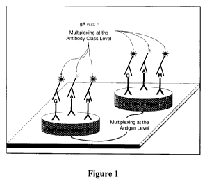
Figure 1 is a schematic illustration of the multiplex analyte detection method
of
the present invention;
Figure 2 is a bar graph plotting the ratio of the average measured
fluorescence
intensity for captured IgA against the average measured fluorescence intensity
for IgM
internal calibrator for two samples, NS and RF#3;
Figure 3 is a bar graph plotting the ratio of the average measured
fluorescence
intensity for captured IgM against the average measured fluorescence intensity
for IgM
internal calibrator for two samples, NS and RF#3; and
Figure 4 is a plot comparing the composite fluorescent intensities for IgA,
IgG and
IgM antibodies using the method of the present invention.
DESCRIPTION
The present invention provides a method for the detection and quantification
of
multiple target analytes in a test sample, within a single reaction well, per
test cycle. The
method disclosed herein provides for the simultaneous incubation of an assay
device with
two or more fluorescently labelled reporters in the same detection mixture as
shown in
Figure 1. The method disclosed herein can detect more than one analyte in
using a single
reaction vessel instead of separate reaction vessels to detect each analyte.
For example,
when the target analytes of interest are different classes of human antibodies
(i.e. hIgG,
hIgA, and hIgM) directed to the same antigen (i.e. the Fc region of hIgG), the
detection
CA 02748707 2011-06-29
WO 2010/075632 PCT/CA2009/001899
-9-
and quantification of each of the target antibodies requires separate assays
when
convention methods are employed. With conventional methods, one assay is
performed
to detect and quantify the amount of hIgG present in the test sample. A second
assay
must be performed to detect and quantify the amount of hIgM and a third assay
must be
performed to detect and quantify the amount of hIgG. In contrast, the method
of the
present invention eliminates the need for multiple detection steps thus
reducing costs and
time. Using the method of the present invention, target hIgG, hIgA and hIgM
molecules
contained in a test sample can be bound to as single capture spot in an assay
device. In the
disclosed method, the different classes of antibodies can be detected in a
single test by
using a cocktail of fluorescently labelled antibodies directed to each of the
hIgG, hIgM
and hIgA targets. As the antibodies are labelled with different optically
excited and
emitted fluorescent probes, the each of the targets bound to a single capture
spot can be
detected and quantified using an appropriate calibrator. The use of multi-
channel
detectors allows for substantially simultaneous detection of multiple analytes
in a single
assay.
The methods disclosed herein employ assay devices useful for conducting
immunoassays. The assay devices may be microarrays in 2 or 3-dimensional
planar array
format.
In one embodiment, the method may employ the use of a multi-well plate and
wherein each well has a microarray printed therein. A single well is used as a
reaction
vessel for assaying the desired plurality of target analytes for each test
sample.
The microarray may comprise a calibration matrix and an analyte capture matrix
for each target analyte.
As used herein, the term "calibration matrix" refers to a subarray of spots,
wherein
each spot comprises a predetermined amount of a calibration standard. The term
"predetermined amount" as used herein, refers to the amount of the calibration
standard
as calculated based on the known concentration of the spotting buffer
comprising the
calibration standard and the known volume of the spotting buffer printed on
the reaction
vessel.
CA 02748707 2011-06-29
WO 2010/075632 PCT/CA2009/001899
-10-
The choice of the calibration standard will depend on the nature of the target
analyte. The calibration standard may be the target analyte itself in which
case, the
calibration standard. In such embodiments, the microarray will comprise a
separate
calibration standard for each target analyte. Alternatively, the microarray
may comprise a
single calibration matrix having calibration spots containing each of the
target analytes.
In alternate embodiments, the calibration standard is a surrogate compound.
For
example if the target analyte is an antibody, the surrogate compound may be
another
different antibody but of the same class of immunoglobulin. For example,
Figure 1
illustrates an assay device useful for capturing six different antibodies
which selectively
bind to two different antigens. In such embodiments, only one calibration
matrix may be
required for each of the three different classes of immunoglobulins.
The calibration matrix may be printed on the base of the individual reaction
vessel
in the form of a linear, proportional dilution series with the predetermined
amounts of the
calibration standard falling within the dynamic range of the detection system
used to read
the microarray.
As used herein, the term "analyte capture matrix" refers to a subarray of
spots
comprising an agent which selectively binds to the target analyte. In
embodiments where
the target analyte is a protein, the agent may be an analyte specific antibody
or fragment
thereof. Conversely, in embodiments wherein the target analyte is an antibody,
the agent
may be an antigen specifically bound by the antibody. For example, Figure 1
illustrates
an assay device useful for capturing six different antibodies which
selectively bind to two
different antigens.
A predetermined volume of a test sample is applied to the assay device. The
each
of the target analytes will bind to their specific capture spot. Thus, in a
single capture
spot, multiple target analytes may be bound. To detect each of the target
analytes, a
fluorescently labelled antibody which specifically binds to the target analyte
is used.
Each antibody is coupled to a unique fluorescent dye with a specific
excitation and
emission wavelength to obtain the desired Stokes shift and excitation and
emission
coefficients. The fluorescent dyes are chosen based on their respective
excitation and
emission spectra such that each of the labelled antibodies comprises a
different
CA 02748707 2011-06-29
WO 2010/075632 PCT/CA2009/001899
-11-
fluorescent dye having emission and excitation spectra which do not overlap
with each
other. The fluorescently labelled antibodies can be applied to the assay
device in a single
step in the form of a cocktail.
A signal intensity value for each spot within the assay device is then
measured.
The fluorescent signals can be read using a combination of scanner components
such as
light sources and filters. A signal detector can be used to read one optical
channel at a
time such that each spot is imaged with multiple wavelengths, each wavelength
being
specific for a target analyte. An optical channel is a combination of an
excitation source
and an excitation filter, matched for the excitation at a specific wavelength.
The emission
filter and emission detector pass only a signal wavelength for a specific
fluorescent dye.
The optical channels used for a set of detectors are selected such that they
do not interfere
with each other, i.e. the excitation through one channel excites only the
intended dye, not
any other dyes. Alternatively, a multi-channel detector can be used to detect
each of the
differentially labelled antibodies. The use of differential fluorescent labels
allows for
substantially simultaneous detection of the multiple target analytes bound to
a single
capture spot.
The intensity of the measured signal is directly proportional to the amount of
material contained within the printed calibration spots and the amount of
analyte from the
test sample bound to the printed analyte capture spot. For each calibration
compound, a
calibration curve is generated by fitting a curve to the measured signal
intensity values
versus the known concentration of the calibration compound. The concentration
for each
target analyte in the test sample is then determined using the appropriate
calibration curve
and by plotting the measured signal intensity for the target analyte on the
calibration
curve.
The method disclosed herein can be used to detect and quantify multiple
clinically
relevant biomarkers in a biological sample for diagnostic or prognostic
purposes. The
measured concentrations for a disease related biomarker can be compared with
established index normal levels for that biomarker. The measured
concentrations levels
which exceed index normal levels may be identified as being diagnostic of the
disease.
The method disclosed herein can also be used to monitor the progress of a
disease and
also the effect of a treatment on the disease. Levels of a clinically relevant
biomarker can
CA 02748707 2011-06-29
WO 2010/075632 PCT/CA2009/001899
-12-
be quantified using the disclosed method a plurality of times during a period
of treatment.
A trending decrease in biomarker levels may be correlated with a positive
patient
response to treatment.
The method disclosed herein can be used to detect and quantify biomarkers
diagnostic for rheumatoid arthritis. In one embodiment, the method comprises
the
provision of an assay device having a microarray printed thereon. The
microarray may
comprise: i) a calibration matrix comprising plurality of spots, each spot
comprising a
predetermined amount of one of: a human IgA antibody, a human IgG antibody,
and a
human IgM antibody; ii) a first analyte capture matrix comprising a plurality
of spots
comprising a predetermined amount of rheumatoid factor; and iii) a second
analyte
capture matrix comprising a plurality of spots comprising a predetermined
amount of
cyclic citrullinated peptide. A predetermined volume of a biological sample,
preferably a
serum sample, is applied to the assay device. A cocktail comprising a first
fluorescently
labelled reporter compound which selectively binds to IgA antibodies, a second
fluorescently labelled reporter compound which selectively binds to IgG
antibodies, and a
third fluorescently labelled reporter compound which selectively binds to IgM
antibodies
is then applied to the assay device. The first, second and third fluorescently
labelled
antibodies are chosen such that each of the antibodies comprise a different
fluorescent
dye having emission and excitation spectra which do not overlap with each
other. A
signal intensity value for each spot within the assay device is then measured
using a
single or multi-channel detector as discussed above. Using the measured signal
intensity
values, calibration curves are then generated by fitting a curve to the
measured signal
intensity values for the each of the calibration spots versus the known
concentration of the
human IgA, IgG and IgM antibodies. The concentration for each of captured
rheumatoid
factor-IgA, rheumatoid factor-IgG, rheumatoid factor-IgM, anti-cyclic
citrullinated
peptide-IgG, anti-cyclic citrullinated peptide-IgA, and/or anti-cyclic
citrullinated peptide-
IgM is the determined using the calibration curves.
In certain embodiments, the method disclosed herein can be used to diagnose or
monitor the progress of autoimmune diseases. For example, in the case of
rheumatoid
arthritis, the detection and quantification of predominantly rheumatoid factor-
IgM and
anti-cyclic citrullinated peptide-IgM antibodies is diagnostic for an early
stage of
rheumatoid arthritis whereas the detection and quantification of rheumatoid
factor-IgA
CA 02748707 2011-06-29
WO 2010/075632 PCT/CA2009/001899
-13-
and anti-cyclic citrullinated peptide-IgA antibodies is diagnostic for a
transitional stage of
disease progression and the detection and quantification of rheumatoid factor-
IgG and
anti-cyclic citrullinated peptide-IgG antibodies is diagnostic for a late
stage of disease
progression. In other embodiments, the method disclosed herein can be used to
monitoring the progress of treatment in a subject suffering from rheumatoid
arthritis. For
example, the concentration levels of rheumatoid factor-IgA, rheumatoid factor-
IgG,
rheumatoid factor-IgM and at least one of anti-cyclic citrullinated peptide-
IgG, anti-cyclic
citrullinated peptide-IgA, and anti-cyclic citrullinated peptide-IgM can be
measured a
plurality of times during the treatment.
Example 1- Detection and Ouantification of Three Different Target Antibodies
in a
Serum Sample
Four concentrations each of human IgM, IgG, IgA are printed in the same sample
well on a 16-well slide, pretreated to create an epoxysilane substrate
surface. The protein
printed slides were incubated overnight with fish gelatin to block unreacted
epoxysilane
binding sites in the well.
To perform the assay, serum samples were diluted 1 in 9 to I in 200 in buffers
containing fish gelatin. Each sample was diluted to four dilutions, 1:9, 1:30,
1:100, 1:300
in duplicate. The two diluted samples (named NS and RF #3, see Figures 2 and
3) were
incubated for 45 min. The slide was washed five times, in Tris buffered
saline. A
cocktail of goat antihuman antibody conjugated to FITC, two mouse antihuman
IgA
antibodies conjugated to DY652 (Dyomics, Germany), and a mouse antihuman IgG
antibody conjugated to Cy3 dye, each in about I g/ml concentration, was added
to all
wells of the slide.
The reagent was incubated for 45 minutes, followed by a five fold wash. The
slide
was finally spun dry and read in a fluorescent image scanner to read
fluorescence
emission intensity for the three combinations of excitation and emission
wavelengths. The
resulting images were analyzed to derive each analyte concentration.
The detection of IgA RF is shown in Figure 2, which plots the average of
fluorescent signals for the captured IgA signal was divided with the average
of the
calibrator signals for an IgM calibrator and the resulting ratio plotted
against the
CA 02748707 2011-06-29
WO 2010/075632 PCT/CA2009/001899
-14-
sample/dilution. The eight bars on the left side denote the 8 wells on the
left side of a
slide and the eight bars on the right side denotes the 8 wells on the right
side of a sixteen
well slide.
The detection of IgM RF is shown in Figure 3, which plots the average of
fluorescent signals for the captured IgM signal was divided with the average
of the
calibrator signals for an IgM calibrator and the resulting ratio plotted
against the
sample/dilution. The eight bars on the left side denote the 8 wells on the
left side of a
slide and the eight bars on the right side denotes the 8 wells on the right
side of a sixteen
well slide.
As seen in Figures 2 and 3, the ratio of IgA (Figure 2) and IgM (Figure 3)
signal,
when compared to the calibrator signal decreased in proportion to the test
sample
dilutions, from 1 in 9 to I in 200. These results validate the detection and
quantification
IgA and IgM using differential fluorescent labelled antibodies in a single
assay and
without multiple detection steps. In addition, the left and right columns on
the slide
confirmed consistent results between the corresponding duplicates.
Figure 4 shows the respective composite signal intensities for each of the
IgA,
IgM and IgG capture spots. These results demonstrate validate multiplexing at
both the
capture level and at the detection level.
Various embodiments of the present invention having been thus described in
detail by way of example, it will be apparent to those skilled in the art that
variations and
modifications may be made without departing from the invention. The invention
includes
all such variations and modifications as fall within the scope of the appended
claims.