Note: Descriptions are shown in the official language in which they were submitted.
CA 02749142 2011-07-05
WO 2009/111572 PCT/US2009/036044
CELL, METHOD AND KIT FOR CONDUCTING AN ASSAY FOR NEUTRALIZING
ANTIBODIES
CROSS-REFERENCE TO RELATED APPLICATIONS
[0001] This application claims the benefit of priority from
U.S. provisional application no. 61/033,621, filed March 4, 2008,
the entire content of which is herein incorporated by reference.
BACKGROUND OF THE INVENTION
Field of the Invention
[0002] The present invention relates to a reporter gene assay,
and to the cells and kit for conducting such an assay.
Description of the Related Art
[0003] Cell surface proteins permit intracellular transduction
of extracellular signals. Cell surface proteins provide
eukaryotic, as well as prokaryotic, cells a means to detect
extracellular signals and transduce such signals intracellularly
in a manner that ultimately results in a cellular response or a
concerted tissue or organ response. Cell surface proteins, by
intracellularly transmitting information regarding the
extracellular environment via specific intracellular pathways
induce an appropriate response to a particular stimulus. The
response may be immediate and transient, slow and sustained, or
some mixture thereof. By virtue of an array of varied membrane
surface proteins, eukaryotic cells are exquisitely sensitive to
their environment.
[0004] Extracellular signal molecules, such as cytokines,
growth factors, certain hormones, vasodilators and
neurotransmitters, exert their effects, at least in part, via
interaction with cell surface proteins. For example, some
extracellular signal molecules cause changes in transcription of
CA 02749142 2011-07-05
WO 2009/111572 PCT/US2009/036044
2
target gene via changes in the levels of secondary messengers,
such as cAMP. Other signals indirectly alter gene expression by
activating the expression of genes, such as immediate-early genes
that encode regulatory proteins, which in turn activate
expression of other genes that encode transcriptional regulatory
proteins. Other extracellular signal molecules cause activation
of latent cytoplasmic signal transducers and activators of
transcription (STAT) protein that enhance the transcription of
specific sets of genes.
[0005] Cell surface receptors and ion channels are among the
cell surface proteins that respond to extracellular signals and
initiate the events that lead to this varied gene expression and
response. Ion channels and cell surface-localized receptors are
ubiquitous and physiologically important cell surface membrane
proteins. They play a central role in regulating intracellular
levels of various ions and chemicals, many of which are important
for cell viability and function.
Cell Surface Receptors
[0006] Cell surface-localized receptors are membrane spanning
proteins that bind extracellular signalling molecules or detect
changes in the extracellular environment and transmit the signal
via signal transduction pathways to effect a cellular response.
Cell surface receptors bind circulating signal molecules, such as
cytokines, growth factors and hormones, etc., as the initiating
step in the activation of numerous intracellular pathways.
Receptors are classified on a structural basis or on the basis of
the particular type of pathway that is induced. Among these
classes of receptors are classes of cytokine receptors which
include those that bind growth factors and have intrinsic
tyrosine kinase activity, such as the heparin binding growth
factor (HBGF) receptors, the immunoglobulin receptor superfamily,
the hematopoietin/cytokine receptor superfamily, the nerve-growth
CA 02749142 2011-07-05
WO 2009/111572 PCT/US2009/036044
- 3 -
factor receptor superfamily, other receptor tyrosine or serine
kinases, and those that couple to effector proteins through
guanine nucleotide binding regulatory proteins, which are
referred to as G protein coupled receptors and G proteins,
respectively.
[0007] Cytokines are intercellular messengers which coordinate
communication between cells within a particular tissue, for
example, antibody and T cell immune system interactions, and
serve to modulate or modify the biological response. They are
pleiotropic and have a broad spectrum of biological effects on
more than one type of cell or tissue. The receptors for
cytokines are broadly grouped into two classes, where the Class I
cytokine receptors include receptors that bind various
interleukins (IL-2, IL-3, IL-4, IL-6, IL-7, IL-9, IL-11, IL-12,
IL-15), erythropoietin (EPO), growth hormone (GH), granulocyte
colony stimulating factor (G-CSF), granulocyte macrophage colony
stimulating factor (GM-CSF), leukemia inhibitory factor (LIF),
and ciliary neurotrophic factor (CNTF), TNFa, TGF(3, Fas-ligand,
and the Class II cytokine receptors include receptors that bind
interferon (IFN) a/(3, IFNy, and IL-10.
Interferon receptors
[0008] Human interferons (IFNs) are a family of homologous
helical cytokines composed of three distinct classes: type I,
type II, and type III based on nucleotide and amino acid sequence
homology. Human Type I IFNs consist of IFN-a, IFN-R, IFN-c, IFN-
K, and IFN-a). Human IFN-a includes a group of closely related
proteins encoded by at least 12 functional IFN-(x genes. IFN-(3,
IFN-c, IFN-K, and IFN-a), are encoded by single more distantly
related genes. Type II IFN, or IFNY, is encoded by an unrelated
gene and binds to a distinct cell surface receptor (De Maeyer et
al., 1988; Pestka et al., 1987 and Diaz et al., 1993). Recently,
CA 02749142 2011-07-05
WO 2009/111572 PCT/US2009/036044
4 -
a novel group of interferons designated IFN-X or type III IFNs
has been described. The group has three members IFN-7,1, IFN-22,
and IFN-2 3 also termed interleukin-29 (IL-29) (kl) , and IL-28A/B
(x,2/3). (Sheppard et al., 2003; and Ank et al., 2006).
[0009] Type I IFNs bind to a common receptor, as shown by
their ability to cross-compete for receptor binding (Pestka et
al., 1987; Branca et al., 1981; and Merlin et al., 1985). The
Type 1 interferon receptor has the largest number of natural
ligands, some 14 in all, of all known cytokine receptors.
Binding of interferons to their cell surface receptor represents
the initial and probably most specific step in the IFN signaling
pathway.
[0010] The Type I IFN receptor is composed of two
transmembrane glycoproteins, IFNAR1 and IFNAR2 (Uze et al., 1990;
Novick et al., 1994; Lutfalla et al., 1995; Domanski et al.,
1995), which are rapidly tyrosine-phosphorylated following IFN
binding (Platanias et al., 1994; Constantinescu et al., 1994; and
Abramovich et al., 1994). Both subunits belong to the class II
cytokine receptor superfamily (Bazan et al., 1990 and Thoreau et
al., 1990) and are required for high affinity ligand binding and
the establishment of biological activity (Langer et al., 1996 and
Domanski et al., 1996). Class II cytokine receptors are
distinguished from Class I receptors on the basis of the pattern
of the conserved pairs of cysteine residues that are thought to
form disulfide bonds.
[0011] The Type II IFN (IFN y) receptor is composed of two
transmembrane glycoproteins, IFNGR1 and IFNGR2 that are
preassembled at the cell surface. Binding of IFN y to its
receptor activates the tyrosine kinases Jakl and Jak2 resulting
in tyrosine-phosphorylation and formation of a Stat1 homodimer.
The activated Statl homodimer is then translocated to the nucleus
CA 02749142 2011-07-05
WO 2009/111572 PCT/US2009/036044
-
where it binds to the GAS (Gamma Activated Sequence) resulting in
transcriptional activation of IFN y activated genes.
[0012] Type III interferons bind to a unique receptor
comprising the IL-28Ra,which is specific for chain the IFN-)s,
and the IL-10RP chain which is also part of the receptors for IL-
10, IL-22, and IL-26 (Ank et al, 2006).
[0013] In contrast to other cytokine receptors, particularly
the IFN-y receptor, neither IFNAR1 nor IFNAR2 alone bind to IFNc
or IFN(3 with an affinity comparable to the heterodimer. Despite
the fact that IFNAR2 plays a prominent role in ligand binding,
IFNAR1 contributes to IFN binding by increasing the affinity of
the receptor complex (4-10 fold) relative to that of IFNAR2
alone. IFNAR1 also modulates the specificity of ligand binding
relative to that observed with IFNAR2 alone (Cohen et al., 1995;
Russell-Harde et al., 1995; Cutrone et al., 1997; and Cook et
al., 1996). IFNAR1 has a larger extracellular domain than most
other class II cytokine receptors, composed of 4 immunoglobulin-
like subdomains separated by di- or tri-proline motifs which can
be divided into two tandem repeats (Novick et al., 1994; Lutfalla
et al., 1992; and Uzd et al., 1995).
[0014] Human, murine and bovine IFNAR1 have been cloned. and
expressed in human and murine cells. Studies performed with
transfected cells show that IFNAR1 plays a central role in ligand
binding, cellular responses to IFNs and in the induction of the
biological activities of the Type I interferons (Novick et al.,
1994; Abramovich et al., 1994; Uze et al., 1992; Mouchel-Vielh et
al., 1992; Lim et al., 1993; Cleary et al., 1994; Constantinescu
et al., 1995; Hwang et al., 1995; Vandenbroek et al., 1995; and
Colamonici et al., 1994). The IFN receptor also determines the
high degree of species specificity characteristic of the IFNs.
Thus, transfection of mouse cells with IFNAR1 and IFNAR2 renders
mouse cells sensitive to human type I IFNs since both human and
CA 02749142 2011-07-05
WO 2009/111572 PCT/US2009/036044
6
mouse cells share a common signaling pathway and common IFN
responsive elements in the promoter regions of IFN regulated
genes. Furthermore, the intracellular domain of IFNAR1 has been
shown to play a key role in the transduction of the signal
initiated at the cell surface to the nucleus following binding of
Type I interferons (Basu et al., 1998). Targeted disruption of
the IFNAR1 gene results in the loss of the antiviral response to
Type I IFNs demonstrating that this receptor polypeptide is an
essential component of the receptor complex and that both IFNAR1
and IFNAR2 subunits are required for IFNa and IFN(3 signaling
(Vandenbroek et al., 1995; Muller et al., 1994; Fiette et al.,
1995; Steinhoff et al., 1995; and van den Broek et al., 1995).
[0015] Binding of type I interferon to the receptor complex
activates two Janus kinases, Tyk2 and JAK1, which mediate the
tyrosine phosphorylation and activation of two latent cytoplasmic
transcription factors STAT1 and STAT2 which form a complex
(ISGF3) with a p48 DNA binding protein, interferon responsive
protein 9 (IRF 9), which is translocated to the nucleus to
promote specific gene transcription (Fu et al., 1992; Schindler
et al., 1992; Darnell et al., 1994; Ihle et al, 1995; and
Taniguchi, 1995). Both Tyk2 and STAT2 are constitutively
associated with the membrane proximal region of the IFNAR1 chain,
while JAK1 and STAT1 are physically associated with IFNAR2 and
all four factors are rapidly activated during IFNa stimulation
(Lutfalla et al., 1995; Bazan, 1990; Basu et al., 1998; Barbieri
et al., 1994; Velazquez et al., 1995; Uddin et al., 1995; Yan et
al., 1996 (a) and 1996(b).
[0016] Binding of type III IFNs to their cell-surface receptor
also activates the ISGF3 complex suggesting that type III IFNs
also activate a number of genes in common with type I IFNs (Ank
et al., 2006).
CA 02749142 2011-07-05
WO 2009/111572 PCT/US2009/036044
7 -
Pattern Recognition Receptors
[0017] Key populations of cells including dendritic cells
(DCs) distributed throughout the peripheral tissues act as
sentinels capable of recognizing infectious agents through
pattern-recognition receptors (PRR). These include the Toll-like
receptor (TLR) family of cell surface and endosomal membrane
receptors (Uematsu and Akira, 2007) and the retinoic acid-
inducible gene I (RIG-I)-like cytosoloic receptor proteins RIG-I,
MDA5, and LGP2 (Yoneyama and Fujita, 2007). Thirteen members of
the TLR family have been identified in mammals (Uematsu and
Akira, 2007). Each TLR mediates a distinctive response in
association with different combinations of four Toll/IL-1
receptor (TIR) domain-containing adaptor proteins (MyD88, TRIF,
TIRAP/MAL, and TRAM). All the TLRs except TLR3 interact with
MyD88. TLR3, which recognizes single-stranded or double-stranded
viral RNA, is localized in the endosomes of myeloid DCs and
requires acidification of vesicles for activation. TLR3 signals
via TRIF and activates TBK1/IKKe which phosphorylates the
interferon regulatory factor 3 (IRF3) and NFkB, resulting in
production of IFN(3 (Hemmi et al, 2004, Perry et al., 2004). The
RIG-I-like receptor proteins are DExD/H box RNA helicases two of
which, RIG-I and MDA5, carry caspase activation and recruitment
domain (CARD)-like motifs at the N-terminus (Yoneyama and Fujita,
2007). The CARD domain interacts with IPS-1 resulting in
activation of IRF3 and NFkB and production of IFN(3. Thus,
activation of PRRs leads to the production of pro-inflammatory
cytokines including type I IFNs and activation of the innate
immune response.
[0018] Dendritic cells signal principally through TLRs while
RIG-I-like receptors predominate in other cell types. Two major
DC sub-sets can be distinguished in man, CDllc(+) monocyte
derived myeloid DCs, present in most tissues, and CD11c(-)
CA 02749142 2011-07-05
WO 2009/111572 PCT/US2009/036044
- 8 -
plasmacytoid DCs (pDCs), present principally in lymph nodes.
Plasmacytoid DCs are the principal producers of type I IFNs in
response to viruses (Steinmann and Hemmi, 2006). Plasmacytoid DCs
express high levels of TLR7/8 and TLR9 that recognize single-
stranded RNA (ssRNA) and CpG DNA respectively (Diebold et al.,
2004, Heli et al., 2004). Hemmi et al., 2000). Activation of both
TLR7/8 and TLR9 leads to the formation of a complex with MyD88
and phosphorylation of IRF7 and production of high levels of type
I IFNs (Uematsu and Akira, 2007).
TNF receptors
[0019] Tumor necrosis factor alpha (TNF-a) is a
multifunctional cytokine that exerts pleiotropic effects on
different cell types. TNF-a is synthesized as pro-TNF, a 26 kDa
membrane bound protein, which is released upon cleavage of its
pro domain by TNF-converting enzyme (TACE) to yield a 17 kDa
protein consisting of 157 amino acids that exists as a homotrimer
in solution. TNF-a bind to two distinct receptors TNFR-1 (p55)
and TNFR2 (p75). TNFR1 contains a death domain (absent from
TNFR2) which is involved in the induction of apoptosis. Binding
of the TNF-a homotrimer to TNFR-1 results in trimerization of
TNFR-1 and the silencer of death domain (SODD) is released. The
TNFR-associated death domain (TRADD) binds to the death domain of
TNFR-1 and recruits the adaptor proteins, receptor interacting
protein (RIP), TNFR-associated factor 2 (TRAF-2), and the Fas-
associated death domain (FADD). TNFR-1 signals apoptosis, by FADD
binding pro-caspase-8 the activation of which leads to induction
of a protease cascade resulting in apoptosis. TNFR-1 signals
survival by recruitment of TRAF-2 which inhibits apoptosis via
the cytoplasmic inhibitor of apoptosis protein (cIAP). One of the
principal signaling pathways triggered by recruitment of TRAF-2
and RIP to the TNFR-1 receptor complex is the NF-KB pathway which
CA 02749142 2011-07-05
WO 2009/111572 PCT/US2009/036044
9 -
transduces a signal to the nucleus culminating in transcriptional
activation of a number of TNF target genes (Schwamborn et al.,
2003). NF-KB is a ubiquitous transcription factor induced by a
number of cytokines (including IFNy, IL2, IL5 and IFNa2). NF-KB
is involved in the regulation of numerous genes involved in
processes including, the inflammatory response, apoptosis,
cancer, neuronal survival, and innate immunity. Activation of NF-
KB is controlled principally at the posttranscriptional level by
degradation of the inhibitory subunit IxB of the p55/p65/IKB
complex present in the cytoplasm. Activating stimuli such as TNFa
activate a kinase complex composed of two IxB-specific kinases
(IKKa and IKKI3) and a modulatory subunit (NEMO or IKKy). This
leads to phosphorylation of the inhibitory subunit, which is then
ubiquitinylated and degraded via the proteasome. This triggers
translocation of NF-KB into the nucleus, where it initiates
transcription by binding to regulatory sequences (NF-KB
recognition/binding sequences) present in the promoter region of
NF-KB target genes.
G-coupled receptors
[0020] The G protein transmembrane signaling pathways consist
of three proteins: receptors, G proteins and effectors. G
proteins, which are the intermediaries in transmembrane signaling
pathways, are heterodimers and consist of a, [i and y subunits.
Among the members of a family of G proteins the a subunits
differ. Functions of G proteins are regulated by the cyclic
association of GTP with the a subunit followed by hydrolysis of
GTP to GDP and dissociation of GDP.
[00211 G protein coupled receptors are a diverse class of
receptors that mediate signal transduction by binding to G
proteins. Signal transduction is initiated via ligand binding to
the cell membrane receptor, which stimulates binding of the
CA 02749142 2011-07-05
WO 2009/111572 PCT/US2009/036044
- 10 -
receptor to the G protein. The receptor G protein interaction
releases GDP, which is specifically bound to the G protein, and
permits the binding of GTP, which activates the G protein.
Activated G protein dissociates from the receptor and activates
the effector protein, which regulates the intracellular levels of
specific second messengers. Examples of such effector proteins
include adenyl cyclase, guanyl cyclase, phospholipase C, and
others.
Growth Factors and Growth Factor Receptors
[0022] Polypeptide growth factors are modulators of cell
proliferation and differentiation whose biological functions are
mediated by the interaction of the growth factor with cell
surface receptors and subsequent alterations in gene expression.
Growth factors bind to specific receptors and appear to induce
tyrosine phosphorylation and c-fos mRNA synthesis. In addition,
at least some growth factors, such as platelet-derived growth
factor (Yeh et al., 1987) and heparin-binding growth factor-2 or
basic fibroblast growth factor (Bouche et al., 1987), are
translocated to the nucleus.
[0023] Activation of growth factor receptors by interaction
with specific growth factors or with agents such as phorbol
mistric acetate (PMA) activates protein kinase C, which is a
family of phospholipid- and calcium-activated protein kinases.
This activation results in the transcription of an array of
proto-oncogene transcription factor encoding genes, including c-
fos, c-myc and c-jun, proteases, protease inhibitors, including
collagenase type I and plasminogen activator inhibitor, and
adhesion molecules, including intercellular adhesion molecule I.
Protein kinase C activation antagonizes growth factor activity by
the rapid phosphorylation of growth factor receptors, which
thereby decreases tyrosine kinase activity. Growth factors and
other mitogens that induce cell proliferation and cell growth are
CA 02749142 2011-07-05
WO 2009/111572 PCT/US2009/036044
- 11 -
believed to play a role in tumor growth, which often carry
identifiable cell surface receptors specific for growth factors
and other extracellular signals.
[0024] The interaction of nerve growth factor (NGF) with its
receptor is typical of the array of responses such an
extracellular signal induces. NGF is a polypeptide growth hormone
that is necessary for differentiation and growth of the neural
crest-derived sensory neuron. NGF binds to its specific cell
surface receptor and is retrogradely transported to the cell body
(Changelian et al., 1989). This initiates a cascade of
intracellular events, culminating in a differentiated phenotype.
PC12 cells, which are a rat pheochromocytoma cell line, are used
as a model for the study of NGF-mediated differentiation. When
treated with NGF, PC12 cells change from replicating adrenal-
chromaffin-like cells to nonreplicating, electrically excitable
sympathetic-neuron-like cells.
[0025] Concomitant with the phenotypic changes, there is
induction and expression of specific genes. Binding of NGF to
PC12 cells induces the immediate and rapid expression of certain
genes, including the c-fos, NGF1-A and NGF1-B genes, which are
referred to as early genes. Such early genes are believed to
encode transcriptional regulators. The NGF-1A gene product
contains tandemly repeated "zinc finger" domains that are
characteristic of DNA-binding proteins, and the NGF1-B protein is
homologous to members of the glucocorticoid receptor family and,
thus, may function as a ligand-dependent modulator of
transcription. The c-fos gene product, FOS appears to function as
a transcriptional regulatory molecule.
The c-fos Gene and Related Genes
[0026] As discussed above, induction of expression of the c-
fos gene is an event that is common to a number of response
CA 02749142 2011-07-05
WO 2009/111572 PCT/US2009/036044
- 12 -
pathways that are initiated by the activity of a variety of cell
surface proteins.
[0027] The c-fos gene product, FOS, associates with the
transcription activator JUN, which is the product of the c-jun
gene, to form a complex that forms a transcription activation
complex, AP-1. Transcription of both c-fos and c-jun is induced
rapidly and transiently following stimulation. The induced mRNAs
accumulate for 1-2 hours in the cytoplasm where the FOS and JUN
proteins, which are short-lived, are translated and then
translocated to the nucleus to form a heterodimeric protein
complex that binds to the DNA regulatory element, the AP-1
binding site.
[0028] The c-fos and c-jun genes are members of gene families
that encode proteins that participate in the formation of
heterodimeric complexes that interact with AP-1 binding sites.
Transcription factor AP-1 is composed of several protein
complexes whose concentrations change upon cell stimulation.
These complexes specifically interact with a seven-base core
nucleotide sequence motif, that is known to be a relatively
common constituent of both positive and negative transcriptional
regulatory elements and that is required for both basal and
induced levels of gene expression.
[0029] The gene products, FOS and JUN cooperate in the
regulation of target genes that underlie many cellular and
adaptive responses to the environment. They are involved in a
number of neurophysiological processes.
[0030] Thus, c-fos induction involves distinct second
messenger pathways that act via separate regulatory elements and
that differentially modify, the resulting gene product, FOS,
which in turn interacts in different ways with differentially
modified JUN protein. Therefore, a multitude of extracellular
events induce expression of a small number of inducible proteins
CA 02749142 2011-07-05
WO 2009/111572 PCT/US2009/036044
- 13 -
that form an array of protein complexes that can differentially
bind to DNA regulatory elements that contain AP-1 binding sites.
Therefore, numerous cell surface proteins can act via overlapping
transduction pathways and transduce extracellular signals that
ultimately induce a variety of responses.
[0031] There are many assays that may rely on in vivo activity
in a living cell line. One example is a cell line having an
Interferon Stimulatory Response Element (ISRE) connected to a
luciferase gene, or another reporter gene, so that when the cell
line is subjected to the presence of interferon as an
extracellular signal, the signal transduction activity of
endogenous interferon cell surface receptors produces a signal
that activates the ISRE, which then causes transcription of the
luciferase gene. Thus, the activity of luciferase in creating
light can be measured and is related to the amount of interferon
which is present in the sample, and which is proportional to the
amount of interferon over a particular range (Lallemand et al.,
1996).
[0032] Lleonart et al. (1990) described a reporter gene assay
for Type I interferon based on monkey Vero cells transfected with
Type I interferon inducible mouse Mx promoter linked to the human
growth hormone (hGH) gene as the reporter gene. This Type I
interferon assay was developed further by transfecting monkey
Vero cells with a plasmid carrying the luciferase reporter gene
under the control of the Type I interferon inducible mouse Mxl
promoter (Canosi et al., 1996).
[0033] A further type of interferon reporter gene assay was
developed by Hammerling et al. (1998) who used a human
glioblastoma cell line transfected with a reporter gene construct
of glia.l fibrillary acidic protein (GFAP) promoter and an E. coli
(3-galactosidase (lacZ) reporter gene. In this particular assay,
it is the reduction/inhibition of J3-galactosidase expression by
CA 02749142 2011-07-05
WO 2009/111572 PCT/US2009/036044
- 14 -
either human Type I or Type II interferon in a selective and dose
dependent manner that is measured.
[0034] Therapeutic proteins and in particular recombinant
biopharmaceuticals represent an important and growing class of
therapeutic agents. The safety and efficacy of therapeutic
proteins can be severely impaired, however, by their
immunogenicity. In addition to affecting pharmacokinetics,
pharmacodynamics, bioavailability, and efficacy, anti-drug
antibodies can also cause immune complex disease, allergic
reactions and in some cases severe autoimmune reactions
(Casadevall et al., 2002; and Neumann et al., 2000). It is widely
accepted that injection of foreign proteins into humans can
elicit an immune reaction leading to the production of binding
and in some cases neutralizing antibodies (NAbs). Neutralizing
antibodies block the biological activity of a biopharmaceutical
either by binding directly to an epitope within or close to the
active site of the protein or to an epitope that prevents binding
of the drug to a cell surface receptor. It is becoming
increasingly apparent, however, that repeated injection of
recombinant homologues of authentic human proteins, such as
interferon beta (IFN(3) or erythropoietin especially when
aggregated or partially denatured, can result in a break in
tolerance to self-antigens leading to the production of NAbs
(Schellekens, 2008). This is of particular concern in the
treatment chronic diseases such as certain forms of cancer and
autoimmune disease. This can result in the failure of the patient
to respond to therapy and may even prove to be life threatening
in the case of NAbs that cross react with an essential non
redundant endogenous protein such as erythropoietin (Casadevall
et al., 2002) or megakaryocyte growth and developmrnt factor,
MGDF (Neumann et al., 2000). Assessment of immunogenicity is
therefore an important component of the evaluation of drug safety
CA 02749142 2011-07-05
WO 2009/111572 PCT/US2009/036044
- 15 -
in both pre-clinical and clinical studies and is a prerequisite
for the development of less immunogenic and safer
biopharmaceuticals. Monitoring patients for the presence of NAbs
to biopharmaceuticals and the correlation of immunogenicity with
clinical data is key for determining the safety of treatment and
for the interpretation of clinical data.
[0035] The results of a number of large randomized clinical
studies have shown that interferon beta (IFN3) reduces the
frequency and severity of clinical relapses, slows disease
progression, and improves the quality of life in patients with
relapsing-remitting multiple sclerosis (RRMS) (Clerico et al.,
2007; and McCormick et al., 2004). Repeated treatment with
recombinant IFN(3, however, can cause a break in immune tolerance
to self-antigens in some patients, resulting in the production of
neutralizing antibodies (NAb) to the recombinant protein
homologue (Hartung et al., 2007; Noronha, 2007; and Namaka et
al., 2006). Appearance of NAbs is associated with both reduced
pharmacodynamics (induction of IFN(3 responsive gene products;
Deisenhammer et al., 2004), and a reduced clinical response
determined by either magnetic resonance imaging (MRI) or disease
progression (Hartung et al., 2007; Noronha, 2007; and Namaka et
al., 2006). The frequency and titers of anti-IFN(3 antibodies
vary as a function of the type of IFN(3 preparation used to treat
the patient, as well as the frequency and route of
administration. Although direct comparisons among many of the
studies is difficult due to the use of different neutralization
assays and standards, comparative studies have shown that IFN(3-1b
is more immunogenic than IFN(3-1a (Bertolotto et al., 2002)
possibly due to the lower specific activity of IFN(3-1b and hence
the higher protein mass injected (Antonetti et al., 2002). Amino
acid differences, lack of glycosylation of recombinant IFN(3-1b
compared with the native protein or currently licensed forms of
CA 02749142 2011-07-05
WO 2009/111572 PCT/US2009/036044
- 16 -
IFN(3-1a, or formulation characteristics may also contribute to
the immunogenicity of IFN(3-lb (Giovannoni, 2004).
[0036] Two principal approaches are used to quantify anti-drug
NAbs: the constant antigen method in which a constant amount of
drug (e.g., IFN) is mixed with serial dilutions of serum, and the
constant antibody method in which a fixed dilution of serum is
mixed with varying concentration of drug. In both cases the
titration end-point is usually taken as the median of the maximum
and minimum values of the dose-response curve which is defined as
one laboratory unit (LU). NAb titer is usually determined using
the Kawade method of calculation that determines the serum
dilution that reduces drug activity from 10 to 1 LU/ml (Grossberg
et al., 2001a and 2001b). Residual drug activity is usually
determined using a cell-based assay. Such assays are notoriously
difficult to standardize and are at best semi-quantative due to
the absence of appropriate standards for anti-drug NAbs.
[0037] Current methods for detecting the presence of
neutralizing antibodies to IFNa or IFN(3 are based on the
inhibition of IFN activity determined using either antiviral
bioassays (Grossberg et al., 2001a and 2001b) or induction of an
IFN induced protein (Deisenhammer et al., 2004). Bioassays based
on the ability of IFNs to inhibit virus replication 1) are
imprecise and require skilled operators in order to obtain
reproducible results, 2) only two fold or greater differences can
be detected, 3) give variable results, and 4) take several days
to complete. Measurement of the induction of an IFN-induced
antiviral protein such as MxA requires use of cell lines or
peripheral blood, and subsequent evaluation of protein levels by
ELISA or measurement of MxA mRNA levels (Deisenhammer et al.,
2004).
[0038] A highly sensitive and reproducible method for
quantifying type I IFN activity has recently been developed,
CA 02749142 2011-07-05
WO 2009/111572 PCT/US2009/036044
- 17 -
based on human pro-monocytic U937 cells, transfected with the
firefly luciferase reporter-gene controlled by an IFN responsive
chimeric promoter (Lallemand et al., 2008), which allows IFN
activity to be determined selectively with a high degree of
precision, and within a few hours. Treatment of these cells
(PIL5) with the anti-mitotic drug vinblastin allows cells to be
stored frozen for prolonged periods without loss of IFN
sensitivity or the need for cell cultivation and avoids assay
variation associated with cell proliferation (Lallemand et al.,
2008). Although this assay overcomes many of the limitations of
conventional cell-based neutralization assays or other reporter-
gene assays (Lam et al., 2008) for the determination of IFN
activity or for the quantification of anti-IFN Nabs, it remains
relatively labor intensive. Thus, quantification of anti-IFN NAbs
requires serial dilutions of the serum sample to be tested, a
simultaneous IFN dose-response curve, and positive and negative
controls to be included in each assay as well as the availability
of reference reagents.
[0039] Bioassays for TNF-a are based on the ability of TNFa to
induce apoptosis in susceptible cells such as mouse L929 cells,
usually in the presence of actinomycin D. Such assays are
imprecise and difficult to use for the determine of NAbs to TNFa
antagonists such as Infliximab, Adalimumab or etanercept (Meager
A, 2006).
[0040] Citation of any document herein is not intended as an
admission that such document is pertinent prior art, or
considered material to the patentability of any claim of the
present application. Any statement as to content or a date of
any document is based on the information available to applicant
at the time of filing and does not constitute an admission. as to
the correctness of such a statement.
CA 02749142 2011-07-05
WO 2009/111572 PCT/US2009/036044
- 18 -
SUMMARY OF THE INVENTION
[0041] The present invention provides a cell for use in
assaying for antibodies against an extracellular ligand that
initiates a ligand-specific signal at the nucleus of the cell.
The cell according to the present invention contains (1) a first
DNA construct having a sequence that includes a first set of one
or more transcription control elements, which is inducible by the
ligand, and also encodes a first measurable tag (first reporter
gene product), whose expression is driven by the first set of one
or more transcription control elements when induced by the
presence of the ligand and (2) a second DNA construct having a
sequence that includes a second set of one or more transcription
control elements different from the first, a DNA segment encoding
a second measurable tag (second reporter gene product) whose
expression is driven by the second set of one or more
transcription control elements, and on a separate cistron a
segment encoding a ligand, whose expression is also driven by the
second set of one or more transcription control elements.
[0042] The present invention also provides a kit containing a
plurality of the cell according to the present invention, which
kit is used for determining in a sample the level of an
extracellular ligand that initiates a ligand-specific signal at
the nucleus of the cell or of a neutralizing antibody against
either the extracellular ligand or an antagonist of the
extracellular ligand. Additionally, the present invention
further provides a method for determining such a level in a
sample.
BRIEF DESCRIPTION OF THE DRAWINGS
[0043] Figure 1 is a schematic flow diagram of the steps
performed in the recent "iLite" cell-based assay and in the
"NanoLite" one-step assay according to the present invention.
CA 02749142 2011-07-05
WO 2009/111572 PCT/US2009/036044
- 19 -
[0044] Figure 2 is a schematic illustration of an embodiment
in which the ligand (cytokine) and the Renilla luciferase (RL)
reporter gene are expressed from the same promoter. The Renilla
luciferase remains in the cell while the expressed cytokine is
secreted, interacting with a receptor for the cytokine ligand
which initiates signal transduction to drive expression of
firefly luciferase (FL) from a cytokine ligand-responsive
promoter. The presence of neutralizing antibodies (NAb) for the
cytokine prevents the cytokine from interacting with its cell
surface receptor and results in a corresponding reduction in the
activity of the cytokine (as determined by the relative activity
of the cytokine-responsive firefly luciferase reporter, FL1/RL
(control) > FL2/RL).
[0045] Figure 3 is a schematic illustration of two separate
constructs, an ISRE/SV40 minimal promoter driving the expression
of the firefly luciferase reporter gene and a cytomegalovirus
(CMV) promoter driving the expression of both interferon a2a
(IFNa2a) and the Renilla luciferase reporter gene.
[0046] Figure 4 is a schematic illustration of two separate
constructs, an ISRE/SV40 minimal promoter driving the expression
of firefly luciferase gene reporter and the minimal immediate
early promoter of cytomegalovirus (CMV) driving the expression of
both interferon a2a (IFNa2a) and Renilla luciferase gene
reporter, but with the order of IFNa2a and Renilla luciferase
expression reversed from that shown in Fig. 3.
[0047] Figure 5 is a schematic illustrations of two separate
constructs, an ISRE/SV40 minimal promoter driving expression of
the firefly luciferase reporter gene, and a tetracycline (Tet)-
responsive element (TRE)/the CMV immediate early minimal promoter
driving the expression of the Renilla luciferase reporter gene
and IFNa2a (Tet-On). In the absence of tetracycline or
doxycycline, the reverse Tet repressor (rTetR) binds to the TRE,
CA 02749142 2011-07-05
WO 2009/111572 PCT/US2009/036044
- 20 -
silencing transcription. Also depicted is the binding of the
reverse tetracycline transactivator (rtTA) to the TRE following
addition of tetracycline or doxycycline leading to activation of
transcription.
[0048] Figure 6 is a schematic illustrations of two separate
constructs; an ISRE/SV40 minimal promoter driving expression of
the firefly luciferase reporter gene, and a tetracycline (Tet)
responsive element (TRE)/CMV immediate early minimal promoter
driving the expression of the Renilla luciferase reporter gene
and IFN(3 (Tet-On) .
[0049] Figure 7 is a schematic illustration of two separate
constructs for a gene reporter assay for anti-TNFa NAb, a 5x
tandem repeat of the canonical NFKB recognition site/SV40 minimal
promoter driving the expression of firefly luciferase gene
reporter, and a Tet-responsive element (TRE)/CMV immediate early
minimal promoter driving the expression of both TNFa and the
Renilla luciferase gene reporter (Tet-On).
[0050] Figure 8 is a schematic illustration of three separate
constructs for a gene reporter assay for anti-TNFa antagonist
NAbs: a 5x tandem repeat of the canonical NFKB recognition
site/SV40 minimal promoter driving the expression of Renilla
luciferase reporter gene; a Tet-responsive element (TRE)/CMV
immediate early minimal promoter driving the expression of both
TNFa and the CBRLuc reporter gene (Tet-On); and a chimeric
mifepristone inducible promoter driving transcription of the
TNFa antagonist and the CBG68Luc reporter gene. The mifepristone-
inducible chimeric promoter consists of the GAL4-UAS and the TATA
sequence from the Adenovirus Elb minimal promoter that is
transcriptionally silent in the absence of activation. The Ga14
DNA binding domain which binds the regulatory protein to the
GAL4-Elb promoter and the truncated human progesterone receptor
ligand binding domain (hPR-LBD) which undergoes conformational
CA 02749142 2011-07-05
WO 2009/111572 PCT/US2009/036044
- 21 -
change when it binds the progesterone antagonist mifepristone are
expressed from a minimal TK promoter on the vector. Thus, upon
addition of mifepristone, the antagonist binds to the hPR-LBD
region of the vector causing a conformational change in the
regulatory protein resulting in transcription of the TNFa
antagonist and the CBG68Luc reporter gene.
[0051] Figure 9 is a schematic illustration of two separate
constructs for a gene reporter assay for anti-erythropoietin
(EPO) NAb, a 5x tandem repeat of the signal transducer and
activator of transcription #5 (STAT5)/SV40 minimal promoter
driving the expression of the firefly luciferase reporter gene,
and a Tet-responsive element (TRE)/CMV immediate early minimal
promoter driving the expression of both EPO and the Renilla
luciferase reporter gene (Tet-On).
[0052] Figure 10 shows a schematic representation of a
luciferase reporter gene construct where luciferase expression is
under the control of a chimeric promoter containing an interferon
sensitive response element (ISRE) from the ISG15 gene and a
minimal SV40 promoter.
[0053] Figure 11 shows a schematic representation of an
enhanced green fluorescent protein (EGFP-1) reporter gene
construct where EGFP-1 expression is under the control of a
chimeric promoter containing an ISRE from the ISG15 gene and a
minimal SV40 promoter.
[0054] Figure 12 is a graph showing the titration curve of
anti-IFNa neutralizing antibodies using the method of the present
invention.
[0055] Figure 13 is a graph of the relative luminescence units
(RLU) observed in assaying for neutralizing antibodies to IFNa
according to the method of the present invention in cells
untransfected or transfected with the pIRES IFNA2 RL as the
second DNA molecule.
CA 02749142 2011-07-05
WO 2009/111572 PCT/US2009/036044
- 22 -
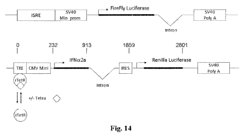
[0056] Figure 14 is a schematic illustration of two separate
constructs for the One-step assay in the Example herein below.
The abbreviations are as follows: ISRE: Interferon Sensitive
Response Element; SV40 Min. Prom: SV40 minimal promoter; Intron:
Intron from the human (3-globulin gene; SV40 Poly A: SV40
polyadenylation site; Firefly Luciferase: Coding region of the
firefly luciferase gene; TRE: tetracycline responsive element;
rTetR: Reverse tetracycline repressor; Tetra: Tetracycline;
CMV Mini: CMV minimum promoter; IFNa2a: Signal peptide and coding
region of the human interferon alpha2a gene; IRES: Internal
ribosomal entry site; and Renilla Luciferase: Coding region of
the Renilla luciferase gene
[0057] Figures 15A and 15D are graphs showing the effect of
doxycycline centration on the expression of Firefly and Renilla
luciferase activity in the One-step assay. One-step assay cells
(PIL,5C2.2) were treated with varying concentrations of
doxycycline as described in the Materials and Methods and
incubated overnight in duplicate with doxycycline alone or
together with a 1/1,000 dilution of a polyclonal anti-human IFNa
antibody as indicated in the figure. The activities of both
Firefly and Renilla luciferase determined sequentially in the
same well using the Dual-Glo luciferase assay system as described
in the Materials and Methods. The cells were then lysed by the
addition of 75 l/well of the Firefly luciferase substrate
containing reagent, and FireFly luciferase activity was
determined as described in the Materials and Methods. Renilla
luciferase activity was then determined following addition in the
same well of 50 l the Renilla luciferase substrate. The
neutralizing activity of the NAb sample was then determined from
the ratio of the activity of Firefly luciferase of the NAb
containing sample (FL2) normalized relative to Renilla luciferase
CA 02749142 2011-07-05
WO 2009/111572 PCT/US2009/036044
- 23 -
expression (RL2) and Firefly luciferase activity of the control
sample (FL1) normalized relative to Renilla luciferase expression
of the control sample (RL1): (FL2/RL2)/(FL1/RL1). In Figs. 15A
and 15B, RL represents Renilla luciferase and luc represents
Firefly luciferase.
[0058] Figures 16A and 16B are graphs showing the effect of
doxycycline concentration on the expression of Firefly and
Renilla luciferase activity in the One-step assay. One-step
assay cells (PIL5C2.2) were treated with varying concentrations
of doxycycline as indicated in the figure and incubated overnight
in duplicate with doxycycline alone or together with a 1/10 or
1/100 dilution of the human serum indicated in the figure. The
activities of both Firefly and Renilla luciferase determined
sequentially in the same well using the Dual-Glo luciferase assay
system as described in the Materials and Methods. The cells were
then lysed by the addition of 75 l/well of the Firefly
luciferase substrate containing reagent, and FireFly luciferase
activity was determined as described in the Materials and
Methods. Renilla luciferase activity was then determined
following addition in the same well of 50 l of the Renilla
luciferase substrate. The neutralizing activity of the NAb sample
was then determined from the ratio of the activity of Firefly
luciferase of the NAb containing sample (FL2) normalized relative
to Renilla luciferase expression (RL2) and Firefly luciferase
activity of the control sample (FL1) normalized relative to
Renilla luciferase expression of the control sample (RL1):
(FL2/RL2)/(FL1/RL1).
[0059] Figure 17 is a graph showing the effect of doxycycline
concentration on the expression of Firefly and Renilla luciferase
activity in the One-step assay. One-step assay cells (PIL5C2.2)
were treated with varying concentrations of doxycycline as
described in the Materials and Methods and incubated overnight in
CA 02749142 2011-07-05
WO 2009/111572 PCT/US2009/036044
- 24 -
duplicate. The activities of both Firefly and Renilla luciferase
determined sequentially in the same well using the Dual-Glo
luciferase assay system as described in the Materials and
Methods. The cells were then lysed by the addition of 75 l/well
of the Firefly luciferase substrate containing reagent, and
FireFly luciferase activity was determined as described in the
Materials and Methods. Renilla luciferase activity was then
determined following addition in the same well of 50 l of the
Renilla luciferase substrate.
[0060] Figure 18 is a graph showing the effect of varying
concentration of doxycycline on the neutralization activities of
human sera in the One-step assay. One-step assay cells
(PIL5C2.2) were treated with varying concentrations of
doxycycline as indicated in the figure and incubated overnight in
duplicate with doxycycline alone or together with a 1:20 dilution
of the human serum indicated in the figure. The activities of
both Firefly and Renilla luciferase determined sequentially in
the same well using the Dual-G1o luciferase assay system as
described in the Materials and Methods. The cells were then lysed
by the addition of 75 l/well of the Firefly luciferase substrate
containing reagent, and FireFly luciferase activity was
determined as described in the Materials and Methods. Renilla
luciferase activity was then determined following addition in the
same well of 50 l of the Renilla luciferase substrate. The
neutralizing activity of the NAb sample was then determined from
the ratio of the activity of Firefly luciferase of the NAb
containing sample (FL2) normalized relative to Renilla luciferase
expression (RL2) and Firefly luciferase activity of the control
sample (FL1) normalized relative to Renilla luciferase expression
of the control sample (RL1): (FL2/RL2)/(FL1/RL1).
[0061] Figures 19A and 19B are graphs showing NAb
quantification using a constant IFN concentration (100 IU/ml)
CA 02749142 2011-07-05
WO 2009/111572 PCT/US2009/036044
- 25 -
versus varying serum concentrations (Fig. 19A) or varying IFN
centration versus constant serum concentration (1/100) (Fig.
192). Serial dilutions of human serum were incubated in
duplicate for 1 hour at 37 C followed by 2 hours at 4 C with a
constant quantity (10 LU/ml) of a IFNa2 as described in the
Materials and Methods (Figure 19A), or a constant dilution of
serum (1:100) was incubated under the same conditions with serial
dilutions of IFN (Figure 19B). Residual IFN activity was then
assayed using the PIL5 gene-reporter assay as described in the
Materials and Methods. The IFN preparation used in each
neutralization test was also assayed simultaneously to determine
its precise IFN activity in that day's assay. The lowest dilution
of serum tested was also assayed alone for the presence of IFN
activity or toxicity. Neutralizing titer was determined using
the Kawade methodology (Grossberg et al., 2001b; and Lallemand et
al., 2008) which determines the reciprocal of the antibody
dilution that reduces IFN activity from 10 to 1.0 LU/ml and
expressed as TRU/ml as described in the Materials and Methods.
Neutralization titers were corrected for the actual number of
LU/ml of IFN used in the neutralization assay from the value
obtained in the simultaneous IFN titration.
[0062] Figures 20A-20C are graphs comparing determination of
neutralizing titer using different methods/assays. The
neutralizing titer of a series of human sera was determined by
the constant antibody method using the reporter-gene assay and
the results were compared with those obtained for the same sera
determined. using the one-step assay (Figure 20A). The
neutralizing titer of the same series of human sera was
determined by the constant IFN method using the CPE assay as
described in the Materials and Methods and the results were
compared with those obtained for the same sera determined using
the reporter-gene assay and the constant antibody method (Figure
CA 02749142 2011-07-05
WO 2009/111572 PCT/US2009/036044
- 26 -
20B). The neutralizing titer of the same series of human sera was
determined by the constant IFN method using the CPE assay as
described in the Materials and Methods and the results were
compared with those obtained for the same sera determined using
the one-step assay (Figure 20C).
DETAILED DESCRIPTION OF THE INVENTION
[0063] Conventional cell based assays for the quantification
of neutralizing antibodies (NAbs) are imprecise, give variable
results, and often require two or more days to complete.
Furthermore, conventional cell-based assays require specialized
personnel and biological containment facilities, are labor
intensive, and difficult to automate. The use of division-
arrested frozen cells transfected with a reporter gene controlled
by a ligand-responsive chimeric promoter (WO 2004/039990 and US
2004/023517, incorporated herein by reference) in an assay for
neutralizing antibodies would allow anti-ligand NAbs to be
quantified with precision within hours. Although such an assay
would overcome many of the limitations of conventional cell-based
neutralization assays, it would still remain relatively labor
intensive and require serial dilutions of both the test sample
and ligand, positive and negative controls, and reference
reagents, to be included in the assay. Furthermore, assay
precision is adversely affected by loss of assay cells (or carry-
over of ligand or NAb following serial dilution). Such assays
also remain relatively difficult to automate.
[0064] The present invention avoids the limitations of the
currently available assays as discussed above by developing a
cell, and an assay for the quantification of neutralizing
antibodies based on using such a cell, which has been engineered
to express and secrete the ligand (extracellular ligand) of
interest and a reporter gene transcribed from the same inducible
CA 02749142 2011-07-05
WO 2009/111572 PCT/US2009/036044
- 27 -
promoter. The cell also contains another reporter gene
controlled by a chimeric promoter which is ligand-responsive.
Expression of the former reporter product gene is strictly
proportional to the expression of the ligand and allows ligand
expression to be quantified (i.e., by determining the amount of
expressed reporter gene product). Expression of the latter
ligand-responsive reporter gene allows ligand activity to be
quantified as well. The presence of anti-ligand NAbs in the
immediate environment of the cell will neutralize a quantity of
extracellular ligand (secreted from the cell) proportional to the
neutralization capacity of the antibody, and thus prevent the
extracellular ligand from interacting with its specific cell
surface receptor (or with a pattern recognition receptor). This
will result in a corresponding reduction in the activity of the
extracellular ligand., and hence the expression of the ligand-
responsive reporter-gene, the activity of which can be
quantified. Figure 2 schematically illustrates this system using
levels of firefly luciferase (FL) and Renilla luciferase (RL)
activity.
[0065] The degree of reduction in the expression of the
ligand-responsive reporter gene in the presence or absence of the
NAb sample to be quantified will allow the relative neutralizing
titer of the sample to be quantified, relative to a given level
of expression of a different reporter gene transcribed from the
same promoter as the ligand.
[0066] The cell and the cell-based assay method (termed
"NanoLite" as opposed to the "iLite" assay method of
W02004/039990 and US2004/023517) according to the present
invention, when used for assaying neutralizing antibodies, has
many advantages over the conventional cell-based assay (i.e.,
CPE) and even over the more recent "iLite" cell-based assay in
that it is essentially a one-step assay (where only undiluted
CA 02749142 2011-07-05
WO 2009/111572 PCT/US2009/036044
- 28 -
sample need to be added to the cells). Figure 1 presents a flow
diagram comparison between the recent iLite cell-based assay and
the NanoLite assay of the present invention, which clearly shows
that NanoLite involves less steps and less time to perform. It
should be noted that the NanoLite assay according to the present
invention, using the cell of the present invention, is a one-step
assay where, in contrast to the iLite or other conventional cell-
based assays, neither addition of ligand (cytokine) nor dilution
of the sample is required. Table 1 below further summarizes the
many advantages that the NanoLite assay method of the present
invention has over the CPE and iLite assays.
Table 1
CPE iLite Nano Lite
Time (hours) 96 18 5
eagents Required + + -
Serial Dilutions + + -
ositive Control + + -
Negative Control + + -
igand Standard Curve + + -
Results/Cell Number + + -
Maximum Samples/plate 10 10 96
TS Automated - +/- +
[0067] As contemplated by the present inventors, the cell of
the present invention, for use in assaying antibodies to an
extracellular ligand that initiates a ligand-specific signal at
the nucleus of the cell, contains at least (a) a first DNA
construct, which has a sequence that includes a first set of one
or more transcription control elements that is inducible by the
ligand, and a portion encoding a first measurable tag (i.e.,
reporter gene product) driven by the first set of one or more
CA 02749142 2011-07-05
WO 2009/111572 PCT/US2009/036044
- 29 -
transcription control elements, where the first tag can be
detected when the first set of one or more transcription control
elements is induced by the ligand, (b) a second DNA construct,
which has a sequence that includes (i) a second set of one or
more transcription control elements different from the first set,
(ii) a DNA segment, driven by the second set of one or more
measurable tag (i.e., second reporter gene product different from
the first) which can be independently measured in the presence of
the first tag, and vice versa, and (iii) on a separate cistron, a
DNA segment encoding the ligand, also driven by the second set of
one or more transcription control elements.
[0068] The cell according to the present invention may be any
mammalian or avian cell line, with human cells most preferred.
Preferred cell lines include but are not limited to, human
promonocytic (i.e., U937), myeloid (i.e., U266R), T-cell lymphoma
(i.e., Jurkatt), breast adenocarcinoma (i.e., MCF7) cell lines
and mouse lymphoma (i.e., L120) and erythroid leukemia cell
lines.
[0069] The extracellular ligand (or its antagonist/antibody),
for which the titer of neutralizing antibodies thereto are
determined in the method according to the present invention
discussed below, is intended to encompass any therapeutic agent,
such as therapeutic proteins, which activates (or blocks, in the
case of an antagonist of/antibody against the extracellular
ligand) the signal transduction activity of a cell surface
protein, and for which neutralizing antibodies generated thereto
in the mammalian subject treated with the therapeutic agent would
be undesirable. The extracellular ligand may also encompass
components of molecules or preparations such as live or
attenuated virus or bacterial vaccines, which components interact
with pattern recognition receptors. Preferred non-limiting
examples of such an extracellular ligand include cytokines,
CA 02749142 2011-07-05
WO 2009/111572 PCT/US2009/036044
- 30 -
chemokines and growth factors, such as interferon-a, interferon-
(3, interferon-y, erythropoietin (EPO), TNFa, interleukins, growth
hormone, granulocyte colony stimulating factor (G-CSF), and
granulocyte macrophage colony stimulating factor (GM-CSF);
gonadotropins, insulin and other hormones; integrins;
immunoglobulins (polyclonal, monoclonal, chimeric, humanized or
single chain, etc.); and other proteins that interact with a cell
surface molecule or with a pattern recognition receptor to
transmit a signal to the nucleus. Non-limiting examples of
antagonists (i.e., antibodies) of the extracellular ligand, which
antagonist the neutralizing antibodies bind to, include TNFa
antagonists such as Enbrel and Infliximab (a chimeric antibody),
Adalimumab (a fully human antibody), and Etanercept (an IgGlFc
TNFp75 receptor fusion protein).
[0070] Neutralizing antibody (NAb) assays are clinically very
important today because those patients being treated continuously
for a chronic disease, such as remitting/relapsing MS treated
with interferon (3, cease obtaining benefit from treatment with
the therapeutic agent once an immune response, in particular
production of NAbs, has been mounted against the therapeutic
agent by the patient. Thus, it is important to be able to detect
when and if a patient has developed NAbs in order to stop
treatment at that point. Also, it will prevent the possibility
of adverse reactions such as anaphylactic shock and perfusion
reactions, and allow the patient to be treated with an
alternative effective therapy. Furthermore, NAb testing can
provide considerable cost savings to the health care
provider/insurer and to the patient by avoiding continued use of
an ineffective and expensive biopharmaceutical.
[0071] The cell surface protein from which its signal
transduction activity, in response to an extracellular signal
from a therapeutic agent or protein, regulates the expression of
CA 02749142 2011-07-05
WO 2009/111572 PCT/US2009/036044
- 31 -
a reporter gene product can be any such cell surface protein that
is known to those of skill in the art or that may be identified
by those of skill in the art. Exemplary cell surface proteins
include, but are not limited to, cell surface receptors and ion
channels. Non-limiting examples of cell surface receptors
include cytokine receptors (e.g., receptors for Type I
interferon, Type II interferon, interleukins, growth hormone,
erythropoietin (EPO), granulocyte colony stimulating factor (G-
CSF), granulocyte macrophage colony stimulating factor (GM-CSF),
TNFa, TGF3, Fas ligand, leukemia inhibitory factor (LIF), ciliary
neurotrophic factor (CNTF), etc.), growth factor receptors,
hormone receptors, T cell receptors, antigen receptors,
complement receptors, and neuroreceptors. The reference text, J.
M. Cruse and Robert E. Lewis, Atlas of Immunology, CRC Press,
Washington, DC, 1999, which discloses many receptors involved in
immune response and immune system interactions is entirely
incorporated herein by reference. Cell surface receptors also
include, but are not limited to, muscarinic receptors (e.g.,
human M2 (GenBank accession #M16404); rat M3 (GenBank accession
#M16407); human M4 (GenBank accession #M16405); human M5 (Bonner
et al., 1988); and the like); neuronal nicotinic acetylcholine
receptors (e.g., the a2, a3 and X32 subtypes); the rat a2 subunit
(Wada et al., 1988); the rat a3 subunit (Boulter et al., 1986);
the rat a4 subunit (Goldman et al., 1987); the rat a5 subunit
(Boulter et al., 1990); the rat (32 subunit (Deneris et al.,
1988) ; the rat 13 subunit (Deneris et al . , 1989) ; the rat (34
subunit (Duvoisin et al., 1989); combinations of the rat a
subunits, i subunits and a and (3 subunits; GABA receptors (e.g.,
the bovine al and (31 subunits (Schofield et al., 1987); the
bovine a2 and a3 subunits (Levitan et al., 1988); thev-subunit
(Pritchett et al . , 1989) ; the (32 and (33 subunits (Ymer et al . ,
1989); the 6 subunit (Shivers, B.D., 1989); and the like);
CA 02749142 2011-07-05
WO 2009/111572 PCT/US2009/036044
- 32 -
glutamate receptors (e.g., receptor isolated from rat brain
(Hollmann et al., 1989); and the like); adrenergic receptors
(e.g., human f31 (Frielle et al., 1987); human a2 (Kobilka et al.,
1987); hamster (32 (Dixon et al., 1986); and the like); dopamine
receptors (e.g., human D2 (Stormann et al., 1990); rat (Bunzow et
al., 1988); and the like); NGF receptors (e.g., human NGF
receptors (Johnson et al., 1986); and the like); serotonin
receptors (e.g., human 5HT1a (Kobilka et al., 1987); rat 5HT2
(Julius et al., 1990); rat 5HTlc (Julius et al., 1988); and the
like).
[0072] The pattern recognition receptor from which its signal
transduction activity, in response to an extracellular signal
from a component(s) of a molecule or preparation such as a live
or attenuated virus or bacterial vaccine regulates the expression.
of a reporter gene product, includes but is not limited to Toll-
like receptors (TLR) cell surface or endosomal membrane receptors
(Uematsu and Akira, 2007), or the retinoic acid-inducible gene 1
(GIG-I)-like cytosolic receptor proteins RIG-I, MDA5, and LGP2
(Yoneyama and Fujita, 2007) that recognize or interact with
components of live or attenuated virus or bacterial vaccines.
Evaluation of neutralizing antibodies generated in the mammalian
subject treated with the vaccine is important in order to
determine the degree of protection afforded by vaccination.
[0073] Thirteen members of the TLR family have been identified
in mammals (Uematsu and Akira, 2007). Each TLR mediates a
distinctive response in association with different combinations
of four Toll/IL-1 receptor (TIR) domain-containing adaptor
proteins (MyD88, TRIF, TIRAP/MAL, and TRAM). All the TLRs except
TLR3 interact with MyD88. TLR3, which recognizes single-stranded
or double-stranded viral RNA, is localized in the endosomes of
myeloid DCs and requires acidification of vesicles for
activation. TLR3 signals via TRIF and activates TBK1/IKKC which
CA 02749142 2011-07-05
WO 2009/111572 PCT/US2009/036044
- 33 -
phosphorylates the interferon regulatory factor 3 (IRF3) and
NFKB, resulting in production of IFN (3 (Hemmi et al, 2004, Perry
et al., 2004). The RIG-I-like receptor proteins are DExD/H box
RNA helicases two of which, RIG-I and MDA5, carry caspase
activation.
[0074] Ion channels include, but are not limited to, calcium
ion channels (e.g., human neuronal a2 subunit (see W089/09834);
rabbit skeletal muscle al subunit (Tanabe et al. 1987); rabbit
skeletal muscle a2 subunit (Ellis et al., 1988); rabbit skeletal
muscle f3 subunit (Ruth et al., 1989); rabbit skeletal muscle 1'
subunit (Jay et al., 1990); and the like); potassium ion channels
(e.g., rat brain (BK2) (McKinnon, D., 1989); mouse brain (BK1)
(Tempel et al., 1988); and the like); sodium ion channels (e.g.,
rat brain I and II (Noda et al., 1986); rat brain III (Kayano et
al., 1988); and others).
[0075] It will be appreciated by those of skill in the art
that the cell surface protein or pattern recognition receptor
discussed above is preferably endogenous to the cell of the
present invention. However, it will also be appreciated that the
cell surface protein or pattern recognition receptor may be
expressed from cloned DNA, such as to supplement the number of
pattern recognition receptors or the number of the cell surface
protein at the surface of the cell, or the cell surface protein
or pattern recognition receptor may be expressed from cloned DNA
but is a cell surface protein or pattern recognition receptor
that is heterologous to the host cell.
[0076] For signal transduction, the intracellular signal that
is transduced is initiated by the specific interaction of an
extracellular signal with a receptor or ion channel present on
the cell surface. This interaction sets in motion a cascade of
intracellular events, including a ligand-specific signal at the
nucleus of the cell, the ultimate consequence of which is a rapid
CA 02749142 2011-07-05
WO 2009/111572 PCT/US2009/036044
- 34 -
and detectable change in the expression of a gene product, which
in the cell of the present invention is preferably a reporter
gene product. The extracellular signal or effector molecule is
any compound or substance that acts as a ligand to specifically
alter the activity of a cell surface protein or pattern
recognition receptor. Examples of such signals include, but are
not limited to, molecules such as cytokines (i.e., interferons),
growth factors, hormones, endorphins, neurotransmitters,
acetylcholine, and mitogenic substances, such as phorbol myristic
acetate (PMA), that bind to cell surface receptors and ion
channels and modulate the activity of such receptors and
channels. Other examples include components of live and
attenuated virus and bacterial vaccines.
[0077] The DNA constructs carried by the cell according to the
present invention are DNA constructs that include a nucleotide
sequence encoding a reporter gene product operatively linked to
transcriptional control elements/sequences. Transcription of the
reporter gene is controlled by these sequences. The activity of
at least one or more of these control sequences is directly or
indirectly regulated by the cell surface protein or pattern
recognition receptor. The transcriptional control sequences
include but are not limited to promoters and other regulatory
regions, such as enhancer sequences and repressor and activator
binding sites, that modulate the activity of the promoter, or
control sequences that modulate the activity or efficiency of the
RNA polymerase that recognizes the promoter, or control sequences
that are recognized by effector molecules, including those that
are specifically induced by interaction of an extracellular
signal with a cell surface protein or a pattern recognition
receptor. For example, modulation of the activity of the
promoter may be affected by altering the RNA polymerase binding
to the promoter region, or, alternatively, by interfering with
CA 02749142 2011-07-05
WO 2009/111572 PCT/US2009/036044
- 35 -
initiation of transcription or elongation of the mRNA. Such
sequences are herein collectively referred to as transcriptional
control elements or sequences. In addition, the constructs may
include sequences of nucleotides that alter translation of the
resulting mRNA, thereby altering the amount of reporter gene
product expressed.
[0078] A promoter that is regulated or mediated by the
activity of a cell surface protein or pattern recognition
receptor is a promoter whose activity changes when a cell is
exposed to a particular extracellular signal (ligand) by virtue
of the presence of cell surface proteins or pattern recognition
receptors whose activities are affected by the extracellular
signal. For example, the c-fos promoter is specifically activated
upon the specific interaction of certain extracellular signals,
such as growth hormones, with a cell surface protein, such as a
growth hormone receptor. In particular, the regulation of such
promoters by the cell surface protein, though indirect, occurs
within minutes of the interaction of the cell surface protein
with the extracellular signal. As used herein, operative linkage
refers to the linkage of a transcriptional control element, i.e.,
promoter, to a nucleotide coding sequence such that the
transcriptional control element is properly positioned for its
activity of binding RNA polymerase and initiating transcription
of the nucleotide coding sequence. Thus, a nucleotide coding
sequence in operative linkage with a promoter is downstream, with
respect to the direction of transcription, from the promoter, is
in the correct reading frame with respect to the transcription
initiation site and is inserted in a manner such that
transcription elongation proceeds through the nucleotide coding
sequence.
[0079] Suitable transcriptional control elements may be
obtained or derived from the transcriptional regulatory regions
CA 02749142 2011-07-05
WO 2009/111572 PCT/US2009/036044
- 36 -
of genes whose expression is rapidly induced, generally within
minutes, of contact between the cell surface protein or pattern
recognition receptor and the effector ligand that modulates the
activity of the cell surface protein or pattern recognition
receptor. Examples of such genes include, but are not limited
to, the immediate early genes (Sheng et al., 1990), such as c-
fos. Immediate early genes are genes that are rapidly induced
upon binding of a ligand to a cell surface protein. The
transcriptional control elements that are preferred for use in
the DNA (reporter gene) constructs include transcriptional
control elements from immediate early genes, elements derived
from other genes that exhibit some or all of the characteristics
of the immediate early genes, or synthetic elements that are
constructed such that genes in operative linkage therewith
exhibit such characteristics. The characteristics of preferred
genes from which the transcriptional control elements are derived
include, but are not limited to, low or undetectable expression
in quiescent cells, rapid induction at the transcriptional level
within minutes of extracellular simulation, induction that is
transient and independent of new protein synthesis, subsequent
shut-off of transcription requires new protein synthesis, and
mRNAs transcribed from these genes have a short half-life. It is
not necessary for all of these properties to be present.
[0080] Suitable promoters and transcriptional control elements
include, but are not limited to, the cytomegalovirus promoter
(CMV), the simian virus 40 (SV40) promoter and minimal promoters
thereof, the vasoactive intestinal peptide (VIP) gene promoter
(cAMP responsive; Fink et al., 1988); the somatostatin gene
promoter (cAMP responsive; Montminy et al . , 19186). the
proenkephalin promoter (responsive to cAMP, nicotinic agonists,
and phorbol esters; Comb et al. 1986); the phosphoenolpyruvate
carboxy-kinase gene promoter (cAMP responsive; Short et al.,
CA 02749142 2011-07-05
WO 2009/111572 PCT/US2009/036044
- 37 -
1986); the NGFI-A gene promoter (responsive to NGF, CAMP, and
serum; Changelian et al., 1989); the transcriptional control
elements obtained or derived from the c-fos gene; and others that
may be known to or prepared by those of skill in the art.
[0081] The c-fos proto oncogene is the cellular homologue of
the transforming gene of FBJ osteosarcoma virus. It encodes a
nuclear protein that is most likely involved in normal cellular
growth and differentiation. Transcription of c-fos is transiently
and rapidly activated by growth factors and by inducers of other
cell surface proteins, including hormones, differentiation-
specific agents, stress, mitogens and other known inducers of
cell surface proteins. Activation is protein synthesis
independent. The c-fos regulatory elements include a TATA box
that is required for transcription initiation, two upstream
elements for basal transcription, and an enhancer, which includes
an element with dyad symmetry and which is required for induction
by TPA, serum, EGF, and PMA. The 20 bp transcriptional enhancer
element located between -317 and -298 bp upstream from the c-fos
mRNA cap site, which is essential for serum induction in serum
starved NIH 3T3 cells. One of the two upstream elements is
located at -63 to -57 and it resembles the consensus sequence for
cAMP regulation.
[0082] Transcriptional control elements, particularly as they
relate to a preferred embodiment of the present invention where
Type I and/or Type II interferon is the extracellular signal, are
preferably an interferon stimulatory response element (ISRE)
and/or a gamma activated sequence (GAS). There are a number of
ISREs characterized from different human genes responsive to Type
I interferon and a consensus sequence, ggraaagwGAAActg (SEQ ID
NO:1; capital letters denote core sequence; underlines denote
high conservation), to which the STAT1/STAT2/IRF9 complex binds,
was identified for ISRE (Levy et al., 1988). A preferred ISRE is
CA 02749142 2011-07-05
WO 2009/111572 PCT/US2009/036044
- 38 -
from the human ISG15 gene and is presented as SEQ ID NO:2 where
nucleotides 41-55 correspond to the consensus ISRE sequence.
ISRE is also highly conserved among species. For example, a
sequence present in the promoter region of the interferon
inducible chicken Mx gene (Schumacher et al., 1994) is similar to
that found in primates and conforms to the ISRE consensus
sequence for mammalian interferon responsive genes including
rodents and cows (see Fig. 2 of Perry et al., 1999).
[0083] Regarding GAS, to which the STAT1 homodimer binds in
genes responsive to Type II interferon, a consensus sequence,
nnnsanttccgGGAAntgnsn (SEQ ID NO:3; capital letters denote core
sequence; underlines denote high conservation), from many
selected binding sequences was identified (Horvath et al., 1995).
[0084] In the instance where Type I interferon or Type II
interferon is the extracellular ligand signal, a preferred
combination of transcriptional control elements is an interferon
responsive chimeric promoter in which an ISRE and/or GAS controls
a SV40 minimal promoter operatively linked to a nucleotide
sequence encoding a first reporter gene product as a first
measurable tag.
[0085] When the extracellular ligand is TNFa, a preferred
combination of transcriptional control elements is a TNF-a-
responsive chimeric promoter in which repeats (i.e., 5x tandem
repeats; SEQ ID NO:11) of the NFiB recognition site controls a
SV40 minimal promoter operatively linked to a nucleotide sequence
encoding a first reporter gene product.
[0086] When the extracellular ligand is erythropoietin (EPO),
a preferred combination of transcriptional control elements is an
EPO-responsive chimeric promoter in which repeats of the signal
transducer and activator of transcription #5 (STATS) sequence(5x
tandem repeats is tcgagTTCGAAGAAaacTTCTTGGAAgaTTCCTGGAgcTTCTAG
GAAgaTTCCGGGAA (SEQ ID NO:4), where the sequence in capital
CA 02749142 2011-07-05
WO 2009/111572 PCT/US2009/036044
- 39 -
letters represent variants of the STAT5 consensus sequence),
through which EPO signals from its cell surface receptor to the
nucleus, controls a SV40 minimal promoter operatively linked to a
nucleotide sequence encoding a first reporter gene product.
[0087] The first reporter gene product (also known herein as a
first measurable tag), whose level is a measure of the presence
and/or the level of an extracellular ligand that activates the
signal transduction activity of a cell surface protein or pattern
recognition receptor, may be RNA or protein, as long as it is
readily detectable, although it is preferably a protein. For
instance, luciferases, such as firefly luciferase, Renilla
luciferase, Gaussia luciferase and Metridia luciferase, enhanced
green fluorescent protein (EGFP) and jellyfish aequorin are most
preferred embodiments of reporter gene products (measurable tags)
in the cell according to the present invention. In the case of
the enzyme firefly luciferase (deWet et al., 1987) and other
luciferases, and jellyfish aequorin (Rider et al., 2003), the
result of its enzymatic activity, light, is detected and measured
using a luminometer, whereas in the case of EGFP, a fluorescence
activated cell sorter or analyzer (FACS) can be used at an
appropriate wavelength to detect and quantify the amount of EGFP
expressed in a cell. The distribution curve of the amount of
luciferase, aequorin or EGFP expressed in a sample of cells will
be determined by the amount of ligand to which the cell is
exposed in the immediate external environment surrounding the
cell. Non-limiting examples of other suitable reporter gene
products include dsRED, chloramphenicol acetyl transferase (CAT)
(Alton et al., 1979) other enzyme detection systems, such as (3-
galactosidase, bacterial luciferase (Engebrecht et al., 1984 and
Baldwin et al. 1984), alkaline phosphatase (Toh et al. 1989 and
Hall et al. 1983), and bacterial or humanized P-lactamase
(Zlokarnik et al., 1998).
CA 02749142 2011-07-05
WO 2009/111572 PCT/US2009/036044
- 40 -
[0088] The second reporter gene product (also known as the
second measurable tag), whose level is a measure of the level of
the ligand, expressed together with the second reporter gene
product (and on a separate cistron from the same promoter) and
secreted into the immediate external environment surrounding the
cell, and which ligand is capable of activating the signal
transduction activity of a cell surface protein/receptor or a
pattern recognition receptor. The second reporter gene product
can be any of those disclosed above with regard to the first
reporter gene product except that the first and second reporter
gene products must be different from each other such that one
reporter gene product can be independently measured in the
presence of the other reporter gene product, and vice versa. The
term "cistron" is intended to have the meaning commonly
understood in the art as a segment of DNA coding for a single
polypeptide but expressed from the same set of one or more
transcription control elements (i.e., promoter) as the second
reporter gene product.
[0089] When the cell is to be used to assay for neutralizing
antibodies to a ligand antagonist/antibody (e.g., TNFa
antagonists such as Eubrel, Infliximab, Adalimumab, Etanercept,
etc.), the cell would further carry a third construct which
includes a segment encoding a third reporter gene product (third
measurable tag) driven by a third set of one or more
transcription control elements different from the first and
second set of transcription control elements in the first and
second constructs. This third reporter gene product is expressed
together with a ligand antagonist (on a separate cistron driven
from the same transcription control elements/promoter) and whose
level is a measure of the level of expressed ligand antagonist.
The ligand antagonist is the expressed and secreted into the
immediate external environment surrounding the cell along with
CA 02749142 2011-07-05
WO 2009/111572 PCT/US2009/036044
- 41 -
the ligand expressed from the second construct. The level of
measurable ligand activity is a measure of the amount of
neutralizing antibodies to the ligand antagonist that block the
ligand antagonist from inhibiting the signaling activity of the
ligand. The third reporter gene product can be any of those
disclosed above with regard to the first and second reporter gene
products, including preferably a CBG68Luc reporter gene, except
that the third reporter gene product and the first and second
reporter gene products must each be different such that each
reporter gene product can be independently measured in the
presence of the other two.
[0090] In the case of extracellular ligands that inhibit cell
proliferation, induce apoptosis, or induce receptor down-
regulation, the expression of the ligand is controlled by a set
of one or more transcription control elements that is inducible
(i.e., no expression unless an inducer is present). This would
prevent the undesirable inhibitory activity of the extracellular
ligand while the cell is growing or before the cell is ready for
use in a cell-based assay. Another instance in which inducible
transcription control elements are desirable for expressing the
ligand is when the cell is used in a cell-based assay where only
the level of the extracellular ligand in a sample, without any
such ligand being produced from the second set of one or more
transcription control elements, is sought to be determined rather
than the level of neutralizing antibodies to the extracellular
ligand.. In the case that a ligand antagonist is to be expressed
in a ligand antagonist is to be expressed in a third construct,
the expression of the ligand antagonist is preferably controlled
by a set of one or more transcription control elements that is
inducible.
[0091] Inducible promoters and other transcriptional control
elements, some of which are disclosed above, are well-known in
CA 02749142 2011-07-05
WO 2009/111572 PCT/US2009/036044
- 42 -
the art. A preferred well known inducible transcriptional
control element for use in controlling the expression of the
ligand and a reporter gene product is a tetracycline-responsive
element from the Tet-On/Tet-Off gene expression system (such as
provided by Clontech Laboratories, Inc., Palo Alto, CA). This
element, to which a reverse tetracycline repressor (rTetR; a
mutated version of the tetracycline repressor) attaches, thereby
inhibiting transcription from the Tet-On construction, is placed
upstream of preferably a minimal promoter, such as the cytomegalo
virus (CMV) immediate early minimal promoter. In the presence of
an inducer, e.g., tetracycline or doxycycline, the mutated
version of the TetR (rTetR) becomes a reverse tetracycline-
controlled transactivator (rtTA) and binds to the TRE allowing
transcription to start.
[0092] In another embodiment, the inducible promoter is a Tet-
Off promoter in which the TetR binds to the TRE, silencing
transcription in the presence of tetracycline or doxycycline.
Following removal of tetracycline or doxycycline, the
tetracycline-controlled transactivator (tTA) binds to the TRE,
thereby activating transcription.
[0093] One embodiment of the cell according to the present
invention is derived from the human pro-monocytic cell line U937
transfected with the firefly luciferase reporter gene controlled
by an interferon responsive chimeric promoter containing a SV40
minimal promoter and the ISRE from the ISG 15 gene as described
previously in WO 2004/039990 and US 2004/023517, which are
incorporated herein by reference and shown in Fig. 3. These
cells were then transfected with the 5991 bp pIRES/IFNA2/hRL
vector (SEQ ID NO:5) which comprises the coding sequence of human
IFN a2a gene (nucleotides 1-586 of SEQ ID NO:5), the IRES
(Internal Ribosome Entry Site; SEQ ID NO:6) of cytomegalovirus
(CMV), together with the coding sequence of Renilla luciferase
CA 02749142 2011-07-05
WO 2009/111572 PCT/US2009/036044
- 43 -
reporter gene (nucleotides 1532-2484 of SEQ ID NO:5), under the
control of a constitutive CMV promoter (nucleotides 5282-5991 of
SEQ ID NO:1) as shown in Fig. 3. Thus, this construction allows
the primary RNA transcript to be translated into two distinct
native proteins (IFNa2a and Renilla luciferase) so as to preserve
the tertiary structure of the human IFNa2a protein and hence its
recognition by anti-IFNa antibodies.
[0094] In a preferred embodiment of the cell of the present
invention, the Renilla luciferase reporter-gene was cloned
upstream of the IRES and the human IFNa2a gene (Fig. 4) in order
to increase the low levels of Renilla gene expression observed
when the human IFNa2a gene, or other human type I IFN genes
(i.e., IFN(3), was cloned upstream of the Renilla luciferase
reporter gene.
[0095] In a second preferred embodiment of the cell of the
present invention, the Renilla luciferase reporter-gene and the
human IFNa2a gene were expressed under the control of an
inducible promoter in order to prevent continued expression of
human type I IFNs from inhibiting cell proliferation and hence
preventing cultivation of the transfected cell line. Thus, the
Renilla luciferase reporter gene and the human IFNa2a gene, were
expressed under the control of a CMV promoter (Fig. 5), the
expression of which is induced in the presence of doxycycline or
tetracycline (Tet-On).
[0096] In a third preferred embodiment cell of the present of
the invention, the Renilla luciferase reporter-gene and the human
IFN(31a gene, are expressed under the control of a Tet-On CMV
promoter (Fig. 6).
[0097] In a further embodiment of the cell of the present
invention, the Renilla luciferase reporter gene and the human
TNFa gene, are expressed under the control of a tet-on CMV
promoter (Fig. 7). The use of an inducible promoter is essential
CA 02749142 2011-07-05
WO 2009/111572 PCT/US2009/036044
- 44 -
for the expression of TNFa, the uncontrolled production of which
would induce apoptosis in the TNFa-sensitive host cells. The
sequence of the 5x tandem repeats of the canonical NFKB
recognition site used in the other construct in Fig. 7 to drive
the expression of firefly luciferase in combination with a SV40
minimal promoter is SEQ ID NO:11.
[0098] In still a further embodiment of the cell of the
present invention, the Renilla luciferase reporter gene and the
human erythropoietin (EPO) gene, are expressed under the control
of a Tet-On CMV promoter (Fig. 9). The use of an inducible
promoter is essential for the expression of EPO, the continuous
production of which would cause down-regulation of EPO-specific
receptors on the EPO sensitive host cells.
[0099] In yet a further embodiment of the cell of the present
invention, the Renilla luciferase reporter gene is expressed
under the control of chimeric promoter consisting of a 5x tandem
repeat of the canonical NFKB recognition site/SV40 minimal
promoter. In addition, the TNFa and the CBRLuc reporter gene are
both expressed under the control of a Tet-responsive element
(TRE)/CMV immediate early minimal promoter(Tet-On) as a second
construct, and the TNFa antagonist of interest and the CBG68Luc
reporter gene are expressed in a third construct under the
control of a different inducible promoter such as the chimeric
mifepristone inducible promoter. In this system, the GAL4-UAS and
the TATA sequence are expressed from the Adenovirus Elb minimal
promoter that is transcriptionally silent in the absence of
activation. The Ga14 DNA binding domain, which binds the
regulatory protein to the GAL4-Elb promoter, and the truncated
human progesterone receptor ligand binding domain (hPR-LBD),
which undergoes conformational change when it binds the
progesterone antagonist mifepristone, are expressed from a
minimal TK promoter on the vector. Thus, upon addition of
CA 02749142 2011-07-05
WO 2009/111572 PCT/US2009/036044
- 45 -
mifepristone, the antagonist binds to the hPR-LBD region of the
vector, causing a conformational change in the regulatory protein
resulting in transcription of the TNFa antagonist and the
CBG68Luc reporter gene (Fig. 8).
[00100] In order to make the cell according to the present
invention a one time use cell that cannot be propagated for
further use, the cell (after having been transformed/transfected
with the first, second, and optionally, third DNA construct) is
treated with an anti-mitotic or pro-apoptotic agent so as to
acquire the property that it will maintain the ligand-specific
signal transduction activity for at least about 1 hour but no
more than about 30 days at a temperature above freezing before
losing the signal transduction activity and undergoing cellular
death.
[00101] One preferred embodiment of the present invention is
where the anti-mitotic or pro-apoptotic agent is y-radiation and
the cell has been treated by irradiating with y-radiation at an
intensity and for a sufficient time such the irradiated cell
maintains the signal transduction activity of a cell surface
protein/receptor or a pattern recognition receptor for a period
of at least about 1 hour, preferably 7 days but no more than 30
days at a temperature above freezing following irradiation, after
which period of time the irradiated cell immediately undergoes
cellular death (i.e., apoptotis).
[00102] It is known that y-irradiation at a high dose causes a
cell to lose its signal transduction activity. Irradiation at a
somewhat lower dose causes a cell to cease replication and
undergo cellular death. The present inventors previously
discovered that it is possible to determine a dose which inhibits
replication but still allows a cell to maintains its signal
transduction activity for a period of time before undergoing cell
death. For example, y-irradiation at about 9 Grays allows U937
CA 02749142 2011-07-05
WO 2009/111572 PCT/US2009/036044
- 46 -
cells to retain signal transduction activity for 14 days, after
which the cells undergo cell death. However, during those 14
days, the signal transduction activity in response to, for
example, Type I interferon that is being assayed functions as
well as in a non-irradiated control. Thus, by irradiating a cell
with y radiation, the treated cell has a 14-day shelf life, but
which becomes inactive (undergoes cellular death) after a period
of about 14 days so that it cannot be maintained and reproduced
by an end user. The dose of y-irradiation required will vary as
a function of the particular cell line employed but this can be
determined with only routine experimentation based on the
guidance in WO 2004/039990 and US 2004/023517.
[00103] The dose (intensity and duration) of y radiation to
which the transformed cell is treated is preferably about 6 to 12
Grays (Gy). As the experiments in WO 2004/039990 and US
2004/023517 demonstrate, the temperature above freezing, at which
the cell is kept or stored, affects the shelf-life of the cell.
Preferably, this temperature is room temperature, which
advantageously maintains maximum interferon sensitivity while
providing for ease of storage and shipping of the commercial one
time use cell.
[00104] A second preferred embodiment of the present invention
is where the cell (after having been transformed/transfected with
the first, second, and optionally, third DNA construct) is
treated with an anti-mitotic or pro-apoptotic chemical agent such
as vinblastin, 5-fluorouracil (5-Fu), cisplatin or an anti-tumor
intercalating agent (i.e., mitomycin C) in a sufficient amount
and for a sufficient time such that the treated cell maintains
the signal transduction activity of the cell surface protein or
pattern recognition receptor for a period of at least about 1
hour but no more than about 30 days at a temperature above
freezing following treatment with the agent, after which period
CA 02749142 2011-07-05
WO 2009/111572 PCT/US2009/036044
- 47 -
of time the treated cell immediately undergoes cellular death.
An anti-mitotic or pro-apoptotic agent will affect a treated cell
when it begins to replicate, such as for example by preventing
spindle formation, thereby inducing apoptosis and killing the
cell. Thus, cells which have been treated with an anti-mitotic
or pro-apoptotic agent, such as transformed human promonocytic
cells, will have a shelf life of about 24 hours during which the
signal transduction assay can be conducted and after which period
of time the cells will die. It will be appreciated that a cell
having only a 24 hour shelf life is not desirable from a
commercial standpoint. In order to extend the shelf life, the
treated cells may be immediately frozen, in which state they will
have a much longer shelf life, depending upon the manner of
freezing and thawing. Once thawed, however, they must be used
within 24 hours, after which they will undergo cellular death
(i.e., apoptosis).
[00105] It should be understood that conventional wisdom is
that cryopreservation of cells requires a special freezing and
thawing process (and equipment) in which the cells are frozen at
a rate of 1 C per minute until it reaches -80 C or liquid
nitrogen temperatures of about -200 C, where it may be stored
indefinitely, and after which it must be thawed very rapidly.
Often, dimethyl sulfoxide (DMSO) or another cryopreservative is
also used in order to help protect the cells. As most
laboratories do not have storage facilities at -200 C or even
-80 C, it would be useful to allow freezing of the cells to occur
at -20 C. However, it is known that cell viability is poor when
cells are frozen at -20 C and then thawed. It was previously
found by the present inventors that DMSO will protect the cells
even when. frozen at -20 C without any special freezing or thawing
techniques or equipment. While glycerol, a known
cryopreservative compound, will protect cells at -20 C, there is
CA 02749142 2011-07-05
WO 2009/111572 PCT/US2009/036044
- 48 -
the possibility that it may prevent protein ligands from
interacting with surface receptors at the high percentage (50%)
of glycerol conventionally used for cryopresevation. However, a
low percentage of glycerol (much less than the 50% conventionally
used) can be used. DMSO does not have this disadvantage. DMSO
can thus protect cells frozen at -20 C without any special
freezing or thawing techniques or equipment being required and
without adversely affecting their sensitivity to IFN. After
treating with an anti-mitotic or pro-apoptotic agent, a cell may
achieve a long shelf life even at standard freezer temperatures
of -20 C if further treated with DMSO and that once thawed such a
cell will remain active, i.e., for signal transduction assays
used for determining the amount of ligand or neutralizing
antibodies to the ligand or to an anti-ligand antibody, for
approximately 24 hours until it undergoes apoptosis as a result
of being treated with an anti-mitotic and pro-apoptotic agent.
Any anti-mitotic or pro-apoptotic agent which kills cells during
the process of replication by inducing apoptosis, such as y-
radiation and chemical agents such as vinbastin, 5-FU, cisplatin,
doxorubicin, or an anti-tumor intercalating agent (i.e.,
mitomycin C) can be used for this purpose as it would be expected
that the cells will remain biologically active during a quiescent
period and until such time the treated cells start to die.
[00106] The treated transformed cell (transformed with the
first, second, and optionally, third DNA construct) is frozen at
a temperature and under conditions such that it will resume
signal transduction after thawing. While the cell is preferably
frozen at a temperature between -20 C and -200 C, more preferably
at -80 C, cells may be subsequently stored at -20 C, a commonly
available freezer temperature in almost all laboratories, it is
intended that other suitable temperatures for cryopreservation of
cells, such as the liquid nitrogen temperature of about -200 C,
CA 02749142 2011-07-05
WO 2009/111572 PCT/US2009/036044
- 49 -
be encompassed. It is further preferred that the treated
transformed cell be resuspended in a solution containing a
cryopreservative before freezing the cell. Dimethyl sulfoxide
(DMSO) is the preferred cryopreservative although other suitable
cryopreservatives which have a high bonding affinity to water,
such as ethylene glycol, polyethylene glycol, propylene glycol,
glycerol, butane diol, propanediol, and formamide, may be used so
long as they do not interfere with the use of the cell after
thawing. When DMSO is used alone as the cryopreservative, the
solution containing DMSO preferably contains about 10% DMSO.
More preferably, 2.5% DMSO is used in combination with 100
glycerol as the cryopreservative.
[00107] The cell according to the present invention is
preferably a mammalian or avian cell, more preferably a human
cell, and most preferably a human promonocytic cell. A preferred
human promonocytic cell carrying the ISRE-luc vector containing
the firefly luciferase gene reporter construct is a PIL5 cell.
Other preferred cell lines include, but are not limited, to human
myeloid (i.e., U266R), human T-cell lymphoma (i.e., Jurkatt),
human breast adenocarcinoma (i.e., MCF7) cell lines and mouse
lymphoma (i.e., L1210) and mouse erythroid leukemia cell lines.
The cell is treated to make a commercial cell line that has the
commercially desirable properties of a sufficient shelf life for
the purpose of the assay and of being a one time use cell that
cannot be propagated for possible further use. Preferably, the
cell is treated either 1) by irradiating with 6 to 12 Gy of y
radiation, more preferably about 9 Gy, and storage at room
temperature for up to 14 days after irradiation or 2) by exposure
to an anti-mitotic or pro-apoptotic agent, such as vinblastin,
cisplatin, or 5-fluorouracil, most preferably vinblastin, for 10
minutes at 37 C prior to resuspending in a solution containing
CA 02749142 2011-07-05
WO 2009/111572 PCT/US2009/036044
- 50 -
40% fetal bovine serum (FBS) and 2.5% DMSO + 10% glycerol and
freezing at -80 C.
[00108] In order to optimize the method of obtaining a cell
with an indefinite shelf life during frozen storage, but which
will die approximately 24 hours after being thawed (once thawed,
however, the product has excellent sensitivity, and precision as
well as selectivity), the parameters which can be varied in the
course of such optimization include:
1) Concentration of FBS. Besides FBS, most any serum
could be used as it acts as a toxic sink to protect the cells
from toxins, such as while being thawed or while being treated
with an anti-mitotic or pro-apoptotic agent. The concentration
of FBS can cause the results to vary.
2) Time is a variable. The amount of time of exposure
to an anti-mitotic or pro-apoptotic chemical agent, such as
vinblastin, before the cells are centrifuged out and washed to
remove the agent (i.e., vinblastin).
3) Using vinblastin as a non-limiting example, the
formulation of the vinblastin makes a difference. Presently,
soluble vinblastin in a proprietary prebuffered formulation sold
by Eli Lilly under the name Velbe in France is preferably used.
A different formulation may require slightly different
combination of parameters.
4) The concentration of vinblastin.
5) Cell concentration during the vinblastin treatment.
6) The amount of cryopreservative or combination of
cryopreservatives.
[00109] All of these parameters can be varied empirically and
the results after freezing tested for sensitivity and precision,
assuming that the cells stay alive for approximately 24 hours
after being thawed. This can be readily determined by one of
ordinary skill in the art without undue experimentation,
CA 02749142 2011-07-05
WO 2009/111572 PCT/US2009/036044
- 51 -
particularly in view of the guidance provided in the experiments
shown in Figs. 11-24 for PIL5 cells in WO 2004/039990 and US
2004/023517, in order to arrive at a product having substantially
the same sensitivity as the untreated live cells for a period of
at least one hour, preferably 8-24 hours, following thawing but
having a viability of no more than 30 days, preferably no more
than 14 days, more preferably no more than 5 days, most
preferably no more than 3 days.
[00110] Exemplified below are protocols for preparation of
microtiter assay plates and ampoules/vials of PIL5 cells (as
model cells) treated with the anti-mitotic and pro-apoptotic
agent 1 g/ml vinblastin for 10 minutes at 37 C prior to frozen
storage at -20 C and thawing at a later time for purposes of
conducting the assay.
PREPARATION OF MICROTITER ASSAY PLATES
1. PIL5 cells at a concentration of about 2x10⁵ to 7xl05
cells/ml in RMPI 1640 medium with 10% fetal bovine serum (FBS)
are treated with a fresh solution of 1 g/ml vinblastin
(commercially available from Eli Lilly under the pre-buffered
formulation VELBE), diluted from 1 mg/ml in H2O, for 10 minutes
at 37 C in an atmosphere of 5% CO2 in air. A CO2 incubator can be
used for convenience.
2. The PIL5 cells are centrifuged at 800 x g for 10
minutes at 4 C, and washed once with the same volume of RPMI 1640
medium with 10% FBS to remove the vinblastin.
3. The PIL5 cells are re-suspended at a concentration of
2x10⁷ cells/ml in RMPI 1640 medium with 40% fetal bovine serum
(FBS) and 2.5% dimethylsulfoxide + 10% glycerol.
4. The cell suspension is dispensed into the wells of a
flat-bottom micro-plate to give 300,000 cells per well
(equivalent to 25 Al of cell suspension per well).
CA 02749142 2011-07-05
WO 2009/111572 PCT/US2009/036044
- 52 -
5. The micro-plate is frozen at -80 C in an aluminum bag
sealed under vacuum with the cover uppermost.
6. The micro-plates can be subsequently stored for limited
periods at -20 C until use.
[00111] Alternatively, PIL5 cells at a concentration of 2x10⁷
cells/ml in RMPI 1640 medium with 40% fetal bovine serum (FBS)
and 2.5% dimethylsulfoxide + 10% glycerol can be frozen at -80 C
or -200 C in a single or multiple cryopreservation vials.
Immediately prior to use the viale is thawed rapidly and the
cells distributed into one or more microtiter plates. Vials may
also be prepared containing sufficient cells for half or a
quarter of a microtiter plate as required.
PREPARATION OF CRYOPRESERVATION AMPOULES/VIALS
1. PIL5 cells at a concentration of about 2x10⁵ to 7xl05
cells/ml in RMPI 1640 medium with 10% fetal bovine serum (FBS)
are treated with a fresh solution of 1 g/ml vinblastin
(commercially available from Eli Lilly under the prebuffered
formulation VELBE), diluted from 1 mg/ml in H2O for 10 minutes at
37 C in an atmosphere of 5% CO2 in air. A CO2 incubator can be
used for convenience.
2. The PIL5 cells are centrifuged at 80 x g for 10 minutes
at 4 C, and washed once with the same volume of RPMI 1640 medium
with 10% FBS to remove the vinblastin.
3. The PIL5 cells are re-suspended at a concentration of
2xl07 cells/ml in RMPI 1640 medium with 40% fetal bovine serum
(FBS) and 2.5% dimethylsulfoxide + 10% glycerol.
4. The cell suspension (1 ml) is dispensed into a
cryopreser'v'atioii vial and frozen at -8010.
5. The cryopreservation vial can be subsequently stored at
-20 C for limited periods until use.
CA 02749142 2011-07-05
WO 2009/111572 PCT/US2009/036044
- 53 -
[00112] The present invention also provides a method of using
the cell according to the present invention for determining the
level in a sample of an extracellular ligand that initiates a
ligand-specific signal at the nucleus (i.e., by signal
transduction from a cell surface receptor or from a pattern
recognition receptor) or the level of neutralizing antibodies
either against the extracellular ligand or an antagonist against
the extracellular ligand, or the level of a soluble form of the
ligand receptor. This method involves incubating the cell of the
present invention in a mixture with a sample in which the level
of the extracellular ligand or the neutralizing antibody is
sought to be determined. The level of the first measurable tag
(first reporter gene product, such as firefly luciferase in the
embodiments shown in the drawings) in the mixture is determined
relative to the level of the first measurable tag in the absence
of the sample to calculate the level in the sample of the
extracellular ligand or neutralizing antibody.
[00113] The present invention also provides a means of
detecting the presence of residual drug in the sample to be
tested for the presence of anti-drug NAbs. The presence of
residual drug can render the results of neutralization assays
uninterpretable. In the case of patients treated with a
biopharmaceutical drug which is, for instance, a recombinant form
of a cytokine such as IFN(3 or a growth factor such as EPO, the
presence of the drug (IFN(3 or a growth factor) in the sample
(serum or other biological fluid) to be assayed for the presence
of anti-cytokine or anti-growth factor NAbs can be determined
using the method of the present invention prior to carrying out
the neutralization assay according to the present invention by
simply first incubating the sample with the assay cell of the
present invention for an appropriate time (3 to 6 hours).
Activation of firefly luciferase, in the absence of addition of
CA 02749142 2011-07-05
WO 2009/111572 PCT/US2009/036044
- 54 -
tetracycline or doxycycline, will indicate the presence of
residual drug. The degree of activation of the firefly luciferase
will allow the level of residual drug to be quantified. Residual
drug can then be removed by the use of an appropriate procedure.
For example in the case of IFNf3, the drug can be separated from
any anti-IFN antibodies present in the sample by the use of a
molecular sieve with a 20 to 30 kDa cut-off or an anti-IFN(3
affinity column. Alternatively, anti-IFN NAbs can be removed from
the sample using a protein-A or protein G affinity column.
[00114] The sample can then be assayed for the presence of
anti-IFN NAbs following activation of the ligand-Renilla
luciferase construct in the presence of tetracycline or
doxycycline.
[00115] In the case of TNFa antagonists such as Infliximab, a
chimeric antibody, Adalimumab, a fully human antibody, or
Etanercept, an IgG1FC-TNFp75 receptor fusion protein, the
presence of residual drug can again interfere with the detection
of NAbs against the TNFa, antagonist. The presence of residual
drug in the sample can be detected using the one-step assay
method according to the present invention simply by incubating
the serum sample with the assay cells in the presence of
tetracycline or doxycycline prior to activation of the inducible
Infliximab construct. In this construct, the nucleic acid
encoding Infliximab is under the control of an inducible promoter
different from the Tet-On or Tet-Off promoter. For example, a
mifepristone-regulated promoter can be employed such as that
commercialized by Invitrogen (Carlsbad, CA). In this system, a
chimeric promoter, consisting of the GAL4-UAS and the TATA
sequence from the Adenovirus Elb minimal promoter, is
transcriptionally silent in the absence of activation. The Gal4
DNA binding domain, which binds the regulatory protein to the
GAL4-Elb promoter, and the truncated human progesterone receptor
CA 02749142 2011-07-05
WO 2009/111572 PCT/US2009/036044
- 55 -
ligand binding domain (hPR-LBD), which undergoes conformational
change when it binds the progesterone antagonist mifepristone,
are expressed from a minimal TK promoter on the vector. Thus,
upon addition of mifepristone, the antagonist binds to the hPR-
LBD region of the vector, causing a conformational change in the
regulatory protein resulting in transcription from the GAL4-Elb
promoter.
[00116] Thus, a reduction in the Tet-On regulated TNFa-induced
Renilla luciferase signal, due to the production of endogenously
expressed TNFa, following addition of a sample to the cells of
the present invention, will indicate the presence of the TNFa
antagonist in the sample to be assayed for the presence of anti-
TNFa antagonist NAbs. The degree of reduction of the Renilla
signal produced by endogenously expressed TNFa, will allow the
concentration of the TNFa antagonist in the sample to be
quantified. The TNFa antagonist can then be removed from the
sample by an appropriate means. For example in the case of
Infliximab, which is composed solely of kappa light chains, the
drug can be removed from the sample to be assayed for anti-
Infliximab NAbs using an anti-kappa affinity column.
Alternatively, anti-Infliximab NAbs can be removed from the
sample using a protein-A or protein G affinity column and then
quantified using the "One-Step" assay method according to the
present invention.
[00117] The sample which is assayed in the method according to
the present invention is a biological fluid of a mammalian
subject, preferably a human subject, in which the extracellular
ligand or neutralizing antibodies are expected to be present,
such as blood. Most preferably the sample is serum, saliva,
bronchoaveolar lavage, or cerebrospinal fluid.
[00118] A preferred embodiment of the method according to the
present invention is where the cell used in the assay is a cell
CA 02749142 2011-07-05
WO 2009/111572 PCT/US2009/036044
- 56 -
treated with an anti-mitotic or pro-apoptotic agent frozen as
described above, and which is then thawed, prior to use, within a
period of time that the thawed cell maintains the ligand-specific
signal transduction activity.
[00119] When the method according to the present invention is
used for determining the level in a sample of an extracellular
ligand that initiates a ligand-specific signal at the nucleus,
the cell according to the present invention would need to have
any endogenous production of the ligand by the cell itself to be
negligible or absent. Since the ligand is expressed from a
second set of one or more transcription control elements in the
second DNA construct present in the cell, the expression of the
ligand in this situation is controlled from an inducible set of
one or more transcription control elements which is turned off so
as to not interfere with the determination of the level of
extracellular ligand in the sample itself.
[00120] When the method according to the present invention is
used to determine the level in a sample of neutralizing
antibodies either against the extracellular ligand or against an
antagonist to the ligand, the ligand can be expressed from a
constitutive promoter or from an inducible set of one or more
transcription control elements. However, for the case of
determining the level of neutralizing antibodies against a ligand
antagonist (i.e., against Enbrel, Infliximab, etc., for TNFa as
the extracellular ligand), the cell according to the present
invention preferably includes an additional construct (i.e.,
third DNA construct) from which the ligand antagonist and a third
different measurable tag are expressed. In this way, the cell
according to the present invention would have all the necessary
components of ligand, ligand antagonist and ligand-responsive
expression of a reporter gene present in a single cell to assay
for neutralizing antibodies in a sample. Such a cell would. only
CA 02749142 2011-07-05
WO 2009/111572 PCT/US2009/036044
- 57 -
require addition of an undiluted sample to initiate the assay
(and the presence of substrate for the reporter-gene product).
[00121] When the method of the present invention is assaying
for the level of a Type I interferon to determine either the
titer of the Type I interferon as extracellular ligand or of
neutralizing antibodies for Type I interferon, the first reporter
gene product is preferably firefly luciferase, jellyfish
aequorin, or enhanced green fluorescent protein (EGFP) and is
preferably under the control of an interferon-sensitive chimeric
promoter containing the ISRE from ISG15 and a minimal SV40
promoter. Examples of such reporter gene constructs are
presented in Figures 9 and 10. Figure 9 is a schematic
representation of a luciferase gene reporter construct in an
ISRE-luc vector (SEQ ID NO:7), where the ISRE from ISG15 (SEQ ID
NO:2) is positioned at nucleotides 38-97 of SEQ ID NO:7, the SV40
minimal promoter is positioned at nucleotides 103-288 of SEQ ID
NO:7, and the coding sequence of the luciferase reporter gene
having the amino acid sequence of SEQ ID NO:8 is positioned at
nucleotides 328-1980 of SEQ ID NO:7. Similarly, Figure 10 is a
schematic representation of a EGFP gene reporter construct in an
ISRE-EGFP vector (SEQ ID NO:9), where the ISRE from ISG15 is
positioned at nucleotides 30-89 of SEQ ID NO:9, the SV40 minimal
promoter is positioned at nucleotides 95-290 of SEQ ID NO:9, and
the coding sequence of the EGFP reporter gene having the amino
acid sequence of SEQ ID NO:10 is positioned at nucleotides 358-
1077 of SEQ ID NO:9.
[00122] The sample of serum (10 [tl) to be tested for the
presence of anti-ligand antibodies is added to the well of a 96-
well plate containing 50,000 One-Step cells (cells according to
the present invention) cells in 50 ml of RPMI 1640 medium
containing 2% BPS and a suitable concentration of doxycycline in
the range 1.0 ng/ml to 10.0 g/ml, most preferably 1.0 g/ml. The
CA 02749142 2011-07-05
WO 2009/111572 PCT/US2009/036044
- 58 -
sample is incubated with the cells at 37 C in an atmosphere of 5%
CO2 in air for a period ranging from 4 to 18 hours, most
preferably 5 to 6 hours. The activities of the firefly luciferase
and Renilla luciferase can then be determined sequentially
following addition of 50 gl of the DUAL-GLOW luciferase assay
reagent (Promega, Madison, WI). Thus, expression of the firefly
luciferase can first be quantified using the Luciferase Assay
Reagent II (Promega, Madison, WI). This reaction is then quenched
and the Renilla luciferase reaction is initiated by addition of
50 l of the STOP & GLO reagent (Promega, Madison, WI) to the
same sample and expression of Renilla luciferase is quantified.
Neutralization titer is determined from the level of expression
of the firefly luciferase gene expressed in relative luminescence
units (RLU) following addition of the anti-ligand antibody (FL2)
relative to the level of expression in RLU of the Renilla
luciferase (RL2) divided by level of expression of the firefly
luciferase gene without addition of the anti-ligand antibody
(FL1) relative to that of the Renilla luciferase (RL1). Thus,
neutralization titre = FL2/RL2 divided by FL1/RL1. This titer can
then be quantified relative to an anti-ligand NAb reference
preparation of known titer. A titration curve of anti-INFa
neutralizing antibodies using the present method is presented in
Fig. 11.
[00123] An assay for the quantification of anti-IFN alpha NAbs
is described herein that overcomes many of the limitations of
conventional cell-based neutralization assays or other reporter-
gene assays noted in the "Description of the Related Art"
section. The assay is based on a cell that has been engineered to
express and secrete IFNa2 and to express the Renilla luciferase
reporter-gene transcribed from the same inducible promoter. The
cell also contains the firefly luciferase reporter gene
controlled by a chimeric IFN-responsive promoter. Expression of
CA 02749142 2011-07-05
WO 2009/111572 PCT/US2009/036044
- 59 -
the Renilla reporter gene is strictly proportional to expression
of IFNa2 and therefore allows IFN expression to be quantified
with precision while expression of firefly luciferase allows IFN
activity to be quantified. The presence of anti-IFNa NAbs in the
immediate environment of the cell will neutralize a quantity of
IFNa secreted from the cell proportional to the neutralizing
capacity of the antibody, and thus prevent IFN from interacting
with its specific cell-surface receptor. This will result in a
corresponding reduction in the activity of IFNa and hence
expression of the IFN-responsive firefly luciferase reporter
gene, the activity of which can be quantified. The degree of
reduction in the expression of the IFNa-responsive reporter gene
in the presence or absence of the NAb sample will allow the
relative neutralizing titer of the sample to be quantified,
relative to a given level of expression of the renilla reporter
gene transcribed from the same promoter as IFNa.
[00124] The one-step assay according to the present invention
is applicable to a wide range of biopharmaceuticals and allows
residual drug levels to be quantification in a sample from the
expression the drug-responsive reporter gene prior to induction
of autocrine drug synthesis. Drug synthesis is then induced and
NAb activity is quantified in the same sample from the change in
expression of the drug-responsive reporter gene in the presence
or absence of anti-drug antibodies, without the need for serial
dilution of the sample, or addition of exogenous drug. Although
the one-step NAb assay is based on the same principal as a
conventional constant antibody neutralization assay, results are
normalized relative to the expression of an internal standard and
are consequently independent of cell number affording a high
degree of assay precision. The one-step assay is thus ideally
suited to high through-put analysis of anti-drug NAbs.
CA 02749142 2011-07-05
WO 2009/111572 PCT/US2009/036044
- 60 -
[00125] In another embodiment of the invention, the cell line
used as the basis for the method of the present invention, the so
called NanoLite One-Step neutralization assay, was derived from
the human pro-monocytic cell line U937 transfected with the
Renilla luciferase reporter gene under the control of an
interferon-responsive chimeric promoter comprising a SV40 minimal
promoter, and the ISRE from the ISG 15 gene. The cell line is
also transfected with a second construct consisting of a vector
expressing the ligand of interest and a firefly luciferase
reporter gene product under the control of an inducible promoter
such as the Tet-On CMV promoter.
[00126] The ligand reporter activity of the Renilla luciferase
reporter gene is determined at various time points following
addition of a luciferase reagent such as EnduRen, or ViviRen
(Promega, Madison, WI) that allows Renilla luciferase activity to
be determined continuously within live-cells. Alternatively,
non-limiting examples of luciferases that can be used instead of
Renilla luciferase include luciferases such as Gaussia Luciferase
or Metridia luciferase together with the appropriate luciferase
substrate. Thus, 10 gl of serum to be tested for the presence of
anti-ligand antibodies is added to the well of a 96-well plate
containing 50,000 One-Step cells (cells according to the present
invention) in 50 gl of RPMI 1640 medium containing 2% BFS and 1.0
g/ml of doxycycline and the appropriate luciferase substrate.
The sample is incubated with the cells at 37 C in the luminometer
for a period ranging from 4 to 18 hours, most preferably 5 to 6
hours and RLU readings are taken at regular intervals. The
neutralizing titer of the sample is calculated from the time
taken, T2, in the presence of the anti-ligand antibody, to reach
a relative level of Renilla luciferase expression obtained at a
time, T1, in the absence of anti-ligand antibody.
CA 02749142 2011-07-05
WO 2009/111572 PCT/US2009/036044
- 61 ---
[00127] Another aspect of the present invention is directed to
a kit for determining the level in a sample of an extracellular
ligand that initiates a ligand-specific signal at the nucleus of
a cell. This kit includes a reagent containing a plurality of
the cell of the present invention and either a testing device
having a plurality of wells or a container for storing the
reagent prior to use. The testing device is preferably a multi-
well microtiter plate (e.g., 96 well microtiter plate), but can
also be any type of receptacle such as petri dishes or plates
with a plurality of wells in which an assay can be conducted.
The reagent containing the cells may be disposed in the wells of
the testing device, although it will be appreciated that such
cells can instead be dispensed in the wells of the testing device
by the end user just prior to conducting the assay. The kit may
further include a set of instructions for using the kit in an
assay. Preferably, the reagent in the kit is supplied frozen
and, most preferably, the frozen cells according to the present
invention as contained in the reagent have been treated with an
anti-miotic or pro-apoptotic agent, as discussed above, prior to
being frozen in a cryopreservative.
[00128] Having now generally described the invention, the same
will be more readily understood through reference to the
following example which is provided by way of illustration and is
not intended to be limiting of the present invention.
EXAMPLE
[00129] A unique one-step cell-based assay for interferon alpha
(IFNa) has been developed that allows both drug concentration and
anti-drug neutralizing antibodies (NAbs) to be quantified in a
single serum sample without the need for sample dilution,
addition of exogenous drug, or other manipulation. IFN activity
is quantified with a high degree of precision and within a few
CA 02749142 2011-07-05
WO 2009/111572 PCT/US2009/036044
- 62 -
hours using cells, transfected with the firefly luciferase
reporter-gene controlled by an IFN-responsive chimeric promoter.
The assay cells have also been engineered to express and secrete
IFNa, the production of which is normalized relative to the
expression of the Renilla luciferase reporter gene transcribed
from a common doxycycline inducible promoter. Thus, following
quantification of residual IFN levels in a serum sample from an
IFNa treated patient, autocrine IFN synthesis is induced and NAb
activity can then be quantified instantaneously from the
difference in expression of the IFN-responsive reporter gene, in
the presence or absence of the sample. Assay results are
normalized relative to the expression of an internal standard
that renders results independent of cell number or differences in
cell viability thus affording a high degree of assay precision.
This unique assay platform is ideally suited for high throughput
analysis of samples and is applicable to the quantification of
both the activity and NAb levels for a number of
biopharmaceuticals allowing comparison of immunogenicity data
between assays and compounds.
MATERIALS AND METHODS
[00130] PILS Reporter-Gene Assay. The synthetic double-stranded
oligonucleotide CTCGGGAAAGGGAAACCGAAACTGAAGCC (SEQ ID NO:12),
corresponding to the IRSE from the ISG-15 gene, controlling a
SV40 minimal promoter was cloned upstream of the luciferase
reporter-gene by insertion into the Xho/BglII site of the pGL2-
promoter vector (Promega, Madison, WI) as described previously
(Lallemand et al., 2008). Human promonocytic U937 cells were
transfected with the IFN regulated gene reporter construct and
stable transfectants were isolated and cloned. A human cell line,
PIL5, carrying the luciferase reporter gene under the control of
an IFN responsive chimeric promoter was thus established. Assay-
CA 02749142 2011-07-05
WO 2009/111572 PCT/US2009/036044
- 63 -
ready vinblastin-treated, division-arrested PILS cells (iLite
Alpha-Beta assay) were obtained from Biomonitor Ltd, Galway,
Ireland, and stored frozen at -80 C until use, according to the
manufacturer's instructions. Briefly, frozen cells were thawed
rapidly and incubated overnight in a 96-well micro-titer plate
(50,000 cells/well), in duplicate with serial dilutions of IFN in
a total volume of 100 gl/well. Cells were then lysed by the
addition of 100 gl/well of the luciferase substrate containing
reagent, and luciferase activity was determined in a luminometer
(LumiCount1'M, Packard Instriuments Inc, Downers, Grove IL).
Interferon activity was determined from the dose-response curve
of relative luminescence units (RLU) against dilutions of the
appropriate international IFN reference preparation using ExcelTM
software. Results are expressed in IU/ml.
[00131] Construction of the pTRE/IFNa2/hRL Vector. The
TRE/IFNalpha2/hRL vector was constructed as follows: The coding
region of the human IFNalpha2a gene was amplified by PCR from a
human genomic extract using the following primers, which contain
respectively EcoRI and BamHI restriction sites at their 5'
extremities:
IFNalpha2 Sense: 5' ACGTGAATTCGCAACATCTACAATGGCCTTGACCTTT 3'
(SEQ ID NO:13)
FNalpha2 Anti-sense: 5' GATCGGATCCAGTTTTCATTCCTTACTTCTTAAAC 3'
(SEQ ID NO:14)
The humanized version of the Renilla luciferase gene (hRenilla)
was amplified by PCR from the psiCHEK-2 vector (Promega,
Catalog ref C8011) using the following primers which contained
respectively Smal and XbaI restriction sites at their 5'
extremities:
hRenilla Sense : 5' TCGTCCCGGGATGGCTTCCAAGGTGTACGACCCC 3'
(SEQ ID NO:15)
CA 02749142 2011-07-05
WO 2009/111572 PCT/US2009/036044
- 64 -
hRenilla Anti-sense 5' CTAGTCTAGATTACTGCTCGTTCTTCAGCACG 3'
(SEQ ID NO:16)
The IFNalpha2 and the hRenilla amplification products were
cloned in the EcoRI/BamHI and the Smal /XbaI sites respectively
of the pIRES2Neo plasmid (Clontech, Palo Alto, CA, Catalog ref
6938-1). The EcoRI/XbaI restriction fragment of this construct,
containing the coding region of the human IFNalpha2 gene, the
IRES and the hRenilla gene, were cloned in the EcoRI/XbaI
restrictions site of the pTRE-Tight vector (Clontech, Catalog
ref. 631059). The integrity of each construct was verified by
sequencing.
[00132] Patient Sera. Sera from patients treated with
recombinant IFN a, or IFN (3 and monitored for the presence of
neutralizing anti-IFN antibodies were randomly selected for
evaluation in the present study.
[00133] Recombinant IFNa2a (Roferon-ATM) was purchased from
Hoffmann-La Roche, Neuilly-sur-Seine, France. The preparation
used in this study had a titer of 9.0 x 10⁶ IU/ml on human HuH7
cells challenged with vesicular stomatitis virus (VSV). The
preparation was standardized against the human IFNa international
reference preparation (G023-901-527). The specific activity of
the interferon preparation was 2 x 10⁸ IU/mg protein.
[00134] Recombinant IFNa2b (Intron-ATM) was purchased from
Schering-Plough, Levallois-Perret, France. The preparation used
in this study had a titer of 1.0 x 10⁷ IU/ml on human HuH7 cells
challenged with VSV. The preparation was standardized against the
human IFNa international reference preparation (G023-901-527).
The specific activity of the interferon preparation was 2 x 10⁸
IU/mg protein.
CA 02749142 2011-07-05
WO 2009/111572 PCT/US2009/036044
- 65 -
[00135] IFN Bioassay. IFN activity was assayed by the
inhibition of the cytopathic effect (CPE) of VSV on human HuH7
cells as described previously (Lallemand et al., 2008).
[00136] Neutralization Assays. Briefly, serial dilutions of
human serum were incubated in duplicate for 1 hour at 37 C
followed by 2 hours at 4 C with a constant quantity (10 LU/ml) of
a particular IFN preparation diluted in RPMI 1640 medium + 2%
fetal bovine serum (FBS) in a 96-well micro-titer plate (constant
IFN method), or a constant dilution of serum was incubated under
the same conditions with serial dilutions of IFN (constant
antibody method). Residual IFN activity was then assayed using
either the IFN viral cytopathic effect (CPE) bioassay or the PIL5
gene-reporter assay. The IFN preparation used in each
neutralization test was also assayed simultaneously to determine
its precise IFN activity in that day's assay. The lowest dilution
of serum tested was also assayed alone for the presence of IFN
activity or toxicity.
[00137] Neutralizing titer was determined using the Kawade
methodology (Grossberg et al., 2001a and 2001b) which determines
the reciprocal of the antibody dilution that reduces IFN activity
from 10 to 1.0 LU/ml according to the formula; t = f (n-1)/9,
where f = the reciprocal of the antibody dilution, and n = IFN
concentration in LU/ml (Grossberg et al., 2001b; and Lallemand et
al., 2008). Thus, when n = 10 LU/ml, t = f. Neutralizing titers
are expressed as Ten-fold Reduction Units/ml, or TRU/ml
(Grossberg et al., 2001a and 2001b). Neutralization titers were
corrected for the actual number of LU/ml of IFN used in the
neutralization assay from the value obtained in the simultaneous
IFN titration.
[00138] One-Step Neutralization Assay. PIL5C2.2 were incubated
overnight in a 96-well micro-titer plate (37,500 cells/well), in
duplicate with a single dilution of the serum sample to be tested
CA 02749142 2011-07-05
WO 2009/111572 PCT/US2009/036044
- 66 -
in a total volume of 75 l/well and 25ng/ml Doxycycline (Clontech
Catalog ref. 631311). The activities of both Firefly and Renilla
luciferase were determined sequentially in the same well using
the Dual-Glo luciferase assay system (Promega, Catalog ref.
E2940) according to the manufacturer's instructions. The cells
were first lysed by the addition of 75 l/well of the Firefly
luciferase substrate containing reagent, and FireFly luciferase
activity was determined in a luminometer (LumiCount'M, Packard
Instriuments Inc, Downers Grove IL). Renilla luciferase activity
was then determined following addition in the same well of 50 1
the Renilla luciferase substrate. The neutralizing activity of
the NAb sample was determined from the ratio of Firefly
luciferase activity in the presence of the NAb containing sample
(FL2) normalized relative to Renilla luciferase expression (RL2)
and Firefly luciferase activity of the control sample (FL1)
normalized relative to Renilla luciferase expression of the
control sample (RL1): (FL2/RL2)/(FL1/RL1).
RESULTS
Establishment of the IFN Secreting IFN Responsive Reporter-Gene
Cell Line
[00139] The human pro-monocytic cell line U937 was transfected
with the firefly luciferase reporter gene controlled by an
interferon responsive chimeric promoter containing a SV40 minimal
promoter and the interferon sensitive response element (ISRE)
from the ISG 15 gene as described previously (Lallemand et al.,
2008). These cells were then co-transfected firstly with the
5,000 bp pTRE/IRES/IFNA2/hRL vector (SEQ ID NO:17) comprising the
coding sequence of human IFNa2a gene including its natural signal
peptide (nucleotides 323-913), the internal ribosome entry site
(IRSE) of cytomegalovirus (CMV), nucleotides 914-1,859, together
with the coding sequence of Renilla luciferase (nucleotides
CA 02749142 2011-07-05
WO 2009/111572 PCT/US2009/036044
- 67 -
1,860-2,801), under the control of a composite Tet-responsive
element (TRE)-CMV promoter (nucleotides 1-323). This construction
allows the primary RNA transcript to be translated into two
distinct native proteins (IFNa2a and Renilla luciferase) so as to
preserve the tertiary structure of the human IFNa2a protein and
hence its recognition by anti-IFNa antibodies. Secondly, with a
vector encoding the reverse Tet-controlled transactivation
protein that confers tetracycline/doxycycline (Tet)-induced gene
expression, as shown in Figure 14.
[00140] The Renilla luciferase reporter-gene and the human
IFNa2a gene were expressed under the control of a doxycycline
inducible (Tet-On) CMV promoter in order to prevent continued
expression of human type I IFNs from inhibiting cell
proliferation and hence preventing cultivation of the transfected
cell line.
[00141] Stable clones were isolated and tested for both strict
inducibility of IFN expression and IFN responsiveness, following
induction with doxycycline. Clone C2.2 (PIL5C2.2) was then
characterized further.
[00142] Treatment of PILC2.2 cells with increasing
concentrations of doxycycline (0.1 to 100 ng/ml) resulted in a
dose-dependent increase in the expression of Renilla luciferase
(Figure 15A). Increased expression of Renilla luciferase (RL-
hatched bars) was accompanied by a corresponding increase in
IFNa2a expression as demonstrated by increased production of
IFNa2a in the culture supernatent (data not shown). Increased
expression of IFNa2a was also accompanied by a corresponding
increase in the expression of IFN-responsive firefly luciferase
(FL) expression (Figure 15A). Addition of a constant
concentration of a polyclonal anti-IFNa neutralizing antibody
resulted in a marked decrease in the expression of firefly
luciferase (solid white bars-designated Luc in Figs. 15A-15B),
CA 02749142 2011-07-05
WO 2009/111572 PCT/US2009/036044
- 68 -
relative to the value observed in the presence of an equivalent
dilution of control serum, without affecting the expression of
Renilla luciferase expression significantly over a wide range of
doxycycline concentrations (Figure 15B).
[00143] The ratio of firefly luciferase expression (abbreviated
"FL") to Renilla luciferase (abbreviated "RL") expression in the
absence of antibody relative to that in the presence of anti-IFNa
antibody (FL1/RL1)/(FL2/RL2) remained relatively constant at
doxycycline concentrations ranging from 0.1 to 10 ng/ml even
though IFNa2 expression increased some 8 fold (Figure 16A). The
ratio (FL1/RL1)/(FL2/RL2) also remained relatively constant at
either a 1:10 or 1:100 dilutions of individual sera from patients
with chronic hepatitis C treated with IFNa2a or IFNa2b and
containing neutralizing anti-IFNa antibodies (Figure 16B).
[001441 Expression of firefly luciferase was found to follow a
typical sigmoidal dose-response curve characteristic of a classic
IFN dose-response curve following treatment of cells PILC2.3
cells with increasing concentrations of doxycycline (Figure 17).
In contrast, expression of Renilla luciferase did not saturate at
the concentrations of doxycycline tested (Figure 17).
[00145] Treatment of PILC2.2 cells with different
concentrations of doxycyclin.e (25 or 250 ng/ml) did not change
significantly the anti-IFNa neutralizing activity of the human
sera tested. Thus a series of sera from patients with chronic
hepatitis C containing varying anti-IFNa neutralizing activities
were quantified using the one-step assay following treatment with
either 25 or 250 ng/ml of doxycycline (Figure 18).
[001461 Two principal approaches are used to quantify anti-IFN
NAbs: the constant IFN method and the constant antibody method.
In the former, a constant quantity of IFN is mixed with
increasing dilutions of serum while in the later a fixed dilution
of serum in mixed with varying concentrations of IFN. Although
CA 02749142 2011-07-05
WO 2009/111572 PCT/US2009/036044
- 69 -
both methods give similar results (Figures 19A and 19B) the
constant antibody method has been reported to be the more
sensitive of the two approaches and to be able to detect weakly
neutralizing sera not detected by the constant IFN method (Lam et
al., 2008). As the one-step NAb assay is based on the same
principal as the constant antibody method, sera from patients
with chronic hepatitis C treated with IFNa2a or IFNa2b were
tested for the presence of neutralizing antibodies to IFNa using
the constant antibody method and either a cytopathic inhibition
(CPE) assay or luciferase reporter-gene assay to quantify IFNa
activity. In keeping with a previous report (Lallemand et al.,
2008), similar results were obtained for the neutralizing titers,
expressed as TRU/ml, for individual sera using the constant IFN
NAb assay, when tested using either the CPE or luciferase
reporter-gene assays to measure IFN activity (R2 = 0.85, Figure
20A). The results were then compared with those obtained using
the one-step method. Not surprisingly a somewhat lower degree of
correlation (R2 = 0.63) was observed between the results obtained
using the CPE assay to quantify anti-IFNa NAb levels using the
constant IFN method and those obtained using the one-step assay
(equivalent to the constant antibody method) to quantify the
neutralizing titer in TRU/ml of the same samples (Figure 20B). In
contrast, a high degree of correlation (R2 = 0.85), was observed
for the anti-IFNa neutralizing titers of individual human sera,
expressed as TRU/ml, using the constant IFN NAb assay and the
luciferase reporter-gene assay to measure IFN activity compared
with the results obtained for the same sera using the one-step
method (Figure 20C).
[00147] Although the one-step NAb assay is based on the same
principal as a conventional constant antibody neutralization
assay, the results are normalized relative to the expression of
CA 02749142 2011-07-05
WO 2009/111572 PCT/US2009/036044
- 70 -
an internal standard and consequently are not influenced by
variations in cell number or errors in sample dilution.
[00148] Having now fully described this invention, it will be
appreciated by those skilled in the art that the same can be
performed within a wide range of equivalent parameters,
concentrations, and conditions without departing from the spirit
and scope of the invention and without undue experimentation.
[00149] While this invention has been described in connection
with specific embodiments thereof, it will be understood that it
is capable of further modifications. This application is
intended to cover any variations, uses, or adaptations of the
inventions following, in general, the principles of the invention
and including such departures from the present disclosure as come
within known or customary practice within the art to which the
invention pertains and as may be applied to the essential
features hereinbefore set forth as follows in the scope of the
appended claims.
[00150] All references cited herein, including journal articles
or abstracts, published or corresponding U.S. or foreign patent
applications, issued U.S. or foreign patents, or any other
references, are entirely incorporated by reference herein,
including all data, tables, figures, and text presented in the
cited references. Additionally, the entire contents of the
references cited within the references cited herein are also
entirely incorporated by reference.
[00151] Reference to known method steps, conventional methods
steps, known methods or conventional methods is not in any way an
admission that any aspect, description or embodiment of the
present invention is disclosed, taught or suggested in the
relevant art.
[00152] The foregoing description of the specific embodiments
will so fully reveal the general nature of the invention that
CA 02749142 2011-07-05
WO 2009/111572 PCT/US2009/036044
- 71. -
others can, by applying knowledge within the skill of the art
(including the contents of the references cited herein), readily
modify and/or adapt for various applications such specific
embodiments, without undue experimentation, without departing
from the general concept of the present invention. Therefore,
such adaptations and modifications are intended to be within the
meaning and range of equivalents of the disclosed embodiments,
based on the teaching and guidance presented herein. It is to be
understood that the phraseology or terminology herein is for the
purpose of description and not of limitation, such that the
terminology or phraseology of the present specification is to be
interpreted by the skilled artisan in light of the teachings and
guidance presented herein, in combination with the knowledge of
one of ordinary skill in the art.
CA 02749142 2011-07-05
WO 2009/111572 PCT/US2009/036044
- 72 -
REFERENCES
Abramovich et al. (1994) Differential tyrosine phosphorylation of
the IFNAR chain of the type I interferon receptor and of an
associated surface protein in response to IFN-alpha and IFN-
beta. Embo J. 13:5871.
Alton et al. (1979) Nucleotide sequence analysis of the
chloramphenicol resistance transposon Tn9. Nature 282:864-
869
Ank et al JICR 2006, 26:373-379
Antonetti F, Finocchiaro 0, Mascia M, Terlizzese MG, Jaber A. J.
Interferon & Cytokine Res. (2002) 22:1181-1184.
Baldwin et al. (1984) Cloning of the luciferase structural genes
from Vibrio harveyi and expression of bioluminescence in
Escherichia coli. Biochemistry 23:3663-3667
Barbieri et al. (1994) Activation of the protein tyrosine kinase
tyk2 by interferon alpha/beta. Eur J Biochem. 223:427.
Basu et al. (1998) The antiviral action of interferon is
potentiated by removal of the conserved IRTAM domain of the
IFNAR1 chain of the interferon alpha/beta receptor: effects
on JAK-STAT activation and receptor down-regulation.
Virology. 242:14.
Bazan, (1990). Structural design and molecular evolution of a
cytokine receptor superfamily. Proc Natl Acad Sci U S A.
87:6934.
Bertolotto A, Malucchi S, Sala A, Orefice G, Carrieri PB,
Capobianco M, Milano E, Melis F, Giordana MT. J. Neuro
Neurosurg. Psychiatry, (2002) 73:148-153.
Bouche et al. (1987) Basic fibroblast growth factor enters the
nucleolus and stimulates the transcription of ribosomal
genes in ABAE cells undergoing G0----G1 transition. Proc.
Natl Acad. Sci. U.S.A. 84:6770-6774
Boulter et al. (1986) Isolation of a cDNA clone coding for a
possible neural nicotinic acetylcholine receptor alpha-
subunit. Nature 319:368-374
Boulter et al. (1990) Alpha 3, alpha 5, and beta 4: three members
of the rat neuronal nicotinic acetylcholine receptor-related
gene family form a gene cluster. J. Biol. Chem. 265:4472-
4482
CA 02749142 2011-07-05
WO 2009/111572 PCT/US2009/036044
- 73 -
Branca et al. (1981) Evidence that types I and II interferons
have different receptors. Nature. 294:768.
Bunzow et al. (1988) Cloning and expression of a rat D2 dopamine
receptor cDNA. Nature 336:783-787
Canosi et al. (1996) A highly precise reporter gene bioassay for
type I interferon. Journal of Immunological Methods 199:69-
76
Casadevall N, Nataf J, Viron B, Kolta A, Kiladjian JJ, Martin-
Dupont P, Michaud P, Papo T, Ugo V, Teyssandier I, Varet B,
and Mayeux P. Pure red-cell aplasia and antierythropoietin
antibodies in patients treated with recombinant
erythropoietin. N Engl J Med 346: 469-475, 2002.
Changelian et al. (1989) Structure of the NGFI-A gene and
detection of upstream sequences responsible for its
transcriptional induction by nerve growth factor. Proc.
Natl. Acad. Sci. USA 86:377-381
Changelian et al. (1989) Structure of the NGFI-A gene and
detection of upstream sequences responsible for its
transcriptional induction by nerve growth factor. Proc.
Natl. Acad. Sci. 86:377-381
Cleary et al. (1994) Knockout and reconstitution of a functional
human type I interferon receptor complex. Journal of
Biological Chemistry. 269:18747.
Clerico M, Contessa G, Durelli L. Interferon-beta la for the
treatment of multiple sclerosis. Expert Opin. Biol. Ther.
(2007) 7:535-542.
Cohen et al. (1995) Ligand-induced association of the type I
interferon receptor components. Mol Cell Biol. 15:4208.
Colamonici et al. (1994) Direct binding to and tyrosine
phosphorylation of the alpha subunit of the type I
interferon receptor by p135tyk2 tyrosine kinase. Mol. Cell.
Biol. 14:8133.
Comb et al. (1986) Nature 323:353-356
Constantinescu et al. (1994) Role of interferon alpha/beta
receptor chain 1 in the structure and transmembrane
signaling of the interferon alpha/beta receptor complex.
Proc Natl Acad Sci U S A. 91:9602.
CA 02749142 2011-07-05
WO 2009/111572 PCT/US2009/036044
- 74 -
Constantinescu et al. (1995) Expression and signaling specificity
of the IFNAR chain of the type I interferon receptor
complex. Proc Natl Acad Sci USA. 92:10487.
Cook et al. (1996) Differential responsiveness of a splice
variant of the human type I interferon receptor to
interferons. J Biol Chem. 271:13448.
Cutrone et al. (1997) Contributions of cloned type I interferon
receptor subunits to differential ligand binding. FEES Lett.
404:197.
Darnell et al. (1994) Jak-STAT pathways and transcriptional
activation in response to IFNs and other extracellular
signaling proteins. Science. 264:1415.
De Maeyer et al. (1988) Interferons and other regulatory
cytokines. John Wiley, New york:69.
Deisenhammer F, Schellekens H, Bertolotto A., Measurement of
neutralizing antibodies to interferon beta in patients with
multiple sclerosis, J. Neurol. (2004) 251(Suppl. 2):11:31-
11:39.
Deneris et al. (1988) Primary structure and expression of beta 2:
a novel subunit of neuronal nicotinic acetylcholine
receptors. Neuron 1:45-54
Deneris et al. (1989) Beta 3: a new member of nicotinic
acetylcholine receptor gene family is expressed in brain. J.
Biol. Chem. 264: 6268-6272
deWet et al. (1987) Firefly luciferase gene: structure and
expression in mammalian cells. Mol. Cell Biol. 7:725-737
Diaz et al. (1993) Nomenclature of the human interferon genes. J
Interferon Res. 13:443
Diebold SS, Kaisho T, Hemmi H, Akira S, Reis, E., and Sousa C.
(2003). Innate antiviral responses by means of TLR7-mediated
recognition of single-stranded RNA. Science. 303, 1529-1531.
Dixon et al. (1986) Cloning of the gene and cDNA for mammalian
beta-adrenergic receptor and homology with rhodopsin. Nature
321:75-79
Domanski et al. (1995) Cloning and expression of a long form of
the beta subunit of the interferon alpha beta receptor that
is required for signaling. J Biol Chem. 270:21606.
CA 02749142 2011-07-05
WO 2009/111572 PCT/US2009/036044
- 75 -
Domanski et al. (1996) The type-I interferon receptor. The long
and short of it. Cytokine Growth Factor Rev. 7:143.
Duvoisin et al. (1989) The functional diversity of the neuronal
nicotinic acetylcholine receptors is increased by a novel
subunit: beta 4. Neuron 3:487-496
Ellis et al. (1988) Sequence and expression of mRNAs encoding the
alpha 1 and alpha 2 subunits of a DHP-sensitive calcium
channel. Science 241:1661-1664
Engebrecht et al. (1984) Identification of genes and gene
products necessary for bacterial bioluminescence. PNAS
1:4154-4158
Fiette et al. (1995) Theiler's virus infection of 129Sv mice that
lack the interferon alpha/beta or interferon gamma
receptors. Journal of Experimental Medicine. 181:2069.
Fink et al. (1988), The CGTCA sequence motif is essential for
biological activity of the vasoactive intestinal peptide
gene cAMP-regulated enhancer. Proc. Natl. Acad. Sci.
85:6662-6666
Frielle et al. (1987) Cloning of the cDNA for the human beta 1-
adrenergic receptor. Proc. Natl. Acad. Sci. 84:7920-7924
Fu, (1992) A transcription factor with SH2 and SH3 domains is
directly activated by an interferon alpha-induced
cytoplasmic protein tyrosine kinase(s)., Cell. 70:323.
Giovannoni G. Optimizing MS disease-modifying therapies:
antibodies in perspective. J. Neurol. (2004) 251(Supl. 5)
v30-v35.
Goldman et al. (1987) Members of a nicotinic acetylcholine
receptor gene family are expressed in different regions of
the mammalian central nervous system. Cell 48:965-973
Grossberg SE, Kawade Y, Kohase M, and Klein JP. The
neutralization of interferons by antibody. II. Neutralizing
antibody unitage and its relationship to bioassay
sensitivity: the tenfold reduction unit. J Interferon
Cytokine Res 21: 743-755, 2001a.
CA 02749142 2011-07-05
WO 2009/111572 PCT/US2009/036044
- 76 -
Grossberg SE, Kawade Y, Kohase M, Yokoyama H, and Finter N. The
neutralization of interferons by antibody. I. Quantitative
and theoretical analyses of the neutralization reaction in
different bioassay systems. J Interferon Cytokine Res 21:
729-742, 2001b.
Hall et al. (1983) J. Mol. Appl. Gen. 2:101
Hammerling et al. (1998) The 8-gal interferon assay: a new,
precise, and sensitive method. Journal of Interferon and
Cytokine Research 18:451-460
Hartung HP, Polman C, Bertolotto A, Deisenhammer F, Giovannoni G,
Havrdova E, Hemmer B, Hillert J, Kappos L, Kieseier B,
Killestein J, Malcus C, Comabella M, Pachner A, Schellekens
H, Sellebjerg F, Selmaj K, Sorensen PS. Neutralizing
antibodies to interferon beta : Expert panel report. J.
Neurol (2007 Apr 24) (Epub ahead of print)
Hollmann et al. (1989) Cloning by functional expression of a
member of the glutamate receptor family. Nature 342:643-648
Hemmi, H., Takeuchi, 0., Kawai, T., Kaisho, T., Sato, S., Sanjo,
H., Matsumoto, M., Hoshino, K., Wagner, H., Takeda, K.,
and Akira, S. (2000). A Toll-like receptor recognizes
bacterial DNA. Nature, 408, 740-745.
Hemmi, H., Takeuchi, 0., Sato, S., Yamamoto, M., Kaisho, T.,
Santon H., Kawai, T., Hoshino, K., Takeda, K, and Akira, S.
(2004). The roles of two ikappaB kinases in
lipopolysaccharide and double stranded RNA signaling and
viral infection. J. Exp. Med. 199, 1641-1650.
Horvath et al. (1995) A STAT protein domain that determines DNA
sequence recognition suggests a novel DNA-binding domain
Genes Dev. 9:984-994
Hwang et al. (1995) A null mutation in the gene encoding a type I
interferon receptor component eliminates antiproliferative
and antiviral responses to interferons alpha and beta and
alters macrophage responses. Proc Natl Acad Sci USA.
92:11284.
'P ll c 9
Ihle, k11995) Cy .V1 1~l ~le receptor aLgnaL1ng 11T- 111T- U -t.ure. .~7 r
.J,/1.
Jay et al. (1990) Primary structure of the gamma subunit of the
DHP-sensitive calcium channel from skeletal muscle. Science
248:490-492
CA 02749142 2011-07-05
WO 2009/111572 PCT/US2009/036044
- 77 -
Johnson et al. (1986) Cell 47:545-554
Julius et al. (1988) Molecular characterization of a functional
cDNA encoding the serotonin lc receptor. Science 241:558-564
Julius et al. (1990) The 5HT2 receptor defines a family of
structurally distinct but functionally conserved serotonin
receptors. PNAS 87:928-932
Kayano et al, (1988) Primary structure of rat brain sodium
channel III deduced from the cDNA sequence. FEBS Lett.
228:187-194
Kobilka et al. (1987) An intronless gene encoding a potential
member of the family of receptors coupled to guanine
nucleotide regulatory proteins. Nature 329:75-79
Kobilka et al. (1987) Cloning, sequencing, and expression of the
gene coding for the human platelet alpha 2-adrenergic
receptor. Science 238:650-656
Lallemand C, Lebon P, Rizza P, Blanchard B, Tovey MG.
Constitutive expression of specific interferon isotypes in
peripheral blood leukocytes from normal individuals and in
promonocytiv U937 cells. J. Leuk.Biol. (1996) 60:137-146.
Lallemand C, Meritet JF, Erickson R, Grossberg SE, Roullet E,
Lyon-Caen 0, Lebon P, and Tovey MG. Quantification of
neutralizing antibodies to human type I interferons using
division-arrested frozen cells carrying an interferon-
regulated reporter-gene. J Interferon Cytokine Res 28: 393-
404, 2008.
Lam R, Farrell R, Aziz T, Gibbs E, Giovannoni G, Grossberg S, and
tiger J. Validating parameters of a luciferase reporter gene
assay to measure neutralizing antibodies to IFNbeta in
multiple sclerosis patients. J Immunol Methods 336: 113-118,
2008.
Langer et al. (1996) Interferon receptors. Biotherapy. 8:163
Levitan et al. (1988) Structural and functional basis for GABAA
receptor heterogeneity. Nature 335:76-79
Levy et al. (1988) Interferon-induced nuclear factors that bind a
shared promoter element correlate with positive and negative
control Genes Dev. 2:383-393
Lewis, (1995) A sensitive biological assay for interferons.
Journal of Immunological Methods 185:9-17
CA 02749142 2011-07-05
WO 2009/111572 PCT/US2009/036044
- 78 -
Lim et al. (1993) Cloning and characterization of a bovine alpha
interferon receptor. Biochim Biophys Acta. 1173:314.
Lleonart et al., (1990) A novel, quantitative bioassay for type I
interferon using a recombinant indicator cell line.
Biotechnology 8:1263-1267
Lutfalla et al. (1992) The structure of the human interferon
alpha/beta receptor gene. J Biol Chem. 267:2802.
Lutfalla et al. (1995) Mutant U5A cells are complemented by an
interferon-alpha beta receptor subunit generated by
alternative processing of a new member of a cytokine
receptor gene cluster. Embo J. 14:5100.
McCormick PL, Scott LJ. Interferon-beta-lb: a review of its use
in relapsing-remitting ans secondary progressive multiple
sclerosis. CNR Drugs, (2004)18:521-546.
McKinnon, D. (1989) Isolation of a cDNA clone coding for a
putative second potassium channel indicates the existence of
a gene family. J. Biol. Chem. 264:8230-8236
Meager, A. (2006) Measurement of cytokines by bioassys: Theory
and application, Methods 28:237-252.
Merlin et al. (1985) 1251-labelled human interferons alpha, beta
and gamma: comparative receptor-binding data. J Gen Virol.
66:1149.
Montminy et al. (1986), Identification of a cyclic-AMP-responsive
element within the rat somatostatin gene. Proc. Natl. Acad.
Sci. 83:6682-6686
Mouchel-Vielh et al. (1992). Specific antiviral activities of the
human alpha interferons are determined at the level of
receptor (IFNAR) structure. FEES Lett. 313:255.
Muller et al. (1994) Functional role of type I and type II
interferons in antiviral defense. Science. 264:1918.
Namaka M, Pollitt-Smith M, Gupta A, Klowak M, Vasconcelos M,
Turcotte D, Gong Y, Melanson M. Curr. Med. Res. Opin. (2006)
22:223-239.
Neumann TA and Foote M. Megakaryocyte growth and development
factor (MGDF): an Mpl ligand and cytokine that regulates
thrombopoiesis. Cytokines Cell Mol Ther 6: 47-56, 2000.
CA 02749142 2011-07-05
WO 2009/111572 PCT/US2009/036044
- 79 -
Noda et al. (1986) Nature 320:188-192
Noronha A. Neutralizing antibodies to interferon. Neurology,
(2007) 68(24 Suppl 4) :S16-22.
Novick et al. (1994) The human interferon alpha/beta receptor:
characterization and molecular cloning. Cell. 77:391.
Perry et al., (1999) Cloning of interferon-stimulated gene 17:
The promoter and nuclear proteins that regulate
transcription. Molecular Endocrinology, 13:1197-1206
Perry, A. K., Chow, E. K., Goodnougy, J. B., Yeh, W. C., and
Cheng, G. (2004). Differential requirement for TANK-binding
kinase-1 in type I interferon responses
Pestka et al. (1987) Interferons and their actions. A. Rev.
Biochem. 56:727.
Platanias et al. (1994) Tyrosine phosphorylation of the alpha and
beta subunits of the type I interferon receptor. Interferon-
beta selectively induces tyrosine phosphorylation of an
alpha subunit-associated protein. J. Biol. Chem. 269:17761.
Pritchett et al. (1989) Importance of a novel GABAA receptor
subunit for benzodiazepine pharmacology. Nature 338:582-585
Rider et al. (2003) A B cell-based sensor for rapid
identification of pathogens. Science 301:213-215
Rudick RA, Stuart WH, Calabresi PA, Confavreux C, Galetta SL,
Radue EW, Lublin FD, Weinstock-Guttman B, Wynn DR, Lynn F,
Panzara MA, Sandrock AW. N. Engl. J. Med. (2006) 354:911-
923.
Russell-Harde et al. (1995) Reconstitution of a high affinity
binding site for type I interferons. J Biol Chem. 270:26033.
Ruth et al. (1989) Primary structure of the beta subunit of the
DHP-sensitive calcium channel from skeletal muscle. Science
245:1115-1118
Schindler et al. (1992) Interferon-dependent tyrosine
phosphorylation of a latent cytoplasmic transcription factor
[see comments]. Science. 257:809.
Schellekens H. How to predict and prevent the immunogenicity of
therapeutic proteins. Biotechnol Annu Rev 14: 191-202, 2008.
CA 02749142 2011-07-05
WO 2009/111572 PCT/US2009/036044
- 80 -
Schofield et al. (1987) Sequence and functional expression of the
GABA A receptor shows a ligand-gated receptor super-family.
Nature 328:221-227
Schumacher et al. (1994) The chicken Mx promoter contains an ISRE
motif and confers interferon inducibility to a reporter gene
in chick and monkey cells. Virology 15:203(1):144-8
Sheng et al. (1990) The regulation and function of c-fos and
other immediate early genes in the nervous system. Neuron
4:477-485
Sheppard et al Nat Immunol. 2003; 4:63-68
Shivers, B.D. (1989) Neuron 3:327-337
Short et al. (1986) J. Biol. Chem. 261:9721-9726
Steinhoff et al. (1995) Antiviral protection by vesicular
stomatitis virus-specific antibodies in alpha/beta
interferon receptor-deficient mice. Journal of Virology.
69:2153.
Steinman, R.M., and Hemmi, H. (2006). Dendritic cells:
translating innate to adaptive immunity. Curr. Top.
Microbiol. Immunol. 311, 17-58.
Stormann et al. (1990) Molecular cloning and expression of a
dopamine D2 receptor from human retina. Molec. Pharm. 37:1-6
Tanabe et al. (1987) Primary structure of the receptor for
calcium channel blockers from skeletal muscle. Nature
328:313-E318
Taniguchi, (1995) Cytokine signaling through nonreceptor protein
tyrosine kinases. Science. 268:251.
Tempel et al. (1988) Cloning of a probable potassium channel gene
from mouse brain. Nature 332:837-839
Thoreau et al. (1991) Structural symmetry of the extracellular
domain of the cytokine/growth hormone/prolactin receptor
family and interferon receptors revealed by hydrophobic
cluster analysis. FEES Lett. 282:26.
Toh et al. (1989) Isolation and characterization of a rat liver
alkaline phosphatase gene. A single gene with two promoters.
Eur. J. Biochem. 182:231-238
CA 02749142 2011-07-05
WO 2009/111572 PCT/US2009/036044
- 81 -
Uddin et al. (1995) Interaction of the transcriptional activator
Stat-2 with the type I interferon receptor. J Biol Chem.
270:24627.
Uematsu, S., and Akira, S. (2007). Toll-like receptors and type I
interferons. J. Biol. Chem. 282, 15319-15323.
Uze et al. (1990) Genetic transfer of a functional human
interferon alpha receptor into mouse cells: cloning and
expression of its cDNA. Cell. 60:225.
Uze et al. (1992) Behavior of a cloned murine interferon
alpha/beta receptor expressed in homospecific or
heterospecific background. Proc Natl Acad Sci U S A.
89:4774.
Uze et al. (1995) Alpha and beta interferons and their receptor
and their friends and relations. Journal of Interferon &
Cytokine Research. 15:3.
van den Broek et al. (1995) Antiviral defense in mice lacking
both alpha/beta and gamma interferon receptors. Journal of
Virology. 69:4792.
Vandenbroek et al. (1995) Immune defence in mice lacking type I
and/or type II interferon receptors. Immunol Rev. 148:5.
Velazquez et al. (1995) Distinct domains of the protein tyrosine
kinase tyk2 required for binding of interferon-alpha/beta
and for signal transduction. J Biol Chem. 270:3327.
Wada et al. (1988) Functional expression of a new pharmacological
subtype of brain nicotinic acetylcholine receptor. Science
240:330-334
Yan et al. (1996) Molecular characterization of an alpha
interferon receptor 1 subunit (IFNaRl) domain required for
TYK2 binding and signal transduction. Mol Cell Biol.
16:2074.
Yan et al. (1996) Phosphorylated interferon-alpha receptor 1
subunit (IFNaRl) acts as a docking site for the latent form
of the 113 kDa STAT2 protein. EMBO J. 15:1064.
Yeh et al. (1987) Ultrastructural localization of a platelet-
derived growth factor/v-sis-related protein(s) in cytoplasm
and nucleus of simian sarcoma virus-transformed cells. Proc.
Natl. Acad. Sci. U.S.A. 84:2317-2321.
CA 02749142 2011-07-05
WO 2009/111572 PCT/US2009/036044
- 82 -
Ymer et al. (1989) GABAA receptor beta subunit heterogeneity:
functional expression of cloned cDNAs. EMBO J. 8:1665-1670
Yoneyama, M., Fujita, T. (2007). Function of RIG-I-like receptors
in antiviral innate immunity. J. Biol. Chem. 282, 15315-
15318.
Zlokarnik et al. (1998) Quantitation of transcription and clonal
selection of single living cells with il-lactamase as
reporter. Science 279:84-88.