Note: Descriptions are shown in the official language in which they were submitted.
CA 02749673 2011-06-23
WO 2010/105261 PCT/US2010/027323
FLEXIBLE NEURAL LOCALIZATION DEVICES AND METHODS
CROSS REFERENCE TO RELATED APPLICATIONS
[0001] This patent application also claims priority to U.S. provisional patent
application Ser.
No. 61/160,164, titled "FLEXIBLE NEURAL LOCALIZATION DEVICES AND METHODS",
filed on March 13, 2009; U.S. provisional patent application Ser. No.
61/220,314, titled
"SURGICAL TOOLS FOR TREATMENT OF SPINAL STENOSIS", filed on June 25, 2009;
U.S. provisional patent application Ser. No. 61/254,406, titled "FLEXIBLE
NEURAL
LOCALIZATION DEVICES AND METHODS", filed on October 23, 2009; U.S. provisional
patent application Ser. No. 61/292,840, titled "BIO-IMPEDANCE NEURAL
LOCALIZATION
DEVICES AND METHODS", filed on January 6, 2010; U.S. provisional patent
application Ser.
No. 61/299,303, titled "NEURAL LOCALIZATION DEVICES AND METHODS", filed on
January 28, 2010; and U.S. provisional patent application Ser. No. 61/301,568,
titled "DEVICES
AND METHODS FOR TISSUE ACCESS AND MODIFICATION", filed on February 4, 2010,
each of these applications is herein incorporated by reference in its
entirety.
[0002] This patent application may be related to U.S. patent application Ser.
No. 12/504,545,
titled "SPINAL ACCESS AND NEURAL LOCALIZATION", filed on July 16, 2009; which
is a
divisional of U.S. patent application Ser. No. 11/457,416, filed on July 13,
2006 entitled
"SPINAL ACCESS AND NEURAL LOCALIZATION"; which is a continuation-in-part of
U.S.
patent application Ser. No. 11/251,205, filed on Oct. 15, 2005 entitled
"DEVICES AND
METHODS FOR TISSUE ACCESS"; which claims the benefit of U.S. Provisional
Application
No. 60/619,306, filed 15 Oct. 2004; U.S. Provisional Application No.
60/622,865, filed 28 Oct.
2004; U.S. Provisional Application No. 60/681,719, filed 16 May 2005; U.S.
Provisional
Application No. 60/681,864, filed 16 May 2005; and U.S. Provisional
Application No.
60/685,190, filed 27 May 2005, each of these applications is herein
incorporated by reference in
its entirety.
[0003] This patent application may also be related to U.S. patent application
Ser. No.
12/060,229, titled "METHOD, SYSTEM, AND APPARATUS FOR NEURAL
LOCALIZATION", filed on March 31, 2008; which claims priority to U.S.
Provisional patent
application Ser. No. 61/020,670, titled "DEVICES AND METHODS FOR TISSUE
LOCALIZATION AND IDENTIFICATION", filed on Jan. 11, 2008; U.S. Provisional
Patent
Application Ser. No. 61/017,512, titled "METHOD, SYSTEM AND APPARATUS FOR
TISSUE LOCALIZATION AND IDENTIFICATION", filed on Dec. 28, 2007; U.S.
Provisional
Patent Application Ser. No. 60/976,029, titled "METHOD AND APPARATUS FOR
NEURAL
LOCALIZATION", filed on Sep. 28, 2007; and U.S. Provisional Patent Application
Ser. No.
-1-
CA 02749673 2011-06-23
WO 2010/105261 PCT/US2010/027323
60/970,458, titled "NERVE TISSUE LOCALIZATION SYSTEM", filed on Sep. 6, 2007,
each
of these applications is herein incorporated by reference in its entirety.
INCORPORATION BY REFERENCE
[0004] All publications and patent applications mentioned in this
specification are herein
incorporated by reference in their entirety to the same extent as if each
individual publication or
patent application was specifically and individually indicated to be
incorporated by reference.
FIELD OF THE INVENTION
[0005] Described herein are flexible devices, and methods of using them, for
determining if a
nerve is nearby a region of the device as part of a surgical procedure,
specifically which side of a
device a nerve or nerve root (e.g., spinal nerve) is on relative to the
device. In particular,
described herein are flexible neural localization devices that may be used
during a spinal
decompression procedure.
BACKGROUND OF THE INVENTION
[0001] Surgical intervention may require the manipulation of one or more
medical devices in
close proximity to a nerve or nerves, which may risk damage to the nerve
tissue. For example,
medical devices may be used to cut, extract, suture, coagulate, or otherwise
manipulate tissue
including tissue near or adjacent to neural tissue. Spinal decompressions,
which may be
preformed to remove tissue that is impinging on a spinal nerve is another such
example. It
would therefore be beneficial to precisely determine the location and/or
orientation of neural
tissue when performing a medical procedure to prevent damage to the neural
tissue.
[0002] For example, knowing the location or orientation of a nerve in relation
to a medical
device (e.g., a probe, retractor, scalpel, etc.) would enable more accurate
medical procedures,
and may prevent unnecessary damage to nearby nerves. Although systems for
monitoring neural
tissue have been described, these systems are typically imprecise. Further,
many of these
systems require large current densities (which may also damage tissue) and may
be severely
limited in their ability to accurately guide surgical procedures. For example,
in many such
systems a current is applied from an electrode (e.g., a needle electrode) in
order to evoke an
efferent muscular response such as a twitch or EMG response. Such systems
typically broadcast,
via the applied current, from the electrode and the current passes through
nearby tissue until it is
sufficiently near a nerve that the current density is adequate to depolarize
the nerve.
[0003] Because the conductance of biological tissue may vary between
individuals, over
time in the same individual, and within different tissue regions of the same
individual, it has
been particularly difficult to predictably regulate the applied current.
Furthermore, the broadcast
-2-
CA 02749673 2011-06-23
WO 2010/105261 PCT/US2010/027323
fields generated by such systems are typically limited in their ability to
spatially resolve nerve
location and/or orientation with respect to the medical device.
[0004] For example, US patent application 2005/0075578 to Gharib et. al. and
US
2005/0182454 to Gharib et al. describe a system and related methods to
determine nerve
proximity and nerve direction. Similarly, US 6,564,078 to Marino et al.
describes a nerve
surveillance cannula system and US 2007/016097 to Farquhar et al. describes a
system and
method for determining nerve proximity and direction. These devices generally
apply electrical
current to send current into the tissue and thereby depolarize nearby nerves.
Although multiple
electrodes may be used to stimulate the tissue, the devices, systems and
methods described are
do not substantially control the broadcast field. Thus, these systems may be
limited by the
amount of current applied, and the region over which they can detect nerves.
[0005] In addition, many surgical manipulations, particularly spinal
decompressions, must be
performed in difficult to reach regions, and the surgical procedures performed
may necessarily
need to navigate narrow and tortuous pathways. Thus, it would be of particular
interest to
provide devices that are extremely low profile, and/or are adapted for use
with existing low-
profile surgical devices and systems. Furthermore, it would be of particular
interest to provide
extremely low profile devices that are flexible and can be moved toward and
away from a nerve
or nerve root to increase their ability to spatially resolve nerve location
and/or orientation with
respect to the medical device.
[0006] Described herein are devices, systems and methods that may address many
of the
problems and identified needs described above.
SUMMARY OF THE INVENTION
[0007] Described herein are devices, systems and methods for determining which
direction a
nerve is located relative to a device or portion of a device, or along a
pathway through the tissue.
The neural stimulation tools described herein are configured to be flexible
and low-profile, so
that they can be used within body regions that may be tortuous or difficult to
reach, such as
within a compressed or partially occluded neural foramen. In most cases, these
tools described
herein are adapted to be manipulated bimanually, for example, by applying
force to both of the
ends of the devices from separate locations, usually from outside the body.
Thus, in many of the
exemplary devices (tools) described herein, the distal end region of the tools
are configured to
couple to the proximal end of a guidewire, and the methods of using such
devices may include
the step of pulling the devices into position by pulling and/or pushing from
either or both the
distal and/or proximal ends.
-3-
CA 02749673 2011-06-23
WO 2010/105261 PCT/US2010/027323
[0008] The devices and tools described herein may generally be referred to as
"neural
localization ribbon" (or "NLR") tools or devices, or alternatively as "neural
localization"
devices, or "neuro localization" devices. In general, these devices have a
flexible body
supporting one or more electrodes. The electrodes may be configured to project
an
electromagnetic field that can controllably stimulate a nearby nerve (e.g., a
nerve that is within a
predetermined distance from a portion of the device). The electrodes may be
configured to
stimulate only nerves nearby the NLR device based on one or more of: the size
of the exposed
electrode surface; and the position of the electrode(s), including the
distance of the electrode(s)
from the edges of the NLR device and/or the spacing between electrodes
(including the spacing
between electrodes in bipolar or other multi-polar configurations). The power
(e.g., current or
voltage) applied may also be regulated or limited to control the broadcast
field.
[0009] As mentioned, the flexible body may be a flexible ribbon-shaped body.
For example,
the body maybe elongate and very thin, with a width greater than the
thickness, and a length
much greater than the width. The device may be more flexible in some
directions than in others.
For example, the device may be very flexible in the direction perpendicular to
the width, but not
in the direction parallel to the width.
[00010] The NLR devices described herein may be stand-alone tools, and/or they
may be
configured to couple with one or more other tools, including tissue
modification tools. In some
variations, the NLR devices may be integrated with a tissue modification tool.
For example, a
device may include an NLR region distal to a tissue modification region.
[00011] In general, these devices may include multiple electrodes arranged
along one or more
surfaces of the NLR device. For example, the devices may include a series of
bipolar electrodes
(such as alternating anodes and cathodes) to form one variation of a bipole
network. Other
multipolar (e.g., tripolar, quadrapolar, etc.) configurations may also be
used. Thus, the
stimulation electrodes may be arranged in a monopolar, bipolar, tripolar,
quadrapolar, or other
configuration. In particular, a set of electrodes may be arranged in a line or
pattern that extends
at least partially across or along a surface of the device. The set of
electrodes may include a
plurality of electrodes that are electrically coupled (e.g., connected to the
same annodal or
cathodal source). Thus, the electrode or set of electrodes may create a
broadcast field that
extends a controlled (typically small) distance from the flexible body,
allowing the device to
reliably determine proximity of a nerve. The NLR devices described herein may
also include
multiple sets of electrodes for applying neural stimulation. For example, in
some variations, a
first stimulation electrode or set of electrodes are included on a first side
(e.g., the top) of the
device and a second set of separately controllable electrodes are included on
a second side (e.g.,
the bottom) of the device.
-4-
CA 02749673 2011-06-23
WO 2010/105261 PCT/US2010/027323
[00012] As mentioned, the devices described herein may be configured as a
stand-alone NLR
device that may be used independently of a tissue modification device. Such
NLR devices
typically include a flexible body region that has a first (e.g., top) side and
a second (e.g., bottom)
side, a distal end region that is configured to releasably couple to a
guidewire, and a proximal
end region that is configured to include or engage with a handle. An electrode
or set of
electrodes may be arranged on at least one side of the neural localization
ribbon device.
[00013] The neural localization devices described herein are generally adapted
for use in
tortuous and narrow body regions, such as through a neural foramen of the
spine. For example,
the devices described herein may be flexible enough so that they can be drawn
(e.g., pulled)
through a narrow and bending body region to determine if a nerve is nearby.
Thus, the devices
described herein may be adapted for use with a bimanual system for positioning
and operating
tissue modification devices. A bimanual device may be pulled or drawn against
a target tissue
by pulling both end regions of the device from opposite directions. For
example, a bimanual
device may be positioned within a patient by first passing a guidewire from
outside of the
patient, around a target tissue, and back outside of the patient. The
guidewire may then be used
to pull a device, such as the flexible tissue localization devices described
herein, or a tissue
modification device, or both, into position near the target region. For
example, the distal end
region of the tissue modification device and/or neural localization device may
be coupled to the
guidewire, and the guidewire may be pulled from the patient (distally) to
position the device.
The guidewire may also be used to manipulate or operate other devices,
particularly tissue
modification devices that are reciprocated against the tissue.
[00014] In some variations, the NLR devices described herein are configured to
be used in
combination with one or more other devices, including tissue modification
devices. For
example, the NLR device may be adapted to couple with the end, e.g., the
distal end, of a tissue
modification device. Examples of tissue modification devices may be found in
many of the
patent applications previously incorporated by reference, for example, US
Serial No. 12/324,147.
The NLR device may be a separate device that couples with a tissue
modification device, or it
may be an integral portion of the tissue modification device. For example, a
tissue modification
device may include a distal region including a flexible NLR region.
[00015] An NLR device may couple with a tissue modification device in any
appropriate
manner. For example, a flexible neural localization device may be coupled to a
tissue
modification device by coupling to the distal end of the tissue modification
device. The coupling
may be an attachment such as the guidewire attachment region of a tissue
modification device.
Thus, the same coupler at the distal end of a tissue modification device may
be used to couple to
a guidewire and to an NLR device (or an adapter for coupling to an NLR
device). In some
-5-
CA 02749673 2011-06-23
WO 2010/105261 PCT/US2010/027323
variations the flexible neural localization device is configured as a sleeve
into which at least a
portion of the tissue modification device fits. The NLR device may be a tear-
away cover or
sleeve. For example, a tear-away sleeve may cover all or a portion of a tissue
modification
device but is removable by either pulling it distally or pulling it off
through a slit or frangible
region of the NLR device. In some variations, the NLR device includes a track
or channel
through which the tissue modification device may fit.
[00016] The flexible NLR devices described herein may also be adapted to
expand or measure
a body region. For example, a flexible NLR device may be adapted to dilate a
body region. The
flexible neural localization device may include a wedge-shaped, and/or
expandable region. The
flexible neural localization devices described herein may also be adapted to
provide drug
delivery (e.g., including one or more channels for drug delivery). In some
variations, the flexible
neural localization devices described may also include additional electrodes,
or be adapted for
their own electrodes, to apply radio-frequency (RF) energy to coagulate or
ablate tissue.
[00017] Examples of many of these variations are illustrated below. It should
be understood
that aspects of the illustrated examples may be omitted, duplicated or
combined with other
features of flexible neural localization devices and still be within the scope
of the devices,
systems and methods described herein.
[00018] For example, described herein are ribbon neural localization devices
capable of
determining if a nerve is nearby a region of the device, the device
comprising: a ribbon-shaped
flexible elongate body having a first side and a second side, wherein the
first and second sides
are substantially parallel; a stimulation region on the first side including a
stimulation electrode
that is configured to emit a limited neural stimulation field along at least a
portion of the length
of the first side; and a guidewire coupler at the distal end region of the
elongate body.
[00019] In another example, the flexible neural localization devices capable
of determining if
a nerve is nearby a region of the device include: a flexible elongate body
having an axial length,
a width and a thickness, wherein the axial length is greater than the width,
and the width is
greater than the thickness; a stimulation region of the elongate body
including a bipolar network,
wherein the bipolar network comprises an anode and a cathode configured to
form a bipole field;
and a guidewire coupler at the distal end region of the elongate body.
[00020] In yet another example, the flexible neural localization devices
capable of
determining if a nerve is nearby one or more regions of the device include: a
flexible elongate
body having a first side and a second side, wherein the first and second sides
are substantially
parallel; a first bipole network arranged along the first side and configured
to emit an effectively
continuous bipole field along at least a portion of the first side; a second
bipole network arranged
along the second side and configured to emit an effectively continuous bipole
field along at least
-6-
CA 02749673 2011-06-23
WO 2010/105261 PCT/US2010/027323
a portion of the second side; and a guidewire coupler at the distal end region
of the elongate
body.
[00021] In any of these variations, the bipolar electrode pair may be located
at the distal end
of the elongate body. The elongate body may be ribbon-shaped. In some
variations, the width
of the elongate body varies along the length of the elongate body. For
example, the width of the
distal portion of the elongate body may be less than the width of the proximal
portion of the
elongate body. The thickness of the elongate body may vary along the length of
the elongate
body. For example, the thickness of the distal portion of the elongate body
may be less than the
thickness of the proximal portion of the elongate body. The devices may
include one or more
radio-opaque markers distributed along the length of the elongate body.
[00022] Any of the NLR devices described herein may also include a handle or a
handle
attachment region at the proximal end region of the device. Some variations of
the NLR devices
described herein may include an expandable balloon along at least a portion of
the length, and/or
a channel disposed along the length of the elongate body. For example, the
device may include a
channel in fluid communication with a drug reservoir, an irrigation fluid
reservoir, and/or a
suction device.
[00023] The stimulation region of the NLR device may be arranged on one or
more surface of
the NLR device. For example, the NLR device may include a first surface on the
flexible
elongate body, wherein the stimulation region is arranged on the first
surface.
[00024] The electrodes (e.g., the bipole network) on the NLR device may
comprises a
plurality of anodes and a plurality of cathodes, wherein the plurality of
anodes and the plurality
of cathodes are configured to form an effectively continuous bipole field
along a portion of the
flexible elongate body. The plurality of anodes may be in electrical
communication with a first
anodal conductor. The plurality of cathodes may be in electrical communication
with a first
cathodal conductor.
[00025] In some variations, the anodes are arranged in a line, and/or the
cathodes are arranged
in a line. Other arrangement of anodes and cathodes may be used. In general,
the cathodes and
anodes forming a bipole network may extend along a portion of the length of
the NLR device.
[00026] Also described herein are methods of determining if a nerve is nearby
a region of a
device, the method including the steps of. passing a ribbon neural
localization device at least
partially around a target tissue, wherein the ribbon neural localization
device comprises a ribbon-
shaped flexible elongate body having a first side and a second side that are
substantially parallel
and a stimulation region on the first side having a stimulation electrode;
energizing the
stimulation electrode to emit a limited neural stimulation field along at
least a portion of the
length of the first side; and determining if a nerve has been stimulated by
the emitted field.
-7-
CA 02749673 2011-06-23
WO 2010/105261 PCT/US2010/027323
[00027] The step of passing the ribbon neural localization device at least
partially around the
target tissue may include passing a guidewire at least partially around the
target tissue and
pulling the device around the target tissue using the guidewire. The step of
passing the ribbon
neural localization device may comprise applying tension to both the proximal
end and the distal
end of the ribbon neural localization device.
[00028] For any of the methods involving the NLR devices (or systems including
an NLR
device), the target tissue may be any appropriate tissue, including tissue to
be modificed or
removed. For example, the target tissue may comprise tissue within a spinal
foramen. The target
tissue may include, but is not limited to, spinal ligament (such as ligamentum
flavum) and/or
bony tissue (such as an superior articular process, inferior articular
process, pedicle, lamina, or
any other suitable vertebral bony tissue). Non-target tissue may include nerve
(neural) tissue.
[00029] Also described herein are methods of modifying tissue, the method
comprising the
steps of. passing a ribbon neural localization device at least partially
around a target tissue,
wherein the ribbon neural localization device comprises a ribbon-shaped
flexible elongate body
having a first side and a second side that are substantially parallel and a
stimulation region on the
first side having a stimulation electrode; energizing the stimulation
electrode to emit a limited
neural stimulation field along at least a portion of the length of the first
side; determining that a
nerve is not adjacent to the first side of the ribbon neural localization
device; passing a flexible
tissue-modification device at least partially around the target tissue along
the same pathway
through the tissue as the neural localization device, wherein the flexible
tissue-modification
device comprises a flexible elongate body having a tissue modification region
including at least
one cutting edge oriented in the same direction as the first side of the
ribbon neural localization
device; urging the tissue-modification device against the target tissue by
pulling the tissue-
modification device from at least one end of the device; and cutting the
target tissue with the
cutting edge.
[00030] The step of passing the ribbon neural localization device at least
partially around the
target tissue may include: passing a guidewire around the target tissue; and
pulling the neural
localization device around the target tissue using the guidewire.
[00031] Any of the methods described herein may also include the steps of
removing the
ribbon neural localization device by pulling on the proximal end of the neural
localization device
and uncoupling the ribbon neural localization device from the guidewire.
Similarly, the methods
may include the step of coupling the flexible tissue-modification device to
the guidewire.
[00032] The step of passing the flexible tissue-modification device may also
include pulling
the flexible tissue-modification device around the target tissue using a
second guidewire,
-8-
CA 02749673 2011-06-23
WO 2010/105261 PCT/US2010/027323
wherein the ribbon neural localization device is anterior to the flexible
tissue-modification
device.
[00033] The flexible tissue-modification device may be passed at least
partially around the
target tissue by: passing a guidewire around the target tissue; and pulling
the flexible tissue-
modification device around the target tissue using the guidewire. Thus, the
proximal end of the
guidewire may be coupled to the distal end of the neural localization device
in a fixed manner.
The tissue-modification device may be urged against the target tissue by
applying tension to both
the proximal end region and the distal end region of the tissue-modification
device. Tension
may be applied by pulling the distal end of the guidewire and the proximal end
of the neural
localization device.
[00034] Also described herein are systems capable of determining if a nerve is
nearby one or
more regions of a device, comprising: a neural localization device, a
controller configured to
apply energy to emit the bipole field of the first bipole network, and a
guidewire configured to
couple to the guidewire coupler on the neural localization (NLR) device. The
neural localization
device may include a flexible elongate body having an outer surface with a
first region and a
second region; a guidewire coupler at the distal end of the elongate body; and
a first bipole
network including a plurality of anodes and a plurality of cathodes, wherein
the plurality of
anodes and the plurality of cathodes are configured to emit an effectively
continuous bipole field
along the first region of outer surface.
[00035] The system may also include a power source connected to the
controller. In some
variations, the NLR device is configured to be powered from the distal end; in
other variations,
the NDR device is configured to provide power by a connection to the
electrode(s) made at the
distal end of the NLR device. For example, the NLR device may include an
attachment region at
the distal end for connecting to the electrode(s).
[00036] In some variations, the system may include a sensor for detecting
stimulation of a
nerve. Sensors may detect movement (e.g., muscle twitch, gross muscle
movement, etc.), EMG,
or the like.
[00037] The system may also include a handle or a handle attachment region at
the proximal
end of the NLR device.
[00038] In some variations, the system includes a tissue modification region
proximal to the
first region of the outer surface of the neural localization device. In some
variations, the system
includes a tissue modification device. The NLR device may be configured to
couple to the tissue
modification device, or the two may be configured to operate separately.
[00039] Also described herein are neural localization devices capable of
determining if a
nerve is nearby a region of the device. These devices may include: a flexible
elongate body
-9-
CA 02749673 2011-06-23
WO 2010/105261 PCT/US2010/027323
having a first side and a second side, wherein the first and second sides are
substantially parallel;
a stimulation region on the first side including a bipole pair that is
configured to emit a limited
neural stimulation field along at least a portion of the length of the first
side; a proximal coupler
at the proximal end region of the device configured to couple the distal end
of a tissue
modification device; and a guidewire coupler at the distal end region of the
elongate body. The
proximal coupler may include a cavity into which at least a portion of the
distal end of a tissue
modification device may fit. For example, the proximal coupler may be
configured to be
released by flexing the proximal end of the device.
[00040] Also described are methods of modifying tissue in the spine comprising
the steps of:
guiding the distal tip of a guidewire from outside of a patient, around a
target tissue within the
patient, and out of the patient, so that the proximal and distal ends of the
guidewire extend from
the patient; coupling the distal end of a flexible neural localization device
to the proximal end of
the guidewire; positioning the flexible neural localization device around the
target tissue using
the guidewire; determining if a nerve is present between the flexible neural
localization device
and the target tissue; positioning a tissue modification device around the
target tissue using the
guidewire; and urging the tissue modification device against the target tissue
and modifying the
target tissue using the tissue modification device.
[00041] In general, the step of positioning the flexible neural localization
device using the
guidewire may include pulling the distal end of the flexible neural
localization device. The
tissue modification device may be positioned by coupling the tissue
modification device to the
flexible neural localization device and pulling.
[00042] The methods of using any of the NLR devices described herein may also
include the
step of dilating the region around the target tissue using the neural
localization device.
[00043] In any of the variations described herein, the electrodes may project
from the
surface(s) of the neural localization/neuro localization devices. For example,
the electrodes may
extend from the relatively flat surfaces of the top and/or bottom of the neuro
localization ribbon
devices. In this configuration the electrodes may be referred to as proud to
the surface (top
and/or bottom surfaces) of the device, or simply as "proud electrodes." The
proud electrodes
may be formed of any appropriately conductive material. For example, the proud
electrodes may
be formed of a conductive metal extending from the body of the ribbon-shaped
device. All or a
subset of the electrodes may be proud. The proud electrodes extend from a
surface of the device
by more than 0.01 mm, by 0.1 mm, by 0.5 mm, by 1 mm, by 1.5 mm, by 2 mm, etc.
The proud
electrodes described herein may provide a greater sensitivity to the neuro
localization device
compared to configuration having flush or recessed electrodes.
-10-
CA 02749673 2011-06-23
WO 2010/105261 PCT/US2010/027323
[00044] The neural localization/neuro localization devices described herein
may include a
flexible, elongate, ribbon-shaped body having a substantially flat cross-
section. In general, the
ribbon-shaped body may be configured to bend up or down along the length of
the body (e.g.,
above and below the plane of the ribbon). The ribbon-shaped body may be
configured so that it
does not substantially flex to the sides - e.g., in the plane of the ribbon
shaped (along the thin
side of the ribbon-shaped body).
[00045] In some variations the neural localization devices described herein
have an H-shaped
(or I-shaped) cross-sectional configuration. In this variation, the outer
surfaces (the "top" and
"bottom" surfaces) may form bipolar electrode pairs with electrodes on inner
surfaces. This may
limit current emitted by the "top" electrodes on the ribbon-shaped devices to
prevent stimulation
on the bottom of the device, and likewise for electrodes on the bottom outer
surface, that may
pair with electrodes on the bottom inner surface.
[00046] Any of the variations described herein may be used in either bipolar
or monopolar
configurations. In either monopolar or bipolar configurations the polarity of
the electrode (e.g.,
anode/cathode or emitter/ground) may be reversed. In some circumstances a
nerve may be more
sensitive to cathodal rather than annodal stimulation, or vice-versa. Thus, it
may be worthwhile
to reverse the polarity to stimulate the same set of electrodes as either a
cathode or an anode.
[00047] Any of the device variations described herein may also include
electrodes that are
concentrically arranged. For example, a ribbon-shaped device may have a top
surface with one
or more electrode pairs and a bottom surface with one or more electrodes
pairs. The electrode
pairs may be arranged so that an inner (e.g., -) electrode is surrounded by an
outer (+) electrode,
or with an inner (+) electrode surrounded by an outer (-) electrode.
Concentrically arranged
electrodes may provide a limited spread of current compared to bipolar
electrode pairs that are
not concentric (e.g., arranged adjacently). Surrounding the negative pole with
the positive pole
of the bipolar pair may therefore help control the current direction.
[00048] The devices described herein may include one or more markers to aid in
visualization
and orientation during the performance of the procedure. For example, the
devices described
herein may include one or more radioopaque markers to aid in visualization
using imaging
techniques such as fluoroscopy. In some variations the devices include a pair
of markers that
bracket the neuro stimulation region. For example, the device may include a
pair of radioopaque
rings/coils on either side of the neuro stimulation region of the device. The
neuro stimulation
region of the device in these examples may be region in which one or a
plurality of electrodes is
arranged. A marker may be a dense material such as platinum iridium, or it may
be the absence
of a dense material (e.g., a hole). For example, in some variations the
markers are one or more
-11-
CA 02749673 2011-06-23
WO 2010/105261 PCT/US2010/027323
holes through the elongate body of the device, which may show up as lighter
regions on the
device under fluoroscopy.
[000491 Markers may be used to help position the devices appropriately so that
the stimulation
region may be positioned as desired relative to the target tissue.
[000501 In general, the devices and methods described herein are particularly
appropriate for
use as part of a spinal decompression procedure for a neural foramen in the
spine. The ribbon-
shaped devices described herein may be positioned within a spinal foramen as
previously
described, e.g., by pulling in to the foramen using a guidewire coupled to the
distal end of the
ribbon-shaped device. When pulled into position using a guidewire coupled to
the distal end of
the device, the device may be manipulated proximally (e.g., using a handle or
the distal end of
the ribbon-shaped device) and distally using the coupled guidewire. In some
variations the distal
end of the device is configured to extend from the patient so that the
guidewire may be de-
coupled from the device (or so that it may be used without a separate
guidewire). Once in
position, the neural localization device may then be manipulated (e.g.,
positioned within the
body) by pulling on the distal end (e.g., pulling the guidewire that exits the
patient from a second
site), and/or by pushing from the proximal end (e.g., pushing on a handle
region of the neural
localization device).
[00051] Stimulation as described herein may result in a greater response from
a nearby nerve
due to the activation of the electrodes on the first (e.g., top) side of the
ribbon-shaped device,
which may indicate that a nerve such as the spinal nerve root is on this side
of the ribbon, or it
may result in a greater response from a nerve when activating the electrodes
on the second (e.g.,
back) side of the ribbon-shaped device, which may indicate that the nerve is
closer to the other
(back) side of the device. Occasionally, stimulation of the front and back
sides of the device
may not evoke a nerve response, or may evoke only an inconclusive response.
However, it may
be important to unambiguously determine which side of the ribbon-shaped device
the nerve is
located on, particularly when the position of the neural localization device
may be used as a
starting position for a tissue cutting/tissue modification device. In this
case, confirmation of the
nerve position relative to the starting position may confirm that the method
will not result in
cutting the nerve root and harming the patient. Thus, described herein are
methods and systems
for comparing the responses to stimulation from various separate regions or
orientations of the
neural localization device (e.g., front/back) to determine the relationship of
a nerve (or nerves)
relative to a pathway (the pathway of the neural localization device) through
the body. The
pathway typically extends around a target region. In some variations the
methods may be
considered as methods for determining if a nerve is between the target tissue
(to be removed) and
the pathway through the tissue and around the target tissue.
-12-
CA 02749673 2011-06-23
WO 2010/105261 PCT/US2010/027323
[00052] In operation, it may be beneficial to apply force to one or both ends
of the device to
push the device (and particularly one or more electrodes on the device) "down"
(e.g., anteriorly
towards the patient's front or ventral side/column). Urging the stimulation
region of a ribbon-
shaped neural localization device by pushing or pulling the ends may be used
as part of any of
the methods described herein, but may be particularly helpful when an
ambiguous (or no) effect
on the nerve is seen when stimulating to help evoke a response. For example,
pushing both the
distal and proximal end regions of the device when stimulating may help
determine if a nerve is
between the ribbon device and the target tissue, or if the device is on the
opposite side of the
ribbon-shaped device from the target tissue.
[00053] Also described herein are ribbon-shaped neural localization devices
that expand when
delivered in order to help determine nerve location. For example, in some
variations the neural
localization device may include an inflatable element (e.g., balloon) between
the first and second
elongate planar surfaces of the ribbon-shaped devices, at least in the
stimulation region of the
device. In some variations, one or both surfaces of the stimulation region
correspond to the outer
surface of a balloon. In operation, a device including an expandable neural
stimulation region
may be positioned within the body (e.g., within a spinal neural foramen) in an
un-expanded
configuration. Once positioned as desired, the device may be expanded (e.g.,
inflated) until
mechanical stimulation of the nerve is achieved (e.g., measured by EMG, for
example). The
expansion/inflation may then be backed down or decreased until stimulation is
eliminated.
Thereafter, the device may be stimulated as described herein. For example, the
electrode(s) on
the top may be stimulated, then the electrodes on the bottom may be
stimulated, and any
resulting nerve stimulation (e.g., EMG response, direct neural stimulation
recordings, etc.)
compared to determine if the nerve (e.g., nerve root) is above or below the
device.
[00054] As described above, the neuro localization/neuro localization devices
described
herein may be stimulated in any manner appropriate to determine which side of
the ribbon-
shaped device the nerve or nerves are located. In general the methods include
comparing any
stimulation evoked on a nerve by stimulating the "top" of the device with any
stimulation evoked
by stimulating the electrode(s) on the "bottom" of the device. The stimulation
may include a
ramp, step or other stimulation protocol sufficient to evoke a neural response
when a nerve or
nerve root is sufficiently close to the device. Examples of such stimulation
techniques are
described herein. In general, stimulation may mean stimulation to evoke a
threshold response
from the nerve (e.g., the minimum power required to evoke an EMG response).
[00055] In some variations, stimulation from the top and/or bottom of the
devices, e.g., by
energizing the electrode(s) on the first (top) and second (bottom) surfaces,
may not result in a
neural response, even when a ramp or range of stimulation intensities are
used. To prevent
-13-
CA 02749673 2011-06-23
WO 2010/105261 PCT/US2010/027323
damaging the tissue, the applied stimulation may be kept low (e.g., less than
100 mA, less than
50 mA, less than 30 mA, etc.). In general, it may be desirable to stimulate
and confirm that the
nerve is on one or the other side of the pathway taken by the device through
the tissue by: either
pushing or pulling the device from one or both ends (e.g. proximal or distal
ends) to urge it
towards or away from the target tissue; and/or by changing he polarity of the
stimulation; and/or
by changing the manner of stimulation. The manner of stimulation may be
changed by changing
from bipolar to monopolar stimulation. In some variations the manner of
stimulation may be
changed by changing from simultaneous multipolar (e.g., simultaneous
stimulation of multiple
electrode connected to a common source, including multiple anodes and multiple
cathodes) to
sequential multipolar stimulation (e.g., sequentially stimulating each bipolar
pair on the same
stimulation region). This may allow a greater current density from each bipole
pair (or from
each monopole, in monopolar configurations), in neural localization devices
configured to allow
sequential stimulation.
[000561 The process of determining which side of the ribbon-shaped neural
localization
device a nerve is on may include steps of serially altering any of these
stimulation parameters.
For example, in one variation the method of determining or checking which side
of a ribbon-
shaped, or substantially flat, neural localization (neuro localization) device
a nerve or nerve root
is on may start by inserting the device into position, then applying energy to
stimulate the upper
(e.g. top or first surface) electrode(s) in the neural stimulation region of
the device, then applying
energy to stimulate one or more electrode(s) in the lower (e.g., bottom or
second surface)
stimulation region. If a significant signal is not detected indicating
stimulation of a nerve or
nerve root (e.g., by EMG, muscle twitch, etc.), using this initial method, the
stimulation
parameters may be changed. For example, the neural localization device may be
urged away
from the target tissue (towards the anterior or ventral aspect of the subject)
by pushing on the
proximal and distal end regions of the neural localization device (or by
holding one end fixed
and pushing the opposite end), and the stimulation is applied to the top and
then the bottom. If
the results of this stimulation are again inconclusive, then another parameter
may be changed.
For example, the device may be pushed and/or pulled toward the target tissue.
In some
variations, stimulation may be switched from bipolar to monopolar. This may be
achieved by
either allowing one pole of a bipolar pair to `float'(electrically) or by
electrically connecting both
poles and using a ground pad or pin in the patient. In some variations the
device is adapted to
allow switching between bipolar and monopolar application of energy. Again, if
this does not
produce a definite or distinguishing result, another parameter may be changed.
For example, the
poles of the device may be switched (e.g., by switching from anodal to
cathodal stimulation).
These different parameters may be changed either individually or in
combination. For example,
-14-
CA 02749673 2011-06-23
WO 2010/105261 PCT/US2010/027323
the device may be urged against the tissue by pushing or pulling both the
distal and proximal end
regions for both bipolar and/or monopolar stimulation.
[00057] In general, the systems for treating spinal stenosis may include a
guidewire, a
removable distal handle for a guidewire, a probe for inserting a guidewire
around a target tissue,
a tissue modification device for coupling to the proximal end of a guidewire
and a neural
localization device for coupling to the proximal end of a guidewire. The
tissue modification
device, neural localization device, and probe devices may be similarly adapted
for use as a
system, and in particular may be adapted to indicate the orientation of the
devices and to prevent
rotation of the devices during operation. For example, these devices may
include a handle
having a front and back that are marked.
[00058] In some variations, the neural localization devices described herein
may be used to
sense neural tissue via electrical impedance. Thus, the neural localization
devices described
herein may include a flexible body supporting one or more electrodes, where
the electrodes are
configured to receive as well as apply an electrical signal to and from the
target tissue. The
signal may be a non-stimulating electrical output and may characterize the
tissue (target and non-
target) using electrical bio-impedance. Electrical bio-impedance is the
response of living tissues
to externally applied electrical current. Bio-impedance measurements are
carried out while
"sweeping" a frequency of the applied electrical signal. During these
measurements, the
electrodes may be static or may propagate through the body. Alternatively, the
device may
include a series of electrodes which are activated sequentially along the
length of the device. The
measured bio-impedance components (resistance, capacitance, phase, etc.) are
frequency-
dependent thus characterizing the tissue or tissue(s) interacting with the
device and electrodes.
Analysis of the measured parameters enables determining what type of tissue
(for example,
whether a nerve) is nearby a device or portion of a device. The analysis may
be performed in
real time.
[00059] The impedance (i.e., complex impedance) of the tissue may be
calculated at different
frequencies and/or along a predetermined path of the device (e.g. moving a
single electrode pair
along a path or activating a serried of electrode pairs along a length of the
device) and the body
tissue type may be identified. The measured impedance may be continuously
compared with an
impedance data (e.g. known impedance values for blood, muscle, nerve, etc.).
[00060] In some variations, the electrodes may be configured to vary the size
of the exposed
electrode surface, the position of the electrode(s), including the distance of
the electrode(s) from
the edges of the NLR device, and/or the spacing between electrodes (including
the spacing
between electrodes in bipolar or other multi-polar configurations). The power
(e.g., current or
voltage) applied may also be regulated or limited to control the broadcast
field.
-15-
CA 02749673 2011-06-23
WO 2010/105261 PCT/US2010/027323
[00061] Also described herein is a method of increasing foraminal height by
removing
primarily boney tissue on the inferior edge of a pedicle, cephalad to a
targeted nerve root. In
some embodiments, a method for increasing foraminal height includes the steps
of advancing a
tissue access instrument into a patient and toward a target tissue from a
first location, around at
least part of the target tissue, and out of the patient from a second
location, so that both ends of
the tissue access instrument are external to the patient, wherein the target
tissue is an edge of a
pedicle; positioning a tissue modification device adjacent to the edge of a
pedicle using the tissue
access instrument; and modifying the edge of a pedicle with the tissue
modification device by
moving the tissue modification device against the tissue.
[00062] In some embodiments, a method for increasing foraminal height in a
patient's spine
includes the steps of advancing a wire into the patient from a first location,
through a neural
foramen, around an edge of a pedicle of the spine, and out of the patient from
a second location;
connecting a tissue modification device to the wire; positioning the tissue
modification device
through the neural foramen and around the edge of the pedicle using the wire;
and modifying
tissue in the spine by moving the tissue modification device against the
tissue.
[00063] Also described herein are bimanually controlled neural localization
devices capable
of determining if a nerve is nearby a region of the device. In some
embodiments, the device
includes a flexible elongate body, a stimulation region on the elongate body,
and a guidewire
coupler at the distal end region of the elongate body. The guidewire coupler
is configured such
that the elongate body is removably attachable to a proximal end region of a
guidewire such that
the stimulation region can be pulled into position by pulling on the guidewire
while the proximal
end region of the guidewire is held stationary by the guidewire coupler with
respect to the distal
end region of the elongate body.
[00064] In some embodiments, the bimanual neural localization devices include
a flexible
elongate body, a first stimulation region on the elongate body that is
configured to emit a
stimulation field in a first direction from the elongate body and a second
stimulation region on
the elongate body that is configured to emit a stimulation field in a second
direction from the
elongate body. The second direction is different than the first direction. The
device may also
include a flexible guide at the distal end of the elongate body that has a
sharp distal end for
penetrating tissue and is configured such that the stimulation region can be
pulled into position
by pulling on the guide. This variation may not need to couple to a separate
guidewire, as the
flexible guide region may act as an integral guidewire similar to the
detachably coupling
guidewires also described herein.
[00065] In some embodiments, the device includes a flexible elongate body
including a
proximal end configured to extend out of a first portion of a patient's body
for manipulation of
-16-
CA 02749673 2011-06-23
WO 2010/105261 PCT/US2010/027323
the proximal end, a distal flexible guide region configured to extend from a
second portion of the
patient's body for manipulation of the distal end, and a stimulation region
between the proximal
end and distal flexible guide region. The distal flexible guide region has a
sharp distal end for
penetrating tissue and is configured such that the stimulation region can be
pulled into position
by pulling on the distal flexible guide region. In some embodiments, the
distal flexible guide
region has a free length of at least 3 inches, while in some embodiments, the
distal flexible guide
region has a free length of at least 10 inches. The proximal end of the device
may be at least 5
inches, at least 10 inches, at least 15 inches, or any length appropriate for
allowing the device to
be manipulated proximally when extending from the patient when the stimulation
region is
positioned near the target tissue.
[00066] In some embodiments, the flexible elongate body has an axial length, a
width and a
thickness, wherein the axial length is greater than the width, and the width
is greater than the
thickness. In some embodiments, the flexible elongate body is ribbon shaped
having a first side
and a second side, wherein the first and second sides are substantially
parallel. The stimulation
region may be on the first side of the elongate body and may be configured to
emit a stimulation
field along at least a portion of the length of the first side of the elongate
body. In embodiments
including a second stimulation region, it may be on the second side of the
elongate body and
may be configured to emit a stimulation field along at least a portion of the
length of the second
side of the elongate body.
[00067] In some embodiments, the stimulation region includes a stimulation
electrode that is
configured to emit a stimulation field. In some embodiments, the electrode is
a proud electrode.
In some embodiments, the stimulation region includes a pair of bipolar
electrodes or a bipolar
network, wherein the bipolar network comprises an anode and a cathode
configured to form a
bipolar stimulation field. The bipolar network may a plurality of anodes and a
plurality of
cathodes, wherein the plurality of anodes and the plurality of cathodes are
configured to form an
effectively continuous bipole field along a portion of the flexible elongate
body. The plurality of
anodes may be in electrical communication with a first anodal conductor, while
the plurality of
cathodes may be in electrical communication with a first cathodal conductor.
[00068] In some embodiments, the elongate body further includes an insulation
element
disposed along the length of the stimulation region configured to insulate a
first portion of the
stimulation region from a second portion of the stimulation region.
[00069] In some embodiments, the device further includes a proximal handle,
coupled to the
elongate body, having a control for selecting activation of either a
stimulation field in a first
direction (e.g. on the first side of the device) from the elongate body or a
stimulation field in a
second direction (e.g. from the second side of the device) from the elongate
body.
-17-
CA 02749673 2011-06-23
WO 2010/105261 PCT/US2010/027323
[00070] In some embodiments, the device further includes radio-opaque markers
distributed
along the length of the elongate body. For example, the device may include a
radio-opaque
marker on the elongate body distal to the stimulation region and a radio-
opaque marker proximal
to the stimulation region, such that the proximal and distal radio-opaque
markers delineate the
stimulation region. In some embodiments, the radio-opaque marker comprises a
ring coil, while
alternatively, the radio-opaque marker comprises a hole defined by the
elongate body.
[00071] Also described herein are systems capable of determining if a nerve is
nearby a region
of a device. In some embodiments, the system includes a a bimanually
controlled neural
localization device and a guidewire. As described above, the device may
include a flexible
elongate body, a stimulation region on the elongate body, and a guidewire
coupler at the distal
end region of the elongate body. The guidewire may be configured to removably
couple to the
guidewire coupler of the neural localization device such that the stimulation
region can be pulled
into position by pulling on the guidewire while the proximal end region of the
guidewire is held
stationary by the guidewire coupler with respect to the distal end region of
the elongate body.
[00072] In some embodiments, the system may also include a distal handle
configured to
couple to the distal end of the guidewire such that the stimulation region can
be pulled into
position by pulling on distal handle thereby pulling on the guidewire and/or a
proximal handle
configured to couple to the proximal end of the elongate body such that the
stimulation region
can be pulled into position by using at least one of the distal handle and the
proximal handle.
[00073] In some embodiments, the guidewire coupler is configured to couple to
a guidewire
such that the stimulation region may be positioned using the guidewire without
the guidewire
disengaging from the guidewire coupler.
[00074] Also described herein are methods of determining if a nerve is nearby
a region of a
bimanually controlled device. In some embodiments, the method includes the
steps of passing a
distal end of a neural localization device in a first direction toward a
target tissue, at least
partially around a target tissue, and away from the target tissue, such that a
stimulation region on
the neural localization device is positioned adjacent to the target tissue;
energizing the
stimulation region to emit a stimulation field from the elongate body; and
determining if a nerve
has been stimulated by the emitted field.
[00075] In some embodiments, the method may include the steps of passing a
distal tip of a
guidewire into a patient, around a target tissue, and out of the patient so
that proximal and distal
ends of the guidewire extend from the patient; coupling the distal end of a
neural localization
device to the proximal end region of the guidewire such that the proximal end
region of the
guidewire is held stationary with respect to the distal end of a neural
localization device; pulling
the neural localization device into position within the patient using the
guidewire; energizing a
-18-
CA 02749673 2011-06-23
WO 2010/105261 PCT/US2010/027323
stimulation region of the device to emit a stimulation field; and determining
if a nerve has been
stimulated by the emitted field. The guidewire (or guide portions of some
devices may be passed
around the target tissue at an angle, so that the pathway is curved).
[00076] In some embodiments, the step of passing distal end of a neural
localization device
comprises passing a guidewire in a first direction toward a target tissue
(e.g. tissue within a
spinal foramen), at least partially around a target tissue, and away from the
target tissue and
pulling the stimulation region on the neural localization device adjacent to
the target tissue using
the guidewire. The step of passing the distal end of a neural localization
device may also include
applying tension to both the proximal end and the distal end of the neural
localization device. In
some embodiments, the method may further include the step of coupling a
flexible tissue-
modification device to the guidewire.
[00077] In some embodiments, the method may further include the steps of
passing a flexible
tissue-modification device in a first direction toward a target tissue and at
least partially around a
target tissue, such that a tissue modification region of the device is
positioned adjacent to the
target tissue, wherein the flexible tissue-modification device comprises a
flexible elongate body
having the tissue modification region including at least one tissue modifying
element oriented in
the same direction as stimulation region of the ribbon neural localization
device; and urging the
tissue modification region against the target tissue by pulling the tissue-
modification device from
at least one end of the device.
[00078] In some embodiments, the determining step further includes determining
a first
threshold stimulation amount from the first stimulation region to elicit an
EMG response and a
second threshold stimulation amount from the second stimulation region to
elicit an EMG
response. The method may then further include the step of comparing the first
threshold
stimulation amount to the second threshold stimulation amount. In some
embodiments, the
determining step further includes determining if a nerve is present between
the flexible neural
localization device and the target tissue.
[00079] In some embodiments, the pulling step further includes pulling the
neural localization
device into position within the patient using a distal handle coupled to the
guidewire and/or
pulling the neural localization device into position within the patient using
at least one of the
distal handle and a proximal handle coupled to the neural localization device.
In some
embodiments, the pulling step further includes pulling the neural localization
device into position
within the patient using the guidewire without disengaging the guidewire from
the neural
localization device.
[00080] Also described herein is a method of determining if a nerve is nearby
a region of a
device that includes the steps of passing a flexible distal end of a neural
localization device
-19-
CA 02749673 2011-06-23
WO 2010/105261 PCT/US2010/027323
having a stimulation region into a patient, around a target tissue, and out of
the patient so that
proximal and distal ends of the neural localization device extend from the
patient and the
stimulation region is adjacent to the target tissue; pulling on one or both of
the proximal and
distal ends of the neural localization device to move the stimulation region
closer to the target
tissue; energizing the stimulation region to emit a stimulation field in a
first direction from the
elongate body, wherein the first direction is toward the target tissue;
pushing on one or both of
the proximal and distal ends of the neural localization device to move the
stimulation region
away from the target tissue; energizing the stimulation region to emit a
stimulation field in a
second direction from the elongate body, wherein the second direction is away
the target tissue;
and determining the position of the nerve with respect to the elongate body.
[00081] In some embodiments, the pulling step may further include pulling on
one or both of
the proximal and distal ends of the neural localization device to move the
stimulation region
closer to the target tissue without disengaging the guidewire from the neural
localization device,
while the pushing step may further include pushing on one or both of the
proximal and distal
ends of the neural localization device to move the stimulation region away
from the target tissue
without disengaging the guidewire from the neural localization device. As
mentioned, the
coupling between the guidewire and the neural localization device may be
configured so that the
guidewire and neural localization device may be disengaged but may remain
secure when axially
pushing or pulling the neural localization device and guidewire relative to
each other. For
example, the guidewire and neural localization device may be configured so
that the two are
decoupled by rotating and/or bending the guidewire and neural localization
device at their
coupling region (e.g., relative to the long axis of the neural localization
device).
[00082] Also described herein are methods of determining if a nerve is nearby
a region of a
device that includes the steps of positioning a stimulation region of a neural
localization device
along a curved path such that the stimulation region is adjacent to a nerve;
moving the
stimulation region off of the curved path and toward the nerve; energizing the
stimulation region
to emit a stimulation field in a first direction from the neural localization
device, wherein the first
direction is toward the nerve; moving the stimulation region off of the curved
path and away
from the nerve; energizing the stimulation region to emit a stimulation field
in a second direction
from the neural localization device, wherein the second direction is away the
nerve; and
determining the position of the nerve with respect to the neural localization
device.
[00083] In some embodiments, the moving steps may further include pushing a
distal portion
of the neural localization device by pushing a tube device against the distal
portion of the neural
localization device. The pushing step may also include advancing the tube
device along the distal
end of the neural localization device toward a distal end of the stimulation
region of the neural
-20-
CA 02749673 2011-06-23
WO 2010/105261 PCT/US2010/027323
localization device. In some embodiments, the moving steps may further include
moving the
stimulation region steps further comprising moving the stimulation region of
the elongate body
using at least one of the proximal and distal ends of the neural localization
device.
[00084] Also described herein are methods of determining if a nerve is nearby
a region of a
device that includes the steps of advancing a flexible elongate body of a
neural localization
device into a patient and around a target tissue; energizing a stimulation
region of the elongate
body to emit a stimulation field in a first direction from the elongate body;
determining a first
threshold amount of energy required to stimulate a measurable response from
neural tissue with
the stimulation field in the first direction; energizing a stimulation region
of the elongate body to
emit a stimulation field in a second direction from the elongate body, wherein
the second
direction is different from the first direction; determining a second
threshold amount of energy
required to stimulate a measurable response from neural tissue with the
stimulation field in the
second direction; and applying a ratio of the first threshold and the second
threshold, and a
magnitude of one of the first threshold and the second threshold to determine
if the nerve is in
the first direction from the elongate body or in the second direction from the
elongate body.
[00085] In some embodiments, the energizing steps further include energizing a
stimulation
region of the elongate body until an EMG response is elicited. In some
embodiments, the method
further includes the step of removing tissue from the intervertebral foramen
when the first
threshold is less than 5 mA and the ratio is greater than or equal to 4, when
the first threshold is
greater than or equal to 5mA and the ratio is greater than or equal to 2, when
the first threshold is
greater than or equal to 1 OmA and the ratio is greater than or equal to 1.5,
and/or when the first
threshold is greater than or equal to 25 mA and the ratio is greater than or
equal to 1.3.
BRIEF DESCRIPTION OF THE DRAWINGS
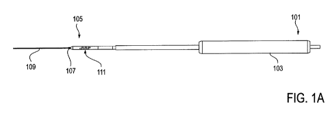
[00086] FIGS. 1 A-1 F illustrate one variation of an NLR device as described
herein. FIG. 1 A
shows a perspective view of this NLR device, and FIGS. lB show a cross-section
through the
device shown in FIG. IA. FIG. 1 C shows a partial cross-section through
another variation of an
NLR device. FIG. 1 D shows a partial top view of the device shown in FIG. 1 C.
FIGS. 1 E and
1 F illustrate proud electrodes formed as part of an NLR device.
[00087] FIGS. 2A-2C illustrate another variations of an NLR device; FIG. 2A
shows a top
view, FIG. 2B shows an expanded view of one region of the device of FIG. 2A,
and FIG. 2C
shows a slightly expanded view of yet another variation of the device shown in
FIG. 2A.
[00088] FIGS. 3A-3E illustrate different monopolar configurations of NLR
devices.
[00089] FIGS. 4A and 4B show a monopolar variation of a device including a
plurality of
electrodes on each side (top and bottom) of the NLR device.
-21-
CA 02749673 2011-06-23
WO 2010/105261 PCT/US2010/027323
[00090] FIG. 5A shows a schematic cross-section through one variation of a
device having
proud (protruding) electrodes.
[00091] FIGS. 5B and 5C illustrate switching the polarity of electrodes in an
NLR device.
[00092] FIGS. 6A-6E illustrate various configurations of bipolar NLR devices.
[00093] FIGS. 6F illustrates a configuration of bipolar NLR devices having a
shield.
[00094] FIGS. 7A-7B illustrate various configurations of tripolar NLR devices.
[00095] FIGS. 8A-8C illustrate various configurations of multipolar NLR
device.
[00096] FIG. 10 illustrates an NLR device coupled to a power source such as an
EMG system.
[00097] FIG. 9 illustrates an alternative variation of a neural localization
device including a
single monopolar wire.
[00098] FIGS. 11A-11C show cross-sections through different variations of NLR
device.
FIG. 11A shows an NLR device having a round cross-section; FIGS. 11B shows a
flattened (e.g.,
crushed) extrusion similar to that in FIG. 11 A. FIG. 11 C illustrates an NLR
device having an
oval or ribbon-shaped cross-section.
[00099] FIGS. 12A-12G show top (12A-12D) and end perspective views (FIGS. 12E-
12G) of
various embodiments of the NLR devices described herein.
[000100] FIGS. 13A-13B illustrate an NLR device having markers, such as radio-
opaque
markers.
[000101] FIGS. 14A-14B illustrate different sizing and/or dilating features of
NLR devices.
[000102] FIG. 15 illustrates another variation of an NLR device.
[000103] FIGS. 16A-16B illustrate an alternative embodiment of an NLR device.
[000104] FIGS. 17 and 18 schematically methods of operation of variations of
the NLR
device.
[000105] FIG. 19 is another variation of an NLR device.
[000106] FIGS. 20A and 20B shows variations of NLR devices configured for
coupling with
another device.
[000107] FIGS. 21A and 21B are semi-exploded views of devices including an NLR
region at
the distal end.
[000108] FIG. 21C shows a variation of anNLR device configured to be powered
from the
distal end.
[000109] FIG. 22A illustrates a detail view of a guidewire coupler and a
proximal end of a
guidewire.
[000110] FIGS. 22B-23F illustrate various methods of securing (e.g., locking
or releasably
locking) guidewire connectors as described herein.
-22-
CA 02749673 2011-06-23
WO 2010/105261 PCT/US2010/027323
[000111] FIG. 24A shows one variation of a guidewire lock, and FIGS. 24B-24E
illustrate one
method of using the guidewire lock shown in FIG. 24A.
[000112] FIG. 25A is another variation of a guidewire lock which maybe used
with any of the
devices and systems described herein. FIGS. 25B-25D illustrate one method of
using the
guidewire lock shown in FIG. 25A. FIGS. 25E-25F illustrate a method of
unlocking the
guidewire lock shown in FIG. 25A.
[000113] FIG. 26A is another variation of a guidewire lock or coupler,
configured as a leader.
FIGS. 26B and 26C show exploded views of different regions of the guidewire
coupler shown in
FIG. 26A.
[000114] FIGS. 27A and 27B illustrate one variation of a device (e.g., anNLR
device)
including a connector and guidewire coupler.
[000115] FIGS. 28A - 28C show another variation of a coupler.
[000116] FIGS. 29A-29C show another variation of a guidewire coupler.
[000117] FIG. 30 is one variation of a distal handle configured to couple to a
distal end of a
guidewire.
[000118] FIG. 31A shows one variation of a neural localization device.
[000119] FIG. 31B illustrates the neural localization device of FIG. 31A
coupled to a guidewire
and positioned within a neural foramina, above a spinal nerve root.
[000120] FIGS. 31C and 32A-32C illustrate various details of the neural
localization device of
FIG. 31 A.
[000121] FIGS. 33A and 33B graphically depict various relationships between
threshold
stimulation current values relative to a neural localization devices position
along a pathway
through a patient's body.
[000122] FIGS. 34A and 34B illustrate different ways that the NLR devices
described herein
may be inserted within the spine as part of a spinal decompression procedure.
[000123] FIG. 35 is one embodiment of an NLR device.
[000124] FIGS. 36A-36C illustrate the application of force to push or pull
anNLR device
within a spinal foramen to control the position of the NLR within the foramen.
[000125] FIGS. 37A-37C illustrate another variation of a method for
controlling the
configuration and/or position of an NLR device within a spinal foramen,
similar to that shown in
FIGS. 36A-36C.
[000126] FIG. 38 illustrates the NLR device used in the method illustrated in
FIG. 37A-37C.
[000127] FIG. 39A and 39B show one variation of an NLR device having an
expandable
stimulation region.
[000128] FIG. 40 is one variation of an NLR device having an H-shaped cross-
section.
-23-
CA 02749673 2011-06-23
WO 2010/105261 PCT/US2010/027323
[000129] FIG. 41 shows one variation of a patterned pair of electrodes.
[000130] FIG. 42 is one variation of a device such as that shown in FIG. 35
having a pre-biased
distal end that is curved.
[000131] FIG. 43 is a distal guidewire coupler that may be used to couple to a
guidewire and
allow the guidewire to push and/or pull the device into position.
[000132] FIG. 44 shows another enlarged view of the distal end of an NLR
device having three
pairs of electrodes on each of the top and bottom surfaces.
[000133] FIG. 45 is an enlarged view of the stimulation region of FIG. 44.
[000134] FIG. 46 shows a cross-sectional view through the insulating tubing of
the stimulation
region for a device such as the one shown in FIG. 35.
[000135] FIG. 47 illustrates another variation of a distal end of an NLR
device (without the
outer insulator tubing) including a substrate and three pairs of proud
electrodes formed of two
wires on the visible top surface.
[000136] FIG. 48 shows a side-on view of the variation shown in FIG. 47.
[000137] FIG. 49 shows an enlarged view of the conductors forming the three
pairs of
electrodes on the top surface.
[000138] FIG. 50 is another view of a distal end of an NLR device similar to
that shown in
FIG. 47, also including a pair of ring coil markers on either side of the
stimulation region.
[000139] FIGS. 51-53 graphically illustrate the principles of a method of the
neural localization
device: FIG. 51 shows three graphs representing measured tissue impedance as a
function of
frequency, for three different tissues respectively. FIG. 52 illustrates an
electric current as a
function of the depth (i.e. length of device) including different tissue
layers, for measured (Al)
and modeled (A2) data; FIG. 53 illustrates the transition between different
tissue layers having
different impedance characteristics, illustrated by a change in the impedance
measurement as a
function of depth (i.e. length of device).
[000140] FIGS. 54-56B illustrate various embodiments of an NLR device and
operation of
various devices.
[000141] FIG. 57 is a posterior view of the spine indicating decompression
paths at disk level
and along the nerve root.
[000142] FIG. 58 is a posterior view of the spine indicating a decompression
path for adjacent
level lateral recess decompression.
[000143] FIGS. 59-61 illustrate a method of increasing foraminal height.
-24-
CA 02749673 2011-06-23
WO 2010/105261 PCT/US2010/027323
DETAILED DESCRIPTION OF THE INVENTION
[000144] Described herein are devices, systems and methods for determining the
location of a
nerve or neural tissue relative to a pathway through a patient passing near,
around or adjacent to
a target tissue. In particular, described herein are flexible neural
localization devices that may be
ribbon-shaped, for use in tortuous and difficult-to-reach body regions, such
as the neural foramen
of the spine. In general, these devices may be referred to as neural
localization ribbon devices,
NLR devices, neural localization devices, or neuro localization devices. These
devices are
generally useful for determining if a nerve is nearby a surface of the device.
In some variations,
the devices may determine which side of the device a nerve is on (e.g.,
`above' or `below' the
flat surface of the device, relative to the patient) or if a nerve is located
between a pathway
through the tissue taken by the device and the target tissue. The devices
generally include a
flexible elongate body having a stimulation region. The stimulation region may
include an
electrode or a series of electrodes. These electrodes may be arranged as
bipoles, monopoles,
tripoles, or the like, but are generally configured so that the stimulation
field or broadcast field
emitted by the electrodes is limited to regions immediately adjacent to the
device, so that they
detect (e.g., by stimulating) nerves only in a particular orientation relative
to the neural
localization device. For example, the electrodes may be arranged as bipoles
(that can also be
referred to as tight bipoles), and may include a cathode and an anode that are
spaced relatively
close together to form a limited broadcast field. The broadcast field may be
referred to as the
bipole field, or the field formed by the excitation of the bipole pair. In
general, the bipole filed is
a controlled or "tight" broadcast field that extends from the bipole pair(s).
Similarly, the
broadcast field from monopolar, and particularly tripolar, quadrapolar, etc.
electrodes may also
be limited. Limiting the broadcast field in this manner may avoid the
(undesirable) detection of
nerves adjacent to other regions or surfaces of the neural localization
device. This is illustrated
in US Serial No. 12/060,229, previously incorporated by reference.
[000145] The tight broadcast field emitted by the electrode(s) described
herein may be limited
so that it stimulates nerves only within a predetermined distance. This
distance is typically quite
narrow (e.g., within about 2 mm, within about 1 mm, within about 0.5 mm,
within about 0.1
mm, within about 0.05 mm, etc. of the surface of the NLR device). Beyond this
broadcast range,
the current or voltage applied by the device falls below levels sufficient to
stimulate the nerve.
The broadcast field may be controlled by the combination of the power supplied
to the
stimulation electrode(s) as well as the configuration of the electrode(s) on
the NLR device.
[000146] In particular, the NLR electrodes may be configured as mutlipolar
electrodes,
including one or more anodes and one or more cathodes. By placing the anodes
and cathodes
relatively close to each other, the current flowing between the anodes and
cathodes may be
-25-
CA 02749673 2011-06-23
WO 2010/105261 PCT/US2010/027323
limited. In some variations a plurality of anodes and a plurality of cathodes
may be arranged in a
pattern along or across a surface (e.g., the top and/or bottom surface of the
NLR device) to form
a bipole network that permits a larger area of the NLR device to stimulate
only nerves passing
within a predetermined range of the surface. This sort of bipolar network may
allow a relatively
"flat" profile of broadcast field, so that although a large area of the NLR
device may emit the
broadcast field, the field does not penetrate deeply, preventing stimulation
of nerves outside of
the (typically narrow) range desired. These configurations may also prevent
stimulation of
nerves located on the opposite side(s) of the NLR device.
[0001471 In some embodiments, as described in greater detail below, the
electrodes may be
configured to apply and receive an electrical signal to and from the target
tissue. In this
embodiment, the signal may be a non-stimulating electrical output and may
characterize the
tissue (target tissue and non-target tissue) using electrical bio-impedance.
Electrical bio-
impedance is the response of living tissues to externally applied electrical
current. Bio-
impedance measurements are carried out while "sweeping" a frequency of the
applied electrical
signal. During these measurements, the electrodes may be static or may be
moved through the
body. Alternatively, the device may include a series of electrodes which are
activated
sequentially along the length of the device. The measured bio-impedance
components
(resistance, capacitance, phase, etc.) are typically frequency-dependent, thus
the use of multiple
frequencies may help in characterizing the tissue or tissue(s) interacting
with the device and
electrodes. Real-time analysis of the measured parameters enable the system to
determine what
type of tissue (for example, whether a nerve) is nearby a device or portion of
a device.
[0001481 The elongate body region of anNLR device may also be referred to as a
probe or
probe body. In general, the NLR device may include one or more regions on the
outer surface of
the device that are configured to determine if a nerve is nearby the region
(or one or more of the
regions) of the device. In some variations, each region includes an electrode
or a set of
electrodes (e.g., a multi-polar network) that is arranged to emit energy to
stimulate a nearby
nerve so that it can be detected. The regions may be arranged around or along
the outer surface
of the device. In general the NLR devices described herein are flat, for
example, including a
first side (top) and a second side (bottom); the sides joining the top and
bottom may be narrow
(the more flat the structure, the more narrow), or they may have sufficient
thickness for inclusion
of one or more electrodes. Thus, each region may include one or more
electrodes (e.g., bipole
pairs or networks), which may be used to detect a nearby nerve.
[0001491 FIG. 1 A illustrates a top view of one variation of an NLR device
coupled to a
guidewire 109. In FIG. 1 A, the device has a proximal end 101 that is
configured to include or be
attached to a proximal handle 103. The distal end 105 includes a coupler 107
for coupling to a
-26-
CA 02749673 2011-06-23
WO 2010/105261 PCT/US2010/027323
guidewire 109. The coupler may therefore be referred to as a guidewire
coupler. Near the distal
end, but extending proximally, the body of the NLR is a flexible, ribbon-
shaped structure, 111.
This ribbon-shaped body region is typically flat or flattened, so that the top
and bottom have a
width that is much greater than the thickness between these surfaces. In this
example, the top
and bottom surfaces each include a set of multipolar electrodes. In FIG. 1 A,
the top shows a
bipole network including a line of cathodes and a line of anodes, shown in
more detail in FIGS.
1C and 1D.
[000150] FIG. 1 B shows a cross-section through the ribbon region 111 of a
device such as the
one shown in FIG. IA. In this exemplary cross-section, each side of the NLR
device (the top,
bottom and both sides) include a pair of electrodes forming part of a bipole
network. Four
separate networks are formed. As shown, there is a top electrode pair 112, a
bottom electrode
pair 113, and side electrode pairs 114. In FIG. 1 B, the electrodes are set
inward from the edges
between the top and sides and bottom, which may help limit the spread of the
emitted filed from
one side (e.g., the top) to activate a nerve facing another side (e.g., the
sides or bottom). In other
variations, the thickness of the NLR device (the sides between the top and
bottom) may be more
narrow. In some variations, the device may include only a top stimulation
region or may only
include a top and bottom stimulation region. In some variations, the
electrodes on the surface(s)
may be part of a multi-polar network (e.g., having a plurality of cathodes
and/or a plurality of
anodes). For example, FIG. 1 C shows a partial cross-section through another
variation of an
NLR device. As shown, on the bottom side of the device for example, the device
includes two
sets of anodal conductors 115 and 116 and two sets of cathodal conductors 117
and 118. The
anodal conductors and cathodal conductors run along the length of the device
for both the top
and bottom surface of the device. As shown, on the top side of the device for
example, the
anodes and cathodes are exposed at discrete locations along the device to form
individual
electrodes (e.g. individual anodes and cathodses). For example, anode 119 is
formed by an
anodal conductor on the top surface and cathode 120 is formed by a cathodal
conductor on the
top surface. As shown, the anodal conductors and cathodal conductors are
housed in an
insulating material 121. The insulating material 121 is removed or has holes
in discrete locations
over the anodal conductors and cathodal conductors and along both the top and
bottom surfaces
of the device to form the electrodes (e.g. anodes and cathodes). The
electrodes may be flush to
the surface, or they may alternatively be below the surface of the insulator
or may be proud with
respect to the surface of the insulator. For example, the electrode may be
formed by removing
material (e.g., skiving) to expose a portion of the anodal or cathodal
conductor or wire. The
exposed wire regions may be filled or coated with a conductive material. In
other variations,
electrodes may be formed by laser ablating material to expose the wire and
filling with a
-27-
CA 02749673 2011-06-23
WO 2010/105261 PCT/US2010/027323
conductive material or bonding a flex circuit to them to form the electrode.
Multiple electrodes
may be formed from each wire.
[000151] These electrodes may be configured as a single network (including a
quadrapolar
network) spanning the surface, or they may be configured as two bipolar
networks, or the like.
FIG. 1 D illustrates a top view of the same arrangement as shown in FIG. 1 C.
As shown in FIG.
1 D, anodes 119 are the exposed regions of the anodal conductors that run
along the length of the
device, while the cathodes 120 are the exposed regions of the cathodal
conductors that also run
along the length of the device. The non-exposed portions of the two anodal
conductors and two
cathodal conductors are depicted as dotted lines as they are positioned below
the insulating
material 121.
[000152] FIGS. 1E and 1F show detail of a series of proud electrodes exposed
along the
surface of the NLR device. In this example, as shown in FIG. 1 F, each
electrode 119 is formed
by a bump or ridge in the conductor 122. The conductor, either anodal or
cathodal, may include a
series of bumps or ridges such that the conductor forms a number of electrodes
in series. As
shown in FIG. IE, the bumps or ridges are exposed through a window or hole in
the insulating
material 121 to form discrete electrodes 119. The electrodes 119 formed by
this method may
preferably be proud with respect to the surface of the device or they may be
flush with the
insulating material 121 (e.g., non-proud).
[000153] In this example, the electrodes are formed by pairs of metal wires
(e.g., an upper
anode wire and an upper cathode wire for the top surface and a lower anode
wire and a lower
cathode wire for the bottom surface). Each wire is connected to or includes a
plurality of
electrodes; in this example, each wire includes to three electrodes that each
individually extend
"proud" from the surface of the ribbon-shaped device. In one particular
embodiment, the
electrodes are each 4mm long, and extend 0.020" from the surface. The cathodal
electrodes on
each surface are staggered along the length of the NLR device with anodal
electrodes, as
illustrated. The "proud" electrode shown may afford a greater sensitivity than
flush or recessed
electrodes.
[000154] An alternative variation is shown in FIGS. 2A-2C. Similar to the
devices shown in
FIGS. lA-1F, in FIG. 2A an NLR device includes an elongate body 200 having a
distal end with
a guidewire coupler 202 and a proximal end 205 that may be connected to a
proximal handle.
[000155] In any of these variations, the flexible elongate body may be
configured to be blunt
(atraumatic). In general, the outer body (e.g. insulating material) of the
device may be formed of
any appropriate material, including polymeric materials such as PEBAX, PEEK or
the like.
Non-conducting and biocompatible materials may be particularly preferred. FIG.
2B illustrates a
partial view of the electrodes forming the bipole network on the top surface
of the device shown
-28-
CA 02749673 2011-06-23
WO 2010/105261 PCT/US2010/027323
in FIG. IA. In this example, the exposed electrodes alternate between anodal
electrodes 209
(connected to a single anodal line 210) and cathodal electrodes 207 (connected
to a single
cathodal line 211). Exemplary sizes and arrangements for variations of these
devices are
illustrated in FIGS. 6A-6E. FIG. 2C illustrates another variation of an NLR
device in which the
exposed electrodes 212 are not round, but are oval.
[000156] In addition to the networks of multipolar electrodes described above,
the NLR
devices as taught herein may be configured as a network of (or of individual)
monopolar
electrodes 300, as illustrated in FIGS. 3A-3E. In this example, the NLR device
may be used
with a ground pad or stimulus return electrode coupled to the patient. The
current evoked by the
device may be `spread' further with such monopolar devices.
[000157] FIGS. 4A and 4B show another variation of an NLR device having a
plurality of
monopolar electrodes extending along the length of each side (top and bottom)
of an NLR
device. In this variation the stimulation region on the top and bottom of the
ribbon-shaped
device may be formed from a flexible circuit, and thus, in this variation, the
electrodes may not
be "proud" (extending substantially from the surface of the device). As shown
in FIGS. 4A and
4B, the device has a proximal end 401 that is configured to include or be
attached to a proximal
handle 403. The distal end 405 includes a coupler 407 for coupling to a
guidewire (not shown).
The coupler may therefore be referred to as a guidewire coupler. Near the
distal end, but
extending proximally, the body of the NLR is a flexible, ribbon-shaped
structure, 411. This
ribbon-shaped body region is typically flat or flattened, so that the top and
bottom have a width
that is much greater than the thickness between these surfaces. In this
example, the top and
bottom surfaces each include a set of monopolar electrodes 413.
[000158] FIG. 5A shows a schematic cross-section through one variation of the
stimulation
region of an NLR device. In this example, the NLR device includes an electrode
501 on the
upper surface that is a "proud" electrode (extending from the upper surface of
the ribbon). This
electrode is configured as an cathode (negative) electrode. A proud anode
(positive) electrode
503 is shown on the bottom of the device. In this embodiment, current may be
applied in a
bipolar fashion between the upper and lower electrodes, as illustrated in FIG.
5B. In this
example, the stimulation is anodal stimulation. FIG. 5C illustrates the
alternative configuration
of cathodal stimulation. These figures also illustrate reversing the anode and
cathode by
changing the applied current to different electrodes. For example, merely
switching the supplied
power connection, it may be possible to switch the anode and cathode;
alternatively the device
(e.g., handle region) may include a switch for switching anode and cathode. In
some variations
it may be preferable to have the anodes and cathodes be arranged in a bipolar
(or multipolar) set
on the same side of the device to minimize the current spreading around the
edges of the device.
-29-
CA 02749673 2011-06-23
WO 2010/105261 PCT/US2010/027323
[000159] FIGS. 5B and 5C also illustrate one method of determining which side
a nerve is on
by switching between anodal and cathodal stimulation. If it is known that a
particular nerve or
nerve root is more sensitive to anodal stimulation, the response of a nerve to
both anodal and
cathodal stimulation (by switching between the two as illustrated in FIGS. 5B
and 5C) can be
determined. For example, the threshold for evoking an EMG (or a robust EMG)
may be
determined for each configuration. If the nerve is more sensitive to anodal
stimulation, then the
nerve is likely to be below the device if the threshold for triggering an EMG
in anodal
stimulation is greater than the threshold for triggering an EMG in cathodal
stimulation, and vice-
versa.
[000160] FIGS. 6A-6E illustrate other variations, similar to the arrangement
shown in FIG. 2B,
of a network of bipolar pairs (a bipole network). For example, FIG. 6A
illustrates a bipole
network on the top surface of the NLR device (within an activation range that
extends
longitudinally along the NLR device) in which the exposed electrodes 600 are
round, and are 1
mm in diameter, and spaced 1 mm apart. In any of the variations described
herein, the exposed
electrodes may be any appropriate shape, including round, square, oval, etc.
FIGS. 6B-6D
illustrate other geometries of bipolar electrodes, including electrodes 601
that are 1 mm diameter
and spaced 2 mm apart (FIG. 6B), electrodes 602 that are 1.5 mm diameter and
spaced 1 mm
apart (FIG. 6C), and electrodes 603 that are 1.5 mm in diameter and spaced 2
mm apart (FIG.
6D). FIG. 6E illustrates bipolar electrodes that are exposed along the length
of (or exposed
along a portion of the length of) the NLR device. In this embodiment, the
device includes a
single elongate anode 604 and a single elongate cathode 605 that are disposed
along the length of
the stimulation region of the NLR device. Other variations of electrodes and
electrode
arrangements are contemplated, including smaller or larger electrodes and
smaller or larger
separations. In addition, the electrodes shown are arranged in lines
corresponding to the
underlying anodal or cathodal lines (wires, etc. - not shown). This
arrangement may vary, and
may span curves, arcs, sinusoids, or the like, extending either longitudinally
along the NLR,
diagonally across the NLR, or perpendicular to the longitudinal access.
[000161] As mentioned above, a bipole pair forming part of a multipolar
network may include
an anode and a cathode and may have a very limited broadcast field (e.g., a
`tight bipole pair').
In some variations the size of the anode and cathode forming the bipole pair
are relatively small,
particularly (e.g., less than 5 mm2, less than 3 mm2, less than 2 mm2, less
than 1 mm), and the
anode and cathode are positioned sufficiently nearby so that the majority of
current passes
between the anodes and cathodes. For example, the anode and cathode of a
bipole pair may be
separated by less than 5 mm, less than 2 mm, less than 1 mm, etc.
-30-
CA 02749673 2011-06-23
WO 2010/105261 PCT/US2010/027323
[000162] As mentioned above, an NLR device may include multiple electrode
networks. For
example, different regions on the surface of the device may include different
electrode networks
(e.g., each region may have its own network). The bipole networks in different
regions may be
non-overlapping, and may form effectively non-overlapping continuous fields
(e.g., continuous
bipole fields). "Effectively non-overlapping bipole fields" means that the
broadcast fields of two
or more networks do not substantially overlap. For example, the component of a
broadcast field
(e.g., intensity) due to a second bipole network is less than 15% (or 10%, or
8% or 5% or 1%) of
the component due to a first bipole network at any position near the first
bipole network,
particularly at the excitation ranges described herein.
[000163] In any of the embodiments described above, particularly FIG. 6E for
example, the
device may further include an insulating element or shield 607, as shown in
FIG. 6F. The
insulating element may be coupled to a single surface (e.g. the top surface)
or to both surfaces. In
some embodiments the insulating element may slide over and/or wrap around the
elongate body
of the NLR device. The insulating element may function to prevent an
electrode, or portion of
electrode, from delivering stimulation to the surrounding area (e.g. to a
nerve or other non-target
tissue). In some variations, if the shield is limiting the amount of exposed
electrode(s) the current
density emitted from the exposed electrodes (anode 604 and cathode 605, for
example) will
increase. In use, the insulating element may first be pulled back such that a
large area of
electrode(s) are exposed. A user may then slide the insulating element over
the electrodes (as
shown by the arrow in FIG. 6F) such that a smaller area of electrodes 604 and
605 is exposed.
Alternatively, a smaller area may first be exposed and then the insulating
element may be pulled
back (distally or proximally) to expose a larger portion of electrode(s). In
some embodiments,
the insulating element may include a window (not shown) through which current
may be
delivered. In this embodiment, the window may be moved along the length of the
elongate body
of the device such that different portions of the electrode(s) are exposed
sequentially.
[000164] FIGS. 7A to 8C also illustrate arrangements of electrodes forming a
network on the
surface of the NLR device. As indicated in each of these figures, the size and
spacing of these
electrodes, forming tripolar networks in FIGS. 7A and 7B, and quadrapolar
networks in FIGS.
8A and 8C may vary. In some variations, the size and spacing of the electrodes
may vary on the
same NLR device.
[000165] As mentioned above, when a region of the outer surface of a device
includes more
than one electrode, the electrodes (e.g. bipoles) may be arranged as a bipole
network. A bipole
network includes at least two bipoles that are formed by at least three
electrodes (e.g., two
anodes and a cathode or two cathodes and an anode). The bipole network is
typically arranged
so that all of the bipoles in the network are activated synchronously to
create an effectively
-31-
CA 02749673 2011-06-23
WO 2010/105261 PCT/US2010/027323
continuous bipole field along the outer surface. In the examples shown in
FIGS. 6A-6D above,
the anodes and cathodes forming the bipolar network are arranged so that the
current between the
two electrodes forms a zigzag pattern. Bipole pairs are located adjacent to
each other and share
either an anode or a cathode. In some variations, adjacent bipole pairs do not
share anode or
cathodes. In general, the multipolar networks described herein may form an
effectively
continuous field along an active region of the outer surface of an NLR device.
Adjacent bipole
pairs may be positioned close to each other.
[000166] As described above, all of the cathodes forming a network may be
electrically
connected to each other and all of the anodes forming the network may be
electrically connected.
For example, the anodes of a network may all be formed from a single anodal
connector, and the
cathodes of a network may be formed from a single cathodal connector.
Alternatively, all or a
subset of the cathodes of the network may be formed separately so that they
can be separately
activated or jointly activated. For example, each of the cathodes may be wired
to a connector
that connects to a power source or controller configured to energize the
network in a particular
region.
[000167] FIG. 9 illustrates an embodiment of the neural localization device
that includes a
simply a single monopolar wire 900. The wire 900 may include a distal end that
may be
connected to a distal handle 901 and a proximal end that may be connected to a
proximal handle
902. The handles remain exterior to the skin of a patient such that the
handles may be controlled
in a bimanual fashion, as described in detail below. For example, the handles
may be pulled up
to pull the wire away from the nerve root and then may be pushed down to push
the wire closed
to the nerve root. By creating a maximum distance and a minimum distance from
the nerve root,
two distinct threshold current amounts may be established and used to
determine the location of
the nerve root with respect to the monopolar wire. Methods of using the neural
localization
devices is described in more detail below.
[000168] The monopolar wire may include a sharp (tissue penetrating) distal
end and may be
long (e.g., elongated) and flexible such that the wire may penetrate tissue
and be positioned
along a path around target anatomy. For example, the target anatomy may
include a facet joint
and a ligament (not shown) while non-target tissue may include a nerve root.
Current may be
delivered to the wire such that a portion of the wire stimulates a nerve root.
In some
embodiments, the wire may then be used to couple to, position, and activate a
tissue modification
device. In these embodiments, the proximal end of the wire may include a
feature (not shown)
that allows it to be coupled to a guidewire coupling member securely. For
example, the wire
may include a ball or other shaped end (which may be conical, tubular, ring,
etc.) at the distal
-32-
CA 02749673 2011-06-23
WO 2010/105261 PCT/US2010/027323
end for coupling to a guidewire coupling member. In some embodiments, the wire
may further
include an insulating element or shield as described above in reference to
FIG. 6F.
[000169] The devices described herein may be connected to a power source
proximally, or
distally. For example, the cathodal and annodal lines which may be used to
form the electrodes
may extend proximally toward the proximal handle of the device. In this
variation, a connector
at the proximal region of the device may be used to connect the device to a
power source. In one
embodiment, as shown in FIG. 10, the device 1000 has a proximal handle 1001
and a stimulus
region (inside the patient and not shown) that is coupled to a guidewire 1003
which is coupled to
a distal handle 1004. The stimulus region is placed within a patient such that
it wraps around
target anatomy (such as a facet joint, for example) and is either above
(preferable, in most
embodiments described throughout) or below a nerve root. A nerve root is the
initial segment of
a nerve (nerve 1007 for example) leaving the central nervous system. The
cathodal and anodal
conductors of the device (not shown) are connected to connectors 1002
extending from the
proximal handle region of the device. As shown in FIG. 10, the connectors 1002
connect the
device 1000 to an EMG system 1005 and a stimulus output box 1006. As shown,
current (or
voltage) will flow from the EMG system through the stimulus output box to the
device 1000.
EMG electrodes 1008 may be placed in a leg muscle of the patient as shown.
Multiple EMG
electrodes may be placed in a plurality of muscles or myotomes as described
below with
reference to Table 1 below. When the current passes from the device into a
nerve root, the
current activates the nerve such that a current passes through nerve 1007. The
activated nerve
thereby activates the corresponding muscle cells. An EMG electrode 1008
coupled to the leg
muscle detects the electrical potential generated by the muscle cells when
these cells are
electrically or neurologically activated by the nerve (nerve 1007, for
example). The electrical
potential that is detected is known as an EMG response. This response may be
amplified by a
Patient Interface Amplifier 1009. The response may be detected or measured as
a voltage. This
voltage may be delivered to the EMG 1005 system for analysis.
[000170] As shown below in Tables 1 and 2, EMG electrodes may be placed in any
combination of muscles, such as leg muscles for example, to best pick up an
EMG response
elicited by a neural stimulation of a particular nerve root. Selection of a
muscle for placement of
EMG electrode(s) may be determined by the nerve root(s) that innervate that
muscle. For
example, as shown below in Table 1, the Adductor longus is innervated by
primarily a L3 nerve
root (as indicated by a capitol "X") and also by L2 and L3 nerve roots (as
indicated by a lower
case "x"). The table below lists several muscles typically monitored during a
lumbar spinal
surgery. The information provided by the table below may be used to determine
ideal placement
of EMG electrodes prior to use of an NLR device, or may alternatively be used
once an EMG
-33-
CA 02749673 2011-06-23
WO 2010/105261 PCT/US2010/027323
response in elicited by the NLR device to determine the most likely nerve root
that has been
stimulated based on which muscle returns an EMG response. For example, if the
EMG response
is elicited in the Extensor Hallucis longus, the NLR device has most likely
stimulated a L5 nerve
root (as indicated by a capitol "X") and also may have stimulated an L4 and Si
nerve roots (as
indicated by a lower case "x").
Muscle Level of maximum response erve Root)
L2 L3 L4 L5 Si S2
Iliopsoas X X
Adductor longus x X X
Vastus medialis x X
Vastus lateralis x X x
Tibialis anterior X X
Peroneus longus x X x
Extensor Hallucis longus x X X
Flexor Hallucis longus x X x
medial Gastrocnemius x X x
lateral Gastrocnemius X X x
Gluteus maximus x X x
Biceps femoris x x x
Anal Sphincter x
Table 1: Nerve Root to Myotome Map
[000171] As listed below in Table 2, depending on the location of the surgery,
particularly a
disc level(s) in spinal surgery, different muscle groups may be targeted for
placement of the
EMG electrodes. For example, as listed in the table below, when a spinal
surgery is performed at
level L3/L4, the nerve root that is most likely at risk to be damaged during
the procedure is a L3
nerve root for a "disc level pass" and a L4 nerve root for an "along the nerve
root pass". As
illustrated in FIG. 34A and described in further detail below, the path
through the spine may be
an "above the pedicle pass" or "disc level pass" (as indicated by arrow 3401)
so that the
guidewire may pull in the NLR device tangential to the direction of the
exiting spinal L3 nerve
root 3400. The path may also be parallel to the exiting L4 nerve root 3403
(e.g., "below the
pedicle pass" or "along the nerve root pass" as indicated by arrow 3402). In
order to most
effectively monitor the L3 and/or L4 nerve roots, it is desirable to place the
EMG electrodes in
muscles innervated by those nerves. For example, in Setup 2 as listed in Table
2 below, the EMG
electrodes may be placed in the Vastus Medialis and Tibialus Anterior muscles.
As indicated, the
Vastus Medialis is innervated by the L2 and L3 nerve roots and the Tibialus
Anterior is
innervated by the L4 and L5 nerve roots. For a more comprehensive setup, as
indicated by Setup
4, the EMG electrodes may be placed in the Vastus lateralis, the Tibialis
Anterior, the Adductor
-34-
CA 02749673 2011-06-23
WO 2010/105261 PCT/US2010/027323
longus. As indicated, this comprehensive setup option provides overlap between
the muscles and
nerve roots of interest that innervate them.
Surgical At Risk Setup 1 Setup 2 Setup 3 Setup 4
Level Nerve Muscles Muscles Muscles Muscles
DP = L2 *Adductor Vastus medialis (2,3), Vastus medialis (2,3),
L2/L3 AN = L3 longus(2,3,4), Vastus Vastus Medialis(2,3) Vastus lateralis
(2,3,4), Vastus lateralis (2,3,4),
lateralis(2,3,4) Iliopsoas(2,3) Adductor longus(2,3,4)
*Adductor Vastus medialis (2,3), Vastus lateralis (2,3,4),
L3/L4 DP = L3 longus(2,3,4), Vastus Tibialus Vastus Medialis(2,3), Vastus
lateralis (2,3,4), Tibialis Anterior(4,5),
AN = L4 lateralis(2,3,4) Anterior(4,5) Tibialis Anterior(4,5) Adductor
longus(2,3,4)
Vastus lateralis(2,3,4), Tibialus Anterior(4 5) Tibialis Anterior (4,5),
Vastus lateralis(2,3,4),
DP = L4 Tibialis anterior(4,5),
L4/L5 Extensor Hallucis Biceps femoris Gastrocnemius
AN = L5 Longus (4,5,1) (5,S1,S2) (5,S1,S2) Gastrocnemius
S,Sl,s2
Gastrocnemius Gastrocnemius
Extensor Hallucis Tibialus Anterior(4,5), (5,S1,S2), Extensor
DP = L5 Longus (4,5,S1), Medial Gastrocs hallucis longus Anterior(4,5), 5
Extensor
LS/S1 AN=S1 Medial Gastrocnemius (5,S1,S2), Biceps (4,5,SI), Flex. hallucis
longus
(5,S1,S2) femoris. (5,S1,S2) longus (5,S1,S2), Anal Hallucis longus
(4,5,S l)
sphincter S2,S3
*Adductor Vastus Medialis(2,3), Vastus lateralis (2,3,4),
longus(2,3,4), Vastus Tibialus Anterior(4,5) Tibialis Anterior(4,5),
L3/L4/L5 lateralis(2,3,4), Medial Gastrocs Adductor
dduct longus(2,3,4),
Extensor Hallucis (5,S1,S2), Biceps Gastrocnemius
Longus (4,5,1) femoris. (5,S1,S2) (5,S1,S2)
Vastus lateralis(2,3,4), Vastus lateralis (2,3,4),
Extensor Hallucis Tibialus Anterior(4,5) Tibialis Anterior(4,5),
L4/L5/S1 Longus (4,5,1), Medial Gastrocs Extensor Hallucis
Medial Gastrocnemius (5,Sl,s2), Biceps longus (4,5,51),
(5,S1,S2) femoris (5,S1,S2) Gastrocnemius
(5,SI,S2
*Adductor Vastus lateralis (2,3,4),
longus(2,3,4), Vastus Vastus Medialis(2,3), Tibialis Anterior(4,5),
L3/L4/L5 lateralis(2,3,4), Tibialus Anterior(4,5) Extensor Hallucis
/S1 Extensor Hallucis Medial Gastrocs longus (4,5,51),
Longus (4,5,SI), (5,S1,S2), Biceps Gastrocnemius
Medial Gastrocnemius femoris. (5,S1,S2) (5,S1,S2)
(5,S1,S2
Table 2: EMG Electrode Placements (DP = Disc Pass, AN = Along Nerve)
[0001721 The power source may be configured for stimulating one or more
regions of the NLR
device either simultaneously or sequentially, or individually. For example, in
variations in
which the NLR device includes a top region and a bottom region, each with its
own electrode
network (e.g., bipole network), the stimulator (or a controller communicating
with the stimulator,
such as EMG system 1005) may apply energy to only the top region network or
the bottom
region network in a controllable, or indicated fashion, so that the patient
can be monitored to
determine if a nerve is nearby the top region or bottom region. For example,
the patient may be
monitored for muscle twitch or EMG response immediately following one or more
stimulation
protocols. The stimulation may be preset (e.g., a predetermined power level or
series of power
levels, a ramp of power levels, etc.) or it may be varied depending on
feedback. For example,
-35-
CA 02749673 2011-06-23
WO 2010/105261 PCT/US2010/027323
the power level may be increased to a threshold (of muscle or simulation
response). The power
level may be limited or capped to prevent damage to tissue or the like. Thus,
an NLR device for
determining if a nerve is nearby may also include a controller for controlling
the application of
energy to the electrodes. For example, the device may include a switch that
can select the
activation of either the top stimulation region or the bottom stimulation
region. In particular, the
application of energy to the electrodes may be coordinated as described in the
methods sections
below, so that the activation of a nerve can be correlated to a particular
region of the surface of
the device.
[000173] The nerve localization devices and systems described herein may
include one or more
indicators or outputs. The detectors may provide a user-identifiable signal to
indicate the
location of the nerve or the status of the system. For example, the nerve
localization devices
may include one or more light emitting diodes (LEDs), buzzers (or other sound
output), a video
display, or the like. An LED may be illuminated based on signals generated by,
received by, or
generated in response to the energized electrode(s) as discussed above. In
some variations the
system or devices create a vibration or sound that a user manipulating the
device 20 may feel or
hear. The intensity of the output may vary as a function of detected signal.
Alternatively, all
indicators or outputs may be located on an external EMG system as described
above with respect
to FIG. 10.
[000174] The NLR devices described herein may be fabricated in any appropriate
manner. For
example, the devices may be formed by extrusion. FIGS. 11A-11C illustrate
methods of
fabricating variations of NLR devices. For example, FIG. 11 A and 11 B show a
device that is
fabricated as an extrusion of material (e.g., PBAX) as an elongated cylinder
having a round
cross-section, as shown in FIG. 11 A. The walls of the cylinder include
openings 1100 into
which the conductive channel such as the annodal and cathodal channels
connecting to the
electrodes may pass. In some variations, these conductive channels may include
a wire or a
conductive filling. The structure may be extruded around the wire (or
conductive material), or it
may be added after the extrusion. Some of these channels, or additional
channels (not shown),
may include cables or tensioning elements for steering, or for attaching to a
guidewire or other
components. As mentioned above, the electrodes may be formed by laser cutting
to form vias to
the conductive channels, and filling with a conductive matrix to form the
surface electrode. In
some embodiment, the surface electrode may be preferably flush with the
surface of the NLR
device, to prevent `snagging' or damaging adjacent tissues. Alternatively, the
electrodes may be
proud to provide enhanced approximation to a nerve root.
[000175] The circular cross-section shown in FIG. 11A may be flattened (e.g.,
crushed,
compressed, etc.) into an oval, rectangular, or substantially flat shape. FIG.
11B illustrates an
-36-
CA 02749673 2011-06-23
WO 2010/105261 PCT/US2010/027323
example of a substantially flat shape. This shape may be completely flattened,
or it may include
a central lumen 1101. The central lumen may be used to hold one or more cables
or stiffening
members, or for passages of a tool, guidewire, or the like.
[000176] In some variations, the NLR body may be formed in the ribbon or oval
cross-section.
For example, the NLR body may be extruded as an oval shape, as shown in FIG.
11 C (also in the
example shown in FIG. 46). Fabricating the NLR body as an extruded oval or
other flattened
cross-section may enable better control and reliability of the dimensions and
integrity of the
lumen formed in the device. The flattened, ribbon-shaped devices described
herein may more
readily access narrow, tortuous or difficult to reach body regions, as
mentioned above, including
neural foramen. In addition, the separation between different electrodes in
the devices such as
those shown for FIGS. 11 B and 11 C, in which the electrodes connected to the
wires or
conductive filling, may be controlled so that the electrodes are relatively
close to each other (e.g.,
forming bipole pairs) while separated from the edges of the device and/or the
other electrodes.
The shape of the NLR device (e.g., the ribbon shape) may be similar to shape
of the tissue
modification device that it is to be used with during a procedure, which may
help predict
placement of the tissue modification device. As described below, the device
may also be shaped
as an expander or dilator, or may be used with an expander or dilator. For
example, the body of
the NLR device (or a portion of the NLR body) may be expandable or inflatable.
[000177] FIGS. 12A to 12G illustrate other exemplary NLR body shapes. For
example, FIG.
12A (shown in cross section in FIG. 12E) shows an NLR body shape that is
approximately
rectangular, although ribbon-shaped, as previously illustrated in FIGS 1 A and
2A. FIG. 12B
shows another variation of a ribbon-shaped body region in which the device is
tapered
longitudinally, though still substantially "flat," as shown in FIG. 12F. As
mentioned, the NLR
body may be configured as a dilator or expander. Thus, in one variation
similar to that shown in
FIGS. 12B and 12F, the ribbon shaped body is flexible in at least one axis
(e.g., up 1201 and
down 1203, as illustrated by the arrows in FIG. 12F, but relatively
incompressible, particularly in
the direction perpendicular to the axis of flexibility. Thus, the device shown
in FIG. 12B may be
inserted into a body region by the narrower distal tip region 1205, and may
dilate the opening by
pulling the device distally into the space so that the wider proximal region
moves into the
opening.
[000178] The NLR device or device body may have a curved or arcuate body
region, as
illustrated in FIG. 12C. In some variations the NLR body may be shape-
changing. For example
the NLR body be transformable from a linear shape (such as that shown in FIG.
12A) along the
length to a curved or S-shaped configuration, as shown in FIG. 12C and in
profile in FIG. 12G.
For example, the body may include a wire or tensioning element to transition
the device from
-37-
CA 02749673 2011-06-23
WO 2010/105261 PCT/US2010/027323
one configuration to another. In addition to wires and tensioning elements,
other transitioning
elements include balloons that may inflate and/or deflate to change the shape
of the NLR body.
Any of the NLR body shapes described herein may be combined or modified. For
example, FIG.
12D is another example of a tapered NLR body region including a distal end
that is rectangular.
[000179] In some embodiments, as shown in FIGS. 13A and 13B, the NLR elongate
body may
include radioopaque markers that may be used help localize and accurately
position the NLR
device, particularly the stimulation region of the device. For example, the
NLR device may
include one or more radio-opaque regions that can be used to orient or mark
the device. In
general, the NLR devices described herein may be inserted and positioned
relative to the body so
that the top and bottom stimulation regions (which are typically opposite each
other) are
positioned correctly relative to the anticipated orientation of the a nerve or
nerve root. As shown
in FIG. 13A, the region of the NLR device 1300 in which the electrodes extend
may be referred
to the active region or the stimulation region 1301. The stimulation region
1301 in this example
is shown as marked by radioopaque markers 1302 and 1303 on the proximal and
distal end of the
stimulation region, respectively. As shown, the markers may allow for
visualization of the NLR
device while inserted into a body region (e.g., using fluoroscopy or the
like). For example,
radioopaque markers will show up under fluoroscopy darker than the rest of the
device. As
shown in FIG. 13A, it may be preferable to position the device 1300 such that
when viewed in a
lateral view under fluoroscopy the proximal marker 1302 is located at the
bottom of the
curvature of the device body and the distal marker is located half way between
the proximal
marker and the distal tip 1304 of the device. As shown, distal tip 1304
includes a guidewire
coupler and is coupled to a guidewire 1305.
[000180] Markers may also help with determining the size of a body region into
which the
NLR device is inserted. For example, as shown in FIG. 13B, the NLR device 1306
may include
one or more marker 1307 at a fixed position along the length of the NLR body
region. The fixed
position may have a known width and height. In some variations, the marker is
indicative of the
position along the length of the device. For example, when a device includes a
plurality of
markers, the markers may be differently sized or shaped, or multiple markers
may be arranged in
a way that indicates position and/or orientation within the tissue. Thus, when
radioopaque
markers are used, the NLR devices may be used in conjunction with a
fluoroscope, as indicated
in FIG. 13A. FIG. 13B illustrates one variation of a tapering NLR body (seen
from the top)
having identifying markers along the length, as shown.
[000181] As mentioned above, the NLR devices may be dilating devices, or may
be used in
combination with dilating devices. FIGS. 14A and 14B show two variations of
NLR body
regions that are configured as dilators. FIG. 14A illustrates an NLR body
region that dilates by
-38-
CA 02749673 2011-06-23
WO 2010/105261 PCT/US2010/027323
increasing the thickness (by 5x in this example) from the distal (thin) end
1400 to the proximal
(thick) end 1401. The electrodes on the surface(s) of the NLR body are not
shown in the
example of FIGS. 14A and 14B, but may be arranged in any configuration, as
described above.
FIG. 14B illustrates a width dilating NLR body, which expands to twice the
width from the distal
end 1402 to the proximal end 1403. In some variations, the expanding region of
the NLR body
may be inflatable. For example, the expandable region may be a balloon along a
portion (or the
entire) ribbon body.
[000182] FIG. 15 illustrates another variation of an NLR device in which is
fabricated as a
ribbon-shaped device including two parallel cables or wires 1501 forming the
sides of the device,
and a plastic overmolded piece 1505 extending between them. The resulting
structure is thin and
flexible. The ends of the cables (or wires) 1501 can be connected to form
connections to other
portions of the device, including the connector or coupler (e.g., guidewire
connector 1503) or a
shaft region (e.g., proximal shaft 1509). See FIGS. 26A-29C for further
details about the parallel
cables or wires 2600 disposed along a length of the device. In some
variations, guidewire
connector 1503 is connected to a guidewire 1506. In FIG. 15 a plurality of
electrodes 1507 are
arranged along the length of the plastic overmolded region. In this example, a
cathodal wire
1508 and annodal wire 1510 include a plurality of ridges or bumps that extend
beyond the plastic
overmold, as described above, to form a series of proud electrodes 1507. Thus,
all of the
cathodes may be connected to a single cathodal line and all of the anodes may
be connected to a
single annodal line. The annodal wire or line and the cathodal wire or line
may be embedded in
the plastic overmold. The cathodes and anodes formed in the top surface may be
isolated from
any cathodes and anodes formed in the bottom surface. For example, the
cathodal line and
anodal line forming the cathodes and anodes may be separate for the top and
bottom. In some
variations the surface includes more than one anodal and/or cathodal lines. In
some variations the
NLR device includes electrodes only on one surface (e.g., the top surface).
Methods of Operation
[000183] The neural localization ribbon devices described herein are typically
used to
determine if a nerve is near at least one side or region of the device. For
example, an NLR
device can be used to determine if a nerve is on one side of the NLR device
before cutting or
otherwise modifying the tissue; this could be used to prevent cutting or
otherwise damaging the
nearby tissue, particularly neural tissue. Thus, any of the NLR devices
described herein may be
used before modifying the tissue to determine if a particular pathway through
the tissue is safe
for use with a tissue modification device.
-39-
CA 02749673 2011-06-23
WO 2010/105261 PCT/US2010/027323
[000184] In operation, the NLR devices described herein are particularly
useful for use with
bimanual systems in which both the NLR device and the tissue modification
device are passed
through a narrow and/or tortuous body region from a first site outside of the
patient, around a
tissue to be modified (e.g., target tissue) and either has a portion extending
out of the patient or
couples to another device (e.g., guidewire) extending out of the patient,
often (though not
necessarily) out of a second site outside of the patient. As described more
fully in many of the
patent applications incorporated by reference above, such bimanual systems may
provide an
advantage to the surgeon or medical professional performing the procedure,
because both ends of
the devices may be manipulated (e.g., pulled), providing a mechanical
advantage and/or a control
advantage.
[000185] Thus, in some variations, the NLR devices describe herein are
inserted into the
patient and near a target tissue using a guidewire that has first been
threaded through the patient
and around the target tissue. A guidewire may be inserted into a patient using
an introducer,
including a curved introducer or probe. Thus, the guidewire may be inserted
into the body at a
first angle of approach, then may be guided around a target tissue (e.g.,
through a neural
foramen) using a curved introducer/guide or a steerable guide (or, in some
variations using a
steerable guidewire), so that the distal end of the guidewire, which may be
tissue penetrating,
exits the patient while a portion remains curved around the target tissue. The
proximal end of
the guidewire may be adapted so that it can be connected to the NLR device
and/or a tissue
modification device. For example, the proximal end region of the guidewire may
be coupled to
the distal end of the NLR device and the NLR device can be positioned near the
target region by
pulling on the distal end of the guidewire. This variation, in which the
distal end of the
guidewire is pulled from the patient to position the NLR device and/or tissue
modification device
may be advantageous because the guidewire may not take up space in a narrow
target region,
allowing the NLR device and/or tissue modification device to enter this
otherwise limited space.
Alternatively, in some variations, the NLR devices described herein may be
used in an "over the
wire" configuration, in which the devices are threaded over the guidewire and
typically pushed
into position.
[000186] In an alternative embodiment, the NLR device may include a flexible
distal end
region, such as an integrated guidewire at the distal end of the device, which
may be inserted into
a patient (for example, using an introducer, including a curved introducer or
probe). The distal
end of the device may be inserted into the body at a first angle of approach,
then may be guided
around a target tissue (e.g., through a neural foramen) using a curved
introducer/guide or a
steerable guide (or, in some variations using a steerable guidewire), so that
the distal end, which
may be tissue penetrating, straightens out and exits the patient while a
portion remains curved
-40-
CA 02749673 2011-06-23
WO 2010/105261 PCT/US2010/027323
around the target tissue. As the distal end of the device is pulled around
this path, the remainder
of the NLR device will be pulled along behind it, such that the stimulation
region of the NLR
device is pulled into position around the target tissue. If a curved
introducer/guide or a steerable
guide is used to position the distal end of the NLR device around a target
tissue, the guide may
be removed before the remainder of the NLR device is pulled into position. The
curved
introducer may be configured such that it can be pulled/torn off and away from
the flexible distal
end of the NLR device instead of pulled back over the proximal end of the NLR
device itself.
[000187] In some variations, the neural localization device may have an
integral guide region at
the distal end so that the device does not need any additional
guidewire/coupler. With reference
now to FIGS. 16A and 16B, more detailed figures of one embodiment of an NLR
device 1600
with a flexible distal end 1601 are shown. Referring to Fig. 16A, NLR device
1600 may include
elongate body 1603 having proximal portion 1602 and flexible distal end 1601,
a stimulation
region 1611 disposed along elongate body 1603, and proximal handle 1604. In
various
embodiments, elongate body 1603 may have any number of dimensions, shapes,
profiles and
amounts of flexibility. For example, distal portion 1601 is shown having a
curved shape to
demonstrate that at least a portion of elongate body 1603 may be flexible. The
distal portion is
preferably flexible in at least one direction, such that it may wrap around a
target tissue, while
being more stiff in at least one direction such that the distal end may
penetrate tissue without
buckling. In some embodiments, the distal end may have a sharp distal tip
configured to
penetrate and/or pierce tissue. In various embodiments, elongate body 1603 may
have one or
more of a round, ovoid, ellipsoid, flat, cambered flat, rectangular, square,
triangular, symmetric
or asymmetric cross-sectional shape. As shown in FIG 16A, elongate body 1603
has a relatively
flat configuration, which may facilitate placement of body 1603 between target
and non-target
tissues. Distal portion 1601 of body 1603 may be tapered, to facilitate its
passage into or through
narrow spaces as well as through small incisions on a patient's skin. Body
1603 may also
include a slightly widened portion around the stimulation region 1611. Distal
end 1601 may be
long enough to extend through a first incision on a patient, between target
and non-target tissue,
and out a second incision on a patient. In some embodiments, the distal end
may have a length
greater than or equal to 3 inches such that it may extend from around the
proximal end of the
stimulation region to outside the patient where it may be grasped by a user
and/or a distal handle.
In some alternative embodiments, the distal end may have a length greater than
or equal to 10
inches while in some other alternative embodiments, the distal end may have a
length greater
than or equal to 16 inches. Alternatively, distal end 1601 may be long enough
to extend through
a first incision, between the target and non-target tissue, and to an
anchoring location within the
patient. In another alternative embodiment, distal end 1601 may be long enough
to extend
-41-
CA 02749673 2011-06-23
WO 2010/105261 PCT/US2010/027323
through a first incision, between the target and non-target tissue, to a
location nearby but distal to
the target tissue within the patient, with some portion of NLR device 1600
anchored to the
patient in some fashion.
[0001881 In some embodiments, it may be advantageous to include one or more
rigid sections
in elongate body 1603, such as to impart pushability to a portion of the body
or to facilitate
application of force to the stimulation region 1611 without causing unwanted
bending or kinking
of elongate body. In such embodiments, rigidity may be conferred by using
additional materials
in the body or by making the rigid portions thicker or wider or of a different
shape. For example,
a stiffening member may be disposed along the length, or a portion of the
length, of the elongate
body and/or stimulation region.
[0001891 Referring now to FIG. 16B, one embodiment of a method for using an
NLR device in
a spine is demonstrated in simplified, diagrammatic, cross-sectional views of
a portion of a
patient's back and spine. FIG. 16B shows a portion of the patient's back in
cross section, with a
portion of a vertebra, the spinal cord with branching nerve roots, and target
tissue, which in this
illustration is the ligamentum flavum and possibly a portion of the facet
capsule. The target
tissue is typically impinging directly on one or more of the group including
nerve roots,
neurovascular structures, dorsal root ganglia, cauda equina, or individual
nerves.
[0001901 In Fig. 16B, the NLR device 1600 has been positioned in the patient's
back to
determine if a nerve is nearby a region of the device and/or the location of
the nerve with respect
to the NLR device. Various methods, devices and systems for introducing NLR
device into the
patient and advancing it to the position are described in further detail in
many of the patent
applications previously incorporated by reference. Generally, the NLR device
may be positioned
via a percutaneous or open surgical procedure, according to various
embodiments. In one
embodiment, device 1600 may be inserted into the patient through a first
incision, advanced into
the spine and between target tissue and non-target tissue (such as spinal
cord, nerve roots, nerves
and/or neurovascular tissue), and further advanced so a distal portion of
elongate body 1603 exits
a second (or distal) incision to reside outside the patient. In positioning
device 1600, a first or
"top" surface may be positioned to face the target tissue, while a second or
"bottom" surface may
be positioned to face non-target tissue.
[0001911 Once device 1600 is positioned in a desired location, anchoring force
may be applied
at or near the distal portion of elongate body 1603. In one embodiment,
applying anchoring
force involves a user 244 grasping body 1603 at or near its distal portion
1601. In alternative
embodiments, anchoring force may be applied by deploying one or more anchor
members
disposed at or near the distal portion of body 1603. Once the anchoring force
is applied,
proximally-directed tensioning force may be applied to device 1600, such as by
pulling up
-42-
CA 02749673 2011-06-23
WO 2010/105261 PCT/US2010/027323
proximally on handle 1604. This tensioning force may help urge the NLR device
against the
target tissue (one-directional, vertical arrows near target tissue), thus
enhancing contact with the
target tissue and facilitating nerve location determination. Alternatively, a
user may push down
on the distal end of the device while holding the proximal handle in place or
may push down on
both the distal end of the device and the proximal handle. This downward force
will push the
NLR device, and the stimulation region in particular down toward the non-
target tissue, thus
enhancing contact with the non-target tissue and facilitating nerve location
determination. This
method of moving the stimulation region of the NLR device with respect to the
target and non-
target tissue is described in further detail below.
[000192] With respect to both the guidewire embodiment and the integrated
flexible distal end
embodiment described above (or any other suitable configuration), once near
the target region
(e.g., the region around which the guidewire is curved), the NLR device may be
used to
determine if a nerve is nearby. In particular, the NLR device may be used to
determine if a nerve
is nearby one side of the NLR device, such as the `top' of the device,
corresponding to the side
of the target tissue which will be modified by the tissue modification device.
Thus, in some
variations, the NLR device is oriented so that it includes a distinct first
(e.g., top) and second
(e.g., bottom) surfaces. The orientation of the ribbon-shaped NLR device may
be maintained as
the device is passed into the target tissue (e.g., by pulling the guidewire).
In some variations, the
device includes one or more markers (e.g., radioopaque regions) indicating the
orientation of the
NLR device within the tissue. In some variations, the orientation of the NLR
device is
maintained proximally, thus the orientation of the portion of the NLR device
in the tissue may be
determined (or maintained) as it is positioned distally within the tissue. For
example, the
proximal end of the device may be marked. Once the NLR device is positioned
near the target
tissue, energy may be applied to the electrode(s) to determine if a nerve is
nearby.
[000193] The method of determining if a nerve is nearby an NLR device, or a
region of an
NLR device, generally includes the steps of exciting a electrodes (or network
of electrodes) to
pass current between electrodes (e.g., bipole pairs) and creating a
stimulation field (such as a
limited broadcast field) that can selectively stimulate a nearby neuron. As
mentioned above, the
broadcast field may be limited by the geometry of the electrodes and the
networks, and by the
applied energy. The subject can then be monitored (directly or indirectly) to
determine if a nerve
has been stimulated in response to the emitted broadcast field; the magnitude
of the response can
also be compared for different bipole networks (or bipole pairs) in different
regions of the device
to determine which region is nearest the nerve.
[000194] For example, in some variations the NLR device has a top and a bottom
surface that
each includes a network of anodes and cathodes. Once the NLR device is
positioned near the
-43-
CA 02749673 2011-06-23
WO 2010/105261 PCT/US2010/027323
target tissue (e.g., around the target tissue), for example, by pulling in to
place using a guidewire
coupled to the distal end of the NLR device, the NLR device may stimulate
either the top or the
bottom to determine if a nerve is nearby either surface. The stimulation may
be applied in a
pattern. For example, the level of stimulation may be applied first to one
side, then to the other,
or it may alternate between the two sides (e.g., exiting at the same level on
each side before
increasing the power and then re-stimulating on each side again).
[000195] In some variations, the method may include repeatedly energizing only
a subset of the
bipole networks (or bipole pairs) until a nerve is detected, and then other
bipole networks on the
device may be energized to determine with more accuracy the relationship
(e.g., orientation) of
the nerve with respect to the device.
[000196] In some variations, multiple regions on the same side of the device
are stimulated to
determine if a nerve is nearby. For example, a second region of the device
having its own
network may be stimulated proximally or distally along the NLR device.
Additional energizing
and monitoring steps may be included for other regions of the device, if
present. The responses
to the different regions can be compared, to determine if a nerve is nearby.
Optionally, it may be
determined which region of the device is closer to the nerve. FIG. 17
illustrates a method of
determining which region of the device is closer to the nerve. For example, if
a nerve is closer to
the bottom region, a threshold stimulation current required from the bottom
surface of the NLR
device to elicit an EMG response, for example, may be lower than a threshold
stimulation
current required from the top surface of the NLR device to elicit an EMG
response.
[000197] If the nerve is detected, the tissue may be acted on (e.g., cut,
ablated, removed, etc.),
particularly when the nerve is on the side of the device facing away from the
tissue to be acted
on by the tissue modification device. In some variations the device may moved,
and the
excitation steps may be repeated until the pathway around the target tissue
avoiding the nerve is
determined. Thus, the steps may be repeated until the device is positioned as
desired, and a
procedure may then be performed. In some variations, the NLR device may be
withdrawn. For
example, the NLR device may be removed by pulling proximally, leaving the
guidewire in
position so that the guidewire can be used to pull in the tissue modification
device or other
devices (e.g., tissue shields, etc.) along the same pathway. Alternatively,
the NLR device may
be pulled distally, particularly when the proximal end of the NLR device has
been coupled to a
tissue modification device. The NLR device can then be removed distally
(uncoupled from the
tissue modification device), or it can remain attached, and the NLR device can
be used to pull,
position and/or actuate the tissue modification device. For example, the
tissue modification
device can be pulled to urge the tissue modification device against the
tissue. In some variations,
the tissue modification device may be coupled to (or integral with) the NLR
device.
-44-
CA 02749673 2011-06-23
WO 2010/105261 PCT/US2010/027323
[000198] As mentioned, the step of monitoring or detecting a response may be
performed
manually (e.g., visually), or using a sensor or sensor. For example, using an
accelerometer may
be coupled to muscle. The accelerometer may be a multiple axis accelerometer
that detects the
movement of the muscle in any direction, and movement coordinated with
stimulation may be
detected. In some variations, a strain gauge may be used on muscle innervated
by a nerve
passing through or originating in the region of tissue being examined. The
strain gauge may be a
multiple axis strain gauge that detects the movement of the muscle in any
direction. In some
variations, an EMG probe may be used to measure evoked potentials of the
muscle. The
magnitude of any response may also be determined.
[000199] FIG. 18 illustrates some variations of the methods of using the NLR
devices
described herein. In FIG. 18, the method may include the steps of positioning
a guide or probe
to insert the guidewire near (e.g., around) the target tissue. The guidewire
may then be used to
pull in the NLR device ("stim ribbon"). After stimulation to determine if a
nerve is nearby (e.g.,
between the NLR device and the target tissue, the device may be removed and/or
exchanged for
a tissue modification tool (e.g., decompression tool), or it may be left in
place and used as a
shield while the tissue modification tool is deployed over it, as illustrated
by the variation shown
in FIG. 19.
[000200] In FIG. 19, the NLR device 1901 is coupled at the distal end to a
first guidewire
1907. The NLR device includes a channel or guide 1909 for a second guide wire
(or "exchange
wire") 1905. The second guidewire 1905 may be pulled in with the NLR ribbon as
the NLR
device is pulled into position, and then extended from the distal end of the
NLR device once the
position of the nerve has been confirmed. The proximal end of the second
guidewire 1305 may
be coupled to a tissue modification device (not shown) and then used to pull
the tissue
modification device in place over the NLR device, while keeping the NLR device
in position.
Thus, the NLR device may act as a shield or barrier to prevent damage to a
nearby nerve, which
is detected on the opposite side of the NLR device (e.g., the bottom) away
from the second
guidewire, and therefore the tissue modification device.
Integrated Embodiments
[000201] As mentioned above, the NLR device described herein may couple to one
or more
tissue modification devices. FIGS. 20A and 20B illustrate two examples of NLR
devices 2000
that are configured to couple to tissue modification devices 2001. In FIG.
20A, the tissue
modification device 2001 is an elongate flexible tissue removal device that
includes tissue
modification elements 2003 (e.g., "teeth") on one side, and has a coupling
element at the distal
end 2005. The coupling element may be a guidewire coupler, or it may be
configured to couple
-45-
CA 02749673 2011-06-23
WO 2010/105261 PCT/US2010/027323
directly to an NLR device 2000. The NLR device 2000 (shown adjacent to the
distal end of the
tissue modification device in FIG. 20A) includes an opening or cavity into
which the tissue
modification device may at least partially enter and engage. In other
variations, the proximal end
of the NLR device is configured to couple to the distal end of the tissue
modification device
without entering the NLR device.
[000202] The NLR device in this example may therefore be configured as a sock
or sleeve that
fits over the tissue modification device. In this example, the NLR device
includes at least a first
(top or upper) surface that includes one or more electrodes, such as a network
of electrodes 2011
as described above. The NLR device and/or tissue modification device may be
configured so
that that two are oriented relative to each other when they are engaged. For
example, the NLR
device may be coupled with the tissue modification device so that the tissue
modifying elements
2003 face the same direction as the first (top) surface. In some variations,
all or a substantial
portion of the tissue modification device may fit within the NLR device. In
the example shown
in FIG. 20A, the NLR device attaches to a guidewire 2020 at the distal end of
the NLR device
2015. In some variations, the NLR device engages the tissue modification
device which is
attached at the distal end to the guidewire, and the guidewire 2019 passes
through the NLR
device, as illustrated in the example shown in FIG. 20B. In this example the
NLR device is
configured as a sleeve which at least partially covers the tissue modification
device. The sleeve
may be a `break away' sleeve, so that it can be removed (e.g., unpeeled) from
the tissue
modification device after it has been used to position, or to confirm the
position, of the tissue
modification device.
[000203] FIGS. 21A and 21B illustrate variations in which the NLR device is
incorporated into
a tissue modification device. For example, in FIG. 21A, the distal end of the
device includes an
NRL region 2103. Distal to the NLR region is a connector 2101 configured to
releasably
connect to a guidewire. The body of the device includes ferrules 2102 recessed
from the cutting
surface. The ferrules may act as guides along which one or more cables (not
shown) may
extend. The cables may hold one or more rungs 2105 that are configured to have
tissue
modification elements 2106, such as blades or teeth for cutting tissue. In
FIG. 21A, the NLR
region may include a plurality of electrodes, as described above. These
electrodes (e.g., bipoles)
may be formed from a conductive line or lines embedded in the body of the
device, or from a
flex circuit attached to the distal end, or from a lumen filled holding an
electrically conductive
element (e.g., wires) to which vias filled with a conductive material (e.g.,
epoxy) connect.
[000204] FIG. 21B illustrates a variation in which the NLR region is formed of
one or more
flex circuits. In FIG. 2113, the flex circuits 2104 snap together through a
thinner region of
extrusion. The flex circuits (e.g., printed on a flexible material) may then
be connected to one or
-46-
CA 02749673 2011-06-23
WO 2010/105261 PCT/US2010/027323
more connectors on or in the body of the device 2100. In general, devices such
as these, which
include an NLR region and a tissue modification region, may be also referred
to as "NLR"
devices.
[000205] In any of these variations, the power may be connected to the
electrodes through the
conductors (e.g., wires, conductive material) extending proximally to a
controller and/or power
supply located proximally. In some variations the conductor supplying power to
the electrodes
may be part of the cable. Alternatively, the power supply may be separate from
the cables. FIG.
21 C illustrates an example of an NLR device in which power is supplied from
the distal end,
rather than the proximal end.
[000206] In FIG. 21C, the flexible, elongate and ribbon-shaped NLR device
includes a coupler
2115 (e.g., guidewire coupler) at the distal end, and proximal to that, two
power `pads' or
connectors 2119 to which the power supply for powering the device may be
clipped to provide
power to the NLR region of the device. For example the connectors may include
an anodal
connector 2120 connected to an anodal wire 2122 and a cathodal connector 2121
connected to a
cathodal wire 2123. In this example, the distal end of the NLR device is
pulled distally until the
power connectors 2119 are accessible from the distal end. For example, the
distal end of the
NLR device may be pulled distally until the power connectors 2119 extend
through the patient's
skin. Thus, the region between the power connectors 2119 and the electrodes of
the NLR region
1203' may be sufficiently long (e.g., inches) so that the NLR region can be
positioned near the
target tissue (e.g., the tissue of the neural foramen) while providing access
to the power
connectors.
[000207] The power supply may be connected to the power connectors 2119 by one
or more
clips (e.g., clip on electrodes). The connectors 2119 may be configured as
plugs, or any other
connector, and may be configured to mate with connectors from the power supply
or power
controller (not shown).
[000208] FIG. 21 C is also another example of a device having an NRL region
2103' and a
tissue modification region 2125 (having blades 2126); these two regions are
separated by
optional blocks or crimps 2124 that prevent axial loads or tension when the
devices are actuated
(e.g., by urging against the tissue). As described above, in some variations
an NLR device is
coupled to a tissue modification device to form a combined NLR device, similar
to the example
shown in FIG., 21 C. The connection between the two devices may be releasable
connections,
such as the guidewire connectors. Furthermore, these connectors may be
configured to
withstand the axial loads applied when the tissue modification devices are
urged against the
tissue.
-47-
CA 02749673 2011-06-23
WO 2010/105261 PCT/US2010/027323
Systems
[000209] Any of the devices described herein may be used as part of a system,
which may be
referred to as a nerve localization system or NLR system. Systems may include
components
(e.g., hardware, software, or the like) to execute the methods described
herein.
[000210] Many of the devices described herein may be used with a guidewire for
either or both
positioning of the device and operation of the device. Thus, many of the
devices include
guidewire management features to help position, orient, grasp, and regulate
the guidewire.
Guidewire management may help with the correct operation of the device, and
may decrease the
risk of misuse of the device and prevent harm to the operator (e.g., surgeon
or other medical
professional).
[000211] The guidewire, as described throughout, is typically long (e.g.,
elongated) and
flexible, and may have a sharp (tissue penetrating) distal end and a proximal
end that allows it to
be coupled to a guidewire coupling member securely. Similarly, the proximal
end of the
guidewire may be configured to pass through a probe or introduction device so
that the probe
may be removed from over the proximal end of the guidewire during operation.
[000212] FIG. 22A shows a side perspective view of the guidewire connector
2240, within
which a guidewire 2241 has been connected. The proximal end of the guidewire
2241 includes a
cap or end piece 2243 having a slightly larger diameter than the rest of the
guidewire. This end
piece 2243 may reliably engage the connector 1640 by fitting into an opening
in the side of the
connector and sliding into an engaged position within the connector. FIGS. 22B-
22D illustrate
one method of connecting a proximal end of a guidewire to a distal end of a
device, such as a
NLR device and/or a tissue modification device. As shown in FIG. 22B. To
engage, the proximal
guidewire is held substantially perpendicularly to the device having a
guidewire coupler at the
distal end. In some alternative variations, the guidewire may be positioned at
an acute angle to
the coupler, such as 45 degrees, or alternatively, the guidewire may be
positioned at an obtuse
angle to the coupler such as 100 degrees. The guidewire may be positioned at
any suitable angle
to the guidewire coupler. Once the guidewire is positioned with respect to the
coupler, the
guidewire is advanced toward the coupler such that the proximal shaped end of
the guidewire is
placed within the shaped recess of the coupler, as shown in FIG. 22C. Once the
proximal end of
the guidewire is placed within the coupler, the guidewire is rotated with
respect to the coupler
such that the guidewire is now in-line with the coupler and the device (i.e.
positioned at 180
degrees with respect to the device), similar to the embodiment shown in FIGS.
22D.
[000213] As shown, the guidewire coupler is configured such that a guidewire
and the
guidewire coupler may connect in an end-to end configuration. Furthermore, the
device having
the guidewire coupler can be pulled into position by pulling on the guidewire
while the proximal
-48-
CA 02749673 2011-06-23
WO 2010/105261 PCT/US2010/027323
end region of the guidewire is held stationary by the guidewire coupler with
respect to the distal
end region of the elongate body. For example, the guidewire does not move
longitudinally within
the guidewire coupler. The guidewire coupler is further configured such that
the device and
guidewire, when coupled, can be pulled and pushed distally or proximally
without uncoupling
the guidewire from the guidewire coupler. Furthermore, the guidewire and/or
the proximal end
of the device can be pushed down or pulled up also without uncoupling the
guidewire from the
guidewire coupler. The guidewire coupler may be configured to withstand a
large amount of
force (e.g., enough force to modify tissue such as bone and cartilage). The
coupling mechanism
and guidewire may be configured to withstand forces within the range of 10 lbs
to 60lbs. For
example, the coupling mechanism and guidewire may be configured to transmit up
to 40b of
force, up to 501b of force, up to 60 lb of force, up to 100 lb of force, etc.
[0002141 FIGS. 23A-29C illustrates alternative connectors (e.g., guidewire
connectors) that
may be used with any of the devices described herein. For example, FIGS. 23A-
23F illustrate
alternative embodiments of these guidewire connectors, including a push lock
variation (FIG.
23A), in which the guidewire 2300 is pushed into the distal end of the
connector 2301 and is
locked or held in place; a top rotating lock variation (FIG. 23B) similar to
the variation shown in
FIG. 22; and a side rotating lock (FIG. 23C). FIGS. 23D-23F illustrate key
lock top variations
(where the "top" may refer to the first surface of the NRL device), key lock
side variations, and
twist lock variations, which may be threaded.
[0002151 FIGS. 24A-24E illustrate another variation of a connector for a
guidewire. FIGS.
24B-24E illustrate how the connector is engaged and locks to a guidewire and
illustrate one
variation of a "retroflex" unlocking and disengagement of a guidewire from a
connector. In this
example, bending or flexing the portion of the device including the connector
causes it to unlock.
This is also illustrated in FIGS. 25A-25F for another type of connector.
[0002161 In addition to the guidewire connectors shown above, in some
variations of the NLR
devices described herein, a connector may be configured as a substrate
connector. FIG. 25A
illustrates on variation of a substrate connector. For example, a substrate
connector may be used
to connect an NLR device to a tissue modification device. FIGS. 26A-26C
illustrate another
variation of a connector, including a retroflex leader that may be used to
connect the devices
described herein (including tissue modification devices) to other portions of
the device or
system. FIGS. 27-29C show enhanced view of these different regions of the
connector, and
devices including these connectors.
[0002171 The distal end of the guidewire 3000 (which may be sharp in some
embodiments)
may be fed into a distal handle 3001. FIG. 30 illustrates a distal handle 3001
that may be
attached to the distal end of a guidewire 3000, and may be included as part of
the systems
-49-
CA 02749673 2011-06-23
WO 2010/105261 PCT/US2010/027323
described herein. For example, FIG. 30 shows one variation of a distal handle
3001, including a
central passageway through which the guidewire may be passed, and including a
lock for locking
the guidewire within the passageway. The handle may also include a guidewire
capture chamber
3022 for securing the (typically sharp) distal end of the guidewire which may
otherwise pose a
danger to the user. In this variation the guidewire handle may slide over the
distal end of the
guidewire, and then the guidewire may be looped through the guidewire capture
chamber and be
locked in position so that the handle may then be used to apply force to pull
the guidewire
distally (and thereby manipulate any of the devices described above, that may
be coupled distally
to the proximal end of the guidewire.
[000218] This variation also includes a control 3009 (shown as a button) that
may control the
locking/unlocking of the guidewire in the handle. For example, the button may
be pushed to
unlock the guidewire, allowing it to be advanced into the handle, or withdrawn
from the handle.
In some variations, the control may be pressed or activated continuously to
unlock (e.g.,
maintaining the hold on the button), while in other variations the control may
be engaged to
remain either locked or unlocked.
Variations and Methods of Use
[000219] In general, any of the variations of the neuro localization devices
described herein
may be used as described above to determine (or check) if a nerve is on one
side or the other of
the ribbon-shaped device. Described below are examples of methods of
operation, as well as
variations and embodiments of neuro localization devices. Any of the methods
and features
described herein may be used in combination with any of the other methods and
features
described, except where the context makes clear that the features or
combinations cannot be
combined.
[000220] In particular, described herein are devices (e.g. NLR devices and
tissue modification
devices) and systems including these devices that are configured for use
together as a system.
For example, the devices described herein may all be coordinated so that they
may function
together, and may include markings, orienting structures and other features
that are common
between the different devices within the system. In some variations the
devices all include
front/back, top/bottom, or other orientation structures on the handles of the
devices. The handles
may be structured in common.
[000221] The devices described herein may include handles that allow the
devices to be hand
operated using one hand or two hands (or both). In some variations the
proximal handle portion
of the NLR device may be configured for improved operation, including an
indication of what
portion (e.g., what side) of the neural localization device is being
activated, the orientation of the
-50-
CA 02749673 2011-06-23
WO 2010/105261 PCT/US2010/027323
distal end of the neural localization device, and/or a control for controlling
stimulation provided
by the neural localization device.
[000222] For example, FIGS. 31A-31C illustrate a handle 3101 of a neural
localization device.
In this example, the handle includes a window 3109 on one or more sides of the
device that
indicates visually where and when stimulation is being applied. For example,
as shown in FIGS.
32A-32C, a window may indicate "top" (FIG. 32A), "bottom" (FIG. 32B), and/or
"off' (FIG.
32B). As shown in FIG. 32A, the white square 3110 indicates that the white
surface, also the top
surface, is selected for activation. As shown in FIG. 32C, the black square
3111 indicates that the
black surface, also the bottom surface, is selected for activation. As shown
in FIG. 32B, the
circle 3112 indicates that no surface is selected for activation and/or that
the device is off. The
window may alternatively show other suitable indicators such as a graphic,
including a color, an
alphanumeric message, a symbol, or the like. The handle also includes a
control 3105 (shown
here as a slider) for toggling stimulation between the back and front; the
control may also be
used to turn the stimulation "on" or "off' and in some variations can also be
used to determine
the level of stimulation.
[000223] FIG. 31A is one variation of a neural localization device. As
described above, the
device includes a flexible distal end that includes a guidewire coupling
member at the distal end.
The flexible ribbon-shaped distal end region 3103 has an upper first region
with a plurality of
electrodes arranged along the length, and a lower second region with a
plurality of electrodes
arranged along the length. The device also includes a rigid more proximal
region 3106, and a
proximal handle with at least one control 3105 for selecting the upper or
lower surfaces (or both)
of the flexible ribbon region for activation or simulation.
[000224] This neural localization device may be used to identify which side of
the device
motor nerves are on as part of the spinal surgery. Any of the neural
localization devices
described herein may be used as part of a spinal decompression procedure. For
example, the
device (which may be used in either or both monopolar and bipolar modes) may
be connected to
an EMG intra-operative neuromonitoring stimulus output box that provides power
to the
electrodes on the device. In this example, the EMG system may be set to output
the following
stimulus settings:
Frequency Pulse Width Output Current
Useable Ranges 3.13 - 5.00 Hz 150 - 300 s 0.5 - 30mA
Recommended 4.13 Hz 300 s 0.5 - 30mA
Stimulus Settings
Table 3: Neural Localization Ribbon Stimulus Conditions
-51-
CA 02749673 2011-06-23
WO 2010/105261 PCT/US2010/027323
[000225] These operation parameters are exemplary only, and other or
additional stimulation
parameters may be used.
[000226] The neural stimulation device may be used to stimulate either the
dorsal or ventral
device surfaces (e.g., "top" or "bottom" of the ribbon structure). A control
(e.g., the slider switch
3105 on the handle) may be used to determine the stimulation surface
(top/bottom) and/or the
mode (monopolar/bipolar, etc). In the example shown in FIGS. 32A-32B, an
indicator on the
handle indicates the mode of operation. For example, as described above, with
the white field
visible through the device window, stimulation occurs at the electrodes
corresponding to the
white (e.g., dorsal) surface of the device. When the all black field is
visible, stimulation is active
on the corresponding black (e.g., ventral) surface of the device. When the
circle is visible, the
device does not transmit current (off state).
[000227] Once the device is positioned (e.g., pulled into position as
mentioned above), as
illustrated in FIG. 31 B, stimulation may be applied by stimulating the white
or dorsal/posterior
surface 3102 of the ribbon-shaped device. This may be accomplished by sliding
the switch 3105
until the white field is visible, as illustrated in FIG. 32A. The current may
then be slowly
increased (e.g., from 0 mA up to 30 mA) until an EMG response is attained. As
mentioned
above, other appropriate responses may be monitored (e.g., muscle twitch,
direct electrical
recording of nerve activity, etc.). Once the threshold EMG stimulation is
achieved, the user may
manually (or the system may automatically) note the required threshold
stimulation current, and
then the current may be reduced back down (to OmA).
[000228] The control may then be set to stimulate the opposite side of the
flexible ribbon-
shaped device (e.g. the black or bottom/anterior surface 3104), e.g., by
sliding the switch until
the black field is visible in the window in the example illustrated in FIG.
32C. Again, the
stimulation may be slowly increases (e.g., increasing the current from 0 mA to
30 mA) until a
threshold response (e.g., an EMG response) is elicited. Current applied may
then again be
reduced back to OmA.
[000229] The required threshold stimulation current may be a minimum current
required to
elicit any EMG response at all (most likely the lowest measureable EMG
response), or may be
the minimum current required to elicit a predetermined EMG response. This
method may be
desirable as it may require generally lower current levels being delivered to
the nerve root.
Alternatively, the amount of current delivered to the NLR device may be held
constant and the
resulting EMG response may be measured and compared. For example, the NLR
device may
automatically deliver a current of 30mA to the top surface and then deliver a
current of 30mA to
the bottom surface. The resulting EMG response may then be measured for both
the top surface
and then the bottom surface, respectively.
-52-
CA 02749673 2011-06-23
WO 2010/105261 PCT/US2010/027323
[000230] If the stimulation suggests that the nerve is above the Ribbon (i.e.
nerve has been
inadvertently "hooked" and could be damaged when modifying the tissue by
pulling a tissue
modification device dorsally), this may be an indication that the neural
stimulation device and
guidewire should be removed, and repositioned by re-inserting and
repositioning the probe
(described above) and then repeating the stimulation sequence just described
until stimulation
suggests that the nerve is below the ribbon-shaped device.
[000231] The stimulation may suggest the location of the nerve root with
respect to the NLR in
any number of ways. For example, in a first variation, the threshold
stimulation current for the
top surface and the threshold stimulation current for the bottom surface may
be compared. For
example, if the top threshold stimulation current is larger than the bottom
stimulation current, the
nerve root is most likely below the NLR device, adjacent to the bottom
surface. This may be true
because when the nerve root is closer to a surface, it will take a lower
amount of current from
that surface to activate the nerve and elicit an EMG response from that nerve.
As described
above, is the current delivered is held constant, the resulting EMG responses
for the top and
bottom surfaces may be compared. A larger EMG response will most likely
indicate that the
nerve is located adjacent to that side of the device.
[000232] In a second variation, a ratio of the first threshold current to the
second threshold
current may be calculated and used to determine the likelihood that the nerve
is on one side or
the other of the pathway around the target tissue taken by the neural
localization device. For
example, if the threshold stimulation current for the top surface is 19mA, and
the threshold
stimulation current for the bottom surface is 6mA, a ratio of 19/6 = 3.17 may
be calculated. In
some embodiments, a minimum ratio may be required to indicate that the nerve
root is below the
NLR device and the tissue modification procedure may be safely carried out.
For example the
minimum ratio may be equal to 2. In this example, the ratio calculated above
(3.17) is greater
than 2, and the stimulation would therefore suggest that the nerve root is
below the NLR device
and that the tissue modification procedure may be safely carried out.
[000233] Alternatively, the minimum ratio required may be referenced to a
range of values for
the magnitude of one or the other of the threshold current values. For
example, the minimum
required ratio may be smaller for larger values of the top threshold current
value. For example, as
shown in FIG. 33A, if the stimulation of the NLR device indicated a threshold
current value for
the top surface of 9mA, the threshold current value for the bottom surface
should be greater or
equal to 4mA and preferably greater or equal to 5mA. As shown, the minimum
ratio may be
about equal to 2 when the threshold current value for the top surface is
between 6mA and l OmA
(inclusive). Alternatively, if the stimulation of the NLR device indicated a
threshold current
value for the top surface of 28mA, the threshold current value for the bottom
surface should be
-53-
CA 02749673 2011-06-23
WO 2010/105261 PCT/US2010/027323
greater or equal to 22mA and preferably greater or equal to 24mA. As shown,
the minimum ratio
may be about equal to 1.3 when the threshold current value for the top surface
is between 26mA
and 40mA (inclusive). As shown in FIG. 33B, the plot includes exemplary actual
data
accumulated with an NLR device. Each data point indicates a top threshold
current value and a
bottom threshold current value where the stimulation suggested that the nerve
root was below the
NLR device and the tissue modification procedure was safely carried out. As
shown by the data,
the trendlines indicate preferred minimum ratios (y = top threshold value to x
= bottom threshold
value) above which the stimulation may suggest that the nerve root is safely
below the NLR
device.
[000234] FIGS. 33A and 33B illustrate specific examples. Alternatively, the
minimum ratio
may vary with the threshold current value for the top surface or the threshold
current value for
the bottom surface in any other appropriate relationship, including curve
fitting to data such as
that shown in FIGS. 33A and 33B.
[0002351 Once nerve localization is achieved, the stimulus signal may be
turned off, and the
neural localization device pulled dorsally (pulling the guidewire back through
the tissue from the
distal end) so that the NLR device can be disengaged and removed, leaving the
guidewire in
place.
[0002361 As mentioned, a neural localization device may be used as part of a
spinal
decompression procedure to remove impinging material (e.g., bone, disc, etc.)
from a spinal
neural foramen, without cutting through the bone completely. As illustrated in
FIG.34A, any
one of a number of different pathways through the neural foramen may be
chosen. For example,
the path through the spine may be an "above the pedicle pass" (as indicated by
arrow 3401) so
that the guidewire may pull in the NLR device tangential to the direction of
the exiting spinal
nerve root 3400. The path may also be parallel to the exiting nerve root 3403
(e.g., "below the
pedicle pass" as indicated by arrow 3402). The guidewire may be positioned
using one or more
probes or needles, including curved or curvable probes, as previously
described. Once the
guidewire is positioned through the spinal foramen (e.g., around the pedicle),
the position of the
nerve or nerve root relative to the path taken by the guidewire may be
confirmed using any of the
neural localization device described herein. This confirmation of position is
particularly
important in spinal decompression procedures in which a flexible tissue
modification device is
pulled into position by the guidewire because of the risk of "hooking" the
spinal nerve root with
the guidewire; this means that the path taken by the guidewire through the
compressed spinal
foramen passes under or around the nerve root so that the nerve root is
located dorsal or posterior
to the nerve root. In this case, the application of bimanual force on the
proximal and distal ends
-54-
CA 02749673 2011-06-23
WO 2010/105261 PCT/US2010/027323
of a tissue modification device (e.g., a cutting device) may cause the tissue
modification device
to cut through the tissue and harm the patient.
[000237] Confirmation of the relative position of a nerve, particularly the
spinal nerve root, as
described herein, is particularly difficult given the many different pathways
through the same
neural foramen that the devices may take. This is illustrated by the different
arrows (labeled 1-3)
in FIG. 34B. For example, the safest path along which to deliver the NLR
device may be along
path 1 because it may be the easiest path along which to deploy an
introduction probe and/or it
may be the most difficult path along which to inadvertently hook the nerve
root - it can be seen
in FIG. 34B that the nerve root 3400 is far from path 1. However, it may be
difficult to stimulate
the nerve root and elicit an EMG response for that same reason. In this
example, it may be
advantageous to deliver the NLR device along path 1 and then once the NLR
device is in
position, the device may be turned or torqued such that the stimulation region
is moved toward
the nerve root 3400. This may be done by a stiffened region along the elongate
body that can
deliver a force from the proximal and/or distal handle or alternatively, the
elongate body may be
shape changing as described above to moved toward the nerve root to be
stimulated.
[000238] The neural localization devices described herein may be configured to
emit only a
relatively `weak' current (or field) so that the nerve may be localized as
either above or below
the neural localization device. The lower energy applied here may also prevent
unnecessary and
undesirable stimulation and/or damage to the tissue. Thus, if the device is
not positioned
sufficiently close to the nerve (e.g., spinal nerve root), stimulation of the
top and/or bottom of the
device may not resolve the relative location of the nerve.
[000239] As described above, and as shown in FIG. 35, the NLR elongate body
may include
radioopaque markers 3502 and 3503 that may be used help localize and
accurately position the
NLR device, particularly the stimulation region 3501 of the device. For
example, the NLR
device may include one or more radio-opaque regions that can be used to orient
or mark the
device. As shown in FIG. 35, the region of the NLR device in which the
electrodes 3500 extend
may be referred to the active region or the stimulation region 3501. The
stimulation region 3501
in this example is shown as marked by radioopaque markers 1302 and 1303 on the
proximal and
distal end of the stimulation region, respectively. As shown, the markers may
allow for
visualization of the NLR device while inserted into a body region (e.g., using
fluoroscopy or the
like). For example, radioopaque markers will show up under fluoroscopy darker
than the rest of
the device.
[000240] FIGS. 36A-36B illustrate one variation of a method to help resolve
the location of the
nerve in such inconclusive situations. These figures illustrate the ability to
apply distal and/or
proximal tension to the NLR device to probe within the foramen for the spinal
nerve.
-55-
CA 02749673 2011-06-23
WO 2010/105261 PCT/US2010/027323
[000241] FIG. 36B shows the NLR device inserted and in the "neutral" position,
without
applying tension to pull or push either the proximal and distal ends of the
device. In this
example, The curved region of the neural localization device includes a
plurality of stimulation
electrodes (e.g., bipolar pairs) arranged along the top and bottom surfaces.
The distal end of the
NLR device is coupled to a guidewire 3505 (by a releasable guidewire coupler
3504 that is
adapted to allow both pushing and pulling). As shown in FIG. 36B, it may be
preferable to
position the device 3500 such that when viewed in a lateral view under
fluoroscopy the proximal
marker 3502 is located at the bottom of the curvature of the device body and
the distal marker
3503 is located half way between the proximal marker and the distal tip 3504
of the device.
Distal marker 3503 is located along dotted line 3506. Furthermore, it may be
preferable to
position the device 3500 such that the proximal marker 35002 is aligned with
the medial aspect
of the adjacent pedicle(s).
[000242] For example, in FIG. 36A the distal end of the device is pulled (by
pulling distally on
the guidewire from a position outside of the patient's body) while the
proximal handle of the
NLR device is held or also pulled. This draws the region of the device
including the stimulation
dorsally or posteriorly (up in this figure). As shown, distal marker 3505 is
raised higher within
the spine. 3505 is positioned along dotted line 3506, which is higher than the
dotted line in FIG.
36B.
[000243] In FIG. 36C the distal end of the device is pushed down by applying
force to push the
guidewire (and/or distal handle) while either holding the proximal end (or
proximal handle) still
or anchored, or by pushing down on the proximal handle. Thus, the NLR device
may be urged
ventrally or anteriorly (down in FIGS. 36A-36C). Energy may be applied to the
NLR device
while the device is held in any of these positions shown in FIGS. 36A and 36C
to determine
which side of the NLR device the nerve root is located, in order to confirm
that the nerve root is
below (anterior/ventral) to the pathway taken by the NLR device and
potentially a tissue
modification device positioned by the same guidewire.
[000244] For example, if the nerve root is located below the NLR device, when
the NLR
device is pulled up (posteriorly), the device is pulled away from the nerve
root, thereby
increasing the distance between the top (posterior) side of the device and the
nerve root. By
increasing the distance between the nerve root and the top (posterior) side of
the device, this
should increase the threshold stimulation current applied to the top
(posterior) surface of the
device that will elicit and EMG response, as described in more detail below.
Alternatively, when
the NLR device is pushed down (anteriorly), the device is pushed toward the
nerve root, thereby
decreasing the distance between the bottom (anterior) side of the device and
the nerve root. By
decreasing the distance between the nerve root and the bottom (anterior) side
of the device, this
-56-
CA 02749673 2011-06-23
WO 2010/105261 PCT/US2010/027323
should decrease the threshold stimulation current applied to the bottom
(anterior) surface of the
device that will elicit and EMG response. By increasing the threshold
stimulation current applied
to the top (posterior) surface and decreasing the threshold stimulation
current applied to the
bottom (anterior) surface, a greater differential is created between the two
threshold values
thereby more clearly indicating the location of the nerve with respect to the
device.
[000245] FIGS. 37A-37C illustrate another variation of this technique, in
which the NLR is
positioned and an external device 3706 (introducer catheter) is used to push
down on the distal
end of the device as described above with respect to FIG. 36C. These figures
also illustrate a
method for controlling the configuration of the NLR device, specifically the
configuration of the
stimulation region of the NLR device. For example, as shown in FIG. 37C, the
stimulation
region of the NLR device has been pushed down and flattened against the nerve
root.
[000246] As shown in FIG.38, an introducer catheter, such as a stiff tubular
member 3706, is
advanced over the guidewire 3705 where it exits the patients skin 3800. The
tubular member is
advanced along the guidewire and into the patient such that the distal end of
the catheter is
brought into contact with the distal end of the neural localization device. In
particular, the distal
end of the catheter is brought into contact with the distal end of the
elongate body and/or the
guidewire coupler 3704, as shown in FIG. 38. The tubular member may be further
advanced
such that the elongate body of the Ribbon device is moved away from the target
tissue and in
some instances moved toward a neural structure3801, as shown in FIG. 38.
[000247] FIGS. 39A and 39B illustrate one variation of an expandable NLR
device in which
the upper and/or lower surfaces of the device may be expanded outward to help
contact a nerve
or nerve root during use, as mentioned above. In this example, the upper
surface of the device is
formed from part of the inflatable member along which the electrodes are
positioned. Inflation
of the device causes the upper electrodes to move outward from the device.
Prior to inflation the
device may be positioned and stimulated as described in the variations above.
[000248] FIGS. 40-49 illustrate different variations of ribbon-shaped neural
localization
devices and alternate features from such devices.
[000249] FIG. 40 illustrates one variation of an NLR device having an H-shaped
cross-section.
In this example, the electrode(s) 4000 on the top outer surface of the device
(-) are paired with
the (+) electrodes 4001 on the top inner surfaces of the device, while the
electrodes 4002 on the
bottom outer surface (-) are paired with the electrodes 4003 on the bottom
inner surface (+).
Applying current to the top bipolar pair(s) will result in a "pseudo
monopolar" broadcast, as
illustrated, since the broadcast field between the upper outside and inner
outside surfaces may
resemble that of a monopolar electrode. The broadcast field between the bottom
outer and inner
electrode pair(s) is also pseudo monopolar.
-57-
CA 02749673 2011-06-23
WO 2010/105261 PCT/US2010/027323
[000250] FIG. 41 illustrates one variation of a pair of electrodes (bipole
pair) for use in any of
the variations described herein. This variation includes an inner electrode
4101 that is
concentrically surrounded by an outer electrode 4102. As shown in this
example, the inner
electrode 4101 is negatively (-) charged, while the outer electrode 4102 has a
positive charge (+).
Alternatively, the polarities may be switched. The outer electrode may
completely surround the
electrode, or it may only partially surround it. The inner and outer electrode
thereby form a
bipolar field in which the emission pattern for the bipole may be very tightly
regulated, limiting
the spread of the field, and preventing stimulation of nerves located at any
substantial distance
from the pair.
[000251] FIGS. 42-49 show detailed illustrations of various aspects of a
single NLR device
similar to the device shown in FIG. 35, for example. In this example, the NLR
device includes an
upper cathode and an upper anode array that each include three proud
electrodes, and a lower
cathode and lower anode array that also includes three proud electrodes. FIGS.
42 and 44 show
a perspective view of this exemplary device, including the flexible ribbon-
shaped distal end
region (which has a distal guidewire coupling member 4401 and a stimulation
region 4402 on
each of an upper surface and a lower surface). The device may also include a
more stiff region
4403 located proximal o the distal stimulation region. A handle 4404 includes
a switch 4405
that can toggle between selection for activation of the electrodes 4406 on the
upper surface
(shown in detail in FIG. 45) and the electrodes on the lower surface. A cable
4407 connects to a
current source (positive and negative leads). FIG. 43 shows a detailed view of
the distal end
region of this variation, including the guidewire coupler 4401 at the distal
end.
[000252] In some variations the flexible distal end of the NLR device may be
pre-shaped or
biased to have set shape, even while the device is flexible. For example, the
distal end region of
the device may be curved, as illustrated in FIG. 42. This variation, which
also includes the distal
guidewire coupler 4401, may assist with navigation of the device in the tissue
and around target
tissue.
[000253] Any of the variations described herein may include a guidewire
coupler (e.g., at the
distal end or distal end region) of the device. In some variations this distal
guidewire coupler is
configured to connect end-to-end to the proximal end of a guidewire so that
the guidewire can be
used both to push and to pull the device that is coupled thereto. Thus, the
coupler may have a
side-entry that releasably secures the proximal end of the guidewire to the
distal end of the NLR,
as illustrated in FIG. 43. In some variations this device may include a
releasable lock to lock
guidewire in position.
[000254] FIG. 44 shows a slightly enlarged view of the distal end, and FIG. 45
is an even
further enlarged view illustrating the proud electrodes extending from the
surface (formed by
-58-
CA 02749673 2011-06-23
WO 2010/105261 PCT/US2010/027323
tubing, as shown in FIG. 46 in this example) of the flexible distal end.
Although not visible in
this illustration the opposite side also includes a mirror-image of the proud
electrodes.
[000255] FIG. 46, as described above, is a cross-sectional view through the
flexible distal end
region of the NLR device shown in FIGS. 43-45. In this example, the distal end
region includes
four channels 4600 for conductors (e.g., wires), two for the upper wires (+/-)
and two for the
lower wires (+/-), as well as a central channel 4601 hat may be used to hold a
substrate. The
substrate may modify or determine the stiffness/flexibility of the distal end.
The positions
(spacing) of the electrodes relative to each other and to the edges of the
device may be important
for controlling the broadcast field of the upper and lower bipolar fields
emitted. For example,
spacing the upper and lower electrodes from the edges between the upper and
lower surfaces
may help prevent wrap-around of the emitted field; wrap-around may result in
stimulation of a
nerve on the opposite side of the surface that is being stimulated, which may
muddle or lead to
confusing results. Some amount of wrap-around may be inevitable, but spacing
may help
minimize this effect.
[000256] FIG. 47 shows another variation of the distal end region of an NLR
device. This
variation is shown without the outer (electrically insulating) layer so that
the substrate 4700 may
be clearly illustrated, as well as the wires 4701 and 4702 forming the
electrodes 4703. In this
variation the distal end of the device is marked by a hole 4704 through the
substrate that may be
visible as a lighter region under fluoroscopy.
[000257] FIG. 48 shows a side-on view of the structures illustrated in FIG.
47, and includes
both the upper (top) pair of conductors forming the three pairs of proud
electrodes on the top
surface, and the pair of conductors forming three pairs of proud electrodes on
the lower (bottom)
surface of conductors forming the five proud electrodes each.
[000258] FIG. 49 illustrates just the conductors forming the electrodes for
the upper surface, in
detail.
[000259] Markers 5002 and 5003 may be present at the proximal and distal edges
of the
conductive region, as illustrated in the partial view shown in FIG. 50. In
this example, a pair of
markers (configured as ring coils are radioopaque markers positioned on either
side of the
conductive region, distally and proximally. The markers are formed of platinum
iridium,
although any appropriate radioopaque (e.g., electrodense) material may be
used. In some
variations, markers may be indicated by the absence of an electrodense
material or region. For
example, the marker may be a hole, gap, etc. in the device.
[000260] In some embodiments, the electrodes may be configured to apply and
receive an
electrical signal to and from the target tissue. In this embodiment, the
signal may be a non-
stimulating electrical output and may characterize the tissue (target tissue
and non-target tissue)
-59-
CA 02749673 2011-06-23
WO 2010/105261 PCT/US2010/027323
using electrical bio-impedance. Electrical bio-impedance is the response of
living tissues to
externally applied electrical current. Bio-impedance measurements are carried
out while
"sweeping" a frequency of the applied electrical signal. During these
measurements, the
electrodes may be static or may propagate through the body. Alternatively, the
device may
include a series of electrodes which are activated sequentially along the
length of the device. The
measured bio-impedance components (resistance, capacitance, phase, etc.) are
frequency-
dependent thus characterizing the tissue or tissue(s) interacting with the
device and electrodes.
Real-time analysis of the measured parameters enables determining what type of
tissue (for
example, whether a nerve) is nearby a device or portion of a device.
[000261] The impedance of the tissue may be calculated at different
frequencies and/or tissue
position (e.g. depth) such that the body tissue type may be identified. In a
first embodiment, the
tissue positon may be varied by moving a single electrode pair through the
tissue. Alternatively,
the device may be positioned within the tissue and then a series of electrode
pairs may be
activated along a length of the device, each electrode being at a different
position along the
length of the device. The measured impedance may be continuously compared with
impedance
data (e.g. known impedance values for blood, muscle, nerve, fat, ligament,
etc.).
[000262] For example, FIG. 51 illustrates simulation data for three different
tissue impedance
profiles as a function of operating frequency, for three different tissues.
This is a general
representation of the frequency dependence of the impedance measured from
different tissues.
The complex impedance parts can be measured (capacitance/resistance), adding
the phase
information.
[000263] To characterize all the tissues in the immediate vicinity of the
device, a theoretical
model is calculated taking into account the general properties of each tissue
such as the tissue
thickness, geometry, density and the electric constants characteristic of the
tissue, and the tissue
dielectric properties such as the resistivity and the capacitance of the
tissue. The general tissue
properties might be found in the literature, while the tissue dielectric
properties are measured as a
function of an electromagnetic frequency. Data indicative of the expected
values measured by
the tissue characterization system is therefore calculated. The theoretical
model of the tissue
structure enables to predict the current behavior on the probing portion.
[000264] The measurements are continuously compared to the modeled data to
determine the
type of tissue adjacent to the device. For example, FIG. 52, illustrates the
transition between two
types of tissue, Layer 1/Layer 2 and Layer 2/Layer 3, having different
impedance characteristics
illustrated by a change in the impedance measurement as a function of depth
within a body
portion. Curve Al represents the measured current as a function of depth
through three different
layers (different tissue types). Curve A2 represents the theoretical
calculated current as a
-60-
CA 02749673 2011-06-23
WO 2010/105261 PCT/US2010/027323
function of depth through the respectively three different layers. Arrows
between the curves
show the correspondence between the theoretical and the measured data.
[000265] FIG. 53 represents hypothetical impedance measurements for a muscle-
nerve bi-layer
along a length of a device. The muscle-nerve transition point is marked as the
zero (0) point in
the graph. The impedance measurements reveal a significant difference between
the muscle and
nerve impedance. In this example, the muscle impedance may be in the range of
about 70-130
ohm, while the nerve impedance may be in the range of about 350-430 ohm.
[000266] As shown in FIG. 54, the NLR device may include a plurality of
electrode pairs
(labeled 1-4) along the length of the device. As shown in FIG. 55, the NLR
device includes a
plurality of electrode pairs (labeled 1-3) along the length of the device on
both the top surface of
the device and the bottom surface of the device. As shown in FIG. 54, once the
NLR device is
positioned within a patient, a nerve may be adjacent to a portion of the
device. In this example,
the nerve is in the closest proximity to the electrode pair labeled 3. One the
device is positioned,
the device may activate a single electrode pair at a time. While each pair is
activated, the
impedance or other characteristic of the adjacent tissue may be indicated
and/or recorded. For
example, the electrode pairs 1, 2, and 4 may be adjacent to blood or muscle or
fat and may
indicate at least a first impedance measurement, while electrode pair 3 may be
adjacent to the
nerve and may indicate a second, distinct impedance measurement. For example,
as shown in
FIG. 53, depths 0 to -6mm may correspond to electrode pair 3 (indicating
nerve) while depths 10
to 0mm may correspond to electrode pairs 1 and 2 (indicating muscle). As shown
in FIG. 55,
each of the electrode pairs along the top surface may be activated and then
each of the electrode
pairs along the bottom surface may be activated. Alternatively, the top and
bottom surfaces may
be alternated or the electrodes may be activated in any other suitable order.
In some
embodiments, all electrode pairs may be activated simultaneously such that the
device is
"searching" for the nerve. The device may then report back the reading from
the electrode pair
that indicates it is adjacent to the nerve. Based on which electrode pair
indicates that it is
adjacent to nerve, the user will be able to identify if the nerve is adjacent
to the top surface
(indicating it is not safe to proceed with tissue modification, for example)
or the bottom surface
(indicating it is safe to proceed with tissue modification, for example).
[000267] As shown in FIGS. 56A and 56B, rather than having multiple electrode
pairs along
the length of the device, the NLR device may include a single electrode pair
1. The electrode pair
may be activated as the NLR device is pulled by the guidewire into position.
For example, as
shown in FIG. 56A, the NLR device and electrode pair are not adjacent to a
nerve. Once again,
as shown in FIG. 53, this may correspond to depths 10 to 0mm. As shown in FIG.
56B, the NLR
device has been pulled into position such that the NLR device and electrode
pair are adjacent to a
-61-
CA 02749673 2011-06-23
WO 2010/105261 PCT/US2010/027323
nerve. As shown in FIG. 53, this may correspond to depths 0 to -6mm. In this
embodiment, the
NLR device may also includes an electrode pair (not shown) on the bottom
surface of the device.
This electrode pair may also be activated while the top electrode pair is
activated or alternatively,
the top and bottom pairs may be alternated as the device is advanced into
position.
[000268] Also described herein is a method of increasing foraminal height by
removing
primarily boney tissue on the inferior edge of a pedicle, cephalad to a
targeted nerve root. The
devices, systems and methods may be configured to decompress spinal nerve
roots on the
unilateral or contralateral side from an access point. A probe or guide may be
introduced into the
spinal epidural space (or along or just within the ligamentum flavum) at an
appropriate spinal
level using image guidance and/or tracking (e.g., electromagnetic tracking).
Introduction may be
either via percutaneous puncture or open laminotomy. As shown in FIG. 57, a
tissue
modification device may be used to decompress an ipsilateral or contralateral
proximal nerve (in
a lateral recess). A guide or probe may be deployed immediately cephalad to
the caudal segment
pedicle on the appropriate side (e.g., location 10). This access point can be
confirmed
radiographically. If neural structures adjacent to the guide cannot be
directly visualized, the
relationship of these structures to the guide or tissue modification devices
can be determined as
described above.
[000269] As shown in FIG. 57, the guidewire maybe threaded along a path from
location 10 to
where it exits through the foramen, as shown by at least one of arrows 12 (for
ipsilateral
decompression of the nerve root origin at the disk level) and 14 (for
contralateral decompression
of the nerve root origin at the disk level). Alternatively, as shown in FIG.
7, the guidewire may
be threaded along a path from location 10 to where it exits through the
foramen, as shown by at
least one of arrows 16 (for ipsilateral decompression along the nerve root)
and 18 (for
contralateral decompression along the nerve root). In some embodiments, the
probe/guide is
removed once the guidewire has been positioned. As shown in FIG. 58, the
devices described
herein can used to decompress the ipsilateral (arrow 20) or contralateral (not
shown), or both,
regions adjacent the level proximal to the nerve root (lateral recess). A
guide may be deployed
in the same access point (location 10) as described above. As shown in FIG.
58, the guidewire
can then be threaded along a path from location 10 to where it exits through
the foramen, as
shown by arrow 20 (for ipsilateral decompression of the adjacent nerve root
origin).
[000270] The guidewire may include a wire exchange tip on its proximal end, as
described in
more detail above. A flexible tissue modification device is attached to the
proximal wire
exchange tip, and a distal handle may be secured to guidewire at the distal
wire tip. The device
can then be introduced into the epidural space and then into the lateral
recess by careful upward
force applied to the distal handle. In some embodiments, the device is pulled
by the guidewire on
-62-
CA 02749673 2011-06-23
WO 2010/105261 PCT/US2010/027323
the path through the spinal anatomy. As described above, suitable paths
include paths shown by
arrows 12, 14, 16, 18, and/or 20 to decompress the nerve root origin at disk
level and/or along
the nerve root, respectively. As shown in FIG. 59, a probe 5900 may be
inserted and a guidember
5901 deployed along a path through the spinal anatomy such that the tissue
modification surface
may be positioned adjacent to target bony tissue 5902 on the inferior edge of
a pedicle 5904,
cephalad to a targeted nerve root 5903 as described below. As shown in FIG.
60, a tissue
modiufication device 6000 may be pulled by a guidewire (not shown) on the path
through the
spinal anatomy as shown in FIG. 59, such that the tissue modification surface
is adjacent to the
target bony tissue. The probe/guide may be reinserted to decompress the
ipsilateral or
contralateral distal (foraminal) portion of the nerve root, so that the same
(or a different) tissue
modification device may be used to decompress another region of the spine (or
nerve root) using
the same access or entry site.
[000271] Once the device is in place as confirmed visually or
radiographically, bimanual
reciprocating strokes may be utilized to decompress dorsal impinging bone or
soft tissue at the
nerve root origin. As shown in FIGS. 59-61, bimanual reciprocating strokes may
be utilized to
increase foraminal height by removing primarily boney tissue on the inferior
edge of a pedicle,
cephalad to a targeted nerve root. As shown in FIG. 61, foraminal height (FH)
may be defined as
the distance between the inferior edge of the cephalad pedical and the
superior edge of the caudal
pedicle. Foraminal width (FW) may be defined as the distance from the
posterior aspect of the
disc or vertebra to the anterior aspect of the lamina and/or facet joint. The
target boney tissue
6001 may be located on the inferior edge of the cephalad pedicle. In the
example shown in FIG.
61, the targeted nerve root is exciting through foramen 6101.
[000272] The examples and illustrations included herein show, by way of
illustration and not of
limitation, specific embodiments in which the subject matter may be practiced.
Other
embodiments may be utilized and derived there from, such that structural and
logical
substitutions and changes may be made without departing from the scope of this
disclosure.
Such embodiments of the inventive subject matter may be referred to herein
individually or
collectively by the term "invention" merely for convenience and without
intending to voluntarily
limit the scope of this application to any single invention or inventive
concept, if more than one
is in fact disclosed. Thus, although specific embodiments have been
illustrated and described
herein, any arrangement calculated to achieve the same purpose may be
substituted for the
specific embodiments shown. This disclosure is intended to cover any and all
adaptations or
variations of various embodiments. Combinations of the above embodiments, and
other
embodiments not specifically described herein, will be apparent to those of
skill in the art upon
reviewing the above description.
-63-