Note: Descriptions are shown in the official language in which they were submitted.
CA 02749817 2011-07-14
WO 2010/083463 PCT/US2010/021272
METHODS OF DETERMINING PATIENT RESPONSE
BY MEASUREMENT OF HER-2 EXPRESSION
Cross-Reference To Related Applications
This application claims the benefit of and priority under 35 USC 119(e) to
U.S.
Provisional Application 61/145,029, filed January 15, 2009, which is
incorporated by
reference in its entirety.
Field of the Invention
The present invention provides methods for determining whether a cancer
patient is
likely to respond to treatment with a HER2-acting agent. The methods provide
probable time
to progression of a subclass of HER2 positive patients by further stratifying
them using
another biomarker, such as the presence or the absence of HER3 markers.
Background of the Invention
Expression levels of individual cell surface receptors, such as Her-2, have
been used
as biomarkers. Conventional immunohistochemical (IHC) or fluorescence in situ
hybridization (FISH) analysis has been used to detect Her-2 overexpression to
determine
whether treatment with a Her2-acting agent, e.g., trastuzumab, is warranted.
Also, U.S. Patent
4,968,603 describes Her-2 expression as a cancer biomarker. However, in two
different
studies, only 20% or 35% of patients overexpressing Her-2 objectively
responded to
trastuzumab treatment. See Baselga et al., 1996, J. Clin. Oncol. 14:737-44;
Cobleigh et al.,
1999, J. Clin. Oncol. 17:2639-48; and Vogel et al., 2002, J. Clin. Oncol.
20:719-26. Further,
in other studies of the combination of trastuzumab plus chemotherapy in the
metastatic breast
cancer setting, only approximately 50% of patients overexpressing Her-2
objectively
responded to trastuzumab combination therapy. See Slamon et al. NEngl JMed
344: 783-92.
At the current time it can be problematic to determine whether a subject with
a cancer
is likely or unlikely to respond to treatment with a Her-2-acting agent, such
as trastuzumab.
Determining whether such patients are unlikely to respond to trastuzumab
and/or other Her-
acting agents would avoid providing costly but ineffective treatment to those
patients. For
example, an assay to determine patient sensitivity to Her-2 acting agents may
also be used to
identify patients that are unlikely to respond to a chemotherapeutic agent in
addition to the
Her-2 acting agent thus allowing the subject to avoid the potentially toxic
effects of the
chemotherapeutic agent. Also, an assay to determine patient sensitivity to Her-
2 acting
agents may be used to predict a time course of disease or a probability of a
significant event
in the disease for Her-2 positive patients. Thus, there is a need for a method
to determine
1
CA 02749817 2011-07-14
WO 2010/083463 PCT/US2010/021272
whether a cancer patient will be responsive to Her-2 acting agents so as to
maximize therapy
for the patient.
Summary of the Invention
The present invention provides methods for determining whether a subject with
a
cancer is likely to respond to treatment with a Her2-acting agent. For
example, in certain
embodiments, the present invention comprises methods to correlate the relative
levels of the
amount of at least one of total Her-2 (H2T) or Her-2 homodimers in a
biological sample from
a subject with a prognosis for the likelihood that the subject will respond to
treatment with a
Her-2 acting agent comprising: (a) detecting in a biological sample from the
subject's cancer
the amount of at least one of Her-2 or Her-2 homodimers; and (b) correlating
the amount of
Her-2 or Her-2 homodimers to a prognosis for the likelihood that the subject
will respond to
treatment with a Her-2 acting agent.
In certain embodiments, the invention also provides methods for predicting a
time
course of disease and/or a probability of a significant event in the time
course of disease in a
subject with a cancer based on the predicted sensitivity of a patient to a
Her2-acting agent. In
certain embodiments, the methods comprise detecting a biomarker or combination
of
biomarkers associated with responsiveness to treatment with a Her2-acting
agent as described
hereinafter, and determining whether the subject is likely to respond to
treatment with the
Her2-acting agent. In certain embodiments, the methods comprise detecting a
biomarker or
combination of biomarkers and predicting a time course associated with
progression of
disease or a probability of a significant event in the time course of disease
in a subject with
cancer.
For example, in certain embodiments, the invention comprises methods for
predicting
whether a subject with a cancer and being treated with a Her-2 acting agent is
likely to have
a significant event comprising the steps of: (a) detecting in a biological
sample from the
subject's cancer the amount of at least one of Her-2 or Her-2 homodimers; and
(b) correlating
the amount of Her-2 or Her-2 homodimers to the likelihood that the subject
will have a
significant event.
In other aspects, the invention is drawn to a method for determining whether a
subject
with a cancer is likely to respond to treatment with a Her2-acting agent. In
another aspect, the
invention is drawn to a method for predicting a time course of disease. In
another aspect, the
method is drawn to a method for predicting a probability of a significant
event in the time
course of the disease.
-2-
CA 02749817 2011-07-14
WO 2010/083463 PCT/US2010/021272
In certain embodiments of each of the methods of the invention, the method
comprises detecting in a biological sample from the subject's cancer the
amount of Her-2
and/or Her-2 homodimers and determining if the Her-2 and/or Her-2 homodimers
correlate to
a low or high level of Her-2 expression.
In certain embodiments of each of the methods of the invention, high Her-2
expression is a log10H2T (log 10 of total Her-2) > about 1.14-1.125. In
certain
embodiments of each of the methods disclosed herein, the high Her-2 expression
comprises expression that is very high and/or moderately high. In certain
embodiments
of each of the methods disclosed herein, very high Her-2 expression is a log
10H2T >
about 1.84 -2.21. In certain embodiments of each of the methods disclosed
herein,
moderately high Her-2 expression is between a log 10H2T between about 1.14 -
1.25
and 1.84-2.21 (i.e., > 1.14 - 1.25 and :S 1.84 - 2.21. Or, other ranges may be
used
depending upon the patient cohort and/or the significant event being
monitored. Thus,
each of the threshold values and/or threshold ranges described herein may vary
by 0.5
log units when described on a log10 scale or by about 25% on a linear scale or
less (i.e.,
be :S 25% larger and/or < 25% smaller than the specific ranges disclosed
herein), or by
about 20% or less, or by about 15% or less, or by about 10% or less, or by
about 5% or
less.
In certain embodiments, if the amount of Her-2 and/or Her-2 homodimers is
high,
then the patient is likely to respond to the Her-2 acting agent and/or the
patient has a long
time course.
In some embodiments, if the amount of Her-2 and/or Her-2 homodimers is
moderately high, then the patient is likely to respond to the Her-2 acting
agent and/or the
patient has a long time course.
Also, in some embodiments, if the amount of Her-2 and/or Her-2 homodimers is
very
high and/or low, then the patient is unlikely to respond to the Her-2 acting
agent and/or the
patient has a short time course.
Thus, in certain embodiments, if the amount of the at least one of the Her-2
or Her-2
homodimers is above a first threshold level (e.g., "high"), the subject's
prognosis is to be
likely to respond to the Her-2 acting agent. Additionally and/or
alternatively, in certain
embodiments and as discussed in more detail herein, if the amount of the at
least one of the
Her-2 or Her-2 homodimers is above a second threshold level higher than the
first threshold
level (e.g., "very high"), the subject's prognosis is to be unlikely to
respond to the Her-2
acting agent.
-3-
CA 02749817 2011-07-14
WO 2010/083463 PCT/US2010/021272
In certain embodiments, the cancer is breast cancer. In some embodiments, the
breast
cancer is metastatic. In some embodiments, the breast cancer is early stage
(i.e., adjuvant)
breast cancer. In certain embodiments, the Her-2 acting agent is trastuzumab.
In certain
embodiments, the method is performed with an VERATAG assay. In certain
embodiments,
likeliness to respond is measured with respect to overall survival rate, time
to progression
and/or using the RECIST or other response criteria.
In certain embodiments, a predetermined measure is created by dividing patient
samples into at least three patient subgroups. For example, in certain
embodiments, the
patients are divided into a subgroup with low Her-2 expression (i.e., Her2
total and/or Her-2
dimers) and a subgroup with high Her-2 expression. The subgroup with high Her-
2
expression may then be subdivided into a group having very high Her-2 and
moderately high
Her-2 expression. Thus, in certain embodiments, the number of subgroups is
three so that the
patient sample is divided into a subgroup of patients whose Her-2 and/or Her-2
homodimers
is very high, a subgroup whose Her-2 and/or Her-2 homodimers is low, and a
subgroup
whose Her-2 and/or Her-2 homodimers is moderately high. In certain
embodiments, the
amount of Her-2 and/or Her-2 homodimers in the subject are compared to either
the high
subgroup or the low subgroup; if the amount of Her-2 and/or Her-2 homodimers
in the
patient are moderately high, then the patient is likely to respond to a Her-2
acting agent
and/or the patient is likely to have a long time course.
For example, in certain embodiments of each of the methods disclosed herein, a
predetermined measure is created by dividing a plurality of subject samples
into at least three
subgroups, wherein the first subgroup comprises samples having the Her-2 or
Her-2
homodimers at a low level, wherein the low level comprises having an amount of
the at least
one of the Her-2 or Her-2 homodimers equal to or below a first threshold
level, and the
samples having an amount of the at least one of the Her-2 or Her-2 homodimers
equal to or
above the first threshold comprise samples having a high level of the Her-2 or
Her-2
homodimers, and wherein the samples having a high level of the Her-2 or Her-2
homodimers
is then divided into two subgroups, a very high subgroup comprising samples
having an
amount of the at least one of the Her-2 or Her-2 homodimers equal to or above
a second
threshold level higher than the first threshold level, and a moderately high
subgroup
comprising samples having an amount of the at least one of the Her-2 or Her-2
homodimers
greater than or equal to the first threshold level and less than or equal to
the second threshold
level.
-4-
CA 02749817 2011-07-14
WO 2010/083463 PCT/US2010/021272
In each of the embodiments of the methods of the invention, where Her-2 is
measured, the Her-3 expression may comprise Her-2, Her-2 homodimers, or Her-2
heterodimers. For example, in certain embodiments, the amount of total Her-2,
Her-2
homodimers and/or Her-2 heterodimers is measured using an assay capable of
measuring
and/or quantifying an amount of protein-protein interactions in a sample.
In another embodiment, the subgroup whose Her-2 and/or Her-2 homodimers is
moderately high (i.e., medium) is further subdivided into subgroups that
express another
biomarker. The other biomarker can be at least one of FOXM1, PRAME, Bc 12,
STK15,
CEGP1, Ki-67, GSTM1, CA9, PR, BBC3, NME1, SURV, GATA3, TFRC, YB-1, DPYD,
GSTM3, RPS6KB1, Src, Chkl, ID1, EstRl, p27, CCNB1, XIAP, Chk2, CDC25B, IGF1R,
AK055699, P13KC2A, TGFB3, BAGI1, CYP3A4, EpCAM, VEGFC, pS2, hENTI, WISP1,
HNF3A, NFKBp65, BRCA2, EGFR, TK1, VDR, Contig51037, pENT1, EPHX1, IF1A,
CDH1, HIFIa, IGFBP3; CTSB, Her3 or DIABLO. In certain embodiments, the other
biomarker can be VEGF, CD3 1, KDR, p95, or Her3. In other embodiments, the
biomarker
can be Her3. The additional marker may be used to further distinguish the Her-
2 subgroups.
In certain embodiments, a predetermined measure is created by dividing Her-2
moderately high patient samples into at least two patient subgroups. In
certain embodiments,
the number of subgroups is two so that the patient sample is divided into a
subgroup of
patients whose Her-3 expressoion is high and another subgroup of patients
whose Her3
expressoion is low.
Thus, in certain embodiments of each of the methods of the invention, wherein
the
moderately high subgroup is further divided based upon Her-3 expression
wherein a high
level comprises having Her-3 equal to or above a first threshold level and a
low level
comprises having Her-3 below the first threshold level, and wherein a subject
with
moderately high levels of the at least one Her-2 and/or Her-2 dimers and low
levels of Her-3
is likely to respond to the Her-2 acting agent and/or wherein a subject with
moderately high
levels of the at least one Her-2 and/or Her-2 dimers and high levels of Her-3
is unlikely or
less likely to respond to the Her-2 acting agent.
Where Her-3 is measured, the Her-3 expression may comprise Her-3, Her-3
homodimers, or Her-3 heterodimers. For example, in certain embodiments, the
amount of
total Her-3, Her-3 homodimers and/or Her-3 heterodimers (e.g., Her2/Her-3) is
measured
using an assay capable of measuring and/or quantifying an amount of protein-
protein
interactions in a sample.
-5-
CA 02749817 2011-07-14
WO 2010/083463 PCT/US2010/021272
For example, in one embodiment the cut-off for Her3 high expression (as
compared
to Her3 low expression is 0.158). Or, values about 25% lower and/or 25% higher
may be
used. Thus, each of the threshold values and/or threshold ranges described
herein may
vary by about 0.5 log units for a log10 scale and/or 25% on a linear scale or
less (i.e., be
< 25% larger and/or < 25% smaller than the specific ranges disclosed herein),
or by
about 20% or less, or by about 15% or less, or by about 10% or less, or by
about 5% or
less.
The actual value for a Her3 high vs. low cut-off may vary depending upont the
patient
cohort and/or the significant event being monitored. In certain embodiments,
the number of
subgroups is greater than three, including, without limitation, four
subgroups, five subgroups
and six subgroups. In certain embodiments, likeliness to respond is measured
with respect to
overall survival rate, time to progression and/or using the RECIST or other
response criteria.
In certain preferred embodiments, the Her-2 acting agent is trastuzumab.
In another aspect, the invention is drawn to a method for determining whether
a
subject with a Her-2 positive cancer is likely to respond to treatment with a
Her2-acting agent
and/or the time course of the disease is long. In another aspect, the
invention is drawn to a
method for predicting a time course of disease in a subject with a Her-2
positive cancer. In
another aspect, the invention is drawn to a method for predicting the
probability of a
significant event in a subject with a Her-2 positive cancer.
Thus, in certain embodiments of each of the methods disclosed, the method
comprises
measuring in a biological sample from the subject's cancer an amount of Her-2
and/or Her-2
homodimers, wherein if the amount of Her-2 and/or Her-2 homodimers is
moderately high
and Her-3 expression is low, then the patient is likely to respond to the Her-
2 acting agent
and/or the patient has a long time course. In certain embodiments, the method
comprises
measuring in a biological sample from the subject's cancer an amount of Her-2
and/or Her-2
homodimers, wherein if the amount of Her-2 and/or Her-2 homodimers is
moderately high
and Her-3 expressionis high, then the patient is unlikely to respond to the
Her-2 acting agent
and/or the patient has a short time course. In certain embodiments, the
biological sample
comprises FFPEs. In certain embodiments, the subject's cancer is breast
cancer. In certain
embodiments, the breast cancer is metastatic. In certain embodiments, the
breast cancer is
early stage (i.e., adjuvant) breast cancer. In certain embodiments, the Her-2
acting agent is
trastuzumab.
In certain embodiments, an amount of Her-2 is measured. In certain
embodiments, an
amount of Her-2 homodimers is measured. For example, in certain embodiments,
the level of
-6-
CA 02749817 2011-07-14
WO 2010/083463 PCT/US2010/021272
total Her-2 is correlated to the level of Her-2 homodimers such that
measurement of either
provides the same prognostic indications (i.e., whether a patient will respond
to a Her-2
acting agent). In certain embodiments, the amount of Her-2 homodimers is
measured using
an assay capable of measuring and/or quantifying an amount of protein-protein
interactions in
a sample. In certain embodiments, the assay is the VERATAG assay. In certain
embodiments, likeliness to respond is measured with respect to overall
survival rate, time to
progression and/or using the RECIST or other response criteria.
In another aspect, the invention provides a method for determining whether a
subject
with Her2-positive cancer is unlikely to respond to treatment with a Her2-
acting agent and/or
the patient is likely to have a short time course. In certain embodiments, the
method
comprises detecting in a biological sample from the subject's cancer the
amount of Her-2,
wherein if the amount of Her-2 is low, the subject is unlikely to respond to
treatment with the
Her2-acting agent and/or the patient is likely to have a short time course. In
certain preferred
embodiments, the Her2-acting agent is trastuzumab.
In another aspect, the invention is drawn to a method for determining whether
a
subject with a Her-2 positive cancer is unlikely to respond to treatment with
at least one
chemotherapeutic agent in addition to a Her2-acting agent and/or the patient
is likely to have
a short time course. In certain embodiments, the method comprises measuring in
a biological
sample from the subject's cancer an amount of Her-2 and/or Her-2 homodimers,
wherein if
the level of Her-2 and/or Her-2 homodimers is high and/or very high, then the
patient is
unlikely to respond to at least one chemotherapeutic agent in addition to a
Her-2 acting agent.
In certain embodiments, the biological sample comprises FFPEs. In certain
embodiments, the subject's cancer is breast cancer. In certain embodiments,
the breast cancer
metastatic. In other embodiments, the breast cancer is early stage (i.e.,
adjuvant) breast
cancer. In certain embodiments, the Her-2 acting agent is trastuzumab. In
certain
embodiments, the chemotherapeutic agent is paclitaxel. In certain embodiments,
an amount
of total Her-2 is measured. In certain embodiments, an amount of Her-2
homodimers is
measured. In certain embodiments, the amount of Her-2 homodimers is measured
using an
assay capable of measuring and/or quantifying an amount of protein-protein
interactions in a
sample. In certain embodiment, the assay is the VERATAG assay. In certain
embodiments,
likeliness to respond is measured with respect to overall survival rate, time
to progression
and/or using the RECIST or other response criteria.
In yet another aspect the present invention provides a kit for measuring Her-2
and
instructions for correlating Her-2 expression to the likelihood that a patient
is likely to
-7-
CA 02749817 2011-07-14
WO 2010/083463 PCT/US2010/021272
respond to treatment with a Her2-acting agent. In certain embodiments, the
present invention
also provides kits for predicting a time course of disease and/or a
probability of a significant
event in the time course of disease in a subject with a cancer based on the
predicted
sensitivity of a patient to a Her2-acting agent. In certain embodiments the
kit comprises
reagents to measure additional markers. The other biomarker can be at least
one of FOXM1,
PRAME, Bcl2, STK15, CEGP1, Ki-67, GSTM1, CA9, PR, BBC3, NME1, SURV, GATA3,
TFRC, YB-l, DPYD, GSTM3, RPS6KB1, Src, Chkl, ID1, EstRi, p27, CCNB1, XIAP,
Chk2,
CDC25B, IGFIR, AK055699, P13KC2A, TGFB3, BAGI1, CYP3A4, EpCAM, VEGFC,
pS2, hENTI, WISP1, HNF3A, NFKBp65, BRCA2, EGFR, TK1, VDR, Contig51037,
pENT1, EPHX1, IF1A, CDH1, HIFla, IGFBP3; CTSB, Her3 or DIABLO. In certain
embodiments, the other biomarker can be VEGF, CD31, KDR, p95, or Her3.
In further aspects of each of the embodiments disclosed herein, the invention
provides
methods of treating a subject with cancer. In one aspect, the methods comprise
determining
that the subject is afflicted with a cancer that is likely to respond to
treatment with a Her-2-
acting agent and/or has a long time course according to a method of the
invention, and
administering an effective amount of a Her-2-acting agent to the subject as a
result of said
determination. In another aspect, the methods comprise determining that a
subject is afflicted
with a cancer that is likely to respond to treatment with a Her-2-acting agent
according to a
method of the invention, then advising a medical professional of the treatment
option of
administering to the subject an effective amount of a Her-2-acting agent. In
another aspect,
the methods comprise determining that a subject is afflicted with a cancer
that has a short
time course and/or that is unlikely to respond to a chemotherapeutic agent in
addition to a
Her-2 acting agent. In certain embodiments, the Her-2-acting agent is
trastuzumab. In certain
embodiments, the chemotherapeutic agent is paclitaxel. In certain embodiments,
the cancer is
breast cancer. In certain embodiments, the breast cancer is metastatic.
Brief Description of the Drawings
The present invention may be better understood by reference to the following
non-
limiting figures.
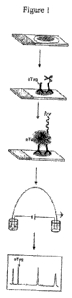
Figure 1 shows an outline of an FFPE VERATAG assay in accordance with an
embodiment of the present invention.
Figure 2A and Figure 2B show an VERATAG reaction where diffusing reactive
singlet oxygen cleaves the covalent linker between an VERATAG reporter
molecule and an
antibody in accordance with alternate embodiments of the present invention.
-8-
CA 02749817 2011-07-14
WO 2010/083463 PCT/US2010/021272
Figure 3 shows representative electropherograms of the VERATAG signal
generated for four well characterized breast cancer cell lines along with a
parallel Her-2 IHC
micrograph. The left side of the graph indicates the cell line, the middle
shows the
corresponding electropherogram and the right side shows the corresponding IHC
in
accordance with an embodiment of the present invention.
Figure 4 shows the time to progression (TTP) for all HER2 positive patients in
accordance with an embodiment of the present invention.
Figure 5 shows the TTP for patients that are HER2 negative or positive
determined by
FISH detection of Her2 gene amplification in accordance with an embodiment of
the present
invention.
Figure 6 shows the TTP for patients that are low HER2 expressors that do not
respond to treatment with trastuzumab regardless of categorization by FISH
measurement of
gene amplification in accordance with an embodiment of the present invention.
Figure 7 shows in accordance with an embodiment of the present invention the
TTP
for patients that are very high HER2 expressors (H2T > 1.84) and/or low HER2
expressors
(H2T < 1.14 regardless of FISH) do not respond to treatment with trastuzumab
as well as
patients having moderately high levels (H2T between 1.14 and 1.84) of HER2
expression.
Figure 8 shows in accordance with an embodiment of the present invention the
TTP
for patients that have moderately high amounts of HER2 and that are positive
as determined
by FISH, can be further stratified by HER3 over-expression (H3Thi) or under-
expression
(H3Tlo) wherein HER3 under-expression is associated with increased
responsiveness to
treatment with trastuzumab as compared to HER3 over-expression.
Figure 9 shows a study of primary early stage (i.e., adjuvant) Her-2 positive
samples
in accordance with an embodiment of the present invention, where Panel B shows
that CISH
positive, very high VERATAG H2T patients having a cut-off of H2T > 2.21 show
little
benefit from adding trastuzumab to chemotherapy as compared to samples with
Her-2 that is
not very high as shown in Panel A.
Detailed Description of the Invention
As used herein, the terms "embodiment" and "aspect" are used interchangeably.
The term "about," as used herein, unless otherwise indicated, refers to a
value that is
no more than 10% above or below the value being modified by the term. For
example, the
term "about 5 g/kg" means a range of from 4.5 gg/kg to 5.5 g/kg. As another
example,
"about 1 hour" means a range of from 48 minutes to 72 minutes.
-9-
CA 02749817 2011-07-14
WO 2010/083463 PCT/US2010/021272
"Antibody" means an immunoglobulin that specifically binds to, and is thereby
defined as complementary with, a particular spatial and polar organization of
another
molecule. The antibody can be monoclonal, polyclonal, or recombinant and can
be prepared
by techniques that are well known in the art such as immunization of a host
and collection of
sera (polyclonal) or by preparing continuous hybrid cell lines and collecting
the secreted
protein (monoclonal), or by cloning and expressing nucleotide sequences or
mutagenized
versions thereof coding at least for the amino acid sequences required for
specific binding of
natural antibodies. Antibodies may include a complete immunoglobulin or
fragment thereof,
which immunoglobulins include the various classes and isotypes, such as IgA,
IgD, IgE,
IgGI, IgG2a, IgG2b and IgG3, IgM, etc. Fragments thereof may include Fab, Fv
and F(ab')2,
Fab', and the like. Antibodies may also be single-chain antibodies or an
antigen-binding
fragment thereof, chimeric antibodies, humanized antibodies or any other
antibody derivative
known to one of skill in the art that retains binding activity that is
specific for a particular
binding site. In addition, aggregates, polymers and conjugates of
immunoglobulins or their
fragments can be used where appropriate so long as binding affinity for a
particular binding
site is maintained. Guidance in the production and selection of antibodies and
antibody
derivatives for use in immunoassays, including such assays employing
releasable molecular
tag (as described below) can be found in readily available texts and manuals,
e.g., Harlow
and Lane, 1988, Antibodies: A Laboratory Manual, Cold Spring Harbor Laboratory
Press,
New York; Howard and Bethell, 2001, Basic Methods in Antibody Production and
Characterization, CRC Press; Wild, ed., 1994, The Immunoassay Handbook,
Stockton Press,
New York.
"Antibody binding composition" means a molecule or a complex of molecules that
comprises one or more antibodies, or antigen-binding fragments that bind to a
molecule, and
derives its binding specificity from such antibody or antibody-binding
fragment. Antibody
binding compositions include, but are not limited to, (i) antibody pairs in
which a first
antibody binds specifically to a target molecule and a second antibody binds
specifically to a
constant region of the first antibody; a biotinylated antibody that binds
specifically to a target
molecule and a streptavidin protein, which protein is derivatized with
moieties such as
molecular tags or photosensitizers or the like, via a biotin moiety; (ii)
antibodies specific for a
target molecule and conjugated to a polymer, such as dextran, which, in turn,
is derivatized
with moieties such as molecular tags or photosensitizers, either directly by
covalent bonds or
indirectly via streptavidin-biotin linkages; (iii) antibodies specific for a
target molecule and
conjugated to a bead, or microbead, or other solid phase support, which, in
turn, is derivatized
-10-
CA 02749817 2011-07-14
WO 2010/083463 PCT/US2010/021272
either directly or indirectly with moieties such as molecular tags or
photosensitizers, or
polymers containing the latter.
"Antigenic determinant," or "epitope" means a site on the surface of a
molecule,
usually a protein, to which a single antibody molecule binds. Generally, a
protein has several
or many different antigenic determinants and reacts with antibodies of
different specificities.
A preferred antigenic determinant is a phosphorylation site of a protein.
"Binding compound" shall refer to an antibody binding composition, an
antibody, a
peptide, a peptide or non-peptide ligand for a cell surface receptor, a
protein, an
oligonucleotide, an oligonucleotide analog, such as a peptide nucleic acid, a
lectin, or any
other molecular entity that is capable of specifically binding to a target
protein or molecule or
stable complex formation with an analyte of interest, such as a complex of
proteins. In one
aspect, a binding compound, which can be represented by the formula below,
comprises one
or more molecular tags attached to a binding moiety.
"Binding moiety" means any molecule to which molecular tags can be directly or
indirectly attached that is capable of specifically binding to an analyte.
Binding moieties
include, but are not limited to, antibodies, antibody binding compositions,
peptides, proteins,
nucleic acids and organic molecules having a molecular weight of up to about
1000 daltons
and containing atoms selected from the group consisting of hydrogen, carbon,
oxygen,
nitrogen, sulfur and phosphorus. Preferably, binding moieties are antibodies
or antibody
binding compositions.
"Cancer" and "cancerous" refer to or describe the physiological condition
organism,
including mammals, that is typically characterized by unregulated cell growth.
Examples of
cancer include, but are not limited to, carcinoma, lymphoma, blastoma, sarcoma
and
leukemia. More particular examples of such cancers include squamous cell
cancer, lung
cancer, e.g., small-cell lung cancer or non-small cell lung cancer;
gastrointestinal cancer,
pancreatic cancer, glioblastoma, cervical cancer, ovarian cancer, liver
cancer, bladder cancer,
hepatoma, breast cancer, colon cancer, colorectal cancer, endometrial
carcinoma, salivary
gland carcinoma, kidney cancer, prostate cancer, vulval cancer, thyroid
cancer, hepatic
carcinoma and various types of head and neck cancer.
"Chemotherapeutic agent" means a chemical substance, primarily a cytotoxic or
cytostatic agent, that is used to treat a condition, particularly cancer.
Chemotherapeutic
agents shall include such compounds as paclitaxel, as set forth herein.
-11-
CA 02749817 2011-07-14
WO 2010/083463 PCT/US2010/021272
A "cleavable linkage," as used herein, refers to a chemical linking group that
may be
cleaved under conditions that do not degrade the structure or affect detection
characteristics
of a molecular tag connected to a binding moiety with the cleavable linkage.
A "cleavage-inducing moiety," or "cleaving agent," as used herein, is a group
that
produces an active species that is capable of cleaving a cleavable linkage,
preferably by
oxidation. Preferably, the active species is a chemical species that exhibits
short-lived activity
so that its cleavage-inducing effects are only in the proximity of the site of
its generation.
A "cleaving probe," as used herein, refers to a reagent that comprises a
cleavage-
inducing moiety as defined herein and an antibody binding composition, an
antibody, a
peptide, a peptide or non-peptide ligand for a cell surface receptor, a
protein, such as biotin or
streptavidin, an oligonucleotide, an oligonucleotide analog, such as a peptide
nucleic acid, a
lectin or any other molecular entity that is capable of specifically binding
to a target protein
or molecule or stable complex formation with an analyte of interest, such as a
complex of
proteins.
"VERATAG " "VERATAG " and "VERATAG assay" are used interchangeably
and refer to single and multiplexed and multi-label assays, materials, methods
and techniques
for performing and utilizing such assays, including but not limited to
reagents, analytical
procedures and software related to those assays. Such assays are disclosed in
this application
as well as in United States Patent 7,105,308, which is incorporated by
reference herein
including any drawings.
"FFPE" shall refer to a group of cells or quantity of tissue that are fixed,
particularly
conventional formalin-fixed paraffin-embedded samples. Such samples are
typically, without
limitation, used in an assay for receptor complexes in the form of thin
sections, e.g. 3-10 gm
thick, of fixed tissue mounted on a microscope slide or equivalent surface.
Such samples also
typically undergo a conventional re-hydration procedure, and optionally, an
antigen retrieval
procedure as a part of, or preliminary to, assay measurements.
"Hazard ratio", as used herein, refers to a statistical method used to
generate an
estimate for relative risk. "Hazard ratio" is the ratio between the predicted
hazard of one
group versus another group. For example, patient populations treated with
versus without a
Her-2 acting agent can be assessed for whether or not the Her-2 acting agent
is effective in
increasing the time to distant recurrence of disease. The hazards ratio can
then be compared
to an independent measure, such as the ratio of Her-2 homodimer to total Her-
2. At Her-2
homodimer to total Her-2 ratios at which the hazards ratio is less than one,
treating with a
Her-2 acting agent has a greater chance of efficacy. At Her-2 homodimer to
total Her-2 ratios
-12-
CA 02749817 2011-07-14
WO 2010/083463 PCT/US2010/021272
at which the hazards ratio is indistinguishable from one, treating with a Her-
2 acting agent
has a lower chance of efficacy.
"Her-2" ` ErbB2" "c-Erb-B2" "HER2" "Her2" and "neu" are used interchangeably
herein and refer to native Her-2, and allelic variants thereof, as described,
for example, in
Semba et al., 1985, P.N.A.S. USA 82:6497-650 and Yamamoto et al., 1986, Nature
319:230-
234 and Genebank accession number X03363. Unless indicated otherwise, the
terms "Her-2",
"ErbB2", "c-Erb-B2", "HER2" and "Her2" when used herein refer to the human
protein. The
gene encoding Her2 is referred to herein as "erb132." As used herein, H2T
shall refer to total
Her-2 expression as shown, for example without limitation, by VERATAG .
"Her-2-acting agent," as used herein, refers to a compound that can inhibit a
biological activity of Her-2 or a Her-2 expressing cell or a Her-2 positive
cancer cell. Such
biological activities include, but are not limited to, dimerization,
autophosphorylation,
phosphorylation of another receptor, signal transduction and the like.
Biological activities can
include, without limitation, cell survival and cell proliferation, and
inhibition of such
activities by a Her-2 acting agent could be direct or indirect cell killing
(eg, ADCC),
disruption of protein complexes or complex formation, modulation of protein
trafficking or
enzyme inhibition. Biological activities can also include patient response as
set forth in this
application. Exemplary Her-2-acting agents include, but are not limited to,
the large
molecules 4D5 and trastuzumab and small molecules such as AEE-788 and
lapatinib. "Her-2
homodimer" in reference to cell surface Her-2 membrane receptors means a
complex of two
or more membrane-bound Her-2 proteins. Dimers usually consist of two receptors
in contact
with one another. Dimers may be created in a cell surface membrane by passive
processes,
such as Van der Waal interactions, and the like, or dimers may be created by
active
processes, such as by ligand-induced dimerization, covalent linkages,
interaction with
intracellular components or the like. See, e.g., Schlessinger, 2000, Cell
103:211-225. As used
herein, the term "dimer" is understood to refer to "cell surface membrane
receptor dimer,"
unless understood otherwise from the context. As used herein, H22D shall refer
to quantified
dimer as shown, for example without limitation, by VERATAG .
"Her-2 homodimer to total Her-2 ratio" refers to a measure that describes the
amount
of Her-2 homodimers divided by the total amount of Her-2 in a sample from a
subject's tissue
according to any single quantitative method available to one skilled in the
art.
A "Her-2 positive" cancer, cancer cell, subject or patient, as used herein,
refers to a
cancer, cell subject or patient exhibiting a score of at least 2 when using a
HercepTest
(DakoCytomation California Inc., Carpenteria, CA) or a cancer, cancer cell,
subject or patient
-13-
CA 02749817 2011-07-14
WO 2010/083463 PCT/US2010/021272
that has been identified as such by FISH. In certain embodiments, the Her-2
positive cell
exhibits a score of at least 2+ or 3+ using HercepTest .
"High" refers to a measure that is greater than normal, greater than a
standard such as
a predetermined measure or a subgroup measure or that is relatively greater
than another
subgroup measure. For example, high Her-2 refers to a measure of Her-2 that is
greater than a
normal Her-2 measure. A normal Her-2 measure may be determined according to
any method
available to one skilled in the art. High Her-2 may also refer to a measure
that is equal to or
greater than a predetermined measure, such as a predetermined cutoff. High Her-
2 may also
refer to a measure of Her-2 wherein a high Her-2 subgroup has relatively
greater levels of
Her-2 than another subgroup. For example, without limitation, according to the
present
specification, two distinct patient subgroups can be created by dividing
samples around a
mathematically determined point, such as, without limitation, a median, thus
creating a
subgroup whose measure is high (ie, higher than the median) and another
subgroup whose
measure is low. Her-2 can be measured by any method known to one skilled in
the art such
as, for example, without limitation, using VERATAG or using any standard
immunohistochemical (IHC) method such as HercepTest . As another example, high
Her-2
homodimers refers to a measure of Her-2 homodimers that is greater than a
normal measure
of Her-2 homodimers in a particular set of samples or patients that are Her-2
positive. A
normal Her-2 homodimer measure may be determined according to any method
available to
one skilled in the art. High Her-2 homodimers may also refer to a measure that
is greater than
a predetermined measure, such as a predetermined cutoff. High Her-2 homodimers
may also
refer to a measure of Her-2 homodimers wherein a high Her-2 homodimer subgroup
has a
relatively higher level of Her-2 homodimers than another subgroup. Her-2
homodimers can
be measured by any method known in the art such as Fluorescence resonance
energy transfer
(FRET), Biolumenescent resonance energy transfer (BRUT), proximity ligation
assay (PLA),
dimer-specific antibodies or VERATAG or any other method that is well known
to one
skilled in the art. As another example, high Her-2 homodimer to total Her-2
ratio may refer to
the one or more subgroups of Her-2 homodimer to total Her-2 ratios that have
measures
greater than either intermediate or low ratio subgroups. High Her-2 homodimer
to total Her-2
ratios may be determined according to any individual quanitative method
available to one
skilled in the art. In some cases, a "high" expression level may comprise a
range of
expression that is very high and a range of expression that is "moderately
high" where
moderately high is a level of expression that is greater than normal, but less
than "very high".
-14-
CA 02749817 2011-07-14
WO 2010/083463 PCT/US2010/021272
Example ranges for high (including very high and moderately high) Her-2
expression are
provided herein.
"Moderately high" "medium" or "intermediate", as used herein, refers to a
measure
that is greater than "low" and less than very "high". For example,
"intermediate" may be used
to describe one or more of the at least 3 subgroups that fall in the middle
range of measures
of Her-2 homodimer to total Her-2 ratios.
"Likely to," as used herein, refers to an increased probability that an item,
object,
thing or person will occur. Thus, in one example, a subject that is likely to
respond to
treatment with trastuzumab has an increased probability of responding to
treatment with
trastuzumab relative to a reference subject or group of subjects.
"Long," as used herein, refers to a time measure that is greater than normal,
greater
than a standard such as a predetermined measure or a subgroup measure that is
relatively
longer than another subgroup measure. For example, with respect to a patient's
longevity, a
long time progression refers to time progression that is longer than a normal
time
progression. Whether a time progression is long or not may be determined
according to any
method available to one skilled in the art. Long could include, for example,
no progression.
In one embodiment, "long" refers to a time that is greater than the median
time course
required for a significant event to occur in a disease.
"Low" is a term that refers to a measure that is less than normal, less than a
standard
such as a predetermined measure or a subgroup measure that is relatively less
than another
subgroup measure. For example, low Her-2 means a measure of Her-2 that is less
than a
normal Her-2 measure in a particular set of samples of patients that is Her-2
positive. A
normal Her-2 measure may be determined according to any method available to
one skilled in
the art. Low Her-2 may also mean a method that is less than a predetermined
measure, such
as a predetermined cutoff. Low Her-2 may also mean a measure wherein a low Her-
2
subgroup is relatively lower than another subgroup. For example, without
limitation,
according to the present specification, two distinct patient subgroups can be
created by
dividing samples around a mathematically determined point, such as, without
limitation, a
median, thus creating a group whose measure is low (ie, less than the median)
with respect to
another group whose measure is high. Her-2 can be measured by any method known
to one
skilled in the art such as, for example, without limitation, using the VERATAG
method or
using any standard immunohistochemical (IHC) method such as HercepTest . As
another
example, low Her-2 homodimers means a measure of Her-2 homodimers that is less
than a
normal measure of Her-2 homodimers in a particular set of samples or patients
that is Her-2
-15-
CA 02749817 2011-07-14
WO 2010/083463 PCT/US2010/021272
positive. Low Her-2 homodimers may also mean a measure that is less than a
predetermined
measure, such as a predetermined cutoff. Low Her-2 homodimers may also mean a
measure
wherein a low Her-2 homodimer subgroup is relatively less than another
subgroup. Her-2
homodimers can be measured by any method known in the art such as Fluorescence
resonance energy transfer (FRET), Biolumenescent resonance energy transfer
(BRET),
proximity ligation assay (PLA), dimer-specific antibodies or VERATAG or any
other
method that is well known to one skilled in the art. As another example, low
Her-2
homodimer to total Her-2 ratio may refer to the one or more subgroups of Her-2
homodimer
to total Her-2 ratios that have measures less than either intermediate or high
ratio subgroups.
Low Her-2 homodimer to total Her-2 ratios may be determined according to any
individual
quanitative method available to one skilled in the art. Example ranges for low
values of Her-2
expression are provided herein.
A "molecular tag," as used herein, refers to a molecule that can be
distinguished from
other molecules based on one or more physical, chemical or optical differences
among the
molecules being separated, including but not limited to, electrophoretic
mobility, molecular
weight, shape, solubility, pKa, hydrophobicity, charge, charge/mass ratio,
polarity or the like.
In one aspect, molecular tags in a plurality or set differ in electrophoretic
mobility and optical
detection characteristics and can be separated by electrophoresis. In another
aspect, molecular
tags in a plurality or set may differ in molecular weight, shape, solubility,
pKa,
hydrophobicity, charge, polarity and can be separated by normal phase or
reverse phase
HPLC, ion exchange HPLC, capillary electrochromatography, mass spectroscopy,
gas phase
chromatography or like technique.
"Optimal cutoff `as used herein, refers to the value of a predetermined
measure on
subjects exhibiting certain attributes that allow the best discrimination
between two
categories of an attribute. For example, finding a value for an optimal cutoff
that allows one
to best discriminate between two categories, high H2T expression and low H2T
expression,
for determining OS. Optimal cutoffs are used to separate the subjects with
values lower than
or higher than the optimal cutoff to optimize the prediction model, for
example, without
limitation, to maximize the specificity of the model, maximize the sensitivity
of the model,
maximize the difference in outcome, or minimize the p-value from hazard ratio
or a
difference in response.
"Overall survival" or "OS" refers to a time as measured from the start of
treatment to
death or censor. Censoring may come from a study end or change in treatment.
Overall
survival can refer to a probability as, for example, a probability when
represented in a
-16-
CA 02749817 2011-07-14
WO 2010/083463 PCT/US2010/021272
Kaplan-Meier plot of being alive at a particular time, that time being the
time between the
start of the treatment to death or censor.
"Photosensitizer" shall mean a light-adsorbing molecule that when activated by
light
converts molecular oxygen into singlet oxygen.
"RECIST" shall mean an acronym that stands for "Response Evaluation Criteria
in
Solid Tumours" and is a set of published rules that define when cancer
patients improve
("respond"), stay the same ("stable") or worsen ("progression") during
treatments. Response
as defined by RECIST criteria have been published, for example, at Journal of
the National
Cancer Institute, Vol. 92, No. 3, February 2, 2000 and RECIST criteria may
include other
similar published definitions and rule sets. One skilled in the art would
understand definitions
that go with RECIST criteria, as used herein, such as "PR," "CR," "SD" and
"PD."
"Relative fluorescence units" or "RFUs" are used interchangeably and shall
refer to
the time integral of a particular capillary electrophoresis peak using
arbitrary fluorescence
units in comparison to a standard. With respect to VERATAG , the RFU is
proportional to
the concentration of VERATAG injected into capillary electrophoresis with
some expected
variability introduced by, for example, injection and capillary differences.
"Relative peak area" or "RPA" are used interchangeably and shall refer to the
ratio
between an RFU of a particular VERATAG and an RFU of a known internal
fluorescence
standard of known and constant concentration.
"Responsiveness," to "respond" to treatment, and other forms of this verb, as
used
herein, refer to the reaction of a subject to treatment with a Her-2-acting
agent. As an
example, a subject responds to treatment with a Her2-acting agent if growth of
a tumor in the
subject is retarded about 10%, 20%, 30%, 40%, 50%, 60%, 70%, 80%, 90% or more.
In
another example, a subject responds to treatment with a Her-2-acting agent if
a tumor in the
subject shrinks by about 5%, 10%, 20%, 30%, 40%, 50% or more as determined by
any
appropriate measure, e.g., by mass or volume. In another example, a subject
responds to
treatment with a Her2-acting agent if the subject experiences a life
expectancy extended by
about 5%, 10%, 20%, 30%, 40%, 50% or more beyond the life expectancy predicted
if no
treatment is administered. In another example, a subject responds to treatment
with a Her-2-
acting agent if the subject has an increased disease-free survival, overall
survival or increased
time to progression. Several methods may be used to determine if a patient
responds to a
treatment including the RECIST criteria, as set forth above.
"Sample" or "tissue sample" or "patient sample" or "patient cell or tissue
sample" or
"specimen" each refer to a collection of similar cells obtained from a tissue
of a subject or
-17-
CA 02749817 2011-07-14
WO 2010/083463 PCT/US2010/021272
patient. The source of the tissue sample may be solid tissue as from a fresh,
frozen and/or
preserved organ or tissue sample or biopsy or aspirate; blood or any blood
constituents;
bodily fluids such as cerebral spinal fluid, amniotic fluid, peritoneal fluid
or interstitial fluid;
or cells from any time in gestation or development of the subject. The tissue
sample may
contain compounds that are not naturally intermixed with the tissue in nature
such as
preservatives, anticoagulants, buffers, fixatives, nutrients, antibiotics or
the like. Cells may be
fixed in a conventional manner, such as in an FFPE manner.
"Short," as used herein, refers to a time measure that is shorter than normal,
shorter
than a standard such as a predetermined measure or a subgroup measure that is
relatively
shorter than another subgroup measure. For example, with respect to a
patient's longevity, a
short time progression refers to time progression that is shorter than a
normal time
progression. Whether a time progression is short or not may be determined
according to any
method available to one skilled in the art. In one embodiment, "short" refers
to a time that is
less than the median time course required for a significant event to occur in
a disease.
As used herein, "significant event" shall refer to an event in a patient's
disease that is
important as determined by one skilled in the art. Examples of significant
events include, for
example, without limitation, primary diagnosis, death, recurrence, the
determination that a
patient's disease is metastatic, relapse of a patient's disease or the
progression of a patient's
disease from any one of the above noted stages to another. A significant event
may be any
important event used to assess OS, TTP and/or using the RECIST or other
response criteria,
as determined by one skilled in the art.
As used herein, the terms "subject" and "patient" are used interchangeably. As
used
herein, the terms "subject" and "subjects" refer to an animal, preferably a
mammal including
a non-primate (e.g., a cow, pig, horse, donkey, goat, camel, cat, dog, guinea
pig, rat, mouse,
sheep) and a primate (e.g., a monkey, such as a cynomolgous monkey, gorilla,
chimpanzee
and a human).
As used herein, "time course" shall refer to the amount of time between an
initial
event and a subsequent event. For example, with respect to a patient's cancer,
time course
may relate to a patient's disease and may be measured by gauging significant
events in the
course of the disease, wherein the first event may be diagnosis and the
subsequent event may
be metastasis, for example.
"Time to progression" or "TTP" refers to a time as measured from the start of
the
treatment to progression or a cancer or censor. Censoring may come from a
study end or from
a change in treatment. Time to progression can also be represented as a
probability as, for
-18-
CA 02749817 2011-07-14
WO 2010/083463 PCT/US2010/021272
example, in a Kaplein-Meier plot where time to progression may represent the
probability of
being progression free over a particular time, that time being the time
between the start of the
treatment to progression or censor.
"Treat," "treatment," and other forms of this word refer to the administration
of a
Her-2-acting agent to impede growth of a cancer, to cause a cancer to shrink
by weight or
volume, to extend the expected survival time of the subject and or time to
progression of the
tumor or the like.
"Unlikely to" refers to a decreased probability that an event, item, object,
thing or
person will occur with respect to a reference. Thus, a subject that is
unlikely to respond to
treatment with paclitaxel in addition to trastuzumab has a decreased
probability of responding
to treatment with paclitaxel and trastuzumab relative to a reference subject
or group of
subjects.
Thus, embodiments of the invention provide methods for determining whether a
subject with a cancer is likely to respond to treatment with a Her-2-acting
agent and/or for
predicting a time course of disease and/or a probability of a significant
event in the time
course of disease in a subject with a cancer. In certain embodiments, the
method comprises
detecting a biomarker or combination of biomarkers associated with
responsiveness to
treatment with a Her-2-acting agent as described herein, and determining
whether the subject
is likely to respond to treatment with the Her2-acting agent. In certain
embodiments, the
methods comprise detecting a biomarker or combination of biomarkers and
predicting a time
course associated with progression of disease or a probability of a
significant event in the
time course of disease in a subject with cancer.
In one aspect, the invention is drawn to a method for determining whether a
subject
with a cancer is likely to respond to treatment with a Her-2-acting agent. In
another aspect,
the invention is drawn to a method for predicting a time course of disease. In
another aspect,
the method is drawn to a method for predicting a probability of a significant
event in the time
course of the disease.
In certain embodiments, a time course is measured by determining the time
between
significant events in the course of a patient's disease, wherein the
measurement is predictive
of whether a patient has a long time course. For example, in a preferred
embodiment, the
significant event is the progression from primary diagnosis to death. In a
preferred
embodiment, the significant event is the progression from primary diagnosis to
metastatic
disease. In a preferred embodiment, the significant event is the progression
from primary
diagnosis to relapse. In a preferred embodiment, the significant event is the
progression from
-19-
CA 02749817 2011-07-14
WO 2010/083463 PCT/US2010/021272
metastatic disease to death. In a preferred embodiment, the significant event
is the
progression from metastatic disease to relapse. In a preferred embodiment, the
significant
event is the progression from relapse to death. In certain embodiments, the
time course is
measured with respect to overall survival rate, time to progression and/or
using the RECIST
or other response criteria.
In certain embodiments, the method comprises detecting in a biological sample
from
the subject's cancer the amount of Her-2 and/or Her-2 homodimers wherein if
the amount of
Her-2 and/or Her-2 homodimers is high, then the patient is likely to respond
to the Her-2
acting agent and/or the patient has a long time course.
Thus, in certain embodiments, the invention comprises methods to correlate the
relative levels of the amount of at least one of Her-2 or Her-2 homodimers in
a biological
sample from a subject with a prognosis for the likelihood that the subject
will respond to
treatment with a Her-2 acting agent comprising: (a) detecting in a biological
sample from the
subject's cancer the amount of at least one of Her-2 or Her-2 homodimers; and
(b) correlating
the amount of Her-2 or Her-2 homodimers to a prognosis for the likelihood that
the subject
will respond to treatment with a Her-2 acting agent.
In certain embodidments, if the amount of the at least one of the Her-2 or Her-
2
homodimers is equal to or above a first threshold level, the subject's
prognosis is to be likely
to respond to the Her-2 acting agent. Additionally and/or alternatively, if
the amount of the at
least one of the Her-2 or Her-2 homodimers is equal to or above a second
threshold level
higher than the first threshold level, the subject's prognosis is to be
unlikely to respond to the
Her-2 acting agent
Also, in certain embodiments, a predetermined measure is created by dividing a
plurality of subject samples into at least three subgroups, wherein the first
subgroup
comprises samples having the Her-2 or Her-2 homodimers at a low level, wherein
the low
level comprises having an amount of the at least one of the Her-2 or Her-2
homodimers is
equal to or below a first threshold level, and the samples having an amount of
the at least one
of the Her-2 or Her-2 homodimers equal to or above the first threshold
comprise samples
having a high level of the Her-2 or Her-2 homodimers, and wherein the samples
having a
high level of the Her-2 or Her-2 homodimers is then divided into two
subgroups, a very high
subgroup comprising samples having an amount of the at least one of the Her-2
or Her-2
homodimers equal to or above a second threshold level higher than the first
threshold level,
and a moderately high subgroup comprising samples having an amount of the at
least one of
-20-
CA 02749817 2011-07-14
WO 2010/083463 PCT/US2010/021272
the Her-2 or Her-2 homodimers greater than or equal to the first threshold
level and less than
or equal to the second threshold level.
As discussed herein, in certain embodiments, the amount of Her-2 homodimers is
measured using an assay capable of measuring and/or quantifying an amount of
protein-
protein interactions in a sample.
In certain embodiments of each of the methods of the present invention, high
Her-2 expression is a log10H2T > about 1.14-1.125. In certain embodiments of
each of
the methods disclosed herein, the high Her-2 expression comprises expression
that is
very high and/or moderately high. In certain embodiments of each of the
methods
disclosed herein, the very high Her-2 expression is a loglOH2T > about 1.84 -
2.21. In
certain embodiments of each of the methods disclosed herein, the moderately
high
expression is between 1.14 - 1.25 and 1.84-2.21. Or, other ranges (i.e., up to
about 25%
more or less as described herein) may be used depending upon the patient
cohort and/or
the significant event being monitored.
In certain embodiments, the cancer is breast cancer. In some embodiments, the
breast
cancer is metastatic. In some embodiments, the breast cancer is early stage
(i.e., adjuvant)
breast cancer. Or, any cancer that may be sensistive to a Her-2 acting agent
may be
monitored. The Her-2 acting agent may be any Her-2 acting agent. In certain
embodiments,
the Her-2 acting agent is one of the agents described herein. For example, in
certain
embodiments, the Her-2 acting agent is trastuzumab.
In certain embodiments of each of the methods of the invention, the moderately
high
Her-2 subgroup is further subdivided into subgroups that express at least one
other
biomarker. For example, in some embodiments, a predetermined measure is
generated by
dividing the Her-2 medium and/or Her-2 high samples into at least two
subgroups, based on
the level of the at least one other biomarker
The at least one other biomarker may, in alternate embodiments, comprise at
least one
of FOXM1, PRAME, Bc12, STK15, CEGP1, Ki-67, GSTM1, CA9, PR, BBC3, NME1,
SURV, GATA3, TFRC, YB-l, DPYD, GSTM3, RPS6KB1, Src, Chkl, IDI, EstRi, p27,
CCNB1, XIAP, Chk2, CDC25B, IGF1R, AK055699, P13KC2A, TGFB3, BAGI1, CYP3A4,
EpCAM, VEGFC, pS2, hENTI, WISP1, HNF3A, NFKBp65, BRCA2, EGFR, TK1, VDR,
Contig51037, pENTI, EPHX1, IF1A, CDH1, HIFIa, IGFBP3; CTSB, Her3, DIABLO,
VEGF, CD31, KDR, or p95.
For example, in certain embodiments, the moderately high subgroup is further
divided based upon Her-3 expression wherein a high level comprises having Her-
3 equal to
-21-
CA 02749817 2011-07-14
WO 2010/083463 PCT/US2010/021272
or above a first threshold level and a low level comprises having Her-3 below
the first
threshold level, and wherein a subject with moderately high levels of the at
least one Her-2
and/or Her-2 dimers and low levels of Her-3 is likely to respond to the Her-2
acting agent
and/or wherein a subject with moderately high levels of the at least one Her-2
and/or Her-2
dimers and high levels of Her-3 is unlikely or less likely to respond to the
Her-2 acting
agent.. Where Her-3 is measured, the Her-3 expression comprises Her-3, Her-3
homodimers,
or Her-3 heterodimers (e.g., Her-2/Her-3).
In certain embodiments, the assay is performed with an VERATAG assay. In
certain embodiments, likeliness to respond is measured with respect to overall
survival rate,
time to progression and/or using the RECIST or other response criteria.
In certain embodiments, the method comprises detecting in a biological sample
from
the subject's cancer the amount of Her-2 and/or Her-2 homodimers wherein if
the amount of
Her-2 and/or Her-2 homodimers is moderately high (e.g., medium), then the
patient group
may be further sub-divided into high Her-3 expressors and low Her-3
expressors. In this
embodiment, the patient with moderately high Her-2 and/or Her-2 homodimers and
low Her-
3 is likely to respond to the Her-2 acting agent and/or the patient has a long
time course. In
alternate embodiments, the Her-3 expressors can be expression of Her-3, Her-3
homodimers,
or Her-2/Her3 heterodimers.
In certain embodiments of measuring both Her2 and Her3, the cancer is breast
cancer.
In some embodiments, the breast cancer is metastatic. In other embodiments,
the breast
cancer is early stage (i.e., adjuvant) breast cancer. Or, other Her-2
sensitive cancers may be
monitored. The Her-2 acting agent may be any Her-2 acting agent. In certain
embodiments,
the Her-2 acting agent is one of the agents described herein. For example, iln
certain
embodiments, the Her-2 acting agent is trastuzumab. In certain embodiments,
the assay is
performed with an VERATAG assay. In certain embodiments, likeliness to
respond is
measured with respect to overall survival rate, time to progression and/or
using the RECIST
or other response criteria.
In certain embodiments, a predetermined measure is created by dividing patient
samples into at least two patient subgroups. In certain embodiments, the
number of subgroups
is three so that the patient sample is divided into a subgroup of patients
whose Her-2 and/or
Her-2 homodimers is very high, a subgroup whose Her-2 and/or Her-2 homodimers
is low,
and a subgroup whose Her-2 and/or Her-2 homodimers is moderately high (i.e.,
medium).
Each of the sub-groups can then be further subdivided into a subgroup whose
Her-3 is high or
medium.
-22-
CA 02749817 2011-07-14
WO 2010/083463 PCT/US2010/021272
The level of Her-2 homodimers may be tightly correlated to the total leverls
of Her-2.
Thus, in certain embodiments of each of the methods of the present invention,
the amount of
Her-2 and/or Her-2 homodimers in the subject are compared to at least one of
the very high
subgroup, the moderately high subgroup, or the low subgroup. If the amount of
Her-2 and/or
Her-2 homodimers in the patient are moderately high, and the Her-3 expressors
are low, then
the patient is likely to respond to a Her-2 acting agent and/or the patient is
likely to have a
long time course. If the amount of Her-2 and/or Her-2 homodimers in the
patient are
moderately high, and the Her-3 expressors are high, then the patient is
unlikely to respond to
a Her-2 acting agent and/or the patient is likely to have a short time course.
In certain
embodiments, the number of subgroups is greater than two, including, without
limitation,
three subgroups, four subgroups, five subgroups and six subgroups. In certain
embodiments,
likeliness to respond is measured with respect to overall survival rate, time
to progression
and/or using the RECIST criteria. In certain preferred embodiments, the Her-2
acting agent is
trastuzumab.
In certain embodiments, the predetermined measure is an optimal cutoff. Such
optimal cutoffs are disclosed herein, and certain embodiments of the invention
are meant to
include amounts that are approximate to the amounts mentioned and disclosed
herein. In
certain embodiments, the amount of Her-2 and/or Her-2 homodimers in the
subject are
compared to the optimal cutoff, if the amount of Her-2 and/or Her-2 homodimers
in the
patient are moderately high (e.g., between about 1.14-1.25 and 1.84-2.21 H2T),
then the
patient is likely to respond to a Her-2 acting agent and/or the patient's time
course is likely to
be long. In another embodiment, if the amount of Her-2 is high, then the
patient is likely to
respond to a Her-2 acting agent and/or the time course is likely to be long.
In another
embodiment, if the amount of Her-2 is high, and the amount of Her-2 homodimers
and/or the
ratio of Her-2 homodimers to Her-2 are low, then the patient is likely to
respond to a Her-2
acting agent and/or the time course is likely to be long. In another
embodiment, if the amount
of Her-2 is high and the amount of Her-2 dimers is high, then the patient is
likely to respond
to a Her-2 acting agent and/or the time course is likely to be long.
In another aspect, the invention is drawn to a method for determining whether
a
subject with a Her-2 positive cancer is likely to respond to treatment with a
Her-2-acting
agent and/or the time course of disease is long. In another aspect, the
invention is drawn to a
method for predicting a time course of disease in a subject with a Her-2
positive cancer. In
another aspect, the invention is drawn to a method for predicting the
probability of a
significant event in a subject with a Her-2 positive cancer.
-23-
CA 02749817 2011-07-14
WO 2010/083463 PCT/US2010/021272
For example, the invention may comprise methods for predicting whether a
subject
with a cancer and being treated with a Her-2 acting agent is likely to have a
significant event
comprising the steps of: (a) detecting in a biological sample from the
subject's cancer the
amount of at least one of Her-2 or Her-2 homodimers; and (b) correlating the
amount of Her-
2 or Her-2 homodimers to the likelihood that the subject will have a
significant event.
In an embodiment, the significant event is a reduced time between diagnosis
with the
cancer and at least one of primary diagnosis, progression of the cancer from
one stage to a
more advanced stage, progression to metastatic disease, relapse, surgery or
death. Also in
certain embodiments, the method may further comprise predicting a time course
during
which the significant event can occur.
In an embodiment, if the amount of the at least one of the Her-2 or Her-2
homodimers
is equal to or below a first threshold level, the significant event is that
the subject is less
likely to respond to the Her-2 acting agent. Additionally and/or
alternatively, if the amount of
the at least one of the Her-2 or Her-2 homodimers is equal to or above a
second threshold
level higher than the first threshold level, the subject's prognosis is to be
unlikely to respond
to the Her-2 acting agent.
In a preferred embodiment, a time course is measured by determining the time
between significant events in the course of a patient's disease, wherein the
measurement is
predictive of whether a patient has a long time course. In one embodiment, the
significant
event is the progression from primary diagnosis to death. In another
embodiment, the
significant event is the progression from primary diagnosis to metastatic
disease. In yet
another embodiment, the significant event is the progression from primary
diagnosis to
relapse. In another embodiment, the significant event is the progression from
metastatic
disease to death. In another embodiment, the significant event is the
progression from
metastatic disease to relapse. In another embodiment, the significant event is
the progression
from relapse to death. In certain embodiments, the time course is measured
with respect to
overall survival rate, time to progression and/or using the RECIST or other
response criteria.
In certain embodiments, the method comprises measuring in a biological sample
from
the subject's cancer an amount of Her-2 and/or Her-2 homodimers, wherein if
the amount of
Her-2 and/or Her-2 homodimers is high and/or moderately high, then the patient
is likely to
respond to the Her-2 acting agent and/or the patient has a long time course.
In certain
embodiments, the biological sample comprises FFPEs. In certain embodiments,
the subject's
cancer is breast cancer. In certain embodiments, the breast cancer is
metastatic. In other
embodimetns, the cancer is early stage (i.e., adjuvant) breast cancer. Or,
other cancers
-24-
CA 02749817 2011-07-14
WO 2010/083463 PCT/US2010/021272
sensitive to Her-2 aciting agents may be monitored. As noted herein, the Her-2
acting agent
may be one of the agents known. In certain embodiments, the Her-2-acting agent
is
trastuzumab. Or other Her-2 acting agents may be used.
In certain embodiments, an amount of Her-2 is measured. In certain
embodiments, an
amount of Her-2 homodimers is measured. In certain embodiments, the amount of
Her-2
homodimers is measured using an assay capable of measuring and/or quantifying
an amount
of protein-protein interactions in a sample. In certain embodiment, the assay
is the
VERATAG assay. In certain embodiments, likeliness to respond is measured with
respect
to overall survival rate, time to progression and/or using the RECIST
criteria.
In certain embodiments, the method comprises measuring in a biological sample
from
the subject's cancer an amount of Her-3 and/or Her-3 homodimers as well as the
amount of
Her-2 and/or Her-2 homodimers, wherein if the amount of Her-3 and/or Her-3
homodimers is
high, then the patient is likely to respond to the Her-2 acting agent and/or
the patient has a
long time course. In certain embodiments, the biological sample comprises
FFPEs. In certain
embodiments, the subject's cancer is breast cancer. In certain embodiments,
the breast cancer
is metastatic. Or the breast cancer may be an early stage (i.e., adjuvant)
breast cancer. Or,
other cancers sensitive to Her-2 aciting agents may be monitored. As noted
herein, the Her-2
acting agent may be one of the agents known and/or described herein. In
certain
embodiments, the Her-2-acting agent is trastuzumab.
In certain embodiments, an amount of Her-3 homodimers is measured. In certain
embodiments, the amount of Her-3 homodimers is measured using an assay capable
of
measuring and/or quantifying an amount of protein-protein interactions in a
sample. In
certain embodiment, the assay is the VERATAG assay. In certain embodiments,
likeliness
to respond is measured with respect to overall survival rate, time to
progression and/or using
the RECIST criteria.
In certain embodiments, the method comprises measuring in a biological sample
from
the subject's cancer an amount of Her-2 and/or Her-2 homodimers, wherein if
the amount of
Her-2 and/or Her-2 homodimers is moderately high (i.e., medium), then the
biological sample
is further analyzed for the amount of Her-3 expressors, wherein the Her-3
expressors can be
expression of Her-3, Her-3 homodimers, or Her-2/Her3 heterodimers. If the
amount of Her-2
and/or Her-2 homodimers in the patient is moderately high and the amount of
Her-3
expressors is high, then the patient is likely to respond to the Her-2 acting
agent and/or the
patient has a long time course. Conversely, if the amount of Her-2 and/or Her-
2 homodimers
in the patient is moderately high and the amount of Her-3 expressors is low,
then the patient
-25-
CA 02749817 2011-07-14
WO 2010/083463 PCT/US2010/021272
is unlikely to respond to the Her-2 acting agent and/or the patient has a
short time course. In
certain embodiments, the biological sample comprises FFPEs. In certain
embodiments, the
subject's cancer is breast cancer. In certain embodiments, the breast cancer
is metastatic. Or,
the breast cancer may be early stage (i.e., adjuvant) breast cancer. Or, any
other cancer
responsive to Her-2 acting agents may be monitored. As noted herein, the Her-2
acting agent
may be one of the agents known and/or described herein. In certain
embodiments, the Her-
2-acting agent is trastuzumab. In certain embodiment, the assay is the VERATAG
assay. In
certain embodiments, likeliness to respond is measured with respect to overall
survival rate,
time to progression and/or using the RECIST criteria.
In certain embodiments, a predetermined measure is created by dividing patient
samples into at least two patient subgroups. In certain embodiments, the
number of subgroups
is three so that the patient sample is divided into a subgroup of patients
whose Her-2 and/or
Her-2 homodimers is high, and a subgroup whose Her-2 and/or Her-2 homodimers
is low. In
certain embodiments, the high Her-2 subgroup is divided into a a subgroup
whose Her-2
and/or Her-2 homodimers is very high, and a subgroup whose Her-2 and/or Her-2
homodimers is moderately hight (i.e., medium). In an embodiment; the amount of
Her-2
and/or Her-2 homodimers in the subject are compared to either the very high
subgroup, the
moderately high subgroup, or the low subgroup. If the amount of Her-2 and/or
Her-2
homodimers in the patient are moderately high, then the patient is likely to
respond to a Her-2
acting agent and/or the patient is likely to have a long time course. If the
amount of Her-2
and/or Her-2 homodimers in the patient are very high or low, then the patient
is unlikely to
respond to a Her-2 acting agent and/or the patient is likely to have a short
time course.
In another embodiment, if the amount of Her-2 and/or Her-2 homodimers in the
patient is moderately high and the amount of Her-3 expressors is high, then
the patient is
unlikely to respond to the Her-2 acting agent and/or the patient has a short
time course. In
another embodiment, if the amount of Her-2 and/or Her-2 homodimers in the
patient is
moderately high and the amount of Her-3 expressors is low, then the patient is
likely to
respond to the Her-2 acting agent and/or the patient has a long time course.
In certain
embodiments, the number of subgroups is greater than three, including, without
limitation,
four subgroups, five subgroups and six subgroups. In certain embodiments,
likeliness to
respond or time course is measured with respect to overall survival rate, time
to progression
and/or using the RECIST criteria. In certain preferred embodiments, the Her-2
acting agent is
trastuzumab.
-26-
CA 02749817 2011-07-14
WO 2010/083463 PCT/US2010/021272
In certain embodiments, the predetermined measure is an optimal cutoff. Such
optimal cutoffs are disclosed herein, and certain embodiments of the invention
are meant to
include amounts that are approximate to the amounts mentioned and disclosed
herein. In
certain embodiments, the amount of Her-2 and/or Her-2 homodimers in the
subject are
compared to the optimal cutoff; if the amount of Her-2 and/or Her-2 homodimers
in the
patient are high or moderately high, then the patient is likely to respond to
a Her-2 acting
agent and/or the patient's time course is likely to be long. In another
embodiment, if the
amount of Her-2 is high, and the amount of Her-2 homodimers and/or the ratio
of Her-2
homodimers to Her-2 is low, then the patient is likely to respond to a Her-2
acting agent
and/or the time course is likely to be long. In another embodiment, if the
amount of Her-2 is
high and the amount of Her-2 homodimers and/or the ratios of Her-2 homodimers
to Her-2 is
high, then the patient is likely to respond to a Her-2 acting agent and/or the
time course is
likely to be long.
In another aspect, the invention provides a method for determining whether a
subject
with Her2-positive cancer is unlikely to respond to treatment with a Her2-
acting agent and/or
the patient is likely to have a short time course. In certain embodiments, the
method
comprises detecting in a biological sample from the subject's cancer the
amount of Her-2,
wherein if the amount of Her-2 is low, the subject is unlikely to respond to
treatment with the
Her2-acting agent and/or the patient is likely to have a short time course. In
certain preferred
embodiments, the Her2-acting agent is trastuzumab.
Any method known to one of skill in the art to be useful for determining an
amount of
Her-2 expression and/or Her-2 homodimers, and/or Her-3 expression and/or Her-3
homodimers, and/or Her-2/Her-3 heterodimers can be used in accordance with the
present
invention. For example, any quantitative assay that determines the amount of
such expression
or dimers can be used to determine how much signal is generated by a cell or
cancer, then the
signal compared to the signal generated in the VERATAG assay to determine a
correspondence between the two assays. Such methods may include, but not
necessarily be
limited to, FRET, BRET, Biomolecular Fluoresence Complementation and Proximity
Ligation Assay.
In certain embodiments, the amounts are determined by contacting a biological
sample from a subject with cancer with a binding compound having a molecular
tag attached
thereto by a cleavable linkage and a cleaving probe having a cleavage inducing-
moiety and
detecting whether and what molecular tag is released. Figure 1 provides an
outline of such an
FFPE VERATAG assay where tissue sections are fixed (top or first panel) and
then allowed
-27-
CA 02749817 2011-07-14
WO 2010/083463 PCT/US2010/021272
to bind to a first antibody having a cleavage-inducing agent (depicted as a
cutting tool) and a
second antibody linked to a detectable moiety (ETAG ) (second panel), photo-
induction of
the cleavage-inducing agent by light (hv) (third panel), electrophoretic
separation of the e-
Tag(s) (fourth panel) and a readout of the data (bottom or fifth panel).
In certain embodiments, the binding compound and the cleaving probe each
specifically binds Her-2 or Her-3. In certain embodiments, the cleaving probe
and the binding
probe do not both bind the same epitope. In certain embodiments, if the
binding compound is
within an effective proximity of the cleavage-inducing moiety of the cleaving
probe, the
cleavage-inducing moiety cleaves the cleavable linker so that the molecular
tag is released. In
certain embodiments, the molecular tag released if Her-2 homodimers, Her-3
homodimers, or
Her-2/Her-3 heterodimers are present is distinguishable from the molecular tag
released if
Her-2 monomers and/or Her-3 monomers are present. Examples of detection of Her-
2 by an
assay for detection of total Her-2 and/or Her-2 homodimers is provided in
commonly owned
U.S. Patent Application Publication No. 2009/0191559 incorporated by reference
in its
entirety herein. A similar strategy can be used to measure other biomarkers
such as Her-3, p-
95 and the like.
In certain embodiments, activating the cleavage-inducing moiety cleaves the
cleavable linker. In certain embodiments, the binding compound specifically
binds a Her-2 or
Her-3 epitope. In certain embodiments, the binding compound comprises an
antibody or
antigen-binding fragment. In certain embodiments, the binding compound
specifically binds a
Her-2 ligand binding site or a Her-3 ligand binding site. In certain
embodiments, the binding
compound comprises a Her-2 ligand and/or a Her-3 ligand. In certain
embodiments, the
binding compound and the cleaving probe bind the same Her-2 epitope and/or a
Her-3
epitope.
In certain embodiments, and as illustrated in Figure 2A showing measurement of
Her-
2 total using two different antibodies, one with a cleaving agent and one with
a tag, and
Figure 2B showing measurement of Her-2 dimers using a single antibody
alternately attached
to either a cleaving agent or a binding moiety, the step of measuring the
amounts of one or
more Her-2 homodimers, Her-3 homodimers, or Her-2/Her-3 heterodimers comprises
the
following steps: (i) providing for each of the one or more Her-2 homodimers a
cleaving probe
(e.g., antibody 15 in Figure 2A, and antibody 8 in Figure 2B) specific for a
first Her-2 protein
in each of the one Her-2 homodimers, each cleaving probe having a cleavage-
inducing
moiety with an effective proximity; (ii) providing one or more binding
compounds (e.g.,
antibody 8 in both Figures 2A and 2B) specific for a second protein of each of
the one or
-28-
CA 02749817 2011-07-14
WO 2010/083463 PCT/US2010/021272
more Her-2 homodimers, such that each binding compound has one or more
molecular tags
each attached thereto by a cleavable linkage, and such that the one or more
molecular tags
attached to different binding compounds have different separation
characteristics so that upon
separation molecular tags from different binding compounds form distinct peaks
in a
separation profile; (iii) mixing the cleaving probes, the binding compounds,
and the one or
more complexes such that cleaving probes specifically bind to first proteins
of the Her-2
homodimers and binding compounds specifically bind to the second proteins of
the Her-2
homodimers and such that cleavable linkages of the binding compounds are
within the
effective proximity of cleavage-inducing moieties of the cleaving probes so
that molecular
tags are released; and (iv) separating and identifying the released molecular
tags to determine
the presence or absence or the amount of the Her-2 homodimers.
In certain embodiments, the step of measuring the amounts of one or more Her-2
homodimers, Her-3 homodimers, or Her-2/Her-3 heterodimers comprises the
following steps:
(i) providing for each of the one or more Her-2 homodimers a cleaving probe
specific for a
first Her-2 protein in each of the one Her-2 homodimers, each cleaving probe
having a
cleavage-inducing moiety with an effective proximity; (ii) providing one or
more binding
compounds specific for a second protein of each of the one or more Her-3
homodimers, such
that each binding compound has one or more molecular tags each attached
thereto by a
cleavable linkage, and such that the one or more molecular tags attached to
different binding
compounds have different separation characteristics so that upon separation
molecular tags
from different binding compounds form distinct peaks in a separation profile;
(iii) mixing the
cleaving probes, the binding compounds, and the one or more complexes such
that cleaving
probes specifically bind to first proteins of the Her-3 homodimers and binding
compounds
specifically bind to the second proteins of the Her-3 homodimers and such that
cleavable
linkages of the binding compounds are within the effective proximity of
cleavage-inducing
moieties of the cleaving probes so that molecular tags are released; and (iv)
separating and
identifying the released molecular tags to determine the presence or absence
or the amount of
the Her-3 homodimers.
The Her-2/Her-3 heterodimers can be similarly determined using cleaving probes
and
binding probes specific to Her-2 and Her-3, e.g., a cleaving probe specific to
Her-2 and a
binding probe specific to Her-3 and/or a cleaving probe specific to Her-3 and
a binding probe
specific to Her-2.
The invention relates to Her-2-acting agents. A Her-2-acting agent can be any
such
agent known to one of skill in the art. In certain embodiments the Her2-acting
agent is
-29-
CA 02749817 2011-07-14
WO 2010/083463 PCT/US2010/021272
selected from the group consisting of 4D5, trastuzumab, AEE-788 and lapatinib.
In a
preferred embodiment, the Her-2-acting agent is trastuzumab (Herceptin ). See,
e.g.,
Goldenberg, 1999, Clin Ther. 21:309-18; and Shak, 1999, Semin Oncol. 26:71-7.
Also, other
Her-2 acting agents may be evaluated using the methods described herein.
Samples containing Her-2 and/or Her-2 homodimers and/or Her-3 and/or Her-3
homodimers and/or Her-2/Her-3 heterodimers suitable for use as biomarkers may
come from
a wide variety of sources, including cell cultures, animal or plant tissues,
patient biopsies or
the like. Preferably, samples are human patient samples. Samples are prepared
for assays of
the invention using conventional techniques, which may depend on the source
from which a
sample is taken. For biopsies and medical specimens, guidance is provided in
the following
references: Bancroft JD & Stevens A, eds. 1977, Theory and Practice of
Histological
Techniques, Churchill Livingstone, Edinburgh,; Pearse, 1980, Histochemistry.
Theory and
applied. 4th ed., Churchill Livingstone, Edinburgh.
In the area of cancerous disease status, examples of patient tissue samples
that may be
used include, but are not limited to, breast, prostate, ovary, colon, lung,
endometrium,
stomach, salivary gland or pancreas. The tissue sample can be obtained by a
variety of
procedures including surgical excision, aspiration or biopsy. The tissue may
be fresh or
frozen. In one embodiment, assays of the invention are carried out on tissue
samples that
have been fixed and embedded in paraffin and a step of deparaffination is be
carried out. A
tissue sample may be fixed (i.e., preserved) by conventional methodology. See,
e.g., Lee G.
Luna, HT (ASCP) Ed., 1960, Manual of Histological Staining Method of the Armed
Forces
Institute of Pathology 3rd edition, The Blakston Division McGraw-Hill Book
Company, New
York; Ulreka V. Mikel, Ed., 1994, The Armed Forces Institute of Pathology
Advanced
Laboratory Methods in Histology and Pathology, Armed Forces Institute of
Pathology,
American Registry of Pathology, Washington, D.C. One of skill in the art will
appreciate that
the choice of a fixative is determined by the purpose for which the tissue is
to be
histologically stained or otherwise analyzed. One of skill in the art will
also appreciate that
the length of fixation depends upon the size of the tissue sample and the
fixative used.
Generally, a tissue sample is first fixed and is then dehydrated through an
ascending
series of alcohols, infiltrated and embedded with paraffin or other sectioning
media so that
the tissue sample may be sectioned. Alternatively, one may section the tissue
and fix the
sections obtained. By way of example, the tissue sample may be embedded and
processed in
paraffin by conventional methodology according to conventional techniques
described by the
references provided above. Examples of paraffin that may be used include, but
are not limited
-30-
CA 02749817 2011-07-14
WO 2010/083463 PCT/US2010/021272
to, Paraplast, Broloid, and Tissuemay. Once the tissue sample is embedded, the
sample may
be sectioned by a microtome according to conventional techniques. Sections may
have a
thickness in a range from about three microns to about twelve microns, and
preferably, a
thickness in a range of from about 5 microns to about 10 microns. In one
aspect, a section
may have an area of from about 10 mm2 to about 1 cm2. Once cut, the sections
may be
attached to slides by several standard methods. Examples of slide adhesives
include, but are
not limited to, silane, gelatin and poly-L-lysine. Paraffin embedded sections
may be attached
to positively charged slides and/or slides coated with poly-L-lysine.
If paraffin has been used as the embedding material, the tissue sections are
generally
deparaffinized and rehydrated to water prior to detection of biomarkers.
Tissue sections may
be deparaffinized by several conventional standard methodologies. For example,
xylenes and
a gradually descending series of alcohols may be used according to
conventional techniques
described by the references provided above. Alternatively, commercially
available
deparaffinizing non-organic agents such as Hemo-De (CMS, Houston, Tex.) may
be used.
Mammalian tissue culture cells, or fresh or frozen tissues may be prepared by
conventional cell lysis techniques (e.g., 0.14 M NaCl, 1.5 mM MgC12, 10 mM
Tris-Cl (pH
8.6), 0.5% Nonidet P-40, and protease and/or phosphatase inhibitors as
required). For fresh
mammalian tissues, sample preparation may also include a tissue disaggregation
step, such as
crushing, mincing, grinding or sonication.
Many advantages are provided by measuring dimer populations using releasable
molecular tags, including (1) separation of released molecular tags from an
assay mixture
provides greatly reduced background and a significant gain in sensitivity; and
(2) the use of
molecular tags that are specially designed for ease of separation and
detection provides a
convenient multiplexing capability so that multiple receptor complex
components may be
readily measured simultaneously in the same assay. Assays employing such tags
can have a
variety of forms and are disclosed in the following references: U.S. Patent
Numbers
7,105,308 and 6,627,400; published U.S. Patent Application Nos. 2002/0013126,
2003/0170915, 2002/0146726, and 2009/0191559; and International Patent
Publication No.
WO 2004/0 1 1 900, each of which are incorporated herein by reference in their
entireties. For
example, a wide variety of separation techniques may be employed that can
distinguish
molecules based on one or more physical, chemical or optical differences among
molecules
being separated including electrophoretic mobility, molecular weight, shape,
solubility, pKa,
hydrophobicity, charge, charge/mass ratio or polarity. In one aspect,
molecular tags in a
plurality or set differ in electrophoretic mobility and optical detection
characteristics and are
-31-
CA 02749817 2011-07-14
WO 2010/083463 PCT/US2010/021272
separated by electrophoresis. In another aspect, molecular tags in a plurality
or set may differ
in molecular weight, shape, solubility, pKa, hydrophobicity, charge, polarity
and are
separated by normal phase or reverse phase HPLC, ion exchange HPLC, capillary
electrochromatography, mass spectroscopy or gas phase chromatography.
Sets of molecular tags are provided that can be separated into distinct bands
or peaks
by a separation technique after they are released from binding compounds.
Identification and
quantification of such peaks provides a measure or profile of the presence
and/or amounts of
receptor dimers. Molecular tags within a set may be chemically diverse;
however, for
convenience, sets of molecular tags are usually chemically related. For
example, they may all
be peptides or they may consist of different combinations of the same basic
building blocks
or monomers or they may be synthesized using the same basic scaffold with
different
substituent groups for imparting different separation characteristics. The
number of molecular
tags in a plurality may vary depending on several factors including the mode
of separation
employed, the labels used on the molecular tags for detection, the sensitivity
of the binding
moieties and the efficiency with which the cleavable linkages are cleaved.
Measurements made directly on tissue samples may be normalized by including
measurements on cellular or tissue targets that are representative of the
total cell number in
the sample and/or the numbers of particular subtypes of cells in the sample.
The additional
measurement may be preferred, or even necessary, because of the cellular and
tissue
heterogeneity in patient samples, particularly tumor samples, which may
comprise substantial
fractions of normal cells.
As mentioned above, mixtures containing pluralities of different binding
compounds
may be provided, wherein each different binding compound has one or more
molecular tags
attached through cleavable linkages. The nature of the binding compound,
cleavable linkage
and molecular tag may vary widely. A binding compound may comprise an antibody
binding
composition, an antibody, a peptide, a peptide or non-peptide ligand for a
cell surface
receptor, a protein, an oligonucleotide, an oligonucleotide analog, such as a
peptide nucleic
acid, a lectin or any other molecular entity that is capable of specifically
binding to a target
protein or molecule or stable complex formation with an analyte of interest,
such as a Her-2
homodimer. In one aspect, a binding compound can be represented by the
following formula:
B-(L-E)k
wherein B is binding moiety; L is a cleavable linkage and E is a molecular
tag. In
homogeneous assays, cleavable linkage, L, may be an oxidation-labile linkage,
and more
preferably, it is a linkage that may be cleaved by singlet oxygen. The moiety
"-(L-E)k"
-32-
CA 02749817 2011-07-14
WO 2010/083463 PCT/US2010/021272
indicates that a single binding compound may have multiple molecular tags
attached via
cleavable linkages. In one aspect, k is an integer greater than or equal to
one, but in other
embodiments, k may be greater than several hundred, e.g. 100 to 500 or k is
greater than
several hundred to as many as several thousand, e.g. 500 to 5000. Usually each
of the
plurality of different types of binding compounds has a different molecular
tag, E. Cleavable
linkages, e.g. oxidation-labile linkages, and molecular tags, E, are attached
to B by way of
conventional chemistries.
Preferably, B is an antibody binding composition that specifically binds to a
target,
such as an antigenic determinant on Her-2. Antibodies specific for Her-2
epitopes are
provided in the examples set forth herein. Antibody compositions are readily
formed from a
wide variety of commercially available antibodies, either monoclonal or
polyclonal. In
particular, antibodies specific for epidermal growth factor receptors are
disclosed in U.S.
Patent Nos. 5,677,171; 5,772,997; 5,968,511; 5,480,968; 5,811,098, each of
which are
incorporated by reference in its entirety. U.S. Patent 5,599,681, hereby
incorporated by
reference in its entirety, discloses antibodies specific for phosphorylation
sites of proteins.
Commercial vendors, such as Cell Signaling Technology (Beverly, MA), Biosource
International (Camarillo, CA) and Upstate (Charlottesville, VA) also provide
monoclonal and
polyclonal antibodies.
Cleavable linkage, L, can be virtually any chemical linking group that may be
cleaved
under conditions that do not degrade the structure or affect detection
characteristics of the
released molecular tag, E. Whenever a cleaving probe is used in a homogeneous
assay
format, cleavable linkage, L, is cleaved by a cleavage agent generated by the
cleaving probe
that acts over a short distance so that only cleavable linkages in the
immediate proximity of
the cleaving probe are cleaved. Typically, such an agent must be activated by
making a
physical or chemical change to the reaction mixture so that the agent produces
a short lived
active species that diffuses to a cleavable linkage to effect cleavage. In a
homogeneous
format, the cleavage agent is preferably attached to a binding moiety, such as
an antibody,
that targets prior to activation the cleavage agent to a particular site in
the proximity of a
binding compound with releasable molecular tags. In such embodiments, a
cleavage agent is
referred to herein as a "cleavage-inducing moiety."
In a non-homogeneous format, because specifically bound binding compounds are
separated from unbound binding compounds, a wider selection of cleavable
linkages and
cleavage agents are available for use. Cleavable linkages may not only include
linkages that
are labile to reaction with a locally acting reactive species, such as
hydrogen peroxide, singlet
-33-
CA 02749817 2011-07-14
WO 2010/083463 PCT/US2010/021272
oxygen or the like, but also linkages that are labile to agents that operate
throughout a
reaction mixture, such as base-labile linkages, photocleavable linkages,
linkages cleavable by
reduction, linkages cleaved by oxidation, acid-labile linkages and peptide
linkages cleavable
by specific proteases. References describing many such linkages include Greene
and Wuts,
1991, Protective Groups in Organic Synthesis, Second Edition, John Wiley &
Sons, New
York; Hermanson,1996, Bioconjugate Techniques, Academic Press, New York; and
U.S.
Patent No. 5,565,324.
In one aspect, commercially available cleavable reagent systems may be
employed
with the invention. For example, a disulfide linkage may be introduced between
an antibody
binding composition and a molecular tag using a heterofunctional agent such as
N-
succinimidyl 3-(2-pyridyldithio)propionate (SPDP), succinimidyloxycarbonyl-6.-
methyl-a-(2-
pyridyldithio)toluene (SMPT) or the like, available from vendors such as
Pierce Chemical
Company (Rockford, IL). Disulfide bonds introduced by such linkages can be
broken by
treatment with a reducing agent, such as dithiothreitol (DTT),
dithioerythritol (DTE), 2-
mercaptoethanol or sodium borohydride. Typical concentrations of reducing
agents to effect
cleavage of disulfide bonds are in the range of from 10 to 100 mM. An
oxidatively labile
linkage may be introduced between an antibody binding composition and a
molecular tag
using the homobifunctional NHS ester cross-linking reagent, disuccinimidyl
tartarate
(DST)(available from Pierce) that contains central cis-diols that are
susceptible to cleavage
with sodium periodate (e.g., 15 mM periodate at physiological pH for 4 hours).
Linkages that
contain esterified spacer components may be cleaved with strong nucleophilic
agents, such as
hydroxylamine, e.g., 0.1 N hydroxylamine, pH 8.5, for 3-6 hours at 37 C. Such
spacers can
be introduced by a homobifunctional cross-linking agent such as ethylene
glycol
bis(succinimidylsuccinate)(EGS) available from Pierce (Rockford, IL). A base
labile linkage
can be introduced with a sulfone group. Homobifunctional cross-linking agents
that can be
used to introduce sulfone groups in a cleavable linkage include bis[2-
(succinimidyloxycarbonyloxy)ethyl]sulfone (BSOCOES), and 4,4-difluoro-3,3-
dinitrophenylsulfone (DFDNPS). Exemplary basic conditions for cleavage include
0.1 M
sodium phosphate, adjusted to pH 11.6 by addition of Tris base, containing 6 M
urea, 0.1%
SDS, and 2 mM DTT, with incubation at 37 C for 2 hours. Photocleavable
linkages also
include those disclosed in U.S. Patent No. 5,986,076.
When L is oxidation labile, L may be a thioether or its selenium analog; or an
olefin,
which contains carbon-carbon double bonds, wherein cleavage of a double bond
to an oxo
group, releases the molecular tag, E. Illustrative oxidation labile linkages
are disclosed in
-34-
CA 02749817 2011-07-14
WO 2010/083463 PCT/US2010/021272
U.S. Patent Nos. 6,627,400 and 5,622,929 and in published U.S. Patent
Application Nos.
2002/0013126 and 2003/0170915; each of which is hereby incorporated herein by
reference
in its entirety.
Molecular tag, E, in the present invention may comprise an electrophoric tag
as
described in the following references when separation of pluralities of
molecular tags are
carried out by gas chromatography or mass spectrometry: See, e.g., Zhang et
al., 2002,
Bioconjugate Chem. 13:1002-1012; Giese, 1983, Anal. Chem. 2:165-168; and U.S.
Patent
Nos. 4,650,750; 5,360,819; 5,516,931; and 5,602,273, each of which is hereby
incorporated
by reference in its entirety.
Molecular tag, E, is preferably a water-soluble organic compound that is
stable with
respect to the active species, especially singlet oxygen, and that includes a
detection or
reporter group. Otherwise, E may vary widely in size and structure. In one
aspect, E has a
molecular weight in the range of from about 50 to about 2500 daltons, more
preferably, from
about 50 to about 1500 daltons. E may comprise a detection group for
generating an
electrochemical, fluorescent or chromogenic signal. In embodiments employing
detection by
mass, E may not have a separate moiety for detection purposes. Preferably, the
detection
group generates a fluorescent signal.
Molecular tags within a plurality are selected so that each has a unique
separation
characteristic and/or a unique optical property with respect to the other
members of the same
plurality. In one aspect, the chromatographic or electrophoretic separation
characteristic is
retention time under a set of standard separation conditions conventional in
the art, e.g.,
voltage, column pressure, column type, mobile phase or electrophoretic
separation medium.
In another aspect, the optical property is a fluorescence property, such as
emission spectrum,
fluorescence lifetime or fluorescence intensity at a given wavelength or band
of wavelengths.
Preferably, the fluorescence property is fluorescence intensity. For example,
each molecular
tag of a plurality may have the same fluorescent emission properties, but each
will differ from
one another by virtue of a unique retention time. On the other hand, one or
two or more of the
molecular tags of a plurality may have identical migration or retention times,
but they will
have unique fluorescent properties, e.g. spectrally resolvable emission
spectra, so that all the
members of the plurality are distinguishable by the combination of molecular
separation and
fluorescence measurement.
Preferably, released molecular tags are detected by electrophoretic separation
and the
fluorescence of a detection group. In such embodiments, molecular tags having
substantially
identical fluorescence properties have different electrophoretic mobilities so
that distinct
-35-
CA 02749817 2011-07-14
WO 2010/083463 PCT/US2010/021272
peaks in an electropherogram are formed under separation conditions.
Preferably, pluralities
of molecular tags of the invention are separated by conventional capillary
electrophoresis
apparatus, either in the presence or absence of a conventional sieving matrix.
During or after
electrophoretic separation, the molecular tags are detected or identified by
recording
fluorescence signals and migration times (or migration distances) of the
separated compounds
or by constructing a chart of relative fluorescent and order of migration of
the molecular tags
(e.g., as an electropherogram). Preferably, the presence, absence and/or
amounts of molecular
tags are measured by using one or more standards as disclosed by published
U.S. Patent
Application No. 2003/0170734A1, which is hereby incorporated by reference in
its entirety.
Figure 3 shows an example of electrophoretic separation of a Her-2 tag in
accordance with an
embodiment of the present invention.
Pluralities of molecular tags may also be designed for separation by
chromatography
based on one or more physical characteristics that include molecular weight,
shape,
solubility, pKa, hydrophobicity, charge, polarity or the like, e.g. as
disclosed in published
U.S. Patent Application No. 2003/0235832, which hereby is incorporated by
reference in its
entirety. A chromatographic separation technique is selected based on
parameters such as
column type, solid phase, mobile phase and the like, followed by selection of
a plurality of
molecular tags that may be separated to form distinct peaks or bands in a
single operation.
Several factors determine which HPLC technique is selected for use in the
invention,
including the number of molecular tags to be detected (i.e., the size of the
plurality), the
estimated quantities of each molecular tag that will be generated in the
assays, the availability
and ease of synthesizing molecular tags that are candidates for a set to be
used in multiplexed
assays, the detection modality employed and the availability, robustness, cost
and ease of
operation of HPLC instrumentation, columns and solvents. Generally, columns
and
techniques are favored that are suitable for analyzing limited amounts of
sample and that
provide the highest resolution separations. Guidance for making such
selections can be found
in the literature, such as, for example, Snyder et al., 1988, Practical HPLC
Method
Development, John Wiley & Sons, New York; Millner, 1999, High Resolution
Chromatography: A Practical Approach, Oxford University Press, New York; Chi-
San Wu,
1999, Column Handbook for Size Exclusion Chromatography, Academic Press, San
Diego;
and Oliver, 1989, HPLC of Macromolecules: A Practical Approach, Oxford
University Press,
Oxford, England.
In one aspect, molecular tag, E, is (M, D), where M is a mobility-modifying
moiety
and D is a detection moiety. The notation "(M, D)" is used to indicate that
the ordering of the
-36-
CA 02749817 2011-07-14
WO 2010/083463 PCT/US2010/021272
M and D moieties may be such that either moiety can be adjacent to the
cleavable linkage, L.
That is, "B-L-(M, D)" designates binding compound of either of two forms: "B-L-
M-D" or
"B-L-D-M."
Detection moiety, D, may be a fluorescent label or dye, a chromogenic label or
dye or
an electrochemical label. Preferably, D is a fluorescent dye. Exemplary
fluorescent dyes for
use with the invention include water-soluble rhodamine dyes, fluoresceins, 4,7-
dichlorofluoresceins, benzoxanthene dyes and energy transfer dyes, as
disclosed in the
following references: Anonymous, 2002, Handbook of Molecular Probes and
Research
Reagents, 8rh ed., Molecular Probes, Eugene, OR; U.S. Patent Nos. 6,191,278,
6,372,907,
6,096,723, 5,945,526, 4,997,928, and 4,318,846; and Lee et al., 1997, Nucleic
Acids
Research 25:2816-2822. Preferably, D is a fluorescein or a fluorescein
derivative.
Once each of the binding compounds is separately derivatized by a different
molecular tag, it is pooled with other binding compounds to form a plurality
of binding
compounds. Usually, each different kind of binding compound is present in a
composition in
the same proportion; however, proportions may be varied as a design choice so
that one or a
subset of particular binding compounds are present in greater or lower
proportion depending
on the desirability or requirements for a particular embodiment or assay.
Factors that may
affect such design choices include, but are not limited to, antibody affinity
and avidity for a
particular target, relative prevalence of a target, fluorescent
characteristics of a detection
moiety of a molecular tag and the like.
A cleavage-inducing moiety, or cleaving agent, is a group that produces an
active
species that is capable of cleaving a cleavable linkage, preferably by
oxidation. Preferably,
the active species is a chemical species that exhibits short-lived activity so
that its cleavage-
inducing effects are only in the proximity of the site of its generation.
Either the active
species is inherently short lived, so that it will not create significant
background beyond the
proximity of its creation, or a scavenger is employed that efficiently
scavenges the active
species, so that it is not available to react with cleavable linkages beyond a
short distance
from the site of its generation. Illustrative active species include singlet
oxygen, hydrogen
peroxide, NADH, and hydroxyl radicals, phenoxy radical, superoxide and the
like. Illustrative
quenchers for active species that cause oxidation include polyenes,
carotenoids, vitamin E,
vitamin C, amino acid-pyrrole N-conjugates of tyrosine, histidine and
glutathione. See, e.g.
Beutner et al., 2000, Meth. Enzymol. 319:226-241.
One consideration in designing assays employing a cleavage-inducing moiety and
a
cleavable linkage is that they not be so far removed from one another when
bound to a
-37-
CA 02749817 2011-07-14
WO 2010/083463 PCT/US2010/021272
receptor complex that the active species generated by the cleavage-inducing
moiety cannot
efficiently cleave the cleavable linkage. In one aspect, cleavable linkages
preferably are
within about 1000 nm and preferably within about 20-200 nm, of a bound
cleavage-inducing
moiety. More preferably, for photosensitizer cleavage-inducing moieties
generating singlet
oxygen, cleavable linkages are within about 20-100 nm of a photosensitizer in
a receptor
complex. The range within which a cleavage-inducing moiety can effectively
cleave a
cleavable linkage (that is, cleave enough molecular tag to generate a
detectable signal) is
referred to herein as its "effective proximity." One of ordinary skill in the
art will recognize
that the effective proximity of a particular sensitizer may depend on the
details of a particular
assay design and may be determined or modified by routine experimentation.
A sensitizer is a compound that can be induced to generate a reactive
intermediate, or
species, usually singlet oxygen. Preferably, a sensitizer used in accordance
with the invention
is a photosensitizer. Other sensitizers included within the scope of the
invention are
compounds that on excitation by heat, light, ionizing radiation or chemical
activation will
release a molecule of singlet oxygen. The best known members of this class of
compounds
include the endoperoxides such as 1,4-biscarboxyethyl-1,4-naphthalene
endoperoxide, 9,10-
diphenylanthracene-9,10-endoperoxide and 5,6,11,12-tetraphenyl naphthalene
5,12-
endoperoxide. Heating or direct absorption of light by these compounds
releases singlet
oxygen. Further sensitizers are disclosed by Di Mascio et al., 1994, FEBSLett.
355:287; and
Kanofsky, 1983, J.Biol. Chem. 258:5991-5993; Pierlot et al., 2000, Meth.
Enzymol. 319:3-20.
Photosensitizers may be attached directly or indirectly, via covalent or non-
covalent
linkages, to the binding agent of a class-specific reagent. Guidance for
constructing such
compositions, particularly for antibodies as binding agents are available in
the literature, e.g.
in the fields of photodynamic therapy, immunodiagnostics, and the like.
Exemplary guidance
may be found in Ullman et al., 1994, Proc. Natl. Acad. Sci. USA 91, 5426-5430;
Strong et al.,
1994, Ann. New York Acad. Sci. 745: 297-320; Yarmush et al., 1993, Crit. Rev.
Therapeutic
Drug Carrier Syst. 10: 197-252; and U.S. Patent Nos. 5,709,994, 5,340,716,
6,251,581, and
5,516,636.
A large variety of light sources are available to photo-activate
photosensitizers to
generate singlet oxygen. Both polychromatic and monochromatic sources may be
used as
long as the source is sufficiently intense to produce enough singlet oxygen in
a practical time
duration. The length of the irradiation depends on the nature of the
photosensitizer, the nature
of the cleavable linkage, the power of the source of irradiation and its
distance from the
sample. In general, the period for irradiation may be less than about a
microsecond to as long
-38-
CA 02749817 2011-07-14
WO 2010/083463 PCT/US2010/021272
as about 10 minutes, usually in the range of about one millisecond to about 60
seconds. The
intensity and length of irradiation should be sufficient to excite at least
about 0.1 % of the
photosensitizer molecules, usually at least about 30% of the photosensitizer
molecules and
preferably, substantially all of the photosensitizer molecules. Exemplary
light sources include
lasers such as, e.g., helium-neon lasers, argon lasers, YAG lasers, He/Cd
lasers and ruby
lasers; photodiodes; mercury, sodium and xenon vapor lamps; incandescent lamps
such as,
e.g., tungsten and tungsten/halogen and flashlamps. An exemplary
photoactivation device
suitable for use in the methods of the invention is disclosed International
Patent Publication
No. WO 03/051669. In such embodiments, the photoactivation device is an array
of light
emitting diodes (LEDs) mounted in housing that permits the simultaneous
illumination of all
the wells in a 96-well plate.
Examples of photosensitizers that may be utilized in the present invention are
those
that have the above properties and those disclosed by U.S. Patent Nos.
5,536,834, 5,763,602,
5,565,552, 5,709,994, 5,340,716, 5,516,636, 6,251,581, and 6,001,673;
published European
Patent Application No. 0484027; Martin et al., 1990, Methods Enzymol. 186:635-
645; and
Yarmush et al., 1993, Crit. Rev. Therapeutic Drug Carrier Syst. 10:197-252. As
with
sensitizers, in certain embodiments, a photosensitizer may be associated with
a solid phase
support by being covalently or non-covalently attached to the surface of the
support or
incorporated into the body of the support. In general, the photosensitizer is
associated with
the support in an amount necessary to achieve the necessary amount of singlet
oxygen.
Generally, the amount of photosensitizer is determined empirically according
to routine
methods.
In one embodiment, a photosensitizer is incorporated into a latex particle to
form
photosensitizer beads, e.g. as disclosed by U.S. Patent Nos. 5,709,994 and
6,346,384; and
International Patent Publication No. WO 0 1/84157. Alternatively,
photosensitizer beads may
be prepared by covalently attaching a photosensitizer, such as rose bengal, to
0.5 micron latex
beads by means of chloromethyl groups on the latex to provide an ester linking
group, as
described in J. Amer. Chem. Soc., 97:3741 (1975). This reaction may be carried
out, for
example, in a conventional 96-well or 384-well microtiter plate, or the like,
having a filter
membrane that forms one wall, e.g. the bottom, of the wells that allows
reagents to be
removed by the application of a vacuum. This allows the convenient exchange of
buffers, if
the buffer required for specific binding of binding compounds is different
than the buffer
required for either singlet oxygen generation or separation. For example, in
the case of
antibody-based binding compounds, a high salt buffer is required. If
electrophoretic
-39-
CA 02749817 2011-07-14
WO 2010/083463 PCT/US2010/021272
separation of the released tags is employed, then better performance is
achieved by
exchanging the buffer for one that has a lower salt concentration suitable for
electrophoresis.
As an example, a cleaving probe may comprise a primary haptenated antibody and
a
secondary anti-hapten binding protein derivatized with multiple
photosensitizer molecules. A
preferred primary haptenated antibody is a biotinylated antibody and preferred
secondary
anti-hapten binding proteins may be either an anti-biotin antibody or
streptavidin. Other
combinations of such primary and secondary reagents are well known in the art.
Exemplary
combinations of such reagents are taught by Haugland, 2002, Handbook of
Fluorescent
Probes and Research Reagents, Ninth Edition, Molecular Probes, Eugene, OR. An
exemplary
combination of such reagents is described below. There binding compounds
having
releasable tags ("mTi" and "mT2"), and primary antibody derivatized with
biotin are
specifically bound to different epitopes of receptor dimer in membrane. Biotin-
specific
binding protein, e.g. streptavidin, is attached to biotin bringing multiple
photosensitizers into
effective proximity of binding compounds. Biotin-specific binding protein may
also be an
anti-biotin antibody and photosensitizers may be attached via free amine group
on the protein
by conventional coupling chemistries, e.g., Hermanson (supra). An exemplary
photosensitizer
for such use is an NHS ester of methylene blue prepared as disclosed in
published European
Patent Application 0510688.
The following general discussion of methods and specific conditions and
materials
are by way of illustration and not limitation. One of skill in the art will
understand how the
methods described herein can be adapted to other applications, particularly
with using
different samples, cell types and target complexes.
In conducting the methods of the invention, a combination of the assay
components is
made, including the sample being tested, the binding compounds and optionally
the cleaving
probe. Generally, assay components may be combined in any order. In certain
applications,
however, the order of addition may be relevant. For example, one may wish to
monitor
competitive binding, such as in a quantitative assay. Or one may wish to
monitor the stability
of an assembled complex. In such applications, reactions may be assembled in
stages.
The amounts of each reagent can generally be determined empirically. The
amount of
sample used in an assay will be determined by the predicted number of target
complexes
present and the means of separation and detection used to monitor the signal
of the assay. In
general, the amounts of the binding compounds and the cleaving probe can be
provided in
molar excess relative to the expected amount of the target molecules in the
sample, generally
at a molar excess of at least about 1.5, more desirably about 10-fold excess,
or more. In
-40-
CA 02749817 2011-07-14
WO 2010/083463 PCT/US2010/021272
specific applications, the concentration used may be higher or lower,
depending on the
affinity of the binding agents and the expected number of target molecules
present on a single
cell. Where one is determining the effect of a chemical compound on formation
of oligomeric
cell surface complexes, the compound may be added to the cells prior to,
simultaneously with
or after addition of the probes, depending on the effect being monitored.
The assay mixture can be combined and incubated under conditions that provide
for
binding of the probes to the cell surface molecules, usually in an aqueous
medium, generally
at a physiological pH (comparable to the pH at which the cells are cultures),
maintained by a
buffer at a concentration in the range of about 10 to 200 mM. Conventional
buffers may be
used, as well as other conventional additives as necessary, such as salts,
growth medium,
stabilizers, etc. Physiological and constant temperatures are normally
employed. Incubation
temperatures normally range from about 4 to 70 C, usually from about 15 to
45 C, more
usually about 25 to 37 C.
After assembly of the assay mixture and incubation to allow the probes to bind
to cell
surface molecules, the mixture can be treated to activate the cleaving agent
to cleave the tags
from the binding compounds that are within the effective proximity of the
cleaving agent,
releasing the corresponding tag from the cell surface into solution. The
nature of this
treatment will depend on the mechanism of action of the cleaving agent. For
example, where
a photosensitizer is employed as the cleaving agent, activation of cleavage
can comprise
irradiation of the mixture at the wavelength of light appropriate to the
particular sensitizer
used.
Following cleavage, the sample can then be analyzed to determine the identity
of tags
that have been released. Where an assay employing a plurality of binding
compounds is
employed, separation of the released tags will generally precede their
detection. The methods
for both separation and detection are determined in the process of designing
the tags for the
assay. A preferred mode of separation employs electrophoresis, in which the
various tags are
separated based on known differences in their electrophoretic mobilities.
As mentioned above, in some embodiments, if the assay reaction conditions may
interfere with the separation technique employed, it may be necessary to
remove, or
exchange, the assay reaction buffer prior to cleavage and separation of the
molecular tags.
For example, assay conditions may include salt concentrations (e.g. required
for specific
binding) that degrade separation performance when molecular tags are separated
on the basis
of electrophoretic mobility. Thus, such high salt buffers may be removed,
e.g., prior to
-41-
CA 02749817 2011-07-14
WO 2010/083463 PCT/US2010/021272
cleavage of molecular tags, and replaced with another buffer suitable for
electrophoretic
separation through filtration, aspiration, dilution or other means.
In certain embodiments, the subject may be administered a combination therapy
that
includes trastuzumab. The combination therapy can include trastuzumab in
combination with
one or more of any chemotherapeutic agent known to one of skill in the art
without
limitation. Preferably, the chemotherapeutic agent has a different mechanism
of action from
trastuzumab. For example, the chemotherapeutic agent can be an anti-metabolite
(e.g., 5-
flourouricil (5-FU), methotrexate (MTX), fludarabine, etc.), an
antimicrotubule agent (e.g.,
vincristine; vinblastine; taxanes such as paclitaxel and docetaxel; etc.), an
alkylating agent
(e.g., cyclophosphamide, melphalan, bischloroethylnitrosurea, etc.), platinum
agents (e.g.,
cisplatin, carboplatin, oxaliplatin, JM-216, CI-973, etc.), anthracyclines
(e.g., doxorubicin,
daunorubicin, etc.), antibiotic agents (e.g., mitomycin-C, actinomycin D,
etc.), topoisomerase
inhibitors (e.g., etoposide, camptothecins, etc.) or other any other
chemotherapeutic agents
known to one skilled in the art.
Particular examples of chemotherapeutic agents that can be used in the various
embodiments of the invention, including pharmaceutical compositions, dosage
forms, and
kits of the invention, include, without limitation, cytarabine, melphalan,
topotecan,
fludarabine, etoposide, idarubicin, daunorubicin, mitoxantrone, cisplatin
paclitaxel, and
cyclophosphamide.
Other chemotherapeutic agents that may be used include abarelix, aldesleukin,
alemtuzumab, alitretinoin, allopurinol, altretamine, amifostine, anastrozole,
arsenic trioxide,
asparaginase, BCG live, bevaceizumab, bexarotene, bleomycin, bortezomib,
busulfan,
calusterone, camptothecin, capecitabine, carboplatin, carmustine, celecoxib,
cetuximab,
chlorambucil, cinacalcet, cisplatin, cladribine, cyclophosphamide, cytarabine,
dacarbazine,
dactinomycin, darbepoetin alfa, daunorubicin, denileukin diftitox,
dexrazoxane, docetaxel,
doxorubicin, dromostanolone, Elliott's B solution, epirubicin, epoetin alfa,
estramustine,
etoposide, exemestane, filgrastim, floxuridine, fludarabine, fluorouracil,
fulvestrant,
gemcitabine, gemtuzumab ozogamicin, gefitinib, goserelin, hydroxyurea,
ibritumomab
tiuxetan, idarubicin, ifosfamide, imatinib, interferon alfa-2a, interferon
alfa-2b, irinotecan,
letrozole, leucovorin, levamisole, lomustine, meclorethamine, megestrol,
melphalan,
mercaptopurine, mesna, methotrexate, methoxsalen, methylprednisolone,
mitomycin C,
mitotane, mitoxantrone, nandrolone, nofetumomab, oblimersen, oprelvekin,
oxaliplatin,
paclitaxel, pamidronate, pegademase, pegaspargase, pegfilgrastim, pemetrexed,
pentostatin,
pipobroman, plicamycin, polifeprosan, porfimer, procarbazine, quinacrine,
rasburicase,
-42-
CA 02749817 2011-07-14
WO 2010/083463 PCT/US2010/021272
rituximab, sargramostim, streptozocin, talc, tamoxifen, tarceva, temozolomide,
teniposide,
testolactone, thioguanine, thiotepa, topotecan, toremifene, tositumomab,
trastuzumab,
tretinoin, uracil mustard, valrubicin, vinblastine, vincristine, vinorelbine,
and zoledronate.
In another aspect, the invention is drawn to a method for determining whether
a
subject with a Her-2 positive cancer is unlikely to respond to treatment with
at least one
chemotherapeutic agent in addition to a Her2-acting agent and/or the patient
is likely to have
a short time course. In certain embodiments, the method comprises measuring in
a biological
sample from the subject's cancer an amount of Her-2 and/or Her-2 homodimers,
wherein if
the level of Her-2 and/or Her-2 homodimers is high or very high, then the
patient is unlikely
to respond to at least one chemotherapeutic agent in addition to a Her-2
acting agent.
In certain embodiments, may comprise stratifying the patients with high Her-2
into
two groups: very high and moderately high. The stratification may comprise the
use of Her-2
levels as described herein. In some embodiments the method may further
comprise detecting
in a biological sample from the subject's cancer the amount of Her-2 and/or
Her-2
homodimers wherein if the amount of Her-2 and/or Her-2 homodimers is
moderately high,
then the patient group is further sub-divided (i.e., stratified) into high Her-
3 expressors and
low Her-3 expressors. In some embodiments, the patient with moderately high
(i.e., medium)
Her-2 and/or Her-2 homodimers and high Her-3 is less likely to respond to the
Her-2 acting
agent and/or the patient has a long time course than a patient with moderately
high (i.e.,
medium) Her-2 and/or Her-2 homodimers and low Her-3. In certain embodiments of
each
of the methods of the present invention, high Her-2 expression is a log 10H2T
> about
1.14-1.125. In certain embodiments of each of the methods disclosed herein,
the high
Her-2 expression comprises expression that is very high and/or moderately
high. In
certain embodiments of each of the methods disclosed herein, the very high Her-
2
expression is a log l OH2T > about 1.84 -2.21. In certain embodiments of each
of the
methods disclosed herein, the moderately high expression is between 1.14 -
1.25 and
1.84-2.21. Or, other ranges may be used depending upon the patient cohort
and/or the
significant event being monitored.
In certain embodiments, the biological sample comprises FFPEs. In certain
embodiments, the subject's cancer is breast cancer. In certain embodiments,
the breast cancer
is metastatic. In some embodiments, the breast cancer is early stage (i.e.,
adjuvant) breast
cancer. Or, any cancer that may be sensistive to a Her-2 acting agent may be
monitored. The
Her-2 acting agent may be any Her-2 acting agent. In certain embodiments, the
Her-2 acting
agent is one of the agents described herein. For example, in certain
embodiments, the Her-2
-43-
CA 02749817 2011-07-14
WO 2010/083463 PCT/US2010/021272
acting agent is trastuzumab. In certain embodiments, the chemotherapeutic
agent is
paclitaxel.
In certain embodiments, an amount of Her-2 is measured. In certain
embodiments, an
amount of Her-2 homodimers is measured. In certain embodiments, if the Her-2
is medium,
then an amount of Her-3 is measured. In certain embodiments, if the Her-2 is
moderately
high (i.e. medium), then an amount of Her-3 homodimers and/or Her-2/Her-3
heterodimers is
measured. In certain embodiments, the amount of Her-2 and/or her-3 is measured
using an
assay capable of measuring and/or quantifying an amount of protein-protein
interactions in a
sample. In a certain embodiment, the assay is the VERATAG assay. In certain
embodiments, likeliness to respond is measured with respect to overall
survival rate, time to
progression and/or using the RECIST criteria or other response criteria.
In another aspect, the invention is drawn to a method for determining whether
a
subject with a Her-2 positive cancer is likely to respond to treatment with at
least one
chemotherapeutic agent in addition to a Her2-acting agent. In certain
embodiments, the
method comprises measuring in a biological sample from the subject's cancer an
amount of
Her-2 and/or Her-2 homodimers, wherein if the level of Her-2 and/or Her-2
homodimers is
low, then the patient is likely to respond to at least one chemotherapeutic
agent in addition to
the Her-2 acting agent. In certain embodiments, the biological sample
comprises FFPEs. In
certain embodiments, the subject's cancer is breast cancer. In certain
embodiments, the breast
cancer is metastatic. In some embodiments, the breast cancer is early stage
(i.e., adjuvant)
breast cancer. Or, any cancer that may be sensitive to a Her-2 acting agent
may be monitored.
The Her-2 acting agent may be any Her-2 acting agent. In certain embodiments,
the Her-2
acting agent is one of the agents described herein. For example, in certain
embodiments, the
Her-2 acting agent is trastuzumab. In certain embodiments, the additional
chemotherapeutic
agent is paclitaxel. Or, other additional chemotherapeutic agents as known in
the art and/or
disclosed herein may be evaluated. In certain embodiments, likeliness to
respond or time
course is measured with respect to overall survival rate, time to progression
and/or using the
RECIST criteria.
In another aspect, the invention is drawn to a method for determining whether
a
subject with a Her-2 positive cancer is likely to respond to a Her-2 acting
agent and/or
predicting whether the time course of the disease is long and/or predicting
whether the
subject will have a significant event, the method comprising detecting in a
biological sample
from the subject's cancer the amount of Her-2 and Her-2 homodimers and
determining the
ratio of Her-2 homodimers to total Her-2, wherein the subject's ratio is
determined to be in
-44-
CA 02749817 2011-07-14
WO 2010/083463 PCT/US2010/021272
one of at least 3 subgroups and if the subject's ratio is in the low, or high
subgroup, then the
subject is likely to respond to the Her-2 acting agent, the subject is likely
to have a long time
course and/or the subject is not likely to have a significant event. In a
preferred embodiment,
the at least three subgroups are determined by comparing the Her-2 homodimer
to total Her-2
ratio to the hazards ratio for populations treated with versus without a Her-2
acting agent,
wherein if the hazard ratio is less than 1, then the subject is more likely to
respond to the Her-
2 acting agent, the patient is more likely to have a long time course and/or
the patient is less
likely to have a significant event.
For example, the H2D/H2T ratio, Her-2 expression (H2T) and Her-2 homodimer
levels (H2D) were also examined with respect to trastuzumab responsiveness in
the FIN HER
clinical trial, which was designed to test the effectiveness of trastuzumab in
the early stage
(i.e., adjuvant) setting. H2T, H2D and H2D/H2T were compared with IHC, CISH
and clinical
outcomes in the study. Since Her-2 positivity by IHC or FISH has been shown to
correlate
with adverse prognosis and improved clinical outcomes with trastuzumab, an
ability to
discriminate between likely responders and nonresponders with a quantitative
assay, such as
the HERMark assay, was anticipated. However, as shown by multivariate Cox
proportional
hazards analysis, neither H2T nor H2D correlated significantly with outcome.
H2D/H2T, on
the other hand, was independently associated with time to any recurrence (TAR)
and nearly
significantly associated with time to distant recurrence (TDR). Because of
these findings,
STEPP (subpopulation treatment effect pattern plot) analysis was performed to
examine
hazard ratios for treated versus control patients across the distributions of
H2T, H2D and
H2D/H2T. Subpopulations of 80 patients were used for these analyses. While
neither H2D
nor H2T identified any group of patients who did not benefit from trastuzumab,
H2D/H2T
was shown to discriminate between groups of patients that respond to
trastuzumab and
groups of patients that do not respond to trastuzumab. This latter group has
intermediate
H2D/H2T ratios that fall in-between a low H2D/H2T ratio group and a high
H2D/H2T ratio
group. While the applicants do not wish to be confined to a mechanistic
theory, one possible
explanation for this observation is that H2D/H2T is a measure of Her-2
activation in breast
tumors and is therefore a prognostic biomarker for Her-2-positive patients in
the early stage
(i.e., adjuvant) setting who do not receive trastuzumab and a predictive
biomarker for the
degree of clinical benefit that patients experience when treated with
trastuzumab in the early
stage (i.e., adjuvant) setting.
In certain embodiments, the subject's cancer is breast cancer. In certain
embodiments,
the subject's cancer is metastatic or primary early stage (i.e., adjuvant). In
certain
-45-
CA 02749817 2011-07-14
WO 2010/083463 PCT/US2010/021272
embodiments, the Her-2 acting agent is trastuzumab. In certain embodiments,
Her-2, Her-2
homodimers, Her-3, Her-3 homodimers, and Her-2/Her-3 heterodimers are detected
using the
VERATAG assay. In certain embodiments, the likeliness to respond, likeliness
to have a
long time course and/or likeliness to have a significant event is measured as
an overall
survival rate, as time to progression, as time to distant recurrence and
disease-free survival
and/or response or clinical benefit using the RECIST criteria. In certain
embodiments,
whether the cancer is Her-2 positive is determined by IHC or FISH or CISH. In
other
embodiments, the invention is drawn to a method comprising determining whether
the Her-2
homodimer to total Her-2 ratio is low, intermediate or high by comparing the
Her-2
homodimer to total Her-2 ratio of the subject's cancer to optimal cutoffs. In
yet further
embodiments, if the ratio of Her-2 homodimers to total Her-2 is intermediate
and/or the
hazard ratio is equal to or greater than 1, then the patient is less likely to
respond to a Her-2
acting agent and/or the patient is less likely to have a long time course
and/or the patient is
more likely to have a significant event.
In a further aspect, the invention provides methods of treating a subject with
cancer.
In one aspect, the methods comprise determining that the subject is afflicted
with a cancer
that is likely to respond to treatment with a Her2-acting agent and/or has a
long time course
according to a method of the invention and administering an effective amount
of a Her2-
acting agent to the subject as a result of said determination. In another
aspect, the methods
comprise determining that a subject is afflicted with a cancer that is likely
to respond to
treatment with a Her2-acting agent and/or has a long time course according to
a method of
the invention, then advising a medical professional of the treatment option of
administering to
the subject an effective amount of a Her2-acting agent. In another aspect, the
methods
comprise determining that a subject is afflicted with a cancer that has a
short time course
and/or that is unlikely to respond to a chemotherapeutic agent in addition to
a Her-2 acting
agent. These aspects of the invention each comprise each of the various
embodiments (e.g.,
stratification by expression of Her-2 and other markers as disclosed herein;
evaluation of
various Her-2 acting agents and/or other chemotherapeutic agents; analysis of
various cancer
types). In certain embodiments, the Her2-acting agent is trastuzumab. In
certain
embodiments, the chemotherapeutic agent is paclitaxel. In certain embodiments,
the cancer is
breast cancer. In certain embodiments, the breast cancer is metastatic or
primary early stage
(i.e., adjuvant).
-46-
CA 02749817 2011-07-14
WO 2010/083463 PCT/US2010/021272
Examples
Example 1: Antibodies, VERATAG -antibody, Biotin and Molecular Scissors
Monoclonal antibodies, Ab8 against cytoplasmic domain of HER2 and Ab 15
against
C-terminus of HER2, were purchased from Lab Vision. VERATAG reporters (Proll
and
Pro 14) and streptavidin-conjugated methylene blue ("molecular scissors") were
synthesized
and purified according to protocol described previously (See, for example,
above and United
States Patent 7,105,308, which is incorporated by reference herein, including
any drawings).
Antibody-VERATAG and antibody-biotin conjugates, i.e., Ab8-Proll and Ab15-
biotin,
were made using sulfo-NHS-LC-LC-biotin (Pierce) as linker according to
manufacturer's
protocol and conjugation products purified by HPLC (Agilent).
Example 2: Cell Culture, Fixation, Processing and Paraffin Embedding
Four breast cancer cell lines, MDA-MB-468, MCF-7, MDA-MB-453 and SKBR-3,
were purchased from American Type Cell Culture Collection. All cell-lines were
maintained
at 37 C and 5% CO2 in Dulbecco's modified Eagle medium (DMEM): F12 (50:50),
10%
FBS, 1% PSQ (10% fetal bovine serum, 1% penicillin-streptomycin) and 2mM L-
glutamine.
Cells were grown to near confluence on at least ten 150-mm culture plates for
each cell line.
After removal of medium, the cells were washed once with cold 1 xPBS and 15 mL
of 10%
NBF (neutral buffered formalin) was added to each plate. Cells were fixed over
night (>16
hrs) at 4 C. After removal of the fixative solution, the cells were harvested
by scraping with
residual fixative solution and centrifuged at 3200xg for 15 min. The cell
pellet was
transferred to a rubber O-ring, wrapped with filter paper and placed in a
processing cassette.
Automatic Tissue-Tek processor was used for processing. Briefly, cell pellet
was exposed to
increasing concentrations of alcohol, Clear-rite (xylene substitute) and
paraffin. After
processing, pellet was embedded in a block using a paraffin embedding station.
All solvents
used for cell pellet processing were obtained from Richard-Allen Scientific.
Example 3: Breast Tissues, Fixation, Processing and Paraffin Embedding
Frozen breast tissues with different Her-2 expression levels were purchased
from
Biooptions. The tissue chunks (0.9-1.9 grams) were fixed in 10% NBF for -24
hrs at 4 C,
and processed and paraffin-embedded as described for cell line pellets.
Example 4: Microtomy
Sections of 7 urn in thickness were sliced with a microtome (LEICA) and placed
on
positively charged glass slides (VWR) with serial number labeled. Slides were
air-dried for
30 min and then baked in a heated oven set at 60 C for 1 hr. All sample slides
were stored at
4 C for future assay.
-47-
CA 02749817 2011-07-14
WO 2010/083463 PCT/US2010/021272
Example 5: Immunohistochemistry and H&E Staining
Immunohistochemistry for Her-2 was performed on Ventana Discovery XT system
according to manufacturer's instructions. Primary antibody against Her-2 (CB
11) and other
reagents were purchased from Ventana. H&E staining of FFPE breast tissues was
conducted
according to standard protocol.
Example 6: Her-2 VERATAG Assay in Formalin Fixed, Paraffin Embedded Cell
Lines and
Breast Tissue
FFPE samples were deparaffinized/rehydrated using a series of solvents.
Briefly,
slides were sequentially soaked in xylene (2x, 5 min), 100% ethanol (2x, 5
min), 70% ethanol
(2x, 5 min) and deionized water (2x, 5 min). Heat-induced epitope retrieval of
the rehydrated
samples was performed in a dish containing 250 mL of lx citrate buffer (pH
6.0) (Lab
Vision) using microwave oven (Spacemaker II, GE): 3 min at power 10 followed
by 10 min
at power 3. After being cooled down for 20 min at room temperature, the slides
were rinsed
once with deionized water. A hydrophobic circle was drawn on slide using a
hydrophobic pen
(Zymed) to retain reagents on slides. The samples were then blocked for lhr
with blocking
buffer that contains I% mouse serum, 1.5% BSA and a cocktail of protease and
phosphatase
inhibitors (Roche) in 1 xPBS. After removal of the blocking buffer with
aspiration, a mixture
of VERATAG - and biotin-conjugated antibodies (both at concentration of
4ug/mL)
prepared in blocking buffer was added and binding reactions were incubated
overnight in a
humidified chamber at 4 C with shaking. The antibody mix was aspirated and
samples were
washed with wash buffer containing 0.25% TritonX-100 in IxPBS and streptavidin-
conjugated methylene blue at concentration of 2.5ug/mL in 1 xPBS was added.
The
concentrations of the antibody and streptavidin-photosensitizer conjugates
were all optimized
based on signal specificity and assay readout dynamic range using both cell
line and breast
tissue samples. After 1 hr incubation at room temperature, the
streptavidinmethylene blue
reagent was aspirated and the samples were washed in wash buffer once followed
by 3
changes of deionized water. Illumination buffer containing 3 pM fluorescein
and two CE
internal markers (MF and ML) in 0.01 xPBS was added on sample sections. The
bound
VERATAG was released at - 4 C by photo-activated cleavage using an in-house
LED array
illuminator equipped with an electronic ice cube (Torrey Pine Scientific). The
CE sample
containing the released VERATAG reporters was collected from above the tissue
section
on the slides and the released VERATAG reporters in the CE samples were
separated and
detected on ABI3100 CE instrument (22-cm capillary array) (Applied Biosystems)
under CE
injection condition of 6kV and 50 sec at 30 C.
-48-
CA 02749817 2011-07-14
WO 2010/083463 PCT/US2010/021272
Example 7: Data Analysis
The identification and quantification of VERATAG was carried out using
VERATAG Informer software (see, for example, United States publication number
2007/0203408-A1, which is incorporated by reference herein, including any
drawings). To
analyze the VERATAG signals in a raw CE electropherogram, two CE internal
markers,
MF (first marker) and ML (last marker), were used to identify the VERATAG
peaks
according to their electrophoretic mobility or migration time, t, relative to
the two markers,
i.e., [t(VERATAG )-t(MF)]/[t(ML)-t(MF)] . The identified VERATAG peaks were
then
quantified by peak area calculation for each VERATAG . To correct for
variability in
VERATAG recovery from the tissue section, and the run variability in CE
injection
efficiency and/or detection sensitivity across capillary array, fluorescein (3
pM) was included
in the illumination and VERATAG recovery buffer, and co-electrophoresed as an
internal
reference control in each sample run. The area of each VERATAG peak is then
reported as
RFU or RPA by area normalization of the VERATAG peak (VERATAG peak area) to
the internal fluorescein peak (fluorescein peak area/1 pM) and having units of
concentration
(pM). The final quantification terms for the target protein detected by the
VERATAG assay
can be either RPA (pM) for similar samples or the RPA*IB vol/TA for variable
tumor
samples (=Relative peak area multiplied by the illumination buffer volume (IB)
loaded onto
sample section; divided by the tumor area in mm2 (RPA*IB vol/TA =
pmole/L*L/mm2 =
pmole/mm2).
Example 8: Titration of Sample Section Size and Estimation of Tumor Area
To evaluate the ability of the VERATAG assay to quantify the target proteins
in the
same sample specimen, section size of the cell line samples cut at 7 m on
slides was titrated
serially using a razor blade and different numbers of microtome-cut sections
of breast tissues
were captured on one slide for each titration of the tissue material. After
the VERATAG
assay, the cell line slides were air-dried and photo-scanned. Section area of
the samples in
mm2 was measured and calculated on the scanned images using ImageJ software.
For breast
tissue sample, post-VERATAG assay slides were H&E stained and mounted with a
mounting medium (Richard-Allan Scientific). The tumor content of the tissue
samples was
defined by a certified pathologist using a pen marker and area of the tumor
content in mm2
was measured and calculated with the ImageJ software in the same manner as for
the cell line
samples.
-49-
CA 02749817 2011-07-14
WO 2010/083463 PCT/US2010/021272
Example 9: Development of VERATAG Assay for FFPE Cells
An outline of the FFPE VERATAG assay is shown in Figure 1. Before the start
of
the assay, FFPE microtome sections were generated from human breast cancer
cell lines or
tumor tissues and baked onto glass slides as described above. The FFPE cell
line or tumor
tissue sections were deparaffinized and rehydrated by standard
xylene/ethanol/water
protocols, then subjected to heat-induced antigen retrieval followed by the
VERATAG
assay. The VERATAG assay was initiated by the addition of the VERATAG -
conjugated
and biotin-conjugated antibody pair followed by washing and incubation with a
streptavidin-
conjugated photo-sensitizer (ie SA-methylene blue, or SA-MB). The cell line
and tumor
sections were exposed to light illumination at 670 nm during which the photo-
sensitizer
bound to the biotin antibody converted dissolved oxygen to a more reactive,
singlet state
oxygen (02) in buffer solution. This occurs via absorption, intersystem
crossing and 02
production.
The 02 molecules are short-lived (-4 s in water) and thus have a limited
average
diffusion distance, e.g., 50% of the 02 produced will diffuse -80 nm and <0.1%
will diffuse
250 nm before being quenched (Latch, Science, 2006). Consequently, the
diffusing 02 reacts
with the covalent linker between the VERATAG reporter molecule and the
antibody,
leading to proximity-based cleavage of the thio-ether bonds and release of
VERATAG
reporter molecules bound on the tissue cells (See eg, Figure 2A and 2B).
Applied to
conventional capillary electrophoresis (CE) instruments, the released VERATAG
reporter
is separated according to its migration properties and detected as a
fluorescence peak in an
electropherogram, which can be identified and quantified as the peak area
using
VERATAG Informer software. The VERATAG fluorescent reporter molecule peak
area
is, therefore, directly proportional to the amount of the target antigen
present in the cells. The
VERATAG peak area is initially calculated in RFU. To correct the VERATAG
signal for
variable recovery from tissue sections and injection into CE, the peak area
(RPA) is
calculated relative to that of a known concentration of the internal standard
fluorescein.
To identify a proximity pair of Her-2 antibodies suitable for VERATAG assay
development, five antibodies were conjugated with either VERATAG fluorescent
reporter
groups or biotin, and ten proximity pairs were tested at I ug/mL each on FFPE-
prepared
human breast tumor cell lines. The performance of each antibody pair was
evaluated by their
ability to parallel the relative Her-2 protein expression levels, determined
by FACS analysis
and other independent methods in SK-BR-3 (6 x 10⁵ per cell), MDA-MB-453 (1.5 x
105 per
cell); BT-20 (6 x 10⁴ per cell); MCF-7 (2 x10⁴ per cell), and MDA-MB-468 cells
(negative
-50-
CA 02749817 2011-07-14
WO 2010/083463 PCT/US2010/021272
control; <104 per cell). The Her-2 antibody pair Abl5 and Ab8 generated the
greatest
dynamic range of signal, consistent with the relative Her-2 expression level
quantified by
other methods. Representative electropherograms of the VERATAG signal
generated for
four well characterized FFPE breast cancer cell lines are shown in Figure 3,
along with a
parallel Her-2 IHC micrographs utilizing DAB color development. The peak area
of the
VERATAG generated from the Her-2 VERATAG assay parallels IHC signal
intensity
and is consistent with accepted IHC test categories of Her-2 expression level
(ie HercepTest:
SK-BR-3 = 3+; MDA-MB-453 = 2+; MCF-7 = 0-1+; MDA-MB-468 = 0).
Example 10: VERATAG Antibody and Assay Optimization
Having identified an antibody pair suitable for VERATAG assay development in
the proximity format, relative affinity and specificity were determined for
the individual
antibodies under non-proximal, direct VERATAG release conditions, as well as
a K172 and
saturating concentrations under proximal conditions. Antibody titrations were
performed with
VERATAG conjugated Ab8-Prol l or Abl5-Prol l on positive (SKBR-3) and negative
(MDA-MB-468) Her-2-expressing FFPE cell lines utilizing a saturating
concentration
(200uM) of the 02 sensitizer methylene blue for VERATAG release. This non-
proximal
release of Pro 11 VERATAG from increasing concentrations of bound antibody
reflects the
antibody's relative affinity. The multi-parameter curve fitting result for Ab8-
Prol 1 is most
consistent with a single binding site of KD = 6-8ug/mL (40-5OnM) and similar
to the single
site binding of Ab 15-Pro 11 with a KD of 2-3ug/mL (12-18nM). The non-specific
binding of
Ab8-Prol 1 can be estimated from the negative control MDA-MB-468 as <4%
percent of the
total SK-BR-3 signal, whereas the non-specific binding of Ab15-Prol l is
estimated to be
-10%.
Optimal Ab8-Prol 1 and Ab15-biotin concentrations for the proximity assay of
total
Her-2 were determined by antibody titrations on FFPE breast cancer cell lines
and human
breast tumor samples. The concentrations of both antibodies were held equal
during the
titration from 0.25ug/mL to 8ug/mL. A K112 of maximal VERATAG signal equal to
approximately 2ug/mL was observed for both antibodies, and a saturating
concentration
reached at 3-4ug/mL. In this and other similar titration studies, the optimal
signal-to-
background ratio of 100-200 is 2-4ug/mL for both Ab8-Prol I and Ab15-biotin.
Additional
optimization experiments determined that the concentration of the 02-
sensitizing reagent SA-
MB of 2.5ug/mL is saturating under most conditions and the optimal
illumination time is 2
hours. Given these results, a concentration of 4ug/mL (26nM) was chosen for
both Ab8-Pro
-51-
CA 02749817 2011-07-14
WO 2010/083463 PCT/US2010/021272
11 and Ab15-biotin, and 2.5ug/mL for SA-MB, for further assay optimization and
characterization of performance.
Three Her-2 VERATAG assay formats were compared at 4ug/mL antibody
concentration to identify conditions that result in the best assay
performance. These are two
proximity formats, consisting of Ab15-biotin plus Ab8-Prol I and Ab8-biotin
plus Ab15-Pro
11, and the non-proximity direct release of VERATAG from Ab8-Pro 11 in the
presence of
saturating methylene blue. Although the methylene blue direct release format
provides
highest overall signal, both proximity assay methods result in lower
background, higher
signal to background ratio and dynamic range, and tracked most closely with
expected
receptor number per cell determined by independent methods. The proximity
format using
Ab15-biotin and Ab8-Prol l results in the best signal to background ratio and
was selected as
the final assay format for further study.
The biological sample where the amount of Her-2 and/or Her-2 homodimers is
medium was further analyzed for the expression of Her-3 using the methods
described above.
Thus, the medium Her-2 expressors were further stratified or classified by the
level of Her-3
expressors being high or low.
Example 11: Application of Her-2 and Her-3 classification in patients with
metastatic breast
cancer
The VERATAG assay was performed in a cohort of patients with MBC. This
cohort (N=103) was derived from the International Serum Her2/neu Study Group
(ISHSG)
and is called the Lipton cohort. IHC was performed on the patients at a
central location - the
University of Vienna in Austria - by a single pathologist, where the patients
were IHC 3+ or
2+/FISH positive. From the 103 patients, 99 had central FISH measurements, 98
had H2T
measurements, and 79 had H3T measurements. Of the 79 patients thus selected, 3
were
excluded that were FISH negative with H2T > 1.14. Thus, a final test group of
76 patients
were tested.
Five groups were tested as follows:
Group 1 FISH negative H2T < 1.14
Group 2 FISH positive H2T < 1.14
Group 3 FISH positive H2T > 1.84
Group 4 FISH positive H2T > 1.14, <1.84, H3T high
-52-
CA 02749817 2011-07-14
WO 2010/083463 PCT/US2010/021272
Group 5 FISH positive H2T > 1.14, ,1.84, H3T Low
As shown in Figure 4, the total pool of Her-2 positive patients had a median
time to
progression (TTP) of 7.9 months. The data for patients in Group 1 is shown in
Figure 5, and
shows a median TTP of 4.4 months.
The data for patients in Group 2 that are FISH positive and low HER-2
expressors is
shown in Figure 6, and shows a median TTP for FISH negative and H2T <1.14 of
4.4
months, FISH positive and H2T <1.14 of 3.2 months, and FISH positive, and H2T
> 1.14 of
11.3 months. Relative to the FISH positive, H2T >1.14 group, the FISH
negative, H2T <
1.14 group (HR=2.7; p=0.0002) and FISH positive, H2T < 1.14 group (HR=2.9;
p=0.004)
experienced worse outcomes.
The data for patients in Group 3 that are FISH positive and high HER-2
expressors is
shown in Figure 7, and shows a TTP for FISH positive, and H2T > 1.14 < 1.84 of
12.21
months. Thus, this data shows that patients that were FISH positive having
moderately high
H2T (e.g., patients with H2T > 1.14 and < 1.84) perform better that FISH
positive patients
with low H2T (e.g., H2T < 1.14) (HR=4.0; p=0.0002), FISH negative patients
with low H2T
(HR=3.5; p<0.0001) or FISH positive patients with high H2T (e.g., H2T > 1.84)
(HR=3.0;
p=0.0005).
The data for patients in Groups 4 and 5 that are FISH positive and high HER-2
expressors and that are further stratified by either high or low Her-3
expression is shown in
Figure 8, and shows a median TTP for FISH positive, and H2T > 1.14, < 1.84
(e.g.,
moderately high H2T) and high Her-3 expression of 7.4 months and a median TTP
for FISH
positive, and H2T > 1.14 < 1.84 (e.g., moderately high H2T) and a median TTP
for low Her-
3 expression of 15.0 months.
For the five groups, the median TTP (months) and hazard ratio relative to FISH
negative, H2T < 1.14 is given below:
median TTP Hazard Ratio (p- Hazard Ratio (p-
(months) value) vs. Group 1 value) vs. Group 5
Group 1 4.4 N/A 4.9 (<0.0001)
Group 2 3.2 1.1 (0.84) 5.7 (<0.0001)
Group 3 4 1.3 (0.53) 4.2 (<0.0001)
-53-
CA 02749817 2011-07-14
WO 2010/083463 PCT/US2010/021272
Group 4 7.4 0.53 (0.051) 3.1 (0.0003)
Group 5 15 0.2 (<0.0001) N/A
The data thus indicates that a significant number of patients that were
treated with
trastuzumab based on IHC are FISH-negative and did not respond to treatment. A
sub-group
of FISH-positive patients that had low Her-2 expression as measured by HERmark
did not
respond to treatment with trastuzumab while another sub-group of FISH-positive
patients that
had very high Her-2 expression as measured by HERmark also did not respond to
treatment
with trastuzumab. The third subgroup of FISH-positive patients that had
moderately high
(e.g., medium) Her-2 expression as measured by HERmark showed the best
response to
treatment with trastuzumab.
The third subgroup of FISH-positive patients that had moderately high (e.g.,
medium)
Her-2 expression could be further sub-divided based on the level of Her-3
expression. The
data shows that in the third sub-group, patients with high Her-3 expression
(Group 4) had a
significantly longer TTP (p=0.051) than FISH negative, low Her-2 expressing
patients
(Group 1) and had about half the risk of progression. In contrast, patients in
the third sub-
group with low Her-3 expression (Group 5) had the best response of any sub-
group, with a 5-
fold decrease in risk compared to FISH-negative, low Her-2 expression patients
(Group 1)
and had significantly better response than intermediate Her-2 and high Her-3
expressors
(p=0.0003).
The data thus show that it is possible to determine whether a patient with a
Her-2
positive cancer is likely to respond to treatment with a Her2-acting agent
and/or for
predicting a time course of disease by measuring the amount of Her-2 and/or
Her-2
homodimers, wherein if the amount of Her-2 and/or Her-2 homodimers is
moderately high
(e.g.,medium), then further classifying the patient by measuring the amount of
Her-3. Patients
that are treated with Her-2 acting agent, and that have medium Her-2
expression but high
Her-3 expression will have a longer TTP, while patients that have medium Her-2
expression
but low Her-3 expression will respond the best.
Example 12 Application of Her-2 and Her-3 classification in patients with
breast cancer in
the early stage (i.e., adjuvant) setting
FinHer (Joensuu et al, NEngl JMed 2006, J Clin Oncol 2009) is one of the
several
prospective randomized clinical trials that show a clinical benefit from
trastuzumab added to
early stage (i.e., adjuvant) chemotherapy.We investigated the relationship
between clinical
-54-
CA 02749817 2011-07-14
WO 2010/083463 PCT/US2010/021272
benefit from trastuzumab and quantitative HER2 protein expression (H2T) as
determined by
the HERmark assay.Formalin-fixed, paraffin-embedded (FFPE) tissue from 899
invasive
breast cancer cases of the FinHer study that had adequate invasive tumor
tissues for the
HERmark assay were included. 196 of these were HER2-positive by CISH. In this
study
patients with HER2-positive cancer (n=232) were randomly assigned to receive
either
trastuzumab administered concomitantly with chemotherapy for 9 weeks or to
chemotherapy
alone. In this study we focused on the patients with HER2-positive cancer who
were
randomized to trastuzumab treatment or control. Positional scanning analyses
were conducted
to identify the optimal cutoff discriminating the very high H2T group. As
shown in Figure 9,
patients with very high H2T values (log H2T>=2.21; >125.9 HERmark units) did
not benefit
from trastuzumab plus chemotherapy treatment relative to controls (HR=1.23, P
= 0.75 for
TDR, HR=1.05, P = 0.95 for OS), while those with H2T values <125.9 did
(HR=0.52, P =
0.05 for TDR, HR=0.48, P =0.1 for OS).
All printed patents and publications referred to in this application are
hereby
incorporated herein in their entirety by this reference.
While the preferred embodiment of the invention has been illustrated and
described, it
will be appreciated that various changes can be made therein without departing
from the spirit
and scope of the invention.
-55-