Note: Descriptions are shown in the official language in which they were submitted.
CA 02749828 2015-01-06
1
Method And Apparatus For Stimulating Pelvic Floor Muscles
This invention relates to a method and apparatus for stimulating pelvic floor
muscles in a
patient, especially but not limited to stimulating such muscles to treat
stress urinary
incontinence.
Background
Stress urinary incontinence (SUI) is a major medical problem that affects up
to one third of
middle aged women and has a significant impact on quality of life. A major
contributing
factor to the development of SUI is weakness and dysfunctional reflex
activation of the
muscles of the pelvic floor during times of increased bladder pressure. As
with any muscle,
these muscles will respond well to strengthening and re-education yet patients
with SUI often
find it very difficult to produce the effective voluntary contractions
necessary for successful
rehabilitation ¨ due a combination of difficulty in establishing conscious
control and disuse
atrophy. There is a clear need for effective neuroprosthetic methodologies to
facilitate
effective pelvic floor contractions and promote successful rehabilitation for
patients with SUI.
Previous research efforts have attempted to find effective means of using
support mechanisms
to facilitate pelvic floor contractions. One such method is the use of
neuromuscular electrical
stimulation (NMES) ¨ a therapeutic approach that has been used in
musculoskeletal
rehabilitation for many years. NMES has been employed in the rehabilitation of
SUI for
some time now with generally positive, but mixed, results.
Since the early 1990s the most commonly used NMES method for incontinence
rehabilitation
is to use a vaginal or rectal electrode probe to deliver the electrical
stimulation. By definition
these probes are invasive and thus less appealing to many patients and
clinicians. Patient
comfort levels tend to be poor and the probe can also cause local tissue
trauma with
associated post treatment bleeding and tenderness. Whilst these invasive
electrodes induce
some pelvic floor contraction it is typically of limited strength; thus
reducing its likely
efficacy.
CA 02749828 2011-07-14
WO 2010/084391
PCT/1B2010/000018
2
Current density around the area of the invasive electrode is high leading to
an uncomfortable
sensation for the patient, this is exacerbated by the tendency of the
electrodes to fall away
from the tissue. Further to this, the invasive electrodes can also cause minor
tissue damage
leading to further discomfort. This greatly limits the tolerance of NMES as a
therapeutic
approach for SUI and rehabilitation gains are often limited by poor compliance
as a result.
In previous times NMES protocols for SUI were centred on use of pairs of
relatively small
electrodes situated over the belly and the muscle. Current flows from one to
the other to
produce a relatively simple electrical field in the area between the
electrodes to produce the
required pelvic floor contractions. However, the strength of contractions
produced by these
external electrode systems were generally poor and unpredictable and more
recent approaches
to using NMES in SUI treatment have employed an invasive approach.
There is therefore a need to develop an effective NMES treatment that avoid
the need for
invasive electrodes and relies instead on the use of external electrodes to
produce a more
acceptable and comfortable yet clinically effective treatment for SUI and
other conditions.
Summary of the Invention
According to the invention there is provided a method and apparatus for
stimulating pelvic
floor muscles in a patient, comprising applying at least one electrode
externally to each side
of the patient's body in the region of the pelvis, and energising the
electrodes to apply a
muscular stimulation current which flows laterally across the patient through
the patient's
pelvic floor.
The invention is based upon passing current across the pelvis from one leg/hip
region to the
other via the pelvic floor.
Acute effect ultrasound imaging studies have demonstrated very good
recruitment of pelvic
floor musculature using the invention, and have shown that this approach
offers highly
significant advantages over commercially available invasive electrode methods.
CA 02749828 2011-07-14
WO 2010/084391
PCT/1B2010/000018
3
The method has also been tested to good effect in management of incontinence
post
hysterectomy and could have a role to play in management of fecal incontinence
and urge
incontinence.
Nonetheless, it is possible with the present method to add a sensor to detect
pelvic floor
activity. This could be a very small vaginal or anal EMG, pressure, or
acceleration sensor.
In a second aspect of the invention there is provided an apparatus for
stimulating pelvic floor
muscles in a patient, comprising at least one electrode for application
externally to each side
of the patient's body in the region of the pelvis, and drive circuitry
arranged to energise the
electrodes to apply a muscular stimulation current which flows laterally
across the patient
through the patient's pelvic floor.
Preferably the electrodes are incorporated in a garment worn by the patient,
the garment and
electrodes being configured so that current flows laterally across the midline
of the body.
Brief Description of the Drawings
An embodiment of the invention will now be described, by way of example, with
reference to
the accompanying drawings, in which:
Figures 1(a), 1(b) and 1(c) are schematic views of, respectively, the front,
back and right-hand
side of a patient wearing a set of electrodes according to the embodiment.
Figure 2 is a plan view of the right-hand side part of a two-part garment
incorporating the
electrodes of Figure 1.
Figure 3 is an example of a wiring arrangement for the garment of Figure 2.
Figure 4 is a modification of the embodiment of Figures 2 and 3.
Figure 5 shows an embodiment of the invention in the form of a pair of shorts
incorporating
the electrodes of Figure 1.
CA 02749828 2011-07-14
WO 2010/084391
PCT/1B2010/000018
4
Figures 6a and 6b are front and back views respectively of a further pair of
shorts embodying
the invention.
Figures 7 and 8 are timing diagrams showing examples of therapy using the
electrodes of the
preceding embodiments.
Description of the Preferred Embodiment
Referring to Figure 1, in the embodiment a method of treating stress urinary
incontinence in a
patient comprises applying a respective set of large-area electrodes 10 to 16
externally to each
side of the patient's body 18 in the region of the pelvis. The two sets of
electrodes are
disposed on the patient's skin at least approximately symmetrically relative
to the patient's
midline 20. The electrodes comprise, on each side of the body 18, a first
electrode 10 on the
posterior pelvic, close to the mid-line cleft, centred near or below the point
of maximum
convexity, a second electrode 12 on the hip approximately horizontally in line
with the
electrode 10, a third electrode 14 on the upper front thigh, and a fourth
electrode 16 on the
upper back thigh. As used herein, expressions of orientation such as
"vertical" and
"horizontal" refer to the patient when standing. Contact wires (not shown) are
connected
individually to the electrodes which allow a muscular stimulation current to
be applied to the
patient, as will be described.
In this embodiment each electrode measures approximately 16x12cm, giving a
total area of
electrodes on each side of the body of approximately 768cm2. The electrodes 10
to 16 are
orientated with their major axes approximately horizontally on the body 18.
In general there may be fewer, or more, than four electrodes on each side of
the body, but in
all cases they should be located so that when the electrodes are energised a
muscular
stimulation current flows laterally across the patient through the patient's
pelvic floor. We
have found that the total area of the electrode(s) on each side of the body is
preferably at least
about 500cm2 in order to achieve strong and predominant pelvic floor
contractions. Less
electrode area can lead to localised contractions under the electrodes that
limit the stimulation
intensities and thus the stimulation of the pelvic floor. The optimal
electrode pad area has
CA 02749828 2011-07-14
WO 2010/084391
PCT/1B2010/000018
been found to be 768cm2 per side of the body, as used in the present
embodiment. With this
arrangement the electrical impulses pass through the pelvis stimulating the
pelvic nerves.
However many electrodes are used, we have found that it is advantageous for
the electrodes,
5 when in position on the body, to aggregately subtend a large arc in the
horizontal plane with
respect to the perineum. This ensures that the perineum is effectively
encircled and so the
field pattern of current flux diverges as much as possible.
Ideally, the electrodes on each side of the body would subtend an angle of at
least 90 , and
preferably 120 , with respect to the perineum.
This arrangement is a significant departure from current approaches to using
NMES
technology in rehabilitation.
When applied to the pelvic region as described, the electrodes are energised
to apply a pulsed
muscular stimulation current which flows laterally across the patient through
the patient's
pelvic floor in order to produce strong pelvic floor contractions. In the
present context
"laterally" means from side-to-side across the midline 20 of the patient's
body.
Although the electrodes may be attached at their desired positions
individually to the patient's
skin, e.g. using hydrogel, it is preferred that they are incorporated in a
garment which locates
the electrodes more reliably at the desired positions.
An example is shown in Figure 2, which is a plan view of the inside surface of
the right-hand
side part 22 of a two-part garment. The left-hand side part of the garment is
substantially a
mirror image of the right-hand side part, and is therefore not shown.
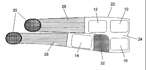
The part 22, referred to herein as a wrap, incorporates the right-hand set of
electrodes of
Figure 1. The wrap 22 comprises a main electrode-bearing portion 24 having two
lateral
finger-like extensions (straps) 26, 28 extending from one edge of the portion
24 and disposed
one immediately above the other. In use the wrap 22 is wrapped around the
patient's hip and
thigh region with the electrodes on the inside, so that the electrodes 10 to
16 bear against the
patient's skin at the appropriate locations on the patient's body. The straps
26, 28 are
CA 02749828 2015-01-06
6
elasticated so that the wrap is stretched around the region for snug fitting.
The wrap 22 is
secured in place by Velcro hooksTM 30 at the free ends of the straps which
engage a region of
Velcro loops (not shown) on the outer surface of the electrode-bearing portion
24. Where the
straps 26, 28 meet, that portion 24 of the wrap may be pleated to further
facilitate the snug
fitting of the wrap. The electrode-bearing portion 24 may also have an
elasticated region 32
between the electrodes 14 and 16, to enhance the stretchability of the portion
24 in that area.
Having areas of differing stretchability allows for a better fit.
The wrap 22 is fitted by first positioning the electrodes 10 and 16 at the
appropriate locations
on the rear of the pelvic region on the right side of the body, and then
bringing the straps 26,
28 round to the front of the right thigh, threading them between the legs, and
finally fixing
their free ends to the portion 24 using the Velcro hooks 30. The left-hand
side wrap, which is
substantially a mirror-image of the right-hand side wrap 22, is fitted to the
left hand side of
the body in a similar way.
To reduce the number of wrap sizes required, the printing of the electrode
positions may be
different for small/medium/large body sizes. Electrodes 10 and 16 are
positioned in relation
to the mid-line and so do not differ greatly, regardless of subject size. A
large subject would
position electrodes 12 and 14 further outward on the wrap.
The material of the main electrode-bearing portion 24 of the wrap 22 may be a
material that is
resiliently deformable, such as neoprene, although less elastic than the
straps 26, 28. To
avoid separation of the electrodes from the portion 24 when the wrap is
stretched around a
subject, the latter may an inelastic material in the electrode regions.
Wiring (not shown) to the electrodes is integrated into the wrap 22;
techniques are known to
do this. The electrodes may be pre-fixed to the wrap at manufacture, or they
may be fixed by
the user at pre-printed locations on the portion 24. In the latter case the
wiring for each
electrode may terminate in an exposed stud in the centre of the electrode
area. Adhesive
electrodes are then placed onto the wrap in the designated areas. One side of
the electrode
sticks to the inner surface of the wrap and the other bears against the skin
when the wrap is
worn. The wiring allows each electrode to be individually energised if
desired.
CA 02749828 2011-07-14
WO 2010/084391
PCT/1B2010/000018
7
Figure 3 is an example of the electrode wiring for the two-part garment of
Figure 2. Figure 3
shows the complete garment comprising both the right- and left-hand side
wraps. The drive
circuitry 100 is also schematically illustrated and this may be constructed
according to
principles well-known in the NMES field.
The electrodes 10, 12, 14 and 16 of the right-hand side wrap are connected
individually to
respective driver nodes A, C, D and B, and the corresponding electrodes of the
left-hand side
wrap are connected to respective driver nodes A', C', D' and B'. Each driver
node can be
programmed to act, independently of the others, as a current source or a
current sink, or to
remain at high impedance. Under control of a microprocessor, each electrode
may therefore
be selected as a current source, a current sink or may be de-selected and take
no part in the
delivery of current. In this way electrodes may be combined into sets; a set
with one polarity,
a set with the opposite polarity, and a set not conducting at all. This allows
for current to be
directed between the eight electrodes of the garment via every possible
pathway.
Importantly, each electrode has a defined anatomical position and is connected
through the
garment wiring and connectors to a particular driving node in the drive
circuitry. Therefore, a
current pathway established between nodes has a defined anatomical pathway,
for example
across the midline of the body. In a pre-wired garment these connections
cannot be changed
by the user so the intended anatomical pathways for the current cannot be
changed by the
user.
In the case shown in Figure 3 where the garment is in two parts, one part for
the left and one
for the right, it is essential that each garment part is connected to the
drive circuitry such that
the intended anatomical relationship is preserved. This is readily achieved by
keyed and/or
polarised connectors which ensure that the garment connections cannot be
swapped or mixed
up.
In the simplest embodiment, the electrodes on one side of the body are
selected as one
polarity, and the electrodes on the other side are selected for the opposite
polarity. In such a
case individual energisation of the electrodes is not required, and therefore
on each side of the
body the electrodes may simply be connected together electrically within the
garment and a
single conductor brought to the drive circuitry. For example, the right-hand
side electrodes
CA 02749828 2011-07-14
WO 2010/084391
PCT/1B2010/000018
8
10, 12, 14 and 16 could be electrically connected together to form one large
area multi-
segment electrode on the right, and the corresponding left-hand side
electrodes could be
electrically connected together to form one large area multi-segment electrode
on the left.
Whilst a two-part garment with a wrap for each leg is shown in Figures 2 and
3, other
embodiments have been found to have advantages.
For example, at least the upper parts of the inner edges of the electrode-
bearing portions 24 of
the right- and left-hand side wraps forming the garment of Figure 2 and 3 may
be joined
together to form a one-piece garment, as shown in Figure 4. In fitting the
garment the user
may simply place the gluteal electrodes 10 first and then close the wrap as in
the single leg
version. Partially spitting the two sides of the garment (i.e. forming the
separate straps 26, 28)
has been found to ease its application.
=
The wiring may be integrated into the garment, as previously discussed. In
Figure 4 the four
electrodes 10-16 on each side of the garment are shown connected in common to
the drive
circuitry 100. However, the electrodes may be individually connected, in the
manner shown
in Figure 3. Also, since there is material continuity across both sides of the
garment all the
wiring may come out bundled at a single point for easy insertion into the
drive circuitry. In
this and all embodiments the drive circuitry 100 may be incorporated into the
garment itself.
In two-part embodiments that have a separate garment part for each side of the
body the
wiring on one side of the body may connect directly to the drive circuitry or
it may be
connected to the drive circuitry via the other part of the garment.
It will be evident that the shape of the garment may be slightly modified so
that it more
closely resembles a typical disposable diaper or nappy, with flaps. The user
may then
position the garment as you would a nappy and close it using fasteners. The
garment itself
may also be made of disposable material (or at least a part of it).
Instead of a two-piece garment as described, both sets of electrodes could be
incorporated on
the inside surface of a one-part garment, such as a wrap-around skirt with a
Velcro or buckle
fixing, or a pair of, preferably elasticated, shorts.
CA 02749828 2011-07-14
WO 2010/084391
PCT/1B2010/000018
9
Figure 5 shows a pair of shorts 40 embodying the invention. In this case the
electrodes 10-16
are provided on the inside surface of the shorts so that they are
appropriately positioned
against the skin when the shorts are worn (for clarity the wiring to the
electrodes is not
shown). When used in conjunction with adhesive gel electrodes or any electrode
that may
cause shearing on the skin it is advantageous to have the shorts loose when
initially applying
the garment, and then tightening it to bring the electrodes to bear against
the skin in the
correct position. This may be facilitated by providing closable slits 42 in
the material of the
shorts. Advantageously these slits may extend up either side of the shorts.
The opposite edges of the slits 42 may be fastened together after the garment
is applied using
a standard mechanism such as a strip of Velcro 44. When the material of the
shorts is
partially or fully elastic this has the further advantage of pressing the
electrodes against the
skin, improving connectivity. The shorts may incorporate the wiring to the
electrodes. The
direction of the wiring will depend on the position of the slits and whether
they extend fully to
the garment edges. In the embodiment shown the slits 42 extend up the side of
the leg, but
other arrangements are possible providing the slit(s) are appropriately placed
to avoid
interfering with the electrodes.
Another mechanism which has been found to be very advantageous in positioning
the
electrodes is the use of flaps (i.e. overlapping areas of material) on the
garment. A flap may
carry both an electrode and a wiring connection to that electrode, and in use
is tucked under
the outer material of the garment to bring the electrode directly against the
skin. Placing an
electrode on a flap which is tucked under the outer material of the garment
frees the electrode
from the outer garment, allowing more even compression. Importantly, the outer
garment
may have a slit going over the electrode area without interfering with the
electrode itself.
The flaps have also been found to be particularly helpful in allowing users to
position the
electrodes while wearing standard underwear. There is a tendency for underwear
to cover
some of the electrode. This is particularly frequent in the outer and upper-
outer portion of the
gluteal electrode (electrode 10). A flap allows for the underwear to be
positioned between the
flap and the main garment without the need to reposition (e.g. uncomfortably
hitch up) the
underwear.
CA 02749828 2011-07-14
WO 2010/084391
PCT/IB2010/000018
When using electrodes that may be easily pulled over the skin, e.g. silicone
rubber electrodes,
it is not necessary that the garment be applied loose and tightened after.
Extra compression of
the electrodes against the skin may given by external straps, the intrinsic
elasticity of the
material or compressive techniques such as are used in the underwear industry,
particularly
5 those garments designed to shape/contour/compress the body. It will be
evident that areas of
differential stretch will be advantageous as indeed the use of flaps and tabs,
etc.
An example in the form of shorts 50 is shown in Figures 6a and 6b. The shorts
themselves
may be made of a conductive material or have conductive parts applied to it,
e.g. a conductive
10 fabric. Some areas 52 may be masked from skin contact to avoid unwanted
stimulation, e.g.
cutaneous stimulation of the perineum/scrotum/anal region. Alternatively,
these areas may
simply be made with non-conductive material. There may be slits or areas of
non-conductive
material between some or all of the electrode areas. This helps keep the
electrodes discrete
and ensures that the current passes deep within the tissue rather than passing
along the surface
between adjacent electrodes. Preferably the electrodes on each side of the
body are separated
from one another by at least a centimetre of non-conductive material for
optimal stimulation.
In Figure 6 the electrodes on each side of the midline are not shown with a
non-conducting
area between them, because in some embodiments the left side acts as
essentially one
electrode and the right as another. It should be noted that this type of
arrangement allows for
an even greater surface area, and compensates for the inferior quality of
connectivity typical
for this type of fabric electrode.
For clarity the wiring of the shorts has not been shown. The shorts may be
single layered or
may have an external layer which is non-conductive, serving to further
insulate the electrodes
from touch from the outside. The external layer may add additional compression
to the
garment, and/or may carry conductors or electrical contacts to distribute
current to the internal
layer which may be positioned first.
In operation, the drive circuitry for the electrodes delivers biphasic current
pulses, i.e. the
current comprises alternate first and second phases in which every second
phase pulse is
inverted relative to the immediately preceding first phase pulse. It is not
necessary that each
second phase pulse have the same duration as the first phase pulse, but it
preferably has the
same total charge.
CA 02749828 2011-07-14
WO 2010/084391
PCT/1B2010/000018
11
In order to reliably achieve significant pelvic floor contractions we found
that a pulse current
amplitude of at least about 80mA was required, distributed over the four
electrodes 10 to 16.
Typically 80mA is initially used and is then increased to 140mA during the
session. In
subsequent sessions higher pulse currents may be used, but preferably not
exceeding 200mA.
In a typical treatment session, Figure 7, the overall duration of each first
phase pulse is
620 microseconds, followed by an interphase delay of 100 microseconds when no
current
flows. Each second phase pulse is also 620 microseconds, but of opposite
(negative) polarity.
The large electrode size allows the current density to remain tolerable going
through the
legs/hips/gluteal muscles. However it concentrates in crossing the pelvis,
contracting the
pelvic floor. For a peak current pulse amplitude of 80mA, distributed over the
area of the
four pads on each side of the pelvic region, the peak current density at each
electrode is:
80mA/768cm2 = 0.1mA/cm2
We have found that a simple pulse technique works well in most people, giving
a strong
pelvic floor contraction that can be readily tolerated. By a "simple pulse
technique" we mean
that the current pulses pass simultaneously from all the electrodes on the
right to those on the
left, and vice versa, according to the phase. Each set of electrodes 10 to 16
on each side of the
pelvic region therefore acts as a single, multi-part electrode. Figure 7 is an
example of a
simple pulse technique.
However, we have found that the pelvic floor contractions are further enhanced
in some
individuals if the current is passed selectively between one or more
electrodes on one side to
one or more electrodes on the other side, either during every current phase or
in selected
current phases, or portions of a phase.
In a preferred embodiment, Figure 8, each first phase is divided into two sub-
phases, each
first sub-phase current pulse has a duration of 413 microseconds and passes
from all four
electrodes on one side of the body to all four electrodes on the other side.
Each second sub-
phase current pulse has a duration of 207 microseconds but passes only between
electrode 10
CA 02749828 2011-07-14
WO 2010/084391
PCT/1B2010/000018
12
on one side to electrode 10 on the other side. The reverse polarity second
phase follows the
same pattern with current passing in the opposite direction between the same
electrodes at the
corresponding sub-phases. The sequence is as follows:
First Phase
First sub-phase: 413 microsecond pulse from all four electrodes on one side to
all four
electrodes on the other side.
Second sub-phase: 207 microsecond pulse from electrodes 10 and 12 on one side
to the same
electrodes on the other side.
Interphase interval 100 microsecond
Second Phase
First sub-phase: 413 microsecond pulse from all four electrodes on one side to
all four
electrodes on the other side.
Second sub-phase: 207 microsecond pulse from electrodes 10 and 12 on one side
to the same
electrodes on the other side.
The embodiments use relatively large currents, typically with peak pulse
currents up to
200mA and rms currents up to 50mA. The charge per pulse phase can range from
40 up to
120 microcoulombs, compared to typical electrotherapy pulses which are usually
limited in
the region of 30 microcoulombs. These large currents and phase charges are
tolerable only if
they are dispersed over very large surface area electrodes. It is important
therefore, for safety
and comfort, that the apparatus can detect when the electrode surface area is
reduced or if
there is an increase in contact resistance with the skin.
While electrode impedance is related to surface area, the relationship varies
with skin type,
hair, amount of subcutaneous fat, cleanliness of the skin and condition of the
electrodes.
Therefore, impedance alone is not a reliable indicator of electrode surface
area. Non-uniform
impedance over the available surface area will lead to "hot spots" of current
density at those
points where the impedance is lowest. This problem may be overcome by passing
test
currents between the electrodes to compare one electrode impedance with
others. In this way
it is possible to establish if one electrode has an unexpectedly high, or low,
circuit impedance
which would lead to an imbalance of current density in the electrode array.
Since, in the case
CA 02749828 2011-07-14
WO 2010/084391
PCT/1B2010/000018
13
where the electrodes are individually selectable, each electrode can be paired
with any other
electrode it is possible by a simple process of elimination to find the
electrode, or electrodes,
which are outliers in terms of electrical impedance. Having identified the
electrode, the user
can be alerted to take corrective action.
Accordingly, the circuitry includes means to determine the impedance of each
electrode by
measuring the voltage drop across each electrode due to a known test current,
and an
algorithm which analyses and compares the readings for each electrode. Pre-
defined
acceptance criteria for each electrode can be used to determine a fault
condition, for example
if there is a significant impedance imbalance between corresponding electrodes
on opposite
sides of the body. In addition, the electrode impedances so determined can be
statistically
analysed to identify outliers which fall outside a predefined range of the
mean impedance of
all the electrodes. The circuitry could also reject an electrode set if the
statistical variance of
the impedance values is higher than a preset amount.
The electrical impedance of an electrode in contact with the skin in the
presence of an
electrolyte is known to contain a large capacitative component which is due to
the outer skin
layer called the stratum come= acting as a high impedance dielectric. The
value of this
, capacitance increases with the area of contact. The impedance also has a
parallel resistance
component which decreases with area of contact. This resistive component also
depends on
the quality of the electrolyte. There is also a series resistance component
which is primarily
due to the internal resistance of the body. The quality of the electrolyte and
its distribution, as
well as presence of skin residues etc, also affects these impedance
components.
The drive circuitry includes a means for measurement of electrode capacitance
and estimation
of the area of surface contact of each electrode, or any set of electrodes.
The drive circuitry
may be configured to signal a fault condition, and prevent the treatment
beginning, or ceasing
a treatment which is under way, when the estimated area of contact is below a
pre-defined
threshold, or lower by a pre-defined amount than an electrode of the same size
elsewhere in
the array.
The measurement of capacitance may be carried out by various techniques well
known in
electronic engineering. In a pulsed electrical stimulator it is convenient to
take time domain
CA 02749828 2011-07-14
WO 2010/084391
PCT/1B2010/000018
14
measurements of voltage changes due to constant current test pulses in the
microsecond
range. By sampling the voltage across a pair of electrodes at several pre-
defined time points
in the charge and/or discharge phases of the pulse, it is possible to estimate
charge and/or
discharge time constants and therefore estimate each, or some, of the
components in the
model, i.e. the series resistance component, the parallel resistance component
and the
capacitance.
The current paths may change somewhat depending on the alignment of the
pelvis, etc.
Optimal stimulation of the pelvic floor is more likely with the subject in
particular
positions/postures. In particular there appears to be a benefit in having the
patient standing.
This also has the advantage of being more 'physiological', i.e. for mechanical
reasons stress
incontinence often happens when the person is upright. The weight of the
abdominal contents
place extra pressure on the pelvic floor. Exercising the muscles in this
position replicates
what the muscles must do in reality. Intra-vaginal probe electrodes are
usually used
lying/sitting as otherwise the electrode has a tendency to fall making the
contact with the
target tissue poor and hence more uncomfortable.
Further observations include that the wearing of high-heels can disimprove the
pelvic floor
contractions. Additionally, the posture that typically gives the best
contraction is standing
with the feet apart and the subject leaning forward slightly with their hands
on a ledge or table
near hip height.
In trials, some women have, surprisingly, noticed an immediate effect upon
using this type of
stimulation. These effects are too sudden and quick to be accounted for by
"training" of the
muscles. Instead a re-awakening or activation of previously inhibited muscles
or unused
muscle is a possibility or an education/biofeedback in what contraction of
these muscle fibres
feels like.
This falls into two categories. Firstly, some women learn how to contract the
pelvic floor that
were previously unable to do so even under expert guidance with the assistance
of ultrasound.
The inability of many women to voluntarily contract these muscles means that
exercises are
not going to do any good. With just a session of this new electrical
stimulation they were able
to voluritafily- -contract their pelvic floor.
CA 02749828 2015-01-06
Secondly, some women, especially post-hysterectomy patients, who had been
incontinent for
years got a sudden and immediate improvement in their incontinence after just
a session using
the new device.
5
In addition to the categories already discussed, the invention may be used to
treat vaginal
prolapse in females. We found in one case that after only eight sessions of
stimulation the
prolapse had resolved. This is probably because training the pelvic floor
improved the tissue
that keeps the vagina in position. This represents an alternative treatiaent
for this condition,
10 which had often required surgery.
Furthermore some women reported enhanced sexual gratification after using the
machine.
The scope of the claims should not be limited by the preferred embodiment and
examples,
15 but should be given the broadest interpretation consistent with the
description as a whole.