Note: Descriptions are shown in the official language in which they were submitted.
CA 02750149 2016-08-12
SYSTEM AND METHOD FOR
OCULAR IONTOPHORESIS WITH BUFFERING
won
FIELD
100021 The technology described herein is generally related to a system and
process for ophthalmic transfer of a therapeutic substance across a surface of
an
eyeball via iontophoresis. In some embodiments, the technology is related to
buffering systems and methods that regulate pH of therapeutic substances
during
iontophoresis.
BACKGROUND
[0003] Ocular iontophoresis is the application of an electrical source to
propel
charged and/or active molecules from a reservoir into the intraocular tissues
of a
mammal, including a human or an animal. Positively charged ions can be driven
into the ocular tissues by electro-repulsion at the anode while negatively
charged
ions are repelled from the cathode. The simplicity and safety of
iontophorectic
application includes enhanced targeted delivery of compound(s) of interest,
and the
reduction of adverse side effects have resulted in extensive use of
iontophoresis in
laboratory, clinical research and commercial use. Unlike ocular injections
(intravitreal, retrobulbar, and peribulbar) and intraocular implants,
iontophoresis is a
noninvasive technique used to deliver compounds of interest into the anterior
and/or
posterior compartments of the eye. Iontophoretic delivery can be used to
obtain
intraocular concentrations and residence times that are equal to or greater
than those
achieved by conventional modalities such as topical drops, ointments, and
gels.
CA 02750149 2016-08-12
[00041 Iontophoresis has been widely used in dermal applications in which
therapeutic compounds are transported across a patient's skin using electrical
currents. Due to the relative high impedance of the skin, the electrical
currents are
generally relatively low. Consequently, dosage times tend to be relatively
long, for
example being greater than an hour. In such applications, iontophoresis can be
applied to the patient's skin with an active drug-containing adhesive patch.
[0005] Ocular iontophoresis devices are typically constituted by a direct
current
(DC) electric field source coupled to two electrodes, referred to respectively
as
"active" and "passive" electrodes. The active electrode provides an
electromotive
force, when energized, that acts on an electrolyte containing therapeutic
composition(s) to transfer one or more therapeutic substance(s) across a
surface of
the eyeball, while the passive electrode serves as a return electrode and
enables the
electric circuit to be looped through the patient's body. The compound of
interest is
transported via the active electrode across the tissue when a current is
applied to the
electrodes through the tissue. Compound transport may occur as a result of a
direct
electrical field effect (e.g., electrophoresis), an indirect electrical field
effect resulted
from the bulk volume flow of solution from the anode to cathode (e.g.,
electroosmosis), electrically induced pore or transport pathway formation
(e.g.,
electroporation), or a combination of any of the foregoing. Examples of
currently
known iontophoretic devices and methods for ocular drug delivery may be found
in
the United States patents 7,164,943; 6,697,668; 6,319,240; 6,539,251;
6,579,276;
6,697,668, and PCT publications WO 03/030989 and WO 03/043689.
[0006] Ocular iontophoresis, however, presents several unique challenges. For
example, the applicator must conform to the spheroidal geometry of the
eyeball.
That is, the portion of the applicator in contact with a surface of the eye
must be
specifically formed to minimize loss of therapeutic substance and to reduce
discomfort. Also, since the electrical impedance of the eye is relatively
lower than
that of the epidermis, higher currents can be achieved at still reasonably low
current
densities. Accordingly, dosage times tend to be relatively short, often much
less
than one hour.
2
CA 02750149 2011-06-27
WO 2010/078246 PCT/US2009/069580
SUMMARY
[0007] Iontophoretic transfer of a therapeutic substance may result in
unwanted
changes in pH that result in patient discomfort, and in some instances, tissue
damage. There remains a need to regulate the pH of a therapeutic preparation
within
the physiologically acceptable range during iontophoresis while maintaining
the
therapeutic substance at the highest ionization state for optimal delivery.
Further,
there remains a need to improve the delivery efficiency of a therapeutic
substance
while reducing the risks of any possible damage (e.g., irritation or burning
of
tissues) that could limit the use of ocular iontophoresis. The present
technology is
related to buffering systems and methods that regulate pH of therapeutic
substances
during iontophoresis while improving delivery efficiency and safety.
[0008] In one embodiment, a delivery device for transferring a therapeutic
substance across and/or through a surface of an ocular globe includes at least
one
iontophoretic chamber configured to store the therapeutic substance. The
device
also includes an electrode disposed relative to the at least one iontophoretic
chamber. The electrode is configured to provide an electromotive force that,
when
energized, transfers at least a portion of the therapeutic substance stored
within the
iontophoretic chamber across the surface of the ocular globe. A buffer system
is
disposed at least partially within the at least one iontophoretic chamber. The
buffer
system is configured to regulate the pH of the therapeutic substance and to
maintain
the pH at the surface of the ocular globe within a range of about 3 to 8
during
iontophoretic transfer of the therapeutic substance.
[0009] In one embodiment, the buffer system can be a buffering agent to reduce
the risk of damage to ocular tissue. The buffer agent can be at least one of
an ion
exchange resin, polymeric particles, insoluble buffer particles, cationic
particles,
anionic particles and zwitterionic particles. The ion exchange resin can be at
least
one ion exchange material having a characteristic nature of at least one of a
strong
acid, a strong base, a weak acid, and a weak base. In one embodiment, the
buffer
system can further include a therapeutic substance.
3
CA 02750149 2011-06-27
WO 2010/078246 PCT/US2009/069580
[00101 In one embodiment, the pH can be maintained at a level substantially
equal
to the highest ionization level of the therapeutic substance to enhance
transport
efficiency of the therapeutic substance. In another embodiment, the buffer
system
can be electrically conductive capable of conducting an electric field
supplied from
the electrode.
[0011] In one embodiment, the buffer system can be at least one of a porous
material, a liquid solution, a gel, a packed bed resin, a hydrogel film, and
membrane.
The porous material can be at least one of a foam, a fabric, a nonwoven
material,
and a sintered material. The gel can be at least one of a hydrogel matrix and
an
aerogel matrix. The membrane can be at least one of a mono-laminar, a multi-
laminar film, hydrophobic (semi permeable) membrane, and a non-permebale/solid
membrane.
[0012] In one embodiment, the iontophoretic chamber can further include at
least a
first layer and at least a second layer, the first layer including the buffer
system and
the second layer including a therapeutic substance. The first layer can be
disposed
between the electrode and the second layer. In another embodiment, the
iontophoretic chamber can further include a membrane disposed between the
first
layer and the second layer. The membrane can have a low water vapor
permeability
to maintain water content in each layer. In yet another embodiment, the layers
can
be concentrically relative to each other. The membrane can be disposed between
the
first layer and the second layer. In another embodiment, the first layer can
have a
higher buffering capability than the second layer.
[0013] In one embodiment, the iontophoretic chamber can further include a
first
layer, a second layer, and a third layer, the first layer and the second layer
including
the buffer system and the third layer including a therapeutic substance. The
first
layer can be disposed closest to the electrode and the second layer is
disposed
between the first layer and the third layer. In another embodiment, the
iontophoretic
chamber can further include a membrane disposed between the first layer and
the
second layer or the second layer and the third layer. The membrane has low
water
4
CA 02750149 2011-06-27
WO 2010/078246
PCT/US2009/069580
vapor permeability to maintain water content in each layer. In yet another
embodiment, the layers can be arranged concentrically relative to each other.
The
membrane can be between the first layer and the second layer or the second
layer
and the third layer. In another embodiment, the third layer can include the
therapeutic substance is removeably coupled to the iontophoretic chamber, the
first
layer can have a higher buffering capability than the second layer, and/or the
second
layer can include a ionic composition that optimizes electro-transport of the
therapeutic substance in the third layer.
[0014] In another embodiment, the buffer system can be arranged as a buffered
surface coating. The buffer system can further include a rehydrating agent.
The
buffer system can be disposed adjacent to the electrode.
[0015] In another embodiment, a process for transferring a therapeutic
substance
across a surface of an ocular globe includes positioning a delivery device
directly to
the surface of an ocular globe. The delivery device includes at least one
iontophoretic chamber storing at least one therapeutic substance. A potential
is
applied to an active electrode disposed relative to the iontophoretic chamber
to
iontophoretically transfer a portion of the at least one therapeutic substance
across
the adjacent surface of the ocular globe. The buffer system is configured to
regulate
the pH of the therapeutic substance and to maintain the pH at the surface of
the
ocular globe within a range between about 3 and about 8 during iontophoretic
transfer of the therapeutic substance.
[0016] In another embodiment, an ocular iontophoresis device for transferring
a
dosage of therapeutic substance across and/or through a surface of an eyeball,
includes an iontophoretic chamber with an open end configured to be positioned
on
the surface of the eyeball. A reservoir medium is disposed within the
iontophoretic
chamber, configured to retain a therapeutic substance. The device also
includes an
electrode positioned with respect to the iontophoretic chamber, to provide an
electromotive force, that when energized, transfers the dosage of therapeutic
substance across the surface of the eyeball. A buffer system is disposed
within the
iontophoretic chamber, containing at least one buffer element configured to
regulate
CA 02750149 2011-06-27
WO 2010/078246 PCT/US2009/069580
pH change during iontophoretic transfer of the dosage of therapeutic substance
within a range between about 3 and about 8.
[0017] In another embodiment, a process for manufacturing an ocular
iontophoresis device includes providing an iontophoretic chamber having an
open
end configured to be positioned on a surface of an eyeball. A reservoir medium
is
located within the iontophoretic chamber. The reservoir medium contains a
therapeutic substance deliverable to the eyeball. An electrode is arranged
opposite
the open end of the iontophoretic chamber. The electrode is associated with
the
iontophoretic chamber to provide an electromotive force, when energized, that
transfers a dosage of the therapeutic substance across the surface of the
eyeball. A
buffer system is located within the iontophoretic chamber. The buffer system
is
configured to regulate the pH of the therapeutic substance and to maintain the
pH at
the surface of the ocular globe within within a range between about 3 and
about 8
during iontophoretic transfer of the therapeutic substance.
[0018] In yet another embodiment, a process for transferring a dosage of
therapeutic substance across and/or through a surface of an eyeball includes
positioning an open end of an iontophoretic chamber including a therapeutic
substance onto the surface of the eyeball. An electrical potential is applied
to the
iontophoretic chamber to induce transfer of the dosage of therapeutic
substance
across the surface of the eye. Change of pH of the therapeutic substance is
regulated
within a range between about 3 and about 8 during an extended period during
which
the dosage of therapeutic substance is transferred. Regulation of the pH
change is
accomplished using a buffer system.
BRIEF DESCRIPTION OF THE DRAWINGS
[0019] The foregoing and other objects, features and advantages of the
invention
will be apparent from the following more particular description of preferred
embodiments of the invention, as illustrated in the accompanying drawings.
[0020] FIG. IA shows a longitudinal cross section of a single layer buffered
reservoir ocular iontophoresis device;
[0021] FIG. 1B shows a distal-end view of the ocular iontophoresis device
shown
in FIG. 1A;
6
CA 02750149 2011-06-27
WO 2010/078246 PCT/US2009/069580
[0022] FIG. 2A shows a longitudinal cross section of a two layer buffered
reservoir ocular iontophoresis device;
[0023] FIG. 2B shows a distal-end view of the ocular iontophoresis device
shown
in FIG. 2A;
[0024] FIG. 3A shows a longitudinal cross section of a three layer buffered
reservoir ocular iontophoresis device;
[0025] FIG. 3B shows a distal-end view of the ocular iontophoresis device
shown
in FIG. 3A;
[0026] FIG. 4A shows a longitudinal cross section of a two layer buffered
reservoir with membrane ocular iontophoresis device;
[0027] FIG. 4B shows a distal-end view of the ocular iontophoresis device
shown
in FIG. 4A;
[0028] FIG. 5A shows a longitudinal cross section of a three layer buffered
reservoir with membrane ocular iontophoresis device;
[0029] FIG. 5B shows a distal-end view of the ocular iontophoresis device
shown
in FIG. 5A;
[0030] FIG. 6A shows a longitudinal cross section of a two concentric layer
buffered reservoir ocular iontophoresis device;
[0031] FIG. 6B shows a distal-end view of the ocular iontophoresis device
shown
in FIG. 6A;
[0032] FIG. 7 shows a longitudinal cross section of a two concentric layer
buffered
reservoir with membrane ocular iontophoresis device; and
[0033] FIG. 8 shows a longitudinal cross section of a three layer buffered
reservoir
with drug loaded ring ocular iontophoresis device.
[0034] In the drawings, identical reference numbers may identify similar
elements
or acts. The shapes, sizes, and relative positions of device elements in the
drawings
are not necessarily precise or drawn to scale. For example, the shapes and
sizes of
elements may not be drawn to scale, and/or one or more of the elements may be
arbitrarily enlarged or positioned to improve drawing legibility. Furthermore,
the
particular shapes of the elements as drawn are not intended to convey any
information regarding the actual shape of the particular elements, and have
been
solely selected for ease of recognition in the drawings.
7
CA 02750149 2011-06-27
WO 2010/078246
PCT/US2009/069580
DEFINITIONS
[0035] The terms 'therapeutic substance' and 'active pharmaceutical ingredient
(API)' are used interchangeably throughout the specification, and by
definition refer
to a substance intended for use in the diagnosis, cure, mitigation, treatment,
or
prevention of a disease of the eye. Such substance is intended for use as a
component of a medicine, and in some embodiments of this invention a
component,
part, or accessory of an iontophoresis device.
[0036] The terms 'therapeutic preparation', 'therapeutic composition' and
'drug
preparation' are used interchangeably throughout the specification, and by
definition
refer to a product of mixing or combining various pharmaceutically acceptable
active and inactive elements or ingredients.
DETAILED DESCRIPTION
[0037] As described herein, various embodiments of compositions, devices,
methods of use and methods of manufacture for ophthalmic transfer of a
therapeutic
substance across a surface of an eyeball via iontophoresis are directed to
achieve at
least one (e.g., principle) objective of buffering the therapeutic substance
to a
biologically acceptable pH range during iontophoretic treatment. An additional
objective of at least some of the various embodiments is to maximize electro-
transport delivery of the therapeutic substance by maintaining the pH to
achieve the
highest ionization level of the therapeutic substance. Another benefit of
maintaining
the pH of the therapeutic substance is a consistent/predictable dose delivery.
Yet
another objective of at least some of the various embodiments is to increase
the
delivery of the therapeutic substance(s) to the eye by reducing the amount of
competing ions, and to maintain the stability of the therapeutic substance(s)
during
iontophoretic treatment and storage.
[0038] Figs. 1A-1B show an exemplary single layer buffered reservoir ocular
iontophoresis device 100 for buffering a therapeutic substance to a
biologically
acceptable pH range at the surface of an eyeball being treated during
iontophoretic
treatment. The ocular iontophoresis device 100 includes a distal end 110 and a
proximal end 120. The distal end 110 defines a cavity 112 for receiving the
8
CA 02750149 2011-06-27
WO 2010/078246 PCT/US2009/069580
therapeutic substance. The proximal end 120 defines an annular reservoir 122
(iontophoretic chamber) for delivering the therapeutic substance to the ocular
area of
the eyeball surface. The ocular area is typically a part of the sclera of the
eyeball.
An active electrode 130 is disposed between the distal end 110 and the
proximal end
120; and is typically disposed at the beginning of the annular reservoir 122.
In one
embodiment, a buffer system 160 containing a buffering agent and/or the
therapeutic
substance can be disposed in the annular reservoir 122. In one embodiment, the
therapeutic substance can be an active pharmaceutical ingredient (API).
[0039] The addition of the buffering system 160 to the iontophoretic chamber
122
allows the iontophoresis device to self-buffer. A self-buffering iontophoresis
device
reduces the risk of damage to ocular tissue as a result of dramatic pH changes
that
can occur in a non-buffered system.
[0040] The buffer system 160 is configured to provide a buffer action that
maintains pH within the vicinity of a treatment surface 170, within a
biocompatible
range, during the duration of delivery of a dose. The treatment surface 170 is
the
area of the annular reservoir that contacts or is in close contact with the
surface of
the eyeball. The biocompatible range of pH for ocular delivery may depend upon
the individual, but is generally within the range of about 3 to about 8. In a
preferred
embodiment, the biocompatible range of pH is maintained within a range of
about 3
to about 7 throughout the duration of delivery. Even more preferably, the pH
is
maintained at a level equal to the highest ionization level of the therapeutic
substance in order to enhance transport efficiency of the therapeutic
substance.
[0041] In some embodiments, the buffer system 160 can be electrically
conductive
medium capable of conducting an electric field supplied by the active
electrode 130
to deliver the therapeutic substance. In other embodiments, the buffer system
160
can be disposed in electrical conductive medium. Additional exemplary
embodiments of the buffer system 160 are further described below including a
porous material, a buffered gel (liquid or solid), a packed bed resin (ion
exchange
resin), a hydrogel film or membrane, and combinations of any of these
components.
[0042] As described above, in one embodiment, the annular reservoir or
iontophoretic chamber 122 includes a buffer system 160. The buffer system 160
includes a buffer medium having at least one buffer agent (composition) and a
9
CA 02750149 2011-06-27
WO 2010/078246
PCT/US2009/069580
therapeutic substance, such as an active pharmaceutical ingredient (API). In
some
embodiments, the buffer medium includes one or more porous materials for
containing a preparation (e.g., API, inactive ingredients, buffer, etc.). The
API
preparation may be a liquid solution preparation. The liquid solution
preparation
may include one or more therapeutic substances together with a buffering
composition. At least one of the therapeutic substances may be dissolved in a
liquid
solution preparation. Likewise, the buffering composition may also include a
soluble buffer composition. The porous material may be saturated with the
liquid
solution preparation. In such embodiments, the iontophoretic chamber 122 may
contain a buffer medium and an API, each within the same porous material, such
as,
for example, foam, containing a solution preparation of the one or more
therapeutic
substances and buffer.
[0043] In some embodiments, the API medium itself provides a buffering action
sufficient to maintain pH at the point of contact between the device 100 and
the eye
within a preferred range, including any of the pH ranges described herein. The
ability of an API to act as a buffering agent arises from its acid-base
dissociation
constants (pKal, pKa2, etc.). The pKa distributions of drugs are influenced by
the
nature and frequency of occurrence of the functional groups that are commonly
observed in pharmaceuticals and the typical range of pKa values they span. For
instance, dexamethasone phosphate in its triprotic acid form exhibits two pKa
values
of 1.9 and 6.4. As a result, an aqueous solution of this compound at a dosing
concentration of 40 mg/mL (pH 5.7) is capable of resisting to pH variations
resulting
from cathodal iontophoresis.
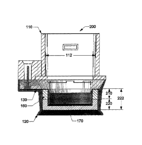
10044] Figs. 2A-2B show a two layer buffered ocular iontophoresis device 200
for
buffering a therapeutic substance to a biologically acceptable pH range at the
surface
of an eyeball being treated during iontophoretic treatment. In one embodiment,
at
least two layers 210, 220 are disposed in the annular reservoir or
iontophoretic
chamber 222, with at least one of the two layers including a buffering system
160.
The buffer system 160 includes a buffer medium, which may be a first porous
material of the first layer 210. The second layer 220 can include an API
medium,
which may be a second porous material for containing a therapeutic substance.
In
CA 02750149 2011-06-27
WO 2010/078246
PCT/US2009/069580
some embodiments, the buffer medium can be disposed in the second layer 220
and
the API medium can be disposed in the first layer 210.
[0045] In some embodiments, the first porous material of the first layer 210
can be
positioned between the second porous material of the second layer 220 and the
active electrode 130. As described above, the first porous material of the
first layer
210 may be saturated with a preparation (such as a liquid solution preparation
and/or
a liquid colloidal preparation) containing a buffer composition or a buffer
composition and at least one therapeutic substance. The second porous material
of
the second layer 210 may be saturated with a preparation (such as a liquid
solution
preparation and/or a liquid colloidal preparation) containing at least one
therapeutic
substance. In some embodiments, the iontophoretic chamber 222 contains (i) a
buffer medium made of a first porous material (e.g., an open-cell foam)
containing
at least one buffer element (and may or may not include a therapeutic
substance) and
(ii) an API medium made of a second porous material, such as, for example, an
open-cell foam, containing a solution preparation of one or more therapeutic
substances.
[0046] In some embodiments, the preparation may include pharmaceutically
acceptable inactive ingredients for ophthalmic delivery. In some embodiments,
one
or both of the first porous material and the second porous material include a
soluble
buffer composition. In other embodiments, the first porous material and the
second
porous material are made of similar or different compositions. For example,
the first
porous material and the second porous material are made of different porous
materials and/or are saturated with different preparations (in composition
and/or
concentration). The different preparations may include different elements, or
the
same elements in different concentrations.
[0047] Figs. 3A-3B show a three layer buffered ocular iontophoresis device 300
for buffering a therapeutic substance to a biologically acceptable pH range at
the
surface of an eyeball being treated during iontophoretic treatment. In one
embodiment, at least three layers 310, 320, 330 are disposed in the annular
reservoir
or iontophoretic chamber 322, with at least one of the three layers including
a first
buffering system 160 and another of the three layers including a second
buffering
system 160'. The first buffering system 160 includes a buffer medium, which
can
11
CA 02750149 2011-06-27
WO 2010/078246
PCT/US2009/069580
be a first porous material of the first layer 310. The second buffering system
160'
includes a buffer medium, which can include a second porous material of the
second
layer 320. The third layer 320 can include an API medium, which may be a third
porous material for containing a therapeutic substance. In some embodiments,
the
buffer mediums and the API medium can be disposed in any configuration.
[0048] In one embodiment, as shown in Figs. 3A-3B, the first porous material
of
the first layer 310 can be positioned closest to the active electrode 130, the
third
porous material of the third layer 330 can be positioned closest to a surface
of an
eyeball (not shown) during use of the device 300, and the second porous
material of
the second layer 320 can be positioned between the first porous material and
the
third porous material.
[0049] In some embodiments, the first porous material and the second porous
material can each include a respective buffer composition including at least
one
respective buffer element. For example, as discussed above, the first porous
material (i.e., the porous material closest to the active electrode 130) and
the second
porous material may be loaded with respective buffer compositions as described
above with respect to Figs. 1A-2B. In some embodiments, the buffer system 160,
160' contains (i) a buffer medium including a first porous material (e.g.,
foam) and a
second porous material, each containing at least one respective buffer
element, and
optionally containing at least one therapeutic substance, and (ii) a reservoir
medium
including a third porous material, such as, for example, foam, containing a
solution
preparation of the one or more therapeutic substances.
[0050] In some embodiments, the first porous material and the second porous
material may differ in buffer composition and/or concentration of the same
buffer.
The first porous material and the second porous material may be made of
different
porous materials. In further embodiments, the third porous material may also
include a buffer composition that is weaker, for example, than that of the
first porous
material and the second porous material. In some embodiments, the first porous
material, the second porous material, and the third porous material may be
made of
similar or different compositions. It should be noted that any number of
porous
materials may be included within the buffer system.
12
CA 02750149 2011-06-27
WO 2010/078246
PCT/US2009/069580
[0051] In some embodiments, the first porous material may contain a buffer
composition and the second porous material may contain an ionic composition
that
optimizes electro-transport of the therapeutic substance in the third porous
material.
[0052] Figs. 4A-4B show a two layer buffered ocular iontophoresis device 400
with a membrane for buffering a therapeutic substance to a biologically
acceptable
pH range at the surface of an eyeball being treated during iontophoretic
treatment.
The membrane may also exhibit low water vapor permeability to maintain water
content in each layer. In one embodiment, at least two layers 410, 420 are
disposed
in the annular reservoir or iontophoretic chamber 422, with at least one of
the two
layers including a buffering system 160. The buffer system 160 includes a
buffer
medium, which may be a first porous material of the first layer 410. The
second
layer 420 can include an API medium, which may be a second porous material for
containing a therapeutic substance. A buffering membrane (e.g. ion exchange
membrane) 430 can be disposed between the first layer 410 and the second layer
420
to provide a more stable system. In some embodiments, the buffer medium can be
disposed in the second layer 420 and the API medium can be disposed in the
first
layer 410, while the buffering membrane 430 can be disposed before or after
any of
the layers.
[0053] At least one of the first porous material and the second porous
material may
be saturated with a preparation containing the therapeutic substance described
herein. For example, the preparation may be a liquid preparation. The liquid
preparation may include one or more therapeutic substances. At least one of
the
therapeutic substances may be dissolved in the liquid preparation. At least
one of
the first porous material and the second porous material may be saturated with
the
liquid preparation. In some embodiments, the liquid preparation may include
pharmaceutically acceptable inactive ingredients for ophthalmic delivery. The
first
porous material and/or the second material may be buffered as discussed
herein. In
other embodiments, the first porous material and/or the second material may be
non-
buffered. In some embodiments, the liquid preparation may contain a
significant
amount of water. In this instance, the buffering membrane 430 may be a mono-
laminar, a multi-laminar film, or hydrophobic (semi permeable) membrane in
nature
to retain the water content of the first layer 410 for stability.
13
CA 02750149 2011-06-27
WO 2010/078246
PCT/US2009/069580
[0054] In some embodiments, a mono-laminar or a multi-laminar ion-exchange
film or membrane may be placed in the iontophoretic chamber 422 in contact or
approximately near the active electrode 130. In further embodiments, the
membrane
may be rolled or otherwise disposed in an annular space of the iontophoretic
chamber 422 in one piece or multiple pieces. In some embodiments, the
iontophoretic chamber 422 may be filled with multiple small membrane pieces.
In
further embodiments, the membrane may be laminated along with the porous
material matrix.
[0055] Figs. 5A-5B show a three layer buffered ocular iontophoresis device 500
with membrane for buffering a therapeutic substance to a biologically
acceptable pH
range at the surface of an eyeball being treated during iontophoretic
treatment. In
one embodiment, at least three layers 510, 520, 530 are disposed in the
annular
reservoir or iontophoretic chamber 522, with at least one of the three layers
including a first buffering system 160 and another of the three layers
including a
second buffering system 160'. The first buffering system 160 includes a buffer
medium, which can be a first porous material of the first layer 510. The
second
buffering system 160' includes a buffer medium, which can include a second
porous
material of the second layer 520. The third layer 530 can include an API
medium,
which may be a third porous material for containing a therapeutic substance.
In
some embodiments, the buffer mediums and the API medium can be disposed in any
configuration. A buffering membrane (e.g. ion exchange membrane) 540 can be
disposed between the second layer 510 and the third layer 530 to provide a
more
stable system. Another buffering membrane (not shown) can be disposed between
the first layer 510 and the second layer 520. In some embodiments, the buffer
medium can be disposed in the second layer 520 and the API medium can be
disposed in the second layer 520, while the buffering membrane 540 can be
disposed
before or after any of the layers. In some embodiments, the first layer 510
may
contain a buffer composition and the second layer 520 may contain an ionic
composition that optimizes electro-transport of the therapeutic substance in
the third
porous material.
14
CA 02750149 2011-06-27
WO 2010/078246
PCT/US2009/069580
[0056] Figs. 6A-6B show two concentric layer buffered ocular iontophoresis
device 600 for buffering a therapeutic substance to a biologically acceptable
pH
range at the surface of an eyeball being treated during iontophoretic
treatment. In
one embodiment, at least two layers 610, 620 are disposed in the annular
reservoir or
iontophoretic chamber 622 as concentric rings, with at least one of the two
layers
including a buffering system 160. The buffer system 160 includes a buffer
medium,
which may be a first porous material of the first layer 610. The second layer
620
can include an API medium, which may be a second porous material for
containing
a therapeutic substance. In some embodiments, the buffer medium can be
disposed
in the second layer 620 and the API medium can be disposed in the first layer
610.
In some embodiments, multiple layers of concentric rings can be disposed in
the
annular reservoir or iontophoretic chamber 622 with any configuration of
mediums
(e.g., buffer and/or API).
[0057] In some embodiments, the first porous material can be saturated with a
preparation (such as a liquid solution preparation and/or a liquid colloidal
preparation) containing a buffer composition and a therapeutic substance. In
some
embodiments, the buffer system 160 contains (i) a buffer medium made of a
first
porous material (e.g., foam) containing at least one buffer element, and
optionally
containing at least one therapeutic substance, and (ii) a therapeutic
reservoir medium
made of a second porous material, such as, for example, foam, containing a
solution
or a colloidal preparation of one or more therapeutic substances, with the
buffer
medium being concentrically arranged within the API medium.
[0058] In some embodiments, one or more of the therapeutic and buffer
preparations may include pharmaceutically acceptable inactive ingredients for
ophthalmic delivery. In some embodiments, one or both of the first porous
material
and the second porous material may include a soluble buffer composition. In
various embodiments, the first porous material and the second porous material
may
be made of similar or different compositions. In further embodiments, the
second
porous material may include a buffer composition that is weaker, for example,
than
that of the first porous material.
CA 02750149 2011-06-27
WO 2010/078246
PCT/US2009/069580
[0059] In various other embodiments, the layers 610, 620 containing the buffer
medium and the API medium (including various porous materials of each) may be
arranged, shaped, or otherwise configured in any suitable arrangement, such
as, but
not limited to, a semi-circular API medium complementing a semi-circular
buffer
medium, and alternating layers of API mediums and buffer mediums.
[0060] Fig. 7 shows two concentric layer buffered ocular iontophoresis device
700
for buffering a therapeutic substance to a biologically acceptable pH range at
the
surface of an eyeball being treated during iontophoretic treatment. In one
embodiment, at least two layers 710, 720 are disposed in the annular reservoir
or
iontophoretic chamber 722 as concentric rings, with at least one of the two
layers
including a buffering system 160. The buffer system 160 includes a buffer
medium,
which may be a first porous material of the first layer 710. The second layer
720
can include an API medium, which may be a second porous material for
containing
a therapeutic substance. A membrane 730 can be disposed between the first
layer
710 and the second layer 720 to provide a more stable system. In some
embodiments
the membrane 730 is a buffering membrane (e.g. ion exchange membrane). In
other
embodiments the membrane 730 may be a solid partition. In some embodiments,
the buffer medium can be disposed in the second layer 720 and the API medium
can
be disposed in the first layer 710, while the buffering membrane 730 can be
disposed
before or after any of the layers. In some embodiments, multiple layers of
concentric
rings can be disposed in the annular reservoir or iontophoretic chamber 622
with any
configuration of mediums (e.g., buffer and/or API) or buffering membranes.
[0061] Fig. 8 shows a three layer buffered ocular iontophoresis device 800 for
buffering a therapeutic substance to a biologically acceptable pH range at the
surface
of an eyeball being treated during iontophoretic treatment. In one embodiment,
at
least three layers 810, 820, 830 are disposed in the annular reservoir or
iontophoretic
chamber 822, with at least one of the three layers including a first buffering
system
160 and an optionally another layer including a second buffering system 160'.
The
first buffering system 160 includes a buffer medium, which can be a first
porous
material of the first layer 810. The second buffering system 160' includes a
buffer
medium, which can include a second porous material of the second layer 820.
The
16
CA 02750149 2011-06-27
WO 2010/078246 PCT/US2009/069580
third layer 830 can include an API medium, which may be a third porous
material
for containing a therapeutic substance.
[0062] In some embodiments, the third layer 830 containing the API medium is
supplied separate from the iontophoresis device 800. In these instances, the
end user
combines the third layer 830 with the iontophoresis device 800 just prior to
use.
[0063] In some embodiments, the iontophoretic chambers 122-822 may include a
rehydrating agent that may be added to at least one of the buffer medium
and/or API
medium to facilitate homogeneous hydration within the film/membrane.
[0064] It should be understood that buffer medium(s), alone or in combination,
described in any of the above embodiment (Figs. 1A-8) can be a porous material
(with or without a solution or colloidal dispersion), a gel (e.g., liquid gel,
solid gel),
and/or a buffering resin (e.g., a packed base resin).
[0065] The gel may include one or more buffer elements. hi some embodiments,
the buffer medium also includes at least one therapeutic substance together
with a
buffer composition. At least one of the therapeutic substances may be
dissolved
within the gel. In some embodiments, the buffer system includes a therapeutic
reservoir medium made of a gel containing one or more therapeutic substances
and a
buffer. In some embodiments, the gel may include pharmaceutically acceptable
inactive ingredients for ophthalmic delivery.
[0066] In some embodiments, the buffer system may further include a buffer
medium having a gel including a soluble buffer composition. In other
embodiments,
such as in a case where the therapeutic substance is a drug that has "self-
buffering"
capabilities, the gel may not require a separate buffer composition.
[0067] In other embodiments, at least one of the therapeutic substances may be
insoluble in the gel. Accordingly, the therapeutic substances may exist as
nanometer-sized particulates, for example. In yet other embodiments, at least
one of
the therapeutic substances may be encapsulated in nanometer-sized
particulates, for
example.
[0068] In some embodiments, the buffer composition may include ion exchange
resin particles that may include cation and/or anion exchange resins. In some
embodiments, the buffer composition includes polymeric particles that may
include
cationic and/or anionic particles. In some embodiments, the buffer composition
17
CA 02750149 2011-06-27
WO 2010/078246
PCT/US2009/069580
includes insoluble buffer substance particles of polymeric or non-polymeric
nature.
The particles may have regular shapes (e.g., round, spherical, cube, cylinder,
fiber,
cone, needle, and the like), irregular shapes, or a combination of regular and
irregular shapes.
[0069] In some embodiments, the therapeutic composition may be a liquid
colloidal preparation. The liquid colloidal preparation may include one or
more
therapeutic substances. In some embodiments, the liquid colloidal preparation
may
include pharmaceutically acceptable inactive ingredients for ophthalmic
delivery.
At least one of the therapeutic substances may be insoluble in the liquid
colloidal
preparation. Accordingly, the therapeutic substances may exist as nanometer-
sized
particulates, for example. In yet other embodiments, at least one of the
therapeutic
substances may be encapsulated in nanometer-sized particulates, for example.
In
other embodiments, the medium may be a gel, containing the therapeutic
substance.
[0070] In some embodiments, the buffered therapeutic preparation may be a
liquid
colloidal preparation. The liquid colloidal preparation may include one or
more
therapeutic substances and a buffer composition. In some embodiments, the
liquid
colloidal preparation may include pharmaceutically acceptable inactive
ingredients
for ophthalmic delivery. At least one of the therapeutic substances may be
insoluble
in the liquid colloidal preparation. Accordingly, the therapeutic substances
may
exist as nanometer-sized particulates, for example. In yet other embodiments,
at
least one of the therapeutic substances may be encapsulated in nanometer-sized
particulates, for example. In other embodiments, the API medium may be a gel
for
containing the therapeutic substance.
[0071] In various embodiments, the therapeutic substance may be in a form of a
free drug (i.e., non-encapsulated or non-dissolved). In other embodiments, the
therapeutic substance may be in a form of nano-particles or may be nano-
encapsulated.
[0072] In various embodiments, the therapeutic substance may be present in the
iontophoretic chamber in an aqueous solution, or dispersed or dissolved in a
liquid
or solid gel.
18
CA 02750149 2011-06-27
WO 2010/078246
PCT/US2009/069580
[0073] In various embodiments, encapsulated drug nanoparticles may include at
least one of nanospheres, nanocapsules, coated nanospheres, and coated
nanocapsules. As used herein, nanometer-sized particles, nano-particles,
nanocapsules, nanospheres and the like refer to structures having sub-micron
dimensions. For example, nanometer-sized structures may be dimensioned no
larger
than 100 nm, or tens of nanometers, or even smaller.
[0074] Porous materials provided in various embodiments, such as any of those
described above, can include an open cell porous material. Such an open cell
porous
material may include, but not be limited to, foam, fabric, nonwoven material,
and/or
sintered material that contain a buffer in at least one of its components. In
other
embodiments, the porous material is made of, but is not limited to
polyethylene,
polyurethane, polypropylene, PTFE, PVDF, EVA, nylon, ceramic, and the like.
[0075] Gels provided in various embodiments, such as any of those described
above, can include any type of gel, including solid or liquid gels that
contain a buffer
as one of its components. The gel may be made of, but is not limited to,
carbomer
homopolymers (Type A, B, and C), polyethylene glycols, polyvinyl alcohol
(PVA),
methylcellulose, carboxymethyl cellulose (CMC), hydroxypropyl cellulose (HPC),
hydroxypropylmethyl cellulose (HPMC), hydroxyethyl cellulose (HEC), alginate,
gellan gum, xanthan gum, agarose, and the like.
[0076] In some embodiments, the resin, such as a packed bed resin, for
example, is
any type of ion exchange resin packed as a layer in the iontophoretic chamber.
The
ion exchange resins may have buffering capabilities and may be located as a
layer
contained in porous material, for example. The resin may be made of, but is
not
limited to, anion exchange resins and cation exchange resins, either of which
may be
characterized by having a strong acid, strong base, weak acid, and weak base.
[0077] In some embodiments, the buffer composition includes ion exchange resin
particles that may include cation and/or anion exchange resins. In some
embodiments, the buffer composition may include polymeric particles that may,
in
turn, include cationic and/or anionic particles. In some embodiments, the
buffer
composition may be a plurality of insoluble buffer substance particles of
polymeric
or non-polymeric nature. The particles may have regular shapes (e.g., round,
19
CA 02750149 2011-06-27
WO 2010/078246
PCT/US2009/069580
spherical, cube, cylinder, fiber, cone, needle, and the like), irregular
shapes, or
combinations of regular and irregular shapes.
[0078] In some embodiments, the membrane is made of any material that has
buffering capabilities. The membrane may be made of, but is not limited to,
for
example, amino methacrylate copolymer, methacrylic acid copolymers (Type A and
B), HPMCAS (hydroxypropyl methylcellulose acetate succinate), CAP (cellulose
acetate phthalate) and the like. The membrane may be made of, but is not
limited to,
anion exchange resins and cation exchange resins, either of which may be
characterized by having a strong acid, strong base, weak acid, and weak base.
In
some embodiments, the membrane may be semi-permeable to allow passage of
selective therapeutic substances, but not other inactive ingredients as
described
herein.
[0079] In some embodiments, the buffer composition of the membrane includes
ion exchange resin particles that may include cation and/or anion exchange
resins.
In some embodiments, the buffer composition of the membrane may include
polymeric particles that may, in turn, include cationic and/or anionic
particles. In
some embodiments, the buffer composition of the membrane may be a plurality of
insoluble buffer substance particles of polymeric or non-polymeric nature. The
particles may have regular shapes (e.g., round, spherical, cube, cylinder,
fiber, cone,
needle, and the like), irregular shapes, or a combination of regular and
irregular
shapes.
[0080] In some embodiments, the buffering medium may include a buffering
element or agent (or a composition having a buffering agent), such as, but not
limited to a polymeric buffering agent. The polymeric buffering agent may be
suitable for regulating the pH of a preparation containing the therapeutic
substance
(i.e., drug preparation) within a given pH range during iontophoresis. The
polymeric buffering agent may be any polymer that ionizes at a given pH by
consuming hydrogen ions or hydroxyl ions and maintains a pH of the preparation
in
the iontophoretic chamber within a desired range.
[0081] In some embodiments, the buffering agent may be a polymeric buffer that
cannot pass through the buffer medium of the device to the therapeutic medium
containing the therapeutic substance in the iontophoretic chamber. Because of
the
CA 02750149 2011-06-27
WO 2010/078246 PCT/US2009/069580
large molecular size of the polymeric buffer, an ionized polymeric buffering
agent
has low ionic mobility and does not significantly compete with the preparation
containing the therapeutic substance or fluid ions for carrying electric
charge.
Therefore, the polymeric buffering agent does not decrease compound delivery
efficiency.
[0082] In some embodiments, the polymeric buffering agent may have a molecular
weight sufficiently high to prevent passage of the polymeric buffering agent
to the
eyeball surface. The polymeric buffering agent may be water soluble or water
insoluble. For example, in one embodiment, the polymeric buffering agent may
be a
water insoluble polymeric buffer in a form of fine particles to maximize its
surface
area. Furthermore, the buffer medium may include small particles of the
polymeric
buffering agent suspended in a hydrogel membrane. In other embodiments, the
water insoluble polymeric buffering agent may be formed into a porous polymer
membrane that may cover the active electrode and/or the internal wall of the
iontophoretic chamber and/or the porous material. The porous polymer membrane
may also be used as a semi-permeable membrane.
[0083] In some embodiments, the polymeric buffering agent may be an ion
exchange resin that may be selected from, but not limited to, the following
group:
methacrylic acid/divinylbenzene copolymers and styrene/divinylbenzene
copolymers, and the like. Methacrylic acid/divinylbenzene polymers have weak
acid (carboxyl group) functionality and are available in hydrogen or potassium
form.
Styrene/divinylbenzene polymers have either strong acid (sulfonate group) or
strong
base (tertiary amine group) functionality. The former resins may be available
in
hydrogen, sodium or calcium form and the latter resins may be available in
chloride
form. The ion exchange resins are commercially available in a powder,
granular, or
fiber form, or as a membrane, or the like.
[0084] In some embodiments, the buffer composition may include an amino acid
buffer or a combination of amino acids with cationic behavior. Amino acids
with
cationic behavior are positively charged and may be used for cathodic
iontophoresis.
In such embodiments, the electrotransport of buffer cations through the eye
can be
reduced or eliminated. A poorly transported buffer may help to avoid depletion
of
the buffer composition from the iontophoretic chamber as well as any
irritation
21
CA 02750149 2011-06-27
WO 2010/078246
PCT/US2009/069580
associated with buffer cations being transported into the eye tissues. In
other
embodiments, the cathodic iontophoresis may be buffered using an anionic or
negatively charged acid buffer. In further embodiments, mixtures of a cationic
amino acid buffer and an anionic acid buffer may also be used.
100851 Concentration of the buffer composition required in the cathodic
reservoir
may depend, for example, on the properties of a specific buffer selected.
Cationic
amino acids may be selected from (but not limited to) the following group:
arginine,
aspartic acid, cycteine, glutamic acid, histidine, lysine, and tyrosine.
Anionic acids
may be selected from (but not limited to) the following group: acetic acid,
adipic
acid, aspartic acid, benzoic acid, citric acid, ethylenediamine tetracetic
acid, formic
acid, fumaric acid, glutamic acid, glutaric acid, maleic acid, malic acid,
malonic
acid, phosphoric acid, and succinic acid.
[0086] In some embodiments, the buffer composition may include an amino acid
buffer or a combination of amino acids with anionic behavior. Amino acids with
anionic behavior are negatively charged and may be used for anodic
iontophoresis.
In some embodiments buffers may include zwitterions. In some embodiments, the
anodic iontophoresis may be buffered using an anionic acid. In such
embodiments,
competition between the anodic buffer and the therapeutic substance (i.e., the
drug
formulation) for delivery into the eyeball may be reduced or eliminated. In
other
embodiments, the anodic iontophoresis may be buffered using a cationic or
positively charged base buffer, or an amino acid displaying cationic behavior
at the
reservoir pH. In further embodiments, mixtures of an anionic acid buffer and a
cationic base or amino acid buffer may also be used.
100871 Concentration of buffer composition required in the anodic reservoir
depends on the properties of a specific buffer selected. Anionic amino acids
may be
selected from (but are not limited to) the following group: cycteine,
histidine, and
tyrosine. Zwitterions may be selected from (but are not limited to) the
following
group: N-2(2-acetamido)-2-aminoethane sulfonic acid [ACES], N-2-acetamido
iminodiacetic acid [ADA], N,N-bis(2-hydroxyethyl)-2-aminoethane sulfonic acid
[BES], 2-[Bis-(2-hydroxyethyl)-amino]-2-hydroxymethyl-propane-1,3-diol [Bis-
Tris], 3-cyclohexylamino-1-propane sulfonic acid [CAPS], 2-cyclohexylamino-1-
ethane sulfonic acid [CHES], N,N-bis(2-hydroxyethyl)-3-amino-2-hydroxypropane
22
CA 02750149 2011-06-27
WO 2010/078246
PCT/US2009/069580
sulfonic acid [DIPSO], 4-(2-hydroxyethyl)-1-piperazine propane sulfonic acid
[EPPS], N-2-hydroxyethylpiperazine-N'-2-ethane sulfonic acid [HEPES], 2-(N-
morpholino)-ethane sulfonic acid [MES], 4-(N-morpholino)-butane sulfonic acid
[MOBS], 2-(N-morpholino)-propane sulfonic acid [MOPS], 3-morpholino-2-
hydroxypropanesulfonic acid [MOPSO], 1,4-piperazine-bis-(ethane sulfonic acid)
[PIPES], piperazine-N,N'-bis(2-hydroxypropane sulfonic acid) [POP SO], N-
tris(hydroxymethyl)methy1-2-aminopropane sulfonic acid [TAPS], N-
[tris(hydroxymethyl)methy1]-3-amino-2-hydroxypropane sulfonic acid [TAPSO], N-
tris(hydroxymethyl) methyl-2-aminoethane sulfonic acid [TES], and 2-Amino-2-
hydroxymethyl-propane-1,3-diol [Tris]. Anionic acid buffers may be selected
from
(but are not limited to) the following group: acetic acid, adipic acid,
benzoic acid,
carbonic acid, citric acid, ehtylenediamine tetracetic acid, fumaric acid,
glutamic
acid, lactic acid, maleic acid, malic acid, malonic acid, phosphoric acid,
tartaric acid,
and succinic acid. Cationic bases and amino acids may be selected from (but
are not
limited to) the following group: arginine, histidine, imidazole, lysine,
triethanolamine, and tromethamine.
[0088] In some embodiments, the buffer composition may be a cross-linked
polymer or a combination of polymers with anionic or cationic behavior.
Although
not necessarily so limited, the polymeric buffers used in the cathodic
iontophoresis
may be those displaying anionic behavior whereas the polymeric buffers used in
the
anodic iontophoresis may be those displaying cationic behavior. The use of
polymeric buffers eliminates or minimizes competition from buffer ions and/or
counter ions, for example, with the therapeutic substance for delivery to the
eyeball.
[0089] In some embodiments, the buffer composition may be a polymer or a
combination of polymers with anionic or cationic behavior. Although not
necessarily so limited, the polymeric buffers used in the cathodic
iontophoresis may
be those displaying anionic behavior whereas the polymeric buffers used in the
anodic iontophoresis may be those displaying cationic behavior. The use of
polymeric buffers eliminates or minimizes competition from buffer ions and or
counter ions, for example, with the therapeutic substance for delivery to the
eyeball.
23
CA 02750149 2011-06-27
WO 2010/078246
PCT/US2009/069580
[0090] The anionic polymer may be selected from (but is not limited to) the
following group: poly(acrylic acid), poly(acrylic acid) cross-linked with
polyalkenyl
ethers or divinyl glycol, poly(methacrylic acid), styrene/maleic anhydride
copolymers, methyl vinyl ether/maleic anhydride copolymers, poly(vinyl acetate
phthalate), cellulose acetate phthalate, cellulose acetate trimellitate,
hydroxypropyl
methylcellulose acetate succinate, ethyl acrylate/methacrylic acid copolymers,
methyl methacrylate/methacrylic acid copolymers, and alginic acid, and the
like.
The cationic polymer may be selected from (but is not limited to) the
following
group: polyvinylpyridine, methyl methacrylate/ butyl
methacrylate/dimethylaminoethyl methacrylate terpolymers,
vinylpyrrolidone/quaternized dimethyl aminoethyl methacrylate copolymers,
vinylcaprolactam/vinylpyrrolidone/dimethyl aminoethyl methacrylate
terpolymers,
and chitosan, and the like.
[0091] In some embodiments, the buffer composition may be a low molecular
weight compound with anionic or cationic behavior. Although not necessarily so
limited, the buffers used in the cathodic iontophoresis may be those
displaying
anionic behavior whereas the buffers used in the anodic iontophoresis may be
those
displaying cationic behavior. Examples of this type of buffer can include, but
are
not limited to, sodium/potassium acetate, sodium/potassium citrate, and/or all
of the
"Good's buffers," which includes MES, ADA, PIPES, ACES, BES, TES, HEPES,
cholamine chloride, acetomidoglycine, tricine, glycinamide, and bicine.
[0092] In various embodiments, at least one buffer element or agent may be
incorporated into the buffer medium through (i) chemical bonding (e.g., the
buffer
agent may be covalently bonded to the buffer medium); (ii) physical bonding
(e.g.,
an electrostatic charge of buffer binds it to the buffer medium); (iii)
mechanical
bonding (e.g., a size of the buffer material may be larger than a pore size of
the
buffer medium, thus trapping the buffer material in the reservoir); (iv)
coating (e.g.,
the buffer medium may be coated with buffered material); (v) emulsion (e.g., a
liquid buffer may be suspended in a liquid reservoir); and (vi) solid
suspension (e.g.,
a buffer is suspended in a solid reservoir).
24
CA 02750149 2016-08-12
[00931 In various embodiments, the addition of many of the above described
buffer mediums and/or buffer compositions may reduce available space of a
therapeutic reservoir medium used to house the therapeutic substance (or
active
pharmaceutical ingredient (API)) containing preparation. As a result, an
overall
volume needed to fill the API medium may be reduced. For example, an
approximately 3-mm thick gel/membrane buffer system may result in an overall
reduction of needed API containing solution by at least half. Each 1 mm of
porous
material replaced by a gel/membrane buffer system from the iontophoretic
chamber
may result in a 16% reduction in API containing solution needed to fill the
API
medium, As another example, an iontophoretic chamber having a foam insert (API
medium) of approximately 1-2 nun requires only 100-300 ut of API containing
solution. A secondary result of the additional buffer mediums is that the
distance of
the active electrode is extended from the ocular surface creating and added
safety
benefit.
[0094] The embodiments disclosed herein are to be considered in all respects
as
illustrative, and not restrictive of the invention, and the scope of the
claims should
not be limited by the embodiments set forth in the-drawings, but should be
given the
broadest interpretation consistent with the description as a whole.
=