Note: Descriptions are shown in the official language in which they were submitted.
=
CA 02750242 2016-08-04
52486-15
DRUG DELIVERY THROUGH HYDROGEL PLUGS
TECHNICAL FIELD
The technical field relates broadly to ophthalmologic prostheses, and more
particularly to medical canalicular inserts such as punctum plugs.
BACKGROUND
Drug delivery to the eye is conventionally accomplished by periodic
administration
of eye drops, pastes-and-bandages, lenses impregnated with drugs applied to
the cornea,
direct injection, or drug depots inserted into the eye. For example, after
cataract and
vitreoretinal surgery, antibiotics may need to be administered every few hours
for several
days. In addition, other drugs such as non-steroidal anti inflammatory drugs
(NSAJDS)
may also need to be given frequently.
SUMMARY OF THE INVENTION
In general, ocular drugs for treating an eye disease or condition are directed
to treat
ocular surface conditions, anterior segment diseases', or posterior/back-of-
the-eye diseases.
Most drugs that are delivered to the ocular surface and the front of the eye
are
administered in the form of eye drops. There are several problems with this
form of
delivery. Firstly, for older patients who may be arthiritic, getting the drop
into the eye can
be difficult. Secondly, it has been estimated that upwards of 95% of the
medication in the
drop does not end up penetrating the eye and is wasted. This wastage not only
results in
inefficient utilization of the drug, but also can lead to systemic side
effects (e.g., beta
blockers for glaucoma may lead to cardiovascular problems). Finally, to be
able to achieve
the required therapeutic level, a user has to administer larger concentrations
of drugs, which
can lead to local problems, e.g., burning and stinging or ocular surface
discomfort which
lead to non-compliance as well as discomfort). However, to date, drops have
remained as
the mainstay of ophthalmic pharmaceutical delivery.
Various drug depots have been made in attempting to administer ocular drugs.
The
delivery of a consistent dose of a drug over time is a difficult problem that
has given rise to
1
CA 02750242 2016-08-04
52486-15
an entire drug release industry since the first large scale commercialization
of drug release
on the 1950s. Some approaches have included intravitreal implant reservoir
type systems or
implants that need to be removed (non-erodeable). These implants are thus made
to be very
small with a very high drug concentration. Even though they are small, they
still need to be
deployed with needles larger than 250 (25 gauge) in size, or a surgical
approach delivery
system for implantation or removal as needed. For instance, POSURDEXTM
(Allergan) is a
biodegradable pellet implanted for use in diabetic macular edema (DME) or
retinal vein
occlusions, with a 22G delivery system used for delivery into the vitreous
cavity. And for
instance, a MEDIDURETM implant is about 3 mm in diameter, cylindrical in
shape, and non-
erodeable. It is placed with a 250 injector delivery system and has a nominal
delivery life
of 18 or 36 months.
Many other approaches to ocular eye delivery are known, for instance, as
reviewed
in the background section of US2008/0038317 ( with the instant specification
controlling in
case of conflict), which teaches a
punctum plug made with an interior reservoir and certain biodegradable
polymers that
further has an impermeable member or other particular release controllers to
control rate of
release of a drug in the plug. And, for instance, U.S. 6,196,993 with the
instant specification
controlling in case of conflict)
teaches a punetum plug with an interior drug-loaded reservoir that has a pore
that can have a
size and shape tailored to release the drug in the reservoir at a useful rate.
Despite these advances, the retention of plugs is an ongoing problem, with an
unduly
high percentage of them falling out before their intended life cycle is
complete. A robust
delivery system is needed. Indeed, conventional systems can be used only for a
few types
of drugs and a limited number of diseases due to delivery, dosage, and size
limitations.
Certain embodiments solve this problem with a hydrogel plug that swells and
locks-
in place for retention, is made from a degradable hydrogel that would not
require removal,
and does not rely on a reservoir-system for release. The plug is particularly
well suited to
drug delivery to the ocular surface or anterior chamber of the eye. Disclosed
herein are
synthetic hydrogel punctal plugs that are high-swelling to be firmly
positioned and release
drugs at a predetermined rate that can be adjusted to the drug and disease
condition. These
hydrogels are soft and resilient for comfort and biodegrade at a predictable
rate so that the
plugs expire after the treatment time is over or are easily flushed out for
replacement. These
systems provide for a high rate of patient compliance while avoiding the need
to create
punctures in the eye to place drug release systems. Embodiments herein provide
for a
2
CA 2750242 2017-05-29
s 81623694
consistent matrix formulation that can he adapted to use with drugs of very
different chemical
properties for delivery as needed to meet a wide range of delivery dosages and
times. The use
of consistent tools for a wide variety of conditions is a very significant
advance because it
provides for a single platform that can be used repeatedly without custom-
making the entire
system. This approach enhances safety because clinical experience can be
generated with one
system and provides efficiency by eliminating a step for creation of future
therapies.
The invention as claimed relates to:
a medical prosthesis for blocking or reducing tear flow through a punctum
or canaliculus of a human eye and delivering a drug to the eye that comprises:
a punctal
plug comprising_a dehydrated covalently crosslinked synthetic hydrophilic
polymer
hydrogel wherein hydrogel precursors are covalently cross-linked to form the
hydrogel and
then stretched in length and dried, with the plug having dimensions to pass
through a puncta
lacrirnali, with the dehydrated hydrogel absorbing physiological water to
swell to at least
1 mm in cross-sectional width to expand the plug to conformably fit a
canaliculus, with
the hydrogel thereby directly contacting tissue of the canaliculus in the
expanded state and
having a proximal face in fluid communication with a tear film of the eye and
with the
punctal plug further comprising a drug dispersed through the hydrogel for
sustained
release through the proximal face to the tear film of an eye in an effective
amount over a
period of time that is at least seven days, with the hydrogel having a water
content of at
least about 50% by weight or volume when allowed to fully hydrate in vitro in
physiological saline and the punctal plug has no more than about 5% w/w
saccharides;
a process for making a medical prosthesis as described herein, comprising:
forming a plurality of microspheres from hydrolytically degradable materials,
with the
microspheres containing the drug, washing the microspheres, separating the
microspheres to
obtain a collection of microspheres with a diameter range of between about 20
and about 300
microns, mixing the microspheres with a synthetic polymer hydrogel precursor
and forming a
hydrogel from the precursor inside a tube, with the microspheres being
dispersed throughout
the hydrogel, stretching the hydrogel length by a factor of at least about 2,
with the resultant
3
CA 2750242 2017-05-29
81623694
maximum cross-sectional width of the hydrogel being less than about 1 mm,
dehydrating the
hydrogel, cutting or breaking the dried hydrogel and/or tube into lengths of
less than about 5
mm, and selecting a distribution of microsphere diameters within the range for
release of the
drug in an effective amount over a period of time that is at least seven days;
with the
dehydrated hydrogel absorbing physiological water to swell to at least 1 mm in
cross-sectional
width and conformably fit a canaliculus, and with the hydrogel having a water
content of at
least about 50% by weight or volume when allowed to fully hydrate in vitro in
physiological
saline; and
use of the medical prosthesis as described herein for treating an eye
condition
of an eye.
BRIEF DESCRIPTION OF THE DRAWINGS
Fig. 1 is an illustration of an eye and the lacrimal system;
Fig. 2A depicts a punctal plug in the grasp of an applicator;
Fig. 2B depicts the applicator of Fig. 2A in use for placement of the plug;
Fig. 2C depicts the plug in place entirely within a canaliculus;
Fig. 2D depicts the plug swelling in place;
Fig. 2E depicts an alternative placement of the plug, with a proximal portion
extending out of the canaliculus and the distal portion disposed within the
same;
Fig. 2F depicts a swelling of the plug of the embodiment of Fig. 2E;
Fig. 3A depicts a syringe-type applicator for placing precursors into a site
for
in situ punctal plug formation;
Fig. 3B depicts the applicator introducing the precursors to form a punctal
plug
in situ;
3a
CA 2750242 2017-05-29
81623694
Fig. 4A depicts an applicator after placement of precursors that form a plug
in situ;
Fig. 4B depicts the formation of the plug of Fig. 3B;
Fig. 4C depicts swelling of the plug of Fig. 4B;
Fig. 5 is a flow chart depicting options for formation of microparticles for
drug
delivery;
Fig. 6 is a photomicrograph of microspheres containing a drug;
Fig. 7 is a graph depicting swelling of variously formed embodiments of
punctal plugs, as detailed in Example 10;
Fig. 8 is a graph of dimensional changes of punctal plugs, as detailed in
Example 11;
Fig. 9 is a graph of volume changes of variously formed punctal plugs, as
detailed in Example 12;
3b
CA 02750242 2011-07-20
WO 2010/093873 PCT/US2010/024029
Fig. 10 depicts release of a drug from punctal plug hydrogels and/or from
microspheres, as detailed in Example 13;
Fig. 11 depicts release of a drug from punctal plug hydrogels and/or from
microspheres, as detailed in Example 14;
Fig. 12 is a graph demonstrating the reduction in swelling of hydrogel plugs
by
incorporation of hydrophobic domains;
Fig. 13 depicts certain options for punctal plug placement.
Fig. 14 is a plot showing release of drug from a hydrogel-microparticle
combination,
as detailed in Examples 18-19;
Fig. 15A is a plot showing fabrication and release rate kinetics, as detailed
in
Examples 18-19;
Fig. 15B is a plot of release kinetics as related to micropartiele size
ranges;
Fig. 15C is a plot of PLGA molecular weight effects on release kinetics;
Fig. 15D is a plot showing release kinetic adjustments through molecular
weight and
concentrations;
Fig. 15E is a plot relating in vivo and in vitro kinetics;
Fig. 16 is a plot showing release profiles for various embodiments of
microspheres,
as detailed in Example 20;
Fig. 17A provides an example of the effects of blending multiple types of
polymers
containing the same agents, as detailed in Example 20;
Fig. 17B provides an example of the effects of particle size range;
Fig. 17C provides an example of the effects of blending multiple microspheres;
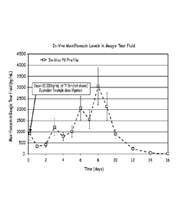
Fig. 18 depicts pharmacokinetic data for drug release from a hydrogel as
detailed in
Example 21;
Fig. 19 depicts pharmacokinetic data for drug release from a hydrogel as
detailed in
Example 22;
Fig. 20 is a plot showing in-vitro release of drugs entrapped in hydrogel
compared to
drug substance alone in saline solution, with the hydrogel affecting the rate
of release, as
detailed in Example 23;
Fig 21A relates to stretching of hydrogels;
Fig 21B relates to stretching of hydrogels; and
Fig 22 is a graph demonstrating alterations of release profiles by
manipulation of
polymer molecular weights used to make microsphere.
4
CA 02750242 2011-07-20
WO 2010/093873 PCT/US2010/024029
DETAILED DESCRIPTION
One embodiment is punctal plug formed of a covalently erosslinked hydrophilic
polymer that absorbs water to form a hydrolytically biodegradable hydrogel
that contacts
and absorbs water to swell in situ to expand a canaliculus for fimi and stable
placement and
to conformably fit the canaliculus, with the hydrogel comprising a drug for
controlled
release to an eye and having a high water content.
Controlled release is a complex subject area. Many drugs need to be present at
a
concentration that at least meets a threshold value. At the same time, a
concentration that is
too high may have unwanted side effects. In general, a zero-order release
profile is useful.
Zero-order release refers to a system wherein release is constant over time,
at a rate which is
independent of changes to the concentration of the reactant(s). Diffusion
processes tend to
be a function of the concentration of the drug, however, so that the amount of
drug released
per unit of time tends to drop as the concentration of the drug declines. In
the case of
degradable materials, the situation can be more complex if degradation affects
the drug
release rate. Various approaches have been developed. One approach is to trap
a matrix in
a device that allows fluid access only to a portion of a reservoir, with a
diffusion-limiting
material controlling release. Or another approach is a material that allows a
surface of
constant area to be eroded. Or reservoir-based approaches have been used.
Disclosed and exemplified herein, however, are hydrogel materials with agent
release rates that are substantially zero-order over a predetermined time,
wherein the
materials have an the agent dispersed throughout the material, either with or
without
encapsulation. These agents may be free of non-hydrogel materials, e.g., no
reservoir area,
no diffusion membrane barriers, and no sleeves that control release rates. The
hydrogel
materials are hydrophilic and allow aqueous solutions (physiological fluids)
to penetrate
through the material. Moreover, the hydrogel systems may be degradable, and
hydrolytically degradable. As is evident, the design factors to obtain a zero-
order release
hydrogel system are in competition with each other. Prior to performing
experimentation as
described herein, it was not known if such systems could be developed to
deliver effective
amounts of drugs to an eye.
In the case of punctal plugs, it is hypothesized, without being bound to a
particular
theory, that physiological fluids build up on top of a canalicular plug and
provide a fluid
column that tends to allow agent release to be limited by the cross-sectional
area of the
proximal portion of the plug. The walls of the canaliculus seem to elute drug
at a rate that is
much slower relative to the depletion of the therapeutic agent through the
fluid column.
5
CA 02750242 2011-07-20
WO 2010/093873 PCT/US2010/024029
Alternatively, or additionally, the canaliculus walls may become saturated
with the drug so
that release through the walls is slowed, and egress of the drug shifts to the
ends of a plug.
Therefore many of the barrier or reservoir or other relatively more complex
systems that are
conventionally proposed may provide little true benefit. In the case of plugs
with a portion
outside the canaliculus, it is hypothesized that the fluid column penetrates
into the hydrogel
to accomplish the same effect. Nonetheless, there are considerable challenges
to be
overcome when concentrating drugs into a small volume while releasing only a
small
portion per unit of time.
The hydrogel plug imbibes water and thereby generates force to stay in the
punctum
or lacrimal canal; in some embodiments, the hydrogel swells 500% or more in
water when it
is not constrained. The hydrogel is covalently crosslinked so that it is
resilient, draws water
into itself and holds it in the hydrogel to generate swelling forces, and is
not re-formed into
a different shape when a patient rubs their eye or the hydrogel is otherwise
strained and
deformed. The drug is incorporated into the hydrogel so as to provide a
desired release
profile, with microencapsulation, micellization, or dispersion being
embodiments for drug
release. Conjugation of drug to the hydrogel molecular network or to a large
molecule
trapped within the hydrogel matrix are also motifs that can be used to
modulate drug
release.
Punctal plugs fall into one of two groups: punctal plugs, which are placed at
the tops
of the puncta (referred to herein as punctum plugs), or intracanalicular
plugs, which are
inserted into the eanalicula. Both permanent (stable until retrieved) and
temporary plugs
(biodegradable) are available. Temporary plugs are usually made of collagen
and are
conventionally designed to last long enough to determine whether a patient can
benefit from
plugging. Extended duration temporary plugs are typically made of synthetics
such as
poly(caprolactone-co-lactide) and poly(glycolide-co-trimethylenecarbonate).
Permanent
punctum plugs and intracanalicular plugs are generally made of silicone. One
peituanent
plug is made of a hydrophobic acrylic polymer that changes shape as it is
warmed to body
temperature and changes from rigid to pliable. Another petnianent plug is made
of a non-
degradable dried hydrogel that swells when exposed to tear fluid.
Figure 1 depicts the punctum and lacrimal canals. The eye 100 has upper eyelid
102, lower eyelid 104, pupil 106, lacrimal gland 108, superior punctum 110,
inferior
punctum 112, superior lacrimal canal 114, inferior lacrimal canal 116,
lacrimal sac 118, and
nasolacrimal canal 120. The lacrimal canaliculi, also known as the lacrimal
canals 112, 114
or lacrimal ducts, are the small channels in each eyelid 102, 104 that
commence at minute
6
CA 02750242 2016-08-04
52486-15
orifices, termed puncta lacrimalia, or punctums, 110, 112 on the summits of
the papillae
lacrimales, seen on the margins of the lids at the lateral extremity of the
lacus lacrimalis.
The superior lacrimal canal 114, the smaller and shorter of the two, at first
ascends, and then
bends at an acute angle, and passes medialward and downward to the lacrimal
sac 118. The
inferior lacrimal canal 116 at first descends, and then runs approximately
horizontally to
lacrimal sac 118. At the angles they are dilated into ampullae.
Microscopically, they are
lined by nonkeratinizing stratified squamous epithelium surrounded by fibrous
tissue having
a further outer layer of striped muscle, continuous with the lacrimal part of
the Orbicularis
oculi. At the base of each lacrimal papilla the muscle fibers are arranged
circularly and
form a kind of sphincter.
Figure 2A depicts forceps 200 gripping dehydrated plug 202. Figure 2B shows
use
of forceps 200 to place plug 202 into inferior punctum 112 and/or inferior
lacrimal canal
116. The plug is initially not swollen, as at Figure 2C, which depicts
placement of all of the
plug into a lacrimal canal. The plug imbibes physiological fluid from its
surroundings and
swells as at Fig 2D. Alternatively, the plug may be placed with at least a
portion passing
through a punctum, as at Figure 2E, with subsequent swelling leaving a head
portion. The
hydrogel has internal covalent crosslinks between its polymeric members so
that even when
it swells in all directions, the constraints on its volume caused by the lumen
of the canal
prevent it from unduly lengthening; the swelling firmly positions the plug in
place but the
swelling does not force the plug out of its location.
US 3,949,750 describes a
conventional punctum plug. A rod-like plug is formed with an oversized tip or
barb portion
that dilates and blockingly projects into the canaliculus, a smaller neck or
waist portion
upon which the punctum sphincter ring tightens, and a relatively larger,
smooth head
portion which rests upon the top of the punctal opening and prevents the plug
from passing
down into the canaliculus. The head portion sits upon the body portion, which
optionally
has the waist portion. A typical method for inserting the plug into the
punctal opening
utilizes a dilator tool for enlarging the punctum and associated canaliculus
and an inserter
tool for facilitating the grasping, manipulation and insertion of the plug.
Figure 3A depicts syringe system 300 with barrel 302, needle hilt 303, needle
304
with rounded tip 306 having outlet 308 and plunger 310 with pusher 312. A
solution of
hydrogel precursors 314 may be placed in barrel 302 and dispensed through
needle 304 and
out outlet 308. One embodiment of syringe system 300 is depicted in Figure 3B,
which
shows alternative needle 318 with a hydrophobic coating 320 that produces a
high contact
7
CA 02750242 2011-07-20
WO 2010/093873 PCT/US2010/024029
angle between the needle and precursor solution 314 to assist forming drop 322
and/or assist
in leaving the solution 314 after it is placed in the patient by virtue of the
resistance of the
needle to spreading of solution 314.
Figure 4A depicts syringe system 300 being used to introduce hydrogel
precursors
314 into canal 112, with the precursors being left in canal 112. The
precursors form
covalent bonds with each other to create a crosslinked hydrogel plug 402. The
hydrogel
402 swells as fluids are imbibed from its surroundings, as at Figure 4C
showing plug 402 in
swollen state 404, pressing against the lumen of canal 112 and expanding the
canal. The
introduction of hydrogel precursors in a fluid state with subsequent formation
of the
hydrogel is referred to as in situ formation of the hydrogel because the
hydrogel is created at
the site of its intended use.
Hydrogels are materials that do not dissolve in water and retain a significant
fraction
(more than 20%) of water within their structure (Szycher's dictionary of
Biomaterials and
Medical Devices, Technomic Pub. Co., Lancaster, 1992). In fact, water contents
in excess
of 90% are often known. Hydrogels are often fanned by crosslinking water
soluble
molecules to form networks of essentially infinite molecular weight. Hydrogels
with high
water contents are typically soft, pliable materials. When made with pliable
materials, high-
content hydrogels are comfortably worn in the eye and avoid the foreign-object
sensation
that can accompany more rigid materials, for instance plugs made of polylactic
acid (PLA)
and/or polyglycolic acid (PGA). A hydrogel that has been dried is referred to
herein as a
dehydrated hydrogel if it will return to a hydrogel state upon exposure to
water (also
referred to as a xerogel); this hydrogel that would expand in volume if it
were exposed to an
excess of water and not constrained. The tenn desiccated refers to a hydrogel
essentially
having no fluids, bearing in mind that some trace amounts of water may
nonetheless be
present.
A hydrogel network may be formed in a non-water solvent with a therapeutic
agent
optionally being present at the time of hydrogel network formation or loaded
afterwards.
The non-water solvent can then be replaced with water by a suitable means to
than the
hydrogel. The term therapeutic agent includes diagnostic agents, imaging
agents, and
drugs. The term drug refers to an agent intended to provoke a biological
response so as to
treat a patient. The hydrogels may be biodegradable or non-biodegradable.
The therapeutic agent may be dispersed (meaning spread substantially
throughout a
structure, either as a solution, suspension, or a colloid) within the
hydrogel. The agent may
be dispersed in the same phase as the fluid hydrating the hydrogel or it may
be contained in
8
CA 02750242 2016-08-04
52486-15
a phase discontinuous from the fluid in the hydrogel. A phase discontinuous
from the
hydrogel may be a micelle, droplet, or a particle. Accordingly, a drug
entrapped within
microspheres dispersed within a hydrogel is a drug dispersed within the
hydrogel. By way
of contrast, a drug localized to a reservoir is not dispersed. A micelle,
droplet, or a particle
may include, for instance, a mixture of the drug with another material, e.g.,
a polymer. One
embodiment of a particle is a capsule with the drug inside the capsule.
Another
embodiment of a particle is a solid formed by a polymer that associates with
the drug. A
particle may release a drug as it degrades, by diffusion, or a combination
thereof. These
features may be combined to provide a desired agent-release profile. A
hydrogel with an
agent dispersed through the hydrogel refers to a continuous hydrogel matrix
with a
substantially even distribution of agent throughout the structure.
Placement
Punctal plugs may be placed in, or partially within a lacrimal canal. Forceps
or
other applicators may be used to grasp the plugs and insert them. Or
precursors may be
placed into a lacrimal canal and allowed to crosslink to form a plug. Another
option is
placement of microspheres and/or a hydrogel (dehydrated, desiccated, partially
hydrated, or
hydrated) within the conjunctival cul-de-sac, between the lower lid and the
eye. Examples
of other devices that use this placement sit are disclosed in U.S. Pat. No.
3,618,604, U.S.
Pat. No. 3,626,940, U.S. Pat. No. 3,845,770, U.S. Pat. No. 3,962,414, U.S.
Pat. No.
3,993,071, and U.S. Pat. No. 4,014,335, ,
with the specification herein controlling in case of conflict.
Another option is to place microspheres and/or a hydrogel subconjunctivally
between the conjunctiva and the sclera. For instance, a syringe may be used to
pierce the
conjunctiva without piercing the sclera, and the hydrogel and/or microspheres
and/or
hydrogel precursors injected to form a depot. One or more of these components
may be
formed as a degradable material. Another option is to form such material(s)
topically on a
surface of the eye (e.g., cornea or a site that is topical but avoids the
cornea). Therapeutic
agents in these material(s) may then be released over time to accomplish a
therapy. As is
evident, the various embodiments set forth herein may be thusly administered.
A micropdepot may be formed with such materials and optionally at a
subconjunctival site. For instance, a volume of 5-400 ul may be formed;
artisans will
immediately appreciate that all the ranges and values within the explicitly
stated ranges are
contemplated, e.g, about 5 to about 30 al or from about 20 to about 100 1.
9
CA 02750242 2016-08-04
52486-15
Hydrogel Precursors
Hydrogels may be made from precursors. The precursors are not hydro gels but
are
covalently crosslinked with each other to form a hydrogel and are thereby part
of the
hydrogel. Crosslinks can be formed by covalent or ionic bonds, by hydrophobic
association
of precursor molecule segments, or by crystallization of precursor molecule
segments. The
precursors can be triggered to react to form a crosslinked hydrogel. The
precursors can be
polymerizable and include crosslinkers that are often, but not always,
polymerizable
precursors. Polymerizable precursors are thus precursors that have functional
groups that
react with each other to form polymers made of repeating units. Precursors may
be
polymers.
Some precursors thus react by chain-growth polymerization, also referred to as
addition polymerization, and involve the linking together of monomers
incorporating double
or triple chemical bonds. These unsaturated monomers have extra internal bonds
which are
able to break and link up with other monomers to form the repeating chain.
Monomers are
polymerizable molecules with at least one group that reacts with other groups
to form a
polymer. A macromonomer (or macromer) is a polymer or oligomer that has at
least one
reactive group, often at the end, which enables it to act as a monomer; each
macromonomer
molecule is attached to the polymer by reaction the reactive group. Thus
macromonomers
with two or more monomers or other functional groups tend to form covalent
crosslinks.
Addition polymerization is involved in the manufacture of, e.g., polypropylene
or polyvinyl
chloride. One type of addition polymerization is living polymerization.
Some precursors thus react by condensation polymerization that occurs when
monomers bond together through condensation reactions. Typically these
reactions can be
achieved through reacting molecules incorporating alcohol, amine or carboxylic
acid (or
other carboxyl derivative) functional groups. When an amine reacts with a
carboxylic acid
an amide or peptide bond is formed, with the release of water. Some
condensation reactions
follow a nueleophilic acyl substitution, e.g., as in U.S. Pat. No. 6,958,212,
referenced herein in its entirety to the extent it does not contradict what is
explicitly disclosed herein.
Some precursors react by a chain growth mechanism. Chain growth polymers are
defined as polymers formed by the reaction of monomers or macrornonomers with
a
reactive center. A reactive center is a particular location within a chemical
compound that
is the initiator of a reaction in which the chemical is involved. In chain-
growth polymer
chemistry, this is also the point of propagation for a growing chain. The
reactive center is
CA 02750242 2016-08-04
52486-15
commonly radical, anionic, or cationic in nature, but can also take other
forms. Chain
growth systems include free radical polymerization, which involves a process
of initiation,
propagation and termination. Initiation is the creation of free radicals
necessary for
propagation, as created from radical initiators, e.g., organic peroxide
molecules.
Termination occurs when a radical reacts in a way that prevents further
propagation. The
most common method of termination is by coupling where two radical species
react with
each other forming a single molecule.
Some precursors react by a step growth mechanism, and are polymers formed by
the
stepwise reaction between functional groups of monomers. Most step growth
polymers are
also classified as condensation polymers, but not all step growth polymers
release
condensates.
Monomers may be polymers or small molecules. A polymer is a high molecular
weight molecule formed by combining many smaller molecules (monomers) in a
regular
pattern. Oligomers are polymers having less than about 20 monomeric repeat
units. A
small molecule generally refers to a molecule that is less than about 2000
Daltons.
The precursors must thus be small molecules, such as acrylic acid or vinyl
caprolactam, larger molecules containing polymerizable groups, such as
acrylate-capped
polyethylene glycol (PEG-diacrylate), or other polymers containing
ethylenically-
unsaturated groups, such as those of U.S. Pat. No. 4,938,763 to Dunn et al,
U.S. Pat. Nos.
5,100,992 and 4,826,945 to Cohn et at, or U.S. Pat. Nos. 4,741,872 and
5,160,745 to
DeLuca et al., each of which is referenced herein in its entirety to the
extent it does not contradict what is explicitly disclosed herein.
To form covalently crosslinked hydrogels, the precursors must be crosslinked
together. In general, polymeric precursors will form polymers that will be
joined to other
polymeric precursors at two or more points, with each point being a linkage to
the same or
different polymers. Precursors with at least two reactive groups can serve as
crosslinkers
since each reactive group can participate in the formation of a different
growing polymer
chain. In the case of functional groups without a reactive center, among
others, crosslinking
requires three or more such functional groups on a precursor. For instance,
many
electrophilic-nucleophilic reactions consume the electrophilic and
nucleophilic fiinctional
groups so that a third functional group is needed for the precursor to form a
crosslink. Such
precursors thus may have three or more functional groups and may be
crosslinked by
precursors with two or more functional groups. A crosslinked molecule may be
crosslinked
11
CA 02750242 2011-07-20
WO 2010/093873 PCT/US2010/024029
via an ionic or covalent bond, a physical force, or other attraction. A
covalent crosslink,
however, will typically offer stability and predictability in reactant product
architecture.
In some embodiments, each precursor is multifunctional, meaning that it
comprises
two or more electrophilic or nucleophilic functional groups, such that a
nucleophilic
functional group on one precursor may react with an electrophilic functional
group on
another precursor to form a covalent bond. At least one of the precursors
comprises more
than two functional groups, so that, as a result of electrophilic-nucleophilic
reactions, the
precursors combine to form crosslinked polymeric products.
The precursors may have biologically inert and hydrophilic portions, e.g., a
core. In
the case of a branched polymer, a core refers to a contiguous portion of a
molecule joined to
arms that extend from the core, with the arms having a functional group, which
is often at
the terminus of the branch. The hydrophilic precursor or precursor portion
preferably has a
solubility of at least 1 g/100 mL in an aqueous solution. A hydrophilic
portion may be, for
instance, a polyether, for example, polyalkylene oxides such as polyethylene
glycol (PEG),
polyethylene oxide (PEO), polyethylene oxide-co-polypropylene oxide (PPO), co-
polyethylene oxide block or random copolymers, and polyvinyl alcohol (PVA),
poly (vinyl
pyrrolidinone) (PVP), poly (amino acids, dextran, or a protein. The precursors
may have a
polyalkylene glycol portion and may be polyethylene glycol based, with at
least about 80%
or 90% by weight of the polymer comprising polyethylene oxide repeats. The
polyethers
and more particularly poly (oxyalkylenes) or poly (ethylene glycol) or
polyethylene glycol
are generally hydrophilic.
A precursor may also be a macromolecule (or macromer), which is a molecule
having a molecular weight in the range of a few thousand to many millions. In
some
embodiments, however, at least one of the precursors is a small molecule of
about 1000 Da
or less. The macromolecule, when reacted in combination with a small molecule
of about
1000 Da or less, is preferably at least five to fifty times greater in
molecular weight than the
small molecule and is preferably less than about 60,000 Da; artisans will
immediately
appreciate that all the ranges and values within the explicitly stated ranges
are
contemplated. A more preferred range is a macromolecule that is about seven to
about
thirty times greater in molecular weight than the crosslinker and a most
preferred range is
about ten to twenty times difference in weight. Further, a macromolecular
molecular weight
of 5,000 to 50,000 is useful, as is a molecular weight of 7,000 to 40,000 or a
molecular
weight of 10,000 to 20,000.
12
CA 02750242 2016-08-04
52486-15
Certain macromeric precursors are the crosslinkable, biodegradable, water-
soluble
macromers described in U.S. Pat. No. 5,410,016 to Hubbell et al, -which is
referenced herein in its entirety to the extent it does not contradict what is
explicitly disclosed. These macromers are characterized by having at least two
polymerizable groups, separated by at least one degradable region.
Synthetic precursors may be used. Synthetic refers to a molecule not found in
nature
or not normally found in a human. Some synthetic polymers are free of amino
acids or free
of amino acid sequences that occur in nature. Some synthetic molecules are
polypeptides
that are not found in nature or are not normally found in a human body, e.g.,
di-, tri-, or
tetra-lysine. Some synthetic molecules have amino acid residues but only have
one, two, or
three that are contiguous, with the amino acids or clusters thereof being
separated by non-
natural polymers or groups. Polysaccharides or their derivatives are thus not
synthetic.
Precursors may be made with a hydrophobic portion provided that the resultant
hydrogel retains the requisite amount of water, e.g., a t least about 20%. In
some cases, the
precursor is nonetheless soluble in water because it also has a hydrophilic
portion. In other
cases, the precursor makes dispersion in the water (a suspension) but is
nonetheless
reactable to from a crosslinked material. Some hydrophobic portions may
include a
plurality of alkyls, polypropylenes, alkyl chains, or other groups. Some
precursors with
hydrophobic portions are sold under the trade names PLURONIC F68, JEFFAMINE,
or
TECTRONIC. A hydrophobic portion is one that is sufficiently hydrophobic to
cause the
macromer or copolymer to aggregate to form micelles in an aqueous continuous
phase or
one that, when tested by itself, is sufficiently hydrophobic to precipitate
from, or otherwise
change phase while within, an aqueous solution of water at pH from about 7 to
about 7.5 at
temperatures from about 30 to about 50 degrees Centigrade.
Precursors may have, e.g., 2-100 arms, with each arm having a terminus,
bearing in
mind that some precursors may be dendrirners or other highly branched
materials. An arm
on a hydrogel precursor refers to a linear chain of chemical groups that
connect a
crosslinkable functional group to a polymer core. Some embodiments are
precursors with
between 3 and 300 arms; artisans will immediately appreciate that all the
ranges and values
within the explicitly stated ranges are contemplated, e.g., 4 to 16, 8 to 100,
or at least 6
arms.
Thus hydrogels can be made, e.g., from a multi-armed precursor with a first
set of
functional groups and a low molecular-weight precursor having a second set of
functional
groups. For example, a six-armed or eight-armed precursor may have hydrophilic
arms,
13
CA 02750242 2016-08-04
52486-15
e.g., polyethylene glycol, terminated with primary amines, with the molecular
weight of the
arms being about 1,000 to about 40,000; artisans will immediately appreciate
that all ranges
and values within the explicitly stated bounds are contemplated. Such
precursors may be
mixed with relatively smaller precursors, for example, molecules with a
molecular weight
of between about 100 and about 5000, or no more than about 800, 1000, 2000, or
5000
having at least about three functional groups, or between about 3 to about 16
functional
groups; ordinary artisans will appreciate that all ranges and values between
these explicitly
articulated values are contemplated. Such small molecules may be polymers or
non-
polymers and natural or synthetic.
Precursors that are not dendrimers may be used. Dendritic molecules are highly
branched radially symmetrical polymers in which the atoms are arranged in many
arms and
subanns radiating out from a central core. Dendrimers are characterized by
their degree of
structural perfection as based on the evaluation of both symmetry and
polydispersity and
require particular chemical processes to synthesize. Accordingly, an artisan
can readily
distinguish dendrimer precursors from non-dendrimer precursors. Dendrimers
have a shape
that is typically dependent on the solubility of its component polymers in a
given
environment, and can change substantially according to the solvent or solutes
around it, e.g.,
changes in temperature, pH, or ion content.
Precursors may be dendrimers, e.g., as in Patent Application Pub. Nos.
US20040086479, US20040131582, W007005249, W007001926, W006031358, or the
U.S. counterparts thereof; dendrimers may also be useful as multifunctional
precursors, e.g.,
as in U.S. Pat. Pub. Nos. US20040131582, US20040086479 and PCT Applications
No.
W006031388 and W006031388; each of which US and PCT applications are
referenced herein in its entirety to the extent they do not contradict what is
explicitly disclosed herein. Dendrimers are highly ordered possess high
surface area to
volume ratios, and exhibit numerous end groups for potential
functionalization.
Embodiments include multifunctional precursors that are not dendrimers.
Some embodiments include a precursor that consists essentially of an
oligopeptide
sequence of no more than five residues, e.g., amino acids comprising at least
one amine,
thiol, carboxyl, or hydroxyl side chain. A residue is an amino acid, either as
occurring in
nature or derivatized thereof. The backbone of such an oligopeptide may be
natural or
synthetic. In some embodiments, peptides of two or more amino acids are
combined with a
synthetic backbone to make a precursor; certain embodiments of such precursors
have a
molecular weight in the range of about 100 to about 10,000 or about 300 to
about 500
14
CA 02750242 2011-07-20
WO 2010/093873 PCT/US2010/024029
Artisans will immediately appreciate that all ranges and values between these
explicitly
articulated bounds are contemplated.
Precursors may be prepared to be free of amino acid sequences cleavable by
enzymes present at the site of introduction, including free of sequences
susceptible to attach
by metalloproteinases and/or collagenases. Further, precursors may be made to
be free of
all amino acids, or free of amino acid sequences of more than about 50, 30,
20, 10, 9, 8, 7,
6, 5, 4, 3, 2, or 1 amino acids. Precursors may be non-proteins, meaning that
they are not a
naturally occurring protein and can not be made by cleaving a naturally
occurring protein
and can not be made by adding synthetic materials to a protein. Precursors may
be non-
collagen, non-fibrin, non-fibrinogen), and non-albumin, meaning that they are
not one of
these proteins and are not chemical derivatives of one of these proteins. The
use of non-
protein precursors and limited use of amino acid sequences can be helpful for
avoiding
immune reactions, avoiding unwanted cell recognition, and avoiding the hazards
associated
with using proteins derived from natural sources. Precursors can also be non-
saccharides
(free of saccharides) or essentially non-saccharides (free of more than about
5% saccharides
by w/w of the precursor molecular weight. Thus a precursor may, for example,
exclude
hyaluronic acid, heparin, or gellan. Precursors can also be both non-proteins
and non-
saccharides.
Peptides may be used as precursors. In general, peptides with less than about
10
residues arc preferred, although larger sequences (e.g., proteins) may be
used. Artisans will
immediately appreciate that every range and value within these explicit bounds
is included,
e.g., 1-10, 2-9, 3-10, 1, 2, 3, 4, 5, 6, or 7. Some amino acids have
nucleophilic groups (e.g.,
primary amines or thiols) or groups that can be derivatized as needed to
incorporate
nueleophilic groups or electrophilic groups (e.g., carboxyls or hydroxyls).
Polyamino acid
polymers generated synthetically are normally considered to be synthetic if
they are not
found in nature and are engineered not to be identical to naturally occurring
biomolecules.
Some hydrogels are made with a polyethylene glycol-containing precursor.
Polyethylene glycol (PEG, also referred to as polyethylene oxide when
occurring in a high
molecular weight) refers to a polymer with a repeat group (CH2CH20)., with n
being at
least 3. A polymeric precursor having a polyethylene glycol thus has at least
three of these
repeat groups connected to each other in a linear series. The polyethylene
glycol content of
a polymer or arm is calculated by adding up all of the polyethylene glycol
groups on the
polymer or arm, even if they are interrupted by other groups. Thus, an arm
having at least
1000 MW polyethylene glycol has enough CH2CH20 groups to total at least 1000
MW. As
CA 02750242 2011-07-20
WO 2010/093873 PCT/US2010/024029
is customary terminology in these arts, a polyethylene glycol polymer does not
necessarily
refer to a molecule that terminates in a hydroxyl group.
Initiating Systems
Some precursors react using initiators. An initiator group is a chemical group
capable of initiating chain growth (e.g., a free radical) polymerization
reaction. For
instance, it may be present as a separate component, or as a pendent group on
a precursor.
Free radical initiator groups include thermal initiators, photoactivatable
initiators, and
oxidation-reduction (redox) systems. Long wave UV and visible light
photoactivatable
initiators include, for example, ethyl eosin groups, 2, 2-dimethoxy-2-phenyl
acetophenone
groups, other acetophenone derivatives, thioxanthone groups, benzophenone
groups, and
camphorquinone groups. Examples of thermally reactive initiators include 4, 4'
azobis (4-
cyanopentanoic acid) groups, and analogs of benzoyl peroxide groups.
Several
commercially available low temperature free radical initiators, such as V-044,
available
from Wako Chemicals USA, Inc., Richmond, Va., may be used to initiate free
radical
crosslinking reactions at body temperatures to form hydrogels with the
aforementioned
monomers.
Metal ions may be used either as an oxidizer or a reductant in redox
initiating
systems. For example, ferrous ions may be used in combination with a peroxide
or
hydroperoxide to initiate polymerization, or as parts of a polymerization
system. In this
case, the ferrous ions would serve as a reductant. Alternatively, metal ions
may serve as an
oxidant. For example, the eerie ion (4+ valence state of cerium) interacts
with various
organic groups, including carboxylic acids and urethanes, to remove an
electron to the metal
ion, and leave an initiating radical behind on the organic group. In such a
system, the metal
ion acts as an oxidizer. Potentially suitable metal ions for either role are
any of the
transition metal ions, lanthanides and actinides, which have at least two
readily accessible
oxidation states. Particularly useful metal ions have at least two states
separated by only
one difference in charge. Of these, the most commonly used are ferric/ferrous;
cupric/cuprous; ceric/cerous; cobaltic/cobaltous; vanadate V vs. IV;
permanganate; and
manganichnanganous.
Peroxygen containing compounds, such as peroxides and
hydroperoxides, including hydrogen peroxide, t-butyl hydroperoxide, t-butyl
peroxide,
benzoyl peroxide, cumyl peroxide may be used.
An example of an initiating system is the combination of a peroxygen compound
in
one solution, and a reactive ion, such as a transition metal, in another. In
this case, no
16
CA 02750242 2016-08-04
52486-15
external initiators of polymerization are needed and polymerization proceeds
spontaneously
and without application of external energy or use of an external energy source
when two
complementary reactive functional groups containing moieties interact at the
application
site.
=
Functional Groups
The precursors have functional groups that react with each other to form the
material, either outside a patient, or in situ. The functional groups
generally have
polymerizable groups for polymerization or react with each other in
electrophile-
nucleophile reactions or are configured to participate in other polymerization
reactions.
Various aspects of polymerization reactions are discussed in the precursors
section herein.
Thus in some embodiments, precursors have a polymerizable group that is
activated
by photoinitiation or redox systems as used in the polymerization arts, e.g.,
or electrophilic
functional groups that are carbodiimidazole, sulfonyl chloride,
chlorocarbonates, n-
hydroxysuccinimidyl ester, succinimidyl ester or sulfasuccinimidyl esters, or
as in U.S. Pat.
Nos. 5,410,016, or 6,149,931, each of which are referenced herein in
its entirety to the extent they do not contradict what is explicitly disclosed
herein. The
nucleophilic functional groups may be, for example, amine, hydroxyl, carboxyl,
and thiol.
Another class of electrophiIes are acyls, e.g., as in U.S. Pat. No. 6,958,212,
which describes,
among other things, Michael addition schemes for reacting polymers.
Certain functional groups, such as alcohols or carboxylic acids, do not
normally
react with other functional groups, such as amines, under physiological
conditions (e.g., pH
7.2-11.0, 37 C). However, such functional groups can be made more reactive by
using an
activating group such as N-hydroxysuceinimide. Certain activating groups
include
carbonyldiimidazole, sulfonyl chloride, aryl halides, sulfosuccinimidyl
esters, N-
hydroxysuccinimidyl ester, succinimidyl ester, epoxide, aldehyde, maleimides,
imido esters
and the like. The N-hydroxysuccinimide esters or N-hydroxysulfosuccinimide
(NHS)
groups are useful groups for crosslinlcing of proteins or amine-containing
polymers, e.g.,
amino terminated polyethylene glycol. An advantage of an NHS-amine reaction is
that the
reaction kinetics are favorable, but the gelation rate may be adjusted through
pH or
concentration. The NHS-
amine crosslinking reaction leads to formation of N-
hydroxysuceinimide as a side product. Sulfonated
or ethoxylated forms of N-
hydroxysuceinirnide have a relatively increased solubility in water and hence
their rapid
clearance from the body. An NHS-amine crosslinking reaction may be carried out
in
17
CA 02750242 2011-07-20
WO 2010/093873 PCT/US2010/024029
aqueous solutions and in the presence of buffers, e.g., phosphate buffer (pH
5.0-7.5),
triethanolamine buffer (pH 7.5-9.0), or borate buffer (pH 9.0-12), or sodium
bicarbonate
buffer (pH 9.0-10.0). Aqueous solutions of NHS based crosslinkers and
functional
polymers preferably are made just before the crosslinking reaction due to
reaction of NHS
groups with water. The reaction rate of these groups may be delayed by keeping
these
solutions at lower pH (pH 4-7).
In some embodiments, each precursor comprises only nucleophilic or only
electrophilic functional groups, so long as both nucleophilic and
electrophilic precursors are
used in the crosslinking reaction. Thus, for example, if a crosslinker has
nucleophilic
functional groups such as amines, the functional polymer may have
electrophilic functional
groups such as N-hydroxysuccinimides. On the other hand, if a crosslinker has
electrophilic
functional groups such as sulfosuccinimides, then the functional polymer may
have
nucleophilic functional groups such as amines or thiols. Thus, functional
polymers such as
proteins, poly(ally1 amine), or amine-terminated di-or multifunctional
poly(ethylene glycol)
can be used.
One embodiment has reactive precursor species with 3 to 16 nucleophilic
functional
groups each and reactive precursor species with 2 to 12 electrophilic
functional groups each;
artisans will immediately appreciate that all the ranges and values within the
explicitly
stated ranges are contemplated.
The functional groups may be, e.g., electrophiles reactable with nucleophiles,
groups
reactable with specific nucleophiles, e.g., primary amines, groups that form
amide bonds
with materials in the biological fluids, groups that form amide bonds with
carboxyls,
activated-acid functional groups, or a combination of the same. The functional
groups may
be, e.g., a strong electrophilic functional group, meaning an electrophilic
functional group
that effectively forms a covalent bond with a primary amine in aqueous
solution at pH 9.0 at
room temperature and pressure and/or an electrophilic group that reacts by a
of Michael-
type reaction. The strong electrophile may be of a type that does not
participate in a
Michaels-type reaction or of a type that participates in a Michaels-type
reaction.
A Michael-type reaction refers to the 1,4 addition reaction of a nucleophile
on a
conjugate unsaturated system. The addition mechanism could be purely polar, or
proceed
through a radical-like intermediate state(s); Lewis acids or appropriately
designed hydrogen
bonding species can act as catalysts. The term conjugation can refer both to
alternation of
carbon-carbon, carbon-heteroatom or heteroatom-heteroatom multiple bonds with
single
bonds, or to the linking of a functional group to a macromolecule, such as a
synthetic
18
CA 02750242 2016-08-04
52486-15
polymer or a protein. Michael-type reactions are discussed in detail in U.S.
Pat. No.
6,958,212, which is referenced herein in its entirety for all purposes
to the extent it does not contradict what is explicitly disclosed herein.
Examples of strong electrophiles that do not participate in a Michaels-type
reaction
are: succinimides, succinimidyl esters, or NHS-esters. Examples of Michael-
type
electrophiles are acrylates, methacrylates, methylmethacrylates, and other
unsaturated
polymerizable groups.
Hydro gels and Hydrogel Formation
In general, the precursors may be combined to make a covalently-crosslinked
hydrogel. The hydrogel may comprise a therapeutic agent, or agents, released
over a
suitable period of time. Hydrogels may be made beforehand or in situ.
When made in situ, the crosslinking reactions generally occur in aqueous
solution
under physiological conditions. The crosslinking reactions preferably do not
release heat of
polymerization or require exogenous energy sources for initiation or to
trigger
polymerization. Photochemical initiation, for instance, is generally to be
avoided in the eye
so as to avoid damage to the eye. In the case of injected materials, the
viscosity may be
controlled so that the material is introduced through a small diameter
catheter or needle.
When hydrogels are made beforehand, the polymers made be made in aqueous
and/or
organic solvents.
The hydrogel is generally high-swelling, as measurable by the hydrogel having
a
weight increasing more than about 50% upon exposure to a physiological
solution in the
absence of physical restraints for twenty-four hours relative to a weight of
the hydrogel at
the time of formation. Swelling may be measured or expressed by weight or
volume. Some
embodiments swell by weight or by volume more than about 1000%, more than
about
500%, or more than about 100%; artisans will immediately appreciate that all
the ranges and
values within the explicitly stated ranges are contemplated, e.g., more than
about 200% or
from about 300% to about 1000%. Accordingly, some embodiments include
hydrogels that
swell by weight or by volume between about 100% to about 2000%, between about
200%
to about 1500%, or between about 300% and about 1100%; artisans will
immediately
appreciate that all the ranges and values within the explicitly stated ranges
are
contemplated.
One approach for high-swelling is to control the number of crosslinks. Another
embodiment is mixing-into the hydrogel precursors a high molecular weight
water soluble
19
CA 02750242 2011-07-20
WO 2010/093873 PCT/US2010/024029
synthetic or natural polymer that does not covalently cross-link with the
precursors to
achieve a crosslinked hydrogel with these other materials dispersed therein.
Examples of
such materials are carboxy methyl cellulose, hyaluronic acid, and high
molecular weight
PEG, e.g., the high molecular weight being more than about 100,000 MW, e.g.,
from about
100,000 to about 10,000,000; artisans will immediately appreciate that all the
ranges and
values within the explicitly stated ranges are contemplated, e.g., from about
250,000 to
about 1,000,000). These added materials can greatly increase swelling of the
cross linked
hydrogel as the highly water soluble large polymers remain entangled within
the network
causing an increased osmotic pressure within the hydrogel structure, thus
causing the
hydrogel to swell more.
In pre-foimed dehydrated hydrogels, a degree of molecular orientation can be
imparted by stretching the material then allowing it to solidify, locking in
the molecular
orientation. This can be accomplished by drawing the material while heated to
a
temperature above the melting point of the crystallizable regions of the
material, then
allowing the crystallizable regions to crystallize. Alternatively, the glass
transition
temperature of the dried hydrogel can be used to lock in the molecular
orientation. Still
another alternative is to draw the gel prior to complete dehydration (or
drying) and then
drying the material while under tension. The molecular orientation provides a
mechanism
for anisotropic swelling upon introduction into a hydrating medium. A rod can
be formed,
however, that will swell only in the radial dimension, neither increasing or
decreasing in
length. Radial swelling may be desirable in a punctum plug, but growth or
shrinkage in
length is sometimes a problem with retention of the device where placed by the
surgeon.
The change in length causes the punctal plug to be forced out or to be
difficult to retrieve.
Accordingly, a radial-swelling punctal plug may be made that is free of
longitudinal
shrinking and/or swelling. The term istotropic means to swell consistently in
all directions
when not constrained. The term anisotropie means to swell preferentially in
one direction
as opposed to another, as in a cylinder that swells predominantly in the
radial direction to
conform to the canaliculus and/or punctum, but does not appreciably expand or
contract in
the longitudinal dimension, thus maintaining its position as placed by the
surgeon. Minimal
length increases in combination with significant radial increases provides
improved
retention of the plug during the course of the therapy.
Another embodiment to increase swelling is to choose precursors that have a
low
degree of solvation at the time of crosslinking but subsequently become more
solvated and
having a radius of solvation that effectively increases; in other words, the
precursor is
CA 02750242 2011-07-20
WO 2010/093873 PCT/US2010/024029
spread-out in solution after crosslinking but relatively contracted when
crosslinked.
Changes to pH, temperature, solids concentration, and solvent environment can
cause such
changes.
Unless otherwise indicated, swelling of a hydrogel relates to its change in
volume
(or weight) between the time of its formation when crosslinking is effectively
complete and
the time after being placed in vitro aqueous solution in an unconstrained
state for twenty-
four hours, at which point it may be reasonably assumed to have achieved its
equilibrium
swelling state. For most embodiments, crosslinking is effectively complete
within no more
than about fifteen minutes such that the initial weight can generally be noted
at about 15
minutes after formation as Weight at initial formation. Accordingly, this
formula is used: %
swelling --- [(Weight at 24 hours - Weight at initial formation)/ Weight at
initial formation] *
100. The weight of the hydrogel includes the weight of the solution in the
hydrogel. A
hydrogel formed in a location wherein it is constrained may nonetheless be
considered a
high-swelling hydrogel because it is the expansion in the unconstrained state
that defines the
amount of swelling. For instance, a swellable hydrogel created in a body may
be
constrained from swelling by its surroundings but nonetheless may be a highly
swellable
hydrogel as evidenced by measurements of its swelling when unconstrained
and/or the
forces against a constraint.
Reaction kinetics are generally controlled in light of the particular
functional groups
unless an external initiator or chain transfer agent is required, in which
case triggering the
initiator or manipulating the transfer agent can be a controlling step. In
some embodiments,
the molecular weights of the precursors are used to affect reaction times.
Precursors with
lower molecular weights tend to speed the reaction due to their higher
concentration of
reactive groups, so that some embodiments have at least one precursor with a
molecular
weight of less than about 1000 or about 2000 Daltons; artisans will
immediately appreciate
that all the ranges and values within the explicitly stated ranges are
contemplated, e.g., from
100 to about 900 Daltons or from 500 to about 1800 Daltons. Preferably the
crosslinking
reaction leading to gelation occurs within less than about 2 to about 10 or to
about 30
minutes; artisans will immediately appreciate that all the ranges and values
within the
explicitly stated ranges are contemplated, e.g., at least 120 seconds, or
between 180 to 600
seconds. Gelation time is measured by applying the precursors to a flat
surface and
determining the time at which there is substantially no flow down the surface
when it is
titled at an angle of about 60 degrees (i.e., a steep angle, close to
perpendicular). In the case
of hydrogel formation in situ, a gelation time of less than about 2 minutes,
or about 1
21
CA 02750242 2011-07-20
WO 2010/093873 PCT/US2010/024029
minute or about 30 seconds is useful; artisans will immediately appreciate
that all the ranges
and values within the explicitly stated ranges are contemplated, e.g., from
about 5 to about
90 seconds or from about 10 to about 40 seconds.
The crosslinking density of the resultant biocompatible crosslinked polymer is
controlled by the overall molecular weight of the crosslinlcer and functional
polymer and the
number of functional groups available per molecule. A lower molecular weight
between
crosslinks such as 500 will give much higher crosslinking density as compared
to a higher
molecular weight such as 10,000. The crosslinking density also may be
controlled by the
overall percent solids of the crosslinker and functional polymer solutions.
Increasing the
percent solids increases the probability that an electrophilic functional
group will combine
with a nucleophilic functional group prior to inactivation by hydrolysis. Yet
another
method to control crosslink density is by adjusting the stoichiometry of
nucleophilic
functional groups to electrophilic functional groups. A one to one ratio leads
to the highest
crosslink density. Precursors with longer distances between crosslinks are
generally softer,
more compliant, and more elastic. Thus an increased length of a water-soluble
segment,
such as a polyethylene glycol, tends to enhance elasticity to produce
desirable physical
properties. Thus certain embodiments are directed to precursors with water
soluble
segments having molecular weights in the range of 3,000 to 100,000 or, e.g.,
10,000 to
35,000.
The solids content of the hydrogel can affect its mechanical properties and
biocompatibility and reflects a balance between competing requirements. A
relatively low
solids content is useful, e.g., between about 2.5% to about 25%, including all
ranges and
values there between, e.g., about 2.5% to about 10%, about 5% to about 15%, or
less than
about 15%.
It has surprisingly been found that loading hydrogels with hydrophobic domains
in
sufficient concentration will be effective to reduce the swelling of
hydrogels. Figure 12
documents this effect. Increased loading of cyclosporine A in the hydrogels
decreased the
overall swelling.
Processes for making hydrogels
Processes for making hydrogels that incorporate a drug include, for example,
making a hydrogel in an organic solvent or in aqueous solution with the drug
present at the
time of formation of the hydrogel or added the hydrogel after its formation.
The hydrogel
22
CA 02750242 2011-07-20
WO 2010/093873 PCT/US2010/024029
may be made beforehand (a preformed device) and at least partially dehydrated
or
desiccated, or made in situ in a solution at the site of formation.
One embodiment for making a hydrogel is to make a preformed device in an
organic-solvent in the presence of a therapeutic agent. A first hydrogel
precursor with a
first type functional groups is dissolved with a second hydrogel precursor
with a second
type of functional groups in an organic solvent in the presence of a
therapeutic agent that is
miscible in the organic solvent. The solution is introduced into a mold and
left until the
precursors crosslink with each other by covalent bond formation between the
first functional
groups and the second functional groups. The hydrogel is fully or partially
dried to folin a
dehydrated or desiccated hydrogel (xerogel). The hydrogel is then removed and
optionally
cut or otherwise trimmed to another shape or size. This embodiment may be
used, for
example, for loading the hydrogel with non-water soluble agents or
encapsulated agents
tolerant to the organic phase.
An embodiment for making a preformed device in an organic-solvent in the
presence of a therapeutic agent is to dissolve a branched polyethylene glycol
with
electrophilic functional groups at each arm terminus with a nucleophilic
precursor in
methanol containing a therapeutic agent. The precursors are formed into a
hydrogel, dried,
and shaped as desired.
Another embodiment for making a hydrogel is to make a preformed device in an
organic solvent and, after the hydrogel is formed, load the hydrogel with a
therapeutic
agent. A first hydrogel precursor with a first type functional groups is
dissolved with a
second hydrogel precursor with a second type of functional groups in an
organic solvent.
The solution is introduced into a mold and left until the precursors crosslink
with each other
by covalent bond foiniation between the first functional groups and the second
functional
groups. The hydrogel is fully or partially dried to form a dehydrated or
desiccated hydrogel.
An organic solvent (the same or different from the one used during
crosslinking) that swells
the cross-linked hydrogels is added. This solvent contains dissolved agents at
high
concentrations. The hydrogels are allowed to swell with the organic drug
solution, causing
some drug to permeate into the hydrogel matrix. Gels are removed, and either
dried again
as above, or placed into a non-solvent, e.g., hexane. The non-solvent causes
the organic
solvent to leave the gel and the agent to precipitate-out in the gel matrix,
leaving an agent-
loaded plug. This embodiment may be used, for example, for loading of agents
incompatible with the particular crosslinking functional groups, e.g., agents
with primary
amines when precursor amine functional groups are intended to be reacted
during
23
CA 02750242 2011-07-20
WO 2010/093873 PCT/US2010/024029
crosslinking. This separation of drug loading and crosslinking steps removes
problems with
chemical incompatibility between the therapeutic agent and the crosslinking
reaction.
An embodiment for making a hydrogel is thus dissolving a branched polyethylene
glycol with electrophilic functional groups at each arm terminus with a
nucleophilic
precursor. The precursors are mixed or otherwise activated to foim the
crosslinked
hydrogel in a mold and then dried of solvent. The pre-formed devices are added
to a
solvent that swells the cross-linked hydrogel. The solvent contains dissolved
drugs at high
concentrations. Plugs are allowed to swell with the solvent-agent solution,
causing some
drug to permeate into the hydrogel matrix. Gels are removed, and either dried
again as
above, or placed into a precipitating agent such as hexane. If the
precipitating agent is
compatible with the solvent, bit incompatible with the gel network and the
therapeutic
agent, it causes the solvent to migrate from the gel leaving the drug to
precipitate out in the
gel matrix, forming a drug loaded plug.
Another embodiment for making a hydrogel is to make a preformed device in an
aqueous solvent in the presence of a therapeutic agent. A first hydrogel
precursor with a
first type of functional groups is dissolved with a second hydrogel precursor
with a second
type of functional groups in an aqueous solvent in the presence of a
therapeutic agent in the
solvent. The solution is introduced into a mold and left until the precursors
crosslink with
each other by covalent bond formation between the first functional groups and
the second
functional groups. The hydrogel is fully or partially dried to form a
dehydrated or
desiccated hydrogel. The hydrogel is then removed and optionally cut or
otherwise
trimmed to another shape or size. This embodiment may be used, for example,
for loading
the hydrogel with non-water soluble agents or encapsulated agents tolerant to
the aqueous
phase. The agent may be dispersed in the aqueous solvent, e.g., in solution or
suspension.
A suspension may be, for instance, a particle comprising the agent or a
suspension of
encapsulated agent. This embodiment is useful for, for example, loading of
hydrogels with
agents already encapsulated in other polymer systems. The aqueous based
manufacture
may also be used to avoid extraction of the encapsulated agent, which could
occur with
some organic solvents.
An embodiment for making a hydrogel is thus dissolving a branched polyethylene
glycol with electrophilicly activated termini with a nucleophilic precursor in
water
containing an agent, e.g., a suspension of drug. The hydrogel is formed in a
mold and dried.
The dried plugs are removed from the mold, and optionally further processed,
e.g., for size
or shape.
24
CA 02750242 2011-07-20
WO 2010/093873 PCT/US2010/024029
An embodiment for making a hydrogel in situ in the presence of a therapeutic
agent
is to combine precursors in an aqueous solution that can be administered with
an applicator
to the punctum and/or canaliculus and thereafter fotin the hydrogel. The
precursors may be
mixed with an activating agent before, during, or after administration. The
hydrogel may be
placed with a therapeutic agent dispersed therein, e.g., as a solution,
suspension, particles,
micelles, or encapsulated. Crosslinking, in one embodiment, entraps the agent.
In another
embodiment, the crosslinking causes the agent to precipitate or move from
solution to
suspension.
Thus one embodiment relates to combining a first hydrogel precursor with a
first
type of functional groups with a second hydrogel precursor having a second
type of
functional groups in an aqueous solvent in the presence of a therapeutic agent
in the solvent.
In one embodiment, the precursors are dissolved separately and combined in the
presence of
an activating agent that provides for effective crosslinking. Alternatively,
the mere mixing
of the precursors triggers crosslinking. Accordingly, one embodiment is
providing
branched polymer having a plurality of succinimidyl termini dissolved in a low
pH (4.0)
diluent solution) containing a low molecular weight precursor comprising
nucleophiles.
This solution is activated by combination with a higher pH solution (8.8),
initiating the
crosslinking mechanism. The agent is pre-loaded as a suspension in the diluent
solution.
The solution is applied to a canaliculus, or drawn into a small (e.g., 1 cc)
syringe with a
suitable cannula (e.g., 27G) and injected into the canaliculus. The gel forms
in situ.
The crosslinking chemistry may also be carried out in a volatile organic
solvent.
Thus one embodiment relates to combining a first hydrogel precursor with a
first type of
functional groups with a second hydrogel precursor having a second type of
functional
groups in a volatile organic solvent, optionally in the presence of a
therapeutic agent in the
solvent. Or a precursor may be combined with a volatile organic solvent
optionally with an
agent present to otherwise make the hydrogel. The precursors are reacted to
form a device,
e.g., a rod-shaped punctal plug. A volatile organic solvent refers to a
solvent that has a
boiling point of less than about 100 C. Examples of volatile organic solvents
are: methanol
(65 C), ethanol (78 C), acetonitrile (81 C). In some embodiments, the
therapeutic agent
has low water solubility. This process advantageously provides for ready
entrapment of the
agent in the crosslinked hydrogel.
In some embodiments, the therapeutic agent is mixed with the precursors prior
to
making the aqueous solution or during the aseptic manufacturing of the
functional polymer.
CA 02750242 2011-07-20
WO 2010/093873 PCT/US2010/024029
This mixture then is mixed with the precursor to produce a crosslinked
material in which the
biologically active substance is entrapped.
Phase separation of agents for delivery
In some embodiments, the therapeutic agent or agents are present in a separate
phase
when precursor(s) are reacted to produce a crosslinked polymer hydrogel. This
phase
separation prevents participation of therapeutic agents in the chemical
crosslinking reaction
such as reaction between NHS ester and amine group. The separate phase also
helps to
modulate the release kinetics of active agent from the crosslinked material or
gel, where
'separate phase' could be oil (oil-in water emulsion), biodegradable vehicle,
and the like.
Biodegradable vehicles in which the active agent may be present include:
encapsulation
vehicles, such as microparticles, microspheres, microbeads, micropellets, and
the like,
where the active agent is encapsulated in a bioerodable or bioresorbable
polymer such as
polymers and copolymers of: poly(anhydiide), poly(hydroxy acid)s,
poly(lactone)s,
poly(trimethylene carbonate), poly(glycolic acid), poly(lactic acid),
poly(glycolic acid)-co-
poly(glycolic acid), poly(orthocarbonate), poly(caprolactone), caging and
entrapping
molecules, like cyclodextrin, molecular sieves and the like. Microspheres made
from
polymers and copolymers of poly(lactone)s and poly(hydroxy acid)s are useful
biodegradable encapsulation vehicles.
Certain embodiments of the invention are accomplished by providing
compositions
and methods to control the release of relatively low molecular weight
therapeutic species
using hydrogels. A therapeutic agent first is dispersed or dissolved within
one or more
relatively hydrophobic rate modifying agents to form a mixture. The mixture
may be
formed into particles or microparticles, which are then entrapped within a
bioabsorbable
hydrogel matrix so as to release the water soluble therapeutic agents in a
controlled fashion.
Alternatively, the microparticles may be formed in situ during crosslinking of
the hydrogel.
Drugs can be encapsulated using a variety of techniques for the purpose of
controlled
release prior to incorporation into punctum plugs. These controlled release
systems may be
forms of suspensions, oil solutions, emulsions, liposomes, micelles, implants
and
microparticles.
Polymeric controlled release systems are commonly used in the
pharmaceutical industry to provide sustained release with well over a dozen
marketed
products based on biodegradable polymers. Synthetic aims of biodegradable
polymers
may include polyorthoesters, polyanhydrides, polyphosazenes, polyamino acids
polyalkylcyano acrylates, polyesters (such as polycaprolactone, polydioxanone,
26
CA 02750242 2011-07-20
WO 2010/093873 PCT/US2010/024029
polytrimethylenecarbonate, etc.), and the more frequently employed polyesters
(poly(lactide)(PLA) and poly(lactide-co-glycolicle) (PLGA)).
With respect to microparticle fabrication techniques, hydrophilic drugs are
typically
incorporated in the inner aqueous phase (see multiple emulsion method) or as
solids
dispersed in the oil phase (see dispersion method), whereas lipophilic drugs
are generally
dissolved in the organic/oil phase (see cosolvent method). With respect to
solvent casting,
the drugs are incorporated similarly to the cosolvent method minus the
continuous phase
necessary for microparticle formation. With respect to melt extrusion or
compression
techniques, the drugs may be incorporated in their initial state in the
absence of solvent.
Variations of these incorporation techniques exist and may be adjusted, as
multiple
variables (e.g., drug loading, solubility, solvent selection and blends,
polymer concentration,
polymer type and blends, excipients, targeted release duration, drug
stability) play a role in
selecting the best choice to incorporate the drug into the polymer matrix.
In one method, hydrogel microspheres are formed from polymerizable macromers
or
monomers by dispersion of a polymerizable phase in a second immiscible phase,
wherein
the polymerizable phase contains at least one component required to initiate
polymerization
that leads to crosslinking and the immiscible bulk phase contains another
component
required to initiate crosslinking, along with a phase transfer agent. Pre-
fothied
microparticles containing the water soluble therapeutic agent may be dispersed
in the
polymerizable phase, or formed in situ, to form an emulsion. Polymerization
and
crosslinking of the emulsion and the immiscible phase is initiated in a
controlled fashion
after dispersal of the polymerizable phase into appropriately sized
microspheres, thus
entrapping the microparticles in the hydrogel microspheres. Visualization
agents may be
included, for instance, in the microspheres, microparticles, and/or
microdroplets.
Embodiments of the invention include compositions and methods for forming
composite hydrogel-based matrices and microspheres having entrapped
therapeutic
compounds. In one embodiment, a bioactive agent is entrapped in microparticles
having a
hydrophobic nature (also termed hydrophobic microdomains), to retard leakage
of the
entrapped agent. In some cases, the composite materials that have two phase
dispersions,
where both phases are absorbable, but are not miscible. For example, the
continuous phase
may be a hydrophilic network (such as a hydrogel, which may or may not be
crosslinked)
while the dispersed phase may be hydrophobic (such as an oil, fat, fatty acid,
wax,
fluorocarbon, or other synthetic or natural water immiscible phase,
generically referred to
herein as an "oil" or "hydrophobic" phase).
27
CA 02750242 2016-08-04
52486-15
The oil phase entraps the drug and provides a barrier to release by slow
partitioning
of the drug into the hydrogel. The hydrogel phase in turn protects the oil
from digestion by
enzymes, such as lipases, and from dissolution by naturally occurring lipids
and surfactants.
The latter are expected to have only limited penetration into the hydrogel,
for example, due
to hydrophobicity, molecular weight, conformation, diffusion resistance, etc.
In the case of
a hydrophobic drug which has limited solubility in the hydrogel matrix, the
particulate form
of the drug may also serve as the release rate modifying agent.
Hydrophobic microdomains, by themselves, may be degraded or quickly cleared
when administered in vivo, making it difficult to achieve prolonged release
directly using
microdroplets or raicroparticles containing the entrapped agent in vivo. In
accordance with
the present invention, however, the hydrophobic microdomains are sequestered
in a gel
matrix. The gel matrix protects the hydrophobic microdomains from rapid
clearance, but
does not impair the ability of the microdroplets or microparticles to release
their contents
slowly. Visualization agents may be included, for instance, in the gel matrix
or the
microdomains.
In one embodiment, a microemulsion of a hydrophobic phase and an aqueous
solution of a water soluble molecular compound, such as a protein, peptide or
other water
soluble chemical is prepared. The emulsion is of the "water-in-oil" type (with
oil as the
continuous phase) as opposed to an "oil-in-water" system (where water is the
continuous
phase). Other aspects of drug delivery are found in U.S. patents 6,632,457;
6,379,373; and
6,514,534, each of which are referenced herein in its entirety.
Moreover, drug delivery schemes as described in U.S. Serial No. 12/012,808
filed February
6, 2008 and its priority document 60/899,898 filed February 6, 2007, which are
each
referenced herein in its entirety, and accordingly may also be used with the
hydrogels and punctal plugs and particles herein.
Controlled rates of therapeutic agent delivery also may be obtained with the
system
disclosed herein by degradable, covalent attachment of the therapeutic agents
to the
crosslinked hydrogel network. The nature of the covalent attachment can be
controlled to
enable control of the release rate from hours to weeks or longer. By using a
composite
made from multiple linkages with a range of hydrolysis times, a controlled
release profile
may be extended for longer durations.
Agents can be encapsulated using a variety of techniques for the purpose of
controlled release prior to incorporation into punctal plugs. These controlled
release
systems may be forms of suspensions, oil solutions, emulsions, liposomes,
micelles,
28
CA 02750242 2011-07-20
WO 2010/093873 PCT/US2010/024029
implants and microparticles. Polymeric controlled release systems are commonly
used in
the pharmaceutical industry to provide sustained release with well over a
dozen marketed
products based on biodegradable polymers. Synthetic forms of biodegradable
polymers
may include polyorthoesters, polyanhydrides, polyphosazenes, polyamino acids
polyalkylcyanoacrylates, polyesters (such as polycaprolactone, polydioxanone,
polytrimethylenecarbonate, etc.), and the more frequently employed polyesters
(poly(lactide)(PLA) and poly(lactide-co-glycolide) (PLGA)).
Polyesters such as poly(lactide) (PLA) and its glycolic acid copolymer
poly(lactide-
co-glycolide) (PLGA) may be used as a drug carrier due to their
biocompatibility,
biodegradability and mechanical strength. They degrade by hydrolysis of the
ester
backbone and their degradation products (i.e. lactic and glycolic acids) are
metabolic
compounds. Degradation by hydrolysis refers to the spontaneous breaking of
covalent
bonds in water without a role for enzymes; hydrolytically degradable materials
thus will
degrade over time in a solution of water that is free of enzymes. Polyesters
such as
poly(lactide) (PLA) and its glycolic acid copolymer poly(lactide-co-glycolide)
(PLGA) are
most connnonly used as a drug carrier due to their excellent biocompatibility,
biodegradability and mechanical strength. They degrade by hydrolysis of the
ester
backbone and their degradation products (i.e. lactic and glycolic acids) are
metabolic
compounds. Incorporation of drug into polyesters can be perfoluied using a
variety of
techniques, such as: melt extrusion, compression, solvent casting, injection
molding, in situ
polymerization and micro and/or nanopartieles. Microparticles can be formed by
granulation of the aforementioned extruded, compressed or cast polymer
systems, or they
can be formed using techniques such as: spray drying, spray-freeze drying,
phase separation
(coacervation) and solvent evaporation. Solvent evaporation may employ
different
techniques, as illustrated in the figure below to encapsulate the drug
dependent upon the
lipophilicity and/or hydrophilicity of the drug.
Incorporation of agents into polyesters can be performed using a variety of
techniques, such as: melt extrusion, compression, solvent casting, injection
molding, in-situ
polymerization and micro and/or nanoparticles. Microparticles can be formed by
granulation of the aforementioned extruded, compressed or cast polymer
systems, or they
can be formed using techniques such as: spray drying, spray-freeze drying,
phase separation
(coacervation) and solvent evaporation. Solvent evaporation may employ
different
techniques, as illustrated in Figure 5 below to encapsulate the drug dependent
upon the
lipophilicity and/or hydrophilicity of the agent.
29
CA 02750242 2011-07-20
WO 2010/093873 PCT/US2010/024029
With respect to microparticle fabrication techniques, hydrophilic agents are
typically
incorporated in the inner aqueous phase (see multiple emulsion method of
Figure 5) or as
solids dispersed in the oil phase (see dispersion method of Figure 5), whereas
lipophilic
agents are generally dissolved in the organic/oil phase (see cosolvent method
of Figure 5).
With respect to solvent casting, the agents are incorporated similarly to the
cosolvent
method minus the continuous phase necessary for microparticle formation. With
respect to
melt extrusion or compression techniques, the agents are generally
incorporated in their
initial state in the absence of solvent. Variations of these incorporation
techniques exist and
should be adjusted on a case-by-case basis, as multiple variables (e.g., drug
loading,
solubility, solvent selection and blends, polymer concentration, polymer type
and blends,
excipients, targeted release duration, drug stability, etc.) play a role in
selecting the best
choice to incorporate the drug into the polymer matrix.
Agent release from implants, microparticles and to a large extent in situ
forming
devices is dependent upon and can be adjusted by multiple factors to modulate
the sustained
and/or burst release. Some of the factors that influence agent release from
these
biodegradable polymer systems are listed in Table 1.
Table 1.
Factors Potentially Influencing the Sustained and/or Burst Release from PLA
and
PLGA Biodegradable Systems (not ordered for significance)
Polymer molecular weight Polymer concentration
Molecular weight distribution Drug load
End-group chemistry Inner-emulsion size
Lactide/glycolide ratio Inner-particulate size
Crystalline vs. amorphous Excipients
Drug load Drug product stability
Drug solubility Product drying and rate of drying
Device geometry (surface area) Residual solvents
Hydrophilicity/hydrophobicity pH
Porosity Temperature
Drug/polymer interactions Solvent type and concentration
injection location and concentration
CA 02750242 2011-07-20
WO 2010/093873 PCT/US2010/024029
Biodegradation
The hydrogel is, in general, water-degradable, as measurable by the hydrogel
being
dissolvable in vitro in an excess of water by degradation of water-degradable
groups. This
test is predictive of hydrolytically-driven dissolution in vivo, a process
that is in contrast to
cell or protease-driven degradation. The hydrogels can be selected to be
absorbable over
days, weeks, or months, depending on the drug selected, disease being treated,
the duration
for release that is needed, and the release profile of the specific drug
selected.
The biodegradable linkage may be water-degradable or enzymatically degradable.
Illustrative water-degradable biodegradable linkages include polymers,
copolymers and
oligomers of glycolide, dl-lactide, 1-lactide, dioxanone, esters, carbonates,
and trimethylene
carbonate. Illustrative enzymatically biodegradable linkages include peptidic
linkages
cleavable by metalloproteinases and collagenases. Examples of biodegradable
linkages
include polymers and copolymers of poly(hydroxy acid)s, poly(orthocarbonate)s,
poly(anhydride)s, poly(lactone)s, poly(aminoacid)s, poly(carbonate)s,
poly(phosphonate)s.
Significantly, however, polyanhydrides or other conventionally-used degradable
materials that degrade to acidic components tend to cause inflammation in the
eye. The
hydrogels, however, may exclude such materials, and may be free of
polyanhydrides,
anhydride bonds, or precursors that degrade into acid or diacids. Instead, for
example, SG
(succinimidyl glutarate), SS (succinimidyl succinate), SC (succinimidyl
carbonate), SAP
(succinimidyl adipate), carboxymethyl hydroxybutyric acid (CM-HBA) may be used
and
have esteric linkages that are hydrolytically labile. More hydrophobic
linkages such as
suberate linkages may also be used, with these linkages being less degradable
than
succinate, glutarate or adipate linkages.
If it is desired that the biocompatible crosslinked polymer be bioresorbable
or
absorbable, one or more precursors having biodegradable linkages present in
between the
functional groups may be used. The biodegradable linkage optionally also may
serve as the
hydrophilic core of one or more of the precursors. For each approach,
biodegradable
linkages may be chosen such that the resulting biodegradable biocompatible
crosslinked
polymer will degrade or be absorbed in a desired period of time.
The crosslinked hydrogel degradation will generally proceed by the water-
driven
hydrolysis of the biodegradable segment when water-degradable materials are
used. If
polyglycolate is used as the biodegradable segment, for instance, the
crosslinked polymer
could be made to degrade in about 1 to about 30 days depending on the
crosslinking density
of the network. Similarly, a polycaprolactone based crosslinked network can be
made to
31
CA 02750242 2011-07-20
WO 2010/093873 PCT/US2010/024029
tend to degrade in about 1 to about 8 months. The degradation time generally
varies
according to the type of degradable segment used, in the following order:
polyglycolate <
polylactate < polytrimethylene carbonate < polycaprolactone. Polymers that
include ester
linkages may also be included to provide a desired degradation rate, with
groups being
added or subtracted near the esters to increase or decrease the rate of
degradation. Thus it is
possible to construct a hydrogel with a desired degradation profile, from a
few days to many
months, using a degradable segment.
Similarly, biodegradable capsules or particles may be made for inclusion
within a
hydrogel matrix. The capsules or particles may have a degradation time that is
the same as,
or different from, the hydrogel matrix.
Hydrogels for Punctal Plugs
The hydrogels may be used as punctal plugs (punctum plug or intracanalicular
plug).
Various shapes and sizes for punctal plugs are known. The simplest shape may
be a solid
rod that is flat or rounded at its ends. The rod may be substantially
cylindrical in its exterior
shape, meaning that it has no protuberances, but has straight sides.
Alternatively, the rod
may have a head portion and a shaft portion before swelling. For use as a
punctal plug, the
length of a desiccated or dehydrated hydrogel may be, for instance, from about
0.5 mm to
about 15 mm; artisans will immediately appreciate that all the ranges and
values within the
explicitly stated ranges are contemplated, e.g., from about 2 to about 4 mm.
For a generally
oblong hydrogel, a width of, e.g., from about 0.1 to about 1.0 mm may be used.
For
instance, about 0.3 mm diameter in the case of swelling of about 2x after
placement will
provide a snug fit for most patients. As is evident from reading this
disclosure, a greater
swelling rate will provide for smaller diameter plugs to be used, and such
smaller sizes are
contemplated. A "universal fit" plug is preferred over plugs that need to be
sized for a
particular punctum (as is the case for poly(caprolactone-co-lactide) or
silicone plugs)
Hydrogels as described herein may be used as a punctal plug. The high-swelling
hydrogels are useful for providing a firm positioning once swollen in place.
The firm
positioning and comfort of a pliable and smooth hydrogel contributes to a high
retention,
and a high patient compliance for treatment by drugs delivered from the plug.
The high
swelling also provides for one-size-fits-all-users since it may be made to
swell to fit. The
all-synthetic nature eliminates variability and sources of contaminants,
impurities,
immunogens, and allergens. The plugs may be made and used as a single
material, meaning
that the plugs are made with one matrix material that is the same throughout,
e.g., with no
32
CA 02750242 2011-07-20
WO 2010/093873 PCT/US2010/024029
added coverings, sleeves, sheathes, overcoats, reservoir portions, or other
added materials.
Some embodiments are made with a uniform macroscopic structure such that they
have no
openings into the hydrogel, i.e., the material lack tunnels or macro-pore
structures, bearing
in mind that a hydrogel will have a certain native porosity. A homogenous plug
may
include agents dispersed therein. The plugs may be sized for placement without
incisions or
invasive processes, in contrast to other ophthalmological drug release
systems.
The plugs may be hydrolytically biodegradable. As explained, hydrolytic
degradation refers to a process that is spontaneous in aqueous solution, i.e.,
occurring
without enzymatic action. The biodegradation may be between about 5 to about
365 days;
artisans will immediately appreciate that all the ranges and values within the
explicitly
stated ranges are contemplated, e.g., from about 1 week to about 30 weeks, at
least about 30
days, from about 30 days to about 90 days; about 45 days. Hydrolytic
degradation is
advantageously available for a predictable rate of degradation relative to
cell-based or
enzyme-based degradation.
The covalently crosslinked hydrogels have advantages over non-crosslinked
hydrogels that are not conventionally appreciated. One advantage is that
coacervates
(alginate) or ionic crosslinks tend to break and re-form as the gel is
strained such that there
is a permanent shape-change. Thus water can be squeezed out of such materials
in a punctal
plug setting. But the covalently crosslinked hydrogels absorb water and do not
re-form their
crosslinks in response to a strain. The covalent crosslinking also may be used
to provide for
a gel with structural strength that allows firm grasping and forceful removal
by forceps or
tools even when hydrated.
Some punctal plugs include therapeutic agents that are trapped in particles or
capsules that degrade at a different rate than the hydrogel in the punctal
plug. A faster rate
accelerates drug delivery while a slower rate delays it. A combination of such
rates can be
used to deliver a desired dosage regimen over time. For instance, a
combination of one or
more delivery systems as depicted in the Figures will allow for a delivery
profile that is a
sum of the described rates. For example, Figure 10 shows a drug that is
included in a non-
encapsulated form and also provided as a microsphere. The non-encapsulated
drug is
delivered more quickly and the encapsulated drug is delivered with a delay so
that there is a
continuous and effective dosage.
Figure 13 depicts methods for punctal plug placement. One embodiment is to
place
the punctal plug entirely within the patient so that none of the plug is
outside the punctum.
The top of the plug may be placed flush with the punctum to provide a seat in
the punctum
33
CA 02750242 2011-07-20
WO 2010/093873 PCT/US2010/024029
without extending from the patient. Another embodiment is the placement of the
punctal
plug with a portion outside the punctum so that, upon swelling, a "head" for
the plug is
created. The head may be readily grasped for easy removal in the case of an
adequately
swelling material, e.g., at least 2x or 4x increase in volume.
Some embodiments include punctal plugs with a hydrophobic coating that delays
swelling without stopping the swelling. The coating can assist the user by
delaying
swelling long enough to assure a suitable fit. In some embodiments, the
coating is chosen
and applied with a thickness so as to bc effective to delay swelling by an
average value of
between about 10 to about 300 seconds; artisans will immediately appreciate
that all the
ranges and values within the explicitly stated ranges are contemplated, e.g.,
an average
delay of about 15 seconds or 30 seconds or from about 30 seconds to about 60
seconds.
Exemplary thicknesses are about 1 to about 1000 microns; artisans will
immediately
appreciate that all the ranges and values within the explicitly stated ranges
are
contemplated, e.g., from about 10 to about 100 microns, less than about 800
microns, or
about 100 to about 500 microns.
Punctal plugs may be made using the materials as described herein and also
used for
drug delivery as described herein. Accordingly, an embodiment of a punctal
plug and
process for making the same is a rod formed from a microparticulate basic
salts (e.g.,
sodium borate dihydrate or sodium phosphate dibasic) and microparticulate
trilysine. The
components are added into a solution of a modified polyethylene glycol (PEG)
in
dimethylcarbonate (DMC) to form a suspension. The suspension is then dried to
form a
solid containing the suspended particles. The solid can then be melted and
shaped to form a
punctum plug. The PEG is modified such that it can react with the trilysine to
form a
crosslinked hydrogel. By forming the suspension in DMC, a solvent for PEG but
a non-
solvent for trilysine and the salts, the reactive species cannot comingle and
react. Addition
of water provides a common solvent for all three components, which allows the
reaction to
proceed. The plug is then inserted into the moist punctum. On contact with
moisture, the
plug absorbs the water, liquefies and swells as it reacts to form a hydrogel
in situ. The
process of forming the suspension can be varied to achieve gel time, swelling
and modulus
as described herein. Drugs may be incorporated into the suspension for release
into the tear
fluid or tissues for therapeutic purposes.
34
CA 02750242 2011-07-20
WO 2010/093873 PCT/US2010/024029
Therapeutic Agents for Delivery
The hydrogels may include a therapeutic agent. Punctal plugs that have the
hydrogel may be used to deliver the therapeutic agents. Treatment of specific
eye
conditions depends on delivering a suitable dose of the agent to the eye over
a suitable
period of time. Table 2 sets forth some embodiments of conditions and
treatments.
Table 2: Conditions and Corresponding Treatment.
Item Condition Drug Class For Treatment Examples
Dry Eye Immunosuppressant Cyclosporine A
2 Keratoconjunctivitis sicca Immunosuppressant Cyclosporine A
Anti-inflammatory Prednisolone acetate
3 Blepharitis Anti-inflammatory Dexamethasone
Antibiotic Tobramycin
4 Keratitis Antibiotic Moxifloxacin
Gatifloxacin
4 S cleritis Anti-inflammatory (NSAID) Ibuprofen
Anti-inflammatory Prednisolone acetate
Antibiotic Moxifloxacin
Gatifloxacin
5 Iritis Anti-inflammatory Prednisolone acetate
6 Uveitis Anti-inflammatory Prednisolone acetate
7 Conjunctivitis Antibiotic Moxifloxacin
Gatifloxacin
8 Glaucoma Prostaglandins Latanoprost
Travaprost
Beta Blockers Timolol
9 Corneal Ulcer Antibiotic Moxifloxacin
Gatifloxacin
Corneal Abrasion Antibiotic Moxifloxacin
Gatifloxacin
Anticholinergic / Cycloplegic Atropine
Tropicamide
CA 02750242 2011-07-20
WO 2010/093873 PCT/US2010/024029
The hydrogel may be used to deliver classes of drugs including steroids, Non-
steroidal anti-inflammatory drugs (NSAIDS), intraocular pressure lowering
drugs,
antibiotics, or others. The hydrogel may be used to deliver drugs and
therapeutic agents,
e.g., an anti-inflammatory (e.g., Diclofenac), a pain reliever (e.g.,
Bupivacaine), a Calcium
channel blocker (e.g., Nifedipine), an Antibiotic (e.g., Ciprofloxacin), a
Cell cycle inhibitor
(e.g., Simvastatin), a protein (e.g., Insulin). The rate of release from the
hydrogel will
depend on the properties of the drug and the hydrogel, with factors including
drug sizes,
relative hydrophobicities, hydrogel density, hydrogel solids content, and the
presence of
other drug delivery motifs, e.g., microparticles.
The hydrogel precursor may be used to deliver classes of drugs including
steroids,
NSAIDS (See Table 3), intraocular pressure lowering drugs, antibiotics, pain
relievers,
inhibitors or vascular endothelial growth factor (VEGF), chemotherapeutics,
anti viral drugs
etc. The drugs themselves may be small molecules, proteins, RNA fragments,
proteins,
glycosaminoglycans, carbohydrates, nucleic acid, inorganic and organic
biologically active
compounds where specific biologically active agents include but are not
limited to:
enzymes, antibiotics, antineoplastic agents, local anesthetics, hormones,
angiogenic agents,
anti-angiogenic agents, growth factors, antibodies, neurotransmitters,
psychoactive drugs,
anticancer drugs, chemotherapeutic drugs, drugs affecting reproductive organs,
genes, and
oligonucleotides, or other configurations. The drugs that have low water
solubility may be
incorporated, e.g., as particulates or as a suspension. Higher water
solubility drugs may be
loaded within microparticles or liposomes. Microparticles can be formed from,
e.g., PLGA
or fatty acids.
Table 3: NSAIDS that may be delivered.
Item Drug Structure Solubility
1 Ibuprofen 10mg/m1 @ pH __ 7
IS/ COON
2 Meclofenamatc COO W CI < 5012g/ mL @ pH 7.2
sodium =40 50mg/mL @ pH 9.0
CI
36
CA 02750242 2011-07-20
WO 2010/093873
PCT/US2010/024029
3 Mefanamic Acid COOT 40 g/m1 @ pH 7.1
N 401
4 Salsalate COON 41110
401 0
0 OH
Sulindac Practically insoluble below
cooH pH 4.5: Very soluble >pH 6
4101
6 Tolrnetin sodium 0 Freely soluble in water
IS 1/ COONa
7 Ketoprofen 0 Not less than 0.25 mg/ml
/1101 COOH @pH 7.35
8 Diflunisal COOH 3.43mg/ml@ pH 7 ______
F 1100 I/ OH
9 Piroxicam 0 0 0.03mg/m1
NH
0
0
Naproxen Freely soluble at pH 8
4100 COOH
Me0
11 Etodolac Insoluble in water
\ /0
.$)
00C
37
CA 02750242 2016-08-04
52486-15
12 Flurbiprofen 0.9 mg/mL __________
411) COOH
1101
13 Fenoprofen 0 ___________ Slightly soluble in water
0-c'o
Calcium
SI 411
0
14 Indomethacin COO @ pH 7
V N Form 1: 0.54mg/m1
CI
Form II: 0.80mg/m1
Me0
Celecoxib 51-1g/m1
CF3
/
N
H2NO2S
16 Ketorolac 10.5mg/m1 in 1PB; 25mg/m1
as tromethamine salt.
o
1 I
17 Nepafenaz 0 NH2 <1mg/m1
At' NH2 (The drug is available as
411 0.1, suspension)
A variety of drugs or other therapeutic agents may be delivered using these
systems.
A list of agents or families of drugs and examples of indications for the
agents are provided.
The agents may also be used as part of a method of treating the indicated
condition or
TM
5 making a composition for treating the indicated condition. For example,
AZOPT (a
brinzolamide opthalmic suspension) may be used for treatment of elevated
intraocular
38
CA 02750242 2016-08-04
52486-15
TM
pressure in patients with ocular hypertension or open-angle glaucoma.
I3ETADINE in a
Povidone-iodine ophthalmic solution may be used for prepping of the periocular
region and
TM
irrigation of the ocular surface. BETOPTIC (betaxolol HC1) may be used to
lower
intraocular pressure, or for chronic open-angle glaucoma and/or ocular
hypertension.
FM
CILOXAN (Ciprofloxacin HC1 opthalmic solution) may be used to treat infections
caused
TM
by susceptible strains of microorganisms. NATACYN (Natamycin opthalmic
suspension)
TM
may be used for treatment of fungal blepharitis, conjunctivitis, and
keratitis. NEVANAC
(Nepanfenac opthalmic suspension) may be used for treatment of pain and
inflammation
TM
associated with cataract surgery. TRAVATAN (Travoprost ophthalmic solution)
may be
used for reduction of elevated intraocular pressure - open-angle glaucoma or
ocular
TM
hypertension. FML FORTE (Fluorometholone ophthalmic suspension) may be used
for
treatment of corticosteroid-responsive inflammation of the palperbral and
bulbar
TM
conjunctiva, cornea and anterior segment of the globe. LUMIGAN (Bimatoprost
ophthalmic solution) may be used for reduction of elevated intraocular
pressure - open-
TM
angle glaucoma or ocular hypertension. PRED FORTE (Prednisolone acetate) may
be used
for treatment of steroid-responsive inflammation of the palpebral and bulbar
conjunctiva,
TM
cornea and anterior segment of the globe. PROPINE (Dipivefrin hydrochloride)
may be
TM
used for control of intraocular pressure in chronic open-angle glaucoma.
RESTASIS
(Cyclosporine ophthalmic emulsion) may be used to increases tear production in
patients,
TM
e.g., those with ocular inflammation associated with keratoconjunctivitis
sicca. ALREX
(Loteprednol etabonate ophthalmic suspension) may be used for temporary relief
of
TM
seasonal allergic conjunctivitis. LOTEMAX (Loteprednol etabonate ophthalmic
suspension) may be used for treatment of steroid-responsive inflammation of
the palpebral
TM
and bulbar conjunctiva, cornea and anterior segment of the globe. MACUGEN
(Pegaptnnib
sodium injection) may be used for Treatment of neovascular (wet) age-related
macular
TM
degeneration. OPTIVAR (Azelastine hydrochloride) may be used for treatment of
itching
TM
of the eye associated with allergic conjunctivitis. XALATAN (Latanoprost
ophthalmic
solution) may be used to reduce elevated intraocular pressure in patients,
e.g., with open-
TM
angle glaucoma or ocular hypertension. BETIMOL (Timolol opthalmic solution)
may be
used for treatment of elevated intraocular pressure in patients with ocular
hypertension or
open-angle glaucoma. Latanoprost is the pro-drug of the free acid form, which
is a
prostanoid selective FP receptor agonist. Latanoprost reduces intraocular
pressure in
glaucoma patients with few side effects. Latanoprost has a relatively low
solubility in
39
CA 02750242 2016-08-04
52486-15
aqueous solutions, but is readily soluble in organic solvents typically
employed for
fabrication of microspheres using solvent evaporation.
One embodiment comprises extended release of a medication for allergic
conjunctivitis. For instance, ketotifen, an antihistamine and mast cell
stabilizer, may be
released to the eye as described herein in effective amounts to treat allergic
conjunctivitis.
Seasonal Allergic Conjunctivitis (SAC) and Perennial Allergic Conjunctivitis
(PAC) are
allergic conjunctival disorders. Symptoms include itching and pink to reddish
eyes. These
two eye conditions are mediated by mast cells. Non specific measures to
ameliorate
symptoms conventionally include: cold compresses, eyewashes with tear
substitutes, and
avoidance of allergens. Treatment conventionally consists of antihistamine
mast cell
stabilizers, dual mechanism anti-allergen agents, or topical antihistamines.
Corticosteroids
might be effective but, because of side effects, are reserved for more severe
forms of
allergic conjunctivitis such as vernal keratoconjunctivitis (VKC) and atopic
keratoconjunctivitis (AKC).
TM
Moxifloxacin is the active ingredient in VIGAMOX, which is a fluoroquinolone
approved for use to treat or prevent ophthalmic bacterial infections. Dosage
is typically
one-drop of a 0.5% solution that is administered 3 times a day for a period of
one-week or
more. Herein is described a moxifloxacin loaded hydrogel, e.g, a punctal plug,
which has a
dried polyethylene glycol (PEG) based hydrogel rod designed to be placed in
the vertical
canaliculus with forceps following probe dilation. After placement the plug
swells on
contact with moisture thereby occluding the lumen and locking it in place for
the duration of
therapy. Embedded in the rod are both polylactide-co-glycolide microspheres
containing
encapsulated moxifloxacin and non-encapsulated free drug substance
(moxifloxacin). The
microspheres are bioresorbable particles which encapsulate the drug and are
formulated to
release the drug via hydrolysis over an extended period of time. The non-
encapsulated free
drug substance immediately releases upon hydration of the plug. Such a device
may be
used, for example for treatment of bacterial conjunctivitis caused by strains
of bacteria with
pre-determined susceptibility to topical ophthalmic moxifloxacin. The
moxifloxacin
punctal plug is to be retained in the canaliculus over a course of about 10
days.
Alternatively, other formulations may be used for a different time course,
e.g., about 6 or
about 14 days, or another time within the range of about 3 days to about 30
days; artisans
will immediately appreciate that all the ranges and values within the
explicitly stated ranges
are contemplated. Exemplary loading is from about 100 to about 1000 g per
plug, with the
plug having a volume of less than about 1 cubic millimeter.
CA 02750242 2011-07-20
WO 2010/093873 PCT/US2010/024029
VKC and AKC are chronic allergic diseases where eosinophils, conjunctival
fibroblasts, epithelial cells, mast cells, and/or TH2 lymphocytes aggravate
the biochemistry
and histology of the conjunctiva. VKC and AKC can be treated by medications
used to
combat allergic conjunctivitis.
Accordingly, embodiments include hydrogels that incorporate one or more of the
agents. The agents may be incorporated using one or more processes herein,
e.g., with or
without microspheres. The hydrogels may be used to make medicaments for
administration
of an effective amount of the agent over a predetermined time to treat the
conditions
indicated.
Some therapeutic agents are visualization agents. A visualization agent may be
used
with a hydrogel; it reflects or emits light at a wavelength detectable to a
human eye so that a
user applying the hydrogel can observe the gel. Some useful visualization
agents are FD&C
BLUE #1, FD&C BLUE #2, and methylene blue. Such agents may be used with
punctal
plug and/or microsphere and/or hydrogel embodiments set forth herein.
These agents, when dispersed in a hydrogel, are preferably present at a
concentration
of more than 0.05 mg/m1 and preferably in a concentration range of at least
0.1 to about 12
mg/ml, and more preferably in the range of 0.1 to 4.0 mg/ml, although greater
concentrations may potentially be used, up to the limit of solubility of the
visualization
agent. These concentration ranges can give a color to the hydrogel without
interfering with
crosslinking times for electrophilic-nucleophilic reactive precursor
embodiments (as
measured by the time for the reactive precursor species to gel).
Visualization agents may be selected from among any of the various non-toxic
colored substances suitable for use in medical implantable medical devices,
such as FD&C
BLUE dyes 3 and 6, eosin, methylene blue, indocyanine green or colored dyes
normally
found in synthetic surgical sutures. The visualization agent may be present
with either
reactive precursor species, e.g., a crosslinker or functional polymer
solution. The preferred
colored substance may or may not become chemically bound to the hydrogel. The
visualization agent may generally be used in small quantities, preferably less
than 1%
weight/volume, more preferably less that 0.01% weight/volume and most
preferably less
than 0.001% weight/volume concentration.
Additional machine-aided imaging agents may be used, such as fluorescent
compounds, x-ray contrast agents (e.g., iodinated compounds) for imaging under
x-ray
imaging equipment, ultrasonic contrast agents, or MRI contrast agents (e.g.,
Gadolinium
containing compounds).
41
CA 02750242 2011-07-20
WO 2010/093873 PCT/US2010/024029
Visualization agents have advantages that were not foreseen prior to studying
degradation of materials used for punctal applications. Plugs or hydrogels are
subject to
compressive forces that tend to displace them as the materials degrade. But
the materials
need to remain in place when used for drug delivery. A comfortably placed
material,
however, can be displaced without a patient noticing this fact. Incorporation
of a
visualization agent at a concentration that is effective for visualization,
however, allows for
the user to monitor the ongoing presence of the material and take steps to
obtain a
replacement if the plug or other material is displaced prior to completion of
a therapeutic
regimen. Agents that are visible to the naked eye without machine-aid can thus
be used in
an aniount effective for a patient to visualize the presence or absence of the
material.
Accordingly, embodiments include using an effective amount of a visualization
agent and
checking the plug or other material periodically (e.g., daily) for the
presence of the same.
The materials or devices described herein may be used to deliver drugs or
other
therapeutic agents (e.g., imaging agents or markers) to eyes or tissues
nearby. For instance,
a punctal plug may be used to deliver an agent to a surface of an eye, i.e.,
topically. It has
also been discovered that the agents may be delivered in an amount effective
to have a
therapeutic effect in the anterior segment of an eye. Such delivery may be
accomplished by
a plug or by a depot or microdepot placed on or near the eye.
Some of the disease states that may be thus treated are back-of-the-eye
diseases.
The term back-of-the eye disease is recognized by artisans in these fields of
endeavor and
generally refers to any ocular disease of the posterior segment that affects
the vasculature
and integrity of the retina, macula or choroid leading to visual acuity
disturbances, loss of
sight or blindness. Disease states of the posterior segment may result from
age, trauma,
surgical interventions, and hereditary factors. Some back-of-the-eye disease
are; age-
related macular degeneration (AMD) cystoid macular edema (CME), diabetic
macular
edema (DME), posterior uveitis, and diabetic retinopathy. Some back-of-the-eye
diseases
result from unwanted angiogenesis or vascular proliferation, such as macular
degeneration
or diabetic retinopathy. Drug treatment options for these and other conditions
are further
discussed elsewhere herein.
In using the crosslinked composition for drug delivery as mentioned above, the
amount of crosslinkable polymer, crosslinker and the dosage agent introduced
in the host
will necessarily depend upon the particular drug and the condition to be
treated.
Administration may be by any convenient means such as syringe, cannula,
trocar, catheter
and the like.
42
CA 02750242 2011-07-20
WO 2010/093873 PCT/US2010/024029
Kits or Systems
Kits or systems for making hydrogels may be prepared. The kits are
manufactured
using medically acceptable conditions and contain precursors that have
sterility, purity and
preparation that is pharmaceutically acceptable. The kit may contain an
applicator as
appropriate, as well as instructions. A therapeutic agent may be included pre-
mixed or
available for mixing. Solvents/solutions may be provided in the kit or
separately, or the
components may be pre-mixed with the solvent. The kit may include syringes
and/or
needles for mixing and/or delivery.
In some embodiments, the kit has at least one precursor and an applicator. In
some
embodiments, a biodegradable, polymeric, synthetic hydrogel is formed by the
reaction of
multi-armed polyethylene glycol (PEG) having succinimidyl esters on each
terminus of
each arm with trilysine (which has primary amine nucleophiles) in phosphate or
other buffer
solutions.
In some embodiments, kits having precursors and other materials as needed to
form
a hydrogel in situ with a therapeutic agent may be provided, with the
component parts
including those described herein. In some aspects, features of the hydrogels
can thus be
chosen to make hydrogels that are high-swelling and delivered through a small
needle. The
hydrogel is not inflammatory or angiogenic, relies on biocompatible
precursors, and is soft,
hydrophilic, and swells the space wherein it is placed. The hydrogel may be
easily
removable or self-removing, and can be biodegradable or suited to delivery to
easily
accessible areas without dispersal. It can be made so it is easy to mix and
use, with an
option to combine all the precursors in a single container. The hydrogel may
be made with
safe, all-synthetic materials. The degradation and/or delivery rate may be
controlled to fit
the time periods described. Patient compliance may be enhanced by avoiding
repeated
dosing. Similarly kits with an applicator may be made that include a material
as set forth
herein, e.g., a preformed dehydrated hydrogel.
The use of fluent aqueous precursors to form a punctal plug in situ allows for
administration through small (e.g., 30 gauge) needles. Also, since the
hydrogel can be made
to minimize acidic by-products, the plugs are well tolerated by sensitive
tissues, such as the
eye.
Manufacture and kinetics, release profiles
Various examples are set forth of methods for the manufacture of hydrogels and
hydrogel punctal plugs with drug delivery capabilities. In general, the
working examples
43
CA 02750242 2011-07-20
WO 2010/093873 PCT/US2010/024029
are cast in terms of an erodible or non-erodible device made comprising
polyethylene glycol
containing agents targeted at topical delivery the tear fluid of the eye.
Examples of agents
include prostaglandins, anti-inflammatories, immunomodulators, antihistamines,
NSAIDS,
antibiotics, steroids, and anesthetics. The device may be pre-formed and
provided as a
dried, drug loaded device, or provided as a liquid formulation to be
administered to the eye
and formed in-situ in the punctum. As is evident, the working examples
demonstrate the
more general disclosure without limiting it.
Examples 1, 2, and 3 describe options for an organic-solvent based, methanol-
based,
or aqueous-based process, respectively, for forming a hydrogel, e.g., as a
punctal plug or a
depot (or microdepot). The hydrogels may also be formed in situ, e.g, in a
canaliculus or as
a depot, e.g, topically or subconjunctivally, as in Example 4. Various
microsphere
formation methods are described. Example 5 uses the hydrophobic drug
latanoprost as an
example. The Willi hydrophobic is known to artisans and refers to a material
that is
substantially insoluble in water even if pH and ionic conditions are adjusted,
recognizing
that hydrophobic materials theoretically have some very small amount of
solubility. A
water soluble drug or polymer has a solubility of at least 1 g/100 mL in an
aqueous solution.
A substantially water soluble material is not hydrophobic but does not
dissolve at 1 g/100
mL in water. Figure 6 depicts a collection of microspheres made using an
organic solvent
based method; images of particles made by the other methods are comparable.
Example 6
provides a prophetic example of a variation of this method, with a transfer
agent used.
Films and wafers may be made containing drugs, with the drug dispersed in the
same. The material may be chopped or ground to make particles, or used whole
as a
hydrogel. Examples 7-9 and 18 describe how this process was performed, or
variations that
could be performed.
Example 10 describes a high-swelling hydrogel, see also Figure 7. Volume
changes
of over 500% were observed. Variously formed hydrogels using organic or
aqueous
chemistries were observed to be useful for making swellable hydrogels, as in
Examples 11-
12 and Figures 7-9.
Figure 10 (Examples 5 and 13) shows release kinetics for a hydrophobic drug
entrapped within particular hydrogels, dispersed freely within the hydrogel,
or dispersed
after encapsulation in microspheres. The neat (free) drug was released quickly
but,
significantly, in a zero-order profile. In contrast, the drug in the
microspheres entrapped in
the hydrogel had a lengthy delayed release (no initial burst). The loading of
the drug into
the hydrogel was at 2.5%. The percentage loading was discovered to have an
unexpected
44
CA 02750242 2011-07-20
WO 2010/093873 PCT/US2010/024029
effect on release: Figure 11 (Example 14) shows release of a hydrophobic drug
(cyclosporine) that was not encapsulated, with only the percent loading being
varied. More
loading caused the kinetics of the release to drop as measured in terms of the
percentage of
total release from the material. This effect points to an unpredicted property
of the systems
that can be used to contribute to making a desired release profile. Without
being bound to a
particular theory, it seems that the hydrogel simply has a limited amount of
internal space
for fluid, with the space becoming saturated and limiting release. On the
other hand, this
property can have a confounding effect on predicting if a release profile can
be achieved.
The hydrophobic drugs also limited swelling, as shown in Figure 12. As loading
was increased, less swelling was observed. This effect was not predicable a
priori. It might
be expected that the teiminal swelling would be the same for all the hydrogels
after the drug
was mostly released, but such was not the case. The shrinkage after
substantial release was
also not expected.
Examples 15 and 16 exemplify the formation of short-teon degradable hydrogels
and relatively longer-term degradable hydrogels. The short-term materials
(Example 15)
were made with a succinimidyl glutarate whereas the relatively longer-
degradable materials
(Example 16) were made with succinimidyl adipates. Examples demonstrating long-
term
release used the longer term materials (as in implantation of latanoprost-
containing
materials releasing drugs over weeks for longer-acting inserts) and the short-
term release
studies used the short-term degrading materials (as is moxifloxacin released
over days).
Example 17 describes embodiments that were made for releasing a hydrophilic or
a
hydrophobic material over a long or short term. Microspheres were prepared for
these
systems, with Example 17C describing a variation that could have been used.
The size range of microspheres was discovered to be manipulable to affect
control
release kinetics in systems with hydrogels containing the particles. Figure
15A (Example
19A) shows that the smaller microp articles released drug more quickly, as is
evident by
comparing the plots and observing the release trend from small to large
molecules. Figure
15B is a plot of some the same data as 15A that shows the trend more plainly.
The
molecular weight and distribution of the PLGA in the microspheres was another
variable
was manipulated to control release kinetics. A higher molecular weight created
a lag period
of 2-3 days before release, whereas a lower rate showed a linear release from
about day 1
(Figure 15C). As demonstrated in Figure 15D, by further changing both the
concentration
as well as the molecular weight, a formulation was created allowing immediate
release but
CA 02750242 2011-07-20
WO 2010/093873 PCT/US2010/024029
for a prolonged duration. A higher polymer concentration apparently creates a
denser
microsphere to allow the release period to extend for a longer period.
The elimination of the lag period is useful particularly for immediate action
therapeutics like antibiotics. The in vitro lag period observed in Figure 15D
directly
translated to an in vivo lag release period in Figure 15E. Reformulation using
a lower
molecular weight formulation (denoted as CB-ITX-152-2) compared to the higher
molecular weight formulation C (denoted as B-ITX-124-1) demonstrated an
immediate
release opportunity in the animal model.
Further investigation of polymeric variations in microsphere content showed
that
release kinetics for other drugs (latanoprost) could also be manipulated, as
at Figure 16
(Example 20). Figures 17A, 17B, and 17C detail how it was in fact possible to
balance the
competing design factors to blend microsphere sizes and compositions to obtain
desired
control release rates.
Testing in animals showed that a punctal plug system could in fact deliver
drugs
over a predeteimined time and that a tear film could receive the drug and
build a
concentration with a high initial elevation and then a sustained effective
amount of drug, as
demonstrated in Figures 18 and 19 (Examples 21 and 22). The canaliculus
provides a
limited volume for receiving a plug so that the total volume of drug, the
release rates, the
effective threshold concentrations, and duration of retention that makes the
application
clinically desirable, and the necessary hydrogel volume for restraining the
plug therein were
limiting and competing factors. One aspect of this puzzle is the loading
(expressed as a
percentage w/w) of the hydrogels, with a higher load affecting longevity,
release rates, eye
dosages, kinetics, and also mechanical integrity, i.e., there is a limit to
what loading can be
achieved. Therefore the unpredictability of this result is to be appreciated.
Further testing with additional drugs (various steroids) showed further
release
kinetics control (Figure 20).
Another challenge in the use of the hydrogel-microparticulate systems has been
the
breakage of the hydrogels during processing. It has been determined that a
variable that
effects this result is the size of the microparticles (Table 4, Example 24).
Considering the
pliable nature of the hydrogel systems involved, and the microscopic size of
the particles,
this variable was not foreseeable. Example 25 (Table 5) sets forth other
results showing
how variables may be manipulated to achieve a balance of the many design
factors.
Another factor that was determined to be affect the success or failure of the
device
was microsphere density (Figure 22). In brief, higher density particles were
found to have
46
CA 02750242 2011-07-20
WO 2010/093873 PCT/US2010/024029
more of the drug, and techniques were developed to make the higher density
molecules,
bearing in mind the interrelatedness of manipulable factors on the overall
performance of
the device.
EXAMPLES
Example 1. Organic-Solvent-Based Manufacture of Pre-Formed Device
A first protocol based on drugs miscible in an organic solvent was developed
as
follows. 90 mg of 8-armed 15,000 molecular weight polyethylene glycol with
succinimidyl
glutarate at each arm terminus (8a15K PEG SG) was dissolved with 6.7mg of
trilysine in
329.1mg of methanol (Me0H) containing 24.2mg of drug substance. This
correlates to a
20% concentration of polymer in the Me0H solution, and a 20% w/w drug loading
in the
final dried punctum plug.
The solution is drawn into silastic tubing with a known diameter, closed with
a clip,
and held vertical until the crosslinking reaction is complete. The clips are
removed, and the
gel filled tubes placed into a vacuum chamber. The gels are dried for 24 hours
at 100mTorr
and removed the following day. The dried plugs are removed from the silastic
tubing, and
cut to size; the optimal length ranges between 2.0 and 4.0mm.
This method may be used for loading of non-water soluble drugs, or
incorporation of
encapsulated drugs tolerant to the organic phase.
Example 2. Methanol-Based Manufacture of Pre-Formed Device.
90 mg of 8a15K PEG SG was dissolved with 6.7mg of trilysine in 353.3mg of
Me0H. This correlates to a 20% concentration of polymer in the Me0H solution.
The
solution is drawn into silastic tubing with a known diameter, closed with a
clip, and remains
vertical until the crosslinking reaction is complete. The clips are removed,
and the gel filled
tubes placed into a vacuum chamber. The gels are dried for 24 hours at
100mTorr and
removed the following day. The dried plugs are removed from the silastic
tubing, and cut to
size; the optimal length ranges between 2.0 and 4.0mm.
The pre-formed devices are added to a solvent such as Me0H, known to swell
cross-
linked ITX hydrogels. The Me0H contains dissolved drugs at high
concentrations. Plugs
are allowed to swell with organic drug solution, causing some drug to permeate
into the
hydrogel matrix. Gels are removed, and either dried again as above, or placed
into a non-
47
CA 02750242 2011-07-20
WO 2010/093873 PCT/US2010/024029
solvent such as hexane. This causes the Me0H to leave the gel and the drug to
precipitate
out in the gel matrix, leaving a drug loaded plug.
This method is suitable, e.g., for loading of drugs incompatible with the
crosslinking
technology, such as drugs with primary amines. This separates drug loading and
crosslinking steps to remove problems with incompatibility.
Example 3. Aqueous-Based Manufacture of Pre-Formed Device
90 mg of 8a15K PEG SG was dissolved with 6.7mg of trilysine in 329.1ing of
water
containing 24.2mg of a suspension of drug. This correlates to a 20%
concentration of
polymer in the Me0H solution, and a 20% w/w drug loading in the final dried
punctum
plug. The drug suspension may be, e.g., insoluble drug particles or a
suspension of
encapsulated drug formulation.
The solution is drawn into silastic tubing with a known diameter, closed with
a clip,
and held vertical until the crosslinking reaction is complete. The clips are
removed, and the
gel filled tubes placed into a vacuum chamber. The gels are dried for 24 hours
at 100mTorr
and removed the following day. The dried plugs are removed from the silastic
tubing, and
cut to size; the optimal length ranges between 2.0 and 4.0mm.
This method is suitable for, e.g., loading of drugs encapsulated in other
polymer
systems. The aqueous based manufacture may be used to avoid extraction of the
encapsulated drug, which could occur with some organic solvents.
Example 4. Liquid in situ Formed Device
8a15K PEG SG polymer is dissolved at a 20% concentration in a low pH (4.0)
diluent solution) containing the small molecular weight trilysine crosslinker.
This solution
is activated by combination with a higher pH solution (8.8), initiating the
gelation
mechanism. Drug is pre-loaded as a suspension in the diluent solution. The
drug
suspension may consist of, e.g., insoluble drug particles or a suspension of
encapsulated
drug formulation.
The solution is applied to the vertical canaliculus, or drawn into a small lcc
syringe
with a 27G cannula and injected into the vertical canaliculus. The gel forms
in-situ. Due to
the high polymer concentration, the formulation swells significantly,
providing a custom fit
to the canaliculus.
48
CA 02750242 2011-07-20
WO 2010/093873 PCT/US2010/024029
Example 5. Microsphere formation
In general, microspheres were prepared by solvent evaporation incorporating
latanoprost as the drug substance. Latanoprost is the pro-drug of the free
acid form, which
is a prostanoid selective FP receptor agonist. Latanoprost reduces intraocular
pressure in
glaucoma patients with few side effects. Latanoprost has a relatively low
solubility in
aqueous solutions, but is readily typically soluble in organic solvents useful
for fabrication
of microspheres using solvent evaporation.
Approximately 250 mg of PLGA (50:50 lactide:glycolide ratio, ¨60 kD molecular
weight, Lakeshore Biomaterials, Inc.) was dissolved in 1.67 mL of methylene
chloride.
This solution was added to approximately 25 mg of latanoprost and mixed until
homogeneous. The drug/polymer solution was then injected over an approximate
20 second
period through a 25-gauge needle into a 150-mL beaker containing 67 mL of 1.5%
polyvinyl alcohol (31-50 kD, 89% hydrolyzed) in water for injection (WFI)
while stirring a
300 rpm using a 1 inch stir bar. The solution was stirred overnight for
approximately 18
hours to evaporate the solvent. The microspheres were collected on a membrane
filter
under vacuum and rinsed 3 times with 50 mL of WFI. The washed microspheres
were then
transferred to a 20 mL scintillation in a an approximate volume of 3 mL, and
this vial was
frozen and subsequently lyophilized over the weekend to dry the microspheres,
prior to
incorporation into the punctum plugs. A representative photograph of the
microspheres is
shown in Figure 6.
Example 6. Micros 'here formation
This process is similar to Example 5, plus methylene chloride is to be added
to the
continuous phase to attain a 1% concentration. This addition will reduce the
transfer rate of
solvent from the discontinuous to the continuous phase and will result in
tighter skin
formation.
Examples 7-9. Fabrication of Polymer Wafers/Films via Solvent Casting
Approximately 200 mg of PLGA (50:50 lactide:glycolide ratio, ¨60 kD molecular
weight, Lakeshore Biomaterials, Inc.) was dissolved in 1 mL of methylene
chloride. This
solution was added to approximately 10 mg of latanoprost and mixed until
homogeneous in
a 20 mL scintillation vial. The solvent from the drug/polymer solution was
then allowed to
evaporate for approximately 72 hours in a fume hood. The resulting film was
then
49
CA 02750242 2011-07-20
WO 2010/093873 PCT/US2010/024029
additionally dried overnight at ambient temperature under vacuum. Similarly,
these methods
may be performed using acetone and chloroform as the solvent, respectively.
Example 10. Swelling Capabilities, Pre-Folined Plug
Plugs were manufactured using the methods of Example 1 or Example 3. The
branched polyethylene glycol was 8-armed 15,000 MW teintinated with
succinimidyl
glutarate (8a15KSG). The nucleophilic precursor was trilysine. The hydrogel
was
crosslinked in methanol (Me0H) or water (WFI). Percent changes due to swelling
are
valued as changes in the specified dimensions, such as length or width of the
plug, as a
function of time. The amount of solids in the hydrogel at the time of
formation of the
hydrogel was varied from 10% to 30%. As shown in Figure 7, unconstrained
volume
changes at equilibrium were obtained that ranged from about 500% to about
1200%
depending on the solvent at time of formation or solids content.
Example 11: Erodible Punctal Plug, 8A15kSG
Plugs were manufactured using the methods of Example 1 or Example 3. The
branched polyethylene glycol was 4-armed 20,000 MW terminated with
succinimidyl
glutarate (4a15KSG). The nucleophilic precursor was trilysine. The hydrogel
was
crosslinked in methanol (Me0H) or water (WFI). Percent changes due to swelling
are
valued as changes in the specified dimensions, such as length or width of the
plug, as a
function of time. The amount of solids in the hydrogel at the time of
formation of the
hydrogel was varied from 20% to 30%. As shown in Figure 8, unconstrained
dimensional
changes at equilibrium were obtained that ranged from about 500% to about
1200%
depending on the solvent at time of formation or solids content.
Example 12
Plugs were manufactured using the methods of Example 1 or Example 3. The
branched polyethylene glycol was 4-armed 20,000 MW terminated with
succinimidyl
glutaramide (4a2OKSGA). The nucleophilic precursor was trilysine. The hydrogel
was
crosslinked in methanol (Me0H) or water (WFI). Percent changes due to swelling
are
valued as changes in the specified dimensions, such as length or width of the
plug, as a
function of time. The amount of solids in the hydrogel at the time of
formation of the
hydrogel was varied from 20% .to 30%. As shown in Figure 9, unconstrained
dimensional
CA 02750242 2011-07-20
WO 2010/093873 PCT/US2010/024029
changes at equilibrium were obtained that ranged from about 1100% to about
1700%
depending on the solvent at time of formation or solids content.
Example 13. PLGA Encapsulated 8A15kSG Punctum Plug
Hydrogels were manufactured using the methods of Example 3, with the hydrogels
being made in water in the absence of a therapeutic agent. An 8-armed 15,000
MW
polyethylene glycol with succinimidyl glutarate termini was reacted with
trilysine. The
hydrogel was loaded with latanoprost or latanoprost-containing microspheres
made
according to Example 5. As shown in Figure 10, neat latanoprost that was not
encapsulated
was released from the hydrogel at an effectively zero-order release profile
until it reached
about 90% delivery. Hydrogels loaded with 300 micrometer microspheres did not
release
the agent until about day 6 and then released over time with a large portion
of that time
being approximately zero-order release. Similarly, films cast from
dichloromethane had a
similar but distinct profile.
Example 14. Cyclosporine
Hydrogels were manufactured using the methods of Example 1, with cyclosporine
being pre-dissolved in the organic medium used to make the hydrogel, and
without
encapsulation. One of the unexpected results reported herein is that increased
drug loading
can cause slower release, which runs counter to expectations in drug delivery.
As shown in
Figure 11, the rate of release as a percentage of the total release was
unexpectedly faster for
lower-loaded plugs than higher-loaded plugs.
Example 15, Aqueous-Based Manufacture of Pre-Formed Device ¨ Short Term
432 mg of 4A2OkSG was weighed into a 10mL syringe and mixed with 1.2mL of a
1.7% moxifloxacin solution (400mg of moxifloxacin base in 20mL of WFI adjusted
to pH
4.5 with 1N HC1). Excess air was removed. In a second syringe, 2g of
Moxifloxacin
Microspheres mixed with 2.4mL of a 1.7% moxifloxacin solution (400mg of
moxifloxacin
base in 20mL of WET adjusted to pH 4.5 with 1N HC1). These two syringes were
combined
by exchange of material slowly back and forth between the syringes. Material
was collected
into a single 10mL syringe. In a 31d syringe, 1.2mL of a trilysine solution
(31.2mg of
trilysine dissolved in 3mL sodium phosphate dibasic) was added. This was
exchanged with
the previously mixed syringe, then injected into silastic tubing with a known
diameter,
closed with a clip, and suspended until the crosslinking reaction is complete.
The clips are
51
CA 02750242 2011-07-20
WO 2010/093873 PCT/US2010/024029
removed, and the gel/silastic tubing is stretched at 2.5x its original length.
This is allowed
to dry for 48 hours at 30 C. Stock dried plug is removed and cut to 3.5 -4.5
mm lengths.
This method is suitable for loading of drugs encapsulated in other polymer
systems.
Example 16 Aqueous-Based Manufacture of Pre-Fatined Device ¨ Longer Term
432 mg of 4A2OkSAP was weighed into a 10mL syringe and mixed with 1.2mL of a
WFI (adjusted to pH 4.5 with sodium phosphate monobasic). Excess air was
removed. In a
second syringe, 2g of Latanoprost Microspheres mixed with 2.4mL of a WFI
(adjusted to
pH 4.5). These two syringes were combined by exchange of material slowly back
and forth
between the syringes. Material was collected into a single 10mL syringe. In a
3'd syringe,
1.2mL of a trilysine solution (31.2mg of trilysine dissolved in 3mL of sodium
phosphate
dibasic) was added. This was exchanged with the previously mixed syringe, then
injected
into silastic tubing with a known diameter, closed with a clip, and suspended
until the
crosslinking reaction is complete. The clips are removed, and the gel/silastic
tubing is
stretched at 2.5x its original length. This is allowed to dry for 48 hours at
30 C. Stock
dried plug is removed and cut to 3.5 -4.5inm lengths.
Example 17¨ Fabrication of Microspheres
Microspheres were prepared by solvent evaporation incorporating latanoprost or
moxifloxacin as the drug substance. Latanoprost is the pro-drug of the free
acid form,
which is a prostanoid selective FP receptor agonist. Latanoprost reduces
intraocular
pressure in glaucoma patients with few side effects. Latanoprost has a
relatively low
solubility in aqueous solutions, but is readily soluble in organic solvents
typically employed
for fabrication of microspheres using solvent evaporation. Moxifloxacin is the
active
ingredient in VIGAMOX, which is a fluoroquinolone approved for use to treat or
prevent
ophthalmic bacterial infections. Moxifloxacin is readily soluble in aqueous
solutions. Thus
both a hydrophobic and a hydrophilic agent are exemplified in microsphere
formation.
Example 17A: Approximately 2.8g of moxifloxacin base was dissolved in 8mL of
methylene chloride. To this, 5.18g of 2A 50:50 PLGA was added and mixed until
dissolved. The drug/polymer solution was then injected rapidly into one liter
of a 0.5%
poly-vinyl alcohol, 0.2% sodium phosphate dibasic, 2.5% sodium chloride
solution while
stirring at 880 rpm. The solution was stirred overnight for approximately 60
minutes at
C to evaporate the solvent. The microspheres are washed with 8L of WFI in
500mL
aliquots, then transferred to multiple 20 mL scintillation vials in a an
approximate volume
52
CA 02750242 2011-07-20
WO 2010/093873 PCT/US2010/024029
of 3 mL. These vials were frozen and subsequently lyophilized over about 48
hours to dry
the microspheres, prior to incorporation into a hydrogel or other use. The
dried
microspheres were then incorporated into punctal plugs by mixing the
components (loaded
microspheres/macromer/trilysine/buffer) in a syringe and injecting into a
silicone tube for
hydrogel cross-linking while rolling at room temperature to prevent
microspheres from
settling to bottom. The tube is then stretched to a desired length and dried
in the oven at 30
C for 1 to 2 days. The dried material is removed from the tubing and cut into
specific
length plugs.
Example 17B: Approximately 250 mg of PLGA or PLA was dissolved in 1.67 mL of
methylene chloride. This solution was added to approximately 25 mg of
latanoprost and
mixed until homogeneous. The drug/polymer solution was then injected over an
approximate 20 second period through a 25-gauge needle into a 150-mL beaker
containing
67 mL of 1.5% polyvinyl alcohol (31-50 kD, 89% hydrolyzed) in water for
injection (WFI)
while stirring a 300 rpm using a 1 inch stir bar. The solution was stirred
overnight for
approximately 18 hours to evaporate the solvent. The microspheres were
collected on a
membrane filter under vacuum and rinsed 3 times with 50 mL of water. The
washed
microspheres were then transferred to a 20 mL scintillation tube in an
approximate volume
of 3 mL, and this vial was frozen and subsequently lyophilized over about 48
hours to dry
the microspheres, prior to incorporation into a hydrogel or other use. The
following PLGA
and PLA systems were utilized to produce microspheres: 1. 50:50 PLGA 2.5A; 2.
50:50
PLGA 4A; 3. 75:25 PLGA 4A; 4. 100 PLA 4A; 5. 100 PLA 2.5E; 6. 100 PLA 4.5E; 7.
100 PLA 7E. These abbreviations follow a nomenclature known to artisans and
published
by Lakeshore Biomaterials, with A meaning carboxylic acid end group, E meaning
ester end
group. The 2.5 and 4 numbers refer to the IV number (inherent viscosity of a
solution in
chloroform at a specific concentration) which is relative to the molecular
weight. The ratios
are the L:G ratio (lactide:glycolide). This nomenclature comes from the
manufacturer,
Lakeshore Biomaterials (division of Surmodics).
Example 17C: Similar to example 17A, plus methylene chloride is added to the
continuous
phase to attain a 1% concentration. This reduces the transfer rate of solvent
from the
discontinuous to the continuous phase by means of tighter skin formation.
53
CA 02750242 2011-07-20
WO 2010/093873 PCT/US2010/024029
Example 18: Fabrication of Polymer Wafers/Films via Solvent Casting
Example 18A: Approximately 200 mg of PLGA (50:50 lactide:glycolide ratio, ¨60
kD
molecular weight, Lakeshore Biomaterials, Inc.) was dissolved in 1 mL of
methylene
chloride. This solution was added to approximately 10 mg of latanoprost and
mixed until
homogeneous in a 20 mL scintillation vial. The solvent from the drug/polymer
solution was
then allowed to evaporate for approximately 72 hours in a fume hood. The
resulting film
was then additionally dried overnight at ambient temperature under vacuum.
Example 18B: As at Example 18A, with acetone as the solvent.
Example 1 8C: As at Example 18A, with ehlorofoim as the solvent.
Example 19A: In vitro Release Short Term
This Example was performed using moxifloxacin as the therapeutic agent. The in
vitro release profile for a prototype moxifloxacin punctal plug like Example
17A has been
evaluated at 37 C in phosphate buffered saline (PBS). The release is shown in
Figure 14.
Release from the above prototype utilized a Lakeshore 2A 50:50 PL:GA polymer
for
encapsulation. The fabrication and release rate kinetics is shown in Figure
15A. Figure
15B depicts how the release kinetics were manipulated by changing the range of
particle
sizes. As depicted in Figure 15B, the broader particle range of CB-ITX-152-2
leads to
longer drug release duration from the hydrogel plug compared to the smaller
particle size
range of formulation CB-ITX-l52-10.
Example 19B: Release Profile
Modifications to Example 19A include altering the release duration and profile
by
changing the average molecular weight and distribution of the PLGA used during
fabrication. As demonstrated in Figure 15C, the higher molecular weight PLGA
used in
formulation CB-ITX-124-1 shows a pronounced lag period before sustained
release can
occur, whereas the lower molecular PLGA used in formulation CB-ITX-114-11
shows a
linear release from day 1. A lag period has been observed to in PLGA or PLA
microsphere
formulations but these can be addressed by adjusting the molecular weight of
the polymer
and the rnicrosphere particle size range and distribution.
Example 19C: Release Profile
Modifications to Examples 19A and 19B include altering the release duration
and
profile by changing both the average molecular weight and distribution of the
PLGA and
the polymer concentration used in the fabrication reaction. As demonstrated in
Figure 15D,
by changing both the concentration and molecular weight, a formulation can be
created
54
CA 02750242 2011-07-20
WO 2010/093873 PCT/US2010/024029
allowing immediate release for a prolonged duration. The higher polymer
concentration
apparently creates a denser microsphere that may allow the release period to
extend for a
longer period.
The elimination of the lag period is useful particularly for immediate action
therapeutics like antibiotics. The in vitro lag period observed in Figure 15D
directly
translated to an in vivo lag release period in Figure 15E. Reformulation using
a lower
molecular weight CB-1TX-152-2 fonnulation compared to the higher molecular
weight
formulation CB-ITX-124-1 demonstrated an immediate release in the preclinical
model.
Example 20 - In vitro Release Long Term Latanoprost Plug
The in vitro release profile for the individual microsphere types used to
produce the
latanoprost punctum plug will vary according to the molecular weight used,
composition
(ratio L to G), and end group (acid vs. ester end). An example of individual
release rates
are shown in Figure 16.
An example of the effects of blending multiple types of polymer containing the
same
agent is shown in Figure 17. An example of the effects of blending multiple
types of
polymer containing the same active is shown in Figure 17A. By blending the
dried
mierospheres in different amounts one can tailor the release profile to the
application needs.
Smaller particle sizes yielded a lesser lag release period (a faster initial
release) compared to
the larger particle sizes, as is shown in Figure 17B. Figure 17C shows other
effects
resulting from blending.
Example 21 ¨ Relatively Shorter Duration Hydrogel
The pharmacokinetic performance of a hydrogel-and-microparticle release system
was evaluated using moxifloxacin as a model. A Moxifloxacin Punctal Plug was
evaluated
in a canine eye model to determine in-vivo correlation of moxifloxacin
release. Prototype
plugs of 9% 4A2OkSG containing 2A 50:50PLGA solvent evaporation microspheres
were
implanted into canine inferior punctum, tear samples analyzed by LC-MS/MS over
16 days.
The pharmacokinetic data is shown in Figure 18. The pharmacokinetic profile
indicates that
the hydrogel punctal plug is capable of delivering to the canine eye a
therapeutic amount at
a constant rate in vivo for the predetermined 10 day delivery time.
A large postoperative endophthalmitis study containing bacteriological data
determined that 94.2% of the confirmed growth isolates responsible for post-
operative
endophthalmitis were gram-positive pathogens, most commonly Staphylococcus
aureus and
CA 02750242 2011-07-20
WO 2010/093873 PCT/US2010/024029
Staphylococcus epiclermidis (Han DP et al., Spectrum and susceptibilities of
microbiological isolates in the Endophthalmitis Victrectomy Study, Am J
Ophthalmol,
1996; 122:1-17). The profile in Figure 18 demonstrates tear fluid levels above
the reported
moxifloxacin MIC90 for S. ctureus (60ng/mL) and S. epiclermidis (13Ong/mL) for
the period
of 10 days (Hariprasad, S. et al., Penetration Pharmacokinetics of Topically
Administered
0.5% Moxifloxacin Ophthalmic Solution in Human Aqueous and Vitreous, Arch
Ophthalmology, Jan 2005;123: 39-44).
Example 22¨ Longer Duration Hydrogel with microspheres
The phannacokinetic performance of a hydrogel-and-microparticle release system
was evaluated using moxifloxacin as a model. The performance of a Moxifloxacin
Punctal
Plug was evaluated in a canine eye model to determine in-vivo correlation of
moxifloxacin
release. Prototype plugs of 20% 4A2OkSAP containing a blend of PLGA / PLA
solvent
evaporation microspheres were implanted into canine inferior punctum, tear
samples
analyzed by LC-MS/MS over 16 days. The phannacokinetic data is shown in Figure
19.
Example 23: Entrapment of drugs into the hydrogel matrix
A 9% 4a2OKSG hydrogel was prepared incorporating three different steroids
(flunisolide, budesonide, and triamcinolone acetonide). The release profile of
the steroids in
the hydrogel compared to the free drug in PBS is shown in Figure 20. It can be
seen that
confining or entrapping the steroid in the hydrogel results in slower more
sustained drug
release profile.
Example 24: Stretching and particle sizing
Certain embodiments relate to stretching hydrogels. It has been discovered
that
particle size affects the stretchable length during drying as a function of
tubing size.
Fabrication of punctal plugs in smaller diameter tubing and incorporating a
relatively larger
particle range are much more apt to fracture during the drying process
resulting in unusable
hydrogel plugs. By controlling the particle size range and the stretch factor
for a particular
tubing diameter, then hydrogel/mierosphere rods can be dried successfully for
subsequent
cutting into, e.g., punctal plugs as is shown in Table 4. The larger particle
size
microspheres reduce ability of the hydrogel to be successfully stretched.
56
CA 02750242 2011-07-20
WO 2010/093873 PCT/US2010/024029
Table 4: Influence of microsphere size, stretch factor and tubing ID on dried
plug fracture.
Number of
Fractures
Final Final Final During
Microsphere Polymer Microspheres filicrotipheres
Stretching Stretth Drying Dried
Sieved Fraction per Total Load Per Load Per Tubing 1.11
Drying tit. 37'C !Diameter
hinti Liquid % Total Volume Total Solids (mm) Factor
for 18 hatirs = (mm)
60-100 9% 24% 77% 1.8 2.5 . 1
; 100-160 9% 24% 77% 1.6 2.6 14
160-250 9% 24% 77% 1.6 2.5 40
50.160 9% 24% 77% 1.5 2.5 0 0.6
50-160 24% 77% 1.5 41
60-150 9% 24% 77% 1,6 4
Example 25. Hydrogel size and parameters
Stretching, drying, drug loading, swelling, degradation, release rates, total
volume,
and drug dosages present competing design parameters. A feasibility experiment
was
perfoimed varying microsphere loading per total plug volume, stretching factor
during
drying, and silicone tubing inner diameter on final plug length and estimated
drug
(moxifloxacin used as a model) dose per 3.5 mm length plug. Results listed in
Table 5
demonstrated that a targeted plug dose of > 200 ptg with a final dried
diameter of 0.50 mm
could be achieved with a 2.5X stretch factor when prepared in a 1.47 mm ID
tubing.
Table 5: Impact of tubing diameter, microsphere loading and stretching factor
on final dried
plug diameter and moxifloxacin dose per 3.5 mm plug.
Lot: CB-ITX-152- 18C 18B 17B 14A 16A 14B 15B 16B
Estimated moxifloxacin plug dose (lig) 168 121 212 226 269
204 223 242
tubing innerdiameter (mm) 1.02 1.02 1.47 1.57 1.57 1.57
1.57 1.57
mss load per total volume 33.3% 33.3% 27.5% 22.5% 27.5% 22.5%
25.0% 27.5%
stretching factor during drying 1.8 2.5 2.5 2.25 2.25 2.5
2.5 2.5
dried diameter (mm) 0.44 0.39 0.51 0.55 0.64 0.51 0.54
0.56
Example 26: Stretching factors experiments
Modifications to Example 1 include an understanding that stretch drying will
result
in a plug with different dried diameters to target sizes best suited for
punctum plugs or other
applications, but similar hydrated diameters as is demonstrated in Figure 21A.
A shorter
57
CA 02750242 2011-07-20
WO 2010/093873 PCT/US2010/024029
hydrated length correlates with a greater stretch factor even though they all
have a similar
dried length as is shown in Figure 21B. Therefore, it is possible to create
plugs with a long
dried length, but a short hydrated length as might be suitable for use in
punctal plugs as the
plug retracts into the canaliculus upon hydration in the punctum.
Example 27: Microsphere polymer weight blending
It is possible to undertake blending of polymer weights to alter a release
profile as is
shown in Figure 22. This allows a broader range of PLGA molecular weights and
therefore
a modification to the release profile. By incorporating greater proportions of
lower
molecular weight PLGA into a formulation it was possible to get a faster
release or higher
drug encapsulation efficiency for hydrophilic drugs. Conversely larger
molecular weights
provided a longer duration of release.
Denser microspheres were found to contain more agent. Microspheres prepared
without sodium chloride in the PVA/continuous phase had a tapped density of
0.4 g/mL,
whereas those with sodium chloride had a higher bulk tapped density of 1 g/mL.
Adjustment of the density is controlled by a variety of factors but some
factors were
observed that markedly increased the drug encapsulation efficiency and
resulted in denser
microspheres. The combination of a high PLGA concentration plus a large volume
(100
times the organic phase) of continuous phase containing sodium chloride
yielded dense
microspheres with a high encapsulation efficiency (>70%). The high PLGA
concentration
and large volume of continuous phase result in rapid precipitation of the PLGA
at the
mierosphere surface forming a pseudo semi-permeable membrane. The addition of
salt in
the continuous phase increased the osmotic pressure which prevented influx of
the
continuous/water phase into the dispersed/organic phase and reduced the
formation of
channels through the microsphere surface. Additionally, sodium chloride
increases the
polarity of the continuous phase thereby reducing the solubility of methylene
chloride in the
polar continuous phase which thereby precipitates the polymer more slowly and
forms
relatively dense microparticles.
Additional Disclosure
1. A medical prosthesis for blocking or reducing tear flow through a punctum
or
canaliculus of a human eye and delivering a drug to the eye that comprises a
dehydrated
covalently crosslinked synthetic hydrophilic polymer hydrogel with dimensions
to pass
through a puncta lacrimali, with the dehydrated hydrogel absorbing
physiological water to
58
CA 02750242 2011-07-20
WO 2010/093873 PCT/US2010/024029
swell to at least about 1 mm in cross-sectional width (or at least about 1.5
mm or at least
about 2 mm) and conformably fit a canaliculus, with the hydrogel comprising a
therapeutic
agent dispersed through the hydrogel for release to an eye, with the hydrogel
having a water
content of at least about 50% by weight or volume when allowed to fully
hydrate in vitro in
physiological saline. The volume of the prostheses may be, e.g, between 0.2
and 100 cubic
millimeters; artisans will immediately appreciate that all the ranges and
values within the
explicitly stated ranges are contemplated.
2. The prosthesis of 1 wherein the dehydrated hydrogel swells to at least the
1 mm width
within 10 minutes of placement in a canaliculus.
3. The prosthesis of 1 or 2 wherein the hydrogel is stretched in length and
dried.
4. The prosthesis of any of 1-3 wherein the amount of stretching increases the
length of the
hydrogel by a factor of at least about 1 or 1.5 or 2.
5. The prosthesis of any of 1-4 further comprising a visualization agent that
present in a
concentration effective to provide visibility of the agent to a human without
a machine-aid.
6. The prosthesis of any of 1-5 providing a substantially zero-order release
kinetic for the
drug, as measurable by the slope of a plot of release of the drug over time
being a
substantially straight line between the total cumulative release percentage at
about day 2 and
about 75% total cumulative release when the dehydrated hydrogel is placed into
a
physiological solution in vitro.
7. The prosthesis of any of 1-6 wherein the drug is encapsulated within a
collection of
microspheres dispersed throughout the hydrogel, with the collection being
selected to have a
range of particle diameters only from about 20 to about 300 microns and a size
distribution
that is necessary to provide the zero order release as a result of relatively
smaller
microspheres releasing drug more rapidly relative to larger particles within
the range.
8. The prosthesis of any of 1-7 wherein the diameter range is from about 20 to
about 150
microns, or 25 to 150 microns; artisans will immediately appreciate that all
the ranges and
values within the explicitly stated ranges are contemplated.
9. The prosthesis of any of 1-8 further comprising an additional amount of the
drug, with
the additional amount being dispersed within the hydrogel without
encapsulation in the
microspheres, with the additional amount providing an initial burst of release
of the
additional amount of drug, as measurable by a plot of cumulative release of
the drug over
time, with the initial burst being between 0% and about 35% (artisans will
immediately
appreciate that all the ranges and values within the explicitly stated ranges
are contemplated
or 10% of 20%, w/w) total drug cumulative release when the dehydrated hydrogel
is placed
59
CA 02750242 2011-07-20
WO 2010/093873 PCT/US2010/024029
into a physiological solution in vitro and wherein drug associated with the
microspheres
does not substantially contribute to the initial burst.
10. The prosthesis of any of 1-9, with the hydrogel spontaneously degrading by
chemical
hydrolysis in water.
11. The prosthesis of any of 1-10, consisting essentially of the hydrogel, the
microspheres
containing the drug, and the additional amount of the drug. Buffers and saline
are not
essential to the character of the invention. Alternatively, the device may
consist essentially
of a visualization agent, the hydrogel, and the microspheres containing the
drug and/or the
additional amount of the drug not in the microspheres.
12. The prosthesis of any of 1-10 wherein the drug is substantially insoluble
in aqueous
solution.
13. The prosthesis of any of 1-10, 12 wherein the drug is substantially
soluble in aqueous
solution.
14. The prosthesis of any of 1-10, 12-13 wherein the hydrogel is a reaction
product of a
first synthetic polymer that comprises a plurality of polymerizable groups
that are
polymerized by free radical initiation.
15. The prosthesis of any of 1-12, 10-14 wherein the hydrogel is a reaction
product of a
first synthetic water soluble polymer comprising at least three first
functional groups and a
second synthetic water soluble polymer that comprises at least three second
functional
groups, with the first and second functional groups reacting with each other
to form
covalent bonds and thereby form the hydrogel as a synthetic crosslinked
hydrogel.
16. The prosthesis of any of 1-12, 10-15 wherein the first polymer comprises
polyethylene
glycol and the first functional groups are succinimides, and wherein the
second functional
groups are selected from the group consisting of amine and thiol.
17. The prosthesis of any of 1-12, 10-16 wherein the drug is latanoprost and
the latanoprost
is released in vivo from the hydrogel as placed in a canaliculus in an amount
effective to
reduce elevated intraocular pressure in patients suffering from open angle
glaucoma or
ocular hypertension over a time of at least about four weeks, with the
hydrogel as swollen in
place having a volume of no more than about 1 cubic millimeter.
18. The prosthesis of any of 1-10 or 10-17 wherein the drug is moxifloxacin
and the
moxifloxacin is releasable in vivo from the hydrogel into a tear film of an
eye as placed in a
canaliculus in an amount effective to substantially reduce S. aureus or S.
epidermidis at the
eye over a time of at least about six days, with the hydrogel as swollen in
place having a
volume of no more than about 1 cubic millimeter..
CA 02750242 2011-07-20
WO 2010/093873 PCT/US2010/024029
19. The prosthesis of 18 wherein the moxifloxacin is in a base form.
20. A process for making a prosthesis for blocking or reducing tear flow
through a punctum
or canaliculus of a human eye and delivering a drug to the eye comprising
forming a
plurality of microspheres from hydrolytically degradable materials, with the
microspheres
containing the drug, washing the microspheres, separating the microspheres to
obtain a
collection of microspheres with a diameter range of between about 20 and about
300
microns, mixing the microspheres with a synthetic polymer hydrogel precursor
and forming
a hydrogel from the precursor inside a tube, with the microspheres being
dispersed
throughout the hydrogel, extruding the hydrogel from the tube, stretching the
hydrogel
length by a factor of at least about 2 (or at least 1.5 or at least 2.5 or
from between 1 and 3;
artisans will immediately appreciate that all the ranges and values within the
explicitly
stated ranges are contemplated), with the resultant maximum cross-sectional
width of the
hydrogel being less than about 1 mm, dehydrating the hydrogel, cutting or
breaking the
dried hydrogel into lengths of less than about 5 mm, and selecting a
distribution of
microsphere diameters within the range for the collection to display
substantially zero-order
release kinetic for the drug, as measurable by the slope of a plot of release
of the drug from
the prosthesis over time being a substantially straight line between about the
total
cumulative percentage drug release at about day 2 and about 75% total
cumulative release
when the dehydrated hydrogel prosthesis is placed into a physiological
solution in vitro;
with the dehydrated hydrogel absorbing physiological water to swell to at
least 1 mm in
cross-sectional width and conformably fit a cannaliculus, and with the
hydrogel having a
water content of at least about 50% by weight or volume when allowed to fully
hydrate in
vitro in physiological saline.
21. The process of 20 wherein the dehydrated hydrogel swells to at least the 1
mm width
within 10 minutes of placement in a canaliculus.
22. The process of 20 or 21 further comprising placing a visualization agent
into the
hydrogel at a concentration effective to provide visibility of the prosthesis
to a human
without a machine-aid.
23. The process of any of 20-22 further comprising mixing an additional amount
of the
drug within the hydrogel without encapsulation in the microspheres, with the
additional
amount providing an initial burst of release of the additional amount of drug,
as measurable
by a plot of cumulative release of the drug over time, with the initial burst
being between
0% and about 35% total drug cumulative release when the dehydrated hydrogel is
placed
into a physiological solution in vitro and wherein drug associated with the
microspheres
61
CA 02750242 2011-07-20
WO 2010/093873 PCT/US2010/024029
during microsphere manufacture or loading does not substantially contribute to
the initial
burst.
24. The process of any of 20-23 wherein the hydrogel spontaneously degrades by
chemical
hydrolysis in water.
25. The process of any of 20-24 wherein the hydrogel is a reaction product of
a first
synthetic polymer that comprises a plurality of polymerizable groups that are
polymerized
by free radical initiation.
26. The process of any of 20-25 wherein the hydrogel is a reaction product of
the synthetic
polymer hydrogel precursor that further comprises at least three first
functional groups and a
second synthetic water soluble polymer that comprises at least three second
functional
groups, with the first and second functional groups reacting with each other
to form
covalent bonds and thereby form the hydrogel as a synthetic crosslinked
hydrogel.
27. A method comprising using any of the 1-26.
28. A process comprising making any of the prostheses of 1-26.
29. A use or method of using a prosthesis or process of any of 1-28 wherein
the drug is
latanoprost and the latanoprost is released in vivo from the hydrogel as
placed in a
canaliculus in an amount effective to reduce elevated intraocular pressure in
patients
suffering from open angle glaucoma or ocular hypertension over a time of at
least about
four weeks.
30. A use or method of using a prosthesis or process of any of 1-28 method
wherein the
drug is moxiftoxacin and the moxifloxacin is released in vivo from the
hydrogel into a tear
film of an eye as placed in a canaliculus in an amount effective to
substantially reduce S.
aureus or S. epidermidis at the eye over a time of at least about six days.
31. A use or method of using a drug set forth herein to treat the condition
associated
therewith, or to make a medicament therefor.
32. A medicament as in any of 1-30, or formed thereby.
33. A microsphere as set forth herein placed subconjunctivally for delivery of
a drug from
said microsphere, a medicament or use therof, or a therapeutic method of using
the same in
a patient.
The headings and subheadings in this specification are intended to be for the
convenience of the reader. These do not in any way limit the disclosure or
embodiments
within a section. Various embodiments with particular features have been set
forth. These
62
CA 02750242 2011-07-20
WO 2010/093873 PCT/US2010/024029
various features may be freely mixed-and-matched by the artisan skilled in
these arts, as
guided by the need to make a workable device.
63