Note: Descriptions are shown in the official language in which they were submitted.
= CA 02750493 2011-08-24
KNOTLESS SUTURE ANCHOR
Background
[0001] This application relates to suture anchors and more particularly to
knotless suture anchors.
[0002] Suture anchors are commonly employed to attach soft tissue such
as tendons or ligaments to bone. For instance, in a rotator cuff repair suture
is
passed through a detached or damaged portion of a rotator cuff tendon. A
suture
anchor is implanted into the adjacent bone. By attaching the suture to the
anchor
the tendon is pulled into contact with the bone to promote adhesion of the
tendon
to the bone.
[0003] Such procedures are often performed arthroscopically through a
narrow cannula. This reduces trauma to the patient but makes attachment of the
suture to the anchor using a knot more difficult. Knotless suture anchors may
be
employed which allow a surgeon to tension the suture to a desired degree and
then affix to suture to the anchor without having to tie a knot. A typical
knotless
anchor is shown in US Patent Publication No. 20080033460 wherein the suture is
trapped between an inner member and outer member of an anchor in coaxial
relation to one another. While such anchors work well their complexity
increases
manufacturing cost and makes it difficult to form the anchor of bioabsorbable
materials which often are more frangible and less strong than metals or
traditional polymers.
Summary of the Invention
[0004] The present invention overcomes these and other limitations of the
prior art in a simple and elegant design.
[0005] A suture anchor according to the present invention comprises a
tubular body having an axial bore therethrough and with one or more purchase
CA 02750493 2011-08-24
enhancements on an exterior surface of the body adapted to enhance purchase of
the body within a bone hole. A lateral port passes through the body from the
bore to the exterior surface. A length of suture passing down along the
exterior
surface over the one or more purchase enhancements, over a distal end of the
body, up into the bore through and then back out of the bore and up along the
exterior surface over the one or more purchase enhancements.
[0006] Preferably, the one or more purchase enhancements comprise at
least one screw thread about the exterior surface. More preferably, a proximal
portion of the body carries a multi-fluted external thread. Preferably, the
lateral
port is located at the proximal portion which has the multi-fluted external
thread.
[0007] In one aspect of the invention the at least one thread increases in
one or more of major diameter and pitch at a proximal portion of the body.
[0008] Preferably, the body has a longitudinal axis passing down the
axial bore and wherein the lateral port passes through the body at an oblique
angle to the longitudinal axis such that the suture passing therethrough forms
an
oblique angle with respect to itself.
[0009] In one aspect of the invention a drive tool receiving engagement is
provided at a proximal portion of the suture anchor body. The body can be
formed of a biodegradable material. In one aspect of the invention one or more
additional lengths of suture pass down along the exterior surface over the one
or
more purchase enhancements, over the distal end of the body, up into the bore
through and then back out of the bore and up along the exterior surface over
the
one or more purchase enhancements.
[0010] A method for affixing tissue to bone according to the present
invention comprises the steps of. passing a length of suture through the
tissue;
passing the length of suture through a suture anchor which comprise a tubular
body having an axial bore therethrough, one or more purchase enhancements on
2
CA 02750493 2011-08-24
an exterior surface of the body adapted to enhance purchase of the body within
a
bone hole, and a lateral port through the body from the bore to the exterior
surface, the suture passing down along the exterior surface over the one or
more
purchase enhancements, over a distal end of the body, up into the bore and
then
back out of the bore through the lateral port and up along the exterior
surface
over the one or more purchase enhancements; and embedding the suture anchor
into the bone adjacent to the tissue and trapping the suture between the
suture
anchor body and the bone.
[0011] Preferably, the method further comprises the step of cinching the
suture between the tissue and the anchor to a desired tension prior to
completing
the step of embedding the suture anchor into the bone. Preferably, the one or
more purchase enhancements comprise exterior threads and the step of
embedding the suture anchor into the bone comprises threading the suture
anchor
body into a bone hole in the bone. More preferably, the exterior threads
comprise one or more additional thread leads at a portion of the anchor
proximal
to the port and wherein the method comprises implanting this portion within
cortical bone.
[0012] In one aspect of the invention the step of embedding the suture
anchor into the bone comprises engaging the suture between the suture anchor
and the bone at a bone hole and then threading the suture anchor into the bone
hole while maintaining an essentially fixed length of the suture between the
bone
hole and the tissue.
Brief Description of the Drawings
[0013] FIG. 1 is a front plan view of a suture anchor according to the
present invention;
[0014] FIG. 2 is a cross-sectional view of the suture anchor of FIG. 1
implanted into a bone;
3
CA 02750493 2011-08-24
[0015] FIG. 3 is a graph of failure modes with respect to the location and
angle of a suture passing port of the suture anchor of FIG. 1;
[0016] FIG. 4 is a graph of fixation strength with respect to the location
and angle of a suture passing port of the suture anchor of FIG. 1;
[0017] FIG. 5 is a graph of fixation strength versus bone quality for
several threading options of the suture anchor of FIG. 1;
[0018] FIGS. 6 A to C are side sectional views of the suture anchor of
FIG. 1 and a driver therefor;
[0019] FIG. 7 is a cross-section taken along lines 7 - - 7 of FIG. 6A;
[0020] FIG. 8 is a perspective view of an alternate driver head according
to the present invention;
[0021] FIG. 9 is a wire drawing in perspective of the driver head of FIG.
8 received within a further embodiment of a suture anchor according to the
present invention;
[0022] FIG. 10 is a close-up perspective view of the driver and suture
anchor of FIG. 9;
[0023] FIG. 11 is a perspective view of the driver and suture anchor of
FIG. 9;
[0024] FIG 12 is a front plan view of a further embodiment of a suture
anchor according to the present invention;
[0025] FIG. 13 is a sectional view taken along lines 13 - - 13 of FIG. 11;
[0026] FIG. 14 is an end view of a further embodiment of a suture
retaining clutch according to the present invention;
4
CA 02750493 2011-08-24
[0027] FIG. 15 is an end view of a further embodiment of a suture
retaining clutch according to the present invention;
[0028] FIG. 16A is a front elevation view of a further embodiment of a
suture retaining clutch according to the present invention;
[0029] FIG.. 16B is an end view from a distal end of the suture retaining
clutch of FIG. 16A;
[0030] FIGS. 17 A and B are sectional views of a further embodiment of
a suture retaining clutch according to the present invention;
[0031] FIG. 18A is a perspective view of a suture driver handle
embodying a further embodiment of a suture retaining clutch according to the
present invention;
[0032] FIG. 18B is an end view from a proximal end of the suture driver
handle of FIG. 18A;
[0033] FIG. 19 is a side elevation view of a suture threader according to
the present invention;
[0034] FIG. 20 is a side elevation view of an alternate usage of the suture
threader of FIG. 19;
[0035] FIG. 21 is a side elevation view of a further embodiment of a
suture threader according to the present invention;
[0036] FIG. 22 A to D illustrate a further embodiment of a suture
threader according to the present invention;
[0037] FIG. 23A is a top plan view of a further embodiment of a suture
threader according to the present invention showing the braided tube in
partial
cut-awy; and
CA 02750493 2011-08-24
[0038] FIG. 23B is an end view of the suture threader of FIG. 23A.
Detailed Description
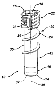
[0039] FIG. 1 depicts a knotless suture anchor 10 according to the present
invention. It comprises a body 12 having a distal end 14 and proximal end 16.
The proximal end 16 has a hexagonal-shaped tool receiving recess 18. It will
be
understood to one of skill in the art that alternative tool engagements may be
employed. A slight inward taper 19 is provided at the distal end 14 to ease
insertion of the anchor 10 into a bone hole (not shown in FIG. 1) and provides
an
initial fixation of the suture (not shown in FIG. 1) prior to threading the
anchor
into the hole.
[0040] The body 12 has a distal threaded portion 20 and a proximal
threaded portion 22. A single exterior thread 24 threads about the body 12 to
form the distal threaded section 20. This thread 24 extends nearly to the
distal
end 14, ending about 0.1 to 0.3 inches short thereof for easier insertion into
a
bone hole (not shown in FIG 1). However, one or more additional thread leads
26 begin towards the proximal end 16 to form a multi-fluted threading which
distinguishes the proximal threaded portion 22. Each individual thread start
24
and 26 have the same pitch as the thread 24 in the distal threaded section 20,
the
presence of the one or more additional thread leads 26 provides the proximal
threaded portion 22 with an increased effective thread pitch. However, the
pitch
of each thread lead in the proximal threaded portion 22 remains the same as
the
pitch of the thread 24 to eliminate axial compression effects from the threads
as
the anchor 10 is threaded into a bone hole. Preferably, there are four thread
leads
in the proximal threaded portion 22, the thread 24 and three additional thread
leads 26. The major diameter of the proximal threaded portion 22 is preferably
somewhat larger than that of the distal threaded portion 20. Rather than have
threads with a sharp outer edge the threads 24 and 26 preferably have a
rounded
our blunted profile to minimize stress on suture that is compressed against
them.
6
= CA 02750493 2011-08-24
While the anchor body 12 is shown with threads 24 and 26, especially for
smaller
diameters, the threads could be replaced with annular flanges or other
purchase
enhancements appropriate for a push-in anchor versus, a threaded anchor. Even
with the threads 24 and 26, smaller diameters of the anchor body 12 may be
appropriate to push in rather than thread in.
[0041] A lateral port 28 passes through the body 12 at an oblique angle to
a distally extending longitudinal axis 30 of the body 12 and is disposed
within
the proximal threaded portion 22. It provides for passage of suture (not shown
in
FIG. 1) between an inner axial cannulation 32 through the body 12 and an
exterior 35 of the body 12. Such function will be explained in detail below.
[0042] The body 12 is formed of a suitable biocompatible material and is
preferably provided sterile and packaged within a bacteria-proof enclosure
(not
shown) such that it is ready for a sterile surgical procedure. Many
biodegradable
materials have less strength and are more brittle than non-biodegradable
materials such as PEEK or stainless steel. The simple design of the body 12,
without complicated moving or interacting parts, allows easier use of such
biodegradable materials while maintaining the structural integrity of the
anchor
10.
[0043] The novel suture anchors of the present invention may be made
from a metallic material, a non-biodegradable polymer, a biodegradable
polymer, or a composite of a biodegradable polymer or copolymer and a
bioceramic. The term biodegradable as used herein is defined to mean materials
that degrade in the body and then are either absorbed into or excreted from
the
body. The term bioceramic as defined herein is defined to mean ceramic and
glass materials that are compatible with body tissue. The bioceramics are
preferably biodegradable.
[0044] The metallic materials that can be used to manufacture the
anchors of the present invention include stainless steel, titanium, alloys of
nickel
7
CA 02750493 2011-08-24
and titanium, or other biocompatible metallic materials.
[0045] The non-biodegradable materials that can be used to manufacture
the anchors of the present invention include polyethylene, polypropylene,
PEEK,
or other biocompatible non-absorbable polymers.
[0046] The biodegradable polymers that can be used to manufacture the
anchors used in the present invention include biodegradable polymers selected
from the group consisting of aliphatic polyesters, polyorthoesters,
polyanhydrides, polycarbonates, polyurethanes, polyamides and polyalkylene
oxides. Preferably, the biodegradable polymers are aliphatic polyester
polymers
and copolymers, and blends thereof. The aliphatic polyesters are typically
synthesized in a ring opening polymerization. Suitable monomers include but
are
not limited to lactic acid, lactide (including L-, D-, meso and D,L mixtures),
glycolic acid, glycolide, .epsilon.-caprolactone, p-dioxanone (1,4-dioxan-2-
one),
trimethylene carbonate (1,3-dioxan-2-one), .delta.-valerolactone, and
combinations thereof.
[0047] The bioceramics that can be used in the composite anchors of the
present invention include ceramics comprising mono-, di-, tri-, .alpha.-tri-,
.beta.-
tri-, and tetra-calcium phosphate, hydroxyapatite, calcium sulfates, calcium
oxides, calcium carbonates, magnesium calcium phosphates. It is particularly
preferred to use a .beta.-tritricalcium phosphate. In addition to bioceramics,
bioglasses may also be used in the composite screws. The bioglasses may
include
phosphate glasses and bioglasses.
[0048] Suitable biocompatible synthetic polymers can include polymers
selected from the group consisting of aliphatic polyesters, poly(amino acids),
copoly(ether-esters), polyalkylene oxalates, polyamides, tyrosine derived
polycarbonates, poly(iminocarbonates), polyorthoesters, polyoxaesters,
polyamidoesters, polyoxaesters containing amine groups, poly(anhydrides),
polyphosphazenes, polyurethanes, poly(ether urethanes), poly(ester urethanes),
8
= CA 02750493 2011-08-24
poly(propylene fumarate), poly(hydroxyalkanoate) and blends thereof.
[00491 For the purpose of this invention aliphatic polyesters include, but
are not limited to, homopolymers and copolymers of lactide (which includes
lactic acid, D-,L- and meso lactide); glycolide (including glycolic acid);
.epsilon.-caprolactone; p-dioxanone (1,4-dioxan-2-one); trimethylene carbonate
(1,3-dioxan-2-one); alkyl derivatives of trimethylene carbonate; .delta.-
valerolactone; .beta.-butyrolactone; gamma.-butyrolactone; .epsilon.-
decalactone; hydroxybutyrate; hydroxyvalerate; 1,4-dioxepan-2-one (including
its dimer 1,5,8,12-tetraoxacyclotetradecane-7,14-dione); 1,5-dioxepan-2-one;
6,6-dimethyl-1,4-dioxan-2-one; 2,5-diketomorpholine; pivalolactone;
.alpha.,.alpha. diethylpropiolactone; ethylene carbonate; ethylene oxalate; 3-
methyl-1,4-dioxane-2,5-dione; 3,3-diethyl-1,4-dioxan-2,5-dione- ; 6,6-dimethyl-
dioxepan-2-one; 6,8-dioxabicycloctane-7-one and polymer blends thereof.
Additional exemplary polymer or polymer blends include, by non-limiting
example, a polydioxanone, a polyhydroxybutyrate-co-hydrox- yvalerate,
polyorthocarbonate, a polyaminocarbonate, and a polytrimethylene carbonate.
Aliphatic polyesters used in the present invention can be homopolymers or
copolymers (random, block, segmented, tapered blocks, graft, triblock, etc.)
having a linear, branched or star structure. Poly(iminocarbonates), for the
purpose of this invention, are understood to include those polymers as
described
by Kemnitzer and Kohn, in the Handbook of Biodegradable Polymers, edited by
Domb, et. al., Hardwood Academic Press, pp. 251-272 (1997). Copoly(ether-
esters), for the purpose of this invention, are understood to include those
copolyester-ethers as described in the Journal of Biomaterials Research, Vol.
22,
pages 993-1009, 1988 by Cohn and Younes, and in Polymer Preprints (ACS
Division of Polymer Chemistry), Vol. 30(1), page 498, 1989 by Cohn (e.g.,
PEO/PLA). Polyalkylene oxalates, for the purpose of this invention, include
those described in U.S. Pat. Nos. 4,208,511; 4,141,087; 4,130,639; 4,140,678;
4,105,034; and 4,205,399. Polyphosphazenes, co-, ter- and higher order mixed
9
CA 02750493 2011-08-24
monomer based polymers made from L-lactide, D,L-lactide, lactic acid,
glycolide, glycolic acid, para-dioxanone, trimethylene carbonate and E-
caprolactone such as are described by Allcock in The Encyclopedia of Polymer
Science, Vol. 13, pages 31-41, Wiley Intersciences, John Wiley & Sons, 1988
and by Vandorpe, et al in the Handbook of Biodegradable Polymers, edited by
Domb, et al., Hardwood Academic Press, pp. 161-182 (1997). Polyanhydrides
include those derived from diacids of the form HOOC--C. sub. 6H4--O--(-
CH2)m--O--C. sub. 6H4--COOH, where "m" is an integer in the
range of from 2 to 8, and copolymers thereof with aliphatic alpha-omega
diacids
of up to 12 carbons. Polyoxaesters, polyoxaamides and polyoxaesters containing
amines and/or amido groups are described in one or more of the following U.S.
Pat. Nos. 5,464,929; 5,595,751; 5,597,579; 5,607,687; 5,618,552; 5,620,698;
5,645,850; 5,648,088; 5,698,213; 5,700,583; and 5,859,150. Polyorthoesters
such
as those described by Heller in Handbook of Biodegradable Polymers, edited by
Domb, et al., Hardwood Academic Press, pp. 99-118 (1997).
[0050] Turning also to FIG. 2, the suture anchor 10 is shown disposed
within a bone hole 34 with a length of suture 36 passing through the anchor
body
12 and also through a tendon (such as a tendon in a rotator cuff) 38. A loop
40 of
the suture 36 passes through the tendon 38 and its free ends 42 then pass down
along a first side 44 of the anchor body 12, being trapped between the anchor
body 12, especially by the threads 24 and 26, and bone 46 forming the bone
hole
32. The free ends 42 then pass over the distal end 14, into the axial
cannulation
32 and then back out of the cannulation 32 through the lateral port 28. From
here
they pass between a second side 48 of the anchor body 12, being trapped
between
the body 12 and the bone 46. Other threading arrangements are possible. For
instance, rather than passing the loop 40 through the tendon 38 a second
anchor,
or row of anchors, (not shown) can be placed beneath the tendon 38 with the
suture 36 passing from these anchor(s) up through the tendon 38 and to the
anchor body 12 or to multiple anchor bodies 12.
CA 02750493 2011-08-24
[0051] Turning also to FIGS. 3 and 4, the location of the lateral port 28
affects the strength of the fixation of the anchor body 12 to the bone 46 and
also
the affixation of the suture 36 to the bone 46 and body 12. A more distal
location
of the port 28 provides higher fixation strength but the failure mode then
tends to
be evulsion of the anchor body 12 from the bone hole 34. A failure mode which
involves slipping of the suture 36 rather than evulsion of the anchor body 12
is
preferred so as to not leave a foreign body free within a patient's joint in
an event
of failure. Also, an evulsion failure could lead to damage of the bone 46. The
angle at which the port 28 passes through the body 12 with respect to the
longitudinal axis 30 affects fixation strength with a more oblique angle
enhancing fixation.
[0052] Additionally, the size and direction which the port 28 passes
through the body can affect the functionality and fixation strength of the
design.
The cross sectional area of the port 28 is provided with sufficient dimension
to
pass a desired size and quantity of suture(s) through the port 28. The port 28
should not be so small as to damage the suture(s) while transiting the port 28
during loading, deployment or in use. Similarly, passing a disproportionate
quantity of suture through an undersized port 28 may result in damage to the
anchor body 12 itself. Conversely, the port 28 should not be so large as to
minimize the benefit to fixation strength which is derived from the meandering
course of suture 36 through the system. An excessively large port size may
result
in an undesirable degradation of the structural strength of the anchor body.
The
size of the port may be optimized to provide ease of use and avert damage to
the
system, while providing benefit within the context of additional fixation
strength.
[0053] It is favorable to choose the direction of the port 28 as it passes
through the body at such angles and locations which promote passage of suture
36 through the system. Obtuse angles formed by the suture 36 during loading
and use are most desirable, as they minimize contact friction at corners and
subsequently, reduce loading forces and wear and increase robustness of the
11
CA 02750493 2011-08-24
entire system. The direction of the port 28 may be optimally provided in a
compound, oblique direction and offset location with respect to the
longitudinal
axis. The compound oblique direction and offset location provide an exit of
the
port 28 which coarsely approximates the tangent of the helices of the thread
starts
in a distal-to-proximal direction.
[0054] This direction and location has been shown to positively affect
fixation strength. As the anchor is threaded into a bone hole, it is theorized
that
the compound oblique direction and offset location of the port 28 promotes a
gentle fold of the suture 36 as it exits the port 28, causing the suture 36 to
fall
easily within the roots between the proximal thread starts. In this context, a
port
28 oriented radially normal to the longitudinal axis, for example, would
require a
sharp fold of the suture 36 as it exits the port 28. The sharp fold thusly
presents a
sharp transition as the anchor descends into the bone hole past the port 28,
thereby weakening the bone by shearing along the wall of the bone hole,
ultimately reducing fixation. By not creating sharp bends in the suture 36 it
is
possible to provide an anchor having smaller dimensions without adding too
much additional stress to the suture 36.
[0055] Other forms of providing a gentle transition may include the use
of a "break edge", fillet or chamfer in the vicinity of the port 28. However,
in
designs incorporating minimum wall thickness of the anchor, large transition
features may result in undesirable increases in the cross sectional area of
the port
28.
[0056] Turning also to FIG. 5, one can see that the number of thread
leads 26 in the proximal threaded section 22 affects suture 36 fixation
between
the bone 46 and the anchor body 12. More thread leads enhance such suture 36
fixation. The top line shows optimal fixation with four leads, the thread 24
and
three additional thread leads 26.
[0057] Ideally, anchor body 12 fixation and suture 36 fixation are
12
CA 02750493 2011-08-24
optimized to provide maximum anchor body 12 fixation while still providing
suture 36 slip as the predominate failure mode over anchor body 12 evulsion.
[0058] Turning also now to FIGS. 6A, 6B and 6C, the suture anchor body
12 is shown loaded onto an anchor driver 50. The driver comprises an elongated
cannula 52 having a driving handle 54 at a proximal portion 56 thereof and a
driver tip 58 at a distal portion 59 thereof. The driver tip 58 engages the
tool
recess 18 on the anchor body 12. Preferably the driver tip 58 is keyed to the
anchor body tool recess 18 in such a fashion that the anchor body 12 is placed
onto the driver 50 in only one rotational orientation such that a surgeon can
determine such orientation by the rotational position of the handle 54. (See
FIG.
7 in which a spline 60 on the driver tip 58 fits into a spline receiving cut-
out 62
on the anchor boy 12.
[0059] A suture passer 64, such as the CHIA PERCPASSER (available
from DePuy Mitek, Inc., Raynham, MA), an elongated braided Nitinol wire 66
with a distal suture grasping loop or kite 68, is engaged to the driver 50 and
anchor body 12. It passes into a central lumen 70 of the cannula 52 from a
proximal slot 72, out of the lumen 70 from a distal slot 74, over a removable
ramp 76 and into the anchor body cannulation 32 through the lateral port 28,
with
the suture loop 68 extending out of the distal end 14 of the body 12. The wire
66
is flexible but retains some rigidity and the ramp 76 provides a smooth entry
angle into the lateral port 28. A tensioning clutch 78 is interposed between
the
handle 54 and the cannula 52. A proximal portion 80 of the wire 66 passes
through a suture management passage 82 through the clutch 78. During a
procedure, after the suture 36 has been passed through the tendon 38, the free
ends 42 are pulled out of the procedure cannula (not shown) to a point outside
of
the patient's body and loaded through the suture loop 68.
[0060] After the free ends 42 are loaded into the suture passer 64 it is
drawn up the cannula 52 leaving the free ends 42 to pass up through the anchor
13
CA 02750493 2011-08-24
body cannulation 32 from its distal end 14, out through the lateral port 28,
over
the ramp 76, into the lumen 70 through the distal slot 72, out of the lumen 70
through the proximal slot 72 and through the clutch suture management passage
82 as depicted in FIG. 6B. The ramp 76 no longer being needed is removed as
shown in FIG. 6C. Preferably, the ramp 76 fits to the cannula 52 via a snap-
fit to
provide easy removal. The anchor is now ready for implantation.
[00611 To complete the procedure the suture 36 is tensioned through the
suture tension clutch 78 to a desired tension. The anchor body 12 is then
threaded into the pre-drilled bone hole 34 via the driver 50. The clutch 78
plays
out the free ends 42 as the body 12 approaches and enters the hole 34 to
maintain
proper tension on the suture 36 and allows the suture 36 to move into the bone
hole 34 from the clutch 78 rather than from the tissue and thus avoids
spooling of
the suture 36 onto the anchor body 12 as it is threaded into the hole 34. The
anchor body preferably completes only a partial turn, such as one quarter turn
from the time the suture 36 is pinched by the port 28 entering the hole 34 and
the
anchor body 12 is fully seated therein. The anchor body 12, especially in its
interior, and the suture 36 can be formed of materials or have their surfaces
enhanced with materials or procedures which lower friction and enhance
slipping
of the suture 36 as the anchor is deployed. When fully deployed the proximal
end 22 of the anchor body 12 is preferably below the bone 46 within the bone
hole 34. The driver 50 is removed and the free ends 42 trimmed leaving the
anchor 10 in place as shown in FIG. 2.
[0062] FIG. 8 illustrates an alternative embodiment of an insertion tool
100 and FIG. 9 illustrates an alternative embodiment of an anchor 102
according
to the present invention, each of these being adapted for use together. The
anchor 102 has a structure similar to the anchor 10 with the exception of an
axial
boss 104 within its axial cannulation 106 which mates with a distal axial slot
108
in a distal driving portion 110 of the insertion tool 100. Also, the axial
cannulation is enlarged radially where the driving portion 110 is received
such
14
CA 02750493 2011-08-24
that an interior cannulation 112 of the driving portion 110 has the same
interior
diameter as a distal portion 114 the anchor axial cannulation 106 and the boss
104 extends radially into the slot 108 to a depth matching the interior
diameter of
the interior cannulation 112, providing a smooth transition within the of the
interior cannulation 112 and axial cannulation 106 eliminating discontinuities
upon which suture can snag during rotational deployment of the anchor 102. The
boss 104 provides additional engagement between the insertion tool 100 and the
anchor 102.
[0063] Turning also to FIGS. 10 and 11, the boss 104 aligns
circumferentially with a lateral port 116 on the anchor. A suture ramp 118
aligns
on the insertion tool 100 with the port 116. The alignment of the boss 104
with
respect to the port 116 and the slot 108 with respect to the ramp 118 puts the
port
116 and ramp 118 into circumferential alignment with one another.
[0064] The ramp 118 is formed of a molded polymer having an arcuate
suture receiving groove 120 which extends radially outwardly to guide suture
and/or a suture grasper 122 out of a slot 124 on the insertion tool 100 and
into the
port 116 without sharp transitions and with the suture or suture grasper 122
forming an oblique angle with respect to itself as it enters the port 116. The
ramp
118 also bears a pair of C-shaped snap clips 126 which snap onto and off of
the
insertion tool 100 for easy removal of the ramp 118 during the procedure
previously described. A grasping tab 128 provides a gripping surface for easy
manual removal of the ramp 118 and also provides a surface upon which to place
instructions for use.
[0065] As shown in FIG. 11 a T-shaped handle 130 on the suture grasper
122 preferably has finger lands 132 for easy manipulation of the suture
grasper
122. A suture clutch 134 which normally holds the suture and then releases it
as
torque is provided to a handle 136 on the insertion tool 100 is shown distal
of the
handle 136 but could be incorporated therein. Details on preferred clutch
CA 02750493 2011-08-24
mechanisms are provided later herein.
[0066] FIG. 12 illustrates a further embodiment of a suture anchor 140
according to the present invention. It is similar to the prior suture anchors
10 and
102; however, instead of a port it carries an axial slot 142 at its proximal
end.
The slot 142 terminates at its distal end 144 with a return portion 146 which
extends proximally and circumferentially along a path of a thread start 147
providing an overall hook shape to the slot 142. Being open at its proximal
end
allows for easier threading of a suture grasper (not shown in FIG. 12).
[0067] Ease of threading is so improved that the grasper can be omitted
in which case during the procedure a surgeon can directly thread a suture 148
through a main axial cannulation 150 of the anchor 140, feeding it into the
slot
142 and seating it within the slot return portion 146. A procedure with the
anchor 140 would proceed as previously described with the surgeon pre-drilling
a
hole in a bone and passing suture 148 through tissue, preferably in an
arthroscopic procedure through a cannula (the cannula, tissue and bone not
being
shown in FIG. 12). With free ends of the suture 148 outside of the patient's
body
the surgeon passes them through the cannulation 150 and seats the suture
within
the return portion 146. The anchor 140 would then be loaded onto an insertion
tool such as the tool 100 or 50 and installed into the bone as previously
described, the return portion 146 holding the suture similarly to the
aforementioned ports. Preferably the return portion passes into the
cannulation
150 at an oblique angle as described with respect to the prior ports thus
allowing
the suture 148 to pass into the cannulation 150 through the return portion 146
while keeping an oblique angle with respect to itself.
[0068] The clutch 134 comprises a disk shaped body 152 having a distal
portion 154 which attaches to an elongated cannula 156 which itself terminates
in
the hexagonal driving portion 110. A proximal portion 158 of the body 152
attaches to the insertion tool handle 136 outwardly radially of where the
cannula
16
CA 02750493 2011-08-24
156 attaches to the body 152. An axial slot 160, as best seen in FIG. 13,
leads
into the body 152 and receives and grabs the suture 148. Preferably its
interior
surface 162 is formed of a rubber or other resilient material to enhance the
grip
with the suture 148. Torque applied to the handle 136 is transmitted through
the
clutch body 152 to the cannula 156. The body 152 is formed of a material, such
as a hard rubber, having sufficient resilience to allow the slot 160 to open
under
the influence of such torque and relax the grip on the suture 148. Thus, the
clutch 134 normally grips the suture to maintain tension but relaxes that grip
as
the handle 136 is torqued during implantation of the anchor 140 allowing
suture
148 to slide through the clutch 134.
[0069] FIG. 14 illustrates an alternate embodiment of a clutch body 164
according to the present invention. It comprises a pair of somewhat radial
slots
166 which spiral inwardly radially in a direction in which torque would be
applied to an associated handle (not shown in FIG. 14).
[0070] FIG. 15 illustrates a further embodiment of a clutch body 170
comprising a plurality of radially extending arms 172, each having
circumferential suture receiving slots 174 therein. A cannula attachment
location
176 is located in the center of the body 170 and handle attachment locations
178
are located on the arms outwardly radially of the slots 174.
[0071] FIGS. 16 A and B illustrate a further embodiment of a clutch
mechanism 180 which comprises a rigid outer handle gripping portion 182 and a
radially interior resilient insert 184. A proximal end 186 of the insert 184
attaches to the outer handle 182 and a distal end 188 of the insert 184
attaches to
a cannula 190. Suture 192 feeds into a gap 194 between the outer handle 182
and
the insert 184 through a radial slot 196 in the handle 182. The gap 194 is
sized to
grip the suture 192. Application of torque to the outer handle 182 twists the
insert 184 thereby opening the gap 194 and allowing slippage of the suture 192
therethrough.
17
CA 02750493 2011-08-24
[0072] FIGS. 17 A and B illustrate a further embodiment of a clutch
mechanism 200 comprising a pair of radial flanges 202 extending outwardly
radially from a cannula proximal portion 204. A resilient material 206 such as
rubber affixes to both sides of the flanges 202. An outer handle 208 comprises
two halves 210, each of which attach to one of the flanges 202 and which are
spaced apart from the opposing flange 202 to create suture receiving slots
212.
The slots 212 can have flared openings 214 with a suture retaining lip 216
therein. Suture 218 is gripped within the slots 212 by compression between the
outer handle 208 and the resilient material 206 on the flange 202 as shown in
FIG. 17 A. Application of torque to the outer handle 208 compresses the
resilient material between the handle 208 and flanges 202 to open the slots
212 to
release the suture as shown in FIG. 17 B.
[0073] FIGS. 18 A and B illustrate an additional embodiment of a clutch
mechanism 220. A handle 222 comprise an outer cylindrical gripping portion
224 and a central axial core 226, the gripping portion 224 being attached to
the
core 226 via a plurality of radial ribs 228. One pair of ribs 230 extend
slightly
off axis and adjacent to each other and the gripping portion 224 is open
between
them forming a radially extending axial slot 232 in the handle 222. Near a
proximal end 234 of the handle 222 a retainer member 236 sits within the slot
232 extending from one of the ribs 230 toward the adjacent rib 230. It has a
flared opening 238 and a retaining lip 240 to ease entry of suture 242 into
the slot
232 with the lip 240 holding it from falling out. A resilient material 244 in
the
slot 232 grips the suture 242. Torque applied to the gripping portion 224
tends to
open the slot 232 releasing the tension on the suture 242.
[0074] Threading the suture 148 through the cannulation 150 of the
suture anchor 140 of FIG. 12 can be accomplished manually without assistance
from a threading device. However, a simple converging threader 300 as
illustrated in FIG. 19 can further simplify the procedure. The threader 300
comprises an open braided tube 302 having one end 304 inserted through the
18
CA 02750493 2011-08-24
cannulation 150 and a second expanded end 306 into which one or more sutures
148 can be pushed by hand. The threader 300 is preferably woven from a
flexible biocompatible material and provided in combination with the anchor
140, with the threader 300 received through the cannulation 150, and with both
the threader 300 and anchor being sterile and packaged within a sterile
bacteria-
proof package (not shown). When a surgeon is ready to load sutures 148 into
the
anchor 140 the combination of the anchor 140 and threader 300 are removed
from the sterile package and the sutures 148 are pushed into the threader
expanded end 306. Tension is applied to the other end 304 causing the expanded
end 306 to close and travel through the cannulation 150 carrying the sutures
148
therethrough. The procedure can then be completed as aforementioned.
[0075] Alternatively, as shown in FIG. 20, the sutures 148 can be merely
stitched through the braided tube 302. If the weave is open enough they can be
stitched by hand or they can be stitched with needles (not shown). The tube
302
is then drawn through the cannulation 150 as in FIG. 19.
[0076] As shown in FIG. 21, a threader 310 can be formed from a tube
312 which is not necessarily braided but rather provided with axial slits 314
at
one end 316 to form a mouth 318 for receiving the suture 148. Gripping
enhancements such as teeth 320 can be provided within the mouth 318 to help
retain the suture 148 therein as the threader 310 passes through the
cannulation
150.
[0077] To ensure good closure of the expanded end 306 of the threader
300 of FIG. 19 it can be modified with additional closures as shown in FIGS.
22
A through D. For instance a simple spring metal snap element 322 can be
provided to a braided tube 324, the element 322 having a first open position
as
shown in FIG. 22B and a second relaxed closed position as shown in FIG. 22C.
After insertion of the sutures 148 with the element 322 in the open position
is
squeezed to pop it into the closed position. A loading suture loop 324 can be
19
CA 02750493 2011-08-24
employed about the element 322 to provide the squeezing force for closure and
also to further compress the sutures 148 within the tube 324. A separate
loading
suture loop 324 can also be provided alone and woven through the braid of the
tube 324 in substitution of the element 322.
[0078] Alternatively, the braiding of the tube 324 can be woven to
encourage closure, especially if the material is resilient, and to hold the
expanded
end 316 open a stretcher 326 can be inserted therein as shown in FIGS. 23 A
and
B. In its simplest form the stretcher 326 comprises a tube 328 having a full
length side opening 330 whereby after the suture 148 is loaded into the
expanded
end 316 the tube 328 is removed therefrom with the suture 148 passing through
the opening 330 to allow removal of the tube 328.
[0079] The invention has been described with reference to the preferred
embodiments. Obviously, modifications and alterations will occur to others
upon
reading and understanding the preceding detailed description. It is intended
that
the invention be construed as including all such modifications and alterations
insofar as they come within the scope of the appended claims or the
equivalents
thereof.