Note: Descriptions are shown in the official language in which they were submitted.
CA 02750531 2011-07-22
WO 2010/085658 PCT/US2010/021821
TITLE
Apparatus and Methods for Detecting Inflammation Using Quantum Dots
BACKGROUND
Inflammatory bowel disease (IBD) encompasses two chronic, related
inflammatory conditions, ulcerative colitis (UC) and Crohn's disease (CD). In
addition, organs other than the intestinal tract can be involved by the
underlying
inflammation of IBD thus making IBD a multi-organ disease. As many as 4
million
people (including one million Americans) worldwide suffer from a form of IBD.
In
the U.S. alone, IBD accounts for approximately 152,000 hospitalizations each
year.
The annual medical cost for the care of IBD patients in the United States is
estimated
at over $2 billion. When adjusted for loss of productivity, the total economic
burden
is estimated to be nearly $3 billion.
Inflammatory bowel disease is a complex, multifactorial sequelae
characterized by severe derangements in the structure and function of local
tissue
architecture and increased presence of neutrophils and lymphocytes and other
pro-
inflammatory cells. In addition, epithelial, endothelial, mesenchymal, adipose
tissue
and nerve cells all can exhibit a broad range of damage as a result of the
inflammatory
process. Effector, regulatory and immune-like functions interact abnormally
with
lymphoid cells to further contribute to the pathogenesis of inflammatory
disease.
Heart disease, arthritis, asthma, allergy, infection and diabetes all have
elements of
chronic inflammation. Examples of inflammatory disease also include, but are
not
limited to, stroke, cardiovascular disease, acute coronary syndromes, acute
myocardial infarction, pericarditis, periodontal disease, cancer, Alzheimer's
disease,
and inflammatory bowel disease. Inflammatory disease can also affect multiple
organ
systems, as in autoimmune diseases.
Inflammation is a significant contributor to the pathogenesis of both the
acute
and chronic stages of IBD. The diagnosis of IBD is rarely straightforward,
involving
an extensive process of examination and invasive testing, including biopsy
during
endoscopy. Even with these specialized studies, it is often still difficult to
tell which
type of IBD a person has, leading to a diagnosis of "indeterminate colitis"
and
rendering disease management more difficult. UC carries a significant risk for
the
development of colorectal cancer, but remains difficult to differentiate from
CD.
Since UC in particular is associated with a 35% higher risk of developing
colorectal
CA 02750531 2011-07-22
WO 2010/085658 PCT/US2010/021821
2
cancer than the general population, making a proper diagnosis is essential to
good
patient care.
While there is no medical cure for IBD, effective medical treatment is
available which can calm the inflammation and relieve the symptoms of
diarrhea,
abdominal pain, and rectal bleeding. Since the disease tends to manifest
itself with
multiple attacks and remissions, continuous monitoring of patients is
essential to
provide the necessary medical treatment to reduce inflammation and prevent the
development of clinical sequelae.
The current noninvasive tests which are used clinically to distinguish between
UC from CD, are based on the presence of antibodies such as perinuclear
antineutrophilic cytoplasmic antibody (p-ANCA) and anti-Saccharomyces
cerevisiae
antibody (ASCA) in serum, and have less than 70% specificity. Mostly invasive
biopsies are used to confirm presence of a particular disease.
Therefore, there is an unmet need for a technology to quantify inflammatory
markers quickly, cost effectively, and with high sensitivity. Such
quantification could
lead not only to differential diagnosis but also to the evaluation of response
to therapy
in inflammatory diseases. More specifically, there is a long-standing need for
non-
invasive diagnostic tools that are able to distinguish non-IBD symptoms from
IBD,
accurately distinguish UC from CD, and monitor disease progression, remission
or
relapse. In particular, there is a need for a technology to determine low
levels of
inflammatory markers and to utilize the ability to detect these markers as
predicators
of inflammatory diseases, responses to therapy for inflammatory diseases, and
progressions to cancer.
Additionally, there is a need to improve the stability of fluorescence
intensity
emitted from quantum dots over time as the quantum dots during storage or
diagnostic
imaging, so as to avoid the loss of valuable information.
SUMMARY
The present invention includes an apparatus for detecting a biomarker
indicative of an inflammatory condition. The apparatus has a capillary tube
adapted
for one or more biomarkers to adhere to an interior surface thereof, a light
source for
energizing quantum dots conjugated with the biomarkers within the capillary
tube,
and a detection system for detecting and quantifying fluorescent energy
emitted by the
quantum dots in one or more predetermined wavelength ranges, each wavelength
CA 02750531 2011-07-22
WO 2010/085658 PCT/US2010/021821
3
range being correlated to one and only one of the biomarkers.
In one embodiment of the apparatus, the capillary tube comprises at least one
material selected from the group of polymethyl methacrylate (PMMA), polyvinyl
acetate, and polystyrene tube. A hypodermic needle can be connected to an end
of the
capillary tube for supplying a sample to the capillary tube. The capillary
tube can be
supported externally by a glass capillary tube.
In another embodiment, the apparatus also includes a fluid handling unit
adapted to hold multiple capillary tubes and a mechanical positioning system
for
successively positioning each capillary tube to enable the sample contained
therein to
be exposed to the light source and visible to the detection system.
In another embodiment, the capillary tube has a volume in the range of about
100 nanoliters to about 1 microliter.
In another embodiment of the apparatus, the light source comprises an LED, a
laser diode, or an array having a plurality of LEDs or laser diodes. One or
more of the
LEDs can be ultraviolet LEDs. The light source can further comprise a lens for
focusing the LED or LEDs onto the capillary tube.
In another embodiment of the apparatus, the detection system comprises a
broadband filter. In a still another embodiment, the detection system
comprises a
photodetector. The photodetector can be a spectrometer coupled to at least one
photomultiplier tube, avalanche photodiode detector, or a CCD camera. The
detection
system can include a mirror disposed around at least a portion of the
capillary tube for
increasing the amount of the fluorescent energy emitted by the quantum dots
that can
be detected by the photodetector. The mirror can be selected from the group of
a
spherical mirror, a cylindrical mirror, and a parabolic mirror.
In another embodiment of the apparatus, the capillary tube is a polymethyl
methacrylate (PMMA) capillary tube having a volume of less than about 1.5
microliters, wherein the light source comprises an ultraviolet LED, and
wherein the
detection system comprises a CCD camera. In yet another embodiment, the
detection
system comprises a spherical mirror for focusing energy emitted by the quantum
dots
to the CCD camera. In one variation, a first LED of the light source is
directed into
an end of the capillary tube and the CCD camera detects energy emitted through
the
wall of the capillary tube. Additionally, a second LED of the light source can
be
directed into an opposite end of the capillary tube. In another variation, at
least one
LED of the light source is disposed adjacent to a wall of the capillary tube
and the
CA 02750531 2011-07-22
WO 2010/085658 PCT/US2010/021821
4
CCD camera detects energy emitted through an end of the capillary tube.
In another embodiment of the apparatus, said biomarker is selected from the
group consisting of myeloperoxidase (MPO), IL-Ia, TNFa, perinuclear anti-
neutrophil cytoplasmic antibody (p-ANCA), anti-Saccharomyces cerevisiae
antibody
(ASCA), angiotensin converting enzyme, lactoferrin, C-reactive protein (CRP),
and
calprotectin.
In another embodiment, the apparatus further comprises a composition for
detecting a biomarker in a biological sample contained in the capillary tube,
wherein
said composition comprises at least one conjugate comprising a quantum dot and
an
antibody that specifically binds to a biomarker. The antibody can be bound to
a
substrate surface.
The present invention also includes a method of diagnosing an inflammatory
condition in a subject by detecting a biomarker in a sample. The method
includes
providing a sample to a capillary tube coated with an antibody, the sample
potentially
including a biomarker indicative of the inflammatory condition, contacting the
sample
with a conjugate comprising a quantum dot and an antibody that specifically
binds to
the biomarker, energizing the quantum dot with a light source, detecting
fluorescent
emission from the quantum dot, and correlating the fluorescent emission to the
concentration of the biomarker in the sample.
In one embodiment of the diagnostic method, said biomarker is selected from
the group consisting of an enzyme, an adhesion molecule, a cytokine, a
protein, a lipid
mediator, an immune response mediator, and a growth factor.
In another embodiment of the method, said biomarker is selected from the
group consisting of myeloperoxidase (MPO), IL-la, TNFa, perinuclear anti-
neutrophil cytoplasmic antibody (p-ANCA), anti-Saccharomyces cerevisiae
antibody
(ASCA), angiotensin converting enzyme, lactoferrin, C-reactive protein (CRP),
and
calprotectin.
In on embodiment, the capillary tube is functionalized using NaOH.
Alternatively, the capillary tube is functionalized using plasma or
ultraviolet light.
In another embodiment of the diagnostic method, said inflammatory condition
comprises at least one inflammatory disease selected from the group consisting
of
inflammatory bowel disease, ulcerative colitis, Crohn's disease, stroke,
myocarditis,
cardiovascular disease, acute coronary syndromes, acute myocardial infarction,
pericarditis, periodontal disease, cancer, Alzheimer's disease, and autoimmune
CA 02750531 2011-07-22
WO 2010/085658 PCT/US2010/021821
diseases.
The invention further includes a method of stabilizing the fluorescence of
quantum dots over time comprising exposing the quantum dots to a fluorescence
stabilizing medium.
5 In one embodiment of the stabilization method, the fluorescence stabilizing
medium is a solution having a low ionic strength.
In other embodiments of the stabilization method, the fluorescence stabilizing
medium is a solution having a pH greater than or equal to about 7.0, or a pH
greater
than or equal to about 8Ø
In another embodiment of the stabilization method, the fluorescence
stabilizing medium comprises water-soluble free radical quenchers. In still
another
embodiment, the fluorescence stabilizing medium comprises TrisPro and an
amount
of water-soluble vitamin E. In one variation, the amount of vitamin E is at
least about
0.001% of the medium.
In another embodiment, the quantum dots each comprise a CdSe core and a
ZnS protective layer.
BRIEF DESCRIPTION OF THE DRAWINGS
For the purpose of illustrating the invention, there are depicted in the
drawings
certain embodiments of the invention. However, the invention is not limited to
the
precise arrangements and instrumentalities of the embodiments depicted in the
drawings.
Figure 1 is a schematic depicting an apparatus for detecting quantum dot (QD)
fluorescence from a sample in waveguide mode.
Figure 2 is an overview flow chart of a method for detecting and diagnosing
inflammatory bowel disease.
Figure 3 is a schematic depicting a process for binding QD conjugates to
antigens.
Figure 4 is a schematic depicting a sandwich Quantum-Linked
ImmunoSorbent Assay (QLISA) process.
Figure 5 is a schematic depicting a competitive QLISA process.
Figure 6A and 6B are schematics depicting an apparatus for detecting
fluorescence form a QD-linked sample in waveguide mode.
CA 02750531 2011-07-22
WO 2010/085658 PCT/US2010/021821
6
Figure 7 is a photograph of the apparatus of Figure 6B in use.
Figure 8 is a schematic depicting an apparatus for causing a sample to
fluoresce by exposure to LED light sources in side illumination mode.
Figure 9 is a schematic depicting an apparatus for causing a sample to
fluoresce by exposure to focused LED light sources in side illumination mode.
Figure 10 is a schematic depicting the conversion of raw image data from an
apparatus as in Figure 8 or Figure 9 into a processed image from which
intensity
information can be obtained.
Figures 11 A through 11 D are schematics depicting alternate apparatuses for
detecting fluorescence from a QD-linked sample.
Figure 12 is a graph correlating concentration of a biomarker (MPO) in a
sample with intensity of fluorescence emissions.
Figure 13 is a graph correlating the concentration of MPO with intensity of
fluorescence emissions to determine a detection threshold.
Figure 14 is graph correlating the concentration of MPO with intensity of
fluorescence emissions in an animal sample as compared with a control sample.
Figure 15 is a process flow chart of a diagnostic protocol in an embodiment of
the present invention.
Figures 16A and 19B are schematic illustrations of configurations of apparatus
for collecting fluorescent signals from QDs in a PMMA capillary.
Figures 17A through 17F are optical micrographs of capillaries demonstrating
the effect of blocking.
Figures 18A and 18B are optical micrographs of PMMA capillaries treated
with DB-Ab to demonstrate the effect of surfactant in the wash buffer.
Figures 19A and 19B show the sensitivity of an embodiment of the QLISA
method of the present invention to MPO and representative images of a PMMA
capillary comparing a sample of 0.3 nM MPO with a control.
Figure 20 is a comparison of fluorescence intensities of QD solutions obtained
with side illumination and waveguide configurations of the apparatus of the
present
invention.
Figures 21 A through 21 C compare the fluorescence intensity at various MPO
concentrations obtained by side illumination.
Figure 22 shows the fluorescence intensity from MPO-spiked animal stool
samples.
CA 02750531 2011-07-22
WO 2010/085658 PCT/US2010/021821
7
Figure 23 is a graph showing interference from non-specific binding to MMP-
13.
Figure 24 is a chart comparing disease time line and fluorescence intensity of
MPO bound to quantum dots.
Figure 25 is a schematic depicting a fluid handling unit for multiple PMMA
tubes.
Figure 26 is a photograph showing an exemplary fluid handling unit for
multiple PMMA tubes.
Figure 27 is a schematic showing the designation of reservoirs for use with a
fluid handling unit as in Figure 25 or Figure 26.
Figure 28A to 28D are graphics depicting the fabrication of a multiple sample
holder using a mold.
Figures 29A and 29B are comparisons showing the effect of storage buffer on
the fluorescence intensity of QDs and on QD stability over a period of time.
Figure 30 shows the fluorescence intensity from lactoferrin-spiked human
stool samples.
Figure 31 is a schematic of an embodiment of a sampling manifold.
Figure 32 is a schematic of an embodiment of a detachable multiple capillary
holder.
Figures 33A and 33B show the molecular structure of a buffer medium and a
water-soluble vitamin E used to stabilize QD fluorescence intensity.
Figures 34A and 34B are graphs comparing the decay in fluorescence intensity
of QDs over time as a function of pH, in solutions without and with vitamin E,
respectively.
Figures 35A and 35B are graphs comparing the decay in fluorescence intensity
of QDs over time as function of pH, in solutions without and with vitamin E,
respectively.
Figures 36A and 36B are graphs comparing the decay in fluorescence intensity
of QDs over time as a function of vitamin E concentration at 6.5 pH, for Ocean
Nanotech QDs and Invitrogen QDs, respectively.
Figures 37A and 37B are graphs comparing the decay in fluorescence intensity
of QDs over time as a function of vitamin E concentration at 7.5 pH, for Ocean
Nanotech QDs and Invitrogen QDs, respectively.
Figures 38A and 38B are graphs comparing the decay in fluorescence intensity
CA 02750531 2011-07-22
WO 2010/085658 PCT/US2010/021821
8
of QDs over time as a function of vitamin E concentration at 8.5 pH, for Ocean
Nanotech QDs and Invitrogen QDs, respectively.
DETAILED DESCRIPTION
The present invention discloses the development of a simple and inexpensive
quantum dot based immunoassay for detecting myeloperoxidase (MPO) in
biological
samples is reported. The acronym QLISA is introduced to represent Quantum-
Linked
ImmunoSorbent Assay. In a preferred embodiment, the detection method utilizes
polymethylmethacrylate (PMMA) micro-capillaries as substrate for performing a
sandwich assay. UV-LEDs both high power (80 mW) and low power (10 mW) were
tested for their efficiency in maximizing detection sensitivity in either a
waveguide
illumination or a side illumination mode. The results discussed herein
indicate that
both waveguide and side illumination modes can be employed for detecting MPO
down to 15 ng/ml; however, using the high power LED in a side illumination
mode
unexpectedly improves sensitivity and simplifies the data acquisition. A
testing
protocol and robustness of embodiments of sensors were evaluated with animal
stool
samples spiked with MPO and the results indicate that the sensitivity of the
capture
and reporter antibodies is not compromised when used in stool samples.
Further, the
effect of the ionic strength of the environment on the fluorescence stability
of
quantum dots was evaluated and found to affect the assay, particularly if long
imaging
times are necessary. Notably, replacing the buffer with glycerol or another
non-polar
or weakly polar substance during imaging increased the fluorescence intensity
of
quantum dots while significantly minimizing the loss in intensity, even after
relatively
short times of two hours.
In general, the invention is directed to an apparatus and a method for
detecting
and quantifying biomarkers indicative of inflammatory disease, particularly
inflammatory bowel disease, with sufficient sensitivity to distinguish non-IBD
symptoms from IBD symptoms and to differentiate UC from CD. An apparatus
according to the invention holds a sample in which one or more biomarkers have
been
conjugated with quantum dots and provides a light source for energizing the
quantum
dots and a detection system for detecting and quantifying the quantum dots. A
method of using an apparatus according to the invention includes collecting a
sample,
conjugating quantum dots to biomarkers in the sample, energizing the quantum
dots
CA 02750531 2011-07-22
WO 2010/085658 PCT/US2010/021821
9
with a light source, detecting emission from the quantum dots, and determining
the
concentration of the biomarker in the sample based on a correlation between
detected
emission and biomarker concentration.
In one embodiment, an apparatus is provided for detecting a biomarker
indicative of an inflammatory condition. The apparatus includes a capillary
tube
adapted for one or more biomarkers to adhere to an interior surface thereof.
The
apparatus further includes a light source for energizing quantum dots
conjugated with
the biomarkers within the capillary tube and a detection system for detecting
and
quantifying fluorescent energy emitted by the quantum dots in one or more
predetermined wavelength ranges, each wavelength range being correlated to one
and
only one of the biomarkers.
In another embodiment, a method is provided for diagnosing an inflammatory
condition by detecting a biomarker in a sample. The method includes providing
a
sample to a capillary tube coated with an antibody, the sample potentially
including a
biomarker indicative of the inflammatory condition. The method further
includes
contacting the sample with a conjugate comprising a quantum dot and an
antibody
that specifically binds to the biomarker, energizing the quantum dot with a
light
source, detecting fluorescent emission from the quantum dot, and correlating
the
fluorescent emission to the concentration of the biomarker in the sample.
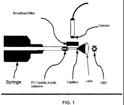
There is shown in Figure 1 an embodiment of an apparatus for detecting QD
fluorescence from a sample as part of a method for rapidly identifying and
measuring
biochemical and immunological markers for inflammatory disease. An apparatus
such as that shown in Figure 1, or alternatively apparatuses as shown in
Figures 6A-9,
11A-11D, and 16A-16B can be used in a diagnostic method as depicted generally
in
Figure 2 and more specifically in Figure 15 for detecting the presence of
inflammatory diseases including but not limited to inflammatory bowel disease
(IBD),
and, for example, for diagnosing whether the IBD is characterized by
ulcerative colitis
(UC) or Crohn's disease (CD).
Methods of quantitatively assessing inflammation with biosensing
nanoparticles are described in detail in commonly assigned PCT Application No.
PCT/US2007/015748 filed on July 11, 2007, and incorporated by reference herein
in
its entirety. The nanoparticles include quantum dots conjugated to targeting
moieties
that specifically bind to a biomarker protein or a nucleic acid encoding a
biomarker,
where dysregulation of the biomarker is associated with inflammatory disease.
CA 02750531 2011-07-22
WO 2010/085658 PCT/US2010/021821
In particular, methods disclosed herein use monoclonal antibodies conjugated
to quantum dots as a means of detecting nanolevels of biomarkers. As used
herein,
the methods have collectively been dubbed "Quantum-Linked ImmunoSorbent
Assay" (QLISA) as differentiated from the technique known in the art as Enzyme-
5 Linked ImmunoSorbent Assay (ELISA). QLISA possesses advantages over ELISA,
as will be described herein.
Any of the apparatus described herein can be provided as a test kit comprising
a single assay with customized antibody coated micro-columns and ready-to-use
reagents for rapid and easy detection. The assay may comprise MPO, IL-la,
TNFa,
10 calprotectin, lactoferrin, fibronectin, ASCAm p-ANCA, and/or other markers,
particularly those that may be found in fecal samples as indicators of various
forms of
IBD. The apparatus and methods can be adapted to detect, at pico- or nano-
molar
concentrations, single markers in sequence or multiple markers simultaneously.
The
test kit is adapted to measure inflammatory biomarkers in biological samples
(e.g.,
fluids and fecal samples) using QLISA, i.e., quantum dot immobilization and
fluorescence. The test kit can be used in a physician's office as a point of
care
screening device, or as part of a battery of tests done at a diagnostics
laboratory.
The present approach is based on using a combination of available biomarkers
(Myeloperoxidase-MPO, p-ANCA, ASCA) to lead to differential diagnosis of
Inflammatory Bowel Disease (IBD) from Inflammatory Bowel Syndrome (IBS) and
to differentiate Ulcerative Collitis (UC) from Crohn's Disease (CD).
Definitions:
As used herein, each of the following terms has the meaning associated with it
in this section.
The articles "a" and "an" refer to one or to more than one (i.e. to at least
one)
of the grammatical object of the article. By way of example, "an element"
means one
element or more than one element.
The terms "about" and "approximately" will be understood by persons of
ordinary skill in the art and will vary to some extent on the context in which
it is used.
The term "antibody" refers to an immunoglobulin molecule which is able to
specifically bind to a specific epitope on an antigen. Antibodies can be
intact
immunoglobulins derived from natural sources or from recombinant sources and
can
be immunoreactive portions of intact immunoglobulins. Antibodies are typically
CA 02750531 2011-07-22
WO 2010/085658 PCT/US2010/021821
11
tetramers of immunoglobulin molecules. The antibodies in the present invention
may
exist in a variety of forms including, for example, polyclonal antibodies,
monoclonal
antibodies, intracellular antibodies ("intrabodies"), Fv, Fab and F(ab)2, as
well as
single chain antibodies (scFv), camelid antibodies and humanized antibodies.
An "antigen" or "Ag" refers to a molecule that provokes an immune response.
This immune response may involve either antibody production, or the activation
of
specific immunologically-competent cells, or both. The skilled artisan will
understand that any macromolecule, including virtually all proteins or
peptides, can
serve as an antigen. Furthermore, antigens can be derived from recombinant or
genomic DNA. A skilled artisan will understand that any DNA, which comprises a
nucleotide sequences or a partial nucleotide sequence encoding a protein that
elicits
an immune response therefore encodes an "antigen" as that term is used herein.
Furthermore, one skilled in the art will understand that an antigen need not
be
encoded solely by a full length nucleotide sequence of a gene. It is readily
apparent
that the present invention includes, but is not limited to, the use of partial
nucleotide
sequences of more than one gene and that these nucleotide sequences are
arranged in
various combinations to elicit the desired immune response. Moreover, a
skilled
artisan will understand that an antigen need not be encoded by a "gene" at
all. It is
readily apparent that an antigen can be generated synthesized or can be
derived from a
biological sample. Such a biological sample can include, but is not limited to
a tissue
sample, a tumor sample, a cell or a biological fluid.
A "biological sample" refers to any sample comprising a cell, a tissue, or a
bodily fluid obtained from an organism in which expression of a biomarker can
be
detected. An example of such a biological sample includes a "body sample"
obtained
from a human patient.
A "body sample" includes, but is not limited to blood, lymph, urine,
gynecological fluids, biopsies, amniotic fluid, stool samples, fecal samples,
and
smears. Samples that are liquid in nature are referred to herein as "bodily
fluids."
Body samples may be obtained from a patient by a variety of techniques
including,
for example, by scraping or swabbing an area or by using a needle to aspirate
bodily
fluids. Methods for collecting various body samples are well known in the art.
The term "dysregulation" refers to an over- or under-expression of a
biomarker present and detected in a biological sample obtained from a putative
at-risk
individual, then compared with a biomarker in a sample obtained from one or
more
CA 02750531 2011-07-22
WO 2010/085658 PCT/US2010/021821
12
normal, not-at-risk individuals. In some instances, the level of biomarker
expression
is compared with an average value obtained from more than one not-at-risk
individuals. In other instances, the level of biomarker expression is compared
with a
biomarker level assessed in a sample obtained from one normal, not-at-risk
sample.
In yet another instance, the level of biomarker expression in the putative at-
risk
individual is compared with the level of biomarker expression in a sample
obtained
from the same individual at a different time.
The terms "peptide," "polypeptide," and "protein" are used interchangeably,
and refer to a compound comprised of amino acid residues covalently linked by
peptide bonds. A protein or peptide must contain at least two amino acids, and
no
limitation is placed on the maximum number of amino acids that can comprise a
protein's or peptide's sequence. Polypeptides include any peptide or protein
comprising two or more amino acids joined to each other by peptide bonds. As
used
herein, the term refers to both short chains, which also commonly are referred
to in
the art as peptides, oligopeptides and oligomers, for example, and to longer
chains,
which generally are referred to in the art as proteins, of which there are
many types.
"Polypeptides" include, for example, biologically active fragments,
substantially
homologous polypeptides, oligopeptides, homodimers, heterodimers, variants of
polypeptides, modified polypeptides, derivatives, analogs, fusion proteins,
among
others. The polypeptides include natural peptides, recombinant peptides,
synthetic
peptides, or a combination thereof.
The term "quantum dot" (QD) refers to a semiconductor nanostructure that
confines the motion of conduction band electrons, valence band holes, or
excitons
(bound pairs of conduction band electrons and valence band holes) in all three
spatial
directions. The confinement can be due to electrostatic potentials (generated
by
external electrodes, doping, strain, impurities), the presence of an interface
between
different semiconductor materials (e.g. in core-shell nanocrystal systems),
the
presence of the semiconductor surface (e.g. semiconductor nanocrystal), or a
combination of these. A quantum dot has a discrete quantized energy spectrum.
The
corresponding wave functions are spatially localized within the quantum dot,
but
extend over many periods of the crystal lattice. A quantum dot contains a
small finite
number (of the order of 1-100) of conduction band electrons, valence band
holes, or
excitons, i.e., a finite number of elementary electric charges. One of the
optical
features of small excitonic quantum dots immediately noticeable to the unaided
eye is
CA 02750531 2011-07-22
WO 2010/085658 PCT/US2010/021821
13
coloration. While the material which makes up a quantum dot defines its
intrinsic
energy signature, more significant in terms of coloration is the size. The
larger the
dot, the redder (the more towards the red end of the spectrum) the
fluorescence. The
smaller the dot, the bluer (the more towards the blue end) it is. The
coloration is
directly related to the energy levels of the quantum dot. Quantitatively
speaking, the
bandgap energy that determines the energy (and hence color) of the fluoresced
light is
inversely proportional to the square of the size of the quantum dot.
The term "conjugate" refers to a physical or chemical attachment of one
molecule to a second molecule.
The term "specifically binds" refers to an action of a molecule, such as an
antibody, which recognizes and binds to a cell surface molecule or feature,
but does
not substantially recognize or bind other molecules or features in a sample.
The term "variant" refers to a nucleic acid sequence or a peptide sequence
that
differs in sequence from a reference nucleic acid sequence or peptide sequence
respectively, but retains essential properties of the reference molecule.
Changes in the
sequence of a nucleic acid variant may not alter the amino acid sequence of a
peptide
encoded by the reference nucleic acid, or may result in amino acid
substitutions,
additions, deletions, fusions and truncations. Changes in the sequence of
peptide
variants are typically limited or conservative, so that the sequences of the
reference
peptide and the variant are closely similar overall and, in many regions,
identical. A
variant and reference peptide can differ in amino acid sequence by one or more
substitutions, additions, deletions in any combination. A variant of a nucleic
acid or
peptide can be a naturally occurring such as an allelic variant, or can be a
variant that
is not known to occur naturally. Non-naturally occurring variants of nucleic
acids and
peptides may be made by mutagenesis techniques or by direct synthesis.
The term "inflammatory condition" refers generally to a continued presence of
inflammation in a mammal past the initial, beneficial immune response.
Inflammatory conditions include, but are not limited to, chronic wounds,
arthritis,
atherosclerosis, and inflammatory diseases, such as autoimmune diseases,
stroke,
cardiovascular disease, acute coronary syndromes, acute myocardial infarction,
pericarditis, periodontal disease, cancer in terms of it's connection to
inflammatory
disease, Alzheimer's disease, and inflammatory bowel disease.
A "biomarker" refers to any gene, protein, or metabolite whose level of
expression in a tissue, cell or bodily fluid is dysregulated compared to that
of a normal
CA 02750531 2011-07-22
WO 2010/085658 PCT/US2010/021821
14
or healthy cell, tissue, or biological fluid. In one embodiment, a biomarker
to be
measured according to the method of the invention selectively responds to the
presence and progression of inflammatory disease in an individual.
By "selectively respond to the presence and progression of inflammatory
disease" it is intended that the biomarker of interest is specifically over-
or under-
expressed in response to the onset and subsequent progression of inflammatory
disease in an individual. This biomarker is not dysregulated during the course
of
other diseases, or other conditions not considered to be clinical disease.
Thus,
measuring the levels of biomarkers in the methods of the invention permits
differentiation between samples collected from an individual with inflammatory
disease and an individual without inflammatory disease.
Biomarkers Correlated to Diseases
Specific biomarkers can be designed to be associated with specific diseases.
A disease specific biomarker is a biomarker which is dysregulated in response
to a
particular disease but is not dysregulated during the course of other diseases
or other
conditions that are not considered clinical diseases. To make use of the
disease
specific association between a biomarker and a disease, an apparatus and
method
according to the invention can be used to detect a particular biomarker and
the
particular biomarker can be correlated with its respective associate disease,
to indicate
the presence of the disease. In particular, by using disease specific
biomarkers
associated with diseases such as IBD, UC, or CD, one of these specific
inflammatory
diseases can be detected.
In one embodiment, a biomarker to be measured selectively responds to the
presence and progression of inflammatory disease in an individual, meaning
that the
biomarker of interest is specifically over- or under-expressed in response to
the onset
and subsequent progression of inflammatory disease in an individual. Measuring
the
levels of disease specific biomarkers in the methods disclosed herein permits
differentiation between samples collected from an individual with inflammatory
disease versus an individual without inflammatory disease, as well as an
individual
with UC versus an individual with CD.
In one aspect of the invention, the inflammatory bowel disease is ulcerative
colitis. In another aspect of the invention, the inflammatory bowel disease is
Crohn's
disease. Further, by measuring the levels of the biomarkers in the method of
the
CA 02750531 2011-07-22
WO 2010/085658 PCT/US2010/021821
invention, a practitioner would be able to distinguish different forms of IBD,
specifically UC from CD.
A biomarker that can be measured according to the invention includes proteins
and variants and fragments thereof, that exhibit dysregulation during
inflammatory
5 disease. Biomarker nucleic acids useful in the invention should be
considered to
include both DNA and RNA comprising the entire or partial sequence of any of
the
nucleic acid sequences encoding the biomarker, or the complement of such a
sequence. Similarly, a biomarker protein should be considered to comprise the
entire
or partial amino acid sequence of any of the biomarker proteins or
polypeptides.
10 By way of a nonlimiting example, serological samples obtained from patients
with IBD that are positive for perinuclear antineutrophil cytoplasmic antibody
(pANCA) but negative for anti-Saccharomyces cerevisiae antibody (ASCA) are
indicative of ulcerative colitis, while serological samples positive for ASCA
but
negative for pANCA are indicative of Crohn's disease. Biomarkers useful in the
15 present invention include myeloperoxidase (MPO), IL-1a and TNFa. Other
biomarkers useful in the present invention include, but are not limited to,
perinuclear
anti-neutrophil cytoplasmic antibody (p-ANCA), anti-Saccharomyces cerevisiae
antibody (ASCA), angiotensin converting enzyme, lactoferrin, C-reactive
protein,
fibronectin, lactoferrin, and calprotectin. Additional biomarkers can include
an
enzyme, an adhesion molecule, a cytokine, a protein, a lipid mediator, and a
growth
factor.
In one embodiment, the biological activity of a biomarker of the invention is
the ability of the biomarker to respond in a predictable way to the onset and
progression of IBD. In one aspect, a biomarker responds to the onset and
progression
of UC. In another aspect, a biomarker responds to the onset and progression of
CD.
Although a method of the invention requires the detection of at least one
biomarker in a body sample, two or more biomarkers may be used to practice the
method of the present invention. Therefore, in an embodiment, two or more
biomarkers are used. In an aspect of the invention, two or more complementary
biomarkers are used. Simultaneous detection of multiple biomarkers can be
accomplished by conjugating differently sized quantum dots with different
corresponding biomarkers such that each biomarker can be detected by a
different
wavelength emission associated with each size of the quantum dots.
When used to refer to a biomarker herein, the term "complementary" is
CA 02750531 2011-07-22
WO 2010/085658 PCT/US2010/021821
16
intended to mean that detection of the combination of biomarkers in a body
sample
results in the successful identification of a patient with inflammatory
disease in a
greater percentage of cases than would be identified if only one biomarker was
used.
In one embodiment of the invention, two biomarkers may be used to more
accurately
identify a patient with IBD than when one biomarker is used. In one aspect of
the
invention, two or more biomarkers may be used to diagnose ulcerative colitis.
In
another aspect of the invention, two or more biomarkers are used to identify a
patient
with Crohn's disease.
Accordingly, where at least two biomarkers are used, at least two antibodies
directed to distinct biomarker proteins will be used to practice the
immunocytochemistry methods disclosed herein. The antibodies may be contacted
with the body sample simultaneously or sequentially.
The invention may be practiced in any subject diagnosed with, or at risk of
developing, inflammatory bowel disease. Preferably, the subject is a mammal
and
more preferably, a human.
Binding of QD Conjugates to Biomarkers
One method of measuring the concentration of a biomarker in a sample is to
conjugate QDs to the biomarker and then to detect and quantify the presence of
the
QDs by fluorescence. The conjugation of QDs to a biomarker can be done by
conjugating a QD to an intermediary, such as a targeting moiety, which is
selected
based on its ability to specifically bind to a biomarker of interest.
A QD conjugate comprises at least one quantum dot (i.e., a semiconductor
nanocrystal) that can be detected by means of its fluorescent properties.
Quantum
dots are ultra-sensitive non-isotopic reporters of biomolecules in vitro and
in vivo.
QDs are attractive fluorescent tags for biological molecules due to their
large quantum
yield and photostability. As such, QDs overcome many of the limitations
inherent to
the organic dyes used as conventional fluorophores. QDs range from 2 nm to 10
nm
in diameter, contain approximately 500-1000 atoms of materials such as cadmium
and
selenium, and fluoresce with a broad absorption spectrum and a narrow emission
spectrum.
A water-soluble luminescent QD, which comprises a core, a cap and a
hydrophilic attachment group is well known in the art and commercially
available
(e.g. Quantum Dot Corp. Hayward, CA; Invitrogen, Carlsbad, CA; U.S. Patent No.
CA 02750531 2011-07-22
WO 2010/085658 PCT/US2010/021821
17
7,192,785; U.S. Patent No. 6,815,064). The core comprises a nanoparticle-sized
semiconductor. While any core of the IIB VIB, IIIB VB or IVB--IVB
semiconductors can be used, the core must be such that, upon combination with
a cap,
a luminescence results.
The cap or shell is a semiconductor that differs from the semiconductor of the
core and binds to the core, thereby forming a surface layer on the core. The
cap must
be such that, upon combination with a given semiconductor core, a luminescence
results. Two of the most widely used commercial QDs come with a core of CdSe
or
CdTe with a shell of ZnS and emissions ranging from 405nm to 805nm.
The attachment group, refers to any organic group that can be attached, such
as by any stable physical or chemical association, to the surface of the cap
of the QD.
In one embodiment, the attachment group can render the QD water-soluble
without
rendering the QD no longer luminescent. Accordingly, the attachment group
comprises a hydrophilic moiety. In one aspect, the attachment group may be
attached
to the cap by covalent bonding and is attached to the cap in such a manner
that the
hydrophilic moiety is exposed. Suitable hydrophilic attachment groups include,
for
example, a carboxylic acid or salt thereof, a sulfonic acid or salt thereof, a
sulfamic
acid or salt thereof, an amino substituent, a quaternary ammonium salt, and a
hydroxy. In another aspect, QD may be rendered water soluble by capping the
shell
with a polymer layer that contains a hydrophobic segment facing inside towards
the
shell and a hydrophilic segment facing outside. The hydrophilic layer can be
modified to include functional groups such as -COOH and -NH2 groups for
further
conjugation to proteins and antibodies or oligonucleotides as described in
Chan and
Nie, 1998, (Science 281:2016-8), Igor et al., 2005, (Nature Materials 4:435-
46),
Alivisatos et al., 2005, (Annu. Rev. Biomed. Eng. 7:55-76) and Jaiswal et al.,
2003,
(Nature Biotech. 21:47-51) and incorporated herein in their entirety by
reference.
A QD can be conjugated to a targeting moiety. The targeting moiety
specifically binds to the biomarker of interest and may comprise an antibody,
a
peptidomimetic, a polypeptide or aptamer, a nucleic acid or any other molecule
provided it binds specifically to a biomarker of interest. When the targeting
moiety
comprises an antibody, the antibody preferably specifically binds to a
biomarker that
is dysregulated during the onset and progression of inflammatory disease. In
one
embodiment, the antibody specifically binds to a biomarker that is
dysregulated by the
onset and progression of inflammatory bowel disease. In another embodiment,
the
CA 02750531 2011-07-22
WO 2010/085658 PCT/US2010/021821
18
antibody specifically binds to a biomarker that is dysregulated by the onset
and
progression of ulcerative colitis. In still another embodiment, the antibody
specifically binds to a biomarker that is dysregulated during the onset and
progression
of Crohn's disease. Biomarkers of interest in the present invention include,
but are
not limited to, MPO, or cytokines involved in inflammation, such as IL-la or
TNFa.
In another embodiment, the QD may be conjugated to a targeting moiety
comprising a nucleic acid binding moiety. The nucleic acid binding moiety may
comprise any nucleic acid, protein, or peptide that binds to nucleic acids,
such as a
DNA binding protein. A preferred nucleic acid is a single-stranded
oligonucleotide
comprising a stem and loop structure and the hydrophilic attachment group is
attached
to one end of the single-stranded oligonucleotide.
The antibody or nucleic acid can be attached to the QD, such as by any stable
physical or chemical association, directly or indirectly by any suitable
means.
Quantum dot conjugation may be achieved by a variety of strategies that
include but
are not limited to passive adsorption, multivalent chelates or classic
covalent bond
formation described in Jaiswal et al., 2003 (Nature Biotechnol. 21:47-5 1) and
incorporated by reference herein.
The covalent bond formation is the simplest in execution and hence widely
used for conjugation. The antibody or nucleic acid is attached to the
attachment
group directly or indirectly through one or more covalent bonds. If the
antibody is
attached indirectly, the attachment preferably is by means of a "linker,"
i.e., any
suitable means that can be used to link the antibody or nucleic acid to the
attachment
group of the water-soluble QD. The linker should not render the water-soluble
QD
water-insoluble and should not adversely affect the luminescence of the QD.
Also,
the linker should not adversely affect the function of the attached antibody
or nucleic
acid. If the conjugate is to be used in vivo, desirably the linker is
biologically
compatible. Crosslinkers, e.g. intermediate crosslinkers, can be used to
attach an
antibody to the attachment group of the QD. Ethyl-3-(dimethylaminopropyl)
carbodiimide (EDAC) is an example of an intermediate crosslinker. Other
examples
of intermediate crosslinkers for use in the present invention are known in the
art. See,
e.g., Bioconjugate Techniques (Academic Press, New York, (1996)).
In one embodiment, amine groups on QDs are treated with a malemide group
containing a crosslinker molecule. These "activated" QDs can be then be
directly
conjugated to a whole antibody molecule. However the direct conjugation may
result
CA 02750531 2011-07-22
WO 2010/085658 PCT/US2010/021821
19
in steric hindrance restricting access of the antibody to the antigen of
interest. In
those instances where a short linker could cause steric hindrance problems or
otherwise affect the functioning of the targeting moiety, the length of the
linker can be
increased, e.g., by the addition of from about a 10 to about a 20 atom spacer,
using
procedures well-known in the art. One possible linker is activated
polyethylene
glycol, which is hydrophilic and is widely used in preparing labeled
oligonucleotides.
The Stretptavidin Biotin reaction provides another conjugation method where
the biotinylated protein/biomolecule is attached to a streptavidin coated QD.
One of skill in the art will appreciate that it may be desirable to detect
more
than one antigen or protein of interest in a biological sample. Therefore, in
particular
embodiments, at least two antibodies directed to two distinct antigens or
proteins are
used. Where more than one antibody is used, these antibodies may be added to a
single sample sequentially as individual antibody reagents or simultaneously
as an
antibody cocktail. Alternatively, each individual antibody may be added to a
separate
sample from the same source, and the resulting data pooled.
Quantum dots are conjugated to antibody fragments using a
heterobiofunctional crosslinker 4-(maleimidomethyl)-1-cyclohexanecarboxylic
acid
]V-hydroxysuccinimide ester (SMCC). The commercial Quantum dots (Invitrogen
Corporation, Carlsbad, CA) come with -NH2 groups on their surface. These amino
groups are reacted with the crosslinker SMCC to create malemide groups on the
QDs
surface. Antibodies of interest are reduced by DTT (Dithiothreitol) and
disulfide
bonds are broken to create thiol (-SH) groups. The final conjugation relied on
the
covalent bond formed between the malemide group on activated QDs and the thiol
group on the antibodies. The ratio of antibody conjugated to QDs is 1:4 and
the
typical yield of the reaction at the end of conjugation procedure is anywhere
between
500 l to 800 l.
CA 02750531 2011-07-22
WO 2010/085658 PCT/US2010/021821
Table I presents a list of QDs conjugated to antibodies using the procedure
outlined above:
Quantum Dots Antibodies Stock
Concentration
QD565 MPO (Santa Cruz BT) 1.2pM
QD655 MPO (Santa Cruz BT) 500 nM
QD655 Anti- Testosterone 1.5NM
QD605 Anti-TNFa I pm
QD705 Anti-TNFa 1.2NM
QD 605 Anti-IL-la 1.5NM
QD 705 Anti-IL-la 1.5 pM
Table 1: Different color QDs conjugated to various antibodies.
Detection using OD as Fluorophores
5 Given the disclosure set forth herein, the skilled artisan will understand
how to
use any methods available in the art for identification or detection of a
protein, nucleic
acid, or a biomolecule of interest. Methods for detecting a molecule of
interest
comprise any method that determines the quantity or the presence of the
biomarker
protein or nucleic acid.
10 In one embodiment, the biomarker of interest is detected at the protein
level.
The method comprises contacting the sample with a QD-antibody conjugate,
wherein
the antibody of the conjugate specifically binds to the biomarker protein, and
detecting fluorescence, wherein the detection of fluorescence indicates that
the
conjugate bound to a protein in the sample.
15 In another embodiment, the target molecule of interest is detected at the
nucleic acid level. The method comprises contacting the sample with a QD-
conjugate, wherein the targeting moiety of the conjugate specifically binds to
the
nucleic acid, and detecting residual fluorescence, wherein the detection of
fluorescence indicates that the conjugate bound to the nucleic acid in the
sample.
20 Preferably, the targeting moiety of the conjugate is a nucleic acid.
Alternatively, the
targeting moiety of the conjugate is a protein or a fragment thereof that
binds to a
nucleic acid, such as a DNA binding protein.
The term "probe" refers to any molecule that is capable of selectively binding
to a specifically intended target molecule, for example, a nucleotide
transcript or a
protein encoded by or corresponding to a target molecule. Probes can be
synthesized
by one of skill in the art, or derived from appropriate biological
preparations. As
contemplated in the present invention, a probe may be conjugated to a QD of a
CA 02750531 2011-07-22
WO 2010/085658 PCT/US2010/021821
21
particular size. Examples of molecules that can be used as probes include, but
are not
limited to, RNA, DNA, proteins, antibodies, and organic molecules.
The present invention also provides a method whereby two or more different
target molecules and/or two or more regions on a given target molecule can be
simultaneously detected in a sample. The method involves using a set of QD
conjugates, wherein each of the conjugates in the set has a differently sized
QD or a
QD of different composition attached to a targeting moiety that specifically
binds to a
different target molecule or a different region on a given target molecule in
the
sample. In an embodiment, the QD of the conjugates range in size from 2 nm to
6.5
nm, which sizes allow the emission of luminescence in the range of blue to
red. The
QD size that corresponds to a particular color emission is well-known in the
art.
Within this size range, any size variation of QD can be used as long as the
differently
sized QD can be excited at a single wavelength and differences in the
luminescence
between the differently sized QD can be detected. In another embodiment, the
differently sized QD have a capping layer that has a narrow and symmetric
emission
peak. Similarly, QD of different composition or configuration will vary with
respect
to particular color emission. Any variation of composition between QD can be
used
as long as the QD differing in composition can be excited at a single
wavelength and
differences in the luminescence between the QD of different composition can be
detected. Detection of the different biomarkers in the sample arises from the
emission
of multicolored luminescence generated by the QD differing in composition or
the
differently sized QD of which the set of conjugates is comprised. This method
also
enables different functional domains of one or more single proteins, for
example, to
be distinguished.
Accordingly, the present invention provides a method of simultaneously
detecting two or more different biomarkers and/or two or more regions of a
given
biomarker in a sample. The method comprises contacting the sample with two or
more conjugates of a water-soluble QD and an antibody, wherein each of the two
or
more conjugates comprises a QD of a different size or composition and an
antibody
that specifically binds to a different molecule or a different region of a
given target
molecule in the sample. The method further comprises detecting luminescence,
wherein the detection of luminescence of a given color is indicative of a
conjugate
binding to a molecule in the sample.
CA 02750531 2011-07-22
WO 2010/085658 PCT/US2010/021821
22
Diagnostic Assays
The present invention has application in various diagnostic assays for the
detection of any inflammatory disease, including, but not limited to IBD, UC,
and
CD. The present invention can be used to detect inflammatory disease such as
IBD
by removing a sample to be tested from a patient; contacting the sample with a
water-
soluble QD conjugated to a targeting moiety that specifically binds to a
biomarker
associated with a given disease state, and detecting the luminescence, wherein
the
detection of luminescence indicates the existence of a given disease state,
such as
IBD. In these cases, the sample can be a cell or tissue biopsy or a bodily
fluid, such
as blood, serum, urine, or fecal sample.
The biomarker can be a protein, a nucleic acid or enzyme associated with a
given disease, the detection of which indicates the existence of a given
disease state.
The detection of a disease state can be either quantitative, as in the
detection of an
over- or under-production of a protein, or qualitative, as in the detection of
a non-
wild-type (mutated or truncated) form of the protein. In regard to
quantitative
measurements, preferably the luminescence of the QD conjugate is compared to a
suitable set of standards. A suitable set of standards comprises, for example,
the QD
conjugate of the present invention in contact with various, predetermined
concentrations of the biomarker being detected. One of ordinary skill in the
art will
appreciate that an estimate of, for example, amount of protein in a sample,
can be
determined by comparison of the luminescence of the sample and the
luminescence of
the appropriate standards, as described in detail elsewhere herein.
Test Apparatus
An apparatus is provided for practicing one or more Quantum-Linked
ImmunoSorbent Assay (QLISA) methods of using quantum dots for detecting
antigens indicative of IBD, UC, CD, or other inflammatory disease. The
apparatus
can be provided in the form of a kit for use in a physician's office or
equipment for a
diagnostic laboratory. The apparatus includes any manufacture (e.g., a package
or a
container) comprising at least one reagent, (e.g., an antibody, a nucleic acid
probe,
etc.) for specifically detecting the expression of a biomarker for IBD, UC,
CD, or
other inflammatory disease.
The QLISA technology utilizes antibodies conjugated to fluorescent
nanoparticles (quantum dots) for detection and quantitation of the desired
antigen or
CA 02750531 2011-07-22
WO 2010/085658 PCT/US2010/021821
23
antibody, rather than horseradish peroxidase mediated, chemiluminescence based
enzyme linked immunosorbent assay (ELISA). The volume of sample required for
detecting MPO at picomolar concentrations is thus reduced from 50 L (96 well
plate
ELISA set up) to in the range of about 1 gL to about 5 L. The antibody for
capturing
MPO is covalently bound to the substrate, as opposed to non-specific binding
methods used in traditional ELISA or other immunoassay techniques.
Experimental
apparatus has been proven to be capable of detecting MPO at picomolar
concentrations in solution and in animal samples.
In one embodiment, the apparatus comprises at least two reagents, e.g.,
antibodies, for specifically detecting the expression of at least two distinct
biomarkers. Each antibody may be provided in the apparatus as an individual
reagent
or, alternatively, as an antibody cocktail comprising all of the antibodies
directed to
the different biomarkers of interest. Furthermore, any or all of the reagents
may be
provided within containers that protect them from the external environment,
such as
in sealed containers.
Positive and/or negative controls may be included in the apparatus to validate
the activity and correct usage of reagents employed in accordance with the
invention.
Controls may include samples, such as tissue sections, cells fixed on glass
slides, etc.,
known to be either positive or negative for the presence of the biomarker of
interest.
The design and use of controls is standard and well within the routine
capabilities of
those of ordinary skill in the art.
One of skill in the art will further appreciate that any or all steps in the
methods of the invention could be implemented by personnel or, alternatively,
performed in an automated fashion. Thus, the steps of body sample preparation,
sample staining, and detection of biomarker expression may be automated.
In one embodiment, an apparatus or kit is provided to measure
myeloperoxidase (MPO), interleukinla (IL-1 a), tumor necrosis factor (TNF-a),
calprotectin, lactoferrin, fibronectin, anti-sacchharomyces cerevisiae (ASCA),
perinuclear anti-neutrophil cytoplasmic antibodies (pANCA) in the stools using
a
noninvasive measurement technique that can provide robust, sensitive, and
specific
early detection of inflammatory bowel disease. The apparatus can measure
inflammatory biomarkers in biological fluids using QD immobilization and
fluorescent light detection.
According to one embodiment depicted schematically in Figure 1, the
CA 02750531 2011-07-22
WO 2010/085658 PCT/US2010/021821
24
apparatus comprises a capillary tube for holding a sample to be analyzed; a
needle
connected to the capillary tube to provide a sample thereto; an LED or
equivalent
light source, without or without a focusing lens, to provide an excitation
energy to QD
conjugates bound to antigens in the sample; and a detection system including
an
optical detector and a broadband filter to improve signal-to-noise ratio. In
one
embodiment, the light source is an ultraviolet LED. Alternatively, a laser
diode, or a
plurality of laser diodes, can be used in place of an LED. In another
embodiment, the
light source is a violet laser. In yet another embodiment, the light source is
a blue
laser.
Capillary based assays can present several advantages over conventional 96
well plate methods, including the need for a small amount of analytes and
proportionally less volume of required reagents. The confluence of
developments in
nano-fluidic handling systems has enabled capillary based microreactors and
sensors
to be employed in high throughput environments. However, the cylindrical
nature of
the capillary geometry does pose several challenges in the ability to properly
couple
and collect light (for colorimetric or fluorometric assays), thus limiting the
final
sensitivity of capillary based assay detection. Some of these challenges can
be
addressed by more powerful and sensitive optics, and the prior art has
approached
these challenges by using very expensive customized optical systems and/or
electrochemical instruments to enable capillary assays to achieve sensitivity
of
femtomolar detection. An advance disclosed herein is the design and
implementation
of an inexpensive, capillary based assay able to detect myeloperoxidase (MPO)
at
100pM sensitivity and at a total volume of about 1 L and no more than about 5
l.
Free standing capillary tubes offer superior simplicity in manufacturing and
handling compared to developing a full scale lab-on-a-chip type device.
Capillary
based methods have already been used as immunosensors for detecting trace
amounts of explosives, as high throughput automated genome analysis systems,
as
drug assays, for example in measuring paclitaxel in blood plasma, and even for
the
detection of helicobacter hepaticus that causes hepatitis in mice. The
examples listed
above are mostly immunoassays in conjunction with fluorescence spectroscopy.
Immunoassays used for the detection of various biomolecules and biochemicals,
rely
on the interaction between an antigen and its antibody and possess high
specificity
depending on the antibody-to-antigen interaction. This specificity allows
development of assays detecting multiple analytes in one capillary. Enzyme
Linked
CA 02750531 2011-07-22
WO 2010/085658 PCT/US2010/021821
ImmunoSorbent Assay (ELISA) is a commonly followed bioassay that relies on the
antigen-antibody specificity and chemistry, with signal amplification
capabilities. In
a conventional ELISA technique sensing is mostly accomplished by
chemiluminescence, although both colorimetric titration or fluorescence can be
used.
5 Fluorescence based ELISA has the capability to detect more than one antigen
or antibody by multiplexing. Multiplexing, although a lucrative way to detect
multiple markers, has remained thus far a challenge for ELISA methods due to
bleed
through in the emission bands, the requirement of multiple excitation and
emission
filter pairs, the low fluorescence life time of fluorophores, and the need for
high
10 power light sources. In sophisticated flow cytometry systems (FACS) such
bleed
through has been accounted for by software but this necessitates complicated
data
analysis and expensive instruments. Recent developments in quantum dots (QDs)
enable significant reduction in photo bleaching due to the unique optical
properties of
these semiconductor nanocrystals. These properties include single excitation
maxima
15 irrespective of emission maxima, and narrow emission spectra which allows
multiplexing without any bleed through. Quantum dots (QDs) have found
significant
applications in biology especially in live cell imaging to follow and
understand
signaling pathways. Although commercially available QDs are expensive on a per
pound basis, the ability to carry out reactions in nano to microliter volumes
coupled
20 with moderately sensitive CCD cameras or photon counters can produce a cost-
effective assay; the raw material costs per unit mass remain low due to the
small
amount of QDs necessary to carry out the assay.
Both glass and polymer based capillaries have been used to carry out
immunoassays. Specifically, fused silica, polystyrene, polymethylpentene, and
25 polymethyl methacrylate have been used in fabricating capillary biosensors
to
estimate biomarkers in a volume range of 0.5 to 5 L. Any transparent polymer
material capable of transmitting light down to 365 nm, including
polycarbonate, can
be used for making the capillary tubes. Polymeric capillaries are of
particular interest
due to readily available functional groups on their surface offering an
appropriate
substrate for immobilizing antibodies or antigens. Furthermore, photochemical
methods are available to functionalize polymeric materials. Several strategies
have
been devised to detect low concentrations of antigens by solid phase
immunoassay in
capillaries, primarily focusing on excitation of fluorophores followed by
collection of
the emitted photons. One such approach takes advantage of the evanescent field
at
CA 02750531 2011-07-22
WO 2010/085658 PCT/US2010/021821
26
the interface of the polymer/liquid interface for collecting the emitted
signal; this
particular method requires the material of the capillary to function as a
waveguide.
The excellent optical properties of PMMA allow use of PMMA capillaries as
waveguides and fabrication of sensors to measure optical properties of
molecules.
In one QLISA method using an apparatus as disclosed herein, antigen can be
captured and analyzed at levels ranging as low as picograms to nanograms. With
reference to Figure 3, in one embodiment, the method comprises providing an
untreated polymethyl methacrylate (PMMA) capillary tube, coating the capillary
tube
with antigen, blocking antigens that are not disease specific, and binding
disease
specific conjugates to the remaining antigens. In another embodiment, the
method
comprises functionalizing a PMMA capillary tube, conjugating an appropriate
polyclonal antibody specific to the antigen desired to be measured within the
capillary
tube, reacting the antibody-antigen complex with a quantum dot tagged
secondary
antibody specific to the antigen, exposing the capillary tube to a light
source to excite
the quantum dots, and determining the concentration of the antigen by
measuring the
fluorescence of the quantum dots. In particular, an untreated PMMA tube is
coated
with antigen, nonspecific antigens are blocked with antibodies such as
immunoglobulin, as is well known in the art, and specific antigens are bound
to
quantum dot conjugated antibodies. As depicted schematically in various
configurations in Figures 6A-9, 11 A-11 D, and 16A-16B, excitation photons can
be
provided directly through one or both ends or through the sidewall of the
capillary
tube, and emitted photons can be collected into a spectrometer or CCD camera
either
directly or via a fiber optic cable. Alternatively, another optical emission
detectors,
including but not limited to photomultiplier tubes (PMTS), avalanche
photodiode
detectors (APDs), multi-pixel photon counters (MPPCc) can be used.
The apparatus includes a low cost PMMA microcapillary biosensor using QDs
as the fluorescent probe for detection of picomolar quantities of analytes.
PMMA is a
preferred capillary material because of its optical properties and the
capability to
selectively functionalize its surface for antibody immobilization. Capillary
dimensions of 250 m ID and 2.5cm long allow for a volume of about 1 1. Such
capillaries are commercially available. The high quantum yield of QDs coupled
with
the ability to excite QDs that emit at different wavelengths with a single UV
light
guided the choice of reporter probes. The inexpensive capillary based
immunofluorescent assay described herein was used for detecting and estimating
the
CA 02750531 2011-07-22
WO 2010/085658 PCT/US2010/021821
27
concentration of myeloperoxidase, an inflammatory marker over-expressed in
inflammatory diseases including those of the gastrointestinal tract.
The use of a PMMA microcapillary biosensor in combination with QDs as the
fluorescent probe for detection of picomolar quantities of analytes has been
demonstrated to be effective at detecting myeloperoxidase (MPO). The selection
of
PMMA was based on its optical properties and the capability to selectively
functionalize its surface for antibody immobilization. Capillary dimensions of
250
m ID and 2.5cm long allow use of a volume of -1 l, and these capillaries are
commercially available. The high quantum yield of QDs coupled with the ability
to
excite QDs that emit at different wavelengths with a single UV light guided
the choice
of reporter probes. The inexpensive capillary based immunofluorescent assay
described herein has proved useful for detecting and estimating the
concentration of
myeloperoxidase, an inflammatory marker over-expressed in inflammatory
diseases
including those of the gastrointestinal tract. Thus, a low cost, robust
immunofluorescence sensor has been developed, the sensor being capable of
operating with 1 to 2 L of analyte and detecting subnanomolar concentrations.
The
capillary immunoassay methodology and design discussed herein can be further
improved with regard to sensitivity, but for many clinical applications the
sensitivity
demonstrated by herein is adequate to distinguish diseased from healthy
individuals.
Improvements to increase sensitivity are possible both by chemistry
optimization
approaches as well as with more elaborate optics. However, a focus of the
present
disclosure is on a low cost easy to deploy assay that is a substantial advance
over
anything in the prior art.
As depicted in Figures 6A and 6B a test apparatus includes a capillary tube
for
containing a sample tagged with QD conjugates, an LED light source to excite
the
quantum dots, a spherical or flat mirror to concentrate the fluorescence
emitted by the
quantum dots, and an optical detection system for detecting and measuring the
fluorescence signal. Alternatively, a cylindrical or parabolic reflector or
mirror can be
used to concentrate the fluorescent emissions form the quantum dots. The
optical
detection system can include a broadband filter to improve signal quality, and
can
utilize a photodiode based detector, a spectrometer with one or more
photomultiplier
tubes, a CCD camera, or other optical detection system, depending on the
sensitivity
required. In one embodiment, QD intensity is be measured using a standard
fluorescence meter (Fluoromax 3), which permits determination of QD
bioconjugates
CA 02750531 2011-07-22
WO 2010/085658 PCT/US2010/021821
28
concentrations of the order of femto molars. A photograph of an apparatus in
operation is shown in Figure 7.
Figure 8 depicts a light source comprising a plurality of LEDs arranged around
a capillary tube, and Figure 9 depicts a light source comprising a plurality
of LEDs
focused by lenses arranged around a capillary tube. By using multiple LEDs,
with or
without lenses, more power can be supplied per unit volume of the capillary
tube to
increase the fluorescence intensity emitted by the quantum dots. In an
exemplary
embodiment of the apparatus as depicted schematically in Figures 8 and 9, the
entire
apparatus has dimensions of about 1 inch square. Figure 10 shows a raw image
obtained by a configuration as in Figure 8, and a processed image from which
the QD
fluorescence intensity can be determined.
Figures 11A-11D depict various arrangements of an apparatus for carrying out
the QLISA analytic method. In Figure 11A, a capillary tube is held in place by
a
micromanipulator at one end of the capillary tube and an LED light source is
provided
at an opposite end of the tube. A spherical mirror is provided around at least
a portion
of the tube to concentrate the fluorescent emissions of the quantum dots to a
CCD-
based optical detection system. In Figure 11 B, a micromanipulator is arranged
to be
away from the ends of the tube so that a first LED can be provided at one end
of the
tube and a second LED can be provided at an opposite end of the tube, to
enhance the
excitation energy and thus the fluorescent emission of the quantum dots. In
Figure
11 C, an LED light source is provided adjacent to a capillary tube to provide
excitation
energy to the quantum dots, and a CCD-based detection system measures
fluorescent
emissions from one end of the tube. In Figure 11 D, the arrangement of Figure
11 C is
enhanced by the addition of at least one more LED light source. In one
embodiment,
the apparatus configuration uses one capillary per measurement.
QLISA Method
An exemplary QLISA method for diagnosing IBD is shown in Figure 2. A
stool sample is provided to the test apparatus. As a threshold test, the
presence or
absence of MPO and/or calprotectin (and/or lactoferrin) can be detected to
determine
whether the patient has irritable bowel syndrome (IBS) or inflammatory bowel
disease (IBD). For a sample indicating IBD, the amount of MPO and/or
calprotectin
(and/or lactoferrin) can be quantified to determine the severity of the
condition. To
further diagnose whether the IBD condition is Crohn's disease (CD) or
ulcerative
CA 02750531 2011-07-22
WO 2010/085658 PCT/US2010/021821
29
colitis (UC), the apparatus can be used to measure ASCA (indicative of CD) and
pANCA (indicative of UC). Although the flow chart of Figure 2 shows the
detection
and measurement steps being performed sequentially, the QLISA method can be
applied to simultaneously detect the presence and concentration of MPO,
calprotectin,
lactoferrin, ASCA, pANCA, and any of various other antigens. provided that
each
antigen is tagged with a different wavelength QD conjugate, as described
herein.
The quantity of sample required to detect an antigen using QLISA is in the
range of 100 nanoliters to 1.5 microliter, as compared with existing methods
such as
ELISA, which require 50 to 100 microliters. Experiments with using QLISA have
shown that concentrations of antigen as low as 100 picomolar to 10 nanomolar
can be
detected in animal stool samples of 1 microliter, as compared with
concentrations of
1.25 to 62.5 picomolar detectable by ELISA. When starting with a primary
immobilized capillary tube, a QLISA analysis can be performed in approximately
3
hours. In one embodiment, a test kit is provided comprising a single assay
(MPO and
other markers in stool samples for IBD) with customized antibody-coated micro-
columns (0.025 l in volume) and ready-to-use reagents for rapid and easy
detection.
An exemplary procedure is described herein for MPO antibody, noting that a
substantially identical procedure can be used with respect to other selected
biomarkers, including but not limited to calprotectin, lactoferrin, p-ANCA,
ASCA.
An immobilization assay using QLISA can be performed with the test
apparatus or kit to detect nanoscale quantities of desired biomarkers based on
a
sandwich assay or a competitive assay. In a sandwich QLISA assay, as depicted
in
Figure 4, a sample is sandwiched between two antibodies. A monoclonal antibody
against MPO is used to sandwich the MPO (antigen) between the primary anti-MPO
antibody and QD conjugated anti-MPO monoclonal antibody. The methodology is
simple, rapid and catered to a point of care service. An advantage of this
method is
that the MPO present in the biological sample need not be purified and
unconjugated
QDs present in the mixture will not bind MPO and will be removed during
washing.
The assay can be optimized for detecting MPO at femto to picomolar levels,
because
the detection methods require only minimal binding of MPO. An appropriately
functionalized polystyrene (PS), polyvinylacetate (PVA) or polyvinylchloride
(PVC)
substrate can be used. In one embodiment, a polystyrene capillary is used, the
inner
wall of the capillary being coated with primary unconjugated monoclonal
antibody
specific to MPO. A sample potentially containing MPO is injected into the
capillary,
CA 02750531 2011-07-22
WO 2010/085658 PCT/US2010/021821
and the capillary is then washed to remove any excess MPO not attached to the
antibody on the column. QD-MPO antibody conjugates are the passed through the
column and are allowed to interact with the capillary. Binding occurs between
the
already immobilized MPO and the QD-MPO antibody conjugates. The capillary is
5 again be washed to remove unconjugated QDs.
In a competitive QLISA assay, as depicted in Figure 5, a sample competes
with a known antigen for antibody binding. In the competitive assay, the
primary
antibody is coated on the surface of a polyvinylchloride (PVC) capillary. A
sample
potentially containing unlabeled MPO is injected into the capillary, the MPO
binding
10 to the primary antibody. QD-conjugated MPO is then added to bind the still
available
primary antibody coated to the capillary tube. The QD-conjugates bind to the
primary
antibody wherever binding sites are not already occupied by unlabeled MPO.
Therefore the more unlabeled MPO present in the sample, the lower the amount
of
conjugated MPO that binds in the column.
15 A methodology of surface preparation and immobilization is also provided to
take advantage of Quantum Dots by preparing flat PMMA well plates where the
functionalization chemistry of the capillary can be carried out. These well
plates can
be read a conventional ELISA reader. Although the well plates would not
necessarily
be useful in the needle and capillary apparatus discussed herein, other
applications
20 may more easily be converted to use flat substrates.
A person of skill in the art of ELISA measurements and other similar
diagnostic techniques will be familiar with sandwich and competitive assay
techniques, so further detailed explanation is not deemed necessary. Using a
QLISA
apparatus as provided herein, QD conjugates can reliably detect and measure
antigen
25 expression, which can readily be correlated with disease activity indices.
These
assays are of value and use to a variety of conditions requiring
quantification of
biomarkers beyond IDS, including but not limited to transplant rejection
(cytokine
detection), heart disease (MPO, CRP), and rheumatoid arthritis.
As described herein, quantum dots can be conjugated to antibodies, including
30 but not limited to MPO, IL-1 a, TNFa, lactoferrin, calprotectin, and
nonspecific
antibodies. Further, colitis disease activity can be correlated to expression
of MPO
alone, or expression of a combination of MPO, IL-la, and TNFa. The expression
can
be measured by fluorescence intensity of QD conjugates and the expression
correlated
with disease activity index in the DSS model of colitis. In particular,
calibration
CA 02750531 2011-07-22
WO 2010/085658 PCT/US2010/021821
31
curves can be established for antibody coated micro-capillary tubes.
Experimental studies were conducted to quantify the biomarkers for IDS. The
biomarkers selected were visualized with the corresponding QD conjugate and
their
expression was quantified using simple image quantification techniques. The
results
demonstrate increase in the intensity of biomarkers with increased
inflammation. An
excellent correlation was established, as depicted in Figure 12, between the
quantified
intensity of MPO and disease activity index which is based on clinical
parameters.
Similar correlations can be established for other biomarkers.
With reference to Figure 13, MPO has been successfully captured at
concentrations as low as 100 picoMolar in solution. Known concentrations of
MPO
were captured by polyclonal antibody immobilized on the surface of PMMA
capillary
tubes. Monoclonal antibody against MPO was conjugated to QDs (),em = 605 nm)
and
allowed to react with the captured MPO. Washing excess antibodies and blocking
the
surface to prevent nonspecific binding were carried out inside the capillaries
using a
syringe pump. Capillaries were then imaged using a monochromatic CCD camera
and the average intensity as a function of MPO concentration was obtained.
Figure 13 demonstrates the increase in fluorescence intensity as a function of
MPO concentration. The minimum detectable concentration of MPO at a 95%
confidence interval was found to be 100 picoMolar, when using an 80 mW/cm2 UV
LED as the light source. The minimum detectable concentration can be further
reduced by taking steps to increase the signal to noise ratio. In particular,
it has been
determined that the resolution of the QLISA assay in capturing MPO with the
current
optical system is approximately 10 picoMolar. Improvements in the optical
system
can further increase the resolution. The greater the resolution, the more
effective the
QLISA method in situations in which sensitivity is of paramount importance,
for
example in the early detection of cancer.
The ability of the QLISA protocol to detect MPO in the presence of non-
specific binding to other proteins was demonstrated using animal samples.
Figure 14
shows the concentration of MPO in spiked samples from stools of animals with
disease simulating IBD. A statistically significant difference between the
control
samples and the 10 nm MPO spiked samples indicates that protocol developed can
be
used in animal samples. Similarly, functionalized quantum dots have been used
to
quantitatively assess the presence of Myeloperoxidase (MPO), Interleukin 1 a
(IL-1 a),
and Tumor Necrosis Factor-alpha (TNF- a), either alone or in combination, in
tissues
CA 02750531 2011-07-22
WO 2010/085658 PCT/US2010/021821
32
and have correlated biomarker expression to clinical disease activity in the
Dextran
Sulfate model of colitis.
Experimental Results
An approach for detecting MPO is a sandwich assay depicted in the flow chart
of Figure 15. (Note that the detection of MPO, as depicted in the flow chart,
can be
supplemented with detection of lactoferrin.) Briefly, a polyclonal MPO
antibody
(pAb) is immobilized on the inner surface of a PMMA capillary (capture
antibody).
MPO from the sample of interest is captured by this pAb and immobilized on the
surface. Addition of a QD-mAb complex allows MPO detection by fluorescence.
PMMA capillaries (pCAP5) were selected for use in a sensitive assay due to
the readily available functional groups on the surface of PMMA and the
excellent
optical properties of PMMA. Capillaries used in the experiments disclosed
herein
were obtained from Paradigm Optics Inc. PMMA capillaries having an outside
diameter of 500 microns and an inside diameter of 250 microns. The capillaries
were
cut into 3 cm long pieces, either before or after functionalization depending
on the
experiment, and held straight using a custom-built spring loaded holder. The
holder
eliminated the natural tendency of PMMA capillaries to "buckle."
Functionalization of PMMA capillaries was carried out by alkaline ester
hydrolysis of the methacrylate on the inner walls of the capillary following a
known
method. Briefly, IN NaOH at 60 C was pumped through the PMMA capillary using
a peristaltic pump (100 L/min) for one hour followed by washing with 1X PBS
buffer (pH 7.4). This step hydrolyzes the acrylate ester group on the surface
of the
PMMA capillary resulting in COOH termination that is crucial for covalently
bonding
the MPO antibody to the inner walls of the capillary. A rabbit anti-human
polyclonal
MPO antibody was purchased from ABD Serotec, Raleigh, NC, USA. Functionalized
PMMA capillaries were then treated with EDC/NHS (104.7mM EDC 21.7mM NHS)
for 5 hours followed by loading the MPO capture antibody using a concentration
of
1 OOnM. Optimal immobilization of the polyclonal MPO antibody on the inner
surface of the PMMA capillary was accomplished by incubation at 4 C for 16
hours.
Non-immobilized antibodies were then removed from the capillary by washing
with a
buffer containing 0.1% Tween and 0.03% sodium azide (purchased from Sigma
Aldrich) in 1X PBS at pH 7.4. Subsequently, a blocking buffer containing 2%
FBS in
1X PBS buffer was introduced into the capillaries to reduce nonspecific
binding of
CA 02750531 2011-07-22
WO 2010/085658 PCT/US2010/021821
33
proteins, and excess blocking buffer was washed away with the same wash
buffer.
The desired analyte, 1 L of pure MPO or properly prepared animal sample was
then
introduced into the pAb immobilized capillaries with the aid of a Hamilton
septum
adapter and allowed to interact with the pAb for one hour at room temperature
followed by injection of wash buffer at a flow rate of 50 L/min. Monoclonal
anti-
human Myeloperoxidase antibody (mAb) conjugated to amine terminated QDs (Xem =
605 nm, from Invitrogen) was used as the reporter molecule. Conjugation of QDs
to
mAb was carried out by following the protocol provided by Invitrogen. Both MPO
and the mAb were purchased from Lee Biosolutions Inc. QD conjugated mAb (QD-
Ab) at IOOnM concentration was then introduced into the PMMA capillary,
incubated
at room temperature for one hour followed by washing with the wash buffer. The
intensity of the QD-Ab from the capillaries was obtained by the optical set up
shown
in Figure 16B and described in detail below. Several capillaries were imaged
at
various steps of the process to evaluate and optimize the immobilization and
reaction
conditions by fluorescence microscopy (Leica DMRX upright fluorescence
microscope). Calibration curves were generated using MPO solutions of known
concentrations and this allowed evaluation of the lowest detection limit
(LDL), and
establishing the sensitivity of the assay. The amount of fluid inside the
capillary was
restricted to 1-2 pL by limiting the length of the liquid plug inside the
capillary. The
QLISA assay was then carried out to determine its sensitivity and selectivity
for
detecting MPO in solution and in animal stool samples. Various steps involved
in the
QLISA protocol are summarized in Figure 15.
Optical Detection and Optimization of Optical Systems
PMMA capillaries (pCAPs) were inserted in a larger glass capillary for
experiments where the capillaries acted as waveguides (Figure 16A), and in
those
cases a cylindrical mirror was used to collect the scattered fluorescent
light. UV-
LEDs rated at 10mW and 80mW optical power from Nichia Corporation were used as
the excitation source and a bandpass filter (600 20 nm) was used on the
detection
side to remove any UV signal from the excitation source. An aspheric lens (f--
6 mm)
served the purpose of concentrating the UV light to a spot size of -1.5 mm
while a
separator mounted in front of the lens holder allowed us to position the
capillary
(irrespective of the experimental configuration) in the focal plane of the
lens. A three
axis manual positioner was used to align the mirror or the UV source depending
on
CA 02750531 2011-07-22
WO 2010/085658 PCT/US2010/021821
34
the experiment. Various configurations were tested to assess the capability of
the
system as shown in Figures 16A and 16B. Capillaries containing relatively high
concentrations (>1 nM) of MPO or calibration experiments with unconjugated QD
solutions were imaged with a monochrome CCD camera (COHU 4900). A firewire
monochrome CCD camera (Stingray, AVT-FS-033B) purchased from 1stVision
Incorporated was used in all capillaries where the MPO concentration was lower
than
1 nM. BMP images (640 x 480 px) were collected using a frame grabber (VCE-Pro,
PCMCIA, Imprex Inc.). Images collected from the CCD were analyzed and resulted
in quantification of the concentration of MPO based on the fluorescence
intensity of
the QD-MPO-Antibody. The average intensity value of pixels within a 30 x 15
pixel
window located between the inner walls of the capillary was obtained using
ImageJ.
After optimization of the signal to noise ratio, the lowest detection limit
(LDL)
of the QLISA protocol in PMMA capillaries was determined by capturing MPO (0.1
to 10 nM) in solution and is referred to as the assay sensitivity of
detection. Single
low power LED (LPLED) with X.. = 375 nm was used in a waveguide mode as
illustrated in Figure 16A. The antibody immobilization step was carried out
with 100
nM pAb. Use of higher concentrations of pAb did not demonstrate any
significant
increase in the sensitivity level of MPO detection. The highest concentration
of QD-
Abs used in all experiments was 100 nM; non-specific binding is expected to be
maximum at this concentration irrespective of the blocking step.
Figure 19A demonstrates a non-linear relationship between the concentrations
of MPO in solution and the fluorescence intensity obtained using the capillary
in a
waveguide mode with the low power LED (10 mW) as the excitation light source.
Five PMMA capillaries were used at each MPO concentration. Detection down to
300 pM of MPO is possible, but the resolution of the system decreases at
concentrations below 1 nM. Regardless of the loss in resolution, intensity
values
obtained at these low concentrations were still above the control at a
statistically
significant level (p values for t-test are 0.05 and 0.0065 when comparing the
control
to 100 pM and the control to 300 pM respectively).
Representative CCD images of PMMA capillaries used for detecting 0.3 nM
of MPO and a control PMMA capillary are shown in Figure 19B. An average
intensity of QD fluorescence was calculated from image analysis of the bright
bands
within the capillaries. The dark regions above and below these bright bands
represent
the UV absorption of the glass capillary; these glass capillaries were used to
align the
CA 02750531 2011-07-22
WO 2010/085658 PCT/US2010/021821
PMMA capillaries in the focal plane of the cylindrical mirror.
The loss in resolution at lower concentrations is believe to primarily be due
to
limitations in the optical hardware i.e. the CCD camera, an analog camera with
maximum integration time (shutter speed) of 16 ms. In addition, at such low
5 concentrations, fluorescence measurements become more sensitive to any
inhomogeneities in QD coverage. Furthermore, since PMMA capillaries are not as
rigid as glass capillaries, perfect alignment on the focal plane of the mirror
and the
excitation source becomes exceedingly difficult. To overcome this challenge,
we
used a larger glass capillary as a support for the PMMA capillary during
imaging;
10 PMMA capillaries were left protruding out slightly towards the excitation
source in
order to minimize interference from the glass capillary. Although this
approach
works with difficulty in the laboratory, it is recognized that a commercial
implementation may require a more robust arrangement to decrease the amount of
time required to manually align the capillaries in the optical system.
However, rather
15 than trying to optimize the waveguide approach for a commercial set up, a
more
efficient arrangement for a scalable, cost effective optical immunosensor
based on
quantum dots is disclosed. In particular, the experimental results indicate
that the
waveguide approach, which would have been chosen by those skilled in this
field
based on prior work, is not optimal and under-utilizes the efficiency of
quantum dots.
20 Additionally, Low Power LEDs themselves have inherent fluorescence which
results
in further loss in sensitivity. Therefore, it was determined that a side
illumination
mode or configuration (Figure 16B) and high power LED (HPLED) substantially
circumvented the problems of using a LPLED in a waveguide mode and improve the
sensitivity of the assay. Moreover, the side illumination mode minimized, and
likely
25 could be optimized to eliminate the time consuming issues pertaining to
optical
alignment.
The effect of the mode of excitation i.e. side illumination as compared with
waveguide mode, is shown in Figure 20, using a high power LED (80 mW) as the
UV
excitation source. PMMA capillaries were loaded with QD solutions of known
30 concentration and imaged in both modes with a total volume of the liquid
plug at -1
L. The advantages of side illumination can be clearly seen, although the
difference
diminishes at concentrations below 100 picoMolar (0.1 nM).
The loss in detection sensitivity by using the PMMA capillary as a waveguide
in the disclosed system can be attributed to several factors: 1) optical
misalignment in
CA 02750531 2011-07-22
WO 2010/085658 PCT/US2010/021821
36
the waveguide mode leading to reduced coupling efficiency, 2) insufficient
path
length resulting in decreased fluorescence volume, and most importantly 3)
availability of only few QDs at the PMMA/solution interface; since the
penetration
depth of the evanescent field is between 30-300 rim from the surface, only
those QDs
that are within that range get excited.
In contrast, side illumination results in exciting almost all of the QDs in a
volume of -50 nL (beam diameter = 1 mm), which could be the main reason for
the
observed difference in intensities between the two illumination modes at
relatively
higher concentrations (>100 pM). As the concentrations approach the 50 pM
range
the difference in intensity is practically zero. This suggests that although a
capillary
tube acting as a waveguide can be the perfect solution for single molecule
spectroscopy, side illumination appears to be a better choice due to its
simplicity and
superior performance at higher concentration levels. These concentrations
levels,
although higher than single molecules, are still at the nanomolar detection
range and
can be relevant to biomarkers of human disease.
Specificity of QLISA Protocol Applied to MPO and Assay Optimization
Optical micrographs of capillaries after carrying out the entire QLISA
protocol
using varying concentrations of MPO is shown in Figures 17A through 17C.
Figures
17A through 17F depict homogenous fluorescence from the capillaries and most
importantly an absence of bright spots (indicating aggregation) even at
increased
concentrations of QDs. The effect of using an optimal concentration of
blocking
agent is depicted in Figures 17A through 17C. Figures 17D through 17F
represent
PMMA capillaries that were not blocked when capturing 500 nM of MPO and
control
samples that were not blocked during preparation. For reasons of easy
visualization,
only 500 nM MPO images before and after application of blocking buffer are
shown
here although similar response was observed with lower concentrations of MPO.
Regions of higher QD intensity (Figure 17D) in the absence of blocking agent
are believe to occur because the functionalization of PMMA capillary leaves
vacant
sites that can contribute to non-specific binding of Qd-Ab. As most of the
biological
moieties are charged species at any given pH, it is expected that the target
species to
bind to the substrate via charge-charge interactions between the target
molecule and
the sensor substrate. In contrast to these charge-charge interactions, the
antigen-to-
antibody interaction is very specific, thus biosensor fabrication always
includes a step
CA 02750531 2011-07-22
WO 2010/085658 PCT/US2010/021821
37
to prevent this nonspecific binding. Functionalization of the capillaries by
NaOH
results in COOH termination on the PMMA surface which is then used to
covalently
bind the pAbs to the inner walls of the capillary. Fetal bovine serum (FBS)
added to
the blocking buffer binds non-specifically to the vacant sites on the inner
surface of
the PMMA capillary, while leaving the pAb sites with which the MPO can
interact.
The capillaries can alternatively be functionalized by using plasma or
ultraviolet light.
Problems related to non-specific binding are not unique to PMMA capillaries;
borosilicate or fused silica capillaries used in immunosensors have exhibited
similar
issues. Non-specific binding of proteins and enzymes used in the assay can be
minimized further by using surfactants during the washing steps. The effect of
washing buffer in improving the signal to noise ratio in the QLISA protocol is
shown
in Figures 18A and 18B. Bright fluorescent spots in Figure 18A correspond to
nonspecific binding of QD-Abs to the inner walls of the PMMA capillaries, and
they
disappeared (Figure 18B) when a surfactant was included in the wash buffer
formulation. Non specific binding of Qd-Ab to PMMA capillary results in bright
spots (Figure 18A), which disappear after washing with wash buffer containing
Tween (0.1%) (Figure 18B).
The MPO detection limit was determined as follows. Figure 21A depicts the
performance of the assay in detecting MPO at pico molar concentrations using
side
illumination. Fluorescence intensity at various locations on a capillary was
collected
by moving the excitation source that was mounted on a translation stage. This
allowed for collection at a rapid rate statistically reliable data from
individual
capillaries mounted on a custom made spring loaded sample holder, which kept
the
capillary stretched and aligned.
Representative CCD images (Figure 21 B - 100pM MPO bound capillaries)
show improved signal to noise ratio and spot free images that improve the
reliability
of the data acquisition method. These images were captured at various
locations on a
capillary and demonstrate uniformity of the fluorescence signal. The bands
that
appear in the CCD images are result of the architecture of the UV-LED itself.
Figure
21 C summarizes the change in fluorescence intensity as a function of MPO
concentration in both systems and demonstrates clearly the advantage of the
side
illumination method. The intensity of control capillaries, i.e. capillaries
filled with
buffer but containing no QDs, was subtracted from the intensity of PMMA
capillaries
to yield the data used in Figure 21 C.
CA 02750531 2011-07-22
WO 2010/085658 PCT/US2010/021821
38
The QLISA protocol was followed to detect MPO in animal stool samples and
false positive conditions were simulated by adding Matrix Metalloproteinase
(MMP-
13) to the tests. MMP- 13 is a marker that may be expressed in the case of
inflammation and could via non-specific binding lead to false positive results
for
MPO. Our results indicate possible interference from high levels of MMP- 13 (1
nM)
still however, well below 10% of the intensity of MPO itself, as shown in
Figure 23.
This predicts the specificity of the protocol to be above 90%. This type of
work must
be done with other key markers in the IBD panel to further establish the
specificity of
the QLISA protocol.
Animal Samples
It is known that myeloperoxidase (MPO) is present in stool samples of animals
with symptoms mimicking the human Irritable Bowel Disease (IBD). MPO was
extracted from stool samples by following the protocol previously established
in the
art. Briefly, weighed stool samples were digested with an extraction buffer
(IX PBS,
pH 7.4) supplemented with 12mM EDTA, I% Fetal Bovine Serum (FBS), Protease
Inhibitor (consisting of AEBSF 0.2 mM, E-64 1.4 M, Bestatin 13 M, Leupeptin
0.09 M, Aprotinin 0.03 M, EDTA 0.1 mM), 20% Glycerol and 0.05% Tween 20,
for 15 minutes at 4 C. The digestion step was followed by homogenization at
5000
RPM till a stable suspension could be obtained. Digestion of the homogenized
sample was allowed to continue for 15 minutes at 4 C, followed by
centrifugation at
14000 RPM at 4 C for 30 minutes. The supernatant separated from the
centrifuged
sample was used in the data disclosed herein. Spiking experiments were carried
out
by adding 1.0 nM MPO to the stool extract.
The next step in validating the MPO bioassay developed was to test its
performance in a more complex system simulating clinical samples. Presence of
various biologically relevant moieties that are present in the animal stool
samples is a
major concern in developing a sandwich assay since the entire assay relies on
the
specificity and cross reactivity of the polyclonal (capture antibody) and the
monoclonal antibody (reporter molecule) towards the antigen. Sensitivity of
the
QLISA protocol thus depends on the sensitivity and specificity towards one
another in
the pAb/MPO/QD-Ab sandwich. Although chemistry optimization steps were taken
to minimize nonspecific interactions between the sensor substrate and the
analyte, it is
imperative that the robustness of the protocol and the device be evaluated
with actual
CA 02750531 2011-07-22
WO 2010/085658 PCT/US2010/021821
39
samples rather than solutions of the antigen.
The QLISA protocol disclosed herein was tested in spiked animal stool
samples to evaluate its ability to detect MPO at trace levels in biological
samples.
Stool samples from disease-free mice were collected and prepared as described
above.
The extract obtained served the purpose of being the control and MPO in
solution (1.0
nM) was added to the extract (spiking) from the stool samples. The stool
sample
extract was spiked such that the final concentration of MPO in the extract was
1, 0.5
and 0.1 nM respectively. In order to get an extract with 1 nM external MPO, 10
L of
external MPO (10 nM) was added to 90 L of the stool extract and so on. In the
mouse model, before inducing disease, the animals bear no MPO in their stools.
Intensity values of MPO obtained from the spiked stool extracts were then
compared
with the intensities from MPO solution. This served as an appropriate test bed
for
understanding and identifying influence from unknown variables (mostly non-
specific
binding) that can result in false positive or false negative results. Figure
22 shows the
fluorescence intensity data from stool samples collected from the animals
along with
data from MPO in solution (standard curve). Spiked stool samples exhibit
response
that is similar to that of MPO in solution, illustrating the specificity of
the
antigen/antibody complex and the robustness of the QLISA protocol. The
intensity
value obtained from the stool sample that does not contain any MPO is
essentially the
same as the control PMMA capillary of the MPO solution set, indicating the
absence
of non-specific interaction between the capture antibody and/or the mAb. This
was
further confirmed by the t-test results, p > 0.994, which indicate that the
two data sets
are identical. The MPO-spiked animal data affirm the expectation that the
QLISA
bioassay protocol in a full disease model to quantify the presence of MPO in
stools
and its correlation to clinical disease activity indices.
The QLISA protocol was tested on an animal model simulating ulcerative
colitis. In particular, the QLISA protocol was tested with animal stool
samples to
evaluate its ability to detect MPO at trace levels in biological samples. It
is known
that that MPO level in mice do increase substantially after inducing
inflammation by
DSS (dextran sodium sulfate) the animals. Our studies therefore compare the
level of
MPO in stool samples on day 7 and day 0 as shown in Figure 24. On day 7 the
disease is obvious from clinical symptoms, while on day 0 the animals are
healthy
since DSS feeding has not yet started to induce the disease. The level of MPO
(estimated by the QLISA protocol) seems to be almost doubled on day 7 compared
to
CA 02750531 2011-07-22
WO 2010/085658 PCT/US2010/021821
day 0. These results are consistent with previously published work on
differences in
MPO between diseased and healthy subjects. We have therefore established the
capability of the QLISA assay to detect MPO in biological samples and
differentiate
diseased (day 7) from healthy (day 0) MPO levels.
5
Fluid Handling Unit and Multiple Capillary Holder
In one embodiment, streamlined sample preparation and measurement
processes allow for simultaneous handling of multiple samples and minimization
of
errors. Accordingly, a fluid handling unit is provided to capture MPO (10 nM)
10 following the QLISA protocol, in a unit with 12 capillaries in series.
Further, an
automated mechanical positioning system is provided for the capillaries such
that the
fluorescent signal from 12 capillaries can be collected by the optical reader
in a
single pass. Still further, sensitivity of the system is enhanced for
detecting MPO in
solution and in animal samples and compared with a single capillary system.
The
15 multiple sample system utilizes off-the-shelf components for fluid
handling,
inexpensive high power UV LEDs with extended life (>10,000 hours) for exciting
the
QDs, and a modular detection system.
Capture antibody immobilized PMMA capillaries attached to a 18 gauge
needle are used to capture and detect MPO. This arrangement facilitates fast
20 translation of the technology to a commercialization stage. A computer
controlled
metering peristaltic pump forces fluids in and out of the capillary tubes. A
positioner
having two linear stages (XZ plane), each with maximum travel distance of
about 6 to
8 inches, is used to position the end of the capillaries over reservoirs.
As depicted schematically in Figure 25, a fluid handling unit holding multiple
25 PMMA capillary tubes can be used to process the QLISA protocol in a
plurality of
samples simultaneously. An exemplary prototype of the fluid handling unit with
12
detached PMMA capillaries is shown in Figure 26. The optical system remains
the
same as that described above with respect to Figures 6A and 6B. In this
system, UV
LEDs (80 mW,ax = 405 nm) from Nichia Inc., act as the excitation source, and a
30 cylindrical mirror (f--6.48 mm) is used to collect and focus the
fluorescent signal onto
a monochrome CCD camera. PMMA capillaries used in the prototype act as fluid
conduits and also as waveguides for the excitation of photons, enabling
collection of
the fluorescence emission signal at a 90 degrees angle to the excitation
source.
Each capillary tube can be designated for a particular purpose. For example,
CA 02750531 2011-07-22
WO 2010/085658 PCT/US2010/021821
41
in the embodiment depicted in Figure 27, the purpose of all 12 capillaries is
shown
schematically. More specifically, the fluid handling unit has 12 capillaries
in series:
three for standard solution (MPO of different known concentrations, e.g., 0.1
nM, 1
nM, and 10 nM), three titer capillaries with duplicates for analyzing the
sample, two
capillaries for controls (protocol carried out without MPO), and one capillary
for
spare. Parameters such as flow rate, temperature, and incubation time during
MPO
capture and detection can be optimized to identify the shortest time required
for
capturing the antigen. At the end of the capture reaction, the capillaries are
detached
from the manifold and the fluorescence intensity can be measured using the
described
optical system.
Transporting the capillaries between reservoirs can be carried out manually,
or
can be automated. Automated mechanical positioning of the capillaries allows
the
fluorescent signal from the 12 capillaries to be collected by the optical
system in a
single pass. An automated version of the QLISA method adapted to process
multiple
samples simultaneously has been dubbed "Automated Microliter ImmunoSorbent
Analysis" (AMISA). A feedback mechanism for positioning the capillaries both
in
the focal plane of the collection optics and in the focal plane of the
excitation source
is necessary.
A disposable MPO capture substrate for use with a QLISA process has been
developed by immobilizing capture antibodies. Briefly, sodium hydroxide
treated
PMMA capillaries (250 m ID, 500 m OD) affixed to glass capillaries of 600 m
ID
serve as the MPO capture substrate. The glass capillaries serve as a
mechanical
support for the PMMA capillaries. Glass/PMMA composite structures are then
attached to the hub of the 18 gauge needle after trimming the cannular shaft.
The
flange at the end of the hub facilitates the attachment of the structure to
the barrel at
the ports of the fluid handling unit.
Capillary force is typically sufficient for the uptake of solutions into the
capillary. However a positive pressure can be also set up at the hub to force
the
solutions out of the capillary. Negative pressure at the hub ensures that
liquid is
drawn to a predetermined level inside the capillary, if capillary force alone
is found to
be inadequate. A four way solenoid valve and a peristaltic pump are used to
cycle the
pressure at the hub and provide fresh reagents to the MPO capture sites. A 96
well
plate serves as the reservoir for wash buffer, blocking agents, capture and
detecting
antibody, and the sample. The fluid handling unit can be automatically cycled
CA 02750531 2011-07-22
WO 2010/085658 PCT/US2010/021821
42
through samples using combination of X and Z axis linear stages.
The excitation source comprises a high power UV LED from Nichia Inc, a
forced cooling system, and focusing optics. Commercially available electronic
drivers and cooling system are used in the LED source system, and short focal
length
fused silica or quartz planoconvex lenses are used to focus the UV light from
the
LED. The fluorescence emission from the QD solution taken in the glass/PMMA
composite structure is used to determine the distance between the tip of the
capillary
and the light source as well as the placement of the capillary tube at the
optical axis of
the cylindrical mirror. A CCD camera focused on the capillary tube functions
as the
signal collection device. Custom Matlab routines for image analysis are used
to
analyze and compute the intensity of the fluorescent signal. A linear stage
with
feedback control mechanism completes the task of acquiring data from a set 12
capillaries, attached to the fluid handling unit. Commercially available
motion
control packages from Labview are used to control the linear stage and
placement of
individual capillaries at the focal plane of the mirror.
A castable multiple capillary holder has been designed for handling a
plurality
of capillaries simultaneously. As depicted in Figures 28A through 28D, a mold
is
provided for making an embodiment of the multiple capillary holder adapted to
handle three capillaries simultaneously. By using a multiple capillary holder,
processing can be reduced and triplicate experiments can be run in parallel.
As depicted, the multiple capillary holder includes one inlet port for
charging
the capillaries and multiple outlet ports to which capillaries can be coupled.
The inlet
port can be coupled to a pump via standard fluid couplings or glued to a 21
gage or 26
gage needle. A holder with a needle as the inlet port has an advantage of
being able
to use hassle-free luer lock type connectors. Figures 28A through 28C depict a
silicone master mold that has been fabricated to form the holder.
Each of the inlet port and outlet ports is preferably made integral with a
glass
capillary. The master mold has raised portions, as indicated in Figures 28A
and 28B,
such that a holder can be formed in relief, as shown in Figure 28C. The
multiple
capillaries are then placed into recesses in the holder and permanently
coupled to each
other and the holder by applying an epoxy, as shown in Figure 28D.
Figure 31 depicts a further exemplary embodiment of a sampling manifold for
holding a plurality of capillaries each supported by stainless steel sleeves.
Figures 32
depicts a detachable multiple capillary holder that allows the capillaries to
be loaded
CA 02750531 2011-07-22
WO 2010/085658 PCT/US2010/021821
43
and dispensed simultaneously via a common air header at one end of the
capillaries.
Improving Stability of Quantum Dots by Altering Their Microenvironment
In many instances, particularly when testing of a sample cannot be completed
immediately, it is important to stabilize the fluorescence of quantum dots,
i.e., to
reduce the reduction of fluorescence intensity of quantum dots, during storage
and
imaging. Based on the work described herein, the degradation of fluorescence
of QDs
over time appears to be the result of a combination of mechanisms. As
disclosed,
various fluorescence stabilizing solutions can be formulated having one or
more
characteristics that serve to minimize decay of QD fluorescence intensity, or
in some
case to increase fluorescence intensity, over time.
For example, continued exposure of quantum dots to solutions of medium to
high ionic strength results in a continuous loss of their fluorescence over a
period of
time. Additionally, the loss of fluorescence depends not only on the ionic
strength but
also on the pH of the solution. Some commercially available quantum dots
exhibit
higher stability in solutions above pH 8.0 and loose their intensity rapidly
in mildly
acidic conditions (below pH 7.0). For those QDs, the rate of loss of intensity
increases with decrease in pH. Other commercially available QDs appear to have
a
maximum loss of fluorescence around approximately 7.5 pH. Also, while the
degradation of fluorescence was detected with all QDs tested without exposure
to a
fluorescence stabilizing medium, the rate and amount of loss of fluorescence
can vary
to some extent depending on which commercially available QD is tested, even
when
testing QDs that are believed to be structurally the same.
As described below, methods disclosed herein rely on altering the local
environment of the quantum dots by replacing the buffered solution or medium
in
which the QDs are typically stored with appropriate liquids of low or zero
ionic
strength, or by conditioning the liquid in which QDs are stored by the
addition of an
antioxidant such as vitamin E. Other known substances with antioxidant
properties
can be used to stabilize the fluorescence of QDs in a solution or medium,
including
but not limited to phenolic antioxidants (sterically hindered or not), NOR
chemicals,
lactone, hydroxylamine, antioxidant enzymes such as superoxide dismutase or
species
that can quench free radical damage, and other antioxidants known in the art
such as
those sold commercially by Ciba. As with the vitamin E described herein, the
antioxidant used should be water soluble so as to be able to disperse in the
QD-
CA 02750531 2011-07-22
WO 2010/085658 PCT/US2010/021821
44
containing medium. Different antioxidants reduce the loss of fluorescence and
may
provide additional means of stabilization.
All work herein was performed with CdSe/ZnS quantum dots which have a
CdSe core with a ZnS protective layer. Quantum dots tested include those
manufactured by Ocean Nanotech LLC ("Ocean Nanotech") and by Invitrogen
Corporation ("Invitrogen"). Both manufacturers use polyethylene glycol as a
protective group. However, based on observed differences in optical and other
properties of QDs supplied by the two manufacturers, as well as data related
to the
decay of fluorescence intensity, there appears to be a proprietary difference
between
the two groups of QDs. Ongoing stability studies continue using free radical
scavengers, and are investigating a non-polar polymeric system with varying
refractive index values and their effect in reducing the loss of fluorescence
from
quantum dots.
Figures 29A and 29B compare the effect of ionic strength on the intensity and
stability of the QD fluorescence signal over periods of time. MPO in solution
(0.5
nM) was used in this experiment and the effect of ionic strength on the
intensity of the
QDs was investigated by replacing the wash buffer with glycerol. It is
understood
that other buffers of that are similarly non-polar or weakly polar could be
used.
Increase in fluorescence intensity was observed when the storage buffer (Trs-
Buffered
Saline, TBS) was replaced with glycerol (Figure 29A).
The photo stability of QDs and hence their fluorescence yield depends heavily
on their local environment, especially the ionic strength of the local
environment.
This effect has been utilized to fabricate optical metal ion sensors and
intracellular pH
sensors. Therefore, replacing the buffer with a non-ionic solution such as
glycerol
increases the signal to noise ratio, as observed in Figure 29A, by two
possible means.
First, with glycerol the system is deprived of ions that would otherwise
destabilize
QDs, and therefore the maximum attainable quantum yield of QDs can be
attained.
Second, scattering is minimized by confining more of the photons in the walls
of the
capillary because the refractive index of glycerol (1.398) is closer to that
of PMMA
(1.491) than the storage buffer (-1.33). As expected, PMMA capillaries with
glycerol
display a marked increase in intensity, which is thought to be due to a
combined effect
of increased photon coupling and its role in stabilizing the fluorescence
itself of the
quantum dots. I n order to substantiate that ionic strength of the local
environment of
QDs is a causal reason for the difference in fluorescence intensity between
the two
CA 02750531 2011-07-22
WO 2010/085658 PCT/US2010/021821
liquids, fluorescence intensities from images of PMMA capillaries with storage
buffer
and with glycerol were collected over a period of 2 hours and compared.
Figure 29B compares the effect of ionic strength on the stability of QD
fluorescence over a period of 2 hours. A steady decline in signal intensity
was
5 observed when QDs were exposed to an environment of high ionic strength (Trs-
Buffered Saline, TBS), leading to a loss in intensity of nearly 50%. However,
replacing the wash buffer inside the capillary with glycerol, resulted in
minimal loss
of signal intensity (about 15% loss versus about 50% loss). In both
environments
there is a fast decay in signal intensity within the first 60 minutes. This
experiment
10 was conducted by collecting images every five minutes and the QDs were
excited
only during a brief period of image acquisition mode. This ensures that the
QDs or
the PMMA capillaries were not heated by the excitation source during the
course of
the experiment. Replacing the storage buffer provides a critical advantage for
the
reliability of the assay, as shown in Figure 29B.
15 In another set of experiments, water-soluble vitamin E (D-a-Tocopherol
polyethylenhe glycol 1000 succinate, Chemical Abstracts Services Reference
Number
9002-94-4, shown chemically in Figure 33B) was dissolved in a water-based
buffering agent (1,3-Bis[tris(hydroxymethyl)methylamino]propane ("TrisPro"),
shown chemically in Figure 33A), and the effect on fluorescence decay over
time was
20 measured. The water solubility of the vitamin E is about 1 gram per 10
milliliters, or
about 10%. Solutions having a pH in the range of about 6 to about 9.5 were
tested,
and data is presented herein for solutions of pH at about 6.5, about 7.5, and
about 8.5.
Figures 34A and 34B compare the decay of fluorescence intensity over time
using Invitrogen QDs in a solution without vitamin E versus a solution
including
25 about 0.01% vitamin E. Note that the Y-axis scale differs on the two
graphs, with
Figure 34A ranging from about 0.25 to 1.0 and Figure 34B having a much tighter
span of about 0.91 to 1.00. It can be seen that for all three pH levels
tested, the decay
of fluorescence intensity was dramatically decreased in the vitamin E medium
as
compared with the non-vitamin E medium. In particular, at a pH of 6.5, the
30 fluorescence of QDs in the vitamin E solution decreased by only about 8%
after 240
minutes as compared with a decrease of about 60% in the non-vitamin E
solution.
Similar, at a pH of 7.5, a decrease of only about 5% in fluorescence intensity
was
observed at 240 minutes in the vitamin E solution as compared with a decrease
of
over 60% in the non-vitamin E solution. At a pH of 8.5, an approximately 40%
CA 02750531 2011-07-22
WO 2010/085658 PCT/US2010/021821
46
decrease in fluorescence intensity after 240 minutes in the non-vitamin E
solution was
reduced to a decrease of only about 7% in the vitamin E solution.
Additionally, the decay of fluorescence intensity was rapid in the non-vitamin
E solutions, with the QDs losing between about 40% and about 55% of their
intensity
in the first 50 minutes, whereas the fluorescence intensity in the vitamin E
solutions
was maintained at above about 97% of the original intensity after 50 minutes,
regardless of the pH of the solution. Further, by about 125 minutes in the
vitamin E
solutions, the decay of fluorescence appeared to stabilize and the
fluorescence
intensity even increased slightly, while in the non-vitamin E solutions,
particularly for
the solutions at 6.5 and 7.5 pH, the fluorescence intensity continued to
degrade for as
long as measurements were taken. The amount of response to the presence of
vitamin
of the same concentration of vitamin E differed depending on the pH of the
buffer
solution. In particular, in the non-vitamin E solutions, the QDs held at a pH
of 7.5
showed the most degradation in fluorescence and the QDs held at a pH of 8.5
showed
the least, while in the 0.01 % vitamin E solutions, the QDs held at a pH of
7.5 lost the
least amount of fluorescence intensity and the QDs held at a pH of 6.5 lost
the most.
Figures 35A and 35B compare the change in fluorescence intensity over time
using Ocean Nanotech QDs in a medium without vitamin E versus a medium
including about 0.01% vitamin E. At a pH of 6.5, the fluorescence intensity in
the
non-vitamin E solution decreased by about 35% after 240 minutes while the
intensity
in the vitamin E solution actually increased by about 50% over the same time
period.
At a pH of 7.5, the fluorescence intensity in the non-vitamin E solution
decreased by
nearly 50% after 240 minutes while the intensity in the vitamin E solution
remained
nearly constant over the same time period. At a pH of 8.5, the fluorescence
intensity
in the non-vitamin E solution decreased by over 20% after 240 minutes while
the
intensity in the vitamin E solution increased by about 20% over the same time
period.
As with the Invitrogen QDs, the amount of response to the presence of the same
concentration of vitamin E differed depending on the pH of the buffered
solution. In
particular, in the non-vitamin E solutions, the QDs held at a pH of 7.5 showed
the
most degradation in fluorescence and the QDs held at a pH of 8.5 showed the
least,
which was a similar result to the Invitrogen QDs. However, in the 0.01%
vitamin E
solutions, the QDs held at a pH of 6.5 had the greatest increase of
fluorescence
intensity and the QDs held at a pH of 7.5 had the smallest increase (of
approximately
zero), which was a different result to the Invitrogen QDs. The cause of the
different
CA 02750531 2011-07-22
WO 2010/085658 PCT/US2010/021821
47
responses to vitamin E at varying pH levels between the Invitrogen QDs and the
Ocean Nanotech QDs is the subject of ongoing investigation.
The change of fluorescence of the each of the two types of QDs in a vitamin E
solution was relatively consistent regardless the concentration of vitamin E,
provided
the concentration was equal to or greater than a threshold concentration of
about
0.001% vitamin E. It should be noted that lesser concentrations may be
effective but
were not tested, so that an absolute minimum threshold vitamin E concentration
was
not definitively determined. The fluorescence intensity of QDs over time was
measured in solutions at three different pH levels and at four non-zero
vitamin E
concentrations, about 0.05%, about 0.01%, about 0.005%, and about 0.001%, as
well
as a control of 0% vitamin E.
As shown in Figure 36A, for Ocean Nanotech QDs in a medium of 6.5 pH, the
fluorescence intensity increased over 240 minutes by between about 30% and
about
50% for the vitamin E solutions, while the intensity decreased by nearly 40%
in the
non-vitamin E solution. A vitamin E concentration of about 0.01% yielded the
greatest increase in intensity, while concentrations of about 0.05%, about
0.005%, and
about 0.001% were all similarly effective at improving fluorescence intensity.
As
shown in Figure 36B, for Invitrogen QDs in a medium of 6.5 pH, the
fluorescence
intensity remained about the same over 240 minutes for a vitamin E solution of
about
0.001% and increased by as much as about 15% for the other vitamin E
solutions,
while decreasing by almost 30% for the non-vitamin E solution.
As shown in Figure 37A, for Ocean Nanotech QDs in a buffer of 7.5 pH, the
fluorescence intensity over 240 minutes dropped by less than about 20% in the
0.001% vitamin E solution, by less than about 10% in the 0.005% vitamin E
solution,
and remained about the same in the higher concentration vitamin E solutions,
while
the intensity decreased by nearly 50% in the non-vitamin E solution. As shown
in
Figure 37B, for Invitrogen QDs in a buffer of 7.5 pH, the fluorescence
intensity was
reduced over 240 minutes in the vitamin E solutions by between about 5% and
about
15%, while decreasing by nearly 70% in the non-vitamin E solution. Thus,
across the
board, the QDs of both manufacturers fared significantly worse in the
solutions of 7.5
pH as compared with solutions of 6.5 pH, although at both pH levels the
presence of
even 0.001% vitamin E dramatically reduced the degradation in fluorescence
intensity, and in some cases increased fluorescence intensity in the 6.5 pH
solutions.
As shown in Figure 38A, for Ocean Nanotech QDs in a buffer of 8.5 pH, the
CA 02750531 2011-07-22
WO 2010/085658 PCT/US2010/021821
48
fluorescence intensity over 240 minutes varied from about the same to an
increase of
about 30% in the vitamin E solutions. In particular, the intensity dropped
initially but
recovered to its original level in the 0.001 % vitamin E solution, increased
by about
30% in the 0.05% vitamin E solution, and increased by about 15% in the 0.01 %
and
0.005% vitamin E solutions. In contrast, the fluorescence intensity dropped by
over
20% in the non-vitamin E solution. As shown in Figure 38B, for Invitrogen QDs
in a
buffer of 8.5 pH, the fluorescence intensity was reduced over 240 minutes for
every
solution, but was reduced less in the vitamin E solutions than in the non-
vitamin E
solution. In particular, the intensity dropped by about 15% in the 0.005%
vitamin E
solution, by about 20% in the 0.00 1% and 0.05% vitamin E solutions, by over
30% in
the 0.01 % vitamin E solution, and by nearly 40% in the non-vitamin E
solution.
Accordingly, vitamin E ameliorated the deterioration of fluorescence intensity
in a
solution of 8.5 pH for the Invitrogen QDs and still was able to cause in
increase in
fluorescence intensity for the Ocean Nanotech QDs.
Expansion of OLISA Protocol to Additional Biomarkers
Current available tests (not using QDs or a QLISA protocol) to distinguish
IBD from IBS include calprotectin and lactoferrin ELISAs. Calprotectin has
been
shown to have 89% sensitivity and 96% specificity in differentiating IBD from
IBS.
Lactoferrin has been identified as a reliable marker for differentiating IBD
from IBS
(90% specificity and 87-92% sensitivity). More specific tests for
distinguishing
between Ulcerative Colitis (UC) and Crohn's Disease (CD), are based on the
presence
of antibodies like perinuclear antineutrophilic cytoplasmic antibody (p-ANCA)
and
anti-Saccharomyces cerevisiae antibody (ASCA) in serum, and have specificity
less
than 70%. Presence of lactoferrin and myeloperoxidase (MPO) and their ratio
has
also been shown to differ sufficiently to differentiate IBD from infectious
diarrhea.
Thus early detection of MPO could facilitate in differentiating IBD from IBD
and also
IBD from infectious diarrhea. The currently best available method for
Myeloperoxidase detection in stool samples is radioactive labeling. This
method is
time consuming, requires expensive labeling facilities and could expose
patients to
radiation risks. Radioactive labeling was used in a 2006 study of MPO to
detect
inflammation in IBS and collagenous colitis as the only reliable quantitative
assay.
Therefore, there is a need for a user friendly, fast and inexpensive detection
assay.
Current immunoassays require 24 hours of laboratory preparation time, whereas
a
CA 02750531 2011-07-22
WO 2010/085658 PCT/US2010/021821
49
QLISA test can require less than 3 hours.
Ongoing testing is being done to demonstrate the QLISA method in human
samples of MPO, lactoferrin and calprotectin, and also to develop a test kit
that can
measure simultaneously in triplicates these markers including relevant
standards and
in a semi automated manner. The test kit will include functionalized
capillaries with a
specific antibody or antigen, special reagents for performing the test (e.g.
functionalized QDs, wash buffers), and an optical reader.
Both lactoferrin and calprotectin are antigens similar to MPO and the QLISA
protocol disclosed herein and a similar a protocol is expected to be readily
applied for
detecting lactoferrin and calprotectin in stool samples with appropriate
optimization
steps to fit the capture and reporter antibodies. Sensitivity and specificity
of the
QLISA protocol in determining lactoferrin and calprotectin will be evaluated
in both
solution and human samples. It is clear that both lactoferrin and calprotectin
have
important predictive value and it is expected that combining them with MPO
detection will allow for more accurate initial diagnosis and follow up. The
need for
measurements of all three highlights the importance of QLISA as a
microcapillary
assay, capable of measuring all three with minimal sample obtained during
routine
exams.
Figure 30 shows the fluorescence intensity data from stool samples collected
from humans along with data from lactoferrin in solution (standard curve
values).
Spiked stool samples exhibit response that is similar to that of lactoferrin
in solution,
illustrating the specificity of the antigen/antibody complex and the
robustness of the
QLISA protocol. The intensity value obtained from the stool sample that does
not
contain any lactoferrin is essentially the same as the control PMMA capillary
of the
lactoferrin solution set, indicating the absence of non-specific interaction
between the
capture antibody and/or the mAb. The lactoferrin-spiked data affirm the
expectation
that the QLISA bioassay protocol in a full disease model to quantify the
presence of
lactoferrin in stools and its correlation to clinical disease activity
indices.
The multi-sample holder unit is being improved to include automated fluid
handling to facilitate capture of MPO (1 nM) and lactoferrin (1 nM) following
the
QLISA protocol. The unit will have 15 ports in series: 3 for MPO standard
solution, 3
titer capillaries for MPO, 3 for lactoferrin standard solution, 3 titer
capillaries for
lactoferrin, two capillaries for controls (protocol carried out without MPO
and
lactoferrin) and one capillary for blank. Transporting the capillaries between
CA 02750531 2011-07-22
WO 2010/085658 PCT/US2010/021821
reservoirs will be carried out manually while fluid flow through the
capillaries will be
automated. Parameters such as flow rate, temperature and incubation time
during
MPO and lactoferrin capture and detection will be explored to identify the
shortest
time required for capturing the antigen. At the end of the capture reaction,
capillaries
5 will be detached from the manifold and the fluorescence intensity will be
measured
using the optical reader designed during the first year funding.
Ultimately kit will measure MPO and lactoferrin in the stool sample to enable
the clinician to differentiate IBD from IBS and infectious diarrhea and also
follow the
diseases response to treatment, monitor remission, relapses or success of anti-
10 inflammatory therapies. Additional markers can be then measured to
differentiate
ulcerative colitis (UC) from Crohn's disease (CD).
In particular, it is expected that testing using the kit will be able to
differentiate IBD from IBS by estimating the amount of lactoferrin present in
stool
sample. Lactoferrin has been demonstrated to differentiate IBD from IBS at an
15 accuracy of 90%. The concentration of lactoferrin in healthy individuals
was
estimated to be 3.15+/-1.6 g/g and in IBD patients 1126.29+/-431.21 g/g.
Quantification of lactoferrin in stool samples will be carried out by a QLIS
sandwich
assay method similar to that used with MPO. Polyclonal sheep anti-human
antibody
will be used as the primary antibody to capture lactoferrin in stool samples
and QD
20 conjugated mouse anti-human lactoferrin monoclonal antibody will be used as
the
secondary antibody. MPO, lactoferrin, and calprotectin have been selected as
target
biomarkers for assessing the degree of inflammation in the case of IBD.
Detection
and quantification of MPO has been thoroughly studied, and the same strategy
is
expected apply to similar antigens such as lactoferrin and calprotectin.
A polymer based capillary assay has been developed that relies on the
fluorescence intensity of quantum dots to detect picomolar quantities in
microliter
volumes. Test results have been present of the QLISA device and protocol with
respect to Myeloperoxidase (MPO), an antigen that is over expressed in
inflammatory
conditions. Two different modes of exciting the quantum dots, either using the
capillary as the waveguide or using side illumination, were found to be
viable,
although it was determined that side illumination eliminates problems
pertaining to
optical alignment and is better suited for a high throughput bioassay.
Experimental
results show that polymeric capillaries are suitable for optical immunosensor
CA 02750531 2011-07-22
WO 2010/085658 PCT/US2010/021821
51
fabrication and that a cost effective biosensor can be fabricated with off the
shelf
components. The disclosed device has a lowest detection limit of 100 pM
towards
MPO (-15ng/mL). The stability of QDs in the capillaries is found to be
affected by
the ionic strength of their local environment, and replacing the buffer with a
non-polar
solution such as glycerol improved their stability.
The advantages of the QLISA method and apparatus for detecting and
quantifying MPO, particularly when compared with ELISA, are as follows. First,
the
volume of sample required for detecting MPO at picomolar concentrations is
reduced
from 50 L (96 well plate ELISA set up) to about 1 L to about 5 L. Second,
the
antibody for capturing MPO is covalently bound to the substrate, as opposed to
non-
specific binding methods used in traditional ELISA or other immunoassay
techniques.
This creates a robust system, minimizing operator errors and achieving
sensitivity and
resolution comparable to ELISA. Third, polymethyl methacrylate (PMMA)
capillary
tubes are used instead of well plates. Fluid handling on well plates is
usually carried
out by robotic systems which are ideal for diagnostic laboratories but cost
prohibitive
for hospitals and small clinical laboratories. In contrast, the PMMA capillary
tube
system utilizes off-the-shelf components for fluid handling, inexpensive high
power
UV LEDs with extended life (>10,000 hours) for exciting the QDs and modular
detection system resulting in a highly adaptable design that could be
downsized for
small labs in rural areas globally or in ambulatory settings. Fourth,
multiplexing in
conventional methods (ELISA) would require unique combinations of excitation
end
emission filters for each antigen under investigation unlike the QLISA method
where
a single excitation source can be used to excite several QDs and detect
multiple
antibodies in one sample.
CA 02750531 2011-07-22
WO 2010/085658 PCT/US2010/021821
52
A competitive matrix of available bioassays is shown in below.
Products Parameters
Lab Test (IFT) Cost ($) False Diagnostic Sensitivity and
Positives Value Specificity
Bioxytech EIA-MPO- 400 High Low Low
ANCA (ELISA)
Prometheus IBD 445 Moderate Moderate Low
Immuno Concepts - 369 High Low Low
(MPO-ANCA)
ALPCO Catalog 525 High Low Low
Number: 13-CAP-
MPO-110
QLISA 50-100 Low High High
(per marker)
While this invention has been disclosed with reference to specific
embodiments, it is apparent that other embodiments and variations of this
invention
may be devised by others skilled in the art without departing from the true
spirit and
scope of the invention. The appended claims are intended to be construed to
include
all such embodiments and equivalent variations.