Note: Descriptions are shown in the official language in which they were submitted.
CA 02751115 2014-12-30
1
A SYSTEM, METHOD AND APPARATUS FOR IMPLEMENTING
DENTAL IMPLANTS
FIELD OF THE INVENTION
The present invention relates generally to dental implants and more
particularly to an
improved means and method for the preparation and insertion of an improved
dental
implant.
CA 02751115 2011-07-28
WO 2010/089698
PCT/1B2010/050456
2
BACKGROUND OF THE INVENTION
[01] A dental implant is an artificial prosthesis normally comprised of a
single
cylindrical component to replace the missing root structure of a natural tooth
that has
been lost. This single stage is inserted into a prepared hollowed out site
(osteotomy) in
the patient's jawbone (endosseous) and typically remains buried there for a
period of time
to allow for "osseo-integration" or the growth and adhesion of natural bone
around the
implant "root screw", securing it in place. This cylindrical implant typically
contains
down its internal center a machined threaded internal hollow sleeve that
allows the dental
practitioner upon later surgical exposure of the head or top section of the
cylindrical
implant to screw into place a machined screw-in abutment (either with an
integral screw
on its inferior aspect or a separate connector screw which threads through a
center hollow
sleeve of the abutment) or a transfer abutment screw that is modified and then
sent to a
dental laboratory for fabrication of the abutment. The head section of the
implant is
simply the top segment of the cylindrical implant form and is an integral part
of it. The
abutment (s), which extends into the oral cavity, is then utilized by the
dentist to fabricate
a single fixed prosthesis (crown), a multiple fixed prosthesis (dental
bridges), or can take
the form of a fixed prosthesis (over-denture bar prosthesis) to anchor a
removable
prosthesis such as a permanent denture, using techniques that are widely known
in the
dental field.
[02] There are several major drawbacks to this standard implant design. These
drawbacks are derived from the fact that the standard implant design form is
actually in
very significant variance to the natural root form of human teeth. There are
different
types of teeth in the humans, namely, the upper and lower incisors, canines
(cuspids),
premolars, and molars. These teeth differ to a significant degree in form from
each other
between the different categories, and they differ as well within each category
depending
on whether they are in the upper or lower jaws and which position they have in
each jaw
(a maxillary first molar is significantly different in form from a
corresponding
mandibular first molar and a maxillary second molar is different in form from
a maxillary
third molar). These differences in form apply not only to what is termed in
dentistry as
CA 02751115 2011-07-28
WO 2010/089698
PCT/1B2010/050456
3
the crown portion of the teeth (the part of the tooth that is erupted into the
mouth and
visible to the eye) but extends as well to the forms of the root (s) portion
(buried in the
alveolar bone socket of the jaws) of these different categories of teeth in
both the maxilla
and mandible.
[03] The distal aspects of the natural roots of teeth are basically
cylindrical or
somewhat oval in cross-section. When one though observes in cross-section the
natural
form of the roots of teeth at the level of the transition of the tooth from
its root segment to
its crown segment (this level is referred to in dentistry as the CEJ ¨cemento-
enamel
junction or the cervix of the tooth) one is immediately struck by the fact
that in general
most of the root forms in cross-section of the teeth are anything but
cylindrical in shape
or form (the standard dental implant form is cylindrical in cross-section
along its entire
length). Depending on the type of tooth in question, the natural root form of
the teeth in
cross-section are in fact very oval at this level (at the cervix), either in a
horizontal axis in
relation to the crestal bone ridge of the jaw when one is referring to
incisors, or oval in a
vertical axis in relation to the crestal ridge when one is referring to the
premolars, and
quite rhomboid, or kidney shaped when one is referring to the molars. The
cross-sectional
form at the level of the CEJ and particularly the dimensions of that form of
each type of
these natural teeth (incisors, cuspids, premolars, and molars) vary as well,
depending on
the jaw size and genetic variation of each individual patient. In addition,
when one is
referring to the molars, the natural teeth typically exhibit multiple roots
(typically the
molars are bi-rooted in the mandible and tri-rooted in the maxilla).
[04] The standard dental implant design being cylindrical in form along its
entire
length including the head or top segment of the implant, and consisting of a
very limited
number of different sized single "root screw" cylinder takes none of the above-
mentioned
natural variation of the roots of the different types of teeth into account,
both in the
maxilla and the mandible.
[05] Due to its cylindrical form along its entire length, the standard dental
implant does
not conform at the level of the crest of jawbone (Cervix or CEJ) to the
natural oval,
rhomboid or kidney-shape form of the roots of the natural teeth (the head of
the implant
is cylindrical in cross-section). This major discrepancy in the contour or
emergence
CA 02751115 2011-07-28
WO 2010/089698
PCT/1B2010/050456
4
profile, as it is termed in dentistry, of the crown that is fixed upon the
implant abutment
(which fits into the head of the implant) in relation to the gums results in
large gaps or
spaces between the implant crown and the teeth on either side of it and
prevents the
optimal formation of the interdental papilla (gum tissue between the teeth).
With the
posterior implant, the situation is very much analogous to a large ball
sitting on top of a
thin stick. These large open areas or gaps allow for food debris, plaque, and
pathogenic
bacteria to accumulate between the implant crown and the natural teeth
adjacent to it,
making these areas very difficult for the patient to keep clean and requiring
the patient to
use special cleaning implements to try and maintain them free of food debris
and plaque.
In many cases this situation over the long-term results in poor health of the
gums, causing
periodontal (gum) disease of the adjacent teeth as well as documented cases of
implant
failure due to crestal bone resorbtion.
[06] Additionally, as was previously mentioned, all standard implants on the
market
consist of a single cylindrical "root screw" form or stage that is buried into
the alveolus
(jawbone) to replace the natural root of the missing teeth. A second stage
abutment is
later screwed into the "root screw" (the abutment sits above the bone in the
mouth) and a
crown is made to sit on top of the abutment. This represents your typical
standard two
stage implant (the crown is never considered as a stage of the implant).
Recently, one
stage implants have been designed where the root screw stage and the abutment
stage are
all one integral piece. These are almost exclusively being used at present for
the
replacement of missing anterior teeth only.
[07] This accords to a relatively good degree for the replacement of all the
anterior
teeth in the mouth but is not at all in accord with the natural state for
replacing the
posterior teeth, where as was previously mentioned, the upper molars are
typically tri-
rooted and the lower molars are typically bi-rooted.
[08] The reason why providence formed these molar (posterior) teeth with
multiple
roots is that these teeth are designed to take on the entire burden of
grinding and chewing
most of the food we eat and they also are designed to maintain the proper
vertical jaw
relation between the upper and lower jaws, referred to in the dental field as
the Vertical
Dimension of Occlusion (V.D.O.C.), or maintaining the proper "bite". Multiple
rooted
CA 02751115 2011-07-28
WO 2010/089698
PCT/1B2010/050456
teeth versus single rooted teeth offer the advantages of spreading this
intense load more
efficiently as well as providing far greater stability from tipping or
shifting the position of
the teeth under load. They also provide vastly greater anchorage of these
posterior teeth
in their jawbone sockets as they engage a far greater surface area of bone
buried in their
multiple bone sockets. Because the load is distributed more efficiently, each
singular root
of these multi-rooted molars is individually thinner, shorter, and therefore
smaller than
would be the case if these teeth had instead been formed in the natural state
with a longer,
thicker and therefore larger single root buried in a single larger bone
socket.
[09] Standard implants with their single "root screw" design best tolerate
compressive
loading forces. Compressive forces are forces that are apically directed along
the long
axis of the implant. Tensile forces are forces that are coronally directed
along the long
axis of the implant and are not tolerated well by the implant. Shear forces
are off-axis
forces or loads on the implant that have the potential to be most destructive
to the
integrity of the implant-bone complex. Due to their single "root" design,
standard
implants placed in the molar (posterior) areas of the mouth are most
susceptible to the
negative effects of shear off-axis forces. Crater-shaped bone defects which
are typically
found clinically to form around the "heads" (top portion of the implant
embedded in the
bone) of these implants over time are suspected to be a result of such adverse
loading.
(G.Bergkvist, DDS. Dept. of Dental Materials Science, Malmo University, Sweden
2007).
[01 0] Stress forces when an implant is "loaded" are known to be concentrated
at the
"head" or top part of the implant that is buried in the bone. The relatively
narrow cross-
sectional diameter of the single "head" of most dental implants does not allow
for the
proper distribution of this load for molar implants. Between 10-20% of the
adult
population are bruxers, people who habitually grind or clench their teeth to
reduce stress
(J. Oral Rehabilitation, 2008).
[01 1] The average standard implant (two-stage) can take a vertical
(compressive)
loading force of 450 pounds per square inch or 32 kilos per square centimeter.
The
average bruxer generates a vertical (compressive) loading force of up to 600
pounds per
square inch or 42 kilos per square centimeter, a figure well in excess of what
the standard
CA 02751115 2011-07-28
WO 2010/089698
PCT/1B2010/050456
6
dental implant can comfortably handle over time. As noted above, vertical
(compressive)
loading forces are the forces best handled by standard dental implants, as
opposed to
shear (off-axis) loading forces which are much more damaging over the long
term to the
integrity and viability of the standard dental implants (and particularly
standard posterior
molar implants) in the mouth.
[0 1 2] Splinting (connecting together) implants has been proven to reduce
stress over
unsplinted implants by a very large factor (Univ. of Malmo, Sweden 2007).
[0 13] On an evolutionary level, the upper and lower jaws have adapted
anatomically
over a vast time period to the thinner, shorter and therefore smaller natural
root form of
the multi-rooted posterior molars by lightening the weight of the human skull
and its
considerable load on the spinal column in the following manner:
[0 1 4] The upper jaw (maxilla) in the molar(s) region contain empty spaces
called
sinuses immediately above and in many cases actually wrapping around the tips
of these
multi-rooted teeth. In the lower jaw there is a marked sloping in or reduction
in the width
of the mandible on both the buccal (cheek-side) and lingual (tongue-side) of
the bony
plates from the crest of the jawbone to the inferior line of the mandible.
Additionally, the
inferior alveolar nerve runs in a canal in the mandible in an inferior
position to the lower
teeth.
[0 1 5] All of the above presents significant challenges to the dental
practitioner when
attempting to replace these missing posterior teeth with the standard dental
implant
design. Due to their single large cylindrical "root" form, the anatomy of the
upper and
lower jaws can be particularly unsuitable to accommodate the standard dental
implant
design in these molar regions. This is because typically the standard
posterior implant
dimensions are 4.7 millimeters in cross-sectional diameter and 13 millimeters
in overall
length. These dimensions are necessary in order to place an implant of
sufficient size that
can reasonably handle some of the forces of the load placed upon it in the
posterior upper
and lower jaws.
[0 1 6] This unsuitability of design is the case even more so in patients who
have large
maxillary sinuses in the upper jaw or crestal height resorbtion of the maxilla
or mandible
CA 02751115 2011-07-28
WO 2010/089698
PCT/1B2010/050456
7
(a very common finding in patients who have previously lost their molars).
These
particular cases typically require additional surgical procedures such as
maxillary sinus
lifts (42% of maxillary posterior implants required sinus lifts in a
retrospective seven year
study published in the Journal of Periodontology, 2008) or maxillary and
mandibular
crestal ridge augmentation in order to make these sites better suited to
accommodate the
physical dimensions of the standard dental implant (provide sufficient depth
of bone at
the implant site so as not to puncture the sinus). These procedures are costly
and are
associated with concomitant health risks to the patient. Often these
anatomical limitations
may force the dentist to place the implants in a non-optimal location or if
the limitations
are severe, they may totally preclude the patient from receiving this
restorative treatment
option altogether.
[0 17] The dentist also runs the general risk in many cases of perforating the
maxillary
sinus (compromising its health), perforating the lingual or buccal plates of
the mandible
(causing infection and implant failure), or disturbing or partially severing
the inferior
alveolar nerve in its canal in the mandible (causing a temporary or permanent
parasthesia) when attempting to place a standard posterior dental implant.
[0 1 8] Bone quality and volume are of paramount importance to the dental
surgeon
placing implants. It is important for the dentist to consider bone quality
from a
biomechanical standpoint. Generally, the anterior mandible has the densest
bone followed
by the posterior mandible and then the anterior maxilla, with the posterior
maxilla being
the least dense. Low density bone requires a longer healing period to maximize
bony
adaptation to the implant surfaces.
[0 1 9] The upper and lower jaws are made up of a narrow strip of softer,
spongy,
alveolar bone sandwiched between two outer thin hard cortical plates of bone.
In the
posterior regions the entire width of the jawbones is typically 5 to 7
millimeters thick.
The average interdental (between the teeth) space remaining when a molar tooth
is lost is
to 12 millimeters long. The vertical depth of alveolar bone present where the
tooth
was lost can be as little as 5 to10 millimeters before one encounters either
the maxillary
sinus space (in the upper jaw) and the inferior alveolar nerve (in the lower
jaw).
CA 02751115 2011-07-28
WO 2010/089698
PCT/1B2010/050456
8
[020] Additionally, as was noted above, the loading force on these posterior
teeth
(molars) is much greater than the loading force placed on the anterior teeth.
For this
reason the diameter of the standard implant used to replace these missing
teeth is
significantly larger than the diameter of the implants used to replace missing
anterior
teeth.
[021] To allow for a proper volume or thickness of jaw bone between the
implant and
the adjacent teeth so as to allow for a proper blood supply and health of the
bone between
the implant and the adjacent teeth, it has been accepted in the dental field
to maintain a
minimum distance of 2 millimeters between the implant and the adjacent teeth
on either
side of the implant. As noted above, this means that the head of the implant
at the height
of the crestal bone should not exceed a diameter of 6 to 8 millimeters in a
mesio-distal
dimension (the distance between the adjacent teeth where the missing tooth
used to be),
based on the formula: interdental space (space left by the missing tooth)
minus 4
millimeters (2 millimeters on each side of the implant) = maximum diameter of
implant
head. In the particular case of the posterior teeth (molars) it is typically
either 10-4 =6, or
12-4= 8. As mentioned above, the entire width of the jawbones is typically
between 5 to 7
millimeters thick (referred to in the dental field as its Bucco-Lingual
dimension) in the
posterior area. This means that in order to stay within the confines of the
jawbone and not
puncture the outer cortical plates of the jawbone, the maximum dimension of
the head of
a standard implant which is round in cross-section should typically not exceed
6
millimeters in diameter.
[022] In addition to the above space requirements and limitations, it is well
known in
the dental field that a minimum distance must be maintained between multiple
implants
as well (distance between one implant and the next when placing two implant
next to
each other) in order to maintain the proper bold supply to the bony tissue
between the
implants and prevent resorbtion or "die-back" of said bone.
[023] Several systems have been developed to try and mitigate some of the
significant
drawbacks of the standard "single root" dental implant design described above.
To better
approximate the natural form of the root of the tooth at the cervical
junction, an example
of this is a one-piece dental implant as described in U.S. Pat. No. 6,854,972,
February
CA 02751115 2011-07-28
WO 2010/089698
PCT/1B2010/050456
9
2005, Elian, wherein a flaring cervical portion is incorporated in the
proximal (coronal)
end of the implant.
[024] U.S. Pat. No. 6,093,023, Jul. 2000, Meseguer, describes an implant with
an
"external" polygonal "head". This is a confusion of terminology as what is
being
described is a polygonal abutment which is external (above the crestal height
of the
jawbones) to the endosseous implant. The actual "head" of the implant embedded
in bone
is round in cross-section (not polygonal), and an integral part of the "body"
of the "root
screw" component. This implant aims to provide a more anatomical shape for the
abutment and a better esthetic result for the "peri-implant" (gums) soft
tissue.
[025] U.S. Pat. No. 7,291,013 November 2007, Aravena and Kumar, describes a
standard single root form implant similar to U.S. Pat. No. 6,854,972, yet with
a more
pronounced anatomical flaring of its coronal segment or "head" as well as a
more flared
"abutment" component that closely matches the contour of the "head". It still
maintains a
round cross-sectional form of the integral head of the "single-root" implant.
[026] In an attempt to improve soft tissue attachment, U.S. Pat. No. 6,527,554
March
2003, Hurson and Dragoo, describes a roughened zone on the coronal head of a
standard
single-root form implant to better preserve the "biological width" or
"attachment zone"
between the implant-abutment interfaces.
[027] U.S. Pat. No.US 2010/0003638, Jan. 2010, Collins, Flynn, and Murray,
describes
a modular "single-root" implant design which includes a "head" an intermediate
part
which is porous in an attempt to better engage bone, and on its inferior
aspect a short
length threaded "screw" segment. While the intermediate (middle) section may
possibly
enhance bone adhesion over the long term, it does so at the expense of
allowing for the
initial "bite" into the osteotomy bore shaft and initial fixation of a
standard "root screw"
which features a threaded screw form down most if not all of its length.
[028] In an attempt to provide for a multi-rooted tooth form implant, WO Pat.
App.
No. 2006/082610 August 2006, Cito, D'Ambrosio and Vinci, describes a "multiple-
root"
form dental implant design with a "head" component which it calls a "collar"
and a "root
screw" component which it calls a "fixture". For the sake of clarity the terms
"head" and
CA 02751115 2011-07-28
WO 2010/089698
PCT/1B2010/050456
"root screw" used by the present invention for these components will be used
to describe
these components in regards to this prior art.
[029] This prior art is very limited in its design and incorporates as well
significant
structural defects that could compromise its short and long term viability in
the mouth
and could actually cause over time a catastrophic failure of the components of
its
separate stages as will be explained below. Additionally, the tools described
in this prior
art do not allow for the accurate, precise, and reproducible preparation of
the implant site
as well as the accurate, precise, and reproducible assembly of the components
of the
implant within that site in a three-dimensional manner.
[030] This prior art describes a design wherein the "root screw" components
are by
necessity of smaller diameter or girth than the attachment (connector) holes
of the "head"
component as they need to be inserted through these holes and then via an
extending
circumferential lip on its superior aspect of greater circumference (which
acts as a
limiting stop) engages the smaller circumference of the insertion hole of the
"head"
component in order to relate these two components to each other.
[031] This is a significant drawback in the structural design of the prior art
for the
following reasons: As noted above, there are significant limitations on the
maximum
interdental (mesio-distal distance between the teeth) and bucco-lingual (width
of the
jawbone) dimensions of the implant site. The diameter of the "head" component
that can
typically be accommodated in this limited implant site for missing molar teeth
without
puncturing this three-dimensional volume of the bone in both of the above two
dimensions is itself quite limited. Therefore, the attachment (connector)
holes contained
within it must of necessity be of smaller diameter than the "head" which
contains them.
[032] By incorporating in its basic design a "root screw" that must of
necessity be of
smaller diameter than the retention hole of the "head" component into which it
slides
through requires the "root screw" component of this prior art to be extremely
"thin",
resulting in a critically insufficient diameter or girth for these "root
screw" components.
As these "root screw" components are the primary structures of the implant
that provide
the retention, stability and load support for the entire implant, this design
flaw is of
CA 02751115 2011-07-28
WO 2010/089698
PCT/1B2010/050456
11
critical importance and would jeopardize the long term and even short term
viability of
this implant design in the mouth (as noted above, it is accepted in the dental
field to
always use larger diameter single "root screws" for posterior implants
compared to
anterior implants due to the greater forces normally placed on the posterior
implants).
The inadequate diameter of these "root screws" is even more problematic when
one
considers the fact that all "root screw (s)" do not have a solid core and in
fact must
contain an internal hollow shaft to accommodate the connector screw which
threads into
it. This means that the thickness of the outer walls of the "root screw"
design of this prior
art must be extremely thin and would be very prone to fracture (resulting in
complete
failure of the implant) under even a minimal load.
[033] Additionally, the very small diameter of the "root screw" components
necessitated by the design of the prior art also necessitates that the single
set of
"connector screws" provided by the prior art to secure all three components
(the "head",
"root screw" and abutment components) to each other to be even thinner than
the "root
screws" (as they must thread inside them), which, over time, (or even on
initial load)
could easily lead to their fracture under load. This would cause a separation
of these two
components within the jawbone, resulting in the complete failure of this
implant design
and a nightmare scenario for the dental practitioner to have to deal with.
[034] Additionally, the abutment stage design of this prior art describes
projecting tubes
on the bottom surface of the abutment which extend through the retention holes
in the
head stage and the center shafts of the "root screw" stages in order to relate
these
components to each other. This design feature further limits the maximum
possible
diameter of the connector screws and also necessitates the further thinning of
the outer
walls (which contain the connector screw) of the "root screw" components.
These design
features even further increase the likelihood of the fracture and failure of
these
components, above and beyond what has already been noted, when these
components
would be placed under load in the posterior sections of the upper and lower
jaws.
[035] As mentioned above, the accepted protocol in the dental field is to
allow for the
endosseous (embedded in bone) elements of the implant to osseointergrate
(solidify by
allowing for the intimate bone adhesion to their surfaces during the healing
process). This
CA 02751115 2011-07-28
WO 2010/089698
PCT/1B2010/050456
12
protocol of waiting for healing for several months duration is especially
applicable to
posterior (molar) implants due to the poor bone quality in these posterior
regions of the
jaws and the greater load these implants must support once they are placed
under loading
function (chewing on the crown atop their abutments).
[036] In the prior art the "head" and two "root screw" components only
passively
connect to each other via a small self limiting flange or lip on the superior
end of the
"root screw" embodiment of the prior art at the time of the initial primary
implant surgery
procedure and are only secured to each other actively (with the single set of
connector
screws provided) after the entire healing period has elapsed and the dentist
performs a
small secondary surgery in order to gain access to the superior aspect of the
"head"
component so that he can secure the third abutment stage to the implant
components that
have already been embedded in the jawbone.
[037] This means that during the extensive healing period, shifting is likely
to occur
between the "head" and two "root screw" components of the prior art (which are
only
passively connected to each other during this entire healing period) as the
bone actively
remodels around them and fixes these two components rigidly in their final
position in
the bone. This shifting and fixing in place of the shifted position of these
components
within the bone during healing may result, in the prior art, in a loss of
parallelism of the
"head" and "root screw" components of the prior art and therefore may not
allow for the
insertion of the two internal sleeves or tube features described on the
inferior aspect of
the abutment stage of the prior art into the corresponding two retention
shafts of the
"head" and "root screw" components. This would present an extreme problem for
the
dental practitioner to properly assemble the components of the prior art's
implant and
which might even necessitate the surgical removal of the implant, a highly
undesirable
result. If the shifting is minimal it still may require the dentist to use
excessive force in
order to screw down one or both of the connector screws through the now non-
parallel
shafts between these components. The forceful screwing of these two connector
screws
into the non-parallel shafts may compromise the integrity of the implant of
this prior art,
as it will place undue stress on the components and surrounding bone, and may
result in
bone resorbtion (die-back) and long term failure of the prior art's implant.
CA 02751115 2011-07-28
WO 2010/089698
PCT/1B2010/050456
13
[038] Drawbacks of the Surgical Tools: While WO Pat. App. No. 2006/082610 does
describe a basic template guide for drilling the bore shafts to allow for the
placement of
the "root screw" components, its surgical template does not allow for
preparing an
accurate and precise depth and position of the bone preparation of the "head"
component,
and so does not allow for the accurate and precise insertion of these
components in a
reproducible fashion by the dental practitioner in the implant site.
[039] This is due to the lack in this prior art of any form of clamping device
to
accurately and precisely fix the template over the implant site to allow for
just such an
accurate preparation of the bone at the implant site to receive both the
"head" component
and "root screw" components of the implant. As noted above, this is an
absolutely critical
requirement for any implant system to be successfully placed in an accurate
and
repeatable fashion in relation to the adjacent teeth or specific location in
the jawbone
deemed most advantageous by the dental practitioner for the insertion of the
dental
implant based on various diagnostic criteria known in the field. This prior
art fails to
achieve this basic requirement and so is impractical for the use of the dental
practitioner
who wishes to place implants with a high success rate.
[040] It is important to note as well the method for preparing the osteotomy
and
inserting the components of the multi-root three-stage implant described in WO
Pat.
App. No. 2006/082610 into the osteotomy at the implant site, as there are
further
significant drawbacks in this method of preparation for receiving in the
implant site the
design of the multi-root components of this prior art, as well as the actual
design form of
the multi-root implant components described in this prior art.
[041] WO Pat. No. 2006/082610 allows for the preparation and insertion of one
and
only one "head" stage form (component) in the implant site, and only for
"multiple"
rooted implants, a distinct disadvantage. This is due to the fact that this
prior art describes
only one template form that allows for the creation of bone preparation holes
to
accommodate this one particular "head" stage form (component).
[042] A further major drawback of the entire prior art (including this
particular prior art)
is that they do not allow for surgical templates that allow for the placement
of different
CA 02751115 2011-07-28
WO 2010/089698
PCT/1B2010/050456
14
mesio-distal length "heads". The prior art does describe one other alternate
shape
(elliptical) for the "head" stage form (component) but provides no means for
preparing
the implant site to accommodate this other shape, a serious drawback of the
prior art.
Additionally, the cross-sectional shape of the particular "head" component
described in
this prior art for which it does provide a basic means for preparing the bone
site to
accommodate its form, does not conform to the natural cross-sectional form of
any of the
natural molars at the crestal height of the bone ( the level of the implant-
abutment
interface, known in the dental field as the crucial "biological width" or
attachment zone)
and is therefore a poor choice of "head" form (component) from a biological
perspective
to implant into the posterior jawbones.
[043] The above elements described may be critical requirements, as noted
above, for
the successful implantation of any dental implant and are actually more
critical
requirements for the successful placement by the dental practitioner and long
term
viability of a "multi-rooted" posterior (molar) implant due to the larger
number of
components (compared to a "single-rooted" anterior implant) which must
accurately be
related to each other and related to the bone preparation fashioned to receive
them.
Additionally, a posterior molar implant should be able to handle the
significantly greater
amount of load (stress forces) it must withstand due to its position and
normal function
requirements (holding up the bite and chewing forces).
[044] U.S. Pat. App. No. US 2010/003635, Jan. 2010, Feith, describes a "multi-
root"
implant based on a physical composition of zirconium oxide as opposed to the
standard
titanium or titanium alloy. This is based on a "one-piece" design of the
entire implant
(roots, and abutment). The multiple "roots" described are not threaded
(screws), their
"heads" are integral part of their "roots" and their center axes are parallel
to each other to
allow for a straight path of insertion.
[045] In an attempt to provide for a more anatomically correct abutment form
for
posterior teeth (molars), U.S. Pat. App. No 2008/0293012, November 2008, de
Resende
Chaves and Martinez describe a splint abutment component for a single stage
"root
screw" form of a two stage dental implant. This design in fact results in a
biomechanical
CA 02751115 2011-07-28
WO 2010/089698
PCT/1B2010/050456
disadvantage over the previous art as it embodies a wider platform (known in
the dental
implant field as platform switching) for occlussal loading (chewing) which
then narrows
to the standard round cross-sectional diameter of a standard "head" of a
standard dental
implant. This exaggerated platform switching design (compared to the other
prior art)
may actually lead to increased bone loss, known in the dental filed as
"craterization" or
"saucerization", a well known deleterious consequence commonly found in the
bone
surrounding the "heads" of all the current prior art once the molar implants
are placed
under occlussal load for a sufficient period of time in the oral cavity. This
"craterization"
is due to the "overloading" of the "head" of the implant which caused the bone
to die
back or resorb.
[046] Surgical Guide Clamps: U.S. Pat. App. No. US 2004/0013999, Jan. 2004,
Sussman, and U.S. Pat. App. No. US2009/0202959, Aug. 2009, Ajlouni and
Adjlouni,
both describe a surgical guide clamp to be utilized to guide the bone drills
in the
preparation of the osteotomy at the implant site. U.S Pat. App. No. US
2004/0013999
describes a basic cylindrical ring form from which projects a horizontal cross-
member.
From this horizontal cross-member project, at right angles to it, two short
bars with
"teeth" on their inferior aspect to engage the vestibular regions of the jaw
bone. This
prior art offers no features to adjust the location of the guide ring in any
of the three axes,
nor does it take into account the adjacent teeth, which, based on its design
dimensions,
would interfere with its placement between the adjacent teeth in close
proximity to the
surface of the intended implant site. The guide ring of this prior art also
only allows for
the preparation of the standard round cross-sectional form for the entire
length of the
implant body (standard implant form).
[047] U.S. Patent App. No. US 2009/0202959 is an advancement on the basic
design
of U.S. Pat. App. No. 2004/0013999, as it does allow for the accurate
adjustment of its
ring shaped guide form in three axes, and attaches with clamping members to
the
adjacent teeth on either side of the implant site. This design though, does
not allow for
the clamping of the device in the very common situation requiring dental
implant
restorations, of what is termed in the dental field as a "free-end saddle"
case. This is a
situation where there is a missing tooth or teeth space(s) behind (distal) to
whatever is
CA 02751115 2011-07-28
WO 2010/089698
PCT/1B2010/050456
16
the terminal tooth in that arch or quadrant of teeth of the jawbones (upper
and lower).
Additionally, the design of this prior art does not contain a swiveling
feature as does the
present invention in order to rotate its ring guide in an off-axis manner
relative to the
width and length (bucco-lingual and mesio-distal) dimensions of the alveolar
ridge at the
proposed implant site. The guide ring of this prior art also only allows for
the preparation
of the standard round cross-sectional form for the entire length of the
implant body
(standard implant form).
SUMMARY OF THE INVENTION
[048] Systems, means and methods for the preparation and insertion of improved
anatomically corrected implants that more closely imitate the overall natural
form of the
root system of human teeth. In some embodiments this system includes two
detachable
modular stages with customizable features to which a third abutment stage is
attached or
alternatively, is an integral part of one of the two stages.
[049] Moreover, in accordance with an embodiment of the present invention, a
modular
design two stage multi-root dental implant system is provided, comprising
multiple
detachable and modular stages which are placed endosseously, including a first
stage
including multiple root screw components, and a second stage including a head/
abutment
stage, where the head and the abutment components are integrated.
[050] According to some embodiments the head component and the abutment
component are configured as separate stages. In some embodiments a unified
overdenture
multi-attachment element is secured to the multi-stage multi-root implant. In
further
embodiments the modular design dental implant system includes multiple
cylindrical root
screws that are placed endosseously, where these screws provide a splinting
effect when
coupled with a second stage. In yet other embodiments the head/abutment stage
includes
side walls that conform at the level of the cervix to the natural root form of
a selected
tooth type, and conform to the natural differences in root form of these
different types of
teeth at the level of the cervix of the upper and/or lower jaws.
CA 02751115 2011-07-28
WO 2010/089698
PCT/1B2010/050456
17
[051] In further embodiments the head/abutment stage includes multiple
connecting
holes for the secure attachment of the first and second stages. In other
embodiments the
modular multi-stage dental implants are adapted so that a separate abutment
stage "sinks"
into the separate "head" stage, thereby allowing the abutment to sit below the
crestal
height of the upper and lower jawbones. In additional embodiments the
dimensions
and/or placement directions of each of the multiple root screws is parallel or
offset from
each other, and/or may be attached in parallel and/or otherwise angled to the
"head/abutment" stage. In further embodiments the head-abutment stage is split
into two
stages, and the head component is adapted to allow the intimate fit of the
abutment
component within the second stage. In other embodiments the head and/or
abutment
components include micro-grooves and ridges on the external surfaces to enable
enhanced retention and/or bone adhesion.
[052] In further embodiments the head stage includes hollowed out internal
anti-
rotational rings and internal projecting rings, thereby enabling the abutment
stage to be
coupled to the head stage with anti-rotational features. In yet other
embodiments the first
stage and second stage can be intimately fitted and attached into each other
via fitted
sleeves and collars to minimize micro-gapping between the stages. In some
embodiments
the intimate fitting mechanism includes internal threaded screw retaining
components to
provide tight sealing. In yet additional embodiments an adjustable over-
denture bar
assembly is provided with adjustable locking features, adapted to fit into
multiple
abutment components that have cut out proximal areas for the insertion of
multiple
rotatable and adjustable length over-denture bar components. In some
embodiments
multiple separate over-denture attachment elements are secured to the multi-
stage stage
multi-root implant.
[053] Furthermore, in accordance with an embodiment of the present invention,
a
modular design two stage single root dental implant system is provided,
comprising
two detachable and modular stages which are placed endosseously, wherein the
first stage
includes a single root screw component, and the second stage includes a
head/abutment
stage, wherein the head/abutment stage integrates a head component and an
abutment
component. In some embodiments the head/abutment stage includes a single
connecting
CA 02751115 2011-07-28
WO 2010/089698
PCT/1B2010/050456
18
hole for the secure attachment of the first and second stages. In further
embodiments the
head component and the abutment component are configured as separate stages.
In
additional embodiments the single root screw stage attaches to an anatomically
more
correct anterior head stage for anterior dental implants. In other embodiments
the single
root screw stage attaches to an anatomically more correct head stage for
posterior molar
implants where the mesio-distal bone volume width of the edentulous implant
site does
not allow for the placement of multiple root screw components.
[054] According to some embodiments, an implanting apparatus is provided for
implementing the preparation of the osteotomy and placement within the
osteotomy of
modular design multi-stage dental implants, the apparatus including surgical
templates
and surgical clamps. In some embodiments the surgical templates include self-
limiting
dental implant drills, implant guide pins, and implant component drivers,
where the drills,
pins and drivers are designed to be used in conjunction with the surgical
templates and
the surgical clamps. In still further embodiments the surgical clamps include
a precision
adjustable clamping device adapted to clamp directly into the concave
undercuts of the
gingival vestibules of the oral cavity, thereby enabling a temporary rigid
fixation of the
clamp in the vestibules. In further embodiments the surgical clamps include a
precision
adjustable clamping device adapted to clamp directly to at least one tooth
adjacent to the
implant site.
[055] According to some embodiments, a system is provided for implementing
over-
dentures, comprising an adjustable over-denture bar assembly with adjustable
locking
features, the bar assembly including multiple adjustable over-denture bar
components
that can be fitted into multiple stage implants so that the assembly can
secure a full
denture in an edentulous patient. In further embodiments the multiple stages
include a
head stage and an abutment stage being separated into two stages.
[056] According to some embodiments, an apparatus for the insertion of
improved
anatomically corrected dental implants is provided, comprising a root
component and a
head/abutment component, wherein the root component is inserted into the
jawbone using
precision surgical guide tools in combination with self-limiting surgical
templates and a
precision adjustable clamping device.
CA 02751115 2011-07-28
WO 2010/089698
PCT/1B2010/050456
19
[057] In some embodiments the head/abutment component includes a separate head
component of various geometrical cross-sectional profiles and a separate
corresponding
abutment component of various cross-sectional profiles. In further embodiments
the root
component is adapted to be inserted into the bone to an exact proper vertical
depth within
a bore shaft(s) when utilized in conjunction with a self-limiting "root screw"
driver, so
that the root component can be accurately and intimately coupled to the
head/abutment
component. In other embodiments the root component includes multiple root
screws,
wherein the root screws are splinted together with said head/abutment
component, so that
said splinting together provides expanded implant to bone interface so that
the root
component strength is compounded and the entire fully assembled implant
strength and
durability is compounded. In further embodiments the root component is
modified to
provide a selected form which is optimized for the target bone area, such that
the root
component(s) is/are of an optimal size and strength in relation to the
available treatment
area. In additional embodiments the head component is adapted to be placed in
the bone,
and has an internal extensive hollowed out area encompassing a large
percentage of the
inner surface area of the head component, thereby providing for far greater
frictional fit,
enhanced anti-rotational function, and a wider and more uniform distribution
of the
forces of mastication over the entire structure of the implant. In still other
embodiments
the abutment component is adapted to be coupled by its insertion within a
"basket" form
within the hollowed out area of the head component, so that the frictional
surface area of
the coupling is maximized, thereby minimizing the micro-spaces and rotational
space
between the components. This relation of the abutment and head components
allows for
the transfer of the functional and para-functional loading forces into the
surrounding bone
in a more biologically healthy manner.
[058] In further embodiments the head/abutment component incorporates on its
outer
surfaces micro-threads to enhance primary fixation of the implant, thereby
allowing for
immediate loading of the implant substantially at the time of the primary
implant
procedure. In other embodiments the head/abutment component includes an
underside
collar/rings which is/are related in an intimate manner, from below, to the
root
component(s), thereby preventing the need for the root component to be
threaded through
CA 02751115 2011-07-28
WO 2010/089698
PCT/1B2010/050456
the retention holes of the head component, such that the root component(s) is
of a
significantly greater diameter than the retention hole of the head component.
In other
embodiments the surgical guide tool is adapted to enable precise depth
drilling according
to a three dimensional space plan, so that the root bore hole drilled is
optimized within
the target bone area. In further embodiments the surgical guide tool is
adapted to enable
precise drilling according to a three dimensional plan, so that the upper
portion of the
osteotomy can be precisely prepared for the insertion of the variously shaped
head/abutment components within the target bone area.
[059] In other embodiments a precision adjustable clamping device is adapted
to enable
adjustable yet precise drilling into a target bone area, so that the root bore
hole drilled is
optimized within the target bone area, the clamping device being usable
whether the
patient has adjacent support teeth or has no adjacent support teeth. In other
embodiments
the implant apparatus includes locking features to secure the surgical
templates to the
precision adjustable clamping device at the target bone area. In yet other
embodiments
the locking features secure the surgical template support platform of the
surgical clamp in
a fixed position at the target bone area. In further embodiments the self-
limiting depth
control features of all the surgical bone drills, guide pins, and component
drivers are
adapted so as to be used in conjunction with the self-limiting design features
of the
surgical templates secured into a surgical guide clamp at the target bone
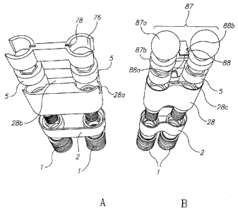
area. In yet other
embodiments the surgical guide clamp incorporates features that allow for the
easy and
accurate repositioning of the surgical templates over another target bone area
while the
surgical guide clamp remains clamped over the initial target bone area. In
additional
embodiments the surgical guide clamp also allows for the securing of the
surgical
templates over a secondary target bone area.
BRIEF DESCRIPTION OF THE DRAWINGS
[060] The principles and operation of the system, apparatus, and method
according to
the present invention may be better understood with reference to the drawings,
and the
CA 02751115 2011-07-28
WO 2010/089698
PCT/1B2010/050456
21
following description, it being understood that these drawings are given for
illustrative
purposes only and are not meant to be limiting, wherein:
[061] FIG. la is a front view of a vertical stacking of the components of one
possible
embodiment of a three-stage single-rooted implant of the improved implant 77
of the
present invention;
[062] FIG. lb is a front view of a vertical stacking of the components of one
possible
embodiment of a two stage single-rooted implant of the improved implant 77 of
the
present invention;
[063] FIG. lc is a front view of a vertical stacking of the components of one
possible
embodiment of a three-stage multi-rooted implant of the improved implant 77 of
the
present invention;
[064] FIG. ld is a front view of a vertical stacking of the components of one
possible
embodiment of a two stage multi-rooted implant of the improved implant 77 of
the
present invention.
[065] FIG. 2a is a top view of one possible embodiment of the "head" component
2 of a
three-stage single-rooted implant of the improved implant 77 of an embodiment
of the
present invention;
[066] FIG. 2b is a top view of the "head" component 2 of one possible
embodiment of
a three-stage multi-rooted implant of the improved implant 77 of an embodiment
of the
present invention;
[067] FIG. 2c is a top view of one possible embodiment of the "head/abutment"
component 6 of a two stage single-rooted implant of the improved implant 77 of
an
embodiment of the present invention;
[068] FIG.2d is a top is a top view of one possible embodiment of the
"head/abutment"
component 6 of a two stage multi-rooted implant of the improved implant 77 of
an
embodiment of the present invention;
[069] FIG. 2e is a bottom view of one possible embodiment of the "head"
component 2
depicted in FIG. 2a;
CA 02751115 2011-07-28
WO 2010/089698
PCT/1B2010/050456
22
[070] FIG. 2f is a bottom view of one possible embodiment of the "head"
component 2
depicted in FIG. 2b;
[071] FIG. 2g is a bottom view of one possible embodiment of the
"head/abutment"
component 6 depicted in FIG. 2c;
[072] FIG. 2h is a bottom view of one possible embodiment of the
"head/abutment"
component 6 depicted in FIG. 2d;
[073] FIG. 3a is a front view of two possible embodiments of the "root screw"
components 1 of the improved implant 77 of the present invention;
[074] FIG. 3b is a front of one possible embodiment of a connector screw 4 and
one
possible embodiment of an abutment screw 5 of the improved implant 77 of the
present
invention;
[075] FIG. 3c is an angled top view illustrating two possible embodiments of
the "root
screw" components 1 of the improved implant 77 of the present invention;
[076] FIG. 3d is an angled top view of one possible embodiment of the abutment
component 3 of a three-stage single-rooted implant of the improved implant 77
of the
present invention;
[077] FIG. 3e is an angled top view of one possible embodiment of the abutment
component 3 of a three-stage multi-rooted implant of the improved implant 77
of the
present invention;
[078] FIG. 3f is an angled bottom view of one possible embodiment of the
abutment
component 3 depicted in FIG. 3d;
[079] FIG. 3g is an angled bottom view of one possible embodiment of the
abutment
component 3 depicted in FIG. 3e;
[080] FIG. 4a is top view of one possible embodiment of the surgical template
precision support insert 17 of the present invention;
[081] FIG. 4b is a top view of one possible embodiment of a primary surgical
template
18 of the present invention;
CA 02751115 2011-07-28
WO 2010/089698
PCT/1B2010/050456
23
[082] FIG. 4c is a top view of one possible embodiment of a secondary surgical
template 19 of the present invention;
[083] FIG. 4d is an angled bottom view of one possible embodiment of the
surgical
template precision support insert 17 depicted in FIG. 4a;
[084] FIG. 4e is an angled bottom view of one possible embodiment of the
primary
surgical template 18 depicted in FIG. 4b;
[085] FIG. 4f is an angled bottom view of one possible embodiment of the
secondary
surgical template 19 depicted in FIG.4c;
[086] FIG.'s 5a-5f are a series of angled top views of several possible
embodiments of
fully assembled single-rooted and multi-rooted implants of the improved
implant 77 of
the present invention;
[087] FIG.'s 5g-5k are a series of angled bottom views of the fully assembled
single-
rooted and multi-rooted implants depicted in FIG.'s 5a-5f of the improved
implant 77 of
some embodiment of the present invention;
[088] FIG. 6a is an angled top and side view of two possible embodiments of a
precision surgical guide clamp 29 of the present invention that clamp on to a
tooth or
teeth in the jawbone to securely and accurately position the surgical
templates 18 and 19
over the implant site;
[089] FIG. 6b is a close-up enlarged view from the top and front perspective
of two
possible embodiments of detachable clamp heads 30 of the present invention;
[090] FIG.7a is an angled top and front view of one possible embodiment of a
free-
standing precision surgical guide clamp 38 of the present invention;
[091] FIG. 7b is a close-up enlarged view of an embodiment of a segment of the
free-
standing precision surgical guide clamp 38 depicted in FIG. 7a;
[092] FIG. 7c is a top view of several possible embodiments of clamping
attachments
for the free-standing precision surgical guide clamp 38 depicted in FIG.'s 7a
and 7b;
CA 02751115 2011-07-28
WO 2010/089698
PCT/1B2010/050456
24
[093] FIG. 8a is an angled top and side close-up view of one possible
embodiment of
the surgical template precision support insert 17 engaged within one possible
embodiment of the surgical template precision support platform 31 of the
present
invention;
[094] FIG. 8b is a front view of an embodiment of the securing bar 55 that can
be
dropped down to secure the precision attachment elements 39 and 39a of the
precision
surgical guide clamps 29 and 38;
[095] FIG. 8c is an angled front view of two possible embodiments of precision
attachment elements 39 and 39a of the precision surgical guide clamps 29 and
38 of the
present invention;
[096] FIG. 8d is a close-up enlarged view from an angled front perspective of
an
embodiment of a segment of the precision attachment elements 39 and 39a
depicted in
FIG. 8c;
[097] FIG. 9a is an angled close-up side view of an embodiment of a segment of
the
precision surgical guide clamp 28 depicted in FIG. 6a of the present
invention;
[098] FIG. 9b is an even closer-up angled side view of a segment of an
embodiment of
the precision surgical guide clamp 29 depicted in FIG. 6a;
[099] FIG. 9c is an angled close-up view from the top perspective of an
embodiment of
a different segment of the precision surgical guide clamp 29 depicted in FIG.
6a;
[0100] FIG. 10a is an angled close-up view from the side perspective of an
embodiment
of a segment of the surgical template precision support platform 31 and more
specifically
of an embodiment of the platform precision adjustment element 51 depicted in
FIG. 8a;
[0101] FIG. 10b illustrates an angled top view of the assembly (from left to
right) of an
embodiment of the parts needed to assemble the platform precision adjustment
element
51 depicted in FIG.'s 8a and 10a;
[0102] FIG. 10c is a close-up top view of an embodiment of the middle parts of
the
platform precision adjustment element 51 depicted in FIG. 10b;
CA 02751115 2011-07-28
WO 2010/089698
PCT/1B2010/050456
[0103] FIG.'s lla-llk are a series of top views of possible embodiments of the
self-
limiting bone drills 59, 61, 62, 63,65, and 66; component drivers 60, 67, 68,
69 and
surgical guide pin 64 of the present invention;
[0104] FIG.'s 12a-12f depict a series of top views of the preparation of one
possible
embodiment of the osteotomy 73 and the insertion of one possible embodiment of
components of the improved implant 77 of the present invention into the
osteotomy 73;
[0105] FIG.'s 13a -13h depict a series of angled top views of one possible
embodiment
of the precision surgical guide clamp 29 clamped at one possible implant site
which
illustrate its use in preparing one possible embodiment of a multi-root
osteotomy 73 for
the insertion within it of one possible embodiment of the improved implant 77
of the
present invention;
[0106] FIG. 14 is a close-up angled top view of one possible embodiment of a
"root
screw" component driver assembly composed of an implant carrier 70 and a slow
speed
driver 67, and one possible embodiment of the root screws 1 engaged within the
self-
limiting feature of one possible embodiment of the secondary surgical template
19;
[0107] FIG. 15 depicts an angled top view of an embodiment of the driver
assembly
illustrated in FIG. 14 fully engaged within the secondary surgical template 19
of FIG.
14, and below that, the same surgical template 19 engaged within the precision
guide
clamp 29 depicted in FIG. 6a;
[0108] FIG. 16 is an angled top and side view of one possible embodiment of
the
precision surgical guide clamp 29 illustrating a free-end saddle situation for
the implant
site;
[0109] FIG. 17a is an angled top view of a vertical stacking of the components
of one
possible embodiment of the improved multi-root implant 77 specifically
designed to
support an over-denture and one possible embodiment of the over-denture
connecting
assembly locking element 76 of the present invention;
[0110] FIG. 17b is an angled top view of a vertical stacking of the components
of one
possible embodiment of the improved multi-root implant 77 specifically
designed to
CA 02751115 2011-07-28
WO 2010/089698
PCT/1B2010/050456
26
accept the insertion of one possible embodiment of a double ball clip
attachment design
87 for an overdenture (instead of an over-denture bar clip) to allow for the
secure
attachment of a full denture to the improved multi-root implant 77 of the
present
invention;
[0111] FIG. 18 is an angled top view of one possible assembly embodiment of
several
possible embodiment of the multi-root improved implant 77 of the present
invention
wherein is depicted overdenture connecting assembly components, 76, 79, 80 and
81
engaged within these implant in order to allow for a fixed full arch support
for a
removable full denture in an edentulous patient;
[0112] FIG.'s 19a-19e are a series of close-up top view of possible
embodiments of the
connecting assembly components 76, 79, 80 and 81 depicted in FIG. 18; and
[0113] FIG.'s 20a-20c are a series of front views of one possible embodiment
of a two
stage overdenture head/abutment component 6a and two possible embodiments of
three
stage over-denture abutments made of components 3 coupled with 89 and 28
coupled
with 87.
[01 14] It will be appreciated that for simplicity and clarity of
illustration, elements shown
in the drawings have not necessarily been drawn to scale. For example, the
dimensions
of some of the elements may be exaggerated relative to other elements for
clarity.
Further, where considered appropriate, reference numerals may be repeated
among the
drawings to indicate corresponding or analogous elements throughout the serial
views.
DETAILED DESCRIPTION OF THE INVENTION
[01 15] The following description is presented to enable one of ordinary skill
in the art to
make and use the invention as provided in the context of a particular
application and its
requirements. Various modifications to the described embodiments will be
apparent to
those with skill in the art, and the general principles defined herein may be
applied to
other embodiments. Therefore, the present invention is not intended to be
limited to the
CA 02751115 2011-07-28
WO 2010/089698
PCT/1B2010/050456
27
particular embodiments shown and described, but is to be accorded the widest
scope
consistent with the principles and novel features herein disclosed. In other
instances,
well-known methods, procedures, and components have not been described in
detail so as
not to obscure the present invention.
[01 1 6] The word "stage" as used herein refers to the different overall types
of separate
sections of the implant. A "component" is a particular version (size, shape or
number) of
the parts of that type of distinct "stage" being used to assemble the
different types of
stages of the implant into one whole implant.
[01 1 7] Non-limiting embodiments of the invention include improved systems,
means and
methods for the preparation and insertion of improved anatomically corrected
implants
that more closely imitate the overall natural form of the root system of human
teeth. In
some embodiments this system includes two detachable modular stages with
customizable features to which a third abutment stage is attached or
alternatively, is an
integral part of one of the two stages.
[01 1 8] The implant system, apparatus, tools, kit and methods of the present
invention
substantially solve many of the problems associated with the prior art. For
example, a
multiple "root screw" implant design would significantly increase the total
bone to
implant interface surface as compared to the current single "root screw"
design, a
significant biomechanical advantage as it would allow for vastly greater bone
adhesion
and "osseo-integration" of the improved implant.
[01 1 9] Initial rigid fixation is desirable for osseo-integration to occur.
This initial rigid
fixation is enhanced by engaging the hard, dense cortical bony plates of the
jawbones. An
implant that would incorporate in its design a more naturally contoured cross-
sectional
dimension of the "head" of the implant may allow the dentist upon initial
placement of
the implant at the implant site to directly engage these cortical bony plates
with the
"head" segment, allowing for the immediate fixation of the implant in the bony
implant
site. This would represent a significant advantage over the prior art, both
for cases where
the dentist plans on "burying" the implant for an extended period of time in
order to
allow for osseo-integration prior to loading of the implant as well as in
cases where the
CA 02751115 2011-07-28
WO 2010/089698
PCT/1B2010/050456
28
dentist intends to immediately load the implant. Immediate loading is a
technique which
has become more popular of the past few years as it allows the dentist to
restore the
missing tooth immediately, allowing for an immediate esthetic solution to the
missing
tooth.
[0120] In order to allow for a stable and durable implant form the diameter of
the "root
screw" component of the implants along its entire shaft must be of sufficient
girth in
order to provide the necessary support for the abutment and crown which sit
atop it and
the forces transmitted through them (when they are placed in function) to the
implant
"root" buried in the jawbone. Based on the above space limitations, it is
determined that
in order to accommodate multi-root screws in a healthy long term biological
manner, this
requires for these multiple "root screws" to be of a smaller diameter than the
current
single "root screw" design of standard implants.
[0121] In order to allow for the placement of a "multi-root" design implant
into this very
limited bone volume, one must consider all the described above limiting
factors and
balance them with the desire to place the largest diameter implant "roots"
possible given
these constraints, so as to allow for the placement of the most stable and
durable "multi-
root" implant into the implant site.
[0122] As noted above, the very limited three-dimensional volume of bone
represents
the implant site and presents severe challenges for the successful
implantation of the
standard "single root" implant design within it in a reproducible and safe
manner. To
achieve this goal successfully in order to provide for a stable and durable
"multi-root"
form implant for the posterior teeth that is able to withstand the
considerable load forces
it must endure both in the short and long term, represents an even greater
challenge, one
which requires the accurate and precise preparation of the implant site in a
three-
dimensional manner as well as the accurate and precise insertion of the
implant
components within this site in a three-dimensional manner. Any system, to be
successful
in achieving this goal, must incorporate features that allow for precisely
this result.
[0123] The improved multi-root posterior implant of embodiments of the present
invention not only splints together multiple implant "root screw" forms but
does so
CA 02751115 2011-07-28
WO 2010/089698
PCT/1B2010/050456
29
endosseously (completely imbedded in bone). This means that the current
invention is
stronger, more stable and more durable than any of the standard posterior
implant
designs.
[0124] There is provided, in accordance with an embodiment of the present
invention, an
apparatus, system, and method for a modular design two or three-stage
anatomical dental
implant system, for supporting a fixed or fixed/removable dental prosthesis
(i.e.
individual crowns, multiple crowns, or over-dentures) comprising in the two
stage
embodiment, modular single and multiple distal "root screw" stage(s) or
components
which are attached to a modular customizable proximal "head/abutment" stage
or; in the
three stage embodiment, modular single and multiple distal "root screw" stage
(s) or
components which intimately fit into a modular customizable proximal "head"
stage
(component) of the improved dental implant into which in turn intimately fits
a third
abutment stage (component). These components allow for the assembly of
improved
single and/or multiple "root" dental implants for anterior and posterior
teeth.
[0125] When the first two stages of the improved implant have been fixed into
the
patient's jawbone, this allows the dentist in the three-stage embodiment to
intimately fit a
third stage ready-made cast or castable abutment utilizing the anatomically
formed
"head" stage of the implant as its attachment. In the two-stage embodiment the
"abutment" is an integral part of the "head/abutment" component. In both
embodiments,
the proximal "head" or "head/abutment" stage does not extend deeply into the
bone, and
the distal "root screw" stage (s) or components intimately fit into the "head"
or
"head/abutment" stage and are smaller or equal in overall dimension to
standard dental
implants, allowing for conservative bone preparation at the implant site. The
"head" or
"head/abutment" stage of the implant may also include in both embodiments a
ready-
made prosthetic crown margin interface extending above the crest of the
jawbone at the
cervical junction (cemento-enamel junction level) allowing for superior
marginal
integrity and seal of the fixed prosthesis (crown). In both embodiments, the
"head" or
"head/abutment" stage and "root screw" stages can be shaped to allow for the
"root
screw" stages to attach to the "head" or "head/abutment" stage either parallel
to or angled
in relation to each other. In both embodiments the "head" component or
"head/abutment"
CA 02751115 2011-07-28
WO 2010/089698
PCT/1B2010/050456
component may include micro-grooves and ridges on its external surface for
enhanced
primary retention and subsequent bone adhesion. In the embodiment that is
designed for
over-dentures, a unique abutment design and unique adjustable over-denture bar
design is
described.
[0126] Furthermore, precision surgical guide tools and methods for the
accurate, precise,
and reproducible implantation of the two and three-stage improved dental
implant system
at the implant site designed specifically for the improved two and three-stage
dental
implant are also disclosed.
[0127] Embodiments of the present invention provides for an improved design of
several
types of dental implants (single-"rooted" anterior implants, and single and
multi-"rooted"
posterior implants) inserted into the alveolus of the upper and lower jaws as
well as a set
of precision surgical tools and a method of utilizing them for the accurate,
precise and
reproducible implantation of the improved dental implants described below. The
improved implant(s) is/are comprised of two detachable and modular stages to
which a
third abutment stage is either connected to as a separate stage or is an
integral part of one
of these two stages. The first stage, the distal modular stage, is comprised
of either single,
a pair, or multiple cylindrical "root screw" forms or implant screws that may
be tapered
or straight along their length and that may have on their exterior surface rod-
shaped,
screw-shaped, and or having fins to assist in retention to the bony alveolus.
These distal
"root screw" stages will be made available in a kit of components of varying
lengths and
diameters, allowing the dentist to choose and "mix and match" on the same
dental
implant different sized implant "root screw" components for "multi-rooted"
implants, and
for both single and multi-"rooted" implants to choose from a kit of different
sized and
different form "head" or "head/abutment" components the one that provides the
most
advantageous composite of the two separate stages for any particular implant
site. These
"root screw(s)"components may attach either parallel to or angled to the
"head"
component and the root screws themselves may be parallel or offset from each
other.
[0128] Criteria for choosing which composite of components are most
advantageous to
employ and assemble may include, but not be limited to, among other criteria,
a
diagnostic consideration of the anatomical limitations of the implant site as
well as an
CA 02751115 2011-07-28
WO 2010/089698
PCT/1B2010/050456
31
evaluation of the requirements for anchorage and stability of the dental
implant to be
placed, based on the location and anticipated functional load the dental
implant will be
expected to bear.
[0129] The proximal detachable "head" or "head/abutment" stage of the implant
in
embodiments of the present invention, which is meant to replace the current
integral
"head" or top section of a standard implant, is shaped to conform more closely
than all
the previous state of the art to the natural cross-sectional form of the
different types of
roots of maxillary and mandibular (upper and lower jaw) teeth (incisors,
cuspids,
premolars and molars) at the level of the crestal ridge (the level of the
cervix). The
"head" components or "head/abutment" components of this stage are relatively
shallow in
their overall vertical dimension in order to allow for the conservative bone
preparation at
the implant site needed to accommodate its more natural cross-sectional form
(oval,
rhomboid, kidney-shaped, or other custom shape).
[0130] The two separate stages ("head" and "root screw") are intimately fitted
and
attached into each other via fitted sleeves and collars or other means of
coupling
including, but not limited to, internal threaded screw retention of the two
stages in order
to provide a tightly sealed coupling between the two stages and minimize any
micro-
gapping between them.
[0131] The "head" or "head/abutment" components in embodiments of the present
invention incorporates single or multiple attachment points on its distal or
inferior
surface, allowing for the assembly of single, bi and tri-"rooted" implant
forms with
smaller or equal sized multiple "root screw" forms to replace the missing
molar teeth
while actually enhancing the stability and ability to withstand the loading
force of
functional and para-functional chewing or grinding. There is an additional
distinct
advantage as it allows several embodiments of the improved implants to be
placed in
locations that would previously not have allowed for the larger sized standard
"single
root" implant root forms to be placed in the alveolus without first subjecting
the patient to
additional surgical procedures such as maxillary sinus lifts or maxillary or
mandibular
ridge augmentation procedures. This saves the patient from the trauma of these
surgeries,
reduces healing time, and saves the dentist valuable treatment time and
overall time
CA 02751115 2011-07-28
WO 2010/089698
PCT/1B2010/050456
32
required to complete each case. It also provides additional advantage as it
allows for
more conservative bone drilling at the implant site due to the fact that the
dentist may
choose to drill smaller sized "root screw" bore shafts in order to insert
smaller
dimensioned implant "root screw" components.
[0132] Additionally, since embodiments of the present invention also include
"head" and
"head/abutment" stages that allow for the connection of multiple "root screw"
stages to
the "head" or "connector" stage to form a "multi-rooted" implant, another
significant
advantage is that it provides enhanced structural advantages both in retention
of the
implant in the bony socket (s) and greater ability to withstand over time the
forces and
load of the natural "bite" and chewing, clenching, and grinding loads both in
normal use
and in patients who grind their teeth or clench their jaws (paranormal
function) over the
standard "single-root" implant.
[0133] Due to the far greater surface area of the improved implant that is
buried in bone,
the implant to bone interface is far greater than in any standard implant.
This may result
in improved healing time as well as providing for a greater amount of
osseointegration
for the implant and therefore greater "loading" capacity of the improved
implant of the
present invention over any standard "single-rooted" implant once healing is
complete.
This enhanced loading capacity of the improved implant is especially desirable
when the
dentist wishes to use several of the improved implants as the fixed support
structure for a
removable over-denture.
[0134] Additionally, embodiments of the present invention contain a three-
stage
embodiment wherein is incorporated in the inner aspect of the proximal "head"
stage an
internal connector or connectors, such as but not limited to, an internal
threaded shaft
machined to accept a separate third stage abutment segment comprising either a
screw-in
or snap-in abutment whose purpose is to act as the supporting structure for
the implant
crown to be fabricated by the dental practitioner. The more anatomically
correct modified
oval, elliptical, or rhomboid cross-sectional shape of the "head" or
"head/abutment
component as well as the uniquely designed multi-point internal connectors of
the "head"
or "head/abutment" component to a third "abutment" component that fits
precisely into it
(hand in glove) and provides for superior anti-rotational features as well.
This is due to
CA 02751115 2011-07-28
WO 2010/089698
PCT/1B2010/050456
33
the intimate frictional contact of the entire internal surface of the "head"
or
head/abutment" component to a large portion of the external surface of the
"abutment"
component as well as the frictional fit to significant internal surfaces of
the abutment.
This secure and completely rotation-free connection between the "head" stage
of the
improved implant and the "abutment" is a significant advantage of the present
invention
over the prior state of the art.
[0135] Additionally, in embodiments of the three stage design for anterior and
posterior
single "rooted" and posterior multi-"rooted" implants, a large portion of the
abutment
"sinks" into the extensive internal hollowed out ("basket") unique design of
the separate
"head" component (which is buried in the bone) and so allows for an
advantageous
distribution of the load forces not found in any of the prior art.
[0136] Additionally, embodiments of the present invention may contain an
implementation wherein the outer border of the area referred in the dental
literature as the
"collar" or "margin" of the proximal "head" or "head/abutment" component is
machined
to provide an ideal 360 degree restorative margin for the dentist to fabricate
the crown to
be fitted upon the present embodiment implant form. This feature is
incorporated to
further enhance the seal and marginal integrity (prevent leaking or seepage)
of the final
restorative crown to the implant. The "head" or "head/abutment" component may
also
have an embodiment which incorporates "micro-threads" or smaller ridges and
grooves
on its external surfaces to increase its external surface area and: 1)
maximizes bone
adhesion to the "head" component; 2) minimize pathogenic microbial
infiltration of the
peri-implant (bone surrounding the implant) space; and 3) enhance the ability
of the
implant to achieve primary fixation by engaging the hard bony cortical plates
of the
jawbones, another distinct advantage of present invention over all the prior
state of the
art.
[0137] As the unique design of the surgical templates provided in embodiments
of the
present invention can be shaped in an infinite variety of shapes and
dimensions, by
varying the length of the "head" in this dimension allows for varying the
distance
between the "root screws" of a "multi-rooted" implant as well. This allows for
numerous
biomechanical advantages over the known art as it allows for the increased
retention,
CA 02751115 2011-07-28
WO 2010/089698
PCT/1B2010/050456
34
strength, and durability of the multi-root implant of the present invention
over all the
prior art, especially relevant in supporting an overdenture bar for a full
removable denture
(in the edentulous patient the mesio-distal dimension of the head can be
substantially
increased as there are no adjacent teeth to consider). Additionally, this
particular design
feature of the present invention allows for greater width of bone volume
between the
"root screw" components of the improved implant, a further biological
advantage over
the entire prior art.
[0138] Embodiments of the present invention also provide for a single "root
screw" stage
that attaches to an anatomically more correct (oval) anterior "head" or
"head/abutment"
stage for anterior dental implants as well as a single "root screw" stage that
connects to a
"head" or "head/abutment" stage for molar teeth where the mesio-distal bone
volume
width of the edentulous implant site does not allow for the placement of two
"root screw"
stages.
[0139] Embodiments of the present invention also provide for unique "abutment"
components for an improved three stage single and multi-root implant shaped to
intimately fit into the corresponding "head" components which contain uniquely
designed
(described in the drawings below) internal hollowed areas for the insertion of
a novel new
adjustable over-denture bar assembly with locking features. This intimate fit
of the
present invention's pre-cast overdenture bar assembly components to the
abutment
components of the implants provides for a more exact fit than the entire prior
art, as the
entire prior art relies on dental impressions and laboratory castings (which
are known in
the dental field to be inherently inaccurate due to the expansion and
contraction of the
materials involved in its fabrication) in order to fabricate the standard over-
denture bar
framework. When the improved overdenture bar assembly of the present invention
is
inserted into either the single or multi-root implants of the present
invention, a full arch
over-denture support may be prepared, assembled and inserted into the
edentulous
patient's mouth. This new design provides for greater retention, stability,
strength, and
durability of the implant-prosthesis over the entire prior art.
[0140] Embodiments of the present invention also provide for a unique two and
three
stage improved implant wherein the two stage design allows for the securing of
two
CA 02751115 2011-07-28
WO 2010/089698
PCT/1B2010/050456
separate over-denture attachments to the "head/abutment" component or in the
three
stage design either two separate overdenture attachments to the separate
abutment
component or a unified double over-denture attachment which has a locking
element to
secure it to the separate abutment component.
[0141] Both the "head", "head/abutment" and "root screw" stages of embodiments
of the
present invention may be made of bio-compatible materials including, but not
limited to,
metal, ceramic, glasses, or any combination thereof, and potentially having
various
coatings of materials such as titanium beads, titanium plasma spray,
hydroxyapetite, or
bone growth chemicals or similar coatings to enhance attachment to bone.
[0142] To simplify, standardize, and allow for consistent and precise
reproducibility of
insertion, the following embodiments of novel surgical devices may be employed
to
prepare the bony alveolus site (osteotomy) to accept the present invention
implant forms:
[0143] A kit of different sized primary self-limiting (in all three
dimensions) surgical
templates (either re-usable or one-time use) which are slightly larger in
dimension but
exactly corresponding to the cross-sectional shape of the different "head" or
"head/abutment" stages at the level of the bony crest height (level of the
cervix) as will
be illustrated in the drawings below. The dental practitioner chooses the most
appropriate
shape and size template from the kit and secures it in a precise location via
the precision
adjustable clamping device described below over the implant site to act as a
drill guide
form for the bone preparation of the "head" component of the three-stage
implant design
or the "head/abutment" component of the two-stage implant design of the
present
invention. These self-limiting primary surgical templates allow for the
precise shape,
depth, levelness of the floor, and accurate location of the top portion of the
bone
preparation (osteotomy) in relation to the adjacent teeth, or in the
edentulous patient, to
the exact jaw location deemed most advantageous by the dental practitioner for
the
placement of the implant.
[0144] A kit of different sized self-limiting (in all three dimensions)
secondary surgical
templates (reusable or one-time use) corresponding in dimension (though
possibly
slightly larger) to the dimensions of the actual "head" or "head/abutment"
stage implant
CA 02751115 2011-07-28
WO 2010/089698
PCT/1B2010/050456
36
form and corresponding to the primary surgical template form it is designed to
be used
with. These secondary surgical templates include in their design self-limiting
drill guide
holes to properly drill the "root screw" bone shafts in the bone at the
implant site at the
proper angulations, position, and depth in relation to the bone preparation at
the implant
site for the "head" or "head/abutment" stage that has already been prepared.
These
secondary surgical templates also act as accurate depth gauges to initially
check both the
depth and levelness of the floor of the initial bone preparation (osteotomy)
of the "head"
or "head/abutment" stages. These same surgical templates utilize these self-
limiting
insertion guide holes to:
a. check for the angulations (parallelism) and location of these bore shafts
in
relation to the adjacent teeth when utilized in conjunction with the self-
limiting surgical
guide pins that are specifically designed for use with the secondary surgical
template.
b. insert the "root screw" stage components of the improved implant of the
current invention in the proper location and to the exact proper vertical
depth within the
bore shaft(s) when utilized in conjunction with the self-limiting "root screw"
driver
specifically designed for use with the secondary surgical template.
c. properly relate the "head" stage component of the improved implant of the
current invention and allow for the intimate connection of the two stages to
each other via
either the connector screws provided for the attachment of the two stages to
each other or
some other means of connection.
[0 1 45] A precision adjustable clamping device of various different designs
is also
provided for in embodiments of the present invention. One embodiment, which
may be
used even in the completely edentulous patient clamps directly into the
concave
undercuts of the gingival vestibules of the oral cavity while another variant
of this
embodiment allows for the temporary rigid fixation of this same clamp in these
same
vestibules after reflection of the gingival tissue via fixation screws or
other fixation
elements. An entirely different embodiment of the clamping device clamps
directly to a
natural tooth or teeth adjacent to the implant site. These clamps act as a
fully adjustable
(in all three dimensions and is even rotatable) support and fixation platform
to secure
both the primary and secondary surgical templates described above accurately,
precisely
CA 02751115 2011-07-28
WO 2010/089698
PCT/1B2010/050456
37
and intimately over the implant site of both the upper and lower jaws as will
be illustrated
in the drawings below. These surgical implant tools both simplify and
standardize the
bony preparation of the implant site by providing an accurate three
dimensional self-
limiting guide when used in conjunction with the self-limiting dental implant
drills and
implant guide pins of the present invention (described below) in order to
prepare the
implant site in a three dimensional manner to receive the improved implant of
the present
invention as well as allowing for the accurate and precise assembly of the
implant
components of the two and three-stage improved implants of the current
invention in a
three dimensional manner within the limited bone volume of the implant site.
[0146] A kit of self-limiting dental implant drills, implant guide pins,
implant component
and implant screw drivers specifically designed to be used in conjunction with
the
surgical templates and surgical clamps of embodiments of the present invention
in order
to properly prepare the alveolus (osteotomy) for the insertion of the improved
two and
three-stage dental implants of the present invention.
[0147] Relation of embodiments of the Present Invention to aspects of known
Art:
[0148] For example, in relating to U.S. Pat. No. 6,854,972, the cross-
sectional form
described at the superior aspect of the "head" of the implant remains round in
form and
so does not reflect the natural more oval cross-sectional form of the natural
anterior teeth
at this level (CEJ). Since the "head" portion is an integral part of the one
piece main
body, it offers none of the advantages of the modular separate "head" or
"head/abutment"
design of the present invention. This modular design of these two stages in
the present
invention allows the dentist to place a more naturally shaped "head", a
distinct biological
advantage, as well as the freedom to assemble the most advantageous composite
of these
two stages for any particular implant site based on many important criteria,
such as, but
not limited to the volume of bone present, the quality of bone at the implant
site, and
esthetic considerations.
[0149] Relating to U.S. Pat. No. 6,093,023, the polygonal abutment described
contains
none of the advantages of the modular natural shaped "head" or "head/abutment"
components of the present invention which are placed endosseously (in the
bone).
CA 02751115 2011-07-28
WO 2010/089698
PCT/1B2010/050456
38
[0150] In relating to U.S. Pat. No. 7,291,013, the suggested implant
configuration still
maintains a round cross-sectional form of the integral head of the "single-
root" implant,
and therefore does not incorporate the advantages of the present invention's
anterior
"single-root" improved implant.
[0151] Relating to U.S. Pat. No. 6,527,554, the implant type described offers
some
improvements on prior implants, yet still maintains a round cross-sectional
form and
offers none of the advantages listed below of the present invention.
Additionally, the
improved implant of the present invention utilizes micro-threads and grooves
to achieve
the same result in a more profound way while also significantly enhancing the
retentive
qualities of the implant to a much greater degree than does this prior art.
[0152] In relating to U.S. Pat. App. No.US 2010/0003638 describes an implant
design
with additional suggested improvements, however this implant maintains the
round cross-
sectional form of its modular "head" segment, it does not offer the
biomechanical,
biological and esthetic advantages of form of the more naturally shaped
modular "head"
design of the "single-root" embodiment of the present invention.
[0153] Relating to WO Pat. App. No. 2006/082610 describes an implant design
with a
"head" component which it calls a "collar" and a "root screw" component which
it calls a
"fixture". The multi-root implants it describes are three-stage only in their
design and do
not include the two-stage design embodiment of the present invention. They are
also only
designed to replace "multiple-root" posterior teeth, while the design of the
present
invention also allows for the replacement of "single-root" and "multiple-root"
posterior
teeth as well as "single-root" anterior teeth.
[0154] Relating again to WO Pat. App. No. 2006/082610 further contains a
minimal
internal hollowed out area in its "head" stage for the intimate insertion of
the "abutment"
stage. In the present invention, the internal hollowed out area of the "head"
component is
extensive, encompassing the entire inner surface area of the "head" component.
This
extensive internal hollowed feature of the present invention, by providing for
a more
intimate fit of these two components in a "hand in glove" manner, also
provides for far
greater frictional fit and so greater anti-rotational function as well as
wider and more
CA 02751115 2011-07-28
WO 2010/089698
PCT/1B2010/050456
39
uniform distribution of the forces of mastication or "load" over the entire
structure of the
improved implant compared to this prior art once the implant is placed in full
function
with a crown placed on it. Due to the intense load and off-axis (shear) force
component
of that load (noted above) placed on posterior dental implants in full
function during
normal chewing and paranormal grinding by the patient, (particularly on the
"multi-
rooted" posterior molars) an improved implant design, such as is described in
the present
invention, that allows for just such a wider and more uniform load
distribution throughout
the structure of the entire implant, represents a major advantage over all the
prior art.
[01 55] Additionally, as opposed to some embodiments of the present invention
where a
three-stage multi-root embodiment is used, WO Pat. No. 2006/082610 does not
contain
an internal screw collar or ring feature projecting up from the top surface of
its "head"
component to further increase the frictional fit between the "head" stage and
the
"abutment" stage. The prior art thus relies to a much greater degree on the
single set of
two "connector" screws between all three stages of these components (abutment,
head,
and two root screws) to achieve a solid and secure connection between all of
these
components. This design is highly undesirable as it places undo forces on this
single pair
of "connector" screws which over time may loosen or fracture, causing either
failure of
the crown, or more seriously, failure of the implant. In the present
invention, due to its
unique "hand in glove" design of the "head" and "abutment" stages, which, as
noted
above, also includes a large surface screw collar (s) or ring (s) feature
projecting upward
from its internal surface of the "head" component as well as the two separate
pairs of
connector screws as also noted above, there is a much reduced reliance on each
individual pair of "connector" screws to achieve an intimate and secure
connection
between these stages, another significant improvement in design over all the
prior art.
[01 56] Further, as opposed to some embodiments of the present invention, WO
Pat. No.
2006/082610 also does not incorporate micro-threads or grooves on the surface
of its
"head" component for the enhancement of the adhesion of bone to this
critically
important component, as this component is the most proximal component of the
implant
and so is closest to the oral cavity and the numerous pathogenic microbes
contained
within it. By enhancing bone adhesion in the initial stages of healing and
subsequently
CA 02751115 2011-07-28
WO 2010/089698
PCT/1B2010/050456
thereafter for the long term, these micro-threads on the "head" stage of the
present
invention help to prevent the infiltration of these pathogenic bacteria into
the coronal
segment of the implant, preserving bone and increasing the longevity of the
implant in
the mouth. These same micro-threads act to enhance primary fixation of the
improved
implant, a highly desirable result as it may allow for the immediate "loading"
of the
implant, so that the patient can actually use the temporary crown placed on it
immediately.
[0157] In further relating to WO Pat. No. 2006/082610, embodiments of the
present
invention incorporate in their design indented (concave) collars or rings on
the underside
surface of the "head" or "head/abutment" component to which is related in an
intimate
manner (from below) the superior aspects of the "root screws". In the improved
implant
of the present invention the "root screws" do not need to be threaded through
the
retention holes of the "head" component in order to engage with the "head"
component as
in the prior art. This novel design of the present invention allows for the
"root screw"
components to be of significantly greater diameter than the retention hole of
the "head"
or "head/abutment" component and of significantly larger diameter than the
"root
screws" of the prior art.
[0158] This design feature of the "head" component of the present invention,
apart from
providing an intimate and positive seat of the "root screw" components to the
underside
of the "head" component also acts to prevent "micro-gapping" between these two
components and so prevents the possible infiltration of pathogenic bacteria
between
them, a further advantage of the design of the present invention. As noted
above, the
novel physical relation and sequence of relation of these components to each
other also
allows for the use of significantly larger diameter connector screws than the
prior art
(since the root screws are of greater diameter, the connector screws can be of
greater
diameter as well). These connector screws are of sufficient diameter (girth)
to withstand
the load forces (including shear forces) placed upon the improved dental
implant for the
long term, and so provide for a robust and secure connection of these
components over
the long term, a major advantage over the prior art, and an absolute
requirement that
allows the dentist to place with confidence the improved multi-root implants
of the
CA 02751115 2011-07-28
WO 2010/089698
PCT/1B2010/050456
41
present invention in the posterior areas of the jawbones without fear of these
implants
fracturing under long term loading.
[0159] Another major drawback in the design of the prior art relates to the
fact that as
mentioned above, it employs only one set of two connector screws to secure all
three
stages of its components (abutment, head and two root screws) to each other
with no
stress breaking feature between them.
[0160] The improved implant of the present invention both in its two and three-
stage
embodiments solves all of the above drawbacks in design of the prior art by
allowing for
the placement of two sets of two connector screws (in the bi-rooted
embodiment). One
set of two connector screws of sufficient diameters actively secure the "head"
or
"head/abutment" component to the two "root screw" components of sufficient
diameters
at the initial insertion of these components into the implant site. This
prevents the
possibility of any shifting between these components during the extensive
healing phase.
The other set of two connector screws actively secure the "abutment" to the
"head"
component after healing in the three-stage embodiment of the present invention
or simply
secure the access shaft of the "head/abutment" component in the two-stage
embodiment
of the present invention.
[0161] By utilizing two sets of two connector screws, a stress breaker feature
is thereby
incorporated into the structural design, another advantage of embodiments of
the present
invention over the know art.
[0162] In relating to U.S. Pat. App. No. US 2010/003635, the implant design
described
contains none of the modular design advantages of embodiments of the present
invention,
and utilizes a non-standard material composition that is less retentive as it
contains no
threads in its "root" design to bite into the surrounding jawbone or allow for
the intimate
adhesion of the surrounding bone during the osseointegration phase of healing.
[0163] Relating to U.S. Pat. App. No 2008/0293012, the splint abutment
component
described offers none of the biomechanical advantages of present invention as
it utilizes
the standard cylindrical "root" form of standard two stage dental implants
implanted in
the jawbone into which it connects a wide platform abutment component which
sits
CA 02751115 2011-07-28
WO 2010/089698
PCT/1B2010/050456
42
above the height of the crestal jawbone in the mouth and to which is connected
the crown
that the patient chews with.
[0164] The present invention in its "single-root" design embodiment (two and
three
stage) for posterior teeth incorporates an anatomically correct "head" which
is inserted
and embedded in the jawbone (endosseous). The abutment component of the
improved
implant of the present invention which connects into the "head" component in
its three-
stage embodiment, does so in an intimate "hand in glove" manner, providing for
the
proper distribution of the biting forces over the entire structure of the
implant, a distinct
advantage over all the prior art, and particularly over the disadvantageous
biomechanical
design of U.S. Pat. No. 2008/0293012 described above, while providing the
proper
"root" form for a posterior (molar) implant abutment. In the two-stage
embodiment of the
"single-root" posterior improved implant of the present invention, the
abutment
component is an integral part of the "head/abutment" component and so also
provides for
the proper distribution of loading forces on the entire implant.
[0165] Relating to U.S. App. Pat. No. US 2004/0013999 and U.S. Pat. No.
U52009/0202959, the surgical guide clamps described do not allow for the
preparation
of the modular design and differently shaped separate "head" and "root screw"
forms
(components) of the present invention. Additionally, this prior art's ring
guide also does
not allow for the preparation of the bone at the implant site to receive
separate and
differently shaped forms (components) of the "head" and "root screw" stages of
the
present invention and is limited to preparing a standard implant form whose
head is round
in cross-section and of the same diameter as the rest of the implant.
Additionally, the
known art does not describe a free-standing embodiment of its guide clamp (not
clamped
to the teeth) as do some embodiments of the present invention.
[0166] A further major drawback of the entire prior art (including WO Pat. No.
2006/082610) is that they do not allow for surgical templates that allow for
the placement
of different mesio-distal length "heads". As the unique design of the surgical
templates
provided in the present invention can be shaped in an infinite variety of
shapes and
dimensions, varying the length of the "head" in this dimension allows for
varying the
distance between the "root screw" components of a "multi-rooted" implant as
well. This
CA 02751115 2011-07-28
WO 2010/089698
PCT/1B2010/050456
43
allows for numerous biomechanical advantages over all the prior art as it
allows for the
increased retention, strength, durability of the multi-root implant of the
present invention
over all the prior art, especially relevant in supporting an overdenture bar
for a full
removable denture (in the edentulous patient the mesio-distal dimension of the
head can
be substantially increased as there are no adjacent teeth to consider).
Additionally, this
particular design feature of the present invention allows for greater width of
bone volume
between the "root screw" components of the improved implant, a further
biological
advantage over the known art.
[0167] Regarding the prior art usage of standard implants with their single
"root screw"
design, in order to allow for a stable and durable implant form, the diameter
of the
implants along their entire shaft must be of sufficient girth in order to
provide the
necessary support for the abutment and crown which sit atop them and the
forces
transmitted through them (when they are placed in function) to the implant
"root"(s)
buried in the jawbone. Based on the above space limitations, it is obvious
that in order to
accommodate multi-root screws in a healthy long term biological manner,
requires for
these multiple "root screws" to be of a smaller diameter than the current
single "root
screw" design of standard implants.
[0168] In the prior art WO 2006/082610 described above, the basic template
described is
utilized to drill the bore shafts (for the "root screws") of the osteotomy
first. Next the
more shallow and wider top portion of the osteotomy is drilled out (for the
"head")
utilizing the same basic template. The "head" component is then placed first
into the
upper portion of the osteotomy and the "root screw" components are then
inserted into
the "head" by sliding them through the small holes contained within the "head"
component and threading them into the bore shafts.
[0169] In various embodiments of the present invention, the entire above
sequence is
exactly reversed. The primary surgical template is utilized to drill out the
top portion of
the osteotomy first (for the "head" component). The secondary surgical
template is then
placed inside the top portion of the osteotomy and utilized to drill the bore
shafts of the
osteotomy (for the "root screw" components). The secondary surgical template
is then
used to check the parallelism of the bore shafts with the adjacent teeth and
then utilized to
CA 02751115 2011-07-28
WO 2010/089698
PCT/1B2010/050456
44
insert the "root screw" components first into the osteotomy. The "head"
component is
then fitted down onto the "root screws" and secured in the top portion of the
osteotomy to
the "root screw" components already in place below it via connector screws
threaded
through the retention holes of the "head" or "head/abutment" component.
[0170] By necessity there is an obvious direct relation between the tools and
method used
to prepare the implant site for the implant's insertion and the design form of
the implant
to be placed within this site. In layman's terms, the "peg" must fit the
"hole". In the
dental field, this fit must also be precise and reproducible in an easy manner
by the dental
practitioner in order to be able to achieve a high rate of success. More
specifically, in the
case of a multi-root implant for posterior teeth, in order to relate and
assemble the
components of the different stages in a three dimensional manner (along all
three axes) in
an accurate and precise manner within the bone preparation (osteotomy) it is
of critical
importance to relate the bone preparation for the "head" component with the
bone shaft
preparations for the "root screw" components. The prior art does not describe
any design
element incorporated into either the bone drills or the template it describes
that would
allow for a self-limiting depth (axis) control feature.
[0171] If the dentist errs in drilling by, for example, less than half a
millimeter in the
vertical depth (axis) of either of these two separate bone preparations, it
will be
impossible to insert the multiple components of the implant in a precise three
dimensional manner. To make matters worse, this prior art (WO 2006/082610)
does
mention a small thin locking pin to be inserted into the body of the template
but does not
describe how one could possibly use this feature to fix the template securely
in the
implant site utilizing this feature in the center of the bloody implant site.
It also provides
no means of precisely relating the position of the template to the adjacent
teeth, making
the practical use of this template at the implant site very problematic.
[0172] Embodiments of the present invention do allow for just such a precise,
accurate,
and easily reproducible preparation of the entire osteotomy (head and root
screw bone
preparation segments of the osteotomy) in a three dimensional manner (as will
be
described in detail below) and the precise, accurate, and easily reproducible
assembly of
the components of the improved multi-root implant in a three dimensional
manner (along
CA 02751115 2011-07-28
WO 2010/089698
PCT/1B2010/050456
all three axes) into the osteotomy, utilizing the surgical guide clamps,
primary and
secondary self-limiting surgical templates, and the self-limiting bone drills
provided in
the improved implant system of the present invention, another distinct
advantage over the
known art.
[0173] Embodiments of the present invention also provides for a robust,
stable, and
durable design of its components (root screw components and connector screws
of
sufficient diameter to withstand the load forces on molars, head components
shaped to
imitate the top root portion of the natural dentition, and abutment components
which
"sink" into the head component for a secure fit and proper distribution of the
load forces)
which should provide in the short and long term a solid multi-root molar
implant the
dental practitioner can rely upon and feel confident in placing in the
patient's mouth over
the long term.
[0174] The implant system design of the present invention allows the dentist
to prepare,
utilizing a kit of different shaped primary surgical templates, virtually any
"head" or
"head/abutment" stage bone preparation (osteotomy) he desires for the accurate
placement of an almost infinite variety of "head" or "head/abutment" stage
components
(limited only by the number of forms provided in the kit) into any particular
implant site
for both "single" and "multiple" rooted implants, a very important feature
based on the
variability of bone volume and bone quality noted above, of the implant site
from patient
to patient and from site to site in each patient. This is a feature unique to
the present
invention and not provided for by the known art.
[0175] Reference is now made to Fig. la which is a front view of a vertical
stacking of
the components of one possible embodiment of a three-stage single-rooted
implant of the
improved implant 77 of the present invention. As can be seen in Fig. la, a
single root
screw component 1 is shown, as well as a head component 2, a screw connector 4
which
secures the root screw 1 in an intimate fashion to the head 2. Also depicted
is the
abutment screw 5, which secures the abutment component 3 to the head 2 in an
intimate
manner.
CA 02751115 2011-07-28
WO 2010/089698
PCT/1B2010/050456
46
[0176] FIG. lb is a front view of a vertical stacking of the components of one
possible
embodiment of a two stage single-rooted implant of the improved implant 77 of
the
present invention wherein are depicted the root screw 1, secured via the
connector screw
4 in an intimate manner to the "head/abutment" component 6. Additionally, an
optional
abutment screw 5 screws into the head/abutment 6 in order to securely seal the
single
access shaft 15 of the head/abutment 6.
[0177] FIG. lc is a front view of a vertical stacking of the components of one
possible
embodiment of a three-stage multi-rooted implant of the improved implant 77 of
the
present invention wherein are depicted two root screws 1 which are secured in
an
intimate manner to the head 2 via two connector screws 4. Additionally, an
abutment 3 is
secured in an intimate manner to the head 2 via two abutment screws 5.
[0178] FIG. ld is a front view of a vertical stacking of the components of one
possible
embodiment of a two stage multi-rooted implant of the improved implant 77 of
the
present invention wherein are depicted two root screws 1 which are secured to
a
head/abutment 6 in an intimate fashion via two screw connectors 4.
Additionally,
optional two abutment screws may be screwed into the two access shafts 14 of
the
head/abutment 6 to securely seal them.
[0179] FIG. 2a is a top view of one possible embodiment of the "head"
component 2 of a
three-stage single-rooted implant of the improved implant 77 of the present
invention
wherein is depicted the internal connecting ring 7 to provide for a more
frictional fit of
the head 2 to the abutment 3 and which is threaded 7b in order to accept the
abutment
screw 5. The superior lip of the head 2 has a beveled edge 8b. The outer side
walls 8 of
the head 2 may be micro-grooved 8a for added initial fixation properties of
the head 2, to
increase bone adhesion to the head 2, and to prevent microbial infiltration
from the oral
environment.
[0180] FIG. 2b is a top view of the "head" component 2 of one possible
embodiment of
a three-stage multi-rooted implant of the improved implant 77 of the present
invention
wherein are depicted a self-limiting circumferential flange 7a of the head
component 2
which acts as a stop for the connector screws 4 and allow for the secure and
intimate
CA 02751115 2011-07-28
WO 2010/089698
PCT/1B2010/050456
47
connection of the head 2 to the root screws 1. The head 2 has a floor 11 and
internal side
walls 9 which together with the internal connecting rings create a significant
internal
hollowed out area for the intimate and frictional fit of the abutment 3 within
it.
[0181] FIG. 2c is a top view of one possible embodiment of the "head/abutment"
component 6 of a two stage single-rooted implant of the improved implant 77 of
the
present invention wherein is depicted the beveled edge 10 of the head/abutment
component 6, the access shaft 15, and the connector hole 14 which allows for
the
connector screws to be inserted through them and into the root screw 1.
[0182] FIG. 2d is a top is a top view of one possible embodiment of the
"head/abutment"
component 6 of a two stage multi-rooted implant of the improved implant 77 of
the
present invention wherein is depicted one possible embodiment of the
head/abutment 6 of
the improved multi-root implant 77 of the present invention. Additionally, two
connector
holes 14, and a superior aspect support floor 12 for the crown are also
depicted.
[0183] FIG. 2e is a bottom view of one possible embodiment of the "head"
component 2
depicted in FIG. 2a wherein are depicted a connector hole 14 and one possible
embodiment of a concave set ring 13 which allows for an positive, accurate,
and intimate
seat of the superior aspect la of the root screw 1 into the inferior
undersurface of the
head 2.
[0184] FIG. 2f is a bottom view of one possible embodiment of the "head"
component 2
depicted in FIG. 2b wherein are depicted two set rings 13, relevant
dimensional
markings 16 for easy recognition by the dental practitioner, and an underside
surface 11
of the multi-root head 2.
[0185] FIG. 2g is a bottom view of one possible embodiment of the
"head/abutment"
component 6 depicted in FIG. 2c wherein is depicted one possible embodiment of
micro-
grooves or threads 8a of the exterior side walls 8 of the head component 2.
[0186] FIG. 2h is a bottom view of one possible embodiment of the
"head/abutment"
component 6 depicted in FIG. 2d wherein is depicted the internal threads for
securing the
abutment screws 5 into the head 2.
CA 02751115 2011-07-28
WO 2010/089698
PCT/1B2010/050456
48
[0187] FIG. 3a is a front view of two possible embodiments of the "root screw"
components 1 of the improved implant 77 of the present invention wherein are
depicted a
straight profile and tapered profile root screw 1 with a non-threaded superior
aspect la,
exterior side walls lg, and one possible embodiment of threads lb.
[0188] FIG. 3b is a front of one possible embodiment of a connector screw 4
and one
possible embodiment of an abutment screw 5 of the improved implant 77 of the
present
invention wherein the connector screw has a head 4c with a concave depression
in the
head's superior aspect surface 4a to accept a driver and a body with threads
4b. An
embodiment of an abutment screw 5 is also depicted with a head section 5c, a
concave
depression in the head's superior aspect 5a to accept a driver, and a threaded
body 5b.
[0189] FIG. 3c is an angled top view illustrating two possible embodiments of
the "root
screw" components 1 of the improved implant 77 of the present invention
wherein are
depicted the outer collar le of the superior aspect of its body, an inner
machined
cylindrical sleeve lc with threads ld to accept and allow for securing the
connector screw
4 into its form.
[0190] FIG. 3d is an angled top view of one possible embodiment of the
abutment
component 3 of a three-stage single-rooted implant of the improved implant 77
of the
present invention wherein are depicted an orientation marker 3g on its top
surface 3h and
a beveled edge 3f to its top surface 3h.
[0191] FIG. 3e is an angled top view of one possible embodiment of the
abutment
component 3 of a three-stage multi-rooted implant of the improved implant 77
of the
present invention wherein are depicted a top surface 3c of the abutment 3,
internal side
walls 3a of the access shaft 3i for engagement with and frictional fit to the
internal
connective ring 7 of the head component 2, outer perpendicular sides walls 3b
for
engagement to and added frictional fit to the perpendicular internal side
walls 9 of the
head component 2. This design allows for a superior anti-rotational fit while
maximizes
the frictional fit between the head component 2 and the separate abutment
component 3
of the three-stage embodiment of the improved implant 77 of the present
invention. This
CA 02751115 2011-07-28
WO 2010/089698
PCT/1B2010/050456
49
"hand in glove" fit also translates into reduced reliance and stress on the
connector screw
(s) 4 to secure these two components to each other.
[01 92] FIG. 3f is an angled bottom view of one possible embodiment of the
abutment
component 3 depicted in FIG. 3d wherein are depicted the access shaft 3i, a
circumferential internal limiting flange 3e for the head 5c of the abutment
screw 5 to
engage, and a flat bottom surface 3d which sits in an intimate manner onto the
inner flat
floor surface 11 of the head component 2.
[01 93] FIG. 3g is an angled bottom view of one possible embodiment of the
abutment
component 3 depicted in FIG. 3e wherein are depicted two circumferential
internal
limiting flanges 3e for the head 5c of two abutment screws 5 to engage, and a
flat bottom
surface 3h.
[01 94] FIG. 4a is top view of one possible embodiment of the surgical
template
precision support insert 17 of the present invention wherein are depicted one
possible
embodiment of the template access hole 17a, a possible embodiment of two
securing
sleeves 21 to secure the surgical template precision support insert 17 to the
surgical
template precision support platform 31, and possible embodiments of template
orientations pegs 20 to orient and secure the primary 18 and secondary 19
surgical
templates to the surgical template precision support insert 17.
[01 95] FIG. 4b is a top view of one possible embodiment of a primary surgical
template
18 of the present invention wherein are depicted the "head" component 2's bone
preparation form 22 which allows for the accurate and easily reproducible
preparation of
the bone to accept the insertion of the cross-sectional shape of the head
component 2.
Also depicted is the circumferential template limiting flange 25 which rests
on the top
surface of the surgical template precision support insert 17 and acts as well
as a self-
limiting feature for the self-limiting primary bone drill 61 in order to
accurately control
the depth of the osteotomy head preparation 71, the resultant level floor of
the head
preparation 71a and the perpendicularly straight side walls 71b of the head
preparation
71. Additionally, one possible embodiment of template securing rings 20a are
depicted
CA 02751115 2011-07-28
WO 2010/089698
PCT/1B2010/050456
within the template limiting flange 25 which are used to orient and engage the
primary
surgical template 18 onto the surgical template precision support insert 17.
[01 96] FIG. 4c is a top view of one possible embodiment of a secondary
surgical
template 19 of the present invention wherein are depicted one possible
embodiment of
two of the self-limiting template access shafts 24 for the insertion of the
rotary surgical
tools and guide pins 64 for the accurate (along all three axes) preparation of
the bone
shafts 74 of the osteotomy in relation to the head preparation 71 and the
accurate
insertion of the root screw(s) 1 and the head 2 or head/abutment component 6
into the
final osteotomy 73 and the precise relation of these components to each other
within the
final osteotomy 73.
[01 97] FIG. 4d is an angled bottom view of one possible embodiment of the
surgical
template precision support insert 17 depicted in FIG.4a wherein is depicted
the bottom
surface 17b of the surgical template precision support insert 17.
[01 98] FIG. 4e is an angled bottom view of one possible embodiment of the
primary
surgical template 18 depicted in FIG. 4b wherein is depicted the extending
circumferential flange 23 of the primary surgical template 18.
[01 99] FIG. 4f is an angled bottom view of one possible embodiment of the
secondary
surgical template 19 depicted in FIG.4c wherein are depicted the underside
surface of the
template limiting flange 25, the flat bottom surface 27 of the secondary
surgical template
19 which acts as a depth gauge to check the depth and levelness of the floor
of the head
preparation 71, and the flat perpendicular side walls 26 of the secondary
surgical template
19 to check the preparation of the internal side walls 71b of the head
preparation 71 of
the osteotomy.
[0200] FIG.'s 5a-5f are a series of angled top views of several possible
embodiments of
fully assembled single-rooted and multi-rooted implants of the improved
implant 77 of
the present invention wherein is depicted one possible embodiment of an over-
denture
form abutment 28 in FIG.'s 5e and 5f.
[0201] FIG.'s 5g-5k are a series of angled bottom views of the fully assembled
single-
rooted and multi-rooted implants depicted in FIG.'s 5a-5f of the improved
implant 77 of
CA 02751115 2011-07-28
WO 2010/089698
PCT/1B2010/050456
51
embodiments of the present invention wherein are depicted an example of one
possible
difference in distance between the root screws 1 depending on the different
dimensioned
head component 2 to which they are assembled.
[0202] FIG. 6a is an angled top and side view of two possible embodiments of a
precision surgical guide clamp 29 of the present invention that clamps on to a
tooth or
teeth in the jawbone to securely and accurately position the surgical
templates 18 and 19
over the implant site wherein is depicted two possible embodiments of the
detachable
clamp heads 30, an embodiment of the ratcheted internal rails 35 with its
notched
segments 36 for accurately positioning the surgical templates 18 and 19 over
the implant
site, an embodiment of the surgical template precision support platform 31 and
its center
cut-out 31a. Also depicted is the cross-beam element 34 which connects to the
ratcheted
internal rails 35 and which contains the swivel ball joint 33 for the off-axis
orientation of
the surgical templates 18 and 19 at the implant site and the attachment of the
various
detachable clamp heads 30 to the body of the precision surgical guide clamp
29.
[0203] FIG. 6b is a close-up enlarged view from the top and front perspective
of two
possible embodiments of detachable clamp heads 30 of the present invention
wherein are
depicted the clamp head screw hole 30a and its circumferential limiting flange
30f, clamp
arms 30b, spreader wings 30c, perpendicular support member 30d, and internal
slot 30e
within the perpendicular support member 30d to allow for the unencumbered
rotation of
the primary bone drill within the precision surgical guide clamp.
[0204] FIG.7a is an angled top and front view of one possible embodiment of a
free-
standing precision surgical guide clamp 38 of the present invention wherein
are depicted
precision attachment elements 39a designed to allow this embodiment's
superstructure to
remain above the level of the teeth so that the teeth adjacent to the implant
site to do not
interfere with the placement of the surgical templates in close proximity to
the surface of
the implant site. An inner frame 47 which swivels and twists on a swivel ball
joint 42 is
also depicted as well as one possible embodiment of clamping elements 41 to
with
flexible clamp arms 40 to secure the free-standing precision surgical guide
clamp 38 to
the vestibules of the jawbones within the mouth. An outer frame 48 is also
depicted to
CA 02751115 2011-07-28
WO 2010/089698
PCT/1B2010/050456
52
which are attached the clamping elements 40, and 41 and the inner frame 47 of
the free-
standing precision surgical guide clamp 38.
[0205] FIG. 7b is a close-up enlarged view of an embodiment of a segment of
the free-
standing precision surgical guide clamp 38 depicted in FIG. 7a wherein are
depicted the
precision attachment elements 39a and the flexible clamping arms 40.
[0206] FIG. 7c is a top view of several possible embodiments of clamping
attachments
for the free-standing precision surgical guide clamp 38 depicted in FIG.'s 7a
and 7b
wherein are depicted other possible embodiments of clamping elements 43 with
flat
securing screws 45 or alternately securing screws with cleats 44 placed within
the
threaded shafts 43a and which along with the bone fixation screws 46 which are
drilled
through a self-limiting access hole 43b into the side alveolar ridges of the
jawbone, allow
for the optional more secure fixation of the free-standing precision surgical
guide clamp
38 at the implant site.
[0207] FIG. 8a is an angled top and side close-up view of one possible
embodiment of
the surgical template precision support insert 17 engaged within one possible
embodiment of the surgical template precision support platform 31 of the
present
invention wherein are depicted possible embodiments of the platform precision
adjustment element 51, as well as an embodiment of the securing sleeve 49 and
sliding
lock 50 to secure the surgical template precision support insert into the
surgical template
precision support platform.
[0208] FIG. 8b is a front view of one possible embodiment of the securing bar
55 that
can be dropped down to secure the precision attachment elements 39 and 39a of
the
precision surgical guide clamps 29 and 38 at on specific location along their
ratcheted
internal rails 35 wherein are depicted as well the locking push button spring
element 37
and counter springs 56 placed on the underside two ends of the securing bar
55.
[0209] FIG. 8c is an angled front view of two possible embodiments of
precision
attachment elements 39 and 39a of the precision surgical guide clamps 29 and
38 of the
present invention wherein are depicted possible embodiments of the center cut-
out 52a of
CA 02751115 2011-07-28
WO 2010/089698
PCT/1B2010/050456
53
the head 52 of the precision attachment elements 39 and 39a which fit and
slide through
the ratcheted internal rails 35 of the respective precision surgical guide
clamps 29 and 38.
[0210] FIG. 8d is a close-up enlarged view from an angled front perspective of
a
segment of the precision attachment elements 39 and 39a depicted in FIG. 8c
wherein
are depicted possible embodiments of the internal vertical height rails 53 as
well as the
lateral positioning notches 54 of the precision attachment elements 39 and
39a. These
features allow for the accurate positioning and securing of the surgical
templates 18 and
19 over the implant site.
[0211] FIG. 9a is an angled close-up side view of an embodiment of a segment
of the
precision surgical guide clamp 28 depicted in FIG. 6a of the present invention
wherein
are depicted the notched segments 36 of the ratcheted internal rails 35, the
precision
attachment elements 39, the clamp head 30 and the platform precision
adjustment
element.
[0212] FIG. 9b is an even closer-up angled side view of an embodiment of a
segment of
the precision surgical guide clamp 29 depicted in FIG. 6a wherein are depicted
the
underside of the securing bar 55 as well as the ratcheted internal rail 35.
[0213] FIG. 9c is an angled close-up view from the top perspective of an
embodiment of
a different segment of the precision surgical guide clamp 29 depicted in FIG.
6a wherein
is depicted one possible embodiment of the swivel ball joint 33 and the
attachment
element 33a.
[0214] FIG. 10a is an angled close-up view from the side perspective of an
embodiment
of a segment of the surgical template precision support platform 31 and more
specifically
of the platform precision adjustment element 51 depicted in FIG. 8a wherein
are depicted
the locking element 37a of the locking push button spring loaded element 37
inserted
within the external housing 51a of the platform precision adjustment element
51. Also
depicted are possible embodiments of the securing sleeve 49, the sliding lock
50 and its
handle 50a.
[0215] FIG. 10b illustrates an angled top view of the assembly (from left to
right) of an
embodiment of the parts needed to assemble the platform precision adjustment
element
CA 02751115 2011-07-28
WO 2010/089698
PCT/1B2010/050456
54
51 depicted in FIG.'s 8a and 10a wherein are depicted the external housing 51a
into
which is inserted the following possible embodiments of the internal
components: two
expansion plates 57 with multiple projections on their outer surfaces 57a
corresponding
with the multiplicity of holes on either side of the outer walls of the
external housing and
meant to project through them to engage the lateral positioning notches 54 of
the
precision attachment elements 39 and 39a which are structural components of
the
precision surgical guide clamps 29 and 38, a center spreader plate 56 to which
are
attached four sets of spring-loaded spreader cylinders 56a that assist along
with the
locking push button spring loaded element 37 (when the push button mechanism
is
engaged) to evenly expand the two expansion plates 57 along their entire
length within
the external housing 51a and allow for the multiple projections 57a to "pop
out".
[0216] FIG. 10c is a close-up top view of an embodiment of the middle parts of
the
platform precision adjustment element 51 depicted in FIG. 10b wherein are
depicted
possible embodiments of a multiplicity of counter springs 58 located along the
external
surfaces of the expansion plates 57 which are located in positions within the
external
housing 51a to maintain the multiple projections 57a within the external
housing as long
as the locking push button spring loaded element 37 is not depressed inward
(engaged).
[0217] FIG. 's lla-llk are a series of top views of possible embodiments of
the self-
limiting bone drills 59, 61, 62, 63,65, and 66; component drivers 60, 67, 68,
69 and
surgical guide pin 64 of the present invention wherein are depicted possible
embodiments
of the vertical depth self-limiting features 59a, 60a, 61a, 62a, 63a, 64a,
64b, 65a, 66a,
and 70a. Also depicted are possible embodiments of the bone fixation screw 46,
the root
screw component carrier 70, a screw connector, and an abutment screw 5.
[0218] More specifically, a possible embodiment of the primary bone drill 61
with its
unique self-limiting feature 61a which engages the circumferential template
limiting
flange of the primary surgical template 18 is depicted, as well as one
possible
embodiment of the self-limiting bore shaft finishing bone drill 66 for the
final shaping of
the bore shafts 74 of the final osteotomy 73.
CA 02751115 2011-07-28
WO 2010/089698
PCT/1B2010/050456
[0219] FIG. 12a depicts a top view of the preparation of one possible shape of
the
osteotomy 73 wherein is depicted the head preparation 71, the level floor 71a
of the head
preparation 71 and the perpendicularly straight internal walls 71b of the head
preparation
71 of the osteotomy.
[0220] FIG. 12b depicts the finished osteotomy 73 at the implant site with one
possible
form of the head preparation 71 and bore shafts 74.
[0221] FIG. 12c depicts one possible embodiment of the non-threaded superior
aspects
la of two root screws 1 accurately inserted (in the completed bore shafts 74)
in relation to
the level floor 71a (exact depth) and internal side walls 71b of the head
preparation so as
to allow for the precise and intimate relation of the superior aspect la to
the head
component 2 or head/abutment component 6.
[0222] FIG. 12d depicts the insertion of one possible embodiment of the head
component 2 of a three-stage implant of the present invention into the head
preparation
71 of the osteotomy 73 and its intimate fit to the root screws 1 already
inserted into the
bore shafts 74 of the osteotomy 73.
[0223] FIG. 12e depicts one possible embodiment of two screw connectors 4
secured
within the internal connecting ring 7 of the head component 2 and securely
connecting
the head 2 to the root screws 1.
[0224] FIG. 12f depicts one possible embodiment of the abutment component 3 of
the
three-stage implant of the present invention secured into the internal
hollowed form of
the head component 2 via two abutment screws 5.
[0225] FIG. 13a depicts a segment of the upper jaw (maxilla) with a missing
first molar
tooth space 87.
[0226] FIG. 13b depicts the primary bone drill 61 inserted into one possible
embodiment
of the primary surgical template 18 and engaged within the precision surgical
guide
clamp 29 at the implant site.
[0227] FIG. 13c depicts an embodiment of the completed head preparation 71 at
the
implant site prepared by utilizing the primary surgical template 18 of FIG.
13b.
CA 02751115 2011-07-28
WO 2010/089698
PCT/1B2010/050456
56
[0228] FIG. 13d depicts an embodiment of an initial pilot hole bone drill 62
inserted into
one possible embodiment of the secondary surgical template 19 which is then
placed
down to the level of the floor 71a of the head preparation 71 while being
engaged as well
within the precision surgical guide clamp 29.
[0229] FIG. 13e depicts an embodiment of a secondary pilot hole bone drill 63
inserted
(note that in the illustration there are two such drills for illustration
purposes, though only
one is needed and would actually be used sequentially in each of the access
shafts 24 of
the secondary surgical template 19) in the same secondary surgical template
depicted.
[0230] FIG. 13f depicts embodiments of two surgical guide pins which would
both be
placed into the secondary surgical template 19 to check the angulations and
position of
the pilot bore shafts in relation to the adjacent teeth.
[0231] FIG. 13g depicts an embodiment of the final depth bore shaft bone drill
65
inserted into the secondary surgical template 19 (note that in the
illustration there are two
such drills for illustration purposes, though only one is needed and would
actually be
used sequentially in each of the access shafts 24 of the secondary surgical
template 19).
[0232] FIG. 13h depicts an embodiment of the bore shaft finishing bone drill
66 inserted
into the secondary surgical template 19 (note that in the illustration there
are two such
drills for illustration purposes, though only one is needed and would actually
be used
sequentially in each of the access shafts 24 of the secondary surgical
template 19) for
completing the preparation of the bore shafts 74 of the osteotomy 73.
[0233] FIG. 14 is a close-up angled top view of one possible embodiment of a
"root
screw" component driver assembly composed of an implant carrier 70 and a slow
speed
driver 67, and one possible embodiment of the root screws 1 engaged within the
self-
limiting feature of one possible embodiment of the secondary surgical template
19.
[0234] FIG. 15 depicts an angled top view of an embodiment of the driver
assembly
illustrated in FIG. 14 fully engaged within the secondary surgical template 19
of FIG.
14, and below that, the same surgical template 19 engaged within the precision
guide
clamp 29 depicted in FIG. 6a. The tools described above allow for the accurate
insertion
of the root screw components 1 in all three axes and the relation of these
components to
CA 02751115 2011-07-28
WO 2010/089698
PCT/1B2010/050456
57
the head component 2 or head/abutment component 6 accurately in all three axes
within
the osteotomy 73 and in relation to the adjacent teeth.
[0235] FIG. 16 is an angled top and side view of one possible embodiment of
the
precision surgical guide clamp 29 illustrating a free-end saddle situation for
the implant
site wherein are depicted the surgical template precision support platform 31
inserted into
the precision surgical guide clamp 29 with one possible embodiment of a head
clamp 30
clamped to two premolar teeth anterior to the first molar missing tooth
implant site 87.
[0236] FIG. 17a is an angled top view of a vertical stacking of the components
of one
possible embodiment of a three-stage implant of the improved multi-root
implant 77
specifically designed to support an over-denture and one possible embodiment
of the
over-denture connecting assembly component 76 of the present invention. Also
depicted
are possible embodiments of the over-denture abutment component 28 with cut-
outs 28a
on its mesial and distal aspects and a cut-out channel 28b running down its
middle and
oriented along the mesio-distal axis of the over-denture abutment 28. Further
depicted is
one possible embodiment of the head component 2, two root screw components 1,
connector screws 4, abutment screws 5, and a locking feature 78 of the over-
denture
connecting assembly 76 which will be described in greater detail below.
[0237] FIG. 17b is an angled top view of a vertical stacking of the components
of one
possible embodiment of the improved multi-root implant 77 specifically
designed to
accept the insertion of one possible embodiment of a double ball clip
attachment design
87 for an overdenture (instead of an over-denture bar clip) to allow for the
secure
attachment of a full denture to the improved multi-root implant 77 of the
present
invention. Also depicted is a locking element 88 with two spring loaded
buttons on two
of its side aspects in order to engage the cut-out surface 28c of the over-
denture abutment
28 and engage as well corresponding concave shafts in said cut-out 28c in
order to
securely lock the double ball clip 87 into the over-denture abutment 28.
[0238] FIG. 18 is an angled top view of one possible overdenture connecting
assembly
embodiment for connecting multiple assembly components, using the multi-root
improved implant 77 of an embodiment of the present invention as the anchors
for the
CA 02751115 2011-07-28
WO 2010/089698
PCT/1B2010/050456
58
overdenture assembly. As can be seen in the figure, overdenture connecting
assembly
components, 76, 79 , 80 and 81 may engage within the over-denture abutments 28
of the
improved implants 77 of the present invention in order to allow for a fixed
full arch
support for a removable full denture in an edentulous patient. Also depicted
is the
rotating head 81 which allows for the connection of multiple implants that are
not non-
parallel along their rotational axes. Depicted as well is an embodiment of
length
adjustment features 80a (a locking screw) and multiple shafts 80c (for the set
screw 80a
to engage) along the top surface of the smaller diameter sliding cross-member
80b which
intimately slides into the internal shaft of the larger diameter cross-member
80d. Also
illustrated is one possible embodiment of the hinge element 79 is which
connects two
cross-members 80 and allows for the adjusting their angle to each other. The
hinge
element assembly 79 with its center rotating hinge 79a also contains two
swiveling
features 79b on either side of the hinge element 79 which allow for the
adjustment of the
angled vertical axis of the cross-members 80, useful in relating implants that
sit at
different heights in the jawbone.
[0239] FIG. 19a and 19b are a close-up top view of two possible embodiments of
the
connecting assembly components 76, 79, and 80 depicted in FIG. 18 wherein are
depicted a hinge element assembly 79 whose center rotating hinge 79a allows
for a
change in the angular relation of the two adjustable length cross-members 80
attached to
it . The cross-members 80 have attached to them rotating heads 81 with notched
concavities on their top surfaces 82 for the engagement of the locking
features 78 of the
over-denture connecting assembly component 76 into these notches to lock it in
place.
[0240] FIG. 19c is a close-up top view of one possible embodiment of a single
cross-
member with two rotating heads 81 attached to it and which also depicts the
smaller
diameter cross-member section 81b which slides into the hollowed center shaft
of the
larger diameter cross-member 80d which allows for adjusting the over-all
length of the
cross-member 80. Additionally, depicted are possible embodiments of the
locking
features 80a (set screw) and notched shafts 80c for the engagement of the set
screw 80a
within them.
CA 02751115 2015-03-26
59
[0241] FIG. 19d is a close-up top view of possible embodiments of cut-outs 84
of one
possible embodiment of the over-denture connecting assembly component 76 that
allow
for the insertion of the terminal rotating heads 81 of the connector bars 80,
a limiting
floor 85 of the connecting assembly component 76, limiting curved elements 83
of the
connector assembly component 76, a short connecting bar 86 (which is an
integral part of
the connecting assembly component 76) and which intimately fits into the cut-
out 28a of
the over-denture abutment component 28 of the present invention.
[0242] FIG. 19e is a close-up top view of another possible embodiment of the
connecting
assembly component 76 illustrating a shorter length integral connecting bar 86
to fit a
shorter mesio-distal length multi-root implant overdenture abutment component
28.
[0243] FIG. 20a is a front view of one possible embodiment of a vertical
stacking of a
two stage over-denture head/abutment 6a into which is inserted two separate
over-
denture ball clips. Also depicted are the ball segment 89a, the neck segment
89b the
screw shank 89c and the threaded screw segment 89d of the over-denture ball
clip 89.
[0244] FIG. 20b is a front view of one possible embodiment of a vertical
stacking of a
three stage configuration wherein are depicted possible embodiments of a
separate head
component 2, an abutment component 3 and two separate overdenture ball clips
89.
[0245] FIG. 20c is a front view of another possible embodiment of a vertical
stacking of
a three stage configuration wherein are depicted possible embodiments of a
separate head
component 2, an over-denture abutment 28, two abutment screws 5, and a one
piece over-
denture double ball clip 87 and locking element 88.
[0246] The foregoing description of the embodiments of the invention has been
presented
for the purposes of illustration and description. It is, therefore, to be
understood that the
scope of the appended claims should not be limited by the preferred
embodiments set
forth in the examples, but should be given the broadest interpretation
consistent with
the description as a whole.