Note: Descriptions are shown in the official language in which they were submitted.
CA 02751438 2011-08-03
WO 2010/091279 PCT/US2010/023359
METHODS AND COMPOSITIONS FOR
TREATMENT OF NEOVASCULARIZATION
CROSS REFERENCE TO RELATED APPLICATIONS
[0001] This application claims the benefit of United States Provisional Patent
Application No. 61/207,202, filed February 6, 2009; the disclosure of which is
incorporated by
reference in its entirety for all purposes.
FIELD
[0002] The present application is in the field of ocular neovascularization as
occurs, for
example, during macular degeneration; and treatments therefor.
BACKGROUND
[0003] Choroidal neovascularization (CNV) refers to abnormal or excessive
formation of
new blood vessels in the choroid layer of the eye, and is a common symptom of
age-related
macular degeneration (AMD). In AMD, which is the major cause of irreversible
blindness
worldwide, CNV is characterized by abnormal growth of choroidal blood vessels
through the
Bruch's membrane into the subretinal space, leading to inflammation (which
generally subsides),
angiogenesis, and finally fibrosis in the macula.
[0004] Current treatments for AMD and other types of choroidal
neovascularization
typically involve administration of anti-angiogenic agents. However, such
treatments do little to
alleviate the inflammation and fibrosis that also result from CNV. Thus,
although encouraging
in the sense of reversing neovascularization; these treatments are not as
clinically effective as
might be desired, because they do not address the fibrotic damage resulting
from CNV.
[0005] Accordingly, anti-fibrotic treatments for ocular neovascularization
(e.g., AMD),
to be used either separately or in conjunction with anti-angiogenic
treatments, would lead to
greater clinical success in alleviating vision loss due to CNV.
SUMMARY
[0006] It is disclosed herein that increases in expression of certain lysyl
oxidase-type
enzymes occur in parallel with the fibrotic damage that follows choroidal
neovascularization
1
CA 02751438 2011-08-03
WO 2010/091279 PCT/US2010/023359
(CNV). Inhibition of the activity of one or more lysyl oxidase-type enzymes
helps to reduce
and/or reverse fibrotic damage following CNV. Further, it has been determined
that a
combination of anti-angiogenic and anti-fibrotic therapies can be used for the
treatment of
disorders characterized by CNV, for example, age-related macular degeneration
(AMD). Anti-
fibrotic therapies include inhibition of the activity of one or more lysyl
oxidase-type enzymes.
Anti-angiogenic therapies include inhibition of the activity of one or more
angiogenic factors
such as, for example, vascular endothelial growth factor (VEGF).
[0007] Compositions for inhibiting the activity of one or more lysyl oxidase-
type
enzymes and/or inhibiting angiogenesis can comprise proteins, (e.g.,
antibodies or small
peptides), nucleic acids (e.g., triplex-forming oligonucleotides, siRNA,
shRNA, microRNA,
ribozymes) or small organic molecules (e.g., with a molecular weight of less
than 1 kD) as can
be synthesized, for example, by combinatorial chemistry.
[0008] Thus, the present disclosure includes, but is not limited to, the
following
embodiments:
[0009] 1. A method for the treatment of ocular neovascularization in an
organism,
wherein the method comprises inhibiting the activity of a lysyl oxidase-type
enzyme in one or
more cells of the organism.
[0010] 2. The method of embodiment 1, wherein inhibiting comprises binding of
an
antibody to a lysyl oxidase-type protein.
[0011] 3. The method of embodiment 2, wherein the lysyl oxidase-type protein
is
lysyl oxidase (LOX).
[0012] 4. The method of embodiment 2, wherein the lysyl oxidase-type protein
is
lysyl oxidase-related protein 2 (LOXL2).
[0013] 5. The method of embodiment 1, wherein the method further comprises
inhibiting the activity of an angiogenic factor in one or more cells of the
organism.
[0014] 6. The method of embodiment 5, wherein the activity of the angiogenic
factor is inhibited by binding of an antibody to the angiogenic factor.
[0015] 7. The method of embodiment 5, wherein the angiogenic factor is a
vascular
endothelial growth factor (VEGF).
[0016] 8. The method of embodiment 7, wherein the VEGF is vascular endothelial
growth factor A (VEGF-A).
2
CA 02751438 2011-08-03
WO 2010/091279 PCT/US2010/023359
[0017] 9. The method of embodiment 1, wherein the ocular neovascularization
occurs in a disease selected from the group consisting of age-related macular
degeneration
(AMD), diabetic retinopathy (DR) and retinopathy of prematurity.
[0018] 10. The method of embodiment 2, wherein the antibody is introduced into
the
eye of the organism.
[0019] 11. The method of embodiment 6, wherein the antibodies are introduced
into
the eye of the organism.
[0020] 12. The method of embodiment 2, wherein a polynucleotide encoding the
antibody is introduced into the eye of the organism.
[0021] 13. The method of embodiment 6, wherein one or more polynucleotides
encoding the antibodies are introduced into the eye of the organism.
[0022] 14. The method of embodiment 10, wherein the antibody is introduced
into
one or more retinal epithelial cells.
[0023] 15. The method of embodiment 11, wherein the antibodies are introduced
into
one or more retinal epithelial cells.
[0024] 16. The method of embodiment 12, wherein the polynucleotide is
introduced
into one or more retinal epithelial cells.
[0025] 17. The method of embodiment 13, wherein the polynucleotide or
polynucleotides are introduced into one or more retinal epithelial cells.
[0026] 18. The method of embodiment 12, wherein the polynucleotide is
encapsidated in a viral vector selected from the group consisting of adeno-
associated virus
(AAV), adenovirus and lentivirus.
[0027] 19. The method of embodiment 13, wherein the polynucleotide or
polynucleotides are encapsidated in a viral vector selected from the group
consisting of adeno-
associated virus (AAV), adenovirus and lentivirus.
[0028] 20. The method of embodiment 18, wherein the viral vector is an adeno-
associated virus (AAV).
[0029] 21. The method of embodiment 19, wherein the viral vector is an adeno-
associated virus (AAV).
[0030] 22. The method of embodiment 20, wherein the viral vector is AAV Type 2
or
AAV Type 4.
3
CA 02751438 2011-08-03
WO 2010/091279 PCT/US2010/023359
[0031] 23. The method of embodiment 21, wherein the viral vector is AAV Type 2
or
AAV Type 4.
[0032] 24. The method of embodiment 1, wherein the organism is a mammal.
[0033] 25. The method of embodiment 24, wherein the mammal is a human.
BRIEF DESCRIPTION OF THE DRAWINGS
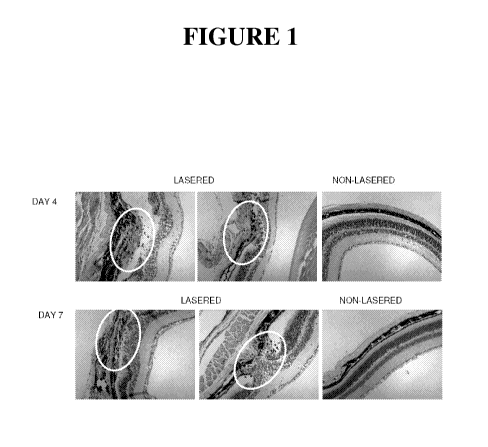
[0034] Figure 1 shows hematoxylin and eosin (H&E)-stained thin sections of
mouse
choroid and retina from laser-treated (left and center panels) and control,
untreated animals (right
panels). The top three photographs show sections of two injured eyes and a
control eye at 4 days
after laser photocoagulation; the bottom photographs show sections of two
injured eyes and one
control eye at 7 days after laser photocoagulation. Lesioned sites are
enclosed within ovals.
[0035] Figure 2 shows red cyanine 3 immunofluorescence, indicative of CD45
immunoreactivity, in thin sections of mouse choroid and retina, from laser-
treated (left and
center panels) and control, untreated animals (right panels). The top three
photographs show
sections of two injured eyes and a control eye at 4 days after laser
photocoagulation; the bottom
photographs show sections of two injured eyes and one control eye at 7 days
after laser
photocoagulation. Lesioned sites are enclosed within ovals.
[0036] Figure 3 shows quantitative analysis of levels of CD45-reactive area in
sections
from control and laser-injured mice at days 14 and 28 after laser
photocoagulation. Degree of
inflammation is expressed as CD45-positive area as a percent of the total
lesion area.
[0037] Figure 4 shows Trichrome-stained thin sections of mouse choroid and
retina from
laser-treated (left and center panels) and control, untreated animals (right
panels). The top three
photographs show sections of two injured eyes and a control eye at 4 days
after laser
photocoagulation; the bottom photographs show sections of two injured eyes and
one control eye
at 7 days after laser photocoagulation. Lesioned sites are enclosed within
ovals.
[0038] Figure 5 shows Sirius Red-stained thin sections of mouse choroid and
retina from
laser-treated (left and center panels) and control, untreated animals (right
panels). The top three
photographs show sections of two injured eyes and a control eye at 4 days
after laser
photocoagulation; the bottom photographs show sections of two injured eyes and
one control eye
at 7 days after laser photocoagulation. Lesioned sites are enclosed within
ovals.
4
CA 02751438 2011-08-03
WO 2010/091279 PCT/US2010/023359
[0039] Figure 6 shows quantitative analysis of collagen deposition in sections
from
control and laser-injured mice at days 4 and 7 after laser photocoagulation.
Collagen deposition
was quantitated by determining the area occupied by collagen fibers (staining
blue with
trichrome and red with Sirius Red) as a percent of the total lesion area.
Sirius Red staining was
analyzed under polarized light. p=0.00003 for trichrome and 0.00005 for Sirius
Red.
[0040] Figure 7 shows quantitative analysis of collagen deposition in sections
from
control and laser-injured mice at days 14 and 28 after laser photocoagulation.
Collagen
deposition was quantitated as described in the legend to Figure 6.
[0041] Figure 8 shows levels of mRNAs encoding lysyl oxidase (LOX) and lysyl
oxidase-like (LOXL) proteins in laser injured eyes at 4, 7, 14 and 28 days
after photocoagulation.
For each of days 4, 7, 14 and 28, each group of five bars represents, from
left to right,
normalized mRNA levels for LOX, LOXL1, LOXL2, LOXL3 and LOXL4. Bars at each
time
point represent data for, from left to right, normalized mRNA levels for mLOX,
mLOXL1,
mLOXL2, mLOXL3 and mLOXL4.
[0042] Figure 9 shows levels of mRNAs encoding lysyl oxidase (LOX) and lysyl
oxidase-like (LOXL) proteins in laser injured eyes at 2, 4, 28 and 35 days
after photocoagulation.
Results were obtained in a separate experiment from the one whose results are
depicted in Figure
8. For each of days 2, 4, 28 and 35, each group of five bars represents, from
left to right,
normalized mRNA levels for LOX, LOXL1, LOXL2, LOXL3 and LOXL4. Bars at each
time
point represent data for, from left to right, mLOX, mLOXL1, mLOXL2, mLOXL3 and
mLOXL4.
[0043] Figure 10 shows quantitative analysis of levels of CD45-reactive area
in sections
from laser-injured mouse eyes at day 35 after laser photocoagulation. Mice had
been treated
with anti-LOXL2 antibody (leftmost bar); anti-LOX antibody (center bar) or
vehicle (rightmost
bar). Degree of inflammation is expressed as CD45-positive area as a percent
of the total lesion
area.
[0044] Figure 11 shows quantitative analysis of levels of CD31-reactive area
in sections
from laser-injured mouse eyes at day 35 after laser photocoagulation. Mice had
been treated
with anti-LOXL2 antibody (leftmost bar); anti-LOX antibody (center bar) or
vehicle (rightmost
bar). Degree of neovascularization is expressed as CD31-positive area as a
percent of the total
lesion area.
CA 02751438 2011-08-03
WO 2010/091279 PCT/US2010/023359
[0045] Figure 12 shows quantitative analysis of collagen deposition, by Sirius
Red
staining, in sections from laser-injured mouse eyes at day 35 after laser
photocoagulation.
Collagen deposition was quantitated by determining the area occupied by
collagen fibers
(staining red) as a percent of the total lesion area. Sirius Red staining was
analyzed under
polarized light.
DETAILED DESCRIPTION
[0046] Practice of the present disclosure employs, unless otherwise indicated,
standard
methods and conventional techniques in the fields of cell biology, toxicology,
molecular biology,
biochemistry, cell culture, immunology, oncology, recombinant DNA and related
fields as are
within the skill of the art. Such techniques are described in the literature
and thereby available to
those of skill in the art. See, for example, Alberts, B. et al., "Molecular
Biology of the Cell," 5d'
edition, Garland Science, New York, NY, 2008; Voet, D. et al. "Fundamentals of
Biochemistry:
Life at the Molecular Level," 3rd edition, John Wiley & Sons, Hoboken, NJ,
2008; Sambrook, J.
et al., "Molecular Cloning: A Laboratory Manual," 3rd edition, Cold Spring
Harbor Laboratory
Press, 2001; Ausubel, F. et al., "Current Protocols in Molecular Biology,"
John Wiley & Sons,
New York, 1987 and periodic updates; Freshney, R.I., "Culture of Animal Cells:
A Manual of
Basic Technique," 4d' edition, John Wiley & Sons, Somerset, NJ, 2000; and the
series "Methods
in Enzymology," Academic Press, San Diego, CA.
Role of lysyl oxidase-type enzymes in choroidal neovascularization
[0047] Lysyl oxidase (LOX) and lysyl oxidase-like (LOXL) proteins are involved
in the
cross-linking of collagen and elastin in the extracellular space. Because of
this activity, these
proteins can play a major role in the process of fibrosis. It is shown herein
that expression of
certain lysyl oxidase-type enzymes increases following laser-induced CNV in a
model system for
age-related macular degeneration (AMD), and that the increases in lysyl
oxidase expression
parallel the observed fibrotic damage (see Examples 4 and 5 below).
Additionally, it shown
herein that treatment of subjects with inhibitors of the activity of lysyl
oxidase (LOX) and lysyl
oxidase-like protein 2 (LOXL2) (e.g., anti-LOX and anti-LOXL2 antibodies)
prevents
neovascularization and fibrosis following laser-induced CNV in the same
system. Accordingly,
inhibition of the activity of lysyl oxidase-type enzymes (e.g., LOX, LOXL2)
can be used to
reverse, mitigate and/or prevent fibrotic damage to the eye resulting from
CNV.
6
CA 02751438 2011-08-03
WO 2010/091279 PCT/US2010/023359
[0048] Thus, in one aspect, compositions that modulate the activity of one or
more lysyl
oxidase-type enzymes as described herein are used in the treatment of
conditions characterized
by neovascularization. A non-limiting example of a condition characterized by
neovascularization is age-related macular degeneration (AMD). Additional
conditions include
diabetic retinopathy and retinopathy of prematurity.
[0049] In certain embodiments, an inhibitor of a lysyl oxidase-type enzyme can
be an
antibody, a small RNA molecule, a ribozyme, a triplex-forming nucleic acid or
a transcription
factor that inhibits expression of a gene encoding a lysyl oxidase-type
protein. See, e.g. US
2006/0127402, US2007/0225242 and co-owned US 2009/0053224; all of which are
incorporated
by reference for disclosure of various types of lysyl oxidase inhibitors. See
also U.S. Patent No.
6,534,261, incorporated by reference, for disclosure of methods for making
transcription factors
that inhibit expression of a gene encoding a lysyl oxidase-type enzyme.
[0050] In certain embodiments, an inhibitor of a lysyl oxidase-type enzyme is
an
antibody that binds to, and inhibits the activity of, a lysyl oxidase-type
enzyme. In additional
embodiments, inhibition is non-competitive. Exemplary antibodies that bind to,
and inhibit the
activity of, one or more lysyl oxidase-type enzymes are disclosed in co-owned
US
2009/0053224; the disclosure of which is incorporated by reference herein for
the purpose of
disclosing the preparation, composition and use of antibodies that bind to
lysyl oxidase-type
enzymes.
[0051] In certain embodiments, a nucleic acid encoding an antibody, or a
functional
antibody fragment, is used as an inhibitor of a lysyl oxidase-type enzyme.
Such nucleic acids
can be administered by any method known in the art. For example, naked nucleic
acid,
optionally in a buffer or pharmaceutical carrier solution, can be injected
into the eye, formulated
as a solution for use as eye drops or administered systemically. Alternatively
a nucleic acid can
be encapsidated in a viral vector (e.g., adenoviral, adeno-associated viral or
lentiviral vectors).
Lysyl -type Enzymes
[0052] As used herein, the term "lysyl oxidase-type enzyme" refers to a member
of a
family of proteins that catalyzes oxidative deamination of c-amino groups of
lysine and
hydroxylysine residues, resulting in conversion of peptidyl lysine to peptidyl-
a-aminoadipic-6-
semialdehyde (allysine) and the release of stoichiometric quantities of
ammonia and hydrogen
peroxide:
7
CA 02751438 2011-08-03
WO 2010/091279 PCT/US2010/023359 C=O C=O CH-CH2-CH2-CH2-CH2-NH2 +H20 - CH-CH2-
CH2-CH2-CH=O +NH3
I +02 I +H202
NH NH peptidyl lysine peptidyl allysine
[0053] This reaction most often occurs extracellularly, on lysine residues in
collagen and
elastin. The aldehyde residues of allysine are reactive and can spontaneously
condense with
other allysine and lysine residues, resulting in crosslinking of collagen
molecules to form
collagen fibrils.
[0054] Lysyl oxidase-type enzymes have been purified from chicken, rat, mouse,
bovines
and humans. All lysyl oxidase-type enzymes contain a common catalytic domain,
approximately
205 amino acids in length, located in the carboxy-terminal portion of the
protein and containing
the active site of the enzyme. The active site contains a copper-binding site
which includes a
conserved amino acid sequence containing four histidine residues which
coordinate a Cu(II)
atom. The active site also contains a lysyltyrosyl quinone (LTQ) cofactor,
formed by
intramolecular covalent linkage between a lysine and a tyrosine residue
(corresponding to 1ys314
and tyr349 in rat lysyl oxidase, and to 1ys320 and tyr355 in human lysyl
oxidase). The sequence
surrounding the tyrosine residue that forms the LTQ cofactor is also conserved
among lysyl
oxidase-type enzymes. The catalytic domain also contains ten conserved
cysteine residues,
which participate in the formation of five disulfide bonds. The catalytic
domain also includes a
fibronectin binding domain. Finally, an amino acid sequence similar to a
growth factor and
cytokine receptor domain, containing four cysteine residues, is present in the
catalytic domain.
[0055] The first member of this family of enzymes to be isolated and
characterized was
lysyl oxidase (EC 1.4.3.13); also known as protein-lysine 6-oxidase, protein-L-
lysine:oxygen 6-
oxidoreductase (deaminating), or LOX. See, e.g., Harris et al., Biochim.
Biophys. Acta 341:332-
344 (1974); Rayton et al., J. Biol. Chem. 254:621-626 (1979); Stassen,
Biophys. Acta 438:49-60
(1976).
8
CA 02751438 2011-08-03
WO 2010/091279 PCT/US2010/023359
[0056] Additional lysyl oxidase-type enzymes were subsequently discovered.
These
proteins have been dubbed "LOX-like," or "LOXL." They all contain the common
catalytic
domain described above and have similar enzymatic activity. Currently, five
different lysyl
oxidase-type enzymes are known to exist in both humans and mice: LOX and the
four LOX
related, or LOX-like proteins LOXL1 (also denoted "lysyl oxidase-like," "LOXL"
or "LOL"),
LOXL2 (also denoted "LOR-1"), LOXL3, and LOXL4. The five genes encoding each
of the
lysyl oxidase-type enzymes each reside on a different chromosome. See, for
example, Molnar et
al., Biochim Biophys Acta. 1647:220-24 (2003); Csiszar, Prog. Nucl. Acid Res.
70:1-32 (2001);
WO 01/83702 published on Nov. 8, 2001, and U.S. Patent No. 6,300,092, all of
which are
incorporated by reference herein. A LOX-like protein termed LOXC, with some
similarity to
LOXL4 but with a different expression pattern, has been isolated from a murine
EC cell line. Ito
et al. (2001) J. Biol. Chem. 276:24023-24029. Two lysyl oxidase-type enzymes,
DmLOXL-1
and DmLOXL-2, have been isolated from Drosophila.
[0057] Although all lysyl oxidase-type enzymes share a common catalytic
domain, they
also differ from one another, particularly within their amino-terminal
regions. The four LOXL
proteins have amino-terminal extensions, compared to LOX. Thus, while human
preproLOX
(i.e., the primary translation product prior to signal sequence cleavage, see
below) contains 417
amino acid residues; LOXL1 contains 574, LOXL2 contains 638, LOXL3 contains
753 and
LOXL4 contains 756.
[0058] Within their amino-terminal regions, LOXL2, LOXL3 and LOXL4 contain
four
repeats of the scavenger receptor cysteine-rich (SRCR) domain. These domains
are not present
in LOX or LOXL1. SRCR domains are found in secreted, transmembrane, or
extracellular
matrix proteins, and are known to mediate ligand binding in a number of
secreted and receptor
proteins. Hoheneste et al. (1999) Nat. Struct. Biol. 6:228-232; Sasaki et al
(1998) EMBO J.
17:1606-1613. In addition to its SRCR domains, LOXL3 contains a nuclear
localization signal
in its amino-terminal region. A proline-rich domain appears to be unique to
LOXL1. Molnar et
al. (2003) Biochim. Biophys. Acta 1647:220-224. The various lysyl oxidase
enzymes also differ
in their glycosylation patterns.
[0059] Tissue distribution also differs among the lysyl oxidase-type enzymes.
Human
LOX mRNA is highly expressed in the heart, placenta, testis, lung, kidney and
uterus, but
marginally in the brain and liver. mRNA for human LOXL1 is expressed in the
placenta,
9
CA 02751438 2011-08-03
WO 2010/091279 PCT/US2010/023359
kidney, muscle, heart, lung, and pancreas and, similar to LOX, is expressed at
much lower levels
in the brain and liver. Kim et al. (1995) J. Biol. Chem. 270:7176-7182. High
levels of LOXL2
mRNA are expressed in the uterus, placenta, and other organs, but as with LOX
and LOXL, low
levels are expressed in the brain and liver. Jourdan Le-Saux et al.(1999) J.
Biol. Chem.
274:12939:12944. LOXL3 mRNA is highly expressed in the testis, spleen, and
prostate,
moderately expressed in placenta, and not expressed in the liver, whereas high
levels of LOXL4
mRNA are observed in the liver. Huang et al. (2001) Matrix Biol. 20:153-157;
Maki and
Kivirikko (2001) Biochem. J. 355:381-387; Jourdan Le-Saux et al. (2001)
Genomics 74:211-
218; Asuncion et al. (2001) Matrix Biol. 20:487-491.
[0060] The expression and/or involvement of the different lysyl oxidase-type
enzymes in
diseases may also vary. See, for example, Kagen (1994) Pathol. Res. Pract.
190:910-919;
Murawaki et al. (1991) Hepatology 14:1167-1173; Siegel et al. (1978) Proc.
Natl. Acad. Sci.
USA 75:2945-2949; Jourdan Le-Saux et al. (1994) Biochem. Biophys. Res. Comm.
199:587-592;
and Kim et al. (1999) J. Cell Biochem. 72:181-188. Lysyl oxidase-type enzymes
have also been
implicated in a number of cancers, including head and neck cancer, bladder
cancer, colon cancer,
esophageal cancer and breast cancer. See, for example, Wu et al. (2007) Cancer
Res. 67:4123-
4129; Gorough et al. (2007) J. Pathol. 212:74-82; Csiszar (2001) Prog. Nucl.
Acid Res. 70:1-32
and Kirschmann et al. (2002) Cancer Res. 62:4478-4483.
[0061] Thus, although the lysyl oxidase-type enzymes exhibit some overlap in
structure
and function, each appears to have distinct structures and functions as well.
For example,
targeted deletion of LOX appears to be lethal at parturition in mice, whereas
LOXL1 deficiency
causes no severe developmental phenotype. Hornstra et al. (2003) J. Biol.
Chem. 278:14387-
14393; Bronson et al. (2005) Neurosci. Lett. 390:118-122.
[0062] Although the most widely documented activity of lysyl oxidase-type
enzymes is
the oxidation of specific lysine residues in collagen and elastin outside of
the cell, there is
evidence that lysyl oxidase-type enzymes also participate in a number of
intracellular processes.
For example, there are reports that some lysyl oxidase-type enzymes regulate
gene expression.
Li et al. (1997) Proc. Natl. Acad. Sci. USA 94:12817-12822; Giampuzzi et al.
(2000) J. Biol.
Chem. 275:36341-36349. In addition, LOX has been reported to oxidize lysine
residues in
histone HE Additional extracellular activities of LOX include the induction of
chemotaxis of
monocytes, fibroblasts and smooth muscle cells. Lazarus et al. (1995) Matrix
Biol. 14:727-731;
CA 02751438 2011-08-03
WO 2010/091279 PCT/US2010/023359
Nelson et al. (1988) Proc. Soc. Exp. Biol. Med. 188:346-352. Expression of LOX
itself is
induced by a number of growth factors and steroids such as TGF-(3, TNF-a and
interferon.
Csiszar (2001) Prog. Nucl. Acid Res. 70:1-32. Recent studies have attributed
other roles to LOX
in diverse biological functions such as developmental regulation, tumor
suppression, cell
motility, and cellular senescence.
[0063] Examples of lysyl oxidase-type proteins from various sources include
enzymes
having an amino acid sequence substantially identical to a polypeptide
expressed or translated
from one of the following sequences: EMBL/GenBank accessions: M94054;
AAA59525.1 --
mRNA; S45875; AAB23549.1-mRNA; S78694; AAB21243.1-mRNA; AF039291;
AAD02130.1-mRNA; BC074820; AAH74820.1-mRNA; BC074872; AAH74872.1 - mRNA;
M84150; AAA59541.1--Genomic DNA. One embodiment of LOX is human lysyl oxidase
(hLOX) preproprotein.
[0064] Exemplary disclosures of sequences encoding lysyl oxidase-like enzymes
are as
follows: LOXL1 is encoded by mRNA deposited at GenBank/EMBL BC015090;
AAH15090.1;
LOXL2 is encoded by mRNA deposited at GenBank/EMBL U89942; LOXL3 is encoded by
mRNA deposited at GenBank/EMBL AF282619; AAK51671.1; and LOXL4 is encoded by
mRNA deposited at GenBank/EMBL AF338441; AAK71934.1.
[0065] The primary translation product of the LOX protein, known as the
prepropeptide,
contains a signal sequence extending from amino acids 1-21. This signal
sequence is released
intracellularly by cleavage between Cys21 and A1a22, in both mouse and human
LOX, to
generate a 46-48 kDa propeptide form of LOX, also referred to herein as the
full-length form.
The propeptide is N-glycosylated during passage through the Golgi apparatus to
yield a 50 kDa
protein, then secreted into the extracellular environment. At this stage, the
protein is
catalytically inactive. A further cleavage, between G1y168 and Asp169 in mouse
LOX, and
between G1y174 and Asp 175 in human LOX, generates the mature, catalytically
active, 30-32
kDA enzyme, releasing a 18 kDa propeptide. This final cleavage event is
catalyzed by the
metalloendoprotease procollagen C-proteinase, also known as bone morphogenetic
protein-1
(BMP-1). Interestingly, this enzyme also functions in the processing of LOX's
substrate,
collagen. The N-glycosyl units are subsequently removed.
[0066] Potential signal peptide cleavage sites have been predicted at the
amino termini of
LOXL1, LOXL2, LOXL3, and LOXL4. The predicted signal cleavage sites are
between G1y25
11
CA 02751438 2011-08-03
WO 2010/091279 PCT/US2010/023359
and G1n26 for LOXL, between A1a25 and G1n26, for LOXL2, between G1y25 and
Ser26 for
LOXL3 and between Arg23 and Pro24 for LOXL4.
[0067] A BMP-1 cleavage site in the LOXL (LOXL1) protein has been identified
between Ser354 and Asp355. Borel et al. (2001) J. Biol. Chem. 276:48944-48949.
Potential
BMP-1 cleavage sites in other lysyl oxidase-type enzymes have been predicted,
based on the
consensus sequence for BMP-1 cleavage in procollagens and pro-LOX being at an
Ala/Gly-Asp
sequence, often followed by an acidic or charged residue. A predicted BMP-1
cleavage site in
LOXL3 is located between G1y447 and Asp448; processing at this site may yield
a mature
peptide of similar size to mature LOX. A potential cleavage site for BMP-1 was
also identified
within LOXL4, between residues A1a569 and Asp570. Kim et al. (2003) J. Biol.
Chem.
278:52071-52074. LOXL2 may also be proteolytically cleaved analogously to the
other
members of the LOXL family and secreted. Akiri et al. (2003) Cancer Res.
63:1657-1666.
[0068] For the purposes of the present disclosure, the term "lysyl oxidase-
type enzyme"
encompasses all five of the lysine oxidizing enzymes discussed above, and also
encompasses
functional fragments and/or derivatives of LOX, LOXL1, LOXL2, LOXL3 and LOXL4
that
substantially retain enzymatic activity; e.g., the ability to catalyze
deamination of lysyl residues.
Typically, a functional fragment or derivative retains at least 50% of its
lysine oxidation activity.
In some embodiments, a functional fragment or derivative retains at least 60%,
at least 70%, at
least 80%, at least 90%, at least 95%, at least 99% or 100% of its lysine
oxidation activity.
[0069] It is also intended that a functional fragment of a lysyl oxidase-type
enzyme can
include conservative amino acid substitutions (with respect to the native
polypeptide sequence)
that do not substantially alter catalytic activity. The term "conservative
amino acid substitution"
refers to grouping of amino acids on the basis of certain common structures
and/or properties.
With respect to common structures, amino acids can be grouped into those with
non-polar side
chains (glycine, alanine, valine, leucine, isoleucine, methionine, proline,
phenylalanine and
tryptophan), those with uncharged polar side chains (serine, threonine,
asparagine, glutamine,
tyrosine and cysteine) and those with charged polar side chains (lysine,
arginine, aspartic acid,
glutamic acid and histidine). A group of amino acids containing aromatic side
chains includes
phenylalanine, tryptophan and tyrosine. Heterocyclic side chains are present
in proline,
tryptophan and histidine. Within the group of amino acids containing non-polar
side chains,
those with short hydrocarbon side chains (glycine, alanine, valine, leucine,
isoleucine) can be
12
CA 02751438 2011-08-03
WO 2010/091279 PCT/US2010/023359
distinguished from those with longer, non-hydrocarbon side chains (methionine,
proline,
phenylalanine, tryptophan). Within the group of amino acids with charged polar
side chains, the
acidic amino acids (aspartic acid, glutamic acid) can be distinguished from
those with basic side
chains (lysine, arginine and histidine).
[0070] A functional method for defining common properties of individual amino
acids is
to analyze the normalized frequencies of amino acid changes between
corresponding proteins of
homologous organisms (Schulz, G. E. and R. H. Schirmer, Principles of Protein
Structure,
Springer-Verlag, 1979). According to such analyses, groups of amino acids can
be defined in
which amino acids within a group are preferentially substituted for one
another in homologous
proteins, and therefore have similar impact on overall protein structure
(Schulz & Schirmer,
supra). According to this type of analysis, the following groups of amino
acids that can be
conservatively substituted for one another can be identified:
(i) amino acids containing a charged group, consisting of Glu, Asp, Lys, Arg
and His,
(ii) amino acids containing a positively-charged group, consisting of Lys, Arg
and His,
(iii) amino acids containing a negatively-charged group, consisting of Glu and
Asp,
(iv) amino acids containing an aromatic group, consisting of Phe, Tyr and Trp,
(v) amino acids containing a nitrogen ring group, consisting of His and Trp,
(vi) amino acids containing a large aliphatic non-polar group, consisting of
Val, Leu and
Ile,
(vii) amino acids containing a slightly-polar group, consisting of Met and
Cys,
(viii) amino acids containing a small-residue group, consisting of Ser, Thr,
Asp, Asn,
Gly, Ala, Glu, Gln and Pro,
(ix) amino acids containing an aliphatic group consisting of Val, Leu, Ile,
Met and Cys,
and
(x) amino acids containing a hydroxyl group consisting of Ser and Thr.
[0071] Thus, as exemplified above, conservative substitutions of amino acids
are known
to those of skill in this art and can be made generally without altering the
biological activity of
the resulting molecule. Those of skill in this art also recognize that, in
general, single amino acid
substitutions in non-essential regions of a polypeptide do not substantially
alter biological
activity. See, e.g., Watson, et al., "Molecular Biology of the Gene," 4th
Edition, 1987, The
Benjamin/Cummings Pub. Co., Menlo Park, CA, p. 224.
13
CA 02751438 2011-08-03
WO 2010/091279 PCT/US2010/023359
[0072] For additional information regarding lysyl oxidase-type enzymes, see,
e.g.,
Rucker et al., Am. J. Clin. Nutr. 67:996S-1002S (1998) and Kagan et al., J.
Cell. Biochem
88:660-672 (2003). See also co-owned US 2009/0053224 (Feb. 26, 2009) and US
2009/0104201 (Apr. 23, 2009); the disclosures of which are incorporated by
reference herein.
Modulators of lysyl oxidase-type enzymes
[0073] Modulators of lysyl oxidase-type enzymes include both activators
(agonists) and
inhibitors (antagonists), and can be selected by using a variety of screening
assays. In one
embodiment, modulators can be identified by determining if a test compound
binds to a lysyl
oxidase-type enzyme; wherein, if binding has occurred, the compound is a
candidate modulator.
Optionally, additional tests can be carried out on such a candidate modulator.
Alternatively, a
candidate compound can be contacted with a lysyl oxidase-type enzyme, and a
biological activity
of the lysyl oxidase-type enzyme assayed; a compound that alters the
biological activity of the
lysyl oxidase-type enzyme is a modulator of a lysyl oxidase-type enzyme.
Generally, a
compound that reduces a biological activity of a lysyl oxidase-type enzyme is
an inhibitor of the
enzyme. In certain embodiments, the biological activity is deamination; in
additional
embodiments, it is peroxide production.
[0074] Other methods for identifying modulators of lysyl oxidase-type enzymes
include
incubating a candidate compound in a cell culture containing one or more lysyl
oxidase-type
enzymes and assaying one or more biological activities or characteristics of
the cells.
Compounds that alter the biological activity or characteristic of the cells in
the culture are
potential modulators of lysyl oxidase-type enzymes. Biological activities that
can be assayed
include, for example, lysyl oxidase enzymatic activity (e.g., deamination,
peroxide production),
levels of lysyl oxidase-type enzyme, levels of mRNA encoding one or more lysyl
oxidase-type
enzymes, and/or one or more functions specific to a lysyl oxidase-type enzyme.
In additional
embodiments of the aforementioned assay, in the absence of contact with the
candidate
compound, the one or more biological activities or cell characteristics are
correlated with levels
or activity of a lysyl oxidase-type enzyme. For example, the biological
activity can be a cellular
function such as migration, chemotaxis, epithelial-to-mesenchymal transition,
or mesenchymal-
to-epithelial transition, and the change is detected by comparison with one or
more control or
reference sample(s). For example, negative control samples can include a
culture with decreased
levels or activity of a lysyl oxidase-type enzyme to which the candidate
compound is added; or a
14
CA 02751438 2011-08-03
WO 2010/091279 PCT/US2010/023359
culture with the same amount of lysyl oxidase-type enzyme activity as the test
culture, but
without addition of candidate compound. In some embodiments, separate cultures
containing
different levels of a lysyl oxidase-type enzyme are contacted with a candidate
compound. If a
change in biological activity is observed, and if the change is greater in the
culture having higher
levels or activity of a lysyl oxidase-type enzyme, the compound is identified
as a modulator of a
lysyl oxidase-type enzyme. Determination of whether the compound is an
activator or an
inhibitor of a lysyl oxidase-type enzyme may be apparent from the phenotype
induced by the
compound, or may require further assay, such as a test of the effect of the
compound on lysyl
oxidase enzymatic activity.
[0075] Methods for obtaining lysysl oxidase-type enzymes, either biochemically
or
recombinantly, as well as methods for cell culture and enzymatic assay to
identify modulators of
lysyl oxidase-type enzymes as described above, are known in the art.
[0076] The enzymatic activity of a lysyl oxidase-type enzyme can be assayed by
a
number of different methods. For example, enzymatic activity can be assessed
by detecting
and/or quantitating production of hydrogen peroxide, ammonium ion, and/or
aldehyde, by
assaying lysine oxidation and/or collagen crosslinking, or by measuring
cellular invasive
capacity, cell adhesion, cell growth or metastatic growth. See, for example,
Trackman et al.
(1981) Anal. Biochem. 113:336-342; Kagan et al. (1982) Meth. Enzymol. 82A:637-
649;
Palamakumbura et al. (2002) Anal. Biochem. 300:245-251; Albini et al. (1987)
Cancer Res.
47:3239-3245; Kamath et al. (2001) Cancer Res. 61:5933-5940; U.S. Patent No.
4,997,854 and
U.S. patent application publication No. 2004/0248871.
[0077] Test compounds include, but are not limited to, small organic compounds
(e.g.,
organic molecules having a molecular weight between about 50 and about 2,500
Da), nucleic
acids and proteins, for example. The compound or plurality of compounds can be
chemically
synthesized or microbiologically produced and/or comprised in, for example,
samples, e.g., cell
extracts from, e.g., plants, animals or microorganisms. Furthermore, the
compound(s) can be
known in the art but hitherto not known to be capable of modulating a lysyl
oxidase-type
enzyme. The reaction mixture for assaying for a modulator of a lysyl oxidase-
type enzyme can
be a cell-free extract or can comprise a cell culture or tissue culture. A
plurality of compounds
can be, e.g., added to a reaction mixture, added to a culture medium, injected
into a cell or
administered to a transgenic animal. The cell or tissue employed in the assay
can be, for
CA 02751438 2011-08-03
WO 2010/091279 PCT/US2010/023359
example, a bacterial cell, a fungal cell, an insect cell, a vertebrate cell, a
mammalian cell, a
primate cell, a human cell or can comprise or be obtained from a non-human
transgenic animal.
[0078] Several methods are known to the person skilled in the art for
producing and
screening large libraries to identify compounds having specific affinity for a
target, such as a
lysyl oxidase-type enzyme. These methods include the phage-display method in
which
randomized peptides are displayed from phage and screened by affinity
chromatography using an
immobilized receptor. See, e.g., WO 91/17271, WO 92/01047, and U.S. Patent No.
5,223,409.
In another approach, combinatorial libraries of polymers immobilized on a
solid support (e.g., a
"chip") are synthesized using photolithography. See, e.g., U.S. Patent No.
5,143,854, WO
90/15070 and WO 92/10092. The immobilized polymers are contacted with a
labeled receptor
(e.g., a lysyl oxidase-type enzyme) and the support is scanned to determine
the location of label,
to thereby identify polymers binding to the receptor.
[0079] The synthesis and screening of peptide libraries on continuous
cellulose
membrane supports that can be used for identifying binding ligands of a
polypeptide of interest
(e.g., a lysyl oxidase-type enzyme) is described, for example, in Kramer
(1998) Methods Mol.
Biol. 87: 25-39. Ligands identified by such an assay are candidate modulators
of the protein of
interest, and can be selected for further testing. This method can also be
used, for example, for
determining the binding sites and the recognition motifs in a protein of
interest. See, for example
Rudiger (1997) EMBO J. 16:1501-1507 and Weiergraber (1996) FEBS Lett. 379:122-
126.
[0080] WO 98/25146 describes additional methods for screening libraries of
complexes
for compounds having a desired property, e.g., the capacity to agonize, bind
to, or antagonize a
polypeptide or its cellular receptor. The complexes in such libraries comprise
a compound under
test, a tag recording at least one step in synthesis of the compound, and a
tether susceptible to
modification by a reporter molecule. Modification of the tether is used to
signify that a complex
contains a compound having a desired property. The tag can be decoded to
reveal at least one
step in the synthesis of such a compound. Other methods for identifying
compounds which
interact with a lysyl oxidase-type enzyme are, for example, in vitro screening
with a phage
display system, filter binding assays, and "real time" measuring of
interaction using, for
example, the BlAcore apparatus (Pharmacia).
16
CA 02751438 2011-08-03
WO 2010/091279 PCT/US2010/023359
[0081] All these methods can be used in accordance with the present disclosure
to
identify activators/agonists and inhibitors/antagonists of lysyl oxidase-type
enzymes or related
polypeptides.
[0082] Another approach to the synthesis of modulators of lysyl oxidase-type
enzymes is
to use mimetic analogs of peptides. Mimetic peptide analogues can be generated
by, for
example, substituting stereoisomers, i.e. D-amino acids, for naturally-
occurring amino acids; see
e.g., Tsukida (1997) J. Med. Chem. 40:3534-3541. Furthermore, pro-mimetic
components can
be incorporated into a peptide to reestablish conformational properties that
may be lost upon
removal of part of the original polypeptide. See, e.g., Nachman (1995) Regul.
Pept. 57:359-370.
[0083] Another method for constructing peptide mimetics is to incorporate
achiral o-
amino acid residues into a peptide, resulting in the substitution of amide
bonds by polymethylene
units of an aliphatic chain. Banerjee (1996) Biopolymers 39:769-777.
Superactive
peptidomimetic analogues of small peptide hormones in other systems have been
described.
Zhang (1996) Biochem. Biophys. Res. Commun. 224:327-331.
[0084] Peptide mimetics of a modulator of a lysyl oxidase-type enzyme can also
be
identified by the synthesis of peptide mimetic combinatorial libraries through
successive amide
alkylation, followed by testing of the resulting compounds, e.g., for their
binding and
immunological properties. Methods for the generation and use of peptidomimetic
combinatorial
libraries have been described. See, for example, Ostresh, (1996) Methods in
Enzymology
267:220-234 and Dorner (1996) Bioorg. Med. Chem. 4:709-715. Furthermore, a
three-
dimensional and/or crystallographic structure of one or more lysyl oxidase
enzymes can be used
for the design of peptide mimetic inhibitors of lysyl oxidase activity. Rose
(1996) Biochemistry
35:12933-12944; Rutenber (1996) Bioorg. Med. Chem. 4:1545-1558.
[0085] The structure-based design and synthesis of low-molecular-weight
synthetic
molecules that mimic the activity of native biological polypeptides is further
described in, e.g.,
Dowd (1998) Nature Biotechnol. 16:190-195; Kieber-Emmons (1997) Current
Opinion
Biotechnol. 8:435-441; Moore (1997) Proc. West Pharmacol. Soc. 40:115-119;
Mathews
(1997) Proc. West Pharmacol. Soc. 40:121-125; and Mukhija (1998) European J.
Biochem.
254:433-438.
[0086] It is also well known to the person skilled in the art that it is
possible to design,
synthesize and evaluate mimetics of small organic compounds that, for example,
can act as a
17
CA 02751438 2011-08-03
WO 2010/091279 PCT/US2010/023359
substrate or ligand of a lysyl oxidase-type enzyme. For example, it has been
described that D-
glucose mimetics of hapalosin exhibited similar efficiency as hapalosin in
antagonizing
multidrug resistance assistance-associated protein in cytotoxicity. Dinh
(1998) J. Med. Chem.
41:981-987.
[0087] The structure of the lysyl oxidase-type enzymes can be investigated to
guide the
selection of modulators such as, for example, small molecules, peptides,
peptide mimetics and
antibodies. Structural properties of the lysyl oxidase-type enzymes can help
to identify natural
or synthetic molecules that bind to, or function as a ligand, substrate,
binding partner or the
receptor of, a lysyl oxidase-type enzyme. See, e.g., Engleman (1997) J. Clin.
Invest. 99:2284-
2292. For example, folding simulations and computer redesign of structural
motifs of lysyl
oxidase-type enzymes can be performed using appropriate computer programs.
Olszewski
(1996) Proteins 25:286-299; Hoffman (1995) Comput. Appl. Biosci. 11:675-679.
Computer
modeling of protein folding can be used for the conformational and energetic
analysis of detailed
peptide and protein structure. Monge (1995) J. Mol. Biol. 247:995-1012; Renouf
(1995) Adv.
Exp. Med. Biol. 376:37-45. Appropriate programs can be used for the
identification of sites, on
lysyl oxidase-type enzymes, that interact with ligands and binding partners,
using computer
assisted searches for complementary peptide sequences. Fassina (1994)
Immunomethods 5:114-
120. Additional systems for the design of protein and peptides are described,
for example in
Berry (1994) Biochem. Soc. Trans. 22:1033-1036; Wodak (1987), Ann. N.Y. Acad.
Sci. 501:1-
13; and Pabo (1986) Biochemistry 25:5987-5991. The results obtained from the
above-described
structural analyses can be used for, e.g., the preparation of organic
molecules, peptides and
peptide mimetics that function as modulators of the activity of a lysyl
oxidase-type enzyme.
[0088] An inhibitor of a lysyl oxidase-type enzyme can be a competitive
inhibitor, an
uncompetitive inhibitor, a mixed inhibitor or a non-competitive inhibitor.
Competitive inhibitors
often bear a structural similarity to substrate, usually bind to the active
site, and are more
effective at lower substrate concentrations. The apparent KM is increased in
the presence of a
competitive inhibitor. Uncompetitive inhibitors generally bind to the enzyme-
substrate complex
or to a site that becomes available after substrate is bound at the active
site and may distort the
active site. Both the apparent KM and the Vmax are decreased in the presence
of an uncompetitive
inhibitor, and substrate concentration has little or no effect on inhibition.
Mixed inhibitors are
capable of binding both to free enzyme and to the enzyme-substrate complex and
thus affect both
18
CA 02751438 2011-08-03
WO 2010/091279 PCT/US2010/023359
substrate binding and catalytic activity. Non-competitive inhibition is a
special case of mixed
inhibition in which the inhibitor binds enzyme and enzyme-substrate complex
with equal avidity,
and inhibition is not affected by substrate concentration. Non-competitive
inhibitors generally
bind to enzyme at a region outside the active site. For additional details on
enzyme inhibition
see, for example, Voet et al. (2008) supra.
Antibodies
[0089] In certain embodiments, a modulator of a lysyl oxidase-type enzyme is
an
antibody. In additional embodiments, an antibody is an inhibitor of the
activity of a lysyl
oxidase-type enzyme.
[0090] As used herein, the term "antibody" means an isolated or recombinant
polypeptide
binding agent that comprises peptide sequences (e.g., variable region
sequences) that specifically
bind an antigenic epitope. The term is used in its broadest sense and
specifically covers
monoclonal antibodies (including full-length monoclonal antibodies),
polyclonal antibodies,
human antibodies, humanized antibodies, chimeric antibodies, nanobodies,
diabodies,
multispecific antibodies (e.g., bispecific antibodies), and antibody fragments
including but not
limited to scFv, Fab, and Fab2, so long as they exhibit the desired biological
activity. The term
"human antibody" refers to antibodies containing sequences of human origin,
except for possible
non-human CDR regions, and does not imply that the full structure of an
immunoglobulin
molecule be present, only that the antibody has minimal immunogenic effect in
a human (i.e.,
does not induce the production of antibodies to itself).
[0091] An "antibody fragment" comprises a portion of a full-length antibody,
for
example, the antigen binding or variable region of a full-length antibody.
Examples of antibody
fragments include Fab, Fab', F(ab')2, and Fv fragments; diabodies; linear
antibodies (Zapata et al.
(1995) Protein Eng. 8(10):1057-1062); single-chain antibody molecules; and
multispecific
antibodies formed from antibody fragments. Papain digestion of antibodies
produces two
identical antigen-binding fragments, called "Fab" fragments, each with a
single antigen-binding
site, and a residual "Fc" fragment, a designation reflecting the ability to
crystallize readily.
Pepsin treatment yields an F(ab')2 fragment that has two antigen combining
sites and is still
capable of cross-linking antigen.
[0092] "Fv" is the minimum antibody fragment which contains a complete antigen-
recognition and -binding site. This region consists of a dimer of one heavy-
and one light-chain
19
CA 02751438 2011-08-03
WO 2010/091279 PCT/US2010/023359
variable domain in tight, non-covalent association. It is in this
configuration that the three CDRS
of each variable domain interact to define an antigen-binding site on the
surface of the VH-VL
dimer. Collectively, the six CDRs confer antigen-binding specificity to the
antibody. However,
even a single variable domain (or an isolated VH or VL region comprising only
three of the six
CDRs specific for an antigen) has the ability to recognize and bind antigen,
although at a lower
affinity than does the entire Fõ fragment.
[0093] The "Fab" fragment also contains, in addition to heavy and light chain
variable
regions, the constant domain of the light chain and the first constant domain
(CHI) of the heavy
chain. Fab fragments were originally observed following papain digestion of an
antibody. Fab'
fragments differ from Fab fragments in that F(ab') fragments contain several
additional residues
at the carboxy terminus of the heavy chain CHI domain, including one or more
cysteines from
the antibody hinge region. F(ab')2 fragments contain two Fab fragments joined,
near the hinge
region, by disulfide bonds, and were originally observed following pepsin
digestion of an
antibody. Fab'-SH is the designation herein for Fab' fragments in which the
cysteine residue(s)
of the constant domains bear a free thiol group. Other chemical couplings of
antibody fragments
are also known.
[0094] The "light chains" of antibodies (immunoglobulins) from any vertebrate
species
can be assigned to one of two clearly distinct types, called kappa and lambda,
based on the
amino acid sequences of their constant domains. Depending on the amino acid
sequence of the
constant domain of their heavy chains, immunoglobulins can be assigned to five
major classes:
IgA, IgD, IgE, IgG, and IgM, and several of these may be further divided into
subclasses
(isotypes), e.g., IgG1, IgG2, IgG3, IgG4, IgAl, and IgA2.
[0095] "Single-chain Fv" or "sFv" antibody fragments comprise the VH and VL
domains
of antibody, wherein these domains are present in a single polypeptide chain.
In some
embodiments, the Fv polypeptide further comprises a polypeptide linker between
the VH and VL
domains, which enables the sFv to form the desired structure for antigen
binding. For a review
of sFv, see Pluckthun, in The Pharmacology of Monoclonal Antibodies, vol. 113
(Rosenburg and
Moore eds.) Springer-Verlag, New York, pp. 269-315 (1994).
[0096] The term "diabodies" refers to small antibody fragments with two
antigen-binding
sites, which fragments comprise a heavy-chain variable domain (VH) connected
to a light-chain
variable domain (VL) in the same polypeptide chain (VH-VL). By using a linker
that is too short
CA 02751438 2011-08-03
WO 2010/091279 PCT/US2010/023359
to allow pairing between the two domains on the same chain, the domains are
forced to pair with
the complementary domains of another chain, thereby creating two antigen-
binding sites.
Diabodies are additionally described, for example, in EP 404,097; WO 93/11161
and Hollinger
et al. (1993) Proc. Natl. Acad. Sci. USA 90:6444-6448.
[0097] An "isolated" antibody is one that has been identified and separated
and/or
recovered from a component of its natural environment. Components of its
natural environment
may include enzymes, hormones, and other proteinaceous or nonproteinaceous
solutes. In some
embodiments, an isolated antibody is purified (1) to greater than 95% by
weight of antibody as
determined by the Lowry method, for example, more than 99% by weight, (2) to a
degree
sufficient to obtain at least 15 residues of N-terminal or internal amino acid
sequence, e.g., by
use of a spinning cup sequenator, or (3) to homogeneity by gel electrophoresis
(e.g., SDS-PAGE)
under reducing or nonreducing conditions, with detection by Coomassie blue or
silver stain. The
term "isolated antibody" includes an antibody in situ within recombinant
cells, since at least one
component of the antibody's natural environment will not be present. In
certain embodiments,
isolated antibody is prepared by at least one purification step.
[0098] In some embodiments, an antibody is a humanized antibody or a human
antibody.
Humanized antibodies include human immununoglobulins (recipient antibody) in
which residues
from a complementary determining region (CDR) of the recipient are replaced by
residues from
a CDR of a non-human species (donor antibody) such as mouse, rat or rabbit
having the desired
specificity, affinity and capacity. Thus, humanized forms of non-human (e.g.,
murine)
antibodies are chimeric immunoglobulins which contain minimal sequence derived
from non-
human immunoglobulin. The non-human sequences are located primarily in the
variable
regions, particularly in the complementarity-determining regions (CDRs). In
some
embodiments, Fv framework residues of the human immunoglobulin are replaced by
corresponding non-human residues. Humanized antibodies can also comprise
residues that are
found neither in the recipient antibody nor in the imported CDR or framework
sequences. In
certain embodiments, a humanized antibody comprises substantially all of at
least one, and
typically two, variable domains, in which all or substantially all of the CDRs
correspond to those
of a non-human immunoglobulin and all or substantially all of the framework
regions are those
of a human immunoglobulin consensus sequence. For the purposes of the present
disclosure,
21
CA 02751438 2011-08-03
WO 2010/091279 PCT/US2010/023359
humanized antibodies can also include immunoglobulin fragments, such as Fv,
Fab, Fab', F(ab')2
or other antigen-binding subsequences of antibodies.
[0099] The humanized antibody can also comprise at least a portion of an
immunoglobulin constant region (Fc), typically that of a human immunoglobulin.
See, for
example, Jones et al. (1986) Nature 321:522-525; Riechmann et al. (1988)
Nature 332:323-329;
and Presta (1992) Curr. Op. Struct. Biol. 2:593-596.
[00100] Methods for humanizing non-human antibodies are known in the art.
Generally, a
humanized antibody has one or more amino acid residues introduced into it from
a source that is
non-human. These non-human amino acid residues are often referred to as
"import" or "donor"
residues, which are typically obtained from an "import" or "donor" variable
domain. For
example, humanization can be performed essentially according to the method of
Winter and co-
workers , by substituting rodent CDRs or CDR sequences for the corresponding
sequences of a
human antibody. See, for example, Jones et al., supra; Riechmann et al., supra
and Verhoeyen
et al. (1988) Science 239:1534-1536. Accordingly, such "humanized" antibodies
include
chimeric antibodies (U.S. Patent No. 4,816,567), wherein substantially less
than an intact human
variable domain has been substituted by the corresponding sequence from a non-
human species.
In certain embodiments, humanized antibodies are human antibodies in which
some CDR
residues and optionally some framework region residues are substituted by
residues from
analogous sites in rodent antibodies (e.g., murine monoclonal antibodies).
[00101] Human antibodies can also be produced, for example, by using phage
display
libraries. Hoogenboom et al. (1991) J. Mol. Biol, 227:381; Marks et al. (1991)
J. Mol. Biol.
222:581. Other methods for preparing human monoclonal antibodies are described
by Cole et al.
(1985) "Monoclonal Antibodies and Cancer Therapy," Alan R. Liss, p. 77 and
Boerner et al.
(1991) J. Immunol. 147:86-95.
[00102] Human antibodies can be made by introducing human immunoglobulin loci
into
transgenic animals (e.g., mice) in which the endogenous immunoglobulin genes
have been
partially or completely inactivated. Upon immunological challenge, human
antibody production
is observed, which closely resembles that seen in humans in all respects,
including gene
rearrangement, assembly, and antibody repertoire. This approach is described,
for example, in
U.S. Patent Nos. 5,545,807; 5,545,806; 5,569,825; 5,625,126; 5,633,425;
5,661,016, and in the
following scientific publications: Marks et al. (1992) BiolTechnology 10:779-
783 (1992);
22
CA 02751438 2011-08-03
WO 2010/091279 PCT/US2010/023359
Lonberg et al. (1994) Nature 368: 856-859; Morrison (1994) Nature 368:812-813;
Fishwald et
al. (1996) Nature Biotechnology 14:845-851; Neuberger (1996) Nature
Biotechnology 14:826;
Lonberg et al. (1995) Intern. Rev. Immunol. 13:65-93.
[00103] Antibodies can be affinity matured using known selection and/or
mutagenesis
methods as described above. In some embodiments, affinity matured antibodies
have an affinity
which is five times or more, ten times or more, twenty times or more, or
thirty times or more
than that of the starting antibody (generally murine, rat, rabbit, chicken,
humanized or human)
from which the matured antibody is prepared.
[00104] An antibody can also be a bispecific antibody. Bispecific antibodies
are
monoclonal, and may be human or humanized antibodies that have binding
specificities for at
least two different antigens. In the present case, the two different binding
specificities can be
directed to two different lysyl oxidase-type enzymes, or to two different
epitopes on a single
lysyl oxidase-type enzyme.
[00105] An antibody as disclosed herein can also be an immunoconjugate. Such
immunoconjugates comprise an antibody (e.g., to a lysyl oxidase-type enzyme)
conjugated to a
second molecule, such as a reporter An immunoconjugate can also comprise an
antibody
conjugated to a cytotoxic agent such as a chemotherapeutic agent, a toxin
(e.g., an enzymatically
active toxin of bacterial, fungal, plant, or animal origin, or fragments
thereof), or a radioactive
isotope (i.e., a radioconjugate).
[00106] An antibody that "specifically binds to" or is "specific for" a
particular
polypeptide or an epitope on a particular polypeptide is one that binds to
that particular
polypeptide or epitope without substantially binding to any other polypeptide
or polypeptide
epitope. In some embodiments, an antibody of the present disclosure
specifically binds to its
target with a dissociation constant (Kd) equal to or lower than 100 nM,
optionally lower than 10
nM, optionally lower than 1 nM, optionally lower than 0.5 nM, optionally lower
than 0.1 nM,
optionally lower than 0.01 nM, or optionally lower than 0.005 nM; in the form
of monoclonal
antibody, scFv, Fab, or other form of antibody measured at a temperature of
about 4 C, 25 C,
37 C or 42 C.
[00107] In certain embodiments, an antibody of the present disclosure binds to
one or
more processing sites (e.g., sites of proteolytic cleavage) in a lysyl oxidase-
type enzyme, thereby
23
CA 02751438 2011-08-03
WO 2010/091279 PCT/US2010/023359
effectively blocking processing of the proenzyme or preproenzyme to the
catalytically active
enzyme, thereby reducing the activity of the lysyl oxidase-type enzyme.
[00108] In certain embodiments, an antibody according to the present
disclosure binds to
human LOX and/or human LOXL2, with a greater binding affinity, for example, 10
times, at
least 100 times, or even at least 1000 times greater, than its binding
affinity to other lysyl
oxidase-type enzymes, e.g., LOXL1, LOXL3, and LOXL4.
[00109] Optionally, an antibody according to the present disclosure not only
binds to a
lysyl oxidase-type enzyme but also reduces or inhibits uptake or
internalization of the lysyl
oxidase-type enzyme, e.g., via integrin beta 1 or other cellular receptors or
proteins. Such an
antibody could, for example, bind to extracellular matrix proteins, cellular
receptors, and/or
integrins.
[00110] Exemplary antibodies that recognize lysyl oxidase-type enzymes, and
additional
disclosure relating to antibodies to lysyl oxidase-type enzymes, is provided
in co-owned U.S.
Patent Application Publication No. 2009/0053224 (February 26, 2009), the
disclosure of which
is incorporated by reference.
Polynucleotides for modulating expression of lysyl oxidase-type enzymes
Antisense
[00111] Modulation (generally inhibition) of a lysyl oxidase-type enzyme can
be effected
by down-regulating expression of the lysyl oxidase-type enzyme at either the
transcriptional or
translational level. One such method of modulation involves the use of
antisense oligo- or
polynucleotides capable of sequence-specific binding with a mRNA transcript
encoding a lysyl
oxidase-type enzyme.
[00112] Binding of an antisense oligonucleotide (or antisense oligonucleotide
analogue) to
a target mRNA molecule can lead to the enzymatic cleavage of the hybrid by
intracellular RNase
H. In certain cases, formation of an antisense RNA-mRNA hybrid can interfere
with correct
splicing. In both cases, the number of intact, functional target mRNAs,
suitable for translation, is
reduced or eliminated. In other cases, binding of an antisense oligonucleotide
or oligonucleotide
analogue to a target mRNA can prevent (e.g., by steric hindrance) ribosome
binding, thereby
preventing translation of the mRNA.
[00113] Antisense oligonucleotides can comprise any type of nucleotide
subunit, e.g., they
can be DNA, RNA, analogues such as peptide nucleic acids (PNA), or mixtures of
the preceding.
24
CA 02751438 2011-08-03
WO 2010/091279 PCT/US2010/023359
RNA oligonucleotides form a more stable duplex with a target mRNA molecule,
but the
unhybridized oligonucleotides are less stable intracellularly than other types
of oligonucleotides
and oligonucleotide analogues. This can be counteracted by expressing RNA
oligonucleotides
inside a cell using vectors designed for this purpose. This approach may be
used, for example,
when attempting to target a mRNA that encodes an abundant and long-lived
protein.
[00114] Additional considerations can be taken into account when designing
antisense
oligonucleotides, including: (i) sufficient specificity in binding to the
target sequence; (ii)
solubility in water; (iii) stability against intra- and extracellular
nucleases; (iv) ability to
penetrate the cell membrane; and (v) when used to treat an organism, low
toxicity.
[00115] Algorithms for identifying oligonucleotide sequences with the highest
predicted
binding affinity for their target mRNA, based on a thermodynamic cycle that
accounts for the
energy of structural alterations in both the target mRNA and the
oligonucleotide, are available.
For example, Walton et al. (1999) Biotechnol. Bioeng. 65:1-9 used such a
method to design
antisense oligonucleotides directed to rabbit (3-globin (RBG) and mouse tumor
necrosis factor-a
(TNF (x) transcripts. The same research group has also reported that the
antisense activity of
rationally selected oligonucleotides against three model target mRNAs (human
lactate
dehydrogenase A and B and rat gp130) in cell culture proved effective in
almost all cases. This
included tests against three different targets in two cell types with
phosphodiester and
phosphorothioate oligonucleotide chemistries.
[00116] In addition, several approaches for designing and predicting
efficiency of specific
oligonucleotides using an in vitro system are available. See, e.g., Matveeva
et al. (1998) Nature
Biotechnology 16:1374-1375.
[00117] An antisense oligonucleotide according to the present disclosure
includes a
polynucleotide or a polynucleotide analogue of at least 10 nucleotides, for
example, between 10
and 15, between 15 and 20, at least 17, at least 18, at least 19, at least 20,
at least 22, at least 25,
at least 30, or even at least 40 nucleotides. Such a polynucleotide or
polynucleotide analogue is
able to anneal or hybridize (i.e., form a double-stranded structure on the
basis of base
complementarity) in vivo, under physiological conditions, with a mRNA encoding
a lysyl
oxidase-type enzyme.
[00118] Antisense oligonucleotides according to the present disclosure can be
expressed
from a nucleic acid construct administered to a cell or tissue. Optionally,
expression of the
CA 02751438 2011-08-03
WO 2010/091279 PCT/US2010/023359
antisense sequences is controlled by an inducible promoter, such that
expression of antisense
sequences can be switched on and off in a cell or tissue. Alternatively
antisense oligonucleotides
can be chemically synthesized and administered directly to a cell or tissue,
as part of, for
example, a pharmaceutical composition.
[00119] Antisense technology has led to the generation of highly accurate
antisense design
algorithms and a wide variety of oligonucleotide delivery systems, thereby
enabling those of
ordinary skill in the art to design and implement antisense approaches
suitable for
downregulating expression of known sequences. For additional information
relating to antisense
technology, see, for example, Lichtenstein et al., "Antisense Technology: A
Practical
Approach," Oxford University Press, 1998.
Small RNA and RNAi
[00120] Another method for inhibition of lysyl oxidase-type enzymes is RNA
interference
(RNAi), an approach which utilizes double-stranded small interfering RNA
(siRNA) molecules
that are homologous to a target mRNA and lead to its degradation. Carthew
(2001) Curr. Opin.
Cell. Biol. 13:244-248.
[00121] RNA interference is typically a two-step process. In the first step,
which is
termed as the initiation step, input double-stranded RNA (dsRNA) is digested
into 21-23
nucleotide (nt) small interfering RNAs (siRNAs), probably by the action of
Dicer, a member of
the RNase III family of double-strand-specific ribonucleases, which cleaves
double-stranded
RNA in an ATP-dependent manner. Input RNA can be delivered, e.g., directly or
via a
transgene or a virus. Successive cleavage events degrade the RNA to 19-21 bp
duplexes
(siRNA), each with 2-nucleotide 3' overhangs. Hutvagner et al. (2002) Curr.
Opin. Genet. Dev.
12:225-232; Bernstein (2001) Nature 409:363-366.
[00122] In the second, effector step, siRNA duplexes bind to a nuclease
complex to form
the RNA-induced silencing complex (RISC). An ATP-dependent unwinding of the
siRNA
duplex is required for activation of the RISC. The active RISC (containing a
single siRNA and
an RNase) then targets the homologous transcript by base pairing interactions
and typically
cleaves the mRNA into fragments of approximately 12 nucleotides, starting from
the 3' terminus
of the siRNA. Hutvagner et al., supra; Hammond et al. (2001) Nat. Rev. Gen.
2:110-119; Sharp
(2001) Genes. Dev. 15:485-490.
26
CA 02751438 2011-08-03
WO 2010/091279 PCT/US2010/023359
[00123] RNAi and associated methods are also described in Tuschl (2001) Chem.
Biochem. 2:239-245; Cullen (2002) Nat. Immunol. 3:597-599; and Brand (2002)
Biochem.
Biophys. Acta. 1575:15-25.
[00124] An exemplary strategy for synthesis of RNAi molecules suitable for use
with the
present disclosure, as inhibitors of a lysyl oxidase-type enzyme, is to scan
the appropriate mRNA
sequence downstream of the start codon for AA dinucleotide sequences. Each AA,
plus the
downstream (i.e., 3' adjacent) 19 nucleotides, is recorded as a potential
siRNA target site. Target
sites in coding regions are preferred, since proteins that bind in
untranslated regions (UTRs) of a
mRNA, and/or translation initiation complexes, may interfere with binding of
the siRNA
endonuclease complex. Tuschl (2001) supra. It will be appreciated though, that
siRNAs
directed at untranslated regions can also be effective, as has been
demonstrated in the case
wherein siRNA directed at the 5' UTR of the GAPDH gene mediated about 90%
decrease in
cellular GAPDH mRNA and completely abolished protein level
(www.ambion.com/techlib/tn/91/912.html). Once a set of potential target sites
is obtained, as
described above, the sequences of the potential target sites are compared to
an appropriate
genomic database (e.g., human, mouse, rat, etc.) using a sequence alignment
software, (such as
the BLAST software available from NCBI at www.ncbi.nlm.nih.gov/BLAST/).
Potential target
sites that exhibit significant homology to other coding sequences are
rejected.
[00125] Qualifying target sequences are selected as templates for siRNA
synthesis.
Selected sequences can include those with low G/C content as these have been
shown to be more
effective in mediating gene silencing, compared to those with G/C content
higher than 55%.
Several target sites can be selected along the length of the target gene for
evaluation. For better
evaluation of the selected siRNAs, a negative control is used in conjunction.
Negative control
siRNA can include a sequence with the same nucleotide composition as a test
siRNA, but
lacking significant homology to the genome. Thus, for example, a scrambled
nucleotide
sequence of the siRNA may be used, provided it does not display any
significant homology to
any other gene.
[00126] The siRNA molecules of the present disclosure can be transcribed from
expression vectors which can facilitate stable expression of the siRNA
transcripts once
introduced into a host cell. These vectors are engineered to express small
hairpin RNAs
(shRNAs), which are processed in vivo into siRNA molecules capable of carrying
out gene-
27
CA 02751438 2011-08-03
WO 2010/091279 PCT/US2010/023359
specific silencing. See, for example, Brummelkamp et al. (2002) Science
296:550-553;
Paddison et al (2002) Genes Dev. 16:948-958; Paul et al. (2002) Nature
Biotech. 20:505-508;
Yu et al. (2002) Proc. Natl. Acad. Sci. USA 99:6047-6052.
[00127] Small hairpin RNAs (shRNAs) are single-stranded polynucleotides that
form a
double-stranded, hairpin loop structure. The double-stranded region is formed
from a first
sequence that is hybridizable to a target sequence, such as a polynucleotide
encoding a lysyl
oxidase-type enzyme (e.g., a LOX or LOXL mRNA) and a second sequence that is
complementary to the first sequence. The first and second sequences form a
double stranded
region; while the non-base-paired linker nucleotides that lie between the
first and second
sequences form a hairpin loop structure. The double-stranded region (stem) of
the shRNA can
comprise a restriction endonuclease recognition site.
[00128] A shRNA molecule can have optional nucleotide overhangs, such as 2-bp
overhangs, for example, 3' UU-overhangs. While there may be variation, stem
length typically
ranges from approximately 15 to 49, approximately 15 to 35, approximately 19
to 35,
approximately 21 to 31 bp, or approximately 21 to 29 bp, and the size of the
loop can range from
approximately 4 to 30 bp, for example, about 4 to 23 bp.
[00129] For expression of shRNAs within cells, plasmid vectors can be employed
that
contain a promoter (e.g., the RNA Polymerase III H1-RNA promoter or the U6 RNA
promoter),
a cloning site for insertion of sequences encoding the shRNA, and a
transcription termination
signal (e.g., a stretch of 4-5 adenine-thymidine base pairs). Polymerase III
promoters generally
have well-defined transcriptional initiation and termination sites, and their
transcripts lack
poly(A) tails. The termination signal for these promoters is defined by the
polythymidine tract,
and the transcript is typically cleaved after the second encoded uridine.
Cleavage at this position
generates a 3' UU overhang in the expressed shRNA, which is similar to the 3'
overhangs of
synthetic siRNAs. Additional methods for expressing shRNA in mammalian cells
are described
in the references cited above.
[00130] An example of a suitable shRNA expression vector is pSUPERTM
(Oligoengine,
Inc., Seattle, WA), which includes the polymerase-Ill H1-RNA gene promoter
with a well
defined transcriptional startsite and a termination signal consisting of five
consecutive adenine-
thymidine pairs. Brummelkamp et al., supra. The transcription product is
cleaved at a site
following the second uridine (of the five encoded by the termination
sequence), yielding a
28
CA 02751438 2011-08-03
WO 2010/091279 PCT/US2010/023359
transcript which resembles the ends of synthetic siRNAs, which also contain
nucleotide
overhangs. Sequences to be transcribed into shRNA are cloned into such a
vector such that they
will generate a transcript comprising a first sequence complementary to a
portion of a mRNA
target (e.g., a mRNA encoding a lysyl oxidase-type enzyme), separated by a
short spacer from a
second sequence comprising the reverse complement of the first sequence. The
resulting
transcript folds back on itself to form a stem-loop structure, which mediates
RNA interference
(RNAi).
[00131] Another suitable siRNA expression vector encodes sense and antisense
siRNA
under the regulation of separate pol III promoters. Miyagishi et al. (2002)
Nature Biotech.
20:497-500. The siRNA generated by this vector also includes a five thymidine
(T5) termination
signal.
[00132] siRNAs, shRNAs and/or vectors encoding them can be introduced into
cells by a
variety of methods, e.g., lipofection. Vector-mediated methods have also been
developed. For
example, siRNA molecules can be delivered into cell using retroviruses.
Delivery of siRNA
using retroviruses can provide advantages in certain situations, since
retroviral delivery can be
efficient, uniform and immediately selects for stable "knock-down" cells.
Devroe et al. (2002)
BMC Biotechnol. 2:15.
[00133] Recent scientific publications have validated the efficacy of such
short double
stranded RNA molecules in inhibiting target mRNA expression and thus have
clearly
demonstrated the therapeutic potential of such molecules. For example, RNAi
has been utilized
for inhibition in cells infected with hepatitis C virus (McCaffrey et al.
(2002) Nature 418:38-39),
HIV-1 infected cells (Jacque et al. (2002) Nature 418:435-438), cervical
cancer cells (Jiang et al.
(2002) Oncogene 21:6041-6048) and leukemic cells (Wilda et al. (2002) Oncogene
21:5716-
5724).
Methods for modulating expression of lysyl oxidase-type enzymes
[00134] Another method of modulating the level or activity of a lysyl oxidase-
type
enzyme is to modulate the expression of its encoding gene, leading to lower
levels of lysyl
oxidase activity if gene expression is repressed, and higher levels if gene
expression is activated.
Modulation of gene expression in a cell can be achieved by a number of
methods.
[00135] For example, oligonucleotides that bind genomic DNA (e.g., regulatory
regions of
a lysyl oxidase-type gene) by strand displacement or by triple helix-formation
can block
29
CA 02751438 2011-08-03
WO 2010/091279 PCT/US2010/023359
transcription, thereby preventing expression of a lysyl oxidase-type enzyme.
In this regard, the
use of so-called "switch back" chemical linking, in which an oligonucleotide
recognizes a
polypurine stretch on one strand on one strand of its target and a homopurine
sequence on the
other strand, has been described. Triple helix formation can also be obtained
using
oligonucleotides containing artificial bases, thereby extending binding
conditions with regard to
ionic strength and pH.
[00136] Modulation of transcription of a lysyl oxidase-type gene can also be
achieved, for
example, by introducing into the cell a fusion protein comprising a functional
domain and a
DNA-binding domain, or a nucleic acid encoding such a fusion protein. A
functional domain
can be, for example, a transcriptional activation domain or a transcriptional
repression domain.
Exemplary transcriptional activation domains include VP16, VP64 and the p65
subunit of NF-
KB; exemplary transcriptional repression domains include KRAB, KOX and v-erbA.
[00137] In certain embodiments, the DNA-binding domain portion of such a
fusion
protein is a sequence-specific DNA-binding domain that binds in or near a gene
encoding a lysyl
oxidase-type enzyme or its regulatory region. The DNA-binding domain can
either naturally
bind to a sequence at or near a gene encoding a lysyl oxidase-type enzyme (or
its regulatory
region), or can be engineered to so bind. For example, the DNA-binding domain
can be obtained
from a naturally-occurring protein that regulates expression of a lysyl
oxidase-type gene.
Alternatively, the DNA-binding domain can be engineered to bind to a sequence
of choice in or
near a lysyl oxidase-type gene or regulatory region.
[00138] In this regard, the zinc finger DNA-binding domain is useful, inasmuch
as it is
possible to engineer zinc finger proteins to bind to any DNA sequence of
choice. A zinc finger
binding domain comprises one or more zinc finger structures. Miller et al.
(1985) EMBO J
4:1609-1614; Rhodes (1993) Scientific American, February: 56-65; U.S. Patent
No. 6,453,242.
Typically, a single zinc finger is about 30 amino acids in length and contains
four zinc-
coordinating amino acid residues. Structural studies have demonstrated that
the canonical
(C2H2) zinc finger motif contains two beta sheets (held in a beta turn which
generally contains
two zinc-coordinating cysteine residues) packed against an alpha helix
(generally containing two
zinc coordinating histidine residues).
[00139] Zinc fingers include both canonical C2H2 zinc fingers (i.e., those in
which the zinc
ion is coordinated by two cysteine and two histidine residues) and non-
canonical zinc fingers
CA 02751438 2011-08-03
WO 2010/091279 PCT/US2010/023359
such as, for example, C3H zinc fingers (those in which the zinc ion is
coordinated by three
cysteine residues and one histidine residue) and C4 zinc fingers (those in
which the zinc ion is
coordinated by four cysteine residues). Non-canonical zinc fingers can also
include those in
which an amino acid other than cysteine or histidine is substituted for one of
these zinc-
coordinating residues. See e.g., WO 02/057293 (July 25, 2002) and US
2003/0108880 (June 12,
2003).
[00140] Zinc finger binding domains can be engineered to have a novel binding
specificity, compared to a naturally-occurring zinc finger protein; thereby
allowing the
construction of zinc finger binding domains engineered to bind to a sequence
of choice. See, for
example, Beerli et al. (2002) Nature Biotechnol. 20:135-141; Pabo et al.
(2001) Ann. Rev.
Biochem. 70:313-340; Isalan et al. (2001) Nature Biotechnol. 19:656-660; Segal
et al. (2001)
Curr. Opin. Biotechnol. 12:632-637; Choo et al. (2000) Curr. Opin. Struct.
Biol. 10:411-416.
Engineering methods include, but are not limited to, rational design and
various types of
empirical selection methods.
[00141] Rational design includes, for example, using databases comprising
triplet (or
quadruplet) nucleotide sequences and individual zinc finger amino acid
sequences, in which each
triplet or quadruplet nucleotide sequence is associated with one or more amino
acid sequences of
zinc fingers which bind the particular triplet or quadruplet sequence. See,
for example, U.S.
Patent Nos. 6, 140,081; 6,453,242; 6,534,261; 6,610,512; 6,746,838; 6,866,997;
7,030,215;
7,067,617; U.S. Patent Application Publication Nos. 2002/0165356;
2004/0197892;
2007/0154989; 2007/0213269; and International Patent Application Publication
Nos. WO
98/53059 and WO 2003/016496.
[00142] Exemplary selection methods, including phage display, interaction
trap, hybrid
selection and two-hybrid systems, are disclosed in U.S. Patent Nos. 5,789,538;
5,925,523;
6,007,988; 6,013,453; 6,140,466; 6,200,759; 6,242,568; 6,410,248; 6,733,970;
6,790,941;
7,029,847 and 7,297,491; as well as U.S. Patent Application Publication Nos.
2007/0009948 and
2007/0009962; WO 98/37186; WO 01/60970 and GB 2,338,237.
[00143] Enhancement of binding specificity for zinc finger binding domains has
been
described, for example, in U.S. Patent No. 6,794,136 (Sept. 21, 2004).
Additional aspects of
zinc finger engineering, with respect to inter-finger linker sequences, are
disclosed in U.S. Patent
No. 6,479,626 and U.S. Patent Application Publication No. 2003/0119023. See
also Moore et al.
31
CA 02751438 2011-08-03
WO 2010/091279 PCT/US2010/023359
(2001a) Proc. Natl. Acad. Sci. USA 98:1432-1436; Moore et al. (2001b) Proc.
Natl. Acad. Sci.
USA 98:1437-1441 and WO 01/53480.
[00144] Further details on the use of fusion proteins comprising engineered
zinc finger
DAN-binding domains are found, for example, in U.S. Patents 6,534,261;
6,607,882;
6,824,978; 6,933,113; 6,979,539; 7,013,219; 7,070,934; 7,163,824 and
7,220,719.
[00145] Additional methods for modulating the expression of a lysyl oxidase-
type enzyme
include targeted mutagenesis, either of the gene or of a regulatory region
that controls expression
of the gene. Exemplary methods for targeted mutagenesis using fusion proteins
comprising a
nuclease domain and an engineered DNA-binding domain are provided, for
example, in U.S.
patent application publications 2005/0064474; 2007/0134796; and 2007/0218528.
Formulations, kits and routes of administration
[00146] Therapeutic compositions comprising compounds identified as modulators
of the
level or activity of a lysyl oxidase-type enzyme (e.g., inhibitors of a lysyl
oxidase-type enzyme)
are also provided. Such compositions typically comprise the modulator and a
pharmaceutically
acceptable carrier. Supplementary active compounds can also be incorporated
into the
compositions. Modulators, particularly inhibitors, of lysyl oxidase-type
enzyme(s) are useful,
for example, in combination with an anti-angiogenic agent, to reduce or
eliminate fibrotic
damage resulting from neovascularization. Accordingly, therapeutic
compositions as disclosed
herein can contain both a modulator of the level and/or activity of a lysyl
oxidase-type enzyme
and an anti-angiogenic agent. In additional embodiments, therapeutic
compositions comprise a
therapeutically effective amount of a modulator of the level and/or activity
of a lysyl oxidase-
type enzyme, but do not contain an anti-angiogenic agent, and the compositions
are administered
separately from the anti-angiogenic agent.
[00147] As used herein, the term "therapeutically effective amount" or
"effective amount"
refers to an amount of a therapeutic agent that when administered alone or in
combination with
another therapeutic agent to a cell, tissue, or subject (e.g., a mammal such
as a human or a non-
human animal such as a primate, rodent, cow, horse, pig, sheep, etc.) is
effective to prevent or
ameliorate the disease condition or the progression of the disease. A
therapeutically effective
dose further refers to that amount of the compound sufficient to result in
full or partial
amelioration of symptoms, e.g., treatment, healing, prevention or amelioration
of the relevant
medical condition, or an increase in rate of treatment, healing, prevention or
amelioration of such
32
CA 02751438 2011-08-03
WO 2010/091279 PCT/US2010/023359
conditions. A therapeutically effective amount of, for example, an inhibitor
of the level and/or
activity of a lysyl oxidase-type enzyme varies with the type of disease or
disorder, extensiveness
of the disease or disorder, and size of the mammal suffering from the disease
or disorder.
[00148] The therapeutic compositions disclosed herein are useful for, inter
alia, reducing
fibrotic damage resulting from neovascularization. Accordingly, a
"therapeutically effective
amount" of a modulator (e.g., inhibitor) of the level and/or activity of a
lysyl oxidase-type
enzyme is an amount that results in reduction of fibrotic damage resulting
from
neovascularization, such as occurs during macular degeneration. For example,
when the
inhibitor of a lysyl oxidase-type enzyme is an antibody and the antibody is
administered in vivo,
normal dosage amounts may vary from about 10 ng/kg to up to 100 mg/kg of
mammal body
weight or more per day, for example, about 1 g/kg/day to 50 mg/kg/day,
optionally about 100
g/kg/day to 20 mg/kg/day, 500 g/kg/day to 10 mg/kg/day, or 1 mg/kg/day to 10
mg/kg/day,
depending upon, e.g., body weight, route of administration, severity of
disease, etc.
[00149] When a modulator of the level and/or activity of a lysyl oxidase-type
enzyme is
used in combination with an anti-angiogenic agent, one can also refer to the
therapeutically
effective dose of the combination, which is the combined amounts of the
modulator and the anti-
angiogenic agent that result in reduction of fibrotic damage resulting from
neovascularization,
whether administered in combination, serially or simultaneously. More than one
combination of
concentrations can be therapeutically effective.
[00150] Various pharmaceutical compositions and techniques for their
preparation and use
are known to those of skill in the art in light of the present disclosure. For
a detailed listing of
suitable pharmacological compositions and techniques for their administration
one may refer to
the detailed teachings herein, which may be further supplemented by texts such
as Remington's
Pharmaceutical Sciences, 17th ed. 1985; Brunton et al., "Goodman and Gilman's
The
Pharmacological Basis of Therapeutics," McGraw-Hill, 2005; University of the
Sciences in
Philadelphia (eds.), "Remington: The Science and Practice of Pharmacy,"
Lippincott Williams &
Wilkins, 2005; and University of the Sciences in Philadelphia (eds.),
"Remington: The Principles
of Pharmacy Practice," Lippincott Williams & Wilkins, 2008.
[00151] The disclosed therapeutic compositions further include
pharmaceutically
acceptable materials, compositions or vehicles, such as a liquid or solid
filler, diluent, excipient,
solvent or encapsulating material, i.e., carriers. These carriers are involved
in transporting the
33
CA 02751438 2011-08-03
WO 2010/091279 PCT/US2010/023359
subject chemical from one organ, or region of the body, to another organ, or
region of the body.
Each carrier should be "acceptable" in the sense of being compatible with the
other ingredients of
the formulation and not injurious to the patient. Some examples of materials
which can serve as
pharmaceutically-acceptable carriers include: sugars, such as lactose, glucose
and sucrose;
starches, such as corn starch and potato starch; cellulose and its
derivatives, such as sodium
carboxymethyl cellulose, ethyl cellulose and cellulose acetate; powdered
tragacanth; malt;
gelatin; talc; excipients, such as cocoa butter and suppository waxes; oils,
such as peanut oil,
cottonseed oil, safflower oil, sesame oil, olive oil, corn oil and soybean
oil; glycols, such as
propylene glycol; polyols, such as glycerin, sorbitol, mannitol and
polyethylene glycol; esters,
such as ethyl oleate and ethyl laurate; agar; buffering agents, such as
magnesium hydroxide and
aluminum hydroxide; alginic acid; pyrogen-free water; isotonic saline;
Ringer's solution; ethyl
alcohol; phosphate buffer solutions; and other non-toxic compatible substances
employed in
pharmaceutical formulations. Wetting agents, emulsifiers and lubricants, such
as sodium lauryl
sulfate and magnesium stearate, as well as coloring agents, release agents,
coating agents,
sweetening, flavoring and perfuming agents, preservatives and antioxidants can
also be present
in the compositions.
[00152] Another aspect of the present disclosure relates to kits for carrying
out the
administration of a modulator of the level and/or activity of a lysyl oxidase-
type enzyme.
Another aspect of the present disclosure relates to kits for carrying out the
combined
administration of a modulator of the level and/or activity of a lysyl oxidase-
type enzyme and an
anti-angiogenic agent. In one embodiment, a kit comprises an inhibitor of the
activity of a lysyl
oxidase-type enzyme formulated in a pharmaceutical carrier, optionally
containing at least one
anti-angiogenic agent that is not an inhibitor of the activity of a lysyl
oxidase-type enzyme,
formulated as appropriate, in one or more separate pharmaceutical
preparations.
[00153] The formulation and delivery methods can be adapted according to the
site(s) and
degree of fibrotic damage. Exemplary formulations include, but are not limited
to, those suitable
for parenteral administration, e.g., intravenous, intra-arterial, intra-
ocular, or subcutaneous
administration, including formulations encapsulated in micelles, liposomes or
drug-release
capsules (active agents incorporated within a biocompatible coating designed
for slow-release);
ingestible formulations; formulations for topical use, such as eye drops,
creams, ointments and
gels; and other formulations such as inhalants, aerosols and sprays. The
dosage of the
34
CA 02751438 2011-08-03
WO 2010/091279 PCT/US2010/023359
compounds of the disclosure will vary according to the extent and severity of
the need for
treatment, the activity of the administered composition, the general health of
the subject, and
other considerations well known to the skilled artisan.
[00154] In additional embodiments, the compositions described herein are
delivered
locally. Such local delivery can be achieved, for example, by intra-ocular
injection or by
application of eye drops.
Administration
[00155] For treatment of choroidal neovascularization (e.g., AMD) with
inhibitors of the
activity of a lysyl oxidase-type enzyme (e.g., inhibitors of LOX and/or LOXL2
activity), any
method known in the art for delivery of substances to the eye can be utilized.
For example,
direct injection into the eye can be used for delivery of an inhibitor of the
activity of a lysyl
oxidase-type enzyme; e.g., an anti-LOX antibody and/or an anti-LOXL2 antibody.
In certain
embodiments, an inhibitor of the level and/or activity of a lysyl oxidase-type
enzyme (optionally
in combination with an angiogenesis inhibitor, see below) is injected into the
vitreous humor. In
additional embodiments, topical administration of an inhibitor of the level
and/or activity of a
lysyl oxidase-type enzyme is used. For example, the eye can be bathed in a
solution containing
an inhibitor of the level and/or activity of a lysyl oxidase-type enzyme, or
an inhibitor of the
level and/or activity of a lysyl oxidase-type enzyme can be formulated in a
solution to be used as
eye drops. An inhibitor of the level and/or activity of a lysyl oxidase-type
enzyme can also be
administered systemically, provided an effective concentration reaches the eye
and there are no
(or acceptable) extra-ocular side effects.
[00156] Nucleic acids encoding anti-lysyl oxidase antibodies (or any other
type of
inhibitor of a lysyl oxidase-type enzyme, e.g., a ribozyme, siRNA, shRNA or
microRNA) can
optionally be encapsidated in a viral vector. A number of viral vectors are
known in the art,
including parvoviruses, papovaviruses, adenoviruses, herpesviruses,
poxviruses, retroviruses and
lentiviruses.
[00157] One class of recombinant viral vectors is based on the defective and
nonpathogenic parvovirus adeno-associated virus serotype 2 (AAV-2). Vectors
are derived from
a plasmid containing the AAV 145 bp inverted terminal repeat sequence flanking
a transgene
expression cassette. Efficient gene transfer and stable transgene delivery due
to integration into
CA 02751438 2011-08-03
WO 2010/091279 PCT/US2010/023359
the genomes of the infected cell are obtained using this vector system. Wagner
et al. (1998)
Lancet 351:1702-1703; Kearns et al. (1996) Gene Ther. 9:748-755
[00158] Additional adeno-associated virus vehicles include AAV serotypes 1, 5,
6, 7, 8
and 9; as well as chimeric AAV serotypes, e.g., AAV 2/1 and AAV 2/5. Both
single-stranded
and double-stranded (e.g., self-complementary) AAV vectors can be used.
Combination Therapies
[00159] In certain embodiments, treatment of conditions characterized by
neovascularization involves administration of a composition as described
herein that inhibits the
level and/or activity of a lysyl oxidase-type enzyme, together with
administration of a second
composition that inhibits angiogenesis. The compositions can be administered
sequentially in
any order or concurrently. In certain embodiments, both compositions comprise
antibodies. In
other embodiments, both compositions comprise polynucleotides encoding
antibodies. In still
further embodiments, one composition comprises a polynucleotide encoding an
antibody and the
other comprises an antibody polypeptide. In embodiments in which the
compositions are
administered as polynucleotides, a single polynucleotide (e.g., expression
vector) can be used
that encodes both inhibitors.
[00160] In certain embodiments, an inhibitor of angiogenesis is an anti-VEGF
antibody.
Inhibitors of this type are available commercially, for example, under the
trade names Avastin
and Lucentis . However, any anti-VEGF antibody can be used.
[00161] In additional embodiments, an inhibitor of angiogenesis can be a small
RNA
molecule, a ribozyme, a triplex-forming nucleic acid or a transcription factor
that inhibits
expression of a VEGF gene. See, e.g., U.S. Patent No. 7,067,317.
EXAMPLES
Example 1: Mouse model of AMD
[00162] Laser-induced photocoagulation of the retina in mice, leading to CNV,
was used
as a model system for AMD. This treatment induces ruptures in the Bruch's
membrane, induces
neovascularization, and forms scar tissue that seals leaky blood vessels. In
this model,
inflammation was previously observed for up to five days after coagulation,
and was followed by
angiogenesis with a peak at day 14. Rakic et al. (2003) Invest. Ophthalmol.
Vis. Sci. 44:3186-
3193. At even later stages after photocoagulation (3-4 weeks), fibrosis was
observed. In the
36
CA 02751438 2011-08-03
WO 2010/091279 PCT/US2010/023359
present example, the degree and extent of inflammation and fibrosis, along
with the expression
of lysyl oxidase (LOX) and lysyl oxidase-like proteins (LOXLs), in the eye
(choroid and retina)
after laser photocoagulation, was assessed.
[00163] For these experiments, male C57BL/6 mice, at 8-10 weeks of age, were
acclimated at 20 2 C, relative humidity of 55 5% and a 14 hour light/10 hour
dark cycle, for at
least five days, then certain of the animals were subjected to laser
photocoagulation, while other
animals that did not receive the photocoagulation treatment served as
controls.
[00164] For photocoagulation, mice were anesthetized with an intrapeitoneal
injection of
Nembutal (60mg/ml) and the pupils were dilated with topical administration of
Tropicol
(5mg/ml). Three burns were placed with an Argon laser (532nm) at 9, 12, and 3
o'clock
positions in each retina using a slit lamp delivery system. The laser was set
for a 0.05 second
duration at an energy of 400mW, and a 50 m spot size. Rupture of the Bruch's
membrane was
confirmed by production of an air bubble at the site at which the laser had
been aimed, and only
sites at which a bubble was observed were included in the analysis.
[00165] Five animals that had undergone laser injury were sacrificed on days 4
and 7,
along with three uninjured control animals. For analyses conducted on days 14
and 28 after
photocoagulation, three injured animals and three control animals were
sacrificed at each time
point.
[00166] All animals were sacrificed by cervical dislocation. Immediately after
sacrifice,
both eyes were enucleated and choroidea and retinas were dissected. One animal
from the
control group and one from the photocoagulation group, at each of the four
time points, was used
for analysis of the extent of laser-induced injury, inflammation and fibrosis.
Tissue for these
analyses was frozen in 4% paraformaldehyde and embedded in paraffin. Seven m
sections
were cut. Sections were stained with hematoxylin and eosin to detect the
lesions; other sections
were tested for CD45 levels (by immunohistochemistry) to evaluate the degree
of inflammation,
and separate sections were stained with Sirius Red and Trichrome stain to
evaluate the degree
and extent of fibrosis. Images were obtained with a Zeiss Imager Z1 at a
magnification of 10x
and a resolution of 1292 x 968 pixels, and photographs were taken with a Zeiss
Axiocam MrC5.
Images were morphologically analyzed with Zeiss KS300 software. This software
was used to
determine the total area of the lesion, to measure areas within the lesion
that stained positively
for different markers (see below), and to calculate the fraction of the total
lesion area positive for
37
CA 02751438 2011-08-03
WO 2010/091279 PCT/US2010/023359
the particular marker under study. Data were analyzed with Statistica 6.1
statistical software,
using a student T-test for independent samples. P-values smaller than 0.05
were considered
statistically significant.
[00167] The remaining animals (four from the photocoagulation group on days 4
and 7,
two from the photocoagulation group on days 14 and 28, and two controls at
each of the four
time points) were used for analysis of lysyl oxidase mRNA. For mRNA analysis,
fresh tissue
(choroid and retina) was frozen in liquid nitrogen and stored at -80 C until
used for RNA
extraction (below).
Example 2: Lesion detection
[00168] All eyes were examined histologically, using hematoxylin and eosin
(H&E)-
stained sections. Three lesions were detected on each eye that had been
subjected to laser
treatment. An example is shown in Figure 1.
Example 3: Inflammation
[00169] Thin sections were also subjected to immunohistochemistry for CD45, a
leukocyte marker, whose presence is indicative of inflammation. For this
analysis, antigen
retrieval was conducted for 20 minutes at 95 C, and rabbit serum was used as a
blocking agent.
Sections were incubated overnight at room temperature with rat anti-mouse CD45
antibody
(1/100; Beckton Dickinson). The following day the slides were incubated with a
biotinylated
rabbit anti-rat antibody (Dakocytomation) at a 1/300 dilution for 45 minutes
at room
temperature. Sections were then developed using a TSA Cyan 3 System (Perkin
Elmer TSATM;
NEL704A) at room temperature, and washed with TNT washing buffer. Streptavidin
peroxidase
was used at a 1/100 dilution and cyan 3 was diluted 1/50 in working buffer.
[00170] The degree of inflammation was quantitated by determining the area of
the
section exhibiting CD45 immunoreactivity and expressing this as a percentage
of the total area of
the lesion. Areas were determined using the Zeiss KS300 software.
[00171] The results of this analysis indicated that no leukocytes were present
(i.e., no
CD45 immunoreactivity was observed) in untreated eyes. However, inflammation,
as evidenced
by CD45 immunoreactivity, was apparent in laser-treated eyes as early as day 4
after laser
treatment. In laser-injured eyes, CD45 levels remained roughly constant on
days 4, 7 and 14 but,
38
CA 02751438 2011-08-03
WO 2010/091279 PCT/US2010/023359
by day 28, they had approximately doubled. See Figure 2 for examples of
stained samples.
Figure 3 shows quantitation of CD45 levels on days 14 and 28 after laser
injury.
Example 4: Fibrosis
[00172] Thin sections, obtained as described above, were stained with
Trichrome and
Sirius Red, then analyzed by microscopy. Examples of Trichrome-stained and
Sirius Red-
stained sections, at days 4 and 7 after laser injury, are shown in Figures 4
and 5.
[00173] The extent of fibrosis was scored quantitatively by determining the
area of the
section exhibiting collagen staining and expressing this as a percentage of
the total area of the
lesion. Areas were determined using the Zeiss KS300 software.
[00174] Results of the quantitation did not reveal collagen deposition
(indicative of
fibrosis) in non-laser-treated eyes. In laser-injured eyes, collagen
deposition was observed (by
both Trichrome and Sirius Red staining) as early as day 4 after injury, and
collagen levels
continued to increase through days 7 and 14. Collagen levels at day 28 were
approximately
equivalent to those observed at day 14. Figure 6 shows collagen deposition at
days 4 and 7, and
Figure 7 shows collagen deposition at days 14 and 28. (Data obtained from two
separate
experiments.) These results indicate that fibrosis occurs rapidly after laser
injury, increases to an
apparent plateau at day 14, and lasts at least an additional two weeks
thereafter.
Example 5: Lysyl oxidase expression
[00175] RNA was purified from frozen tissue (see Example 1) for analysis of
lysyl
oxidase (LOX) and lysyl oxidase-related protein (LOXL) transcript levels.
[00176] Retina and choroid from each mouse eye were pooled, suspended in 700
l
Qiagen RLT buffer containing (3-mercaptoethanol, and homogenized with a
Polytron hand-held
electric homogenizer. RNA isolation was performed using a RNeasy Mini kit,
according to the
manufacturer's instructions (Qiagen, Valencia, CA). Eluted RNA was DNase-
treated with
Ambion rDNAse I according to reagent specifications.
[00177] Levels of mRNA for lysyl oxidase and lysyl oxidase-like proteins were
determined by quantitative reverse transcription/polymerase chain reaction
(qRT-PCR). Reverse
transcription and amplification reactions were performed using a Stratagene
Brilliant II One-Step
Core Reagents kit according to the manufacturer's instructions, using 100ng
RNA template in
39
CA 02751438 2011-08-03
WO 2010/091279 PCT/US2010/023359
each reaction. Primers and FAM/BHQ-1 probes for target mRNA were designed
using Beacon
Designer software (Premier Biosoft, Palo Alto, CA) and were used at final
concentrations of
400 nM for primers and 250 nM for probes. Nucleotide sequences of the probes
and primers are
presented in Table 1.
CA 02751438 2011-08-03
WO 2010/091279 PCT/US2010/023359
in O
M o~ N z
z z z 0 z cf
- - N
cn w N w a
a
a v v (D U U
U
U U U U v ~
U 0 " a
U a
a U u a a (~
~C V ~ a U U
a H- U <
O Q U Q U
u H-
a (7 H- U 0 U
< U F- F- < U 00
yC
O
a o o
c N C o c u
U cn d N v
a F- F- < 0 a
a U U < < H
00 < 0 u 0 U U 0
y ~. (D < U < U
U U H- <
a
a= "_U U U H a
N
tw U O (~ ~n 00 U ID <
U Z H O< O ( U .7 0 0 0 0 0
=~ 0 L z u z a z a z Z
UI - LU
UI (D < a
a
Q H- <
a a
u
=~. < U a H
a~ fs+ 0 U
a a
- 'a a
U
a U a a
a o a O
0 z U o U z
a PI00 ao
0 O U
azU Hcuzz<C<zz
U 0 z (U7 N Q U N Q
~--I N M
O O O O a
41
CA 02751438 2011-08-03
WO 2010/091279 PCT/US2010/023359
[00178] Primer/probe sets were validated for specificity for their target
mRNAs by in vitro
siRNA knock-down experiments and were tested for their amplification
efficiencies using
dilutions of cell line RNA expressing moderate to high levels of target mRNAs.
An efficiency of
100% corresponds to a doubling in the amount of amplicon during each cycle
that occurs during
the exponential phase of the amplification reaction, and results in a 10-fold
increase in the
amount of amplicon every 3.32 cycles. Efficiency was determined by plotting C,
vs. input RNA
concentration on a semi-logarithmic scale and determining the slope of the
curve so generated.
Percent efficiency (E) was then calculated as follows:
E = (10-1/slope-1) x 100
[00179] The amplification efficiencies of all primer/probe sets were
determined to be
>90%.
[00180] Test mRNA levels were normalized to mRNA levels of ribosomal protein
L19
(RPL19), and results were expressed as fold regulation of relative expression
in laser-coagulated
retina compared to relative expression in control, nontreated retina. Results
were based on
averages of two experimental (4 retina + 4 choroid) and one control (2 retina
+ 2 choroid) animal
for each time point.
[00181] The results of the mRNA analysis (Figure 8) showed that expression of
the lysyl
oxidase gene (LOX) is increased over three-fold at day 4 after laser injury.
Increases in the
levels of mRNAs encoding LOXL1 and LOXL2 were also observed at day 4. In a
separate
experiment in which RNA from four experimental animals and two control animals
was
analyzed at each of days 2, 4, 28 and 35 after laser photocoagulation,
increases in levels of
mRNA for LOX, LOXL1 and LOXL2, at day 4 after injury, were also observed
(Figure 9).
[00182] Additional experiments have shown that levels of LOX and LOXL2 remain
elevated in photocoagulated eyes for at least 35 days after photocoagulation.
Example 6: Inhibition of LOX and LOXL2 activities reduces fiborsis,
inflammation and
neovascularization associated with macular degeneration
[00183] In this example, the effect of treatment with antibodies to LOX and
LOXL2, in a
murine model of age-related macular degeneration, was assessed. In particular,
effects on
fibrosis, inflammation and angiogenesis were investigated. Anti-LOX antibody
M64 and anti-
LOXL2 antibody AB0023 are both described in co-owned US Patent Application
Publication
42
CA 02751438 2011-08-03
WO 2010/091279 PCT/US2010/023359
No. US 2009/0053224 (Feb. 26, 2009) and in co-owned PCT WO 2009/035791 (March
19,
2009), the disclosures of which are incorporated by reference herein for the
purposes of
describing these antibodies, their methods of preparation and their methods of
use. Antibodies
were diluted to a 3.75 mg/ml working solution in sterile PBS pH 7.4, 0.01%
Tween 20 (PBST)
and stored at 4 C.
[00184] Thirty-six male C57B1/6 mice, at 8-10 weeks of age, were used in this
experiment. They were maintained at 20 2 C, at a relative humidity of 55 5%,
with a 14 hour
light/10 hour dark cycle. On day 0, thirty mice were anaesthetized with an
intraperitoneal
injection of Nembutal. A 6 mg/ml solution was used and the injection volume
(in microliters)
corresponded to ten time the body weight of the animal in grams. Under
anaesthesia, the pupils
were dilated by topical administration of one drop of Tropicol (from a 5 mg/ml
stock
solution). Photocoagulation was accomplished using an argon laser (532 nm) to
place three
burns (at 9-, 12- and 3-o'clock) on the retina using a slit lamp delivery
system. The laser was set
for a duration of 0.01 sec at an energy of 400 mW, to generate a 50 um burn
spot. Production of
the spot was confirmed by the observation of a bubble, signifying rupture of
the Bruch's
membrane.
[00185] Animals that had undergone photocoagulation were divided into three
groups of
ten. One group received 0.75 mg of anti-LOX antibody immediately after
photocoagulation and
every two days thereafter. A second group received 0.75 mg of anti-LOXL2
antibody
immediately after photocoagulation and every two days thereafter. The third
group received 200
ul of PBST (vehicle) immediately after photocoagulation and every two days
thereafter.
Antibody solutions or vehicle were administered intraperitoneally in a volume
of 0.2 ml.
[00186] Six naive animals (i.e., animals that did not undergo
photocoagulation) were also
included in the study.
[00187] At day 35 after photocoagulation, all animals were sacrificed by
cervical
dislocation and both eyes were removed and enucleated. (One animal in the anti-
LOX treatment
group died on day 16 of the study.) One eye from each of the 35 animals was
fixed in 4%
paraformaldehyde and embedded in paraffin. Seven micrometer sections were cut
and were
analyzed to determine degree of inflammation, extent of neovascularization and
degree of
fibrosis, as follows.
43
CA 02751438 2011-08-03
WO 2010/091279 PCT/US2010/023359
[00188] The degree of inflammation was quantitated by determining the area of
the
section exhibiting CD45 immunoreactivity and expressing this as a percentage
of the total area of
the lesion, as described in Example 3.
[00189] Thin sections were subjected to immunohistochemistry for CD31, a blood
vessel
marker, whose presence is indicative of neovascularisation. For this analysis,
trypsin digestion
was conducted for 7 minutes at 37 C, and rabbit serum was used as a blocking
agent. Sections
were incubated overnight at room temperature with rat anti-mouse CD31 antibody
(1/500;
Pharmingen). The following day the slides were incubated with a biotinylated
rabbit anti-rat
antibody (Dakocytomation) at a 1/300 dilution for 45 minutes at room
temperature. Sections
were then developed using a TSA Cyan 3 System (Perkin Elmer TSATM; NEL704A) at
room
temperature, and washed with TNT washing buffer. Streptavidin peroxidase was
used at a 1/100
dilution and cyan 3 was diluted 1/50 in working buffer. The extent of
neovascularizaton was
quantitated by determining the area of the section exhibiting CD31
immunoreactivity and
expressing this as a percentage of the total area of the lesion.
[00190] The extent of fibrosis was scored quantitatively by determining the
area of the
section occupied by collagen fibers (determined by Sirius Red staining) and
expressing this area
as a percentage of the total area of the lesion.
[00191] Methods for measurement and quantitation are described infra in
Example 1.
[00192] The results of these analyses are shown in Figures 10-12. Figure 10
shows that
inflammation, as measured by the CD45-positive area of the lesions, was
reduced in subjects
treated with an anti-LOXL2 antibody. Similarly, the degree of
neovascularization, measured by
the CD31-positive area or the lesions, was reduced in subjects that had been
treated with an anti-
LOXL2 antibody (Figure 11). Fibrosis, as measured by collagen density, was
reduced by both
anti-LOX and anti-LOXL2 antibodies (Figure 12).
44