Note: Descriptions are shown in the official language in which they were submitted.
CA 02751649 2011-08-05
WO 2010/091168 PCT/US2010/023177
APPARATUS, SYSTEM AND METHOD FOR CHRONIC DISEASE MONITORING
BACKGROUND
[0001] This disclosure relates to a system for monitoring a person suffering
from a
chronic medical condition in order to predict and assess physiological changes
which could
affect the care of that subject. Examples of such chronic diseases include
(but are not limited
to) heart failure, chronic obstructive pulmonary disease (COPD), asthma, and
diabetes.
[0002] To provide a context for the limitations of conventional approaches, it
is
instructive to briefly review current approaches to chronic disease monitoring
for three major
diseases: heart failure, COPD and asthma.
[0003] Heart failure (HF) is a relatively common and severe clinical
condition,
characterized by the inability of the heart to keep up with the oxygen demands
of the body.
Management of heart failure is a significant challenge to modem healthcare
systems due to its
high prevalence and severity. It is estimated that heart failure accounts for
approximately 2-
3% of the entire healthcare budget of developed nations, and is the number one
cause of
hospitalization of the over-65s in the USA.
[0004] Heart failure is a chronic condition, which is progressive in nature.
Physicians
typically class the severity of the disease according to a New York Heart
Association
(NYHA) subjective grading system from 1 to 4, where 4 is the most severe case.
Heart failure
can also be further broken into classes such as systolic and diastolic heart
failure. The
progression of heart failure is often characterized as relatively stable over
long periods of time
(albeit with reduced cardiovascular function) punctuated by episodes of an
acute nature. In
this acute phases, the patient experiences worsening of symptoms such as
dyspnea (difficulty
breathing), gallop rhythms, increased jugular venous pressure, and orthopnea.
This is
typically accompanied by overt congestion (which is the build up of fluid in
the pulmonary
cavity). This excess fluid often leads to measurable weight gain of several
kilograms. In
many cases, however, by the time overt congestion has occurred, there are
limited options for
the doctor to help restabilize the patients, and in many cases the patient
requires
hospitalization.
1
CA 02751649 2011-08-05
WO 2010/091168 PCT/US2010/023177
[0005] There already exist some approaches to the detection of clinical
deterioration, but
with limitations. For example, a range of chronic disease management programs
have been
developed to improve the healthcare response to HF, with an emphasis on both
increased
patient care and reduced cost. Critical components of successful programs
include a) patient
education, b) telemonitoring of physiological measurements and symptoms, c)
sophisticated
decision support systems to use the reported symptoms and measurements to
predict
clinically significant events, and d) a focus on individualized care and
communication (e.g.,
"teaching in the moment" in response to events affecting a patient's health).
[0006] However, accurate diagnosis of clinical deterioration in heart failure
can be quite
difficult. In particular, prevention of overt congestion which often requires
hospitalization, is
of particular importance. Weight measurement has been shown to be a reasonably
reliable
physiological guide to heart failure deterioration. This can lead to reduced
mortality, when
combined with other accepted strategies for heart failure management.
Moreover, weight
management has the additional psychological benefit of involving the patient
directly in their
own care, as well as being simple and low-cost.
[0007] However, despite the widespread use of recommendations on weight gain
as a
marker of deterioration (e.g., a patient is told that a gain of 2 kg over a 2
to 3 day period
should generate a call to their clinic), there is relatively little published
data on the sensitivity
and specificity of ambulatory monitoring of weight gain in a clinical setting.
Groups who
have investigated the sensitivity of weight gain in distinguishing clinically
stable (CS) Class
IV patients from those with clinical deterioration (CD), have found that the
performance is
quite limited. These researchers found quite modest predictive values for
weight gain in
isolation. For example, the clinical guideline of 2 kg weight gain over 48-72h
has a
specificity of 97% but a sensitivity of only 9%. Reducing the threshold to 2%
of body weight,
improves the sensitivity to 17% (with specificity only dropping marginally).In
general they
conclude that weight gain in isolation has relatively poor sensitivity in
detecting clinical
deterioration (though its specificity is good).
[0008] Thus, what is needed is a system and method to overcome the current
limitation on
the sensitivity of weight gain to predict clinical deterioration.
[0009] Measurement of B natriuretic peptides (BNP) has also been suggested as
a viable
tool for assessment of heart failure status; this could be implemented at a
primary care or
2
CA 02751649 2011-08-05
WO 2010/091168 PCT/US2010/023177
outpatient clinic setting using point-of-care devices, though at present it
can not be clinically
deployed on a daily monitoring basis. In a report on BNP monitoring,
researchers reported a
sensitivity of 92% on a population of 305 subjects, but with a specificity of
only 38%. While
this is a promising approach, there are significant practical issues around
providing point-of-
care assays for BNP in community care due to cost, training and patient
convenience.
Accordingly, there remains a need for development of improved low-cost
convenient
diagnostic markers of clinical deterioration of heart failure which can be
deployed in the
patient's day-to-day environment.
[0010] Thus, what is needed is a system and method to improve the specificity
of
detecting clinical deterioration as compared to approaches such as BNP
monitoring, and for
such systems to be convenient for patient use in their home environment.
[0011] Some potential markers of clinical deterioration in heart failure are
changes in
nocturnal heart rate, changes in sleeping posture, and changes in respiration.
In particular,
heart failure is highly correlated with sleep disordered breathing (SDB),
though the causality
mechanisms are not well understood. For example, in a recent study in Germany,
71 % of
heart failure patients have an Apnea-Hypopnea index greater than 10 per hour
(with 43%
having obstructive sleep apnea and 28% having primarily Cheyne-Stokes
respiration (periodic
breathing). Other researchers reported a prevalence of 68% in their HF
population in a New
Zealand study. Significant sleep disordered breathing has been reported to
correlate with poor
outcomes in heart failure; however, no study has yet been able to track
changes in respiratory
patterns over time to see how it varies with clinical stability. For example,
in the Home or
Hospital in Heart Failure (HHH) European-wide study, overnight respiratory
recording (using
respiratory inductance plethysmography) was carried out for a single night at
baseline in 443
clinically stable HF patients. Apnea Hypopnea Index and Duration of Periodic
Breathing
were shown to be independent predictors of cardiac death and hospitalization
for clinical
deterioration. However no practical system for assessing these respiratory
parameters on a
nightly basis was available for these researchers.
[0012] Measurement of nocturnal heart rate and heart rate variability can also
aid in the
detection of clinical deterioration in heart failure.
[0013] A second chronic medical condition for which the current system can be
used is
Chronic Obstructive Pulmonary Disease (COPD). COPD is a disease of the lungs
in which
3
CA 02751649 2011-08-05
WO 2010/091168 PCT/US2010/023177
the airways are narrowed, which leads to a restricted flow of air to the
lungs. COPD is
currently the fourth leading cause of death in the USA, and its estimated cost
to the healthcare
system is $42.6 billion in 2007. It is associated with dyspnea (shortness of
breath) and
elevated breathing rates (tachypnea). As for heart failure, there can be acute
exacerbations of
COPD, often due to bacterial or viral infections. However, definitions of what
exactly
constitutes an exacerbation, and means to accurately predict it are a subject
of active research
in the medical community. For example, tracking of C-reactive protein or
measurements of
inspiratory capacity have been proposed as means to predict exacerbations.
Changes in peak
expiratory flow have been considered for prediction of clinical deterioration,
but are
considered insufficiently sensitive.
Thus what is needed is a reliable method for accurately recognizing
exacerbations in COPD
patients. Further, what is needed is a system and method for recognizing
clinical deterioration
in COPD patients through tracking of respiratory patterns.
[0014] Respiratory rate is a key indicator of the severity of COPD. For
example, normal
healthy adults may have respiratory rates which are about 14-16 breaths/minute
while asleep;
the resting respiratory rate of a person with severe COPD (but not in acute
respiratory failure)
may be in the range 20-25 breaths/minute, while in an acute respiratory
failure, this rate may
increase to more than 30 breaths/minute. Accordingly a system for simple
monitoring of
respiratory rate has utility in assessing the status of subjects with COPD.
However, current
systems for monitoring respiratory rate are typically based on measurement of
airflow using
nasal cannulae or respiratory effort belts and are not used for continuous
monitoring of
respiratory patterns in the person's own environment due to comfort and
convenience issues.
Thus what is needed is a system for tracking exacerbations in COPD patients
which does not
require the subject to wear an oro-nasal cannula or chest belt.
[0015] An additional chronic medical condition is asthma. This is a common
chronic
condition in which the airways occasionally constrict, become inflamed, and
are lined with
excessive amounts of mucus, often in response to one or more triggers, such as
smoke,
perfume, and other allergens. Viral illnesses are also a possible trigger,
particularly in
children. The narrowing of the airway causes symptoms such as wheezing,
shortness of
breath, chest tightness, and coughing. The airway constriction responds to
bronchodilators.
Between episodes, most patients feel well but can have mild symptoms and they
may remain
4
CA 02751649 2011-08-05
WO 2010/091168 PCT/US2010/023177
short of breath after exercise for longer periods of time than an unaffected
individual. The
symptoms of asthma, which can range from mild to life threatening, can usually
be controlled
with a combination of drugs and environmental changes. The estimated
prevalence of asthma
in the US adult population is 10%, so it represents a significant public
health issue. As for HF
and COPD, the disease is marked by sudden exacerbations.
[0016] A key marker of asthma is peak expiratory flow (PEF) - this can be
obtained from
the patient by asking them to blow into a spirometer. However spirometry only
gives point
measurements of function, and also requires the active involvement of the
subject, and so is
not suited for young children. Researchers have previously noted a link
between PEF and
respiratory rate. Accordingly what is needed is a system and method for
monitoring
respiratory rate in subjects with asthma.
[0017] Furthermore other disease conditions such as cystic fibrosis,
pneumonia,
corpulmonale and infection caused by the respiratory syncytial virus (RSV) may
all be better
monitored by a system capable of monitoring respiratory rate and/or nocturnal
heart rate.
SUMMARY
[0018] This disclosure provides various embodiments of an apparatus, system,
and
method for monitoring subjects with chronic disease, using measurements of
physiological
function such as respiration, heart rate and other clinical measurements. The
typical users of
the system are (a) a person with a chronic disease, and (b) a caregiver with
clinical expertise
responsible for co-ordination of care for the person being monitored.
[0019] In one embodiment, a system for monitoring a subject is described, in
which the
system comprises a sensor configured to output a signal comprising a measured
respiratory
parameter of the subject; an analyzer configured to receive the signal and to
at least store, in a
memory, a plurality of respiratory features derived from the respiratory
parameter, and an
analyzer which is configured to selectively combine the plurality of
respiratory features to
determine an output that provides a health assessment of the subject.
[0020] In another embodiment, a method for monitoring a subject is described,
in which
the method comprises measuring a respiratory parameter of the subject;
generating a plurality
CA 02751649 2011-08-05
WO 2010/091168 PCT/US2010/023177
of respiratory features derived from the respiratory parameter, and combining
the plurality of
respiratory features to calculate an output that provides a health assessment
of the subject.
[0021] The system described herein provides earlier detection of changes to
allow clinical
intervention, and improves the detection of clinical deterioration in heart
failure. Further, the
system described herein works through accurate, cost-effective and convenient
measurement
and analysis of physiological parameters. The physiological basis of the
utility of this system
in heart failure management is based on the observations provided above with
respect to the
markers of clinical deterioration in heart failure.
[0022] Given the significance of night-time respiration in assessment of heart
failure, the
present disclosure overcomes limitations of conventional techniques for
measuring respiratory
patterns, and provides for the measurement of overnight respiratory patterns
over prolonged
periods of time in a manner which is convenient for the patient. Further, an
improved system
and method is provided for analysis of respiratory patterns in heart failure
relevant for the
prediction of clinical deterioration.
[0023] In addition to providing long-term monitoring of subjects with known
chronic
diseases as discussed above, the system and method described herein also
suitable to provide
diagnosis of whether a person has one of the chronic diseases described above
(or other
chronic diseases). In such cases, measurements and analysis are carried out as
in the case of
chronic disease monitoring, but diagnostic decisions are made on the basis of
a limited
number of night (or recording period) measurements.
BRIEF DESCRIPTION OF THE DRAWINGS
[0024] Embodiments of the disclosure will now be described with reference to
the
accompanying drawings in which the acronym "a.u." is placed on the graphs to
represent
"arbitrary units". The units for the signals described below for respiratory
effort and heart rate
can be calibrated to more meaningful units such as liters/minute (for
respiratory tidal volume)
or mm (for ballistocardiogram displacements on the skin).
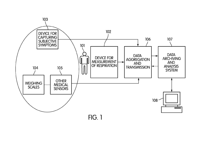
[0025] FIG. 1 is a diagram illustrating the overall schematic of an embodiment
in which
the subject is being monitored, together with devices for measuring
respiratory patterns, heart
rate and other physiological measurements. A device for capturing subjective
symptom data
6
CA 02751649 2011-08-05
WO 2010/091168 PCT/US2010/023177
is also depicted. It denotes the possibility of both local and remote
archiving and analysis of
the measurements.
[0026] FIG. 2 illustrates a representative embodiment in which non-contact
biomotion
sensor is placed near the person being monitored. The motion signal can be
used to derive
respiration and heart rate patterns which are used as part of the chronic
disease monitoring
system.
[0027] FIG. 3 shows how the raw motion signal derived from a bio-motion sensor
can be
decomposed into respiratory movements, cardiac movements, and movements
associated with
large bodily motion such as turning over or arm-movements.
[0028] FIG. 4 illustrates how the process of phase demodulation applied to the
I and Q
signals obtained from a motion sensor can be used to derive a more accurate
combined
movement signal.
[0029] FIGS. 5A and 5B show examples of respiratory patterns which are used in
the
chronic disease monitoring system. FIG. 5A illustrates an episode of periodic
breathing
(Cheyne-Stokes respiration) in which respiratory effort amplitude oscillates
over several
minutes. FIG. 5B illustrates two apneas detected by the biomotion sensor shown
in FIG. 2.
[0030] FIG. 6 shows two estimates of respiratory effort derived from biomotion
sensor
shown in FIG. 2 using quadrature (I and Q) signals and an envelope of the
signal derived from
the respiratory effort signals.
[0031] FIG. 7A shows how the respiratory envelope changes when a hypopnea
occurs,
and FIG. 7B shows how the respiratory envelope changes when an apnea occurs.
[0032] FIGS. 8A-8D show how the respiratory envelope changes over longer
periods of
time in the presence and absence of periodic breathing, including illustrating
the power
spectral densities of the respiratory envelopes in the presence of periodic
breathing, and its
absence. FIG 8A is the respiratory envelope of a person with heart failure
measured over a
five minute period. FIG 8B is the power spectral density of the respiratory
envelope shown in
FIG 8A. FIG 8C is the respiratory envelope of a person without heart failure
measured over a
five minute period. FIG 8D is the power spectral density of the respiratory
envelope shown in
FIG 8C.
7
CA 02751649 2011-08-05
WO 2010/091168 PCT/US2010/023177
[0033] FIG. 9A shows an example of a respiratory effort signal recorded using
the device
shown in FIG. 2, and how the spectrum (FIG. 9B) of a 30-second epoch can be
used to define
respiratory rate.
[0034] FIG. 10 shows an example of the agreement level between the
characteristic
modulation period of subjects with periodic breathing estimated using an
algorithm based on
the signals obtained from the sensor in FIG. 2, versus gold standard
respiratory measurements
using clinical polysomnogram measurements.
[0035] FIG. 11 shows an example of the agreement level between the estimated
Apnea
Hypopnea Index of subjects using an algorithm based on the signals obtained
from the sensor
in FIG. 2, versus gold standard respiratory measurements using clinical
polysomnogram
measurements.
[0036] FIG. 12 shows how the non-contact biomotion sensor of FIG. 2 can also
indicate
heart rate as well as respiration rate.
[0037] FIG. 13 illustrates an embodiment of a method for determining clinical
exacerbations in chronic disease. FIG. 13A illustrates a rule-based method for
determining if
a person with heart failure requires an intervention such as a nurse-call, and
FIG. 13B
illustrates a statistically based classifier approach to making a decision as
to whether a person
with a chronic medical condition has experienced a deterioration.
[0038] FIG. 14 shows an illustration of how the measurements from a person
with chronic
disease could be visualized over prolonger time periods (several weeks or
months).
[0039] FIG. 15 shows an example of Apnea Hypopnea Indices measured in two
subjects
over approximate three week periods, using the system described in this
specification.
DETAILED DESCRIPTION
[0040] FIG. 1 is a diagram illustrating the overall schematic of an embodiment
of this
disclosure. Subject 101 is monitored using respiratory sensor 102. Examples of
respiratory
sensors include abdominal inductance bands, thoracic inductance bands, a non-
contact
biomotion sensor, or an airflow sensor. The monitored respiration parameters
can include
respiratory effort, respiratory movement, tidal volume, or respiratory rate.
Optionally device
8
CA 02751649 2011-08-05
WO 2010/091168 PCT/US2010/023177
for capturing symptoms 103 can also be included. This could be as simple as a
written diary,
or could be an electronic data capture device which asks questions such as "do
you feel
breathless", "did you have discomfort breathing during sleep", "do you feel
better or worse
than yesterday", "do you feel your heart is racing", etc. One embodiment of
such an
electronic device could be a customized tablet PC, or alternatively a cell
phone could be used,
with voice-capture of subjective responses. The person's sleeping position
could be obtained
by asking a simple question such as "how many pillows did you use for
sleeping", or through
use of a position (tilt) sensor.
[0041] Orthopnea is a common symptom in heart failure. For simplicity, symptom
questions could be restricted to requiring only simple yes/no responses.
Optionally, further
devices could be used to assess clinical status. Weight scale 104 has proven
utility in
monitoring heart failure through objective assessment of weight gain due to
fluid retention.
Other medical sensors 105 can be integrated such as ECG monitors, blood
pressure monitors,
point-of-care blood assays of BNP, spirometers (which can measure forced
expiratory
volume, and peak expiratory flow), oximeters (which can measure blood oxygen
levels),
blood glucose monitors, and point-of-care blood assays of C-reactive protein.
[0042] Measurements made from all the sensors mentioned above (respiration,
weighing
scales and other sensors) may be aggregated together in data aggregation
device 106.
Aggregation device 106 could be a cell-phone, a personal computer, a tablet
computer, or a
customized computing device. This aggregation device can also be referred to
as a data hub
and, at a minimum, it may transfer data from the respiratory sensor 102 to the
aggregation
device itself. In one aspect of this embodiment, data aggregation device 106
may also have
the capability of transmitting the collected data to remote data analyzer 107.
Remote data
analyzer 107 may itself be a server computer, personal computer, mobile
computing device or
another customized computing device. Remote data analyzer 107 will typically
have storage,
processing, memory and computational elements. Remote data analyzer 107 will
typically be
configured to provide a database capability, and may include further data
archiving,
processing and analysis means, and would typically have a display capability
via display 108
so that a remote user (e.g., a cardiac nurse) can review data.
[0043] FIG. 2 shows an embodiment of the respiration sensor, in which non-
contact
biomotion sensor 201 is used to monitor the respiratory effort and heart rate
of a subject 202.
9
CA 02751649 2011-08-05
WO 2010/091168 PCT/US2010/023177
This non-contact sensor is described in PCT Publication Number WO 2007/143535
A2 and
US Patent 6,426,716, the entire contents of which are incorporated herein by
reference. Non-
contact sensor 201 is placed near the bed of the person 202 during sleep, and
monitors
movement. It operates by sending out a short impulse of radio-waves (in field-
testing of this
system, a frequency of 5.8 GHz is used with a pulse length of 5 ns). The
reflection of this
impulse is then mixed with a local delayed copy of the transmitted impulse.
The mixer circuit
outputs a signal which is related to the phase difference between the
transmitted and received
pulses - if the target is moving, this movement is modulated onto the phase
signal. This
phase signal is referred to as a raw movement signal. There are other non-
contact motion
sensor technologies which can be used analogously. Infra-red detection systems
can be used
to detect movement, as can ultrasonic transducers. To improve the sensitivity
and robustness
of a non-contact biomotion sensor, it is useful to have a quadrature detection
system in which
there are effectively two sensors with the base phase of their oscillations
offset by it/4 radians.
These two effective sensors can be implemented by using a single source
oscillator, but whose
base phase is modulated periodically by it/4 radians.
[0044] FIG. 3 shows how the raw movement signal from biomotion sensor 301 can
be
decomposed into three components corresponding to significant bodily movement,
respiratory
effort and heart rate. Significant bodily movement would correspond to an
action such as
turning over, moving a leg, or twisting the head. Heart rate signal can be
obtained using a
cardiac activity detector 302 which in one embodiment is a bandpass filter
applied to the raw
movement signal. This bandpass filter preferentially passes signals in the
region 0.5 to 10 Hz,
which reflect heart rate signals. More elaborate processing such as
preprocessing to remove
movement and respiratory artifacts may be necessary. An alternative approach
is to take an
epoch of the raw signal and generate its power spectral density. Peaks in this
spectral density
(e.g., at 1 Hz) can be used to identify the average heart rate over that epoch
(e.g., 1 Hz
corresponds to 60 beats/minute). In this manner, a heart rate signal can be
generated.
[0045] Similarly respiratory effort signal can be generated by a respiratory
detector 303,
which in one embodiment is a bandpass filter applied to the raw movement
signal. This
bandpass filter preferentially passes signals in the region 0.05 to 1 Hz which
reflect
respiratory signals. An alternative approach is to take an epoch of the raw
signal and generate
its power spectral density. Peaks in this spectral density (e.g., at 0.2 Hz)
can be used to
identify the average breathing rate over that epoch (e.g., 0.2 Hz corresponds
to 12
CA 02751649 2011-08-05
WO 2010/091168 PCT/US2010/023177
breaths/minute). Finally, large bodily movements not related to respiration or
cardiac activity
can be identified using the motion detector 304 which implements techniques
for motion
detection 304. One method for detecting motion is to high-pass filter the raw
movement
signal, and then threshold the absolute value of the filtered signal. A second
method is to
calculate the energy of the raw movement signal over short epochs (e.g., 2
seconds). If the
amplitude of the energy exceeds a threshold, a movement is detected. The
amplitude of the
movement can be assessed by calculating the energy value in that epoch. In
that way, an
activity count can be assigned to short epochs. The movement signal is
processed to
determine when the subject is asleep.
[0046] FIG. 4 gives an example of how to combine the I and Q signals obtained
from the
biomotion sensor. In this example, a technique called phase demodulation is
employed. This
is due to the fact that I signal 401 and Q signal 402 are not linearly
correlated with the
position of the moving subject, but rather represent the phase of the
reflected signal. To
compensate for this effect, the arcsine of the I channel, the arccosine of the
Q channel and the
arctangent of the I/Q ratio are calculated. This results in three potential
output signals - one of
these is chosen by calculating the overall amplitude of the signal, its signal-
to-noise ratio, and
its shape. The demodulated signal may then be low pass filtered to give the
final respiratory
movement signal 403. This process is only applied when the I and Q signals are
believed to
represent primarily respiratory movement.
[0047] FIGS. 5A and 5B gives examples of breathing patterns measured in people
suffering from chronic disease. FIG 5A gives an illustration of what is known
as Cheyne-
Stokes respiration or periodic breathing. In this type of breathing the
person's respiratory
effort increases and decreases periodically, with a time scale of 30-90
seconds, typically. It is
caused by an instability in the control of the relative amounts of oxygen and
carbon dioxide in
the blood, and is commonly seen in patients with heart failure. FIG. 5B shows
an example of
another respiratory event seen in chronic disease - an obstructive apnea. In
an obstructive
apnea, the person's respiratory effort is diminished for 10-20 seconds, before
breathing
recommences.
[0048] FIG. 6 is an illustration of a method for recognizing an apnea or
hypopnea event
from a respiratory signal, or set of signals. FIG. 6 shows that the non-
contact biomotion
sensor returns two signals associated with respiratory movement. These are the
so-called I
11
CA 02751649 2011-08-05
WO 2010/091168 PCT/US2010/023177
and Q quadrature signals. They may be generated by using radio-frequency
pulses whose
carrier waves are 90 degrees out of phase. The purpose of this is to smooth
out the sensitivity
response of the system. The I and Q channels both capture the respiratory
movement, but
with different amplitudes and phases. In order to obtain an "average"
breathing signal, we
combine the signals to form a single respiratory effort signal, R(t). One
means to do this is to
calculate
R(t) = I2 (t) + Q2 (t)
where I(t) and Q(t) represent the sampled values of the I and Q signals
respectively. The
envelope of this combined signal can then be obtained using a number of
methods, for
example, a "peak detect and hold" method, or a method using a Hilbert
transform.
[0049] This respiratory envelope signal can then be processed to recognize
apnea and
hypoponeas. As a specific embodiment, consider the results shown in FIG. 7A
and 7B. The
respiratory envelope signal has been normalized over a period of multiple
minutes, and its
value is then shown over time. Using pre-established (or adaptive) rules, the
amplitude of the
respiratory envelope signal is compared to a number of thresholds. For
example, in this case,
if the amplitude stays above 0.7, breathing is considered normal. If the
envelope stays
between 0.2 and 0.7 for more than 10 seconds, then a hypopnea event is
calculated. If the
envelope dips below 0.2 for 10 seconds, then the event is considered an apnea.
The person
skilled in the art will realize that the exact rules will depend upon clinical
definitions of apnea
and hypopnea (which may vary from region to region), and the processing
methods used for
normalization and envelope extraction. In this way, specific events and their
start and end
times can be established. For example, FIG. 7A shows a hypopnea event which
started at
time t = 18s, and finished at t = 31 Is. FIG. 7B shows an apnea event which
started at time
t = 32s and ended at t = 49s.
[0050] An apnea-hypopnea index (AHI) is then calculated by counting the number
of
average number of apneas and hypoponeas per hour of sleep (for example, if a
person has 64
apneas, 102 hypoponeas, and sleeps for 6.3 hrs, then their AHI is
166/6.3=26.3). This is an
important parameter in assessing the overall status of the subject with
chronic disease.
[0051] It is also important in many chronic diseases to monitor episodes of
periodic
breathing (an example of which is shown in FIG. 5A). One embodiment of a
method for
detecting periodic breathing episodes may be implemented is as follows. The
envelope of the
12
CA 02751649 2011-08-05
WO 2010/091168 PCT/US2010/023177
respiratory signal is calculated as discussed in the previous paragraphs. FIG.
8A shows the
respiratory envelope as a function of time over a period of approximately 5
minutes during
which periodic breathing is present. The periodic breathing appears as a
increase and
decrease of the respiratory envelope over a time scale of about 80 seconds in
this example.
[0052] FIG. 8C shows a similar time period for the respiratory envelope during
which no
periodic breathing occurs. In order to recognize the periodic breathing
episode, the power
spectral density of the envelope signal for the 5-minute period is calculated.
This is shown in
FIG. 8B for the periodic breathing signal, and in FIG. 8D for the normal
breathing segment.
The periodic breathing will cause a significant modulation of the envelope at
frequencies
between 0.01 and 0.03 Hz approximately (i.e., characteristic time scales of 33
to 100 s). A
threshold algorithm can then be used to determine whether the modulation is
sufficient to be
considered a periodic breathing episode. The 5 minute period can then be
marked as a
periodic breathing segment. In this way episodes of periodic breathing are
determined. The
total number of 5-minute segments so identified can be used to estimate the
duration of
periodic breathing. The person skilled in the art will realize that the exact
rules for
determining periodic breathing (Cheyne-Stokes respiration) will depend upon
clinical
definitions of periodic breathing (which vary from region to region), and the
processing
methods used for normalization, spectral density estimation and envelope
extraction.
[0053] In this way, the total duration of periodic breathing per night can be
determined,
e.g., a person might have 22 minutes of periodic breathing in total on a
particular night.
[0054] Monitoring the respiration rate itself is also an important parameter
in chronic
disease monitoring. For example, in acute respiratory failure the respiration
rate can rise over
30 breaths/minute in adults, from a more typical baseline of 15 or 16
breaths/minute. One
technique for tracking the respiratory rate during the night is as follows, as
illustrated in FIG
9A. For the case of a respiratory effort signal obtained from the non-contact
sensor discussed
earlier, a sliding window is applied to the data (e.g., 30 seconds in length).
The power
spectral density is then calculated for that epoch (FIG 9B), using techniques
such as the
averaged periodogram. The power spectral density will typically contain a peak
corresponding to the breathing frequency somewhere between 0.1 and 0.5 Hz This
peak can
be identified by using a peak-finding algorithm. In some cases, there may be
excessive
motion artifact on the data - in such a case a technique such as Lomb's
periodogram can be
13
CA 02751649 2011-08-05
WO 2010/091168 PCT/US2010/023177
used to estimate the power spectral density (this interpolates through missing
data).
Alternatively, the respiratory effort signal can be fit with a model using
Auto Regressive or
Auto Regressive Moving Average techniques. The model parameters can then be
used to
estimate the respiration frequency. Kalman filtering techniques can also be
employed. In this
way, an average respiration frequency for the time window can be obtained. The
sliding
window can then advance by 1 or more seconds. In this way, a time series of
the respiration
frequency can be build up over the night. A simple average respiration for the
night can be
obtained by averaging over this time series for the night. Alternatively, more
complex
measurements of respiratory frequency can be calculated such as median
frequency, variance
of the respiratory frequency, percentile distributions of the respiratory
frequency, and auto-
correlation of the respiratory frequency.
[0055] FIG. 10 shows an example of the calculated characteristic modulation
periods in
subjects with sleep apnea, using the signals obtained from a biomotion sensor,
as compared to
the periods calculated using the full respiratory effort and airflow signals
obtained from a
polysomnogram. This characteristic modulation period of Cheyne-Stokes
respiration may
have prognostic significance, as it is related to the circulation time.
Circulation time refers
approximately the time it takes for blood to circulate throughout the complete
cardiac system.
It can be estimated by using the total circulating blood volume (Volume -
liters) and cardiac
output (CO, Volume/time - typically in liters/minute), so that the circulation
time (CT) can be
calculated as (blood volume/cardiac output). In normal adults, CT is typically
about 20
seconds. Increases in central blood volume and/or reductions in cardiac output
lead to a
prolongation of circulation time. Increases in the circulation time cause
feedback delay
between the lungs and carotid chemoreceptors. When the circulation time in
prolonged, it will
take longer for ventilatory disturbances in the lungs to be sensed by the
chemoreceptors. This
delay leads to over- and undershooting of ventilation, and a periodic
breathing pattern of the
central or Cheyne-Stokes type. So in that manner, calculating the modulation
period of
Cheyne-Stokes respiration provides insight into the overall circulation time.
[0056] FIG. 11 shows an example of the agreement level between the estimated
Apnea
Hypopnea Index (AHI) of subjects using an algorithm based on the signals
obtained from the
sensor in FIG. 2, versus "gold standard" respiratory measurements using
clinical
polysomnogram measurements. This is based on measurements from 85 nights, and
shows a
14
CA 02751649 2011-08-05
WO 2010/091168 PCT/US2010/023177
high level of agreement. AHI is known to have prognostic significance in
subjects with heart
failure.
[0057] Variations in nocturnal heart rate can also play an important role in
determining a
person's overall disease status. In an ideal scenario, the person's heart rate
would be
monitored in a simple non-intrusive fashion. In one implementation of the
system, the non-
contact biomotion sensor is used to also monitor the ballistocardiogram (the
mechanical
movement of the person's chest due to the beating heart). In FIG. 12, the
signals measured
using the non-contact biomotion sensor are pictured. A heart rate signal has
been obtained by
bandpass filtering of the received movement signal. Individual pulses are
visible (see the
fourth row of FIG. 12) - these can be compared with the pulses observed by a
pulse oximeter
(fifth row of FIG. 12). The average heart rate can be calculated by taking the
power spectral
density of the heart beat signal and looking for a peak in the range 45 to 120
beats per minute.
In this case, the heart rate is about 55 beats per minute. The average
nocturnal heart rate can
be calculated by simple averaging of the measured heart rate over the time
period from falling
asleep to waking up. This heart rate can be determined from the non-contact
sensor
mentioned above, or other mechanisms such as a pulse oximeter, a chest band
heart rate
monitor, a clinical ECG, or a ballistocardiogram obtained from a pressure
sensitive or charge
sensitive mat.
[0058] Prediction of clinical deterioration can then be obtained by using a
predictive
algorithm based on a classifier engine. The classifier can be rule-based, or a
trained classifier
such as a linear discriminant or logistic discriminant classifier model. In
FIG 13A, an
exemplary embodiment of a rule-based classifier is shown. Various decisions
are possible
based on measurements from the patient, e.g., initiate a nurse call, monitor
data more closely
tomorrow, no-action, etc. These decisions are reached by applying rules to the
measured data,
and data that had been previously collected for that patient (or from other
similar patients).
Demographic information such as age and sex can form part of the previous data
associated
with that subject. For example, in FIG. 13A we show how the presence of two
defined
symptoms will always initiate a nurse call (e.g., the symptom questions might
be "do you feel
breathless" and "do you feel worse than yesterday") . In the absence of
symptoms, the next
rule to be applied could be to check if there has been a significant weight
gain. If so, that
could then initiate a check to see if there has been significant periodic
breathing - if so then a
CA 02751649 2011-08-05
WO 2010/091168 PCT/US2010/023177
nurse call will be made. The person skilled in the art will realize that these
rules can be
derived heuristically or using a number of machine learning algorithms.
[0059] An alternative embodiment of the decision making process could be to
use a more
statistically based approach such as a classifier based on linear, logistic or
quadratic
discriminant as shown in FIG. 13B. In these approaches, the data from the
respiration signal
1301 and cardiac signal 1302 is used to generate features (for example, the
respiration
features could be average nocturnal respiration rate, percentage of periodic
breathing,
variance of the respiration, etc.). Symptom input can be mapped to 0 or 1
(where 1 is a "yes"
and 0 is a "no"). For example, the answer to the question "do you feel
breathless" could map
to a 0 or 1 and input as element 1303. The answer to the question "do you feel
worse than
yesterday" could map to element 1304. The answer to the question "did you use
more than
one pillow" could map to element 1305. Analog measurements such as weight or
blood
pressure could also be used to generate a "point " feature. Measurements from
previous
nights' recordings, and demographic features can also be included. The
features from the
various sources are then combined into a single vector X. The vector is then
multiplied by a
linear vector a, to produce a discriminant value c. This value is compared to
a threshold to
make a decision. The distance from the threshold can also be used to generate
a posterior
probability for a decision.
[0060] As a specific embodiment of a statistically based classifier, consider
the exemplar
where the feature vector Xis composed as follows:
X= [ AVERAGE RESPIRATORY RATE
A (AVERAGE RESPIRATORY RATE) compared to AVG. OF LAST 5 NIGHTS
90th PERCENTILE VALUE OF RESPIRATORY RATE
VARIANCE OF RESPIRATORY RATE
AVERAGE HEART RATE
A (AVERAGE HEART RATE) compared to AVERAGE OF LAST 5 NIGHTS
90th PERCENTILE VALUE OF HEART RATE
A (WEIGHT) compared to AVERAGE OF LAST 5 NIGHTS
RESPONSE TO "DO YOU FEEL BREATHLESS" (0 or 1)
RESPONSE TO "DO YOU FEEL WORSE THAN YESTERDAY" (0 or 1)
RESPONSE TO "DO YOU FEEL BREATHLESS WHEN LYING DOWN" (0 or 1)
16
CA 02751649 2011-08-05
WO 2010/091168 PCT/US2010/023177
AGE
GENDER (MALE=1, FEMALE=O) ]
[0061 ]In this case, the feature vector has 13 elements. The linear row vector
a may take on
the values
[1.4 3.1 0.8 1.2 1.3 2.4 0.9 3.2 4.1 2.5 3.4 0.1 0.2].
The values for a can be determined in a number of ways. One technique for
calculating useful
values of the parameters is to use a training data set of measurements and
previous outcomes,
and then optimize the parameters to most correctly predict the recorded
outcomes. Note that
the values of a will differ for different diseases. They may also vary across
different patient
groups, or even for individual patients. The feature vector X will also
typically vary with
disease category and patient group.
[0062] Based on data recorded from a specific night monitoring a patient, the
product of
aX might provide a discriminant value of c = 34.7. This could be compared to a
threshold of
30, where c >30 indicates clinical deterioration. The distance from the
threshold represents
the confidence of the decision that clinical deterioration has happened (e.g.,
if c = 40, we are
more confident that the person has clinical deterioration than if the value of
c is only 31).
[0063] A person skilled in the art will realize that the values of the feature
vector X can be
obtained through prior training on a database of known values and outcomes, or
can be made
into an adaptive self-training algorithm.
[0064] FIG. 14 shows an example of how the system may be used in the
monitoring of a
chronic disease. In this case, a person with heart failure is being monitored
over a 90 day
period. In this case, the subject is monitored using a respiratory sensor, a
weighing scales and
a device for measuring heart rate over some or all of the night. For each
night of recording,
the following parameters are recorded: (a) weight upon waking and after going
to the
bathroom (so called "dry weight"), (b) an estimated Apnea Hypopnea Index
(AHI), (c) a
periodic breathing index, and (d) an average nocturnal heart rate. Changes in
these
parameters can then be used to predict clinical events. For illustration, we
have shown typical
clinical events which were tracked in the development of the system - office
visits to the heart
failure clinic, and unscheduled calls to the nurse. The clinical prediction
algorithms
illustrated in FIGS. 13A and 13B are used to predict occurrences of events
which require a
nurse call.
17
CA 02751649 2011-08-05
WO 2010/091168 PCT/US2010/023177
[0065] FIG. 15 shows data obtained from two patients monitored over a n
approximate 3-
week period using the non-contact biomotion sensor shown in FIG. 2. It
illustrates that the
AHI does not vary significantly - this is consistent with the stable status of
these subjects'
heart failure during the trial period. The only exception is night 9 for
subject 1 in which the
AHI jumps to approximately 18 from a baseline of 5-10. This may have been due
to a
temporary worsening of symptoms due to excessive salt intake, or poor sleeping
position, for
example.
STATEMENT OF INDUSTRIAL APPLICABILITY
[0066] The apparatus, system and method of this disclosure finds utility in
monitoring of
subjects with chronic disease. In particular, it can be used to measure
changes in clinical
status which can be used as part of a clinical decision process.
18