Note: Descriptions are shown in the official language in which they were submitted.
CA 02751908 2011-08-09
WO 2010/094353 PCT/EP2009/061014
1
A method of treating an ocular pathology by applying ultrasound to the
trabecular meshwork and device thereof
The present invention is generally directed to a surgical treatment for ocular
pathology, and relates more particularly to a device and method for generating
ultrasound onto at least one annular segment of the trabecular meshwork of an
eye
affected by glaucoma
In the field of ophthalmologic diseases, one of the main surgical act that is
practiced is cataract surgery.
Cataracts cause the lens of an eye to become clouded, which interferes with
proper transmission and focusing of light on the retina.
A common practice to alleviate this condition is by surgically removing the
cataractic lens and replacing it with an artificial intraocular lens.
Phacoemulsification enables the removal of a cataractic lens through a small
incision, for example between about 2.5 to about 4 mm.
In this procedure, a needle is inserted through the incision into the capsular
bag of the crystalline lens and the needle is ultrasonically vibrated to
mechanically
emulsify the lens nucleus.
Once fragmented, or emulsified, the lens material is aspirated through a
lumen through the phacoemulsification needle.
While emulsifying the lens and aspirating lens fragments, a simultaneous flow
of irrigation fluid into the lens capsule is provided around the needle
through an
annulus established by a sleeve concentrically disposed over the needle.
This flow of liquid into the eye is necessary to prevent collapse of the
anterior
chamber of the eye during aspiration. In addition, the irrigation fluid cools
the
needle in order to prevent any thermal damage of the corneal or scleral
tissue.
Phacoemulsification machines are very popular in the field of ocular medicine,
and many surgeons have a phacoemulsificator for practicing cataract surgery.
CA 02751908 2011-08-09
WO 2010/094353 PCT/EP2009/061014
2
It has to be noticed that the patients having a cataract often have a
glaucoma.
This is mainly due to the fact that both cataract and glaucoma affects most
frequently aged patients.
It is well known that glaucoma is a significant public health problem, between
1 to 2% of population being suffering from this pathology, because glaucoma is
a
major cause of blindness.
The World health organisation considers glaucoma as the third cause of
blindness in the world, responsible of 15% of declared blindness occurrences,
with
an incidence of 2.4 millions persons per year.
The evolution of glaucoma is slow. Glaucoma is an insidious health disease
because at the first stage glaucoma is asymptomatic; the patient does not feel
any
pain or any visual problem. When the first visual troubles appear, lesions are
commonly already large and despite irreversible.
The blindness that results from glaucoma involves both central and peripheral
vision and has a major impact on an individual's ability to lead an
independent life.
Glaucoma is an optic neuropathy, i.e. a disorder of the optic nerve, which
usually occurs in the setting of an elevated intraocular pressure. The
pressure
within the eye increases and this is associated with changes in the appearance
and function of the optic nerve. If the pressure remains high enough for a
long
period of time, total vision loss occurs. High pressure develops in an eye
because
of an internal fluid imbalance.
The eye is a hollow structure that contains a clear fluid called "aqueous
humor." Aqueous humor is formed in the posterior chamber of the eye by the
ciliary
body. The fluid, which is made at a fairly constant rate, then passes around
the
lens, through the pupillary opening in the iris and into the anterior chamber
of the
eye. Once in the anterior chamber, the fluid drains out of the eye through two
different routes. In the "uveoscleral" route, the fluid percolates between
muscle
fibers of the ciliary body. This route accounts for approximately ten percent
of the
aqueous outflow in humans. The primary pathway for aqueous outflow in humans
CA 02751908 2011-08-09
WO 2010/094353 PCT/EP2009/061014
3
is through the "canalicular" route that involves the trabecular meshwork and
Schlemm's canal.
With the increased pressure in the eye, the aqueous fluid builds up because it
cannot exit fast enough. As the fluid builds up, the intraocular pressure
(IOP) within
the eye increases. The increased IOP compresses the axons in the optic nerve
and also may compromise the vascular supply to the optic nerve. The optic
nerve
carries vision from the eye to the brain. Some optic nerves seem more
susceptible
to abnormally elevated IOP than other eyes.
One approach to treat glaucoma consist in trying to improve aqueous humor
drainage.
The most practiced surgeries intended to improve the aqueous humor
drainage are: canaloplasty, laser trabeculoplasty, laser peripheral iridotomy
(in
case of angle closure glaucoma), trabeculectomy, deep non perforating
sclerectomy and glaucoma drainage implants.
Canaloplasty is an advanced, nonpenetrating procedure designed to enhance
and restore the eye's natural drainage system to provide sustained reduction
of
IOP. Canaloplasty utilizes breakthrough micro catheter technology in a simple
and
minimally invasive procedure. To perform a canaloplasty, a doctor will create
a tiny
incision to gain access to a canal in the eye. A micro catheter will
circumnavigate
the canal around the iris, enlarging the main drainage channel and its smaller
collector channels through the injection of a sterile, gel-like material. The
catheter
is then removed and a suture is placed within the canal and tightened. By
opening
the canal, the pressure inside the eye will be relieved.
Laser trabeculoplasty may be used to treat open angle glaucoma. A laser
spot is aimed at the trabecular meshwork to stimulate opening of the mesh to
allow
more outflow of aqueous fluid. Usually, half of the angle is treated at a
time.
There are two types of laser trabeculoplasty:
= Argon laser trabeculoplasty (ALT) uses a laser to open up the drainage
angle of the eye.
CA 02751908 2011-08-09
WO 2010/094353 PCT/EP2009/061014
4
= Selective laser trabeculoplasty (SLT) uses a lower-level laser to obtain
the same result.
Laser peripheral iridotomy may be used in patients susceptible to or affected
by angle closure glaucoma. During laser iridotomy, laser energy is used to
make a
small full-thickness opening in the iris. This opening equalizes the pressure
between the front and back of the iris, causing the iris to move backward.
The most common conventional surgery performed for glaucoma is the
trabeculectomy. Here, a partial thickness flap is made in the scleral wall of
the eye,
and a window opening made under the flap to remove a portion of the trabecular
meshwork. The scleral flap is then sutured loosely back in place. This allows
fluid
to flow out of the eye through this opening, resulting in lowered intraocular
pressure and the formation of a bleb or fluid bubble on the surface of the eye
under
the conjunctiva.
Trabeculectomy is associated with many problems. Fibroblasts that are
present in the episclera proliferate and migrate and can scar down the scleral
flap.
Failure from scarring may occur, particularly in children and young adults. Of
eyes
that have an initially successful trabeculectomy, eighty percent will fail
from
scarring within three to five years after surgery. To minimize fibrosis,
surgeons now
are applying antifibrotic agents such as mitomycin C (MMC) and 5-fluorouracil
(5-
FU) to the scleral flap at the time of surgery. The use of these agents has
increased the success rate of trabeculectomy but also has increased the
prevalence of hypotony. Hypotony is a problem that develops when aqueous flows
out of the eye too fast. The eye pressure drops too low (usually less than 6.0
mmHg); the structure of the eye collapses and vision decreases.
Antimetabolites
directly applied on the surgical site can be used in order to improve the
surgical
prognosis, especially in high risk of failure (black patients, juvenile
glaucoma...).
Trabeculectomy creates a pathway for aqueous fluid to escape to the surface
of the eye. At the same time, it creates a pathway for bacteria that normally
live on
the surface of the eye and eyelids to get into the eye. If this happens, an
internal
eye infection can occur called endophthalmitis. Endophthalmitis often leads to
CA 02751908 2011-08-09
WO 2010/094353 PCT/EP2009/061014
permanent and profound visual loss. Endophthalmitis can occur anytime after
trabeculectomy. Another factor that contributes to infection is the placement
of a
bleb. Eyes that have trabeculectomy performed inferiorly have about five times
the
risk of eye infection than eyes that have a superior bleb. Therefore, initial
5 trabeculectomy is performed superiorly under the eyelid, in either the nasal
or
temporal quadrant.
In addition to scarring, hypotony and infection, there are other complications
of trabeculectomy. The bleb can tear and lead to profound hypotony. The bleb
can
be irritating and can disrupt the normal tear film, leading to blurred vision.
Patients
with blebs generally cannot wear contact lenses. All of the complications from
trabeculectomy stem from the fact that fluid is being diverted from inside the
eye to
the external surface of the eye.
More recently a new surgical technique has been described, called Non-
perforating deep sclerectomy ab externo. This technique allows avoiding to
open
the anterior chamber of the eye and consequently reduces the risk of
postoperative
complications. The major limitation of this technique is that it is a very
difficult
surgical technique and only a few surgeons are able to perform it
successfully.
When trabeculectomy or sclerectomy doesn't successfully lower the eye
pressure, the next surgical step often is an aqueous shunt device. There are
several different glaucoma drainage implants. These include the original
Molteno
implant, the Baerveldt tube shunt, or the valved implants, such as the Ahmed
glaucoma valve implant or the ExPress Mini Shunt and the later generation
pressure ridge Molteno implants. These are indicated for glaucoma patients not
responding to maximal medical therapy, with previous failed guarded filtering
surgery (trabeculectomy). The flow tube is inserted into the anterior chamber
of the
eye and the plate is implanted underneath the conjunctiva to allow flow of
aqueous
fluid out of the eye into a chamber called a bleb.
The prior art includes a number of such aqueous shunt devices, such as U.S.
4,936,825, U.S. 5,127,901, U.S. 5,180,362, U.S. 5,433,701, U.S. 4,634,418, US
4,787,885, U.S. 4,946,436, U.S. 20040015140A1 and U.S. 5,360,399.
CA 02751908 2011-08-09
WO 2010/094353 PCT/EP2009/061014
6
Many complications are associated with aqueous shunt devices. A thickened
wall of scar tissue that develops around the plastic plate offers some
resistance to
outflow and in many eyes limits the reduction in eye pressure. In some eyes,
hypotony develops because the flow through the tube is not restricted. The
surgery
involves operating in the posterior orbit and many patients develop an eye
muscle
imbalance and double vision post-operatively. Moreover, because they are open
to
the surface of the eye, a pathway is created for bacteria to get into the eye
and
endophthalmitis can potentially occur.
An aim of the present invention is to provide a device for improving aqueous
humor drainage, said device being adaptable on a phacoemulsification machine.
This procedure could be performed systematically before cataract surgery in
patients with early stage Intraocular Pressure (IOP) rising, to reduce the
risk for
them to develop a glaucoma. In fact many patients with cataract, have also an
abnormally elevated IOP, as in both cases aged patients have higher risk to
develop both pathologies. For those patients where this elevation is still not
severe,
it could be useful to take advantage of the cataract procedure to perform a
preventive treatment to avoid further elevation of the IOP leading to a
glaucoma. In
those patients, trabecular meshwork could be "cleaned" with an ultrasonic
method,
just before the cataract procedure so that all the debris removed from the
trabecular meshwork will be washed out by the phacoemulsification hand-piece
and the irrigation/aspiration circuit during cataract surgery.
The above-mentioned need is addressed by the embodiments described
herein in the following description of the invention which allows treating the
whole
circumference of the eye in only one step, without the necessity to manipulate
the
device during the procedure.
In one embodiment, a phacoemulsificator for the removal of lens tissue is
disclosed. The phacoemulsificator comprises:
- a power source configured to provide pulsed electrical power, and
- a pump configured to provide vacuum.
CA 02751908 2011-08-09
WO 2010/094353 PCT/EP2009/061014
7
The phacoemulsificator is remarkable in that it comprises at least one eye
ring
connectable to the pump wherein the proximal end of said eye ring is suitable
to be
applied onto an ocular globe and means to generate ultrasound beam connectable
to the power source wherein said means are fixed on the distal end of the eye
ring.
Preferably, the means fixed on the distal end of the eye ring composed of
piezoelectric transducers are suitable to generate scattered ultrasound beam.
The eye ring may consist in a sawn-off cone element open at both ends
wherein the small base is the proximal end and the large base is the distal
end.
The proximal end of the sawn-off cone element may comprise an external annular
flange suitable to be applied onto the eye globe. The proximal edge of the
sawn-off
cone element may further comprise an annular groove communicating with at
least
one hose formed in the sawn-off cone element and connected to the pump. The
sawn-off cone element may be in medical grade silicon or in medical grade
polymer.
In one embodiment, the means to generate ultrasound energy comprise a
unique transducer having an annular shape, fixed on the distal end of the sawn-
off
cone element in such a way that said transducer extend toward the revolution
axis
of said sawn-off cone element. The transducer may have a flat segment shape.
The means to generate ultrasound energy may include a focusing acoustic lens
extending under the flat transducer.
In another embodiment, the means to generate ultrasound beam fixed on the
distal end of the eye ring comprise a plurality of transducers arranged
according to
a treatment pattern. In particular, the means to generate ultrasound beam
comprise a housing, the transducers being placed peripherally over the housing
according to the treatment pattern. More particularly said transducers are
placed
peripherally over the whole or a part of the housing, and more preferably the
transducers are circumferentially placed over the whole or a part of the
circumference of the housing.
CA 02751908 2011-08-09
WO 2010/094353 PCT/EP2009/061014
8
The invention further concerns a method of treating an ocular pathology by
generating ultrasound onto at least one eye's area. The method comprises at
least
the following steps of:
- connecting an eye ring to a pump configured to provide vaccum,
- connecting means to generate ultrasound beam to a power source
configured to provide pulsed electrical power,
- positioning the eye ring and the means to generate ultrasound beam onto
the eye for directing ultrasound onto at least one annular segment,
- generating ultrasound energy onto said segment to treat at least one annular
segment in the eye.
Preferably, the ultrasound energy is generated onto at least one annular
segment corresponding to at least one segment of the trabecular meshwork of
the
eye. In a variant of the invention, the method further comprises implementing
a
cataract surgery after having treated said annular segment.
The apparatus and the method according to the present invention allows:
- simplifying the operation procedure by providing a device which allows a
treatment of the eye in one time; indeed, once the apparatus is placed and
fixed onto the eye, the apparatus stay in position and the treatment of the
whole circumference of the eye can be realized without the need for the
operator to displace or maintain the apparatus,
- providing a reproducible procedure; indeed unlike the apparatus of the prior
art, the device of the present invention do not need to be displaced many
times to treat different punctual zones of the region to be treated,
- reducing the operative time which reduces the error risk factor and thus
improve the quality of the treatment,
- providing a treatment which is less dependent from the operator, because
very easy to be performed, very easy to be learned with an extremely short
learning curve, and relatively automatic during the treatment time.
It will be understood in the case of the present invention that the treatment
pattern corresponds to the form defined by the regions to be treated. In the
case of
CA 02751908 2011-08-09
WO 2010/094353 PCT/EP2009/061014
9
the treatment of the trabecular meshwork, the treatment pattern may be annular
or
semi-annular. In other cases, the treatment pattern may be elliptical, or
hexagonal
or octagonal.
Embodiments of varying scope are described herein. In addition to the
aspects described in this summary, further aspects will become apparent by
reference to the drawings and with reference to the detailed description that
follows.
- Figure 1 is a representation of a phacoemulsificator,
- Figures 2 and 3 are a schematic perspective view of the device for
treatment of an ocular pathology by applying ultrasound energy according to
the
invention.
We will disclose hereinafter a method and a device suitable for the treatment
of glaucoma; nevertheless, it is obvious that the skilled person could adapt
the
method and the device for the treatment of any ophthalmologic pathology that
necessitate surgery without departing of the scope of the invention.
As described in WO 2008/024795, ultrasound can be used for their vibrating
properties on small particles for the treatment of glaucoma.
In patients with too high intra ocular pressure, and with open angle glaucoma,
the problem is that the trabecular meshwork is no longer efficient enough to
allow
aqueous humor to be drained properly to Schlemm's canal. Trabeculum
permeability is lower than normally, due to the fact that trabecular spaces
are
blocked with small particles as pigments, cell debris, fibrin, etc...
The device according to the invention can easily produce a vibration obtained
with the propagation of an ultrasonic beam, transmitted to the trabecular
meshwork, which unlike the apparatus described in WO 2008/024795 can concern
the whole circumference of the trabeculum at the same time, more rapidly and
in
only one step, as will be apparent from the detailed description that follows.
Advantageously, the present invention for the treatment of glaucoma is
adaptable to existing devices for the treatment of cataract by
phacoemulsification.
CA 02751908 2011-08-09
WO 2010/094353 PCT/EP2009/061014
This allows limiting the number of apparatus necessary to implement a cataract
treatment and a glaucoma treatment during the same surgical procedure.
A phacoemulsificator is an apparatus for the removal of lens tissue.
As show on figure 1, a phacoemulsificator generally includes a control unit 3
5 comprising:
- a power source 31 configured to generate pulsed electrical power
- a pump 32 configured to provide vacuum, and
- a liquid source 33 configured to provide an irrigation fluid.
A first needle 38 is ultrasonically vibrated by a handpiece 39. The handpiece
10 39 is interconnected:
- to the power source 31 of the control unit 3 through a power line 40, and
- to the pump 32 of the control unit 3 through an aspiration line 42.
The first needle is designated to be introduced through the cornea and the
anterior capsule in the crystalline lens nucleus 20.
The first needle 38 fragments or emulsifies the cataractic lens which is then
aspirated along with irrigation fluid through a lumen 41 in the needle 38 as
indicated by the arrow 43.
The needle 38 includes an aspiration port defined by the lumen 41. The
aspiration port allows aspirating the fluid from the capsular bag 20.
As mentioned above, the device according to the present invention is
adaptable on the control unit 3 of a phacoemulsificator with the need of an
upgrade
of the software.
More particularly, the power source 31, and the pump 32 of the control unit
can be used to connect different elements that will be described in more
details
below. The connection of these elements with the control unit 3 of a
phacoemulsificator allows preventing/treating glaucoma by generating
ultrasound
onto the trabecular meshwork of the eye in order to improve drainage
efficiency of
trabeculum.
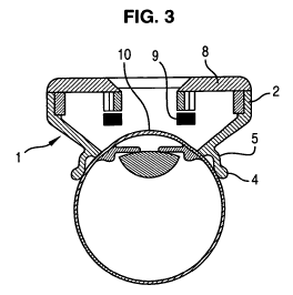
Referring to figure 2, the device according to the invention comprises an eye
ring 1 wherein the proximal end of said eye ring is suitable to be applied
onto the
CA 02751908 2011-08-09
WO 2010/094353 PCT/EP2009/061014
11
globe of the eye to be treated and (see figure 3) means 2 to generate
ultrasound
energy, said means being fixed on the distal end of the eye ring.
Said means are connected to the control unit 3 of the phacoemulsificator.
More particularly, said means 2 are connected to the power source 31
configured
to provide pulsed electrical power.
Preferably, the means 2 generates ultrasounds that are not High Intensity
Focussed Ultrasound.
Indeed, the aim of generating ultrasound onto the trabecular meshwork is to
get the trabecular meshwork to vibrate in order to improve the drainage
efficiency.
The control unit 3 may comprise means specifying the parameters such as
the frequency, the power and the duration of the ultrasound generation,
etc.... The
power source 31 comprises at least a sine-wave signal generator at a
determined
frequency comprised between 1 kHz and 25 MHz, an amplifier and a Power meter.
Referring to figures 2 and 3, the eye ring 1 consists in a sawn-off cone
element opened at both ends wherein the small base is the proximal end and the
large base is the distal end.
Referring to figure 3, the proximal end of the sawn-off cone element 1
comprises an external annular flange 4 suitable to be applied onto the
external
surface of the eyeglobe, at approximately 2mm of the limbus, the limbus being
the
junction between the cornea and sclera of the eyeglobe. The proximal face of
the
annular flange 4 presents a concave profile, the radius of curvature of the
concave
profile being substantially equal to the radius of curvature of the eyeglobe.
Moreover, the proximal edge of the sawn-off cone element 1 comprises an
annular groove 5 connected to the pump 32 of the control unit by at least one
hose
7 passing through the sawn-off cone element 1 and emerging into the annular
groove. The pump 32 is used as a suction device which is advantageously
controlled by the control unit 3.
When the sawn-off cone element 1 is applied onto the eye and the pump 32 is
operated, the depression into the annular groove 5 provide a deformation of
the
conjunctiva of the eye, said deformation forming an o-ring in the annular
groove 5.
CA 02751908 2011-08-09
WO 2010/094353 PCT/EP2009/061014
12
The sawn-off cone element 1 is then closely interlinked in such a manner that
said
sawn-off cone element 1 will follow the micro movements of the eye during the
whole treatment time, and maintaining the quality of the centred position of
the
device on the visual axis.
The sawn-off cone element 1 is advantageously obtained in medical grade
silicon or other medical grade polymer which are materials compatible with the
conjunctiva contact.
It is obvious that the sawn-off cone element 1 can be obtained in any suitable
material for medical purposes well known by the skilled person, and which has
been verified as biocompatible, such as biocompatible PVC, without departing
with
the scope of the invention.
Referring to figures 2 and 3, means 2 to generate ultrasound beam comprise
a standing crown 8 holding a single transducer 9 having an annular shape.
In one embodiment, the transducer 9 has a flat section shape. Said single
transducer 9 having a flat section can be associated to a focusing acoustic
lens
extending under said transducer 9 - i.e. held by the standing crown 8 and
extending between the proximal edge of the standing crown 8 and the proximal
edge of the sawn-off cone element 1. In this case, the focusing acoustic lens
presents a cylindrical shape and a concave edge wherein the concavity is tuned
towards the eyeglobe, and more particularly towards the trabecular meshwork,
to
focalize the ultrasound beam onto the area of interest, i.e. the trabecular
meshwork
of the eye.
In another embodiment, the transducer 9 has a concave section shape,
wherein the concavity is designated to be tuned toward the eyeglobe, and more
particularly toward the trabecular meshwork. In this case, this is the concave
section shape of the transducer 9 which allows the focalization of the
ultrasound
beam onto the trabecular meshwork of the eye.
The external radius of said standing crown 8 is sensibly equal to the internal
diameter of the distal end of the sawn-off cone element 1.
CA 02751908 2011-08-09
WO 2010/094353 PCT/EP2009/061014
13
In this way, the standing crown 8 extends toward the revolution axis of said
sawn-off cone element 1. Said transducer 9 is held in the proximal edge of the
standing crown 8.
The transducer 9 is activated by the control unit 3 to produce a vibration
obtained with the propagation of an ultrasonic beam, transmitted to the
trabecular
meshwork over the whole or a part of its circumference.
In this manner, by positioning correctly the sawn-off cone element 1 onto the
eye to be treated, as described hereinafter, the whole or a part of the
trabecular
meshwork is treated without the need to manipulate the device during the
treatment.
Advantageously, the means 2 to generate ultrasound beam may comprise a
standing crown and a plurality of elementary transducers 9 disposed with
respect
to one another so as to define an annular shape.
For instance, the standing crown 8 of transducers 9 may comprise six
transducers 9. Each transducer 9 is a cylindrical segment able to treat 60 of
the
circumference of the trabecular meshwork.
It will be noted that the standing crown 8 can comprise two or more
transducers 9 distributed among the circumference in any manner without
departing with the scope of the invention.
To apply correctly the sawn-off cone element 1 onto the eye, the surgeon
must manipulate the sawn-off cone element 1 as far as the iris ring and the
periphery of the cornea are centred in the distal opening of the sawn-off cone
element 1. If the white ring corresponding to the visible part of the sclera
trough the
opening of the proximal end of the ring, has a constant thickness, the
centring is
correct. When the sawn-off cone element 1 is centred on the pupil, the
revolution
axis of said sawn-off cone element 1 and the optical axis of the eye are
merging.
Consequently, the planes in which extend the distal edge and the proximal edge
of
the sawn-off cone element 1 are perfectly parallel to the planes of the eye
such as
iris plane, pupil plane or plane of the trabecular meshwork, and the
transducer 9 is
CA 02751908 2011-08-09
WO 2010/094353 PCT/EP2009/061014
14
at the plumb of the trabecular meshwork 10. This allows a better positioning
of the
device according to the invention, and improves the reproducibility of the
treatment.
Moreover, the device can comprise two aiming wires 14 extending crosswise
and diametrally from the internal edge of the standing crown 8 or another
centring
system like a circular pad supposed to be centred on the pupil. This allows
facilitating the centring of the sawn-off cone element with regard to the eye.
To
centre the sawn-off cone element 1, it is necessary to centre the intersection
of the
aiming wires 14 with the centre of the pupil.
It will be understood that the device according to the invention can comprise
other centring system known from the man skilled in the art for facilitating
the
centring of the sawn of cone.
When the sawn-off cone element 1 is correctly centred onto the eye, the
pump 32 is activated to interlink said sawn-off cone element 1 with the eye.
The
depression into the annular groove 5 provides a deformation of the conjunctiva
of
the eye, said deformation forming an o-ring in the annular groove 5. This
insures a
proper maintain in position of the device during all the treatment.
The sawn-off cone element 1 is then filled with a physiological saline
degassed solution, the o-ring formed by the deformation of the conjunctiva of
the
eye in the annular groove ensuring the sealing. The physiological saline
solution
provides a cooling of the eye and the device during the generation of
ultrasound
beam and an ultrasound coupling media that permits the propagation of
ultrasound
from transducer 9 to area of interest, i.e. the trabecular meshwork 10. Note
that the
physiological saline solution moisturizes the cornea of the eye during the
treatment.
It is obvious that the physiological saline degassed solution could be
substituted by any ultrasound coupling agent such as aqueous media or
lipophilic
media without departing of the scope of the invention.
Then, the frequency and/or the power and/or the duration of each pulse are
selected or already predetermined and the transducer 9 (or the plurality of
CA 02751908 2011-08-09
WO 2010/094353 PCT/EP2009/061014
transducers) is (are) activated by the control unit 3 to produce a vibration
of the
trabecular meshwork over the whole or a part of the circumference.
Note that the treatment according to the invention is advantageously a short
treatment which can be performed before the phacoemulsification procedure with
5 the same machine.
The device according to the invention can easily produce a vibration obtained
with the propagation of an ultrasonic beam, transmitted to the trabecular
meshwork, which unlike the apparatus described in WO 2008/024795 can concern
the whole circumference of the trabeculum at the same time, more rapidly and
in
10 only one step. Moreover, with the device according to the invention, thanks
to the
ring which allows centering and fixation on the eye globe, this technique can
be
substantially improved compared to the device described in WO 2008/024795.
The fact that the device according to the present invention, used as a
treatment of open angle glaucoma with the vibration technique applied on the
15 trabecular meshwork, can be combined with a phacoemulsification machine has
many advantages. In particular, this allows implementing both treatments
(cataract
and glaucoma) during a single procedure, which improves the treatment by the
vibration technique.
In fact, when the particles like cell debris, fibrin, pigment or other,
responsible
for the loss of drainage efficiency of trabeculum, are delivered from their
adherence
to the trabecular meshwork, and are circulating in the aqueous humor it is
obvious
that they will rapidly be cached again by trabeculum, reducing consequently
the
efficiency of the treatment by the vibration technique.
If implementing the glaucoma treatment before the cataract surgery, then the
particles delivered from their adherence thanks to the vibration technique
according to the invention will be aspirated during the cataract surgery.
Thus the idea is to combine the glaucoma treatment with a
phacoemulsification machine, and preferably during a cataract surgery, because
during this surgery the anterior chamber and the liquid it contains, are
completely
washed with a balanced salt solution circulating in the irrigation /
aspiration circuit,
CA 02751908 2011-08-09
WO 2010/094353 PCT/EP2009/061014
16
so that if the vibration technique is performed before the cataract surgery,
all the
debris delivered from their adherence on the trabecular meshwork, will be
washed
out of the anterior chamber, increasing the efficiency of the treatment.
It is well known that cataract surgery is more frequent in older population.
It is
well known too that glaucoma is more frequent in the same population.
For this reason, combined surgeries, including cataract and trabeculectomy
are more and more frequent.
This written description uses examples to disclose the invention, including
the
best mode, and also to enable any person skilled in the art to make and use
the
invention. The scope of the subject matter described herein is defined by the
claims, and may include other examples that occur to those skilled in the art.
Such
other examples are intended to be within the scope of the claims if they have
structural elements that do not differ from the literal language of the
claims, or if
they include equivalent structural elements with insubstantial differences
from the
literal languages of the claims.