Note: Descriptions are shown in the official language in which they were submitted.
SEGMENTATION OF STRUCTURES FOR STATE DETERMINATION
Technical Field
[001] The present invention relates generally to medical image processing.
More
specifically, the invention relates to methods and apparatuses for determining
a state
of health or disease using patch-based segmentation and grading of image
structures.
Background
[002] The atrophy of medial temporal lobe structures, such as the hippocampus
(HC) and entorhinal cortex (EC), is disease specific and serves as an early
biomarker
of Alzheimer's disease (AD). In particular, atrophy of the HC and the EC can
be used
as a marker of AD progression since changes in these structures are closely
related
to changes in cognitive performance of the subject. The evaluation of
structure
atrophy is usually estimated by volumetric studies on anatomical MRI,
requiring a
segmentation step that can be very time consuming when done manually.
[003] In recent years, numerous methods have been proposed to automatically
segment the hippocampus. Among these methods, several have been used to
classify AD patients using HC volume, such as Colliot et al. (2008). Despite
the high
segmentation accuracy of the new HC segmentation approaches, using the HC
volume enables a separation between AD and cognitively normal (CN) subjects
with
a success rate of only around 72-74% over the entire Alzheimer's Disease
Neuroimaging Initiative (ADNI) database. This limited capability to classify
AD
patients using the HC volume only, may be due to a simplification of the
complex
atrophy patterns to a volume - a simple scalar. Recently, several shape
analysis
methods have been proposed to capture detailed HC structural modifications in
order
to obtain a more accurate classification. At 77% in the comparison proposed by
Cuingnet et al. (2010), the approach proposed in Gerardin et al. (2009) yields
a
slightly better classification than a volumetric approach. Therefore,
development of
new methods capable of estimating subtle anatomical modifications of HC
appears to
be a critical point to obtain better classification rates. Longitudinal
approaches to the
AD classification problem have also been investigated by estimating the HC
atrophy
Date Recue/Date Received 2022-03-15
CA 02752370 2011-09-16
rate over time. In Wolz et al. (2010), the authors reported a correct
classification rate
of 82% on 568 images of the ADNI dataset. However, this type of approach
requires
several time-points for a given patient. Finally, an emerging method is to
segment
subfields of the hippocampus (Yushkevich et al., 2010). This approach seems
promising since it is potentially able to detect more detailed atrophic
patterns.
However, ultra-high resolution MRI is required, what is not yet the standard
in clinical
practice and thus limits the practical applicability of this approach for the
moment.
The EC volume has also been investigated as a possible biomarker to detect AD.
EC
atrophy seems to appear slightly earlier in AD progression than HC atrophy,
and thus
could be used as a more specific biomarker in the initial stages of the
disease
(Frisoni et al., 2010). However, the high inter-subject variability of the EC
and the
difficulty to define EC boundary in anatomical MRI make volumetric studies on
EC
very challenging. Therefore, studies based on EC volume have been limited to
comparison of manual segmentations. Patient classification accuracy using EC
volume greatly varies according to the dataset, from 67% up to 87%. It seems
that
the theoretical advantage of EC measurements over HC measurements is badly
impacted by the difficulty to segment EC due to the ambiguity in defining its
boundary
in MRI. The development of automatic methods to segment EC is challenging.
However, an accurate and consistent EC segmentation method could have an
important impact on the use of this structure on large datasets and in a more
systematic manner within the study of AD.
Summary
[004] Applicants have discovered an innovative approach to robustly and
accurately
detect Alzheimer's disease (AD) and prodromal forms of AD based on the
distinction
of specific atrophic patterns of anatomical structures such as hippocampus
(HC) and
entorhinal cortex (EC) in regions of interest of an image. The discovery
allows to
efficiently determine a pathological status and grading of pixels of interest
when
compared (weighed) to images from a reference library having pre-defined
states.
The discovery simultaneously performs segmentation and grading of structures
to
efficiently capture the anatomical alterations caused by AD. Based on a
nonlocal
patch-based framework, the grading measure estimates the similarity of the
patch
2
CA 02752370 2011-09-16
surrounding the voxel under study with all the patches present in different
training
populations. The training library was composed of two populations: 50
cognitively
normal subjects (CN) and 50 patients with AD, randomly selected from the ADNI
database. During applicants' experiments, the classification accuracy of
patients (CN
versus AD) using several biomarkers was compared: HC and EC volumes, the grade
of these structures and finally the combination of their volume and their
grade. Tests
were completed in a leave-one-out framework using discriminant analysis.
First,
applicants showed that biomarkers based on HC provide better classification
accuracy than biomarkers based on EC. Second, applicants demonstrated that
structure grading is a more powerful measure than structure volume to
distinguish
both populations with a classification accuracy of 90%. Finally, by adding the
ages of
subjects in order to better separate age-related structural changes from
disease-
related anatomical alterations, applicants obtained a classification accuracy
of 93%.
[005] In addition, the applicants completed tests on magnetic resonance
imaging
data of subjects with mild cognitive impairment to determine which subjects
would
remain stable (MCIs n=75) and which subjects would progress to AD (MCIp n=-75)
within a fixed period of time (12 months). Two training libraires were
evaluated The
first contained 50 cognitively normal subjects (CN) and 50 patients with AD,
randomly
selected from the ADNI database. The second contained 75 MCIp and 75 MCIs,
randomly selected from the ADNI database Classification accuracy (MCIs versus
MCIs) using several biomarkers was compared using both databases: HC and EC
volumes, the grade of these structures, subject age and finally the
combination of
their volume and their grade and age. Tests were completed in a leave-one-out
framework using linear discriminant analysis. Applicants showed that using the
CN+AD library yielded better classification accuracy than when using the
MCIp+MCIs
training library. The best accuracy was obtained when using HC and EC volume
with
HC and EC grade and age, to obtain a classification accuracy of 75% to
differentiate
stable versus progressors in the mild cognitive impairment population.
[006] It is therefore an object of the present invention to provide a computer-
implemented method for processing medical images, the method comprising
calculating non-local means patch-based weights comparing patches surrounding
3
pixels of interest in a test image with a number of patches of pixels
surrounding a
corresponding number of pixels in reference images, calculating for the pixels
of interest
at least one state estimation using the given states assigned to the reference
images
and the weights, and modifying a computer-displayable image dataset of the
reference
images (for example using grading maps) using the at least one state
estimation for the
purposes of displaying a spatial representation of said at least one state
estimation in
said reference images.
[007] In some embodiments of the present invention, the test image is
preprocessed
and the pre-processing comprises isolating a region of interest (ROI) from the
image.
[008] In other embodiments of the present invention, pre-processing comprises
at least
one of image format conversion, denoising, regridding, correcting intensity
inhomogeneity, registration to a library image, isotropic resampling,
intensity clamping,
intensity standardization and non-linear alignment.
[009] In yet other embodiments of the present invention, a patch from a
reference image
used in the calculations is selected according to its relatedness to a test
patch
surrounding the pixel of interest and the relatedness is determined by a mean
and a
standard deviation of intensity values of the test patch pixels and the
reference patch
pixels.
[0010] In other embodiments of the present invention, the state determination
comprises
a structure classification for each pixel of the reference images, and the
value provides
segmentation of the test image and a determination of the volume of the
segmented
structure.
[0011] In other embodiments of the present invention, state determination
comprises a
pathological status of patients related to the reference images, and the
method provides
pathological status grading of the pixels of interest in the test image.
[0012] In other embodiments of the present invention, state estimation
comprises a
pathological status of patients related to the reference images, and the
method provides
segmentation and pathological status grading of the pixels of interest in the
test image.
In other embodiments, a pathological status score is calculated from the
grading of the
pixels of interest, or from the grading of the pixels of interest having a
predetermined
segmentation.
4
CA 2752370 2018-09-18
[0013] It is another object of the present invention to provide an apparatus
for
processing medical images comprising a non-local means patch-based weight
calculator for calculating a weight of a pixel of interest in a test image
with a number of
patches of pixels surrounding a corresponding number of pixels in a reference
image;
and a state calculator for calculating a state of the pixel of interest based
on a given
state assigned to the reference image, and a processor for modifying a
computer-
displayable image dataset of said reference images (for example using grading
maps)
using said at least one state estimation for the purposes of displaying a
spatial
representation of said at least one state estimation in said reference images.
[0014] In some embodiments of the present invention, the state comprises a
structure
classification for each pixel of the reference images, and the apparatus
provides
segmentation of the test image.
[0015] In other embodiments of the present invention, the state comprises a
pathological status of patients related to the reference images, and the
apparatus
provides pathological status grading of the pixels of interest in the test
image.
[0016] In yet other embodiments of the present invention, an image pre-
processor pre-
processes the images prior to the calculating, and in still other embodiments,
a test
image label calculator calculates a label of the pixel of interest using the
weight, and in
still other embodiments, a test image grade calculator calculates a grade of
the pixel of
interest using the pathological status and the weight of the reference images
and can
generate a pathological status score based on the labels and the grades.
[0017] A system for processing medical images comprising a medical imager for
generating a test image, an apparatus for segmenting and grading medical
images and
determining a pathological state determination of said test image according to
the
present invention and a client application for receiving and presenting data
provided by
said apparatus, wherein said imager, apparatus and client application
communicate
data over a network and return to said client application a pathological state
determination.
CA 2752370 2018-09-18
Brief Description of the Drawings
[0018] The invention will be better understood by way of the following
detailed
description of embodiments of the invention with reference to the appended
drawings, in which:
[0019] Figure 1 is a highly schematic drawing of methods according to the
present
invention.
[0020] Figure 2 shows a general summarized overview of the segmentation and
grading method.
[0021] Figures 3a and 3b show sample segmentations and gradings of a control
patient (3a)
and an Alzheimer's disease patient (3b). (Referred together as Figure 3).
[0022] Figure 4 shows volumes and grading values for 100 subjects for the
structures
studied.
[0023] Figure 5 shows the impact of the number of reference images used as
priors
as well as the contribution of age to the success rate of state determination.
[0024] Figure 6 shows the average grading value of control and AD patient
images as
a function of age in years.
[0025] Figure 7 is a graph showing shows the average grading value of control
and
AD patient images as a function of the mini mental status exam (MMSE).
[0026] Figure 8 shows a method of segmenting and grading structures according
to
the present invention.
[0027] Figure 9 shows a block diagram illustrating various components of an
apparatus for segmentation and grading of structures.
[0028] Figure 10 shows a block diagram illustrating a physical embodiment of
an
apparatus for segmentation and grading of medical images.
[0029] Figure 11 shows a block diagram illustrating various components of an
apparatus (processor) for segmentation and grading of medical images.
6
CA 2752370 2019-08-30
CA 02752370 2011-09-16
Detailed Description
[0030] Applicants discovered a new approach designed to: 0 obtain a more
detailed
detection of structural changes caused by the disease and to
perform the
automatic segmentation of complex structures such as the entorhinal cortex
(EC).
Buades et al, (2005) showed nonlocal means estimators for image denoising
purposes. Applicants have recently proposed a new nonlocal patch-based label
fusion method to segment anatomical structures (Coupe et al., 2011b, the
content of
which is incorporated by reference). By taking advantage of pattern redundancy
present within the subject's image, as well as the redundancy across training
subjects, the nonlocal means scheme enables robust use of a large number of
samples during estimation. In Coupe et al. (2011b), applicants applied this
approach
to patch-based label fusion for the segmentation of anatomical structures such
as the
hippocampus (HC) of healthy subjects and lateral ventricles of patients with
Alzheimer's disease (AD). Applicants propose an extension of this patch-based
segmentation method in order to evaluate the similarity (in the nonlocal means
sense) of the intensity content of one test magnetic resonance image (MRI)
compared to several training populations. By using training populations with
different
clinical statuses (e.g., healthy control normal (CN) subjects and patients
with AD), a
nonlocal means estimator is used to evaluate the proximity (i.e., the grade of
the
disease or the degree of anatomical change consistent with disease in the case
of
AD) of each voxel of the MRI under study compared to the training populations
(see
Fig. 2). Since the grade estimation and the label fusion steps require the
same patch
comparison step, simultaneous segmentation and grading of the studied
structure
can be achieved in one pass without extra computation. In the proposed
approach,
the nonlocal patch-based comparison is used to 0 efficiently fuse the labels
of MRI in
a training database in order to segment EC and HC, and simultaneously
aggregate
the clinical status of the populations constituting the training database
(reference
images) in order to detect the presence (or not) of the disease. Finally, the
average
grading value obtained over the segmented structures is proposed as a new
biomarker to estimate the clinical status of the subject under study as a
computerized
aid to diagnosis. This invention: 0 introduces an innovative approach to
better
characterize the patterns of structural modification caused by the disease
(e.g.,
7
CA 02752370 2011-09-16
anatomical changes such as atrophy in case of AD) through the new concept of
grading, ii) presents a method to automatically and simultaneously perform the
segmentation and the grading of EC and HC, and iii) demonstrates that the
proposed
approach can be used as a novel biomarker to efficiently achieve patient
classification in the context of AD.
[0031] Figure 1 shows a highly schematic drawing of methods according to the
present invention. An image acquisition system acquires a test image (Fig.1-
top left),
in this case, an MRI scan of a subject's brain. The subject or the doctor
wants
information from scan data relating to specific brain structures such as the
hippocampus (Fig.1-top middle) that are indicative or biomarkers for mild
cognitive
impairment (MCI) and Alzheimer's disease (AD). The area shown on the scan is
for
illustrative purposes and is not actually the hippocampus. The area blown up
is
shown as a 20x20 square (of 400 patches) where the hippocampus is shown in
black
and two square regions of interest (ROI) are highlighted in grey. One patch
(Fig.1-top
middle) is further blown up to the pixel level as a 5x5 square (it should
actually be
understood as being a volume of 5x5x5 voxels). The computer implemented method
according to the present invention identifies patches within the region of
interest in
order to compare the patches with many (or in some case only with the most
related)
patches from a healthy subject (state 1) and a diseased subject (state 2). A
modification of the Buades nonlocal means estimator allows to determine if the
portion of the hippocampus identified in the region of interest resembles more
that of
a healthy or a diseased reference image taken from a reference image library
that
contains examples of both states. In the schematic example shown, (Fig.1
bottom
images), it is clear that the test subject patch shows a greater resemblance
to state 2,
suggesting that the test image hippocampus has atrophied as a consequence of
MCI
or Alzheimer's disease. It will be understood that figure 1 is highly
simplified. For
example, it should be understood that all pixels of an area of interest can be
segmented and/or graded and not just the central pixel of a patch. In other
words,
each patch is centered on a pixel but the patches for successive pixels
overlap with
each other. It will also be appreciated that the 2 states (healthy and
diseased) are not
represented by only 2 patches, but that each state is represented by an
ensemble of
patches where weightings/stats determine the result.
8
CA 02752370 2011-09-16
The nonlocal means estimator:
[0032] The nonlocal means filter was first introduced by Buades for the
purpose of
image denoising. In nonlocal means-based approaches (Buades et al., 2005;
Coupe
et al., 2008), the patch P(x) surrounding the voxel xi under study is compared
with all
the patches P(x) of the image CI (or a subpart of the image) whatever their
spatial
distance to P(x,) (Le., this is the meaning of the term "nonlocal"). According
to the
patch similarity between P(xi) and P(x), estimated by the sum of squared
differences
(SSD) measure, each patch receives a weight w(xõ x):
P(x'"(xi):
W(X0X j) = e h2 (1)
[0033]where 11.112 is the L2-norm computed between each intensity of the
elements of
the patches P(xi) and P(x), and h2 is the smoothing parameter of the weighting
function. This weighting function is designed to give a weight close to 1 when
the
SSD is close to zero and a weight close to zero with the SSD is high. Finally,
all the
intensities u(x) of the central voxels of the patches P(x) are aggregated
through a
weighted average using the weights w(xi, x). In this way, the denoised
intensity OW
of the voxel x, can be efficiently estimated:
. w(x,x).(x,)
ro,) l'n
LiE.w(xõx,) (2)
[0034] Despite its simplicity, the nonlocal means filter has been demonstrated
to have
excellent denoising performance. This filter is currently one of the most
studied
denoising filters. The efficiency of the nonlocal means filter relies on two
aspects: the
pattern redundancy present in an image (i.e., its self-similarity) and the
robust
detection of samples derived from the same population by using local context
(i.e.,
patch-based comparison):
9
CA 02752370 2011-09-16
[0035] First, to improve the accuracy of an estimator, it is possible to
reduce the
committed error by increasing the number of involved samples. By using an
infinite
number of samples derived from the same population, the error theoretically
converges to zero. To drastically increase the number of samples used, the
nonlocal
means filter takes advantage of the redundancy of information by using all the
similar
voxels present over the entire image.
[0036] Second, to ensure that the used samples are derived from the same
population, the surrounding neighbour of a voxel can be used to robustly
detect
similar realizations of the same process. In the nonlocal means approach, this
task is
achieved by patch-based comparison using SSD. Two voxels with similar
surrounding patches are considered as similar and to belong to the same
population.
More precisely, the nonlocal means filter performs patch comparison to
estimate the
degree of the similarity between two voxels. This way, each involved sample
has a
weight (see Eq. 1) reflecting its relevance.
[0037] Finally, a simple weighted average (see Eq. 2) is used to aggregate the
samples according to their relevance. This way, the resulting estimator
embodies the
two interesting qualities described above: to build on a large number of
samples and
to ensure that the involved samples are derived from the same population.
From denoising to segmentation:
[0038]In Coupe et al. (2010, 2011b), applicants were the first to introduce
the
nonlocal means estimator in the context of segmentation by averaging labels
instead
of intensities. By using a training library of N subjects, whose segmentations
of
structures are known, the weighted label fusion is estimated as follows:
EN E w(x,xs.,)1(x,
v(x,)= __________________________
Es=1 LeS2 W(X19 Xs,./ (3)
[0039] where I(x) is the label (i.e., 0 for background and 1 for structure)
given by the
expert to the voxel xsiat location j in training subject s. It has been shown
that the
nonlocal means estimator v(x,) provides a robust estimation of the expected
label at
CA 02752370 2011-09-16
Xi. With a label set of {0,1} voxels with value v(xp0.5 are considered as
belonging to
the considered structure and the remaining voxels as background.
[0040] In Coupe et al. (2010, 2011b), applicants showed that accurate
segmentations
of anatomical structures can be obtained using this simple patch-based label
fusion
framework. In addition, to take advantage of the self-similarity of the image
as done
for denoising, the nonlocal label fusion also relies on inter-subject
anatomical
consistency. Therefore, many similar patches (self-similarity) can be found in
every
training subject (inter-subject consistency), thus improving the final
estimation.
Finally, compared to atlas-based methods using nonlinear registration, the
nonlocal
patch-based approach has the advantage of better handling the inter-subject
variability problem. Contrary to the one-to-one correspondence assumed by
nonlinear warping methods, the nonlocal means estimator makes it possible to
deal
with one-to-many mappings, which better captures the link between subjects'
anatomies. This interesting aspect of the nonlocal means estimator has been
used to
improve video super-resolution without explicit estimation of inter-frame
motion.
From segmentation to grading:
[0041] Applicants extend this segmentation method to efficiently aggregate
clinical
status (e.g. CN or AD) in order to estimate the proximity (in the nonlocal
means
sense) of each voxel compared to both populations constituting the training
library
(see Fig. 2). To achieve this goal, applicants introduce the new concept of
patch-
based grading that reflects the similarity of the patch surrounding the voxel
under
study with all the patches present in the different training populations. In
this way, the
neighborhood information is used to robustly drive the search of anatomical
patterns
that are specific to a given subset of the training library. When the training
populations include data from subsets of subjects in different clinical
states, this
approach provides an estimation of the grade (i.e., degree of closeness to one
group
or another) for each voxel:
L
g(x,)= __________________________
Es_tE jeow(x,x,,,) (4)
11
CA 02752370 2011-09-16
[0042] where Ps is the clinical status of the training subject s. In
applicants' case,
Ps=-1 was used for AD status and p5=1 for CN status. A negative grading value
(respectively, a positive grading value) g(x1) indicates that the neighborhood
surrounding x, is more characteristic of AD than CN (respectively, of CN than
AD)
(see Fig. 3). The absolute value Ig(x)I provides the confidence given to the
grade
estimation. When Ig(41 is close to zero, the method indicates that the patch
under
study is similarly present in both populations and thus is not specific to one
of the
compared populations and provides little discriminatory information. When
Ig(xdi is
close to 1, the method detects a high proximity of the patch under study with
the
patches present in one of the training populations and not in the other.
Finally, for
each subject, an average grading value is computed over all voxels in the
estimated
structure segmentation (i.e., for all x, with v(xd 0.5) for each side(e.g., kw
40 or
). Since the grading and the segmentation involve the same patch comparison
step,
these structures are extracted at the same time that their grade is estimated
(see Fig.
3).
[0043] Several strategies can be used to fuse the average grading of the
studied
structures. First, each side of the structure can be used separately. Second,
it is
possible to assign the same weight to the left and right HC and EC (e.g.,
_õ00/2). This strategy of fusing both sides appears to be more robust to
segmentation inaccuracy was used by Chupin et al. in a volumetric study
(Chupin et
al., 2009a). During experiments, applicants found that these two strategies
provided
similar results for HC and EC. However, for the HC-EC complex, the best
strategy
was to compute left and right average grading values over HC-EC segmentation
(this
giving more importance to HC because of its larger size) and then to use the
mean of
both sides (k-,õõ
+g,õ,)/2). Therefore, applicants decided to present all the
results using the second strategy.
Training library construction
[0044] Datasets: the publically available ADNI database
(www.loni.ucla.edu/ADNI)
was used to validate the proposed approach. This database contains both 1.5T
and
3.0T T1-w MRI scans. For applicants' experiments, applicants randomly selected
120
12
CA 02752370 2011-09-16
MRI scans, 60 1.5T MRI baseline scans of CN subjects and 60 1.5T MRI baseline
scans of patients with AD.
[0045] Preprocessing: All the selected images were preprocessed as follows: 1)
correction of inhomogeneities using N3 (Sled et al., 1998), 2) registration to
the
stereotaxic space using a linear transform to the ICBM152 template (1x1x1 mm3
voxel size) (Collins et al., 1994) and 3) cross-normalization of the MRI
intensity using
the method proposed in Nyul and Udupa (2000). After preprocessing, all the
MRIs
are coarsely aligned (linear registration), tissue intensities are homogeneous
within
each MRI volume (inhomogeneity correction) and across the training database
(intensity normalization).
[0046] Label propagation: From the 120 processed MRI scans, 20 scans (10 CN
and
AD) were randomly selected to be used as seed dataset for segmentation. The
HC and the EC of this seed dataset were manually segmented by following the
protocol defined in (Pruessner et al., 2002). The manual segmentations of the
seed
dataset were then propagated to the 100 remaining scans constituting
applicants' test
dataset using the method described in (Coupe et al., 2011b). After the
segmentation
propagation step, the test dataset was composed of 100 MRI (50 CN subjects and
50
patients with AD) with their corresponding automatic segmentations (see Fig.
2). In
applicants' test dataset, the average age of the populations is 74.8 ( 4.8)
for CN and
74.9 ( 6.4) for AD. The age for the two populations is not significantly
different
(p=0.36, unpaired t-test). In addition, the Mini Mental State Evaluation
(MMSE) is
29.1 ( 1.2) for CN and 23.2 ( 2.0) for AD.
Implementation details
[0047] In all experiments described here, the optimal parameters empirically
found in
Coupe et al. (2011b) for HC segmentation have been used and thus the patch
size
was fixed to 7x7x7 voxels and the pre-selection threshold set to th=0.95.
[0048] As done in Coupe et al. (2011b), a was replaced by a cubic volume V;
centered on xi . First, this strategy to use a semi-local paradigm instead of
a fully
nonlocal paradigm makes the processing computationally practical. In the
denoising
literature, this approach is used in the majority of the papers and has been
shown to
13
CA 02752370 2011-09-16
produce near-optimal or optimal results except for images with repetitive
textures
(Brox et al., 2008). Second, as shown in Coupe et al. (2011b), in the case of
HC
segmentation, limitation of the search window provides better results (see
left of Fig.
8 in Coupe et al. (2011b)). Since all the images are linearly registered, the
patches
belonging to HC are located within a restricted area. By using a larger search
window, outliers are added that marginally degrade the segmentation and
uselessly
increases the computational time. While in Coupe et al. (2011b) the search
window
size was fixed, applicants used a locally adaptive search window size. The
initialization of the search window was set to 9x9x9 voxels as suggested in
Coupe et
al. (2011b). However, in the case when no similar patches can be found in this
search window (i.e., none of the patches pass through the pre-selection), its
radius is
increased by one voxel until at least one similar patch in each population is
found
(i.e., at least one patch in each population pass through the pre-selection
step). For
all the studied subjects, the largest search window size found was 15x15x15
voxels.
[0049] The automatic local adaptation of the smoothing parameter h2(x) (see
Eq. 1)
proposed in Coupe et al. (2011b) has been slightly modified. During all the
experiments, the squared smoothing parameter was set proportional (with A=0.5)
to
the minimal SSD:
122 (.0= 2\,2 x arg min 1P(x)¨ P(x,, A: +6 (5)
,
[0050]The value of lambda slightly changes the segmentation results. When
applicants validated their segmentation method on the ADNI dataset in Coupe et
al.
(2011a), using A=0.5 instead of A=1 changed the median Dice-Kappa values from
0.882 to 0.883 for CN and from 0.836 to 0.838 for AD.
[0051] Finally, a subject selection was also applied to reduce the number of
training
MRI required. For each structure, the N closest subjects (in terms of SSD over
the
initialization mask as done in Coupe et al. (2011b)) are equally selected from
both
populations (N/2 from the CN population and N/2 from the AD population) (see
Fig.
14
CA 02752370 2011-09-16
2). This is done to ensure that the size of the "patch pool" from the AD
population is
coarsely similar to the size of the "patch pool" from the CN population.
[00521For a given subject with N=20 (i.e., 10 AD training templates and 10 CN
training templates), the segmentation and the grading maps were obtained in
less
than 4 minutes for left and right HC and less than 2 minutes for left and
right EC
using a single core of an Intel Core 2 Quad Q6700 processor at 2.66 GHz.
Validation framework
[0053] Applicants' validation framework was designed to compare the capability
of
different biomarkers to discriminate between patients and controls. The
biomarkers
studied were: HC volume, HC grade, EC volume and EC grade as well as their
combination.
[0054]First, to obtain the segmentation and the grade of the subjects within
the test
dataset, a leave-one-out procedure was performed over the 100 subjects using
their
corresponding automatic segmentations resulting from the label propagation
step
(see Fig. 2). For each subject, the N closest training subjects were selected
from the
99 remaining subjects in the library The average grading value was then
estimated
over the EC and the HC segmentations (for both left and right sides) obtained
at the
same time (see an example in Fig. 3). These segmentations were also used to
measure the HC and EC volumes in the stereotaxic space.
[0055] Once all the subjects had a volume and a grade for each structure, a
quadratic
discriminant analysis (QDA) was performed. Each subject was classified by
performing a QDA over the 99 remaining subjects. This approach was applied to
volume-based classification, grade-based classification and the combination of
both
for HC, EC and HC + EC. Applicants found that QDA slightly improved the
results
compared to linear discriminant analysis, especially when the subject's age
was used
as an additional parameter. The success rate (SR), the specificity (SPE), the
sensitivity (SEN), the positive predictive value (PPV) and negative predictive
value
(NPV) are presented for each of the tested biomarkers (see (Cuingnet et al.,
2010)
for details on these quality metrics).
CA 02752370 2011-09-16
[0056] Figure 3 shows the grading maps obtained for 2 test subjects (1 CN and
1
AD). The corresponding average grading values and the estimated volumes are
also
provided for left and right HC and for left and right EC. Visually, the ON
subject
clearly appears closer to the CN population (mainly red color related to
values close
to 1) while the AD patient is visually closer to the AD population (mainly
purple and
black colors related to values close to -1). In addition, Fig. 3 also provides
a visual
assessment of the quality of the segmentation and grading.
Volumetric study
[0057] The left column of Fig. 4 shows the volumes for the 100 subjects of the
test
dataset for HC and EC for a training library of size N=100 (i.e., 50 ON and 50
AD).
The volumetric approach provided a classification success rate of 80% for HC
and
69% for EC. The use of both structures at the same time produced a success
rate of
78% through applicants' QDA-based classification. This result indicates that
the
estimated HC volume is more powerful than the EC volume to identify patients
with
AD. This observation is in accordance with Frisoni et al. (1999). Applicants'
result
using only HC volume is slightly superior to a recently published method
comparison
(Cuingnet et al., 2010). This might come from differences in the test dataset
used
here or due to a higher accuracy and consistency of the segmentation method
used
compared to Chupin et al. (2009b). The success rate obtained with EC volume is
similar to the results reported in Frisoni et al. (1999) but lower than the
values
reported in other studies using manual segmentations. Figure 4 shows the
higher
variability of EC volume compared to HC volume. As mentioned in the
introduction,
this range of volumes comes from the high inter-subject variability of EC, but
may
also be due to the difficulty to distinguish EC structure boundaries on
anatomical MRI
(e.g., identification of the collateral sulcus and the sulcus semiannularis).
Due to this
last point, less accurate segmentations may be obtained for this structure and
thus
the introduction of segmentation errors may negatively impact the patient's
classification. The use of both structures at the same time did not improve
the result
compared to the method based on HC only, while improvements have been observed
by other groups doing similar experiments on manual segmentations.
16
CA 02752370 2011-09-16
Grading study
[0058] The right column of Fig. 4 shows the grading values for the 100
subjects of the
test dataset for HC and EC for N=80. The success rate of the classification
was 89%
for HC, 78% for EC and 90% for the combination of both structures. For HC, the
success rate obtained by using QDA is similar to thresholding the grading
value at
zero (4 false positives CN and 7 false negatives AD). In fact, in the perfect
case, the
50 first subjects (CN) should have positive average grading values and the 50
last
(AD) should have negative average grading values. This result indicates that
the HC
grade estimator is not biased and thus that the sign of the final grading
value can be
used directly to classify the patient. On the other hand, the EC grade
estimator is
biased in the sense that the optimal threshold obtained using QDA is superior
to
zero. As shown on Fig 4, the EC grades of AD are frequently superior to zero,
thus
indicating a higher similarity with the patches present in CN population. As
applicants
will show later, the normal age-related structural changes in the EC may
disturb the
detection of the disease-related anatomical changes. However, this bias, which
depends on the training library used, can be partially compensated for by
using QDA,
yielding a success rate of 78%. Finally, by the computation of the average
grade
value over the HC and the EC improved the HC results and leads to a very high
success rate of 90%.
Comparison of anatomical biomarkers
[0059] In Tab. 1, the SEN, SPE, PPV and NPV obtained by the different
biomarkers
considered are presented. These results show that for both structures studied,
the
classification based on grading provides significantly better results than the
volumetric approach (89% vs. 80% for HC and 78% vs. 69% for EC). Moreover,
while
the combination of HC+EC tends to spoil the results of volumetric analysis,
the
combination of both slightly improves the results of the grading study. Three
different
combinations of biomarkers obtained a success rate of 90% during applicants'
experiments: HC volume and grade, HC + EC grade, HC + EC volume and grade. In
the three cases, the HC grading was used, indicating a potential key role of
this new
imaging biomarker.
17
CA 02752370 2011-09-16
[0060]Table 1: Results of the patient classification (AD vs CN) for the
different
biomarkers under investigation. These results were obtained by using
discriminant
analysis through a leave-one-out procedure on the test dataset with N = 80
(i.e., 40
CN and 40 AD).
AD vs. CN SR SEN SPE PPV NPV
HC volume 80% 78% 82% 81% 79%
HC grading 89% 86% 92% 91% 87%
HC volume and grading 90% 88% 92% 92% 88%
EC volume 69% 66% 72% 70% 68%
EC grading 78% 74% 82% 80% 76%
EC volume and grading 78% 74% 82% 80% 76%
HC + EC volume 78% 76% 80% 79% 77%
HC + EC grading 90% 86% 94% 93% 87%
HC+EC volume and grading 90% 88% 92% 92% 88%
Impact of the number of selected best training subjects
[0061] Figure 5 presents the impact of the number of selected best training
subjects
on the studied biomarkers. The success rate for all the biomarkers from N=20
(10 CN
and 10 AD) to N=80 (40 CN and 40 AD). Applicants used the subject's age as
supplementary information during QDA in order to increase classification
accuracy.
[0062]Volume (see top of Fig. 5): For HC, the classification accuracy was
quite
stable from N=40 to N=80. In Coupe et al (2011b), applicants showed that a
plateau
in terms of segmentation accuracy was reached around N = 30. For EC, the best
results were obtained for N=80. This result seems to indicate that a large
library is
required to achieve consistent segmentation of EC. Indeed, increasing the size
of the
18
CA 02752370 2011-09-16
"patch pool'' and better address issues related to inter-subject variability.
The addition
of the age as parameter in QDA improved the results of the classification,
especially
for EC and HC+EC biomarkers. By performing the QDA only with age provided a
success rate of 48% in the classification. Finally, at N=60, the HC volume
combined
with the age provided a success rate of 82%.
[0063] Grade (see middle of Fig. 5): For HC, the best classifications were
obtained by
using high N values (N=60 and N=80). For EC, the best classification rate was
obtained for the smallest value of N=20, a result that was not expected.
However, by
also using age, the best results were obtained for N=80 for EC. For HC and for
HC+EC based classifications, the inclusion of age improved the results of the
classification. In these cases, HC-based classification yielded a success rate
of 92%
and HC+EC a success rate of 93% at N=40 and N=60.
[0064] Volume + Grade (see bottom of Fig. 5): By combining the volume and the
grade of the biomarkers, applicants obtained slightly better results than by
using only
the grade, except for EC. By using the age of the subjects, the volume and the
grade
over HC (with N=60) provided 92% classification accuracy. The combination of
all the
parameters (i.e., volume, grade and age) slightly decreased the results for
the
biomarkers involving EC compared to the use of only grade and age.
Relationship between grade and age
[0065] As shown in the previous experiment, using the subject's age improved
the
classification based on the grading measure, except for EC with N=20. This
supplementary information seems to help distinguish age-related MRI changes
from
those related to AD pathology. Figure 6 shows the grade values as a function
of age
on HC + EC with N = 60 (the case with the highest classification accuracy:
93%). It
appears that the grading values decrease with age in both populations. This
variation
indicates that the grading measure captures the age-related anatomical changes
(possibly related to atrophy), and thus this observation may explain the
better results
obtained using age for all the biomarkers except for EC with N=20. As
previously
mentioned, QDA provides slightly better results than LDA during classification
(between 0 to 2% depending on the biomarker studied). This slightly better
fitting is
19
CA 02752370 2011-09-16
assessed by Pearson's coefficient and corresponding p-value of the linear and
quadratic regressions presented in Fig. 6. While for CN, the traditional
linear model
and quadratic regressions provided similar results, it seems that for AD a
quadratic
model fits better than a linear model. The nonlinear nature of the atrophy
related to
AD has recently been studied (Frisoni et al., 2010); and demonstrated that
brain
atrophy during AD is not a linear process while most studies assume a linear
progression of AD. In addition, the grade measure is correlated with age while
the
volume does not appear to be statistically correlated with age since similar
regressions provided correlation of r = 0.31 for CN and r = 0.34 for AD with
respective p-values of 0.09 and 0.06.
Relationship between grade and MMSE score
[0066] Finally, the link between the mini mental state examination (MMSE)
score and
the grade is studied. The MMSE is a test evaluating the cognitive function of
the
patient. A useful imaging biomarker should have a link with the cognitive
decline of
the patient with AD usually estimated by using MMSE. Several studies have
investigated the relationship between the MMSE score and the volume or the
shape
of key structures and EC. Applicants investigated the correlation between MMSE
score and anatomical measurements (i.e., volume and grade) for HC and EC. Fig.
7
shows the plots of the grade and the volume as functions of the MMSE score.
For
both structures, the coefficient of correlation for grade was higher (r = 0.75
for HC
and r = 0.58 for EC) than for the volume (r = 0.55 for HC and r = 0.28 for
EC). A
statistically significant correlation has been found in all cases. Another
trend was that
the HC measurements were more consistent with MMSE scores than EC
measurements (see Fig. 7). Finally, the HC grade was the biomarker most
consistent
with MMSE with a high coefficient of correlation (r = 0.75).
[0067] In Du et al. (2001), the authors obtained a correlation coefficient of
r = 0.48 for
HC and r = 0.48 for EC volume based on manual segmentations with a p-value
less
than 0.001 in both cases. In applicants' experiment, slightly higher
correlation was
obtained for HC, but a significantly lower value was obtained for EC as
assessed by
a higher p-value=0.005. However, applicants' correlation coefficient between
EC
volume and MMSE score is r = 0.34. It should be noted that the estimation of
CA 02752370 2011-09-16
correlation on discrete functions such as MMSE can bias the significance of
correlation. However, applicants wanted to compare applicants' results with
previously published studies using this metric.
[0068] During experiments, applicants showed that: 0 HC-based measures were
more discriminant than EC-based measures, ii) the grading had a higher
discriminatory capability than the volume, fit) by adding the age, the
classification rate
improved, especially when using the HC-grade-based metrics, iv) by computing
the
grade over a larger area (HC+EC) tended to slightly improve results,
especially when
the subjects' ages were used within the classification model, and v) the
optimal size
of the number of selected training subjects were N=60 (60% of the full
library) in the
majority of the situations studied. A balance appears to be required between
using a
large enough training population and potentially introducing outlier subjects
by using
all the available subjects. According to the structure of interest, a
different number of
training subjects could be used. Moreover, by using a larger library, it could
be
possible to select a higher number of subjects without introduction of
outliers. The
difficult segmentation of EC due to inter-subject variability could be
partially
compensated by using non-linear registration of training subjects instead of
linear
registration. However, this type of approach is more computational intensive.
The
introduction of shape priors could also be a possibility to deal with
ambiguity of the
EC boundaries.
[0069] The SEN, SPE, PPV and NPV obtained by applicants' grading approach are
competitive compared to the ten methods compared in Cuingnet et al. (2010)
involving voxel-based morphometry (VBM), cortical thickness, HC volume and HC
shape (Gerardin et al., 2009). In that comparison paper, the best VBM-based
approach obtained 89% accuracy; the best method based on cortical thickness
obtained 85% accuracy, the best approach using HC volume 74% accuracy and the
method using HC shape 77% accuracy. However, during applicants' experiment,
only
a subset of the entire ADNI database has been used, contrary to the
experiments
done in (Cuingnet et al., 2010). Moreover, the classification algorithm used
in
Cuingnet et al. (2010) was a support vector machine while applicants used a
quadratic discriminant analysis approach. Despite these differences, the
classification
21
CA 02752370 2011-09-16
results obtained by using grade only are competitive to the best results
reported in
Cuingnet et al. (2010). Moreover, by adding the subjects' age yielded an
accuracy of
93%. This result is similar to the highest classification accuracy 93.3%
reported on a
similar sized subset of ADNI (51 AD and 52 ON) in Zhang et al. (2011).
However,
Zhang et al. (2011) used a multimodal approach involving positron emission
tomography (PET) and cerebro-spinal fluid (CSF) markers to reach this degree
of
accuracy. By using only MRI, their method based on volumetric features
provided an
accuracy of 86.2%.
[0070] It appears that using a larger area of analysis by grading several
structures
tended to improve the grading estimation. The extension of grading to other
key
structures impacted by AD seems to be an interesting path to follow for
further
research. Structures such as parahippocampal cortex and perihinal cortex or
fornix
and mammillary body could be valuable anatomical structures to improve AD
detection. Moreover, further work should investigate the spatial distribution
of grade
maps over the populations. This information could help to detect more
discriminant
areas for classification and might provide information on the AD progression.
Finally,
the application of the proposed grading measure to other diseases has a great
potential. Moreover, the difficult problem of clinical differentiation (such
as AD and
frontal lobe dementia for instance) should also be investigated.
[0071] Using SSD as similarity metric, applicants' approach is sensitive to
inaccuracy
in inter-subject intensity normalization. In Coupe et al. (2011a; 2011b),
applicants
demonstrated that the proposed preprocessing pipeline involving (Nyul and
Udupa,
2000) provides a sufficiently robust normalization to obtain accurate
segmentations.
In this paper, applicants also showed that the preprocessing pipeline used
yields high
classification accuracy. Nevertheless, any improvements on the inter-subject
normalization should yield further improvements in grading estimation. The use
of
other similarity metrics less sensitive to intensity normalization should be
studied in
future work. However, according to applicants' experiments, there is no
trivial solution
since cross-correlation or correlation ratio cannot distinguish constant areas
with
different means (e.g, in CSF and white matter), mutual information requires a
higher
number of samples (bigger patch) and introduces the binning problem for
histogram
22
CA 02752370 2011-09-16
construction, and finally the SSIM index also requires matching of intensity.
The use
of hybrid metrics based on intensity and derivatives could be further
investigated.
[0072] As for Voxel-Based Morphometry (VBM)-based approaches, applicants'
method requires several scans of each population to be usable. The
construction of a
large enough training library might be an issue for trials based on a small
number of
subjects. However, the number of training subjects required by applicants'
method is
similar to the number required by VBM studies. A group size of 30 to 50
subjects per
population is typical in a VBM study while a group size of 70-90 subjects per
population is optimal for detection of HC volume loss. Applicants found that
30
subjects from each population is sufficient to provide very high
classification rates
[0073] In the proposed grading technique, applicants focused on the problem of
AD
vs. CN classification. However, the prediction of conversion from prodromal AD
(also
known as mild cognitive impairment or MCI) to clinically definite AD is more
useful
from a clinical and diagnostic point of view. The prediction of patients with
MCI who
will convert to AD and those who will stay stable is an extremely complex task
for
which no method has yet provided satisfactory classification results (Cuingnet
et al.,
2010). Proposed methods based on structural MRI have been focusing on gray
matter loss as markers for prediction. Applicants' proposed grading method
adds
valuable information for the problem of prediction.
[0074] A new method is proposed to robustly detect the patterns of anatomical
change in the hippocampus and entorhinal cortex caused by AD. Based on a
nonlocal means estimation framework, the proposed novel grading measure (i.e.,
anatomical change possibly related to atrophy in the context of AD) enables an
accurate distinction between CN subjects and patients with AD leading to a
classification success rate of 90%. When the subject's age is combined with
the
grading measure, a success rate of 93% was obtained. These results are
competitive
compared to the AD detection performance of VBM, cortical thickness, HC volume
and HC shape methods extensively compared in (Cuingnet et al., 2010). In
contrast
to these approaches, applicants' method has the advantage of: 0 simplicity (it
can be
coded in few hundred lines of code), it) low computational cost (as it does
not require
23
CA 02752370 2011-09-16
non-rigid registration), iii) robustness of the process (all the subjects get
final grading
maps) and iv) the possibility to achieve individual classifications based on a
MRI data
from a single time point (contrary to group classifications or longitudinal
studies).
These results indicate that this new structure grading approach is a useful
biomarker
to efficiently detect AD. Further work will investigate the possibility to
discriminate
populations of patients with MCI compared to AD or CN and furthermore, even
the
possibility of predicting AD.
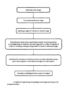
[0075] Figure 8 shows one preferred method of segmenting and grading according
to
the present invention. The method comprises:
a. receiving a test image
b. pre-processing the test image
c. selecting a region of interest on the test image
d. calculating non-local means patch-based weights comparing patches
surrounding pixels of interest in the test image with a number of
patches of pixels surrounding a corresponding number of pixels in
reference images
e. calculating for the pixels of interest at least one state estimation using
a
given state assigned to the reference images and the weights.
f. providing a pathological status score of a subject
[0076] It will be appreciated that most aspects of the above method can be
performed
by a computer using software programmed to carry out the described method (see
figure 11).
[0077] Figure 9 is a block diagram illustrating one possible physical setup of
the
present invention. In this setup, a subject is placed inside an image
generation device
(in this case, an MRI machine) to generate an image of his brain. The imaging
is
performed by radio frequency emitters/sensors that are placed inside the MRI
machine. The RF sensors send data to an image acquisition system 12 for
acquiring
24
CA 02752370 2011-09-16
data that will be used to generate images of the brain. A library of reference
images
16 is compared to the test image in the processing step to determine grading
and/or
volume of a structure for state determination. The image can be pre-processed
and
processed in a processor 14. After the various processing steps occurring in
the
processor 14 (shown in more detail in Figure 11), the image-data-status-score
is
ready to be viewed on an viewer 18a or transmitted via a data transmitter 18b.
In
some cases, the test image belongs to a subject for which a medical diagnosis
has
been reliably obtained. In such cases, the test image can be directly
incorporated into
the Library of reference images or the processor can seed the library with the
images
for which the diagnosis is known. The method and apparatus of the present
invention
rely critically on the reference images in the library and the more images are
used in
the calculations, the more reliable is the pathological status score, state
and volume
estimation. It is therefore advantageous to increase the number of reference
images
for which a medical diagnosis is known. One way to achieve this would be to
anonymyze the test images with a code such that when a patient receives a
medical
diagnosis, the reference library is automatically updated with the
information.
[0078] Figure 10 shows an alternate embodiment of the present invention where
the
image generation device 10 and the image acquisition system 12 are not in the
same
physical location as the processor 14, and wherein an image acquisition system
12
sends via the data transmitter 18b the image data through a network 20 (such
as the
intemet). The data output from the transmitter can be returned to the image
acquisition system 12 or to an alternate location 22, such as a doctor's
computer/office or the subject's computer/home.
[0079] Figure 11 shows a block diagram illustrating a physical embodiment of a
processor 14 for calculating segmentation and grading of medical images. In
this
processor, a test image and a plurality of reference images are received and
pre-
processed, non-local means patch-based weight calculator generates weighted
image data that is used by the test image grade calculator and possibly the
test
image label calculator. All pixels of the patch located in the region of
interest of an
MRI image graded and possibly labelled (structure or non-structure) for
segmentation. The grade data is used (possibly in conjunction with the volume
data)
CA 02752370 2011-09-16
to calculate a pathological status score, thus informing a subject about
whether his
"structure" correlate's more with those of healthy or diseased reference image
structures
[0080] It has been observed that, some cases, EC atrophy appears earlier than
HC
atrophy and thus could be a better temporal predictor (biomarker) of MCI
and/or AD.
[0081] It will be appreciated that non-local mean refers to the method of
Buades for
denoising images presented in (reference Buades 2005)
[0082] It will be appreciated that other structures can be used to improve
status
estimation results and the method of the present invention can be applied to
other
diseases. It is understood that the term structure is not limited to the brain
and can be
any structure identified in an image. Without limiting structures that can be
identified
in an image, the structure can be, for example, a hippocampus, an entorhinal
cortex,
a nuclei, an organ, a muscle, a breast, a blood vessel, a gland, a cartilage,
a ligament
and a bone.
[0083] It will be appreciated that, throughout this description the term state
includes
the state of a disease (including health), as well as a state of being, or
not, a
structure of interest. For example, for segmentation purposes, state can refer
to
being the structure or not being the structure, and can be an all or none
value
whereas for a pathological status estimator, the state can be degree of
disease.
[0084] In other embodiments, the structure can actually be void of any tissue
and thus
defined by its inner or outer surface. The structure can also be a space
filled with a
fluid (cerebro-spinal fluid) such as in the ventricles.
[0085] It will be appreciated that the term subject refers to any person whose
image
has been subjected to the method or apparatus of the present invention. The
subject
can be healthy or diseased. The "pathological status" is a continuum from
completely
healthy to completely diseased. Healthy subjects can perform longitudinal
studies
according to the present invention to estimate their health and this should be
understood as calculating a pathological status that can result in a status
of:
26
CA 02752370 2011-09-16
completely healthy. It will
be appreciated that, in some embodiments, the
pathological status can be prognostic rather than diagnostic.
[0086] It will be appreciated that the method and apparatus of the present
invention
can be used in longitudinal or multi-modal studies in order to determine, for
example,
tumor size/growth rate/progression.
[0087] Although the present document presents one way of selecting and
weighing
patches, many other methods could be used to achieve this goal. For example,
patch
selection and weighting can be based on subject's age, gender or other
clinical data
such as MMSE score, genetic phenotype, or other clinical data.
[0088] Reference images can be obtained, among others, from a template
library,
from a collection of pre-labelled datsets, from a collection of datasets from
subjects
with known pathological states.
[0089] In some embodiments of the present invention, the test image subject's
age
can be matched to the reference images subjects' age (library) in order to
increase
efficiency of the state.
[0090] Averaging a grade (g) within a structure may be sub-optimal and more
optimal
weighing (e.g. multi-variate logistic regression) could be found to weigh both
within
anatomical structures (i.e., anterior part of HC is more diagnostic) or
between
structures. For example, it has been observed that the Cal, Ca4 and subicular
regions of the hippocampus offer a better clinical status estimate. Weightings
could
be optimized over scales and regressions of grade versus time-to-conversion
(or
time-to-event) could be used to estimate time to convert from MCI to AD.
[0091] The terms Pixel and Voxel are used interchangeably in this document and
the
invention works in 2 dimensions (2D), 3 dimensions (3D) and n dimensions using
either a single modality or multiple modalities. The image can be multi-
dimensional,
for example a 2D set of pixels, a 3D set of voxels, a 3D dataset comprising of
20
pixels acquired over time, a 4D dataset of 3D voxels over time, a 40 dataset
of 3D
voxels where each voxel is represented by a spectrogram.
27
CA 02752370 2011-09-16
[0092] The term network should be understood as including internal networks,
the
internet and any displacement of any type of physical media such as CDs and
flash
memory from one place to another.
[0093]The term image in the present invention refers to any image such as an
image
generated in a magnetic resonance imaging (MRI), positron-emission tomography
(PET), computerized tomography (CT), fluoroscopy, X-ray, etc.
[0094] While the invention has been described in connection with specific
embodiments thereof, it will be understood that it is capable of further
modifications
and this application is intended to cover any variations, uses, or adaptations
of the
invention following, in general, the principles of the invention and including
such
departures from the present disclosures as come within known or customary
practice
within the art to which the invention pertains and as may be applied to the
essential
features herein before set forth, and as follows in the scope of the appended
claims.
References
Buades, A., Coll, B., Morel, J.M., 2005. A non-local algorithm for image
denoising.
2005 IEEE Computer Society Conference on Computer Vision and Pattern
Recognition, Vol 2, Proceedings, 60-65.
Chupin, M., Gerardin, E., Cuingnet, R., Boutet, C., Lemieux, L., Lehericy, S.,
Benali,
H., Garnero, L., Colliot, 0., 2009a. Fully automatic hippocampus segmentation
and
classification in Alzheimer's disease and mild cognitive impairment applied on
data
from ADNI. Hippocampus 19, 579-587.
Collins, D.L., Pruessner, J.C., 2010. Towards accurate, automatic segmentation
of
the hippocampus and amygdala from MRI by augmenting ANIMAL with a template
library and label fusion. Neuroimage 52, 1355-1366.
Colliot, 0., Chetelat, G., Chupin, M., Desgranges, B., Magnin, B., Benali, H.,
Dubois,
B., Garnero, L., Eustache, F., Lehericy, S., 2008. Discrimination between
Alzheimer
28
CA 02752370 2011-09-16
disease, mild cognitive impairment, and normal aging by using automated
segmentation of the hippocampus. Radiology 248, 194-201.
Coupe, P., Fonov, V., Eskildsen, S., Manjon, J., Arnold, D., Collins, L.,
2011a.
Influence of the training library composition on a patch-based label fusion
method:
Application to hippocampus segmentation on the ADNI dataset. Alzheimer's and
Dementia 7, S24-S24.
Coupe, P., Manjon, J.V., Fonov, V., Pruessner, J., Robles, M., Collins, D.L.,
2010.
Nonlocal patch-based label fusion for hippocampus segmentation. Med Image
Comput Comput Assist Intery 13, 129-136.
Coupe, P., Manjon, J.V., Fonov, V., Pruessner, J., Robles, M., Collins, D.L.,
2011b.
Patch-based segmentation using expert priors: application to hippocampus and
ventricle segmentation. Neuroimage 54, 940-954.
Coupe, P., Yger, P., Prima, S., Hellier, P., Kervrann, C., Barillot, C., 2008.
An
optimized blockwise nonlocal means denoising filter for 3-D magnetic resonance
images. IEEE Trans Med Imaging 27, 425-441.
Cuingnet, R., Gerardin, E., Tessieras, J., Auzias, G., Lehericy, S., Habert,
M.O.,
Chupin, M., Benali, H., Colliot, 0., 2010. Automatic classification of
patients with
Alzheimer's disease from structural MRI: A comparison of ten methods using the
ADNI database. Neuroimage.
Du, A.T., Schuff, N., Amend, D., Laakso, M.P., Hsu, Y.Y., Jagust, W.J., Yaffe,
K.,
Kramer, J.H., Reed, B., Norman, D., Chui, RC., Weiner, M.W., 2001. Magnetic
resonance imaging of the entorhinal cortex and hippocampus in mild cognitive
impairment and Alzheimer's disease. J Neurol Neurosurg Psychiatry 71, 441-447.
Frisoni, G.B., Fox, N.C., Jack, C.R., Scheltens, P., Thompson, P.M., 2010. The
clinical use of structural MRI in Alzheimer disease. Nature Reviews Neurology
6, 67-
77.
Gerardin, E., Chetelat, G., Chupin, M., Cuingnet, R., Desgranges, B., Kim,
H.S.,
Niethammer, M., Dubois, B., Lehericy, S., Garnero, L., Eustache, F., Colliot,
0.,
29
CA 02752370 2011-09-16
2009. Multidimensional classification of hippocampal shape features
discriminates
Alzheimer's disease and mild cognitive impairment from normal aging.
Neuroimage
47, 1476-1486.
Nyul, L.G., Udupa, J.K., 2000. Standardizing the MR image intensity scales:
making
MR intensities have tissue specific meaning. Medical Imaging 2000: Image
Display
and Visualization 1, 496-504
Pruessner, J.C., Kohler, S., Crane, J., Pruessner, M., Lord, C., Byrne, A.,
Kabani, N.,
Collins, D.L., Evans, AC., 2002. Volumetry of temporopolar, perirhinal,
entorhinal
and parahippocampal cortex from high-resolution MR images: considering the
variability of the collateral sulcus. Cereb Cortex 12, 1342-1353.
Wolz, R., Heckemann, RA., Aljabar, P., Hajnal, J.V., Hammers, A., Lotjonen,
J.,
Rueckert, D., 2010. Measurement of hippocampal atrophy using 4D graph-cut
segmentation: application to ADNI. Neuroimage 52, 109-118.
Yushkevich, P.A., Wang, H., Pluta, J., Das, S.R., Craige, C., Avants, B.B.,
Weiner,
M.W., Mueller, S., 2010. Nearly automatic segmentation of hippocampal
subfields in
in vivo focal T2-weighted MRI. Neuroimage 53, 1208-1224.
Zhang, D., Wang, Y., Zhou, L., Yuan, H., Shen, D., 2011. Multimodal
classification of
Alzheimer's disease and mild cognitive impairment. Neuroimage 55, 856-867.