Note: Descriptions are shown in the official language in which they were submitted.
CA 02752668 2011-08-16
WO 2010/096929 PCT/CA2010/000277
METHOD FOR DETECTING METASTASIS OF GI CANCER
[000] This application claims priority from US provisional applications
61/155,172 filed
on Feb. 25, 2009 and 61/246,197 filed on Sept. 28, 2009, the content of which
is herein
incorporated by reference in their entirety.
Field of the Invention
[001] The present invention relates generally to a method for detecting a
biomarker
target in a sample obtained from a patient. In particular, the present
invention provides a
method for detecting the presence of Guanylyl cyclase C (GCC or GUCY2C)
expressing
cells in human tissues or biological fluids where GCC is not normally
expressed. More
particularly, the present invention provides a method for detecting the
presence of
metastatic cancer cells originating from cancerous lesions of the gastro-
intestinal (Cl)
tract, particularly of colorectal cancer type, in a lymph node, blood, or
another tissue
sample obtained from a patient. Also, the invention relates to the use of RT-
qPCR
methods for the quantification of GCC mRNA for the staging of cancer patients
or to
predict the likelihood that colorectal cancer patients will relapse after
curative surgery.
Background of the invention
[002] The following discussion of the background of the invention is simply
provided to
aid the reader in understanding the invention.
[003] Colorectal cancer (CRC) is the second most common cause of death in
developed
countries despite significant changes in the understanding of the disease and
treatment.
Nevertheless, most treatment options for early stage colorectal cancer are
inefficient and
the current treatment paradigm is unclear. CRC is one of many diseases for
which there is
a tight connection between staging and outcome. Once a patient is diagnosed
with CRC,
the likelihood of a recurrence is related to the degree of tumor penetration
through the
bowel wall and the presence or absence of nodal involvement. These
characteristics are
the basis of the CRC staging system defined by the American Joint Committee on
Cancer
(AJCC). Pathologists, oncologists and surgeons show a great deal of interest
in detecting
metastases in lymph nodes, as lymph node involvement is a strong prognostic
factor in
many solid tumors. While histopathology (HP) remains the mainstay of CRC
staging,
lymph node (LN) examination can be difficult in the presence of single or even
small
-1-
CA 02752668 2011-08-16
WO 2010/096929 PCT/CA2010/000277
clumps of tumor cells from other cell types. The current method to identify
the presence of
CRC metastases in LNs is histopathological examination of tissue sections
stained with
Hematoxylin and Eosin (H&E). This technique is limited by the fact that only a
small
proportion, typically one or two tissue sections of 4-5 pm, of each LN is
usually assessed
for the presence of CRC metastases, leaving most volume (typically > 99%) of
each node
unexamined. As a result, predicting outcome for CRC patients considered free
of lymph
node metastases by HP examination remains challenging as approximately 20% of
these
patients will develop disease recurrence. There is clearly a need to uncover
better
techniques to identify presence of metastatic cancer cells of the GI tract.
[004] To improve the postoperative outcome of CRC patients, both guanylyl
cyclase C
(aka GUCY2C, more commonly GCC, also known as ST receptor) and its alternative
transcript, CRCA-1, have been demonstrated to be good indicators of the
presence of
metastatic colorectal cancer cells in extra-intestinal/colorectal tissues or
bodily fluids. The
transcription product of the GCC gene is uniquely expressed by intestinal
epithelium and
is endogenous downstream target of the transcription factor CDX2. The
expression of
GCC is preserved throughout the transition from adenoma to carcinoma in
colorectal
tissues. More recently, screening and diagnostic methods based on guanylyl
cyclase C or
on CRCA-1 for primary and/or metastatic stomach or esophageal cancer have been
disclosed. See particularly: US 5,601,990, US 5,731,159, US 5,928,873; US
6,060,037;
US 6,120,995; US 6,602,659; US6,767,704; US 7,135,333; US 7,316,902 and US
7,402,401.
[005] Clinical studies have demonstrated that the detection of GCC by reverse
transcription and real-time quantitative polymerase chain reaction (RT-qPCR)
is indicative
of the presence of metastatic cancer cells in lymph nodes (LN) that were
histopathologically negative in patients with colorectal cancer (CRC). These
studies,
including one described by Schulz (Clin Cancer Res. 2006 Aug 1;12(15):4545-52)
and
one recently published by Waldman (JAMA, 2009; 301(7):745-752), are based on
the
detection of the GCC biomarker ("target") in frozen specimens of partial (e.g.
approximately half) lymph nodes.
[006] The use of GCC and other targets in the diagnosis, staging and
monitoring of
cancer requires the development of effective and efficient systems, processes
and
methods which utilize fresh or archived tissue samples. Currently, the
majority of
-2-
CA 02752668 2011-08-16
WO 2010/096929 PCT/CA2010/000277
pathology samples are preserved in fixed, paraffin-embedded (FPE) tissue
blocks as a
standard archival method that allows tissue morphology preservation for years
at ambient
temperature. In order for a test using GCC as a biomarker target to have an
impact on
clinical management, such test needs to be compatible with FPE tissue
preservation.
However, such samples represent a major technical issue because the fixation
process is
known to degrade nucleic acids through protein cross-linking or oxidation over
time due to
addition of mono-methyl groups to nucleic acid bases. Consequently, methods
for
detecting metastatic cancer cells must be migrated to more robust clinical
platforms such
as those enabling reverse transcription and real-time quantitative polymerase
chain
reaction (RT-qPCR). RNA degradation and chemical modifications that occur in
FPE
clinical samples can affect target detection accuracy in RT-qPCR.
[007] The applicant has previously developed a clinical test for colorectal
cancer staging
that uses the ability of RT-qPCR to detect very small fragments of GCC mRNA to
accommodate specimens with degraded RNA. The GCC duplex assay, using the
ScorpionsTM technology for monitoring the qPCR reaction, with beta-actin ([3-
actin or
ACTB) as an internal control RNA, circumvents most of the frequent problems
associated
with absolute quantification by taking into account the efficiency of the
reverse
transcriptase and PCR reactions and monitoring for the presence of reverse
transcriptase
and PCR inhibitors. However, because of its high abundance, ACTB needs to be
adjusted to prevent competition with GCC in the duplex assay. Therefore, use
of ACTB
as a reference gene to monitor RNA degradation or RNA input is currently
limiting
because the assay is insensitive to variations such as time-dependent
degradation and
harsh fixation conditions. In this context, only highly degraded samples
and/or highly
inhibited samples and/or samples with extremely low RNA concentrations produce
ACTB
Ct values below an established limit of detection (LOD). Because GCC is more
affected
than ACTB by all these stress conditions, the current absolute quantification
requires that
all samples be free of any inhibitors, have similar degradation status and RNA
input, thus
lowering sensitivity and accuracy of the assay.
[008] There is therefore a need for a sensitive and/or accurate and/or
repeatable and/or
cost-effective method and system for detecting a target, such as GCC, in a
sample
obtained from a patient.
-3-
CA 02752668 2011-08-16
WO 2010/096929 PCT/CA2010/000277
[009] Unfortunately, simply measuring transcript levels of one or more
prognostic RNA
transcripts does not account to produce a diagnostic test of sufficient
sensitivity and
specificity to determine a clinical outcome associated with molecular markers.
Raw data
obtained from real-time PCR must be processed to obtain the relative quantity
of target
mRNA. Absolute quantification requires a standard curve in each experiment in
order to
determine by interpolation the number of mRNA copies in a given sample
relative to
known amounts of a synthetic DNA or RNA transcript. Relative quantification
(delta Ct
(ACt)) may be used to determine the changes in mRNA level of a target gene
(i.e. GCC)
across samples by expressing this change in relation to the levels of an
endogenous
reference gene (RG; sometimes referred to as housekeeping gene), used as an
internal
control RNA. One such well known and often-used reference gene is ACTB.
Alternatively,
the difference (delta-delta Ct (DACt)) between the average delta Ct (ACt)
value of a target
sample and the average delta-Ct (ACt) for the corresponding control sample is
used to
calculate expression fold change value.
[010] Evaluation of stable endogenous reference genes in clinical samples is a
prerequisite to precise and accurate normalization of relative gene expression
using RT-
qPCR platform or other related amplification methods. The use of a single so-
called
"universal" reference gene may lead to misinterpretation of the expression of
the GCC
gene.
[011] There is also a need for the identification of a reference gene to be
used in
conjunction with GCC for the detection of CRC cells in a patient's sample,
this reference
gene having an expression not affected by the presence of cancer cells in a
lymph node,
and a behavior similar to GCC in samples degraded because of long storage
periods,
poor storage conditions or other stress factors. Many such reference genes
have been
investigated and found to be useful in different contexts. However, there is
presently a
consensus that the optimal, universal reference gene does not exist (Green et
al. 2009,
Diagn Mol Pathol 18 (4), 243-249 and ref 11 therein).
Summary of the invention
[012] It has now been found that beta-glucuronidase (GUSB) is particularly
useful as a
reference gene for normalization of PCR data. In the particular context of
evaluating GCC
for GI tract cancers diagnosis and/or prognosis, GUSB provides unforeseen
advantages
-4-
CA 02752668 2011-08-16
WO 2010/096929 PCT/CA2010/000277
such as allowing one to measure much lower limits of detection (LOD) than
could be
obtained with other reference genes.
[013] The invention provides a method for the detection of cancer cells or GCC
expressing cells in a sample, which method detects and/or measures/quantifies
GCC from
processed harvested tissue or biological fluid, in combination with the
detection and/or
quantification of one or more reference genes in the same sample. GUSB (beta-
glucuronidase) was found to be a superior reference gene. Particularly, GUSB
was found
to be a superior reference gene to be used in complement to GCC in the
detection of CRC
cells in lymph nodes. RT and PCR reactions were designed to obtain an
efficient duplex
test simultaneously amplifying GCC and GUSB mRNAs. The analytical performance
of
this test was verified, showing better strength compared to the GCC/ACTB test.
These
better analytical characteristics (higher informative rate, higher analytical
sensitivity and
relative quantification) of the GCC/GUSB test can lead to a more accurate
stratification of
the recurrence risk (RR) when tested with a population of patients diagnosed
with Stage II
CRC.
[014] The invention also provides a diagnostic method for detection of GCC in
a sample
collected from a patient, comprising the following steps: detecting GCC in the
sample;
detecting (either simultaneously or sequentially) beta-glucuronidase (GUSB) in
the same
sample, and establishing relative quantification of GCC versus GUSB wherein
presence of
GCC versus GUSB above a fixed threshold is indicative of the presence of GCC
positive
cells in the sample.
[015] According to a general aspect, there is provided a method of measuring
GCC in a
sample collected from a patient, comprising the following steps: measuring GCC
in the
sample by RT-qPCR to establish a CtGCC; measuring GUSB in the same sample RT-
qPCR to establish a CtGUSB; and establishing relative quantification (delta-
Ct) of CtGUSB
minus CtGCC wherein a delta-Ct of above about -12 is indicative of the
presence of GCC
positive cells in the sample.
[016] The invention also provides a diagnostic method for the detection of GCC
that
uses the expression fold change (delta-delta-Ct) to determine the changes in
mRNA level
of GCC across samples by expressing this change in relation to the RNA levels
of GUSB.
-5-
CA 02752668 2011-08-16
WO 2010/096929 PCT/CA2010/000277
[017] According to another aspect of the invention, there is provided a method
of staging
or monitoring a patient already diagnosed with GI tract cancer, comprising the
steps of:
detecting GCC in the sample (consisting of human tissues or biological fluids
in which
GCC is not normally expressed, such as particularly extra-
colorectal/intestinal tissue);
detecting GUSB in the same sample; and establishing a relative quantification
(delta-Ct)
of CtGUSB - CtGCC, wherein a delta-Ct of above -12 is indicative of the
presence of GCC
positive cells in the sample, wherein the presence of GCC positive cells is
indicative of
metastasized colorectal, stomach, small intestinal, pancreatic or esophageal
cancer.
[018] According to another aspect of the invention, there is provided a method
of
diagnosing a patient suspected of having a primary stomach, esophageal or
pancreatic
cancer, comprising the steps of: measuring GCC in an extra-
intestinal/colorectal sample;
measuring GUSB in the same sample; and establishing a relative quantification
(delta-Ct)
of CtGUSB - CtGCC, wherein a delta-Ct of above -12 is indicative of the
presence of GCC
positive cells in the sample, wherein the presence of GCC positive cells is
indicative of a
primary stomach, esophageal or pancreatic cancer.
[019] The method of the present invention allows detection of the presence or
absence
of GCC in lymph nodes harvested following a stomach, small intestine,
esophageal,
pancreatic or colorectal resection, thereby allowing molecular staging of a
cancer.
According to another aspect of the invention, there is provided a method,
comprising the
steps described above, to discriminate between cancer patients with
histopathologically
negative and histopathologically positive lymph nodes.
[020] According to this method, cancer patients with GCC positive cells in one
or several
of their lymph nodes have a risk of recurrence and survival comparable to
those of
patients considered as having a higher risk by histopathology, thereby
indicating that
these patients might benefit from treatment with adjuvant chemotherapy.
According to this
method, cancer patients with all LNs negative for GCC are at a lower risk of
disease
recurrence and would not require adjuvant chemotherapy, consequently avoiding
the
negative side effects of these treatments.
[021] The method of the present invention detects the presence of GCC in blood
from
patient previously diagnosed with cancer patients to predict the risk of
recurrence, monitor
the recurrence and the response to cancer therapy of the cancer. According to
another
aspect of the invention, there is provided a method of prognosticating a
patient with
-6-
CA 02752668 2011-08-16
WO 2010/096929 PCT/CA2010/000277
cancer, comprising the steps as defined above, wherein the presence of GCC
positive
cells is indicative of a poor prognosis.
[022] In accordance with another aspect of the present invention, there is
provided a
method of determining if a patient already diagnosed with GI tract cancer will
benefit from
a treatment, comprising the steps of: measuring the expression level of GCC in
an extra-
intestinal/colorectal sample collected from the patient; measuring the
expression level of
GUSB in the same extra-intestinal/colorectal sample; and determining the
quantity of GCC
relative to the quantity of GUSB in the extra-intestinal/colorectal sample;
wherein if the
quantity of GCC in the extra-intestinal/colorectal sample is above a given
level when
compared to GUSB, the patient will benefit from the treatment.
[023] Particularly, the invention provides a method of determining the
quantity of GCC in
an extra-intestinal/colorectal sample collected from a patient already
diagnosed with GI
tract cancer, comprising the steps of : measuring the expression level of GCC
mRNA in
the sample; measuring the expression level of GUSB mRNA in the same sample;
and
using a mathematical calculation to normalize the expression level of GCC mRNA
to the
expression level of GUSB mRNA to next establish a relative GCC expression
(GUSB level
- GCC level); wherein if the relative GCC expression is higher than the limit
of detection of
an external standard, the absolute quantity of GCC in the sample is calculated
and
expressed in number of GCC copies.
[024] Particularly, the invention provides a method of predicting the
likelihood of cancer
recurrence in a patient already diagnosed with GI tract cancer, comprising the
steps of:
determining the quantity of GCC as defined above in one or more lymph nodes
collected
from the patient; classifying each of the one or more lymph nodes as GCC-
negative or
GCC-positive; and establishing a lymph node ratio, wherein the lymph node
ratio is the
number of GCC-positive nodes over the total of GCC-negative and GCC-positive
lymph
nodes; whereby the larger the lymph node ratio means the greater the
likelihood of cancer
recurrence.
[025] Particularly, the invention provides a method of predicting the
likelihood of cancer
recurrence in a patient already diagnosed with GI tract cancer, comprising the
steps of:
measuring the expression level of GCC in one or more lymph nodes collected
from the
patient; measuring the expression level of GUSB in the same one or more lymph
nodes;
and determining the quantity of GCC relative to the quantity of GUSB in each
individual
-7-
CA 02752668 2011-08-16
WO 2010/096929 PCT/CA2010/000277
lymph nodes; wherein a relative quantity of GCC above a pre-established cut-
off is
indicative of the presence of GCC in a lymph node, whereby if GCC is present
in one
lymph node or more, the patient has an increased likelihood of cancer
recurrence.
[026] Particularly, the pre-established cut-off level is between about -6 and -
3. More
particularly, the cut-off level is selected from the group consisting of: -
5.9, -5.5, -5.0; -4.5; -
4.0; -3.5; and -3Ø
[027] Particularly, the invention provides a method of predicting the
likelihood of cancer
recurrence in a patient already diagnosed with GI tract cancer, comprising the
steps of:
quantifying by RT-qPCR RNA levels of GCC in an extra-intestinal/colorectal
sample
collected from the patient to establish a cycle threshold for GCC (CtGCC);
quantifying by
RT-qPCR RNA levels of GUSB in the same sample to establish a cycle threshold
for
GUSB (CtGUSB); and calculating a relative quantification of CtGUSB minus CtGCC
(delta-Ct);
wherein a delta-Ct equal or higher than about -6 is indicative of the presence
of GCC
positive cells in the sample, whereby the presence of GCC positive cells is
indicative that
the patient has increased risk of recurrence of cancer.
[028] Particularly, the invention provides a method of determining if a
patient already
diagnosed with cancer has GCC nodal involvement, comprising the steps of:
measuring
the expression level of GCC in a lymph node collected from the patient to
establish a
cycle threshold for GCC (CtGCC); measuring the expression level of GUSB in the
same
lymph node collected from the patient to establish a cycle threshold for GUSB
(CtGUSB);
and determining a relative quantification of CtGUSB minus CtGCC (delta-Ct);
wherein if delta-
Ct is equal or higher than about -6, the lymph node is GCC-positive.
[029] Particularly, the invention provides a method of determining the GCC
burden of a
patient already diagnosed with cancer, comprising the steps of. measuring the
expression
level of GCC mRNA in an extra-intestinal/colorectal sample collected from the
patient to
establish a cycle threshold for GCC (CtGCC); measuring the expression level of
GUSB in
same extra-intestinal/colorectal sample collected from the patient to
establish a cycle
threshold for GUSB (CtGUSB); and calculating a relative quantification of
CtGUSB minus
CtGCC (delta-Ct); wherein if delta-Ct is equal or higher than about -12, the
quantity of GCC
mRNA may be calculated in terms of number of copies, whereby the GCC burden is
expressed in number of GCC copies in the sample.
-8-
CA 02752668 2011-08-16
WO 2010/096929 PCT/CA2010/000277
[030] Particularly, the invention provides a method of staging or monitoring a
patient
already diagnosed with colorectal cancer, comprising the steps: obtaining an
extra-
intestinal/colorectal sample taken from the patient; measuring GCC in the
sample to
establish a cycle threshold for GCC (CtGCC); measuring GUSB in the same sample
to
establish a cycle threshold for GUSB (CtGUSB); and establishing relative
quantification
(delta-Ct) of CtGUSB minus CtGCC, wherein a delta-Ct of higher than about -6
is indicative of
the presence of GCC positive cells in the sample, wherein the presence of GCC
positive
cells is indicative of metastasized colorectal cancer.
[031] According to another aspect of the invention, the detection of the
presence of GCC
positive cells in tissues harboring metastases of an unknown origin (CUP) is a
confirmation that the primary cancer is a colorectal, a stomach, an
intestinal, a pancreatic
or an esophageal cancer.
[032] In yet another aspect of the invention, the materials for use in the
methods of the
present invention are suited for preparation of a kit. According to another
aspect of the
invention, there is provided a kit for the detection, diagnosis, prognosis
and/or staging of a
cancer in a patient, wherein the kit comprises: PCR reagents for detecting GCC
in the
sample, PCR reagents for detecting GUSB in the same sample, and instructions
on how
to calculate (delta-Ct) or (delta-delta-Ct) between GCC and GUSB.
[033] The invention also provides kits comprising reagents, which may include
gene-
specific probes and/or primers, for quantifying the expression of the
disclosed genes for
predicting the likelihood of developing disease recurrence. Such kits may
optionally
contain reagents for the extraction of RNA from a sample, in particular fixed
paraffin-
embedded tissue samples and/or reagents for RNA amplification.
Detailed description of the invention
Brief description of the figures
[034] The accompanying figures, which are incorporated in and constitute a
part of this
specification, illustrate various embodiments of the present invention, and,
together with
the description, serve to explain the principles of the invention. In the
figures:
[035] Figure 1 represents the expression levels of putative reference genes,
presented
as average Ct values in matched fresh frozen (FF) and FFPE colon cancer LNs;
-9-
CA 02752668 2011-08-16
WO 2010/096929 PCT/CA2010/000277
[036] Figure 2 represents the expression levels of putative reference genes,
presented
as average Ct values in GCC negative and positive FFPE LNs. Targeted Ct range
was
delimited by the two dotted lines;
[037] Figure 3 represents the average expression stability values of control
genes;
[038] Figure 4 represents the determination of the optimal number of control
genes for
normalization;
[039] Figure 5 represents the expression profile of selected reference gene in
GCC
positive and negative LNs. Ct value for GCC, GUSB, HPRT1, PGK1 and TBP in FFPE
samples tested in simplex reaction with 312.5 ng of cDNA. In minus RT
experiments, no
RT enzyme was added during the cDNA synthesis step;
[040] Figure 6 represents the effect of NaOH treatment on RNA quality and gene
expression measures. The extent of RNA degradation following NaOH treatment
was
determined by capillary electrophoresis using the Agilent 2100 Bioanalyzer.
Panel A)
show a representative RNA fragmentation profile compared to intact RNA
isolated from
fresh frozen colon tissues and matched FFPE material. B) Ct values for GCC and
5
reference gene assays including ACTB ScorpionsTM were determined at each time
point
of hydrolysis;
[041] Figure 7 represents the GCC expression variation (delta-delta-Ct) during
NaOH
degradation using five reference genes (GUSB, HPRT1, PGK1, TBP and ACTB) for
normalization. Variations in delta Ct values (delta-delta-Ct = [(Ctccc-CtRC)-
(Ctccc-CtRC)tol
were determined at each time point of hydrolysis. Error bar represent SD from
3
independent RNA pools. To perform comparison between three experiments, the
threshold for each gene was manually fixed to 0.10;
[042] Figure 8 represents the effect of carbonate buffer alkaline treatment on
RNA
quality and gene expression pattern. The quality of RNA isolated at indicated
time points
was measured by capillary electrophoresis using the Agilent 2100 Bioanalyzer.
A)
Representative RNA fragmentation profile compared to intact RNA isolated from
fresh
frozen colon tissues. B) Electropherograms of RNA sample. Each time point
contained an
equal amount of material. C) Ct values for GCC and 5 reference gene assays,
including
the GCC/ACTB duplex assay;
-10-
CA 02752668 2011-08-16
WO 2010/096929 PCT/CA2010/000277
[043] Figure 9 represents the GCC expression variation (delta-delta-Ct)
following
controlled RNA degradation in carbonate buffer alkaline conditions. Variation
in delta Ct
values (delta-delta-Ct = [(Ctccc-CtRC)tx-(Ctccc-CtRC)to] were determined at
each time point
of hydrolysis;
[044] Figure 10 represents the gene expression profiling in frozen and fixed,
paraffin-
embedded (FPE) tissues using TaqMan simplex assays and the GCC/ACTB duplex
assay. The quality of RNA isolated from each condition was measured by
capillary
electrophoresis using the Agilent 2100 Bioanalyzer (Condition A and E are
TRIzoITM
extract, B: Neutral buffered formalin 10%, C: Non-buffered formalin 10%; D:
Bouin's
solution). Expression (Ct values) of GCC and 4 reference genes (GUSB, HPRT1,
PGK1
and TBP) in TaqMan simplex reactions was compared to the GCC/ACTB duplex
assay;
[045] Figure 11 shows the comparison of GCC expression levels (delta-Ct) in
cryo-
sections of colon cancer tissue samples fixed or not. Variation in delta Ct
values (delta-Ct
= (CtGCC-CtRG)) were determined for each RNA extract. The reverse
transcription reactions
were performed with the SuperscriptTM III First-Strand Synthesis SuperMix
(Invitrogen)
according to the manufacturer's recommendation, using gene-specific primers (2
pM) and
500 ng of total RNA based on Quant-IT data. The real-time PCR were carried out
in a 20-
pl reaction volume with the Applied Biosystems 7900HT Fast Real-Time PCR
Systems
using either TaqMan simplex or the GCC/ACTB duplex assay. Primers and probes
concentration for each simplex assays were 900 nM and 250 nM respectively. All
reactions were performed in duplicate;
[046] Figure 12 shows the comparison of GCC Taqman simplex and duplex
amplifications in RNA extracted from a FFPE colon tissue. Upper panel shows
GCC and
various RG PCR amplifications in FFPE samples tested in simplex and duplex
reactions
using gene-specific reverse primers at 2 pM and 1.25 pg of RNA. Lower panel
shows a
comparison of minus RT-PCR amplification for GCC and various reference genes
in
simplex and duplex reactions;
[047] Figure 13 shows the comparison of GCC and GUSB Ct values between duplex
and simplex reactions. The real-time PCR were carried out in duplex or simplex
reactions
of 20 pl with the Applied Biosystems 7900HT Fast Real-Time PCR Systems using
TaqMan Fast Universal PCR Master Mix. Synthesis of cDNA was performed with 250
ng/pl (A) or 25 ng/pl (B) of uRNA spiked or not with 1x10⁶ GCC IVT. For real-
time PCR
- 11 -
CA 02752668 2011-08-16
WO 2010/096929 PCT/CA2010/000277
reaction in simplex, primers concentration was 900 nM and FAM or VIC-labeled
probes
concentration was 250 nM. For duplex reactions, 4 concentrations of reverse
and forward
primers were tested while both FAM and VIC-labeled probes were fixed at 200 nM
in a 20
pl PCR reaction;
[048] Figure 14 represents the GCC and RG delta-Ct variation in duplex and
simplex
assays during NaOH degradation. Expression of GCC was normalized with either
GUSB
or HPRT1. Variations in GCC relative quantification (delta-delta-Ct = [(Ctccc-
CtRC)tx-
(Ctccc-CtRC)to] were determined at indicated time points during NaOH
hydrolysis. Variation
between non-degraded and degraded samples should be lower than 1 delta-Ct to
be
considered not significant;
[049] Figure 15 represents the comparison of FFPE colon cancer LNs tested with
TaqMan and ScorpionsTM duplex assays in minus RT condition. Panel A and C show
Ct
values for ACTB, GUSB, HPRT1 and GCC in 8 FFPE samples tested with 312.5 ng of
cDNA in duplex reaction. In minus RT experiments (B and D), no RT enzyme was
added
during the cDNA synthesis step;
[050] Figure 16 shows the comparison of GCC expression levels (delta-Ct) in
cryo-
sections of colon cancer tissue samples fixed or not. Delta Ct values LCt =
(CtGCC-CtRG)
were determined for each RNA extract using either GUSB (A and B) or HPRT1 (C
and D).
The reverse transcription reactions were performed in simplex using gene-
specific primers
at 2 pM and in duplex with indicated primers concentration. The real-time PCR
were
carried out in a 2Opl reaction volume with the Applied Biosystems 7900HT Fast
Real-Time
PCR Systems using either TagMan simplex or duplex assay were compared to
ScorpionsTM duplex reaction. All reactions were performed in triplicate.
Variation of less
than I delta-Ct between frozen and fixed samples was considered not
significant;
[051] Figure 17 represents the amplification of reference genes in RNA
extracted from
55 FPE pericolonic lymph node tissues with different archiving times from 1
month to 22
years. A) Capillary electrophoresis profiles of RNA extracted from archival
FPE tissues.
One pl of each RNA extract (150ng/pL) was analysed using the Agilent 2100
Bioanalyser
and RNA Nano Chips. B) Ct values for ACTB and GUSB in FPE colon lymph node
tissues. The real-time PCR were carried out in simplex reactions of 20 pl
using the
Applied Biosystems 7900HT Fast Real-Time PCR Systems and TaqMan Fast Universal
PCR Master Mix;
-12-
CA 02752668 2011-08-16
WO 2010/096929 PCT/CA2010/000277
[052] Figure 18 shows the comparison of ACTB and GUSB Ct value observed in
group
of blocks with different archiving time. Box-and-Whisker plots of the ACTB (A)
and GUSB
(B) mRNA expression (Ct value) in FPE colon lymph node tissues. For multiple
comparisons, one-way ANOVA and post-hoc Turkey's test were used and P < 0.05
was
considered statistically significant;
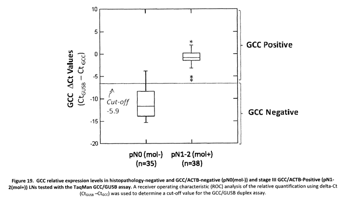
[053] Figure 19 represents the GCC relative expression levels in
histopathology-
negative and GCC/ACTB-negative (pNO(mol-)) and stage III GCC/ACTB-Positive
(pN1-
2(mol+)) LNs tested with the TaqMan GCC/GUSB assay. A receiver operating
characteristic (ROC) analysis of the relative quantification using delta-Ct
(CtGUSB -CtGCC)
was used to determine a cut-off value for the GCC/GUSB duplex assay;
[054] Figure 20 represents the GCC relative expression levels evaluated with
the
TaqMan GCC/GUSB duplex assay. GCC relative expression ACt (CtGUSB -CtGCC) in
colon
cancer stage I, II and III histopathology negative (HP-) and stage III
positive (HP+) LNs.
GCC mRNA positive status was based on the analytical cut-off value of -5.9;
[055] Figure 21 shows the average expression stability values of control genes
in blood
samples using geNorm;
[056] Figure 22 shows the determination of the optimal number of reference
genes for
normalization using geNorm;
[057] Figure 23 represents the GCC expression level in blood samples. GCC mRNA
positive status was based on the analytical cut-off value of 75 GCC units/mL;
[058] Figure 24 shows the distribution of A) ACTB Ct values and B)
Glucuronidase
GUSB Ct values for two different patient cohorts. Wilcoxon rank test p-value:
0.0001;
[059] Figure 25 shows the combination of HDQ and Mass cohorts (n=73) ROC curve
analysis for both GCC/ACTB and GCC/GUSB assays taking the highest GCC copies
of
any given LN of a case as the continuous variable. Sensitivity is defined as
the detection
rate of recurrent cases (after 36 months) while specificity is defined as the
proportion of
negative cases without recurrence;
[060] Figure 26 illustrates the relation between risk of recurrence for
patients with a
GCC positive test result and the GCC expression level used to determine test
positivity;
-13-
CA 02752668 2011-08-16
WO 2010/096929 PCT/CA2010/000277
[061] Figure 27 is a Kaplan-Meier graphical analysis of time to recurrence
based on
GCC positivity. A) GCC/ACTB test with 100 copies cut-off; B) GCC/GUSB test
with 100
copies cut-off; C) GCC/ACTB test with 25 copies cut-off; D) GCC/GUSB test with
25
copies. The GCC Negative cases are represented by the straight black line and
the GCC
Positive cases by the gray dashed line. Patients lost to follow-up were
censored-out and
are represented by straight up marks;
[062] Figure 28 is a Kaplan-Meier graphical analysis of RFS based on the GCC
positivity for the GCC/GUSB test with -5.9 ACt for cut-off. The GCC Negative
cases are
represented by the straight line and the GCC Positive cases by the dashed
line.
Censored-out cases are represented by straight up marks;
[063] Figure 29 is a Kaplan-Meier graphical analysis of time to recurrence
with 2 levels
of stratification for GCC positive patients. A) GCC/ACTB test with 100 copies
cut-off; B)
GCC/GUSB test with 100 copies cut-off; C) GCC/ACTB test with 25 copies cut-
off; D)
GCC/GUSB test with 25 copies. Number of patients at risk is also indicated for
each
group;
[064] Figure 30 is a Kaplan-Meier graphical analysis of time to recurrence
with 2 levels
of stratification for GCC positive patients. A) GCC/GUSB test with -5.9 delta-
Ct for cut-off.
Number of patients at risk is also indicated for each group. B) Comparison of
total
recurrence rate between GCC/GUSB (delta-Ct = -5.9) and GCC/ACTB (GCC copies =
25); and
[065] Figure 31 is a Kaplan-Meier graphical analysis of time to recurrence
with
stratification for the number of GCC positive LNs per patients. A) GCC/ACTB
test with 25
copies cut-off; B) GCC/GUSB test with 25 copies cut-off. Number of patients at
risk is also
indicated for each group.
Definitions
[066] To facilitate an understanding of the invention, a number of terms are
defined
below.
[067] The term "about" as used hereinbelow refers to a margin of + or - 5% of
the
number indicated. For sake of precision, the term "about" when used in
conjunction with,
for example, the integer 10, means 10 +/- 5% i.e. from 9.5 to 10.5.
-14-
CA 02752668 2011-08-16
WO 2010/096929 PCT/CA2010/000277
[068] As used herein, the term "GCC" is meant to refer to the gene
transcription product
(RNA) expressing the cellular protein guanylate cyclase 2C (GUCY2C also
referred to as
the heat stable enterotoxin receptor or ST receptor), which is expressed by
normal
colorectal cells, as well as primary and metastasized colorectal, intestinal,
stomach and
esophageal cancer cells. In normal individuals, GCC is found exclusively in
cells of
intestine, in particular in cells in the duodenum, small intestine (jejunum
and ileum), colon
(caecum, ascending colon, transverse colon, descending colon and sigmoid
colon) and
rectum. The term "GCC" also includes fragments of a GCC gene transcript which
are
functional with respect to nucleic acid molecules with full length sequence,
such as a
functional fragment which may be useful as an oligonucleotide or nucleic acid
probe, a
primer, an antisense oligonucleotide or nucleic acid molecule or a coding
sequence. The
term "GCC" also comprises the CRCA-1 alternative transcript.
[069] As used herein, the term "colorectal cancer" is meant to include the
well-accepted
medical definition that defines colorectal cancer as a medical condition
characterized by
presence of cancer cells in the intestinal tract below the small intestine
(i.e. the large
intestine (colon), including the caecum, ascending colon, transverse colon,
descending
colon, and sigmoid colon, and rectum). Additionally, as used herein, the term
"colorectal
cancer" is meant to further include medical conditions which are characterized
by
presence of cancer cells in the duodenum and small intestine (jejunum and
ileum). The
definition of colorectal cancer used herein is more expansive than the common
medical
definition but is provided as such since the cells of the duodenum and small
intestine also
contain GCC.
[070] As used herein, the term "GI tract cancer" or "gastro-intestinal cancer"
is meant to
include the medical conditions which are characterized by presence of cancer
cells in the
esophagus, the stomach, the pancreas, the small intestine as well as in colon
and rectum.
Additionally, as used herein, the term "GI tract cancer" in meant to further
include medical
conditions which are characterized by presence of cancer cells in the
pancreas, which like
liver and gallbladder is an accessory organ of the GI tract. The definition of
GI tract cancer
used herein is more expansive that the common medical definition but is
provided as such
since pancreatic cancer cells are known to express GCC.
[071] As used herein, the terms "upper GI tract" consists of the mouth cavity,
salivary
glands, pharynx, esophagus, diaphragm, stomach, gall bladder, bile duct,
liver, and
-15-
CA 02752668 2011-08-16
WO 2010/096929 PCT/CA2010/000277
duodenum. The term "upper GI tract cancer" as used herein particularly refers
to the
esophagus, stomach and pancreas.
[072] As used herein, the terms "lower GI tract" means of the bowel or
intestines and the
rectum and comprises the small intestine including duodenum, jejunum, ileum;
and the
large intestine or colon including caecum (and appendix); colon (ascending,
transverse
and descending) and the rectum (anus).
[073] As used herein, the term "stomach cancer" is meant to include the well-
accepted
medical definition that defines stomach cancer as a medical condition
characterized by
presence of cancer cells in the stomach.
[074] As used herein, the term "esophageal cancer" is meant to include the
well-
accepted medical definition that defines esophageal cancer as a medical
condition
characterized by presence of cancer cells in the esophagus.
[075] As used herein, the term "pancreatic cancer" is meant to include the
well-accepted
medical definition that defines pancreatic cancer as a medical condition
characterized by
presence of cancer cells in the pancreas.
[076] As used herein, the term "metastasis" is meant to refer to the process
in which
cancer cells originating in one organ or part of the body, with or without
transit by a body
fluid, and relocate to another part of the body and continue to replicate.
Metastasized cells
can subsequently form tumors which may further metastasize. Metastasis thus
refers to
the spread of cancer, from the part of the body where it originally occurred,
to other parts
of the body.
[077] As used herein, the term "metastasized colorectal cancer cells" is meant
to refer to
colorectal cancer cells which have metastasized. Metastasized colorectal
cancer cells are
localized in a part of the body or body fluid other than the duodenum, small
intestine
(jejunum and ileum), large intestine (colon), including the caecum, ascending
colon,
transverse colon, descending colon, and sigmoid colon, and rectum.
[078] As used herein, the term "metastasized stomach cancer cells" is meant to
refer to
stomach cancer cells which have metastasized. Metastasized stomach cancer
cells are
localized in a part of the body other than the stomach.
-16-
CA 02752668 2011-08-16
WO 2010/096929 PCT/CA2010/000277
[079] As used herein, the term "metastasized esophageal cancer cells" is meant
to refer
to esophageal cancer cells which have metastasized. Metastasized esophageal
cancer
cells are localized in a part of the body other than the esophagus.
[080] As used herein, the term "metastasized pancreatic cancer cells" is meant
to refer
to pancreatic cancer cells which have metastasized. Metastasized pancreatic
cancer cells
are localized in a part of the body other than the pancreas.
[081] As used herein, the terms "non-intestinal/rectal" and "extra-
intestinal/colorectal"
are used herein interchangeably and are meant to refer to a sample of tissue
or body fluid
from a source other than intestinal (small intestine and colon) and rectal
tissue. In some
preferred embodiments, the extra-intestinal/colorectal sample is a sample of
tissue such
as lymph nodes. In some preferred embodiments, the non-intestinal/rectal
sample is a
sample of extra-intestinal/colorectal tissue which is an adenocarcinoma of
unconfirmed
origin. In some preferred embodiments, the non-intestinal/rectal tissue is a
biopsy of a
suspected stomach, pancreatic or esophagus cancer. In some preferred
embodiments,
the non-intestinal/rectal sample is a blood sample.
[082] As used herein, "an individual suffering from an adenocarcinoma of
unconfirmed
origin" or "cancer of unknown primary origin" (CUP) is meant to refer to an
individual who
has a tumor in which the origin has not been definitively identified.
[083] As used herein, the terms "subject" and "patient" refer to any animal,
such as a
mammal like livestock, pets, and preferably a human. Specific examples of
"subjects" and
"patients" include, but are not limited, to individuals requiring medical
assistance, and in
particular, patients with cancer.
[084] As used herein, the term "target" or "target marker" or "biomarker
target" refers to
any molecule that can be derived from a eukaryotic cell. Targets include but
are not
limited to proteins or nucleic acid molecules. In the present invention, the
level of a
messenger RNA that is specifically expressed in cells of gastrointestinal
origin is
measured. Alternatively, a tissue specific protein or DNA alteration (e.g.
methylation or
mutation) could be an equivalent target. In preferred embodiments of the
present
invention, single targets such as mRNA are detected individually. In
alternative
embodiments of the present invention, multiple targets are detected in
combination.
-17-
CA 02752668 2011-08-16
WO 2010/096929 PCT/CA2010/000277
[085] As used herein, the terms "reference gene" or "reference marker" or
"reference
target" or "control" or "control marker" or "control target" refers to a
reference molecule
that controls and/or can be used to control for potential process interfering
factors and/or
provides one or more indications about the sample quality, the effective
sample
preparation and/or assembly of the RT-PCR reaction in the sample. A control
may either
be co-detected or detected separately from targets.
[086] As used herein, the term "sample" refers to a biological material
containing cells or
other material retrieved from the patient. Sample material includes but is not
limited to:
tissue such as lymph node tissue; biopsy material; exhaled breath; or fluids
such as blood
(including serum or plasma); urine; semen; sputum, saliva; and combinations of
these. To
practice the methods of the present invention, the sample is processed (e.g. a
lymph node
is separated from other tissue and/or cut in multiple sections or cores,
exposed or not to a
chemical reaction, subjected to a separation process or blood is enriched in
tumor
circulating cells). Each process may result in a portion of the sample
remaining,
hereinafter referred to as "remaining sample" or simply "sample". The portions
of the
sample may be sized randomly or according to a predetermined scheme or
mathematical
formulaic determination. Sample may be defined as a single tissue sample, such
as a
single lymph node, or sample may define multiple samples, such as multiple
lymph nodes
or lymph node chain. In preferred embodiments of the present invention, single
samples
such as single lymph nodes are processed individually. In alternative
embodiments of the
present invention, multiple samples are "pooled" or processed together. In
another
preferred embodiment, the sample includes at least one entire lymph node.
[087] The term "external standard" as used herein means a synthetic DNA or RNA
transcript in known amount(s) or concentration(s) that is tested separately
from the test
sample, i.e. through interpolation or extrapolation to a standard curve.
[088] As used herein the term "parameters", also known as "process
parameters",
include one or more variables used in the method and system of the present
invention to
detect one or more targets. Parameters include but are not limited to: primer
type; probe
type; amplicon type; concentration of a substance; mass or weight of a
substance; time for
a process; temperature for a process; cycle threshold (Ct); activity during a
process such
as centrifugation, rotating, shaking, cutting, grinding, liquefying,
precipitating, dissolving,
electrically modifying, chemically modifying, mechanically modifying, heating,
cooling,
-18-
CA 02752668 2011-08-16
WO 2010/096929 PCT/CA2010/000277
preserving (e.g. for days, weeks, months and even years) and maintaining in a
still
(unagitated) state. Parameters may further include a variable in one or more
mathematical formulas used in the method of the present invention. Parameters
may
include a threshold used to determine the value of one or more parameters in a
subsequent step of the method of the present invention. In a preferred
embodiment, the
threshold is a cycle count threshold.
[089] As used herein, the term "cycle threshold" (Ct) refers to the threshold
in qPCR at
which the fluorescence generated within a reaction well exceeds an established
threshold
or cutoff level. The cycle threshold refers to the same value than the terms
"crossing point'
(Cp) and "take-off point" (TOP) used by competing manufacturers of real-time
PCR
instruments for reasons of product differentiation. For standardization
purposes, the MIQE
Guidelines (Bustin et al., Clinical Chemistry, 55:4, pp. 611-622 (2009)) have
proposed that
the use of the term "quantification cycle" (Cq) be preferred over all those
alternatives.
[090] The term "hybridization" is to be understood as a bond of an
oligonucleotide to a
complementary sequence along the lines of the Watson-Crick base pairings in
the sample
DNA, forming a duplex structure.
[091] "Stringent hybridization conditions," as defined herein, involve
hybridizing at 68 C
in 5X SSC/ 5X Denhardt's solution/ 1.0% SDS, and washing in 0.2X SSC/ 0.1 %
SDS at
room temperature, or involve the art-recognized equivalent thereof (e.g.,
conditions in
which a hybridization is carried out at 60 C in 2.5X SSC buffer, followed by
several
washing steps at 37 C in a low buffer concentration, and remains stable).
Moderately
stringent conditions, as defined herein, involve including washing in 3X SSC
at 42 C, or
the art-recognized equivalent thereof. The parameters of salt concentration
and
temperature can be varied to achieve the optimal level of identity between the
probe and
the target nucleic acid. Guidance regarding such conditions is available in
the art, for
example, by Sambrook et al., 1989, Molecular Cloning, A Laboratory Manual,
Cold Spring
Harbor Press, N. Y.; and Ausubel et al. (eds.), 1995, Current Protocols in
Molecular
Biology, (John Wiley & Sons, N. Y.) at Unit 2.10.
[092] As used herein, the expression "clinical assessment" is meant to include
a
potential range or continuous or discrete values used for the screening,
diagnosis,
staging, prognosis, treatment planning, monitoring and surveillance of a
cancer patient.
-19-
CA 02752668 2011-08-16
WO 2010/096929 PCT/CA2010/000277
[093] As used herein, the expression "clinical outcome" or "outcome" is meant
to be
expressed in terms of different endpoints such as Disease-Free Survival (DFS),
Relapse-
Free Survival (RFS), Time-to-Recurrence (TR), Cancer-Specific Survival (CSS)
or Overall
Survival (OS), in accordance with the recommendations of Punt CJ et al., J.
Natl. Cancer
Inst. (2007) 99 (13): 998-1003.
[094] As used herein, the "Time-to-Recurrence" (TR) is defined as the time to
any event
related to the same cancer. All same cancer recurrences and deaths from the
same
cancer are events. Second primary same cancers and other primary cancers are
ignored.
Deaths from other cancers, non-cancer-related deaths, treatment-related
deaths, and loss
to follow-up are censored observations.
[095] As used herein, the expression "Relapse-Free Survival" or "Recurrence-
Free
Survival" (RFS) is defined as the time to any event, irrespective of the cause
of this event,
except for any second primary cancer. Recurrence of or death from the same
cancer and
all treatment-related deaths or deaths from other causes are events. Second
primary from
the same cancers and other primary cancers are ignored, and loss to follow-up
is
censored.
[096] As used herein, the "Cancer-Specific Survival" (CSS) is defined as the
time to
death caused by the same cancer, whether the death is caused by the primary
tumor or a
second primary same cancer. Locoregional recurrence, distant metastases,
second
primary same cancers, and second other primary cancers are ignored. Deaths
from other
cancers, non-cancer-related deaths, treatment-related deaths, and loss to
follow-up are
censored.
[097] As used herein, the expression "Disease-Free Survival" (DFS) is defined
as the
time to any event, irrespective of the cause of this event. All events are
included, except
loss to follow-up which is censored.
[098] As used herein, the "Overall Survival" (OS) is defined as the time to
death,
irrespective of cause, whether or not the death was due to cancer.
Locoregional
recurrence, distant metastases, second primary colorectal cancers, and second
other
primary cancers are ignored. Loss to follow-up is censored.
[099] As used herein, the "staging" or "stage" of a cancer refers to the TNM
(for
tumors/nodes/metastases) system, from the American Joint Committee on Cancer
(AJCC)
-20-
CA 02752668 2011-08-16
WO 2010/096929 PCT/CA2010/000277
(Greene et al. (eds.), AJCC Cancer Staging Manual, 6th edition, New York, NY:
Springer;
2002), which depends on the extent of local invasion, the degree of lymph node
involvement and whether there is distant metastasis. Staging is done after
surgery has
been performed and pathology reports reviewed. In the TNM system, "T" denotes
the
degree of invasion of the intestinal wall, "N" the degree of lymphatic node
involvement,
and "M" the degree of metastasis. The broader stage of a cancer is usually
quoted as a
number I, II, III, IV derived from the TNM value grouped by prognosis; a
higher number
indicates a more advanced cancer and likely a worse outcome. Details of this
system for
colorectal cancer are the following:
JGC Dukes Astler-
stage TNM stage TNM stage criteria for colorectal cancer Colter
Stage 0 Tis NO MO Tis: Tumor confined to mucosa; cancer-in- - -
situ
Stage I T1 NO MO T1: Tumor invades submucosa A A
Stage I T2 NO MO T2: Tumor invades muscularis propria A B1
Stage 11- T3 NO MO T3: Tumor invades subserosa or beyond B B2
'A (without other organs involved)
Stage II- T4: Tumor invades adjacent organs or B B3
T4 NO MO
B perforates the visceral peritoneum
Stage T1-2 N1 MO N1: Metastasis to 1 to 3 regional lymph C Cl
III-A nodes. T1 or T2.
Stage N1: Metastasis to 1 to 3 regional lymph C C2, C3
III -B T3-4 Ni MO nodes. T3 or T4.
Stage any T, N2 N2: Metastasis to 4 or more regional lymph C C1,
III-C MO nodes. Any T. C2, C3
{ Stage IV any T, any M1: Distant metastases present. Any T, any - D
N, M1 N.
[0100] The stage can also be reported in letters rather than numbers,
according to the
Dukes and Astler-Coller staging systems, which often combine different AJCC
stage
groupings and are not as precise, as shown in the above table.
-21-
CA 02752668 2011-08-16
WO 2010/096929 PCT/CA2010/000277
[0101] As used herein, the survival rates for colon cancer are from a study of
the National
Cancer Institute's SEER database, looking at nearly 120,000 people diagnosed
with colon
cancer between 1991 and 2000. In this study, survival was better for stage
IIIA than for
stage IIB.
Stage 5-year Survival
Rate
I 93%
IIA 85%
IIB 72%
IA 83%
IIIB 64%
IIIC 44%
IV 8%
[0102] As used herein, the term "lymph node involvement" refers to a
qualitative notion
about the presence of metastases in lymph nodes as determined visually through
a
histopathology procedure. A patient harboring no involved nodes is designated
"NO" or
pNO. When metastases are detected in 1 to 3 lymph nodes, the lymph node
involvement
is designated "N1" or "pN1". "N2" or "pN2" is used to designate a lymph node
involvement,
or presence of metastases, in 4 or more regional lymph nodes. The lymph node
involvement is a criteria used by clinicians to determine whether or not a
patient should
receive adjuvant chemotherapy.
[0103] As used herein, the term "GCC nodal involvement" refer to a qualitative
notion
about the presence or absence of Guanylyl cyclase C (GCC or GUCY2C) mRNA in an
individual lymph node, which is indicative of the presence or absence of nodal
metastases
including occult metastases i.e. metastases or a cluster of cancer cells that
cannot be
detected by histopathology. When a lymph node is invaded by GCC expressing
cells, i.e.
exhibits a detectable quantity of GCC mRNA, it is called "node positive". When
no GCC
can be detected, the lymph node is "negative". A patient harboring at least
one GCC
positive node is called "GCC positive" while a patient or case with no GCC
positive nodes
is called "GCC negative".
[0104] As used herein, the terms "GCC burden" or "GCC load" refer to a
quantification of
the amount of GCC expressing cells found in a particular lymph node or to a
total amount
-22-
CA 02752668 2011-08-16
WO 2010/096929 PCT/CA2010/000277
of GCC mRNA in a group of lymph nodes of a patient. The GCC burden is not
significant
or not clinically significant when the detectable quantity of GCC mRNA in a
given node or
in all the lymph nodes collectively is below a given level. However, when the
quantity of
GCC mRNA detected in a given lymph node or in a group or all of the lymph
nodes of a
patient is above a given level, the GCC burden is significant or clinically
significant and
can be used to discriminate between patients with a lower risk of recurrence
from those
with a higher risk. The level of GCC mRNA in a given lymph node or in a group
of lymph
nodes can be expressed in many ways, such as in terms of copies or copies per
lymph
node mass (absolute quantification) or in terms of delta Ct (ACt ), delta-
delta Ct (AACt ), or
fold change (2- C) (2 exponent minus delta-delta Ct), these last three
parameters being
based on the expression level of GCC relative to the expression level of a
reference gene
(relative quantification), such as, but not limited to, GUSB, in the same
lymph node.
[0105] As used herein, the term "lymph node ratio" or "LNR" refers to the
number of GCC-
positive lymph nodes over the total number of measurable lymph nodes tested
for a given
patient.
Description of particular embodiments
[0106] Particularly, the invention provides a method selected from the ones as
defined
herein and more particularly as defined above, wherein one or more reference
genes is
normally expressed in normal cells of the extra-intestinal/colorectal sample.
Particularly,
the reference gene is beta-glucuronidase (GUSB). More particularly, the
measuring of
expression levels is carried out using RT-qPCR.
Detecting
[0107] According to a general aspect, there is provided a method of detecting
GCC in a
sample collected from a patient, comprising the following sequential steps:
= obtaining the sample from the patient;
= homogenizing the sample;
= extracting nucleic acid from the sample;
= detecting GCC mRNA in the sample for example by measuring its Ct level;
= detecting beta-glucuronidase (GUSB) mRNA in the same sample, for example by
measuring its Ct level; and
-23-
CA 02752668 2011-08-16
WO 2010/096929 PCT/CA2010/000277
= establishing relative quantification (delta-Ct) of CtGUSB minus CtGCC;
wherein a delta-Ct of above a predetermined threshold is indicative of the
presence of
GCC positive cells in the sample.
[0108] According to a particular aspect, there is provided a method for the
detection of
GCC in an extra-intestinal/colorectal sample collected from a subject,
comprising the
steps of:
= detecting Guanylyl Cyclase C (GCC) in said sample;
= detecting beta-glucuronidase (GUSB) in the same said sample; and
= calculating an amount of GCC in relation to an amount of GUSB.
Measuring
[0109] According to a particular aspect, there is provided a method for the
measurement of GCC in a sample, comprising the steps of :
= measuring expression level of GCC mRNA in said sample;
= measuring expression level of GUSB mRNA in the same said sample; and
using a mathematical calculation to normalize the expression level of GCC
mRNA to the expression level of GUSB to establish a relative GCC expression
(GUSB level minus GCC level) or (GCC level minus GUSB level).
[0110] According to a particular aspect, there is provided a method for the
measurement of GCC in a sample, comprising the following steps:
= measuring the expression level of GCC in the sample by RT-qPCR to determine
a
cycle threshold for GCC (CtGCC);
= measuring beta-glucuronidase (GUSB) in the same sample by RT-qPCR to
determine cycle threshold for GUSB (CtGUSB); and
wherein the detection of GCC uses relative quantification (delta-Ct) to
determine the
changes in mRNA level of GCC in a sample and expresses it relative to the mRNA
levels
of beta-glucuronidase (GUSB) (delta-Ct= CtGUSB minus CtGCC).
[0111] Particularly, the method as defined above uses the expression fold
change (delta-
delta-Ct) to determine the changes in mRNA level of GCC in said sample and
expresses it
relative to the mRNA level of beta-glucuronidase (GUSB) in the same sample.
-24-
CA 02752668 2011-08-16
WO 2010/096929 PCT/CA2010/000277
[0112] Still particularly, there is provided a method of determining the GCC
burden of a
patient diagnosed with cancer, comprising carrying the steps of the method as
defined
herein, wherein if delta-Ct is equal or higher than about -12, the quantity of
GCC mRNA is
calculated in terms of number of copies in relation to an external standard,
whereby the
GCC burden is expressed in number of GCC copies in the sample
Diagnosing
[0113] Particularly, there is provided a method of diagnosing cancer in a
patient
suspected of having cancer, comprising the steps of quantifying GCC in an
extra-
intestinal/colorectal sample of said patient in accordance with the method of
the invention;
and determining whether said sample harbors GCC positive cells, whereby the
presence
of GCC positive cells is indicative of colorectal, stomach, small intestine,
esophageal or
pancreatic cancer.
Staging
[0114] Still, particularly, there is provided a method of staging a human
patient already
diagnosed with cancer, comprising the steps of:
a) detecting or measuring GCC in accordance with the method of the invention;
and
b) establishing a disease-stage based on the results of step a.
Monitoring
[0115] Still, particularly, there is provided a method of monitoring, or
diagnosing
metastasis in, a human already diagnosed with cancer, comprising the steps:
= measuring GCC in said sample by RT-qPCR to determine a cycle threshold for
GCC (CtGCC) ) in an extra-intestinal/colorectal sample from said patient;
= measuring beta-glucuronidase (GUSB) in said sample by RT-qPCR to determine a
cycle threshold for GUSB (CtGUSB) in said sample; and
= establishing relative quantification (delta-Ct) of CtGUSB - CtGCC,
wherein a delta-Ct of above about -12 is indicative of the presence of GCC
positive cells
in the sample, wherein the presence of GCC positive cells is indicative of
metastasized
colorectal, stomach, small intestine, pancreatic or esophageal cancer.
-25-
CA 02752668 2011-08-16
WO 2010/096929 PCT/CA2010/000277
Selecting a cancer patient who can benefit from treatment
[0116] Still, there is particularly provided a method to select among cancer
patients
having histopathologically negative lymph nodes those who can benefit from a
course of
treatment, comprising:
= carrying out the steps according to the method of the invention; and
prescribing a course of treatment;
whereby cancer patients with GCC positive cells in at least one lymph node
have a risk of
recurrence and survival rate comparable to that of patients considered of a
higher risk by
histopathology, thereby indicating that these patients might benefit from
treatment with
adjuvant chemotherapy, and whereby cancer patients with GCC negative lymph
nodes
are at a lower risk of disease recurrence and can avoid said treatment.
Predicting risk of recurrence
[0117] Particularly, there is also provided a method of predicting the risk of
cancer
recurrence for a patient already diagnosed with cancer, comprising carrying
the steps
according to the method of the invention, wherein a delta-delta-Ct between -6
and -3 is
indicative of the presence of GCC positive cells in the sample, whereby the
presence of
GCC positive cells is indicative that the patient has increased risk of
recurrence of cancer.
Establishing tumor burden
[0118] There is further provided a method of determining the GCC burden of a
patient
diagnosed with cancer, comprising carrying the steps of the methods as defined
herein,
wherein if delta-Ct is equal or higher than about -12, the quantity of GCC
mRNA is
calculated in terms of number of copies in relation to an external standard,
whereby the
GCC burden is expressed in number of GCC copies in the sample.
Threshold and cut-off
[0119] Particularly, the threshold or cut-off for positive identification of
GCC positive cells
is a delta-Ct above about -12. More particularly, the threshold for positive
identification of
GCC positive cells is a delta-Ct above about -10. More particularly, the
threshold for
positive identification of GCC positive cells is a delta-Ct above about -8.
Still, more
particularly, the threshold for positive identification of GCC positive cells
is a delta-Ct
above about -6. Even more particularly, the threshold for positive
identification of GCC
-26-
CA 02752668 2011-08-16
WO 2010/096929 PCT/CA2010/000277
positive cells is a delta-Ct above about -4. Most particularly, the threshold
for positive
identification of GCC positive cells is a delta-Ct above -2.
[0120] Particularly, the method as defined herein may include one or more
analyses or
algorithms used to detect a target or perform an analysis based on the
detection of the at
least two targets (GCC and GUSB). Such analysis or algorithm may have a bias,
such as
a false-positive or false-negative bias. For example, the analysis or
algorithm may take
into account a combination of disease factors or clinical factors such as:
age, race, an
existing patient condition, use of adjuvant therapy, heredity; and so on.
[0121] More particularly, the method may comprise the inclusion of multiple
parameters
used to perform a step of a procedure or used by an algorithm of the procedure
such as
multiple reference genes detected and measured in addition to GUSB.
Cancer and Patient
[0122] It should be appreciated that the invention is applicable to be
performed with a
sample from a patient with cancer, particularly GI tract cancer of a wide
variety of stages,
level of aggressiveness, level of illness, symptomatic or asymptomatic, or
other adverse
conditions. In a particular embodiment, the patient has a GI tract cancer from
the upper or
lower GI tract. More particularly, the cancer may be selected from a
colorectal cancer, a
small intestine, a stomach cancer, a pancreatic cancer, or an esophageal
cancer.
[0123] Particularly, the patient has a stage I or stage II cancer. More
particularly, the
cancer is colorectal cancer.
[0124] Throughout the application, the patient may be referred to as a cancer
patient such
as a colorectal cancer patient. This terminology shall include patients in
whom the
presence of cancer has been confirmed, currently or historically, as well as
patients that
may, for any reason, be suspected of having cancer or otherwise receive a
cancer
diagnostic test of the present invention. Positive detection of the target may
correlate to
the presence of cancer; a specific prognosis or diagnosis of the cancer; or
other clinical
assessment or recommendation.
Sample
[0125] It should be appreciated that the method of the invention can be
carried out on
numerous forms of samples such as extra-intestinal/colorectal sample including
but not
-27-
CA 02752668 2011-08-16
WO 2010/096929 PCT/CA2010/000277
limited to tissue or biological fluid. Particularly, the sample is taken from
an organ that
does not normally express GCC. In particular, a sample can be a tissue which
has been
preserved or otherwise archived. The sample may be one or more lymph nodes
collected
from a single patient, particularly during a resection procedure. More
particularly, the
lymph nodes are collected during a colorectal, esophagus, stomach or
pancreatic
resection.
[0126] Currently, the histopathologic evaluation of lymph nodes is performed
using
typically one to three Hematoxylin and Eosin (H&E) slides. There is a high,
demonstrated
risk of "missing" metastatic cells due to sampling issues, visual inspection
shortcomings,
human error and other complexities. The method of the present invention avoids
and/or
reduces these issues, and can detect one or more targets indicative of
numerous patient
adverse conditions including but not limited to the presence of: metastases;
micrometastases; occult metastases; isolated tumor cells; clusters of tumor
cells and
combinations of these. The molecular evaluation of the current invention
provides more
systematic, repeatable, automatable tests that can be performed with high
accuracy,
sensitivity and repeatability.
[0127] The sample is particularly an archived lymph node (e.g. a fixed,
formalin-
embedded sample including one or more lymph nodes), but may be fresh or frozen
tissue.
The sample may include tissue from one or more of the following anatomical
locations/organ: breast, prostate, stomach, esophagus, pancreas, kidney,
spleen, cervix,
vagina, ovary, bladder, thyroid, colon, rectum, small intestine, brain, skin,
liver and lung.
[0128] In another particular embodiment, the sample includes multiple nodes
which are
"pooled" or processed together. The number of copies detected is correlated to
a specific
assessment of patient condition including but not limited to cancer stage or
therapy
outcome.
[0129] While a majority of the applications has been described in reference to
samples
including a peri-colonic lymph node, alternatively or additionally, other
lymph nodes, other
tissue and other samples may be processed by the method described herein.
[0130] The method of the present invention may provide an analysis of a sample
that is a
cancer of unknown origin. In a particular embodiment, a cancer sample such as
a brain,
lung or liver tumor, is processed to detect GCC to determine that the origin
of the cancer
-28-
CA 02752668 2011-08-16
WO 2010/096929 PCT/CA2010/000277
as the colon or rectum or stomach, esophagus, small intestine or pancreas
(e.g. vs. the
lung, liver, brain or other location).
[0131] The method of present invention produces results from one or more
molecular
tests, such as a molecular test for GCC in a lymph node harvested in a
surgical procedure
removing a portion of a patient's colon. In a particular embodiment, the lymph
nodes or
other tissues are also histologically analyzed and the results of both the
molecular test(s)
and histological test(s) are combined to perform a subsequent assessment.
[0132] In a particular embodiment, the number of GCC copies is correlated with
the
number of cells identified as cancerous in the histological analysis. The
correlation can be
made on a first patient, or a first set of patients. Subsequently, the number
of copies
detected can be determined via molecular testing, and correlated to a
predicted number of
cells that would be identified in histological tests. This predicted number,
combined with
or without a histological examination for cells, is used to produce a more
specific
assessment of patient condition including but not limited to cancer stage or
therapy
outcome.
[0133] Particularly, the sample may include other body tissues, or biological
fluids such as
exhaled breath, blood, urine, sputum, saliva and/or semen. Particularly, the
sample is
blood.
[0134] In a particular embodiment, precautions are taken throughout each step
to avoid
cross-contamination of tissue, such as contamination between tissue samples
received
from the patient (e.g. two lymph nodes), or contamination from a first patient
to a second
patient. In another particular embodiment, the sample is retrieved from the
patient in a
clinical setting such as a hospital, and one or more further processing steps
are also
performed at that or an additional clinical setting. The sample is then
transferred to a
clinical or medical laboratory, such as a CLIA laboratory, for further
processing. Results
of the further processing may be analyzed, at the laboratory and/or a clinical
setting (e.g.
by a clinician of the patient). In a particular embodiment, the sample
consists of multiple
patient lymph nodes collected in a colorectal resection procedure, typically
consisting of
12 lymph nodes but optionally from 1 up to 100 or more, including sentinel
nodes.
[0135] In the various steps of the method of the present invention, the sample
is
processed (e.g. physically divided such as a lymph node separated from other
tissue
-29-
CA 02752668 2011-08-16
WO 2010/096929 PCT/CA2010/000277
and/or a lymph node cut in multiple sections or cores with a scalpel; exposed
to a physico-
chemical reaction such as a deparaffinization and/or a precipitation
procedure; exposed to
a separation process such as separation in a centrifuge; exposed to a washing
procedure;
and the like). Each process may result in a portion of the sample remaining,
hereinafter
referred to as "remaining sample" or simply "sample". The portions of the
sample may be
sized randomly, or according to a predetermined scheme or mathematical
formulaic
determination. Sample may be defined as a single tissue sample, such as a
single lymph
node, or sample may define multiple samples, such as multiple lymph nodes. In
preferred
embodiments of the present invention, single samples such as single lymph
nodes are
processed individually. In alternative embodiments of the present invention,
multiple
samples are processed in combination. In another preferred embodiment, the
sample
includes at least one entire lymph node, such as to avoid testing a first
portion of a lymph
node that does not include the target wherein a second portion does include
the target.
Samples may be preserved (an "archived sample") such as to prevent degradation
over
time. Preservation methods include but are not limited to: refrigeration such
as freezing;
use of a preservative tissue solution; dehydration; and combinations of these.
Particular
tissue preservative solutions include but are not limited to: commercial
products such as
formalin (a buffered or non-buffered aqueous solution of formaldehyde);
Bouin's solution
(consisting of a mixture of picric acid and formaldehyde); PAXgene Tissue Fix,
PAX gene
Tissue Stabilizer, RNARetainTM solution; RNALaterTM solution; nonaqueous
solutions
such as that described in US7,138,226; and combinations of these. Paraffin-
embedding
and/or other similar material-embedding may or may not be performed after
tissue
fixation, such as to assist in the creation of sections such as slide
sections, to facilitate
transport and non-detrimental storage.
[0136] Particularly, in reference to the method of the invention, the extra-
intestinal/colorectal sample is a lymph node or blood. Particularly, the extra-
intestinal/colorectal sample is a lymph node; more particularly, one single
lymph node,
most particularly two or more lymph nodes, still most particularly at least
four lymph
nodes, and even most particularly at least twelve lymph nodes.
[0137] Particularly, in reference to the method of the invention, when the
positive results
are found in 1 to 3 lymph nodes of the same patient, the relative risk of
recurrence for this
patient according to the GCC/GUSB test is intermediate. More particularly,
when the
-30-
CA 02752668 2011-08-16
WO 2010/096929 PCT/CA2010/000277
positive results are found in 4 or more lymph nodes of the same patient, the
relative risk of
recurrence for this patient according to the GCC/GUSB test is high. Still more
particularly,
the method allows to discriminate between cancer patients having
histopathologically
negative lymph nodes, wherein cancer patients with GCC positive cells in at
least one
lymph node have a risk of recurrence and survival rate comparable to that of
patients
considered at higher risk by histopathology, thereby indicating that these
patients might
benefit from treatment with adjuvant chemotherapy. In contrast, cancer
patients with all
lymph nodes being GCC negative are at a lower risk of disease recurrence and
can avoid
negative side effects of treatment with adjuvant chemotherapy. Most
particularly, the
presence of GCC positive cells is indicative of a poor prognosis.
[0138] Particularly, in reference to the method of the invention, the quantity
of GCC
detected is calculated for each individual lymph node. More particularly, the
quantity of
GCC is the sum of the individual quantities of GCC in all lymph nodes of the
patient.
[0139] Particularly, in reference to the method of the invention, the GCC
burden is
established for each individual lymph node. More particularly, the GCC burden
is
established on the basis of the total amount of GCC in all lymph nodes of the
patient. Still,
more particularly, the GCC burden is determined and a GCC burden above a given
number of GCC copies is indicative of an increased likelihood of cancer
recurrence.
PCR methodology
[0140] According to one aspect of the invention, a GCC RT-qPCR test is used to
detect
the presence of GCC in lymph nodes, tissues or biological fluids obtained from
a colon-,
rectum-, esophagus-, small intestine-, pancreas-, or stomach-cancer patient,
the
detection correlating to one or more clinical assessment related to that
patient's cancer.
[0141] In a particular embodiment, formalin-fixed paraffin-embedded (FFPE)
lymph nodes
are processed and a RT-qPCR assay is used to quantitatively detect GCC. The
processing includes homogenization of the lymph node tissue followed by
nucleic acid
(e.g. RNA) extraction. The RT-qPCR assay may use a non-specific (e.g. SYBR
green) or
specific (e.g. ScorpionsTM , Molecular Beacons, Locked Nucleic Acid (LNA)
Fluorescent
Probes, Amplifluor, or Taqman) detection chemistries.
[0142] In a particular embodiment, GCC is quantified relative to the average
expression of
beta-glucuronidase (GUSB) as a reference or control gene. In an alternative
embodiment,
-31-
CA 02752668 2011-08-16
WO 2010/096929 PCT/CA2010/000277
PCR efficiency correction is used. In a particular embodiment, the assay is a
duplex
assay detecting GCC and GUSB as reference gene. In an alternative embodiment,
GCC
and GUSB are detected from simplex assays. In an alternative embodiment, the
assay is
a triplex assay detecting GCC, GUSB and another reference marker, such as:
GAPDH,
HPRT1, PGK1 and TBP and/or a spiked internal control. Particular examples of
endogenous genes that can be used as reference genes are those associated with
SEQ
ID NOs: 2-16 and listed in Table 1. Despite the degradation caused by the
fixation (e.g.
formalin fixation) and embedding the lymph nodes in a supporting medium, a
high
accuracy and/or sensitivity and/or repeatability is achieved. Typical
supporting material is
paraffin wax. However other materials can be used such as certain inert
plastics or
epoxies, or other supportive material lacking reactivity with the sample.
Table 1. Endogenous reference genes evaluated in this study.
= Accession GeneName =
ID No. number Symbol length Boundary ef f'y
2 NM_000181 Beta glucuronidase GUSB Carbohydrate 81 11-12 99.7
metabolic process
Structural
3 NM_001101 Beta Actin ACTB constituent of 69 3-4 102.5
cytoskeleton
4 NM_004048 Beta-2-microglobulin B2M MHC class 1 75 2-3 99.5
protein complex
5 NM_002046 Glyceraldehyde-3- GAPDH Glucose metabolic 73 3-4 106.8
phosphate dehydrogenase process
Hypoxanthine Nucleoside
6 NM_000194 phosphoribosyltransferase HPRT1 metabolic process 62 7-8 101.9
Glycolysis,
7 NM_000291 Phosphoglycerate kinase 1 PGKI Metabolism of 75 4-5 93.3
carbohydrates
Mannose
8 NM_002676 Phosphomannomutase 1 PMMI biosynthetic 60 1-2 79.4
process
DNA directed RNA Regulation of
9 NM_021128 polymerase II polypeptide POLR2L transcription from 74 1-2 99.3
L RNA polymerase 1
promoter
10 NM 021130 Peptidylprolyl isomerase A PPIA Protein folding 68 3-4 101.4
- (cyclophilin A)
Ubiquitin-
11 NM002798 Proteasome subunit beta 6 PSMB6 dependent protein 55 5-6 102.9
catabolic process
Ribosomal protein, large, Ribosome
12 NM_001002 PO RPLPO biogenesis and 61 5-6 101.5
assembly
-32-
CA 02752668 2011-08-16
WO 2010/096929 PCT/CA2010/000277
= Accession =
ID No. number Symbol length Boundary eff'y
Transcription
13 NM_003194 TATA box binding protein TBP initiation from 62 4-5 100.4
RNA polymerase
II prom ter
14 NM_003234 Transferrin receptor TFRC Endocytosis 79 13-14 99.5
15 NM 003295 Tumor protein, TPTI Calcium ion 68 2-3 103.9
translationally-controlled 1 transport
Protein
16 NM_021009 Ubiquitin C UBC modification 71 1-2 101.5
process
[0143] Although PCR methods, and more specifically RT-qPCR, are preferred,
other
similarly reliable, sensitive and specific amplification and detection methods
such as
Rolling Circle Amplification methods (RCA), Branched-Chain DNA Amplification
(BCA),
Ligase Chain Reaction methods (LCR), Strand Displacement Amplification methods
(SDA), Nucleic Acid Sequence Based Amplification methods (NASBA),
Transcription-
Mediated Amplification methods (TMA) and others can also be used. Detection
technologies, which may or may not follow nucleic acid amplification, may
include MALDI-
TOF mass spectrometry, capillary electrophoresis, and similar detection
methods.
Reverse transcriptase primers
[0144] In another embodiment, GCC is quantified relative to the average
expression
GUSB as a reference or control gene. According to another aspect of the
invention, the
primers for GCC and GUSB are RT primers and are added in the same assay at a
predetermined ratio in order to optimize the detection of each marker by
itself and in
relation to the other marker. Particularly, in an assay comprising an amount
of 1.25 pg of
total extracted RNA, the RT primers for GCC are selected from polynucleotides
capable of
hybridizing to: NM_004963 (SEQ ID No.1) and are added to the assay in a
quantity of
about 10pM to 30pM. More particularly, the primers for GUSB are selected from
polynucleotides capable of hybridizing to: NM_000181 (SEQ ID No.2) and are
added to
the assay in a quantity of about 1 pM to 3 pM. Particularly, such primers are
capable of
hybridizing to a location on the GCC or GUSB coding regions, preferably such
primers are
spanning two exons. More particularly, such primers are free from single
nucleotide
polymorphism (SNP).
[0145] Particularly, primers and probes of the useful for this method comprise
polynucleotides having 90% identity to SEQ ID NOs: 17-43. More particularly,
primers
-33-
CA 02752668 2011-08-16
WO 2010/096929 PCT/CA2010/000277
comprising polynucleotides having 90% identity to SEQ ID NOs: 17, 18, 20, 21,
23, 24,
26, 26, 29, 30, 32, 33, 35, 36, 38, 39, 41 and 42 can be useful for this
method. Particular
examples of polynucleotide primers and probes of the invention are shown as
SEQ ID
NOs: 17-43 and are listed in Table 2. More particularly, primers and probes of
SEQ ID
NOs. 20, 21, 22, 38, 39 and 40 are useful for the present method.
Table 2 Selected designs for GCC and endogenous reference genes evaluated
Gene Accession Exons ReagentType Reagent Sequence e
Name ID No
GCC NM 004963 2-3 Forward Primer GCC F1 GCGACTGCCGGAGTAGCA 17
Reverse Primer GCC_R1 CCGTTGTGCATTTGAAATTT 18
TC
Probe GCC T 1 CTGTGAAGGCCTCG 19
3-4 Forward Primer GCC_F2 CCACCTTCCAGATGTACCTT 20
GAC
Reverse Primer GCC_R2 CCAAAACTTCCAGCTGAGA 21
TCA
Probe GCC T q2 CAGAATTGAGCTACCCC 22
6-7 Forward Primer GCC_F3 AGTGGCTGAAGACATTGTC 23
ATTATTC
Reverse Primer GCC R3 GGCTGTGACATTGTCCTCC 24
AA
Probe GCC_Tg3 AGTGGATCTTTTCAATGACC 25
AG
12-13 Forward Primer GCC_F4 ATGTTAGCCTCAAGATCGAT 26
GATG
Reverse Primer GCC_R4 TCGTATTTGCACTGTCGTAG 27
TCTCT
Probe GCC T 4 CAAAAGACGAGATACAATC 28
15-16 Forward Primer GCC_F5 CCCTCCGGGAAGTTTTAAAT 29
G
Reverse Primer GCC_R5 TCTTAAACTCCCAATCCATG 30
AATG
Probe GCC T q5 CACAATTTCCTACCCTGATG 31
19-20 Forward Primer GCC F6 GAAACCCTTCCGCCCAGAT 32
Reverse Primer GCC_R6 ATCTTCCTCCCAACAGTTTT 33
TTACA
Probe GCC_Tg6 AAAAAGAGCTAGAAGTGTA 34
CCTAC
21-22 Forward Primer GCC_F7 GCTTCCAAGGCTAGTGGTA 35
AAGTC
Reverse Primer GCC R7 TCATATAGTTCCGGCTCCAC 36
AA
Probe GCC T 7 CTGAAGGAGAAAGGC 37
GUSB NM000181 11-12 Forward Primer GUSB_F1 TGGTTGGAGAGCTCATTTG 38
GA
Reverse Primer GUSB_R1 ACTCTCGTCGGTGACTGTT 39
CAG
Probe GUSB TO TTTTGCCGATTTCATG 40
10-11 Forward Primer GUSB_F2 AAGCCCATTATTCAGAGCG 41
AGTA
Reverse Primer GUSB R2 CAGAGGTGGATCCTGGTGA 42
AA
Probe GUSB T Q2 AGCAGAAACGATTGCAG 43
GAPDH NM_002046 2-3 Forward Primer GAPDH_F2 CCACATCGCTCAGACACCA 44
T
Reverse Primer GAPDH R2 GTGACCAGGCGCCCAAT 45
Probe GAPDH T 2 AGTCAACGGATTTGGTC 46
-34-
CA 02752668 2011-08-16
WO 2010/096929 PCT/CA2010/000277
Gene Accession Exons ReagentType Reagent Sequence w
Name ID No
1-2 Forward Primer GAPDH F4 CTGTTCGACAGTCAGCCGC 47
Reverse Primer GAPDH R4 CCCCATGGTGTCTGAGCG 48
Probe GAPDH T q4 TCGCCAGCCGAGCC 49
HPRT1 NM_000194 6-7 Forward Primer HPRT1_F1 CCTTGGTCAGGCAGTATAA 50
TCCA
Reverse Primer HPRT1_R1 GGTCCTTTTCACCAGCAAG 51
CT
Probe HPRT1 T q1 AGATGGTCAAGGTCG 52
2-3 Forward Primer HPRT1_F2 TTATGGACAGGACTGAACG 53
TCTTG
Reverse Primer HPRT1_R2 GCACACAGAGGGCTACAAT 54
GTG
Probe HPRT1 T q2 AAGGAGATGGGAGGCCA 55
PGK1 NM_000291 4-5 Forward Primer PGK1_F2 TGGAGAACCTCCGCTTTCA 56
T
Reverse Primer PGK1_R2 TGGCTCGGCTTTAACCTTGT 57
T
Probe PGK1 T 2 AAGGGAAAAGATGCTTCT 58
1-3 Forward Primer PGK1_F5 GATCGACTTCAATGTTCCTA 59
TGAAGA
Reverse Primer PGK1_R5 GCTTGGGACAGCAGCCTTA 60
A
Probe PGK1 T q5 CAACCAGATAACAAACAA 61
TBP NM 003194 4-5 Forward Primer TBP_F1 CGAATATAATCCCAAGCGG 62
TTT
Reverse Primer TBP R1 CCGTGGTTCGTGGCTCTCT 63
Probe TBP T q1 CTGCGGTAATCATG 64
5-6 Forward Primer TBP_F2 CAGGAGCCAAGAGTGAAGA 65
ACA
Reverse Primer TBP_R2 I TGGAAAACCCAACTTCTGTA 66
CAAC
Probe TBP T q2 AGACTGGCAGCAAGAA 67
[0146] Particularly, the RT primers for GCC are present at about 10-20pM and
the
primers for GUSB are added to the assay at about 1-4pM.
[0147] More particularly, the RT primers for GCC:GUSB are present in the assay
at a
ratio of about 10:1.
Probes
[0148] According to another aspect of the invention, probes specific for GCC
or GUSB
are selected from the group consisting of: polynucleotides capable of
hybridizing to GCC
short coding sequences or GUSB short coding sequences under stringent
conditions.
Particular examples of these probes are listed in Table 2.
[0149] Particularly, probes specific for GCC are added to the assay in an
amount of about
200 nM.
[0150] Particularly, probes specific for GUSB are added to the assay in an
amount of
about 200 nM.
-35-
CA 02752668 2011-08-16
WO 2010/096929 PCT/CA2010/000277
Amplicons
[0151] According to another aspect of the invention, a short GCC amplicon
length and a
short GUSB amplicon length are used to detect GCC expressing cells. In a
particular
embodiment, a test of the present invention analyzes RNA of less than 100
nucleotides in
a sample. In another particular embodiment, a test of the present invention
analyzes RNA
of less than 80 nucleotides. In yet another particular embodiment, a test of
the present
invention analyzes RNA of less than 70 nucleotides.
Table 3. Comparison of recommended conditions for the GCC/GUSB TaqMan assay
and
GCC/ACTB ScorpionsTMGCC/GUSB TaqMan GCC/ACTB ScorpionsTM
Amplicon size GCC (64 bp) /GUSB (61 bp) GCC (66bp) / ACTB (69bp)
RT Reverse primers
GCC 10-20 pM (GCC_R2) 20 pM (GCC_R2)
reference Gene 1-4 pM (GUSB_R1) 0.02 pM (ACTB_R3)
qPCR
Number of Cycles 40 40
GCC primers 400-900 nM 350 nM
Reference Gene primers 200-600 nM 350 nM
Probes (GCC and RG) 200 nM n/a
Detection
[0152] According to a particular embodiment, the present method provides the
detection
of the GCC gene transcription product in a sample. The term detection
particularly refers
to identifying, locating, obtaining a positive signal, measuring, or
quantifying.
[0153] Positive detection of the target may include the detection of one or
more targets.
Positive detection of the target may require detection of the target above a
threshold.
Positive detection may be a direct measure of finding cancer cells (e.g.
target is cancer
-36-
CA 02752668 2011-08-16
WO 2010/096929 PCT/CA2010/000277
cell), and/or a direct measure that provides a diagnosis or prognosis of
cancer.
Alternatively, positive detection of the marker may be associated with a
surrogate to an
assessment, the surrogate being the measure of finding cancer cells, and/or
surrogate
measure that provides a diagnosis or prognosis of cancer.
Delta-Ct
[0154] The present invention provides a method for detecting GCC in a sample
collected
from a patient. Detection of GCC includes a quantification of GCC relative to
the
quantification of GUSB found in the same sample, and may be used to diagnose,
stage,
prognosticate, monitor and/or manage the treatment of an adverse patient
condition such
as the presence of cancer.
[0155] The method of the present invention includes one or more analyses or
algorithms
used to detect a target or perform an analysis based on the detection of the
at least two
targets (GCC and GUSB). The cycle threshold (Ct) or cycle number in qPCR is
the
threshold at which the fluorescence generated within a reaction exceeds an
established
threshold or cutoff level. Positive and negative signals are respectively
defined as being
beyond and below an established cutoff level (threshold). The cutoff level is
established
by testing two populations of samples with known conditions, one collected
from donors
having the condition (positive) and the second one collected from donors not
having the
condition (negative). For example, the population of positive samples may be
lymph
nodes collected from colorectal cancer patients and having been identified as
pN1 or pN2
by histopathology; the population of negative samples may be lymph nodes
collected from
patients having other conditions than colorectal cancer, such as breast
cancer, lung
cancer, gastrointestinal inflammatory conditions, etc. Alternatively, the
population of
positive samples may be lymph nodes collected from colorectal cancer patients
having
recurred from the disease during the 5 years following the resection of the
primary tumor
and having been identified as pN1 or pN2 by histopathology; the population of
negative
samples may be lymph nodes collected from colorectal cancer patients having
not
recurred or died from the disease during the 5 years following the resection
of the primary
tumor and having been identified as pNO by histopathology. Alternatively, the
population
of positive samples may be blood samples collected from colorectal cancer
patients
having recurred or died from the disease during the 5 years following the
resection of the
primary tumor; the population of negative samples may be blood samples
collected from
-37-
CA 02752668 2011-08-16
WO 2010/096929 PCT/CA2010/000277
colorectal cancer patients having not recurred from the disease during the 5
years
following the resection of the primary tumor.
[0156] An analysis or algorithm may have a bias, such as the false-positive or
false-
negative bias. An analysis or algorithm may be modified by an existing patient
condition,
such as has been described hereinabove.
[0157] The method of the present invention includes multiple parameters used
to perform
a step of a procedure or used by an algorithm of the procedure. The parameters
may be
established and/or modified through testing of various types of tissue not
from the patient
of the present invention, such as lymph nodes or other tissue harvested from
other
humans, pigs and cows.
[0158] Particularly, in reference to the method of the invention, a delta-Ct
equal or higher
than -6 (if CtGUSB-Ctccc) or equal or lower than +6 (if CtGCC minus CtGUSB) is
indicative of
the presence of GCC positive cells in the sample, whereby the presence of GCC
positive
cells is indicative that the patient has increased risk of recurrence of
cancer. More
particularly, a delta-Ct equal or higher than -5.9 represents a GCC positive
result and a
delta-Ct lower than -5.9 represents a GCC negative result, whereby said result
allow
discrimination for risk of recurrence and relapse-free survival (RFS) between
GCC-
negative and GCC-positive results.
[0159] Particularly, in reference to the method of the invention, a delta-Ct
equal or higher
than about -6 represents a GCC positive result and a delta-Ct lower than about
-6
represents a GCC negative result, where the result allow discrimination for
risk of
recurrence and relapse-free survival (RFS) between GCC-negative and GCC-
positive
results. More particularly, in reference to the method of the invention, a
delta-Ct equal or
higher than -5.9 represents a GCC positive result and a delta-Ct lower than -
5.9
represents a GCC negative result, where the result allows discrimination for
risk of
recurrence and relapse-free survival (RFS) between GCC-negative and GCC-
positive
results.
[0160] Particularly, a pre-established cut-off level for delta-Ct of between
about -6 and -3
is suitable to determine the status of GCC positive or GCC-negative cells.
More
particularly, the cut-off level is selected from the group consisting of: -
5.9, -5.5, -5.0; -4.5; -
4.0; -3.5; and -3Ø
-38-
CA 02752668 2011-08-16
WO 2010/096929 PCT/CA2010/000277
Kit
[0161] Numerous kit configurations are also to be considered within the scope
of this
application. A kit may include one or more components, supports, vials,
substances or
reagents as well as instructions booklet, as is described in detail herein.
[0162] Particularly, the kit for the detection, diagnosis, prognosis,
monitoring and/or
staging of a cancer in a patient, comprises reagents for detecting GCC in an
extra-
intestinal/colorectal sample from the patient; reagents for detecting GUSB in
the same
sample; and instructions on how to quantify GCC in relation to GUSB.
[0163] More particularly, the kit for the detection, diagnosis, prognosis,
monitoring and/or
staging of a cancer in a patient, wherein the kit comprises: PCR reagents for
detecting
GCC in an extra-intestinal/colorectal sample from the patient; instructions on
how to
determine a cycle threshold for GCC (CtGCC); PCR reagents for detecting GUSB
in the
same sample; instructions on how to determine a cycle threshold for GUSB
(CtGUSB); and
instructions on how to calculate (delta-Ct) or (delta-delta-Ct) between Ctccc
and CtGUSB.
[0164] It should be understood that numerous other configurations of the
method and kit
described herein can be employed without departing from the spirit or scope of
this
application. Portions of the method described above may individually be
considered a
unique invention. Other embodiments of the invention will be apparent to those
skilled in
the art from consideration of the specification and practice of the invention
disclosed
herein. It is intended that the specification and examples be considered as
exemplary
only, with a true scope and spirit of the invention being indicated by the
following claims.
In addition, where this application has listed the steps of a method or
procedure in a
specific order, it may be possible, or even expedient in certain
circumstances, to change
the order in which some steps are performed and/or combine one or more steps,
and it is
intended that the particular steps of the method or procedure claim set forth
herein below
not be construed as being order-specific unless such order specificity is
expressly stated
(for example as "sequentially").
Informative Rate
[0165] One of the interesting features of the GCC/GUSB test observed is the
possibility to
increase the informative rate with formalin-fixed paraffin-embedded (FFPE)
tissue
samples. Using a cut-off Ct value of 35 for GUSB, the GCC/GUSB test
outperformed the
-39-
CA 02752668 2011-08-16
WO 2010/096929 PCT/CA2010/000277
informative rate obtained with the reference GCC/ACTB test with only five
inadequate LNs
(1264/1269 or 99.6%, C195% 99.3-100 vs. 1253/1269 or 98.7% C195% 98.1-99.4,
respectively), an overall gain of 11 adequate LNs (Table 4). This increase of
informative
rate detected with 1269 FFPE LNs tissues from two independent cohorts
confirmed our
initial observations.
Table 4: Informative rate of GCC/ACTB and GCC/GUSB tests with 1269 LNs.
Informative Rate
GCC/GUSB (:535) 1264/1269 99,6%
GCC/ACTB (:529) 1253/1269 98,7%
[0166] Another reason for changing the reference gene (RG) from ACTB to GUSB
was to
take advantage of the relative quantification to determine the level of GCC
mRNA in each
LN. We had shown previously that a longer archival time in FFPE block could
impair the
quantification of gene expression. Because GCC is more affected than ACTB in
these
conditions, we were unable to use ACTB as a reference gene to monitor RNA
degradation
since the assay was insensitive to small variations. The age of the blocks
(number of
years from surgery through nucleic acid extraction) was analysed per site. The
samples
from the University of Massachusetts (UMass) patient cohort were much older
than those
of Hotel-Dieu de Quebec (HDQ) (12 vs. 6 years in average). The distribution of
ACTB and
GUSB Ct values is presented in Figure 24A. When compared to HDQ, a significant
increase in the GUSB Ct value was observed (p <0.0001) for the UMass cases
(Figure
24B), suggesting a time-dependent degradation of GUSB mRNA. Ultimately, when
relative expression of GCC was measured using GUSB as the reference gene,
there was
no difference between the relative level of GCC in positive cases from HDQ and
Mass
(mean delta-Ct: -2.7 vs. -3.0; p-value: 0.5682). Conversely, when a delta-Ct
was
calculated with the 100 copies external standard (Ctstd,oo - Ctsampie) for the
GCC/ACTB
assay, a greater decrease in the GCC delta-Ct per specimen was observed for
the UMass
cases compared to HDQ samples (mean delta-Ct: -4 vs. -2). This difference did
not
however reach a statistical significance (p=0.072), probably due to an
insufficient number
of samples tested.
-40-
CA 02752668 2011-08-16
WO 2010/096929 PCT/CA2010/000277
Examples
[0167] Evaluation of stable endogenous reference genes in clinical samples is
a
prerequisite to precise normalization of relative gene expression using
quantitative real-
time reverse transcription-PCR. The use of a single so-called "universal"
reference gene
may lead to misinterpretation of the expression of the GCC gene. We set out
to: a) identify
a reference gene that is less abundant than ACTB in FPE LNs and confirm a
stable
expression not affected by the LN GCC status; b) if necessary, develop custom
assays
with amplicons compatible with FPE samples; and c) select the 5 most stable
genes to
further develop duplex assays.
1 Evaluation of putative reference genes in matched frozen (FF) and formalin-
fixed
paraffin-embedded (FFPE) pericolonic lymph nodes.
[0168] In real-time quantitative reverse transcription PCR (RT-qPCR), relative
quantification using reference genes is a common approach but the
determination of a
suitable reference gene should be first assessed in the tissues under
investigation. The
use of "universal" reference genes may lead to misinterpretation of the
expression of the
target genes. The aim of this study was to identify 5 stably expressed
reference genes
suitable for normalization in the GCC Lymph Node Colon Cancer staging test.
Fifteen
reference genes with different abundance and functional classes (Table 3) were
evaluated
for stable expression. Fundamentally, a suitable normalization gene candidate
has to be
stably expressed in the tissues of interest and have expression levels above
background.
In the GCC assay format of the invention, the suited normalization gene should
not be
differentially expressed between GCC positive and GCC negative LNs, have
expression
values (Ct) 4-8 cycles higher than ACTB and have PCR efficiencies between 90-
110%.
[0169] Several parameters have been standardized in order to obtain reliable
quantitative
expression measures for reference genes analysis including initial sample
amount, RNA
integrity and efficiency of cDNA synthesis. The reverse transcription
reactions were
performed with the SuperscriptTM III First-Strand Synthesis SuperMix
(Invitrogen)
according to the manufacturer's recommendation, using random hexamers (50
ng/pl) and
1 pg total RNA. The real-time PCR were carried out in a 20-pl reaction volume
with the
Applied Biosystems 7900 HT Fast Real-Time PCR Systems using TaqMan Fast
Universal
PCR Master Mix and 50 ng cDNA. Primers and FAM-labeled probes concentration
for
-41-
CA 02752668 2011-08-16
WO 2010/096929 PCT/CA2010/000277
each PCR assay was 900 nM and 250 nM respectively. All reactions were
performed in
duplicate, and results were averaged. PCR efficiencies in individual samples
were
evaluated for each gene using the LinReg software.
[0170] In a first experiment, a subset of endogenous reference genes (ACTB,
B2M,
GUSB, UBC, POLR2L, PGK1 and TRFC) were evaluated in two matched fresh frozen
(FF) and formalin-fixed paraffin-embedded (FFPE) colon cancer LNs. For these 7
endogenous reference genes, the amplicon lengths of the pre-designed TaqMan
gene
expression assays available from Applied Biosystems were between 69-81 bp
(Table 1).
All genes were amplified with Ct values <35 (Figure 1) and PCR efficiencies
were
between 90-110% in both FFPE and FF samples. FFPE samples showed less
variation
than FF tissues with similar PCR efficiencies (Figure 1). In a second
experiment, we
designed custom Taqman assays for GAPDH, HPRT1, PMM1, PPIA, PSMB6, RPLPO,
TBP, TPT1 with amplicons compatible with FFPE samples. The amplicon lengths of
the
custom designed assays were restricted to 75 bp and covered one exon-exon
junction
(Table 1). The evaluation of these custom TagMan assays was performed on the
same
data set. The expression levels of the selected reference genes using these
custom
assays were between Ct values of 19 to 35. Similarly, we observed less
variation between
FFPE samples than FF tissues. Seven of the eight custom TaqMan assays met our
initial
criterion and had PCR efficiencies between 90-110%. Only the PMM1 assay had
PCR
efficiency below 90% in both FF and FFPE samples. This is most likely due to
the low
level of expression of PMM1 gene in LN (mean Ct of 34.5).
[0171] Based on geNorm criteria, all of the genes tested had a stable
expression between
GCC negative and GCC positive LN. The most stable reference genes identified
were
GAPDH, PGK1, HPRT1, TBP and GUSB. ACTB was ranked 11th. The most stable
reference genes had PCR efficiencies between 90-110% and expression levels 2-6
cycles
lower than ACTB.
[0172] Our results confirm that there is no single universal reference gene
for any tissue
type or condition and underline the importance of specific evaluation of
potential reference
genes for any experimental condition. The stable reference genes identified
are putative
candidates to replace ACTB in the assay format described by Beaulieu et at.
(Diagnostic
Molecular Pathology, 2010) (i.e. the GCC/ACTB ScorpionsTM duplex assay) to
establish a
relative quantification approach independent of the GCC IVT-based standard
curve.
-42-
CA 02752668 2011-08-16
WO 2010/096929 PCT/CA2010/000277
2. Confirmation of reference genes suitable for RT-gPCR normalization.
[0173] To identify endogenous reference genes with stable expression in colon
cancer
LNs, expression of all 15 candidate endogenous reference genes was determined
in 32
GCC negative and 31 GCC positive FFPE colon LNs. The RT-qPCR was performed as
described above in example 1 and each plate included a control reaction using
commercial human universal RNA. The GCC levels in positive LN from stage II-
III CRC
patients ranged from 200 to over 35 000 GCC copies/well while negative LN had
GCC
level below the predetermined cut-off (100 GCC copies/well) The average values
of the
expression levels for each selected reference gene and the standard errors are
shown in
Figure 2 for GCC negative and GCC positive FFPE colon LNs. The relative
expression
levels observed corresponded well with the preliminary results presented in
Figure 1. An
ideal reference gene should maintain constant expression in both GCC negative
and GCC
positive FFPE LN tested. From all the genes tested using 63 individual
specimens, we
observed that the average expression in both populations fluctuated by less
than 1 cycle
(Figure 2). Additionally, we used commercial human universal RNA to monitor
plate-to-
plate variation. For each endogenous reference gene tested, Ct values in the
universal
RNA were constant with an intra-plate standard deviation of Ct < 0.2 and inter-
plate
coefficient of variation (CV) < 4%.
[0174] Expression stability was analyzed using the geNorm software. GeNorm
uses a
pair-wise comparison model to select the gene pair showing the least variation
in
expression ratio across samples. The software computes a measure of gene
stability (M)
for each endogenous reference gene. Figure 3 shows the M values for all tested
genes.
Our analysis of the expression level of the 15 reference genes revealed that
all genes
demonstrated M values lower than the geNorm default threshold of 1.5,
confirming that
this selection of genes from the literature corresponds to adequate stable
reference
genes. Although all reference genes selected have stable expression, they are
not
equivalently stable. GAPDH and PGK1 were identified as the most stable gene
pair
followed by HPRT1, TBP and GUSB (Figure 3). ACTB, on which is based the
GCC/ACTB
ScorpionsTM duplex assay is in eleventh position and PP1A showed the highest
variability
in expression in the FFPE colon LNs.
[0175] It has been a standard practice in quantitative PCR to use a single
reference gene
for RNA expression normalization. However, our preliminary study have
documented that
-43-
CA 02752668 2011-08-16
WO 2010/096929 PCT/CA2010/000277
reference gene expression can vary considerably, which suggests that the use
of multiple
reference genes may improve accuracy in relative quantification studies.
Therefore, it is
important to identify the appropriate combination of reference genes used for
the tissue
being tested. To determine the optimal number of reference genes required for
quantitative PCR normalization, the geNorm software calculates a pairwise
variation V for
each sequentially increasing number of reference genes added. Figure 4 shows a
graph
of the pairwise variation calculated by the geNorm software. The geNorm
default V value
of 0.15 was used as a cutoff to determine the optimal number of genes. This
analysis
reveals that the optimal number of reference genes is three (GAPDH, PGK1 and
HPRT1)
when using RNA extracted from FFPE colon cancer LNs (Figure 4).
[0176] The optimal reference genes among those tested for GCC relative
quantification
analysis using FFPE colon cancer LNs were GAPDH, PGK1, HPRT1, TBP and GUSB.
Besides, these five endogenous reference genes are less abundant than ACTB in
FPE
LNs and are not as affected by the presence of GCC mRNA in the LN. Since these
reference genes have expression levels 3-6 cycles lower than ACTB, they are
less likely
to compete with GCC in the reverse transcription reaction. This suggests that
in contrast
to ACTB, the primer concentration in the RT reaction should not be limited.
Due to its high
expression, ACTB is not optimal for relative quantification, although we found
its
expression to be stable in pericolonic LNs. For that reason, the combination
of three
genes rather than ACTB alone was considered for the development of a test
format using
relative quantification as opposed to a standard curve-based quantification.
3. Development and evaluation of two TagMan simplex assays for each selected
reference gene.
[0177] For each of the 5 reference genes previously identified (GAPDH, PGK1,
HPRT1
TBP and GUSB), we developed two designs with amplicon length similar to GCC
(65-75
bp) using PrimerExpress (Table 2). The assays developed for each reference
gene
spanned two different exons and their probes and primers were free from single
nucleotide polymorphism (SNP) (Table 2). Serial dilutions of a commercial
human
Universal RNA (uRNA) allowed us to perform an initial qualification of the PCR
efficiency
of the assay designs. The reverse transcription reactions were performed with
the
SuperscriptTM III First-Strand Synthesis SuperMix (Invitrogen) according to
the
manufacturer's recommendation, using gene-specific reverse primers (2 NM) and
RNA
-44-
CA 02752668 2011-08-16
WO 2010/096929 PCT/CA2010/000277
input ranged from 1250 ng to 0.125 ng. The real-time PCR were carried out in
simplex
reactions of 20 pi with the Applied Biosystems 7900HT Fast Real-Time PCR
Systems
using TagMan Fast Universal PCR Master Mix. Primers and FAM or VIC-labeled
probes
concentration for each PCR assay was 900 nM and 250 nM respectively as
recommended. Apart for 1 HPRT1 design, all other assays produced PCR
efficiencies
between 90-110% (Table 1). Expression levels (Ct values) of GUSB, HPRT1, PGK1
and
TBP ranged between 21-23 with 1250 ng and 34-36 with 0.125 ng. Both GAPDH
designs
had Ct values lower than our primary specification (Ct values between 23-36).
4. Reference gene expression in pericolonic lymph nodes samples
[0178] The 5 selected reference genes were evaluated in FFPE RNA extracted
from GCC
positive and negative LN in order to select a reference gene with the lowest
Ct variation
and standard deviation. First, expression levels of the reference genes were
determined in
a serial dilution of RNA from GCC positive LNs using 10 different TaqMan MGB
assays
(Table 2). The Ct values for all 10 reference gene assays ranged from 18 to 34
(Table 5).
GAPDH assays gave Ct values lower than our specification and both GUSB_Tq2 and
HPRT1_Tq2 had PCR efficiencies outside the 90-110% range. Minus RT reactions
were
also performed in parallel and we consistently obtained Ct values below 35 for
TBP_Tg1
and Tq2, GAPDH_Tg1 and HPRT1_Tq2 (Table 5), suggesting a non-specific
amplification
of genomic DNA or PCR byproducts by these assays. Stable relative expression
levels
independent of RNA input between GCC and each reference gene were obtained in
RNA
sequentially diluted. Finally, the smaller amplicons tended to have less Ct
variation than
the larger ones (Table 5).
-45-
CA 02752668 2011-08-16
WO 2010/096929 PCT/CA2010/000277
N 0~0 O~ n
p O frl l0 NO m fY1 N C)
I
Ln NO Dl M 0 ' m r4 LL
a r, o r' o
co
L
riI
ll~o N I.2 rl M O O M 00 Q~1 O P~1 N
H W 00 cn r4 m a lYl N LL
Q1 p
tw
o C o
r4 Ln 0)
Lfl fY1 cop 0) 00 W N
I Q1 O O m N Q1 O
Q n 00 cn d' 't cn m Ol tr1 N O
r~ ~ O N a0+ rl O N LL
Y O
Ln C;,
w , 00 00
CL NI N M 0 0 en =
0 01 O
I, 0 NO O N Ln m Gl N O f0
O rl N O d
a+
- u'I '.4 Z O
M w C11 O M Cl
cr 00 N C L!1 Ch (n > O (01 m V1 rj (0
~ H p N N a= O
N N li
3 O
CL Q. 'O
(U \ Lr) d U c O m lD CJ
HI l0 lD c a d' N U Gl O N of
0
E
L O
rI > 0 00 l0 m
NI tD ' i C 00 M O O M Q tD Ql O N N
C N tD O 0 H
M N LL
N e i
O IL
D LA. ~..~ N O 'n I'D LL o Q~l rt O ICJ
U I rt `"~ 3 c 0 O M 4 N M
0 ^ O M m
H p r-4 C 0) O m N d
O
=~ o lD 7 o p lD "a
CJ
L ~I 00 N N Gl O O p M 'O 00 g O O N 00
0
0 C) cl r4 LL
C -~ O
CO d N
L Q
NI M ~ a) O O M ^ N O cV C1
to rl O
CO
X
E >?. 0 In
M C
Ln
C a+ N
N C o Z o + U Z
p O 3 V1 1D o 7 Cl O C
a 1 _ > C7
C .~ co
'a m
(0 CJ
0 A , M t aL+ M
K cn V C N m 5 -o a N
OA N _ Cl U C C ' O U C -0
-0 w X CJ C) N 40 O COl dl t 01 d V1
a N ~, 3 s t 10 3 3 s CA r0 3 0
cn m v, -0 ~ H a v v
y *' o 3
c cu .0 o v c w y 0C 0
ono
V CJ >7 11 C u co 3 C =y~ C
o
> u 00 H O
E U C) o E Z C O Z 3 R N
L{u A N CI > A W N O > M fO
RS m !E N t .C CO Y N t .C L M i- C it
H w CC F- I- t U W CC - I- IA U U co LA
-46-
CA 02752668 2011-08-16
WO 2010/096929 PCT/CA2010/000277
[0179] We used the results obtained in the previous experiments with uRNA and
FFPE
RNA to select one design per reference gene. We also concluded that the
relative
abundance of GAPDH was too high and decided not to characterize further the 2
GAPDH
Taqman assay designs.
[0180] Expression levels of the four remaining reference gene assays was then
confirmed
in RNA extracted from FFPE LNs of colon cancer patients and used to measure
relative
expression of GCC mRNA. RNA from 3 GCC positive (Cybrdi) and 3 GCC negative
(ABS,
McGill and Tristar) LNs were tested. The nucleic acid remains from 3 GCC
negative
lymph nodes were selected from a previous experiment in which ACTB expression
could
be detected in minus RT controls. Minus RT reactions were also performed in
parallel.
The Ct values measured for each reference gene were between 22 and 26 cycles
compared to 20 for ACTB using the ScorpionsTM duplex assay (Figure 5). The GCC
Ct
values obtained with TagMan simplex assay were comparable to those obtained
with the
GCC/ACTB ScorpionsTM duplex reaction. No GCC, HPRT1 or PGK1 amplification was
detected in the minus RT conditions tested (Figure 1). However, TBP and GUSB
had
measurable Ct values in all minus RT controls.
5. Quantitative RT-PCR in partially hydrolyzed RNA
[0181] Two degradation models were developed to simulate RNA fragmentation in
FFPE
samples. GUSB, HPRT1, PGK1 and TBP were evaluated to identify the reference
gene
having a loss of signal similar to GCC in degraded samples. To develop the
proposed
models, degradation experiments were optimized to obtain GCC levels that cover
the
dynamic range of the GCC/ACTB ScorpionsTM duplex assay, i.e. with at least 5
degradation points with measurable GCC levels.
a. Sodium hydroxide degradation
[0182] An aliquot of twenty micrograms of TRIzoITM -extracted RNA from a fresh
frozen
colon tissue sample was treated with 0.1 N NaOH at 60 C. At various time
points (30 min,
1 h, 2h 3h, 4h and 5h) an equal volume of ice-cold 0.1 M HCI was added to the
sample to
neutralize NaOH and stop RNA degradation. The resulting degraded RNA was
characterized on the Agilent 2100 Bioanalyzer. Controlled nucleic acid
degradation at
elevated pH generated RNA fragments from 50 to 300 nucleotides depending on
the time
of hydrolysis. Substantial RNA degradation with more than 50% of RNA fragments
below
-47-
CA 02752668 2011-08-16
WO 2010/096929 PCT/CA2010/000277
150 nucleotides was observed after 3 h of treatment (Figure 6A). RNA from each
time
point was amplified using the GCC TagMan assay and the four selected
reference gene
designs (GUSB, HPRT1, PGK1 and TBP). For comparison, the GCC ScorpionsTM
duplex
with ACTB was also tested. Because we designed GCC and RG assays with short
amplicons, measurable expression could be detected even after 5 hours of
treatment. As
expected, a constant increase of GCC values was observed throughout treatment
while
only a slight increase can be observed for ACTB Ct values (Figure 6B). The
increase of
the 4 other reference gene Ct values was very similar to GCC mRNA in this
model
(Figure 6B). The resulting GCC relative expression levels (Ct of GCC minus Ct
of the
reference gene candidate) calculated with GUSB, HPRT1 and TBP mostly
compensated
for fragmentation-induced increase of GCC Ct values with less than a 2-fold
delta-Ct
variation between intact and highly degraded RNA specimens (Figure 7).
b. Carbonate degradation
[0183] In this second degradation model, RNA from a TRIzoITM -extracted fresh
frozen
colon tissue sample was dissolved in an equal volume of NaHCO3/Na2CO3 buffer
(pH10)
and incubated at 60 C for various time points (30 min, 1 h, 2h, 3h and 4 h).
To stop the
hydrolysis, 1/10 volume of 3M CH3COONa pH 5.2 was added on ice followed by
precipitation with 3 volumes of 99% ethanol. Small fragmented RNAs were
recovered in
50 pl and purified on mini Quick Spin RNA Columns (Roche Applied Science, IN).
Prior to
Agilent 2100 Bioanalyzer and RT-PCR analyses, RNA was DNase-treated using the
TURBO DNA free kit (Ambion).
[0184] Intact RNA was partially hydrolyzed by incubation at elevated pH and
temperature
(Figure 8A). After 30 minutes, electropherograms of RNA samples show fragments
of
about 500 nucleotides and after 120 minutes an average size of less than 200
nucleotides
was observed (Figure 8B). RNA samples were then subjected to RT-qPCR and Ct
values
for GCC and five reference genes are shown for a representative experiment
(Figure 8C).
In this model, GCC Ct values increased by more than 6 cycles after 4h of
treatment. Most
of the reference genes tested, including ACTB, behave similarly to GCC in this
model
(Figure 8C). However, when we compared delta-Ct value variations (delta-delta-
Ct)
throughout the treatment, only GUSB, HPRT1 and TBP gave stable expression
levels with
less than 2-fold variation between intact and carbonate-degraded RNA samples
(Figure
9). This result strongly suggests that the increase of GCC Ct values in
controlled RNA
-48-
CA 02752668 2011-08-16
WO 2010/096929 PCT/CA2010/000277
degradation can be compensated by the selection of a reference gene with
similar
behaviors allowing robust relative quantification.
c. Comparison of fixation methods from OCT-compound (Optimal Cutting
Temperature)
embedded tissues
[0185] It is known that pre-fixation parameters are critical for RNA quality
and that once
the tissue is fixed, dehydrated and embedded in paraffin, further degradation
is limited. In
order to select reference genes that are minimally affected by pre-fixation
variables, one
OCT-embedded colon tumor tissue was sectioned in 40 slices of 20 pm. Eight non-
continuous sections were pooled and either fixed or extracted with TRIzoITM
reagent.
Effects of tissue fixative on RNA quality were measured in cryo-sections that
were fixed
for 16 hrs with either neutral buffered formalin, non-buffered formalin or
Bouin's solution
by using the Agilent 2100 Bioanalyzer (Figure 10). As expected, each condition
generated a unique RNA profile. Both RNA extracted with TRIzoITM reagent
showed the
typical 18S and 28S fragments. RNA extracted from sections fixed with neutral
buffered
formalin appeared to be smaller than 400 bp, while non-buffered formalin
seemed to have
retained genomic DNA along with RNA molecules with overall less fragmentation.
Bouin's
solution was the most destructive fixative with a majority of RNA molecules
smaller than
150 bp.
[0186] We then measured GCC, ACTB and reference gene mRNA levels from intact
and
fixation-degraded RNA using RT-qPCR assays. Figure 6 shows Ct values for GCC
and 4
reference genes detected with TagMan simplex assay compared to the GCC/ACTB
ScorpionsTM duplex assay. As there is no initial biological variation in RNA
before
freezing or fixing samples, the GCC Ct increase was specifically due to RNA
quality. As
expected from the Bioanalyzer results, Bouin's solution produced the largest
Ct increase
for all genes tested. Because the selected reference gene behave similarly to
GCC, delta-
Ct was rather stable with less than 0.5 delta-Ct variation between fresh
frozen and neutral
buffered formalin fixed samples when normalized with GUSB, HPRT1 and TBP
(Figure
11). This suggests that variations in Ct values caused by pre-analytical
stress factors can
be compensated with a suitable normalization procedure.
[0187] Our results confirm that relative quantification of GCC using newly
characterized
reference genes can be rendered less sensitive to tissue fixation and RNA
degradation.
Based on RNA degradation models that mimic FPE degradation, we were able to
select
-49-
CA 02752668 2011-08-16
WO 2010/096929 PCT/CA2010/000277
three reference genes (GUSB, HPRT1 and TBP) that showed less than a 2-fold
variation
between RNA from fresh frozen samples and highly degraded RNA from the same
sample. Effect of tissue fixatives on reference gene expression was also
measured in
cryo-sections that were fixed in buffered formalin, non-buffered formalin and
Bouin. RNA
extracted from FPE samples presented less than 1 delta-Ct variation compared
to intact
RNA isolated from fresh frozen sections of the same tissue. A duplex assay
simultaneously amplifying GCC and either GUSB, HPRT1 or TBP would allow robust
relative quantification (delta-Ct) in clinical specimens previously processed
under variable
pre-analytical conditions and independent of RNA input.
6. Evaluation of the GCC TagMan duplex assay with selected reference genes
[0188] Based on tissue fixation and RNA degradation models, we identified 3
reference
genes (GUSB HPRT1 and TBP) that behave similarly to GCC in highly degraded and
non-
degraded RNA samples. Duplex designs of GCC with each of these reference genes
were
tested with the TaqMan assay using human uRNA and FFPE colon material (Figure
12).
The reverse transcription reactions were performed with the SuperscriptTM III
First-Strand
Synthesis SuperMix (Invitrogen) according to the manufacturer's
recommendation, using
both GCC and reference gene reverse primer at 2 pM and 1.25 pg of total RNA.
Three
concentrations (900 nM, 600 nM and 300 nM) of primers and one concentration of
TagMan MGB probes (200 nM) were tested by real-time PCR. The GCC probe was
labelled with FAM while VIC was used to detect the reference gene. After
initial duplex
testing, we obtained GCC duplex signal in FFPE colon samples with less than
0.5 Ct
variation compared to the simplex reaction for all tested genes (GCC, GUSB,
HPRT1 and
TBP) (Figure 12A). Only GUSB and HPRT1 had no minus RT amplification signal
(Ct=40)
in duplex reactions for any of the conditions tested. Conversely,
amplification of TBP at
Cts below 35 cycles was observed in both simplex and duplex conditions (Figure
12B).
7. Comparison of HPRT1 and GUSB and selection of the best reference gene
a. Development of two TagMan duplex assays for GCC LN relative
quantification.
[0189] In a first set of experiments, the concentration of primers in the qPCR
reaction was
adjusted in the duplex assay to minimize the competition between GCC primers
and
reference gene primers. Primer titration was performed in human universal RNA
(uRNA)
spiked or not with 1x10⁶ in vitro transcribed (IVT) GCC molecules. Two uRNA
inputs (250
-50-
CA 02752668 2011-08-16
WO 2010/096929 PCT/CA2010/000277
ng/pl and 25 ng/pl) were used in order to obtain data with respectively high
and low
reference gene expression. The reverse transcription reactions were performed
in duplex
reactions using the SuperscriptTM III First-Strand Synthesis SuperMix
(Invitrogen) with
both GCC and reference gene specific reverse primers at 2 pM. This approach,
combined
with spiking experiments, allowed us to evaluate the mutual impact of GCC and
reference
gene expression on each other in conditions that closely related to testing
GCC-positive
and GCC-negative LNs. The concentration of primers tested for each gene varied
from
150 to 900 nM while TagMan MGB probes were fixed to 200 nM.
[0190] Analysis of qPCR primers titration allowed us to determine the most
favorable
conditions for duplex amplification. The selected conditions were the same for
both GUSB
and HPRT1 duplex assays: GCC forward and reverse primers at 900 nM and
reference
gene forward and reverse primer at 300 nM with both TagMan MGB probes at 200
nM
(Figures 13-14). Using these newly established conditions, RT primers were
adjusted in a
duplex assay to provide a saturated RT reaction for each gene. RT primers
titration was
performed in a duplex reaction using fresh frozen colon RNA treated with NaOH.
One goal
was also to reduce the OCt variation due to RNA degradation. For that reason,
we tested
increasing RT primer concentrations from 2 pM up to 20 pM for each gene
reverse primer.
At their best, the performance of GCC and reference gene in duplex should be
similar to
simplex (variation<1 Ct). Among the conditions tested, the best performance
was achieved
with GCC reverse primer at 20 pM and reference gene reverse primer at 2 pM
(Figure
14).
b. Monitoring the amplification in minus RT reactions
[0191] To compare TagMan and ScorpionsTM duplex assays in minus RT
conditions, 8
samples previously found to yield a detectable ACTB signal in minus RT were
tested with
the new assays. Importantly, no amplification was detected in minus RT neither
for GCC
nor for the selected reference gene while ACTB amplification continued to be
observed in
the same samples tested with the reference ScorpionsTM assay (Figure 15).
Moreover, in
two GCC-positive samples tested, the GCC Ct values obtained with the new
assays had
less than 1 Ct difference compared to the GCC CT values of the ScorpionsTM
assay,
confirming that the selected conditions were matching the performance of the
GCC/ACTB
ScorpionsTM assay. Together, these results suggest that both TagMan duplex
assays
are specific to their target gene.
-51-
CA 02752668 2011-08-16
WO 2010/096929 PCT/CA2010/000277
c. Effect of tissue fixatives on GCC relative quantification
[0192] It is known that pre-fixation parameters are critical for RNA quality
and that once a
tissue is fixed and paraffin-embedded, the RNA degradation process is reduced.
Experiments were performed to assess the impact of the fixative type on GCC
mRNA
results. Effect of tissue fixatives on reference gene expression was
determined in cryo-
sections that were fixed in buffered formalin, non-buffered formalin or
Bouin's fixative. The
RNA extracted from FPE samples was compared to intact RNA isolated from fresh
frozen
sections of the same tissue using both GCC/GUSB and GCC/HPRT1 duplex assays.
The
new duplex assay generated a stable delta-Ct quantification between fresh
frozen and
fixed tissues compared to the GCC/ACTB ScorpionsTM duplex (Figure 16). With
less than
1 delta-Ct variations between all tissues fixatives, the GCC/GUSB duplex
combination
was the most effective one to reduce variations due to the tissue fixative.
d. Effect of Archival Time on Quantitation of Fragmented RNA
[0193] To compare TagMan and ScorpionsTM assays in fixed and paraffin-
embedded
(FPE) blocks, we selected 55 FPE pericolonic lymph node tissues with different
archiving
times ranging from 1 month to 22 years. Past 1 year of archival, samples had
similar RNA
fragmentation profiles (Figure 17) and the degradation process seemed to slow
down
considerably. Therefore, we observed less variation in GUSB Ct value from
samples
stored for more than one year old. We also observed that Ct values for GUSB
were higher
than ACTB irrespective of the age of the specimen. Interestingly, the ACTB Ct
values did
not increase significantly between the different groups of block (Figure 18A).
Because
GCC was more affected than ACTB by these conditions, we were unable to use
ACTB as
a reference gene to monitor RNA degradation since the assay was insensitive to
small
variations. In contrast, we observed a direct relationship between GUSB Ct
values and
archival time (Figure 18B). As a result, the GUSB Ct values in specimens older
than 10
years was significantly higher than in those with less than 6 months (26.4
[95%Cl: 26.0-
26.8] vs 24.5 [95%Cl: 23.8-25.3]). Therefore, using a reference gene which
behaves like
the target amplicon in archival material, it is possible to test decades old
specimens and
still obtain informative results.
-52-
CA 02752668 2011-08-16
WO 2010/096929 PCT/CA2010/000277
8. GUSB is a superior reference -gene for the GCC assay
a. Lower Limit of Detection (LOD)
[0194] To determine the limit of detection (LOD), each gene-specific
primers/probes set
was tested for sensitivity using a serial dilution. The duplex reaction was
performed in
commercial LN RNA (250 ng/pl) spiked with 1 x 10⁵ GCC IVTs and serially
diluted until a
theoretical GCC copy number of less than 1 should be reached. These
experiments were
performed using 5 replicates per dilution point. Quantification of GCC copies
was obtained
by interpolation of the Ct value using a GCC standard curve. The performance
characteristics of the calibration curves obtained for these assays are
presented in Table
6. Each log dilution of GCC IVT was detected by additional three cycles of
qPCR which
corresponds to an efficiency of 102% for GCC/GUSB duplex and 97% for GCC/HPRT1
duplex. Usually, an efficiency number greater than 100% indicates a saturation
of the RT,
which was precisely the condition selected above.
Table 6. Comparison of lower limits of detection and other parameters between
GCC/GUSB TagMan, GCC/HPRTI TagMan and GCC/ACTB ScorpionsTM assays
TagMan Scorpions'"'
GCC/GUSB (20;2) GCC/HPRT1 GCC/ACTB (20;
(20;2) 0.02)
Limit of detection (LOD) [ 4/5 replicates different from 0 (Ct = 40)]
reference gene
LN RNA Spiked with 1x10 GCC IVT 0,01 ng/ I 0,05 ng/ l 0.0025 ng/ I
reference gene Ct 36,4 35,1 36,1
GCC
RNA input 0,025 ng/ I 0,025 ng/ l 0,01 ng/ l
GCC Ct 32,9 35,2 37,45
GCC copies 9,3 2,3 5.9 4.1 1.9 1.1
Limit of Quantification
LN RNA Spiked with 1x10 GCC IVT 250 - 0,025 ng/ l 250 - 0,25 ng/ I 250 - 0,25
ng/ l
Dymanic range 4 log 3 log 3 log
Efficiency 102% 97% 90,3%
R2 0,9997 0,9992 0,9975
Data at LOQ 0.025 ng/ I 0.25 ng/ l 0. 25 ng/ I
reference gene Ct 33,3 31,7 29,0
CV (%) 1,47 0,69 0,32
-53-
CA 02752668 2011-08-16
WO 2010/096929 PCT/CA2010/000277
TagMan ScorpionsTM
GCC/GUSB (20;2) GCC/HPRT1 GCC/ACTB (20;
(20;2) 0.02)
GCC Ct 32,9 30,5 31,5
CV (%) 1,09 0,61 0,61
GCC copies 9,3 2,3 82.0 11.1 95.0 13.2
Max ACt 0,13 (250 ng/ l) -0,75 (250 3,15
ng/ l)
Min ACt -0,63 (0.05 ng/ I) -1,18 (0.25 2,44
ng/pl)
max-min 0,76 0,43 0,71
[0195] The LOD was defined as the lowest dilution point giving a signal
different from the
background on at least 4 out of 5 replicates, whereas limit of quantitation
(LOQ) was
defined as the lowest concentration at which fluorescence could be detected
consistently
in all specimens. A summary is presented in Table 6. The LOD was used to
determine a
cut-off Ct value for adequate RNA samples. The Ct values of GUSB and HPRT1 at
LOD
were respectively 36.4 and 35.1. Accordingly, any sample with a reference gene
Ct value
higher than 35 was considered non-informative. Using the GCC/GUSB duplex
assay, the
Ct value of GUSB at LOQ (0.025 ng/pI RNA input) was 33.3 whereas the Ct value
of GCC
was 32.9 corresponding to 9 GCC copies. Both GCC and GUSB Ct values had CV <
1.5
% at LOQ. The LOQ determined with the GCC/HPRT1 was higher at 0.25 ng/pl of
RNA
input and a GCC Ct value of 30.5 was reached, which corresponds to 82 GCC
copies.
The relative expression of GCC calculated with either GUSB or HPRT1 was found
to be
stable with less than 1 delta-Ct within LOQ range. Unexpectedly, when compared
to the
GCC/ACTB ScorpionsTM assay, the GCC/GUSB duplex gave a better analytical
performance as it could bring down the LOQ to approximately 10 GCC copies per
reaction (vs. 95 copies for the GCC/ACTB duplex assay).
9. Analytical performance of the selected GCC/GUSB duplex assay
[0196] Additional experiments were performed to confirm the analytical
performance of
the GCC/GUSB duplex assay. Determination of its lower limit of quantification
and stable
expression in RNA degradation and fixation models can provide robust Relative
Quantification (RQ) measurements in FPE LN from patients with colorectal
cancer. The
main objective of this study was to reproduce the analytical sensitivity and
specificity
-54-
CA 02752668 2011-08-16
WO 2010/096929 PCT/CA2010/000277
obtained with the reference GCC/ACTB ScorpionsTM assay. Nucleic acids were
extracted
from 172 FFPE colon LNs collected from patients with stage 1(12), stage 11
(65) and stage
III (95) colon cancer disease. Of those samples, 14 were found inadequate with
the
GCC/ACTB reference assay (ACTB Ct > 29) and 37 had GCC levels below reportable
range (< 100 copies/reaction) while 17 histology-positive LN were negative for
GCC. GCC
and GUSB mRNA expression (Ct values) was first evaluated in FFPE LNs from 35
stage I-II
histology- and GCC-negative samples (pNO(mol-) and 38 stage III histology- and
GCC-
positive samples (pN1-2(mol+) to determine an analytical cut-off for the new
duplex
assay. A receiver operating characteristic (ROC) analysis was used to
determine the
optimal cut-off (delta-Ct of -5.9). Figure 19 shows a Box-and-Whisker plot of
the signals
expressed as GCC delta-Ct (CtGUSB - Ct GCC) using -5.9 delta-Ct value as the
selected cut-
off. The estimated detection rate of that assay for the 38 GCC-ACTB-positive
samples
(sensitivity) was 97% (37/38; 95%Cl: 86.1-99.6) while that for the 35 GCC/ACTB-
negative
samples was 14% (5/35; 95% Cl: 6.6-33.7).
[0197] Relative GCC mRNA expression was next evaluated in the remaining
samples.
The informative rate of samples tested with the GCC/GUSB assay increased by
2%, as
overall, only 6% of LNs had inadequate GUSB amplification versus 8% for the
GCC/ACTB
assay (Table 7). Comparison of GCC/GUSB and GCC/ACTB assays revealed that qPCR
relative quantification with GUSB achieved concordant results in all the 49
true-positive
(pNl-2(mol+)) samples tested. The relative quantification of GCC mRNA to GUSB
mRNA
increased correct identification of Stage III LN to 66% compared to 52% using
absolute
quantification with the ScorpionsTM assay (Table 7 and Figure 20). When
testing LNs from
Stage I and patients, thus showing no LN metastases by HP, 39 % of the nodes
were GCC
mRNA-positive with the GCC/GUSB assay compared to 19 % using the ScorpionsTM
assay.
One way to achieve a comparable performance with the GCC/ACTB ScorpionsTM
assay is
to lower its reportable range to 25 copies per reaction (ACt < -2). Even when
compared to a
GCC/ACTB assay using this lower cutoff, the TagMan GCC/GUSB assay still
detected
6 stage I and 11 patients that were negative with the GCC/ACTB assay, although
the
difference was not statistically significant. Also, lowering the cut-off value
of the
GCC/ACTB assay had no effect on its informative rate as the GCC Ct value is
independent of the ACTB Ct value used for acceptance of the result.
- 55 -
CA 02752668 2011-08-16
WO 2010/096929 PCT/CA2010/000277
(
N N (O -It
`._. v (D (D
N
O N (D Off)
a
A U
u)
' ~ o 0 0 0
x N U o r~ N e
uj
x oo 0 (D () N v
d ui x
G
Q) o v a
C U '~O M
Z co N D
TT C
Qi) F m
U o 0 0 0
~? FO- oo oo (D (D
U v
O v
O
Z o 0 0 o a o o e
N N m n M (co
m a (n m a ;:::
U N C() N
O, o x m (D v Lo N x
V a
Q3 f v v' v
O U Z M C Z M N
cu . O po G
'9'' o 0 0 oo ~" O c o c 0 o
N N N ro m ro ~? (n oo m n co ro
a a
co t -d
CO 0 m to r- 0 (D I,-
U U U
C~ o 0 0 0
a ~ M
ao m Lo
U a
Q x
o g o ;e
/^ M (D
Q
N CDD M
U Z
N N
Q) d co m m N (O cn
00 z z
O OI
-56-
CA 02752668 2011-08-16
WO 2010/096929 PCT/CA2010/000277
[0198] The GCC/GUSB duplex assay was next tested with nucleic acid extracts
from 43
rectal cancer LNs including 24 LNs harvested from patients treated with neo-
adjuvant
radio and/or chemotherapy. Once again, the informative rate in this selected
population
was higher when samples were tested with the GCC/GUSB duplex assay (Table 8;
81 %
(8/43) vs 70% (13/43)). Only 8 RNA extracts were considered inadequate with
the
GCC/GUSB assay compared to 13 with the GCC/ACTS ScorpionsTM assay,
corresponding to a 38% reduction. Moreover, the number of LNs found positive
for both
histopathology and GCC (with the GCC/GUSB assay) was consistently higher (6 to
12%)
and was not affected by the fact that some patients had received neo-adjuvant
therapy.
Table 8. Performance of ScorpionsTM and TagMan duplex assays with LNs from
rectal
cancer patients treated or not with neo-adjuvant therapy.
ScorpionsTM duplex assay TagMan duplex assay
Cut-off ACt (Std 100 -GCC) < 0 Cut-off ACt (GUSB -GCC) < -
5,8516
Nb ScorpionSTM duplex GCC/ACTB TagMan duplex GCC/GUSB
LNs Inad. % Neg % Pos % Inad. % Neg % Pos %
Non-treated
Total 19 10 53% 3 33% 6 67% 6 32% 2 15% 11 85%
- - :
HP- 3 1 33% 2 100% 0 0% 2 67% 1 100% 0 0%
HP+ 16 9 56% 1 14% 6 86% 4 25% 1 8% 11 92%
Neo-Adjuvant
Total 24 3 13% 16 76% 5 24% 2 8% 15 68% 7 32%
HP- 16 3 19% 13 100% 0 0% 2 13% 13 93% 1 7%
HP+ 8 0 0% 3 38% 5 63% 0 0% 2 25% 6 75%
Study Conclusions
[0199] We successfully identified GUSB as a reference gene not affected by the
presence
of GCC expressing cells in LN and with an analytical behavior similar to GCC
irrespective
of the pre-analytical conditions affecting RNA integrity. RT and PCR reactions
were
optimized to obtain an efficient duplex assay simultaneously amplifying GCC
and GUSB
mRNAs. Selected specimens previously processed under various pre-fixation
conditions
were tested with this new assay and the results demonstrated that robust
relative
quantification (delta-Ct) could be achieved. The sensitivity and the linear
dynamic range of
the GCC/GUSB assay were improved when compared to the GCC/ACTB assay using
-57-
CA 02752668 2011-08-16
WO 2010/096929 PCT/CA2010/000277
delta-Ct of 0 as the cut-off value. Our preliminary results strongly suggest
that the
GCC/GUSB assay, combined with an optimized nucleic acid extraction process,
increases
the informative rate.
10. Detection of circulating GCC positive cells in blood
[0200] Circulating tumor cells (CTCs) are known to exist at ultra-low
concentrations in
peripheral blood of patients with carcinomas. Effective detection of those
cells is most
useful for monitoring response to therapy and detecting early relapse.
Clinical outcomes
in patients with colon cancer could be substantially improved using such a
test.
[0201] Using nested RT-PCR, Carrithers et al. (Proc. NatI. Acad. Sci. U.S.A.
1996;
93(25): 14827-32) and Fava et al. (J. Clin. Oncol., 2001; 19 (19): 3951-59).
demonstrated
that GCC may be useful for detecting circulating colorectal cancer cells in
blood from
Dukes D (Stage IV) patients. On the other hand, Fava et al. also demonstrated
that
CD34+ cells were a source of ectopically expressed epithelial cell-specific
markers
potentially contributing to the high false-positive rate generally observed
when markers
such as GCC are used to detect rare CTCs by RT-PCR. The low level of ectopic
transcription of GCC by CD34+ progenitor cells in healthy donors was reduced
to
undetectable levels by using a limiting quantity of mononuclear cell total RNA
(<_0.8pg) in
the nested RT-PCR.
[0202] The PAXgene Blood RNA System (Qiagen, cat# 762164) consists in (1) a
blood
collection tube, intended for blood collection, storage and RNA stabilization,
and (2) a
nucleic acid purification kit for extraction and purification of intracellular
RNA from whole
blood for subsequent testing with RT-PCR.
[0203] PAXgene blood System allows extraction of good quality RNA (2.7 1.1 pg
of
RNA/mL of blood) as demonstrated by the RNA integrity Number (RIN) of 8.9 and
high
260/280 ratios observed in frozen specimens. No significant genomic DNA (gDNA)
contamination was observed. Furthermore, no significant variation in the yield
(overall
p=0.194) and the RT-qPCR results (p=0.300 for the GCC copies/mL of blood) was
observed neither between fresh and frozen blood specimens nor between 2
specimens of
blood collected at different days from the same donor (p=0.066).
-58-
CA 02752668 2011-08-16
WO 2010/096929 PCT/CA2010/000277
[0204] GUSB and GAPDH were found to be the most stable reference or reference
genes
(RG) in blood among the 7 genes tested (HPRT1, ACTB, B2M, PGK1, RPLPO, TBP,
GAPDH and GUSB) (Figure 21).
[0205] The geNorm pairwise variation V was used to calculate the optimal
number of
reference genes required for the quantitative PCR normalization (Figure 22),
Using the
geNorm default V value cutoff of 0.15, the analysis reveals that the optimal
number of
reference genes is 2 (GUSB and GAPDH) when using RNA extracted from blood
samples.
Clinical blood specimens
[0206] A background GCC level ranging from 34 to 36 Cts was observed with all
14
healthy donors. GCC Ct values observed with colon cancer patients also felt
within that
range showing that the difference of mRNA GCC copies between healthy donors
and
colon cancer patients is very small. Those differences are better visualized
when the
GCC Ct titer is converted into GCC units/mL of blood (GCC copies per per pg/mL
of
blood). All specimens were found adequate using GUSB simplex assay.
[0207] Using a GCC arbitrary cut-off >75 units/mL, the % GCC positivity
(sensitivity)
observed with colon cancer patients was:
= 78% (7/9) for stage IV;
= 80% (4/5) for stage III;
= 33% (3/9) for stage II;
= 0% (0/2) for stage I.
[0208] Using that same cut-off, the specificity observed with healthy donors
was 93%
(13/14). As shown in Figure 23, a significant difference was obtained between
healthy
donors and stage IV colon cancer patients (p=0.001 using the GCC units/mL of
blood).
[0209] Circulating tumor cells (CTCs) being at very low concentrations in
peripheral
blood, the difference in the GCC level of expression between healthy donors
and
metastatic colon cancer patients is very low. Nevertheless, our results
confirm the
potential of the GCC RT-qPCR assay to detect metastatic circulating cancer
cells in colon
cancer patients using the PAXgene Blood System (Qiagen) for RNA stabilization
and
purification.
-59-
CA 02752668 2011-08-16
WO 2010/096929 PCT/CA2010/000277
11. GCC is expressed in other tissues of the GI tract
[0210] To ascertain the specificity of the GCC mRNA marker to the GI tract, we
tested 91
FFPE normal and tumor-matched specimens from 18 organs. FFPE sections of these
specimens were subjected to the GCC/ACTB duplex assay. Among the non-tumor
tissues
tested, 1 of 2 gastric tissues and all the large intestine (8), rectal (2) and
small intestine (1)
tissues tested were positive (Table 9). All other non-tumor containing organs
tested were
GCC mRNA-negative, including liver (1), esophagus (2), lung (4), pancreas (1),
adrenal
gland (2), thyroid (2), brain (1), breast (1), spleen (1), skeletal muscle
(1), skin (2),
prostate (2), ovary (2), bladder (2), and kidney (2). For each of the organs
listed above,
matching tumor tissues were also tested. As expected, strong GCC mRNA signals
were
detected in all of the colon and rectal tumor tissues as well as from
metastatic liver
specimens tested, which were clearly identified as originating from a primary
colon cancer
(Table 4). Three of 4 pancreatic cancer tissues tested gave significant GCC
signals. One
type of lung cancer (squamous cell carcinoma) and 2 gastric cancers tissues
tested also
produced low to moderate GCC mRNA signals. The biological implications of
these
observations are indicative that the detection of GCC in organs other colon
and rectum
may be indicative of a diagnosis of cancer, particularly in organs such as
pancreatic or
gastric cancer. Particularly, the presence of GCC mRNA in pancreatic and
gastric cancer
cells has been previously reported (Birbe R et al.: Hum Pathol 2005, 36: 170-
179; and
Kloeters 0, et al. Scand J Gastroenterol 2008, 43: 447-455).
Table 9. GCC mRNA expression in FFPE specimens from different human tissues
and
matched tumors tested with the RT-qPCR GCC mRNA assay.
Tissue Pathology GCC copies Tissue Pathology GCC copies
Source Diagnosis SEW Source Diagnosis SEM*
Normal 15 8 Normal 22 22
Esophagus 5 2 Adrenal 26 4
Tumor 37 9 Gland Tumor 3 3
57 18 4 4
Normal 17 t 3 Normal 1 t 1
Stomach 2156 589 Thyroid 13 13
Tumor 38295 775 Gland Tumor 3 3
109106 20920 13 13
Small Normal 11971 906 Lung ** Normal 0 0
Intestine Tumor 30990 3114 2 2
-60-
CA 02752668 2011-08-16
WO 2010/096929 PCT/CA2010/000277
Tissue Pathology GCC copies Tissue Pathology GCC copies
Source Diagnosis SEW Source Diagnosis SEW
Normal 71529 3468 0 t 0
Large 99670 11795 14 t 2
Intestine,
Caecum Tumor 54807 5850 0 t 0
82154 1260 0 t 0
37977 3755 19 t 1
Normal 14367 1339 Tumor 41 t 4
4463 113 45 t 6
Large 1313 67 0 0
Intestine,
Colon 88394 21137 138 28
Tumor 214281 8863 Normal 9 1
72472 689 Pancreas 18 3
7632 1210 Gland Tumor 282182 12560
Normal 36992 2313 953 247
Large
Intestine, 26207 135 144 19
,
Sigmoid colon Tumor 38743 253 Normal 15 7
75420 3247 Prostate 19 4
Normal 56748 5921 Gland Tumor 0 0
Large
Intestine, 3212 17 1
,
Rectum Tumor 169925 4703 Skeletal Normal 26 1
111293 3372 Muscle Tumor 82 11
Cirrhosis 0 0 Normal 7 1
0 0 Skin 23 2
Tumor 84 12 Tumor 57 15
Liver 53 14 80 36
Metastatic 14853 t 869 Normal 0 t 0
Neoplasm 32769 1461 Ovary 22 t 0
8826 29 Tumor 20 t 10
Normal 0 0 41 t6
Spleen ** Tumor 0 0 Normal 0 t 0
0 0 Bladder 21 t5
Normal 1 1 Tumor 0 t 0
Brain Tumor 0 0 75 t 16
Ependymoma 0 0 Normal 0 t 0
Normal 2 0 Kidney 10 t 10
Breast Tumor 2 t 2 Tumor 21 t 6
51 t 5
*Indicates standard error of the mean (SEM) of 2 independent triplicate
analyses.
**Indicates specimens that were not paired.
-61-
CA 02752668 2011-08-16
WO 2010/096929 PCT/CA2010/000277
12. Assessment of the prognostic potential of GCC when measured in combination
with
GUSB
[0211] Two independent cohorts of patients diagnosed with node-negative (pNO)
colon
cancer were tested for GCC mRNA expression analysis using a RT-qPCR method.
The
first set of FFPE (formalin-fixed paraffin-embedded) LN tissues was collected
from 98
patients with Stage IIA (T3/N0/M0) colon cancer, all of them having undergone
curative
surgical resection between 1991 and 1998. FFPE LN tissues from these patients
were
obtained from the archives of the Pathology Department of the University of
Massachusetts Medical Hospital (UMass, Worcester, MA, USA). A second cohort of
25
patients diagnosed with Stage I and II colon cancer between 1999 and 2005 was
obtained
from the archives of the Pathology Department of the Hotel-Dieu de Quebec
Hospital
(HDQ, Quebec, Qc, Canada). Following surgical resection of the tumor, none of
the 123
patients from these two cohorts received adjuvant therapy. Of them, a
selection of 73
cases (1283 LNs) was constituted in order to include only cases with at least
(1) distal or
proximal recurrence or 36 months of follow-up for the non-recurrent cases and
(2) a
minimum of 10 LNs tested by qRT-qPCR.
[0212] The method used to measure the GCC mRNA expression levels is RT-qPCR.
The
first step, which includes the gene-specific duplex cDNA synthesis with GCC
reverse
primer and either human ACTB or human beta-glucuronidase (GUSB) reverse
primers,
was performed using nucleic acid extract and the SuperScriptTM III First-
Strand Synthesis
SuperMix (Invitrogen, Carlsbad, CA, USA) in 20 pL reaction volumes as
recommended by
the manufacturer. The cDNA product (including those from samples, external
standards
and controls) were next used in triplicate to conduct duplex real-time PCR and
establish a
cycle threshold (Ct) value for GCC, ACTB and GUSB. GCC Ct values could then be
converted into GCC copies by interpolation using a standard curve.
[0213] The RT-qPCR plate setup was designed to include at least but not
exclusively:
control materials made with three different concentration of GCC in vitro
transcripts (IVTs)
added to human lymph node total RNA, a calibration curve build from a serial
dilution of
purified GCC IVTs diluted in yeast RNA and a no template control. Control
materials used
in each plate served to validate each run and to monitor variability.
-62-
CA 02752668 2011-08-16
WO 2010/096929 PCT/CA2010/000277
13. Detection rate and correlation with outcome
[0214] The relationship between GGC mRNA detection rates and the likelihood of
developing disease recurrence for a patient with a GCC-positive lymph node
involvement
was compared between the GCC/GUSB and the GCC/ACTB tests. To allow this
comparison, all the GCC Ct measurements obtained by RT-qPCR were converted
into
GCC copies by interpolation on the standard curves. A receiver operating
characteristic
(ROC) curve analysis was performed to establish sensitivity, specificity and
cut-off values
used to determine a GCC positive test result (Figure 25), sensitivity being
defined in this
example, but not restricted to, as the detection rate of recurrent cases after
36 months of
follow-up by the test. As shown in Figure 25, the GCC/GUSB assay increased the
area
under the curve (AUC) index by 11 % compared to the reference GCC/ACTS test
(0.692
vs. 0.623), which correlates with a better sensitivity for a given cut-off.
For example,
applying a cut-off of 25 GCC copies, the sensitivity with the GCC/GUSB assay
would be
82% (95% Cl: 57%-96%) compared to 65% (95%Cl: 38%-86%) for the GCC/ACTB
reference test.
[0215] Table 10 shows the association between detection rates at pre-selected
cut-offs
and the proportion of patients at risk of developing recurrent disease.
Subsequent
analyses were done with two different cut-off values: (1) 100 GCC copies and
(2) 25 GCC
copies. Using a cut-off value of 100 GCC copies, 32 patients (44%) were GCC-
positive
with GCC/GUSB compared to 12 (16 %) for the GCC/ACTB test. A similar
difference was
observed with the cut-off value of 25 GCC copies: 43 cases (59%) were GCC
positive with
the GCC/GUSB assay as opposed to 27 (37%) with the GCC/ACTB assay (Table 10).
These results show a clear increase of GCC-detection rate with the GCC/GUSB
test.
Moreover, the superior sensitivity of the GCC/GUSB test allows identifying GCC
mRNA
level in LNs at a threshold that is still predictive of disease recurrence as
illustrated in
Figure 26. A strong correlation (r2 > 0.95) between the risk of recurrence for
a patient with
a GCC-positive test and the GCC mRNA levels used to determine what a GCC-
positive
test result is could only be obtained with the GCC/GUSB assay (Figure 26).
-63-
CA 02752668 2011-08-16
WO 2010/096929 PCT/CA2010/000277
`
^
W
r
.^
O ~~o o ~o ~e o e e ~o 0 o e~aea~e;e
?~ P m oa oo ? m In n rv m 1n o n 0) m m N
0o m m 00 m
U N 00 0a m m m m m m m m m m N 00 co
N m .
N v o o N a o v a e o o N u e E
`~ N N n N N
n 00 00 00 00 N N ago 00 n 00
<A
0 e -g
o cQ~, a LL ~ o\ 0op 2V~ 0 pop LL `e o o S~ o
0 02 0 ~ c2~~
O ~= n 00 m Ol O1 Ol W 00 Ol W 01 00 N N 00 00 00 00
0
r 0) ~ o e woe e C ;eo m m C ~ \oeo ~o
O n 2E oo n a ~ a m m 9 a X 80 m
y N n n co m m 0) 0l co 00 m y l0 n 00 co
C > d ? ai
0 m m 10 lh pp O 10 N m 000 N M 10 n to M n d .-4 m m m N V In
0 Z Z Z Vl to V M N N Z Z Z n V M N N .--1 Z Z Z lf) Cf V M M N
u u u
(6 y ac 2~22 Q~Q y a 2~2 a w 2e 2e 2~2 3 2~2
d N N N Ol 0 0. o C - '.' o 4-1 - d N N N N ,-1 N H
N m 01 0)
d 10 n m M N r-1 .a a0 V1 M M
Z Z z
U)
$o o a 2~2a a ~o ~o
r m 1~ m V N d' lD 10 .-1 Vl CIl .--I N l0 M
O X m m V V m .ti of co rv vi v rv .~ .-i 3e m 00 10 .n v 00
0) 0 0
m N a-1 N m m m a m m e=i N O 0o v ~ d n m ti 10 co 0
Z N n V M N N .--4 z 1D In v m .--~ e-V Z 10 10 10 M M 1O
N
O ~ m m \i ~+ $ v 000 m m rv v .. m
C o2 g0 n n n o0 00 oC 10 n n CO ao 0o a2 m 00 n n n 00 00
L \g ~ 0 2 o a o 0 3 o o o 0 3 3e op
0 m' i u m v~ v u n n v vrv v~ 1D
> \ ~ of o o e \ \ o o a o
O
Q
' a, q a o o Q o 0) a o v o o N o 9 oe ve 9 e o
U y m g U) 110 10 1100 N N w m U) In 10 1100 n n n w co mN m 100 10 n n
N V'1 n - d to M V O m N m - In I~ n n V N .-I
= - O .--I 1.-0 -I lD O
M M V = =~ N N V C ." N M .tl
O a z Z 0 o Z O o Z
o. z n Z
~_ u u u
N ar g~ 0 0 0 0 0n o a a - 10 o
e a m m m m N e v m m `rV N N e .~-~ m v m m N N
C U ) w .a . 4 -1 O - V ul 10 z.0 U) m N. ' In 10 oc m.1 m U) O' 4 N 1D
N (n z - " ' ' z - - - -
m 10 o o g g \
a2 m 3 0100 2C mm m' r, co w `ee Sao 1D-~ m m
oU
0 U 12 a .n pp o m 9 a o N N Nl vt m V1 H d q N N Vl m n
^` Z N N V Vl 10 Z 1 N m Vl Vl 10 Z 10 ,-y N M V 10
L 'n
04 CI
(^,), u CL 0J N OJ N =p= d OJ 0) y
W ~. C O O =n =n =n OJ 0J .a
L M V1 t0 00 `~ u O O O a a a u O O O O' a s
~My0~ u uQ U U U U u Qup u pVp 1 j V U u
Y W V ? N O N LD a p ; O S O N l0 v
U U V F
ring SEW
a~U
R
-64-
CA 02752668 2011-08-16
WO 2010/096929 PCT/CA2010/000277
13. GCC status and Recurrence-Free Survival Analysis
[0216] The stratification of relative risk of recurrence according to GCC
status was
assessed for different cut-off values (Table 11). All analyses showed
discrimination for
risk of recurrence and recurrence- or relapse-free survival (RFS) between GCC-
negative
and GCC-positive results tested with both RT-qPCR assays. The performance of
the
GCC/GUSB test was compared to the GCC/ACTB reference test using the 100 GCC
copies and 25 GCC copies cut-off values. Table 11 shows that at cutoff values
of 100 and
25 copies respectively, GCC-negative patients tested with the GCC/GUSB test
have
higher RFS (88% and 90%) than if the reference GCC/ACTB test was used (80% and
85%). This example illustrates how the GCC/GUSB test could be beneficial to
determine
the negative predictive value (i.e. the proportion of patients with a GCC-
negative test
result who will not develop disease) compared to the reference test.
[0217] Kaplan-Meier analyses were realized with the 73 patients evaluated by
both RT-
qPCR assays (Figure 27). As seen for the RFS, the main effect of the GCC/GUSB
test is
to reduce the rate of recurrence in the GCC negative group [12% (5/41) vs. 20%
(12/61)
at 100 copies and 10% (3/30) vs. 15% (7/46) at 25 copies].
[0218] Another improvement of the GCC/GUSB test is the possibility to take
advantage of
a relative quantification based on a reliable reference gene that compensates
for
variations observed with different amounts and quality of RNA input in the
reaction, a
feature that is not available with ACTB because its high expression rendered
this
reference gene stable towards various stress factors (age, type of fixative,
temperature,
etc) creating a bias when tested in a duplex assay with GCC. Using a method
based on
relative quantification, risks of recurrence were calculated (Table 11) and a
Kaplan-Meier
analysis was performed (Figure 28) using a cutoff corresponding to a OCt of -
5.9 (CtGUSB -
CtGCC). The rate of GCC-positive results (55%; 40/73) obtained with that
cutoff was very
similar to the one obtained with the 25 copies cut-off (59%; 43/73), the only
difference
being three specimens without recurrence that were barely positive with the 25
copies cut-
off but were negative in OCt because of their high GUSB level. As a result,
the hazard
ratio (HR) according to GCC status using delta-Ct calculation was also higher
than for any
other method used to segregate GCC positive status (Table 12). Overall, these
results
show that the GCC/GUSB test can not only identify more GCC positive samples
but can
also increase the statistical power of the discrimination between GCC-positive
and -
-65-
CA 02752668 2011-08-16
WO 2010/096929 PCT/CA2010/000277
negative groups. It can be seen that GCC-negative Stage II patients classified
using a
cutoff of delta-Ct at -5.9 have a recurrence rate (RR) of 9%, very close to
the one
observed with standard Stage I patients (7 %) in the SEER database while the
GCC-
positive patients rather show a recurrence rate (35%) closer to Stage I I I B
patients (36%).
[0219] By analyzing the specimens from the two sites of this cohort
individually (Table
13), it can clearly be demonstrated that the GUSB and GCC delta-Ct cutoff
points could
be adjusted to increase the accuracy of the recurrence prediction as well as
the negative
predictive value (NPV) or positive predictive value (PPV)."
Table 11: Risk of recurrence stratified for GCC positivity using two cut-offs
(100 Copies or
25 Copies) for both tests and the pre-selected -5.9 delta-Ct cut-off for the
GCC/GUSB
test.
73 Specimens of the Cohort
Total Recurrence Risk of Recurrence RFS Log Rank Test
(Nb) (Nb) % 95% CI % 95% CI p value
100 Copies Cut-off
GCC/ACTS
Negative 61 12 20% ( 11% - 29% 80% ( 71% - 89% 0,0098
Positive 12 5 42% ( 30% - 53% 58% ( 47% - 70%
o GCC/GUSS
Negative 41 5 12% ( 5% - 20% 88% ( 80% - 95% 0,0091
'Positive 32 12 38% ( 26% - 49% 63% ( 51% - 74%
M
25 Copies Cut-off
GCC/ACTB
Negative 46 7 15% ( 7% - 23% 85% ( 77% - 93% 0,0027
Positive 27 10 37% ( 26% - 48%) 63% ( 52% - 74%
GCC/GUSS
Negative 30 3 10% ( 3% - 17% 90% ( 83% - 97% 0,0221
Positive 43 14 33% ( 22% - 43% 67% ( 57% - 78%
c
-5.9 ACt Cut-off
m = GCC/GUSB
M Negative 33 3 9% ( 2% - 16% 91% ( 84% - 98% 0,0049
Positive 40 14 35% ( 24% - 46% 65% ( 54% - 76%
-66-
CA 02752668 2011-08-16
WO 2010/096929 PCT/CA2010/000277
Table 12: Analysis of hazard ratio for time to recurrence using Cox's
proportional hazards
models.
GCC/ACTB GCC/GUSB
Hazard Ratio 95% Cl P value Hazard Ratio 95% Cl P value
Absolute 100 Copies 3,45 1.27-9.38 0,0254 3,66 1.29-10.34 0,0099
Quantification 25 Copies 4,12 1.52-11.18 0,0044 3,87 1.12- 13.40 0,0161
Relative
-5.9dCt N/A 5,07 1.46-17.64 0,0035
Quantification IF
Table 13: Effect of cutoffs on the results obtained from 2 different cohorts
HD% Cohort
Cutoff (BCt-GUSB
GCGPositive GCC-Negative
GCC)
Total Recurr-Pos Rewrr-Neg Total Recurr-Pos Recurr-Neg
Nb % Nb % Nb % RFS C195% Nb % Nb % Nb % RFS C195%
100% 100% 106% - 166%
-12.3 19 95% 5 26% 14 74% 74% 54% - 93% 1 5% 0 0% 1
-8.0 17 85% 5 29% 12 71% 71% 51% - 91% 3 15% 0 0% 3 100% 100% 100% - 100%
-5.9 13 704 29% 10 71% 71% 52% - 91% 6 30% 1 17% 5 83% 83% 67% - 100%
-5.0 11 55% 3 27% 8 73% 73% 53% 92% 9 45% 2 22% 7 78% 78% 60% - 96%
-4,0 50% 3 30% 7 70% 70% 5054- 90% 10 50% 2 20% 8 80% 80% 62% - 98%
-30 5 25% 3 60% 2 4054 40% 19% - 6176 15 75% 2 13% 13 87% 87% 72% - 102%
Mass Cohort
Cutoff (ACtt=GUSB
GCC-Positive GCC-Negative
GCC)
Total Recurr-Pos Recurr-Neg Total Rewrr-Pos Recurr-Neg
Nb % Nb % Nb % RFS (.195% Nb % Nb % Nb % BPS G9S%
-12.0 44 83% 9 20% 35 80% 77% 66% - 89% 9 17% 3 33% 6 67% 56% 42% - 69%
-80 32 60% 10 31% 22 69% 69% 5636 - 81% 21 40% 2 10% 19 90% 81% 70% - 92%
-5:9 24 45% 10 42.% 14 58% 58% 45% - 72% 29 55% 2 7% 27 93% 86% 77% - 95%
-5.0 21 40% 10 48% 11 52% 52% 39% - 66% 32 60% 2 6% 30 94% 88% 79% - 96%
-40 16 30% 7 44% 9 5674 55% 4374 - 70% 37 70% 5 14% 32 86% 81% 71% - 92%
-3D 9 17% 2 22% 7 78% 78% 67% - 69% 44 83% 10 23%, 34 77% 73% 61% - 65%
14. Evaluation of Recurrence Risk in discordant and concordant cases
[0220] We compared the detection rate observed with both assays using
different cut-off
levels. The primary objective of this comparison was to determine whether
there is a
significant risk of recurrence associated with patients identified as GCC-
positive with the
GCC/GUSB assay but previously found GCC-negative using the GCC/ACTB assay.
Table
14 shows detection rates and associated risks of recurrence with combined
results from
the GCC/ACTB and the GCC/GUSB tests. Interestingly, at a cut-off value of 25
GCC
copies, the risk of recurrence among discordant patients (i.e. considered
positive for GCC
mRNA with the GCC/GUSB test but negative with the GCC/ACTB 19%; 95%Cl: 10%-
28%) is two times higher than the rate observed with patients having a
concordant GCC-
-67-
CA 02752668 2011-08-16
WO 2010/096929 PCT/CA2010/000277
negative result using both tests (10%; 95%Cl: 3%-17%) suggesting that the
increased
detection rate observed with the GCC/GUSB assay is also associated with an
increased
risk of disease recurrence for colon cancer patients. Conversely, the risk of
recurrence
associated with a positive test response with the GCC/ACTB assay but a
negative
response with the GCC/GUSB is null using the same cutoff value of 25 GCC
copies.
Table 14 Detection rate and risk of recurrence with combined result from
GCC/ACTB and
GCC/GUSB test.
Cases Recurence Risk of Recurrence
(Nb) (Nb) (%) 95% CI
Cut-off 25 copies
GCC/ACTB GCC/GUSB
Positive Positive 27 11 41% ( 29% - 52%
Positive Negative 1 0 0% ( 0% - 0%
Negative Positive 16 3 19% ( 10% - 28%
Negative Negative 29 3 10% ( 3% - 17%
Cut-off 100 copies
GCC/ACTB GCC/GUSB
Positive Positive 8 4 50% ( 39% - 61%
Positive Negative 4 1 25% ( 15% - 35%
Negative Positive 24 8 33% ( 23% - 44%
Negative Negative 37 4 11% ( 4% - 18%
Cut-off dCt -5,9 *
GCC/ACTB GCC/GUSB
Positive Positive 26 11 42% ( 31% - 54%
Positive Negative 2 0 0% ( 0% - 0%
Negative Positive 14 3 21% ( 12% - 31%
Negative Negative 31 3 10% ( 3% - 16%
* A cut-off of 25 copies was used for the GCC/ACTB test
15. Correlation of Recurrence Risk with the number of GCC-positive lymph nodes
[0221] To determine if the GCC/GUSB test could be used to stratify the risk of
recurrence
based on the number of GCC-positive LNs per case we performed a Kaplan-Meier
analysis with GCC-positive patients dichotomized in having a single GCC-
positive LN or
2 GCC-positive LNs (Figure 29). Comparison with GCC/ACTB was also considered
with
cut-off values of 100 GCC copies and 25 GCC copies. Using a cut-off value of
100 GCC
-68-
CA 02752668 2011-08-16
WO 2010/096929 PCT/CA2010/000277
copies, the number of patients with at least 2 GCC-positive LNs is 4 times
higher with the
GCC/GUSB assay, indicating an increase of not only the number of GCC-positive
patients
but also of the number of GCC-positive LNs for a given patient. Even if the
difference in
mean time to recurrence was more important between GCC-positive and GCC-
negative
groups with the GCC/GUSB assay (p=0.0063 vs. p=0.0326 with GCC/ACTB assay),
the
overall recurrence rate for GCC-positive patients is still lower due to a
fairly increased
number of positive cases that will not develop recurrent disease. Again, when
the GCC-
positive status was determined according to relative quantification (LCt
value), total
recurrence rates observed in each group were similar to those observed with
the
GCC/ACTB assay with a cut-off value of 25 GCC copies (Figure 30). Finally,
applying the
AJCC standard definition of pN1 and pN2 for regional LN involvement revealed
that
patients having >_ 4 LNs positive with the GCC/ACTB assay exhibited a
prognostic risk
similar to patients with 1 to 3 positive LNs (43% vs 40%, respectively) (Table
15). Figure
31 shows that the relative risk of recurrence for patients with 1 to 3
positive LNs according
to the GCC/GUSB test is clearly lower than for patients having ? 4 LNs
positive (26% vs
44%). Together, these results demonstrate that the GCC/GUSB test can improve
the
prognostic risk stratification by integrating the number of involved LNs.
Table 15: Risk of recurrence with stratification based on the number of GCC-
positive LNs
for GCC/ACTB tests (cut-off of 25 GCC copies) and the GCC/GUSB test (cut-off
of ACt =
-5.9).
73 Specimens of the Cohort
Total Recurrence Risk of Recurrence RFS Log Rank Test
(Nb) (Nb) % 95% Cl % 95% CI p value
Copies Cut-off
GCC/ACTB 0,0123
Negative 46 6 13% ( 5% - 21% 87% ( 79% - 95%
GCC Pos 1-3 LNs 20 8 40% ( 29% - 51% 60% ( 49% - 71%
GCC Pos>4LNs 7 3 43% ( 32% - 54% 57% ( 46% - 68%
act = -5.9 Cut-off
GCC/GUSB 0,0161
Negative 30 3 10% ( 3% - 17% 90% ( 83% - 97%
GCC Pos 1-3 LNs 27 7 26% (16% - 36% 74% ( 64% - 84%
GCC Pos?4LNs 16 7 44% ( 32% - 55% 56% ( 45% - 68%
-69-
CA 02752668 2011-08-16
WO 2010/096929 PCT/CA2010/000277
Study conclusion
[0222] In a previous study, the prognostic value of a GCC/ACTB test for time
to
recurrence and relapse-free survival was demonstrated in fresh frozen LNs
collected from
Stage I and 11 colon cancer patients (Waldman et al., JAMA, 301(7), pp.745-752
(2009)).
In that context, we intended to validate these findings with the newly
designed
GCC/GUSB assay that uses relative quantification instead of absolute
quantification
obtained by extrapolation to a standard curve. At first, we did confirm that
the GCC/GUSB
increased the informative rate of clinical samples that are 10 to 15 years
old. The
detection rate of recurrent cases could also be increased partly because the
relative
quantification was established with a reliably stable reference gene. Although
a higher
proportion of node-negative patients were classified as GCC-positive with the
novel
assay, the overall prognosis stratification was also better since less than
10% of GCC-
negative patients actually relapsed, a rate close to those reported for CRC
Stage I
patients (5-8%). Beyond the better informative rate and better detection rate
obtained with
the GCC/GUSB assay, results presented here also demonstrate that the GCC/GUSB
assay can improve the statistical power of prognosis stratification for
relative risk of
recurrence and relapse-free survival. Finally, when the number of involved LNs
is taken
into account, there is a better stratification by molecular staging using the
GCC/GUSB
assay than the reference test.
-70-