Note: Descriptions are shown in the official language in which they were submitted.
CA 02753271 2011-08-22
WO 2010/097707
PCT/IB2010/000659
COMPOSITIONS AND METHODS FOR PERFORMING HYBRIDIZATIONS
WITH SEPARATE DENATURATION OF THE SAMPLE AND PROBE
FIELD OF THE INVENTION
The present invention relates to compositions and methods for hybridization
applications
involving separate denaturation of the target and probe. In one embodiment,
the present
invention can be used for the in vivo, in vitro, and in situ molecular
examination of DNA
and RNA. In particular, the invention can be used for the molecular
examination of DNA
and RNA in the fields of cytology, histology, and molecular biology. In other
embodiments, the present invention can be used for in situ hybridization (ISH)
applications.
BACKGROUND AND DESCRIPTION
Double stranded nucleic acid molecules (i.e., DNA (deoxyribonucleic acid),
DNA/RNA
(ribonucleic acid) and RNA/RNA) associate in a double helical configuration.
This
double helix structure is stabilized by hydrogen bonding between bases on
opposite
strands when bases are paired in a particular way (A+T/U or G+C) and
hydrophobic
bonding among the stacked bases. Complementary base paring (hybridization) is
central
to all processes involving nucleic acid.
In a basic example of hybridization, nucleic acid probes or primers are
designed to bind,
or "hybridize," with a target nucleic acid, for example, DNA or RNA in a
sample. One
type of hybridization application, in situ hybridization (ISH), includes
hybridization to a
target in a specimen wherein the specimen may be in vivo, in situ, or in
vitro, for
example, fixed or adhered to a glass slide. The probes may be labeled to make
identification of the probe-target hybrid possible by use of a fluorescence or
bright field
microscope/scanner.
The efficiency and accuracy of nucleic acid hybridization assays mostly depend
on at
least one of three major factors: a) denaturation conditions, b) renaturation
conditions,
and c) post-hybridization washing conditions.
1
CA 02753271 2011-08-22
WO 2010/097707
PCT/1B2010/000659
In order for probes or primers to bind to a target nucleic acid in a sample,
complementary
strands of nucleic acid must be separated. This strand separation step, termed
"denaturation," typically requires aggressive conditions to disrupt the
hydrogen and
hydrophobic bonds in the double helix. The probe and target molecules can
either be
denatured separately or together (co-denaturation). It has been argued that
separate
denaturation preserves morphology better, whereas co-denaturation reduces the
number
of practical steps. For these reasons, separate denaturation steps are most
often used in
molecular cytogenetics applications, and co-denaturation is most often used
when tissue
sections are analyzed.
Traditional hybridization experiments, such as ISH assays, use a formamide-
containing
solution to denature doubled stranded nucleic acid. Formamide disrupts base
pairing by
displacing loosely and uniformly bound hydrate molecules, and by causing
"formamidation" of the Watson-Crick binding sites. Thus, formamide has a
destabilizing
effect on double stranded nucleic acids and analogs.
Once the complementary strands of nucleic acid have been separated, a
"renaturation" or
"rearmealing" step allows the primers or probes to bind to the target nucleic
acid in the
sample. This step is also sometimes referred to as the "hybridization" step.
Although
formamide promotes denaturation of double stranded nucleic acids and analogs,
it also
significantly prolongs the renaturation time, as compared to aqueous
denaturation
solutions without formamide. Indeed, the re-annealing step is by far the most
time-
consuming aspect of traditional hybridization applications. Examples of
traditional
hybridization times are shown in Figures 1 and 2.
In addition, formamide has disadvantages beyond a long processing time.
Formamide is a
toxic, hazardous material, and is subject to strict regulations for use and
waste.
Furthermore, the use of a high concentration of formamide can cause
morphological
destruction of cellular, nuclear, and/or chromosomal structure, resulting in
high
background signals during detection.
2
CA 02753271 2011-08-22
WO 2010/097707
PCT/1B2010/000659
Thus, a need exists for overcoming the drawbacks associated with prior art
hybridization
applications. By addressing this need, the present invention provides several
potential
advantages over prior art hybridization applications.
SUMMARY OF THE INVENTION
It is an object of the present invention to provide methods and compositions
which result
in hybridization applications having at least one of the following advantages
over prior
art hybridization applications: lower background, more homogenous background,
preservation of sample morphology, easier automation, faster procedure, and
safer (less-
toxic) reagents. One way in which the present invention achieves those
objectives is by
.. providing methods and compositions for separate denaturation of the probe
and the target.
The compositions and methods of the invention are applicable to any
hybridization
technique. The compositions and methods of the invention are also applicable
to any
molecular system that hybridizes or binds using base pairing, such as, for
example, DNA,
RNA, PNA, LNA, and synthetic and natural analogs thereof.
The nucleic acid hybridization methods and compositions of the present
invention may be
used for the in vivo, in vitro, or in situ analysis of genomic DNA,
chromosomes,
chromosome fragments, genes, and chromosome aberrations such as
translocations,
deletions, amplifications, insertions, mutations, or inversions associated
with a normal
condition or a disease. Further, the methods and compositions are useful for
detection of
infectious agents as well as changes in levels of expression of RNA, e.g.,
mRNA and its
complementary DNA (cDNA).
Other uses include the in vivo, in vitro, or in situ analysis of messenger RNA
(mRNA),
viral RNA, viral DNA, small interfering RNA (siRNA), small nuclear RNA
(snRNA),
non-coding RNA (ncRNA, e.g., tRNA and rRNA), transfer messenger RNA (tmRNA),
micro RNA (miRNA), piwi-interacting RNA (piRNA), long noncoding RNA, small
nucleolar RNA (snoRNA), antisense RNA, double-stranded RNA (dsRNA),
methylations
and other base modifications, single nucleotide polymorphisms (SNPs), copy
number
variations (CNVs), and nucleic acids labeled with, e.g., radioisotopes,
fluorescent
3
molecules, biotin, digoxigenin (DIG), or antigens, alone or in combination
with unlabeled
nucleic acids.
The nucleic acid hybridization method and compositions of the present
invention are
useful for in vivo, in vitro, or in situ analysis of nucleic acids using
techniques such as
.. northern blot, Southern blot, flow cytometry, autoradiography, fluorescence
microscopy,
chemiluminescence, immunohistochemistry, virtual karyotype, gene assay, DNA
mieroarray (e.g., array comparative genomic hybridization (array CGH)), gene
expression
profiling, Gene ID, Tiling array, gel electrophoresis, capillary
electrophoresis, and in situ
hybridizations such as FISH, SISH, CISH. The methods and compositions of the
.. invention may be used on in vitro and in vivo samples such as bone marrow
smears,
blood smears, paraffin embedded tissue preparations, enzymatically dissociated
tissue
samples, hone marrow, amniocytes, cytospin preparations, imprints, etc.
In one embodiment, the invention provides methods and compositions for
hybridizing at
least one molecule (e.g., a probe) to a target (e.g., a biological sample)
using separate
denaturation steps for the molecule and target. In other embodiments, the
invention may
eliminate the use of, or reduce the dependence on formamide in such
denaturation steps
from, e.g., 70% formamide in traditional denaturation buffers to 50%, 25%,
15%, 10%,
5%, 2%, 1% or 0% v/v formamide in the compositions and methods of the
invention.
Thus, in some aspects, the present invention overcomes several disadvantages
associated
with traditional hybridization assays, including the major toxicity issue and
the time
consuming renaturation step associated with the use of formamide in such
traditional
hybridization assays.
One aspect of the invention is a composition for separately denaturing a
target and a
probe in an in situ hybridization application, said composition comprising
water and at
least one polar aprotic solvent in a concentration of 1% to 30% (v/v), or 5%
to 10% (v/v),
or 10% to 20% (v/v), or 20% to 30% (v/v), and wherein said polar aprotic
solvent has a
lactone, sulfone, nitrile, sulfite, and/or carbonate functional group. The
composition for
denaturing the target may comprise the same components as the composition for
4
CA 2753271 2017-09-29
CA 02753271 2016-06-10
denaturing the probe, or the two compositions may comprise different
components.
Compositions for use in the invention may include an aqueous composition
comprising at
least one polar aprotic solvent in an amount effective to denature double-
stranded
nucleotide sequences. An amount effective to denature double-stranded
nucleotide
sequences is an amount that enables hybridization. For example, one way to
test for
whether the amount of polar aprotic solvent is effective to enable
hybridization is to
determine whether the polar aprotic solvent, when used in the hybridization
methods and
compositions described herein, such as example 1, yield a detectable signal
and/or an
amplified nucleic acid product.
Non-limiting examples of effective amounts of polar aprotic solvents include,
e.g., about
1% to about 95% (v/v). In some embodiments, the concentration of polar aprotic
solvent
is 5% to 60% (v/v). In other embodiments, the concentration of polar aprotic
solvent is
10% to 60% (v/v). In still other embodiments, the concentration of polar
aprotic solvent
is 30% to 50% (v/v). Concentrations of 1% to 5%, 5% to 10%, 10%, 10% to 20%,
20% to
30%, 30% to 40%, 40% to 50%, 50% to 60%, or 60% to 70% (v/v) are also
suitable. In
some embodiments, the polar aprotic solvent will be present at a concentration
of 0.1%,
0.25%, 0.5%, 1%, 2%, 3%, 4%, or 5% (v/v). In other embodiments, the polar
aprotic
solvent will be present at a concentration of 7%, 7.5%, 8%, 8.5%, 9%, 9.5%,
10%,
10.5%, 11%, 11.5%, 12%, 12.5%, 13%, 13.5%, 14%, 14.5%, 15%, 15.5%, 16%, 16.5%,
17%, 17.5%, 18%, 18.5%, 19%, 19.5%, or 20% (v/v).
According to an embodiment of the present invention the aqueous compositions
comprising a polar aprotic solvent have reduced toxicity. For example, a less-
toxic
composition than traditional solutions used in hybridization applications may
comprise a
composition with the proviso that the composition does not contain formamide,
or with
the proviso that the composition contains less than 25%, or less than 10%, or
less than
5%, or less than 2%, or less than 1%, or less than 0.5%, or less than 0.1%, or
less than
0.05%, or less than 0.01% formamide. A less-toxic composition may, in one
embodiment, also comprise a composition with the proviso that the composition
does not
contain dimethyl sulfoxide (DMSO), or with the proviso that the composition
contains
5
CA 02753271 2016-06-10
less than 25%, 10%, 5%, 2%, or less than 1%, or less than 0.5%, or less than
0.1%, or
less than 0.05%, or less than 0.01% DMSO.
Suitable polar aprotic solvents for use in the invention may be selected based
on their
Hansen Solubility Parameters. For example, suitable polar aprotic solvents may
have a
dispersion solubility parameter between 17.7 to 22.0 MPa1/2, a polar
solubility parameter
between 13 to 23 MPa1/2, and a hydrogen bonding solubility parameter between 3
to 13
mpa1/2.
Suitable polar aprotic solvents for use in the invention may be cyclic
compounds. A
cyclic compound has a cyclic base structure. Examples include the cyclic
compounds
disclosed herein. In other embodiments, the polar aprotic solvent may be
chosen from
Formulas 1-4 below:
Formula 1 Formula 2 Formula 3 Formula 4
X 0 0
EX
R1
,-X
C RS=1
,or R1¨ C N
where X is 0 and R1 is alkyldiyl.
Suitable polar aprotic solvents for use in the invention may be chosen from
Formula 5
below:
Formula 5
A\ /
/
B X
where X is optional and if present, is chosen from 0 or S;
where Z is optional and if present, is chosen from 0 or S;
where A and B independently are 0 or N or S or part of the alkyldiyl or a
primary amine;
6
CA 02753271 2016-06-10
where R is alkyldiyl; and
where Y is 0 or S or C.
Examples of suitable polar aprotic solvents according to Formula 5 are
provided in
.. Formulas 6-9 below:
Formula 6 Formula 7 Formula 8 Formula 9
0 0
0 0,
C/0
riN C .-- H3
0 0
where: where: where: where:
X is non-existing; Z and X are 0; X is non-existing; X is non-
existing;
A, B, and Z are 0; A and B are part of A is part of the A is part of
the
Y is C; and the alkyldiyl; alkyldiyl; alkyldiyl;
R is Ethane-1,2 diyl; Y is S; and Y is C; Y is C;
R is Butane-1,4 diyl; B and Z is 0; and B is
methylamine;
R is Propane-1,3 diyl; Z is 0; and
R is Propane-1,3 diyl.
Polar aprotic solvents having lactone, sulfone, nitrile, sulfite, or carbonate
functionality
are distinguished by their relatively high dielectric constants, high dipole
moments, and
solubility in water.
According to embodiments of the invention, the polar aprotic solvent having
lactone
functionality is y-butyrolactone (GBL), the polar aprotic solvent having
sulfone
functionality is sulfolane (SL), the polar aprotic solvent having nitrile
functionality is
acetonitrile (AN), the polar aprotic solvent having sulfite functionality is
glycol
sulfite/ethylene sulfite (GS), and the polar aprotic solvent having carbonate
functionality
7
is ethylene carbonate (EC), propylene carbonate (PC), or ethylene
thiocarbonate (ETC).
In yet another embodiment of the invention, the polar aprotic solvent is not
acetonitrile
(AN) or sulfolane (SL).
According to yet another aspect, the invention relates to an in situ method of
hybridizing
nucleic acid molecules comprising:
¨ denaturing a first nucleic acid molecule with a first composition comprising
water
and at least one polar aprotic solvent in a concentration of 1% to 30% (v/v),
or 5%
to 10% (v/v), or 10% to 20% (v/v), or 20% to 30% (v/v), and wherein said polar
aprotic solvent has a lactone, sulfone, nitrile, sulfite, and/or carbonate
functional
group;
¨ denaturing a second nucleic acid molecule with a second composition
comprising
water and at least one denaturing agent in an amount effective to denature
double-
stranded nucleotide molecules, and
¨ combining the first and the second nucleic acid molecules within the first
and
second compositions for at least a time period sufficient to hybridize the
first and
second nucleic acid molecules.
According to yet another aspect, the invention relates to an in situ method of
hybridizing
nucleic acid molecules comprising:
¨ denaturing a first nucleic acid molecule with a first composition comprising
water
and at least one polar aprotic solvent in concentration of 1% to 30% (v/v), or
5%
to 10% (v/v), or 10% to 20% (v/v), or 20% to 30% (v/v), and wherein said polar
aprotic solvent has a lactone, sulfone, nitrile, sulfite, and/or carbonate
functional
group, and
¨ combining said first nucleic acid molecule within the first composition with
a
second composition comprising water and a second nucleic acid molecule and at
least one denaturing agent in an amount effective to denature double-stranded
8
CA 2753271 2017-09-29
nucleotide molecules for at least a time period sufficient to hybridize the
first and
second nucleic acid molecules.
The invention further relates to an in situ method of hybridizing nucleic acid
molecules
comprising:
¨ denaturing a first nucleic acid molecule with a first composition
comprising water
and at least one polar aprotic solvent in a concentration of 1% to 30% (v/v),
or 5%
to 10% (v/v), or 10% to 20% (v/v), or 20% to 30% (v/v), and wherein said polar
aprotic solvent has a lactone, sulfone, nitrile, sulfite, and/or carbonate
functional
group, and
¨ combining said first nucleic acid molecule within the first composition with
a
second nucleic acid molecule, wherein the second nucleic acid molecule is
denatured, for at least a time period sufficient to hybridize the first and
second
nucleic acid molecules.
In one embodiment, the denaturing agent in the second aqueous composition is a
polar
aprotic solvent. In one embodiment, the first and second aqueous compositions
comprise
the same components. In another embodiment, the first and second aqueous
compositions
comprise different components.
In one embodiment, the first nucleic acid sequence is in a biological sample.
In another
embodiment, the biological sample is a cytology or histology sample.
In one embodiment, the first nucleic acid sequence is a single stranded
sequence and the
second nucleic acid sequence is a double stranded sequence. In another
embodiment, the
first nucleic acid sequence is a double stranded sequence and the second
nucleic acid
sequence is a single stranded sequence. In yet another embodiment, both the
first and
second nucleic acid sequences are double stranded. In yet another embodiment,
both the
first and second nucleic acid sequences are single stranded.
9
CA 2753271 2017-09-29
In one embodiment, a sufficient amount of time to denature the first nucleic
acid
sequence is provided. In one embodiment, a sufficient amount of energy to
denature the
first nucleic acid sequence is provided. In another embodiment, a sufficient
amount of
time to denature the second nucleic acid sequence is provided. In another
embodiment, a
sufficient amount of energy to denature the second nucleic acid sequence is
provided. In
another embodiment, a sufficient amount of energy to hybridize the first and
second
nucleic acids is provided.
The energy may be provided by heating the aqueous compositions and nucleic
acid
sequences. Thus, the method of the invention may include the steps of heating
and
cooling the aqueous compositions and nucleic acid sequences.
The step of providing a sufficient amount of energy may involve a heating step
performed by the use of microwaves, hot baths, hot plates, heat wire, peltier
element,
induction heating, or heat lamps.
According to a further aspect, the invention relates to use of a composition
comprising
water and between 1 and 95% (v/v) of at least one polar aprotic solvent for
separately
denaturing a target and a probe in an in situ hybridization application,
wherein said polar
aprotic solvent has a lactone, sulfone, nitrile, sulfite, and/or carbonate
functional group.
According to yet another aspect, the invention relates to a kit for performing
an in situ
hybridization assay comprising: a first composition according to the present
invention;
and a second composition comprising water and at least one nucleic acid
molecule.
9a
CA 2753271 2017-09-29
CA 02753271 2011-08-22
WO 2010/097707
PCT/1B2010/000659
BRIEF DESCRIPTION OF THE DRAWINGS
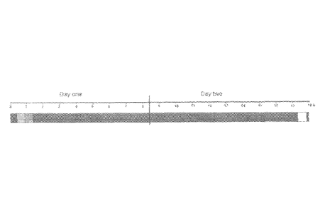
FIG. 1 depicts a typical time-course for single locus detection with primary
labeled FISH
probes co-denatured with formaldehyde fixed paraffin embedded tissue sections
(histological specimens). The bars represent a hybridization assay performed
using
traditional hybridization solutions. The first bar on the left represents the
deparaffination
step; the second bar represents the heat-pretreatment step; the third bar
represents the
digestion step; the fourth bar represents the denaturation and hybridization
steps; the fifth
bar represents the stringency wash step; and the sixth bar represents the
mounting step.
FIG. 2 depicts a typical time-course for single locus detection with primary
labeled FISH
probes co-denatured with cytological specimens. The bars represent a
hybridization assay
performed using a traditional hybridization solutions. The first bar on the
left represents
the fixation step; the second bar represents the denaturation and
hybridization steps; the
third bar represents the stringency wash step; and the fourth bar represents
the mounting
step.
DETAILED DESCRIPTION
A. Definitions
In the context of the present invention the following terms are to be
understood as
follows:
"Biological sample" is to be understood as any in vivo, in vitro, or in situ
sample of one
or more cells or cell fragments. This can, for example, be a unicellular or
multicellular
organism, tissue section, cytological sample, chromosome spread, purified
nucleic acid
sequences, artificially made nucleic acid sequences made by, e.g., a biologic
based
system or by chemical synthesis, microarmy, or other form of nucleic acid
chip. In one
embodiment, a sample is a mammalian sample, such as, e.g., a human, murine,
rat, feline,
or canine sample.
"Nucleic acid," "nucleic acid chain," and "nucleic acid sequence" mean
anything that
binds or hybridizes using base pairing including, oligomers or polymers having
a
CA 02753271 2016-06-10
backbone formed from naturally occurring nucleotides and/or nucleic acid
analogs
comprising nonstandard nucleobases and/or nonstandard backbones (e.g., a
peptide
nucleic acid (PNA) or locked nucleic acid (LNA)), or any derivatized form of a
nucleic
acid.
As used herein, the term "peptide nucleic acid" or "PNA" means a synthetic
polymer
having a polyamide backbone with pendant nucleobases (naturally occurring and
modified), including, but not limited to, any of the oligomer or polymer
segments
referred to or claimed as peptide nucleic acids in, e.g., U.S. Pat. Nos.
5,539,082,
5,527,675, 5,623,049, 5,714,331, 5,718,262, 5,736,336, 5,773,571, 5,766,855,
5,786,461,
5,837,459, 5,891,625, 5,972,610, 5,986,053, 6,107,470 6,201,103, 6,228,982 and
6,357,163, W096/04000. The pendant nucleobase, such as, e.g., a purine or
pyrimidine
base on PNA may be connected to the backbone via a linker such as, e.g., one
of the
linkers taught in PCT/US02/30573 or any of the references cited therein. In
one
embodiment, the PNA has an N-(2-aminoethyl)-glycine) backbone. PNAs may be
synthesized (and optionally labeled) as taught in PCT/US02/30573 or any of the
references cited therein. PNAs hybridize tightly, and with high sequence
specificity, with
DNA and RNA, because the PNA backbone is uncharged. Thus, short PNA probes may
exhibit comparable specificity to longer DNA or RNA probes. PNA probes may
also
show greater specificity in binding to complementary DNA or RNA.
As used herein, the term "locked nucleic acid" or "LNA" means an oligomer or
polymer
comprising at least one or more LNA subunits. As used herein, the term "LNA
subunit"
means a ribonucleotide containing a methylene bridge that connects the 2'-
oxygen of the
ribose with the 4'-carbon. See generally, Kurreck, Eur. J. Biochem., 270:1628-
44 (2003).
Examples of nucleic acids and nucleic acid analogs also include polymers of
nucleotide
monomers, including double and single stranded deoxyribonucleotides (DNA),
ribonucleotides (RNA), a-anomeric forms thereof, synthetic and natural analogs
thereof,
and the like. The nucleic acid chain may be composed entirely of
deoxyribonucleotides,
ribonucleotides, peptide nucleic acids (PNA), locked nucleic acids (LNA),
synthetic or
11
CA 02753271 2011-08-22
WO 2010/097707
PCT/1B2010/000659
natural analogs thereof, or mixtures thereof. DNA, RNA, or other nucleic acids
as defined
herein can be used in the method and compositions of the invention.
"Polar aprotic solvent" refers to an organic solvent having a dipole moment of
about 2
debye units or more, a water solubility of at least about 5% (volume) at or
near ambient
temperature, i.e., about 20 C, and which does not undergo significant hydrogen
exchange
at approximately neutral pH, i.e., in the range of 5 to 9, or in the range 6
to 8. Polar
aprotic solvents include those defmed according to the Hansen Solubility
Parameters
discussed below.
"Alkyldiyl" refers to a saturated or unsaturated, branched, straight chain or
cyclic
hydrocarbon radical having two monovalent radical centers derived by the
removal of
one hydrogen atom from each of two different carbon atoms of a parent alkane,
alkene, or
alkyne.
"Aqueous solution" is to be understood as a solution containing water, even
small
amounts of water. For example, a solution containing 1% water is to be
understood as an
aqueous solution.
"Hybridization application," "hybridization assay," "hybridization
experiment,"
"hybridization procedure," "hybridization technique," "hybridization method,"
etc. are to
be understood as referring to any process that involves hybridization of
nucleic acids.
Unless otherwise specified, the terms "hybridization" and "hybridization step"
are to be
understood as referring to the re-annealing step of the hybridization
procedure as well as
the denaturation step (if present).
"Hybridization composition" refers to an aqueous solution of the invention for
performing a hybridization procedure, for example, to bind a probe to a
nucleic acid
sequence. Hybridization compositions may comprise, e.g., at least one polar
aprotic
solvent, at least one nucleic acid sequence, and a hybridization solution.
Hybridization
compositions do not comprise enzymes or other components, such as
deoxynucleoside
triphosphates (dNTPs), for amplifying nucleic acids in a biological sample.
12
CA 02753271 2011-08-22
WO 2010/097707
PCT/1B2010/000659
"Hybridization solution" refers to an aqueous solution for use in a
hybridization
composition of the invention. Hybridization solutions are discussed in detail
below and
may comprise, e.g., buffering agents, accelerating agents, chelating agents,
salts,
detergents, and blocking agents.
.. "Hansen Solubility Parameters" and "HSP" refer to the following cohesion
energy
(solubility) parameters: (1) the dispersion solubility parameter (5D, "D
parameter"),
which measures nonpolar interactions derived from atomic forces; (2) the polar
solubility
parameter (p, "P parameter"), which measures permanent dipole-permanent dipole
interactions; and (3) the hydrogen bonding solubility parameter (5H, "H
parameter"),
which measures electron exchange. The Hansen Solubility Parameters are further
defined
below.
"Repetitive Sequences" is to be understood as referring to the rapidly
reannealing
(approximately 25%) and/or intermediately reannealing (approximately 30%)
components of mammalian genomes. The rapidly reannealing components contain
small
(a few nucleotides long) highly repetitive sequences usually found in tandem
(e.g.,
satellite DNA), while the intermediately reannealing components contain
interspersed
repetitive DNA. Interspersed repeated sequences are classified as either SINEs
(short
interspersed repeat sequences) or LINEs (long interspersed repeated
sequences), both of
which are classified as retrotransposons in primates. SINEs and LINEs include,
but are
not limited to, Alu-repeats, Kpn-repeats, di-nucleotide repeats, tri-
nucleotide repeats,
tetra-nucleotide repeats, penta-nucleotide repeats and hexa-nucleotide
repeats. Alu
repeats make up the majority of human SINEs and are characterized by a
consensus
sequence of approximately 280 to 300 bp that consist of two similar sequences
arranged
as a head to tail dimer. In addition to SINEs and LINEs, repeat sequences also
exist in
chromosome telomeres at the termini of chromosomes and chromosome centromeres,
which contain distinct repeat sequences that exist only in the central region
of a
chromosome. However, unlike SINEs and LINEs, which are dispersed randomly
throughout the entire genome, telomere and centromere repeat sequences are
localized
within a certain region of the chromosome.
13
CA 02753271 2011-08-22
WO 2010/097707
PCT/1B2010/000659
"Non-toxic" and "reduced toxicity" are defined with respect to the toxicity
labeling of
formamide according to "Directive 1999/45/EC of the European Parliament and of
the
Council of 31 May 1999 concerning the approximation of the laws, regulations
and
administrative provisions of the Member States relating to the classification,
packaging,
and labelling of dangerous preparations"
(ecb.jrc.it/legislation/1999L0045EC.pdf)
("Directive"). According to the Directive, toxicity is defined using the
following
classification order: T+ "very toxic"; T "toxic", C "corrosive", Xn "harmful",
.Xi
"irritant." Risk Phrases ("R phrases") describe the risks of the classified
toxicity.
Formamide is listed as T (toxic) and R61 (may cause harm to the unborn child).
All of
the following chemicals are classified as less toxic than formamide:
acetonitrile (Xn,
R11, R20, R21, R22, R36); sulfolane (Xn, R22); y-butyrolactone (Xn, R22, R32);
and
ethylene carbonate (Xi , R36, R37, R38). At the time of filing this
application, ethylene
trithiocarbonate and glycol sulfite are not presently labeled.
"Denaturation" as used herein means a process in which nucleic acids or
proteins reduce
or lose their tertiary and/or secondary structures by application of
compound(s), such as
e.g. a strong acid or base, a concentrated inorganic salt, an organic solvent,
and/or by
external stress such as e.g. heat. This means that, when denaturation relates
to nucleic
acids, and when said nucleic acid is double stranded, the strands might
separate partially
or completely. This further means that the binding interactions of the double
stranded
nucleic acids are weakened sufficiently by the denaturation so that
hybridization with e.g.
alternative complementary strands can occur more efficiently than without
denaturation.
"Denaturing agent" refers to any substance that is capable of lowering the
mutual binding
affinity of complementary stands of nucleic acids compared to water. Non-
limiting
examples of typical denaturing agents include organic solvents such as
formamide, urea,
DMSO, and tetraalkylammonium halides or combinations thereof. Denaturation
conditions are sequence dependent and are different under different
environmental
parameters. The melting temperature (T,õ) can be used to adjust denaturation
conditions
to decrease complementary base pairing in the presence of a denaturing agent.
Tn, is the
temperature (under defined ionic strength and pH) at which 50% of the target
sequence
14
CA 02753271 2011-08-22
WO 2010/097707
PCT/1B2010/000659
hybridizes to a perfectly matched probe. For DNA-DNA hybrids, the Tin can be
approximated from the following equation:
Tm 81.5 C.+16.6(log M)+0.41(% GC) -0.61(% form)-500/L
where M is the molarity of monovalent cations, % GC is the percentage of
guanosine and
cytosine nucleotides in the DNA, % form is the percentage of formamide in the
hybridization solution, and L is the length of the hybrid in base pairs. Tin
is reduced by
about 1 C for each 1% of mismatching.
"Separate denaturation" as used herein, refers to hybridization methods in
which the
target nucleic acid is denatured in the absence of the probe and/or that the
probe is
denatured in the absence of the target nucleic acid. For example, the target
may be
denatured in a first solution, the probe may be denatured in a second
solution, and then
the denatured probe may be combined with the denatured target for a time
period
sufficient to hybridize the target and probe. In another example, the target
may be
denatured in a first solution and then combined with the probe for a time
period sufficient
to hybridize the target and probe. In still a further example, the probe may
be denatured
in a first solution and then combined with the target for a time period
sufficient to
hybridize the target and probe.
B. Solvent Selection
Suitable polar aprotic solvents for use in the invention may be selected based
on their
Hansen Solubility Parameters. Methods for experimentally determining and/or
calculating HSP for a solvent are known in the art, and I-1SP have been
reported for over
1200 chemicals.
For example, the D parameter may be calculated with reasonable accuracy based
on
refractive index, or may be derived from charts by comparison with known
solvents of
similar size, shape, and composition after establishing a critical temperature
and molar
volume. The P parameter may be estimated from known dipole moments (see, e.g.,
McClellan A.L., Tables of Experimental Dipole Moments (W.H. Freeman 1963))
using
Equation 1:
CA 02753271 2011-08-22
WO 2010/097707
PCT/1B2010/000659
Equation 1: 6p = 37.4(Dipole Moment)/V"2
where V is the molar volume. There are no equations for calculating the H
parameter.
Instead, the H parameter is usually determined based on group contributions.
HSP characterizations are conveniently visualized using a spherical
representation, with
the HSP of an experimentally-determined suitable reference solvent at the
center of the
sphere. The radius of the sphere (R) indicates the maximum tolerable variation
from the
HSP of the reference solvent that still allows for a "good" interaction to
take place. Good
solvents are within the sphere and bad ones are outside. The distance, Ra,
between two
solvents based on their respective HSP values can be determined using Equation
2:
Equation 2: (Ra)2 = 4(8m - 61)2)2 + (8P1 8P2)2 - 61-12)2
where subscript 1 indicates the reference sample, subscript 2 indicates the
test chemical,
and all values are in MPa1/2. Good solubility requires that Ra be less than
the
experimentally-determined radius of the solubility sphere Ro. The relative
energy
difference between two solvents, i.e., RED number, can be calculated by taking
the ratio
of Ra to Ro, as shown in Equation 3.
Equation 3: RED = Rai&
RED numbers less than 1.0 indicate high affinity; RED numbers equal or close
to 1.0
indicate boundary conditions; and progressively higher RED numbers indicate
progressively lower affinities.
In some embodiments, the D parameters of the polar aprotic solvents of the
invention are
between 17.7 to 22.0 MPam. Such relatively high D parameters are generally
associated
with solvents having cyclic structures and/or structures with sulfur or
halogens. Linear
compounds are not likely to be among the most suitable polar aprotic solvents
for use in
.. the invention, but may be considered if their P and H parameters are within
the ranges
discussed below. Since the D parameter is multiplied by 4 in Equation 2, the
limits are
one-half of Ro. In addition, it should be noted that D values of around 21 or
higher are
often characteristic of a solid.
16
CA 02753271 2011-08-22
WO 2010/097707
PCT/1B2010/000659
In some embodiments, the P parameters of the polar aprotic solvents of the
invention are
between 13 to 23 MPa1/2. Such exceptionally high P parameters are generally
associated
with solvents having a high dipole moment and presumably also a relatively low
molecular volume. For example, for V near 60 cc/mole, the dipole moment should
be
between 4.5 and 3.1. For V near 90 cc/mole, the dipole moment should be
between 5.6
and 3.9.
In some embodiments, the H parameters of the polar aprotic solvents of the
invention are
between 3 to 13 MPalf2. Generally, polar aprotic solvents having an alcohol
group are not
useful in the compositions and methods of the invention, since the H
parameters of such
solvents would be too high.
The molar volume of the polar aprotic solvent may also be relevant, since it
enters into
the evaluation of all three Hansen Solubility Parameters. As molar volume gets
smaller,
liquids tend to evaporate rapidly. As molar volume gets larger, liquids tend
to enter the
solid region in the range of D and P parameters recited above. Thus, the polar
aprotic
solvents of the invention are rather close to the liquid/solid boundary in HSP
space.
In some embodiments, the polar aprotic solvents of the invention have lactone,
sulfone,
nitrile, sulfite, and/or carbonate functionality. Such compounds are
distinguished by their
relatively high dielectric constants, high dipole moments, and solubility in
water. An
exemplary polar aprotic solvent with lactone functionality is y-butyrolactone
(GBL), an
exemplary polar aprotic solvent with sulfone functionality is sulfolane (SL;
tetramethylene sulfide-dioxide), an exemplary polar aprotic solvent with
nitrile
functionality is acetonitrile (AN), an exemplary polar aprotic solvent with
sulfite
functionality is glycol sulfite/ethylene sulfite (GS), and an exemplary polar
aprotic
solvents with carbonate functionality are ethylene carbonate (EC), propylene
carbonate
(PC), or ethylene trithiocarbonate (ETC). The structures of these exemplary
solvents are
provided below and their Hansen Solubility Parameters, RED numbers, and molar
volumes are given in Table 1.
17
CA 02753271 2011-08-22
WO 2010/097707
PCT/1B2010/000659
0 0 0
0 0 S 0
ii S//
S 0 //0
'N.o S
I ) \S /
H3C
ethylene glycol 7- sulfolane ethylene
propylene
carbonate sulfite butyrolactone trithiocarbonate carbonate
Table 1
D P H RED Molar
Volume
(cm3/mole)
Correlation 19.57 19.11 7.71 -
(Ro = 3.9)
GBL 19.0 16.6 7.4 0.712 76.5
PC 20.0 18.0 4.1 0.993 85.2
SL 20.3 18.2 10.9 0.929 95.7
EC 19.4 21.7 5.1 0.946 66.0
ETC n/a n/a n/a n/a n/a
GS 20.0 15.9 5.1 n/a 75.1
n/a = not available.
Other suitable polar aprotic solvents that may be used in the invention are
cyclic
compounds such as, e.g., e-caprolactone. In addition, substituted
pyrolidinones and
related structures with nitrogen in a 5- or 6-membered ring, and cyclic
structures with two
nitrile groups, or one bromine and one nitrile group, may also be suitable for
use in the
invention. For example, N-methyl pyrrolidinone (shown below) may be a suitable
polar
aprotic solvent for use in the methods and compositions of the invention.
0
I
C=N---CH3
/
Other suitable polar aprotic solvents may contain a ring urethane group (NHC00-
).
However, not all such compounds are suitable. One of skill in the art may
screen for
18
CA 02753271 2011-08-22
WO 2010/097707
PCT/1B2010/000659
compounds useful in the compositions and methods of the invention as described
herein.
Exemplary chemicals that may be suitable for use in the invention are set
forth in Tables
2 and 3 below.
Table 2
Solvent D P H
Acetanilide 20.6 13.3 12.4
N-Acetyl Pyrrolidone 17.8 13.1 8.3
4-Amino Pyridine 20.4 16.1 12.9
Benzamide 21.2 14.7 11.2
Benzimidazole 20.6 14.9 11.0
1,2,3-Benzotriazole 18.7 15.6 12.4
Butadienedioxide 18.3 14.4 6.2
2,3-Butylene Carbonate 18.0 16.8 3.1
Caprolactone (Epsilon) 19.7 15.0 7.4
Chloro Maleic Anhydride 20.4 17.3 11.5
2-Chlorocyclohexanone 18.5 13.0 5.1
Chloronitromethane 17.4 13.5 5.5
Citraconic Anhydride 19.2 17.0 11.2
Crotonlactone 19.0 19.8 9.6
Cyclopropylnitrile 18.6 16.2 5.7
Dimethyl Sulfate 17.7 17.0 9.7
Dimethyl Sulfone 19.0 19.4 12.3
Dimethyl Sulfoxide 18.4 16.4 10.2
1,2-Dinitrobenzene 20.6 22.7 5.4
2,4-Dinitrotoluene 20.0 13.1 4.9
Dipheynyl Sulfone 21.1 14.4 3.4
1,2-Dinitrobenzene 20.6 22.7 5.4
2,4-Dinitrotoluene 20.0 13.1 4.9
Epsilon-Caprolactam 19.4 13.8 3.9
Ethanesulfonylchloride 17.7 14.9 6.8
Furfural 18.6 14.9 5.1
2-Furonitrile 18.4 15.0 8.2
Isoxazole 18.8 13.4 11.2
Maleic Anhydride 20.2 18.1 12.6
Malononitrile 17.7 18.4 6.7
4-Methoxy Benzonitrile 19.4 16.7 5.4
1-Methoxy-2-Nitrobenzene 19.6 16.3 5.5
1-Methyl Imidazole 19.7 15.6 11.2
3-Methyl Isoxazole 19.4 14.8 11.8
N-Methyl Morpholine-N- 19.0 16.1 10.2
Oxide
Methyl Phenyl Sulfone 20.0 16.9 7.8
19
CA 02753271 2011-08-22
WO 2010/097707 PCT/1B2010/000659
Methyl Sulfolane 19.4 17.4 5.3
Methyl-4-Toluenesulfonate 19.6 15.3 3.8
3-Nitroaniline 21.2 18.7 10.3
2-Nitrothiophene 19.7 16.2 8.2
9,10-Phenanthrenequinone 20.3 17.1 4.8
Phthalic Anhydride 20.6 20.1 10.1
1,3-Propane Sultone 18.4 16.0 9.0
beta-Propiolactone 19.7 18.2 10.3
2-Pyrrolidone 19.4 17.4 11.3
Saccharin 21.0 13.9 8.8
Succinonitrile 17.9 16.2 7.9
Sulfanilamide 20.0 19.5 10.7
Sulfolane 20.3 18.2 10.9
2,2,6,6- 19.5 14.0 6.3
Tetrachlorocyclohexanone
Thiazole 20.5 18.8 10.8
3,3,3-Trichloro Propene 17.7 15.5 3.4
1,1,2-Trichloro Propene 17.7 15.7 3.4
1,2,3-Trichloro Propene 17.8 15.7 3.4
Table 2 sets forth an exemplary list of potential chemicals for use in the
compositions and
methods of the invention based on their Hansen Solubility Parameters. Other
compounds,
may of course, also meet these requirements such as, for example, those set
forth in Table
3.
Table 3
Chemical (dipole moment) RED Melting Point C
Chloroethylene carbonate (4.02) 0.92 -
2-Oxazolidinone (5.07) 0.48 86-89
2-Imidazole 1.49 90-91
1,5-Dimethyl Tetrazole (5.3) -1.5 70-72
N-Ethyl Tetrazole (5.46) -1.5
Trimethylene sulfide-dioxide (4.49) - -
Trimethylene sulfite (3.63) - -
1,3-Dimethy1-5-Tetrazole (4.02) - -
Pyridazine (3.97) 1.16 -8
2-Thiouracil (4.21) - -
N-Methyl Imidazole (6.2) 1.28 -
1-Nitroso-2-pyrolidinone -1.37 -
CA 02753271 2011-08-22
WO 2010/097707 PCT/1B2010/000659
Ethyl Ethyl Phosphinate (3.51)
5-cyano-2-Thiouracil (5.19)
4H-Pyran-4-thione (4.08) 1.35 32-34
4H-Pyran-4-one = gamma pyrone (4.08) 1.49 Boiling Point (BP) 80
2-Nitrofuran (4.41) 1.14 29
Methyl alpha Bromo Tetronate (6.24)
Tetrahydrothiapyran oxide (4.19) 1.75 60-64
Picolinonitrile (2-cyanopyridine) (5.23) 0.40 26-28 (BP 212-215)
Nitrobenzimidazole (6.0) 0.52 207-209
Isatin (5.76) 193-195
N-phenyl sydnone (6.55)
Glycol sulfate (Ethylene glycol) 99 C
Note: not soluble at 40%
Some of the chemicals listed in Tables 2 and 3 have been used in hybridization
and/or
PCR applications in the prior art (e.g., dimethyl sulfoxide (DMSO) has been
used in
hybridization and PCR applications, and sulfolane (SL), acetonitrile (AN), 2-
pyrrolidone,
c-caprolactam, and ethylene glycol have been used in PCR applications). Thus,
in some
embodiments, the polar aprotic solvent is not DMSO, sulfolane, acetonitrile, 2-
pyrrolidone, c-caprolactam, or ethylene glycol. However, most polar aprotic
solvents
have not been used in prior art hybridization applications. Moreover, even
when such
compounds were used, the prior art did not recognize that they may be
advantageously
used to separately denature the probe and target in such hybridization
applications, as
disclosed in this application.
In addition, not all of the chemicals listed in Tables 2 and 3 are suitable
for use in the
compositions and methods of the invention. For example, although DMSO is
listed in
Table 2 because its Hansen Solubility Parameters (HSPs) fall within the ranges
recited
above, DMSO does not function to allow separate denaturation of the probe and
target in
the compositions and methods of the invention. However, it is well within the
skill of the
ordinary artisan to screen for suitable compounds using the guidance provided
herein
including testing a compound in one of the examples provided. For example, in
some
embodiments, suitable polar aprotic solvents will have HSPs within the ranges
recited
above and a structure shown in Formulas 1-9 above.
21
CA 02753271 2016-06-10
C. Compositions, Buffers, and Solutions
(1) Denaturation Solutions
Traditional compositions for separately or co-denaturing a probe and target in
hybridization applications are known in the art. Such compositions may
comprise, for
example, buffering agents, accelerating agents, chelating agents, salts,
detergents, and
blocking agents.
For example, the buffering agents may include SSC, HEPES, SSPE, PIPES, TMAC,
TRIS, SET, citric acid, a phosphate buffer, such as, e.g., potassium phosphate
or sodium
pyrrophosphate, etc. The buffering agents may be present at concentrations
from 0.01x to
50x, such as, for example, 0.01x, 0.1x, 0.5x, lx, 2x, 5x, 10x, 15x, 20x, 25x,
30x, 35x,
40x, 45x, or 50x. Typically, the buffering agents are present at
concentrations from 0.1x
to 10x.
The accelerating agents may include polymers such as FICOLLTM, PVP, heparin,
dextran
sulfate, proteins such as BSA, glycols such as ethylene glycol, glycerol, 1,3
propanediol,
propylene glycol, or diethylene glycol, combinations thereof such as
Dernhardt's solution
and BLOTTO, and organic solvents such as formamide, dimethylformamide, DMSO,
etc.
The accelerating agent may be present at concentrations from 1% to 80% or 0.1x
to 10x,
such as, for example, 0.1% (or 0.1x), 0.2% (or 0.2x), 0.5% (or 0.5x), 1% (or
lx), 2% (or
2x), 5% (or 5x), 10% (or 10x), 15% (or 15x), 20% (or 20x), 25% (or 25x), 30%
(or 30x),
40% (or 40x), 50% (or 50x), 60% (or 60x), 70% (or 70x), or 80% (or 80x).
Typically,
formamide is present at concentrations from 25% to 75%, such as 25%, 30%, 40%,
50%,
60%, 70%, or 75% , while DMSO, dextran sulfate, and glycol are present at
concentrations from 5% to 10%, such as 5%, 6%, 7%, 8%, 9%, or 10%.
The chelating agents may include EDTA, EGTA, etc. The chelating agents may be
present at concentrations from 0.1 mM to 10 mM, such as 0.1mM, 0.2mM, 0.5mM,
1mM, 2mM, 3mM, 4mM, 5mM, 6mM, 7mM, 8mM, 9mM, or 10mM. Typically, the
chelating agents are present at concentrations from 0.5 mM to 5 mM, such as
0.5mM,
1mM, 1.5mM, 2mM, 2.5mM, 3mM, 3.5mM, 4mM, 4.5mM, or 5mM.
22
CA 02753271 2016-06-10
The salts may include sodium chloride, sodium phosphate, magnesium phosphate,
etc.
The salts may be present at concentrations from 1 mM to 750 mM, such as 1mM,
5mM,
10mM, 20mM, 30mM, 40mM, 50mM, 100mM, 200mM, 300mM, 400mM, 500mM,
600mM, 700mM, or 750mM. Typically, the salts are present at concentrations
from 10
mM to 500 mM, such as 10mM, 20mM, 30mM, 40mM, 50mM, 100mM, 200mM,
300mM, 400mM, or 500mM.
The detergents may include TweenTm, SDS, TritonTm, CHAPS, deoxycholic acid,
etc.
The detergent may be present at concentrations from 0.001% to 10%, such as,
for
example, 0.001, 0.01, 0.1, 0.5, 1, 2, 3, 4, 5, 6, 7, 8, 9, or 10%. Typically,
the detergents
are present at concentrations from 0.01% to 1%, such as 0.01%, 0.02%, 0.03%,
0.05%,
0.1%, 0.2%, 0.3%, 0.4%, 0.5%, 0.6%, 0.7%, 0.8%, 0.9%, or 1%.
The nucleic acid blocking agents may include, for example, yeast tRNA,
homopolymer
DNA, denatured salmon sperm DNA, herring sperm DNA, total human DNA, COT1
DNA, etc. The blocking nucleic acids may be present at concentrations of 0.05
mg/mL to
100 mg/mL. However, the compositions and methods of the invention surprisingly
show
significantly reduced background levels without the need for blocking agents.
A great variation exists in the literature regarding traditional denaturation
buffers for
hybridization applications. For example, a traditional solution may comprise
5x or 6x
SSC, 0.01 M EDTA, 5x Dernhardt's solution, 0.5% SDS, and 100 mg/mL sheared,
denatured salmon sperm DNA. Another traditional solution may comprise 50 mM
HEPES, 0.5 M NaC1, and 0.2 mM EDTA. A typical solution for FISH on biological
specimens for RNA detection may comprise, e.g., 2x SSC, 10% dextran sulfate, 2
mM
vanadyl-ribonucleoside complex, 50% formamide, 0.02% RNAse-free BSA, and
1 mg/mL E. coli tRNA. A typical solution for FISH on biological specimens for
DNA
detection may comprise, e.g., 2x SSC, 10% dextran sulfate, 50% formamide, and
e.g., 0.3
mg/mL salmon sperm DNA or 0.1 mg/mL COT1 DNA. Other typical solutions may
comprise 40% formamide, 10% dextran sulfate, 300 mM NaC1, 5 mM phosphate
buffer,
23
CA 02753271 2011-08-22
WO 2010/097707
PCT/1B2010/000659
Alu-PNA (blocking PNA) or COT-1 DNA, and in some cases 0.1 g/I.LI, total
human
DNA (THD). Additional denaturation buffers are discussed below in the section
titled
"Hybridization Conditions."
The compositions of the invention may comprise any of the traditional
components
recited above in combination with at least one polar aprotic solvent. The
traditional
components may be present at the same concentrations as used in traditional
denaturing
solutions, or may be present at higher or lower concentrations, or may be
omitted
completely.
For example, if the compositions of the invention comprise salts such as NaC1
and/or
phosphate buffer, the salts may be present at concentrations of 0-1200 mM NaC1
and/or
0-200 mM phosphate buffer. In some embodiments, the concentrations of salts
may be,
for example, OmM, 15rnM, 30mM, 45mM, 60mM, 75mM, 90mM, 105mM, 120mM,
135mM, 150mM, 165mM, 180mM, 195mM, 210mM, 225mM, 240mM, 255mM,
270mM, 285mM, or 300 mM NaC1 and 5 mM phosphate buffer, or 600 mM NaC1 and 10
mM phosphate buffer. In other embodiments, the concentrations of salts may be,
for
example, the concentrations present in 0.1X, 0.2X, 0.3X, 0.4X, 0.5X, 0.6X,
0.7X, 0.8X,
0.9X, 1X, 2X, 3X, 4X, 5X, 6X, 7X, or 8X SSC.
If the compositions of the invention comprise accelerating agents such as
dextran sulfate,
glycol, or DMSO, the dextran sulfate may be present at concentrations of from
5% to
40%, the glycol may be present at concentrations of from 0.1% to 10%, and the
DMSO
may be from 0.1% to 10%. In some embodiments, the concentration of dextran
sulfate
may be 10% or 20% and the concentration of ethylene glycol, 1,3 propanediol,
or
glycerol may be 1% to 10%. In some embodiments, the concentration of DMSO may
be
1%. In some embodiments, the aqueous composition does not comprise DMSO as an
accelerating agent. In some emboiliments, the aqueous composition does not
comprise
formamide as an accelerating agent, or comprises formamide with the proviso
that the
composition contains less than 25%, or less than 10%, or less than 5%, or less
than 2%,
24
CA 02753271 2011-08-22
WO 2010/097707
PCT/1B2010/000659
or less than 1%, or less than 0.5%, or less than 0.1%, or less than 0.05%, or
less than
0.01%.
If the compositions of the invention comprise citric acid, the concentrations
may range
from 1 mM to 100 mM and the pH may range from 5.0 to 8Ø In some embodiments
the
concentration of citric acid may be 10 mM and the pH may be 6.2.
The compositions of the invention may comprise agents that reduce non-specific
binding
to, for example, the cell membrane, such as salmon sperm or small amounts of
total
.. human DNA or, for example, they may comprise blocking agents to block
binding of,
e.g., repeat sequences to the target such as larger amounts of total human DNA
or repeat
enriched DNA or specific blocking agents such as PNA or LNA fragments and
sequences. These agents may be present at concentrations of from 0.01-100 g/
I_, or
0.01-100 1.t.M. For example, in some embodiments, these agents will be 0.1
g,/ t total
human DNA, or 0.1 ug/ L non-human DNA, such as herring sperm, salmon sperm, or
calf thymus DNA, or 5 M blocking PNA. However, the compositions and methods
of
the invention show significantly reduced background levels without the need
for blocking
agents.
One aspect of the invention is a composition or solution for separately
denaturing the
probe and target in a hybridization application. The composition for
denaturing the target
may comprise the same components as the composition for denaturing the probe,
or the
two compositions may comprise different components. Compositions for use in
the
invention may include an aqueous composition comprising at least one polar
aprotic
solvent in an amount effective to denature double-stranded nucleotide
sequences. An
amount effective to denature double-stranded nucleotide sequences is an amount
that
enables hybridization. For example, one way to test for whether the amount of
polar
aprotic solvent is effective to enable hybridization is to determine whether
the polar
aprotic solvent, when used in the hybridization methods and compositions
described
herein, such as example 1, yield a detectable signal and/or an amplified
nucleic acid
product.
CA 02753271 2011-08-22
WO 2010/097707
PCT/1B2010/000659
Non-limiting examples of effective amounts of polar aprotic solvents include,
e.g., about
1% to about 95% (v/v). In some embodiments, the concentration of polar aprotic
solvent
is 5% to 60% (v/v). In other embodiments, the concentration of polar aprotic
solvent is
10% to 60% (v/v). In still other embodiments, the concentration of polar
aprotic solvent
.. is 30% to 50% (v/v). Concentrations of 1% to 5%, 5% to 10%, 10%, 10% to
20%, 20% to
30%, 30% to 40%, 40% to 50%, 50% to 60%, or 60% to 70% (v/v) are also
suitable. In
some embodiments, the polar aprotic solvent will be present at a concentration
of 0.1%,
0.25%, 0.5%, 1%, 2%, 3%, 4%, or 5% (v/v). In other embodiments, the polar
aprotic
solvent will be present at a concentration of 7%, 7.5%, 8%, 8.5%, 9%, 9.5%,
10%,
10.5%, 11%, 11.5%, 12%, 12.5%, 13%, 13.5%, 14%, 14.5%, 15%, 15.5%, 16%, 16.5%,
17%, 17.5%, 18%, 18.5%, 19%, 19.5%, or 20% (v/v).
If the compositions of the invention are used in a hybridization assay, they
may further
comprise one or more nucleic acid probes. The probes may be directly or
indirectly
labeled with detectable compounds such as enzymes, chromophores,
fluorochromes, and
haptens. The DNA probes may be present at concentrations of 0.1 to 100 ng/pt.
For
example, in some embodiments, the probes may be present at concentrations of 1
to
10 nW L. The PNA probes may be present at concentrations of 0.5 to 5000 nM.
For
example, in some embodiments, the probes may be present at concentrations of 5
to 1000
nM.
In one embodiment, a composition of the invention comprises a mixture of 40%
polar
aprotic solvent (v/v) (e.g., ethylene carbonate, "EC"), 10% dextran sulfate,
300 mM
NaCl, 5 mM phosphate buffer, and 1-10 ng/p,L probe. Another exemplary
composition of
the present invention comprises a mixture of 15% EC, 20% dextran sulfate, 600
mM
NaC1, 10 mM phosphate buffer, and 0.114111 total human DNA. Yet another
exemplary
composition comprises 15% EC, 20% dextran sulfate, 600 mM NaCl, 10 mM citric
acid
pH 6.2, and 0.1 tig/pt non-human DNA (e.g., herring sperm, salmon sperm, or
calf
thymus) OR 0.5% formamide OR 1% glycol (e.g., ethylene glycol, 1,3
propanediol, or
glycerol). Yet another exemplary composition comprises 15% EC, 20% dextran
sulfate,
26
CA 02753271 2011-08-22
WO 2010/097707
PCT/1B2010/000659
600 mM NaC1, 10 mM citric acid pH 6.2. Yet another exemplary composition
comprises
15% EC and 10 mM citric acid pH 6.2.
(2) Polar Aprotic Solvent(s)
Different polar aprotic solvents may impart different properties on the
compositions of
the invention. For example, the choice of polar aprotic solvent may contribute
to the
stability of the composition, since certain polar aprotic solvents may degrade
over time.
For example, the polar aprotic solvent ethylene carbonate breaks down into
ethylene
glycol, which is a relatively stable molecule, and carbon dioxide, which can
interact with
water to form carbonic acid, altering the acidity of the compositions of the
invention.
Without being bound by theory, it is believed that the change in pH upon
breakdown of
ethylene carbonate and DNA damage from long storage makes the compositions of
the
invention less effective for hybridization. However, stability can be improved
by
reducing the pH of the composition, by adding citric acid as a buffer at pH
6.2 instead of
the traditional phosphate buffer, which is typically used at about pH 7.4,
and/or by adding
ethylene glycol at concentrations, e.g., between 0.1% to 10%, or between 0.5%
to 5%,
such as, for example, 1%, 2%, 3%, etc. For example, with 10 mM citrate buffer,
the
compositions of the invention are stable at 2-8 C for approximately 8 months.
Stability
can also be improved if the compositions are stored at low temperatures (e.g.,
-20 C).
In addition, certain polar aprotic solvents may cause the compositions of the
invention to
separate into multi-phase systems under certain conditions. The conditions
under which
multi-phase systems are obtained may be different for different polar aprotic
solvents.
Generally, however, as the concentration of polar aprotic solvent increases,
the number of
phases increases. For example, compositions comprising low concentrations
ethylene
carbonate (i.e., less than 20%) may exist as one phase, while some
compositions
comprising higher concentrations of ethylene carbonate may separate into two,
or even
three phases. For instance, compositions comprising 15% ethylene carbonate in
20%
dextran sulfate, 600 mM NaC1, and 10 mM citrate buffer exist as a single phase
at room
temperature, while compositions comprising 40% ethylene carbonate in 10%
dextran
sulfate, 300 mM NaCl, and 5 mM phosphate buffer consist of a viscous lower
phase
(approximately 25% of the total volume) and a less viscous upper phase
(approximately
27
CA 02753271 2011-08-22
WO 2010/097707
PCT/1B2010/000659
75% of the total volume) at room temperature. However, compositions
comprising, e.g.,
40% polar aprotic solvent (e.g., 40% EC in 10 mM citrate buffer) or 50% polar
aprotic
solvent (e.g., 50% EC in 2xSSC) is an one phase system.
On the other hand, some polar aprotic solvents may exist in two phases at room
temperature even at low concentrations. For example, sulfolane, y-
butyrolactone,
ethylene trithiocarbonate, glycol sulfite, and propylene carbonate exist as
two phases at
concentrations of 10, 15, 20, or 25% (20% dextran sulfate, 600 mM NaC1, 10 mM
citrate
buffer) at room temperature. In contrast, polar aprotic solvent compositions
with lower
percentages of dextran sulfate, or with no dextran sulfate, stay in one phase
at room
temperature (e.g. 20% GBL in 2x SSC and 20% SL in 2xSSC).
It may also be possible to alter the number of phases by adjusting the
temperature of the
compositions of the invention. Generally, as temperature increases, the number
of phases
decreases. For example, at 2-8 C, compositions comprising 40% ethylene
carbonate in
10% dextran sulfate, 300 mM NaC1, and 5 mM phosphate buffer may separate into
a
three-phase system.
It may also be possible to alter the number of phases by adjusting the
concentration of
dextran sulfate and/or salt in the composition. Generally speaking, lowering
the dextran
sulfate concentration (traditional concentration is 10%) and/or salt
concentration may
reduce the number of phases. However, depending on the particular polar
aprotic solvent
and its concentration in the composition, single phases may be produced even
with higher
concentrations of salt and dextran sulfate. For example, a composition
comprising low
amounts of EC (e.g., 15%, 10%, or 5%) can work well by increasing the dextran
sulfate
and salt concentrations, while still keeping a one phase system. In a
particular
embodiment, compositions comprising a HER2 gene DNA probe, a CEN17 PNA probe,
15% EC, 20% dextran sulfate, 600 mM NaC1, and 10 mM phosphate buffer are
frozen at
-20 C. In other embodiments, the compositions are liquid at -20 C.
28
CA 02753271 2011-08-22
WO 2010/097707
PCT/1B2010/000659
Some polar aprotic solvents may allow the probes to produce stronger signals
in one
phase or another. For example, 40% glycol sulfite produces strong signals in
the lower
phase and no signals in the upper phase. Similarly, certain types of probes
may produce
stronger signals in one phase or another. For example, PNA probes tend to show
stronger
signals in the lower phase than the upper phase.
Accordingly, the multiphase systems of the invention may be used to
conveniently
examine different aspects of a sample.
Hybridization applications may be performed with a one-phase composition of
the
invention, with individual phases of the multiphase compositions of the
invention, or with
mixtures of any one or more of the phases in a multiphase composition of the
invention.
For example, in a one phase system, a volume of the sample may be extracted
for use in
the hybridization. In a mulitphase system, one may extract a volume of sample
from the
phase of interest (e.g., the upper, lower, or middle phase) to use in the
hybridization.
Alternatively, the phases in a multiphase system may be mixed prior to
extracting a
volume of the mixed sample for use in the hybridization. However, the
multiphase system
may yield strong and uneven local background staining depending on the
composition.
While, the addition of low amounts of formamide will reduce background in a
one phase
system, it has little effect on a multiphase system with high concentrations
(e.g., 40%) of
a polar aprotic solvent.
Because the composition used in the hybridization step may differ from the
compositions
used in the separate denaturation steps, the dextran sulfate and salt
concentrations of the
compositions of the invention are not critical. Indeed, compositions of the
invention
lacking dextran sulfate, salt, and buffer produce lower background (e.g.,
scores that are
lower by 1 to 2) and more homogenous background in hybridization applications
in
which the probe and target are separately denatured, compared to hybridization
applications in which the probe and target are co-denatured. However,
compositions
comprising a buffer (e.g., 40% EC plus 10mM citrate buffer) produce slightly
higher
background (e.g., scores that are higher by V2) than unbuffered compositions.
In one
29
CA 02753271 2011-08-22
WO 2010/097707
PCT/1B2010/000659
embodiment, compositions with EC and buffer (e.g., 15% EC plus 10mM citrate
buffer)
worked without any dextran sulfate.
Hybridization applications in which the target and probe are separately
denatured using
the compositions of the invention produce more homogenous signal intensities
and a
lower more homogenous background staining than hybridization applications in
which
the target and probe are co-denatured using traditional buffers.
(3) Optimization for Particular Applications
The compositions of the invention can be varied in order to optimize results
for a
particular application. For example, the concentration of polar aprotic
solvent, salt,
accelerating agent, blocking agent, and/or hydrogen ions (i.e. pH) may be
varied in order
to improve results for a particular application.
For example, the concentration of polar aprotic solvent may be varied in order
to improve
signal intensity and background staining. Generally, as the concentration of
polar aprotic
.. solvent increases, signal intensity increases and background staining
decreases. For
example, compositions for denaturing the probe comprising 15% EC tend to show
stronger signals and less background than compositions comprising 5% EC.
However,
signal intensity may be improved for compositions having low concentrations of
polar
aprotic solvent (e.g., 0% to 20%) if the concentrations of salt and/or dextran
sulfate are
.. increased. For example, strong signals may be observed with 5% to 10% EC
when the
salt concentration is raised approximately 3 to 4 times traditional salt
concentrations (i.e.,
approximately 1200 mM NaC1, 20 mM phosphate buffer; traditional salt
concentrations
are about 300mM NaCl). Likewise, as lower concentrations of polar aprotic
solvent are
used, higher concentrations of dextran sulfate are generally required to
maintain good
signal and background intensity.
Accordingly, the concentrations of salt and dextran sulfate may also be varied
in order to
improve signal intensity and background staining. Generally, as the
concentrations of salt
and dextran sulfate in the composition for denaturing the probe increase, the
signal
intensity increases and background decreases. For example, salt concentrations
that are
CA 02753271 2011-08-22
WO 2010/097707
PCT/1B2010/000659
approximately two to four times traditional concentrations (i.e., 300 mM NaCl
5 mM
phosphate buffer) produce strong signals and low background. Surprisingly,
however, the
compositions of the invention can be used even in the complete absence of
salt. Signal
intensities can be improved under no-salt conditions by increasing the
concentrations of
accelerating agent and/or polar aprotic solvent.
Likewise, compositions for denaturing the probe exhibit increased signal
intensity as
dextran sulfate concentration increases from 0% to 20%. However, good signals
may
even be observed at dextran sulfate concentrations of 0%. Signal intensity may
be
improved under low dextran sulfate conditions by increasing the polar aprotic
solvent
and/or salt concentrations.
In addition, the types probes used in the compositions of the invention may be
varied to
improve results. For example, in some aspects of the invention, combinations
of
DNA/DNA probes may show less background than combinations of DNA/PNA probes in
the compositions of the invention or vice versa. On the other hand, PNA probes
tend to
show stronger signals than DNA probes under low salt and/or low polar aprotic
solvent
concentrations. In fact, PNA probes also show signals when no polar aprotic
solvent is
present, whereas DNA probes show weak or no signals without polar aprotic
solvent.
A further optimization in the present invention is to separate the
denaturation of the probe
and target from each other, e.g., using a specific denaturation buffer not
containing the
labeled probe to denature the target. It has been found that the use of the
compositions of
the inventions for such separate denaturations decreases the background
staining and
makes the staining more homogenous both regarding the background and signal
intensities. In addition, the compositions of the invention allow the separate
denaturations
to occur at a low temperatures, which is beneficial, for example, to preserve
sample
morphology and the structure of nucleic acid sequences. As discussed above,
the
compositions of the invention are also less toxic than, e.g., traditional
formamide
denaturation buffers. It is obvious for the one known in the art that such a
denaturation
composition for the target might consist of more traditional denaturation
agent such as
e.g. urea, DMSO or formamide, in place of the polar aprotic solvent, while the
31
CA 02753271 2011-08-22
WO 2010/097707
PCT/1B2010/000659
denaturation compoistion for the probe may contain a polar aprotic solvent in
place of a
more traditoinal denaturation agent. The probe and target may then be combined
in the
hybridizatoin step, for example, with the fast hybridization buffer described
in
PCT/IB09/005893.
.. D. Applications, Methods, and Uses
(1) Analytical Samples
The methods and compositions of the invention may be used fully or partly in
all types of
hybridization applications comprising separate denaturation of the target and
probe in the
fields of cytology, histology, or molecular biology. According to one
embodiment, the
first or the second nucleic acid sequence in the methods of the invention is
present in a
biological sample. Examples of such samples include, e.g., tissue samples,
cell
preparations, cell fragment preparations, and isolated or enriched cell
component
preparations. The sample may originate from various tissues such as, e.g.,
breast, lung,
colorectal, prostate, lung, head & neck, stomach, pancreas, esophagus, liver,
and bladder,
.. or other relevant tissues and neoplasia thereof, any cell suspension, blood
sample, fine
needle aspiration, ascites fluid, sputum, peritoneum wash, lung wash, urine,
feces, cell
scrape, cell smear, cytospin or cytoprep cells.
The sample may be isolated and processed using standard protocols. Cell
fragment
.. preparations may, e.g., be obtained by cell homogenizing, freeze-thaw
treatment or cell
lysing. The isolated sample may be treated in many different ways depending of
the
purpose of obtaining the sample and depending on the routine at the site.
Often the
sample is treated with various reagents to preserve the tissue for later
sample analysis,
alternatively the sample may be analyzed directly. Examples of widely used
methods for
preserving samples are formalin-fixed followed by paraffin-embedding and cryo-
preservation.
For metaphase spreads, cell cultures are generally treated with colcemid, or
anther
suitable spindle pole disrupting agent, to stop the cell cycle in metaphase.
The cells are
then fixed and spotted onto microscope slides, treated with formaldehyde,
washed, and
32
CA 02753271 2011-08-22
WO 2010/097707
PCT/1B2010/000659
dehydrated in ethanol. Probes are then added and the samples are analyzed by
any of the
techniques discussed below.
Cytology involves the examination of individual cells and/or chromosome
spreads from a
biological sample. Cytological examination of a sample begins with obtaining a
specimen
of cells, which can typically be done by scraping, swabbing or brushing an
area, as in the
case of cervical specimens, or by collecting body fluids, such as those
obtained from the
chest cavity, bladder, or spinal column, or by fine needle aspiration or fine
needle biopsy,
as in the case of internal tumors. In a conventional manual cytological
preparation, the
sample is transferred to a liquid suspending material and the cells in the
fluid are then
transferred directly or by centrifugation-based processing steps onto a glass
microscope
slide for viewing. In a typical automated cytological preparation, a filter
assembly is
placed in the liquid suspension and the filter assembly both disperses the
cells and
captures the cells on the filter. The filter is then removed and placed in
contact with a
microscope slide. The cells are then fixed on the microscope slide before
analysis by any
of the techniques discussed below.
In a traditional DNA hybridization experiment using a cytological sample,
slides
containing the specimen are immersed in a formaldehyde buffer, washed, and
then
dehydrated in ethanol. The probes are then added and the specimen is covered
with a
coverslip. The probes and specimen are then co-denatured at a temperature
sufficient to
separate any double-stranded nucleic acid in the specimen (e.g. 5 minutes at
82 C), and
then incubated at a temperature sufficient to allow hybridization (e.g.,
overnight at 45 C).
After hybridization, the coverslips are removed and the specimens are
subjected to a
high-stringency wash (e.g., 10 minutes at 65 C) followed by a series of low-
stringency
washes (e.g., 2 x 3 minutes at room temperature). The samples are then
dehydrated and
mounted for analysis.
In a traditional RNA hybridization experiment using cytological samples, cells
are
equilibrated in 40% formamide, lx SSC, and 10 mM sodium phosphate for 5 mm,
incubated at 37 C overnight in hybridization reactions containing 20 ng of
oligonucleotide probe (e.g mix of labeled 50 bp oligos), 1xSSC, 40% formamide,
10%
33
CA 02753271 2011-08-22
WO 2010/097707
PCT/1B2010/000659
dextran sulfate, 0.4% BSA, 20 mM ribonucleotide vanadyl complex, salmon testes
DNA
(10 mg/ml), E. coli tRNA (10 mg/ml), and 10 mM sodium phosphate. Then washed
twice
with 4xSSC/40% formamide and again twice with 2x SSC/40% formamide, both at 37
C, and then with 2x SSC three times at room temperature. Digoxigenin-labeled
probes
can then e.g. be detected by using a monoclonal antibody to digoxigenin
conjugated to
Cy3. Biotin-labeled probes can then e.g. be detected by using
streptavidin¨Cy5.
Detection can be by fluorescence or CISH.
Histology involves the examination of cells in thin slices of tissue. To
prepare a tissue
sample for histological examination, pieces of the tissue are fixed in a
suitable fixative,
typically an aldehyde such as formaldehyde or glutaraldehyde, and then
embedded in
melted paraffin wax. The wax block containing the tissue sample is then cut on
a
microtome to yield thin slices of paraffin containing the tissue, typically
from 2 to 10
microns thick. The specimen slice is then applied to a microscope slide, air
dried, and
heated to cause the specimen to adhere to the glass slide. Residual paraffin
is then
dissolved with a suitable solvent, typically xylene, toluene, or others. These
so-called
deparaffinizing solvents are then removed with a washing-dehydrating type
reagent prior
to analysis of the sample by any of the techniques discussed below.
Alternatively, slices
may be prepared from frozen specimens, fixed briefly in 10% formalin or other
suitable
fixative, and then infused with dehydrating reagent prior to analysis of the
sample.
In a traditional DNA hybridization experiment using a histological sample,
formalin-
fixed paraffin embedded tissue specimens are cut into sections of 2-6 um and
collected
on slides. The paraffin is melted (e.g., 30-60 minutes at 60 C) and then
removed
(deparaffinated) by washing with xylene (or a xylene substitute), e.g., 2 x 5
minutes. The
samples are rehydrated, washed, and then pre-treated (e.g., 10 minutes at 95-
100 C). The
slides are washed and then treated with pepsin or another suitable
permeabilizer, e.g., 3-
15 minutes at 37 C. The slides are washed (e.g., 2 x 3 minutes), dehydrated,
and probe is
applied. The specimens are covered with a coverslip, the probe and specimen
are co-
denatured by incubating the slide at a temperature sufficient to separate any
double-
stranded nucleic acid (e.g. 5 minutes at 82 C), followed by incubation at a
temperature
sufficient to allow hybridization (e.g., overnight at 45 C). After
hybridization, the
34
CA 02753271 2011-08-22
WO 2010/097707
PCT/1B2010/000659
coverslips are removed and the specimens are subjected to a high-stringency
wash (e.g.,
minutes at 65 C) followed by a series of low-stringency washes (e.g., 2 x 3
minutes at
room temperature). The samples are then dehydrated and mounted for analysis.
In a traditional RNA hybridization experiment using a histological sample,
slides with
5 FFPE tissue sections are deparaffinized in xylene for 2 x 5 mm, immerged
in 99%
ethanol 2 x 3 min, in 96% ethanol 2 x 3 min, and then in pure water for 3 min.
Slides are
placed in a humidity chamber, Proteinase K is added, and slides are incubated
at RT for 5
min- 15 min. Slides are immersed in pure water for 2 x 3 min, immersed in 96%
ethanol
for 10 sec, and air-dried for 5 min. Probes are added to the tissue section
and covered
10 with coverslip. The slides are incubated at 55 C in humidity chamber
for 90 min. After
incubation, the slides are immersed in a Stringent Wash solution at 55 C for
25 min, and
then immersed in TBS for 10 sec. The slides are incubated in a humidity
chamber with
antibody for 30 mm. The slides are immersed in TBS for 2 x 3 min, then in pure
water for
2 x 1 min, and then placed in a humidity chamber. The slides are then
incubated with
substrate for 60 min, and immersed in tap water for 5 min.
In a traditional northern blot procedure, the RNA target sample is denatured
for 10
minutes at 65 C in RNA loading buffer and immediately placed on ice. The gels
are
loaded and electrophoresed with lx MOPS buffer (10X MOPS contains 200mM
morpholinopropansulfonic acid, 50mM sodium acetate, 10mM EDTA, pH 7.0) at 25 V
overnight. The gel is then pre-equilibrated in 20x SSC for 10 min and the RNA
is
transferred to a nylon membrane using sterile 20x SSC as transfer buffer. The
nucleic
acids are then fixed on the membrane using, for example, UV-cross linking at
120 mJ or
baking for 30 min at 120 C. The membrane is then washed in water and air
dried. The
membrane is placed in a sealable plastic bag and prehybridized without probe
for 30 min
at 68 C. The probe is denatured for 5 min at 100 C and immediately placed on
ice.
Hybridization buffer (prewarmed to 68 C) is added and the probe is hybridized
at 68 C
overnight. The membrane is then removed from the bag and washed twice for 5
min each
with shaking in a low stringency wash buffer (e.g., 2x SSC, 0.1% SDS) at room
temperature. The membrane is then washed twice for 15 min each in prewarmed
high
CA 02753271 2016-06-10
stringency wash buffer (e.g., 0.1x SSC, 0.1% SDS) at 68 C. The membrane may
then be
stored or immediately developed for detection.
Additional examples of traditional hybridization techniques can be found, for
example, in
Sambrook et al., Molecular Cloning A Laboratory Manual, 2nd Ed., Cold Spring
Harbor
Laboratory Press, (1989) at sections 1.90-1.104, 2.108-2.117, 4.40-4.41, 7.37-
7.57, 8.46-
10.38, 11.7-11.8, 11.12-11.19, 11.38, and 11.45-11.57; and in Ausubel et al.,
Current
Protocols in Molecular Biology, John Wiley & Sons, Inc. (1998) at sections
2.9.1-2.9.6,
2.10.4-2.10.5, 2.10.11-2.10.16, 4.6.5-4.6.9, 4.7.2-4.7.3, 4.9.7-4.9.15,
5.9.18, 6.2-6.5, 6.3,
6.4, 6.3.3-6.4.9, 5.9.12-5.9.13, 7Ø9, 8.1.3, 14.3.1-14.3.4, 14.9, 15Ø3-
15Ø4, 15.1.1-
15.1.8, and 20.1.24-20.1.25.
(2) Hybridization Techniques
The compositions and methods of the present invention can be used fully or
partly in all
types of nucleic acid hybridization techniques comprising separate
denaturation of the
target and probe for cytological and histological samples. Such techniques
include, for
example, in situ hybridization (ISH), fluorescent in situ hybridization (FISH;
including
multi-color FISH, Fiber-FISH, etc.), chromogenic in situ hybridization (CISH),
silver in
situ hybridization (SISH), comparative genome hybridization (CGH), chromosome
paints, and arrays in situ.
Molecular probes that are suitable for use in the hybridizations of the
invention are
described, e.g., in U.S. Patent Publication No. 2005/0266459. In general,
probes may be
prepared by chemical synthesis, PCR, or by amplifying a specific DNA sequence
by
cloning, inserting the DNA into a vector, and amplifying the vector an insert
in
appropriate host cells. Commonly used vectors include bacterial plasmids,
cosmids,
bacterial artificial chromosomes (BACs), PI diverted artificial chromosomes
(PACs), or
yeast artificial chromosomes (YACs). The amplified DNA is then extracted and
purified
for use as a probe. Methods for preparing and/or synthesizing probes are known
in the art,
e.g., as disclosed in PCT/US02/30573.
In general, the type of probe determines the type of feature one may detect in
a
hybridization assay. For example, total nuclear or genomic DNA probes can be
used as a
36
CA 02753271 2016-06-10
species-specific probe. Chromosome paints are collections of DNA sequences
derived
from a single chromosome type and can identify that specific chromosome type
in
metaphase and interphase nuclei, count the number of a certain chromosome,
show
translocations, or identify extra-chromosomal fragments of chromatin.
Different
chromosomal types also have unique repeated sequences that may be targeted for
probe
hybridization, to detect and count specific chromosomes. Large insert probes
may be
used to target unique single-copy sequences. With these large probes, the
hybridization
efficiency is inversely proportional to the probe size. Smaller probes can
also be used to
detect aberrations such as deletions, amplifications, inversions,
duplications, and
aneuploidy. For example, differently-colored locus-specific probes can be used
to detect
translocations via split-signal in situ hybridization.
In general, the ability to discriminate between closely related sequences is
inversely
proportional to the length of the hybridization probe because the difference
in thermal
stability decreases between wild type and mutant complexes as probe length
increases.
Probes of greater than 10 bp in length are generally required to obtain the
sequence
diversity necessary to correctly identify a unique organism or clinical
condition of
interest. On the other hand, sequence differences as subtle as a single base
(point
mutation) in very short oligomers (<10 base pairs) can be sufficient to enable
the
discrimination of the hybridization to complementary nucleic acid target
sequences as
compared with non-target sequences.
In one embodiment, at least one set of the in situ hybridization probes may
comprise one
or more PNA probes, as defined above and as described in U.S. Patent No.
7,105,294.
Methods for synthesizing PNA probes are described in PCT/US02/30573.
Alternatively,
or in addition, at least one set of the hybridization probes in any of the
techniques
discussed above may comprise one or more locked nucleic acid (LNA) probes, as
described in WO 99/14226. Due to the additional bridging bond between the 2
and 4'
carbons, the LNA backbone is pre-organized for hybridization. LNA/DNA and
LNA/RNA interactions are stronger than the corresponding DNA/DNA and DNA/RNA
interactions, as indicated by a higher melting temperature. Thus, the
compositions and
methods of the
37
CA 02753271 2016-06-10
invention, which decrease the energy required for hybridization, are
particularly useful
for hybridizations with LNA probes.
In one embodiment, the probes may comprise a detectable label (a molecule that
provides
an analytically identifiable signal that allows the detection of the probe-
target hybrid), as
described in U.S. Patent Publication No. 2005/0266459. The probes may be
labeled to
make identification of the probe-target hybrid possible by use, for example,
of a
fluorescence or bright field microscope/scanner. In some embodiments, the
probe may be
labeled using radioactive labels such as 31P, 33P, or 32S, non-radioactive
labels such as
digoxigenin and biotin, or fluorescent labels. The detectable label may be
directly
attached to a probe, or indirectly attached to a probe, e.g., by using a
linker. Any labeling
method known to those in the art, including enzymatic and chemical processes,
can be
used for labeling probes used in the methods and compositions of the
invention. In other
embodiments, the probes are not labeled.
In general, in situ hybridization techniques such as CGH, FISH, CISH, and
SISH, employ
large, mainly unspecified, nucleic acid probes that hybridize with varying
stringency to
genes or gene fragments in the chromosomes of cells. Using large probes
renders the in
situ hybridization technique very sensitive. However, the successful use of
large genomic
probes in traditional hybridization assays depends on blocking the undesired
background
staining derived from, e.g., repetitive sequences that are present throughout
the genome.
Traditional methods for decreasing nonspecific probe binding include
saturating the
binding sites on proteins and tissue by incubating tissue with
prehybridization solutions
containing ficoll, bovine serum albumin (BSA), polyvinyl pyrrolidone, and
nucleic acids.
Such blocking steps are time-consuming and expensive. Advantageously, the
methods
and compositions of the invention reduce and/or eliminate the need for such
blocking
steps, and show significantly reduced background levels without the need for
blocking
agents and without the need for overnight hybridization in formamide-
containing buffers.
However, in one embodiment, repetitive sequences may be suppressed according
to the
methods known in the art, e.g., as disclosed in PCTfUS02/30573.
38
CA 02753271 2011-08-22
WO 2010/097707
PCT/1B2010/000659
Bound probes may be detected in cytological and histological samples either
directly or
indirectly with fluorochromes (e.g., FISH), organic chromogens (e.g., CISH),
silver
particles (e.g., SISH), or other metallic particles (e.g., gold-facilitated
fluorescence in situ
hybridization, GOLDFISH). Thus, depending on the method of detection,
populations of
cells obtained from a sample to be tested may be visualized via fluorescence
microscopy
or conventional brightfield light microscopy.
Hybridization assays on cytological and histological samples are important
tools for
determining the number, size, and/or location of specific DNA sequences. For
example,
in CGH, whole genomes are stained and compared to normal reference genomes for
the
detection of regions with aberrant copy number. Typically, DNA from subject
tissue and
from normal control tissue is labeled with different colored probes. The pools
of DNA are
mixed and added to a metaphase spread of normal chromosomes (or to a
microarray chip,
for array- or matrix-CGH). The ratios of colors are then compared to identify
regions
with aberrant copy number.
FISH is typically used when multiple color imaging is required and/or when the
protocol
calls for quantification of signals. The technique generally entails preparing
a cytological
sample, labeling probes, denaturing target chromosomes and the probe,
hybridizing the
probe to the target sequence, and detecting the signal. Typically, the
hybridization
reaction fluorescently stains the targeted sequences so that their location,
size, or number
can be determined using fluorescence microscopy, flow cytometry, or other
suitable
instrumentation. DNA sequences ranging from whole genomes down to several
kilobases
can be studied using FISH. With enhanced fluorescence microscope techniques,
such as,
for example, deconvolution, even a single mRNA molecule can be detected. FISH
may
also be used on metaphase spreads and interphase nuclei.
FISH has been used successfully for mapping repetitive and single-copy DNA
sequences
on metaphase chromosomes, interphase nuclei, chromatin fibers, and naked DNA
molecules, and for chromosome identification and karyotype analysis through
the
localization of large repeated families, typically the ribosomal DNAs and
major tandem
array families. One of the most important applications for FISH has been in
detecting
39
CA 02753271 2016-06-10
single-copy DNA sequences, in particular disease related genes in humans and
other
eukaryotic model species, and the detection of infections agents. FISH may be
used to
detect, e.g., chromosomal aneuploidy in prenatal diagnoses, hematological
cancers, and
solid tumors; gene abnormalities such as oncogene amplifications, gene
deletions, or gene
fusions; chromosomal structural abnormalities such as translocations,
duplications,
insertions, or inversions; contiguous gene syndromes such as microdeletion
syndrome;
the genetic effects of various therapies; viral nucleic acids in somatic cells
and viral
integration sites in chromosomes; etc. In multi-color FISH, each chromosome is
stained
with a separate color, enabling one to determine the normal chromosomes from
which
abnormal chromosomes are derived. Such techniques include multiplex FISH (m-
FISH),
spectral karyotyping (SKY), combined binary ration labeling (COBRA), color-
changing
karyotyping, cross-species color banding, high resolution multicolor banding,
telomeric
multiplex FISH (TM-FISH), split-signal FISH (ssFISH), and fusion-signal FISH.
CISH and SISH may be used for many of the same applications as FISH, and have
the
additional advantage of allowing for analysis of the underlying tissue
morphology, for
example in histopathology applications. If FISH is performed, the
hybridization mixture
may contain sets of distinct and balanced pairs of probes, as described in
U.S. Patent No.
6,730,474. For CISH, the hybridization mixture may contain at least one set of
probes
configured for detection with one or more conventional organic chromogens, and
for
SISH, the hybridization mixture may contain at least one set of probes
configured for
detection with silver particles, as described in Powell RD et al.,
"Metallographic in situ
hybridization," Hum. Pathol., 38:1145-59 (2007).
The compositions of the invention may also be used fully or partly in all
types of
molecular biology techniques involving hybridization, including blotting and
probing
(e.g., Southern, northern, etc.), and arrays.
(3) Hybridization Conditions
The method of the present invention involves the use of polar aprotic solvents
in
hybridization applications comprising separate denaturation of the target and
probe. The
CA 02753271 2011-08-22
WO 2010/097707
PCT/1B2010/000659
compositions of the present invention are particularly useful for separately
denaturing the
probe and sample in said methods.
Hybridization methods using the compositions of the invention may involve
applying the
compositions to a sample comprising a target nucleic acid sequence, most
likely in a
double stranded form. Usually, in order to secure access for the probe to
hybridize with
the target sequence, the probe and sample are heated together to denature any
double
stranded nucleic acids. It has been argued that separate denaturation
preserves
morphology better, whereas co-denaturation reduces the number of practical
steps. For
these reasons, separate denaturation steps are most often used in molecular
cytogenetics
applications, and co-denaturation is most often used when tissue sections are
analyzed.
Denaturation typically is performed by incubating the target and probe (either
together or
separately) in the presence of heat (e.g., at temperatures from about 70 C to
about 95 C)
and organic solvents such as formamide and tetraalkylammonium halides, or
combinations thereof For example, chromosomal DNA can be denatured by a
combination of temperatures above 70 C (e.g., about 73 C) and a denaturation
buffer
containing 70% formamide and 2x SSC (0.3M sodium chloride and 0.03M sodium
citrate). Denaturation conditions typically are established such that cell
morphology is
preserved (e.g., relatively low temperatures and high formamide
concentrations).
In a traditional hybridization application involving separate denaturation of
the target and
probe, the target may be denatured, for example, at 70 C to 85 C for 5-30 min.
in buffer
comprising 50% to 70% formamide and 2X SSC, while the probe may be denatured,
for
example, at 75 C to 95 C for 5-10 minutes in 50% to 100% formamide. Another
traditional protocol for separately denaturing the probe and target may
involve incubating
the target at 75 C for 2 min. in 70% (v/v) formamide, 10% (v/v) 20X SSC, and
10% (v/v)
phosphate buffer, or in 70% (v/v) formamide, 2X SSC (pH7.0), and 0.1mM EDTA,
pH7.0, and incubating the probe at 75 C for 5-10 min. in 2% (w/v) dextran
sulfate, 50%
formamide, 2X SSC, and 50 mM phosphate buffer. Yet another traditional
protocol for
separately denaturing the probe and target may involve incubating the target
at room
41
CA 02753271 2011-08-22
WO 2010/097707
PCT/1B2010/000659
temperature for about 5 minutes in 0.05M NaOH and 2X SSC, and incubating the
probe
in 2X SSC, 50% formamide, 10% dextran sulfate, 0.15% SDS for 10 mm. at 70-75
C.
In a traditional hybridization application involving co-denaturation of the
target and
probe, a typical protocol might involve incubating the target and probe
together in 2X
SSC, 50% formamide, 10% dextran sulfate, 0.15% SDS for 30 sec. to 5 mm. at 80
C.
Another traditional protocol for co-denaturing the target and probe may
comprise
incubating the target and probe together at 75 C for 2-4 mm. in 70% formamide
and 2X
SSC (adjusted to pH 7.2). Yet another traditional protocol for co-denaturing
the target
and probe may comprise incubating the target and probe together at 65 C to 70
C for 5
minutes in 50% formamide, 10% dextran sulfate, and 0.1% SDS.
In the method of the invention, however, the probe and sample are denatured in
separate
buffers, and then the sample and probe are combined for the hybridization
step. The
sample denaturation buffer and the probe denaturation buffer may comprise the
same
components, or may comprise different components. For example, both buffers
may
comprise at least one polar aprotic solvent, or only one of the two buffers
may comprise a
polar aprotic solvent. The polar aprotic solvent interacts with the nucleic
acids and
facilitates the denaturation and re-annealing steps. The polar aprotic solvent
also allows
for the use of lower denaturation temperatures and avoids the need for toxic
chemicals,
such as formamide. As a result, the polar aprotic solvents specified in the
present
invention produce lower background and more homogenous background, preserve
sample morphology, enable easier automation, and provide safer (less-toxic)
reagents.
Hybridizations using the denaturation compositions of the invention may be
performed
using the same assay methodology as for hybridizations performed with
traditional
compositions. For example, the heat pre-treatment, digestion, denaturation,
hybridization,
washing, and mounting steps may use the same conditions in terms of volumes,
temperatures, reagents and incubation times as for traditional compositions.
However, the
compositions of the invention provide improved results for hybridization
applications
comprising separate denaturation of the probe and sample. A great variation
exists in the
traditional hybridization protocols known in the art. The compositions of the
invention
42
CA 02753271 2011-08-22
WO 2010/097707
PCT/1B2010/000659
may be used in any of traditional hybridization protocols known in the art.
For example,
the compositions may be used in hybridization applications comprising
traditional
formamide hybridization buffers, or may be used in hybridization applications
comprising the polar aprotic solvent hybridization buffers disclosed in
PCT/IB09/005893.
Alternatively, assays using the compositions of the invention can be changed
and
optimized from traditional methodologies, for example, by increasing or
decreasing the
temperatures and/or times used to separately denature the target and probe. It
will be
understood by those skilled in the art that in some cases, e.g., RNA
detection,
denaturation steps are not required.
For example, in some embodiments, the denaturation temperatures used to
separately
denature the target and probe may vary from 55 to 100 C, and the hybridization
temperature may vary from 20 to 55 C. In other embodiments, the denaturation
temperatures may vary from 55 to 70 C, 70 to 80 C, 80 to 90 C or 90 to 100 C,
and the
hybridization temperature may vary from 20 to 30 C, 30 to 40 C, 40 to 50 C, or
50 to
55 C. In other embodiments, the denaturation temperatures may be 67, 72, 82,
or 92 C,
and the hybridization temperature may be 37, 40, 45, or 50 C.
In other embodiments, the times for separately denaturing the sample and probe
are from
0 to 15 minutes and the hybridization time may vary from 0 minutes to 72
hours. In other
embodiments, the denaturation times may vary from 0 to 5 minutes, and the
hybridization
time may vary from 0 minute to 8 hours. In other embodiments, the denaturation
times
may be 0, 1, 2, 3, 4, 5, 10, 15, or 30 minutes, and the hybridization time may
be
0 minutes, 5 minutes, 15 minutes, 30 minutes, 60 minutes, 180 minutes, or 240
minutes.
Accordingly, hybridizations using the compositions of the invention may be
performed in
less than 8 hours. In other embodiments, the hybridization step is performed
in less than 6
hours. In still other embodiments, the hybridization step is performed within
4 hours. In
other embodiments, the hybridization step is performed within 3 hours. In yet
other
embodiments, the hybridization step is performed within 2 hours. In other
embodiments,
the hybridization step is performed within 1 hour. In still other embodiments,
the
hybridization step is performed within 30 minutes. In other embodiments, they
43
CA 02753271 2011-08-22
WO 2010/097707
PCT/1B2010/000659
hybridization step can take place within 15 minutes. The hybridization step
can even take
place within 10 minutes or in less than 5 minutes.
As hybridization time changes, the concentration of probe may also be varied
in order to
produce strong signals and/or reduce background. For example, as hybridization
time
decreases, the amount of probe may be increased in order to improve signal
intensity. On
the other hand, as hybridization time decreases, the amount of probe may be
decreased in
order to improve background staining.
The compositions of the invention also eliminate the need for a blocking step
during
hybridization applications by improving signal and background intensity by
blocking the
binding of, e.g., repetitive sequences to the target DNA. Thus, there is no
need to use
total human DNA, blocking-PNA, COT-1 DNA, RNA, or DNA from any other source as
a blocking agent. In addition, the compositions and methods of the invention
surprisingly
show significantly reduced background levels without the need for overnight
hybridization in formamide-containing buffers.
The aqueous compositions of the invention furthermore provide for the
possibility to
considerably reduce the concentration of nucleic acid sequences included in
the
composition. Generally, the concentration of probes may be reduced from 2 to 8-
fold
compared to traditional concentrations. For example, if HER2 DNA probes and
CEN17
PNA probes are used in the compositions of the invention, their concentrations
may be
reduced by 1/4 and 'A, respectively, compared to their concentrations in
traditional
hybridization compositions. This feature, along with the absence of any
requirement for
blocking DNA, such as blocking-PNA or COT1, allows for an increased probe
volume in
automated instrument systems compared to the traditional 10 uL volume used in
traditional compositions systems, which reduces loss due to evaporation, as
discussed in
more detail below.
Reducing probe concentration also reduces background. However, reducing the
probe
concentration is inversely related to the hybridization time, i.e., the lower
the
concentration, the higher hybridization time required. Nevertheless, even when
extremely
44
CA 02753271 2011-08-22
WO 2010/097707
PCT/1B2010/000659
low concentrations of probe are used with the aqueous compositions of the
invention, the
hybridization time is still shorter than with traditional compositions.
The compositions of the invention often allow for better signal-to-noise
ratios than
traditional hybridization compositions. For example, with certain probes, a
one hour
hybridization with the compositions of the invention will produce similar or
lower
background and stronger signals than an overnight hybridization in a
traditional
compositions. Background is not seen when no probe is added.
Those skilled in the art will understand that different type of hybridization
assays,
different types of samples, different types of probe targets, different
lengths of probes,
different types of probes, e.g. DNA,/RNA/PNA/LNA oligos, short DNA/RNA probes
(0.5-3 kb), chromosome paint probes, CGH, repetitive probes (e.g. alpha-
satellite
repeats), single-locus etc., will effect the concentrations of, e.g., salt and
polar aprotic
solvents required to obtain the most effective hybridizations. The
temperatures and
incubation times are also important variables for hybridization applications.
In view of
the guidance provided herein, one skilled in the art will understand how to
vary these
factors to optimize the methods of the invention.
Traditional assay methods may also be changed and optimized when using the
compositions of the invention depending on whether the system is manual, semi-
automated, or fully automated. For example, by separating the denaturation of
probe and
target, it is possible to use a smaller volume of hybridization buffer in a
more simple
automated manner compared to co-denaturation protocols, which traditionally
demand
temperature ramping. Furthermore, a semi-automated or a fully automated system
will
benefit from the short hybridization times obtained with the compositions of
the
invention. The short hybridization time may reduce the difficulties
encountered when
traditional compositions are used in such systems. For example, one problem
with semi-
automated and fully automated systems is that significant evaporation of the
sample can
occur during hybridization, since such systems require small sample volumes
(e.g., 10-
150 rL), elevated temperatures, and extended hybridization times (e.g., 14
hours). Thus,
proportions of the components in traditional hybridization compositions are
fairly
CA 02753271 2016-06-10
invariable. However, since the compositions of the invention allow for
separate
denaturation of the target and probe, evaporation is reduced, allowing for
increased
flexibility in the proportions of the components in hybridization compositions
used in
semi-automated and fully automated systems.
For example, two automated instruments have been used to perform
hybridizations using
the compositions of the invention in hybridization applications having a
traditional co-
denaturation step (i.e., the sample and probe were denatured together).
Compositions
comprising 40% ethylene carbonate (v/v) have been used in the apparatus
disclosed in
PCT application DK2008/000430, and compositions comprising 15% ethylene
carbonate
(v/v) have been used in the HYBRIMASTER HS-300 (Aloka CO. LTD, Japan). When
the compositions of the invention are used in the HYBRIMASTER HS-300, the
instrument can perform rapid FISH hybridization with water in place of the
traditional
toxic formamide mix, thus improving safety and reducing evaporation. If water
wetted
strips are attached to the lid of the inner part of the Aloka instrument's
reaction unit
(hybridization chamber), e.g., as described in U.S. Patent Application No.
11/031,514,
evaporation is reduced even further. Advantageously, separate denaturation of
the target
and probe would decrease evaporation and sample stress in the two examples of
instruments mentioned above.
Other problems with automated imaging analysis are the number of images
needed, the
huge amount of storage place required, and the time required to take the
images. The
compositions of the invention address this problem by producing very strong
signals
compared to traditional compositions. Because of the very strong signals
produced by the
compositions of the invention, the imaging can be done at lower magnification
than
required for traditional compositions and can still be detected and analyzed,
e.g., by
algorithms. Since the focal plane becomes wider with lower magnification, the
compositions of the invention reduce or eliminate the requirement to take
serial sections
of a sample. The more homogenous background staining and signal intensities
provided
by the separate denaturation methods of the invention will also be beneficial
for imaging
analysis. As a result, the overall imaging is much faster, since the
compositions of the
invention require fewer or no serial sections and each image covers much
greater area. In
46
CA 02753271 2011-08-22
WO 2010/097707
PCT/1B2010/000659
addition, the overall time for analysis is faster, since the total image files
are much
smaller.
Thus, the compositions and methods of the invention solve many of the problems
associated with traditional hybridization compositions and methods.
The disclosure may be understood more clearly with the aid of the non-limiting
examples
that follow, which constitute preferred embodiments of the compositions
according to the
disclosure. Other than in the examples, or where otherwise indicated, all
numbers
expressing quantities of ingredients, reaction conditions, and so forth used
in the
specification and claims are to be understood as being modified in all
instances by the
term "about." Accordingly, unless indicated to the contrary, the numerical
parameters set
forth in the following specification and attached claims are approximations
that may vary
depending upon the desired properties sought to be obtained herein. At the
very least,
and not as an attempt to limit the application of the doctrine of equivalents
to the scope of
the claims, each numerical parameter should be construed in light of the
number of
significant digits and ordinary rounding approaches.
Notwithstanding that the numerical ranges and parameters setting forth the
broad scope
are approximations, the numerical values set forth in the specific example are
reported as
precisely as possible. Any numerical value, however, inherently contains
certain errors
necessarily resulting from the standard deviation found in its respective
testing
measurements. The examples that follow illustrate the present invention and
should not in
any way be considered as limiting the invention.
EXAMPLES
Reference will now be made in detail to specific embodiments of the invention.
While the
invention will be described in conjunction with these embodiments, it will be
understood
that they are not intended to limit the invention to those embodiments. On the
contrary,
the invention is intended to cover alternatives, modifications, and
equivalents, which may
be included within the invention as defined by the appended claims.
47
CA 02753271 2011-08-22
WO 2010/097707
PCT/1B2010/000659
The reagents used in the following examples are from Dako's Histology FISH
Accessory
Kit (K5599) and Cytology FISH Accessory Kit (K5499) (Dako Denmark A/S,
Glostrup
Denmark). The kits contain all the key reagents, except for probe, required to
complete a
FISH procedure for formalin-fixed, paraffin-embedded tissue section specimens.
All
samples were prepared according to the manufacturer's description. The Dako
Hybridizer
(S2451, Dako) was used for the digestion, denaturation, and hybridization
steps.
Evaluation of FISH slides was performed within a week after hybridization
using a Leica
DM6000B fluorescence microscope, equipped with DAPI, FITC, Texas Red single
filters
and FITC/Texas Red double filter under 10x, 20x, 40x, and 100x oil objective.
Evaluation of CISH slides was performed using an Olympus BX51 light
microscope,
under 4x, 10x, 20x, 40x, and 60x objective.
In the Examples that follow, "dextran sulfate" refers to the sodium salt of
dextran sulfate
(D8906, Sigma) having a molecular weight M,, > 500,000. All concentrations of
polar
aprotic solvents are provided as v/v percentages. Phosphate buffer refers to a
phosphate
buffered solution containing NaH2PO4,, 2H20 (sodium phosphate dibasic
dihydrate) and
Na2HPO4, H20 (sodium phosphate monobasic monohydrate). Citrate buffer refers
to a
citrate buffered solution containing sodium citrate (Na3C6H507, 2H20; 1.06448,
Merck)
and citric acid monohydrate (C6H807, H20; 1.00244, Merck).
General histology FISH/CISH procedure for Examples 1-20
The slides with cut formalin-fixed paraffin embedded (FFPE) multiple tissue
array
sections from humans (tonsils, mammacarcinoma, kidney and colon) were baked at
60 C
for 30-60 min, deparaffinated in xylene baths, rehydrated in ethanol baths and
then
transferred to Wash Buffer. The samples were then pre-treated in Pre-Treatment
Solution
at a minimum of 95 C for 10 min and washed 2 x 3 mm. The samples were then
digested
with Pepsin RTU at 37 C for 3 mm, washed 2 x 3 min, dehydrated in a-series of
ethanol
evaporations, and air-dried. The samples were then incubated with 10 uL FISH
probe as
described under the individual experiments. The samples were then washed by
Stringency Wash at 65 C 10 min, then washed 2 x 3 min, then dehydrated in a
series of
48
CA 02753271 2011-08-22
WO 2010/097707
PCT/1B2010/000659
ethanol evaporations, and air-dried. Finally, the slides were mounted with 15
!AL Antifade
Mounting Medium. When the staining was completed, observers trained to assess
signal
intensity, morphology, and background of the stained slides performed the
scoring.
General cytology FISH procedure for Examples 21-22
Slides with metaphases preparation were fixed in 3.7% formaldehyde for 2 mm,
washed 2
x 5 min, dehydrated in a series of ethanol evaporations, and air-dried. The
samples were
then incubated with 10 I, FISH probe as described under the individual
experiments.
The samples were then washed by Stringency Wash at 65 C 10 mm, then washed 2 x
3
min, then dehydrated in a series of ethanol evaporations, and air-dried.
Finally, the slides
were mounted with 15 1.., Antifade Mounting Medium. When the staining was
completed, observers trained to assess signal intensity and background of the
stained
slides performed the scoring as described in the scoring for guidelines for
tissue sections.
General histology FISH/CISH procedure for Examples 23-30
Slides with cut formalin-fixed paraffin embedded (FFPE) multiple tissue array
sections
from humans (tonsils, mammacarcinoma, kidney and colon) were baked at 60 C for
30-
60 min, deparaffinated in xylene baths, rehydrated in ethanol baths and then
transferred to
Wash Buffer. The samples were then pre-treated in Pre-Treatment Solution at a
minimum
of 95 C for 10 mm and washed 2 x 3 min. The samples were then digested with
Pepsin
RTU at 37 C for 3 mm, washed 2 x 3 min, dehydrated in a series of ethanol
evaporations,
and air-dried. The samples were then either co-denatured with 10 L FISH probe,
or the
FISH probe and sample were first separately denatured and then incubated
together, as
described under the individual experiments. The samples were then washed in
Stringency
Wash buffer at 65 C 10 mm, then washed 2 x 3 mm in Wash Buffer, then
dehydrated in a
series of ethanol evaporations, and air-dried. Finally, the slides were
mounted with 15 I,
Antifade Mounting Medium. When the staining was completed, observers trained
to
assess signal intensity, morphology, and background of the stained slides
performed the
scoring.
49
CA 02753271 2011-08-22
WO 2010/097707
PCT/1B2010/000659
Scoring Guidelines of tissue sections
The signal intensities were evaluated on a 0-3 scale with 0 meaning no signal
and 3
equating to a strong signal. The cell/tissue structures are evaluated on a 0-3
scale with 0
meaning no structure and no nuclei boundaries and 3 equating to intact
structure and clear
nuclei boundaries. Between 0 and 3 there are additional grades 0.5 apart from
which the
observer can assess signal intensity, tissue structure, and background.
The signal intensity is scored after a graded system on a 0-3 scale.
0 No signal is seen.
1 The signal intensity is weak.
2 The signal intensity is moderate.
3 The signal intensity is strong.
The scoring system allows the use of Y2 grades.
The tissue and nuclear structure is scored after a graded system on a 0-3
scale.
0 The tissue structures and nuclear borders are completely
destroyed.
1 The tissue structures and/or nuclear borders are poor. This
grade includes
situations where some areas have empty nuclei.
2 Tissue structures and/or nuclear borders are seen, but the
nuclear borders
are unclear. This grade includes situations where a few nuclei are empty.
3 Tissue structures and nuclear borders are intact and clear.
The scoring system allows the use of Y2 grades.
The background is scored after a graded system on a 0-3 scale.
0 Little to no background is seen.
1 Some background.
2 Moderate background.
3 High Background.
CA 02753271 2011-08-22
WO 2010/097707
PCT/1B2010/000659
The scoring system allows the use of grades.
Example 1
This example compares the signal intensity and cell morphology from samples
treated
with the compositions of the invention or traditional hybridization solutions
as a function
of denaturation temperature.
FISH Probe composition I: 10% dextran sulfate, 300 mM NaC1, 5 mM phosphate
buffer,
40% formamide (15515-026, Invitrogen), 5 !AM blocking PNAs (see Kirsten Vang
Nielsen et al., PNA Suppression Method Combined with Fluorescence In Situ
Hybridisation (FISH) Technique inPRTNS and PNA Technologies in Chromosomal
Investigation, Chapter 10 (Franck Pellestor ed.) (Nova Science Publishers,
Inc. 2006)), 10
ng/ 1_, Texas Red labeled CCND1 gene DNA probe (RP11-1143E20, size 192 kb).
FISH Probe composition II: 10% dextran sulfate, 300 mM NaC1, 5 mM phosphate
buffer,
40% Ethylene carbonate (03519, Fluka), 5 !AM blocking PNAs, 10 ng/ .1.., Texas
Red
labeled CCND1 gene DNA probe (RP11-1143E20, size 192 kb).
Phases of different viscosity, if present, were mixed before use. The FISH
probes were
denatured as indicated for 5 min and hybridized at 45 C for 60 minutes.
Results:
Denaturation temperature Signal Cell
morphology
(I) (II) Formamide EC
Formamide EC
72 C 0 2 Good Good
82 C 1,4 3 Good Good
92 C 1/2 3 Not good Not good
Signals scored as "3" were clearly visible in a 20x objective.
51
CA 02753271 2011-08-22
WO 2010/097707
PCT/1B2010/000659
Example 2
This example compares the signal intensity and background staining from
samples treated
with the compositions of the invention or traditional hybridization solutions
as a function
of hybridization time.
FISH Probe composition I: 10% dextran sulfate, 300 mM NaC1, 5 mM phosphate
buffer,
40% fortnamide, 5 M blocking PNAs, 10 ng/ 1, Texas Red labeled CCND1 gene DNA
probe.
FISH Probe composition II: 10% dextran sulfate, 300 mM NaC1, 5 mM phosphate
buffer,
40% Ethylene carbonate, 5 M blocking PNAs, 10 ng/ I, Texas Red labeled CCND I
gene DNA probe.
Phases of different viscosity, if present, were mixed before use. The FISH
probes were
incubated at 82 C for 5 min and then at 45 C for 14 hours, 4 hours, 2 hours,
60 minutes,
30 minutes, 15 minutes, 0 minutes.
Results:
Hybridization time Signal Background staining
(I) (II) Formamide EC
Formamide EC
14 hours 3 3 +1/2 +2
4 hours 1 3 +1
2 hours 3 +0 +1
60 min. 3 +0 +1
30 min. 0 21/2 +0 +1
min. 0 2 +0 +1
52
CA 02753271 2011-08-22
WO 2010/097707
PCT/1B2010/000659
0 min. 0 1 +0 +.1/2
Signals scored as "3" were clearly visible in a 20x objective.
Example 3
This example compares the signal intensity from samples treated with the
compositions
of the invention having different polar aprotic solvents or traditional
hybridization
solutions.
FISH Probe composition I: 10% dextran sulfate, 300 mM NaC1, 5 mM phosphate
buffer,
40% formamide, 5 p.M blocking PNAs, 10 ng/pL Texas Red labeled CCND1 gene DNA
probe.
FISH Probe composition II: 10% dextran sulfate, 300 mM NaC1, 5 mM phosphate
buffer,
40% Ethylene carbonate (EC), 5 uM blocking PNAs, 10 ng/ L Texas Red labeled
CCND1 gene DNA probe.
FISH Probe composition III: 10% dextran sulfate, 300 mM NaC1, 5 mM phosphate
buffer, 40% Propylene carbonate (PC) (540013, Aldrich), 5 pM blocking PNAs, 10
ng/pL Texas Red labeled CCND1 gene DNA probe.
FISH Probe composition IV: 10% dextran sulfate, 300 mM NaCl, 5 mM phosphate
buffer, 40% Sulfolane (SL) (T22209, Aldrich), 5 M blocking PNAs, 10 ng/p.L
Texas
Red labeled CCND1 gene DNA probe.
FISH Probe composition V: 10% dextran sulfate, 300 mM NaC1, 5 mM phosphate
buffer,
40% Aceto nitrile (AN) (CO2CIIX, Lab-Scan), 5 uM blocking PNAs, 10 ng/pL Texas
Red labeled CCND1 gene DNA probe.
FISH Probe composition VI: 10% dextran sulfate, 300 mM NaCl, 5 mM phosphate
buffer, 40% y-butyrolactone (GBL) (B103608, Aldrich), 5 !AM blocking PNAs, 7,5
ng/pL
Texas Red labeled CCND1 gene DNA probe.
53
CA 02753271 2011-08-22
WO 2010/097707 PCT/1B2010/000659
Phases of different viscosity, if present, were mixed before use. The FISH
probes were
incubated at 82 C for 5 min and then at 45 C for 60 minutes.
Results:
Signal
(I) (II) (III) (IV) (V) (VI)
Formamide EC PC SL AN GBL
3 3 3 2 3
Signals scored as "3" were clearly visible in a 20x objective.
Example 4
This example compares the signal intensity from samples treated with the
compositions
of the invention having different concentrations of polar aprotic solvent.
FISH Probe Compositions: 10% dextran sulfate, 300 mM NaC1, 5 mM phosphate
buffer,
10-60% Ethylene carbonate (as indicated), 5 uM blocking PNAs, 7.5 ng/IAL Texas
Red
labeled /GK-constant DNA gene probe ((CTD-3050E15, RP1 1 -1 083E8; size 227
kb) and
7.5 ng/4 FITC labeled /GK-variable gene DNA probe (CTD-2575M21, RP11-122B6,
RP11-316G9; size 350 and 429 kb).
Phases of different viscosity, if present, were mixed before use. The FISH
probes were
incubated at 82 C for 5 min and then at 45 C for 60 minutes.
Results:
Ethylene carbonate (EC)
10% 20% 30% 40% 60%
Signal Texas Red 11/2 2 3 3 2
intensity FITC 1 2 21/2 2
Signals scored as "3" were clearly visible in a 20x objective.
54
CA 02753271 2011-08-22
WO 2010/097707
PCT/1B2010/000659
Example 5
This example compares the signal intensity and background intensity from
samples
treated with the compositions with and without PNA blocking.
FISH Probe Compositions: 10% dextran sulfate, 300 mM NaC1, 5 mM phosphate
buffer,
40% Ethylene carbonate, 7.5 ng/4., Texas Red labeled CCND1 gene DNA probe.
Phases of different viscosity, if present, were mixed before use. The FISH
probes were
incubated at 82 C for 5 min and then at 45 C for 60 minutes.
Results:
Ethylene carbonate (EC)
PNA- blocking Non- PNA blocking
Signal intensity 3 3
Background intensity V2
Signals scored as "3" were clearly visible in a 20x objective.
Example 6
This example compares the signal intensity from samples treated with the
compositions
of the invention as a function of probe concentration and hybridization time.
FISH Probe Compositions: 10% dextran sulfate, 300 mM NaCl, 5 mM phosphate
buffer,
40% Ethylene carbonate, and 10, 7.5, 5 or 2.5 ng/1..tt Texas Red labeled CCND1
gene
DNA probe (as indicated).
Phases of different viscosity, if present, were mixed before use. The FISH
probes were
incubated at 82 C for 5 mM and then at 45 C for 3 hours, 2 hours and 1 hours.
CA 02753271 2011-08-22
WO 2010/097707 PCT/1B2010/000659
Results:
Hybridization Signal Intensity
time
(I) (II) (III) (IV)
ng/IAL 7.5ng/pi, 5 ng/p.L 2.5 ng/IAL
3 hours 3 3 3 3
2 hours 3 3 3 1
1 hours 3 3 3
Signals scored as "3" were clearly visible in a 20x objective.
Example 7
This example compares the signal intensity from samples treated with the
compositions
5 of the invention as a function of salt, phosphate, and buffer
concentrations.
FISH Probe Compositions: 10% dextran sulfate, ([NaCl], [phosphate buffer],
[TRIS
buffer] as indicated in Results), 40% Ethylene carbonate, 7.5 ng/1.11_, Texas
Red labeled
CCND1 gene DNA probe.
Phases of different viscosity, if present, were mixed before use. The FISH
probes were
10 incubated at 82 C for 5 min and then at 45 C for 60 minutes.
Results:
[NaCl]
300 mM 100 mM 0 mM
Signal intensity 2 1
phosphate [0 mM]
Signal intensity 3 21/2 1/2
phosphate [5 mM]
Signal intensity 3
phosphate [35 mM]
56
CA 02753271 2011-08-22
WO 2010/097707
PCT/1B2010/000659
Signal intensity 2
TRIS [40 mM]
Signals scored as "3" were clearly visible in a 20x objective.
Example 8
This example compares the signal intensity from samples treated with the
compositions
of the invention as a function of dextran sulfate concentration.
FISH Probe Compositions: 0, 1, 2, 5, or 10% dextran sulfate (as indicated),
300 mM
NaC1, 5 mM phosphate buffer, 40% Ethylene carbonate, 5 ng/IAL Texas Red
labeled SIL-
TALI gene DNA probe (RP1-278013; size 67 kb) and 6 ng/1.11., FITC SIL-TAL1
(ICRFc112-112C1794, RP11-184J23, RP11-8J9, CTD-2007B18, 133B9; size 560 kb).
Phases of different viscosity, if present, were mixed before use. The FISH
probes were
incubated at 82 C for 5 min and then at 45 C for 60 minutes. No blocking.
Results:
% Dextran Sulfate Signal Intensity
Texas Red Probe FITC
Probe
0% 1 1
1% 1 1
2% 1Y2 1Y2
5% 2 2V2
10% 2 2'/2
NOTE: this experiment did not produce results scored as "3" because the SIL-
TAL1
Texas Red labeled probe is only 67 kb and was from a non-optimized
preparation.
57
CA 02753271 2011-08-22
WO 2010/097707
PCT/1B2010/000659
Example 9
This example compares the signal intensity from samples treated with the
compositions
of the invention as a function of dextran sulfate, salt, phosphate, and polar
aprotic solvent
concentrations.
FISH Probe Composition Ia: 34% dextran sulfate, 0 mM NaCl, 0 mM phosphate
buffer,
0% ethylene carbonate, 10 ng/4, Texas Red labeled HER2 gene DNA probe (size
218
kb) and 50 nM of FITC-labeled CEN-7 PNA probe.
FISH Probe Composition Ib: 34% dextran sulfate, 300 mM NaC1, 5 mM phosphate
buffer, 0% ethylene carbonate, 10 ng/iit Texas Red labeled HER2 gene DNA probe
(size
218 kb) and 50 nM of FITC-labeled CEN-7 PNA probe.
FISH Probe Composition Ic: 34% dextran sulfate, 600 mM NaCl, 10 mM phosphate
buffer, 0% ethylene carbonate, 10 ng/4 Texas Red labeled HER2 gene DNA probe
(size
218 kb) and 50 nM of FITC-labeled CEN-7 PNA probe.
FISH Probe Composition IIa: 32% dextran sulfate, 0 mM NaC1, 0 mM phosphate
buffer,
5% ethylene carbonate, 10 ng/IAL, Texas Red labeled HER2 gene DNA probe (size
218
kb) and 50 nM of FITC-labeled CEN-7 PNA probe.
FISH Probe Composition III): 32% dextran sulfate, 300 mM NaC1, 5 mM phosphate
buffer, 5% ethylene carbonate, 10 ng/pL Texas Red labeled HER2 gene DNA probe
(size
218 kb) and 50 nM of FITC-labeled CEN-7 PNA probe.
FISH Probe Composition IIc: 32% dextran sulfate, 600 mM NaCl, 10 mM phosphate
buffer, 5% ethylene carbonate, 10 ng/pt Texas Red labeled HER2 gene DNA probe
(size
218 kb) and 50 nM of FITC-labeled CEN-7 PNA probe.
FISH Probe Composition IIIa: 30% dextran sulfate, 0 mM NaC1, 0 mM phosphate
buffer, 10% ethylene carbonate, 10 ng/IAL, Texas Red labeled HER2 gene DNA
probe
(size 218 kb) and 50 nM of FITC-labeled CEN-7 PNA probe.
58
CA 02753271 2011-08-22
WO 2010/097707
PCT/1B2010/000659
FISH Probe Composition Mb: 30% dextran sulfate, 300 mM NaC1, 5 mM phosphate
buffer, 10% ethylene carbonate, 10 ng/1.11, Texas Red labeled HER2 gene DNA
probe
(size 218 kb) and 50 nM of FITC-labeled CEN-7 PNA probe.
FISH Probe Composition Ilk: 30% dextran sulfate, 600 mM NaCl, 10 mM phosphate
buffer, 10% ethylene carbonate, 10 ng/iaL Texas Red labeled HER2 gene DNA
probe
(size 218 kb) and 50 nM of FITC-labeled CEN-7 PNA probe.
FISH Probe Composition IVa: 28% dextran sulfate, 0 mM NaC1, 0 mM phosphate
buffer, 15% ethylene carbonate, 10 ng/IAL Texas Red labeled HER2 gene DNA
probe
(size 218 kb) and 50 nM of FITC-labeled CEN-7 PNA probe.
FISH Probe Composition IVb: 28% dextran sulfate, 300 mM NaC1, 5 mM phosphate
buffer, 15% ethylene carbonate, 10 ng/121_, Texas Red labeled HER2 gene DNA
probe
(size 218 kb) and 50 nM of FITC-labeled CEN-7 PNA probe.
FISH Probe Composition IVc: 28% dextran sulfate, 600 mM NaC1, 10 mM phosphate
buffer, 15% ethylene carbonate, 10 ng/1.11., Texas Red labeled HER2 gene DNA
probe
(size 218 kb) and 50 nM of FITC-labeled CEN-7 PNA probe.
FISH Probe Reference V: Standard sales vial of HER2 PharmDx probe mix (K5331,
Dako) containing blocking PNA. Overnight hybridization for 20 hours.
All compositions were present as a single phase. The FISH probes were
incubated at
82 C for 5 min and then at 45 C for 60 minutes with no blocking, except for
FISH Probe
Reference V, which had PNA blocking and was hybridized for 20 hours.
Results:
Signal Strength
DNA Probes PNA Probes
Composition Ia 0 1/2
59
CA 02753271 2011-08-22
WO 2010/097707
PCT/1B2010/000659
Composition lb 0 1/2
Composition Ic V2 2 1/2
Composition Ha 3
Composition II13 1 2
Composition Ile V2 3
Composition IIIa 1 2 1/2
Composition IIIb 1 V2 2 1/2
Composition IIIc 2 3
Composition IVa 2 3
Composition IVb 3 3
Composition IVc 3 3
Reference V 2 2 1/2
NOTE: Composition IVa gave strong DNA signals with no salt. This is not
possible with
standard FISH compositions, where DNA binding is salt dependent.
Example 10
This example compares the signal intensity from samples treated with the
compositions
of the invention as a function of polar aprotic solvent and dextran sulfate
concentration
under high salt (4x normal) conditions.
FISH Probe Composition I: 0% ethylene carbonate, 29% dextran sulfate, 1200 mM
NaC1, 20 mM phosphate buffer, 10 ng/IAL Texas Red labeled HER2 gene DNA probe
and
50 nM of FITC-labeled CEN-7 PNA probe. Composition was a single phase.
CA 02753271 2011-08-22
WO 2010/097707
PCT/1B2010/000659
FISH Probe Composition II: 5% ethylene carbonate, 27% dextran sulfate, 1200 mM
NaC1, 20 rriM phosphate buffer, 10 ng/i.t1, Texas Red labeled HER2 gene DNA
probe and
50 nM of FITC-labeled CEN-7 PNA probe. Composition was a single phase.
FISH Probe Composition III: 10% ethylene carbonate, 25% dextran sulfate, 1200
mM
NaCl, 20 mM phosphate buffer, 10 ngipt Texas Red labeled HER2 gene DNA probe
and
50 nM of FITC-labeled CEN-7 PNA probe. Composition was a single phase.
FISH Probe Composition IV (not tested): 20% ethylene carbonate, 21% dextran
sulfate,
1200 mM NaCl, 20 mM phosphate buffer, 10 ng/4 Texas Red labeled HER2 gene DNA
probe and 50 nM of FITC-labeled CEN-7 PNA probe. Composition had two phases.
.. Results:
Signal Strength
DNA Probes PNA Probes
Composition I 1/2 3
Composition II 2 2 1/4
Composition III 3 3
Composition IV
Note: Composition II gave good DNA signals with only 5% EC and strong DNA
signals
with 10% EC.
Example 11
This example compares the signal intensity and background from samples treated
with
different phases of the compositions of the invention.
FISH Probe Composition: 10% dextran sulfate, 300 mM NaC1, 5 mM phosphate
buffer,
40% Ethylene carbonate, 8 ng/uL Texas Red labeled HER2 gene DNA probe and 600
61
CA 02753271 2011-08-22
WO 2010/097707 PCT/1B2010/000659
nM FITC-labeled CEN-17 PNA probe. The FISH probes were incubated at 82 C for 5
min and then at 45 C for 60 minutes. No blocking.
Results:
Signal Intensity
DNA Probe PNA Probe Background
Upper Phase 3 1 1/2 +2
Lower Phase 3 2 1/2 +1
Mix of Upper and 2 1/2 3 +IA
Lower Phases
NOTE: the upper phase had more background than the lower phase in these
experiments.
Example 12
This example is similar to the previous example, but uses a different DNA
probe and
GBL instead of EC.
FISH Probe Composition: 10% dextran sulfate, 300 mM NaC1, 5 mM phosphate
buffer,
40% GBL, 10 ng/uL Texas Red labeled CCND1 gene DNA probe and 600 nM FITC-
labeled CEN-17 PNA probe.
The FISH probes were incubated at 82 C for 5 mM and then at 45 C for 60
minutes. No
blocking.
62
CA 02753271 2011-08-22
WO 2010/097707 PCT/1B2010/000659
Results:
Signal Strength Background
DNA Probe PNA Probe
Top Phase 3 0-1/2 +1 1/2
Bottom Phase 2 +3
Mixed Phases 2 1/2 1/2 +2 1/2
Example 13
This example examines the number of phases in the compositions of the
invention as a
function of polar aprotic solvent and dextran sulfate concentration.
FISH Probe Compositions: 10 or 20% dextran sulfate; 300 mM NaCl; 5 mM
phosphate
buffer; 0, 5, 10, 15, 20, 25, 30% EC; 10 ng/ 1_, probe.
Results:
% EC Number of Phases Number of Phases
10% Dextran 20% Dextran
0 1 1
5 1 1
1 1
1 1
2 2
63
CA 02753271 2011-08-22
WO 2010/097707
PCT/1B2010/000659
25 2 2
30 2 2
NOTE: 15% EC, 20% dextran sulfate produces the nicest high signal intensities
of the
above one phase solution. Two phases 20% EC has even higher signal intensities
than
15%. (Data not shown).
Example 14
.. This example compares the signal intensity and background from samples
treated with
different compositions of the invention as a function of probe concentration
and
hybridization time.
FISH Probe Composition I: 10 HER2 TxRed labeled DNA probe (standard
concentration) and standard concentration of CEN7 FITC labeled PNA probe (50
nM);
15% EC; 20% dextran sulfate; 600 mM NaCl; 10 mM phosphate buffer.
FISH Probe Composition II: 5 ng/pL HER2 TxRed labeled DNA probe (1/2 of
standard
concentration) and standard concentration (50 nM) of FITC labeled CEN7 PNA
probes;
15% EC; 20% dextran sulfate; 600 mM NaCI; 10 mM phosphate buffer.
FISH Probe Composition III: 2.5 ng/I_LL HER2 TxRed labeled DNA probe (1/4 of
standard concentration) and 1/2 of the standard concentration (25 nM) of CEN7
PNA
probes; 15% EC; 20% dextran sulfate; 600 mM NaCl; 10 mM phosphate buffer.
Compositions I-III existed as a single phase. The FISH probes were incubated
at 82 C for
5 min and then at 45 C for 3 hours, 2 hours and 1 hours.
64
CA 02753271 2011-08-22
WO 2010/097707
PCT/1B2010/000659
Results:
Hybridization Signal Intensity
time
I II III
DNA PNA B.G. DNA PNA B.G. DNA PNA B.G.
3 hours 3 3 +3 3 3 +2.5 3 3 +1.5
2 hours 2.5 2.5 +3 3 3 +3 3 3 +1.5
1 hours 2.5 2.5 +3 3 3 +1.5 2.5 3 +1
Signals scored as "3" were clearly visible in a 20x objective. B.G.: Back
ground.
Example 15
This example compares the signal intensity and background from samples treated
with
the compositions of the invention as a function of blocking agent.
FISH Probe Compositions: 15% EC; 20% dextran sulfate; 600 mM NaCl; 10 mM
phosphate buffer; 2.5 ng/ 1, HER2 TxRed labeled DNA probe (1/4 of standard
concentration) and 1/2 of the standard concentration (300 nM) FITC labeled
CEN17 PNA
probe. Samples were blocked with: (a) nothing; (b) 0.1 ug/4 COT1 (15279-011,
Invitrogen); (c) 0.3 g/AL COT1; or (d) 0.1 li.g/ 1, total human DNA before
hybridization using the compositions of the invention.
All samples were present as a single phase. The FISH probes were incubated at
82 C for
5 mM and then at 45 C for 60 minutes.
Results:
Blocking Agent Background Signal Intensity
DNA PNA
Nothing +1-1.5 3 2.5
CA 02753271 2011-08-22
WO 2010/097707
PCT/1B2010/000659
0.11..ig/pt COT1 +1 3 2.5
0.3 g/p.L COT1 +1.5 3 2.5
0.1 ptg/ 1, total human DNA +1/2 3 2.5
NOTE: Background levels without blocking are significantly lower than what is
normally observed by standard FISH with no blocking. In contrast, if a
standard FISH
composition does not contain a blocking agent, signals normally cannot be
read.
Example 16
This experiment compares different ways of removing background staining using
the
compositions of the invention.
All compositions contained 15% EC, 20% dextran sulfate, 600 mM NaC1, 10 mM
phosphate buffer, 2.5 ng4.11, HER2 DNA probes (114 of standard concentration),
300 nM
CEN17 PNA probe (1/2 of standard concentration), and one of the following
background-
reducing agents:
A) 5 tiM blocking-PNA (see Kirsten Vang Nielsen et al., PNA Suppression Method
Combined with Fluorescence In Situ Hybridisation (FISH) Technique inPRINS and
PNA
Technologies in Chromosomal Investigation, Chapter 10 (Franck Pellestor ed.)
(Nova
Science Publishers, Inc. 2006))
B) 0.11.1g/4 COT-1 DNA
C) 0.1 1.1g/IIL total human DNA (THD) (sonicated unlabelled THD)
D) 0.1 1.1g/p,L sheared salmon sperm DNA (AM9680, Ambion)
E) 0.1 g/i..iL calf thymus DNA (D8661, Sigma)
F) 0.1 pg/fiL herring sperm DNA (D7290, Sigma)
G) 0.5% formamide
H) 2% formamide
1)1% ethylene glycol (1.09621, Merck)
J) 1% glycerol (1.04095, Merck)
K) 1% 1,3-Propanediol (533734, Aldrich)
L) 1% H20 (control)
All samples were present as a single phase. The probes were incubated at 82 C
for 5
minutes and then at 45 C on FFPE tissue sections for 60 and 120 minutes.
66
CA 02753271 2011-08-22
WO 2010/097707
PCT/1B2010/000659
Results:
Background blocking Hybridization/min Background Signal Intensity
DNA PNA
Blocking-PNA 60 +1 3 2.5
Blocking-PNA 120 +1-11/2 3 2.5
COT-1 60 +1/2 3 2.5
COT-1 120 +0-% 3 2.5
THD 60 +0 3 3
THD 120 +y, 3 2.5
Salmon DNA sperm 60 +0 3 3
Salmon DNA sperm 120 +0 3 3
Calf Thymus DNA 60 +0 2.5 3
Calf Thymus DNA 120 +1/2 3 2.5
Hearing sperm DNA 60 +0 3 3
Hearing sperm DNA 120 +1/4 2.5 3
0.5% formamide 60 +0 2.5 3
0.5% formamide 120 +0 3 3
2% formamide 60 +172 2.5 3
2% formamide 120 +1/2 3 3
67
CA 02753271 2011-08-22
WO 2010/097707
PCT/1B2010/000659
1% Ethylene Glycol 60 +IA 2.5 3
1% Ethylene Glycol 120 +PA 3 2.5
1% Glycerol 60 +1/2 0.5 3
1% Glycerol 120 +1 3 2.5
1% 1,3-Propanediol 60 +0 3 2.5
1% 1,3-Propanediol 120 +1 3 2.5
Nothing 60 +1 2.5 2.5
Nothing 120 4.11/2 3 2.5
NOTE: all background reducing reagents, except for blocking-PNA, showed an
effect in
background reduction. Thus, specific blocking against repetitive DNA sequences
is not required.
Example 17
This experiment compares the signal intensity from the upper and lower phases
using two
different polar aprotic solvents.
FISH Probe Composition I: 10% dextran sulfate, 300 mM NaCl, 5 mM phosphate
buffer,
40% ethylene trithiocarbonate (ET) (E27750, Aldrich), 5 1.1.M blocking PNAs,
10 ng/[tL
Texas Red labeled CCND1 gene DNA probe.
FISH Probe Composition II: 10% dextran sulfate, 300 mM NaCl, 5 mM phosphate
buffer, 40% glycol sulfite (GS) (G7208, Aldrich), 5 ttM blocking PNAs, 10
ng/I.IL Texas
Red labeled CCND1 gene DNA probe.
The FISH probes were incubated at 82 C for 5 min and then at 45 C for 60
minutes.
68
CA 02753271 2011-08-22
WO 2010/097707
PCT/1B2010/000659
Results:
Signal Intensity
I (ET) II (GS)
Upper Phase 1 1/2 0
Lower Phase 0 3 5
Mix of Upper and Lower Phases 2 1/2 3
Example 18.
This experiment examines the ability of various polar aprotic solvents to form
a one-
phase system.
All compositions contained: 20% dextran sulfate, 600 mM NaC1, 10 mM phosphate
buffer, and either 10, 15, 20, or 25% of one of the following polar aprotic
solvents:
Sulfolane
y-Butyrolactone
Ethylene trithiocarbonate
Glycol sulfite
Propylene carbonate
Results: all of the polar aprotic solvents at all of the concentrations
examined produced
at least a two-phase system in the compositions used. However, this does not
exclude
that these compounds can produce a one-phase system under other composition
conditions.
Example 19
This experiment examines the use of the compositions of the invention in
chromogenic in
situ hybridization (CISH) analysis on multi FFPE tissue sections.
69
CA 02753271 2011-08-22
WO 2010/097707
PCT/1B2010/000659
FISH Probe Composition I: 4.5 ng/41. TCRAD FITC labelled gene DNA probe (1/4
of
standard concentration) (RP11-654A2, RP11-246A2, CTP-2355L21, RP11-158G6,
RP11-780M2, RP11-481C14; size 1018 kb); 15% EC; 20% dextran sulfate; 600 mM
NaCI; 10 mM citrate buffer, pH 6Ø
FISH Probe Composition II: 4.5 ng4t1., TCRAD FITC labelled gene DNA probe (1/4
of
standard concentration) (size 1018 kb); 15% EC; 20% dextran sulfate; 600 mM
NaCI; 10
mM citrate buffer, pH 6.0; 0.1 ug/uL sheared salmon DNA sperm.
FISH Probe Composition III: 300 nM of each individual FITC labelled PNA CEN17
probe (1/2 of standard concentration); 15% EC; 20% dextran sulfate; 600 mM
NaCl; 10
mM citrate buffer, pH 6Ø
All samples were analyzed using the Dako DuoC1SH protocol (SK108) and
compositions
for split probes with the exception that the stringency wash was conducted for
20 minutes
instead of 10 minutes, and without using the DuoCISH red chromogen step.
Results:
Signal Strength
Composition FITC DNA FITC PNA
3
II 3
3
.. Note: The signal intensities were very strong. Due to the high levels of
background, it
was not possible to discriminate if addition of salmon sperm DNA in
Composition II
reduced the background. Signals were clearly visible using a 10x objective in
e.g. tonsils,
which in general had less background. If tissues possessed high background,
the signals
were clearly visible using a 20x objective.
70
CA 02753271 2011-08-22
WO 2010/097707
PCT/1B2010/000659
Example 20
This example compares the signal intensity and background from FFPE tissue
sections
treated with the compositions of the invention with two DNA probes.
FISH Probe Composition I: 9 ng/p.L IGH FITC labelled gene DNA probe (RP11-
151B17, RP11-112H5,RP11-101G24, RP11-12F16, RP11-47P23, CTP-3087C18; size
612 kb); 6.4 ng/pL MYC Tx Red labeled DNA probe (CTD-2106F24, CTD-2151C21,
CTD-2267H22; size 418 kb); 15% EC; 20% dextran sulfate; 600 mM NaCl; 10 mM
citrate buffer, pH 6Ø
FISH Probe Composition II: 9 ng/p.I., IGH FITC labelled gene DNA probe; 6.4 ng
MYC TxRed labeled DNA probe; 15% EC, 20% dextran sulfate; 600 mM NaCl; 10 mM
citrate buffer, pH 6.0; 0.1 ug/uL sheared salmon sperm DNA.
Signal Strength
Salmon DNA FITC probe Texas Red probe Background
2Y2 2'/2 +2.5
3 3 +1.5
NOTE: the high background was probably due to the fact that standard probe
concentrations were used.
Example 21
This experiment examines the use of the compositions of the invention on
cytological
samples.
FISH Probe Composition: 15% EC; 20% dextran sulfate; 600 mM NaCl; 10 mM
phosphate buffer; 5 ng/t.IL HER2 TxRed labeled DNA probe (1/2 of standard
concentration) and Y2 of the standard concentration of CEN7 (25 nM).
The FISH probes were incubated on metaphase chromosome spreads at 82 C for 5
minutes, then at 45 C for 30 minutes, all without blocking.
71
CA 02753271 2011-08-22
WO 2010/097707
PCT/1B2010/000659
Results:
Signal Strength Background
DNA Probe PNA Probe
3 3 +1
No chromosome banding (R-banding pattern) was observed with the compositions
of the
invention, in contrast with traditional ISII solutions, which typically show R-
banding. A
low homogenously red background staining of the interphase nuclei and
metaphase
chromosomes was observed.
Example 22
This example compares the signal intensity and background from DNA probes on
cytology samples, metaphase spreads, with and without blocking.
FISH Probe Composition I: 6 ng/4 TCRAD Texas Red labelled gene DNA probe
(standard concentration) (CTP-31666K20, CTP-2373N7; size 301 kb) and 4.5
ng/[iL
FITC labelled gene DNA probe (1/4 of standard concentration); 15% EC, 20%
dextran
sulfate; 600 mM NaCl; 10 mM citrate buffer, pH 6Ø
FISH Probe Composition II: 6 ng/4 TCRAD Texas Red labelled gene DNA probe
(standard concentration) (size 301 kb) and 4.5 ng/pt FITC labelled gene DNA
probe (1/4
of standard concentration); 15% EC, 20% dextran sulfate; 600 mM NaCl; 10 mM
citrate
buffer, pH 6.0; 0.1 ug/uL sheared salmon sperm DNA.
The FISH probes were incubated on metaphase spreads at 82 C for 5 mM, then at
45 C
for 60 min.
72
CA 02753271 2011-08-22
WO 2010/097707
PCT/1B2010/000659
Results:
Blocking Agent Background Signal Intensity
Tx Red FITC
Nothing +0 3 3
0.1 ug/uL Salmon DNA +0 3 3
Again, no chromosome banding (R-banding pattern) was observed with the
compositions
of the invention. In addition, no background staining of the interphase nuclei
or the
metaphase chromosomes were observed.
Example 23
This example compares signal intensity and background for experiments
involving co-
denaturation of the probe and specimen before hybridization, and experiments
involving
separate denaturation of the probe and specimen before hybridization.
FISH Probe Composition: 2.5 ng/pi, HER2 TxRed labeled DNA probe (1/4 of
standard
concentration) and 1/2 of the standard concentration (300 riM) of CEN17 PNA
probes;
15% EC, 20% dextran sulfate; 600 mM NaCl; 10 mM citrate buffer, pH 6Ø
Specimen denaturation composition: 15% EC, 20% dextran sulfate; 600 mM NaCl;
10 mM citrate buffer, pH 6Ø
Denaturation was performed as indicated in the table for 5 mM. Hybridization
was
performed at 45 C for 60 min. In the reference sample, the FISH probe and
tissue were
denatured together.
73
CA 02753271 2011-08-22
WO 2010/097707
PCT/1B2010/000659
Results:
Denaturation Denaturation Background Signal Intensity
Temperature Temperature
DNA PNA
Tissue FISH probe
82 C (reference) +2 21/2 21/2
72 C 72 C +0 3 3
82 C 82 C +1 3 3
These results show that background staining was lower when denaturation was
performed
separately on the specimen and probe. The background staining also was much
more
homogenous for the separate denaturation samples (data not shown).
Example 24
This example compares signal intensity and background for experiments
involving co-
denaturation of the probe and specimen before hybridization, and experiments
involving
separate denaturation of the probe and specimen before hybridization.
Specimen denaturation composition I: 15% EC, 10 mM citrate buffer, pH 6.0
Specimen denaturation composition II: 20% EC, 10 mM citrate buffer, pH 6.0
Specimen denaturation composition III: 25% EC, 10 mM citrate buffer, pH 6.0
Specimen denaturation composition IV: 30% EC, 10 mM citrate buffer, pH 6.0
Specimen denaturation composition V: 40% EC, 10 mM citrate buffer, pH 6.0
Specimen denaturation composition VI: 40% EC
Specimen denaturation composition VII: 15% EC, 20% dextran sulfate; 600 mM
NaCl;
10 mM citrate buffer, pH 6Ø
All six buffer compositions above stay in one phase at room temperature.
74
CA 02753271 2011-08-22
WO 2010/097707
PCT/1B2010/000659
FISH Probe Composition: 2.5 ng/pL HER2 TxRed labeled DNA probe (1/4 of
standard
concentration) and 1/2 of the standard concentration (300 nM) of CEN17 PNA
probes;
15% EC, 20% dextran sulfate; 600 mM NaCl; 10 mM citrate buffer, pH 6Ø
The probe buffer was denatured at 82 C for 5 min. The tissue samples were
denatured with
the different Specimen denaturation compositions at 82 C for 5 mM.
The slides were pre-treated as described above until the first dehydration
step. After the
samples had been digested with pepsin, the slides were washed 2 x 3 mM, and
200 uL of
one of the specimen denaturation compositions was added. The slides were then
covered
with a coverglass and incubated on a Hybridizer (Dako) at 82 C for 5 min. The
coverglass was then removed, and the slides were washed 2 x 3 mM in Wash
Buffer,
except for the slide with Specimen denaturation composition I*, which was
washed in
2xSSC. The slides were then dehydrated in 96% ethanol for 2 min. and air-
dried.
The FISH probe was denatured on a heat block in a 1.5 mL centrifuge tube at 82
C for 5
min., and then put on ice. Ten uL of the denatured FISH probe was added to the
denatured dehydrated specimen, the slides were coverslipped and sealed, and
then
hybridized at 45 C for 60 min. Following hybridization, the specimens were
treated as
described above.
In the reference sample, the FISH probe and tissue were denatured together.
Results:
Specimen Background Signal Intensity
denaturation
buffer DNA PNA
3 3
I* +0-V2 3 3
II +0-V2 3 3
III +IA 3 3
CA 02753271 2011-08-22
WO 2010/097707 PCT/1B2010/000659
IV +1/2-1 3 3
V 3 3
VI +1 3 3
VII +1/2 3 3
Reference +2 3 3
* This slide was washed with 2x SSC for 2 x 3 min., instead of Wash Buffer,
after
denaturation and dehydration before probe application, and showed slightly
increased
background staining compared to the corresponding slide washed with Wash
Buffer.
These results show that separate denaturation of the sample and the probe
significantly
.. reduces background compared to co-denaturation of the probe and the
specimen. The
background staining also was more homogenous than when the probe and specimen
were co-
denatured (data not shown).
Example 25
This example compares signal intensity and background for experiments
involving co-
denaturation of the probe and specimen before hybridization, and experiments
involving
separate denaturation of the probe and specimen before hybridization.
FISH Probe Composition: 2.5 ng/pIL HER2 TxRed labeled DNA probe (1/4 of
standard
concentration) and 1/2 of the standard concentration (300 riM) of CEN17 PNA
probes;
15% EC, 20% dextran sulfate; 600 mM NaCI; 10 mM citrate buffer, pH 6Ø
Specimen denaturation composition: 15% EC, 20% dextran sulfate; 600 mM NaCl;
10 mM citrate buffer, pH 6Ø
The slides were pre-treated as described above until the first dehydration
step. After the
samples had been digested with pepsin, the slides were washed 2 x 3 mM, and
200 IAL of
the specimen denaturation composition was added. The slides were covered with
a
coverglass and incubated on a Hybridizer (Dako) at 72 C for 10 min. The
coverglass was
76
CA 02753271 2011-08-22
WO 2010/097707
PCT/1B2010/000659
then removed, and the slides were washed 2 x 3 min, dehydrated in 96% ethanol
for 2
mm., and air-dried.
The FISH probe (aliquots of 11 p,L) was denatured on a heat block in 1.5 mL
centrifuge
tubes as indicated in the table and then put on ice. Ten uL of the denatured
FISH probe
was added to the denatured dehydrated specimen. The slides were coverslipped
and
sealed, and hybridized at 45 C for 60 min. Following hybridization, the
specimens were
treated as described above.
Results:
Probe Probe Background Signal Intensity
denaturation denaturation
DNA PNA
temp time
62 C 1 min +0-V2 3 3
62 C 3 min +0-1/4 3 3
62 C 5 min +0-1/4 3 3
62 C 10 min +0-V2 3 3
72 C 1 min +0-V2 3 3
72 C 3 min +0-1/4 3 3
72 C 5 min +0-1/4 3 3
72 C 10 min +0-v2 3 3
These results show that it is possible to lower the denaturation temperature
of the FISH probe to,
e.g., 62 C for 1 min. without negatively impacting the signal intensity or
background.
Example 26
This example compares signal intensity and background for experiments
involving co-
denaturation of the probe and specimen before hybridization, and experiments
involving
separate denaturation of the probe and specimen before hybridization.
77
CA 02753271 2011-08-22
WO 2010/097707
PCT/1B2010/000659
FISH Probe Composition I: 2.5 ng/ 1_, HER2 TxRed labeled DNA probe (1/4 of
standard
concentration) and 1/2 of the standard concentration (300 nM) of CEN17 PNA
probes;
15% EC, 20% dextran sulfate; 600 mM NaCl; 10 mM citrate buffer, pH 6.2.
Specimen denaturation composition II: 15% EC, 10 mM citrate buffer, pH 6.2.
Specimen denaturation composition III: 15% EC, 20% dextran sulfate; 600 mM
NaCl;
mM citrate buffer, pH 6.2.
The slides were pre-treated as described above until the denaturation step.
After the
samples had been dehydrated, 200 [IL of the specimen denaturation composition
was
added. The slides were covered with a coverglass and incubated on a Hybridizer
(Dako)
10 at 67 C for 10 mM. The coverglass was then removed, and the slides were
washed 2 x
3 mM, dehydrated in 96% ethanol for 2 mM., and air-dried.
The FISH probe was denatured on a heat block in 1.5 mL centrifuge tubes at 67
C for
3 min and used immediately after. Ten pL of the denatured FISH probe was added
to the
denatured dehydrated specimen. The slides were coverslipped and sealed, and
hybridized
at 45 C for 60 mM. Following hybridization, the specimens were treated as
described
above.
In the reference sample, the FISH probe and tissue were denatured together at
67 C for
10 min and hybridized at 45 C for 60 mM.
Results:
Probe Tissue Background Signal Intensity
denaturation denaturation
DNA PNA
I II +0 3 3
3 3
I (reference) +2 3 3
78
CA 02753271 2011-08-22
WO 2010/097707
PCT/1B2010/000659
These results show that separate denaturation of the sample and the probe
significantly
reduced background compared to co-denaturation of the probe and the specimen.
The
background staining also was more homogenous than when the probe and specimen
were
co-denatured. The background staining is slightly lower when the denaturation
buffer
does not contain dextran sulfate and NaCI.
Example 27
This example compares signal intensity and background for experiments
involving co-
denaturation of the probe and specimen before hybridization, and experiments
involving
separate denaturation of the probe and specimen each with different
denaturation agents
before hybridization.
FISH Probe Composition I: 40% formamide, 10% dextran sulfate, 300 mM NaC1, 5
mM
phosphate buffer, 5 pM blocking PNAs, 10 ng/pL HER2 TxRed labeled DNA gene
probe
standard concentration (600 mM) of CEN17 PNA probes.
Specimen denaturation composition II: 40% formamide, 10% dextran sulfate, 300
mM
NaCl, 5 mM phosphate buffer, 5 pM blocking PNAs.
Specimen denaturation composition III: 15% EC, 10 mM citrate buffer, pH 6.2.
The slides were pre-treated as described above until the denaturation step.
After the
samples had been dehydrated, 100 L of the specimen denaturation composition
was
added. The slides were covered with a coverglass and incubated on a Hybridizer
(Dako)
as indicated. The coverglass was then removed, and the slides were washed 2 x
3 min,
dehydrated in 96% ethanol for 2 mM., and air-dried.
The FISH probe was denatured on a heat block in 1.5 mL centrifuge tubes at 82
C for
5 min and used immediately after. Ten [IL of the denatured FISH probe was
added to the
denatured dehydrated specimen. The slides were coverslipped and sealed, and
hybridized
at 45 C for overnight (about 20 h). Following hybridization, the specimens
were treated
as described above.
79
CA 02753271 2011-08-22
WO 2010/097707
PCT/1B2010/000659
In the reference sample, the FISH probe and tissue were denatured together as
indicated
and hybridized at 45 C for overnight (about 20 h).
Results:
Probe Tissue Tissue Background Signal Intensity
denaturation denaturation denaturation
temperature DNA PNA
82 C/5min
I Ii 67 C/10 min +1/2 2V2 2
I Ii 82 C/5min +IA 2 2
i III 67 C/10 min +0 21/2 21/2
I III 82 C/5min +0 2 21/2
I 67 C/10 min (co-denaturation) +0 2 21/2
I (reference) 82 C/5min (co-denaturation) 21/4 21/2
These results show that separate denaturation of the sample with EC provides
equivalent
staining background and signal intensities, when compared to co-denaturation
of the
probe and the specimen with formamide. The DNA signal was though slightly
stronger
with EC (III) than formamide (H) at the lower denaturation temperature. The
background
staining was lower with specimen composition III (EC) than with specimen
composition
II (formamide). The best result for denaturation at 67 C/10 mm for both
separate and co-
denaturation was obtained with separate denaturation using composition III
(EC), which
produced results equivalent to the reference.
Example 28
This example compares signal intensity and background for experiments
involving
separate denaturation of the probe and specimen before hybridization.
FISH Probe Composition I: 3.3 ng/IaL HER2 TxRed labeled DNA probe (1/3 of
standard
concentration) and 1/2 of the standard concentration (300 nM) of CEN17 PNA
probes;
CA 02753271 2011-08-22
WO 2010/097707
PCT/1B2010/000659
15% EC (E26258, Aldrich-Sigma), 20% dextran sulfate; 600 mM NaCl; 10 mM
citrate
buffer, pH 6.2.
FISH Probe Composition II: 3.3 ng/IAL HER2 TxRed labeled DNA probe (1/3 of
standard concentration) and of the standard concentration (300 nM) of CEN17
PNA
probes; 15% SL, 20% dextran sulfate; 600 mM NaCl; 10 mM citrate buffer, pH
6.2.
FISH Probe Composition III: 3.3 ng/p.L HER2 TxRed labeled DNA probe (1/3 of
standard concentration) and 1/4 of the standard concentration (300 nM) of
CEN17 PNA
probes; 15% PC, 20% dextran sulfate; 600 mM NaCl; 10 mM citrate buffer, pH
6.2.
FISH Probe Composition IV: 3.3 ng/4 HER2 TxRed labeled DNA probe (1/3 of
standard concentration) and 1/4 of the standard concentration (300 nM) of
CEN17 PNA
probes; 15% GBL, 20% dextran sulfate; 600 mM NaCl; 10 mM citrate buffer, pH
6.2.
FISH Probe Composition V: 3.3 ng/uL HER2 TxRed labeled DNA probe (1/3 of
standard concentration) and 1/4 of the standard concentration (300 nM) of
CEN17 PNA
probes; 15% formamide, 20% dextran sulfate; 600 mM NaCl; 10 mM citrate buffer,
pH
6.2.
Specimen denaturation composition VI: 15% EC, 10 mM citrate buffer, pH 6.2.
Specimen denaturation composition VII: 15% SL, 10 mM citrate buffer, pH 6.2.
Specimen denaturation composition VIII: 15% PC, 10 mM citrate buffer, pH 6.2.
Specimen denaturation composition IX: 15% GBL, 10 mM citrate buffer, pH 6.2.
Specimen denaturation composition X: 15% formamide, 10 mM citrate buffer, pH
6.2.
The slides were pre-treated as described above until the denaturation step.
After the
samples had been dehydrated, 200 uL of the specimen denaturation composition
was
added. The slides were covered with a coverglass and incubated on a Hybridizer
(Dako)
at 67 C for 10 mM. The coverglass was then removed, and the slides were
washed 2 x
.. 3 mM, dehydrated in 96% ethanol for 2 min., and air-dried.
81
CA 02753271 2011-08-22
WO 2010/097707
PCT/1B2010/000659
The FISH probe was denatured on a heat block in tubes at 67 C for 5 min and
used
immediately after. TenIAL of the denatured FISH probe was added to the
denatured
dehydrated specimen. The slides were coverslipped and sealed, and hybridized
at 45 C
for 60 min. Following hybridization, the specimens were treated as described
above.
Probe Tissue Background Signal Intensity
denaturation denaturation
DNA PNA
I (EC) VI (EC) +1/2 3 3
(SL) VII (SL) +1/2 l'/2 21/2
III (PC) VIII (PC) +2 2 3
IV (GBL) IX (GBL) +2 3 3
V (formamide) X (formamide) +1/4 0 3
These results show that separate denaturation with EC, SL, PC and GBL provide
stronger
signals intensities for DNA probes, compared to separate denaturation with
formamide
when using short hybridization incubation time (60 min).
Example 29
This example compares signal intensity and background for experiments
involving
separate denaturation of the probe and specimen before hybridization.
FISH Probe Composition I: 3.3 ng/tit HER2 TxRed labeled DNA probe (1/3 of
standard
concentration) and 1/2 of the standard concentration (300 nM) of CEN17 F'NA
probes;
15% EC (E26258, Aldrich-Sigma), 20% dextral' sulfate; 600 mM NaCI; 10 mM
citrate
buffer, pH 6.2.
Specimen denaturation composition II: 15% EC, 10 mM citrate buffer, pH 6.2.
Specimen denaturation composition III: 15% SL, 10 mM citrate buffer, pH 6.2.
Specimen denaturation composition IV: 15% PC, 10 mM citrate buffer, pH 6.2.
82
CA 02753271 2011-08-22
WO 2010/097707
PCT/1B2010/000659
Specimen denaturation composition V: 15% GBL, 10 mM citrate buffer, pH 6.2.
Specimen denaturation composition VI: 15% formamide, 10 mM citrate buffer, pH
6.2.
The slides were pre-treated as described above until the denaturation step.
After the
samples had been dehydrated, 200 [II of the specimen denaturation composition
was
.. added. The slides were covered with a coverglass and incubated on a
Hybridizer (Dako)
at 67 C for 10 min. The coverglass was then removed, and the slides were
washed 2 x
3 mM, dehydrated in 96% ethanol for 2 min., and air-dried.
The FISH probe was denatured on a heat block in tubes at 67 C for 5 mM and
used
immediately after. Ten 1.1L of the denatured FISH probe was added to the
denatured
dehydrated specimen. The slides were coverslipped and sealed, and hybridized
at 45 C
for 60 mM. Following hybridization, the specimens were treated as described
above.
Probe Tissue Background Signal Intensity
denaturation denaturation
DNA PNA
I (EC) II (EC) +1 2Y2 3
I (EC) III (SL) +1-11/2 2V2-3 3
I (EC) IV (PC) +1 21/2-3 3
I (EC) V (GBL) +1 3 3
I (EC) VI (formamide) +1 3 3
83
CA 02753271 2011-08-22
WO 2010/097707
PCT/1B2010/000659
These results show that separate denaturation of tissue with EC (II), SL
(III), PC (IV),
GBL (V) and formamide (VI) provides equivalent staining background and signal
intensities when using a polar aprotic based probe buffer (I) and short
hybridization
incubation time (60 min).
Example 30
This example compares signal intensity and background for experiments
involving
separate denaturation of the probe and specimen before hybridization.
FISH Probe Composition I (K5331, Dako): 40% formamide, 10% dextran sulfate,
300
mM NaC1, 5 mM phosphate buffer, 5 uM blocking PNAs, 10 neuL HER2 TxRed
labeled DNA gene probe standard concentration (600 mM) of CEN17 PNA probes.
Specimen denaturation composition II: 15% EC, 10 mM citrate buffer, pH 6.2.
Specimen denaturation composition III: 15% SL, 10 mM citrate buffer, pH 6.2.
Specimen denaturation composition IV: 15% PC, 10 mM citrate buffer, pH 6.2.
Specimen denaturation composition V: 15% GBL, 10 mM citrate buffer, pH 6.2.
Specimen denaturation composition VI: 15% formamide, 10 mM citrate buffer, pH
6.2.
The slides were pre-treated as described above until the denaturation step.
After the
samples had been dehydrated, 200 p,L of the specimen denaturation composition
was
added. The slides were covered with a coverglass and incubated on a Hybridizer
(Dako)
at 67 C for 10 min. The coverglass was then removed, and the slides were
washed 2 x
3 min, dehydrated in 96% ethanol for 2 min., and air-dried.
The FISH probe was denatured on a heat block in tubes at 82 C for 5 mM and
used
immediately after. Ten pl of the denatured FISH probe was added to the
denatured
dehydrated specimen. The slides were coverslipped and sealed, and hybridized
at 45 C
overnight. Following hybridization, the specimens were treated as described
above.
84
CA 02753271 2011-08-22
WO 2010/097707
PCT/1B2010/000659
Probe Tissue Background Signal Intensity
denaturation denaturation
DNA PNA
I (formamide) II (EC) +1/2 2 3
I (formamide) III (SL) +1/2 1 3
I (formamide) IV (PC) +IA-1 3 3
I (formamide) V (GBL) +1/2 2 3
I (formamide) VI (formamide) +'/2 11/2 3
These results show that separate tissue denaturation with EC (II), SL (III),
PC (IV) and
GBL (V) provide equivalent or better signal intensities, when compared to
separate tissue
denaturation with formamide (VI). This was true when using a pre-denaturated
traditional
formamide based probe (I) and long hybridization incubation time (about 20 h).
The
background staining was equivalent.
CA 02753271 2011-08-22
WO 2010/097707
PCT/1B2010/000659
FURTHER EMBODIMENTS
Embodiment 1. A method of hybridizing nucleic acid sequences comprising:
¨ combining a first nucleic acid sequence with a first aqueous composition
comprising at least one polar aprotic solvent in an amount effective to
denature a
double-stranded nucleotide sequence,
¨ combining a second nucleic acid sequence with a second aqueous
composition
comprising at least denaturing agent in an amount effective to denature double-
stranded nucleotide sequence, and
¨ combining the first and the second nucleic acid sequence for at least a
time period
sufficient to hybridize the first and second nucleic acid sequences,
wherein the polar aprotic solvent is not dimethyl sulfoxide (DMSO).
Embodiment 2. A method of hybridizing nucleic acid sequences comprising:
¨ combining a first nucleic acid sequence with a first aqueous composition
comprising at least one polar aprotic solvent in an amount effective to
denature a
double-stranded nucleotide sequence, and
¨ combining said first nucleic acid sequence with a second aqueous
composition
comprising a second nucleic acid sequence and at least one denaturing agent in
an
amount effective to denature double-stranded nucleotide sequences for at least
a
time period sufficient to hybridize the first and second nucleic acid
sequences,
wherein the polar aprotic solvent is not dimethyl sulfoxide (DMSO).
Embodiment 3. A method of hybridizing nucleic acid sequences comprising:
86
CA 02753271 2011-08-22
WO 2010/097707
PCT/1B2010/000659
¨ combining a first nucleic acid sequence with a first aqueous composition
comprising at least one polar aprotic solvent in an amount effective to
denature a
double-stranded nucleotide sequence, and
¨ combining said first nucleic acid sequence with a second nucleic acid
sequence
for at least a time period sufficient to hybridize the first and second
nucleic acid
sequences,
wherein the polar aprotic solvent is not dimethyl sulfoxide (DMSO).
Embodiment 4. The method according to embodiments 1 or 2, wherein the
denaturing
agent in the second aqueous composition is a polar aprotic solvent.
Embodiment 5. The method according to any one of embodiments 1 to 4, wherein
the
first nucleic acid sequence is in a biological sample.
Embodiment 6. The method according to embodiment 5, wherein the biological
sample
is a cytology or histology sample.
Embodiment 7. The method according to any of embodiments 1-6, wherein the
first
nucleic acid sequence is a single stranded sequence and the second nucleic
acid sequence
is a double stranded sequence.
Embodiment 8. The method according to any of embodiments 1-6, wherein the
first
nucleic acid sequence is a double stranded sequence and the second nucleic
acid sequence
is a single stranded sequence.
87
CA 02753271 2011-08-22
WO 2010/097707
PCT/1B2010/000659
Embodiment 9. The method according to any of embodiments 1-6, wherein the
first and
second nucleic acid sequences are double stranded sequences.
Embodiment 10. The method according to any of embodiments 1-6, wherein the
first and
second nucleic acid sequences are single stranded sequences.
Embodiment 11. The method according to any of embodiments 1-10, wherein a
sufficient amount of energy to hybridize the first and second nucleic acids is
provided.
Embodiment 12. The method according to any of embodiments 1-11, wherein a
sufficient amount of energy to denature the first nucleic acid is provided.
Embodiment 13. The method according to any of embodiments 1-12, wherein a
sufficient amount of energy to denature the second nucleic acid is provided.
Embodiment 14. The method according to embodiments 11-13, wherein the energy
is
provided by heating the compositions.
Embodiment 15. The method according to embodiment 14, wherein the heating step
is
performed by the use of microwaves, hot baths, hot plates, heat wire, peltier
element,
induction heating or heat lamps.
Embodiment 16. The method according to any one of embodiments 12-15, wherein
the
temperature for denaturing the first nucleic acid is 70 C to 85 C.
Embodiment 17. The method according to any one of embodiments 12-16, wherein
the
temperature for denaturing the second nucleic acid is 70 C to 85 C.
88
CA 02753271 2011-08-22
WO 2010/097707
PCT/1B2010/000659
Embodiment 18. The method according to any one of embodiments 12-15, wherein
the
temperature for denaturing the first nucleic acid is 60 C to 75 C.
Embodiment 19. The method according to any one of embodiments 12-15 or 18,
wherein
the temperature for denaturing the second nucleic acid is 60 C to 75 C.
Embodiment 20. The method according to any one of embodiments 12-15, wherein
the
temperature for denaturing the first nucleic acid is 62 C, 67 C, 72 C, or 82
C.
Embodiment 21. The method according to any one of embodiments 12-15 or 20,
wherein
the temperature for denaturing the second nucleic acid is 62 C, 67 C, 72 C, or
82 C.
Embodiment 22. The method according to any of embodiments 1-21, wherein a
sufficient amount of time to denature the first nucleic acid is provided.
Embodiment 23. The method according to any of embodiments 1-22, wherein a
sufficient amount of time to denature the second nucleic acid is provided.
Embodiment 24. The method according to embodiment 22 or 23, wherein the time
is 1
minute, 2 minutes, 3 minutes, 4 minutes, 5 minutes, 10 minutes, 15 minutes, or
30
minutes.
Embodiment 25. The method according to any one of embodiments 1-24, wherein
the
step of hybridizing includes the steps of heating and cooling the
compositions.
Embodiment 26. The method according to any one of embodiments 1-25, wherein
the
step of hybridization takes less than 8 hours.
89
CA 02753271 2011-08-22
WO 2010/097707
PCT/1B2010/000659
Embodiment 27. The method according to embodiment 26, wherein the step of
hybridization takes less than 1 hour.
Embodiment 28. The method according to embodiment 27, wherein the step of
hybridization takes less than 30 minutes.
Embodiment 29. The method according to embodiment 28, wherein the step of
hybridization takes less than 15 minutes.
Embodiment 30. The method according to embodiment 29, wherein the step of
hybridization takes less than 5 minutes.
Embodiment 31. The method according to any one of embodiments 1-30, further
comprising a blocking step.
Embodiment 32. The method according to any one of embodiments 1-31, wherein
the
concentration of polar aprotic solvent in the aqueous composition(s) is about
1% to 95%
(v/v).
Embodiment 33. The method according to embodiment 32, wherein the
concentration of
polar aprotic solvent is 5% to 10% (v/v).
Embodiment 34. The method according to embodiment 32, wherein the
concentration of
polar aprotic solvent is 10% to 20% (v/v).
Embodiment 35. The method according to embodiment 32, wherein the
concentration of
polar aprotic solvent is 20% to 30% (v/v).
CA 02753271 2011-08-22
WO 2010/097707
PCT/1B2010/000659
Embodiment 36. The method according to any one of embodiments 1-35, wherein
the
polar aprotic solvent in the aqueous composition(s) is non-toxic.
Embodiment 37. The method according to any one of embodiments 1-36, with the
proviso that the aqueous composition(s) do not contain formamide.
Embodiment 38. The method according to any one of embodiments 1-36, with the
proviso that the aqueous composition(s) contain less than 10% formamide.
Embodiment 39. The method according to embodiment 38, with the proviso that
the
aqueous composition(s) contain less than 2% formamide.
Embodiment 40. The method according to embodiment 39, with the proviso that
the
aqueous composition(s) contains less than 1% formamide.
Embodiment 41. The method according to any of embodiments 1-40, wherein the
polar
aprotic solvent in the aqueous composition(s) has lactone, sulfone, nitrile,
sulfite, and/or
carbonate functionality.
Embodiment 42. The method according to any one of embodiments 1-41, wherein
the
polar aprotic solvent in the aqueous composition(s) has a dispersion
solubility parameter
between 17.7 to 22.0 MPa1/2, a polar solubility parameter between 13 to 23
MPa1/2, and a
hydrogen bonding solubility parameter between 3 to 13 MPa1/2.
Embodiment 43. The method according to any one of embodiments 1-42, wherein
the
polar aprotic solvent in the aqueous composition(s) has a cyclic base
structure.
91
CA 02753271 2011-08-22
WO 2010/097707 PCT/1B2010/000659
Embodiment 44. The method according to any one of embodiments 1-43, wherein
the
polar aprotic solvent in the aqueous composition(s) is selected from the group
consisting
of:
X 0 0
X 0
R1
Rs7
1 C C = N
where X is 0 and RI is alkyldiyl, and
A \
/ kx
B
where X is optional and if present, is chosen from 0 or S,
where Z is optional and if present, is chosen from 0 or S,
where A and B independently are 0 or N or S or part of the alkyldiyl or a
primary amine,
where R is alkyldiyl, and
where Y is 0 or S or C.
Embodiment 45. The method according to any one of embodiments 1-44, wherein
the
polar aprotic solvent in the aqueous composition(s) is selected from the group
consisting
of: acetanilide, acetonitrile, N-acetyl pyrrolidone, 4-amino pyridine,
benzamide,
benzimidazole, 1,2,3-benzotriazole, butadienedioxide, 2,3-butylene carbonate,
y-
butyrolactone, caprolactone (epsilon), chloro maleic anhydride, 2-
chlorocyclohexanone,
chloroethylene carbonate, chloronitromethane, citraconic anhydride,
crotonlactone, 5-
cyano-2-thiouracil, cyclopropylnitrile, dimethyl sulfate, dimethyl sulfone,
1,3-dimethyl-
92
CA 02753271 2011-08-22
WO 2010/097707
PCT/1B2010/000659
5-tetrazole, 1,5-dimethyl tetrazole, 1,2-dinitrobenzene, 2,4-dinitrotoluene,
dipheynyl
sulfone, 1,2-dinitrobenzene, 2,4-dinitrotoluene, dipheynyl sulfone, epsilon-
caprolactam,
ethanesulfonylchloride, ethyl ethyl phosphinate, N-ethyl tetrazole, ethylene
carbonate,
ethylene trithiocarbonate, ethylene glycol sulfate, glycol sulfite, furfural,
2-furonitrile, 2-
imidazole, isatin, isoxazole, malononitrile, 4-methoxy benzonitrile, 1-methoxy-
2-
nitrobenzene, methyl alpha bromo tetronate, 1-methyl imidazole, N-methyl
imidazole, 3-
methyl isoxazole, N-methyl morpholine-N-oxide, methyl phenyl sulfone, N-methyl
pyrrolidinone, methyl sulfolane, methyl-4-toluenesulfonate, 3-nitroaniline,
nitrobenzimidazole, 2-nitrofuran, 1-nitroso-2-pyrolidinone, 2-nitrothiophene,
2-
oxazolidinone, 9,10-phenanthrenequimone, N-phenyl sydnone, phthalic anhydride,
picolinonitrile (2-cyanopyridine), 1,3-propane sultone, f3-propiolactone,
propylene
carbonate, 4H-pyran-4-thione, 4H-pyran-4-one (y-pyrone), pyridazine, 2-
pyrrolidone,
saccharin, succinonitrile, sulfanilamide, sulfolane, 2,2,6,6-
tetrachlorocyclohexanone,
tetrahydrothiapyran oxide, tetramethylene sulfone (sulfolane), thiazole, 2-
thiouracil,
3,3,3-trichloro propene, 1,1,2-trichloro propene, 1,2,3-trichloro propene,
trimethylene
sulfide-dioxide, and trimethylene sulfite.
Embodiment 46. The method according to any one of embodiments 1-44, wherein
the
polar aprotic solvent in the aqueous composition(s) is selected from the group
consisting
of:
0
0 0 9
0
S S
0 0 Or
H3C
,and
93
CA 02753271 2011-08-22
WO 2010/097707
PCT/1B2010/000659
Embodiment 47. The method according to any one of embodiments 1-44, wherein
the
polar aprotic solvent in the aqueous composition(s) is:
0 0
Embodiment 48. The method according to any one of embodiments 1-47, wherein
the
aqueous composition(s) further comprise at least one additional component
selected from
the group consisting of: buffering agents, salts, accelerating agents,
chelating agents,
detergents, and blocking agents.
Embodiment 49. The method according to embodiment 48, wherein the accelerating
agent is dextran sulfate and the salts are NaCl and/or phosphate buffer.
Embodiment 50. The method according to embodiment 49, wherein the dextran
sulfate is
present at a concentration of 5% to 40%, the NaC1 is present at a
concentration of OmM
to 1200mM, and/or the phosphate buffer is present at a concentration of OmM to
50mM.
Embodiment 51. The method according to embodiment 50, wherein the dextran
sulfate is
present at a concentration of 10% to 30%, the NaC1 is present at a
concentration of
300mM to 600mM, and/or the phosphate buffer is present at a concentration of
5mM to
20mM.
Embodiment 52. The method according to embodiment 48, wherein the accelerating
agent is selected from the group consisting of: formamide, DMSO, glycerol,
propylene
94
CA 02753271 2011-08-22
WO 2010/097707
PCT/1B2010/000659
glycol, 1,2-propanediol, diethylene glycol, ethylene glycol, glycol, and 1,3
propanediol,
and the buffering agent is citric acid buffer.
Embodiment 53. The method according to embodiment 52, wherein the formamide is
present at a concentration of 0.1-5%, the DMSO is present at a concentration
of 0.01% to
10%, the glycerol, propylene glycol, 1,2-propanediol, diethylene glycol,
ethylene glycol,
glycol, and 1,3 propanediol are present at a concentration of 0.1% to 10%, and
the citric
acid buffer is present at a concentration of 1 mM to 50 mM.
Embodiment 54. The method according to embodiment 48, wherein the blocking
agent is
selected from the group consisting of: total human DNA, herring sperm DNA,
salmon
sperm DNA, and calf thymus DNA.
Embodiment 55. The method according to embodiment 54, wherein the total human
DNA, herring sperm DNA, salmon sperm DNA, and calf thymus DNA are present at a
concentration of 0.01 to 10 ug/uL.
Embodiment 56. The method according to embodiment 48, wherein the aqueous
composition(s) comprise 40% of at least one polar aprotic solvent, 10% dextran
sulfate,
300mM NaC1, and/or 5 mMphosphate buffer.
Embodiment 57. The method according to embodiment 48, wherein the aqueous
composition(s) comprise 15% of at least one polar aprotic solvent, 20% dextran
sulfate,
600mM NaCl, and/or 10mM phosphate buffer.
CA 02753271 2011-08-22
WO 2010/097707
PCT/1B2010/000659
Embodiment 58. The method according to embodiment 48, wherein the aqueous
composition(s) comprise 15% of at least one polar aprotic solvent, 20% dextran
sulfate,
600mM NaC1, and 10mM citric acid buffer pH 6.2.
Embodiment 59. The method according to any one of embodiments 1-58, wherein
the
aqueous composition(s) comprise one phase at room temperature.
Embodiment 60. The method according to any one of embodiments 1-58, wherein
the
aqueous composition(s) comprise multiple phases at room temperature.
Embodiment 61. The method according to embodiment 60, wherein the aqueous
composition(s) comprise two phases at room temperature.
Embodiment 62. The method according to embodiment 60 or 61, wherein the phases
of
the aqueous composition(s) are mixed.
Embodiment 63. An aqueous composition for performing separate denaturation of
a
target in a hybridization application, said composition comprising at least
one polar
aprotic solvent in an amount effective to denature a double-stranded
nucleotide sequence,
wherein the polar aprotic solvent is not dimethyl sulfoxide (DMSO).
Embodiment 64. The aqueous composition of embodiment 63, wherein the
concentration
of polar aprotic solvent is defined as in any one of embodiments 32 to 35.
Embodiment 65. The aqueous composition of embodiment 63 or 64, wherein the
polar
aprotic solvent is defined as in any one of embodiments 36 or 41 to 47.
96
CA 02753271 2011-08-22
WO 2010/097707
PCT/1B2010/000659
Embodiment 66. The aqueous composition of any one of embodiments 61 to 65,
wherein
the aqueous composition is defined as in any one of embodiments 37 to 40 or 48
to 62.
Embodiment 67. Use of a composition comprising between 1 and 95% (v/v) of at
least
one polar aprotic solvent for performing a separate denaturation of a target
in a
hybridization application.
Embodiment 68. Use of a composition according to embodiment 67, wherein the
concentration of polar aprotic solvent is defined as in any one of embodiments
32 to 35.
Embodiment 69. Use of a composition according to embodiment 67 or 68, wherein
the
polar aprotic solvent is defined as in any one of embodiments 36 or 41 to 47.
Embodiment 70. Use of a composition according to any one of embodiments 67 to
69,
wherein the aqueous composition is defined as in any one of embodiments 37 to
40 or 48
to 62.
Embodiment 71. A kit for performing a hybridization assay comprising:
- a first aqueous composition according to any one of embodiments 63-
66; and
- a second aqueous composition comprising at least one nucleic acid sequence.
Embodiment 72. The kit according to embodiment 71, wherein the second aqueous
composition further comprises at least one denaturing agent in an amount
effective to
denature double-stranded nucleotide sequences.
Embodiment 73. The kit according to embodiment 72, wherein the denaturing
agent in
the second aqueous composition is a polar aprotic solvent.
97
CA 02753271 2011-08-22
WO 2010/097707
PCT/1B2010/000659
Embodiment 74. The kit according to embodiment 73, wherein the concentration
of polar
aprotic solvent in the second aqueous composition is defined as in any one of
embodiments 32 to 35.
Embodiment 75. The kit according to embodiment 73 or 74, wherein the polar
aprotic
solvent in the second aqueous composition is defined as in any one of
embodiments 36 or
41 to 47.
Embodiment 76. The kit according to any one of embodiments 71 to 75, wherein
the
second aqueous composition is defined as in any one of embodiments 37 to 40 or
48 to
62.
98