Note: Descriptions are shown in the official language in which they were submitted.
CA 02753684 2011-08-25
WO 2010/101962 PCT/US2010/025980
Tissue Engineered Human Pulmonary Valves with Cyclic Pressure Bioreactor
Accelerated Seeding Strategies and Methods For Assessing Inflammatory
Potential of Putative Scaffolds for Tissue Engineered Heart Valves
RELATED APPLICATIONS
This application claims priority to United States Provisional Patent
Application No.
61/156,847, filed on March 2, 2009, the teaching and contents of which are
hereby incorporated
by reference.
BACKGROUND
Numerous types of tissue engineered constructs and vascular grafts have been
produced
over the last few decades. Previous tissue constructs have included man-made
polymers as
substitutes for various portions of the organ to which the tissue belongs.
Materials such as
Teflon and Dacron have been used in various configurations including
scaffoldings, tissue
engineered blood vessels, and the like. Nanofiber self-assemblies have been
used as
microscaffolds upon which cells are grown. Textile technologies have been used
in the
preparation of non-woven meshes made of different polymers. The drawback to
these types of
technologies is that it is difficult to obtain high porosity and a regular
pore size, which
contributes to unsuccessful cell seeding. Solvent casting and particulate
leaching is a technique
that allows for an adequate pore size, but the thickness of the graft is
limited. Another
disadvantage of this technique is that organic solvents must be used and fully
removed to avoid
damage to cells seeded on the scaffold. This can be a long and difficult
process. Gas foaming,
where gas acts as a porogen, has been used to avoid the use of organic
solvents. Gas foaming
has the disadvantage of requiring unusually high temperatures in order to form
the gas pores,
thereby prohibiting the incorporation of any temperature labile material into
the polymer mix.
Additionally, the pores do not form an interconnected structure.
Emulsification or freeze-drying
and thermally induced phase separation both have the disadvantage of irregular
pore size and
quality.
1
CA 02753684 2011-08-25
WO 2010/101962 PCT/US2010/025980
Currently approved clinical biological/bioprosthetic heart valve replacement
options
(allografts and xenografts) often result in reduced durability (likely due to
innate inflammation
and immune rejection and consequential calcification), ultimately leading to
accelerated failure.
Significant drawbacks are present with each available prosthetic valve
replacement using
current technology, including durability challenges, thrombogenicity and
immunogenicity.
Further, none have demonstrated the capacity to grow or remodel. What is
needed in the art is a
tissue-engineered valve comprised of a natural extracellular matrix and seeded
cells, which could
mitigate many of the limitations of previous valves. Although a number of
scaffolds, both
biologic and synthetic, have been considered for clinical valve replacement, a
decellularized
allograft avoids many design and antigenicity difficulties present in previous
grafts. Such a
scaffold, re-seeded with appropriate autologous cells, could yield a tissue
engineered heart valve
(TEHV) capable of the growth, and constructive and adaptive remodeling
necessary to maintain
tissue function for the life of the recipient. It is also desired that the
valve be clinically useful,
meaning that it would need to be prepared within tolerable time constraints,
utilizing readily
available cells.
Cryopreserved "viable" (i.e., containing donor cells) homografts as currently
used are
known to have limited durability due to inflammation and immune rejection
resulting in fibrosis
and calcification of the implanted valves resulting in valvular stenosis
and/or insufficiency.
Efficient decellularization can remove antigenic components from donor
homograft valves,
perhaps providing an antigen devoid of collagen/elastin extracellular matrix
(ECM) scaffold that
retains optimal structural elements of normal semilunar valves.
Decellularized homografts are clinically attractive as they surgically can be
tailored
homologously for size and location. Advantageously, they achieve immediate
normal function
postimplantation. Moreover, if the decellularization effectively removes
substantially all and
preferably all of the cells, the proinflammatory potential, other than of the
non-immune wound
healing type, will be greatly reduced or eliminated, thereby increasing the
potential for prolonged
durability. If such decellularized ECM valve scaffolds are not provocative of
inflammation other
than of the nonimmune wound healing type, then these may be suitable
substrates for tissue
engineering of viable valves (TEHVs) using ex vivo cell seeding and/or in vivo
recellularization
methods.
2
CA 02753684 2011-08-25
WO 2010/101962 PCT/US2010/025980
Foreign materials implanted in the human body may elicit various responses
such as
acute or subacute inflammation, wound healing, fibrous encapsulation,
calcification, degradation,
thrombus formation, endothelial hyperplasia and chronic inflammatory cell
infiltration with
fibrous scarring. These reflect a spectrum of responses to challenges by the
innate immune
system typically referred to as "foreign body reaction." Macrophages are
central to the
activation, propagation and titration of this foreign body reaction. Depending
on their source and
inherent characteristics, all biomaterials may provoke either or both
nonspecific and immune
mediated innate inflammation. Such mechanisms have been linked to durability
and
performance issues with bioprosthetic, allograft and xenograft cardiac valves.
Immune mechanisms of inflammation are recognized as critical to the durability
of
bioprosthetic, cryopreserved allografts and even native heart valves.
Bioprosthetic valves
typically fail due to inflammation, fibrosis, and ultimately calcification, as
do biological valves
such as cryopreserved pulmonary and aortic homografts. Interestingly,
autograft pulmonary
valves functioning as neoaortic valves rarely, if ever, calcify or fail due to
stenosis, but rather by
dilatation and aneurysm formation. Homograft (allograft) semilunar valves are
attractive as
proven design optimal platforms for tissue engineering viable "personal"
valves. Completely
decellularized allograft valve scaffolds, such as those of the present
invention, do not retain HLA
or ABO antigenicity and theoretically should not stimulate adaptive immune
rejection and, in the
absence of mechanical irritations or physical-chemical toxicity, might not
significantly provoke
the innate or non-specific immune system. In contrast, the retained viable
cells in cryopreserved
homograft valves are capable of stimulating both innate and adaptive specific
immune responses.
The latter are likely responsible for the observation of second set rejection
causing accelerated
allograft reoperations following a first allograft conduit cardiac
reconstruction. Proinflammatory
stimulation within native aortic valve leaflets involving interstitial cells
has been linked to gene
expression and protein synthesis of inflammation and calcification promoters,
suggesting a
mechanistic role in the pathogenesis of degenerative calcific native aortic
valve stenosis perhaps
analogous to the classic fibro-calcific degeneration of homograft valve
conduit transplants. For
both native and functional biological heart valve implants, heart valves, the
consequences of this
sequence are loss of hydraulic performance, hemodynamic dysfunction, excessive
ventricular
loading (volume, pressure or both), and ultimately surgical replacement.
3
CA 02753684 2011-08-25
WO 2010/101962 PCT/US2010/025980
Although the pathogenesis of valve calcification is multifactorial, the
current most likely
mechanistic theory as to why manufactured xenograft bioprostheses initially do
very well, then
ultimately fail, is that as the collagen crosslinking agents (eg,
glutaraldehyde) dissipate over
time, antigen sites are unmasked leading to immune rejection and inflammation
which result in
degradation, calcification and materials failure.
Tissue decellularization methods are multiple and variable in efficacy.
Retained donor
cells, cell debris, or other antigen rich sources could provoke immune
responses deleterious to
the allograft matrix proteins. If such scaffolds contain only structural
proteins, theoretically,
within species, these should be minimally provocative, behaving similarly to
autologous surgical
tissue transfers. Xenogeneic sources might behave differently. Using a
nonantigenic ECM
scaffold and by using a strategy of seeding with autologous cells, then
theoretically, a viable
structure could be engineered that provokes minimal foreign body reaction. If
so achieved, then
by definition, the early signaling steps in the inflammatory cascade
choreographed by activated
macrophages should be absent or muted demonstrating a "profile" of minimal
cytokine
signaling.
Nonbiologic materials commonly used in cardiovascular applications and
generally felt to
be relatively "inert" such as nitinol and PTFE might be exemplary of materials
with minimal
inflammatory potential, and thus could potentially define a useful scale for
identifying
implantable materials exhibiting minor or "benign foreign body" responses of
the innate immune
system. Such responses would be characterized by low intensity and duration of
inflammation/rejection; reflected quantitatively at the signaling level where
one would postulate,
at most, a brief, low level expression of early (upstream) cytokines such as
TNF-a or IL-1,
which would then rapidly abate.
What is needed in the art are methods for recellularization of tissues that
repopulate the
cells of the tissue in a more efficient and consistent manner, such that the
tissue has a better
chance of being successful long-term in the patient after implantation.
Further, what is needed is
a method for producing bioengineered, specifically, tissue engineered
constructs that have a
reduced inflammatory response when transplanted into a patient. Further, an
assay is need to
determine the inflammatory response of tissues prior to implantation, such
that longevity of
transplanted tissue can be determined. Tissue constructs having these
characteristics are also
desired. What is further needed is a structural scaffold that has been
processed such that
4
CA 02753684 2011-08-25
WO 2010/101962 PCT/US2010/025980
proinflammatory responses are reduced or eliminated. What is still further
needed are tissue
engineered heart valves produced by seeding optimally conditioned scaffolds.
SUMMARY OF INVENTION
The present invention overcomes problems inherent in the prior art and
provides a
distinct advance in the state of the art by providing tissues for use in
bioengineering and tissue
engineering applications that are more efficiently recellularized and have a
reduced
inflammatory response.
Tissue-based circulating monocytes home to the location of any implanted
material and
in response to the challenge, differentiate into macrophages which become
activated thereby
driving the overall foreign body response via the production of inflammatory
mediators such as
cytokines, chemokines and matrix modifying proteins. While other cell types,
such as
lymphocytes, play a subsequent direct local as well as paracrine and
juxtacrine roles in
enhancing adherent macrophage and foreign body giant cell activation, it is
the activated
macrophage which appears to initially coordinate and modulate the intensity
and type of
responses. Material dependent differences in macrophage mediated inflammatory
gene
expression during such foreign body reactions have been previously documented.
These cells
are stimulated by the specific challenge which calibrates the duration and
intensity of immuno-
inflammatory responses, as modulated by cytokine signaling, thus providing the
rationale for
targeting the latter for quantitative assays to assess the inflammatory
potential of a specific
biomaterial.
The present invention provides for tissue engineered heart valves that are
more efficiently
recellularized and/ or have a reduced inflammatory response. The tissue
engineered heart valve
of the present invention preferably has at least 5% of seeded cells present
below the basement
membrane, more preferably at least 10% of the seeded cells, 20% of the seeded
cells, more
preferably, at least 30% of the seeded cells, even more preferably, at least
40% of the seeded
cells, more preferably, at least 50% of the seeded cells, still more
preferably, at least 60% of the
seeded cells, more preferably, at least 70% of the seeded cells, even more
preferably, at least
80% of the seeded cells, still more preferably, at least 90% of seeded cells,
and most preferably,
at least 95% of seeded cells below the basement membrane after about 2 weeks
post-
recellularization or post-seeding. Advantageously, by having the seeded cells
present below the
CA 02753684 2011-08-25
WO 2010/101962 PCT/US2010/025980
basement membrane, they are not washed off the tissue surface or are disturbed
due to the shear
forces and stress of the pulsatile motion within the fluid environment.
The tissue engineered heart valve preferably has a reduced inflammatory
potential or
provokes a reduced inflammatory response, in comparison to other currently
available
replacement heart valves or constructs. Preferably, the tissue engineered
heart valve is based on
or uses a non-inflammatory scaffold. Any non-inflammatory scaffold for tissue
engineering
applications will work for the purposes of the present invention. Preferably,
the scaffold is
selected from the group consisting of decellularized allograft valves,
decellularized xenograft
extracellular matrix ECM valves, biodegradable polymers, or other hybrids with
ECM proteins
plus polymers. In a most preferred embodiment, the scaffold is a
decellularized allograft heart
valve. The reduced inflammatory response or potential is determined by the
measurement of
cytokine expression or the level of cytokine mRNA. The scaffold must be non-
inflammatory or
have a decreased inflammatory potential, as this will affect the outcome of
the inflammatory
response of the tissue engineered construct. The measurement of cytokine
expression falls into
two categories: those measured by amount mRNA produced and those measured by
actual
protein expression. The cytokines measured by protein expression are
preferably selected from
the group consisting of IL-(3, IL-Ira, IL-2, IL-2R, IL-4, IL-5, IL-6, IL-7, IL-
8, IL-10, IL-12(p40),
IL-13, IL-15, IL-17, TNF-a, INF-a, INF-y, GM-CSF, MIP-la, MIP-10, IP-10, MIG,
Exotaxin
RANTES, MCP-1, and combinations thereof. The cytokines preferably measured by
amount of
mRNA are preferably selected from the group consisting of IL-10, TNF-a, TGF-
01, INF-y, IL-2,
IL-6, IL-8, IL-10, CCR7, CD68, CD163, CCL1, CCL11, CCL13, CCL15, CCL16, CCL17,
CCL18, CCL19, CCL2, CCL3, CCL4, CCL5, CCL7, CCL8, CXCL1, CXCL10, CXCL11,
CXCL12, CLCX13, CLCX2, CXCL3, CXCL5, CXCL6, CXCL9, CCR1, CCR2, CCR3, CCR4,
CCR5, CCR6, CCR7, CCR8, CCR10, CCRL1, CCRL2, BLR1, CXCR3, CXCR4, CXCR6,
XYFIP2, AGTRLI, BDNF, C5, C5AR1 (GPR77), CCBP2, CKLF, CMTM1, CMTM2,
CMTM3, CMTM4, CMKLRI, CSF3, CX3CL1, CX3CR1, ECGF1, GDF5, GPR31, GPR77,
CPR81, HIF1A, IL13, IL16, IL18, ILIA, IL4, IL8, IL8RA, LTB4R, MMP2, MMP7,
MYD88,
NFKB1, SCYE1, SDF2, SLIT2, TCP10, TLR2, TLR4, TNF, TNFRSFIA, TNFSF14, TREM1,
BHL, XCL1, XCR1, and combinations thereof. Most preferably, the cytokines are
measured by
protein expression and are preferably selected from the group consisting of
TNF-a, TGF-1-0, IL-
6, IL-2, IL-1-(3-1, and combinations thereof.
6
CA 02753684 2011-08-25
WO 2010/101962 PCT/US2010/025980
In a preferred embodiment, a reduced or decreased inflammatory response is one
where
the cytokine expression or amount of mRNA is considered to be low to very low.
These values
are standardized, as known by those of skill in the art, for each cytokine
measured as shown by
reference to known materials in the art such as the Quantikine Assay Kits
(R&D Systems ,
Minneapolis, MN). For TNF-a expression, very low is considered to be
expression of less than
about 60 pg/mg and low is considered to be from about 60 pg/mg to about 120
pg/mg (See Fig.
7). For TGF-1-0 expression, very low is considered to be expression of less
than about 110
pg/mg and low is considered to be from about 110 pg/mg to about 410 pg/mg (See
Fig. 8 ). For
IL-6 expression, very low is considered to be expression of less than about 25
mg/pg and low is
considered to be from about 25 pg/mg to about 40 pg/mg (See Fig. 9). For IL-2
expression, very
low is considered to be expression of less than about 160 pg/mg and low is
considered to be from
about 160 pg/mg to 400 pg/mg (See Fig. 10). For IL-1-0-1 expression, very low
is considered to
be expression of less than about 18 pg/mg and low is considered to be from
about 18 pg/mg to
about 28 pg/mg (See Fig. 11). Preferably, the cytokines are measured at one to
five different
time intervals, preferably at 6 hours, 24 hours, and 48 hours after challenge.
In one aspect, the invention provides for a method of recellularizing or
repopulating a
decellularized tissue. The method of recellularization generally comprises the
step of
reintroducing cells to a decellularized tissue in an environment where cyclic
pressure induces
pulsatile motion within the environment. The pulsatile motion preferably
mimics the flow of a
system with a beating heart such that the decellularized tissue is conditioned
to operate under
conditions similar to those within a live biologic system. Advantageously, the
method of the
present invention causes the cells used to recellularize the tissue to migrate
further into the
milieu of the tissue, maintain phenotype, and act as a signaling milieu to
attract other cells to the
tissue after it is implanted in the recipient. Preferably, this results in a
recellularized tissue that
more closely resembles a native tissue, when compared to other methods of
recellularization.
Preferably, the method of the present invention comprises recellularizing or
repopulating
a decellularized tissue in an environment in which cyclic pressure has been
induced. The method
of the present invention advantageously provides for a mechanism by which a
greater number of
cells reach the inner portions of the decellularized tissue, meaning that the
cells migrate past the
basement membrane, as well as maintaining the cell phenotype, such that the
cells that migrated
into the decellularized tissue are more likely to differentiate into cells
appropriate for the type of
7
CA 02753684 2011-08-25
WO 2010/101962 PCT/US2010/025980
decellularized tissue being recellularized, and still more preferably are able
to establish
populations of the correct type of cells.
Preferably, the cyclic pressure induced in the environment where the
decellularized tissue
is recellularized does not disrupt or put damaging levels of stress on the
cells therein. Even more
preferably, the cyclic pressure ranges from about -20 mmHg to 200 mmHg, more
preferably,
from about -15 mmHg to 150 mmHg, still more preferably, from about -10 mmHg to
100
mmHg, more preferably, from about -8 mmHg to 50 mmHg, even more preferably,
from -5
mmHg to 30 mmHg, and most preferably, from -3 mmHg to 10 mmHg. The preferred
range for
cyclic pressure is one that does not disrupt or put stress on the cells.
In a preferred form, aspect, or embodiment of the present invention, the
cyclic pressure is
increased or ramped up over time. The cyclic pressure preferably has a
sinusoidal like waveform
motion. Preferably, the cyclic pressure is increased or ramped at less than 48
hour intervals,
more preferably, at less than 36 hour intervals, and most preferably, at about
24 hour intervals.
Preferably there are at least 1 -10 cyclic pressure cycles, more preferably,
at least 1-8 cyclic
pressure cycles, even more preferably, at least 1-6 cycles, more preferably
about 2-5, and most
preferably, about 3 cycles. Preferably, each cycle ramps between a peak
pressure or diastolic
pressure and a minimum pressure or systolic pressure. Preferably, the
diastolic or peak pressure
is from about 3 to 120 mmHg, more preferably, from about 3 to 100 mmHg, more
preferably,
from about 3 to 50 mmHg, and most preferably, from about 3 to 10 mmHg. The
systolic or
minimum pressure is preferably from about -10 to 80 mmHg, more preferably,
from about -10 to
50 mmHg, still more preferably, from about -10 to 30 mmHg, and most
preferably, from about -5
to 3 mmHg. As known in the art, between these cycles of peak pressure and
minimum pressure,
there can potentially be a transient low pressure that ranges from about -5 to
-1 mmHg.
Preferably, this transient low pressure lasts only briefly, preferably less
than 5 minutes, more
preferably less than 1 minute.
In a preferred embodiment where there are 5 cycles, the 5 cycles are
preferably 3/0
(peak/min) mmHg, 5/1 mmHg, 7/3 mmHg, 7/5 mmHg, and 10/5 mmHg, where each cycle
lasts
24 hours, except the final cycle, which preferably lasts until 12 hours prior
to implantation of the
tissue in the recipient. In a preferred embodiment, where there are 4 cycles,
the 4 cycles are
preferably 5/3 mmHg, 7/4 mmHg, 20/11 mmHg, and 33/14 mmHg, where each cycle
lasts 24
hours, except the final cycle, which preferably lasts until 12 hours prior to
implantation of the
8
CA 02753684 2011-08-25
WO 2010/101962 PCT/US2010/025980
tissue in the recipient. In a preferred embodiment where there are 3 cycles,
the 3 cycles are
preferably 3/0 (peak/min) mmHg, 5/3 mmHg, and 7/4 mmHg, where each cycle lasts
24 hours,
except the final cycle, which preferably lasts until 12 hours prior to
implantation of the tissue in
the recipient.
The cells used to recellularize or repopulate the decellularized tissue are
preferably those
with potential to form the phenotypically correct cells for the decellularized
tissue. In a preferred
embodiment where the decellularized tissue is a heart valve, the cell type
would preferably be
selected from the group consisting of autologous differentiated cells,
autologous multipotential
cells, allogenic differentiated cells, allogenic multipotential cells,
xenogenic cells, embryonic
stem cells, and circulating progenitor cells. Autologous differentiated and
allogenic
differentiated cells are preferably selected from the group consisting of
valve interstitial cells and
cells from a vascular organ or tissue such as artery or vein cells. Autologous
multipotent and
allogenic multipotent calls are preferably selected from bone marrow, fat, any
tissue with
resident multipotent cells, umbilical chord cells, and Wharton's Jelly cells.
Preferably, the cells
are autologous multipotent cells, more preferably the cells are autologous
multipotent bone
marrow cells, and most preferably the cells are autologous multipotent
mesenchymal stromal
cells from bone marrow. Those of skill in the art can determine appropriate
cell types for various
tissue types. Preferably, there are 2.4 x 10³ to 2.5 x 10⁹, more preferably,
2.4 x 10⁴ to 2.5 x 10⁸,
and most preferably, 2.4 x 10³ to 2.5 x 10⁷ cells used for recellularization.
In a preferred embodiment, the environment in which the decellularized tissue
is
recellularized is a bioreactor. Any bioreactor appropriate for the type of
decellularized tissue
utilized that has the capability of introducing cyclic pressure in a fluid
environment will work for
purposes of the present invention. It is preferable that the bioreactor has
the appropriate
monitoring capability to monitor hemodynamic biologic parameters. Preferably
any
hemodynamic biologic parameter will be able to be monitored by the bioreactor.
More
preferably, the bioreactor has the ability to monitor the following
parameters: temperature, pH,
P02, PCO2, cyclic pressure, cyclic flow, and combinations thereof.
The decellularized tissue can be decellularized by any means available for
removing cells
from a harvest tissue. Preferably, the tissue is decellularized as described
in United States Patent
Application No. 61/258,666, filed on November 6, 2009, the teaching and
contents of which are
hereby incorporated by reference.
9
CA 02753684 2011-08-25
WO 2010/101962 PCT/US2010/025980
The method of decellularization generally comprises performing the following
steps on a
harvested tissue: a muscle shelf debridement, an enzyme treatment, a detergent
wash, and an
organic solvent extraction. In one embodiment, the method generally comprises
the steps of
reciprocating osmotic shock sequences, a detergent wash, a RNA-DNA extraction,
an enzyme
treatment, and an organic solvent extraction. In a further embodiment, the
method comprises the
steps of reciprocating osmotic shock sequences, a first detergent wash, a
second reciprocating
osmotic shock sequence, a RNA-DNA extraction, an enzyme treatment, a second
detergent
wash, and an organic solvent extraction. In an additional embodiment, the
method comprises
reciprocating osmotic shock sequences, a detergent wash, a second
reciprocating osmotic shock
sequence, a RNA-DNA extraction, a digestion step, an enzyme treatment, a
second detergent
step, an organic solvent extraction, an ion-exchange detergent residual
extraction, and a final
organic extraction. In a particularly preferred embodiment, the method further
comprises an
additional washing step in addition to all of the steps noted above. This
additional washing step
is preferably performed after the second detergent step, but before the
organic solvent extraction.
Preferably, all harvested tissues are harvested and stored according to the
American
Association of Tissue Banks Standards for Tissue Banking 12" edition, the
contents of which are
herein incorporated by reference.
The timing of the method can be altered depending on the type of tissue, size
of tissue,
and other variables. Generally, the method takes about 2-14 days, but the
appropriate amount of
time can be determined by one of skill in the art. For example, in the case of
a pulmonary valve,
the method preferably takes about 2-7 days, more preferably, about 3-6 days,
and, most
preferably, about 3.5 to 4 days. In contrast, an aortic valve preferably takes
about 3-9 days,
more preferably, about 4-7 days, and, most preferably, about 5 days.
In one aspect of the decellularization method, the reciprocating osmotic shock
sequences
include the use of a hypertonic salt solution. The sequence for the
reciprocating osmotic shock
sequences preferably includes treatment of tissue with a hypotonic solution,
preferably double
deionized water ("ddH2O"), followed by a treatment of the tissue with a
hypertonic salt solution,
followed by a second treatment with a hypotonic solution, preferably ddH2O. In
some preferred
forms or embodiments, the hypertonic salt solution includes one or more
chlorides. In another
preferred embodiment, the hypertonic salt solution comprises normal saline,
one or more
chlorides, a sugar or sugar alcohol, and combinations thereof. Still more
preferably, the solution
CA 02753684 2011-08-25
WO 2010/101962 PCT/US2010/025980
comprising normal saline, one or more chlorides, and a sugar or sugar alcohol
will further
comprise NaCl in addition to the "one or more chlorides." Various sugars or
sugar alcohols
including Mannitol, polysaccharides, polyolys, dulcitol, rhamitaol, inisitol,
xylitol, sorbitol,
rharrose, lactose, glucose, galactose, and combinations thereof are
appropriate for use in the
present invention. In a preferred embodiment, the sugar alcohol, preferably
Mannitol, acts as a
free-radical scavenger, removing harmful free radicals from the tissue to
prevent damage. Any
sugar or sugar alcohol having the properties of a free -radical scavenger are
preferred for
purposes of the present invention. Preferred chlorides are selected from the
group consisting of
NaCl, MgC12, KC1, and combinations thereof. In one preferred embodiment, the
sugar is
Mannitol. Preferably, the normal saline solution contains NaCl is in an amount
of about 0.2% to
5%, even more preferably from about 0.4%, to 4%, still more preferably from
about 0.5% to
about 3%, even more preferably from about 0.7% to about 2%, still more
preferably from about
0.8% to about 1.5%, and most preferably about .9%. Preferably, the chloride is
present in the
hypertonic salt solution in an amount of from about 15gm to 75 gm. When NaCl
is present in the
hypertonic salt solution, it is in an amount of from about 10gm to 30gm, even
more preferably
from about 12gm to 26gm, still more preferably from about 14gm to 22gm, even
more preferably
from about 16gm to 19gm, and most preferably about 18gm. When MgC12 is present
in the
hypertonic salt solution, it is in an amount of about 0.5gm to 6gm, more
preferably from about
0.8gm to about 5gm, still more preferably from about 1gm to 4gm, even more
preferably from
about 1.4gm to about 3gm, still more preferably from about 1.8gm to about
2.3gm, and is most
preferably about 2.03gm. When KCI is present in the hypertonic salt solution,
it is generally in
an amount of about 50gm to 100gm, more preferably from about 60gm to 90gm,
even more
preferably from about 68gm to 80gm, still more preferably from about 70gm to
77gm, and most
preferably about 74.3gm. In a preferred embodiment, a sugar alcohol,
preferably Mannitol, is
present in the hypertonic salt solution in an amount of from about 50gm/L to
500gm/L, more
preferably from about 60 gm/L to 400 gm/L, even more preferably from about 75
gm/L to 250
gm/L, more preferably from about 100gm/L to 200gm/L, and most preferably about
125 gm/L.
Preferably, the reciprocating osmotic shock sequences fracture the cell walls
thereby allowing
the enzyme and detergent washes to remove cellular debris.
In a preferred aspect of the decellularization method, the detergent wash
includes the use
of one or more detergents. The detergents can be nonionic, anionic,
zwitterionic, detergents for
11
CA 02753684 2011-08-25
WO 2010/101962 PCT/US2010/025980
the use of cell lysis, and combinations thereof. Any nonionic detergents can
be used in the
present invention. Preferred nononic detergents include, but are not limited
to:
Chenodeoxycholic acid, Chenodeoxycholic acid sodium salt, Cholic acid, ox or
sheep bile,
Dehydrocholic acid, Deoxycholic acid, Deoxycholic acid methyl ester,
Digitonin, Digitoxigenin,
N, N-Dimethyldodecylamine N-oxide, Docusate sodium salt, Glycochenodeoxycholic
acid
sodium salt, Glycocholic acid hydrate, Glycocholic acid sodium salt hydrate,
Glycocholic acid
sodium salt, Glycolithocholic acid 3 -sulfate disodium salt, Glycolithocholic
acid ethyl ester, N-
Laurolysarco sine sodium salt, N-Laurolysarcosine salt solution, Lithium
dodecyl sulfate, Lugol
solution, Niaproof 4, Triton, Triton QS-15, Triton QS-44 solution, 1-
Octanesulfonic acid sodium
salt, Sodium 1-butanesulfonate, Sodium l-deccanesulfonate, Sodium l-
dodecanesulfonate,
Sodium 1-heptanesulfonate anhydrous, Sodium 1-nonanesulfonate, Sodium 1-
propanesulfonate
monohydrate, Sodium 2-bromoethanesulfonate, Sodium choleate hydrate, Sodium
choleate,
Sodium deoxycholate, Sodium deoxycholate monohydrate, Sodium dodecyl sulfate,
Sodium
hexanesulfonate anhydrous, Sodium octyl sulfate, Sodium pentanesulfonate
anhydrous, Sodium
taurocholate, Taurochenodeoxycholic acid sodium salt, Taurochenodeoxycholic
acid sodium salt
monohydrate, Taurochenodeoxycholic acid sodium salt hydrate, Taurolithocholic
acid 3-sulfate
disodium salt, Tauroursodeoxycholic acid sodium salt, Triton X-200, Triton XGS-
20 solution,
Trizma dodecyl sulfate, Ursodeoxycholic acid, and combinations thereof. Any
anionic detergent
will work for the purposes of the present invention. Preferred anionic
detergents for use in the
present invention, include, but are not limited to: BigCHAP, Bis (polyethylene
glycol
bis[imidazoyl carbonyl]), Brij , Brij 35, Brij 56, Brij 72, Brij 76, Brij
92V, Brij 97,
Brij 58P, Cremophor EL (Sigma, Aldrich), N-Decanoyl-N-methylglucamine, n-
Decyl a-D-
glucopyrano side, Decyl b-D-maltopyranoside, n-Dodecyl a-D-maltoside,
Heptaethylene glycol
monodecyl ether, n-Hexadecyl b-D-maltoside, Hexaethylene glycol monododecyl
ether,
Hexaethylene glycol monohexadecyl ether, Hexaethylene glycol monooctadecyl
ether,
Hexaethylene glycol monotetradecyl ether, Igepal CA-630, Methyl-6-O-(N-
heptylcarbamoyl)-a-
D-glucopyrano side, Nonaethylene glycol monododecyl ether, N-Nonanoyl-N-
methylglucamine,
Octaethylene glycol monodecyl ether, Octaethylene glycol monododecyl ether,
Octaethylene
glycolmonooctadecyl ether, Octaethylene glycol monotetradecyl ether, Octyl-b-D-
glucopyrano side, Pentaethylene glycol monodecyl ether, Pentaethylene glycol
monohexadecyl
ether, Pentaethylene glycol monohexyl ether, Pentaethylene glycol
monooctadecyl ether,
12
CA 02753684 2011-08-25
WO 2010/101962 PCT/US2010/025980
Pentaethylene glycolmonooctyl ether, Polyethylene glycol ether,
Polyoxyethylene, Saponin,
Span 20, Span 40, Span 60, Span 65, Span 80, Span 85 (Sigma Aldrich),
Tergitol,
Tetradecyl-b-D-maltoside, Tetraethylene glycol monodecyl ether, Tetraethylene
glycol
monododecyl ether, Tetraethylene glycol monomonotetradecyl ether, Triton CF-
21, Triton CF-
32, Triton DF-12, Triton DF-16, Triton GR-5M, Triton X-100, Triton X-102,
Triton X-15, Triton
X-151, Triton X-207, Triton, TWEEN (Sigma Aldrich), Tyloxapol, n-Undecyl b-D-
glucopyrano side, and combinations thereof. Any zwitterionic detergent will
work for purposes of
the present invention. Preferred zwitterionic detergents include, but are not
limited to the
following: CHAPS, CHAPSO, Sulfobetaine 3-10 (SB 3-10), Sulfobetaine 3-12 (SB 3-
12),
Sulfobetaine 3-14 (SB 3-14), ASB-14, ASB-16, ASB-C80, Non-Detergent
Sulfobetaine (ND
SB) 201, DDMAB, DDMAU, EMPIGEN BB Detergent, 30% Solution, Lauryldimethylamine
Oxide (LDAO) 30% solution, ZWITTERGENT 3-08 Detergent, ZWITTERGENT 3-10
Detergent, ZWITTERGENT 3-12 Detergent, ZWITTERGENT 3-14 Detergent,
ZWITTERGENT 3-16 Detergent, and combinations thereof. In a particularly
preferred
embodiment, a nonionic detergent is used first followed by an anionic or
zwitterionic detergent.
In a preferred embodiment, the detergents used are Triton X-100 (Triton), N-
lauroylsarcosine
Sodium Salt Solution (NLS), and combinations thereof. Preferably, the
detergent wash has the
effect of solubilizing proteins and lysing cells. Generally, the amount
detergent(s) is in an
amount of about 0.01% to 1% by volume, more preferably from about 0.03% to
0.5%, and more
preferably from about 0.04% to 0.6%, and is most preferably is about 0.05%.
Preferably, the RNA-DNA extraction step comprises an enzyme. In aonother
preferred
embodiment, the RNA-DNA extraction comprises an enzyme, one or more salts, a
base, and
combinations thereof. Preferably the enzyme is a recombinant enzyme or
endonuclease. Any
endonuclease will work with the methods of the present invention In a
preferred embodiment,
the enzyme is an endonuclease, even more preferably the endonuclease is
Benzonase .
Theendonuclease, preferably Benzonase , is preferably present in the
extraction in an amount of
about 12.5 units, where one unit of Benzonase is defined as the amount of
enzyme that causes a
AA260 of 1.0 in 30 minutes, which corresponds to complete digestion of 37 g of
DNA (Novagen,
United States). Preferably the endonuclease used has the property of removing
DNA and RNA
that is either single stranded, double stranded, linear or circular. Any
endonuclease exhibiting
similar properties is preferred for purposes of the present invention.
Preferably the salt is a
13
CA 02753684 2011-08-25
WO 2010/101962 PCT/US2010/025980
chloride, with one particularly preferred chloride being Magnesium chloride.
In another
preferred embodiment, the Benzonase is present in a solution of Mg.
Preferably the Mg is a 2-
10mM solution of Mg, and is most preferably about an 8mM solution. The base is
preferably a
weak base, more preferably a hydroxide, and, even more preferably, ammonium
hydroxide. In
one preferred embodiment, the weak base, preferably ammonium hydroxide, is
present in an
amount from about 5u1 to about 40u1, even more preferably from about 10u1 to
about 30u1, still
more preferably from about 15u1 to about 22u1, and is most preferably about
20u1. Preferably,
the RNA-DNA extraction has the effect of avoiding antigenicity issues and
allowing for enzyme
ingestion.
Preferably, the enzyme treatment step includes the use of a recombinant
enzyme. The
recombinant enzyme is preferably Benzonase . Preferably, the enzyme treatment
avoids
antigenicity issues.
In another aspect of the decellularization method, the organic solvent
extraction step
comprises an alcohol. The alcohol used can be any alcohol, and preferred
alcohols are selected
from, but are not limited to, the following group: ethyl alcohol, methyl
alcohol, n-propyl alcohol,
iso-propyl alcohol, n-butyl alcohol, sec-butyl alcohol, t-butyl alcohol, iso-
amyl alcohol, n-decyl
alcohol and combinations thereof. In one preferred embodiment, the alcohol has
a high
concentration, preferably higher than 140 proof, even more preferably higher
than 160 proof, still
more preferably higher than 180 proof, and is most preferably about 200 proof.
In preferred
forms, the alcohol also acts an anti-calcification agent, one such preferred
alcohol is ethyl
alcohol. In another preferred embodiment, the organic solvent extraction step
includes an ion-
exchange detergent residual extraction. The ion-exchange detergent residual
extraction
preferably comprises microcarrier beads in an open reaction chamber where
fluid is continually
exchanged throughout the open reaction chamber. Preferably, the beads used in
the ion-
exchange detergent residual extraction are such that no residual beads are
left on the tissue
therefore minimizing bead-to-bead interaction. In one preferred embodiment,
the extraction has
the effect of sterilizing and disinfecting the valve, as well as removing
lipids and other
hydrophilic residuals. Preferably, the extraction step also has anti-
calcification effects.
Preferably, the organic extraction step comprises a salt. More preferably the
organic
extraction comprises a salt, a saline solution, and water. Even more
preferably, the organic
extraction comprises a salt, a saline-sugar solution, and water. Preferably
the salt is a chloride. In
14
CA 02753684 2011-08-25
WO 2010/101962 PCT/US2010/025980
a preferred embodiment, the chloride is selected from the group consisting of
NaCl, MgC12, KC1,
and combinations thereof. Preferably the chloride is MgC12. In one preferred
embodiment, the
saline-sugar solution includes normal saline and a sugar alcohol. Preferably
the sugar alcohol is
selected from, but not limited to, the following: Glycol, Glycerol,
Erythritol, Threitol, Arabitol,
Cylitol, Ribitol, Sorbitol, Mannitol, Dulcitol, Iditol, Isomalt, Maltitol, and
combinations thereof.
Preferably, the sugar alcohol is Mannitol. Preferably, the organic extraction
step has the effect of
removing the extra water from the interstitium of the tissue reducing the
"softening" effects and
firming the tissue for safer handling and for better suturing, handling, and
surgical
characteristics.
The decellularized tissue can come from any source, including, but not limited
to,
mammals and avian species, more preferably, dogs (canine), cats (feline),
sheep (ovine), cows
(bovine), pigs (porcine), horses (equine), monkeys (primates), mice, birds, or
humans. Preferred
tissues include, but are not limited to, vascular tissue, cardiac tissue, and
muscle tissue. In a
preferred embodiment, the tissue is a human or autologous or mammalian heart
valve.
The present invention provides several advantages. The method of the present
invention,
by using pulsatile motion when recellularizing a decellularized tissue, allows
the cells to migrate
further into the tissue, when compared to those tissues recellularized using
conventional or static
recellularization. When tissues are recellularized using pulsatile motion,
there is greater
consistency of repopulation or distribution of repopulated cells within the
tissue than with tissues
recellularized using static or conventional methods such that the
recellularized tissue of the
present invention appears more like native tissues that have not been
decellularized. For
example, in a heart valve, it was surprisingly found that a greater number of
the leaflets
repopulated with cells in a more consistent manner than in a heart valve
recellularized using
static recellularization. In other words, pulsatile recellularization in
accordance with the present
application results in a repopulation of cells that are distributed more
evenly throughout the
tissue as compared to the cell repopulation using static recellularization
methodologies where the
vast majority of cell repopulation is located closer to the surface of the
tissue. Further, a greater
number of cells remain phenotypically correct, such that a greater number
differentiate into
tissue-specific cells, when compared to the cells used to recellularize
tissues using static
recellularization.
CA 02753684 2011-08-25
WO 2010/101962 PCT/US2010/025980
In another aspect of the present invention, a method for producing
recellularized tissue
that has a decreased inflammatory response is provided. It was surprisingly
discovered that
specific tuning of bioactive materials has the demonstrated potential for
attenuating
proinflammatory cytokine expression by macrophages. Alternatives for valve
scaffolds include:
decellularized allograft valves, decellularized xenograft extracellular matrix
ECM valves,
biodegradable polymers, or hybrids with ECM proteins plus polymers. Because of
the risk of
leaving in-situ residual necrotic cell debris, incomplete decellularization
may be associated with
significant activation of proinflammatory and pro-thrombotic cascades. Such
effects may be
exacerbated by flow related or mechanical effects caused by rough exposed
collagen fibers.
The method preferably comprises the steps of obtaining a harvested tissue,
decellularizing the tissue and recellularizing the tissue using a bioreactor
with pulsatile motion.
Preferably, the decellularization process comprises a muscle shelf
debridement, an enzyme
treatment, a detergent wash, and an organic solvent extraction; and, more
preferably, the
decellularization process comprises the method comprises reciprocating osmotic
shock
sequences, a detergent wash, a second reciprocating osmotic shock sequence, a
RNA-DNA
extraction, a digestion step, an enzyme treatment, a second detergent step, an
organic solvent
extraction, an ion-exchange detergent residual extraction, and a final organic
extraction.
Preferably, a decreased inflammatory response is measured by a reduction in
cytokine
protein expression or a reduction in the level of cytokine mRNA, when compared
to other bio
engineered constructs. The measurement of cytokines fall into two categories:
those measured
by mRNA and those measured by protein expression. The cytokines measured by
protein
expression are preferably selected from the group consisting of IL-(3, IL-Ira,
IL-2, IL-2R, IL-4,
IL-5, IL-6, IL-7, IL-8, IL-10, IL-12(p40), IL-13, IL-15, IL-17, TNF-a, INF-a,
INF-y, GM-CSF,
MIP-la, MIP-10, IP-10, MIG, Exotaxin RANTES, MCP-1, and combinations thereof.
The
cytokines preferably measured by mRNA are preferably selected from the group
consisting of
IL-10, TNF-a, TGF-01, INF-y, IL-2, IL-6, IL-8, IL-10, CCR7, CD68, CD163, CCL1,
CCL11,
CCL13, CCL15, CCL16, CCL17, CCL18, CCL19, CCL2, CCL3, CCL4, CCL5, CCL7, CCL8,
CXCL1, CXCL10, CXCL11, CXCL12, CLCX13, CLCX2, CXCL3, CXCL5, CXCL6, CXCL9,
CCR1, CCR2, CCR3, CCR4, CCR5, CCR6, CCR7, CCR8, CCR10, CCRL1, CCRL2, BLR1,
CXCR3, CXCR4, CXCR6, XYFIP2, AGTRLI, BDNF, C5, C5AR1 (GPR77), CCBP2, CKLF,
CMTM1, CMTM2, CMTM3, CMTM4, CMKLRI, CSF3, CX3CL1, CX3CR1, ECGF1, GDF5,
16
CA 02753684 2011-08-25
WO 2010/101962 PCT/US2010/025980
GPR31, GPR77, CPR81, HIF1A, IL13, IL16, IL18, ILIA, IL4, IL8, IL8RA, LTB4R,
MMP2,
MMP7, MYD88, NFKB1, SCYE1, SDF2, SLIT2, TCP10, TLR2, TLR4, TNF, TNFRSFIA,
TNFSF14, TREM1, BHL, XCL1, XCR1, and combinations thereof. Most preferably,
the
cytokines are measured by protein expression and are selected from the group
consisting of TNF-
a, TGF-1-(3, IL-6, IL-2, and combinations thereof.
In a preferred embodiment, a reduced or decreased inflammatory response is one
where
the cytokine expression or amount of mRNA is considered to be low to very low
according to
standards established in the art for each specific cytokine. These values can
be determined by
one of skill in the art for each cytokine measured. For TNF-a expression, very
low is considered
to be expression of less than about 60 pg/mg and low is considered to be from
about 60 pg/mg to
about 120 pg/mg (See Fig. 7). For TGF-1-0 expression, very low is considered
to be expression
of less than about 110 pg/mg and low is considered to be from about 110 pg/mg
to about 410
pg/mg (See Fig. 8) For IL-6 expression, very low is considered to be
expression of less than
about 25 mg/pg and low is considered to be from about 25 pg/mg to about 40
pg/mg (See Fig. 9).
For IL-2 expression, very low is considered to be expression of less than
about 160 pg/mg and
low is considered to be from about 160 pg/mg to 400 pg/mg (See Fig. 10). For
IL-1-0-1
expression, very low is considered to be expression of less than about 18
pg/mg and low is
considered to be from about 18 pg/mg to about 28 pg/mg (See Fig. 11).
Preferably, the cytokines
are measured at one to five different time intervals, more preferably at 3
time intervals. The time
intervals, in an embodiment where there are three, are preferably at 6 hours,
24 hours, and 48
hours after challenge.
In yet another aspect of the present invention, a quantitative bio-assay is
provided for
evaluating the inflammatory potential of tissues utilized as scaffolds for
tissue-engineering
applications. Preferably, the bio-assay measures the level of cytokines
present in a tissue used
for a scaffold, bio-engineering application, or tissue-engineering
application. Preferably, the
assay measures acute phase human-macrophage-centric inflammatory cytokine
signaling, when
the presence of a foreign body would initially be detected. The bio-assay
preferably takes a
sampling of cells from a tissue, preferably, an aortic valve, more preferably,
a human aortic
valve, and measures the level of cytokine expression at 6 hours, 24 hours, and
48 hours after
challenge. The cytokines are measured using ELISA for each cytokine measured.
17
CA 02753684 2011-08-25
WO 2010/101962 PCT/US2010/025980
A measurement of cytokine expression that falls in the very low or low
parameters is
considered a positive result, meaning that the tissue has a decreased or
reduced inflammatory
response or decreased or reduced inflammatory potential. Preferably, a
decreased inflammatory
response is measured by a reduction in cytokine protein expression. The
measurement of
cytokines fall into two categories: those measured by the amount of mRNA and
those measured
by protein expression. The cytokines measured by protein expression are
preferably selected
from the group consisting of IL-(3, IL-Ira, IL-2, IL-2R, IL-4, IL-5, IL-6, IL-
7, IL-8, IL-10, IL-
12(p40), IL-13, IL-15, IL-17, TNF-a, INF-a, INF-y, GM-CSF, MIP-la, MIP-10, IP-
10, MIG,
Exotaxin RANTES, MCP-1, and combinations thereof. The cytokines preferably
measured by
the amount mRNA are preferably selected from the group consisting of IL-10,
TNF-a, TGF-01,
INF-y, IL-2, IL-6, IL-8, IL-10, CCR7, CD68, CD163, CCL1, CCL11, CCL13, CCL15,
CCL16,
CCL17, CCL18, CCL19, CCL2, CCL3, CCL4, CCL5, CCL7, CCL8, CXCL1, CXCL10,
CXCL11, CXCL12, CLCX13, CLCX2, CXCL3, CXCL5, CXCL6, CXCL9, CCR1, CCR2,
CCR3, CCR4, CCR5, CCR6, CCR7, CCR8, CCR10, CCRL1, CCRL2, BLR1, CXCR3, CXCR4,
CXCR6, XYFIP2, AGTRLI, BDNF, C5, C5AR1 (GPR77), CCBP2, CKLF, CMTM1, CMTM2,
CMTM3, CMTM4, CMKLRI, CSF3, CX3CL1, CX3CR1, ECGF1, GDF5, GPR31, GPR77,
CPR81, HIF1A, IL13, IL16, IL18, ILIA, IL4, IL8, IL8RA, LTB4R, MMP2, MMP7,
MYD88,
NFKB1, SCYE1, SDF2, SLIT2, TCP10, TLR2, TLR4, TNF, TNFRSFIA, TNFSF14, TREM1,
BHL, XCL1, XCR1, and combinations thereof. Most preferably, the cytokines
measured by
protein expression are TNF-a, TGF-1-0, IL-6, IL-2, and combinations thereof.
In a preferred embodiment, a reduced or decreased inflammatory response is one
where
the cytokine expression or amount of mRNA is considered to be low to very low.
These values
can be determined by one of skill in the art. For TNF-a expression, very low
is considered to be
expression of less than about 60 pg/mg and low is considered to be from about
60 pg/mg to about
120 pg/mg (See Fig. 7). For TGF-1-0 expression, very low is considered to be
expression of less
than about 110 pg/mg and low is considered to be from about 110 pg/mg to about
410 pg/mg
(See Fig. 8). For IL-6 expression, very low is considered to be expression of
less than about 25
mg/pg and low is considered to be from about 25 pg/mg to about 40 pg/mg (See
Fig. 9). For IL-
2 expression, very low is considered to be expression of less than about 160
pg/mg and low is
considered to be from about 160 pg/mg to 400 pg/mg (See Fig. 10). For IL-1-0-1
expression,
very low is considered to be expression of less than about 18 pg/mg and low is
considered to be
18
CA 02753684 2011-08-25
WO 2010/101962 PCT/US2010/025980
from about 18 pg/mg to about 28 pg/mg (See Fig. 11). Preferably, the cytokines
are measured at
one to five different time intervals, more preferably at 3 different time
intervals. In a preferred
embodiment, where there are three time intervals, the three time intervals are
preferably at 6
hours, 24 hours, and 48 hours after challenge.
In a further aspect of the present invention, a tissue engineered heart valve
comprising a
previously decellularized tissue that has undergone a cell seeding process is
provided. The
TEHV functions as a valve, but has cell-based biologic properties of tissue
renewal. Preferably
the TEHV is based on a collagen/elastin scaffold derived from allogeneic heart
valves. Still
more preferably, such a TEHV is imbued with the capacity for structural and
adaptive
remodeling wherein the tissue is capable of ongoing regeneration as well as
responding to
changing physiological conditions. Thus, a preferred TEHV of the present
invention can
reestablish both cellular and noncellular tissue components as well as remodel
in response to
growth and changing environmental cues. Preferably, the safety margins and
functional
performance of TEHVs in accordance with the present application are based on
optimal designs
and experience no degradation of essential properties even prior to complete
recellularization.
Still more preferably, the in vitro recellularization process only needs to be
partially completed
in order to establish optimal conditions for effective in vivo cell
repopulation (i.e. tissue
maturation post implantation). In even more preferred forms, the bioengineered
construct
produced herein can completely recellularize in vivo due when undergoing the
decellularization
process described herein. This is because the bioengineered construct or
scaffold has been
optimally prepared to become a living tissue by using the methods described
herein. More
preferably, the scaffold is non-inflammatory as measured by cytokine
expression. Preferably and
advantageously, the tissue engineered heart valve of the present invention is
characterized by
having at least 20% of the cells that remain on or in said previously
decellularized tissue two
weeks after the cell seeding process are located below or interior to the
basement membrane of
said tissue. Even more preferably, the cells below or interior to the basement
membrane are
substantially evenly distributed throughout the tissue. Still more preferably,
at least some,
preferably at least 10%, more preferably at least 20%, still more preferably
at least 30%, and
most preferably at least 40% of the cells that are below the basement membrane
are located past
the flexion point of the leaflet.
19
CA 02753684 2011-08-25
WO 2010/101962 PCT/US2010/025980
DEFINITONS
"Decellularization", for purposes of the present invention, refers to the
process of
removing cells and/or cellular debris from a tissue. In a preferred embodiment
the
decellularization process prepares tissue, such that it is available to accept
new cells into its
biological scaffold.
"Recellularization", for purposes of the present invention, refers to the
process of
repopulating at least a portion of a tissue, scaffold, or other bioengineered
construct with cells.
"Cyclic Pressure", is pressure, or the amount of force acting on a unit area,
wherein the
pressure has a sinusoidal like waveform motion. Thus, in a fluid environment,
cyclic pressure
would cause pulsatile motion within the fluid environment.
"Pulsatile Motion", as used herein, pulsatile motion is a motion that acts as
a throbbing or
beating, as in the way a heart throbs or beats. The motion provides pulses of
motion rather than
continuous steady flow or pressure.
"Bioreactor" any device or system that supports a biologically active
environment in
which cells may remain viable and grow. Preferably, a bioreactor is a vessel
in which a process
is carried out that involves tissue in a fluid environment with the vessel.
Preferably, the
bioreactor has tuneability (or control over certain parameters) of, but not
limited to, temperature,
pH, P02, PCO2, cyclic pressure, cyclic flow, and combinations thereof.
"Reduced or Decreased Inflammatory Response", for purposes of the present
invention,
refers to a cytokine expression level which has decreased in level of
expression or is reduced, in
comparison to a cytokine expression level response to the challenge of another
tissue exposure in
the test chamber. A tissue would be considered to have a reduced inflammatory
response when
the level of expression is categorized as low to very low for the specific
cytokine. It may be
correlated with explant pathological evaluation of implants that do not incite
as much
inflammation and scarring as other known materials.
"Phenotype" or "Phenotypically correct cells", as used herein, refers to the
observable
characteristic of a cell, such a morphology, development, biochemical,
physiological, or
behavioral properties. A phenotypically correct cell exhibits the phenotype
appropriate for the
type of tissue in which the cell is located and location of the cell with the
tissue. A
phenotypically correct cell is or can would differentiate into a cell that has
the characteristics of
a specific cell desired found in native tissue.
CA 02753684 2011-08-25
WO 2010/101962 PCT/US2010/025980
"Fluid Environment", refers to an environment in which movement can be
introduced.
Preferably, it is a liquid environment.
"Cytokine Protein Expression", refers to the level of cytokine proteins that
are expressed
by a cell or cells within a tissue. Preferably, the cytokine protein
expression refers to the
expression of a cytokine used to measure inflammatory response.
"Inflammatory potential" The proclivity for inciting inflammation
characteristic of a
specific material or substance as defined by clinical experience, bioassays or
surrogate marker
testing with methods such as the human macrophage cytokine signaling assay.
"Inflammatory response", refers to the complex biological responses of
vascular tissues
to harmful stimuli, such as pathogens, damaged cells, or irritants.
Preferably, the inflammatory
response, for purposes of the present invention, is measured by the level of
cytokine expression
in a cell or cells within a tissue.
A "bio engineered" construct, for purposes of the present invention, refers to
a construct
that provides a surface for a living component to be incorporated therein or
thereon. Bio
engineered constructs can include scaffolds that are natural or synthetic, as
well as seeded
scaffolds, referred to as tissue engineered constructs, or in the context of
this invention as tissue
engineered heart valves. Bio engineered constructs are preferably selected
from the group
consisting of those made using polymers; extra cellular matrix; manufactured,
synthesized, or
harvested from an animal donor; extra cellular matrix/polymer hybrids; natural
extra cellular
matrix; cryopreserved valves; or native tissue constructs. The bioengineered
constructs of the
present invention are designed to attract cells for repopulation or seeding.
A "tissue engineered" construct, for purposes of the present invention, refers
to a
construct that incorporates living cells. A tissue engineered construct is a
category of bio
engineered constructs where the scaffold has been repopulated with tissue
appropriate cells.
"Native" tissue or heart valve, refers to tissue that is harvested from a
living being.
"Freeze fractured" tissue or heart valve refers to a preparation method where
the fresh
tissue or cell suspension is frozen rapidly (cryofixed) then fractured by
simply breaking or by
using a microtome while maintained at liquid nitrogen temperature.
"Debridement", as used herein, encompasses enzymatic debridement by which
dead,
contaminated or adherent tissue or foreign materials are removed from a
tissue.
21
CA 02753684 2011-08-25
WO 2010/101962 PCT/US2010/025980
"Enzyme treatment", as used herein, refers to the addition of an enzyme to a
solution or
treatment of a material, such as tissue, with an enzyme.
"Detergent Wash", as used herein, refers to the rinsing of a tissue or
solution with a
detergent. The detergent can be any type of detergent including, but not
limited to, nonionic,
anionic, detergents for the use of cell lysis, and combinations thereof.
"Solvent Extraction", as used herein, refers to the separation of materials of
different
chemical types and solubilities by selective solvent action, that is some
materials are more
suitable in one solvent than in another, hence there is a preferential
extractive action. This
process can be used to refine products, chemicals, etc.
"Osmotic Shock" as used herein, is a sudden change in the solute concentration
around a
cell causing rapid change in the movement of water across the cell membrane.
This is possible
under conditions of high concentrations of salts, substrates, or any solute in
the supernatant
causing water to be drawn out of the cells via osmosis. This process inhibits
the transport of
substrates and cofactors into the cell, thus, "shocking" them.
"Organic Extraction", for purposes of the present invention, refers to the
"solvent
extraction" described above, wherein said solvent is of organic nature.
DESCRIPTION OF FIGURES
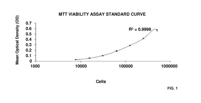
Figure 1: Standard curve for MTT viability assay based on a 7-fold serial
dilution with common
factor 2. Maximum value is 500,000 cells. R2 value represents 4-parametric
regression. Values
are represented as mean (n=3) STD;
Fig. 2: Human aortic valve leaflet interstitial cells showing myofibroblast
phenotype;
Fig. 3A: Cells seeded onto leaflet per surface area in pulsatile, cyclic
pressure culture;
Fig. 3B: Cells seeded onto leaflet per surface are in static culture;
Fig. 4A: Cells seeded onto sinus per surface area in pulsatile, cyclic
pressure culture;
Fig. 4B: Cells seeded onto sinus per surface area in static culture;
22
CA 02753684 2011-08-25
WO 2010/101962 PCT/US2010/025980
Fig. 5A: cells remaining in sinus after pulsatile, cyclic pressure culture per
cells initially
attached, normalized to surface area;
Fig. 5B: cells remaining in leaflet after pulsatile, cyclic pressure culture
per cells initially
attached, normalized to surface area;
Fig. 6: CD-68 positive staining (red) confirms macrophage differentiation
following PMA 400x
Fig. 7: TNF-a titers at all three times for all materials tested;
Fig. 8: T6F-(31 titers at all three times for all materials (sinus wall and
leaflets);
Fig. 9: IL-6 titers at all three times for all materials;
Fig. 10: IL-2 titers at all three time points for all materials tested;
Fig. 11: IL-1(31 titers at all three time points for all materials tested;
Fig. 12: Relative cytokine expressions by human macrophages after six hours of
exposure to
test materials (only controls and leaflets displayed for clarity);
Fig. 13: Relative cytokine expressions by human macrophages after 24 hours of
exposure to test
materials (only controls and leaflets displayed for clarity);
Fig. 14: Relative cytokine expression by human macrophages after 48 hours of
exposure to test
materials (only controls and leaflets displayed for clarity);
Fig. 15: Photograph of a decellularized valve that has not been recellularized
or implanted;
Fig. 16: Photograph of a pulmonary artery sinus wall decelled and conditioned
at 10 weeks post
implant in sheep;
Fig. 17: Photograph of a heart valve that has been decellularized only (no
conditioning) at 20
weeks after implant in a sheep;
Fig. 18: Photograph of a heart valve after pulsatile seeding of conditional
ovine pulmonary valve
leaflet;
23
CA 02753684 2011-08-25
WO 2010/101962 PCT/US2010/025980
Fig. 19: Photograph of normal native leaflet fresh;
Fig. 20: Photograph of a heart valve after static seeding; and
Fig. 21: Photograph of a heart valve recellularized using pulsatile seeding at
52 weeks post
implant.
DETAILED DESCRIPTION
The following examples are representative of preferred embodiments of the
present
invention. It is understood that nothing herein should be taken as a
limitation upon the overall
invention.
EXAMPLE 1
Significant drawbacks are present with each available prosthetic valve
replacement
including durability challenges, thrombogenicity, immunogenicity, and of
course, surgically
related risks. Further, none have demonstrated the capacity to grow or
remodel. A tissue-
engineered valve comprised of an extracellular matrix and seeded cells could
mitigate many of
these limitations. Although a number of scaffolds, both biologic and
synthetic, have been
considered for clinical valve replacement, a decellularized allograft avoids
many design and
antigenicity difficulties. Such a scaffold, re-seeded with appropriate
autologous cells, could
yield a tissue engineered heart valve (TEHV) capable of the growth,
constructive and adaptive
remodeling necessary to maintain tissue function for the life of the
recipient. To be clinically
useful, such a valve would need to be prepared within tolerable time
constraints, utilizing readily
available cells.
MATERIALS AND METHODS
Human aortic valve leaflet interestitial cells (hVICs) were isolated from
cryopreserved
aortic valve leaflets using .25% trypsin and two iterations of cell scraping.
Population purity was
confirmed after two passages using an immunocytochemical battery with
fluorescent labeling.
24
CA 02753684 2011-08-25
WO 2010/101962 PCT/US2010/025980
Human pulmonary cryopreserved valves were thawed using continuous flow sterile
fluid.
Intact valves were decellularized using a novel, multi-solvent, reciprocating
osmolarity, double
detergent, enzyme catalyzed protocol. Complete decellularization was confirmed
using H+E,
Movat's pentachrome and DAPI nuclear stains.
Leaflets and sinuses were surgically resected from decellularized human
pulmonary
valves. The sinus was defined as the region of artery wall between the cusp
base and sinotubular
junction. Each was divided into 5 mm x 5 mm pieces for separate assay. N=8
individual
biopsies per cell density were transferred into inserts of HTS Transwell-24
well plates. Biopsies
were seeded at one of three cell densities under static conditions for 24
hours. Static controls
were maintained in well plates for a total of 5 days. Pulsatile samples were
transferred at 24
hours to a novel cyclic pressure bioreactor with adjustable peak pressure for
four additional days.
When biopsy timepoint was reached, n=6 biopsies were taken for MTT cell
viability
quantification assay. N=2 biopsies were taken for histological and
immunohistochemical
analysis. For comparison, two clinically available, manufactured vascular
patch scaffolds
(Photo-oxidized bovine pericardium, expanded polytetrafluoroethylene) were
seeded using the
protocol described above.
For all variables, descriptive statistics (Means and Standard Deviations for
continuous
variables, proportions for categorical variables) were computed. Scaffold
types, seeding
methods and dose response curves were compared using single factor analysis of
variance and a
post-hoc Tukey test. A general linear regression model was used for repeated
measures (eg,
multiple time points). SPSS v.15.0 for Windows Statistical Package was
utilized. P < 0.05 were
considered statistically significant.
RESULTS AND DISCUSSION
Table 1
a. Static b. Pulsatile
c. Leaflet 894 84 80 +12
d. Sinus 838 50 79 +12
e. Pericardium 253 16 117+10
ePTFE 64 11 43+1
Table 1: 4.3x10³ cells/mm2 (2.5x10⁵ cells total) initially seeded and
incubated 120 hours
in pulsatile, pressurized bioreactors. Final cell numbers represented as
cells/mm2 (n=6) SEM.
No statistical difference between leaflet and sinus scaffolds. Seeding methods
yielded different
CA 02753684 2011-08-25
WO 2010/101962 PCT/US2010/025980
cell numbers for leaflet and sinus (p<.0001), but not for Pericardium or
ePTFE. Low and high
dose samples showed similar trends (data not shown). Pericardium showed less
inmigration than
sinus and leaflet scaffolds while ePTFE scaffolds allowed neither attachment
nor migration.
Figure 1 illustrates the standard curve for MTT viability assay based on a 7-
fold serial
dilution with common factor 2. Maximum value is 500,000 cells. R2 value
represents 4-
parametric regression. Values are represented as mean (n=3) STD.
Figure 2 illustrates human aortic valve leaflet interstitial cells showing
myofibroblast
phenotype. Immunofluorescent stain visualized with AlexaFluor 488. A. Vimentin
positive
(100X); B. alpha Smooth Muscle Actin positive (100X); C. Heat Shock Protein 47
positive
(100X); D. Endothelial Nitric Oxide Synthase negative (20X); E. Negative
control (Secondary
antibody only, 20X) demonstrates the absence of non-specific fluorescence.
Figure 3A and 3B illustrate the number of viable, seeded cells on leaflet
tissue at three
seeding densities in (A) pulsatile culture and (E) static culture.
Histological analysis shows
increasing cell penetration from (B) 24 to (C) 48 to (D) 120 hours in
pulsatile culture and
increasing cell number on tissue surface from (F) 24 to (G) 48 to (H) 120
hours in static culture.
Histology stained with H+E and imaged at 20X. Error reported as SEM. **
indicates statistical
significance (p<0.05).
Figure 4A and 4B illustrate the number of viable, seeded cells on sinus tissue
at three
seeding densities in (A) pulsatile culture and (E) static culture.
Histological analysis shows
increasing cell penetration from (B) 24 to (C) 48 to (D) 120 hours in
pulsatile culture and
increasing cell number on tissue surface from (F) 24 to (G) 48 to (H) 120
hours in static culture.
Histology stained with H+E and imaged at 20X. Error reported as SEM. *
indicates statistical
significance (p<0.05).
Figure 5A and 5B illustrate the number of viable, seeded cells on (A) sinus
and (B)
leaflet tissue after pulsatile, cyclic pressure culture per number of attached
cells after 24 hours at
three seeding densities, each normalized to surface area. Error reported as
SEM. * indicates
statistical significance (p<0.05) at 120 hour time point.
After five days in culture, leaflet tissues seeded with 2.5x10⁵ cells (median
dose) and
incubated in pulsatile culture were found to have an 11.2-fold decrease in
cell number from the
same time point in equivalent static assay. However, this quantitative
decrease coincided with
significant upregulation of cell motility and migration into the scaffold
(Figure 3A and 3B).
26
CA 02753684 2011-08-25
WO 2010/101962 PCT/US2010/025980
At the same cell dose in sinus wall, a similar trend appeared; 120 hour
pulsatile culture
yielded a 10.6-fold decrease in cell number compared to static incubation
(Figure 4A and 4B).
Although the absolute quantitative difference varied, static culture always
yielded significantly
more total cells than pulsatile at each timepoint, for all cell doses, in both
leaflet and sinus wall.
Evaluation of the cell quantitative data in context with the histology
suggests that this was
primarily a consequence of significant surface cell proliferation (Figure 3A
and 3B, Figure 4A
and 4B ) in the static environment.
Immunofluorescent labeling of cells growing in culture flasks revealed hVICs
expressing
Vimentin+, HSP 47+, a-SMA+, and eNOS- (See Figure 2). Immunohistochemistry of
scaffolds
incubated under static conditions for 5 days indicated phenotype expression of
Vimentin+, HSP
47-, a-SMA- and eNOS-, consistent with quiescent fibroblasts. Conversely,
experimental
scaffolds incubated under pulsatile conditions were found to be Vimentin+, HSP
47+, a-SMA+,
and eNOS-, consistent with active myofibroblasts.
Vimentin+ and eNOS- expression were seen across all tissue types, seeding
doses and
time points. At 24 and 48 hours of static seeding, HSP 47 and a-SMA showed
faint positive
staining that disappeared by 120 hours (data not shown). In pulsatile culture,
HSP 47 and a-
SMA staining appeared more intense progressing from 24 to 48 to 120 hours post
seeding.
Discussion and Conclusions
For leaflet and sinus wall, cyclic pressure incubation yielded fewer total
cells associated
with the scaffold than static incubation. However, the cells that remained
after pulsatile culture
had migrated into the interstitium and demonstrated enhanced matrix
remodeling. Given that
cells adherent to the surface detach from the scaffold shortly after
implantation in vivo,
protecting the cells from this fate by optimizing cyclic pressure-induced in
vitro migration is an
important variable affecting ultimate cell repopulation following orthotopic
valve implantation.
In addition to its direct applications to seeding a TEHV, this assay has
proven the
feasibility of variably optimizing seeding conditions using valve biopsies; it
can be used to
individually test each facet of cell seeding. This methodology allowed for
evaluating 576
individual scaffold biopsies under a controlled set of conditions with a
single assay.
27
CA 02753684 2011-08-25
WO 2010/101962 PCT/US2010/025980
Figure 6 shows cell numbers for sinus and leaflet in pulsatile culture
normalized to 24
hour values, essentially removing the effect of initial seeding and relating
the relative long-term
advantages of adding additional cells at day 0. Given the absence of
exponential divergence of
the high dose from the middle dose, we concluded that 2.5x10⁵ cells should be
sufficient to seed
a 5x5 mm biopsy of valve tissue and that future optimization should focus on
external conditions
rather than simply higher initial cell seeding dose. In Figure 6, CD-68
positive staining (red)
confirms macrophage differentiation following PMA 400x.
EXAMPLE 2
Currently approved clinical biological//bioprosthetic heart valve replacement
options (allografts
and xenografts) often result in reduced durability (likely due to innate
inflammation and immune
rejection and consequential calcification), ultimately leading to accelerated
failure.
Cryopreserved "viable" (i.e., containing donor cells) homografts as currently
used are known to
have limited durability due to inflammation and immune rejection resulting in
fibrosis and
calcification of the implanted valves resulting in valvular stenosis and/or
insufficiency. Efficient
decellularization can remove antigenic components from donor homograft valves,
perhaps
providing an antigen devoid of collagen/elastin extracellular matrix (ECM)
scaffold that retains
optimal structural elements of normal semilunar valves. Our group has
previously demonstrated
an absence of MHC-1 and MHC-2 positive antigenic debris following adequate
decellularization
of valves.
Decellularized homografts are clinically attractive as they surgically can be
tailored
homologously for size and location. They achieve immediate normal function
post-implantation,
and if not proinflammatory, may have the potential for prolonged durability.
If such
decellularized ECM valve scaffolds are not provocative of Inflammation other
than of the non-
immune wound healing type, then these may be suitable substrates for tissue
engineering of
viable valves (TEHVs) using ex vivo cell seeding and/or in vivo
recellularization methods.
If decellularized heart valves do not induce significant inflammation, then
they may be
ideally suited for use as an ECM based tissue engineered heart valve scaffold.
28
CA 02753684 2011-08-25
WO 2010/101962 PCT/US2010/025980
MATERIALS AND METHODS
Inflammatory responses to putative scaffolds materials were compared at 0, 6,
24, 48
hours of challenge by measuring with ELISA six key cytokine titers: TNF-a; TGF-
bl; IL-6; IL-
2; IL-lb-1. Cytokine expression profiles were standardized as very low, low,
moderate or high
and compared using ANOVA (P 0.05 = significant) statistical methods.
Scaffolds studied:
1.Cryopreserved (-180 Leaflets and sinus wall biopsies after the following
treatments: DMSO
clinical protocols) ovine aortic valves prepared with methods analogous to
current clinical
"viable" homograft valves designated: Fresh
2.Ovine aortic valves decellularized with a multisolvent, multidetergent,
enzyme assisted,
reciprocating osmolarity decellularization method: Decell
3."Freeze Fractured" ovine aortic valves that were subjected to three rapid
thaw (warm bath
37 C) alternating with refreeze (without cryoprotectants) at -80 C to freeze
fracture the cells,
thus maximizing antigen exposure (intracellular + cell surface sites): Freeze
Fractured (FrFx)
4.Manufactured glutaraldehyde cross-linked porcine bioprosthetic valve
leaflets obtained from
Hancock II clinical grade bioprosthetic valves: Hancock
RESULTS AND CONCLUSIONS
Inflammatory responses as measured by human macrophage cytokine release
profiles for
decellularized ovine valve tissues were similar to glutaraldehyde fixed
porcine leaflets despite
the former being both xenogeneic and neither fixed nor cross-linked. These
data suggest similar
initial reductions in inflammatory potential can be achieved simply by
removing all cells and
cellular debris. In contrast to decellularized leaflets, both fresh- and
freeze-fractured leaflets
provoked elevations of TNF-a and IL-6 which are both important to initiating
and regulating the
innate immune response. In contrast to decellularized leaflets, both fresh-
and freeze-fractured
leaflets provoked elevations of TNF-a and IL-6 which are both important to
initiating and
regulating the innate immune response.
29
CA 02753684 2011-08-25
WO 2010/101962 PCT/US2010/025980
This assay provides an in vitro verification methodology for evaluating
potential
proinflammatory characteristics of materials and tissue-derived substrates
being considered for
use as TEHV scaffolds. Optimal scaffold candidates can then be more
efficiently selected for
evaluation with subsequent in vivo animal models.
Our study findings have demonstrated that the macrophage cytokine response to
decellularized valve scaffolds is significantly reduced in comparison to the
response induced by
cryopreserved valves. The use of a decellularized valve, with reduced
inflammatory potential, as
an ECM scaffold for a tissue engineered heart valve is expected to result in a
reduced incidence
of fibrocalcification in the next generation of tissue replacement heart
valves.
Discussion
The muted inflammatory response evoked by porcine glutaraldehyde cross-linked
prosthetic valve leaflets suggests that the satisfactory clinical experience
in adults with these
bioprostheses may be in part due to delayed inflammatory response. However, as
the implant
duration increases, a decrease in glutaraldehyde cross-linking density and an
increased
inflammatory response resulting in leaflet calcification and structural
deterioration may occur.
Non-inflammatory mechanisms (e.g., elevated leaflet residual stresses,
collagen bundle
fracture) also contribute to the progressive loss of durability of cross-
linked porcine bioprosthetic
heart valves. Many of the currently implanted bioprosthetic valves have been
designed to reduce
the residual tissue stresses resulting in a reduction in structural
deterioration.
Older theories as to the mechanisms for the limited durability of cross-linked
xenograft
bioprosthetic heart valves suggested failure modes focused on mechanisms
related to physical
and chemical deterioration leading to calcium accumulation and failure. More
recent evidence
suggests an important role for inflammation and immune mechanisms modulated by
recipient
factors such as age, immune competency, treatment with antirejection
medications, etc., as the
primary pathway to fibrocalcific degeneration. These data are consistent with
a masking and
unmasking of antigen sites.
These assay results suggest that decellularized valve scaffolds may have
significantly
reduced inflammatory potential and thus promise utility as platforms for
tissue engineering
replacement heart valves (TEHV). Since a putative clinical TEHV could be based
on human
ECM scaffolds rather than xenogeneic (e.g., ovine or porcine) results may have
even been better
than demonstrated in these experiments. Late protection might be conferred by
the seeding of
CA 02753684 2011-08-25
WO 2010/101962 PCT/US2010/025980
autologous valve interstitial cells capable of ECM protein
degradation/synthesis and thus active
constructive and perhaps adaptive remodeling.
EXAMPLE 3
Addressing mechanisms fundamentally related to native and prosthetic valve
degeneration, the purpose of this study was to compare human macrophage
cytokine provocation
profiles of candidate materials for therapeutic use in reconstructing the
human cardiovascular
system and specifically for optimizing the scaffold component of tissue
engineered cardiac
valves as compared to current generation bioprosthetics for which clinical
outcomes are well
known. Because one attractive pathway for the development of a tissue
engineered heart valve is
the use of decellularized ECM valve scaffolds derived from cryopreserved
cadaveric tissues,
allograft and xenograft sources of valves were especially examined.
Materials and Methods
Test Materials Preparation
Human activated macrophage cytokine inflammatory responses over 48 hours were
measured for biological samples obtained from porcine, ovine, and human aortic
valves (nine
valves for each species). Because of the significant differences in
microstructure, leaflets and
sinus wall samples were analyzed separately (e.g., absence of vascularity in
leaflets, versus blood
vessels, smooth muscle cells, pericytes and fibrocytes in vessel walls). The
mammalian valve
tissues were prepared in three ways with three valves allocated to each
protocol. First, following
aseptic harvest from juvenile animals, the ovine and porcine fresh valves were
cryopreserved
with preservation of cell viability utilizing a 10% DMSO in RPMI 1640
(Invitrogen, Carlsbad,
CA) with 10% FBS (Invitrogen), cryopreservation at 1 C/min, a technique
analogous to the
preparation of current generation clinical cryopreserved heart valves (cell
viability retained).
These were designated as native, valve tissues further identified by species
of origin. Another
set of valves were harvested then subjected to freeze-thaw for three cycles of
21 C to -80 C
without cryoprotectants. Cycled freeze-fracturing results in massive cell
lysis, thereby
potentially increasing overall antigen exposure. These were designated as
freeze-fractured valve
tissues. And finally, freshly cryopreserved animal heart valves were subjected
to
31
CA 02753684 2011-08-25
WO 2010/101962 PCT/US2010/025980
decellularization using a multi-solvent, multi-detergent, enzyme-assisted,
reciprocating
osmolarity method in which essentially all cells, cell debris and nuclear
material were removed
leaving only a structural protein ECM scaffold. These test materials were
compared to two
routinely used "inert" materials, nitinol and PTFE for which there is
extensive clinical
cardiovascular implant experience. Biopsies were obtained from each test valve
from each of six
sites (the three leaflets and the three sinus walls) and samples weighed for
subsequent
normalization of measurements to wet weight. Control samples were sized to
equivalent weight
samples as the biologics. Nitinol (sterile) was obtained from an Amplatzer TM
size 5mm Septal
Occluder (SN 151438 A6A Medical Corporation, Plymouth, MN, USA) and the PTFE
was
harvested from a sterile GorTexTM 4mm thin wall vascular graft (W.L. Gore &
Associates,
Neward, Delaware, USA). Comparisons were made to materials from two current
clinically used
porcine glutaraldehyde crosslinked FDA-approved aortic valve bioprostheses
(leaflets and sinus
wall from aortic Freestyle stentless and leaflets from the stented Hancock II
, both from
Medtronic Corp., Minneapolis, MN). Human aortic valves were obtained at multi-
organ and
tissue harvests with informed consent for both research and clinical use,
processed with
cryopreservation under AATB guidelines by LifeNet Health Tissue Services
(Virginia Beach,
VA) and stored at -180 C; once out of date for clinical use, the valves were
released for research
use. These were prepared similarly to the test animal valves. All experimental
animal materials
were obtained with IACUC approval and in accordance with NIH, AALAS, and
American Heart
Association guidelines for the Care and Use of Research Animals.
Macrophage Cytokine Assay
Human THP-1 monocytes (ATCC -TIB-202TM, Manassas, VA), were obtained and
prepared in suspension culture per ATCC protocol. Cell counts for each
suspension were
obtained using a automated cell counter (Coulter Counter , Model Z3, Beckman
Coulter, Inc.,
Fullerton, CA) and plated at a concentration of 1x10⁵ in each well of 24-well
plates (Becton,
Dickinson, Franklin Lakes, NJ). Monocytes were differentiated into macrophages
utilizing
PMA/TPA (Phorbol 12-myristate 13 acetate: Sigma P8139, St. Louis, MO). Plates
were
incubated for 24 hours at 37 C, 21% 02, 5% CO2. Cell differentiation was
confirmed with CD68
(Abcam-AB955, Cambridge, MA) immunocytochemical staining of a representative
sample well
from each plate and photographed (Figure 6). Macrophages were incubated in
RPMI 1640 with
32
CA 02753684 2011-08-25
WO 2010/101962 PCT/US2010/025980
10% FBS (THP1-growth media, InvitrogenTM, Carlsbad, CA). Two hours prior to
assay, the
macrophages were activated with LPS (lipopolysaccharides-Sigma L6261, St.
Louis, MO).
Baseline cytokine titers (time 0) were obtained to establish the priming
effect and were
subtracted from the subsequent measurements to calculate the provoked cytokine
expression.
Each prepared test specimen was placed in its respective well of 24 well
plates. At the defined
timepoints (6, 24, 48 hours) the entire supernatant from wells were harvested
and each well used
for a single ELISA. Each data point was determined using three wells/cytokine
for 6 biopsies of
the test type (ie, n=18 determinations). Control and test biomaterials were
assayed for all five
cytokines at each timepoint. Cell supernatants were obtained and frozen at -80
C. After
collection of all supernatant samples, the cytokine assays were analyzed in
batches by ELISA in
triplicate for TNF-a, IL-2, IL-6, TGF-(31 and IL-1-(31 (Quantikine Assay
kits, R&D Systems ,
Minneapolis, MN). Curves were constructed from measurements using standard
controls.
Dilution expression standards were measured with each assay run per kit
instructions.
Quantification was performed at wavelengths of 570nm and 450nm (correction
wavelength) after
twenty minutes incubation with the specific cytokine conjugate substrate.
Based on reference
standards provided by manufacturer, each cytokine titer was then ranked on its
own standardized
expression scale from very low to very high.
Nine aortic valves from each of three mammalian species (n = 27) were
randomized in
groups of three to each of the three preparation methods. Leaflet and sinus
wall tissues were
separately analyzed by assaying six separate tissue samples randomly biopsied
from all 3 leaflets
and sinuses. Titers were measured at three timepoints (6, 24, 48 hours after
zero baseline). Each
of the five cytokine assays was run in triplicate. Total assay determinations
n = 6480. Six
samples from each of the "inert" controls (PTFE, nitinol) and the two
bioprosthetic valve
materials were assayed at the three timepoints with each cytokine specific
ELISA performed in
triplicate (n = 1800).
Histology
Standard sections of each of the test materials were prepared with
HistoChoiceTM MB
Fixative (Amresco, Inc., Solon, OH), paraffin embedded and examined
histologically with
Hematoxylin-Eosin (H&E) staining. Collagen and elastin structures were
visualized with
Movat's pentachrome staining (Figures). (Mastertechs, Lodi/CA, USA)
33
CA 02753684 2011-08-25
WO 2010/101962 PCT/US2010/025980
DNA Content
The decellularization process was evaluated in an additional six ovine valves
by
measuring residual DNA as compared to native fresh aortic valves using a dsDNA
High-
Sensitivity assay kit (Quant-itTM, Invitrogen, Carlsbad, CA), with each
measurement in triplicate.
The histology and DNA quantification verified essentially complete
decellularization (Table 1).
Statistics
Continuous variables were analyzed for differences between groups with two-way
ANOVA (Tukey Kramer and Kruskal-Wallis tests) while ordinal variable were
analyzed with
Wilcox Rank Sum. Student's T-test was used for paired single comparisons (eg,
DNA content
before and after decellularization). (SPSS v.17) P<_0.05 was considered
significant. All
cytokine quantifications are reported in the tables, but because wall and
leaflets tracked
similarly, for clarity the reported figures display leaflet comparisons and
statistical tests unless
otherwise noted.
Results and Conclusions
Table 2. DNA Quantification Assessing Decellularization Method in Ovine Valves
Leaflets Sinus Wall Aortic Wall above valve
Native valves 0.794 0.176* 0.557 0.074* 0.586 0.119*
Decellularized valves 0.000 0.000 0.000 0.000 0.001 0.001
*DNA ugm/mg tissue weight 1 S.D. Students T-test native material versus
decell *
Note that 0 reflects values below the detection limits of the assay.
Cytokine Provocation for Each Biomaterial
The ovine and porcine tissues provoked higher cytokine protein expression than
did
human tissues (Table 3). The highest titers tended to be at six hours with
decay over the ensuing
48 hours. Decellularization reduced the provocation for all mammalian types
but especially so
for human tissues. The "inert" materials and glutaraldehyde-treated porcine
valve prosthetics
had very low titers which were closely matched by the decellularized human
tissues. For each
tissue type, and processing method at each timepoint, the leaflet and sinus
wall results trended
similarly but the titers for wall samples tended to be slightly higher than
their analogous leaflets.
34
CA 02753684 2011-08-25
WO 2010/101962 PCT/US2010/025980
Elevations of TGF(31, TNFa and IL2 had the more prolonged expression profiles
whereas IL1(31
and IL6 typically had returned close to baseline by 48 hours. Freeze-
fracturing of the biological
materials consistently generated the highest cytokine levels. Decellularized
human tissues
provoked much lower cytokine signaling than did the porcine or ovine
decellularized materials.
CA 02753684 2011-08-25
WO 2010/101962 PCT/US2010/025980
0 0 0 0 0 0 O O O r ro n 0 0 0 0 0 0 00 000
0 0 0 0 0 0 O O O r r +I 0 0 0 0 0 0 OO O O O
+1 +1 +1 +1 +1 +1 +I +I +I +I +I N +1 +1 +1 +1 +1 +I +1 +I +1 +1 +1
co oo o00 co o OTcq 0 coo0000 co gq co IooO
6 V 0 0 0 0 0 0 V 0 0 0 co N. r V 0 0 0 0 0 0 V 0 0 It 0 0 0
E
U CO N
CO M CO r M M r CO 0 O T N T
O O N V 0) a M M V V 0) r m M MID a O O r O O O
0 0 +1 +1 +1 +1 +1 +1 +1 +1 +1 +1 +I +I +I +I 0 0 0 0 0
. +I +I 0 OD l0 0 a a to O N a T +I +I O O +I +I +I +I +I
V q 0010(0M .ODOR InM r v 010N O r V 00 r V 000
O T -N1 00 r N In t0 J_ NI N M t0 N w o -J NI N r co N In CO -J (N O O J NI O
O O
O CO 0) V CO 0
_ 0 CO r N
9 N lp N N N r ID r Ti T In V In (O N O O
N N N +I 0 0 +I +I +I +I MLA l0 CO +I +I T N N N In
+I +I +I +I +I r +I +I N V M V +1 +1 +1 +1 T ID cc +1+17,
co co co co N t0 M O O N N t0 IAN In 1: to +1 +1 T V Y
M ICS T O V T 0 = T 0 0 CO CO = O to O M O N 2 = r
W N r O O O r lOI V V r r r r l0l CO
V V CO CO r r l0 l0 CO lOl r r N
N 0 lD co lD CO V r N 0
O V O V 0NN M O N O O O Nr M r q CV r N cq
a;-- In w co co T N O O O)
In 7 v +I I I +1 +1 +1 +1 +1 +I +I +I +I +I +I +I +I
+1 +1 +1 N ID w 0In Nr e l0 rN O) 0) MT 0 0 +1+1
M N13: I n I n N 0 0 N N N T N N CO = +1 +I = N- O Ni
TO N N0 V TO) O N N CO 0 0 0 N 0 CO co q O co 0
N
W T T r N N V r N r r N N V N N r r N N C O O V co OJ ~
N O) r T co O
N
2 M N N O) Inr N N NOM V V m V MO +I CO
N T!D(DMT N TT N TNO N N 0. N M v
+1 +1 +1 +1 +1 +1 = +I +1 +1 +1 +1 +1 +1 +1 +1 +1 +1 0) = co . CO l0 +1
O J r0 I0 M MN-J I- rT V RT J mCon V,CO0 J 0+1 J +1+17
24 1:4 M a - 2O as m V -2l06 -* CIO -=+1 M -=N I0O
O) VIN M T N N V VIO N M V N In VI co 0 w In In r VIM T 7I0 T
Nr rr N(DO NM MIn In OD OD N N V NMO)r N0)r N0)0)r
A N
ODO NIOr rN N MN M
~ON IA 0OD01nf:M NOR V CON MOM
V V T NrT m0 M 0 +1 V 0 N In
N +1 +1+1+1+1+1 +1 +1 +1 +1+1+1 +1+1+1 +1+1 V N N +1+1+1
C V N N N N ID CO r N 0 ID CO V CO r CO M +1 +1 CO CO V
V MODTlD NIO In IOMN V M V In O N w O V M
= I n (O r O =TON V r 0 2 l n O N R N =1 0 = r N N
O (0 V V V O D N W (O M CO (0 l0 T T (O M CO N N 0 I00 OD T IDI r r r
O T N M M M T IO O M N O (O
N M M In m r N V If) Co ll7 0000 r) 00 000
+1 +1 +1 +1 +1 +1 +1 +1 +1 +1 +1 O O O O +1 +1 O O O O O
CL =+1 NN V NN =TN , t0MN =+1+1+1+1 NO =+1+1 =+1+1+1
G r0)-Ma INC CI,-N10n ITn!0000 CI0000NN CIOO CI000
C.I r V
T IO,ONM TT V, In CO r,WO TN
A O N M O V 0 N. (0 R co W +I l0 7 IA M In In IC) l0 OD l0 N
+1 +1 +1 +1 +1 +1 +1 +1 +1 +1 +1 N +1 +1 +1 +1 +1 +1 00 0 +1 +1
0 =O NIO r ION =ln Of~IDN TO IO rT V ' =+1+1 I +1 V N
V O(0 co T CO V N 0 0 0 0 0 J V CO V 0 0 T N V 0 r J V N N CO
NN NIO(0(ON NIONT00) N In ION V V N NIoM NCO,
U O N In
T T In M V O OT
M CO CO = T V co co 0 0 0 0 N N G )o O T IO
V M M l0 +I +I l0 l0 +I +I +I +I l{1 +I Lc; I0 +I +I O N N N
U +1 + 1 + 1 + 1 O D +1+1 V 00 V r +10+1+1NOD +1+1 +1+1+1
NNTMOO nMODNO ":Is V Or)0.) ODT V IOM
w =I O T r N =ID N O,- M In =I T O M T N CO =I In ID NN T
r ODrr lOrr lOrNr
V IO M MO)Trr T co T r rr T co N
C
C) tO OD co N
(p N NNO~r ODOIn V Mr NCMNON M
In t0 +I R 0 n In +I +I +I 4 I6 +I +I O O (D V N
+I +1 +1+10 +1 +1 +1 +1 co CO +1 CO +1 +1 TOD 0 0 +1
IDOr C V =IDOODTr.n =M O IDN V V =+1+1 =++IM
TI O T N co M TI OD V 0 In CO N O T T CO CO OoI O O TI M N O
V r r lnCOTr V Ior- N T NN V IDr O0rr V 00 V 0 f:
=0
COO (ONIOM V N
C 0 M T N. N N I` 0 O C R O O n 0 O In r M O 0 0 N. N.
M CO +I +I T +I +I N +1 co +I V In +1 OD +1 +1 +1 +1 +I
0 0 N
LL + 1 +1+1 +I +1 OD V LL +I + 1 + I N In MN LL +IN N Mr OD LL 00 LL r+1+1
'O Z=r N N Z 21D. W CO O N- 0 Z2O 0 O V M N Z=+I+I Z=+INIO c6 ui ` F VNIM O V
T WT r N CV 0) V N F VI In r- In lD N T VI O r O M M a F VIO O VI0 r C
r r r N co co r r N N N w r r r N N N O O N T
t; 11 A n co O
_ 0. V N 0-0. M 0'i N N N O
N +I Vrl: W N N oi OD O In
N CO +I + CO +I CO V +I +1 +1 ID 0 ID N N
N +1 +1 0 t0 0 CO +1 In V V N +1 +1 CO V 00) 0 +1 r +1 +1
N M r7I6 1:M NOLo 0COIO rr COM +1T +1N-M
y =IC) CO N M 0 V STON N TO S T N- O N CO 0 S T CO = V N
IOIV V r r cm N IDI T r r r cm M IOIIO 0) N 0 0) , IOI N T r
N 0 N C) co NIn v 0
M V OMN V Nle V NID NTTNCOO lnN V
N N T 0) N N N ID CO to CO
N M +I +I +I +I +1 +1 +1 +1 +1 +1 +1 +1 +1 +1 +1 +1 0 O N M 7
+1 +1 m N N T N N co IO V V 0 co N N CMr 0 0 +1 +1 +1
=V TO o = m V OONN =44Tm m =+1+1 =In MT
CO 0000 N mN M CO (OTO CO NCD V lOM CO co 00 co to0 co
C
V In Inr N toC V rrNNlnO V NNrrNN V 00 V cm MN
E CO O In r-: O IO CO CO CO CO
E 7 r r T CO N N 7 N T T M N 7 T N !O N V V N
LQ 0- CO CO +1 +1 +1 +1 0. +1 +1 +1 +1 +1 +1 +1 +1 +1 +1 +1 +1 C. 0 0 N a CO
LL +1 +1 V NODCD LL N M 0 W0)Ln LL TCIOI- r0 LL 00 LL +1+1+1
0 = N V V
NN M N N O t0 = r N I: M O M M T r O M r V = 0 +1 +1
T MMNIOC V (0CM0NCO V mN V 7r0 a V 0c0
O ~ ~ In ONN0w N V V N N N N
~ rr ~ rNNInO 000 N V V OD
T co T In T tD O M V N
O In V M T M 7 V. O O l0 N O N lD
. N O T N M I n N I Lc; N r O (D V r T
M -cr r+I +I +I +I +I +171 +I +I +I +1 +1 +1 +1 +1 +1 N r 6 m M
y +1 +IM co Inr COON V NN V NCON- CO +1+1 +1+1+1
CL 0 OR OONN rT T(OMN N(0 0 N V OD N N0
K l=OI0000(OIOD OOD M IOINNN- NTO l=OI ,-'r0 CO (C O CO (DDICMc-Lni OTD
c
C - C - C C N C
O C -
W 1 0 w > o O) Y O V1 0
o. o. w D. CL -a
F y o.
J O d N d N d > a) N N N CV W
F A N y C U F d N y C > F ?) N y C F (n i.
O U (O C -J U) F l0 -J N o J (n W 0
Y r a._ o o "- -a -a -o-o Qa
O J to a s J N a a J N a a Q LL t a
,Oa m aidrn~~ Q a~ia~idrn ~ O a~ia~iay ~ 00 a~OiE~
z V N N- 7 G U W N N- 7 G V Q N N- 7 U U r m J (I)
O) tO J (n LL LL O7 lO J (!ILL LL Z O7 lO J (n LL LL W Z Y N N J
fr) Q Z
2
d) ? 0 0 N N = ? a N w w a) CD N N N Z O 3.2 a)2
= U L)) a s 0 V V? ID O) 0 ~? ? a a LL c ma c c c
A m a) N lO i LL a) (D N N i a) a) N N i i - 0 N (U d)
OOZZLL LL OOZZILLL OaZZILIL (LZ LL NNT
36
CA 02753684 2011-08-25
WO 2010/101962 PCT/US2010/025980
0 0 0 0 0 0 0 0 0 0 0 0 0 0 0 0 0 0 0
000000 000000 c o o 00 00
+I +I +1 +1 +1 +1 1:1+1+1+1+1+1+1 =I +1 +1 +1 0 0 0 +I +1 +1
2 2
W O o 0 0 0 0 co o 0 0 0 0 o co o 0 0 0 0 W O o
Z 7 0 0 0 0 0 0 7 0 0 0 0 0 0 7 0 0 0 00 7 0 0
E OD
V T M W co W
O O N 1*1 M O O M N,6O O O O O r O O
OO+1+1+1+1 C2 00+1+1+1+1 tl. 000 00 00
t r=+I +I W W 10 OR +1+1 r M 7 r =+1 +1 +1 +1 r=+I+I
7I0 CO 0M 7I00~NIT X000 RR 7 7
N IOO
O J N O O N n W J N O 0 N M o W J N O O O 00
J N O O
Q M
r N W N r r 0 7 7 m 0 0 W
N Nrr+ rNr+I+I ,-N N r0 Or
+I + 1+ 1+ 1+ 1+ 1" +1 +I+I r +1 +1 +I+I!D W O) IT +I +I+I O+1
OD W MOD T T In +1 T +1 +1 +1
Y =I C)TC)zi TO =1 C6 Lo Ovi =II`.~CM Tr 100
!D (N 0) 0) T r 1D N N 0) T r r W r r r CO 1D W co
N o to o
0 Lon;NN 7 W OD W <D Tr N M T
In RR +1+I +I C +1 +1 +1 +1+1 +17 7 N 00
V +1 +1 +1 N- w +1 T N rC n r-H +1 +1+1 0 0
v 2IM NIf InN =I D.MrrrT =IM r' NN +1+1
COT NrN07 W TOM77 W W Nr O 00 CD 00
T 7 OD T T r N N 7 T r r r N N 7 r N- OD OD T 7 0 0
N W T W
M N N T In In In O Lri Lfi CO O M
OD N 01010 OD W N 71n M N 7r M N N W
+1 +1 +1 +1 +1 +1 +1 +1 +1 +1 +1 +1 +1 M W +1 +1
T J r 0 1D M M f- J M W M 7 W 0 J ii +I W 7 J C) +I
3 -2I7 f~7M7 '16 6r.: W -=CONS Ml0 2I QjM
7N MTNNIT MRIn Binh :,.1 W o T Or i
N r r r N W W N r N N W W N T T N T
W 0 7 r T 7 T M
RON~M 0co MM 7 (D
7oir nice M 7 lD l0 ui N- ai ni 70
+1 +1 +1 +1 +1 +1 +1 +1 +1 +1 +1 +1 +1 -H +1 +1 +1 N N
IT Nrf-1-10 Nr7 W M0 IOC 7r +1+1
IT M W W W IT 0 0 N N In M W IT O N W
C 211n W W 0 =1Wf~Mn00 =lr+N M7 21TC
!Dr rR7r0 !D rr77 co co l0r r rr 1D co 0)
.2 O T N W co T co co co co IT
O O O O O O O
V N M M L6 M O r M 710 (6
0 +I +1 +1 +I +1 +I +1 +1 +I +1 +1 CJ CJ CJ 00 00
=1 +I N f~ *: r-: N =I O f:M T In q = +1 -H +1 +1 +1 = +I +1
O_ RM Or MR INS R0 00Lo 0 ~T 0) 10 oW X000 00 RIOO
E
O 0
T W r T N M W W 7 7 W 0 C T 7 M
to N M W 7 In C- l0 N N 7 7 CD +1 l0 N 10 W W
+1 +I +1 +1 +I +1 +I +1 +1 +I +1 (0 +1 r +1 +1 +1 0 0
N J20 N(Dr10N J=ODN OD TRN J C4 -H 7 OD0 J=+I+I
V -7Io co co T OD -7IMr CO NMr - 7IWNN +W 7IO
NN N10 W W N
f- NN r W T r NrOD r r NLo M
O 7
r 7 r
M M W r r 0 7 OD 0 r N O T MOD 00
MM 1D+1 +1 MM +-zr 1+1 +1+1 NN N O
+1 +1 +1 +1 W r +1 +1 0) N r 7 +1 ii +1 +1 +1 +1 +1
r rTMT1o Tr7C-MN M7(0 N10 co T
W 2 O T,cm InO+MI- STN N W M 21i 0
0 M M M 0) W 7 r r r ID r r N r N 0
N T O r
r co
N N N O r r W N
+1 10 7
r In
+1 r r+17+1 W 0 00
+I +I +I +1 +I 0 O +1 +1 +1 +1 N 0 O C O +1 O O
= 100 r W 7 = co W W M =M +I +I +I =1 +1
COIr -OD C- co Mr ODI T 1n W T NON WIO M N- ON OI OO
V 7 r r W W T 7 r r W r r r 7 W r r r 7 0 0
O 10 n
OR
a a M T C N N r N 0 ci 7 7 r
I- l0 +1 +1 ' N +1 +1 +1 +1 O 0
LL +1 +1 +1 +1 co 7 LL +1
+I W X N N LL +I r +I 0 0 LL 0 0
Z=M 7rrNN Z=ODr OIn lDr Z = W +IT Z=OO
~7IOTN N7W I1nOO+lDT 7IO r i I
N r r CO T r r N N M r r r N r T r W N 0 0
r M In
O M T
i 0 7 r r
W L 0 CO N rIn +1 W W N W
+1 +1 +I N 10 N r l0
+1
+I 1 +I+iO W W O M +1 +1 +1 +1 C +1 +1 +1 +1 O +I 0 +1
N + N +I
M 7 rM M0T TM M+1 r +1r +1 co
i =11- CM N 0 7 o11- r N In =1 CV r 0 M 21 T M
lD 77r NN 10 rrrr NN ID rT r co W01r
r0NT co rNIn
M R 0 M N N 7 0 r W N L6 C6 N 7 to N. Co
0
(0 CV C,+I+I+I+I f`MR+1+1+1+1 qc' C"i CV N 00
0 +1 +1 W r r T +1 +1 W IT T M +1 11 +1 +1 +1 O O
_~ OR O In N 27 0 W 7 W T T In M O N +1+1
OD 7IIn I , Nl0n RI 10 0 N N 01N0 VI r NM MN RI 0 0
M O r O
r r T W N N M O m r T W 7 r IT N In r 7
OO
E W ' m C'i+I +I+I +I C2 MI-+I+I+I+I MN 7 7l6
E LL +I+I7r CO W LL +I+1 W rrN U. +1+i +1 +1+1 LL 00
o Mir,: n) r N 010 O =I W T r L t C6 zi =I 7 N 7 Ili W 0 7 =I
7 O O
T M ln r W W IT W to 0N r 0 7 00 06 Ol r
N O 10 N N 10 0 N 10 r m M 0 10 N co 7 7 7 ICJ N O O
C O W T W 7 r
In 0 In co T 7 (D 0) 0 W N 0) Q - N O 0 1D
CM R+I+I+I+I +I+I+I+I+I C6w C6 MN N
+1 +1 M W n r +I co 0) M M W +1 ii +1 +1 +1 +1 +I
0 0 0 T N r 0 0 N N W T Orr r r W N
Q =l (0 N 0 010 10 =l M 0 N N T T =1 CO C6 M C6 06 =1 T 06
DC 1D W T W W W W 10 T r W W W W W W 7 0 ICl 10 1D 7 M
O)
0_ c _ c _ N c c
0 lL 0 _ m U a y 0 0 0) 04 cl) U) V a)
Q Q r" E d y J i. N dl y 0 E N N V co -j
._..~ ¾U._..~ 0 r0V UQ0 QV V._
F N y d ] F d y y C N , F¾ r2 ¾ r2 ¾ F
C U _ J N U (O = _ J N O d rL rL W
Y F N a s F N a s 2 a r
= J to y y Er J N a s IL t a y y
0 aaa~>> O 0-00 ~'>> Oa m= = 2
V Q N N- () () ¾ N N- j () () [O a J CD CD
to F
z (D lL0 (0 lL0 i A d J N
1E 1E N 0 1E l0 N W J N N N =
7 7 7JNLL LL >>J(nLL LL Z.~, N N N N lL
N N N O) N N O) N V 7 N N CU N Z
CD d y <
y Cl >> y y M (S' a a r- a s W C
a OUl N (O N i O O N N i N 0 N OI N N N F
ZZLLLL ma ZZLLIl 0 (n (n N (n V)
a.z
37
CA 02753684 2011-08-25
WO 2010/101962 PCT/US2010/025980
Specific Cytokine Expression
TNF-a
TNF-a titers for all test and control samples at each timepoint are tabulated
in Table 3
and displayed in Figure 6. At each timepoint, both the decellularized leaflet
and sinus wall
components of the aortic valves had significantly lower TNF-a responses as
compared to freeze-
fractured (P < 0.05) and native tissues (P < 0.05) within each species. The
inert controls, the
human decellularized and the glutaraldehyde crosslinked porcine biomaterials
provoked the
lowest TNF-a production. However, unlike the decellularized xenogeneic
tissues, the TNF-a
response to decellularized human was very low and fell towards negligible at
later timepoints.
There was prolonged elevation of TNF-a for the xenogenic tissues. While native
human tissues
provoked lower expression than native ovine or porcine, the freeze-fracturing
treatment elevated
the response for all three suggesting that with intact or fragmented cellular
material, increased
antigen recognition indeed was present.
TGF-(31
TGF-(31 titers were significantly lower for the decellularized tissues (Table
3 and Figure
8), and most notably for human decell versus native (P < 0.05) and freeze-
fractured (P < 0.05).
In contrast, the TGF-(31 responses at later timepoints were elevated for the
xenogenic tissues
suggesting ongoing stimulation, perhaps reflecting the additional signaling
functions of TGF-(31,
which include wound healing and inflammatory amplification. While the porcine
and ovine
decellularized tissues were similar, the presence of cells (either freeze
fractured or native) tended
to result in higher titers for the porcine tissues as compared to ovine.
Figure 8 illustrates T6F-(31
titers at all three times for all materials (sinus wall and leaflets). Very
low stimulation provoked
by human decellularized tissues similar to inert and glutaraldehyde
crosslinked. Note that T6F-
(31 signaling similar to the xenogeneic tissues, suggesting prolonged innate
immune and more
aggressive wound healing responses. Decellularization of the human tissues
eliminated the T6F-
(31 responses at all three time points as compared to native human and decell
human (*P<0.05 as
compared to native tissue from some species at same times; + P<0.05 as
compared to PTFE and
nitinol at same times). In Figure 8, the left vertical axis is pg cytokine/mg
test tissue; the right
vertical axis is standardized cytokine expression; and the error bars =
I.S.D.
38
CA 02753684 2011-08-25
WO 2010/101962 PCT/US2010/025980
IL-6
IL-6 expression was relatively short-lived for all test samples, always being
maximal at
the six-hour timepoint for each tissue preparation. (Table 3, Figure 9).
Decellularized human
leaflets provoked a medium low response at six hours and very low responses
thereafter but
significantly less than the native or (P < 0.05) freeze-fractured human
leaflets (P < 0.05). The
uncrosslinked xenograft materials provoked higher IL-6 titers than did human.
Inert and
glutaraldehyde treated materials had the lowest and briefest expression.
Figure 9 illustrates IL-6
titers at all three times for all materials. Very low stimulation levels were
provoked by the
glutaraldehyde treated materials, PTFE and nitinol. The 6 hour levels for the
human
decellularized barely edge into the low expression range. The glutaraldehyde
treated porcine,
nitinol, PTFE and decellularized human provoked the briefest expression of IL-
6.
Decelluarization did not eliminate or significantly reduce IC-6 signaling
provoked by the
xenogeneic tissues as compared to their respective native unmodified tissue.
(*P<_0.05 as
compared to native tissue from same species at same times; + P<0.05 as
compared to PTFE at
same times). In Figure 9, the left vertical axis is pg cytokine/mg test
tissue; the right vertical axis
is standardized cytokine expression; and the error bars = I.S.D.
IL-2
IL-2 expression was minimally provoked by human decellularized tissues but was
markedly stimulated by the native and freeze-fractured human and by all
uncrosslinked ovine
and porcine tissues. (Table 3 and Figure 10) The glutaraldehyde crosslinked
bioprosthetic
materials and the inert controls were again low stimulators although
glutaraldehyde did not quite
completely blunt the porcine bioprosthetics as compared to the inert
materials. Figure 10
illustrates IL-2 titers at all three time points for all materials tested.
Human decellularized,
PTFE, nitinol, porcine glutaraldehyde treated test tissues all remained at or
below the boundary
between low and very low expression. IL-2 expression fell off rapidly for all
samples after 48
hours consistent with the early signaling role for this cytokine (*P<0.05 as
compared to native
tissue from same species at same time points; + P<0.05 as compared to PTFE at
same time
points). In Figure 10, the left vertical axis is pg cytokine/mg test tissue;
the right vertical axis is
standardized cytokine expression; and the error bars = I.S.D.
39
CA 02753684 2011-08-25
WO 2010/101962 PCT/US2010/025980
IL-1(3-1 Expression
With profiles somewhat similar to the IL-6 expression, the IL-1(3-1 production
was, as
expected, relatively short lived and minimal for the decellularized
(especially human), and the
crosslinked materials (Table 3, Figure 11). The freeze-fractured xenogenic
materials provoked
the highest responses; porcine trended higher than ovine. Inert materials
provoked negligible IL-
131 Expression. Figure 11 illustrates IL-1(31 titers at all three time points
for all materials tested.
Human decellularized provoked low expression at 6 hours but rapidly fell to
zero, whereas the
PTFE, nitinol, and glutaraldehyde treated tissues expressed at very low levels
at 6 hours then fell
to zero. IL-1(31 was the briefest cytokine expression documented (as
expected). Porcine native
and freeze fractured seem to elicit higher responses than ovine native and
freeze fractured
especially at the later time points (24 hours, 48 hours) (*P<0.05 as compared
to native tissue
from some species at same time points; + P<0.05 as compared to PTFE at same
time points). In
Figure 11, the left vertical axis is pg cytokine/mg test tissue; the right
vertical axis is
standardized cytokine expression; and the error bars = I.S.D.
Multi-cytokine Time Dependent Expression Profiles
It is easier to visualize the time dependent expression with transformation of
the absolute
cytokine values to cytokine specific ordinal expression levels depicted as
three dimensional
profiles for each different material at each sampling time. (Figures 12-15).
For example, the
similarity of the 6, 24, and 48 hour profiles for the decellularized human
tissue to the inert and
crosslinked controls is readily apparent in these figures. In contrast, the
human freeze-fractured
and all xenogenic tissues elevated titers provoke higher, earlier, and with a
slower decay. Each
cytokine has a specific time course consistent with their putative signaling
roles (eg, IL-1B-1
rapidly disappears, whereas TGF-B 1 remains elevated). Figure 12 illustrates
relative cytokine
expression by human macrophages after six hours of exposure to test materials
(only controls
and leaflets displayed for clarity). In Figure 12, the Z axis = cytokines
measured; the Y axis =
relative expression; and the X axis = leaflets and control materials tested at
this time point. At
six hours, the freeze-fractured materials, as expected, had the highest
stimulated expressions
followed by the native. (Figure 13) Figure 13 illustrates relative cytokine
expression by human
macrophages after 24 hours of exposure to test materials (only controls and
leaflets displayed for
clarity. In Figure 13, the Z axis = cytokines measured; the Y axis = relative
expression; and the
CA 02753684 2011-08-25
WO 2010/101962 PCT/US2010/025980
X axis = leaflets and control materials tested at this time point. The most
benign profiles were
recorded for decellularized and "inert" nonbiologic materials (PTFE and
nitinol) and the
glutaraldehyde crosslinked porcine bioprosthetic valve samples (Figures 13-
15). Decellularized
tissues had profiles similar to the glutaraldehyde crosslinked ECM scaffolds
except for an early
low level burst of IL-(31 (Figure 13) and slightly higher levels of TNF-a and
IL-6 at 24 hours
(Figure 14) and 48 hours (Figure 15). Figure 14 illustrates relative cytokine
expression by
human macrophages after 48 hours of exposure to test materials (only controls
and leaflets
displayed for clarity). In Figure 14, the Z axis = cytokines measured; the Y
axis = relative
expression; and the X axis = leaflets and control materials tested at this
time point. Except for
IL-1(31, the relative expression of all the cytokines remained elevated at 48
hours for freeze-
fractured materials as compared to decellularized within species suggesting
prolonged
inflammatory stimulation by these antigen-rich materials. For all
uncrosslinked xenogenic
materials as compared to human, there were particularly robust TNF-a responses
(Figures 7, 13,
14, 15). Figure 7 illustrates TNF-a titers at all three times for all
materials tested. Very low
stimulation levels were provoked by the glutaraldehyde treated prosthetic
materials, PTFE,
nitinol and decellularized human valve tissues. Xenogeneic tissues provoked
higher and most
prolonged TNF-a signaling. Freeze-fractured (Frz/Fx) tissues with disrupted
cells expressed
higher levels with less fall-off by 48 hours. In contrast to human valves,
decellularization
reduced by did not eliminate macrophage TNF-a signaling provided by the
xenogeneic tissues
(*P<0.05 as compared to native tissue from some species at same times; +
P<0.05 as compared
to PTFE at same times).
Discussion
This study was designed to establish a quantitative bio-assay method for
evaluating the
inflammatory potential of putative ECM scaffolds for cardiovascular tissue
engineering.
Intentionally, the focus was on acute phase human macrophage-centric
inflammatory cytokine
signaling, when the presence of a foreign body would be initially detected.
Since open heart
surgery is itself a proinflammatory event, using monocytes transformed to
macrophages and
primed for inflammatory provocation is an appealing experimental design. These
data confirm
the original central hypothesis that decellularization of semilunar heart
valves reduces
41
CA 02753684 2011-08-25
WO 2010/101962 PCT/US2010/025980
inflammatory signaling (likely by the putative mechanism of antigen
reduction). In contrast, the
experimental model for abusive processing (freeze-fracture resulting in
retained necrotic cell
debris within the tissue) clearly potentiated recognition and cytokine signals
at various steps of
the inflammatory sequence. Additional insights were also revealed by the
experiments. It is
especially noteworthy given the long clinical experience, that glutaraldehyde
seems to effectively
mask antigen recognition. In contrast, unprocessed xenogenic tissues exhibited
enhanced cross-
species sensitization - not just by cells, but presumably by protein or
carbohydrate xeno-antigens
present in acellular ECM to which human macrophages may respond aggressively.
Clinical Correlations to Inflammatory Potential of Bioprosthetic and
Biological Heart Valves
The historical transition from formaldehyde to glutaraldehyde fixation for the
manufacture of crosslinked porcine xenograft valve prostheses for clinical use
was certainly
fortuitous, given our demonstrated suppression of cytokine signaling by
glutaraldehyde.
Although some nickel alloys have been demonstrated to promote cytokine
expression, the tested
control nitinol was a very low stimulator, consistent with clinical
experience. PTFE is a
nondegradable polymer which has long and salubrious clinical experience in
surgical
cardiovascular reconstructions, and tested well in this bench assay. Such
implantable materials
are felt to provoke minimal inflammatory response and typically are described
as eliciting benign
foreign body responses of the innate immune system related to local wound
healing. This
"benign inflammatory response" can be defined by characteristic quantitative
cytokine signaling
profiles. The current limited clinical durability of "viable" cryopreserved
"homograft" heart
valves demonstrates that despite numerous positive surgical attributes, there
are limitations to
implanting inherently proinflammatory materials as exemplified by our native
and freeze-
fractured test groups.
Mechanistic theories explaining the limited clinical durability of crosslinked
xenograft
bioprosthetic heart valves have suggested sequences related to physical and
chemical
deterioration leading to calcium accumulation and failure. More recent
evidence suggests an
important role for inflammation and immune mechanisms modulated by recipient
factors such as
age, immune competency, treatment with anti-rejection medications, etc., as
the modulators of
bioprosthetic heart valve fibrocalcific degeneration, and may be a similar
process to the
pathogenesis of degenerative native valve disease and atherosclerosis.
However, as the implant
42
CA 02753684 2011-08-25
WO 2010/101962 PCT/US2010/025980
duration increases glutaraldehyde leaching may uncover antigen sites,
potentiating the
inflammatory response, resulting in leaflet calcification and structural
deterioration. This is
especially accelerated and aggressive in younger patients with robust immune
responses. The
data are consistent with a significant role for this hypothesized masking and
unmasking of
antigen sites.
Allograft and xenograft semilunar valves are attractive as scaffolds for
bioengineered
valves for many reasons. The documented early clinical failures of
incompletely decellularized
xenograft tissue valve are also consistent with the findings in this study and
suggest mechanisms
that explain why porcine ECM continues to be proinflammatory when implanted
into humans.
When decellularization is incomplete, results seem to be worse, even with
allograft tissues. The
extensive clinical experience with the variable durability for cryopreserved
homograft valves that
variably contain process dependent residual cells that are viable (somewhat
proinflammatory),
necrotic (very proinflammatory) and apoptotic (non-inflammatory) is consistent
with at least a
semi-quantitative relationship between antigen provocation, inflammatory
signaling, and
bioprosthetic valve failure. Within species, decellularization does appear to
"de-antigenize"
heart valves although it does not necessarily preclude minimal wound healing
type inflammation
as even implants of benign "inert" materials will provoke a brief recognition
marked by slight
macrophage signaling as we documented for nitinol and PTFE. Functional results
at four to five
years with the only currently commercially available decellularized heart
valve in the pulmonary
position are promising but have not yet decisively demonstrated improvement
relative to
standard cryopreserved. This may reflect a spectrum of decellularization
efficacy, substrate
biological variability, patient specific factors (eg, age) or that ultimate
benefit is only to be seen
in the longer term results. Conversely, animal studies suggest that allograft
decellularization
reduces calcification rates, prolongs durability and improves performance when
tested in the
classic and robust juvenile sheep model. Decellularized valves have been tried
in very limited
trials as scaffolds for cell seeded tissue engineered valves with early
encouraging results.
Macrophage Cytokine Signaling and Selection/Optimization of Candidate
Biomaterials for
Scaffolds
The quest for a non crosslinked biological semilunar heart valve of either
xenogeneic or
allogeneic origins has been highly instructive, beginning with the variable
efficacy of various
43
CA 02753684 2011-08-25
WO 2010/101962 PCT/US2010/025980
decellularization protocols. Various endpoints such as residual DNA content,
histological
evidence of residual cells, etc. are useful for gauging process efficiency,
but perhaps the more
pertinent endpoint is when the candidate tissue has been rendered minimally
proinflammatory.
The fabrication of scaffolds for tissue engineering heart valves is subject to
multiple
processing and engineering variables beginning with the selection of the
underlying material
such as a polymer, ECM-derived, and polymer/ECM hybrids. If an ECM scaffold
composition
is chosen, it can be theoretically derived from xenograft material, allograft
material or totally
synthetic constructs. Macrophage signaling data could predict the clinical
experience which, for
example, in the case of unmodified xenograft ECM, suggests that it would be a
poor choice
risking accelerated rejection, inflammation, degradation and deterioration of
tissue functionality.
Alternatively, conceptually once a substrate is selected, various
"conditioning" treatments could
be applied to enhance cell adhesion, migration and differentiation, as well as
to reduce
inflammation, minimize calcification, enhance wound healing, improve rheologic
performance,
or other critical parameters. When not specifically designed or demonstrated
to reduce innate
immune responses each "treatment" to enhance various performance parameters
has itself the
risk of unintentionally introducing proinflammatory characteristics for which
appropriate testing
should be done to exclude such consequences. This approach is already being
used with
manufactured xenogeneic valves that employ "anticalcification" treatments and
rely on
glutaraldehyde to camouflage antigen sites.
Xenogeneic Biomaterials
The muted inflammatory macrophage mediated cytokine profiles evoked by
crosslinked
porcine prosthetic valve leaflets were striking and suggest that despite
xenogeneic origin, the
initial short to medium term satisfactory clinical experience with these
bioprostheses may be, in
part, due to inflammatory signaling delayed or suppressed by the
glutaraldehyde. In contrast, the
unfixed porcine and ovine tissues were much more provocative. These data
suggest highly
significant cross species antigenicity. Rieder and colleagues in Vienna,
Austria, have
demonstrated that porcine decellularized valves stimulate enhanced human
monocyte homing.
While exploring a somewhat different macrophage function, their data are
supportive of our
findings indicating that xenograft structural proteins, in the absence of
masking by methods such
as glutaraldehyde crosslinking, can elicit an enhanced inflammatory response.
Structural
44
CA 02753684 2011-08-25
WO 2010/101962 PCT/US2010/025980
moieties such as collagen and elastin have traditionally been felt to be
genetically generally
conserved across mammalian species. However, there are known specific epitope
exceptions
such as the potent carbohydrate xenoantigen a-gal which is not expressed in
humans and other
Old World primates. Uncrosslinked ovine and porcine tissues tested poorly as
compared to
human, which is consistent with the theory that antigens other than HLA or ABO
related, such as
carbohydrate xenoantigens (eg, (x-galactosyl epitope) may play a critical
role. Even if not due
specifically to a-gal discordancy, our results suggest that using unmodified
porcine or ovine
valve tissue (even when decellularized) as scaffold material for tissue
engineering structures for
clinical human implants may be hazardous. Conversely, these lower mammals
might be too
evolutionarily distant from hominoids to provide valid in vivo milieu for
direct testing of
acellular human tissues.
Rationale for the Choice of the Specific Cytokine Targets for Assessing
Inflammatory Signaling
Responses
There are numerous inflammatory cytokines and chemokines with pleiotropic,
overlapping and intricately related functions. Given the goal of establishing
a quantitative bench
assay that could predict the proinflammatory characteristics of putative
clinical biomaterials, we
selected a tractable set of cytokines with varying roles but with a bias
towards early phase critical
signals. TNF-a is a potent, acute phase, local and systemic, and perhaps the
critical
proinflammatory signaling cytokine that activates NFk(3 and MAPK pathways and
functions in
paracrine, juxtacrine and autocrine fashions. IL-2 induces proliferation of T-
Lymphocytes. IL-
1-(3-1 is an early acute phase responder that activates and recruits
macrophages, is synergistic
with TNF-a, and promotes synthesis of acute phase hepatic proteins, pro-
coagulants and scar
tissue proteins. IL-6 is typically a bit more downstream (stimulated by the
very early activation
of IL-1-(31) and has endocrine functionality. IL-6 has also been linked to
trauma, foreign body
responses, tissue damage inflammation as well as being a known vascular smooth
muscle
proinflammatory cytokine implicated in atherosclerosis, coronary stent
stenosis, and
degenerative valve disease. TGF-(31 is a member of the TGF-(3 family and in
the context of
inflammation, wound healing, fibrosis and calcification, is a particularly
complex and
multifaceted moiety with a panoply of roles, interdigitating with numerous
acute and chronic
signaling pathways, some of which are beneficial and others contribute to
dystrophic responses.
CA 02753684 2011-08-25
WO 2010/101962 PCT/US2010/025980
The TGF(3-BMP pathway has been implicated in the fibrocalcific degeneration of
heart valves,
which supports the mechanistic theory incriminating a subacute chronic
inflammatory process.
Cytokine proteonomic profiles following challenge, have characteristic time
dependent
expression, as demonstrated in our study. Anti-inflammatory cytokines such as
IL-10 may
concomitantly gradually increase with time suggesting that the resolution (or
the lack thereof) of
foreign body inflammatory responses have multiple cytokine effectors.
Limitations of this Study
Material or stress related inflammatory mechanisms unrelated to antigenicity
(eg,
elevated leaflet residual stresses or strains, collagen bundle fracture) also
contribute to the
progressive loss of durability of crosslinked porcine bioprosthetic heart
valves. Many of the
currently implanted bioprosthetic valves have been designed to reduce the
residual tissue stresses
resulting in a reduction in structural deterioration. As a static assay, our
method might not
measure the benefits of such biomechanical effects of processing. Some
prosthetics have been
treated with anticalcification agents that slow the mineralization of calcium
without necessarily
altering the stimulatory elements (ie, a downstream treatment to mitigate the
consequences of
inflammation). By design, our assay does not account for additional
macrophage, or leukocyte
recruitment, thus this assay does not precisely mimic the in vivo milieu in
which continued
resident tissue macrophage recruitment, circulating monocyte homing, and cell-
cell interactions
amplify and modify the cell signaling cascade. For example, immune specific
responses are
enhanced by lymphocyte participation. However, the goal of these studies was
to explore an in
vitro assay method that would measure early phase events as predictors of in
vivo performance.
The profiles defined by these studies are consistent with the clinical
experience for these
materials.
Implications for Tissue Engineering Clinically Useful Cardiac Valves
These current results suggest that when decellularization of human valve
scaffolds is
essentially complete, there is significant reduction of inflammatory cytokine
signaling. From
this perspective, it seems attractive to base a putative clinical TEHV on
decellularized human
ECM scaffolds derived from cryopreserved heart valves acquired during
multiorgan and tissue
harvests, transported, screened and prepared in AATB accredited tissue
processing banks, rather
46
CA 02753684 2011-08-25
WO 2010/101962 PCT/US2010/025980
than using the albeit more convenient xenogenic foodstock animal (eg, ovine or
porcine) sources.
To achieve consistency as platforms for tissue engineered replacement heart
valves (TEHV), it
should be useful to have definable criteria for the relative inflammatory
potential of putative
scaffold materials. Our assay could also be used to assess processing efficacy
for specific
decellularization protocols. Such testing could be extended to any proposed
implant material. It
appears that much of the defining high responder information is present at six
hours. By
adopting multiplex technology rather than the classical ELISA methods we
employed, this assay
could be modified into a very efficient high throughput screening method. With
that approach
many additional chemokines and cytokines could also be assayed to identify
optimal response
profiles. The longer, more complex in vivo wound healing assays could then be
performed on
identified low response candidate materials to further explore inflammation
characteristics.
Alternatively, when the high inflammatory actors are known, direct measurement
of the culprit
antigens could be done with standard immunoblotting techniques.
The current interest in tissue engineering heart valves is based on the
concept that a
carefully selected "nonreactive" protein ECM valve scaffold might achieve
prolonged protection
from dysfunctional deterioration by active participation in the matrix protein
degradation -
resynthesis cycle by seeding autologous valve interstitial cells capable of
continuing ECM
protein degradation/resynthesis cycles, thus providing the appropriate
substrate and the means
for both constructive and adaptive remodeling. The presence of a functional
myofibroblast valve
interstitial cell population within a non-provocative scaffold should provide
a useful engineered
construct for surface endothelial cell repopulation, the presence of which
would diminish
prothrombotic inflammatory provocation, particularly beneficial since the
lumenal surfaces of
such tissue engineered valves would be exposed to both the immunobiology and
the mechanical
stresses (eg, shear) of the circulation. Conversely, a proinflammatory
scaffold may negate the
beneficial effect of cell seeding or even result in scar formation rather than
salubrious healing
and tissue regeneration.
That even the most benign materials elicited measurable, albeit low level
cytokine
expression, is consistent with the essential surveillance, wound healing and
regenerative
functions of macrophages. More recently, tissue based macrophage activation in
vivo has been
explored as a diverse spectrum of polarized phenotypes in which the "MI"
macrophage profile
describes pro inflammatory anti-pathogen responses while "M2" macrophages
promote immuno-
47
CA 02753684 2011-08-25
WO 2010/101962 PCT/US2010/025980
modulatory, tissue repair and remodeling. The relevance of this taxonomy to
our cell based
assay is not clear. We did not measure the defining surface markers (eg, CD
168 and CCR7), but
since LPS activation was used, the sensitized macrophage population for this
assay was primed
for production of inflammatory cytokines. Uniquely, implanted valved conduits
are exposed on
the advential side to vascularized granulation tissue ingrowth and on the
lumenal side to the
circulation. Thus these constructs are presented immediately upon implantation
to both
circulating and tissue based immune mechanisms.
Currently approved clinical biological/bioprosthetic heart valve replacement
options
(allograft and xenografts) exhibit limited post implant durability (likely due
to innate
inflammation and immune rejection and consequential calcification), ultimately
leading to
accelerated failure. Cryopreserved "viable" (ie, containing donor cells)
homografts as currently
clinically used are known to have limited durability due to inflammation and
immune rejection
resulting in fibrosis and calcification of the implanted valves resulting in
valvular stenosis and/or
insufficiency. It has been demonstrated as a result of this investigation that
efficient
decellularization can remove macrophage provocative elements from donor
allograft valves
perhaps providing antigen-reduced collagen and elastin extracellular matrix
(ECM) scaffold that
retains optimal structural elements of normal semilunar valves. HLA antigenic
debris are absent
following adequate decellularization of valves and in the available studies
have blunted post
implant panel reactive antibody titers.
Decellularized valves are attractive clinically as they surgically can be
tailored for size,
location and functional performance. These valves achieve normal immediate
function post
implantation and in the absence of traditional crosslinking, the proteins are
available for
resynthesis, remodeling and perhaps growth, and thus may have the potential
for prolonged
durability. However, these data suggest that non-human valve tissues, even
when decellularized,
retain proinflammatory characteristics and are perhaps a risky choice for an
acellular ECM
scaffold for clinical applications. If, as predicted by the cytokine
expression profile assays,
decellularized human allograft ECM scaffolds are minimally proinflammatory in
vivo, subject
only to benign wound-healing, then these may be highly suitable substrates
either as implantable
acellular constructs or as scaffolds with which to assemble tissue engineered
viable personal
heart valves (TEHV's) using ex vivo bioreactor based cell seeding strategies
and/or in vivo
directed autologous recellularization.
48
CA 02753684 2011-08-25
WO 2010/101962 PCT/US2010/025980
EXAMPLE 4
This example illustrates the preferred decellularization process.
Materials and Methods
Solutions Used:
a. Triton X-100 (Triton): 0.05 % Triton X-100 solution a 1:2000 dilution
derived from
100 % Triton X-100 detergent (Sigma T8787) in ddH2O. 200 mL needed per valve.
Can be made ahead of time.
= For 2L use 1 mL 100 % Triton-X, 1999 mL ddH2O.
b. N-lauroylsarcosine Sodium Salt Solution (NLS) : 1% NLS Solution a 1:20
dilution
derived from 20% Sodium Laureth Sulfate (Sigma- L7414) in ddH2O. 200mL needed
per valve. Can be made ahead of time.
= For 2L use 100 mL 20 % NLS, 1900 mL ddH2O
c. Hypertonic Salt Solution (HSS) : 1% NaCl (Fisher - BP358-1), 12.5% D-
Mannitol
(Sigma- M9647), 5mM MgCl2 (Sigma - M2643), 500mM KCl (Sigma P4504) in NS
(Normal Saline). Can be made ahead of time. 200mL per valve needed.
= For 2L use 2L NS, 18 gm NaCl, 2.03 gm MgC12, 74.3 gm KCl, 250 gm Mannitol.
d. 2 x Saline Mannitol Solution (SMS): 1% NaCl (Fisher - BP358-1), 12.5% D-
Mannitol (Sigma -M9647). 200mL needed per valve needed. Can be made ahead of
time.
= For 2L use 2L NS, 18 gm NaCl, 250 gm Mannitol.
e. RNA - DNA Enzyme Extraction Buffer (BENZ): 12.5KU of Benzonase (Sigma -
E1014) per 200 mL ddH2O, 8 mM MgCl2 (Sigma - M2643), pH to 8.0 using diluted
NH4OH (-100 pL needed of 1M solution). Should be made the day of use. 400 mL
needed per valve.
= For 400 mL use 400 mL ddH2O, 1 vial Benzonase (25 KU), 650 mg MgC12
(Sigma - M2643)
f. Organic Solvent Extraction Buffer (EtOH): 2:5 dilution of ethyl alcohol 200
proof
(Sigma - 459836) in ddH2O - 40% v/v solution. Can be made ahead of time. 200
mL
needed per valve.
49
CA 02753684 2011-08-25
WO 2010/101962 PCT/US2010/025980
= For 2L use 800 mL ethanol, 1200 mL ddH2O
Valves were dissected in a laminar flow safety cabinet using sterile technique
and stored
individually, in 200 mL of preprocessing storage solution in sterile 250 mL
jars for 72 hours at
4 C.
On Day One of processing the detergent and osmotic shock sequences were
performed.
The 250 mL flasks containing the valve tissue were each filled with 200 mL HSS
with one heart
valve in each jar. Flasks were then placed on a rocker plate for 2 hours at
220 RPM at RT. The
valves were then washed for 3 hours in Triton at 220 RMP at RT at a
temperature of 21 C. Each
wash or rinse was conducted in a new sterile 250 mL flask and transfer was
completed under a
sterile laminar flow hood. A rinse was then performed on the valves one time
for 10 minutes in
ddH2O at 220 RPM at RT. The valves were then washed for 2 hours in HSS at 220
RPM at RT.
Another rinse was performed for 1 hour in ddH2O at 220 RPM at RT. The valves
were then
washed for 3 hours in Triton at 220 RPM at RT. Next, a RNA-DNA enzyme
extraction was
performed. A flask containing sterilized BENZ at a pH of 8.0 was used for the
extraction and the
valves were transferred into the BENZ solution to shake O/N on a rocker plate
at 220 RPM at
37 C overnight.
On Day Two of Processing, the valves were risked for 1 hour in ddH2O at 220
RPM at
RT, washed, and then placed in NLS solution on a rocker plate O/N at 220 RPM
at RT.
On Day Three of Processing, an organic extraction was performed. Valves were
rinsed
once for 4 hours in ddH2O at 50 RPM at RT. Next, and extraction was completed
using ethyl
alcohol. For the extraction, the valves were rinsed for 30 minutes with 40%
EtOH at 50 RPM at
RT. After the extraction, an ion exchange detergent residual extraction for
dual chamber was set
up. Figure 1 illustrates how the exchange chamber was assembled. 50 gm of each
type of bead
were used. The beads were soaked in EtOH for 5 minutes and then quickly rinsed
in ddH2O. The
beads were then aseptically added to and 8 L spinner flask. The valves were
then aseptically
added to the 1OL bioreactor flask. Throughout this process, all connections
were sprayed down
with 70% EtOH as needed. The spinner flasks were then filled with 7L ddH2O by
connection
ports to 10 L reservoir via peristaltic pump and silicone tubing. Both stir
plates were spun at 60
RPM and the peristaltic pump was set to 48 RPM (150mL/min, max. setting).
CA 02753684 2011-08-25
WO 2010/101962 PCT/US2010/025980
On Day Four of Processing, a Mannitol soak was performed. The Soak was carried
out
for those valves which were not immediately being placed into the post-
decellularization storage
solution for immediate use. For those valves placed in the soak, they were
soaked for 2 hours in
200 mL SMS on a rocker plate at 50 at RT. A new sterile 250 mL flask with 200
mL post-
decellularization storage solution was used to place each valve in for storage
purposes.
51